Note: Descriptions are shown in the official language in which they were submitted.
WO 2021/156875
PCT/IL2021/050147
MACHINE LEARNING PREDICTION OF THERAPY RESPONSE
CROSS REFERENCE TO RELATED APPLICATIONS
[0001] This application claims the benefit of priority under 35 U.S.C.
119(e) of U.S.
Provisional Application Nos. 62/971,065, filed on February 6, 2020,
63/022,736, filed
on May 11, 2020 and 63/089,304, filed on October 8, 2020. The contents of the
above
applications are all incorporated by reference as if fully set forth herein in
their entirety.
FIELD OF THE INVENTION
[0002] The present invention relates to the field of machine learning.
BACKGROUND
[0003] One of the major complications in various diseases, including but not
limited to,
oncology is resistance to therapy. Many studies have focused on the
involvement of
mutations and epigenetic changes in tumor cells in conferring drug resistance.
However,
in recent years, studies have indicated the contribution of the tumor
microenvironment
to therapy resistance, and that in response to almost any type of anti-cancer
therapy, the
patient (i.e., the host) may generate pro-tumorigenic and pro-metastatic
processes that
may counteract treatment effect.
[0004] The host-response to cancer treatment is relatively newly described
phenomenon
that has made a paradigm shift in understanding cancer progression and
resistance to
therapy, and is suggested in the present invention to be used for the early
identification
of non-responsive patients, and as a discovery tool for targets for medical
intervention
(e.g., selective inhibitors of key factors that can be co-administered with
standard of care
to improve treatment outcome in non-responding patients).
[0005] Therefore, there is a considerable need to identify biomarkers that can
predict
response to therapy.
[0006] The foregoing examples of the related art and limitations related
therewith are
intended to be illustrative and not exclusive. Other limitations of the
related art will
become apparent to those of skill in the art upon a reading of the
specification and a study
of the figures.
1
CA 03166539 2022- 7- 29
WO 2021/156875
PCT/IL2021/050147
SUMMARY OF INVENTION
[0007] The following embodiments and aspects thereof are described and
illustrated in
conjunction with systems, tools and methods which are meant to be exemplary
and
illustrative, not limiting in scope.
[0008] There is provided, in an embodiment, a system comprising at least one
hardware
processor; and a non-transitory computer-readable storage medium having stored
thereon
program instructions, the program instructions executable by the at least one
hardware
processor to: receive, for each of a plurality of subjects having a specified
type of disease
and receiving a specified therapy for treating the disease, (a) a first
biological signature
associated with a biological sample collected at a first time point relative
to the specified
therapy, and (b) a second biological signature associated with a biological
sample
collected at a second time point relative to the specified therapy, calculate,
for each of
the plurality of subjects, a set of values representing a relation between the
first and
second biological signatures associated with the respective subject, and at a
training
stage, train a machine learning model on a training set comprising: (i) the
calculated sets
of values. and (ii) labels associated with an outcome of the specified therapy
in each of
the subjects, to generate a classifier suitable for predicting a response in a
target patient
to said specified therapy.
[0009] There is also provided, in an embodiment, a method comprising:
receiving, for
each of a plurality of subjects having a specified type of disease and
receiving a specified
therapy for treating the disease, (a) a first biological signature associated
with a biological
sample collected at a first time point relative to the specified therapy, and
(b) a second
biological signature associated with a biological sample collected at a second
time point
relative to the specified therapy; calculating, for each of the plurality of
subjects, a set of
values representing a relation between the first and second biological
signatures
associated with the respective subject; and at a training stage, training a
machine learning
model on a training set comprising (i) the calculated sets of values, and (ii)
labels
associated with an outcome of the specified therapy in each of the subjects;
thereby
generate a classifier suitable for predicting a response in said target
patient to said
specified therapy.
[0010] There is further provided, in an embodiment, a computer program product
comprising a non-transitory computer-readable storage medium having program
2
CA 03166539 2022- 7- 29
WO 2021/156875
PCT/IL2021/050147
instructions embodied therewith, the program instructions executable by at
least one
hardware processor to: receive, for each of a plurality of subjects having a
specified type
of disease and receiving a specified therapy for treating the disease, (a) a
first biological
signature associated with a biological sample collected at a first time point
relative to the
specified therapy, and (b) a second biological signature associated with a
biological
sample collected at a second time point relative to the specified therapy,
calculate, for
each of the plurality of subjects, a set of values representing a relation
between the first
and second biological signatures associated with the respective subject, and
at a training
stage, train a machine learning model on a training set comprising: (i) the
calculated sets
of values. and (ii) labels associated with an outcome of the specified therapy
in each of
the subjects, to generate a classifier suitable for predicting a response in
said target patient
to said specified therapy.
[0011] In some embodiments, the first and second biological signatures are
each one of:
a DNA profile, an RNA profile, a protein profile, a metabolomics profile,
microbiome
profile, a transcriptomics profile, a genomics profile, an epigenomics
profile, a cellular
profile, a post-translational modification-based profile, single-cell based
analysis, and a
regulatory RNA profile.
[0012] In some embodiments, the first and second biological signatures are
each protein
expression profiles, and the sets of values each comprise, with respect to
each protein in
the protein expression profiles, a relation between the levels of expression
of the protein
in the first and second biological signatures.
[0013] In some embodiments, the protein expression profile comprises
expression values
for at least two proteins.
[0014] In some embodiments, the method further comprises performing, and the
program instructions are further executable to perform, a dimensionality
reduction stage
with respect to the sets of values, to reduce the number of variables in at
least one of the
sets of values.
[0015] In some embodiments, the dimensionality reduction stage identifies a
subset of
principal proteins in each of the sets of values. In other embodiments, the
dimensionality
reduction generates a new feature that can be predictive for response.
[0016] In some embodiments, the dimensionality reduction involves regarding
all or
some feature values as vector components and calculating its norm.
3
CA 03166539 2022- 7- 29
WO 2021/156875
PCT/IL2021/050147
[0017] In some embodiments, the training set comprises only the subset of
principal
proteins in each of the sets of values.
[0018] In some embodiments, the sets of values are labeled with the labels.
[0019] In some embodiments, each of the biological samples is one of: blood
plasma,
whole blood, blood serum, cerebrospinal fluid (CSF), and peripheral blood
mononuclear
cells (PB MC s).
[0020] In some embodiments, the specified type of disease is a specified type
of cancer.
In some embodiments, the cancer is selected from melanoma, non-small cell lung
cancer
(NSCLC), small cell lung cancer (SCLC), head and neck cancer and urogenital
cancer.
[0021] In some embodiments, the training set further comprises, with respect
to at least
some of the subjects, labels associated with clinical data.
[0022] In some embodiments, the predicting is expressed as one of: a binary
value,
continuous value, and a set of discrete values.
[0023] In some embodiments, the predicting comprises an indication of
secondary
effects in the target subject.
[0024] In some embodiments, the method further comprises at an inference
stage,
applying said classifier to a target set of said values associated with a
target subject,
thereby predicting a response in said target subject to said specified
therapy.
[0025] In some embodiments, the method further comprises determining, and the
program instructions are further executable to determine, based, at least in
part, on the
predicting, at least one of: continuing the specified therapy in the target
subject, adjusting
the specified therapy in the target subject, discontinuing the specified
therapy in the target
subject, and administering a different therapy to the target subject.
[0026] In some embodiments, the specified therapy is an immunotherapy. In some
embodiments, the specified therapy is a combination of immunotherapy and
chemotherapy. In some embodiments, the specified therapy is a combination of
immunotherapy and targeted therapy. In some embodiments, the specified therapy
is a
combination of more than one type of immunotherapy. In some embodiments, the
immunotherapy is selected from anti-PD-1/PD-L1 therapy, anti-CTLA-4 therapy,
and
both.
4
CA 03166539 2022- 7- 29
WO 2021/156875
PCT/IL2021/050147
[0027] In some embodiments of the system, computer program product, and method
provided herein, adjusting the specified therapy or administering a different
therapy to
said target subject is determined by a method comprising: (i) determining
differentially
expressed proteins (DEPs) between responders and non-responders; (ii)
determining, in
the sample obtained from said subject, one or more resistance associated
proteins (RAPs)
selected from the determined DEPs; and (iii) selecting a therapy suitable for
balancing
the level of the one or more RAPs in said subject.
[0028] In some embodiments, determining the one or more RAPs is by providing a
probabilistic measurement of the distance of the DEP expression level from a
defined
group of samples.
[0029] In some embodiments, determining the one or more RAPs in a subject is
by
determining the expression distribution of each DEP in each of the responder
and non-
responder groups, fitting a probability density function for each group, and
calculating
for each subject and based on the DEP expression of said subject, the
probability of the
DEP to be associated with one of the response groups. In specific embodiments,
determining the one or more RAPs in a subject is by determining the
probability of each
DEP to be associated with the responder's distribution. In other embodiments,
determining the one or more RAPs in a subject is by determining the
probability of each
DEP to be associated with the non-responder distribution.
[0030] In some embodiments, the therapy for balancing the level of the one or
more
RAPs in said subject is selected from a list of approved drugs or an
investigational drug.
[0031] In addition to the exemplary aspects and embodiments described above,
further
aspects and embodiments will become apparent by reference to the figures and
by study
of the following detailed description.
BRIEF DESCRIPTION OF THE FIGURES
[0032] Figure 1 is a flowchart of the functional steps in a method for
training a machine
learning model to predict patient response to therapy, according to some
embodiments
of the present disclosure;
[0033] Figure 2 is a schematic illustration of the process steps of Figure 1,
according to
some embodiments of the present disclosure;
CA 03166539 2022- 7- 29
WO 2021/156875
PCT/IL2021/050147
[0034] Figure 3 is a non-limiting schematic illustration of a quality control
process based
on a limit of detection (LOD) threshold, according to some embodiments of the
present
disclosure;
[0035] Figures 4, 5A-5D, 6A-6C, and 7A-7C illustrate experimental results,
according
to some embodiments of the present disclosure (TP - true positive; FN ¨ false
negative;
TN ¨ true negative; FP ¨ false positive, PPV ¨ positive predictive value, NPV
¨ negative
predictive value);
[0036] Figure 8 is a flowchart of the 3 filters for analysis of personalized
potential targets
for intervention, according to some embodiments of the present disclosure.
Solid and
dashed lines indicate a positive and a negative answer to the examined
question,
respectively. On the left, the analysis/data processing steps are indicated,
followed by the
applied filters. The clinical filter appears in the flowchart three times. Fl
designates the
cohort-based statistical filter; F2 designated the personalized filter; F3
designates the
clinical filter;
[0037] Figure 9 is a non-limiting example for the RAP score calculation. The
example
in this figure shows the protein distributions of an exemplary protein
("Protein A") of the
R (light blue) and NR (orange) in the entire cohort (n=52). A patient with
Protein A
expression level of 0.3, marked in a dashed line, has a P(NR) / P(R) ratio of
8, as
calculated based on the area above 0.3 in the NR and in R distributions (the
areas are
marked by filled color). Following 10g2 transformation, the RAP score of this
protein in
this specific patient is 3;
[0038] Figures 10A-10B depict a non-limiting example of RAP score
directionality. The
selection of areas for the RAP score calculation depends on the relative
location of the R
and NR distributions. A. If the median of the NR distribution of the given
differentially
expressed proteins (DEP) is higher than the median of the R distribution, the
areas for
equation 1 are calculated based on the right tail. B. If the median of the R
distribution of
the given DEP is higher than the median of the NR distribution, the areas for
equation 1
are based on the left tail; and
[0039] Figures 11A-11C show that the RAP score distribution may depend on the
difference between R and NR distributions. The RAP score is indicated above
the plot.
A. The R and NR distributions of Protein A expression levels or Ti/TO. B. The
R and
6
CA 03166539 2022- 7- 29
WO 2021/156875
PCT/IL2021/050147
NR distributions of Protein B expression levels or Ti/TO. C. The distribution
of Protein
B RAP scores among NR patients.
[0040] Figure 11D shows that the RAP score can be further used to identify
groups of
non-responders that share similar RAP profiles.
[0041] Figures 12A-12B show a simulation of RAP perturbation. (Figure 12A) A
predictive signature that is based on the list of RAPs of the entire cohort is
generated.
(Figure 12B) For a given patient, a specific RAP (or RAPs) is perturbed. Next,
the
baseline response probability is compared to the perturbed response
probability.
[0042] Figures 13A-13B demonstrate classifier training (Figure 13A) and
validation
(Figure 13B) based on the presented invention to predict response to treatment
in
psoriasis patients. Figure 13A: (Left) SVM yielded an AUC of 0.77. (Right)
Accuracy =
0.7286, sensitivity = 0.75, specificity = 0.6818, PPV = 0.8372 and NPV =
0.5556. Figure
13B: (Left) SVM yielded an AUC of 0.751. (Right) Accuracy = 0.6714,
sensitivity =
0.6458, specificity = 0.7273, PPV = 0.8378 and NPV = 0.4848. TP - true
positive; FN ¨
false negative; TN ¨ true negative; FP ¨ false positive, PPV ¨ positive
predictive value,
NPV ¨ negative predictive value.
[0043] Figure 14 demonstrates differential network analysis. Networks of
correlation
data were constructed separately for each group. From these group-specific
maps, a
differential map can be generated to identify the proteins that are
differentially correlated
in each group.
[0044] Figure 15 shows an example for a differential network between
responders and
non-responders based on the NSCLC dataset.
[0045] Figures 16A-16B show prediction based on protein co-changes. As a non-
limiting
example, two proteins showing differentially-correlated fold-change values
between
responders and non-responders were inspected. (16A) Correlation between
responders
and non-responders was positive (R = 0.37). The dashed line shows a linear fit
to the
responder values. (16B) The residuals (i.e., distances of each point from the
linear fit in
A) of two protein pairs was calculated and used as input for an SVM
classifier. The
resulting predictor achieved an ROC AUC of 0.77.
[0046] Figure 17. Preliminary results for response prediction using the naïve
predictor
in n=67 NSCLC patients. The response quality is quantified by the Area Under
the Curve
(AUC) of the Receiver-Operator Curve (ROC), for training set composed of n=37
7
CA 03166539 2022- 7- 29
WO 2021/156875
PCT/IL2021/050147
responders and independent validation set containing n=15 responders and n=15
non-
responders. The AUC was calculated for 1000 different sets of training and
validation
sets, where the n=37 responders training set were randomly sampled from the
total n=52
responders in the dataset. The resulting 1000 AUC values are shown in a
histogram,
where the median value is shown in solid gray vertical line. The mean value of
a random
classifier AUC=1/2 is indicated by a vertical dashed line. For comparison, the
AUC
distribution of random classifiers for n=15 responders and n=15 non-responders
are
shown in white shading.
DETAILED DESCRIPTION
[0047] Disclosed are a system, method, and computer program product which
provide
for a machine learning model configured to predict patient response to
therapy. Further
disclosed are a system, method, and computer program product which indicate a
suitable
alternative or accompanying therapy to improve the therapeutic outcome in a
patient.
[0048] In some embodiments, the present disclosure provides for training a
machine
learning model using a training dataset comprising a biological profile (e.g.,
protein
expression profile) of biological samples or biological signatures obtained
from a
plurality of subjects, e.g., a cohort or predefined population, having a
specified type of
disease and receiving a specified type of treatment (e.g., a therapy
associated with the
specified type of disease).
[0049] In certain embodiments, the cohort or predefined population of subjects
is based
on, or determined according to, any one of: disease type, disease stage,
disease therapy,
treatment history, clinical profile, and any combination thereof.
[0050] In some embodiments, a trained machine learning model of the present
disclosure
may provide for predicting a response of a target patient, diagnosed with the
specified
disease, to the associated specified treatment or therapy. In some
embodiments, a
machine learning model of the present disclosure may be trained on data from a
cohort
or a predefined population of subjects having a specified disease or type of
disease,
wherein a biological sample is obtained from a cohort participant at at-least
one time
point relative to the treatment, e.g., at To (e.g., pre-treatment) or Ti
(e.g., during-, on- or
post-treatment).
8
CA 03166539 2022- 7- 29
WO 2021/156875
PCT/IL2021/050147
[0051] In some embodiments, the present disclosure further provides for a
process for
the identification and characterization of host response to a specified
therapy. In some
embodiments, the present disclosure is based, at least in part, on identifying
one or more
biological signatures that differ at two time points relative to a specified
treatment, in
order to predict therapy effectiveness and outcome.
[0052] In some embodiments, a machine learning model of the present disclosure
may
be trained on data from a cohort or a predefined population of subjects having
a specified
disease or type of disease, wherein at least two biological samples are
obtained from each
cohort participant at two time points relative to the treatment, e.g., at To
(e.g., pre-
treatment) and Ti (e.g., during-, on- or post-treatment). In some embodiments,
the
biological samples are profiled to extract a biological signature, e.g., a
protein expression
profile.
[0053] Accordingly, in some embodiments, the present disclosure provides (i) a
computational approach for training a machine learning model to predict a
response in
patients, as well as (ii) methods for selecting key proteins whose targeting
may improve
therapy efficacy and/or response to therapy.
[0054] The present disclosure will discuss aspects of the present invention
associated
with predicting response, e.g., host response, in cancer patients. The term
"host response"
as used herein refers to a set of patient-driven factors that may limit or
counteract the
effectiveness of one or more cancer treatment or therapy modalities applied to
the patient.
However, the present method may be equally effective in predicting treatment
and/or
therapy response in the context of other diseases or disorders. Further, the
present method
may be effective for patient population enrichment such as for use in clinical
trials.
Further, the present method may be effective to identify novel combinations of
therapies
suitable for treating a subject.
[0055] In some embodiments, biological samples may be obtained from each
subject in
a cohort of patients, or from at least some of the subjects, at specified
times before,
during, and/or after the conclusion of, the course of therapy. In some
embodiments, the
biological samples may be obtained from each subject, or from at least some of
the
subjects, at specified one or more stages and/or points and/or steps before,
during, and/or
after the conclusion of, the course of therapy, e.g., pre-therapy, on-therapy,
and/or post-
therapy.
9
CA 03166539 2022- 7- 29
WO 2021/156875
PCT/IL2021/050147
[0056] In some embodiments, biological signatures (e.g., protein expression
profiles)
may be obtained from each of the biological samples. In some embodiments, a
set of
biological signatures may comprise statistically-tested biological signatures
obtained at
multiple times (e.g., To and Ti) from a cohort of subjects undergoing a
specified therapy.
In some embodiments, a preprocessing stage may take place to preprocess the
biological
signature data. In some embodiments, the preprocessing stage may comprise at
least one
of data cleaning and normalizing, feature selection, feature extraction,
dimensionality
reduction, and/or any other suitable preprocessing method or technique.
[0057] In some embodiments, the paired biological signatures associated with
each
subject may be analyzed to determine a differential expression within each
pair, e.g.,
values associated with differentially expressed factors (e.g., proteins) in
the paired
biological signatures. In some embodiments, this analysis provides for a
difference in the
relation between at least some of the proteins in each signature. In some
embodiments,
this analysis provides for a set of values representing a difference in
expression of at least
some factors (e.g., proteins) in the paired biological signatures of each of,
or at least some
of, the subjects. In some embodiments, the set of values representing a
relation between
at least some factors (e.g., proteins) in the paired biological signatures may
be based on
one or more mathematical equations, such as multiplication of the expression
values or
a difference in the relation between the expression values. In some
embodiments, the
ratio is between biological signatures at To and Ti. In some embodiments, the
ratio is
between biological signatures at Ti and To. As used herein, the term "paired
biological
signatures", "pairs of biological signatures", and variations thereof, refers
to biological
signatures obtained from multiple (i.e., two or more) biological samples
received at
multiple time points relative to the specified therapy. As such the analysis
may compare
the multiple biological signatures and provide a pattern of the signature over
time. In
some embodiments, monitoring progress of a diseased state of a patient may
require
multiple sampling of biological signatures from the patient.
[0058] Accordingly, in some embodiments, a training dataset for a machine
learning
model of the present disclosure may comprise a plurality of sets of values
associated with
a difference and/or ratio in expression of at least some proteins in
associated pairs of
biological signatures of each of, or at least some of, a cohort of subjects
having each a
specified type of disease and receiving each a specified type of treatment
and/or therapy
associated with the specified type of disease.
CA 03166539 2022- 7- 29
WO 2021/156875
PCT/IL2021/050147
[0059] In some embodiments, the paired biological signatures may be correlated
using
the same factor (e.g., the same protein). In some embodiments, the paired
biological
signatures may be correlated under a plurality of factors (e.g., various
proteins) which
define a network of factors (e.g., a protein network). As demonstrated
hereinbelow
(Figures 14-16), differential correlations of proteins can also provide a tool
for feature
engineering useful for prediction of response of a subject to a specified
therapy. As a
non-limiting example, a protein network can be defined for each biological
signature and
a calculation is perfatmed to define the overall behavior of each cohort
(e.g., a calculation
of the distance from the trendline of the correlation, as demonstrated under
figure 16A).
[0060] In some embodiments, a training dataset for machine learning model of
the
present disclosure may comprise a plurality of sets of values associated with
a difference
(e.g., ratio) in expression of at least some factors (e.g., proteins) in
associated biological
signatures of each of, or at least some of, a cohort of subjects having a
specified type of
disease and receiving a specified type of treatment and/or therapy associated
with the
specified type of disease, wherein at least some of the sets of values may be
annotated
with category labels denoting a response and/or outcome of the treatment in
the
respective subject.
[0061] In some embodiments, a training dataset for a machine learning model of
the
present disclosure comprises, e.g., a plurality of sets of values associated
with a
difference (e.g., ratio) in expression of at least some factors (e.g.,
proteins) in associated
biological signatures of each of. or at least some of, a cohort of subjects
having a
specified type of disease and receiving a specified type of treatment and/or
therapy
associated with the specified type of disease, wherein at least some of the
sets of values
may be annotated with category labels denoting a response and/or outcome of
the
treatment in the respective subject, wherein the annotation may be binary,
e.g.,
positive/negative, responsive/non-responsive, continuous, and/or expressed on
any
numeric scale, e.g., of 1-5 or complete response, partial response, overall
response,
duration of response, progression-free survival, adverse events, stable
disease, or
progressive disease, or the like. In some embodiments, additional and/or other
annotation
schemes may be employed and used for the training dataset. In some
embodiments, the
training dataset may be annotated with category labels denoting, e.g., patient
demographic and/or clinical data.
11
CA 03166539 2022- 7- 29
WO 2021/156875
PCT/IL2021/050147
[0062] In some embodiments, a trained machine learning model of the present
disclosure
may provide for predicting a response of a patient diagnosed with a specified
disease to
the associated specified treatment or therapy.
[0063] In some embodiments, a trained machine learning model of the present
disclosure
provides for predicting a response of a patient to the specified treatment or
therapy as a
binary value, e.g., 'yes/no,' 'responsive/non-responsive,' or 'favorable/non-
favorable
response.' In some embodiments, the prediction may be expressed by values
indicating
a response probability (e.g., at a scale of 1-100%). In some embodiments, the
prediction
may be expressed on a scale and/or be associated with a confidence parameter.
Accordingly, in some embodiments, a machine learning model of the present
disclosure
may provide for predicting a response rate and/or success rate of a specified
treatment in
a patient, e.g., the likelihood of a favorable response of a patient to the
specified treatment
or therapy. For example, in some embodiments, the prediction may be expressed
in
discrete categories and/or on a scale comprising, e g , complete response,'
partial
response,' 'stable disease, ¨progressive disease,' 'pseudo-progression' and
'hyper-
progression disease.' In some embodiments, the prediction may indicate adverse
or any
other secondary effects, e.g., side-effects based on the host response. In
some
embodiments, the prediction may indicate whether a response by a patient is
associated
with adverse or any other secondary effects. In some embodiments, the
prediction may
indicate the overall response of the patient to the specified treatment or
therapy. In some
embodiments, the prediction may indicate the progression-free survival rate
following
treatment of the patient with the specified treatment or therapy. In some
embodiments,
the prediction may indicate the duration of response rate of the patient. In
some
embodiments, additional and/or other scales and/or thresholds and/or response
criteria
may be used, e.g., a gradual scale of 1 (non-responsive) to 5 (responsive).
[0064] In some embodiments, the present disclosure may provide also for
predicting
adverse events associated with the specified treatment or therapy of a target
patient. In
some embodiments, the present disclosure may provide also for predicting
metastasis,
metastasis location and/or tumor burden in a target patient.
[0065] In some embodiments, the present disclosure may provide for predicting
the
overall response, duration of response and progression-free survival of a
target patient
treated with the specified treatment or therapy.
12
CA 03166539 2022- 7- 29
WO 2021/156875
PCT/IL2021/050147
[0066] In the context of cancer, the term "therapy" refers to any method of
treatment of
a specified disease in a subject. In the context of cancer, the terms
"therapy", "anti-cancer
therapy", "cancer therapy modality", "treatment modality", "cancer treatment",
or "anti-
cancer treatment", as used herein, refer to any method of treatment of cancer
in a cancer
patient including radiotherapy; chemotherapy; targeted therapy, immunotherapy
(immune checkpoint inhibitors, immune checkpoint modulators, adoptive-cell
transfer
therapy, oncolytic viruses therapy, treatment vaccines, immune system
modulators and
monoclonal antibodies), hormonal therapy, anti-angiogenic therapy and
photodynamic
therapy; thermotherapy and surgery or a combination thereof. In some
embodiments, the
cancer therapy is immunotherapy. In some embodiments, the immunotherapy
comprises
immune checkpoint modulation. In some embodiments, the immunotherapy comprises
immune checkpoint inhibition. In some embodiments, inhibition comprises
administering an immune checkpoint inhibitor. In some embodiments, the
inhibitor is a
blocking antibody. In some embodiments, the immunotherapy comprises immune
checkpoint blockade. Immune checkpoint proteins are well known in the art and
include,
but are not limited to PD-1, PD-L1, PD-L2, CTLA-4 (Cytotoxic T-Lymphocyte-
Associated protein 4); A2AR (Adenosine A2A receptor), also known as ADORA2A;
B7-
H3, also called CD276; B7-H4, also called VTCN1; B7-HS: LAG-3 (Lymphocyte
Activation Gene-3); BTLA (B and T Lymphocyte Attenuator), also called C272;
TIM-3
(T-cell Immunoglobulin domain and Mucin domain 3); IDO (Indoleamine 2,3-
dioxygenase); TDO (Tryptophan 2,3-dioxygenase); KIR (Killer-cell
Immunoglobulin-
like Receptor); NOX2 (nicotinamide adenine dinucleotide phosphate NADPH
oxidase
isoform 2); SIGLEC7 (Sialic acid-binding immunoglobulin-type lectin 7), also
called
CD328; SIGLEC9 (Sialic acid-binding immunoglobulin-type lectin 9), also called
CD329, TIGIT and VISTA (V-domain Ig suppressor of T cell activation). In some
embodiments, the immunotherapy is anti-PD-1 therapy. In some embodiments, the
immunotherapy is anti-PD-Li therapy. In some embodiments, the immunotherapy is
anti-PD-Li/PD-L2 therapy. In some embodiments, the immunotherapy is combined
with
another immunotherapy. In some embodiments, the immunotherapy is anti-PD-1
and/or
anti-PD-Li therapy. In some embodiments, the immunotherapy is anti-CTLA-4
therapy.
In some embodiments, the immunotherapy is anti-PD-1 and anti-CTLA-4 therapy.
In
some embodiments, the immunotherapy is anti-PD-Li and anti-CTLA-4 therapy. In
some embodiments, the immunotherapy is combined with another treatment
modality.
In some embodiments, the treatment modality is another anticancer treatment.
Examples
13
CA 03166539 2022- 7- 29
WO 2021/156875
PCT/IL2021/050147
of other anticancer treatments include but are not limited to chemotherapy,
radiation,
surgery, and targeted therapy. Any other anticancer treatment may be combined.
In some
embodiments, the immunotherapy is combined with chemotherapy. In some
embodiments, the immunotherapy is combined with targeted therapy. In some
embodiments, the immunotherapy is a combined with more than one type of an
additional
immunotherapy. In some embodiments, the immunotherapy is selected from anti-PD-
1/PD-L1 therapy, anti-CTLA-4 therapy, and both.
[0067] In some embodiments, the additional treatment modality is a treatment
against
side effects of the immunotherapy. The side effects of anticancer therapeutics
in general,
and immunotherapy, are well known. Any such anti-side effect treatment may be
employed, including, but not limited to steroids, folic acid and the like.
[0068] In certain embodiments, the terms "treatment" or "therapy" refer to one
or more
sessions of treatment of a patient. In specific embodiments, the term "pre-
treatment"
refers to a time point before a session of a specified treatment, and the term
"on
treatment" refers to a time point after the session of treatment and before
the next session
of treatment. In alternative specific embodiments, the term "on treatment"
refers to a time
point between the second- and third- sessions of treatment; between the third-
and forth-
sessions of treatment; between the fourth- and fifth- sessions of treatment;
etc. In some
embodiments, the term "post-treatment" refers to a time point after the
completion of the
treatment. In specific embodiments, the term "post-treatment" refers to a time
point after
progression was identified.
[0069] In specific embodiments, the term "pre-treatment" refers to a time
point before
the first session of a specified treatment, and the term "on-treatment" refers
to a time
point after the first session of treatment and before the second session of
treatment.
[0070] In some embodiments, aspects of the invention further provide for
monitoring the
responsiveness of the patient to a therapy over time. In such embodiments, the
analysis
may provide for a difference in the relation between at least some of the
proteins in each
signature between two or more time points following treatment (e.g., T2 and T3
etc.). The
difference in relation between Ti and To is presented in the application only
for the
purpose of illustration. Other differences or ratios are also applicable e.g.,
T2/Ti, T3/T2,
T4/T3, Tn+i/Tn, Tn+x/Tn and the same, and T2/To, T3/To T4/To, Tn/To.
14
CA 03166539 2022- 7- 29
WO 2021/156875
PCT/IL2021/050147
[0071] In some embodiments, paired To and Tiexpression profiles, may
correspond to
before and after a specified one of the sessions of treatment, which may be
the first,
second, third, and/or another session of treatment. In such a case, a first
expression
profiles means obtaining data from biological samples collected from a subject
prior to
receiving a specified one of the sessions of treatment; and a second
expression profile
means obtaining data from biological samples collected from the subject after
receiving
the specified one of the sessions of treatment.
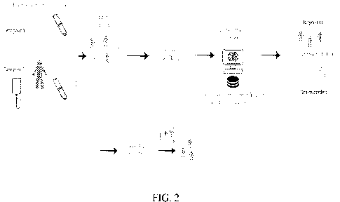
[0072] Figure 1 is a flowchart of the functional steps in a method for
training a machine
learning model to predict patient response to therapy, according to some
embodiments
of the present disclosure. Figure 2 is a schematic illustration of the process
steps of Figure
1.
[0073] In some embodiments, at step 100, a plurality of biological samples may
be
received from a cohort of subjects, e.g., a predefined population of patients
having a
specified type of disease. In some embodiments, a cohort assembled for the
purposes of
the present disclosure may comprise a plurality of patients having the same
and/or a
similar and/or an associated disease and/or category of diseases and/or
syndromes and/or
conditions, and/or associated diseases, syndromes and/or conditions. In some
embodiments, with respect to at least some of the patients in the cohort, the
specified
disease and/or conditions may be at different stages and/or be combined with
co-
morbidities and/or diseases. In some embodiments, a specified disease of the
present
disclosure may be expressed in terms of broad categories (e g , cancer'), sub-
types (e.g.,
melanoma), and/or sub-categories (e.g., a specified type of melanoma).
[0074] In some embodiments, the disease is a proliferative disorder. In some
embodiments, the disease is a disease characterized, by increased
proliferation, decreased
apoptosis, or both. In some embodiments, the disease is cancer. In some
embodiments,
the cancer is a solid cancer. In some embodiments, the cancer is a
hematopoietic cancer.
Types of cancer are well known in the art, and examples of classes of cancer
include, but
are not limited to a sarcoma, a melanoma, a blastoma, a carcinoma, a leukemia
and a
lymphoma. Types of cancer can also be classified by the tissue/cell type of
origin and
include for example, brain cancer, blood cancer, bone cancer, fat cancer,
retinoblastoma,
head and neck cancer, tongue cancer, nasopharyngeal cancer, pharyngeal cancer,
throat
cancer, esophageal cancer, stomach cancer, gastrointestinal cancer, intestinal
cancer,
lung cancer, colon cancer, colorectal cancer, liver cancer, pancreatic cancer,
gallbladder
CA 03166539 2022- 7- 29
WO 2021/156875
PCT/IL2021/050147
cancer, penile cancer, thymus cancer, thyroid cancer, urogenital cancer,
prostate cancer,
kidney cancer, ovarian cancer, cervical cancer, testicular cancer, skin
cancer,
glioblastoma multiforme (GBM), and uterine cancer. In some embodiments, the
cancer
is skin cancer. In some embodiments, the cancer is lung cancer. In some
embodiments,
the cancer is melanoma. In some embodiments, the cancer is small cell lung
cancer
(SCLC) or non-small cell lung cancer (NSCLC). In some embodiments, the cancer
is
urogenital cancer. In some embodiments, the cancer is head and neck cancer. In
some
embodiments, the cancer is a solid tumor. In some embodiments, the cancer is a
cancer
treatable by immunotherapy.
[0075] In some embodiments, the disease is an autoimmune disease. In some
embodiments, the autoimmune disease is psoriasis, In some embodiments, the
disease is
a genetic disease. In some embodiments, the disease is an infectious disease.
In some
embodiments, the disease is a bacterial, viral or fungal infection. In some
embodiments,
the disease is an inflammatory disease. In some embodiments, the disease is a
respiratory
disease. In some embodiments, the disease is degenerative disease. In some
embodiments, the disease is a neurodegenerative disease. In some embodiments,
the
disease is a metabolic disease. In some embodiments, the disease is a
cardiovascular
disease. In some embodiments, the disease is a skeletal disease.
[0076] In some embodiments, biological samples may include any type of
biological
sample obtained from an individual, including body tissues, body fluids, body
excretions,
exhaled breath, or other sources. In some embodiments, the biological sample
is a tumor.
In some embodiments, the biological sample is a non-tumorigenic sample. Body
fluids
may be whole blood, blood plasma, blood serum, peripheral blood mononuclear
cells
(PBMCs), lymph, urine, saliva, semen, synovial fluid and spinal fluid, fresh
or frozen. In
certain embodiments of the method according to the invention, the biological
sample(s)
is blood plasma, whole blood, blood serum, cerebrospinal fluid (CSF), or
PBMCs. In
specific embodiments, the biological sample(s) is blood plasma. In alternative
specific
embodiments, the biological sample(s) is CSF. In some embodiments, the
biological
sample(s) is PBMCs sample. In some embodiments, the biological sample(s) is a
blood
sample.
[0077] In some embodiments, a cohort of the present disclosure comprises a
group of
subjects with similar phenotype and receiving a similar treatment. However,
the cohort
definition may vary according to the classification per cohort and biological
common
16
CA 03166539 2022- 7- 29
WO 2021/156875
PCT/IL2021/050147
denominator of the participating subjects. In some embodiments, a cohort of
the present
disclosure may comprise patients of, e.g., different demographics (e.g., sex,
age,
ethnicity), clinical measurements, disease stage, disease history, disease
treatment
history, general medical history (e.g., including smoking history and drinking
habits,
background diseases) genetic information, physical parameters, and the like.
[0078] In some embodiments, patients in the cohort may undergo and/or receive
different
types of treatments, e.g., mono therapy, combined therapy, multi-stage or
multi-session
therapy, and/or multi -modality therapy.
[0079] In some embodiments, the biological samples may be obtained from each
subject
in the cohort, or from at least some of the subjects, at specified times
before, during,
and/or after the conclusion of, the course of therapy. In some embodiments,
the biological
samples may be obtained from each subject, or from at least some of the
subjects, at
specified one or more stages and/or points and/or steps before, during, and/or
after the
conclusion of, the course of therapy, e.g., pre-therapy, on-therapy, and/or
post-therapy.
[0080] In some embodiments, with respect to at least some of the subjects, at
least a pair
of corresponding To and Ti biological samples may be collected at two or more
different
points during the course of the treatment, e.g.:
= (i) pre-therapy, i.e., before the start of the course of therapy. and
(ii) post-therapy,
i.e., after the conclusion of the entire course of therapy;
= (i) pre-therapy, i.e., before the start of the course of therapy, and
(ii) on-therapy,
i.e., at a specified point in time during the course of therapy;
= in the case of a multi-stage or multi-session treatment, (i) pre-therapy,
i.e., before
the start of the course of therapy, and (ii) after the conclusion of a
specified stage
and/or session of the multi-stage or multi-session therapy; and/or
= in the case of a multi-modality therapy, (i) pre-therapy, i.e., before
the start of the
course of therapy, and (ii) at a specified point and/or stage associated with
one of
the multiple treatment modalities.
= in the case of a multi-modality therapy, (i) pre-therapy, i.e., before
the start of the
course of therapy for each treatment modality, and (ii) at a specified point
and/or
stage associated with one of the multiple treatment modalities.
17
CA 03166539 2022- 7- 29
WO 2021/156875
PCT/IL2021/050147
[0081] In some embodiments, at step 102, each of, or at least some of, the
biological
samples may be analyzed to identify a plurality of biomarkers and/or to
extract a
biological signature. In some embodiments, the analysis obtains, e.g., a
proteomic profile
comprising protein expression for each of the samples. In some embodiments,
the protein
expressions so obtained may identify the proteins in each analyzed biological
sample. In
some embodiments. additional and/or other analyses may be performed with
respect to
the biological samples, to obtain, e.g., one or more profiles selected from:
DNA profile;
RNA profile; circulating DNA profile, single cell RNA sequencing;
metabolomics;
microbiome; transcriptome; genomics; epigenomics; cell profiling; single-cell
based
analysis; and MicroRNA. In some embodiments, the circulating DNA profile is
circulating tumor DNA profile. In some embodiments, the circulating DNA
profile is
methylated circulating DNA profile.
[0082] In certain embodiments, the biological signature is selected from: a
proteome
profile; a DNA profile; an RNA profile; a metabolomics profile (e.g.,
glycomics,
lipidomics); a microbiome profile; a genomics profile; an epigenomics profile;
a cellular
profile; a post-translational modification-based profile; a single-cell based
analysis; and
a regulatory RNA profile. In some embodiments, expression is protein
expression. In
some embodiments, expression is RNA expression. In some embodiments, the RNA
is
mRNA. In some embodiments, the RNA is regulatory RNA. In some embodiments, the
regulatory RNA is microRNA. In some embodiments, the regulatory RNA is a long
non-
coding RNA. In some embodiments, the metabolomics profile is lipids profile.
In some
embodiments, the metabolomics profile is nucleic acids profile. In some
embodiments,
the metabolomics profile is sugars profile. In some embodiments, the
metabolomics
profile is vitamins profile. In some embodiments, the metabolomics profile is
fatty acids
profile. In some embodiments, the metabolomics profile is amino acids profile.
In some
embodiments, the metabolomics profile is phenolic compounds profiles. In some
embodiments, the metabolomics profile is alkaloids profiles. In some
embodiments, the
protein expression is metabolic protein expression. In some embodiments, the
protein
expression is membranal protein expression. In some embodiments, the protein
expression is secreted protein expression. In some embodiments, the protein
expression
is cellular protein expression. In some embodiments, the biological signature
is a profile
of the genome. In some embodiments, the biological signature is a mutational
profile of
the genome. In some embodiments, the biological signature is an epigenetic
profile of
18
CA 03166539 2022- 7- 29
WO 2021/156875
PCT/IL2021/050147
the genome. In some embodiments, the biological signature is a methylome
profile. In
some embodiments, the epigenetic profile is a profile of post-translational
modifications
(PTMs). In some embodiments, the biological signature is PTM profile on
proteins.
PTMs are well known in the art and include, but are not limited to,
methylation,
acetylation, phosphorylation, glycosylation, sumoylation, and ubiquitination.
In some
embodiments, the biological signature is a circulating DNA profile. In some
embodiments, the biological signature is circulating tumor-DNA profile. In
some
embodiments, the biological signature is methylated circulating tumor DNA
profile. In
some embodiments, the biological signature is amount of circulating tumor DNA
profile.
In some embodiments, the biological signature is genotyping of mutations in
circulating
tumor DNA profile. In some embodiments, the biological signature is an
organismal
profile. In some embodiments, the biological signature is a microbiome
profile. In some
embodiments, the biological signature is extracellular vesicles profile
(either number or
content). In some embodiments, the biological signature is microparticles
profile (either
number or content). In some embodiments, the biological signature is exosomes
profile
(either number or content). In some embodiments, the biological signature is
circulating
cells profile. in some embodiments, the biological signature is circulating
tumor cells
profile. in some embodiments, the biological signature is circulating immune
cells
profile. As used herein, the term "profile" is intended to encompass any
variation of the
determined entity including the presence or absence, as well as the type
(e.g., genotype),
amount, percentage or difference in expression, as long as it is suitable for
prediction of
the response to therapy.
[0083] Methods of performing expression profiling are well known in the art.
RNA
expression can be assayed by any known method including, polymerase chain
reaction
(PCR), real-time PCR, quantitative PCR, digital PCR, microarray, northern
blotting, and
sequencing. In some embodiments, the expression profiling comprises PCR. In
some
embodiments, the expression profiling comprises hybridization to a microarray.
In some
embodiments, the expression profiling comprises sequencing. In some
embodiments, the
sequencing is next-generation sequencing. In some embodiments, the sequencing
is deep
sequencing. In some embodiments, the sequencing is massively parallel
sequencing.
Methods of sequencing are well known in the art, and apparatuses for
sequencing are
commercially available. Any known method of sequencing may be used in
accordance
with the method of the invention.
19
CA 03166539 2022- 7- 29
WO 2021/156875
PCT/IL2021/050147
[0084] Protein expression can be assayed by any known method including, an
immunoassay, immunoblotting, immunohistochemistry, FACS, ELISA, Western
blotting, proteomics arrays, proteome sequencing, proximity-extension assay
(PEA)-
based assay, aptamer-based assays, multiplex as say and mass spectrometry. In
some
embodiments, the expression profiling comprises hybridizing to a proteomics
array. In
some embodiments, the proteomics array is an antibody array. In some
embodiments, the
expression profiling comprises whole proteome sequencing. In some embodiments,
the
expression profiling comprises targeted mass spectrometry. In some
embodiments, the
expression profiling comprises untargeted mass spectrometry. In some
embodiments, the
expression profiling comprises shotgun proteomics using mass spectrometry. In
some
embodiments, the expression profiling comprises top-down mass spectrometry. In
some
embodiments, the expression profiling comprises bottom-up mass spectrometry.
In some
embodiments, the expression profiling comprises data-independent acquisition
(DIA)
mass spectrometry. In some embodiments, the expression profiling comprises
data-
dependent acquisition (DDA) mass spectrometry. Proteome/proteomics arrays are
well
known in the art and are commercially available. Examples of proteomics arrays
include,
but are not limited to, the Proteome Profiler Array of R&D Systems, the CP
Human
Proteome array of Creative Proteomics, RPPA (reverse phase protein array), the
human
Kiloplex Quantitative Proteomics array of RayBiotech, Olink Target 96, Olink
Explore
96 and the Membrane Proteome Array of Integral Molecular.
[0085] In some embodiments, at step 104, a preprocessing stage may take place,
comprising at least one of data cleaning and normalizing, data quality
control, and/or any
other suitable preprocessing method or technique.
[0086] Biological data derived from clinical samples may suffer from
variations that can
arise due to different sample collection or sample preparation procedures, due
to
quantification inaccuracies, due to batch effects, and/or due to any other
technical bias
that may lead to mistakes in the analysis. Therefore, in some embodiments,
preprocessing
may comprise a quality control step wherein at least some biological
signatures may be
removed based, at least in part, on a measurability-parameters of proteins
expressed in
the biological signature.
[0087] In some embodiments, quality control and/or data cleaning and/or data
normalization may comprise any one or more of the following:
CA 03166539 2022- 7- 29
WO 2021/156875
PCT/IL2021/050147
= Data transformations: For example, a log2 transformation, Z- score
transformation, median subtraction.
= Statistical tests: key statistical measures, such as median, average, the
first
quartile of the dataset (Q1), the third quartile of the dataset (Q3),
variance,
standard deviation or coefficient of variation (cv), are calculated in order
to assess
the data quality.
= Data visualization: Enables a better understanding of the data, whether
the data
are normally distributed, or whether there are any technical biases, batch
effects
or any outliers that behave substantially different from the rest of the
samples.
= Evaluation of data quality: Includes a step of defining which data should
be
included/removed/normalized in the analysis, thereby generating a new output
containing only the desired and normalized results.
= Handling quality control data issues: In specific cases, mostly due to
technical
biases, extremely different samples are considered for exclusion. In case of
batch
effects due to technical reasons, batch effect removal algorithms and/or data
normalization can be applied.
= Batch effect removal: Can be done in different ways. Non-limiting
examples are:
using batch effect removal algorithms (e.g., Emma); subtracting component/s in
principal component analysis (PCA); median subtraction; Z-scoring; running the
same reference samples in different batches ("bridging samples") and
correcting
based on their values.
= Handling data below limit of detection (LOD): The approach for dealing
with
values below the LOD level can be done by data imputation: As a non-limiting
example, To or Ti values that are below LOD can be assigned the LOD level of
the examined protein. In case both time points are imputed, the Ti/To ratio
equals
to 1, and after 1og2 transformation it equals to 0; in some data analyses, it
can be
assigned as 'not a number' (NaN) value instead. Other approaches for data
imputation can be also used. Figure 3 is a schematic illustration of a quality
control process which may be used to assess measurements below a limit of
detection (LOD) threshold and/or above a maximum threshold.
21
CA 03166539 2022- 7- 29
WO 2021/156875
PCT/IL2021/050147
= Missing values or 0 values handling: Proteins which have missing (NaN),
below
LOD value or 0 values in less than 0-100% of the samples are filtered.
Alternatively, missing (NaN), below LOD value or 0 values can be imputed by
any other imputation method. Following any data imputation, some QC steps may
be repeated.
[0088] Data normalization: If needed, the data are normalized prior to the
bioinformatic
analysis. Data normalization can be performed at any level, e.g., protein-
level, batch
level, etc.
[0089] In some embodiments, at step 106, differential expression values may be
calculated with respect to each pair of biological signatures. In some
embodiments, with
respect to the case of biological signatures that arc protein expression
profiles, the present
disclosure provides for calculating the relations (e.g., a level of difference
in expression
values between each biological signature in a pair of signatures associated
with a subject,
e.g., a difference in, and/or a ratio of, expression values between biological
signatures at
at-least two time points relative to the therapy, e.g., a Ti/To ratio. In some
embodiments,
this analysis does not take into account any biological function of the
proteins and/or any
known interactions between the proteins. In some embodiments, Ti/To ratio is a
numerical value determined by calculating the ratio of on-treatment and
baseline values
(pre-treatment). The Ti/To ratio may be used to predict responsiveness or non-
responsiveness of the patient to the cancer treatment.
[0090] In some embodiments, additional, other, and/or alternative sets of
values may be
calculated, associated with, e.g., biological processes, clinical data, and/or
protein-
interaction driven analysis between Ti/To signatures
[0091] In some embodiments, at step 108, one or more feature selection,
feature
extraction, an ensemble process, and/or dimensionality reduction steps may be
performed
with respect to the value sets.
[0092] In some embodiments, feature selection and/or dimensionality reduction
steps
may be performed, to reduce the number of variables in each sample pair and/or
to obtain
a set of principal variables, e.g., those variables that may have significant
predictive
power such as protein expression levels. Accordingly, in some embodiments, a
feature
selection and/or dimensionality reduction step may result in a reduction of
the number of
proteins in each biological signature and/or set of values. In some
embodiments,
22
CA 03166539 2022- 7- 29
WO 2021/156875
PCT/IL2021/050147
dimensionality reduction selects principal variables, e.g., proteins, based on
the level of
response predictive power a protein generates with respect to the desired
prediction. In
some embodiments, the dimensionality reduction generates a new feature or
features that
can be predictive for response. In specific embodiments, the dimensionality
reduction
involves regarding all or some feature values as vector components and
calculating its
norm.
[0093] In some embodiments, any suitable feature selection and/or
dimensionality
reduction method or technique may be employed, such as, but not limited to:
= ANOVA with So parameter: Analysis of variance with an additional
parameter
(So) that controls for the relative importance of features based on resulted
test p-
values and difference between the group means (see, e.g., Tusher, Tibshirani
and
Chu, PNAS 98, pp5116-21, 2001).
= Scalable EMpirical Bayes Model Selection (SEMMS): An empirical Bayes
feature selection method which applies a parsimonious mixture model to
identify
significant predictors (see, e.g., Bar, Booth, and Wells. A scalable empirical
Bayes approach to variable selection in generalized linear models, 2019).
= L2N: A method for differential expression analysis that uses a three-
component
mixture model. The model consists of two log-normal components (L2) for
differentially expressed features, one component for under-expressed features
and the other for overexpressed features, and a single normal component (N)
for
non-differentially expressed features (see, e.g., Bar and Schifano.
Differential
variation and expression analysis. Stat 8, e237, doi:10.1002/sta4.237, 2019).
= Genetic algorithms: A family of heuristic optimization algorithms that
employ
organic evolutionary techniques such as random mutations, recombination, and
natural selection as methods for achieving optimal configurations (see, e.g.,
Popovic, Sifrim, Pavlopoulos, Moreau, and Bart De Moor. A Simple Genetic
Algorithm for Biomarker Mining. 2012).
= Naïve classifier: The naïve classifier evaluates a response score by
reducing the
dimension to a single score. This is performed by regarding all features
(e.g.,
specific profiles such as protein expression levels) as component of a vector
and
calculating its norm. The dimension reduction reduces the possible risk of an
over-fitting. In some embodiments, the vector components are normalized
23
CA 03166539 2022- 7- 29
WO 2021/156875
PCT/IL2021/050147
according to the typical component value among patients that belong to the
same
response group (e.g., responders), such that the normalized norm quantifies
the
amount of deviation from the typical respective class value. In additional
embodiments, the naive classifier enables training using data of subjects that
belong only to part of the response groups.
[0094] In some embodiments, at step 110, a training dataset for training a
machine
learning model of the present disclosure may be constructed, comprising sets
of values
representing relations (e.g., ratios or difference in expression values) of
the biological
signatures at multiple time points relative to the therapy, with respect to at
least some of
the subjects in the cohort.
[0095] In some embodiments, a training dataset of the present disclosure may
comprise
additional information for training of the machine learning model, such as
clinical,
demographic, and/or physical information with respect to at least some of the
subject in
the cohort. For example, in some embodiments, such data may include
characteristics
obtained from the diseased tissue itself (e.g., from a tumor of a cancer
patient). In some
embodiments, such data may include, but is not limited to: demographic
information (ex,
age, ethnicity); performance status; hematological and chemistry measurements;
cancer
disease history, e.g., date of cancer diagnosis, primary cancer type and
stage, disease
biomarkers (e.g. PD-L1), disease treatment history, histology, TNM stage,
assessment of
measurable lesions, time of tumor progression, site of recurrence, proposed
treatment;
general medical history, including smoking history and drinking habits,
background
diseases including hypertension, diabetes, ischemic heart disease, renal
insufficiency,
chronic obstructive pulmonary disease, asthma, liver insufficiency,
Inflammatory Bowel
Disease, autoimmune diseases, endocrine diseases, and others; family medical
history;
genetic information, e.g. mutations, gene amplifications, and others (e.g.
EGFR, BRAF,
HER2, KRAS, MAP2K1, MET, NRAS, NTRK1, PIK3CA, RET, ROS1, TP53, ALK,
MYC, NOTCH, PTEN, RBI, CDKN2A, KIT, NF1); physical parameters, e.g.,
temperature, pulse, height, weight, BMI, blood pressure, complete blood count
including
all examined parameters, liver function, renal function, electrolytes;
medication
(prescribed and non-prescribed); relative lymphocyte count; neutrophil to
lymphocyte
ratio; baseline protein levels in the plasma (e.g. LDH); and/or marker
staining (e.g. PD-
Li in the tumor or in circulating tumor cells). In some embodiments, a change
in response
24
CA 03166539 2022- 7- 29
WO 2021/156875
PCT/IL2021/050147
to the specified therapy in one or more of the above information may be
analyzed and
provided for training of the machine learning model.
[0096] In some embodiments, one or more annotation schemes may be employed
with
respect to the training dataset. Accordingly, in some embodiments, a training
dataset for
a machine learning model of the present disclosure may comprise a plurality of
sets of
TI/To ratios or difference in expression values with respect to at least some
of the subjects
in the cohort, wherein at least some of these sets of values may be annotated
with
category labels denoting a response and/or outcome of the treatment in the
respective
subject. In some embodiments, such annotation may be binary, e.g.,
positive/negative,
and/or expressed in discrete categories, e.g., on a scale of 1-5. In some
embodiments, a
binary value category label may be expressed, e.g., as yes/no, "responsive/non-
responsive,' or 'favorable/non-favorable response.' In some embodiments,
discrete
category labels and/or annotations may be expressed on a scale, e.g.,
'complete
response, "partial response,' 'stable disease,' 'progressive disease,'
pseudo-
progression,' and 'hyper-progression disease.' In some embodiments, additional
and/or
other scales and/or thresholds and/or response criteria may be used, e.g., a
gradual scale
of 1 (non-responsive) to 5 (responsive). In some embodiments, category labels
may be
associated with adverse or any other secondary effects or response by a
patient, e.g.,
therapy side-effects.
[0097] In some embodiments, additional and/or other annotation schemes may be
employed. In some embodiments, the training dataset may be annotated with,
e.g., patient
demographic and/or clinical data as detailed above. In some embodiments, the
training
dataset may be annotated with overall response rate. In some embodiments, the
training
dataset may be annotated with progression-free survival rate. In some
embodiments, the
training dataset may be annotated with duration of response rate.
[0098] In some embodiments, at step 112, a machine learning model may be
trained on
the training dataset constructed in step 110. In some embodiments, any
suitable machine
learning algorithm or combination of methods may be employed, including, but
not
limited to:
= Support Vector Machine (SVM): A nonparametric model which finds the
optimal
separating hyperplane that discriminate between different classes. It can
perform
linear or non-linear classification.
CA 03166539 2022- 7- 29
WO 2021/156875
PCT/IL2021/050147
= Penalized Logistic Regression (PLR) - a logistic model for regression
that
imposes a penalty to reduce the impact of certain features.
= Generalized linear model (GLM): a generalization of linear regression
that unifies
statistical models such as linear regression, logistic regression and Poisson
regression. GLM extends linear regression by (1) supporting response variables
with error distributions other than the normal distribution (2) a non-linear
relationship between the predictors and the response variable.
= Random forest (RF): involves in the generation of multiple decision trees
that
consist sequences of decision rules for protein expression values. To avoid
over-
fitting, these trees may be pruned. Each tree is constructed by randomly
selecting
different samples.
= eXtreme Gradient Boosting (XGB): a gradient boosted decision trees-based
classification and regression algorithm. The decision trees are built one at a
time,
and each new tree corrects the error of the previously trained decision tree.
[0099] In other embodiments, machine learning model may be trained based on
statistical measures, i.e., variance, median, mean, average and the same.
[0100] In some embodiments, at step 114, the machine learning model results
with a
classifier a target set of Ti/To relations (e.g., ratios or difference in
expression values)
suitable for predicting a response in a target patient and receiving a similar
treatment as
the patient cohort.
[0101] In some embodiments, at an inference step 114, a trained machine
learning model
of the present disclosure may be applied to target data, e.g., a target set of
Ti /To relations
(e.g., ratios or difference in expression values) with respect to a target
patient with similar
phenotype and receiving a similar treatment as the patient cohort. In some
embodiments,
the inference of the trained machine learning model on the target data
produces a therapy
response prediction or response probability.
[0102] In some embodiments, the prediction is for side-effect or adverse
event. In some
embodiments, the prediction is for overall survival rate. In some embodiments,
the
prediction is for progression-free survival rate. In some embodiments, the
prediction is
duration of response rate. In some embodiments, the prediction is for pseudo-
progression. In some embodiments, the prediction is for hyper-progression. In
some
26
CA 03166539 2022- 7- 29
WO 2021/156875
PCT/IL2021/050147
embodiments, the prediction is for progression of the disease. In some
embodiments, a
prediction according to the present disclosure may be further supported and/or
supplemented with a Differentially Expressed Protein Identification and/or
Differentiating Biological Processes analyses, as further detailed herein
below.
[0103] In some embodiments, at step 116, a therapy course with respect to the
target
patient may be administered, adjusted, and/or modified based, at least in
part, on the
inference step 114. In some embodiments, such therapy adjustment may include
prescribing a subsequent and/or supplementary therapy for the target patient.
Differentially Expressed Protein Identification
[0104] In enrichment analyses, including enrichment analyses, some network-
based
analyses when focusing on a subset of the features, or for providing
personalized
potential targets for therapeutic intervention, as defined below, one needs to
first identify
the DEPs between the examined groups (e.g., responders vs. non-responders).
[0105] The term "Differentially Expressed Proteins" (DEPs) refers to proteins
whose
distribution (of expression level or change between two timepoints, e.g., To
and Ti)
differs between responders and non-responders (and possibly other groups,
e.g., stable
disease patients), including any difference in distribution detectable by
numerical
measures (e.g., t-test. ANOVA, Kolmogorov-Smirnov test). In some cases, DEPs
are
defined as proteins who's median, mean, variance or other statistical measure
differ
between responders and non-responders. In some cases, DEPs are defined as
proteins
whose distribution differs between responders and non-responders without
alteration of
the mean or the median, however, the difference can be evaluated by
statistical tests. In
some cases, DEPs are defined as proteins that in at least one patient, do not
follow the
respective protein distribution among a specific subgroup (i.e., responders or
non-
responders).
[0106] Identifying the DEPs is an optional step, as some tools do not require
a list of
DEPs, but rather rely on a selected measure that is calculated for all the
proteins in the
proteomic profile, such as fold change between the two examined groups (i.e.
responders
and non-responders), or the p-value of the t-test.
[0107] The terms "protein profile", "protein expression profile" and
"proteomic profile",
used herein interchangeably, refer to the expression level of a protein or a
list of proteins,
27
CA 03166539 2022- 7- 29
WO 2021/156875
PCT/IL2021/050147
such as cytokines, growth factors and other proteins expressed in plasma, CSF
or other
body fluids, or tissues, at a certain time point. The number of proteins
measured may
vary between 1 and 20,000. A protein profile may be used to diagnose a
disease,
condition, or syndrome and to determine the odds of treatment response. In
some
embodiments, the profile comprises at least 1, 2, 3, 4, 5, 6, 7, 8, 9, 10, 15,
20, 25, 30, 35,
40, 45, 50, 60, 70, 80, 90, 100, 500, 1000, 2000, 5000, 7000 or at least 20000
proteins.
Each possibility represents a separate embodiment of the invention. In some
embodiments, the protein profile is absolute protein expression. In other
embodiments,
the protein expression profile is normalized or relative expression profile.
[0108] Differentiating Biological Processes
[0109] Differentiating Biological Processes can be deciphered by using DEPs as
an
input. Another method is by running per sample pathway enrichment and then
aggregating the enrichment results.
[0110] The term "Differentiating Biological Processes" (DBP) refers to
biological
processes that occur in either the responding patients or the non-responding
patients.
[0111] Differentiating biological processes may be provided based on different
databases, such as the KEGG pathways analysis (https://www.genome.jp/keggi)
and
gene ontology (GO) analysis (geneontology.org).
[0112] Pathway Enrichment Analysis
[0113] Proteomic analysis at the pathway level may imply biological processes
that are
affected by the treatment. The aim of this step is to translate host-response
related
changes at the protein expression level to changes at the biological process
level. Thus,
the DEPs identified can be used as an input for the pathway enrichment
analysis.
[0114] Pathway enrichment analysis translates the proteomic changes into a
list of
estimated biological processes which were either down- or up-regulated in the
body in
response to the cancer therapy treatment. The analysis can identify DBPs,
which are
biological processes enriched in each group. The output of the enrichment
analysis is a
list of DBPs and the proteins that are involved in each one of them.
[0115] Network analysis
[0116] An additional level of analysis that is based on the proteomics data
involves
studying the co-changes in the expression levels of several proteins together;
proteins
28
CA 03166539 2022- 7- 29
WO 2021/156875
PCT/IL2021/050147
that display a correlation (either a negative or a positive correlation)
between their
proteomics profiles may indicate a potentially interesting biological
relation.
Correlations between proteins may occur due to various reasons, such as a
common
regulator that affects both proteins (e.g., a transcription factor, a
phosphatase, etc.) or that
one of the proteins is a regulator of the other. Based on the correlations
between the
expression levels of different proteins one can construct protein networks.
Protein
networks may differ between two different conditions (e.g., responders and non-
responders) and therefore studying these differential networks (Figure 14) may
decipher
biological mechanisms underlying the different phenotypes (e.g., resistance to
therapy).
An example for such differential network is displayed in Figure 15.
[0117] Examination of the differential network between responders and non-
responders
may reveal novel mechanisms that are associated with resistance to therapy and
may be
used for classifier training. Network analysis can be helpful for feature
engineering. For
instance, such analysis can aid in pinpointing features that are changing
together; an
engineered feature that captures this relation may be predictive by using any
mathematical relation between two or more proteins. Figure 16 demonstrates a
non-
limiting example of a predictor based on correlations between protein pairs.
Two proteins
showing differentially correlated fold-change values between responders and
non-
responders were inspected. Figure 16A - Correlation between responders
(triangle) and
non-responders (circles) was positive (R = 0.37). The dashed line shows a
linear fit to
the responder values. Figure 16B - The residuals (i.e., distances of each
point from the
linear fit in Figure 16A) of two protein pairs was calculated and used as an
input for
SVM-based classifier. The resulting predictor achieved a ROCAUC of 0.765. In
addition, network analysis can potentially identify novel approaches for
intervention,
which could be highly valuable for pharmaceutical companies.
Personalized potential targets for intervention
[0118] In another aspect, the invention provides an analysis of personalized
potential
targets for intervention, using a 3-filter approach. The three-filter approach
can be
visualized in a decision-tree like flowchart (Figure 8) that starts with the
first filter, which
makes use of the cohort strength, keeping only the DEPs. A protein that is not
a DEP
does not continue to the next filter and is considered a non-actionable
protein. The DEPs
used for this analysis may be similar or may differ from the DEPs provided for
training
the machine learning model. The second filter focuses on the patient's
specific proteomic
29
CA 03166539 2022- 7- 29
WO 2021/156875
PCT/IL2021/050147
profile of the DEPs, keeping only the patient's resistance associated proteins
(RAPs)
based on a RAP score (described in detail below). A DEP that had a RAP score
below
the threshold (e.g., RAP score of 1) is considered not relevant to the
specific patient and
is filtered out. Next, the third filter for associating a RAP to a drug or
investigational new
drug, is applied. The third filter may include two options; if the RAP has an
approved
drug (e.g., specific for the indication of interest, or a different
indication), then the RAP
is considered an actionable protein. Alternatively, if there is a clinical
trial associating
the RAP and a candidate drug (e.g., for the indication of interest), the RAP
may also be
considered as an actionable protein.
[0119] The analysis provided herein may further include a simulation step
wherein the
RAP (or multiple RAPs) expression value of the patient is modified towards a
balanced
value that may follow the therapeutic intervention. Next, another prediction
analysis is
performed, aiming to assess the effect of the change in the protein expression
value. This
may help the physician in deciding which RAP to select for the patient
(multiple RAPs
that are potential targets for intervention may be received for a selected
patient). Figure
12 demonstrates a non-limiting simulation of a single RAP perturbation
(although
simulation of several RAPs perturbation may be also performed). A predictive
signature
that is based on the list of RAPs of the entire cohort is first generated
(Figure 12A). For a
given patient, a specific RAP, or combination of RAPs, is balanced and the
baseline
response probability is then compared to the perturbed response probability
(Figure 12B).
[0120] Cohort-based statistical filter
[0121] It is first essential to identify DEPs. As described herein, DEPs are
proteins whose
levels change between responders and non-responders and may also change
between To
and Ti in responders and non-responders. In some cases, DEPs are proteins
whose
median differ between responders and non-responders. In some cases, DEPs are
proteins
whose variance differ between responders and non-responders. In other cases,
DEPs are
proteins whose average differ between responders and non-responders. This
analysis
narrows down the number of proteins, yielding a list of proteins that cohort-
wise display
differences between the two or more classes, and thus potentially play a role
in the
resistance or the response to treatment. The DEPs may be identical or may be
independent of the DEPs identified for the machine learning aspects of the
invention.
[0122] Personalized filter
CA 03166539 2022- 7- 29
WO 2021/156875
PCT/IL2021/050147
[0123] The list of proteins from the first filter (the cohort-based
statistical filter) is
examined in a patient-specific manner using the personalized filter and the
resistance-
associated protein (RAP) score.
[0124] RAPs are defined per patient. In some embodiments, a patient's RAP is a
protein
whose levels or fold-change is deviant from the respective protein
distribution in one of
the response group. The deviation may be quantified by numerical means, either
by using
the levels in multiple response group (e.g., both responders and non-
responders), or the
distribution among a specific response group (e.g., responders or non-
responders or
stable disease patients). For a non-limiting example, the expression
distribution of each
DEP in the entire cohort can be examined per response class (meaning- a
distribution for
responders and a distribution for non-responders are generated separately). In
this case,
a probability density distribution can be extracted for each group
distribution, with a total
area under the curve of 1. For each patient, the area (or other measures, such
as the height
in each of the response group distributions) of the tail above or below the
DEP expression
level (e.g., selecting above or below may depend on the order of the non-
responder and
responder medians, as described in detail below) is calculated for each
distribution
(Figure). Next, the RAP score may be calculated per DEP for each patient, such
as based
on the non-limiting Equation 1:
P(NR)
[0125] RAP score = log2 P(R) (Equation 1)
[0126] where P(NR) is the probability that the protein can be attributed to
the non-
responder distribution and P(R) is the probability that the protein can be
attributed to the
responder distribution. The RAP in this example is a probabilistic measure for
the
distance of the DEP expression level from responder group distribution. A high
RAP
score designates a protein with a high probability of being associated with
the non-
responder distribution rather than to the responder distribution.
P(NR)
[0127] The RAP score may be expressed as a simplification of the 1092- P(R)
measure,
whereby the RAP score may be presented in a five-bin format using 5 degrees
that
represent the fold increase in the odds. A score of 5 or higher may be grouped
to a score
designated as 5+.
[0128] For each patient, the DEPs following the first filter are examined, and
the RAP
score is calculated per each DEP. Optionally, the threshold for passing the
personalized
31
CA 03166539 2022- 7- 29
WO 2021/156875
PCT/IL2021/050147
filter can be defined (e.g., a RAP score above 1.0). This list of the
patient's RAPs
continues to the next filter.
[0129] Directionality calculation
[0130] A RAP score above the defined threshold may indicate that the protein
can be
attributed to the non-responder distribution (note that respectively, a
reverse high RAP
score can attribute the RAP to the responder distribution). In a case where
there is a
difference in medians between responders and non-responders, the selection of
the
direction of the tail of the area under the curve can be based on the order of
the
distribution medians. If the median of the non-responder group is higher than
the median
of the responder group, the area selected for the calculation is the tail to
the right of the
DEP expression level of the patient (Figure 10A). If the median of the non-
responder
group is lower than the median of the responder group, the areas under the
left tail of the
distributions are used for the DEP expression level of the patient (Figure
10B). This way,
proteins that are more likely to be attributed to the non-responder
distribution have values
above 1.0 (log2 scale), regardless of the DEP direction in the cohort (higher
in responder
or higher in non-responder). DEPs that are more likely to be attributed to the
responder
group have RAP scores between 0 and 1 (10g2 scale). In cases where medians are
not
differential but other aspects of the distribution change, i.e., variance or
average, a
relative probability at a given range (not necessarily the tail) may also be
used.
[0131] The effect of the DEP statistics on the distribution of the RAP scores
[0132] Since the RAP score is based on the responder and non-responder
distributions
of the entire cohort, the difference between the responder and non-responder
distributions
affects the RAP score distribution (Figures 11A-11D). A DEP with a large
difference
between responders and non-responders displays smaller gaps between RAP
scores,
indicating that patients are more likely to have high RAP scores (Figure 11A
and Figure
11B) compared to a DEP with a smaller difference between responders and non-
responders, which displays larger gaps between RAP scores, indicating that
patients are
less likely to have high RAP scores (Figure 11C).
[0133] RAP-based clusters
[0134] The RAP score can be further used to identify groups of non-responders
that share
similar RAP profiles. For this analysis different clustering algorithms can be
used. A
non-limiting example is the use of consensus clustering algorithm, which finds
the most
32
CA 03166539 2022- 7- 29
WO 2021/156875
PCT/IL2021/050147
robust clusters of samples following multiple iterations of clustering, can be
used. An
example for that is displayed in figure 11D. The different clusters can then
be further
characterized to examine enrichment of various clinical parameters. For
example, cluster
#5 was enriched with patients who recently quit smoking (Fisher exact test p-
value =
0.027).
[0135] Clinical filter
[0136] Following the first two filters described above, a list of personal
RAPs is
generated for the patient. In the current filter (Figures 12A-B), potential
drugs or
investigational new drugs (INDs) that target the patient's RAPs are
identified. The
clinical filter may include the following steps. First, drugs/INDs that target
each RAP are
searched in a suitable database and are associated with the RAP. Next, various
clinical
filters may be applied following data collection and analysis; this may
include biological
reasoning-based examination layer, mode of action related layers (such as
direct/indirect;
specific/non-specific; directionality match) and drug clinical relevance
related layers
(such as drug development status/clinical relevance). RAPs associated with
drugs/INDs
based on the applied filters can be considered as potential intervention
targets for the
patient. Alternatively, they may be considered as a basis for potential
collaboration with
pharmaceutical companies.
Experimental Results
Example 1- Melanoma Cohort
[0137] The present inventors conducted an experiment to test the prediction
power of a
machine learning model of the present disclosure.
[0138] The training dataset of the experiment comprised biological samples
from
melanoma patients. Response to treatment for each patient was determined based
on
either response evaluation criteria in solid tumors (REC1ST) estimation or
clinical benefit
evaluation. Patients with progressive disease (PD) were categorized as non-
responders
(NR). Patients who displayed partial response (PR) or complete response (CR)
were
classified as responders (R). Patients with stable disease (SD) status were
categorized as
SD patients.
33
CA 03166539 2022- 7- 29
WO 2021/156875
PCT/IL2021/050147
[0139] In the bioinformatic analysis, SD samples were excluded in order to
focus on the
two more extreme and distinct groups of responders and non-responders.
Excluding the
SD group is sometimes performed by other research groups, since it acts
differently from
the R and NR groups. Ultimately, the dataset included 33 samples for the
analysis.
Clinical parameters of the training dataset are displayed in Figure 4.
[0140] For each patient, plasma samples pre- (To) and early on- (Ti) treatment
were
collected. Using antibody array technology by RayBiotech (see, Wilson, J. J.
et al.
Antibody arrays in biomarker discovery. Adv Clin Chem 69, 255-324,
doi:10.1016/bs.acc .2015.01.002 (2015)), the proteomic changes during anti-PD
I or anti-
PD1 combined with anti-CTLA4 treatment were profiled. A total of 400 proteins
were
profiled per sample. A predictive biological signature for response to
treatment was
extracted based on a log2 of Ti/To ratios (log fold change) for each protein.
[0141] The data were processed with the following steps. First, To or Ti
values below
limit of detection (LOD) were assigned the LOD value. Following 1og2 fold
change
transformation, proteins with both To and Ti LOD values had a value of 10g2
fold change
of 0. The data was filtered to keep proteins with 0 values (proteins which
both To and Ti
values were below limit of detection) in less than 50% of the samples.
Altogether, this
filtration step resulted in 330 valid proteins for the downstream analysis.
Additionally,
in the QC analysis, a large variation between the samples was observed, while
not all
were centered at the 0 value. Therefore, the data were normalized by
subtracting the
overall median from each sample.
[0142] In order to identify a proteomic signature that would enable the
prediction of
response, patients with relatively large or small overall variability were
excluded from
the training set. Thus, the first step consisted of differential expression
(based on log2
fold change values) analysis between the group of responders (n=5) and non-
responders
(n=8).
[0143] To identify response predictive proteins, the L2N method was applied to
identify
differentially expressed proteins (DEPs) between responders and non-
responders. One
advantage of using this differential expression approach to identify
predictive proteins is
that it relies on normal models for continuous data, which are more powerful
than binary
classification method, and thus require a smaller sample size to obtain the
set of
predictors. The second advantage of using this approach is that it reduces the
chance of
34
CA 03166539 2022- 7- 29
WO 2021/156875
PCT/IL2021/050147
overfitting because it provides a separation between the two steps, the first
being the
process of fitting a model to find differentially expressed proteins, and the
second step
involves fitting a logistic model of the response status of patients using
only the
differentially expressed proteins as predictors. Using this approach, 10
differentially
expressed proteins were identified. Response to treatment was determined based
on
clinical benefit (i.e., responders or non-responders).
[0144] A logistic regression model (GLM) was generated using the 10 selected
proteins
as predictors and the true response status as the dependent variable. A good
prediction
was obtained with an area under the curve (AUC) of 0.84 in the receiver
operating
characteristics (ROC) plot on the entire dataset of 33 patients (Figure 5A),
and a total of
6 misclassifications. Note that three of the misclassified patients were among
the 13
samples used in the first step. This suggests that there is a low probability
for overfitting.
Among the 20 left-out patients, the predicted probability of responding to the
treatment
of 17 patients (85%) was in agreement with their actual status. A point in the
ROC plot
was selected with at least 90% sensitivity and the maximal specificity at this
level, which
resulted in sensitivity and specificity of 0.93 and 0.79, respectively (Figure
5B).
[0145] Another approach for discovering a predictive signature for response to
treatment
based on host-response using the log fold change values was done using linear
Support
Vector Machine (SVM) algorithm. In this approach, response to treatment was
determined based on response evaluation criteria in solid tumors (RECIST)
estimation.
Using this approach, several single protein predictors were identified, and
the top 25
protein predictors showed a varied AUC between 0.7 to 0.822. To maximize
prediction
ROC AUC with a minimal number of proteins, models of multiple proteins were
also
generated. The best prediction was obtained in a model comprised of 3 proteins
that
yielded a ROC AUC of 0.88. (Figure 5C-5D).
[0146] To validate the results of the single protein and the multi-protein
classifiers, a
different cohort of patients was used. This validation cohort dataset
comprised of
biological samples from 14 patients, from which plasma samples pre- (TO) and
early on-
(T1) treatment were collected, and the protcomic changes during ICI treatment
were
profiled, as previously described. Validation of the single protein classifier
obtained
previously on the validation cohort have demonstrated that not all the single
protein-
based models generalized well with the validation cohort dataset. Further
examination of
CA 03166539 2022- 7- 29
WO 2021/156875
PCT/IL2021/050147
the validation of the multi-protein classifiers have demonstrated that the 3-
protein model
showed good predictive results with an AUC of 0.85.
Enrichment analysis
[0147] Previous studies have shown that a combination therapy has improved
response
rates of immunotherapy. In this cohort of melanoma patients, the differences
between the
anti-PD1 monotherapy and combination therapy of anti-PD1 and anti-CTLA4were
tested
by analyzing the main biological pathways and proteins that underlie these
treatment
modalities, and identifying differentiating biological processes. In contrast
to the
classifier analysis, which does not consider the biological function of the
proteins that
are part of the classifier, this analysis aims to characterize the host
response in the context
of differentiating biological processes (DBPs) that change upon treatment and
are
different between responders and non-responders. This exploration can then be
used as
a basis for the identification of driver proteins that can be potential
targets for
intervention as part of a combination therapy with immunotherapy. To this end,
the
proteomics data was analyzed using the MetaCore tool from Clarivate Analyties.
The
great advantage of using this tool is the highly curated database on which the
protein
maps arc based on.
[0148] Before performing the enrichment analysis that can identify the DBPs, a
statistical test was applied to select the differentially expressed proteins
(DEPs; proteins
whose levels change between responders and non-responders, and/or between To
and Ti
in responders and non-responders), that may serve as an input for the
enrichment
analysis.
[0149] In order to get the strongest proteins with the highest potential to
capture the
biological differences between the two groups and the host-response effect of
the
therapy, the focus was on proteins that passed both one-sample t-test (which
identifies
proteins that change between TO and Ti) and two-sample t-test (which
identifies proteins
that differ between responders and non-responders). Using this approach, DEPs
in the
entire cohort and in two subsets of patients (two different treatment
modalities-
monotherapy of anti-PD1 or combination therapy of anti-PD1 + anti-CTLA4) were
identified. The DEPs of the entire cohort reveal differential functional
groups, among
them, differences in MAPK signaling pathway or in metabolism related proteins.
Figure
6A illustrates differentially expressed proteins (DEPs) identified in the
plasma. The
36
CA 03166539 2022- 7- 29
WO 2021/156875
PCT/IL2021/050147
proteins are displayed using Proteomaps in a Voronoi plot where each polygon
designate
a DEP, and the size correlates with the TO to Ti change. The DEPs are grouped
into
KEGG functional groups.
[0150] Next, enrichment analysis was performed using MetaCore with the DEPs as
an
input following multiple comparison correction using FDR adjusted p-value
<0.05. The
non-responder enrichment analysis of the entire cohort reveled multiple
pathways
associated with immune suppression, such as the involvement of T regulatory
cells, as
well as pathways that involve cancer progression or skin sensitization (Figure
6B). The
latter could be part of the side effects of the immunotherapy on the patients.
[0151] An additional enrichment analysis of the non-responder DEPs in each
treatment
modality (monotherapy vs. combination therapy) revealed processes that are
unique to
each modality. Some of the significantly enriched pathways involve immune
suppression
and may be part of the host-response to immunotherapy that attenuates response
to the
treatment (Figure 6C).
Example 2 - Lung Cancer Cohort
[0152] A cohort comprised of 33 stage TV NSCLC patients treated with anti-PD1
(either
Nivolumab or Pembrolizumab) was assembled; The response to treatment was
determined either using response evaluation criteria in solid tumors (RECIST)
1.1 or
estimated based on clinical evaluation. Out of the 33 samples, 15 patients
were defined
as responders (including: complete responders, CR; partial responders, PR; and
stable
disease, SD), and 18 were defined as non-responders (including progressive
disease, PD).
For each patient, plasma samples pre- (TO) and early on- (Ti) treatment were
collected,
and the proteomic changes following anti-PD1 treatment was determined. in
total, 760
proteins were evaluated per sample. Following data normalization and quality
control,
checks were perfoimed to identify technical biases and technical outliers (no
outliers
were removed in this analysis). Aiming to extract a predictive signature for
response to
treatment based on host-response related changes, the TI/To ratios (fold
change) for each
protein following 10g2 transformation were examined. Next, proteins with
values below
limit of detection (LOD) were filtered out, leaving 418 proteins in total for
the analysis.
[0153] The bioinformatic analysis was done in a multi-layer approach.
Following quality
check and normalization of the data, the analysis continued in two parallel
tracks. One
track directed for classification, aiming to generate a classifier that would
enable
37
CA 03166539 2022- 7- 29
WO 2021/156875
PCT/IL2021/050147
prediction of response to treatment based on host-response data, as reflected
by
measuring the changes between To and Ti. The second track aimed to identify
driver
proteins. It involves examination of the proteins in a functional group
perspective, by
applying advanced pathway enrichment tools. Further analysis by causal
reasoning
enabled identification of driver proteins that can initiate the enriched
processes identified
in the analysis.
[0154] Next, Support Vector Machine (SVM) algorithm was used for discovering
potential predictive signature for response based on host-response. Overall, a
3-protein
signature was identified for the NSCLC indication receiving anti-PD1
treatment. The 3-
protein signature had a high predictive power, as indicated by the area under
the curve
(AUC) of the receiver operating characteristics (ROC) plot of 0.89 (Figure
7A). The
results of the confusion matrix are indicated in a Sankey plot (Figure 7B).
The cut-off
point on the predicted probabilities scale was set to identify responders with
93%
sensitivity, resulting in specificity of 61%. The threshold of sensitivity was
set to be
above 90% in order to avoid classifying a responder as a non-responder.
[0155] To validate the results on an independent cohort, a validation cohort
comprised
of 54 samples from stage IllB-1V NSCLC patients undergoing anti-PD1 treatment,
of
whom 15 were responders and 39 were non-responders, was assembled. The 3-
protein
signature was examined in a blind test, i.e. without indicating the response
annotation for
any of the samples. The AUC of the ROC curve of the validation set was 0.72,
with a
significant p-value of 0.013 (Figure 7C). As in the training set performance
analysis, we
set the cutoff of high sensitivity (above 90%) to identify responders with 93%
sensitivity,
resulting in specificity of 26%.
Enrichment analysis
[0156] Next, we aimed to characterize the host response in the context of
differentiating
biological processes (DBPs) that change upon treatment and are different
between
responders and non-responders. This analysis is then used as the basis for the
identification of proteins, termed herein "driver proteins", that can be
potential targets
for intervention, such as part of a combination therapy with the specified
therapy. To this
end, the proteomic data may be analyzed using and proteomic tool, including
but not
limited to the Key Pathway Advisor (KPA) commercial tool from Clarivate
Analytics.
38
CA 03166539 2022- 7- 29
WO 2021/156875
PCT/IL2021/050147
[0157] Before performing the enrichment analysis that can identify the DBPs, a
statistical test that selects the DEPs, whose levels change between TO and Ti
in
responders and non-responders, was applied. The DEPs serve as an input for the
enrichment analysis. To this end, a separate one-sample student's t-test was
performed
on each group. Overall, 42 and 40 DEPs were identified in the non-responder
and
responder groups, respectively (p-value < 0.05).
[0158] Using the selected DEPS as an input, an enrichment analysis was
performed using
KPA under default settings of p-value thresholds of 0.05 and 0.01 for the
enrichment
analysis and the causal reasoning, respectively. Overall, there were 112
significantly
enriched pathways in the non-responders and one enriched pathway in the
responders.
Out of the 112 non-responder enriched pathways, 21 pathways had a prediction
for the
direction of change based on causal reasoning (upregulated or downregulatcd in
Ti
compared with TO). Among the differentiating biological pathways, one can find
multiple pathways related to immune response, involving either immune cell
differentiation, or interleukin associated signaling. In addition, there are
multiple
processes associated with cell adhesion and extracellular matrix (ECM)
regulation. In the
responder group, on the other hand, only a single pathway was enriched.
[0159] Using KPA causal reasoning it is possible to identify the driver
proteins
potentially involved in the host-response related DBPs. In the non-responders,
979 driver
proteins were identified, while in the responder group 5 driver proteins were
identified.
Example 3 - Prediction response in psoriasis afflicted patients
[0160] A classifier based on the algorithm presented herein was trained to
predict
response to treatment in psoriasis patients. The data used for this analysis
was taken from
Lewis E. Tomalin et al. Early Quantification of Systemic Inflammatory Proteins
Predicts
Long-Term Treatment Response to Tofacitinib and Etanercept, Journal of
Investigative
Dermatology (2020) 140, 1026-1034, in which blood samples were taken from 140
patients (96 responders and 44 non-responders) with moderate-to-severe chronic
plaque
psoriasis, treated with the janus kinase inhibiting compound tofacitinib
(Xeljanz, 10 mg
twice per day). A total of 92 inflammation associated (INF) proteins, as well
as 65
proteins associated with cardiovascular disease (CVD), were determined in the
blood
samples that were taken immediately before treatment (WO, baseline) and 4
weeks
posttreatment (W4). Response to treatment was based on PASI75 (Psoriasis Area
39
CA 03166539 2022- 7- 29
WO 2021/156875
PCT/IL2021/050147
Severity Index [PAST] 75 at week-12), which is the classical efficacy endpoint
for
psoriasis, where a patient is deemed a responder if the PAST decreases by >75%
after 12
weeks of treatment and otherwise is a non-responder. A classifier for
predicting whether
a given patient will be a PAST-75 responder after 12-weeks of treatment was
produced
using the method described herein.
[0161] For this purpose, the data derived from the Tofacitinib treatment of
psoriasis was
randomly divided into two subsets of equal size. The first was used for
training the
machine learning algorithm, and the second for validation of the algorithm
results. A
predictive signature was identified (Figure 13A, AUC ROC=0.772), that passed
validation (Figure 13B, AUC ROC=0.751). This predictive signature was limited
to three
protein features to minimize the probability of overfitting. This signature
combines
week4 (Ti) and fold-change data showing that the system and method of the
invention
may be applied to determine responsiveness of psoriasis (i.e., and additional
conditions
other than cancer).
[0162] The present invention may be a system, a method, and/or a computer
program
product. The computer program product may include a computer readable storage
medium (or media) having computer readable program instructions thereon for
causing
a processor to carry out aspects of the present invention.
[0163] The computer readable storage medium can be a tangible device that can
retain
and store instructions for use by an instruction execution device. The
computer readable
storage medium may be, for example, but is not limited to, an electronic
storage device,
a magnetic storage device, an optical storage device, an electromagnetic
storage device,
a semiconductor storage device, or any suitable combination of the foregoing.
A non-
exhaustive list of more specific examples of the computer readable storage
medium
includes the following: a portable computer diskette, a hard disk, a random
access
memory (RAM), a read-only memory (ROM), an erasable programmable read-only
memory (EPROM or Flash memory), a static random access memory (SRAM), a
portable compact disc read-only memory (CD-ROM), a digital versatile disk
(DVD), a
memory stick, a floppy disk, a mechanically encoded device having instructions
recorded
thereon, and any suitable combination of the foregoing. A computer readable
storage
medium, as used herein, is not to be construed as being transitory signals per
se, such as
radio waves or other freely propagating electromagnetic waves, electromagnetic
waves
propagating through a waveguide or other transmission media (e.g., light
pulses passing
CA 03166539 2022- 7- 29
WO 2021/156875
PCT/IL2021/050147
through a fiber-optic cable), or electrical signals transmitted through a
wire. Rather, the
computer readable storage medium is a non-transient (i.e., not-volatile)
medium.
[0164] Computer readable program instructions described herein can be
downloaded to
respective computing/processing devices from a computer readable storage
medium or
to an external computer or external storage device via a network, for example,
the
Internet, a local area network, a wide area network and/or a wireless network.
The
network may comprise copper transmission cables, optical transmission fibers,
wireless
transmission, routers, firewalls, switches, gateway computers and/or edge
servers. A
network adapter card or network interface in each computing/processing device
receives
computer readable program instructions from the network and forwards the
computer
readable program instructions for storage in a computer readable storage
medium within
the respective computing/processing device.
[0165] Computer readable program instructions for carrying out operations of
the present
invention may be assembler instructions, instruction-set-architecture (ISA)
instructions,
machine instructions, machine dependent instructions, microcode, firmware
instructions,
state-setting data, or either source code or object code written in any
combination of one
or more programming languages, including an object oriented programming
language
such as Java, Smalltalk, C++ or the like, and conventional procedural
programming
languages, such as the "C" programming language, R. Python or other
programming
languages. The computer readable program instructions may execute entirely on
the
user's computer, partly on the user's computer, as a stand-alone software
package, partly
on the user's computer and partly on a remote computer or entirely on the
remote
computer or server. In the latter scenario, the remote computer may be
connected to the
user's computer through any type of network, including a local area network
(LAN) or a
wide area network (WAN), or the connection may be made to an external computer
(for
example, through the Internet using an Internet Service Provider). In some
embodiments,
electronic circuitry including, for example, programmable logic circuitry,
field-
programmable gate arrays (FPGA), or programmable logic arrays (PLA) may
execute
the computer readable program instructions by utilizing state information of
the
computer readable program instructions to personalize the electronic
circuitry, in order
to perform aspects of the present invention.
[0166] Aspects of the present invention are described herein with reference to
flowchart
illustrations and/or block diagrams of methods, apparatus (systems), and
computer
41
CA 03166539 2022- 7- 29
WO 2021/156875
PCT/IL2021/050147
program products according to embodiments of the invention. It will be
understood that
each block of the flowchart illustrations and/or block diagrams, and
combinations of
blocks in the flowchart illustrations and/or block diagrams, can be
implemented by
computer readable program instructions.
[0167] These computer readable program instructions may be provided to a
processor of
a general purpose computer, special purpose computer, or other programmable
data
processing apparatus to produce a machine, such that the instructions, which
execute via
the processor of the computer or other programmable data processing apparatus,
create
means for implementing the functions/acts specified in the flowchart and/or
block
diagram block or blocks. These computer readable program instructions may also
be
stored in a computer readable storage medium that can direct a computer, a
programmable data processing apparatus, and/or other devices to function in a
particular
manner, such that the computer readable storage medium having instructions
stored
therein comprises an article of manufacture including instructions which
implement
aspects of the function/act specified in the flowchart and/or block diagram
block or
blocks.
[0168] The computer readable program instructions may also be loaded onto a
computer,
other programmable data processing apparatus, or other device to cause a
series of
operational steps to be performed on the computer, other programmable
apparatus or
other device to produce a computer implemented process, such that the
instructions
which execute on the computer, other programmable apparatus, or other device
implement the functions/acts specified in the flowchart and/or block diagram
block or
blocks.
[0169] The flowchart and block diagrams in the Figures illustrate the
architecture,
functionality, and operation of possible implementations of systems, methods,
and
computer program products according to various embodiments of the present
invention.
In this regard, each block in the flowchart or block diagrams may represent a
module,
segment, or portion of instructions, which comprises one or more executable
instructions
for implementing the specified logical function(s). It will also be noted that
each block
of the block diagrams and/or flowchart illustration, and combinations of
blocks in the
block diagrams and/or flowchart illustration, can be implemented by special
purpose
hardware-based systems that perform the specified functions or acts or carry
out
combinations of special purpose hardware and computer instructions.
42
CA 03166539 2022- 7- 29
WO 2021/156875
PCT/IL2021/050147
[0170] The description of a numerical range should be considered to have
specifically
disclosed all the possible subranges as well as individual numerical values
within that
range. For example, description of a range from 1 to 6 should be considered to
have
specifically disclosed subranges such as from 1 to 3, from 1 to 4, from 1 to
5, from 2 to
4, from 2 to 6, from 3 to 6 etc., as well as individual numbers within that
range, for
example, 1, 2, 3, 4, 5, and 6. This applies regardless of the breadth of the
range.
[0171] The descriptions of the various embodiments of the present invention
have been
presented for purposes of illustration, but are not intended to be exhaustive
or limited to
the embodiments disclosed. Many modifications and variations will be apparent
to those
of ordinary skill in the art without departing from the scope and spirit of
the described
embodiments. The terminology used herein was chosen to best explain the
principles of
the embodiments, the practical application or technical improvement over
technologies
found in the marketplace, or to enable others of ordinary skill in the art to
understand the
embodiments disclosed herein.
43
CA 03166539 2022- 7- 29