Note: Descriptions are shown in the official language in which they were submitted.
WO 2021/161197
PCT/1B2021/051100
ANTI-IDIOTYPE ANTIBODIES TARGETING ANTI-CD19 CHIMERIC ANTIGEN
RECEPTOR
CROSS REFERENCE TO RELATED APPLICATIONS
This application claims the benefit of the filing dates of U.S. Provisional
Application
No. 62/972,750, filed February 11, 2020, the entire contents of which is
incorporated by
reference herein.
BACKGROUND
Chimeric antigen receptor (CAR) T-cell therapy has shown promising therapeutic
effects in cancer treatment. Typically, CAR-T cells are generated by genetic
engineering of
either patient immune cells (autologous) or immune cells from human donors
(allogenic).
Production of high-quality, clinical grade CAR-T cells is a prerequisite for
the wide
application of this technology. It is therefore of great interest to develop
tools for detecting
CAR-expressing T cells.
SUMMARY
The present disclosure is based, at least in part, on the development of
antibody
29E4B5 having high binding affinity and specificity to a single-chain variable
fragment
(scFv) of mouse anti-human CD19 antibody FMC63 (SEQ ID NO:1), particularly to
the scFv
expressed on a cell surface. For example, antibody 29E4B5 (a.k.a., 29E4B5-1)
disclosed
herein displayed high binding affinity and specificity to T cells expressing
an anti-CD19
chimeric receptor (anti-CD19 CAR) having the scFv of SEQ ID NO:1 as the
extracellular
domain. Antibody 29E4B5 also displayed superior binding affinity to anti-CD19
CAR T
cells compared to a reference antibody (rec_mab3) capable of binding to the
same anti-CD19
CAR T cells.
Accordingly, the present disclosure provides, in some aspects, an isolated
antibody,
which binds a single-chain variable fragment (scFv) consisting of the amino
acid sequence of
SEQ ID NO: 1 (anti-scFv antibody). In some instances, the anti-scFv antibody
binds the
same epitope of the scFv as antibody 29E4B5 or competes against antibody
29E4B5 for
binding to the scFv. In some embodiments, the isolated antibody binds the scFv
expressed on
a cell surface, for example, as the extracellular domain of a chimeric antigen
receptor.
In some embodiments, the isolated antibody comprises the same heavy chain
complementary determining regions and the same light chain complementary
determining
1
CA 03167251 2022- 8-5
WO 2021/161197
PCT/1B2021/051100
regions as exemplary antibody 29E4B5. For example, the isolated antibody may
comprise
the same VFI and the same VI, as antibody 29E4B5.
Any of the anti-scFv antibodies disclosed herein may be full-length
antibodies.
Alternatively, the anti-scFv antibodies may he an antigen-binding fragment.
In addition, the present disclosure features a nucleic acid or a set of
nucleic acids (two
individual nucleic acid molecules), which collectively encodes any of the anti-
scFv
antibodies described herein. In some embodiments, the nucleic acid or the set
of nucleic
acids is a vector or a set of vectors, for example, an expression vector(s).
Also provided herein is a host cell comprising the nucleic acid or the set of
nucleic
acids coding for any of thc anti-scFv antibodies disclosed herein. In some
embodiments, the
host cell is a mammalian cell.
In other aspects, the present disclosure features a method for detecting or
quantifying
a single-chain variable fragment (scFv) that consists of the amino acid
sequence of SEQ ID
NO: 1. Such a method may comprise: (i) contacting an anti-scFv antibody as
disclosed herein
(e.g., an antibody having the same heavy chain and light chain CDRs or the
same Vn and VI,
chains as exemplary antibody 29E4B5 with a sample suspected of containing the
scFv of
SEQ ID NO:1, and (ii) detecting binding of the antibody to the scFv. In some
embodiments,
the scFv is the extracellular domain of an anti-CD19 chimeric antigen receptor
(CAR)
expressed on a cell surface. In some embodiments, the anti-scFv antibody can
be conjugated
to a detectable label.
In some embodiments, the sample may comprise a plurality of T cells, which are
genetically engineered to express an anti-CD19 CAR that comprises the scFv of
SEQ ID
NO:1 as the extracellular domain. In some embodiments, the plurality of T
cells may further
comprise a disrupted TRAC gene, a disrupted ,62M gene, or both. In some
examples, the
plurality of T cells are prepared from T cells obtained from one or more
donors.
In some instances, the sample is derived from a manufacturing process for
producing
the plurality of T cells that are genetically engineered for expressing the
anti-CD19 CAR.
In some examples, the sample is a biological sample obtained from a subject
administered a plurality of T cells, which are genetically engineered to
express the anti-CD19
CAR. in some embodiments, the sample is a blood sample. The subject may be a
human
cancer patient, for example, a human cancer patient having a relapsed or
refractory B-cell
malignancy. Exemplary B-cell malignancy includes, but is not limited to, non-
Hodgkin
lymphoma or B-cell lymphoma.
2
CA 03167251 2022- 8-5
WO 2021/161197
PCT/1B2021/051100
Further, the present disclosure provides a method of producing any of the anti-
scFv
antibodies disclosed herein. The method may comprise: (i) culturing any of the
host cells
described herein that comprise one or more nucleic acids encoding the anti-
scFv antibody
under conditions allowing for expression of the antibody that binds the scFv;
and (ii)
harvesting the antibody thus produced from the cell culture. In some
embodiments, the
method may further comprise (iii) purifying the antibody after step (ii).
The details of one or more embodiments of the invention are set forth in the
description below. Other features or advantages of the present invention will
be apparent
from the following drawings and detailed description of several embodiments,
and also from
the appended claims.
BRIEF DESCRIPTION OF THE DRAWINGS
The following drawings form part of the present specification and are included
to
further demonstrate certain aspects of the present disclosure, which can be
better understood
by reference to the drawing in combination with the detailed description of
specific
embodiments presented herein.
FIGs. 1A-1B arc photos showing recombinant FMC63-ScFv protein analyzed by
SDS-PAGE (FIG. 1A) and Western-blot analysis (FIG. 1B). Lane Mi: Protein
Marker
(Takara Bio USA, Mountain View, CA, Cat. No. 3452). Lane M2: Protein Marker
(GenScript Biotech, Piscataway, NJ, Cat. No. M00521). Lane 1: Reducing
conditions. Lane
2: Non-reducing conditions. Lane P: Human IgGI, Kappa (Sigma-Aldrich, St.
Louis, MO,
Cat. No. 15154) as a positive control. Primary antibody: Mouse-anti-His niAb
(GenScript
Biotech, Piscataway, NJ, Cat. No. A00186).
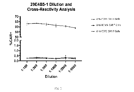
FIG. 2 is a diagram showing that antibody clone 29E4B5 binds specifically to
anti-
CD19 CAR T cells (CAR T cells that express a CAR containing anti-FMC63-scFv,
but not
anti-BCMA CAR T cells or anti-CD70 CAR T cells.
DETAILED DESCRIPTION
Provided herein are antibodies capable of binding to a single-chain variable
fragment
(scFv) having the amino acid sequence of SEQ ID NO:1 (derived from mouse anti-
human
CD19 antibody FMC63), e.g., capable of binding to the scFv expressed on cell
surface as the
extracellular domain of an anti-CD19 chimeric antigen receptor (CAR). As such,
the
antibodies disclosed herein may be used for detecting presence of cells (e.g.,
T cells)
expressing such an anti-CD19 CAR in a sample, e.g., samples obtained from a
manufacturing
3
CA 03167251 2022- 8-5
WO 2021/161197
PCT/1B2021/051100
process for producing anti-CD19 CAR-T cells or samples obtained from patients
who are
administered anti-CD19 CAR-T cells.
I. Antibodies Binding to Anti-CD19 Single-Chain Variable
Fragment (scFv)
The present disclosure provides antibodies (e.g., antibody 29E4B5) binding to
a
single-chain variable fragment (scFv) having the amino acid sequence of SEQ ID
NO: 1
(provided below), which comprises the heavy chain variable domain (Vii) and
light chain
variable domain (VL) derived from mouse anti-human CD19 antibody FMC63. As
such, the
antibodies provided herein may be referred to as anti-scFv antibodies or anti-
idiotypic (anti-
ID) antibodies. In some embodiments, the antibodies disclosed herein are
capable of binding
to the scFv expressed on a cell surface. In specific examples, the antibodies
disclosed herein
bind to a cell-surface expressed anti-CD19 chimeric antigen receptor (CAR)
comprising the
scFv of SEQ ID NO:1 as the extracellular domain. The linker fragment is in
boldface.
Amino Acid Sequence of the scFv Antigen (SEQ ID NO: I):
D IQMTQT T S SLSASLGDRVTI SCRASQD I SKYLNWYQQKP DGTVKLL IYHTSRLHSGVPSRF
SGSGSGTDYSLT I SNLEQEDIATYFCQQGNT LP YTFGGGTKLE ITGSTSGSGKPGSGEGSTK
GEVKLQE SGPGLVAPSQSL SVICTVSGVSLPDYGVSWIRQPPRKGLEWLGVIWGSET TYYNS
ALKSRLT I IKDNSKSQVF LKMNSLQTDDTAIYYCAKHYYYGGSYAMDYWGQGT SVTVS S
An antibody (interchangeably used in plural form) is an immunoglobulin
molecule
capable of specific binding to a target, such as the scFv of SEQ ID NO:1 in
the present
application, through at least one antigen recognition site, located in the
variable region of the
immunoglobulin molecule. As used herein, the term "antibody" encompasses not
only intact
(e.g., full-length) polyclonal or monoclonal antibodies, but also antigen-
binding fragments
thereof (such as Fab, Fab', F(ab')2, Fv), single-chain antibody (scFv), fusion
proteins
comprising an antibody portion, humanized antibodies, chimeric antibodies,
diabodies, single
domain antibody (e.g., nanobody), single domain antibodies (e.g., a VH only
antibody),
multispecific antibodies (e.g., bispecific antibodies) and any other modified
configuration of
an immunoglobulin molecule that comprises an antigen recognition site of the
required
specificity, including glycosylation variants of antibodies, amino acid
sequence variants of
antibodies, and covalent! y modified antibodies. An antibody as disclosed
herein includes an
antibody of any class, such as IgD, IgE, IgG, IgA, or IgM (or sub-class
thereof), and the
antibody need not be of any particular class. Depending on the antibody amino
acid sequence
of the constant domain of its heavy chains, immunoglobulins can be assigned to
different
classes. There are five major classes of immunoglobulins: IgA, IgD, IgE, IgG,
and IgM, and
4
CA 03167251 2022- 8-5
WO 2021/161197
PCT/1B2021/051100
several of these may be further divided into subclasses (isotypes), e.g.,
IgGl, IgG2, IgG3,
IgG4, IgAl and IgA2. The heavy-chain constant domains that correspond to the
different
classes of immunoglobulins are called alpha, delta, epsilon, gamma, and mu,
respectively.
The subunit structures and three-dimensional configurations of different
classes of
immunoglobulins are well known.
A typical antibody molecule comprises a heavy chain variable region (VH) and a
light
chain variable region (VL), which are usually involved in antigen binding. The
Vll and VL
regions can be further subdivided into regions of hypervariability, also known
as
"complementarity determining regions" ("CDR"), interspersed with regions that
are more
conserved, which arc known as "framework regions" ("FR"). Each VH and VL is
typically
composed of three CDRs and four FRs, arranged from amino-terminus to carboxy-
terminus
in the following order: FR], CDR1, FR2, CDR2, FR3, CDR3, FR4. The extent of
the
framework region and CDRs can be precisely identified using methodology known
in the art,
for example, by the Kabat definition, the Chothia definition, the AbM
definition, and/or the
contact definition, all of which are well known in the art. See, e.g., Kabat,
E.A., et al. (1991)
Sequences of Proteins of Immunological Interest, Fifth Edition, U.S.
Department of Health
and Human Services, NIH Publication No. 91-3242, Chothia ct al., (1989) Nature
342:877;
Chothia, C. et al. (1987) J. Mol. Biol. 196:901-917, Al-lazikani et al (1997)
J. Molec. Biol.
273:927-948, and Almagro, J. Mol. Recognit. 17:132-143 (2004). See also
hgmp.mrc.ac.uk
and bioinf.org.uk/abs.
The anti-scFv antibodies described herein may be a full-length antibody, which
contains two heavy chains and two light chains, each including a variable
domain and a
constant domain. Alternatively, the anti-scFv antibodies described herein can
be an antigen-
binding fragment of a full-length antibody. Examples of binding fragments
encompassed
within the term "antigen-binding fragment" of a full length antibody include
(i) a Fab
fragment, a monovalent fragment consisting of the VL, VH, CL and C1-11
domains; (ii) a F(ab)2
fragment, a bivalent fragment including two Fab fragments linked by a
disulfide bridge at the
hinge region; (iii) a Fd fragment consisting of the VH and CH1 domains; (iv) a
Fv fragment
consisting of the VL and VH domains of a single arm of an antibody, (v) a dAb
fragment
(Ward et al., (1989) Nature 341:544-546), which consists of a VH domain; and
(vi) an
isolated complementarity determining region (CDR) that retains functionality.
Furthermore,
although the two domains of the Fv fragment, VL and VH, are coded for by
separate genes,
they can be joined, using recombinant methods, by a synthetic linker that
enables them to be
made as a single protein chain in which the VL and VH regions pair to form
monovalent
5
CA 03167251 2022- 8-5
WO 2021/161197
PCT/1B2021/051100
molecules known as single chain Fv (scFv). See e.g., Birder al. (1988) Science
242:423-426;
and Huston et al. (1988) Proc. Natl. Acad. Sci. USA 85:5879-5883.
The anti-scFv antibodies described herein can be of a suitable origin, for
example,
murine, rat, or human. Such antibodies are non-naturally occurring, i.e.,
would not be
produced in an animal without human act (e.g., immunizing such an animal with
a desired
antigen or fragment thereof or isolated from antibody libraries). Any of the
anti-scFv
antibodies described herein, e.g., antibody 29E4B5, can be either monoclonal
or polyelonal.
A "monoclonal antibody" refers to a homogenous antibody population and a
"polyclonal
antibody" refers to a heterogeneous antibody population. These two terms do
not limit the
source of an antibody or the manner in which it is made.
In some embodiments, the anti-scFv antibodies described herein are human
antibodies, which may be isolated from a human antibody library or generated
in transgenic
mice. For example, fully human antibodies can be obtained by using
commercially available
mice that have been engineered to express specific human immunoglobulin
proteins.
Transgenic animals that are designed to produce a more desirable (e.g., fully
human
antibodies) or more robust immune response may also be used for generation of
humanized
or human antibodies. Examples of such technology are XcnornouscTM from Amgen,
Inc.
(Fremont, Calif.) and HuMAb-MouseTm and TC MouseTM from Medarex, Inc.
(Princeton,
N.J.). In another alternative, antibodies may be made recombinantly by phage
display or
yeast technology. See, for example, U.S. Pat. Nos. 5,565,332; 5,580,717;
5,733,743; and
6,265,150; and Winter et al., (1994) Annu. Rev. Immunol. 12:433-455.
Alternatively, the
antibody library display technology, such as phage, yeast display, mammalian
cell display, or
mRNA display technology as known in the art can be used to produce human
antibodies and
antibody fragments in vitro, from immunoglobulin variable (V) domain gene
repertoires from
u nimmunized donors.
In other embodiments, the anti-scFv antibodies described herein may be
humanized
antibodies or chimeric antibodies. Humanized antibodies refer to forms of non-
human (e.g.,
murine) antibodies that are specific chimeric immunoglobulins, immunoglobulin
chains, or
antigen-binding fragments thereof that contain minimal sequence derived from
non-human
immunoglobulin. In general, humanized antibodies are human immunoglobulins
(recipient
antibody) in which residues from a CDR of the recipient are replaced by
residues from a
CDR of a non-human species (donor antibody) such as mouse, rat, or rabbit
having the
desired specificity, affinity, and capacity. In some instances, one or more Fv
framework
region (FR) residues of the human immunoglobulin are replaced by corresponding
non-
6
CA 03167251 2022- 8-5
WO 2021/161197
PCT/1B2021/051100
human residues. Furthermore, the humanized antibody may comprise residues that
are found
neither in the recipient antibody nor in the imported CDR or framework
sequences, but arc
included to further refine and optimize antibody performance. In some
instances, the
humanized antibody may comprise substantially all of at least one, and
typically two, variable
domains, in which all or substantially all of the CDR regions correspond to
those of a non-
human immunoglobulin and all or substantially all of the FR regions are those
of a human
immunoglobulin consensus sequence. The humanized antibody optimally also will
comprise
at least a portion of an immunoglobulin constant region or domain (Fe),
typically that of a
human immunoglobulin. Antibodies may have Fe regions modified as described in
WO
99/58572. Other forms of humanized antibodies have one or more CDRs (one, two,
three,
four, five, or six) which are altered with respect to the original antibody,
which are also
termed one or more CDRs "derived from" one or more CDRs from the original
antibody.
Humanized antibodies may also involve affinity maturation. Methods for
constructing
humanized antibodies are also well known in the art. See, e.g., Queen et al.,
Proc. Natl.
Acad. Sci. USA, 86:10029-10033 (1989).
In some embodiments, the anti-scFv antibodies described herein can be a
chimeric
antibody. Chimeric antibodies refer to antibodies having a variable region or
part of variable
region from a first species and a constant region from a second species.
Typically, in these
chimeric antibodies, the variable region of both light and heavy chains mimics
the variable
regions of antibodies derived from one species of mammals (e.g., a non-human
mammal such
as mouse, rabbit, and rat), while the constant portions are homologous to the
sequences in
antibodies derived from another mammal such as human. In some embodiments,
amino acid
modifications can be made in the variable region and/or the constant region.
Techniques
developed for the production of "chimeric antibodies" are well known in the
art. See, e.g.,
Morrison et al. (1984) Proc. Natl. Acad. Sci. USA 81, 6851; Neuberger et al.
(1984) Nature
312, 604; and Takeda et al. (1984) Nature 314:452.
In some embodiments, the anti-scFv antibodies described herein specifically
bind to
the corresponding target antigen (i.e., the anti-CD19 scFv of SEQ ID NO: 1 or
a polypeptide
such as a chimeric antigen receptor comprising such) or an epitope thereof. An
antibody that
"specifically binds" to an antigen or an epitope is a term well understood in
the art. A
molecule is said to exhibit "specific binding" if it reacts more frequently,
more rapidly, with
greater duration, with greater avidity, and/or with greater affinity with a
particular target
antigen than it does with alternative targets. An antibody "specifically
binds" to a target
antigen or epitope if it binds with greater affinity, avidity, more readily,
and/or with greater
7
CA 03167251 2022- 8-5
WO 2021/161197
PCT/1B2021/051100
duration than it binds to other substances. For example, an antibody that
specifically (or
preferentially) binds to an antigen or an antigenic epitope therein is an
antibody that binds
this target antigen with greater affinity, avidity, more readily, and/or with
greater duration
than it binds to other antigens or other epitopes in the same antigen. It is
also understood
with this definition that, for example, an antibody that specifically binds to
a first target
antigen may or may not specifically or preferentially bind to a second target
antigen. As
such, "specific binding" or "preferential binding" does not necessarily
require (although it
can include) exclusive binding. In some examples, an antibody that
"specifically binds" to a
target antigen or an epitope thereof may not bind to other antigens or other
epitopes in the
same antigen (i.e.., only baseline binding activity can be detected in a
conventional method).
In some embodiments, the anti-scFv antibodies described herein (e.g., antibody
29E4B5) have a suitable binding affinity for the target antigen (i.e., the
anti-CD19 scFv of
SEQ ID NO: 1 or a polypeptide such as a chimeric antigen receptor comprising
such) or
antigenic epitopes thereof. As used herein, "binding affinity" refers to the
apparent
association constant or KA. The KA is the reciprocal of the dissociation
constant (Kn). The
antibody described herein may have a binding affinity (KID) of at least 100mM,
10mM, 1mM,
0.1mM, 100 M, 10 M, l[tM, 0.41M, 100nM, lOnM, mM, 0.1 nM, or lower for the
scFv
from antibody FMC63. An increased binding affinity corresponds to a decreased
KD. Higher
affinity binding of an antibody for a first antigen relative to a second
antigen can be indicated
by a higher KA (or a smaller numerical value KD) for binding the first antigen
than the KA (or
numerical value KD) for binding the second antigen. In such cases, the
antibody has
specificity for the first antigen (e.g., a first protein in a first
conformation or mimic thereof)
relative to the second antigen (e.g., the same first protein in a second
conformation or mimic
thereof; or a second protein). Differences in binding affinity (e.g., for
specificity or other
comparisons) can be at least 1.5, 2, 3, 4, 5, 10, 15, 20, 37.5, 50, 70, 80,
90, 100, 500, 1000,
10,000 or 10 fold. In some embodiments, any of the antibodies disclosed herein
may be
further affinity matured to increase the binding affinity of the antibody to
the target antigen or
antigenic epitope thereof.
Binding affinity (or binding specificity) can be determined by a variety of
methods
including equilibrium dialysis, equilibrium binding, gel filtration, ELISA,
surface plasmon
resonance, or spectroscopy (e.g., using a fluorescence assay). Exemplary
conditions for
evaluating binding affinity are in HBS-P buffer (10 mM HEPES pH7.4, 150 mM
NaCl,
0.005% (v/v) Surfactant P20). These techniques can be used to measure the
concentration of
bound binding protein as a function of target protein concentration. The
concentration of
8
CA 03167251 2022- 8-5
WO 2021/161197
PCT/1B2021/051100
bound binding protein (Bound]) is generally related to the concentration of
free target
protein ([Frec]) by the following equation:
[Bound] = [Free]/(Kd+[Free])
It is not always necessary to make an exact determination of KA, since
sometimes it is
sufficient to obtain a quantitative measurement of affinity (e.g., determined
using a method
such as ELISA or FACS analysis), which is proportional to KA. The quantitative
measurement thus can be used for comparisons, such as determining whether a
higher affinity
is, e.g., 2-fold higher, so as to obtain a qualitative measurement of
affinity, or to obtain an
inference of affinity, e.g., by activity in a functional assay, e.g., an in
vitro or in vivo assay.
The structural information (heavy chain and light chain variable domains) of
an
exemplary antibody 29E4B5 is provided below. The heavy chain CDRs and light
chain
CDRs (determined by the Kabat approach; see, e.g., Kabat, E.A., et al. (1991)
Sequences of
Proteins of Immunological Interest, Fifth Edition, U.S. Department of Health
and Human
Services, NIH Publication No. 91-3242, imgt.org/IMGTindex/V-QUEST.php, and
ncbi.nlm.nih.gov/igblast/) are identified in boldface. See also Table 7 below.
Table 1. Vi and VL Sequences of anti-scEv antibody 29E4B5.
Description SEQ ID NO: Sequences (CDRs in
boldface)
Heavy chain EVKLLQSGGGLVQPGGSLKL SCAA SG I D F
SRYWMSWVRRAP
variable 2 GKGLEW I GEINLDSSTKNYAP SLKDKF II
SRDNAKNTLYLQ
(VW MSKVRSED TALYYCARNYVGMDYWGQGT SVTVSS
D IVLTQ SPAS LAV S LGQRAT I SCRASKSVSSSDYTYMHWYQ
Light chain 3 QKPGQPPKLL TYLASNLESGVPARF SGSGSGT
DF TLNIHPV
variable (VL) EEEDAATY YCQHSEtELPP TF GGGTKLE 1K
In some embodiments, the anti-scFv antibodies described herein bind to the
same
epitope in SEQ ID NO: 1 as the exemplary antibody 29E4B5 or compete against
the
exemplary antibody for binding to the scFv antigen (SEQ ID NO:1). An "epitope"
as used
herein refers to the site on a target antigen that is recognized and bound by
an antibody. The
site can be entirely composed of amino acid components, entirely composed of
chemical
modifications of amino acids of the protein (e.g., glycosyl moieties), or
composed of
combinations thereof. Overlapping epitopes include at least one common amino
acid residue.
An epitope can be linear, which is typically 6-15 amino acids in length.
Alternatively, the
epitope can be conformational. The epitope to which an antibody binds can be
determined by
routine technology, for example, the epitope mapping method (see, e.g.,
descriptions below).
9
CA 03167251 2022- 8-5
WO 2021/161197
PCT/1B2021/051100
An antibody that binds the same epitope as an exemplary antibody described
herein may bind
to exactly the same cpitope or a substantially overlapping epitope (e.g.,
containing less than 3
non-overlapping amino acid residues, less than 2 non-overlapping amino acid
residues, or
only 1 non-overlapping amino acid residue) as the exemplary antibody. Whether
two
antibodies compete against each other for binding to the cognate antigen can
be determined
by a competition assay, which is well known in the art.
In some examples, the anti-scFv antibodies disclosed herein comprises the same
V11
and/or VL CDRs as the exemplary antibody 29E4B5. Two antibodies having the
same VH
and/or VL CDRs means that their CDRs are identical when determined by the same
approach
(e.g., the Kabat approach, the Chothia approach, the AbM approach, the Contact
approach, or
the IMGT approach as known in the art. See, e.g., bioinf.org.uk/abs/). Such
antibodies may
have the same VH, the same VL, or both as compared to an exemplary antibody
described
herein. The heavy chain and light chain CDRs of exemplary antibody 29E4B5,
determined
by the various approaches as noted, are provided in Table 7 below.
Also within the scope of the present disclosure are functional variants of
exemplary
antibody 29E4B5. Such functional variants are substantially similar to the
exemplary
antibody, both structurally and functionally. A functional variant comprises
substantially
similar VD and VL CDRs as the exemplary antibody. For example, it may comprise
only up
to 8 (e.g., 8, 7, 6, 5, 4, 3, 2, or 1) amino acid residue variations in the
total CDR regions of the
antibody and binds the same epitope in SEQ ID NO: 1 with substantially similar
affinity (e.g.,
having a KD value in the same order). In some instances, the functional
variants may have
the same heavy chain CDR3 as the exemplary antibody, and optionally the same
light chain
CDR3 as the exemplary antibody. Alternatively or in addition, the functional
variants may
have the same heavy chain CDR2 as the exemplary antibody. Such an antibody may
comprise a VD fragment having CDR amino acid residue variations in only the
heavy chain
CDR1 as compared with the VH of the exemplary antibody. In some examples, the
antibody
may further comprise a VL fragment having the same VL CDR3, and optionally the
same VL
CDR1 or VL CDR2 as the exemplary antibody.
In some instances, the amino acid residue variations (e.g., in one or more of
the heavy
chain and light chain CDRs of antibody 29E4B5) can be conservative amino acid
residue
substitutions. As used herein, a "conservative amino acid substitution" refers
to an amino
acid substitution that does not alter the relative charge or size
characteristics of the protein in
which the amino acid substitution is made. Variants can be prepared according
to methods
for altering polypeptide sequence known to one of ordinary skill in the art
such as are found
CA 03167251 2022- 8-5
WO 2021/161197
PCT/1B2021/051100
in references which compile such methods, e.g. Molecular Cloning: A Laboratory
Manual, J.
Sambrook, et al., eds., Second Edition, Cold Spring Harbor Laboratory Press,
Cold Spring
Harbor. New York, 1989, or Current Protocols in Molecular Biology, F.M.
Ausubel, et al.,
eds., John Wiley & Sons, inc., New York. Conservative substitutions of amino
acids include
substitutions made among amino acids within the following groups: (a) M, 1, L,
V; (b) F, Y,
W; (c) K, R, H; (d) A, G; (e) S, T; (f) Q, N; and (g) E, D.
In some embodiments, the anti-scFv antibodies disclosed herein may comprise
heavy
chain CDRs that are at least 80% (e.g., 85%, 90%, 95%, or 98%) identical,
individually or
collectively, as compared with the VH CDRs of the exemplary antibody 29E4B5.
Alternatively or in addition, the anti-scFv antibodies disclosed herein may
comprise light
chain CDRs that are at least 80% (e.g., 85%, 90%, 95%, or 98%) identical,
individually or
collectively, as compared with the VL CDRs as the exemplary antibody 29E4B5.
As used
herein, -individually" means that one CDR of an antibody shares the indicated
sequence
identity relative to the corresponding CDR of the exemplary antibody.
"Collectively" means
that three VH or VI. CDRs of an antibody in combination share the indicated
sequence
identity relative the corresponding three VH or VL CDRs of the exemplary
antibody in
combination.
The "percent identity" of two amino acid sequences is determined using the
algorithm
of Karlin and Altschul Proc. Natl. Acad. Sci. USA 87:2264-68, 1990, modified
as in Karlin
and Altschul Proc. Natl. Acad. Sci. USA 90:5873-77, 1993. Such an algorithm is
incorporated into the NB LAST and XBLAST programs (version 2.0) of Altschul,
et al. J.
Mol. Biol. 215:403-10, 1990. BLAST protein searches can be performed with the
XBLAST
program, score=50, wordlength=3 to obtain amino acid sequences homologous to
the protein
molecules of interest. Where gaps exist between two sequences, Gapped BLAST
can be
utilized as described in Altschul et al., Nucleic Acids Res. 25(17):3389-3402,
1997. When
utilizing BLAST and Gapped BLAST programs, the default parameters of the
respective
programs (e.g., XBLAST and NBLAST) can be used.
In some embodiments, the heavy chain of any of the anti-scFv antibodies as
described
herein may further comprise a heavy chain constant region (CH) or a portion
thereof (e.g.,
CH1, CH2, CH3, or a combination thereof). The heavy chain constant region can
of any
suitable origin, e.g., human, mouse, rat, or rabbit. Alternatively or in
addition, the light chain
of the antibody may further comprise a light chain constant region (CL), which
can be any
CL known in the art. In some examples, the CL is a kappa light chain. In other
examples,
the CL is a lambda light chain. Antibody heavy and light chain constant
regions are well
11
CA 03167251 2022- 8-5
WO 2021/161197
PCT/1B2021/051100
known in the art, e.g., those provided in the IMGT database (www.imgt. org) or
at
www.vbasc2.org/vbstat.php., both of which arc incorporated by reference
herein.
Preparation of Anti-Single-Chain Variable Fragment (scFv) Antibodies
The anti-scFv antibodies described herein (e.g., antibody 29E4B5) can be made
by
any method known in the art. See, for example, Harlow and Lane, (1998)
Antibodies: A
Laboratory Manual, Cold Spring Harbor Laboratory, New York.
In some embodiments, the anti-scFv antibody may be produced by the
conventional
hybridoma technology. The full-length anti-CD19 scFv antigen of SEQ ID NO: 1
or a
fragment thereof, optionally coupled to a carrier protein such as KLH, can be
used to
immunize a host animal for generating antibodies binding to that antigen. The
route and
schedule of immunization of the host animal are generally in keeping with
established and
conventional techniques for antibody stimulation and production, as further
described herein.
General techniques for production of mouse, humanized, and human antibodies
are known in
the art and are described herein. It is contemplated that any mammalian
subject including
humans or antibody producing cells therefrom can be manipulated to serve as
the basis for
production of mammalian, including human hybridoma cell lines. Typically, the
host animal
is inoculated intraperitoneally, intramuscularly, orally, subcutaneously,
intraplantar, and/or
intraclermally with an amount of inununogen, including as described herein.
Hybridomas can be prepared from the lymphocytes and immortalized myeloma cells
using the general somatic cell hybridization technique of Kohler, B. and
Milstein, C. (1975)
Nature 256:495-497 or as modified by Buck, D.W., et al., In Vitro, 18:377-381
(1982).
Available myeloma lines, including but not limited to X63-Ag8.653 and those
from the Salk
Institute, Cell Distribution Center, San Diego, Calif. USA, may be used in the
hybridization.
Generally, the technique involves fusing myeloma cells and lymphoid cells
using a fusogen
such as polyethylene glycol, or by electrical means well known to those
skilled in the art.
After the fusion, the cells are separated from the fusion medium and grown in
a selective
growth medium, such as hypoxanthine-aminopterin-thymidine (HAT) medium, to
eliminate
unhybridized parent cells. Any of the media described herein, supplemented
with or without
serum, can be used for culturing hybridomas that secrete monoclonal
antibodies. As another
alternative to the cell fusion technique, EBV immortalized B cells may be used
to produce the
anti-scFv monoclonal antibodies of the subject invention. The hybridomas are
expanded and
subcloncd, if desired, and supernatants arc assayed for anti-immunogcn
activity by
12
CA 03167251 2022- 8-5
WO 2021/161197
PCT/1B2021/051100
conventional immunoassay procedures (e.g., radioimmunoassay, enzyme
immunoassay, or
fluorescence immunoassay).
Hybridomas that may be used as a source of antibodies encompasses all
derivatives,
progeny cells of the parent hybridomas that produce monoclonal antibodies
capable of
binding to SEQ Ill NO: 1. Hybridomas that produce such antibodies may be grown
in vitro
or in vivo using known procedures. The monoclonal antibodies may be isolated
from the
culture media or body fluids, by conventional immunoglobulin purification
procedures such
as ammonium sulfate precipitation, gel electrophoresis, dialysis,
chromatography, and
ultrafiltration, if desired. Undesired activity if present, can be removed,
for example, by
running thc preparation over adsorbents made of the immunogen attached to a
solid phase
and eluting or releasing the desired antibodies off the immunogen.
Immunization of a host
animal with a target antigen or a fragment containing the target amino acid
sequence
conjugated to a protein that is immunogenic in the species to be immunized,
e.g., keyhole
limpet hemocyanin, serum albumin, bovine thyroglobulin, or soybean trypsin
inhibitor using
a bifunctional or derivatizing agent, for example maleimidobenzoyl
sulfosuccinimide ester
(conjugation through cysteine residues), N-hydroxysuccinimide (through lysine
residues),
glutaraldehyde, succinic anhydride, SOC1, or R1N=C=NR, where R and R1 are
different
alkyl groups, can yield a population of antibodies (e.g., monoclonal
antibodies).
If desired, an antibody (monoclonal or polyclonal) of interest (e.g., produced
by a
hybridoma cell line) may be sequenced and the polynucleotide sequence may then
be cloned
into a vector for expression or propagation. The sequence encoding the
antibody of interest
may be maintained in the vector in a host cell and the host cell can then be
expanded and
frozen for future use. In an alternative, the polynucleotide sequence may be
used for genetic
manipulation to, e.g., humanize the antibody or to improve the affinity
(affinity maturation),
or other characteristics of the antibody. For example, the constant region may
be engineered
to more resemble human constant regions to avoid immune response if the
antibody is from a
non-human source and is to be used in clinical trials and treatments in
humans. Alternatively,
or in addition, it may be desirable to genetically manipulate the antibody
sequence to obtain
greater affinity and/or specificity to the target antigen. It will be apparent
to one of skill in
the art that one or more polynucleotide changes can be made to the antibody
and still
maintain its binding specificity to the target antigen.
Antigen-binding fragments of an intact antibody (full-length antibody) can be
prepared via routine methods. For example, F(ab')2 fragments can be produced
by pepsin
13
CA 03167251 2022- 8-5
WO 2021/161197
PCT/1B2021/051100
digestion of an antibody molecule, and Fab fragments that can be generated by
reducing the
disulfide bridges of F(ab')2 fragments.
Genetically engineered antibodies, such as humanized antibodies, chimeric
antibodies, single-chain antibodies, and hi-specific antibodies, can he
produced via, e.g.,
conventional recombinant technology. In one example, DNA encoding a monoclonal
antibody specific to a target antigen can be readily isolated and sequenced
using conventional
procedures (e.g., by using oligonucleotide probes that are capable of binding
specifically to
genes encoding the heavy and light chains of the monoclonal antibodies). The
hybridoma
cells serve as a preferred source of such DNA. Once isolated, the DNA may be
placed into
one or more expression vectors, which are then transfected into host cells
such as E. coli
cells, simian COS cells, Chinese hamster ovary (CHO) cells, or myeloma cells
that do not
otherwise produce immunoglohulin protein, to obtain the synthesis of
monoclonal antibodies
in the recombinant host cells. See, e.g., PCT Publication No. WO 87/04462. The
DNA can
then be modified, for example, by substituting the coding sequence for human
heavy and
light chain constant domains in place of the homologous murine sequences,
Morrison et al.,
(1984) Proc. Nat. Acad. Sci. 81:6851, or by covalently joining to the
immunoglobulin coding
sequence all or part of the coding sequence for a non-immunoglobulin
polypeptide. In that
manner, genetically engineered antibodies, such as -chimeric" or -hybrid"
antibodies; can be
prepared that have the binding specificity of a target antigen.
Antibodies obtained following a method known in the art and described herein
can be
characterized using methods well known in the art. For example, one method is
to identify
the epitope to which the antigen binds, or "epitope mapping." There are many
methods
known in the art for mapping and characterizing the location of epitopes on
proteins,
including solving the crystal structure of an antibody-antigen complex,
competition assays,
gene fragment expression assays, and synthetic peptide-based assays, as
described, for
example, in Chapter 11 of Harlow and Lane, Using Antibodies, a Laboratory
Manual, Cold
Spring Harbor Laboratory Press, Cold Spring Harbor, N.Y., 1999. In an
additional example,
epitope mapping can be used to determine the sequence to which an antibody
binds. The
epitope can be a linear epitope, i.e., contained in a single stretch of amino
acids, or a
conformational epitope formed by a three-dimensional interaction of amino
acids that may
not necessarily be contained in a single stretch (primary structure linear
sequence). Peptides
of varying lengths (e.g., at least 4-6 amino acids long) can be isolated or
synthesized (e.g.,
recombinantly) and used for binding assays with an antibody. In another
example, the
epitope to which the antibody binds can be determined in a systematic
screening by using
14
CA 03167251 2022- 8-5
WO 2021/161197
PCT/1B2021/051100
overlapping peptides derived from the target antigen sequence and determining
binding by
the antibody. According to the gene fragment expression assays, the open
reading frame
encoding the target antigen is fragmented either randomly or by specific
genetic constructions
and the reactivity of the expressed fragments of the antigen with the antibody
to be tested is
determined. The gene fragments may, for example, be produced by PCR and then
transcribed and translated into protein in vitro, in the presence of
radioactive amino acids.
The binding of the antibody to the radioactively labeled antigen fragments is
then determined
by immunoprecipitation and gel electrophoresis. Certain epitopes can also be
identified by
using large libraries of random peptide sequences displayed on the surface of
phage particles
(phage libraries). Alternatively, a defined library of overlapping peptide
fragments can be
tested for binding to the test antibody in simple binding assays. In an
additional example,
mutagenesis of an antigen binding domain, domain swapping experiments and
alanine
scanning mutagenesis can be performed to identify residues required,
sufficient, and/or
necessary for epitope binding. For example, domain swapping experiments can be
performed
using a mutant of a target antigen, in which various fragments of the single-
chain variable
fragment (scFv) protein have been replaced (swapped) with sequences from a
closely related,
but antigenically distinct protein. By assessing binding of the antibody to
the mutant scFv
polypeptide, the importance of the particular antigen fragment to antibody
binding can be
assessed.
Alternatively, competition assays can be performed using other antibodies
known to
bind to the same antigen to determine whether an antibody binds to the same
epitope as the
other antibodies. Competition assays are well known to those of skill in the
art.
In some embodiments, the anti-scFv antibodies disclosed herein can be produced
using the conventional recombinant technology as exemplified below.
Nucleic acids encoding the heavy and light chain of an antibody described
herein can
be cloned into one expression vector, each nucleotide sequence being in
operable linkage to a
suitable promoter. In one example, each of the nucleotide sequences encoding
the heavy
chain and light chain is in operable linkage to a distinct prompter.
Alternatively, the
nucleotide sequences encoding the heavy chain and the light chain can be in
operable linkage
with a single promoter, such that both heavy and light chains are expressed
from the same
promoter. When necessary, an internal ribosomal entry site (IRES) can be
inserted between
the heavy chain and light chain encoding sequences.
In some examples, the nucleotide sequences encoding the two chains of the
antibody
are cloned into two vectors, which can be introduced into the same or
different cells. When
CA 03167251 2022- 8-5
WO 2021/161197
PCT/1B2021/051100
the two chains are expressed in different cells, each of them can be isolated
from the host
cells expressing such and the isolated heavy chains and light chains can be
mixed and
incubated under suitable conditions allowing for the formation of the
antibody.
Generally, a nucleic acid sequence encoding one or all chains of an antibody
can be
cloned into a suitable expression vector in operable linkage with a suitable
promoter using
methods known in the art. For example, the nucleotide sequence and vector can
be contacted,
under suitable conditions, with a restriction enzyme to create complementary
ends on each
molecule that can pair with each other and be joined together with a ligase.
Alternatively,
synthetic nucleic acid linkers can be ligated to the termini of a gene. These
synthetic linkers
contain nucleic acid sequences that correspond to a particular restriction
site in the vector.
The selection of expression vectors/promoter would depend on the type of host
cells for use
in producing the antibodies.
A variety of promoters can be used for expression of the antibodies described
herein,
including, but not limited to, cytomegalovirus (CMV) intermediate early
promoter, a viral
LTR such as the Rotas sarcoma virus LTR, HIV-LTR, HTLV-1 LTR, the simian virus
40
(SV40) early promoter, E. coli lac UV5 promoter, and the herpes simplex tk
virus promoter.
Regulatable promoters can also be used. Such regulatable promoters include
those
using the lac repressor from E. coli as a transcription modulator to regulate
transcription from
lac operator-bearing mammalian cell promoters (Brown, M. et al., Cell, 49:603-
612 (1987)),
those using the tetracycline repressor (tetR) (Gossen, M., and Bujard, H.,
Proc. Natl. Acad.
Sci. USA 89:5547-5551 (1992); Yao, F. et al., Human Gene Therapy, 9:1939-1950
(1998);
Shockelt, P., et al., Proc. Natl. Acad. Sci. USA, 92:6522-6526 (1995)). Other
systems
include FK506 dimer, VP16 or p65 using astradiol, RU486, diphenol murislerone,
or
rapamycin. Inducible systems are available from Invitrogen, Clontech and
Ariad.
Regulatable promoters that include a repressor with the operon can be used. In
one
embodiment, the lac repressor from E. coli can function as a transcriptional
modulator to
regulate transcription from lac operator-bearing mammalian cell promoters (M.
Brown et al.,
Cell, 49:603-612 (1987)); Gossen and Bujard (1992); (M. Gossen et al., Natl.
Acad. Sci.
USA, 89:5547-5551 (1992)) combined the tetracycline repressor (tetR) with the
transcription
activator (VP 16) to create a tetR-mammalian cell transcription activator
fusion protein, tTa
(tetR-VP 16), with the tet0-hearing minimal promoter derived from the human
cytomegalovirus (hCMV) major immediate-early promoter to create a tetR-tet
operator
system to control gene expression in mammalian cells. In one embodiment, a
tetracycline
inducible switch is used. The tetracycline repressor (tetR) alone, rather than
the tetR-
16
CA 03167251 2022- 8-5
WO 2021/161197
PCT/1B2021/051100
mammalian cell transcription factor fusion derivatives can function as potent
trans-modulator
to regulate gene expression in mammalian cells when the tetracycline operator
is properly
positioned downstream for the TATA element of the CMVIE promoter (Yao et al..
Human
Gene Therapy, 10(11):1811-1818, 1999). One particular advantage of this
tetracycline
inducible switch is that it does not require the use of a tetracycline
repressor-mammalian cells
transactivator or repressor fusion protein, which in some instances can be
toxic to cells
(Gossen et al., Natl. Acad. Sci. USA, 89:5547-5551 (1992); Shockett et al.,
Proc. Natl. Acad.
Sci. USA, 92:6522-6526 (1995)), to achieve its regulatable effects.
Additionally, the vector can contain, for example, some or all of the
following: a
selectable marker gene, such as the neomycin gene for selection of stable or
transient
transfectants in mammalian cells; enhancer/promoter sequences from the
immediate early
gene of human CMV for high levels of transcription; transcription termination
and RNA
processing signals from SV40 for mRNA stability; SV40 polyoma origins of
replication and
ColE1 for proper episom al replication; internal ribosome binding sites
(IRESes), versatile
multiple cloning sites; and T7 and SP6 RNA promoters for in vitro
transcription of sense and
antisense RNA. Suitable vectors and methods for producing vectors containing
transgenes
are well known and available in the art.
Examples of polyadenylation signals useful to practice the methods described
herein
include, but are not limited to, human collagen I polyadenylation signal,
human collagen II
polyadenylation signal, and SV40 polyadenylation signal.
One or more vectors (e.g., expression vectors) comprising nucleic acids
encoding any
of the antibodies may be introduced into suitable host cells for producing the
antibodies. The
host cells can be cultured under suitable conditions for expression of the
antibody or any
polypeptide chain thereof. Such antibodies or polypeptide chains thereof can
be recovered by
the cultured cells (e.g., from the cells or the culture supernatant) via a
conventional method,
e.g., affinity purification. If necessary, polypeptide chains of the antibody
can be incubated
under suitable conditions for a suitable period of time allowing for
production of the
antibody.
In some embodiments, methods for preparing an antibody described herein
involve a
recombinant expression vector that encodes both the heavy chain and the light
chain of an
antibody described herein. The recombinant expression vector can be introduced
into a
suitable host cell (e.g., a dhfr- CHO cell) by a conventional method, e.g,
calcium phosphate-
mediated transfection. Positive transformant host cells can be selected and
cultured under
suitable conditions allowing for the expression of the two polypeptide chains
that form the
17
CA 03167251 2022- 8-5
WO 2021/161197
PCT/1B2021/051100
antibody, which can be recovered from the cells or from the culture medium.
When
necessary, the two chains recovered from the host cells can be incubated under
suitable
conditions allowing for the formation of the antibody.
In one example, two recombinant expression vectors are provided, one encoding
the
heavy chain of an antibody described herein (e.g., antibody 29E4B5) and the
other encoding
the light chain of the antibody described herein (e.g., antibody 29E4B5). Both
of the two
recombinant expression vectors can be introduced into a suitable host cell
(e.g., dhfr- CHO
cell) by a conventional method, e.g., calcium phosphate-mediated transfection.
Alternatively,
each of the expression vectors can be introduced into a suitable host cells.
Positive
transformants can bc selected and cultured under suitable conditions allowing
for the
expression of the polypeptide chains of the antibody. When the two expression
vectors are
introduced into the same host cells, the antibody produced therein can be
recovered from the
host cells or from the culture medium. If necessary, the polypeptide chains
can be recovered
from the host cells or from the culture medium and then incubated under
suitable conditions
allowing for formation of the antibody. When the two expression vectors are
introduced into
different host cells, each of them can be recovered from the corresponding
host cells or from
the corresponding culture media. The two polypeptide chains can then be
incubated under
suitable conditions for formation of the antibody.
Standard molecular biology techniques are used to prepare the recombinant
expression vector, transfect the host cells, select for transformants, culture
the host cells and
recovery of the antibodies from the culture medium. For example, some
antibodies can be
isolated by affinity chromatography with a Protein A or Protein G coupled
matrix.
Any of the nucleic acids encoding the heavy chain, the light chain, or both of
an anti-
scFv antibody as described herein (e.g., antibody 29E4B5), vectors (e.g.,
expression vectors)
containing such, and host cells comprising the vectors are within the scope of
the present
disclosure.
In other embodiments, the anti-scFv antibodies described herein can be single-
chain
antibody fragments (scFv). A single-chain antibody can be prepared via
recombinant
technology by linking a nucleotide sequence coding for a heavy chain variable
region and a
nucleotide sequence coding for a light chain variable region. Preferably, a
flexible linker is
incorporated between the two variable regions. Alternatively, techniques
described for the
production of single chain antibodies (U.S. Patent Nos. 4,946,778 and
4,704,692) can be
adapted to produce a phage or yeast scFv library and scFv clones specific to a
single-chain
variable fragment (scFv) of SEQ ID NO: 1, which can be identified from the
library
18
CA 03167251 2022- 8-5
WO 2021/161197
PCT/1B2021/051100
following routine procedures. Positive clones can be subjected to further
screening to
identify those that bind the scFv of SEQ ID NO: 1.
III. Applications of Anti-Single-Chain Variable Fragment (scFv) Antibodies
The present disclosure also provides methods for detecting or quantifying a
single-
chain variable fragment (scFv) consisting of the amino acid sequence of SEQ ID
NO: 1
(specific to CD19) in a sample using any of the anti-scFv antibodies as
described herein (e.g.,
antibody 29E4B5). To perform the method disclosed herein, any of the anti-scFv
antibodies
can be brought in contact with a sample suspected of containing a target
antigen as disclosed
herein --the anti-CD19 scFv of SEQ ID NO:1 or a polypeptide such as a CAR
construct
comprising such. In general, the term "contacting" or "in contact" refers to
an exposure of
the anti-scFv antibody disclosed herein with the sample suspected of
containing the target
antigen for a suitable period under suitable conditions sufficient for the
formation of a
complex between the anti-scFv antibody and the target antigen in the sample,
if any. In some
embodiments, the contacting is performed by capillary action in which a sample
is moved
across a surface of the support membrane. The antibody-antigen complex thus
formed, if
any, can be determined via a routine approach. Detection of such an antibody-
antigen
complex after the incubation is indicative of the presence of the target
antigen in the sample.
When needed, the amount of the antibody-antigen complex can be quantified,
which is
indicative of the level of the target antigen in the sample.
In some embodiments, a target antigen disclosed herein (i.e., the anti-CD19
scFv of
SEQ ID NO:1 or a polypeptide comprising such) in a sample can be detected or
quantified
using any of the anti-scFv antibodies disclosed herein via an immunoassay.
Examples of
immunoassays include, without limitation, immunoblotting assay (e.g., Western
blot),
immunohistochemical analysis, flow cytometry assay, immunofluorescence assay
(IF),
enzyme linked immunosorbent assays (ELISAs) (e.g., sandwich ELISAs),
radioimmunoassays, electrochemiluminescence-based detection assays, magnetic
immunoassays, lateral flow assays, and related techniques. Additional suitable
immunoassays for detecting the target antigen in a sample will be apparent to
those of skill in
the art.
In some examples, the anti-scFv antibodies as described herein (e.g.,
antibodies
comprising the same heavy chain and light chain CDRs or comprising the same VH
and the
same VL as antibody 29E4B5) can be conjugated to a detectable label, which can
be any
agent capable of releasing a detectable signal directly or indirectly. The
presence of such a
19
CA 03167251 2022- 8-5
WO 2021/161197
PCT/1B2021/051100
detectable signal or intensity of the signal is indicative of presence or
quantity of the target
antigen in the sample. Alternatively, a secondary antibody specific to the
anti-scFv antibody
or specific to the target antigen may be used in the methods disclosed herein.
For example,
when the anti -scFv antibody used in the method is a full-length antibody, the
secondary
antibody may bind to the constant region of the anti-scFv antibody. In other
instances, the
secondary antibody may bind to an epitope of the target antigen that is
different from the
binding epitope of the anti-scFv antibody. Any of the secondary antibodies
disclosed herein
may be conjugated to a detectable label.
Any suitable detectable label known in the art can be used in the assay
methods
described herein. In some embodiments, a detectable label can be a label that
directly
releases a detectable signal. Examples include a fluorescent label or a dye. A
fluorescent
label comprises a fluorophore, which is a fluorescent chemical compound that
can re-emit
light upon light excitation. Examples of fluorescent label include, but are
not limited to,
xanthene derivatives (e.g., fluorescein, rhodamine, Oregon green, eosin, and
Texas red),
cyanine derivatives (e.g., cyanine, indocarbocyanine, oxacarbocyanine,
thiacarbocyanine, and
merocyanine), squaraine derivatives and ring-substituted squaraines (e.g.,
Seta and Square
dyes), squaraine rotaxane derivatives such as SeTau dyes, naphthalene
derivatives (e.g.,
dansyl and prodan derivatives),
coumarin derivatives, oxadiazole derivatives (e.g., pyridyloxazole,
nitrobenzoxadiazole and
benzoxadiazole), anthracene derivatives (e.g., anthraquinones, including
DRAQ5, DRAQ7
and CyTRAK Orange), pyrene derivatives such as cascade blue, oxazine
derivatives (e.g.,
Nile red, Nile blue, cresyl violet, and oxazine 170), acridine derivatives
(e.g., proflavin,
acridine orange, and acridine yellow), arylmethine derivatives (e.g.,
auramine, crystal violet,
and malachite green), and tetrapyrrole derivatives (e.g., porphin,
phthalocyanine, and
bilirubin). A dye can be a molecule comprising a chrornophore, which is
responsible for the
color of the dye. In some examples, the detectable label can be fluorescein
isothiocyanate
(FITC), phycoerythrin (PE), biotin, Allophycocyanin (APC) or Alexa Fluor 488.
In some embodiments, the detectable label may be a molecule that releases a
detectable signal indirectly, for example, via conversion of a reagent to a
product that directly
releases the detectable signal. In some examples, such a detectable label may
be an enzyme
(e. g. , 13-gal actosidase, HRP or AP) capable of producing a colored product
from a colorless
substrate.
Any of the anti-scFv antibodies disclosed herein can be used for detecting
and/or
quantifying cells (e.g., immune cells such as T cells) that arc genetically
engineered to
CA 03167251 2022- 8-5
WO 2021/161197
PCT/1B2021/051100
express a chimeric antigen receptor comprising the anti-CD19 scFv of SEQ ID
NO: 1. As
used herein, a chimcric antigen receptor (CAR) refers to an artificial immune
cell receptor
that is engineered to recognize and bind to an antigen expressed by undesired
cells, for
example, disease cells such as cancer cells. A T cell that expresses a CAR
polypeptide is
referred to as a CAR T cell. Generally, a CAR is a fusion polypeptide
comprising an
extracellular domain that recognizes a target antigen (e.g., a single-chain
variable fragment
(scFv) of an antibody or other antibody fragment) and an intracellular domain
comprising a
signaling domain of the T-cell receptor (TCR) complex (e.g., CD3) and, in most
cases, a co-
stimulatory domain. (Enblad et al., Human Gene Therapy. 2015; 26(8):498-505).
A CAR
construct may further comprise a hinge and transmembrane domain between the
extracellular
domain and the intracellular domain, as well as a signal peptide at the N-
terminus for surface
expression.
The anti-CD19 CAR to be detected by any of the anti-scFv antibodies discloses
herein
comprise the anti-CD19 scFv of SEQ ID NO:1, which can be the extracellular
domain when
the anti-CD19 CAR is expressed on cell surface. In addition to the anti-CD19
scFv of SEQ
ID NO:1, the anti-CD19 CAR disclosed herein may comprise an intracellular
domain (e.g.,
the signaling domain of CD3), and optionally one or more co-stimulatory
domains (e.g., a
co-stimulatory domain of CD28 or 4-1BB). In some instances, such an anti-CD19
CAR may
further comprise a transmembrane domain (e.g., a transmembrane domain of
CD8oc).
Optionally, the anti-CD19 CAR may further comprise a hinge domain, which may
comprise
up to 300 amino acids (e.g., 10 to 100 amino acids, or 5 to 20 amino acids).
In some
embodiments, the hinge domain may be a CD8 hinge domain. Other hinge domains
may be
used.
Examples of anti-CD19 CARs comprising the anti-CD19 scFv of SEQ ID NO:1 can
be found in WO 2019/097305A2, the relevant disclosures of which are
incorporated by
reference herein for the purpose and subject matter referenced herein. In
specific examples,
the anti-CD19 CAR may comprise the amino acid sequence of SEQ ID NO: 7
(provided in
Table 6 below).
In some embodiments, any of the anti-scFv antibodies disclosed herein can be
used
for measuring T cells expressing an anti-CD19 CAR comprising SEQ ID NO:1 as
the
extracellular domain during a manufacturing process for producing such anti-
CD19 CAR T
cells, for example, a manufacturing process for producing CTX110 cells. See,
e.g., U.S.
Provisional Application No.: 62/934,991, filed on November 13, 2019, the
relevant
21
CA 03167251 2022- 8-5
WO 2021/161197
PCT/1B2021/051100
disclosures of which are herein incorporated by reference for the purposes and
subject matter
referenced herein. CTX110 cells arc a population of genetically engineered T
cells
expressing an anti-CD19 CAR comprising the amino acid sequence of SEQ ID NO:7
and
having disrupted endogenous TRAC and 132M genes.
In some instances, a manufacturing process for producing genetically modified
T cells
expressing an anti-CD19 CAR comprising the anti-CD19 scFv of SEQ ID NO:1
(e.g.,
CTX110 cells) may involve enriching and activating T cells, which may be
obtained from
human donors, introducing genetic modifications into the T cells thus
activated to produce T
cells, at least a portion of which express the anti-CD19 CAR and the other
desired genetic
edits, depleting TCRa13-expressing T cells from the population of genetically
modified T cells
thus produced, and harvesting the resultant anti-CD19 CAR-expressing T cells.
See, e.g.,
U.S. Provisional Application No.: 62/934,991, filed on November 13, 2019, the
relevant
disclosures of which are herein incorporated by reference for the purposes and
subject matter
referenced herein.
To monitor such a manufacturing process for producing T cells expressing the
desired
anti-CD19 CAR, one or more samples may be obtained during any stage of the
manufacturing process, e.g., before or after a nucleic acid encoding an anti-
CD19 CAR
comprising the scFv of SEQ ID NO: 1 is introduced into T cell, or both, and
the amount of
anti-CD19 CAR-expressing T cells in the sample may be measured according to
methods
described herein. For example, a fluorescent dye-conjugated anti-scFv antibody
as disclosed
herein may be incubated with the one or more samples under suitable conditions
for a
suitable period allowing for binding of the anti-scFv antibody to the cell
surface-expressed
anti-CD19 CAR. The presence of level of the T cells expressing the anti-CD19
CAR can
then be determined via a routine method, for example, by fluorescence-
activated cell sorting
(FACS).
For example, after incubating T cells with components for genetically
modifying the
T cells (including introducing into the cells a nucleic acid encoding the
desired anti-CD19
CAR), a sample containing the resultant T cells may be obtained and the anti-
scFv antibodies
disclosed herein may be used to detect or quantify the portion of T cells in
the sample that
express the anti-CD19 CAR. Alternatively, or in addition to, one or more
samples
comprising the genetically modified T cells may be obtained after the
depleting step for
removing TCRoc13 T cells, after any of in vitro expansion steps after the
genetic manipulation,
and/or after harvesting the resultant genetically engineered T cells. The
amount of anti-CD19
22
CA 03167251 2022- 8-5
WO 2021/161197
PCT/1B2021/051100
CAR-expressing T cells in these samples may be determined using the anti-scFv
antibody
disclosed herein.
In some examples, a sample may be obtained from a population of T cells
genetically
engineered to express the anti-CD19 CAR disclosed herein after
cryopreservation and before
administration to a patient. The amount of anti-CD19 CAR-expressing T cells
(CAR' T
cells) in the sample can be measured using the anti-scFv antibody disclosed
herein to make
sure that a sufficient amount of the anti-CD19 CAR-expressing T cells is given
to the patient.
In some embodiments, any of the anti-scFv antibodies disclosed herein can be
used
for clinical assessment of T cells expressing an anti-CD19 CAR comprising the
anti-CD19
scFy of SEQ ID NO:1 (e.g., the CTX110 cells) after such CAR-T cells arc
administered to a
subject in need of the treatment, for example, for evaluating the in vivo
pharmacokinetic (PK)
and/or pharmacodynamic (PD) behavior of the anti-CD19 CAR T cells.
For example, one or more biological samples may be obtained from a human
patient
administered T cells genetically engineered to express the anti-CD19 CAR
(e.g., the CTX110
cells) at one or more time points after the administration. The level of the
CAR + T cells in
the one or more biological samples can be measured by any of the anti-scFv
antibodies
disclosed herein via a conventional method, e.g., FACS. Such CARP T cell
levels, e.g., at
different time point after administration, may be used to analyze PK and/or PD
features of the
anti-CD19 CAR-T cells in that human patient. Such CAR' T cell levels may also
be used for
assessing potential treatment efficacy in that human patient.
As used herein, a "biological sample" refers to a composition that comprises
tissue,
e.g., blood, plasma or protein, from a subject. A biological sample can be an
initial
unprocessed sample taken from a subject or a subsequently processed sample,
e.g., partially
purified or preserved forms. In some embodiments, multiple (e.g., at least 2,
3, 4, 5, or more)
biological samples may be collected from a subject, over time or at particular
time intervals,
for example to assess the level of T cells expressing the anti-CD19 CAR in a
human patient
who has been administered such T cells.
The terms "patient," "subject," or "individual" may be used interchangeably
and refer
to a subject who needs the analysis as described herein. In some embodiments,
the subject is
a human patient, which has been administered a plurality of T cells, which are
genetically
engineered to express the anti-C1319 CAR. In some embodiments, the human
patient is a
cancer patient, for example, having relapsed or refractory B-cell malignancy
such as non-
Hodgkin lymphoma or B -cell lymphoma.
23
CA 03167251 2022- 8-5
WO 2021/161197
PCT/1B2021/051100
IV. Kits for Detectin2 Anti-CD19 scFv of SEO ID NO:1 and Anti-
CD19 CAR
Comorisin2 Such
The present disclosure also provides kits for use in detecting or quantifying
a single-
chain variable fragment (scFv) consisting of the amino acid sequence of SEQ ID
NO: 1 in a
sample, such as a sample obtained from a manufacturing process for producing
anti-CD19
CAR-T cells or a sample obtained from patients who are administered anti-CD19
CAR-T
cells. Such kits can include one or more containers comprising an anti-scFv
antibody, e.g.,
any of those described herein such as antibody 29E4B5.
In some embodiments, the kit can comprise instructions for use in accordance
with
any of the methods described herein. The included instructions can comprise a
description of
detecting or quantifying the scFv in a sample as described herein.
Instructions supplied in the
kits of the invention are typically written instructions on a label or package
insert (e.g., a
paper sheet included in the kit), but machine-readable instructions (e.g.,
instructions carried
on a magnetic or optical storage disk, or available via an internet address
provided in the kit)
are also acceptable.
The kits of this invention are in suitable packaging. Suitable packaging
includes, but
is not limited to, vials, bottles, jars, flexible packaging (e.g., sealed
Mylar or plastic bags),
and the like. The kits may comprise one or more aliquots of an anti-scFv
antibody described
herein.
Kits may optionally provide additional components such as buffers and
interpretive
information. Normally, the kit comprises a container and a label or package
insert(s) on or
associated with the container. In some embodiments, the invention provides
articles of
manufacture comprising contents of the kits described above.
General techniques
The practice of the present disclosure will employ, unless otherwise
indicated,
conventional techniques of molecular biology (including recombinant
techniques),
microbiology, cell biology, biochemistry, and immunology, which are within the
skill of
the art. Such techniques are explained fully in the literature, such as
Molecular Cloning: A
Laboratory Manual, second edition (Sambrook, et al., 1989) Cold Spring Harbor
Press;
Oligonucleoticle Synthesis (M. J. Gait, ed. 1984); Methods in Molecular
Biology, Humana
Press; Cell Biology: A Laboratory Notebook (J. E. Cellis, ed., 1989) Academic
Press;
Animal Cell Culture (R. I. Freshney, ed. 1987); Introduction to Cell and
Tissue Culture (J.
P. Mather and P. E. Roberts, 1998) Plenum Press; Cell and Tissue Culture:
Laboratory
24
CA 03167251 2022- 8-5
WO 2021/161197
PCT/1B2021/051100
Procedures (A. Doyle, J. B. Griffiths, and D. G. Newell, eds. 1993-8) J. Wiley
and Sons;
Methods in Enzymology (Academic Press, Inc.); Handbook of Experimental
Immunology
(D. M. Weir and C. C. Blackwell, eds.): Gene Transfer Vectors for Mammalian
Cells (J.
M. Miller and M. P. Cabs, eds., 1987); Current Protocols in Molecular Biology
(F. M.
Ausubel, et al. eds. 1987); PCR: The Polymerase Chain Reaction, (Mullis, et
al., eds.
1994); Current Protocols in Immunology (J. E. Coligan et al., eds., 1991);
Short Protocols
in Molecular Biology (Wiley and Sons, 1999); Immunobiology (C. A. Janeway and
P.
Travers, 1997); Antibodies (P. Finch, 1997); Antibodies: a practice approach
(D. Catty.,
ed., IRL Press, 1988-1989); Monoclonal antibodies: a practical approach (P.
Shepherd and
C. Dean, eds., Oxford University Press, 2000); Using antibodies: a laboratory
manual (E.
Harlow and D. Lane (Cold Spring Harbor Laboratory Press, 1999); The Antibodies
(M.
Zanetti and J. D. Capra, eds. Harwood Academic Publishers, 1995): DNA Cloning:
A
practical Approach, Volumes I and II (D.N. Glover ed. 1985); Nucleic Acid
Hybridization
(B.D. Hames & S.J. Higgins eds. (1985; Transcription and Translation (B.D.
Names &
S.J. Higgins, eds. (1984; Animal Cell Culture (R.I. Freshney, ed. (1986;
Immobilized Cells
and Enzymes (1RL Press, (1986; and B. Perbal, A practical Guide To Molecular
Cloning
(1984); F.M. Ausubcl et al. (eds.).
Without further elaboration, it is believed that one skilled in the art can,
based on the
above description, utilize the present invention to its fullest extent. The
following specific
embodiments are, therefore, to be construed as merely illustrative, and not
limitative of the
remainder of the disclosure in any way whatsoever. All publications cited
herein are
incorporated by reference for the purposes or subject matter referenced
herein.
EXAMPLES
In order that the invention described may be more fully understood, the
following
examples are set forth. The examples described in this application are offered
to illustrate the
methods and compositions provided herein and are not to be construed in any
way as limiting
their scope.
Example 1. Antigen Expression and Purification
This Example reports expression and purification of a His-tagged single-chain
variable fragment of a mouse anti-human CD19 monoclonal antibody (FMC63-scFv),
which
was subsequently used to generate antibodies against FMC63-scFv as described
in Example
2.
CA 03167251 2022- 8-5
WO 2021/161197
PCT/1B2021/051100
The FMC63-scFv protein comprises, from N-terminal to C-terminal, an artificial
signal peptide at the N-terminus, an anti-CD19 scFv fragment consisting of the
amino acid
sequence of SEQ ID NO:1, and a His-tag at the C-terminus. The amino acid
sequence and
the corresponding nucleic acid sequence of this FMC63-scFv protein are shown
in SEQ ID
NO: 4 and SEQ Ill NO: 5, respectively. Sequences corresponding to the
artificial signal
peptide are underlined and the His-tag sequences are shown in bold.
MGWSCI I LF LVATATGVHSDIQMTQTT SSLSASLGDRVT I SCRASQD I SKYLNWYQQKPDGT
VKLLIYHTSRLHSGVP SRF SGSGSGTDYSLT I SNLEQED IATYFCQQGNTLP YTF GGGTKLE
I T GST SGSGKP GSGEGSTKGEVKLQESGP GLVAPSQSLSVTCTVSGVSLPDYGVSWIRQPPR
KGLEWLGVIWGSE T TYYNSALKSRLT I IKDNSKSQVFLKMNSLQTDDTAIYYCAKHYYYGGS
YAMDYWGQGTSVTVSSHHHHHH ( SEQ ID NO: 4)
AT GGGCT GGTCCT GCATCATT CT GTTT CT GGTGGCCACAGCCACCGGCGTGCACAGCGATAT
T CAGATGACCCAGACCACCAGCAGCCT GT CT GCCT CTCTGGGCGATAGAGT GACCAT CAGCT
GTAGAGC CAGCCAGGACAT CAGCAAGTAC CT GAAC T GGTAT CAGCAGAAAC C C GACGGCAC C
GT GAAGC TGCTGAT CTACCACACCAGCAGACTGCACAGCGGCGTGCCAAGCAGAT TT TCTGG
CAGCGGC TCTGGCACCGAC TACAGCCT GACAAT CAGCAACCT GGAACAAGAGGATAT CGCTA
CCTACTT CT GCCAGCAAGGCAACACCCTGCCTTACACCTTT GGCGGAGGCACCAAGCT GGAA
ATCACCGGCTOTACAAGOGGCAGCGGCAAACCT GGATCTGGCGAGGGATCTACCAAGGGCGA
AGTGAAACTGCAAGAGTCTGGCCCTGGACTGGTGGCCCCATCTCAGTCTCTGAGCGTGACCT
GTACAGTCAGCGGAGTGTCCCTGCCTGATTACGGCGTGTCCTGGATCAGACAGCCTCCTCGG
AAAGGCCTGGAATGGCTGGGAGTGATCTGGGGCAGCGAGACAACCTACTACAACAGCGCCCT
GAAGTCCCGGCTGACCATCATCAAGGACAACTCCAAGAGCCAGGTGTTCCTGAAGATGAACA
GCCTGCAGACCGACGACACCGCCATCTACTATTGCGCCAAGCACTACTACTACGGCGGCAGC
TACGCCATGGATTATTGGGGCCAGGGCACCAGCGTGACCGTGTCTAGCCATCACCACCACCA
TCACTGA (SEQ ID NO: 5)
The DNA sequence corresponding to the FMC63-scFv (SEQ ID NO: 5) was
subcloned into pcDNA3.4 vector, and the resulting FMC63-scFv DNA expression
construct
was transfected into Expi293F cells. One-liter of the Expi293F cells were
cultured in
suspension in a serum-free Expi293FTM expression medium (Thermo Fisher
Scientific,
Waltham, MA, Cat. No. A1435101) to transiently express the recombinant FMC63-
scFv
protein. The cell culture supernatant was filtered and loaded onto a HisTrap
FF Crude
column (GE Healthcare, Chicago, IL, Cat.No.17-5286-01). The expressed
recombinant
FMC63-scFv protein was purified and buffer exchanged for PBS (pH 7.2).
Recombinant FMC63-scFv protein was analyzed by SDS-PAGE and Western-blot
under reducing (labeled 1 in FIGs. 1A-1B) and non-reducing (labeled 2 in FICs.
1A-1B)
conditions. The estimated molecular weight (MW) and purity of the recombinant
FMC63-
scFv protein were approximately 27 kDa and 95%, respectively. Based on
Bradford protein
26
CA 03167251 2022- 8-5
WO 2021/161197
PCT/IB2021/051100
assay, the estimated concentration and yield of the recombinant FMC63-scFv
protein were
0.41 mg/ml and 11.67 mg, respectively. Mass spectrometry analysis was used to
determine
the experimental average MW of the purified recombinant FMC63-scFv protein.
The
theoretical and experimental average MWs were 27324.4 Da and 27320.2 Da,
respectively.
MALDI-TOF mass spectrometry was used to authenticate the amino-acid sequence
of the
expressed recombinant FMC63-scFv protein.
Example 2. Anti-FMC63-scFy Antibody Generation
Immunization of mice and serum antibody titer determination was performed as
described herein. Five BALB/c and five C57BL/6 mice were used for anti-FMC63-
scFv
antibody generation. Mice were immunized via intraperitoneal injection with
FMC63-scFv
protein prepared in appropriate adjuvants per the schedule shown in Table 2.
After each boost, serum was separated from the blood samples, and antibody
titers
were determined by indirect ELISA. The coating antigens were:
A: Recombinant FMC63-scFv protein;
B: TGSTSGSGKPGSGEGSTKG (FMC63-scFv linker peptide) (SEQ ID NO: 6);
C: an irrelevant His-tagged protein; and
D: Total human IgG.
The coating antigens were prepared in Phosphate Buffered Saline (PBS), pH 7.4,
at
1tig/m1 and 100 1/well. The secondary antibody was Peroxidase-AffiniPure Goat
Anti-
Mouse IgG, Fey fragment-specific (Jackson ImmunoResearch, West Grove, PA, Cat.
No.
115-035-071). After the third immunization, a serum sample from each mouse was
also
evaluated by flow cytometry.
After the third immunization, mouse #B274 was selected for cell fusion, but no
positive clones were obtained. After the fourth immunization, mouse #B279 was
selected for
cell fusion using a standard hybridoma protocol. Culture supernatants were
subjected to
ELISA screening using the same set of four antigens mentioned above. A total
of six ELISA
positive wells (clones) were identified from the second fusion. Table 3.
27
CA 03167251 2022- 8-5
n
>
a
,
`:3
61
-
r,
8
,..,
9'
. Table 2, Animal Immunization Schedule and Doses.
0
0
N
=
(Day) Primary First First Second Second Third Third Fourth Fourth Fifth Fifth
Sixth ts.)
¨,
Immuniza Boost Bleed Boost Bleed Boost Bleed Boost Bleed Boost Bleed Boost
No. -tion (day 14) (day) (day 28) (day) (day 47)
(day) (day 141) (day) (day 215) (day) (day 247)
-.1
(day 0)
4..
BALB/C B271 50 g 25pg 21 25 g 35 25 g 54
25pg 148 *25pg 222 *25pg
B272 50pg 25pg 21 25p.g 35 25tig 54 25pg 148 *25pg 222 N/A
......................... , ................
B273 501.tg 25pg 21 25pg 35 25ps 54 25pg 148 *25pg 222 *25 g
_ .4., _ _
501.tg
25pg
B274 25pg 21 25 g 35 251ag 54 N/A N/A N/A
N/A
(day 106)
B275 501.tg 25pg 21 25 g 35 251ag 54 25pg 148
*25pg 222 N/A
......................... , ................
t.) C57BL/6 B276 50ps 25pg 21 251.1g 35
251ag 54 25pg 148 *25pg 222 N/A
co
Ã.. ..............
50p.g
251ag *251g
25õ
B277 21 25pg 35 25tig 54 25pg 148 173
(day 166)
(day 215)
................................ Ã
............................................
B278 50ps 25pg 21 251.1g 35 251ag 54 25pg 148
*25pg 222 N/A
,
......................... ,
50 g
25 g
B279 25pg 21 25pg 35 251ag 54 25pg 148
N/A N/A
(day 166)
......................... _. 4.. .õ... ...._ ...r.
_ ....
B280 50ps 25pg 21 25pg 35 :: 25 lag 54 25pg 148
*25pg __ 222 __ N/A
...............................................................................
.......................... ,
*: scFv-KLH rather than scFv was used for boost.
-o
n
;
N
=
N
..k
=B
u,
..,
WO 2021/161197 PCT/IB2021/051100
Table 3. ELISA Results of Hybridoma Parental Culture Supernatants.
Hybridoma Cell Lines 0D450. with different coating
antigens
A B C
D
........................................................... ,
....................
17G2 2.3744 0.1341 0.0633
0.0996
29E4 0.6735 0.0711 0.0525
0.0758
1-- - - -
33C9 2.0791 0.0685 0.0548
0.0918
........................................................... '
....................
41B3 2.422 0.0689 0.0657
2.0814
42A10 2.0192 0.1093 0.0777
1.365
45G2 2.077 0.0572 0.0502
0.1195
Positive Control
2.8536 0.0788 0.9391 2.9009
(mouse #279 antiserum 1:1,000)
........................................................... -
....................
Negative Control
0.0449 0.0426 0.0468 0.0558
(medium)
A: Recombinant FMC63-scFAT protein
B: TGSTSGSGKPGSGEGSTKG (FMC63-scFv linker peptide) (SEQ ID NO: 6)
C: His-tagged protein
D: Total human IgG
After one round of subcloning, 28 ELISA-positive subclones, representing four
of the
ELISA-positive clones (17G2, 29E4, 33C9 and 45G2), were obtained. Table 4.
Table 4. ELISA Results for Subclones of Original Four Clones.
013450 with different coating antigens
Cell line
A B C D
17G12C10 , 2.382 -- , 0.115
0.137 0.108
-
17G12D2 , 3.121 , 0.062
0.068 0.106
3
17G12D10 ! 2.729 , 0.073
0.079 0.112
17G12E1 2.388 1 0.068
0.058 0.095
17G12E3 , 2.402 , 0.072
1 0.080
0.136
17G12F10 1 3.006 , 0.071
0.053 0.108
17G12G7 2.875 0.062 0.067
0.113
17G12G9 , 3.195 , 0.069
? 0.056
0.260
17G12G10 2.504 0.110 0.073
0.136
17G12H11 2.623 0.078 0.097
0.147
-{- f - -
29E4A2 1.986 0.049 0.063
0.063
29E4B2 , 1.493 , 0.077
0.109 0.105
29E4B5 1 1.504 0.087 0.201
0.178
- --- - --S- -
29E4A11 1.531 0.096 0.110
0.116
29E4Al2 1.561 0.091 0.064
0.072
29
CA 03167251 2022- 8-5
WO 2021/161197
PCT/IB2021/051100
29E4B3 1.588 0.083 0.086 0.079
29E4C5 1.645 0.096 0.068 0.069
33C9G1 2.036 0.068 0.377 0.140
33C9G2 2.171 0.072 0.088 0.119
33C9G4 2.212 0.113 0.095 0.190
33C9C5 2.183 0.102 0.071 0.103
33C9C8 2.079 , 0.087 0.062 0.093
33C9D1 2-204 0-102 0-061 0-089
33C9E2 2.088 0.091 0.060 0.087
45G2D9 2.192 0.104 0.075 0.232
45G2H11 2.353 0.115 0.083 0.227
45G2B 0 2.149 0.118 0.083
0.154
45G2C12 2.661 0.170 0.111
0.190
Positive Control
1.757 0.274 0.812
2.733
(mouse #279 antiserum 1:1,000)
Negative Control
0.117 0.046 0.060
0.051
(medium)
A: Recombinant FMC63-scPv protein
B: TGSTSGSGKPGSGEGSTKG (FMC63-scFv linker peptide) (SEQ ID NO: 6)
C: His-tagged protein
D: Total human IgG
The culture supernatants of the 28 ELISA-positive subclones were also
evaluated by flow
cytometry for binding to CAR T cells expressing an anti-CD19 CAR comprising
the anti-CD19
scEv of SEQ ID NO:1 (anti-CD19 CAR T cells). The amino acid sequence of this
anti-CD19
CAR (SEQ ID NO: 7) is provided in Table 6, and described in WO/2019/097305,
the relevant
disclosures of which are herein incorporated by reference for the purposes and
subject matter
referenced herein.
The culture supernatants of the subclones were used as primary antibody. A
control anti-
FMC63-scFv antibody at three different dilutions was used as a positive
control. A negative
culture supernatant (CS) was used as a negative control. Fluorescently labeled
goat anti-mouse
IgG (Jackson ImmunoResearch, West Grove, PA, Cat. No. 115-605-008) was used as
the
secondary antibody.
Among the 28 subclones tested by flow cytometry, those produced from the 29E4
clone
provided superior binding to anti-CD19 CARP T cells. Results from a set of
representative flow
cytometry profiles of the reference anti-FMC63-scFv antibody (rec mab3) at
three different
dilutions and culture supernatants of three subclones (29E4A2, 29E4B2, and
29E4B5) are shown
in Table 5.
CA 03167251 2022- 8-5
WO 2021/161197
PCT/IB2021/051100
Table 5. %CAR+ T Cells from Flow Cytometry Analysis_
Antibody %CAR+
Secondary antibody only 6.42%
control anti-FMC63-scFv antibody (1:100) 73.4%
control anti-FMC63-scFv antibody (1:200) 68.5%
control anti-FMC63-scFv antibody (1:400) 66.7%
29E4A2 71.5%
29E4B2 71.4%
29E4B5 72.1%
Negative culture supernatant (CS) 5.14%
The anti-FMC63-scFv antibodies from the supernatants of subclone 29E4B5 were
purified at a microsc ale and analyzed by flow cytornetry for binding to CAR T
cells expressing
the anti-CD19 CAR of SEQ ID NO: 7 (anti-CD19 CAR T cells). CAR T cells
expressing an
anti-BCMA CAR comprising an anti-BCMA scFV (anti-BCMA CAR T cells) or an anti-
CD70
CAR comprising an anti-CD70 scFV (anti-CD70 CAR T cells) were used as negative
controls.
Anti-FMC63-scFv antibodies were analyzed at various dilutions.
Sequences of anti-BCMA CAR (SEQ ID NO: 8) and the anti-CD70 CAR (SEQ ID NO:
9) are provided in Table 6, and described in W0/2019/097305, and W02019215500,
the
relevant disclosures of which are herein incorporated by reference for the
purposes and subject
matter referenced herein.
Table 6. CAR Sequences.
CAR SEQ ID NO: Amino Acid Sequence
MLLLVT SLLLCELPHPAF LL IP D IQMTQTT SS L SASLGDRV
T I S CRASQD I S KYLNWYQQKPD GTVKL L I YHT SRLHSGVP S
RFSGSGSGTDYSLTI SNLEQED IATYF CQQGNTLPYTFGGG
TKLE T GSTSGSGKP GSGEGS TKGEVKLQF SGPGLVAP SQS
LSVTCTVSGVSLPDYGVSWIRQPPRKGLEWLGVIWGSE TTY
YNSALKSRLT I IKDNSKSQVFLKMNSLQTDDTAIYYCAKHY
Anti-CD19 CAR 7
YYGGSYAMDYWGQGTSVTVSSAAAFVPVFLPAKPTTTPAPR
PP TPAP T IASQPLSLRPEACRPAAGGAVHTRGLDFACD IYI
WAPLAGTCGVLLLSLVITLYCNHRNRSKRSRLLHSDYMNMT
PRRPGP TRKHYQPYAPPRDFAAYRSRVKF SRSADAPAYQQG
QNQLYNELNLGRREEYDVLDKRRGRDPEMGGKPRRKNPQEG
LYNELQKDKMAEAYSE I GMKGE RRRGKGHD GLYQGL S TATK
DTYDALHMQALPPR
MALPVTALLLP LALLLHAARPQVQLVQSGAELKKPGASVKV
Anti-BCMA CAR 8 S CKASGNTLTNYVI HWVRQAP GQRLEWMGY I
LPYND L TKYS
QKFQGRVT I TRDKSAS TAYME L S SLRS ED TAVYYCTRWDWD
GFFDPWGQGTTVTVSSGGGGSGGGGSGGGGSE IVMTQSPAT
31
CA 03167251 2022- 8-5
WO 2021/161197
PCT/IB2021/051100
LSVSPGERAS I SCRASQSLVHSNGNTHLHWYQQRPGQAPRL
L I YSVSNRF SEVPARF SGS GS GTDF TL T I SSVESEDFAVYY
CSQTSHIPYTF GGGTKLE IKSAAAFVPVF LPAKP TT TPAPR
PP TPAP T IASQPLSLRPEACRPAAGGAVHTRGLDFACD IYI
WAPLAGTCGVLLLSLVITLYCNHRNRKRGRKKLLYIFKQPF
MRPVQT TQEED GCS CRFPEEEE GGCELRVKF SRSADAPAYQ
QGQNQLYNELNLGRREEYDVLDKRRGRDPEMGGKPRRKNPQ
EGLYNELQKDKMAEAYSE I GMKGERRRGKGHD GLYQGL STA
TKDTYDALHMQALPPR
MALPVTALLLP LALLLHAARPQVQLVQSGAEVKKPCASVKV
SCKASGYTFTNYGMNWVRQAPGQGLKWMGWINTYTGEP TYA
DAF KGRVTMTRD TS I STAYMELSRLRSDDTAVYYCARDYGD
YGMDYWGQGT TVTVS S GGGGS GGGGSGGGGSGD IVMTQSPD
SLAVSLGERAT INCRASKSVSTSGYSFMHWYQQKPGQPPKL
L I YLASNLE S GVPDRF SGS GS GTDF TL T I SSLQAEDVAVYY
Anti -CD70 CAR 9 CQHSREVPWTF GQGTKVE I KSAAAFVPVF LPAKP
TT TPAPR
PP TPAP T TASQPLSLRPEACRPAAGGAVIFIRGLDFACD IYI
WAPLAGTCGVLLLSLVITLYCNHRNRKRGRKKLLYIFKQPF
MRPVQT TQEED GCS CRFPEEEE GGCELRVKE SRSADAPAYQ
QGQNQLYNELNLGRREEYDVLDKRRGRDPEMGGKPRRKNPQ
EGLYNELQKDKMAEAYSE I GMKGERRRGKGHD GLYQGL STA
TKDTYDALHMQALPPR
As shown in FIG. 2, the anti-FMC63-scFv antibodies from subclone 29E4B5 bound
specifically to CAR T cells expressing the anti-CD19 CAR. No appreciable
binding was
observed between the anti-FMC63-scFv antibodies from subclone 29E4B5 to CAR T
cells
expressing the anti-BCMA CAR or the anti-CD70 CAR. FIG. 2.
The variable region of the mouse anti-FMC63-scFv monoclonal antibody 29E4B5
was
sequenced. Total RNA was isolated from the hybridoma cells using the TRIZOL
Reagent
(Thermo Fisher Scientific, Waltham, MA, Cat. No. 15596-026). cDNA was
generated by
reverse-transcription using the total RNA as a template and isotype-specific
anti-sense primers or
universal primers. The PrimeScriptTM 1st Strand cDNA Synthesis Kit (Takara Bio
USA,
Mountain View, CA, Cat. No. 6215A) was used according to the manufacturer's
technical
manual. The heavy chain and light chain sequences were amplified using rapid
amplification of
cDNA ends (RACE) (GenScript Biotech, Piscataway, NJ). The amplified antibody
fragments
were subcloned. PCR was used to identify clones with the correct insert size.
The heavy chain
variable (VET) domain and the light chain variable (VI) domain sequences were
annotated using
online tools: National Center for Biotechnology Information (NCBI) Nucleotide
BLAST ,
IMGT/V Quest and NCBI IgBLAST . The isotype for the 29E4B5 antibody was
determined to
be IgGI, x.
32
CA 03167251 2022- 8-5
WO 2021/161197
PCT/IB2021/051100
The heavy chain variable (VH) domain and the light chain variable (VL) domain
sequences of the mouse anti-FMC63-scFv monoclonal antibody 29E4B5 are provided
in Table
7-
Table 7. Amino acid sequences of anti-scFv antibody 29E4B5.
SEQ ID Amino Acid Sequence
Kabat HCDR1 NO: 10 RYWMS
HCDR2 NO: 11 EINLDSSTKNYAP SLKD
HCDR1 NO: 12 NYVGMDY
Chothia HCDR1 NO: 13 SG IDFSRY
HCDR2 NO: 14 NLDSST
HCDR3 NO: 12 NYVGMDY
Kabat or LCDR1 NO: 15 RASKSVSSSDYTYMH
Chothia
LCDR2 NO: 16 LASNLES
LCDR3 NO: 17 QHSRELPPT
Signal VH NO: 18 MDFGLIFF IVALLKGVQC
Peptide
VL NO: 19 ME TDTLLLwvLL LWVP GS TG
VH NO: 2
EVKLLQSGGGLVQPGGSLKLSCAASGIDFSRYWMSWVRRAPGK
GLEWIGEINLDSSTKNYAPSLKDKF I ISRDNAKNTLYLQMSKV
RS EDTALYYCARNYVGMD YWGQGT SVTVS S
VL NO: 3
D IVLTQSPASLAVSLGQRAT SCRASKSVSS SDYTYMHWYQQK
PGQPPKLLIYLASNLESGVPARFSGSGSGTDFTLNIHPVEEED
AATYYCQHSRELPPTFGGGTKLEIK
VH
NO: 20 MDF CLIFF IVAL LKGVQCEVKLLQSGGCLVQPGGSLKL S GAAS
(Including Signal
GIDFSRYWMSWVRRAP GKGLEWI GE INLD SSTKNYAPSLKDKF
II SRDNAKNTLYLQMSKVRSEDTALYYCARNYVGMDYWGQGTS
Peptide, underlined)
vTVS S
VL
NO: 21 ME TDTLLLWVLL LWVP GS TGD IVLTQSPASLAVSLGQRAT ISC
(Including Signal
RASKSVS S SDYTYMHWYQQKP GQPPKLL I YLASNLE SGVPARF
SGSGSGTDFTLNIHPVEEEDAATYYCQHSRELPPTFCCGTKLE
Peptide, underlined)
1K
Heavy Chain
NO: 22 MDFGLIFFIVALLKGVQCEVKLLQSGGGLVQPGGSLKLSCAAS
GIDFSRYWMSWVRRAPGKGLEWIGEINLDSSTKNYAPSLKDKF
IISRDNAKNTLYLQMSKVRSEDTALYYCARNYVGMDYWGQGTS
VTVSSAKTTPPSVYPLAPGSAAQTNSMVTLGCLVKGYFPEPVT
VTWNSGSLSSGVHTFPAVLQSDLYTLSSSVTVPSSTWPSQTVT
CNVAHPASSTKVDKKIVPRDCGCKPCICTVPEVSSVFIFPPKP
KDVLTITLTPKVTCVVVDISKDDPEVQFSWFVDDVEVHTAQTK
PREEQINSTFRSVSELPIMHQDWLNGKEFKCRVNSAAFPAPIE
KTISKTKGRPKAPQVYTIPPPKEQMAKDKVSLTCMITNFFPED
33
CA 03167251 2022- 8-5
WO 2021/161197
PCT/IB2021/051100
TVEWQWNGQPAENYKNTQP IMD TD GSYFVYSKLNVQKSNWEA
CNTFTCSVLHEGLHNHHTEKSLSHSPCK
Light Chain
NO: 23 ME TDTLLLWVLL LWVP GS TGD IVLTQSPASLAVSLGQRATISC
RASKSVS S SDYTYMHWYQQKP GQPPKLL I YLASNLE SGVPARF
SGSGSGTDFTLNIHPVEEEDAATYYCQHSRELPPTFGGGTKLE
I KRADAAP TVS I FPP S SE QLT SGGASVVCFLNNEYPRD INVKW
KIDGSERQNGVLNSWTDQDSKDSTYSMSSTLTLTKDEYERHNS
YTCEATHKTSTSP IVKSFNRNEC
Taken together, the results described herein demonstrate generation of
antibodies against
the scEv of mouse anti-human CD19 antibody (FMC63), including generation of
mouse anti-
FMC63-scFy monoclonal antibody 29E4B5.
Example 3. Large Scale Antibody Production.
The 29E4B5 antibody was prepared in large scales using two different methods.
In the first (native) method, hybridoma cells (29E4B5) were cultured in low
IgG culture
medium in a roller bottle for 10 days. The supernatants were collected and
protein A purified to
obtain purified antibodies. The purified antibodies were analyzed for the
ability to bind the
FMC63-scEv protein and anti-CD19 CART cells using ELISA (Table 8). The titer
for
monoclonal antibody 29E4B5 against the recombinant FMC63-scFy protein was
estimated to be
1:512,000 (Table 8). The 29E4B5 antibody showed minimum cross activity to the
FMC63-scEv
linker peptide, the His tagged protein, or total human IgG.
34
CA 03167251 2022- 8-5
Table 8. ELISA Results (0D45,0) of the Purified Native Anti-FMAC63-scFV Mouse
Monoclonal Antibody.
Concentration
Coat 1.000 500 250 125 62.50 31.25 15.62
7.81 3.90 1.95 l=J
(ng/m1)
Blank Titer t=.)
-ing
Dilution 1:1,000 1:2,000 1:4,000 1:8,000 1:16,000 1:32.000 1:64,000
1:128,000 1:256.000 1:512,000
A 1.880 1.891 1.850 1.789 1.706 1.317 0.983 0.676 0.407
0.245 0.073 1:512,000
0.064 0.059 0.062 0.059 0.075 0.061 0.067 0.076 0.068
0.077 0.070 <1:1,000
29E4B5-1
0.059 0.059 0.054 0.057 0.051 0.059 0.088 0.060 0.084
0.085 0.064 <1:1,000
0.073 0.080 0.079 0.069 0.071 0.076 0.067 0.064 0.073
0.076 0.079 <1:1,000
A: Recombinant FMC63-scFv protein
B: TGSTSGSGKPGSGEGSTKG (FMC63-scFy linker peptide) (SEQ ID NO: 6)
C: His-tagged protein
D; Total human IgG
The titer was the highest dilution where the Signal/Blank (S/B) ratio was >
2.1.
0D450 for Blank was the average of two technical replicates.
The starting concentration was 1 mg/ml, and the corresponding dilution ratio
was calculated based on the actual concentration.
JI
-4 5-
WO 2021/161197
PCT/IB2021/051100
In the second (recombinant) method, a plasmid with the VH arid VL sequences of
the
29E4B5 antibody was generated and transiently transfected into HEK293 6E
cells. The
supernatants of the transfected HEK293 6E cells were used for large-scale
purification of the
recombinant 29E4B5 antibody.
The 29E4B5 antibodies produced using the first or the second method were
compared
with a reference anti-FMC63-scFv antibody (rec_mab3) for the ability to bind
to the anti-
CD19 CAR T cells using flow cytometry. Both methods produced 29E4B5 antibodies
with
higher affinity to the anti-CD19 cells than rec_mab3, showing higher CAR
positive
percentage even at 1:6,400 dilutions (Table 9).
Table 9. %CAR T Cells from Flow Cytometry Analysis.
Antibody Dilution %CAR+
1:50 69.8%
1.100 69A%
1:200 69.7%
1:400 68.9%
Reference (Rec_mab3)
1:800 66.47o
1:1600 62.2%
1:3200 56.6%
1:6400 49.1%
1:50 70.1%
1:100 69.2%
1:200 67.9%
1:400 67.6%
29E4B5-1 (Native)
1:800 67.6%
1:1600 67.9%
1 62 9%
. .
1:6400 60.57;
1:50 69.6%
1:100 69.3%
1:200 68.8%
1:400 69.1%
29E4B5-1 (Recombinant)
1:800 68.0%
1:1600 65.9%
1:3200 62.5%
1:6400 58.0%
The amino acid sequences of the VH and VL of the reference anti-FMC63-scFv
antibody (rec_mab3) are shown in Table 10.
36
CA 03167251 2022- 8-5
WO 2021/161197
PCT/IB2021/051100
Table 10. VH and VL of the iefeience anti-FMC63-scFv antibody (iec_mab3).
SEQ ID NO: Amino Acid Sequence
EVKLVESGGGLVQPGGSLKLSCAASGFDFSRYWMSWVRQAP GKGLEWI
VH 24 GE INLDS ST INYTPSLKDKE I I SRDNAKNTLYLQMSKVRSE
DTALYYC
ARRYDAMDYWGQGTSVTVS S
D IVLTQSPASLAVSLGQRAT I SCRASE SVDDYG I SFMNWFQQKP GQPP
VL 25 KLL
IYAAPNQGSGVPARFSGSGSGTDFSLNIHPMEEDDTAMYFCQQSK
DVP YTFGGGTKLE IK
Taken together, these results demonstrate that mouse monoclonal antibody
(29E4B5)
binds with higher affinity to T cells expressing a CAR comprising a FMC63-scFv
(anti-CD19
CAR T cells) than a reference antibody (rec_mab3) in a flow cytometry assay.
Example 4 Measurement of Anti-CD19 CAR-Expressing Cells Mixed with PBMCs.
Genetically engineered T cells expressing an anti-CD19 CAR were mixed with
PBMCs at 0.0%, 0.1 %, 1%, 10%, 25%, 50% and 100% dilution. The genetically
engineered
T cells exhibit approximately 50% CAR expression, thus the expected % CARP T
cells when
mixed with PBMCs is: 0.0%, 0.05 %, 0.5%, 5%, 12.5%, 25% and 50%. The actual
percentage of CARP cells in the mixed cell population was evaluated using flow
cytometry in
technical duplicates using an exemplary anti-CD19 CAR anti-idiotypic antibody.
29E4B5-1,
at 1:200 dilution.
As shown in Table 11, the observed percentage of CARP cells measured by flow
was
highly correlated to the expected percentage of anti-CD19 CAR expressing T
cells when
mixed in PBMCs, suggesting that the anti-CD19 CAR anti-idiotypic antibody
allows for the
detection and the quantification of CAR' cells when mixed with PBMCs. The anti-
idiotype
antibody effectively detects and quantifies anti-CD19 CAR expressing cells in
PMBCs, even
when highly diluted (e.g., 1%). These results indicate that the anti-CD19 CAR
idiotypic
antibodies disclosed herein can be used to measure levels of the anti-CD19 CAR
expressing
cells in blood samples.
37
CA 03167251 2022- 8-5
WO 2021/161197
PCT/IB2021/051100
Table 11. Detection of Anti-CD19 CAR+ T Cells in Mixtures with PBMCs
Observed % CAR+ cells
Expected % CAR+ cells
Gated on CD4+ Gated on CD8+
50 48.9 44.2
25 29.8 31.6
12.5 16.1 16.6
7.07 8.51
0.5 1.21 1.31
0.05 0.34 0.076
0 0.19 0.11
OTHER EMBODIMENTS
All of the features disclosed in this specification may be combined in any
combination. Each feature disclosed in this specification may be replaced by
an alternative
feature serving the same, equivalent, or similar purpose. Thus, unless
expressly stated
otherwise, each feature disclosed is only an example of a generic series of
equivalent or
similar features.
From the above description, one skilled in the art can easily ascertain the
essential
characteristics of the present invention, and without departing from the
spirit and scope
thereof, can make various changes and modifications of the invention to adapt
it to various
usages and conditions. Thus, other embodiments are also within the claims.
EQUIVALENTS
While several inventive embodiments have been described and illustrated
heroin,
those of ordinary skill in the art will readily envision a variety of other
means and/or
structures for performing the function and/or obtaining the results and/or one
or more of the
advantages described herein, and each of such variations and/or modifications
is deemed to
be within the scope of the inventive embodiments described herein. More
generally, those
skilled in the art will readily appreciate that all parameters, dimensions,
materials, and
configurations described herein are meant to be exemplary and that the actual
parameters,
dimensions, materials, and/or configurations will depend upon the specific
application or
38
CA 03167251 2022- 8-5
WO 2021/161197
PCT/IB2021/051100
applications for which the inventive teachings is/are used. Those skilled in
the art will
recognize, or be able to ascertain using no more than routine experimentation,
many
equivalents to the specific inventive embodiments described herein. It is,
therefore, to be
understood that the foregoing embodiments are presented by way of example only
and that,
within the scope of the appended claims and equivalents thereto, inventive
embodiments may
be practiced otherwise than as specifically described and claimed. Inventive
embodiments of
the present disclosure are directed to each individual feature, system,
article, material, kit,
and/or method described herein. In addition, any combination of two or more
such features,
systems, articles, materials, kits, and/or methods, if such features, systems,
articles, materials,
kits, and/or methods are not mutually inconsistent, is included within the
inventive scope of
the present disclosure.
All definitions, as defined and used herein, should be understood to control
over
dictionary definitions, definitions in documents incorporated by reference,
and/or ordinary
meanings of the defined terms.
All references, patents and patent applications disclosed herein are
incorporated by
reference with respect to the subject matter for which each is cited, which in
some cases may
encompass the entirety of the document.
The indefinite articles "a" and "an," as used herein in the specification and
in the
claims, unless clearly indicated to the contrary, should be understood to mean
"at least one."
The phrase "and/or," as used herein in the specification and in the claims,
should be
understood to mean "either or both" of the elements so conjoined, i.e.,
elements that are
conjunctively present in some cases and disjunctively present in other cases.
Multiple
elements listed with "and/or" should be construed in the same fashion, i.e.,
"one or more of
the elements so conjoined. Other elements may optionally be present other than
the elements
specifically identified by the "and/or" clause, whether related or unrelated
to those elements
specifically identified. Thus, as a non-limiting example, a reference to "A
and/or B", when
used in conjunction with open-ended language such as "comprising" can refer,
in one
embodiment, to A only (optionally including elements other than B); in another
embodiment,
to B only (optionally including elements other than A); in yet another
embodiment, to both A
and B (optionally including other elements); etc.
As used herein in the specification and in the claims, "or" should be
understood to
have the same meaning as "and/or" as defined above. For example, when
separating items in
a list, "or" or "and/or" shall be interpreted as being inclusive, i.e., the
inclusion of at least
one, but also including more than one, of a number or list of elements, and,
optionally,
39
CA 03167251 2022- 8-5
WO 2021/161197
PCT/IB2021/051100
additional unlisted items. Only terms clearly indicated to the contrary, such
as "only one of'
or "exactly one of," or, when used in the claims, -consisting of," will refer
to the inclusion of
exactly one element of a number or list of elements. In general, the term -or"
as used herein
shall only be interpreted as indicating exclusive alternatives (i.e. "one or
the other but not
both") when preceded by terms of exclusivity, such as "either." "one of,"
"only one of," or
"exactly one of" "Consisting essentially of," when used in the claims, shall
have its ordinary
meaning as used in the field of patent law.
As used herein in the specification and in the claims, the phrase "at least
one," in
reference to a list of one or more elements, should be understood to mean at
least one element
selected from any one or more of the elements in the list of elements, but not
necessarily
including at least one of each and every element specifically listed within
the list of elements
and not excluding any combinations of elements in the list of elements. 'Ibis
definition also
allows that elements may optionally be present other than the elements
specifically identified
within the list of elements to which the phrase "at least one" refers, whether
related or
unrelated to those elements specifically identified. Thus, as a non-limiting
example, "at least
one of A and B" (or, equivalently, "at least one of A or B," or, equivalently
"at least one of A
and/or B") can refer, in one embodiment, to at least one, optionally including
more than one,
A, with no B present (and optionally including elements other than B); in
another
embodiment, to at least one, optionally including more than one, B, with no A
present (and
optionally including elements other than A); in yet another embodiment, to at
least one,
optionally including more than one, A, and at least one, optionally including
more than one,
B (and optionally including other elements); etc.
It should also be understood that, unless clearly indicated to the contrary,
in any
methods claimed herein that include more than one step or act, the order of
the steps or acts
of the method is not necessarily limited to the order in which the steps or
acts of the method
are recited.
CA 03167251 2022- 8-5