Note: Descriptions are shown in the official language in which they were submitted.
WO 2021/168517
PCT/AU2021/050168
- 1 -
A METHOD OF AND SYSTEM FOR CALCIUM SCORING OF CORONARY
ARTERIES
Field of the Invention
The present invention relates to a method of and system for calcium scoring of
coronary arteries.
Background of the Invention
Coronary Artery Calcium (CAC) scores are an important indicator of Coronary
Artery
Disease (CAD) and are commonly calculated using Agatston's method of density
weighted area calculation. In current clinical practice, the calculation of
CAC scores is
a semi-autonomous process that uses software to detect potential areas of
calcification, but requires a trained expert to delineate between artery
calcification,
other vessel calcification, such as aortic calcification, and other calcium
containing
features such as ribs or spine. This manual process is time consuming and
prone to
human error.
Known partially automatic calcium scoring techniques typically require
registration of a
contrast computed tomography (CT) scan with a known feature mask, or require
one or
more "atlas" images indicative of expected locations of body features to be
able to
spatially locate the position of the coronary arteries in CT scans.
Methods of detecting coronary calcifications using only non-contrast CT scans
are
known, but these methods are not able to automatically identify and label
individual
coronary arteries and significant manual intervention is required.
US 7,907,766 describes a method of automatically generating a calcium score
but
requires manual intervention to position, rotate and modify a reticle tool on
CT images
of the patient's heart. The reticle is then used as a reference to identify
the locations of
coronary features.
US 8,867,822 describes a method of generating a coronary artery calcium score.
This
method is similar to the method in US 7,907,766, in that although the method
does not
require manual addition of a reticle to spatially identify the location of
coronary features
CA 03168136 2022- 8- 16
WO 2021/168517
PCT/AU2021/050168
- 2 -
in a scan, it requires addition of a model of the heart and a manual process
of aligning
the heart model with a CT scan, then using the alignment model to locate the
position
of the coronary arteries.
Commercial vendors of CT scanners typically provide software to assist
radiographers
to identify, delineate and label calcifications on CT scans. However, since
each
company has different CT scanning technology and associated software for
calcium
scoring, inconsistencies between results from different vendors exist. In
addition, the
reliance on human operators to identify and delineate the extent of calcified
plaques
i o may lead to additional inconsistency through human error.
Summary of the Invention
In accordance with a first aspect of the present invention, there is provided
a method of
is automatically determining a calcium score for at least one
coronary artery, the method
comprising:
receiving cardiac non-contrast CT data indicative of a cardiac non-contrast CT
scan carried out on a patient;
analysing the cardiac non-contrast CT data in a calcified components
identifier
20 to detect candidate coronary artery calcified components;
analysing cardiac non-contrast CT data associated with the candidate coronary
artery calcified components using a radiomics analyser to determine radiomic
characteristics of the candidate coronary artery calcified components;
applying machine learning to the determined radiomic characteristics
25 associated with each candidate coronary artery calcified
component to identify any
calcifications that are located on a coronary artery;
analysing the cardiac non-contrast CT data to identify at least one body
component in the cardiac non-contrast CT data not associated with a coronary
artery
of the patient; and
30 using the identified at least one body component in the cardiac non-
contrast CT
data to remove or avoid misclassification of calcifications on a coronary
artery that are
located on the at least one identified body component.
In an embodiment, the method comprises using machine learning to analyse the
35 cardiac non-contrast CT data to identify at least one body
component in the cardiac
non-contrast CT data not associated with a coronary artery of the patient. The
CA 03168136 2022- 8- 16
WO 2021/168517
PCT/AU2021/050168
- 3 -
machine learning step may use a convolutional neural network. The
convolutional
neural network may be a Unet or Vnet neural network.
In an embodiment, the method comprises applying a connected component analysis
to
voxels of the cardiac non-contrast CT data to identify neighbouring voxels
that belong
to the same body component.
In an embodiment, the method comprises:
analysing the cardiac non-contrast CT data to identify aortic components in
the
o cardiac non-contrast CT data associated with an aorta of the patient; and
using the identified aortic components of the cardiac non-contrast CT data to
remove or avoid misclassification of calcifications on a coronary artery that
are located
on the aortic components.
In an embodiment, the method comprises analysing the cardiac non-contrast CT
data
to identify ascending and descending portions of the aorta.
In an embodiment, the method comprises using machine learning to predict
whether
each voxel of the cardiac non-contrast CT data is part of the ascending or
descending
aorta and produce candidate aorta voxels.
In an embodiment, the method comprises applying a connected component analysis
to
the candidate aorta voxels to identify neighbouring voxels that belong to the
same
aortic component. The connected component analysis may use 8, 16 or 26
connectivity.
In an embodiment, the step of analysing the cardiac non-contrast CT data to
identify
aortic components of the cardiac non-contrast CT data associated with an aorta
of the
patient comprises analysing the identified aortic components using size, shape
and
position of the identified aortic components.
In an embodiment, the step of analysing the cardiac non-contrast CT data to
identify
aortic components in the cardiac non-contrast CT data associated with an aorta
of the
patient comprises progressively processing single slices of the cardiac non-
contrast CT
data, and assembling the results of a plurality of individual slices into a
volumetric
segmentation.
In an embodiment, the method comprises the step of analysing the cardiac non-
CA 03168136 2022- 8- 16
WO 2021/168517
PCT/AU2021/050168
- 4 -
contrast CT data to identify aortic components in the cardiac non-contrast CT
data
associated with an aorta of the patient comprises processing volumetric inputs
or
cross-hair type orthogonal inputs.
In an embodiment, the method comprises the step of analysing the cardiac non-
contrast CT data to identify aortic components in the cardiac non-contrast CT
data
associated with an aorta of the patient uses a convolutional neural network.
In an embodiment, the method comprises:
i o analysing the cardiac non-contrast CT data to identify a cardiac
region of
interest (ROI) around a heart in the cardiac non-contrast CT data; and
using the identified cardiac ROI to remove or avoid misclassification of
calcifications on a coronary artery that are located outside the cardiac POI.
is In an embodiment, the method comprises using machine learning
to analyse the
cardiac non-contrast CT data to identify the cardiac ROI.
In an embodiment, the method comprises using machine learning to predict
whether
each voxel of the cardiac non-contrast CT data is part of the cardiac ROI.
In an embodiment, the step of analysing the cardiac non-contrast CT data to
detect
candidate coronary artery calcified components comprises applying a
radiodensity test
to voxels of the cardiac non-contrast CT data, and passing only voxels that
have a
radiodensity above a defined threshold. The radiodensity test may be a
Hounsfield
Unit test, such as a 130 Hounsfield Unit test.
In an embodiment, the method comprises applying a connected component analysis
to
voxels passed by the radiodensity test to identify neighbouring voxels that
belong to
the same calcified component.
In an embodiment, the determined radiomic characteristics include position,
shape,
size and/or density.
In an embodiment, the step of applying machine learning to the determined
radiomic
characteristics associated with each candidate coronary artery calcified
component
comprises using at least one classifier.
CA 03168136 2022- 8- 16
WO 2021/168517
PCT/AU2021/050168
- 5 -
In an embodiment, the method comprises using a first classifier to classify
each
candidate coronary artery calcified component as located on a coronary artery
or not
located on a coronary artery, and a second classifier to identify each
coronary artery.
In an embodiment, the at least one classifier includes a random forest and/or
a K-
nearest-neighbour classifier.
In an embodiment, the outputs of the classifiers are combined according to a
weighted
io voting mechanism.
In an embodiment, the step of applying machine learning to the determined
radiomic
characteristics associated with each candidate coronary artery calcified
component
comprises using at least one neural network.
In an embodiment, the method comprises analysing the cardiac non-contrast CT
data
indicative of the candidate coronary artery calcified components to determine
image
patch data associated with a region of the cardiac non-contrast CT data around
each
candidate coronary artery calcified component, and applying machine learning
to the
determined image patch data to identify any calcifications that are located on
a
coronary artery.
In an embodiment, the step of applying machine learning to the determined
image
patch data to identify any calcifications that are located on a coronary
artery comprises
using a convolutional neural network.
In an embodiment, the method comprises using a hybrid neural network to
combine
the output of the step of applying machine learning to the determined image
patch data
to identify any calcifications that are located on a coronary artery using a
convolutional
neural network, and the output of the step of determining radiomic
characteristics
associated with each candidate coronary artery calcified component using at
least one
neural network.
In an embodiment, the method comprises directly applying machine learning to
the
cardiac non-contrast CT data indicative of the candidate coronary artery
calcified
components to identify any calcifications that are located on a coronary
artery.
In an embodiment, the method comprises using outputs of the directly applied
machine
CA 03168136 2022- 8- 16
WO 2021/168517
PCT/AU2021/050168
- 6 -
learning and outputs of the radiomic machine learning to identify any
calcifications that
are located on a coronary artery.
In an embodiment, the method comprises combining the outputs of the directly
applied
machine learning and the outputs of the radiomic machine learning using a
voting
mechanism.
In an embodiment, the method comprises:
analysing the cardiac non-contrast CT data to identify a mitral valve in the
io cardiac non-contrast CT data; and
using the identified mitral valve to remove or avoid misclassification of
calcifications.
In an embodiment, the method comprises:
analysing the cardiac non-contrast CT data to identify a heart in the cardiac
non-contrast CT data; and
using the identified heart to remove or avoid misclassification of
calcifications
on a coronary artery that are located outside the heart.
In an embodiment, the method comprises:
analysing the cardiac non-contrast CT data to identify coronary arteries by
identifying the ostia and tracking from the ostia across the coronary arteries
using
machine learning or sematic segmentation.
In an embodiment, the method comprises adding calibration markers manually to
the
cardiac non-contrast CT data and using the added markers to provide the
machine
learning with positional information.
In accordance with a second aspect of the present invention, there is provided
a
system for automatically determining a calcium score for at least one coronary
artery,
the system comprising:
a calcified components identifier for analysing received cardiac non-contrast
CT
data indicative of a cardiac non-contrast CT scan carried out on a patient to
detect
candidate coronary artery calcified components;
a radiomics analyser for analysing cardiac non-contrast CT data associated
with the candidate coronary artery calcified components to determine radiomic
CA 03168136 2022- 8- 16
WO 2021/168517
PCT/AU2021/050168
- 7 -
characteristics of the candidate coronary artery calcified components;
a radiomic machine learning component arranged to apply machine learning to
the determined radiomic characteristics associated with each candidate
coronary
artery calcified component to identify any calcifications that are located on
a coronary
artery;
a body component identifier arranged to analyse the cardiac non-contrast CT
data to identify at least one body component in the cardiac non-contrast CT
data not
associated with a coronary artery of the patient; and
a misclassification remover that uses the identified at least one body
o component in the cardiac non-contrast CT data to remove or avoid
misclassification of
calcifications on a coronary artery that are located on the at least one
identified body
component.
In accordance with a third aspect of the present invention, there is provided
a method
is of automatically determining a calcium score for at least one coronary
component, the
method comprising:
receiving cardiac non-contrast CT data indicative of a cardiac non-contrast CT
scan carried out on a patient;
analysing the cardiac non-contrast CT data in a calcified components
identifier
20 to detect at least one candidate coronary calcified component associated
with at least
one target coronary anatomical structure;
analysing cardiac non-contrast CT data associated with the at least one
candidate coronary calcified component using a radiomics analyser to determine
radiomic characteristics of the at least one candidate coronary calcified
component;
25 applying machine learning to the determined radiomic characteristics
associated with each candidate coronary calcified component to identify any
calcifications that are located on the at least one target coronary anatomical
structure;
analysing the cardiac non-contrast CT data to identify at least one body
component in the cardiac non-contrast CT data not associated with the at least
one
30 target coronary anatomical structure; and
using the identified at least one body component in the cardiac non-contrast
CT
data to remove or avoid misclassification of calcifications on the at least
one coronary
target anatomical structure that are located on the at least one identified
body
component.
In accordance with a fourth aspect of the present invention, there is provided
a system
CA 03168136 2022- 8- 16
WO 2021/168517
PCT/AU2021/050168
- 8 -
for automatically determining a calcium score for at least one coronary
component, the
system comprising:
a calcified components identifier for analysing received cardiac non-contrast
CT
data indicative of a cardiac non-contrast CT scan carried out on a patient to
detect at
least one candidate coronary calcified component associated with at least one
target
coronary anatomical structure;
a radiomics analyser for analysing cardiac non-contrast CT data associated
with the at least one candidate coronary calcified component to determine
radiomic
characteristics of the at least one candidate coronary calcified component;
i o a radiomic machine learning component arranged to apply machine
learning to
the determined radiomic characteristics associated with each candidate
coronary
calcified component to identify any calcifications that are located on the at
least one
target coronary anatomical structure;
a body component identifier arranged to analyse the cardiac non-contrast CT
is data to identify at least one body component in the cardiac
non-contrast CT data not
associated with the at least one target coronary anatomical structure; and
a misclassification remover that uses the identified at least one body
component in the cardiac non-contrast CT data to remove or avoid
misclassification of
calcifications on the at least one coronary target anatomical structure that
are located
20 on the at least one identified body component.
Brief Description of the Drawings
The present invention will now be described, by way of example only, with
reference to
25 the accompanying drawings, in which:
Figure 1 is a schematic block diagram of a system for calcium scoring
according to an
embodiment of the present invention;
30 Figure 2 is a flow diagram illustrating a method of calcium
scoring using cardiac non-
contrast CT data according to an embodiment of the present invention;
Figure 3a is a flow diagram illustrating a training process for a machine
learning
component of an aortic feature identification process referred to in Figure 2;
Figure 3b is a flow diagram illustrating the aortic feature identification
process referred
CA 03168136 2022- 8- 16
WO 2021/168517
PCT/AU2021/050168
- 9 -
to in Figure 2;
Figure 4a is a flow diagram illustrating a training process for a machine
learning
component of a cardiac ROI identification process referred to in Figure 2;
Figure 4b is a flow diagram illustrating the cardiac region of interest (ROI)
process
referred to in Figure 2;
Figure 5 is a flow diagram illustrating a process for identification of
candidate calcified
io components and obtaining a set of (radionnic) characteristics
for the candidate
components;
Figure 6a is a flow diagram illustrating a training process for a machine
learning
component of a process for classifying candidate calcified components;
Figure 6b is a flow diagram illustrating the process for classifying candidate
calcified
components;
Figure 7 is a test patient demographic table associated with example
implementations
of a coronary artery calcium scoring system and method;
Figure 8 is schematic block diagram of a first example system for calcium
scoring of
coronary arteries according to an embodiment of the invention;
Figure 9 is schematic block diagram of a second example system for calcium
scoring
of coronary arteries according to an embodiment of the invention;
Figure 10 is schematic block diagram of a third example system for calcium
scoring of
coronary arteries according to an embodiment of the invention;
Figure 11 is a calcium scoring results table associated with the system shown
in Figure
10;
Figure 12 shows overall accuracy figures for the example systems shown in
Figures 8,
9 and 10; and
CA 03168136 2022- 8- 16
WO 2021/168517
PCT/AU2021/050168
- 1 0 -
Figure 13 shows overall precision figures for the example systems shown in
Figures 8,
9 and 10.
Description of an Embodiment of the Invention
The present disclosure relates to an automated method for detection of
calcifications
on coronary arteries using cardiac computed tomography (CT) scans. The method
and
system disclosed are able to detect and characterise calcifications in
coronary arteries
of a patient from non-contrast CT scans, and label coronary arteries, without
the need
o to inject a contrast agent into the patient.
The current method and system circumvents the need for a reticle or other
spatial
alignment mechanism, such as a heart model, to locate the coronary arteries
and
subsequently determine whether calcifications are present on the coronary
arteries.
The disclosed method includes a sequence of steps configured using machine
learning
to detect and identify coronary calcifications.
The system and method described uses machine learning techniques and
radiomics,
which enables enough information to be extracted from a non-contrast CT scan
to
correctly identify coronary calcifications and the artery they pertain to,
without the need
for contrast enhancement of the arteries or manual guidance. The method uses
machine learning to determine the most likely classification of every voxel in
the CT
scan, and machine learning to identify non-coronary artery features, which can
then be
used to remove or avoid misclassifications of components as calcified coronary
artery
components.
In the present example system and method, two groups of machine learning
classifiers
are used to classify voxels of candidate calcifications, and the non-coronary
artery
features are identified using semantic segmentation of the ascending and
descending
aorta and identification of a cardiac region of interest (R01).
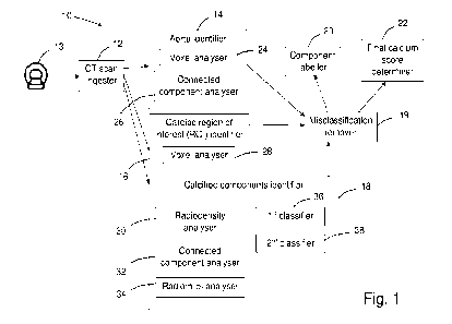
Referring to Figure 1 of the drawings, an example system 10 for calcium
scoring of
coronary arteries is shown. The system 10 includes a CT scan ingester 12
arranged to
receive cardiac non-contrast CT data from a CT scanning device 13, an aorta
identifier
14 for identifying ascending and descending aorta components in the cardiac
non-
CA 03168136 2022- 8- 16
WO 2021/168517
PCT/AU2021/050168
- 11 -
contrast CT data, a cardiac region of interest (ROI) identifier 16 for
identifying a cardiac
ROI in the cardiac non-contrast CT data, a calcified components identifier 18
for
identifying calcified components in the cardiac non-contrast CT data, a
misclassification remover 19 that uses the information from the aorta
identifier 14 and
the cardiac ROI identifier 16 to remove calcified volumes from consideration,
a
component labeller 20 and a final calcium score determiner 22.
In this example, the aorta identifier 14 includes a voxel analyser 24 arranged
to predict
using machine learning whether each voxel in received patient cardiac non-
contrast CT
i o data is part of the ascending or descending aorta of the
patient, and a connected
component analyser 26 arranged to use a connected component technique to
identify
neighbouring voxels that belong to particular components of the ascending or
descending aorta.
is The aorta identifier 14 produces a machine learning voxel mask
that can be used to
remove from consideration calcifications present on the ascending or
descending aorta
and therefore not on the coronary arteries.
In this example, the cardiac region of interest (ROI) identifier 16 includes a
voxel
20 analyser 28 arranged to predict, using machine learning,
voxels in received patient
cardiac non-contrast CT data that are part of a region of interest around the
heart of
the patient.
In this example, the calcified components identifier 18 includes a
radiodensity analyser
25 30 arranged to identify candidate voxels associated with
calcified components, for
example by applying a Hounsfield Unit thresholder to the voxel data so that
only voxels
with an associated radiodensity above a defined level are passed.
The calcified components identifier 18 also includes a connected component
analyser
30 32 arranged to use a connected component technique to identify
neighbouring voxels
that belong to the same calcified component, and a radiomics analyser 34
arranged to
analyse the identified calcified components to obtain a set of characteristics
for each
component.
3 5 In the field of medicine, radiomics is used to extract
information from radiographic
medical images. The present inventors have realised that such radiomic
features have
CA 03168136 2022- 8- 16
WO 2021/168517
PCT/AU2021/050168
- 12 -
the potential to be used in a machine learning system to identify and locate
coronary
artery calcifications. By analysing each candidate calcification component
using a
radiomics engine, characteristics describing the relative position, shape,
size and
texture of the components are obtained, and these characteristics are chosen
to
provide a rich description of the components that can be used by machine
learning
systems to learn to distinguish bone from coronary arteries as well as the
specific
artery in which the component is located. Prior to training, radiomic feature
selection is
performed by a principal component analysis (PCA) and variance thresholding.
PCA is
used to automatically determine which features provide the most discriminative
power
i o for the machine learning system. This approach provides
additional benefits over the
traditional prior art approach of hand-crafting specific features. A deep
learning model
may also look at image patches of raw CT data around each component in order
to
provide greater context.
is The calcified components identifier 18 also includes a machine
learning component, in
this example a first classifier 36 and a second classifier 38, the classifiers
trained to
output a determination as to whether a candidate calcification is present on a
coronary
artery, and the particular coronary artery in which the calcification is
disposed.
20 Figure 2 is a flow diagram 40 illustrating a method of
detecting calcifications on
coronary arteries using the system 10. According to the present method,
cardiac non-
contrast CT data is ingested 41 and the scan data is then analysed using an
aorta
identification process 42, a cardiac region of interest (ROI) identification
process 44
and a calcified components identification process 46.
In the examples described, each of the calcified components identification
process 42,
the aorta identification process 44 and the cardiac region of interest (ROI)
identification
process 46 uses a machine learning system that is trained using a sufficient
number of
relevant, known outcome, non-contrast CT scans. In the example described in
relation
to Figures 1 to 6b, the aorta identification process 44 and the cardiac region
of interest
(ROI) identification process 46 use a convolutional neural network (CNN), and
the
calcified components identification process 42 uses a plurality of
classifiers, in this
example 2 classifiers. However, other arrangements are possible. For example,
instead of using classifiers for the calcified components identification
process 42, a
standard neural network may be used. The output of the standard neural network
may
be combined with other data relevant to coronary artery calcified components
CA 03168136 2022- 8- 16
WO 2021/168517
PCT/AU2021/050168
- 13 -
identification, such as the output of a convolutional neural network arranged
to analyse
image patches around candidate calcified components in the cardiac non-
contrast CT
data.
In this embodiment, the aorta identification process 42 uses machine learning,
in this
example one or more deep learning models, to perform a semantic segmentation
process on the scan data to identify the 3D structures of the ascending and
descending aorta. The predicted ascending and descending aorta information is
subsequently used as a machine learning mask to remove from consideration
o candidate calcifications that are present on the ascending or descending
aorta and not
on a coronary artery.
The aorta identification process is shown in more detail in Figures 3a and 3b.
Figure
3a illustrates a training process for the machine learning component of the
aorta
is identification process, and Figure 3b illustrates the aorta
identification process during
use.
The aorta identification process is arranged to identify the spatial extents
of the aorta
using semantic segmentation, and uses a deep learning approach to generate for
each
20 voxel a probability that the voxel belongs to the aorta. The resultant
voxel probability
map is then used to determine components in the CT scan that are most likely
to
correspond to components of the ascending and descending aorta.
Referring to Figure 3a, in order to train the machine learning component of
the aorta
25 identification process 42, non-contrast CT scan data covering the
coronary region of a
plurality of patients is received 56 and CT scan images of the ascending and
descending aorta components are annotated 58 by experts so that a library of
ground
truth training data is produced. The ground truth aorta training data derived
from each
scan constitutes a map of voxels identified as being part of the ascending or
30 descending aorta, and the aorta machine learning component is trained 60
using the
aorta maps to recognise components of a CT scan that are part of the ascending
or
descending aorta. However, it will be appreciated that the training process
should
cover representative samples of the expected patient variation in the input
cardiac non-
contrast CT data.
In the present method and system, the aorta machine learning component is
CA 03168136 2022- 8- 16
WO 2021/168517
PCT/AU2021/050168
- 14 -
configured to progressively process single "axial" slices of the CT scan, and
assemble
the results from a series of individual slices of the CT scan into a
volumetric
segmentation. However, it will be understood that other implementations are
envisaged. For example, the present method and system is not limited to
processing
multiple individual slices but may also be configured to process volumetric
inputs or
cross-hair type orthogonal inputs. A cross-hair type volumetric analysis uses
an
approximation methodology wherein 3 orthogonal slices, each centred on the
voxel of
interest, are processed to produce an approximation of a full volumetric
analysis
centred on the voxel of interest.
After the aorta machine learning component has been trained, the aorta
identification
process 42 illustrated in Figure 3b can be applied to cardiac non-contrast CT
data to
produce an ascending/descending aorta mask that can be used to remove
misclassifications of candidate calcified components.
Referring to Figure 3b, received patient cardiac non-contrast CT data is
received 62
and analysed 64 using the trained aorta machine learning component to predict
whether each voxel in the cardiac non-contrast CT data is part of the
ascending or
descending aorta, then the voxel data is analysed 66 using a connected
component
technique to identify neighbouring voxels of the ascending or descending aorta
and in
turn identify components of the ascending and descending aorta in the cardiac
non-
contrast CT data.
Those skilled in the art of will appreciate that various suitable machine
learning
arrangements are envisaged for implementing aorta feature recognition, for
example a
wide variety of convolutional neural networks (CNN) can be effectively
employed for
semantic segmentation. In medical applications, the Unet and Vnet type CNN
architectures are cornmonly used.
In the present example, the predicted voxel data is processed using a
connected
component technique to identify voxels that correspond to adjacent connected
components of the ascending or descending aorta and to detect outliers by
identifying
neighbouring voxels using 8, 16 or 26 connectivity, although it will be
understood that
other techniques are envisaged.
Each aortic component identified using the connected component technique is
CA 03168136 2022- 8- 16
WO 2021/168517
PCT/AU2021/050168
- 15 -
analysed according to its size, shape and position, and the most likely
candidate for
each part of the aorta is chosen. Outlier detection rejects any identified
connected
component that is too small or in a position that is inconsistent with the
ascending and
descending aorta. The region, size and position constraints for outlier
detection is
dependent on the characteristics of the CT scan, including spatial resolution
and
position of the scan relative to the patient.
In this embodiment, the cardiac ROI process 44 uses machine learning, in this
example one or more deep learning models, to identify a region of interest
(ROI)
o adjacent the heart. The predicted cardiac ROI information is used as a
mask to remove
outlier candidate calcifications that are present outside the cardiac ROI and
therefore
not present on a coronary artery. A deep learning approach is used to predict
the
probability that each voxel belongs to the cardiac ROI. It will be understood
that the
cardiac ROI indicates an area of the scan in which coronary arteries are
located and
is therefore coronary artery calcification may occur, and by removing areas
outside the
cardiac ROI from consideration, features such as the lungs, ribs and spine are
ignored.
This improves both the speed and accuracy of classifying potential
calcifications.
By removing cardiac non-contrast CT data that is associated with regions
outside the
20 heart, the likelihood of false positive classifications is reduced, and
unnecessary
radiomic analysis of calcifications outside the heart, such as of the spine
and ribs, can
be avoided. As ROI segmentation is a relatively fast method of identifying
calcifications outside the heart as non-coronary artery calcifications, the
total time to
produce a final calcium score result is reduced.
The cardiac ROI process 44 is shown in more detail in Figures 4a and 4b.
Figure 4a
illustrates a training process 70 for the machine learning component of the
cardiac ROI
process 44, and Figure 4b illustrates the cardiac ROI process 44 during use.
Referring to Figure 4a, in order to train the machine learning component of
the cardiac
ROI identification process, non-contrast cardiac non-contrast CT data covering
the
coronary region of a plurality of patients is received 72 and the CT scan
images of the
ROI around the heart are annotated 74 by experts so that a library of ground
truth
training data is produced. The ground truth cardiac ROI training data derived
from
each scan constitutes a map of voxels identified as being part of the cardiac
ROI, and
the cardiac ROI machine learning component is trained 76, 78 using the cardiac
ROI
CA 03168136 2022- 8- 16
WO 2021/168517
PCT/AU2021/050168
- 16 -
maps to recognise components of a CT scan that are part of the cardiac ROI.
Those
skilled in the art of deep learning will appreciate that the maps should cover
a
representative sample of the expected patient variation in the input cardiac
non-
contrast CT data.
In the present method and system, the cardiac ROI identification process is
configured
to progressively process single "axial" slices of the CT scan, and assemble
the results
from a series of individual slices of the CT scan into a volumetric
segmentation.
However, it will be understood that other implementations are envisaged. For
example, the present method and system is not limited to using single slice
but may
also be configured process volumetric inputs or cross-hair type orthogonal
inputs.
After the cardiac ROI machine learning component has been trained, the cardiac
ROI
identification process 44 illustrated in Figure 4b can be applied to cardiac
non-contrast
CT data.
Referring to Figure 4b, during use patient cardiac non-contrast CT data is
received 80
and analysed 82 using the trained cardiac ROI machine learning component to
predict
whether each voxel in the cardiac non-contrast CT data is part of the cardiac
ROI and
produce 84 a cardiac ROI mask that can be used to remove outlier candidate
calcifications that are present outside the cardiac ROI.
The calcified components identification process 46 is shown in more detail in
Figures
5, 6a and 6b. Figure 5 illustrates a process 90 for obtaining candidate
calcification
radiomic data for input into a calcification machine learning system. Figure
6a
illustrates a training process 92 for the machine learning component of a
radiomic
characteristics analysis process 94, and Figure 6b illustrates the radiomic
characteristic analysis process 94 during use.
In this example, the radiomics characteristics describe the relative position,
shape, size
and/or density of each component, although it will be understood that any
radiomic
characteristic associated with an identified calcified volume and obtainable
from
radiographic medical imaging data is envisaged.
In addition to the radiomic characteristic information, other information that
is capable
of assisting identification and classification of coronary artery
calcifications may be
CA 03168136 2022- 8- 16
WO 2021/168517
PCT/AU2021/050168
- 17 -
used. For example, raw CT scan image patch information indicative of a region
around
each candidate calcification may be input to the classifiers or to an
additional machine
learning system. Such image patches are capable of providing useful contextual
information for each calcification.
In an example implementation, the component characteristics are input into a
plurality
of trained machine learning classifiers that have been trained to detect the
locations of
the components based on the characteristics. Alternatively, the component
characteristics are used as inputs, for example with raw image data, to a
trained deep
1 o learning model which predicts the location of the components
based on the
characteristics.
Referring to Figure 5, the process 90 for obtaining candidate calcification
radiomic data
comprises receiving 96 non-contrast cardiac non-contrast CT data, applying 98
a
radiodensity test, such as a 130 Hounsfield Unit threshold test, to identify
candidate
calcification voxels and produce a map of candidate voxels, applying 100 a
connected
component analysis process to the candidate voxel map so as to predict
neighbouring
voxels that are part of the same calcified volume, using 102 a radiomics
analyser 34 to
extract radiomic data from the candidate volumes and produce a set of radiomic
characteristics for each candidate volume, and inputting 104 the radiomic data
to a
calcification volume machine learning system, in this example that comprises
one or
more machine learning classifiers.
Referring to Figure 6a, in order to train the machine learning component of
the
radiomics characteristics analysis process, non-contrast CT scan image data
covering
the coronary region of a plurality of patients is received 106, and the CT
scan images
are labelled by an expert to mark the coronary arteries and any non-coronary
artery
components, such as for example related to bone. Candidate radiomic data
associated with the CT scan images are also received. The annotated CT scans
and
associated candidate radiomic data constitutes a library of ground truth
training data
that is used to train 2 machine learning classifiers, as indicated at step
110. Those
skilled in the art of deep learning will appreciate that the masks should
cover a
representative sample of the expected patient variation in the input cardiac
non-
contrast CT data.
After the classifiers have been trained, the radiomics analysis process 94
illustrated in
CA 03168136 2022- 8- 16
WO 2021/168517
PCT/AU2021/050168
- 1 8 -
Figure 6b can be applied to the radiomics data associated with the candidate
volumes
produced by the process 90 shown in Figure 5. According to the method,
candidate
calcification volume (radiomic) data is received 112 and input 114, 116 to a
first
classifier arranged to classify each volume as belonging to a coronary artery
or not,
and a second classifier arranged to determine the specific coronary artery on
which a
calcified volume is considered to be present. The classifier results are then
used to
predict 118 the calcifications that belong to a coronary artery and label the
coronary
arteries.
io A range of models are envisaged for the classifiers, including
random forest and K-
nearest-neighbour classifiers. The classifiers may be combined according to a
weighted voting mechanism that relates to the training performance of the
individual
models. Those skilled in the art will appreciate that ensemble vote
classification
mechanisms including hard and soft voting are appropriate implementations of
the
is weighted voting mechanism.
In this embodiment, each classifier's prediction is combined through a voting
mechanism to produce a final predicted probability for each candidate
component,
although other arrangements are envisaged. For example, labelling calcified
plaques
20 may involve a deep learning architecture that learns to
delineate between coronary
artery calcifications on the left main (LM), Left anterior descending (LAD),
right
coronary artery (RCA), left circumflex (LCX), and that also learns to detect
false
positives that are due to noise in the scan, the spine, ribs and aorta.
25 In an alternative arrangement, the final training process
involves generation of expert
annotations by trained professionals, who label each coronary artery as well
as
components that are either bone or noise. Image patches and the
characteristics
generated by the process in Figure 5 are input to the deep learning model. The
model
trains by backpropagation to optimise classification of each component.
As indicated at step 48 of the flow diagram in Figure 2, the predicted
candidate
calcifications produced by the trained machine learning classifiers are cross-
checked
against the predicted ascending and descending aorta information and the
predicted
cardiac ROI information and any candidate calcifications that are considered
to relate
to noise, or to be present on the ascending or descending aorta, or located
outside the
cardiac ROI, are removed. The Agatston score and calcium volumes are then
CA 03168136 2022- 8- 16
WO 2021/168517
PCT/AU2021/050168
- 19 -
calculated on the remaining candidate components, as indicated at steps 50 and
52,
and the coronary arteries labelled, as indicated at step 53.
In the present embodiment, the misclassification removal step, wherein
candidate
calcifications are cross-checked against the predicted ascending and
descending aorta
information and the predicted cardiac ROI information, is carried out after
all candidate
calcified volumes have been analysed by the calcified components identifier
and
radiomic characteristics produced. However, it will be understood that other
arrangements are possible. For example, the misclassification removal step may
be
i o carried out after candidate volumes have been identified by
the radiodensity analyser
30 and the connected component analyser 32, but before analysis by the
radiomics
extractor; or for example the misclassification removal step using the cardiac
ROI
information is carried out on raw CT image data. In this way, unnecessary
radiomic
processing of calcified volumes that are located on the ascending or
descending aorta
is or outside a region around the heart is avoided.
It will be appreciated that the present method reduces risk to a patient by
removing
need for contrast enhancement, reduces cost to a patient by removing need for
second
CT scan, and reduces cost to a clinic by reducing labour required to produce
calcium
20 score.
In a variation, the aorta identifier 14 may also segment the mitral valve.
Similar to the
process described in Figure 3b, this variation relies on ground-truth
annotations of the
mitral valve in order to train a deep learning model to output a prediction
for each voxel
25 as to the likelihood that the voxel is associated with the
mitral valve. A Hounsfield Unit
threshold may then be applied to the valve's segmentation mask in order to
calculate
calcification.
A heart segmentation process may also be carried out in order to improve false
30 positive detection and thereby prevent misclassification of
ribs or spine as coronary
arteries. This segmentation may take the form of a further deep learning
model, such
as a CNN, trained for detection of large aspects of the CT scan that indicate
the
location of the heart, including the ribs, spine, lungs and heart itself.
35 Aorta segmentation may also be used to create relative
position features for each
candidate calcified component. An additional use of the aorta segmentation
involves
CA 03168136 2022- 8- 16
WO 2021/168517
PCT/AU2021/050168
- 20 -
performing the process prior to, rather than simultaneously with, the
calcified
components identification process shown in Figures 5 and 6b. With information
regarding the spatial extent of the ascending and descending aorta, additional
characteristics such as the location of a calcified component relative to each
aorta
structure create a richer description, and therefore more accurate
classification, of the
components.
Classification of voxels using CNN machine learning architecture may also be
carried
out using raw or processed image data to augment radiomic feature-based
i o classification of components. In a variation of classification
by pre-calculated radiomic
characteristics, a deep learning model, such as a CNN, whose only input is the
raw
image of the component, may perform classification of each component, and the
output of this process then provided, with the output of the radiomic
characteristic
based classification, into a voting mechanism to determine the most probable
is classification for each component.
A further aspect may include localisation of the coronary arteries prior to
the image
analysis and feature creation step of Figure 5. Detection of the arteries in a
non-
contrast scan may occur by techniques such as artery tracking, which locate
the ostia
20 and move step-by-step through the arteries with guidance from
a deep learning model
such as a CNN, or by semantic segmentation in a way similar to the aorta
identification
proves described in Figure 3b. Each component can then be additionally
characterised
by its proximity to the coronary arteries, creating a richer description that
is input to the
machine learning classifiers.
Further enhancement of the characteristics used to describe calcified
components may
come from use of manually inserted calibration markers at the top of scan.
Given the
significant variability in the position of the heart in CT scans, such markers
would
provide the machine learning classifiers with a more meaningful description of
the
position of each component.
Example implementations of the coronary artery calcium scoring system and
method
will now be described with reference to Figures 7 to 13. The examples
described are
referred to as example method (and system) A, B and C.
Referring to Figure 7, a test patient demographic table 120 is shown. In the
examples,
CA 03168136 2022- 8- 16
WO 2021/168517
PCT/AU2021/050168
- 21 -
the same patients were used for methods B and C, and these patients were
different to
the patients used for method A.
The demographic information includes the number of patients 122 used in the
training
phase wherein machine learning aspects of the methods are trained, the age and
age
standard deviation of the patients used 124, the gender 126 of the patients
used, and
known calcium risk score data 128 indicative of how many test patients have a
score of
0, 1-10, 11-100, 101-400 and greater than 400. In the examples, 1055 patients
were
used for the training phase for method A, 4807 patients were used for the
training
phase for methods B and C, 241 patients were used for testing method A and
1958
patients were used for testing methods B and C.
Referring to Figures 8, 9 and 10, example systems A, B and C for implementing
example methods A, B and C are shown.
As shown in Figure 8, method and system A 130 is arranged to receive a non-
contrast
cardiac non-contrast CT data 131, and carry out an aorta identification
process 132 on
the received CT data using a U-Net convolutional neural network (CNN) 134 in
order to
segment ascending and descending portions of the aorta.
The system also passes the cardiac non-contrast CT data through a 130
Hounsfield
Unit analyser 136, and the passed voxels are analysed by a radiomics unit 138
that
generates candidate calcified volumes from the passed voxels and radiomic
characteristic data for each candidate volume for analysis by a standard
neural
network 140 that is used instead of one or more classifiers to provide
predictions for
each candidate volume as to whether the volume is associated with a coronary
artery.
The passed voxels are also used with raw CT image data to generate an image
patch
142 for each candidate volume, each image patch providing CT image context
data for
the region of the CT scan around the associated candidate volume. The image
patches are analysed using a convolutional neural network 144 to provide
predictions
for each candidate volume as to whether the volume is associated with a
coronary
artery. In this example, the convolutional neural network is a standard
AlexNet neural
network arranged to analyse image patches in a 2D axial plane.
The predictions produced using the radiomic information and the image patches
are
input to a hybrid neural network 146 that uses the combined radiomic and image
patch
CA 03168136 2022- 8- 16
WO 2021/168517
PCT/AU2021/050168
- 22 -
predictions to produce predictions for the candidate calcified volumes that
are
considered to be present on the coronary arteries, and predict the specific
coronary
arteries 148 on which calcified volumes are present.
The predictions are then updated, if necessary, by comparing with the aorta
segmentation information and removing any calcified volumes that are actually
present
on the ascending or descending aorta, but have been misclassified as being
present
on a coronary artery.
io Method and system B 150 shown in Figure 9 is similar to method
and system A 130
shown in Figure 8 except that a custom convolutional neural network 152 is
used to
analyse the image patch information instead of the convolutional neural
network 144.
Like and similar features are indicated with like reference numerals. The
custom
convolutional neural network is arranged to analyse the region surrounding
each image
is patches in multiple dimensions and in this way produces richer
contextual information
about the region surrounding each candidate calcified volume.
Method and system C 160 shown in Figure 10 is similar to method and system B
140
shown in Figure 9 except that a cardiac region of interest (ROI) analyser 162
is also
20 provided to produce cardiac ROI information that is used to
remove candidate calcified
volumes that are present outside the region of interest around the heart, and
therefore
not present on a coronary artery. In this example, the ROI information is used
to filter
out regions of the received non-contrast CT scan before the Hounsfield Unit
analysis is
carried out. Like and similar features are indicated with like reference
numerals.
Results of application of methods A, B and C indicate that method and system B
provides better diagnostic accuracy and precision than method and system A,
and
method and system C provides better diagnostic accuracy and precision than
method
and system B.
Application of method and system C to the test patient data referred to in
Figure 7
produced the results in method C results table 170 shown in Figure 11.
The results table 170 shows results172 of application of the present method
and
system Con a 1958 patient sample size, and results 174 of a conventional
manually
assisted CAC method on the same sample. The results indicate that method and
CA 03168136 2022- 8- 16
WO 2021/168517
PCT/AU2021/050168
- 23 -
system C accurately classifies 880 patients in calcium score risk category 0
(accuracy
99.44% compared to conventional manual assisted CAC), accurately classifies
233
patients in calcium score risk category 1-10 (accuracy 87.92%), accurately
classifies
375 patients in calcium score risk category 11-100 (accuracy 96.15%),
accurately
classifies 267 patients in calcium score risk category 101-400 (accuracy
98.52%), and
accurately classifies 142 patients in calcium score risk category >400
(accuracy
96.60%). The overall accuracy of method and system C is 96.88% compared to
conventional manual assisted CAC.
i o Referring to Figure 12, overall accuracy figures 180 for
methods A, B and Care
shown. It will be understood that the accuracy of method B is greater than
method A,
and the accuracy of method C is greater than method B.
Referring to Figure 13, precision figures 190 for method A 192, method B 194
and
is method C 196 are shown, the precision figures including total
precision, and precision
for each coronary artery ¨ the right coronary artery (RCA), the left main
coronary
(LMCA), the left anterior descending (LAD), and the left circumflex artery
(LCX). It will
be understood that the precision of method B is greater than method A, and the
precision of method C is greater than method B.
While the above examples are described in relation to a method and system that
is
configured for identifying coronary artery calcifications and the particular
coronary
arteries in which the calcifications are located, it will be understood that
the invention
may also be applied to identification of calcifications on other anatomical
structures of
the heart. For example, the method and system may be used to locate and
identify
calcifications on the aorta.
With this arrangement, radiomic analysis is carried out to obtain a set of
radio mic
characteristics associated with the target anatomical component, such as the
aorta or
a portion of the aorta, and a misclassification remover used to avoid or
remove
misclassifications by carrying out segmentation of body components in a
similar way to
the examples described above. Like and similar features and method steps
associated
with the examples described above are applicable.
3 5 In the claims that follow and in the preceding description of
the invention, except where
the context requires otherwise due to express language or necessary
implication, the
CA 03168136 2022- 8- 16
WO 2021/168517
PCT/AU2021/050168
- 24 -
word "comprise" or variations such as "comprises" or "comprising" is used in
an
inclusive sense, i.e. to specify the presence of the stated features but not
to preclude
the presence or addition of further features in various embodiments of the
invention.
It is to be understood that, if any prior art publication is referred to
herein, such
reference does not constitute an admission that the publication forms a part
of the
common general knowledge in the art, in Australia or any other country.
Modifications and variations as would be apparent to a skilled addressee are
deemed
io to be within the scope of the present invention.
CA 03168136 2022- 8- 16