Note: Descriptions are shown in the official language in which they were submitted.
CA 03168837 2022-07-20
WO 2021/158754
PCT/US2021/016563
DEVICES AND METHODS FOR SELECTING STENTS
TECHNICAL FIELD
The present disclosure relates to devices and methods for selecting stents for
vessels,
particularly devices and methods for determining required radial forces to
select an
appropriate stent for a target vessel.
BACKGROUND
The technique of percutaneous transluminal coronary angioplasty has been
extensively
used since the 1980s to restore blood flow in blocked arteries. It is a
relatively common
percutaneous technique that is performed on a daily basis around the world.
While
angioplasty is typically used in the coronary arteries to restore flow, the
techniques have
also been applied to peripheral arteries. In 1960, Charles Dotter developed
the first
balloon-based catheter to dilate the narrowed arteries of the leg to allow
passage of
ever-increasing diameters of catheters. In 1973, the first balloon catheter
designed for
the iliac artery was developed by physicians from the University Hospital of
Zurich.
The typical coronary angioplasty is performed under local anaesthetic with a
thin tube
inserted into the arteries of the heart with a balloon mounted onto the tip
and shaft of the
catheter. The balloon is inflated via the use of a manometer to a specific
pressure. Once
the artery has been sufficiently stretched, a stent is inserted to keep the
artery open and
to preserve blood flow. Stenting is common in modern angioplasty.
Whilst the field of coronary stenting has been developed over several decades
and
balloon-based catheters have been used in peripheral arteries, the field of
venous and
peripheral vascular stenting is still in its infancy. Peripheral venous
vasculature presents
a range of anatomical challenges that were previously unseen in coronary
arterial
stenting. Important consideration factors are the large lumen diameters, long
stent
lengths, flexible venous walls that are vulnerable to compression by external
structures,
and the highly mobile locations of the body in which the vessels are found.
All these
factors require precise positioning and stability of the stent, as well as
radial force
application by the stent to overcome the lesion. However, stents that impede
natural
movement and the underlying anatomy should be avoided. These factors
necessitate a
unique and personalized approach to stenting and angioplasty strategies to
ensure not
only excellent primary and secondary patency rates, but also without risk of
making the
1
CA 03168837 2022-07-20
WO 2021/158754
PCT/US2021/016563
individual worse through stent failure. There are now multiple manufacturers
of arterial
and venous stents, each with unique design features and builds in order to
provide "their
solution" to the problem. However, the procedure for selecting the correct
stent to
overcome the occlusion/compression is essentially guesswork.
The difficulty of correctly choosing the right stent for deployment in
peripheral
vasculature was previously complicated by the presence of numerous devices
designed
for use in coronary artery stenting and the absence of specifically designed
peripheral
venous stents. The different requirements for venous and peripheral stenting
when
compared to coronary artery stenting mean that the available equipment did not
address
the unique features and requirements of stent placement, radial force and
flexibility
needed for success in peripheral venous applications. In a great many
instances,
clinicians resort to using stents designed originally for use in the arterial
system,
repurposing them for venous use. This has resulted in poor patient outcomes as
in
certain cases the implanted devices are simply not fit for purpose. In recent
years,
manufacturers who have developed dedicated venous stents have provided their
own
solution to overcome the venous challenges. However, to date, no one stent
manufacturer has developed a single ideal stent. Stenting into the common
femoral vein
requires a woven, braided stent to prevent stent fracture and flexibility,
where a laser cut
nitinol stent could potentially fracture. Conversely a venous compression such
as a NIVL
or May-Thurner compression requires a large degree of radial force to
overcome. Radial
force is typically superior in laser cut nitinol stent compared to that of the
woven braided
stent. Additionally, the overall goal to restore flow through an occluded
venous segment,
necessitates the aim to achieve a stent that is as circular in shape as
possible, to give
the best inflow/outflow and prevent in-stent restenosis. This in itself
requires high
degrees of radial force, which may impede free movement of the individual,
inflict long-
standing pain through oversizing stents and/or cause premature stent failure
due to
increased torsional forces on the stent. So there is a delicate balance that
needs to be
found, in order to appropriately choose the right stent for the right anatomy
and to
overcome the specific occlusion.
In another example, modern balloons used in balloon-based catheters are
manufactured
from multiple different types of materials to meet the needs and requirements
of the end
product and its intended purpose. These include, but are not limited to:
polyethlene
terephthalate (PET); polyolefin copolymer (POC); nylon; polyether block amide
(PEBA
2
CA 03168837 2022-07-20
WO 2021/158754
PCT/US2021/016563
OR PEBAX0); silicone; and other compound polyurethranes. This is a change from
initial balloon-based catheters, which were initially made of flexible
polyvinyl chloride
(PVC), and then in the second generation cross-linked polyethylene (PEX).
So, complexity is first introduced by the sheer number of different raw
materials. There
are also multiple methods in which to build or construct the balloons,
including but not
limited to: extrusion; moulding; and dip casting. Different balloon properties
are
conferred depending on upon which process is used for manufacturing. The
balloons
can also be manufactured in multiple lengths, diameters, shapes, profiles, and
coatings
to achieve the desired properties.
Regardless of intended use, manufacturers have grouped the various types of
balloons
into 3 broad categories based on the intended use applications: compliant
balloons; non-
compliant balloons; and semi-compliant balloons. In compliant balloons, the
diameter of
the balloon increases proportionally to the increase in inflation force. The
size of a
compliant balloon may grow beyond the ceiling of clinical safety. In non-
compliant
balloons, the diameter of the balloon is highly restricted, so that only small
changes in
diameter are possible. Semi-compliant balloons have a wide working pressure
range
with controlled flexibility in balloon sizing.
Typically a balloon of a single manufacturer has specific characteristics, but
may differ
significantly from those of other manufacturers. As an example, there are
significant and
expected differences in compliance between the three specific types of
balloons i.e.
compliant, non-compliant and semi-compliant. Over a range of increasing
pressures, the
diameter of a non-compliant balloon is relatively constant but the diameters
of semi-
compliant and compliant balloons are much more variable.
This is further complicated when considering balloons of the same size but of
different
manufacturers, as nominal pressure and burst pressure for each balloon vary
quite
considerably. As complexity in balloons is now high, there may also be non-
negligible
differences in diameter at the nominal pressure between balloons of the same
types.
This is because manufacturing complex balloons in a repeatable manner is much
more
difficult.
3
CA 03168837 2022-07-20
WO 2021/158754
PCT/US2021/016563
Moreover, because arteries are resilient vessels that can withstand the
relatively high
forces placed on them by balloons and stents without collapse or
disintegration, there
remain relatively basic methods of inflation and measurement of balloon size
by
translating balloon pressure to lumen diameter. However this often leads to
vessel and
stent overexpansion in order to overcome recoil. Overexpansion may result in
increased
endothelial damage and increased rates of in-stent restenosis, especially in
peripheral
vasculature and the venous system. Accordingly, there is a desire to improve
the
techniques used in venous and peripheral angioplasty so that safety of the
patient is
ensured and maintained.
It is an aim of the present invention to address one or more of the
disadvantages
associated with the prior art.
SUMMARY OF THE INVENTION
According to an aspect of the present invention there is provided a catheter-
based
device for determining the radial expansion force required to displace an
occlusion in a
vessel located in a subject. The device comprises an elongate body defining a
proximal
and a distal termini, the body comprising a sheath that encloses a hollow
lumen within,
which extends along substantially the full length of the body. The proximal
terminal
region comprises: a user-interfacing hub, the hub comprising a handle for
manoeuvring
the body and configured for handling by an operator; a control interface for
controlling
the device; and a sensor configured to measure one or more parameters relevant
to a
force applied to the vessel by the device. The distal terminal region
comprises: an
expandable member movable between a retracted position, in which the
expandable
member is within the hollow lumen, and a deployed position, in which the
expandable
member is disposed beyond the distal terminus, and controllable via the
control interface
to expand radially. The expansion of the expandable member is correlated to a
defined
radial expansion force value.
According to another aspect of the invention, there is provided a method for
determining
the radial expansion force required to displace an occlusion in a vessel
located in a
subject. The method comprises: providing a catheter-based device having an
expandable member expandable to apply force to the occlusion; disposing the
expandable member within the vessel in the region of the occlusion; expanding
the
expandable member to achieve a target profile within the lumen, wherein the
expansion
4
CA 03168837 2022-07-20
WO 2021/158754
PCT/US2021/016563
of the expandable member is correlated to a defined radial expansion force
value; and
determining the radial expansion force value applied by the expandable member
to the
lumen to achieve the target profile based on the correlation.
Within the scope of this application it is expressly intended that the various
aspects,
embodiments, examples and alternatives set out in the preceding paragraphs, in
the
claims and/or in the following description and drawings, and in particular the
individual
features thereof, may be taken independently or in any combination. That is,
all
embodiments and/or features of any embodiment can be combined in any way
and/or
combination, unless such features are incompatible. The applicant reserves the
right to
change any originally filed claim or file any new claim accordingly, including
the right to
amend any originally filed claim to depend from and/or incorporate any feature
of any
other claim although not originally claimed in that manner.
BRIEF DESCRIPTION OF THE DRAWINGS
One or more embodiments of the invention will now be described, by way of
example
only, with reference to the accompanying drawings, in which:
Figure 1 shows a vessel with a compression that is being treated by a stent;
Figures 2A to 2C show (A) simultaneous arterial and venous contrast injection
in a
therapy resistant hypertensive patient, with no signs or symptoms of leg
swelling (LAO
orientation). (B) and (C) demonstrate impeded contrast flow in the vein via
direct
overriding arterial compression taken from both AP and LAO angles
respectively; white
arrows show the location of the venous obstruction;
Figure 3 shows a system including a device for determining radial force
according to an
embodiment of the invention;
Figures 4A to 4E show the use of the device of Figure 3 within a target vessel
according
to an embodiment of the invention;
Figure 5 shows a distal end of a catheter with an inflated balloon according
to an
embodiment of the invention;
5
CA 03168837 2022-07-20
WO 2021/158754
PCT/US2021/016563
Figure 6 shows a distal end of a catheter with an inflated balloon according
to another
embodiment of the invention;
Figure 7 shows a distal end of a catheter with an inflated balloon according
to another
embodiment of the invention;
Figure 8 shows a distal end of a catheter with an inflated balloon according
to another
embodiment of the invention;
Figure 9 shows a distal end of a catheter with an inflated balloon according
to another
embodiment of the invention;
Figure 10 shows a distal end of a catheter with a deflated balloon according
to an
embodiment of the invention;
Figure 11 shows a distal end of a catheter with a deflated balloon according
to another
embodiment of the invention;
Figure 12 shows a distal end of a catheter with a deflated balloon according
to another
embodiment of the invention;
Figures 13A to 13D show different mechanisms for positioning a balloon
relative to a
compression of a target vessel;
Figure 14 illustrates a flow chart governing the use of the device in
determining radial or
local force according to an embodiment of the invention;
Figure 15 shows a distal end and a proximal end of a catheter with a basket
according
to an embodiment of the invention;
Figure 16 shows a distal end and a proximal end of a catheter with a basket
according
to another embodiment of the invention; and
6
CA 03168837 2022-07-20
WO 2021/158754
PCT/US2021/016563
Figure 17 shows a distal end and a proximal end of a catheter with a basket
according
to another embodiment of the invention.
DETAILED DESCRIPTION
All references cited herein are incorporated by reference in their entirety.
Unless
otherwise defined, all technical and scientific terms used herein have the
same meaning
as commonly understood by one of ordinary skill in the art to which this
invention
belongs.
Definitions
Prior to setting forth the invention, a number of definitions are provided
that will assist in
the understanding of the invention.
As used in this description, the singular forms "a," "an," and "the" include
plural referents
unless the context clearly dictates otherwise. Thus, for example, the term "a
sensor" is
intended to mean a single sensor or more than one sensor or to an array of
sensors.
For the purposes of this specification, terms such as "forward," "rearward,"
"front,"
"back," "right," "left," "upwardly," "downwardly," and the like are words of
convenience
and are not to be construed as limiting terms. Additionally, any reference
referred to as
being "incorporated herein" is to be understood as being incorporated in its
entirety.
As used herein, the term "comprising" means any of the recited elements are
necessarily
included and other elements may optionally be included as well. "Consisting
essentially
of" means any recited elements are necessarily included, elements that would
materially
affect the basic and novel characteristics of the listed elements are
excluded, and other
elements may optionally be included. "Consisting of" means that all elements
other than
those listed are excluded. Embodiments defined by each of these terms are
within the
scope of this invention.
The term "kink resistance" refers to a stent's ability to withstand mechanical
bending
loads from the surroundings depending upon the position in the body. Usually,
this is
based upon the smallest radius of curvature a stent can withstand without the
formation
of a kink. In areas of high tortuosity within the body it is necessary for a
stent to have
7
CA 03168837 2022-07-20
WO 2021/158754
PCT/US2021/016563
increased kink resistance to prevent a reduction in lumen patency or even
total
occlusion.
The term "crush resistance" refers to the ability of a stent experiencing
external, focal or
distributed loads to resist collapse. These loads ultimately lead to stent
deformation and
even full or partial occlusion which can result in adverse clinical
consequences. Crush
resistance of an endovascular device may be measured using the parallel plate
method
to determine the effective load required to reduce the luminal diameter by 50%
as
described in ISO 25539-2.
The term 'obstruction' or 'occlusion' refers to any occurrence whereby the
diameter (or
'caliber') of a vessel is reduced when compared to a normal, i.e. non-
occluded, state.
Venous obstruction can occur through the narrowing (stenosis) of a vein,
through
blockage or through externally applied pressure causing a localised
compression of the
vein. The term also includes venous occlusion, whereby the vein's lumen is
partially or
totally obstructed to the flow of blood. Occlusion may result from thrombosis
(e.g. deep
vein thrombosis (DVT)) or may be due to tumour incursion. The term also
includes
'venous compression', which refers to the external compression of the vein.
The source
of external compression may be caused by an adjacently located artery
compressing
the vein against another fixed anatomical structure, which can include the
bony or
ligamentous structures found in the pelvis, the spine itself, or overlapping
arterial
branches. External compression may also arise from tumours, growths, glands,
developing foetuses and/or other developing mass that may occur within the
pelvic
space.
The term 'venous return' is defined by the volume of blood returning to the
heart via the
venous system, and is driven by the pressure gradient between the mean
systemic
pressure in the peripheral venous system and the mean right atrial pressure of
the heart.
This venous return determines the degree of stretch of heart muscle during
filling,
preload and is a major determinant of cardiac stroke volume.
The term `May-Thurner syndrome' (MTS) also known as iliac venous compression
syndrome (which includes Cockett's syndrome) is a form of ilio-caval venous
compression wherein the left common iliac vein is compressed between the
overlying
right common iliac artery anteriorly and the lumbosacral spine posteriorly
(fifth lumbar
8
CA 03168837 2022-07-20
WO 2021/158754
PCT/US2021/016563
vertebra). Compression of the iliac vein may cause a myriad of adverse
effects,
including, but not limited to discomfort, swelling and pain. Other less common
variations
of May-Thurner syndrome have been described such as compression of the right
common iliac vein by the right common iliac artery; this is known as Cockett's
syndrome.
More recently, the definition of May-Thurner syndrome has been expanded to
include
an array of compression disorders associated with discomfort, leg swelling and
pain,
without the manifestation of a thrombus. Collectively, this has been termed
non-
thrombotic iliac vein lesions (NIVL).
The term Intraluminal thickening' (also referred to as venous spurs or
intraluminal spurs)
is related to this external compression of the left common iliac vein by the
right common
iliac artery against the fifth lumbar vertebra. Venous spurs arise due to the
chronic
pulsation of the right common iliac artery. This ultimately results in an
obstruction to
venous outflow. Venous spurs are internal venous obstructions consequent to
chronic
external compression of veins by adjacent structures.
The term 'Deep Vein Thrombosis' (DVT) refers to the formation of blood clots
or
thrombus within the venous segment, and in itself is not life threatening.
However, it may
result in life threatening conditions (such as pulmonary embolism) if the
thrombus were
to be dislodged and embolize to the lungs. Additionally, DVT may lead to loss
of venous
valvular integrity, lifelong venous incompetence and deep venous syndrome
which
includes rest and exercise pain, leg swelling and recurrent risk of DVT and
emboli. The
following is a non-limiting list of factors that reflect a higher risk of
developing DVT
including prolonged inactivity, smoking, being dehydrated, being over 60,
undergoing
cancer treatment and having inflammatory conditions. Anticoagulation which
prevents
further coagulation but does not act directly on existing clots, is the
standard treatment
for deep vein thrombosis. Other potentially adjunct, therapies/treatments may
include
compression stocking, selective movement and/or stretching, inferior vena cave
filters,
thrombolysis and thrombectomy.
The term "nominal pressure" is the balloon inflation pressure at which the
balloon
reaches its stated size without external influence.
The term "rate burst pressure" is the balloon inflation pressure at or below
which 99.9%
of balloons of that type will not burst.
9
CA 03168837 2022-07-20
WO 2021/158754
PCT/US2021/016563
The term "working range" is the range of balloon inflation pressures between
the nominal
and rate burst pressures.
The term "compliant" refers to balloons whose diameter increases
proportionally to the
increase in pressure within the balloon.
The term "non-compliant" refers to balloons that expand to an intended size as
internal
pressure increases. Once the balloon reaches its intended size, its size does
not change
further. These balloons are generally used to transmit force on a lumen wall
or displace
an extrinsic compression.
The term "semi-compliant" refers to balloons that expand to a range of size as
the
internal pressure increases.
Description of embodiments
Figure 1 shows a schematic representation of a blood vessel 10 incorporating a
stent
20. The blood vessel 10 may be an artery or a vein, or even a non-vascular
duct. The
vessel 10 has an occlusion 12. Although referred to as an occlusion here, the
occlusion
may alternatively be a region of stenosis, a compression of the vessel, a
reduced calibre
caused by an external force pressing on the vessel 10, or anything else that
causes a
closing or constriction of the lumen of the vessel 10 that is detrimental to
its flow
characteristics. To restore the lumen of the vessel 10 to its conventional
diameter and
shape, a stent 20 is positioned within the lumen of the vessel 10 and in
direct contact
with the tissue forming the vessel 10. The stent 20 acts to reduce the impact
of the
occlusion 12 on the flow of blood through the vessel 10. The stent 20 expands
the vessel
10 to an aspect ratio of close to or exactly 1.0 at a diameter that is similar
to the
surrounding, healthy, undilated tissue of the vessel 10. This undilated tissue
is typically
found downstream of the occlusion where no congestion is present in the vessel
10. An
aspect ratio of ¨1.0 ensures continuity of flow through the vessel 10 without
a restriction
in the velocity of the flow of blood. An aspect ratio of ¨1.0 also ensures
that turbulence
is avoided in the flow. An aspect ratio of substantially 1.0 may be considered
to be an
aspect ratio of between 0.9 and 1.1, or more preferably between 0.95 and 1.05.
10
CA 03168837 2022-07-20
WO 2021/158754
PCT/US2021/016563
In an example of a constriction of a vessel, an individual may have no
apparent signs or
symptoms of leg swelling but, nevertheless, an obstruction or compression of
the veins
in the ilio-caval region may be suspected. Normal anatomy in this region sees
the vein
assume an upward sigmoidal curve from the femoral vein to the inferior-vena
cave. In
Figure 2A-C an example of arterial compression of an adjacent underlying vein
is
observed using contrast fluoroscopy. It would be apparent to the skilled
person that a
solution is required that allows for the restoration of luminal patency and
normal blood
flow. The skilled person may understand that relieving the obstruction in this
region by
implanting a stent with low flexibility and high crush resistance would
profoundly alter
the local anatomy and may not be in the best interests of the body and in the
longer term
could induce restenosis and intimal hyperplasia resulting in stent failure and
more
severe venous occlusion. The skilled person may therefore understand that a
highly
flexible stent with one or more reinforced regions positioned only at the
specific points
where the compressions are observed (see white arrows in Figure 2C) would be
the
requirement for the stent. The reinforced regions may be provided either as
integrated
within the stent or as individually positionable reinforcing stent elements.
However, the challenge lies in deciphering, from these images alone, the
characteristic
values that a stent positioned within the vessel should apply to the vessel,
such as the
outwardly radial force or crush resistance. An under-performing stent will
have negligible
effect, while an overzealous stent that applies too high forces on the vessel
will be
detrimental to the health of the patient. It is currently difficult for
physicians to assess the
potential success for a given stent to adequately restore lumina! diameter. It
is currently
only after placement of the chosen stent that a physician may realize that
force applied
by the stent is unsuitable for the vessel. A stent applying insufficient force
to resist the
compression will not correct the vessel's obstruction adequately. A stent
applying a too
high force may deform the vessel into an undesirable shape or may cause damage
to
the vessel itself, causing collapse or further complications.
Accordingly, the inventors have devised means for determining a target force
to be
applied by a stent deployed in the target vessel 10 at the site of the
occlusion. In
determining a target force, a medical practitioner is able to select a stent
for placement
within the lumen of the target vessel 10 in order to restore normal or near-
normal blood
flow past the occlusion 12. While existing systems rely on assessing imagery
alone to
11
CA 03168837 2022-07-20
WO 2021/158754
PCT/US2021/016563
effectively guess which stent to choose, the approach described herein
provides data
from several sources to enable a more precise stent choice to be made.
In general, the systems devised by the inventors comprise a catheter or
catheter-based
device, which may be referred to as a force catheter, configured to be passed
along the
target vessel. An elongate body of the catheter device comprises a proximal
terminus
region comprising a user-interface hub and a control interface for controlling
the
progress and operation of the catheter. The user-interface hub and/or control
interface
may comprise a handle of the catheter for handling the device and manoeuvring
the
device by an operator. The control interface may comprise one or more controls
for
enacting actions to performed using the catheter device. At a distal terminus
region, an
expandable member, also referred to as a vessel expander, is mounted to a main
shaft
of the catheter device. The expandable member is configured to be deployed
from a
hollow lumen of the elongate body to extend beyond the distal terminus of the
elongate
body. The expandable member is configured to expand in order to move the
target
vessel to a target profile, i.e. to a target aspect ratio, generally an aspect
ratio of
approximately unity (i.e. 1), and to a target diameter. The expandable member
expands
within the target vessel to expand the lumen of the vessel and to restore
patency of the
target vessel. In expanding the target vessel, the expandable member applies a
force to
the interior of the lumen in the region of an occlusion. The force applied by
the
expandable member on the target vessel to achieve the target profile may be
measured
either directly or indirectly based on the operation of the expandable member
using a
measurement device associated with the expandable member. The systems may also
include one or more imaging systems to enable imaging and therefore guidance
of the
catheter within the target vessel. The force applied by the expandable member,
namely
the radial expansion force, is correlated to the expansion of the expandable
member
and can be determined accordingly.
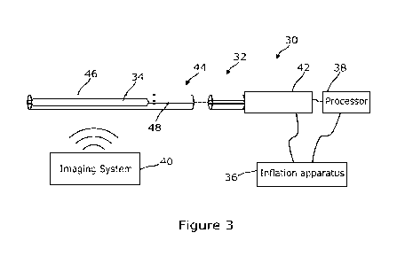
An example system 30 is shown in Figure 3. The system 30 of Figure 3 has a
catheter
32 including an expandable member 34 in the form of an inflatable balloon,
inflation
apparatus 36 for inflating and deflating the balloon, a processor 38 connected
to the
catheter 32 and the inflation apparatus 36, and an imaging system 40.
The imaging system 40 may be any suitable system for use in imaging the target
vessel
10 and/or parts of the catheter device 32. The imaging system 40 may include
an
12
CA 03168837 2022-07-20
WO 2021/158754
PCT/US2021/016563
Intravascular Ultrasound (IVUS), an Optical Coherence Tomography (OCT), a
contrast
fluoroscopy systems, or other imaging modality or a combination of these. IVUS
and
OCT are preferable as they are typically used to determine vessel size and
lumen size
accurately.
It should be noted that, as indicated in Figure 3, the imaging system 40 is
separate from
the catheter device 32 itself. The imaging system 40 is used to visualize the
catheter 32
as it progresses along the target vessel 10 and to identify when the catheter
32 is
correctly positioned. The imaging system 40 may also be used for preparatory
investigations prior to insertion of the catheter 32 into the patient's body,
and even prior
to selection of a balloon size for use in dilating the target vessel 10.
In some embodiments the imaging system 40, or part of the imaging system 40,
may be
incorporated into the catheter device 32 itself. In these embodiments, a
central lumen of
the catheter 32 may be dimensioned to accommodate an IVUS catheter such that
IVUS
can be used at the same time as the balloon is being positioned and inflated.
There may
be one or more slotted windows along the shaft of the delivery catheter 32
that allow for
visualization with IVUS if available for precise positioning of the balloon.
The processor 38 may receive data output from the inflation apparatus 36, the
imaging
system 40, and/or one or more sensors in the catheter 32. The processor 38 may
analyze the received data to determine a radial force that a stent 20 should
apply to the
occluded target vessel 10 to overcome the occlusion. The processor 38 may
perform
one or more further actions, as will be discussed below. In some examples, the
processor 38 instead may be configured to convert the output data it receives
into charts
for interpretation by a medical practitioner instead of or in addition to the
determination
of radial force. The charts generated may be displayed on a display device.
Turning now to the catheter device 32, the catheter device has a handle 42 and
a
catheter body 44. The handle 42 is positioned at a proximal end of the device
32. The
handle 42 is attached to the elongate catheter body 44 that extends to a
distal end of
the device 32. The handle 42 is utilized by the user of the device, typically
a medical
practitioner, to control and manoeuvre the catheter body 44. The catheter body
44
connects to the handle 42 at its proximal end. The catheter body 44 is
configured to be
delivered along the lumen of the target vessel 10. The distal end of the
catheter body
13
CA 03168837 2022-07-20
WO 2021/158754
PCT/US2021/016563
44, forming the distal end of the device 32, is a free end. In some
embodiments, the
catheter body 44 may be passed over a guide wire (not shown in Figure 3). The
use of
a guidewire is discussed in relation to later embodiments.
Catheter bodies, such as the catheter body 44 of Figure 3, are suitably
constructed in a
variety of sizes typically ranging from 0.6 mm up to 3.33 mm in diameter
(corresponds
to French sizes 2 to 10). Guidewires for use with catheters of the invention
are typically
in the size range of 0.05 mm to about 1 mm (about 0.002 inches to about 0.05
inches).
The catheter body is suitably manufactured from plastics or polymeric
biocompatible
materials known in the technical field, for example, PTFE. In one embodiment
of the
invention (not shown), the device catheter body may be manufactured from a
flexible
material so as to enable the device to follow the natural curvature of the
lumen of the
vessel through which it is travelling.
The catheter body 44 in Figure 3 comprises an introducer sheath 46. The
introducer
sheath 46 has a central lumen, within which a shaft 48, such as a hypotube, is
provided.
The introducer sheath 46 and shaft 48 are capable of being advanced together
along
the target vessel 10. As required, the sheath 48 may be withdrawn to expose
the distal
end of the shaft 46 carrying an expandable member 34. The shaft 46 may
alternatively
be capable of being advanced beyond the end of the sheath 48. In either
deployment,
the relative movement is enacted and controlled remotely, either using
controls at the
handle 42 or otherwise.
An expandable member 34 is provided at the distal end of the shaft 48. The
shaft 48
and expandable member 34 may together be advanced over a guidewire deployed
along
the target vessel 10. In the case of Figure 3, the expandable member 34 is a
balloon.
When the shaft 48 and expandable member 34 are within the sheath 46, the
expander
34 is in an unexpanded state to allow passage along the target vessel 10. The
balloon
is in the unexpanded state when it is deflated and folded to fit within the
lumen of the
sheath.
In some embodiments, as will be described later other expandable members may
be
used instead of the balloon. Expandable members that may be used in this
device
include the basket arrangement of Figures 15 to 17. Other expandable members
such
as coils, tethered expandable stents or helical basket arrangements that are
mountable
14
CA 03168837 2022-07-20
WO 2021/158754
PCT/US2021/016563
to the shaft and where the force applied to the vessel by the expander can be
quantified
may be used in conjunction with this device and instead of the balloon.
For now, returning to the embodiment of Figure 3, in which the expandable
member 34
comprises the balloon, the balloon is capable of being inflated and deflated
using the
inflation apparatus 36 connected to the device 32. The inflation apparatus 36,
generally
a manometer and/or another inflation device and pressure gauge, inflates the
balloon
by passing a pressurised solution along an internal lumen that extends along
the shaft
48 to permit fluid communication with the inside of the balloon. The
pressurised solution
is typically a mixture of saline solution and a contrast agent. In some
embodiments, a
gas may be used to inflate the balloon. The inflation apparatus 36 is
configured to inflate
the balloon while measuring the pressure within the balloon. To deflate the
balloon, the
inflation apparatus 36 allows venting of the pressurised solution from the
balloon via the
lumen in the shaft 48.
Figures 4A to 4E illustrate a positioning and inflation of the balloon 34
within the target
vessel 10. Figure 5 provides another representation of the inflated balloon 34
as part of
the catheter 32.
It should be noted that Figures 4A to 4E are schematic depictions only and
that the
interaction of the balloon 34 with the obstruction may be different in
practice. In
particular, although the obstruction is shown as getting smaller in Figures 4D
and 4E,
this is meant to only be representative of an opening of the lumen to restore
patency of
the vessel. In practice, the balloon 34 is likely to displace the obstruction
and vessel wall
to restore the internal diameter.
Initially, the catheter body 44 is advanced along the target vessel 10 until
it reaches the
occlusion 12 as shown in Figure 4A. Once the occlusion 12 has been reached,
the
sheath 46 is drawn back to expose and deploy the expandable structure of the
expandable member, in this case the balloon 34, as shown in Figure 4B. The
balloon 34
is then inflated. The inflation of the balloon 34 is performed in stages, as
will be
discussed in more detail later. The balloon 34 is inflated in stages until the
balloon 34
has restored the target profile of the target vessel 10. When the vessel 10
has reached
the target profile, the balloon 34 will also have the target profile. In
Figure 4C and 4D,
the target profile has not yet been reached ¨ it can be seen that the balloon
34 is not the
CA 03168837 2022-07-20
WO 2021/158754
PCT/US2021/016563
same diameter as the healthy tissue either side of the occlusion 12. In Figure
4E, the
target profile has been reached. At this point, the balloon 34 has been
inflated to a point
at which the target profile of the vessel 10, i.e. the aspect ratio of ¨1.0
and the target
diameter of the surrounding tissue, has been achieved. Generally, this
involves the
balloon 34 moving the occlusion 12 in order to re-open the vessel, and thus
restore
optimal flow. In displacing the occlusion 12, the balloon 34 is effectively
performing the
role that the stent 12 will later perform on a more permanent basis. Once the
target
profile has been reached the force applied by the balloon 34 when the target
profile has
been reached is determined.
The force applied by the balloon 34 is determined, in this embodiment, by
measuring
the hydrostatic pressure within the balloon 34 and correlating this pressure
with an
applied force. The correlation may be performed by the processor 38 and may be
based
on log tables or charts generated by experiments. In other embodiments, other
mechanisms for determining the force may be used, such as a measurement from a
direct or indirect force sensor provided on the expandable member. Based on
the force
readout necessary to displace the occlusion 12 and restore vessel patency, a
medical
practitioner can select an appropriate stent for implanting within the vessel
to apply a
similar force. Physical properties of venous stents are known, for example see
Dabir et
al. (Cadiovasc Intervent Radio! (2018) Jun; 41(6): 942-950).
In certain instances, the force applied by the expandable member to the target
vessel
may be the force required to displace an extrinsic compression and/or kink in
a primary
stent and/or another obstruction.
The balloon 34 has specific properties that permit it to be used as an
expandable
member within the context of this application. In other words, the balloon is
specifically
designed so that the pressure therein is correlatable with the force it
imparts upon the
lumen of the target vessel. Properties of angioplasty balloons and testing
methods
associated therewith are described in ISO 25539.
Particularly, in embodiments of the invention, the balloon 34 is a non-
compliant balloon.
Non-compliant balloons inflate to a predetermined size and shape. Once the
predetermined size and shape are reached further expansion of the balloon with
increasing pressure is negligible until the burst pressure is reached. Because
of its non-
16
CA 03168837 2022-07-20
WO 2021/158754
PCT/US2021/016563
compliance, the balloon 34 is capable of applying a force to the lumen wall in
order to
expand the target vessel in which it is deployed. As the balloon 34 is
selected to have a
diameter substantially equivalent to the diameter of the unoccluded target
vessel 10 and
the diameter of non-compliant balloons once fully inflated remains
substantially the
same at pressures below the burst pressure, the balloon 34 having will not
dilate the
target vessel but will apply a force to restore the target vessel to the
target profile and
aspect ratio.
To enable general use of the balloon 34, there is a repeatable correlation of
the balloon's
pressure with the force it applies to overcome the occlusion 12. This is
achieved by
careful design of the balloon combined with the inflation apparatus enabling
accurate
determination and control of the pressure within the balloon 34. Careful
design of the
balloon 34 is achieved by adhering to strict manufacturing tolerances to
ensure each
balloon has substantially similar inflation and deflation characteristics. The
high
standards applied in these balloons means that inflation of each balloon is
highly
repeatable and that the pressure within each balloon can be correlated to the
radial
expansion force applied to the target vessel 10 upon deployment.
In addition, it can be seen in Figure 5 that the catheter body 44 has a
rounded tip or
nose 52 at its distal end. The rounded nose 52 prevents trauma being caused to
the
vessel 10 should it come into contact with the wall of the vessel 10. To be
clear, Figure
5 also illustrates an inflated balloon 34, the shaft 48 to which the balloon
34 is mounted
and the introducer sheath 46. The balloon 34 is depicted in the deployed and
inflated
state in Figure 5. The vessel and occlusion are not shown in Figures 5 to 12.
In addition to sensing the force, it is also important to understand how the
vessel 10 and
balloon 34 are interacting. One or more sensors may be provided on or in the
balloon in
addition to the imaging apparatus 40 to characterise the interaction of the
balloon 34
and vessel 10, particularly in relation to how the balloon 34 is inflating.
Given that the
occlusion 12 and vessel 10 may apply different forces at different
circumferential and
longitudinal points on the balloon 34, being able to understand the balloon's
inflation
beyond what can be gathered from the imaging apparatus 38 is highly
beneficial.
In particular, it is important to ascertain that the balloon 34 has truly
reached the target
aspect ratio, that the balloon 34 is not kinked or in some way under-inflated,
and/or
17
CA 03168837 2022-07-20
WO 2021/158754
PCT/US2021/016563
where the greatest force is being exerted by the balloon 34. In addition,
determining the
configuration within the central region of the balloon 34, as well as along
its length where
possible, can be useful as these interactions may differ depending upon the
relative
location of the balloon 34 and the occlusion 12.
One or more of several different sensing mechanisms for characterising the
interaction
between the balloon 34 and the occlusion 12 may be used.
Figures 6 to 8 show embodiments of the balloon 34 that include arrangements of
sensors
on the internal or external surface of the balloon 34. This is in contrast to
the
embodiments of Figures 3 to 5, where the balloon 34 is shown without sensors.
As will
be appreciated, the provision of a non-compliant balloon 34 whose internal
pressure is
correlatable with a force applied to a lumen, and the methodologies
surrounding the use
and testing of the balloon is a core concept of the present application. The
addition of
sensors improves the certainty of the measurements for the medical
practitioner.
Figures 6 and 7 illustrate two embodiments incorporating contact sensors 54
onto the
balloon 34. In Figure 6, a band 56 of contact sensors 54 is provided around
the
circumference of the balloon 34 at its centre. The contact sensors 54 are
evenly spaced
around the circumference of the balloon 34. In Figure 7, three bands 56, 57,
58 of contact
sensors 54 are provided around the circumference of the balloon 34. The bands
56-58
of contact sensors 54 are spaced evenly longitudinally along the balloon 34.
It will be
appreciated that two bands, or more than three bands of sensors may be
provided as
desired. In some embodiments, the sensors 54 may not be arranged in bands but
may
be positioned in other ways around the balloon. Similarly, although in Figure
7 the bands
56-58 of sensors 54 are longitudinally aligned, in other embodiments the bands
of
sensors may be staggered relative to one another.
These contact sensors 54 may be configured to detect electrical impedance or
resistance, therefore allowing determination of when the balloon 34 is and is
not in
contact with the wall of the vessel 10. When the balloon 34 is in contact with
the vessel
at all points on its circumference, the balloon 34 and vessel 10 should have
reached an
aspect ratio of substantially 1Ø The practitioner may use the impedance
sensors to
understand the orientation of the balloon 34 within the vessel 10 and to
determine where
there is not contact being made and why. Each contact sensor 54 typically
comprises
18
CA 03168837 2022-07-20
WO 2021/158754
PCT/US2021/016563
an electrode supplied with a direct current and configured to measure the
resistance
through the electrode. The resistance of the electrode changes with changes in
contact
between the electrode and a surface.
By incorporating more sensors 54 around a particular circumference, the
positions at
which the balloon 34 is not in contact with the vessel 10 can be more
accurately
determined. The arrangement of Figure 7, with longitudinally-spaced bands 56-
58 of
sensors 54, allows the surface contact with the occluded region of the vessel
10 to be
monitored as well as the surface contact with the regions either side of the
occluded
region. This is because it is expected that the occluded region will be
contacted by the
central circumference of the balloon 34, and that the balloon 34 will extend
either side
of the occluded region.
Based on the change of impedance and/or resistance within the electrodes, the
pressure
applied between the vessel 10 and the balloon 34 may also be determined.
Therefore,
the contact sensors 54 may be used as both contact sensors and pressure
sensors to
give another means for determining the force required by a stent 20. In some
embodiments, the balloon tolerances may be less strict if pressure values are
also
measured using sensors such as these. In some embodiments, separate force or
pressure sensors may be incorporated into the balloon to characterise the
force between
the balloon and vessel.
Figure 8 illustrates the bands 56-58 of contact sensors 54 with two additional
profile
sensors 60, 61 positioned between the bands 56-58. One profile sensor 60 is
provided
between the left-hand and centre contact sensor bands 57, 56 and the other
profile
sensor 61 is provided between the centre and right-hand contact sensor bands
56, 58.
The profile sensors 60, 61 extend around the circumference of the balloon 34.
Although
the profile sensors 60, 61 are shown here in conjunction with the contact
sensors, it will
be appreciated that they could, in other embodiments be used in isolation or
with
different types of sensors. Similarly, although they are here shown to be
positioned
between the bands of contact sensors, they may, in other embodiments be
positioned
elsewhere. In other embodiments different numbers of profile sensors may be
provided.
In some embodiments, no profile sensors are provided. In others, one profile
sensor is
provided. In yet further embodiments, a plurality of profile sensors are
provided.
19
CA 03168837 2022-07-20
WO 2021/158754
PCT/US2021/016563
The profile sensors 60, 61 are provided to enable determination of the profile
of the
balloon 34 during inflation. As before, profile here is used to describe
aspect ratio and
diameter of the balloon 34, or more simply, size and shape. By determining
size/diameter of the balloon 34, it can be determined when the balloon is
fully inflated to
its correct size. The profile sensors permit determination of the aspect ratio
to ensure
that the balloon inflates correctly around its circumference. Using imaging
techniques
alone, it may be difficult to see if the balloon is inflating incorrectly, for
example if there
are kinks in the balloon or if the balloon is caught up in the vessel. Profile
sensors may
comprise strain gauges
The above sensors may comprise one or more printed electrodes. A printed
electrode
sensor would typically be a printed strip of conductive material on a surface,
typically an
internal surface of the balloon. The sensor may be circumferential around the
balloon.
When used for a profile sensor, the electrode may act as a strain gauge, and
may
comprise two separated halves with interspaced branches so that the
capacitance
between the two halves can be measured and the distance therebetween
determined.
The electrode may be circumferentially arranged around a section of the
balloon, and,
where an array of sensors is provided, the sensors may be spaced along the
length of
the balloon at regular intervals. The shape of the balloon along its length
may be
determined using a sensor array.
Figures 10 to 12 demonstrate how the balloon 34 of Figures 6 to 8 may be
positioned in
the retracted state within the introducing sheath 46 prior to deployment and
inflation.
To complement the sensors, the capabilities of the imaging system 40 may be
enhanced. The catheter body 44 may further incorporate one or more means for
positioning the catheter shaft 48 and balloon 34 using the imaging system 40.
Figure 9 shows the catheter body 44 of Figure 8 being passed over a guide wire
50. The
catheter body 44 of the embodiment shown in Figure 9 also comprises a
plurality of
apertures 64 in the catheter shaft 48 for injecting a contrast agent or other
fluid or
visualization agent such as CO2. As can also be seen in Figure 9, indicated by
the dotted
lines, the catheter shaft 48 also passes through the balloon 34.
20
CA 03168837 2022-07-20
WO 2021/158754
PCT/US2021/016563
Positioning mechanisms may be provided on the sheath 46 or the catheter shaft
44 for
use in cooperation with the imaging system. Figures 13A to 13D provide various
different
examples of these positioning mechanisms for aligning the balloon 34. As
illustrated in
Figure 13A, the catheter may have radiopaque distance markers along its length
for
correct alignment. Figure 13B illustrates how the catheter may also comprise
ultrasound
windows to permit IVUS visualization. Alternatives presented in Figures 13C
and 13D
are respectively that the nose of the catheter may be radiopaque and flexible
and that
the catheter may be advanced over a guide wire for correct positioning. In
other
embodiments, the attachment points of the device may each have a radiopaque
marker
to provide an indication of the locations of the attachment points relative to
one another.
Although discussed in tandem with the device above, the methods of deploying
and use
of the device will now be discussed. In general, the device may be deployed
and utilized
for determining radial force by the steps shown in Figure 14. While the
expandable
member in the method discussed below is a balloon, it will be appreciated that
the
method may also be performed using another type of expandable member instead
of a
balloon.
Before the method 200 of Figure 14 is begun, it is assumed that a target
vessel with a
compression has been identified. The identification of the vessel is performed
using the
imaging system, and may include a venogram using magnetic resonance of
computerized tomography techniques. Prior to the insertion of the catheter,
other
preparatory steps may also be performed. For example, other balloon-based
catheters
may be used to break up any stenosis present in the target vessel or to
otherwise
prepare the vessel. In other examples, guide wires may be passed through the
occlusion
to guide the catheter of the device. Of course, while not mentioned here, all
preparatory
steps to prepare the patient for receiving the catheter are also performed.
In addition, any preparatory measurements are also taken prior to the
introduction of the
device. Preparatory measurements, which are discussed in more detail in
relation to
later methods, may include determining an aspect ratio and diameter of the
target vessel
elsewhere other than the occlusion, i.e. its normal lumina! dimensions. Based
on these
determinations, an appropriate balloon can be selected for use in the method.
21
CA 03168837 2022-07-20
WO 2021/158754
PCT/US2021/016563
Selecting an appropriate balloon may be performed by looking at imaging from
an IVUS
or other venographic imagery. From these images, an initial assessment of the
vessel
diameter may be determined for a normal sizing, an abnormal sizing, and an
adequate,
desired balloon sizing. Based on these sizings, an appropriate balloon can be
selected
from a range of balloons having distinct sizings and based on normal vessel
sizes for
the patient's medical information. The normal vessel size may be based on the
patient
age, weight, sex, and/or other characteristics. The normal vessel size may
also be
determined specifically for the patient by measuring the size of the vessel
where there
is no dilation due to congestion. Based on the normal vessel sizing and the
available
balloons, a balloon capable when dilated of achieving an aspect ratio of 1
having the
vessel size of the healthy part of the vessel is chosen.
In the method 200 of Figure 14, at step 202, the catheter body 44 of the
device 32
according to the invention is introduced into the target vessel 10. The
catheter body 44
is introduced into the target vessel 10 via an entry puncture site and any
access vessels
between the entry site and the target vessel. The handle 42 is maintained
externally to
the patient. At this stage, the balloon 34 is folded and deflated, and
provided within the
introducer sheath 46.
At step 204, the distal end of the catheter body 44 is guided to the target
vessel 10 and
the occlusion 12. The guiding of the catheter body 44 may be performed using
the
imaging system 40, and/or any of the positioning means discussed in relation
to Figures
13A to 13D. As the balloon 34 is disposed at the distal end of the catheter
body 44,
guiding the distal end of the catheter body 44 brings the balloon 34 into
proximity with
the occlusion 12.
At step 206, the balloon 34 is positioned relative to the occlusion 12. The
distal end of
the catheter body 44 has already been guided close to or into the proximity of
the
occlusion 12, and now a fine-tuning of the positioning is performed. Based on
visual data
from the imaging system 40, the balloon 34 is positioned so that it is aligned
with the
occlusion 12 and so that its centre is centrally positioned relative to the
occlusion 12.
This is done so that the forces applied to balloon 34 when inflated are
distributed as
evenly as possible. Where sensors are provided in the balloon 34, centrally
locating the
balloon 34 ensures that the sensors are correctly positioned relative to the
occlusion 12.
The sensors may be marked using a positioning means such as those discussed in
22
CA 03168837 2022-07-20
WO 2021/158754
PCT/US2021/016563
relation to Figures 13A to 13D, which may also be used for fine-tuning of the
balloon's
positioning relative to the occlusion.
At step 208 the balloon 34 is deployed from the sheath ready for inflation by
withdrawal
of the introducer sheath 46.
The balloon 34 is now in position to allow for determination of radial force.
At step 210,
the balloon 34 is inflated. The balloon 34 is inflated until the correct size
and shape of
the lumen of the target vessel 10 is restored to normal shape and size as
identified prior
to inserting the balloon 34. As noted above, the correct size and shape may be
determined based on imagery from the imaging system 40 and/or based on
readings
from sensors provided on the balloon 34.
Once the desired shape and size of the balloon 34 is reached, the radial force
experienced by the balloon 34 at that shape and size is determined at step
212.
The inflation of the balloon 34 may be performed in several ways. The balloon
34 may
be inflated by incrementally increasing the pressure within the balloon 34 to
set points.
The set points may be predetermined set points or set points determined during
the
procedure by the user of the system. At each set point, the pressure is known,
and it
can be determined whether the size and shape of the lumen is restored. This
determination may be made based on evidence of the imaging systems or an IVUS
within the catheter, or based on one or more output signals from sensors.
Where sensors are provided in the balloon, step 210 may comprise increasing
pressure
to a set point, recording the pressure or output of the sensor(s), determining
the shape
and size of the balloon at that pressure based on the pressure or output of
the sensor(s),
and comparing the shape and size with the normal shape and size of the lumen.
If the
shape and size based on the sensor reading matches the shape and size of the
lumen
without an obstruction, the balloon is at the desired size.
Following a first inflation of the balloon, the balloon may be deflated and
reinflated.
Multiple inflations may be useful to determine the residual compression on a
vessel
separate from the initial dilation, for example to dilate and stretch a
fibrotic lesion.
Multiple inflations may be provided at a single position. The catheter may be
moved a
23
CA 03168837 2022-07-20
WO 2021/158754
PCT/US2021/016563
short distance and inflated again to gain another measurement of radial
expansion force
at a different position relative to the occlusion. Based on measurements
gained along
the length of an occlusion using the catheter at different points, an
appropriate single
value may be determined that characterises the radial expansion force required
to
suitably displace the occlusion along its length.
Having determined a radial force, a method for selecting a stent may be
performed.
Stents may be characterised by their 'chronic outward force', i.e. the amount
of radial
force they exert outwardly on the vessel, or by their 'radial resistive force'
i.e. the amount
of radial force they are configured to withstand from the vessel. Accordingly,
the method
of selecting a stent comprises determining a radial force required for a stent
in the target
vessel, obtaining a radial force of one or more stents, and choosing from the
one or
more stents the stent having the most appropriate radial force. The stent
selected may
be a primary stent, for initial placement within the target vessel, based on
manufacturer-
provided data relating to radial expansion force, or may be a secondary stent,
comprising a stent element configured to reinforce a primary stent.
In order to determine the radial force exerted by a stent, each stent will
have been
characterised. The crush resistance and local resistance of the stent may have
been
tested and characterised using the methods described in 'Endovascular
Treatment for
Venous Diseases: Where are the Venous Stents?' A. Schwein et al, Methodist
DeBakey
Cardiovascular Journal 14 (3) 2018.
While the method above is described in relation to a vessel with an
obstruction only, the
method may also be performed within an existing stent to either test its
usefulness, or if
the existing stent is somewhat collapsed, to determine the radial force
required for a
secondary stent or a stent element for placement within the existing stent.
Similarly, while the balloon is here used alone, if a flexible primary stent
is to be provided
in the vessel that will be subsequently reinforced with stent elements or a
secondary
stent that reinforces the primary stent, then the balloon may serve the dual
purpose of
determining the radial force required for the secondary stent to reinforce the
primary
stent and of positioning and deploying the primary stent within the vessel. As
the balloon
expands to the diameter that the reinforcing stent elements will eventually
have, a dual
purpose of deploying the primary stent and measuring the requirements for the
stent
24
CA 03168837 2022-07-20
WO 2021/158754
PCT/US2021/016563
elements is useful in ensuring that the primary stent also has the correct
diameter when
deployed.
In some embodiments, the expandable member comprises basket catheter. Examples
of basket catheter expandable members are shown in Figures 15 to 17. Figures
15 to
17 each schematically show the distal end of the catheter and the proximal end
of the
catheter below the distal end schematic. The proximal end of each comprises
part of the
central shaft and the handle.
As shown in Figure 15, the catheter 132 is substantially similar to the
catheter 32
including the expandable balloon 34. Catheter 132 has a rounded, atraumatic
tip 53, is
passed over a guide wire 50 and comprises an introducer sheath 46 and a
central shaft
48. Where the catheter 32 of Figures 3 to 12 has a balloon 34 and inflation
lumen (not
shown) extending along the shaft 48, the catheter 132 of Figures 15 to 17
instead
comprises an expandable basket 134 between the tip 53 and the shaft 48. The
basket
134 is comprised of a plurality of flexible splines 135 extending
longitudinally between
the shaft 48 and the tip 53 and arranged radially about the central axis of
the shaft 48.
A rod 137 extends coaxially through the central shaft 48 from the handle 142
and is fixed
to the tip 53. The rod 137 is movable relative to the central shaft 48 in a
slidable manner.
The rod is provided within a protective shaft 139 indicated here using dotted
lines.
Retracting the rod 137 moves the tip 53 closer to the shaft 48, bending the
splines 135
of the basket 134. The splines 135 flex outwardly as shown in Figure 15. In
bending, the
splines 135 apply a force to the vessel 10. The force required to move the
target vessel
10 to the target profile using the basket 134 can be determined based on the
force
applied to the tip 53 to achieve the bending of the basket splines 135. As can
also be
seen in Figure 15, in the schematic of the handle 142, the retractable rod 137
extends
through the catheter shaft 48 to the handle 142, where it can be controlled
using a thumb
button 143. The thumb button 143 is configured for reciprocal translation
under manual
control along the handle 142 to move the rod 137 back and forth and in so
doing move
the tip 53 back and forth longitudinally relative to the catheter shaft 48. A
force
determination can be made via a sensor (not shown) connected to the proximal
terminus
of the rod 137. In the embodiment shown in Figure 15, a sensor is located in
the handle
that is adjoined to a spring 145 located at the proximal terminus of the rod
137. The
CA 03168837 2022-07-20
WO 2021/158754
PCT/US2021/016563
sensor may be a strain gauge, such as an electrical strain gauge or a newton
meter, or
another type of force sensor. The force sensor may be connected to a
processor.
The indication of force applied to the rod to displace the occlusion may be
reflected on
the handle as a spring force gauge; displacement of the spring being
proportional to the
force applied to the basket and vessel wall. A basket catheter is useful as it
may be able
to achieve a large range of diameters. The basket configuration also allows
imaging like
intra vascular ultrasound (IVUS) to be used during basket deployment as well
as
allowing the flow of blood in the vessel. A spring force gauge or other
indicators of force
may be used in other embodiments based on data output from sensors such as the
pressure measurement in the balloon-based catheter.
As can be seen in an embodiment depicted in Figure 16, contact or pressure
sensors
154 may also be incorporated into this design on each spline 135. In Figure
17, a
covering 170 is provided around the splines 135 to evenly distribute the force
applied by
them.
In any of the above catheters, an injection port connected to the outer sheath
or a further
hypotube or catheter shaft may be provided as part of the catheter through
which a
contrast medium can be injected to permit visualisation of the vein while
expanding the
expandable member. It will also be appreciated that the marking systems of
Figure 13
may also be applied to a catheter comprising a basket.
In one or more embodiments, force-mapping software may be provided to permit a
medical practitioner using a catheter device as described herein to accurately
track force
measurements within a patient's anatomy. Using the software, the practitioner
may
select a location at which the catheter device has been used to measure a
force
overcoming an occlusion and to enter data relating to the measurement
performed. As
the catheter device is advanced or withdrawn through the target vessel,
further
measurements may be performed and registered in the software. The software may
be
configured to receive data output from the catheter device to permit
registration of the
correct data at the correct location. In relation to location, the software
may create a
model of the patient from imaging data created prior to the use of the
catheter device,
or may update a generic model based on measurements and inputs from the
practitioner
or directly from the catheter device. The software may be configured to permit
26
CA 03168837 2022-07-20
WO 2021/158754
PCT/US2021/016563
identification of the beginning of structures within the patient such as the
access point,
the ends of the catheter, and structures such as start and end points and
paths of
vessels including the internal iliac vein, the external iliac vein, the common
femoral vein.
Points of flexion of the patient may also be indicated. An IVUS system may be
utilised
for this locating, as is discussed further below.
The software may be configured to receive data relating to the target vessel
such as
diameter of the target vessel along its length, dimensions of the occlusion,
dimensions
of the wall of the vessel such as thickness. These dimensions may be
calculated based
on data from the imaging systems and using image processing techniques.
Dimensions
such as the occlusion dimensions and diameter of the vessel may be determined
based
on the point of first contact between the expandable member and the target
vessel.
Where contact sensors are utilised, the first contact between expandable
member and
vessel may be registered using a signal from the contact sensors. Once a
signal from
the contact sensors is identified, the diameter of the expandable member can
be
determined, with the relevant dimensions determined based on the size of the
expandable member. Before the first contact, the relative position or diameter
of the
expandable member can be determined based on the pressure (for a balloon) or
force
(for a basket) at that moment. Where contact sensors are not used, the first
contact may
be determined based on the force or pressure measurements, based on signals
from
profile sensors, or based on imaging data. For example, the expansion of the
expandable member may be smooth until the first contact is made, at which
point the
rate of expansion may change, and this may be determined based on the change
in
pressure or force over time.
The software may associate locations with images of that location within the
body. To
aid the determination of a stent, the software may determine a force to
overcome an
occlusion based on the measurements input to it. The software may compare the
force
measurement against known radial force values for a preselected set of stents
and
select the most appropriate stent to apply the radial force. The medical
practitioner may
also choose a stent based on the force.
The software may be provided to be run on a computer, or may be provided
within
standalone hardware in a plug-and-play arrangement comprising a processor, a
display
device, and input/output ports for data input and output. The catheter device
may be
27
CA 03168837 2022-07-20
WO 2021/158754
PCT/US2021/016563
connected directly to the plug-and-play box, along with the imaging system.
There may
also be provided in the box an output for sending video data to another
display device.
This system may be integrated with a fluoroscopy system so that fluoroscopy
imaging
data and IVUS imaging data may be aligned and overlaid based on fiducial
points on
the body.
In some embodiments, an IVUS system may be provided within the lumen of the
introducer or through a central lumen in the catheter shaft. The IVUS system
may be
used to measure length of a target vessel or portion of the target vessel to
inform stent
length, or distance moved along a target vessel from an access position. This
data may
also be output to the software to determine a location at which a force is
being applied
relative to the access point. The IVUS system may also determine a length of
the
occlusion to which the force is applied.
In some embodiments, means other than the IVUS system may be utilised to
determine
length of or of a portion of the target vessel to identify how long the
selected stent should
be. These means may comprise one or more markers distinguishable from the
catheter
shaft in some way and movable along the shaft. The marker may be distinguished
by
colour, by a distinctive pattern, or otherwise. The marker may be moved up and
down
the shaft from the handle to mark how far a catheter is moved along a vessel.
In some
embodiments, other types of markers may be used ¨ the shaft may have
measurement
points on its surface. Using these means, a length can be determined using the
catheter
device. Once the distal end of the catheter is disposed within the target
vessel, the tip
of the distal end can be positioned at a most distal point of the occlusion.
The most distal
point of the occlusion may be determined based on imagery from the imaging
system
and/or the IVUS. The expandable member is deployed and expanded enough to
touch
the walls of the vessel and occlusion. The expandable member and catheter as a
whole
is then pulled back through the vessel. The slightly-expanded expandable
member
tracks the contour of the vessel. Expanding the member in this way forces the
centre of
the shaft of the catheter to track the route along the vessel, thereby giving
a more precise
measurement of the length than would be achieved if the member were not
expanded.
Once the end point of the occlusion or whichever other position is to be the
end of the
stent within the vessel, the distance that the catheter has been pulled back
is
determined, and this is established as the desired stent length.
28
CA 03168837 2022-07-20
WO 2021/158754
PCT/US2021/016563
One or more pressure sensors may be incorporated onto the hypotube and/or
central
shaft and/or introducer shaft to determine pressure within the vessel. One or
more
pressure sensors may be incorporated into a tip of the catheter device and/or
on the
expandable member to determine pressure within the vessel. Determining
pressure
within the vessel is useful in comparing with the pressure or force applied to
the
expandable member, as well as in determining the dilation of the target vessel
and
therefore the effect of the occlusion on blood flow. The determination of
characteristics
of blood flow may be useful in determining the expected blood flow once
patency is
restored. For the basket catheter, these characteristics may also be useful in
determining the point at which the target profile has been achieved. The
pressure sensor
may comprise a piezoelectric sensor, such as a MEMS pressure sensor,
configured to
measure the fluid pressure within the lumen of the vessel.
In embodiments comprising an expandable member in the form of a basket, the
splines
may be adapted to enable tracking of the internal profile of the vessel wall.
A sensor
may be incorporated to monitor movement, flexing, or distance from a
longitudinal axis
of the splines to determine the vessel profile. For example, the splines may
be sprung
or spring-mounted. When deployed in this spring mounted form, the splines
expand to
the diameter of the vessel and contact the vessel wall directly. The distal
end of the
device may be advanced in a vessel to a location beyond a partial occlusion or
constriction of the vessel. The expandable member may be deployed and then
withdrawn from the advanced position back through the partial occlusion while
the
splines are expanded such that the splines follow the contours of the vessel
wall. By
measuring the output of a sensor configured to measure this movement, the
topography
of the vessel wall can be determined.
Such a system may make use of one or more wires connected to the splines that
move
longitudinally relative to the shaft as the splines move. By measuring the
movement the
wires through the elongate body of the catheter, the change in the wall
diameter can be
tracked, and a radius or diameter determined for the vessel. Laser measurement
systems may also be utilised to make this measurement within the handle at the
proximal
terminal region.
Therefore, from a combination of the force-measurement, the pressure-
measurement,
and the diameter-tracking systems described above, a series of outputs may be
used to
29
CA 03168837 2022-07-20
WO 2021/158754
PCT/US2021/016563
form computer models of the vessel. The output of the strain gauge or force
sensor used
in combination with the splines permits the radial expansive force at discrete
points
along the length of the vessel to be determined. The output of the pressure
sensor
permits the hydrostatic pressure of the fluid flow (e.g. blood pressure) to be
identified at
points along the vessel. The output of the expansion monitoring using the
sprung splines
enables the profile of the vessel to be recorded.
From each of these, a map may be determined. Accordingly, a force map, a
pressure
map, and a topography map may be generated in silico for a region of the
vessel that is
to be stented. An algorithm may utilise each of these maps as inputs for
generating a
computer model of the vessel in question. The computer models may be
interrogated to
inform stenting strategy for the patent. For example, a further stent
selection algorithm
may apply virtual stent models to the vessel model at different lengths and
widths to
determine the optimal stent to apply in the vessel. In addition, the map of
the vessel may
be correlated to landmarks within the anatomy of the patient, such as the main
vessels,
branch vessels, the pelvis, spine, inguinal ligament etc to enable choices to
be made
about which stent to choose.
To ensure repeatability, the catheter device may be connected to a motor
configured to
incrementally or continually move the catheter within the vessel. The motor
may be
configured to withdraw the catheter over a set interval distance or at a
predetermined
speed to allow accurate measurements to be made.
In general, the vessels in which the above methods and devices are used will
be in the
venous system, i.e. veins, although the techniques herein may be applied to
other
vessels. For use in veins, the expandable member may be limited in the maximum
size
it can achieve to restrict overexpansion of the vein which may cause damage in
some
cases.
Although particular embodiments of the invention have been disclosed herein in
detail,
this has been done by way of example and for the purposes of illustration
only. The
aforementioned embodiments are not intended to be limiting with respect to the
scope
of the appended claims, which follow. It is contemplated by the inventors that
various
substitutions, alterations, and modifications may be made to the invention
without
departing from the spirit and scope of the invention as defined by the claims.
In addition,
CA 03168837 2022-07-20
WO 2021/158754
PCT/US2021/016563
the above described embodiments may be used in combination unless otherwise
indicated.
31