Note: Descriptions are shown in the official language in which they were submitted.
PW10015CADOO
TUMOR MARKER AQUAPORIN 2 PROTEIN AND APPLICATION
THEREOF
TECHNICAL FIELD
The present invention pertains to the fields of tumor detection and molecular
targeted therapy and
more specifically, relates to a transmembrane protein AQUAPORIN 2 ("AQP2" for
short) and an
application thereof.
BACKGROUND ART
Tumors are currently the most serious diseases endangering human health.
Studies have found
that the generation of tumors is a complex process of gradual accumulation of
gene mutations,
and the development of modern medical technology and molecular biology has
brought tumor
treatment into the era of individualization and greatly increased the
remission rate of tumor
treatment. Therefore, finding specific targets is crucial to early diagnosis,
treatment and prognosis
of tumors and a key bottleneck restricting the clinical efficacy of tumors.
Head and neck cancer includes neck tumors (thyroid tumors, etc.), ENT tumors
(larynx cancer,
nasopharyngeal cancer, paranasal sinus cancer, etc.) and oral and
maxillofacial tumors (tongue
cancer, gum cancer, cheek cancer, etc.). More than 90% of head and neck tumors
are squamous
cell carcinoma. Head and neck squamous cell carcinoma is the sixth most common
cancer in the
world, with more than 500,000 new cases worldwide each year, and the 5-year
survival rate of
not more than 40%. At present, the treatment methods still mainly include
radiotherapy,
chemotherapy and surgery, with poor clinical prognosis. Therefore, studying in
depth the
pathogenesis of head and neck squamous cell carcinoma and discovering new
biomarkers are of
great significance for the targeted therapy of head and neck squamous cell
carcinoma and the
prognosis of patients.
Kidney cancer, also known as renal cell carcinoma, originates from renal
tubular epithelial cells
1
CA 03169749 2022- 8- 26
PWI0015CADOO
and is the most common renal parenchymal malignancy. There are about 208,500
new cases
every year in the world, and the incidence in China is about 4.5/100,000. At
present, the etiology
of kidney cancer is not clear, and most patients with kidney cancer are found
not sensitive to
radiotherapy and chemotherapy in clinical treatment and mostly relying on
surgery. Therefore,
improving the accuracy of early diagnosis is helpful for the timely treatment
of kidney cancer
patients.
Prostate cancer refers to epithelial malignant tumors that occur in the
prostate and mainly
includes adenocarcinoma (acinar adenocarcinoma), ductal adenocarcinoma,
urothelial carcinoma,
squamous cell carcinoma and adenosquamous carcinoma. The incidence increases
with age and
reaches a peak at the age of 70 to 80 years. There are obvious regional and
racial differences in
the incidence of prostate cancer. According to statistics, the incidence of
prostate cancer is the
lowest in Chinese and the highest in Europeans. In recent years, with the
improvement of living
conditions and the prolongation of life expectancy, the incidence of prostate
cancer in China has
also increased year by year.
Tumor metastasis and invasion are important features of malignant tumors and
the main culprits
of most tumor recurrences. Studies have found that tumor metastasis and
invasion is a continuous
dynamic process involving multiple genes, of which proto-oncogenes and cancer
suppressor
genes play an equally important role. The effects of a large number of proto-
oncogenes such as
PTEN, MYC, RAS, PIK3CA and AKT1 in malignant tumors including head and neck
squamous
cell carcinoma have been revealed in depth, while studies on tumor suppressor
genes except
TP53 have been rarely reported. With the help of bioinformatics methods such
as high-
throughput screening and big data analysis, the discovery of tumor suppressor
genes with
important functions is very important for revealing the pathogenesis of tumors
and proposing
more comprehensive diagnosis and treatment plans.
Aquaporin-2 (AQP2), a member of the aquaporin family, is mainly distributed in
the luminal
membrane and intracellular vesicles of chief cells of the collecting duct, and
is an antidiuretic
hormone-sensitive aquaporin. Current studies have found that AQP2 is mainly
expressed in
kidney tissue and is involved in the pathological processes of diseases such
as neurological
diabetes insipidus and polycystic kidney disease. However, the expression and
functions of AQP2
2
CA 03169749 2022- 8- 26
PW10015CADOO
in tumors have not been reported in the literature. This study discovered for
the first time the
expression level and potential biological functions of AQP2 in different types
of tumors, which is
important for the development of the application value of AQP2 in tumor
detection and treatment.
SUMMARY OF THE INVENTION
To address the existing problem that malignant tumors such as head and neck
squamous cell
carcinoma do not have closely related biomarkers, the present invention
provides a tumor marker
AQP2 protein and successfully applied it in tumor detection and treatment.
Through
bioinformatics methods, clinical tumor samples and biological function
experiments, new
biomarkers closely related to the occurrence, development and metastasis of
head and neck
squamous cell carcinoma, kidney cancer and prostate cancer were discovered.
The present invention adopts the following technical solution:
Application of a transmembrane protein in the preparation of tumor treatment
drugs or the use as
a tumor marker, wherein the marker is transmembrane protein AQP2, and its
amino acid
sequence is shown in SEQ ID NO.2.
Preferably, tumors that this tumor marker is used to detect include head and
neck squamous cell
carcinoma, kidney cancer and prostate cancer.
A kit for detecting the expression of the foregoing marker, wherein the
detection kit includes a
specific primer pair designed for the nucleotide sequence encoding AQP2 (shown
in SEQ ID NO.
1).
Preferably, reagents for detecting biomarker expression can be used in tools
for prognosis of
tumor subjects. The method of prognosis described herein includes: obtaining a
test sample from
a tumor; determining the expression level of the biomarker in the test sample;
and analyzing the
expression level to generate a risk score, which can be used to provide a
prognosis for the subject.
It should be noted that the test samples used in the prognosis are fresh,
frozen, or paraffin-fixed
and -embedded tissue.
Preferably, the foregoing detection reagents are reagents containing anti-AQP2
protein antibody
3
CA 03169749 2022- 8- 26
PW10015CADOO
and can also be composition detection reagents containing anti-AQP2 protein
antibody.
The method for detecting the foregoing biomarker, wherein specific primers are
designed, a PCR
method is used to detect the expression quantity of transmembrane protein AQP2
in tissue cells,
and the primer sequences are shown in SEQ ID NO. 3 and SEQ ID NO. 4.
A recombinant vector achieving overexpression of the transmembrane protein
AQP2, wherein the
recombinant vector can be applied in the preparation of drugs for treating
tumors.
Preferably, the recombinant vector is an overexpression plasmid, lentivirus or
cell line containing
the nucleotide sequence shown in SEQ ID NO. 1 and having the following
functions (al) to (a3):
(al) inhibiting tumor growth;
(a2) inhibiting proliferation of tumor cells;
(a3) inhibiting migration of tumor cells.
Compared with the prior art, the present invention has the following
advantages:
(1) For the first time, it was found that transmembrane protein AQP2 played
an important role
in tumor diagnosis, prognosis and treatment, and could be used as a tumor
marker of head and
neck squamous cell carcinoma, kidney cancer and prostate cancer.
(2) The present invention found that the expression levels of transmembrane
protein AQP2 in
head and neck squamous cell carcinoma cells, kidney cancer cells and prostate
cancer cells were
significantly lower than that in normal epithelial cells, and AQP2
overexpression could
significantly inhibit the proliferation, migration and in vivo tumor growth of
head and neck
squamous cell carcinoma cells, kidney cancer cells and prostate cancer cells,
which demonstrate
the importance of AQP2 for tumor growth and metastasis and suggest that AQP2
has the potential
as a target for drug design. For example, antitumor substances targeting AQP2
(containing
overexpression plasmid vector, lentivirus or transgenic cell line encoding
nucleotide) can be used
to prepare drugs against head and neck squamous cell carcinoma, kidney cancer
and prostate
cancer.
(3) The present invention uses GAPDH as an internal reference gene to detect
the expression
level of AQP2. It is found that the expression quantities of AQP2 protein in
head and neck
4
CA 03169749 2022- 8- 26
PW10015CADOO
squamous cell carcinoma cells SCC4, kidney cancer cells 786-0, and prostate
cancer cells
DU145 were significantly reduced, which proves that AQP2 can be used as a new
biomarker to
diagnose malignant tumors including head and neck squamous cell carcinoma,
kidney cancer and
prostate cancer.
BRIEF DESCRIPTION OF THE DRAWINGS
Fig. 1 is the comparison of the expression quantities of AQP2 gene in head and
neck squamous
cell carcinoma tissue and paracancer tissue of human. The data is from TCGA
database;
Fig. 2 is the comparison of the expression quantities of AQP2 gene in
papillary cell renal
carcinoma tissue and paracancer tissue of human. The data is from TCGA
database;
Fig. 3 is the comparison of the expression quantities of AQP2 gene in clear
cell renal carcinoma
tissue and paracancer tissue of human. The data is from TCGA database;
Fig. 4 is the comparison of the expression quantities of AQP2 gene in
chromophobe cell renal
carcinoma tissue and paracancer tissue of human. The data is from TCGA
database;
Fig. 5 is the comparison of the expression quantities of AQP2 gene in prostate
cancer tissue and
paracancer tissue of human. The data is from TCGA database;
Fig. 6 is the comparison of the expression quantities of AQP2 gene in three
types of tumor cells
and normal cells;
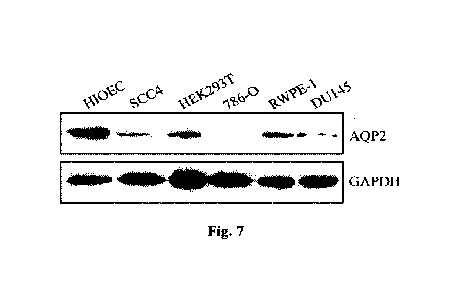
Fig. 7 is the comparison of the AQP2 protein expression quantities of AQP2
gene in three types
of tumor cells and normal cells;
Fig. 8 is the map of a lentiviral overexpression vector of AQP2;
Fig. 9 shows the effect of overexpression AQP2 on the expression quantities of
AQP2 gene and
protein in head and neck squamous cell carcinoma cells;
Fig. 1 shows the effect of overexpression AQP2 on the expression quantities of
AQP2 gene and
protein in kidney cancer cells;
Fig. 11 shows the effect of overexpression AQP2 on the expression quantities
of AQP2 gene and
5
CA 03169749 2022- 8- 26
PWI0015CADOO
protein in prostate cancer cells;
Fig. 12 shows the effect of overexpression AQP2 on the proliferation ability
of head and neck
squamous cell carcinoma cells SCC4;
Fig. 13 shows the effect of overexpression AQP2 on the proliferation ability
of kidney cancer
cells 786-0;
Fig. 14 shows the effect of overexpression AQP2 on the proliferation ability
of prostate cancer
cells DU145.
Fig. 15 shows the effect of overexpression AQP2 on the in vivo tumor growth of
head and neck
squamous cell carcinoma cells SCC4;
Fig. 16 shows the effect of overexpression AQP2 on the in vivo tumor growth of
kidney cancer
cells 786-0;
Fig. 17 shows the effect of overexpression AQP2 on the in vivo tumor growth of
prostate cancer
cells DU145.
DETAILED DESCRIPTION
The present invention will be further described below in conjunction with
specific embodiments.
Embodiment 1
Expression profile microarray analysis of AQP2 in different human tumor
tissues and paracancer
tissues
The Cancer Genome Atlas (TCGA) Program was jointly initiated by the National
Cancer Institute
(NCI) and the National Human Genome Research Institute (NHGRI) of the United
States in
2006. It applies the genome analysis technology based on large-scale
sequencing to conduct
large-scale experiments on 36 types of cancers. The Genome Characterization
Center (GCC) of
TCGA compares tumors and normal tissues to look for gene mutations,
amplifications or
deletions associated with each cancer or subtype and help understand the
molecular mechanism
of cancer and improve the scientific understanding on the molecular basis of
cancer pathogenesis.
6
CA 03169749 2022- 8- 26
PW10015CADOO
The whole gene expression profile data and clinical information of 36 tumors
and their
paracancer tissues were downloaded by the TCGA standard method, R language
(3.1.1 version)
software was used to filter away the tumor types not containing AQP2
expression information,
and AP2 expression was detected in 20 types of tumors.
Table 1. Analysis of expression levels of AQP2 in different tumors in TCGA
database
Tumor type Sample size Value
P
Bladder cancer Tumor (408)
0.0689
Normal (19)
Breast cancer Tumor (1090)
0.1059
Normal (113)
Cervical squamous cell carcinoma Tumor (304)
0.1502
Normal (3)
Gallbladder cancer Tumor (36)
0.0594
Normal (9)
Colon cancer Tumor (454)
0.0721
Normal (41)
Esophageal cancer Tumor (161)
0.0831
Normal (11)
Head and neck squamous cell Tumor (500)
0.0255
carcinoma Normal (44)
Chromophobe carcinoma Tumor (65)
<0.0001
Normal (24)
Clear cell renal carcinoma Tumor (530)
<0.0001
Normal (72)
7
CA 03169749 2022- 8- 26
PW10015CADOO
Papillary cell renal carcinoma Tumor (288)
<0.0001
Normal (32)
Liver cancer Tumor (371)
0.0825
Normal (50)
Pulmonary adenocarcinoma Tumor (513)
0.0613
Normal (59)
Pulmonary squamous cell carcinoma Tumor (501)
0.126
Normal (49)
Pancreatic cancer Tumor (177)
0.198
Normal (4)
Adrenal carcinoma Tumor (178)
0.465
Normal (3)
Prostate cancer Tumor (495)
<0.0001
Normal (52)
Rectal cancer Tumor (165)
0.134
Normal (10)
Stomach cancer Tumor (375)
0.251
Normal (32)
Thyroid cancer Tumor (502)
0.0624
Normal (58)
Endometrial cancer Tumor (543)
0.0582
Normal (23)
Graphpad Prism7 was used to conduct statistical analysis on the expression
levels of AQP2 in 20
types of tumor tissues and corresponding paracancer tissues. All data
underwent statistical t test,
*P<0.05 means significant difference, and **P<0.01 means very significant
difference.
8
CA 03169749 2022- 8- 26
PWI0015CADOO
The specific analysis results are shown in Table 1. Compared with the
corresponding paracancer
tissues, AQP2 showed significant low expression in head and neck squamous cell
carcinoma (Fig.
1), three renal carcinoma subtypes (papillary cell renal carcinoma, clear cell
renal carcinoma,
chromophobe renal carcinoma, Fig. 2 to Fig. 4) and prostate cancer (Fig. 5).
Embodiment 2
In this embodiment, the fluorescence quantitative PCR method was used to
detect the expression
quantities of AQP2 in tumor cells and normal epithelial cells.
1. Materials
Head and neck squamous cell carcinoma cells SCC4 and human's normal oral
epithelial cells
HIOEC, kidney cancer cells 786-0 and human's renal epithelial cells HEK293T,
prostate cancer
cells DU145 and human's normal prostate epithelial cells RWPE-1; the above
cells were all
purchased from U.S. ATCC Cell Repository.
2. Methods
2.1 Extraction of total RNA in tumor cells and normal epithelial
cells
After the foregoing six types of cells were cultured in a 37 DEG C 5% CO2
incubator until the
density was 90%, they were digested and collected by trypsin, the cells were
re-suspended in a
culture solution and counted under a microscope, the concentration of the
cells was adjusted to
5 x10⁵ cells/mL, and then the cell suspension after concentration adjustment
was inoculated to 6-
well plates, 2mL per well, and further cultured in the 37 DEG C 5% CO2
incubator for 24 h.
Extract the total RNA in head and neck squamous cell carcinoma cells SCC4 and
human's normal
oral epithelial cells HIOEC, kidney cancer cells 786-0 and human's renal
epithelial cells
HEK293T, prostate cancer cells DU145 and human's normal prostate epithelial
cells RWPE-1,
respectively according to the Trizol manual of Life Technologies, then
quantify the purity and
concentration of the extracted RNA by the NanoDrop ND-1000 nucleic acid
quantifier, and
ensure the integrity of the extracted RNA through quality inspection by
agarose.
2.2 Synthesis of first-strand cDNA by reverse transcription of RNA
Use TaKaRa kit PrimeScriptTM RT kit with gDNA Eraser (Perfect Real Time) to
reversely
9
CA 03169749 2022- 8- 26
PW10015CADOO
transcribe the extracted total RNA to synthesize cDNA. This kit contains
gDNAEraser DNase
and can effectively remove mingled genomic DNA.
2.3 Real-time quantitative PCR
Design specific primers according to the nucleotide sequences of AQP2 and
GAPDH and use
TaKaRa kit SYBR Premix Ex Taq' II (TliRNaseH Plus) to conduct PCR reaction.
The forward
primer and reverse primer of AQP2 are SEQ ID NO. 3 and SEQ ID NO. 4, and the
forward
primer and reverse primer of GAPDH are SEQ ID NO. 5 and SEQ ID NO. 6. The
reaction system
is shown in the table below:
Table 2. PCR reaction system
Reagent Dose
(IL)
SYBR Premix Ex Taq II (TliRNasell Plus) (2x) 12.5
PCR Forward Primer (10 uM ) 1
PCR Reverse Primer (10 [tM ) 1
DNA template (<100 ug) 2
Sterilized water 8.5
Total 25
After mixing the above components evenly, carry out real-time quantitative PCR
according to the
following procedure: Initially denature at 95 DEG C for 30 s at 40 cycles; 95
DEG C for 5 s and
60 DEG C for 30 s.
Judge the specificity of the reaction according to the melting curve and
calculate the mRNA
expression quantity of AQP2 according to Formula 2'. The result is shown in
Fig. 6.
Compared with the normal epithelial cells of human, the expression quantities
of AQP2 in head
and neck squamous cell carcinoma cells SCC4, kidney cancer cells 786-0 and
prostate cancer
cells DU145 were reduced significantly, consistent with the analysis results
of clinical samples.
Embodiment 3
In this embodiment, the Western blot method was used to detect the expression
quantities of
AQP2 protein in tumor cells and normal epithelial cells.
CA 03169749 2022- 8- 26
PWI0015CADOO
Use trypsin to digest and collect the six types of cells in Embodiment 2 when
the growth density
reached 90%, use a culture solution to re-suspend the cells for multiplication
culture, then collect
the cells when the confluence was 80%, centrifuge, discard the supernatant,
rinse with PBS twice
and discard the supernatant. Add RIPA lysis buffer and lyse on ice for 20 min.
Centrifuge at
12,000 g for 10 min and collect the supernatant. Add 1XSDS loading buffer, mix
well by
pipetting, then boil up and degenerate for 5 min. Separate total protein by
10% SDS-PAGE gel,
then transfer it onto a PVDF membrane. block with 5% BSA at room temperature
for 2 h,
incubate with AQP2 antibody (abeam) overnight at 4 DEG C, and wash with TBST 3
times.
Incubate with the secondary antibody at room temperature for 1 h and wash with
TBST three
times. Develop with an ECL supersensitive chemiluminescence solution, use the
Tannon imaging
system to form images and use GAPDH as an internal reference to compare the
expression levels
of AQP2 protein in different cells.
The results are shown in Fig. 7 and are consistent with AQP2 mRNA expression
difference. The
expression quantities of AQP2 protein in head and neck squamous cell carcinoma
cells SCC4,
kidney cancer cells 786-0 and prostate cancer cells DU145 were reduced
significantly.
Embodiment 4
In this embodiment, the preparation of AQP2 overexpression vector and the
detection of virus
transfection efficiency were conducted.
Synthesize full-length cDNA for AQP2 (see SEQ ID NO. 1 for the specific
sequence) and
introduce to plvx-CMV-ZsGreen 1 plasmid (see Fig. 8 for the atlas). Co-
transfer the above
plasmid, the packaging plasmid psPAX2 and the envelope plasmid pMD2.G into
293T cells to
generate virus. After 48 hours of transfection, collect the viral supernatant
of the cells and infect
SCC4 head and neck squamous cell carcinoma cells, 786-Okidney cancer cells and
DU145
prostate cancer cells, respectively. After 48 hours of infection, screen with
puromycin for two
weeks to obtain a cell line that stably promotes AQP2 gene expression. Collect
the total RNA and
total protein of the blank vectors and overexpression vectors of the three
types of cells, and
compare the expression changes of AQP2 gene and protein by qPCR (the specific
method is the
same as that in Embodiment 2) and Western blot method (the specific method is
the same as that
in Embodiment 3).
11
CA 03169749 2022- 8- 26
PWI0015CADOO
The results are shown in Fig. 9 to Fig. 11. Overexpression of AQP2 caused the
expression
quantities of AQP2 gene and protein in head and neck squamous cell carcinoma
cells SCC4 (Fig.
9), kidney cancer cells 786-0 (Fig. 10) and prostate cancer cells DU145 (Fig.
11) to increase
significantly.
Embodiment 5
This embodiment shows the effect of overexpression of AQP2 on the
proliferation ability of
human tumor cells.
Use trypsin to digest and collect head and neck squamous cell carcinoma cells
SCC4, kidney
cancer cells 786-0 and prostate cancer cells DU145 after they stably
transfected empty vectors
and AQP2-overexpressed cells in a 37 DEG C 5% CO2 incubator until the density
was 90%, re-
suspend the cells in a culture solution, count the cells under a microscope,
adjust cell
concentration to 3.0x10⁴ cells/mL, inoculate the cell suspension to 96-well
plates, 100pL per
well, and culture in a 37 DEG C 5% CO2 incubator for 24h, 48h and 72h,
respectively. Add 20pL
of 5 mg/mL MTT to each well of the 96-well plates, and continue to culture for
4 h. Suck away
the culture medium and add 100 pi, of DMSO for dissolution. Use ELIASA to
detect absorbance
at detection wavelength 570 nm and reference wavelength 630 nm and calculate
proliferation
inhibition (PI) according to the following formula:
PI (%) =1- drug group/negative group
The test was independently repeated three times. The results obtained from the
test were
expressed with mean SD, statistical t test was done, the comparison of two or
more groups of
data adopted One-way ANOVA, statistical significance was expressed with value
P, P<0.05
means significant difference, and P<0.01 means very significant difference.
The results are shown in Fig. 12 to Fig. 14. Compared with empty vector cells
(plvx-crtl), the
proliferation speed of the cells with overexpression of AQP2 (plvx-AQP2) (head
and neck
squamous cell carcinoma cells SCC4 (Fig. 12), kidney cancer cells 786-0 (Fig.
14) and prostate
cancer cells DU145 (Fig. 15)) was reduced obviously. It indicates that
overexpression of AQP2
can significantly inhibit the proliferation of head and neck squamous cell
carcinoma cells SCC4,
kidney cancer cells 786-0 and prostate cancer cells DU145 and further verifies
the importance of
12
CA 03169749 2022- 8- 26
PWI0015CADOO
AQP2 as a cancer suppressor gene.
Embodiment 6
This embodiment shows the effect of overexpression of AQP2 on the migration
ability of human
tumor cells.
Inoculate head and neck squamous cell carcinoma cells SCC4, kidney cancer
cells 786-0 and
prostate cancer cells DU145 to transwell cells, 100 [EL per well, after they
stably transfected
empty vectors and AQP2-overexpressed cells, add 0.6 mL of complete medium
containing 10%
FBS to the transwell cells to stimulate cell migration, and culture in 5% CO2
at 37 DEG C for 24
h. Discard the medium in the wells, fix with 90% alcohol at room temperature
for 30 min, stain
with 0.1% crystal violet at room temperature for 10 min, rinse with clear
water, gently wipe off
the non-migrated cells in the upper layer with a cotton swab, observe under a
microscope and
select four fields of view to take pictures and count. Calculate the migration
inhibition rate (MIR)
according to the following formula:
MI R(%) ¨ 1 Ntest x100%
pr
emurol
where Nteõ is the number of migrated cells in the test group (plvx-AQP2) and
Nc001 is the number
of migrated cells in the blank control group (plvx-ctrl). The test was
independently repeated three
times. The results obtained from the test were expressed with mean SD,
statistical t test was
done, the comparison of two or more groups of data adopted One-way ANOVA,
statistical
significance was expressed with value P, P<0.05 means significant difference,
and P<0.01 means
very significant difference.
Table 3 Inhibitory effect of overexpression of AQP2 on the migration ability
of human tumor
cells
Cell type Group No. of migrated cells (Mean SD) MIR
of cells (%)
SCC4 plvx-ctrl 661 48 0.00
plvx-AQP2 263+37 77.00%**
plvx-ctrl 727A1 0.00
786-0
plvx-AQP2 251 50 70.98%**
13
CA 03169749 2022- 8- 26
PWI0015CADOO
plvx-ctrl 707 39 0.00
DU145
plvx-AQP2 304 28 68.32%**
In the table: * means P<0.05, **means P<0.01.
The results are as shown in Table 3. After AQP2 expression was upregulated,
the migration
abilities of head and neck squamous cell carcinoma cells SCC4, kidney cancer
cells 786-0 and
prostate cancer cells DU145 were reduced obviously.
Embodiment 7
This embodiment shows the effect of overexpression of AQP2 on the in vivo
growth of human
tumor cells.
(1) Massively culture head and neck squamous cell carcinoma cells SCC4,
kidney cancer cells
786-0 and prostate cancer cells DU145 after they stably transfected empty
vectors and AQP2-
overexpressed cells, digest with a 0.25% pancreatin solution, centrifuge the
cell suspension at
1,000 rpm for 5 min after termination of digestion, re-suspend the cells by a
serum-free DMEM
culture medium, then count the cells and adjust cell concentration to 5x10⁷
cells/ml;
(2) Inoculate each nude mouse (female mice at the age of 4-6 weeks and with
a weight of 14-
16 g were ordered and adaptively reared for 1 week in an SPF animal breeding
room) with 100 ul
of the cell suspension of the corresponding group in the left armpit, and the
number of cells
injected is 5x10⁶;
(3) After inoculation, the tumor growth at the inoculation sites of nude mice
was closely
observed. The volume of the transplanted tumor was measured and recorded every
two days. The
calculation formula of tumor volume (TV) is shown below:
TV=0.5 x axbA2
where, a is the length of the transplanted tumor (mm), and b is the width of
the transplanted
tumor (mm).
Compared with the empty vector control group (plvx-ctrl), the cells with
overexpression of AQP2
(plvx-AQP2) (head and neck squamous cell carcinoma cells SCC4 (Fig. 15),
kidney cancer cells
786-0 (Fig. 16) and prostate cancer cells DU145 (Fig. 17)) showed a lower
tumor growth rate
14
CA 03169749 2022- 8- 26
PW10015CADOO
and obviously reduced in-vivo tumorigenicity in nude mice, indicating that
overexpression of
AQP2 can inhibit the in vivo growth ability of malignant tumor cells.
CA 03169749 2022- 8- 26