Note: Descriptions are shown in the official language in which they were submitted.
PWI0016CADOO
NOVEL MICROPEPTIDE HMMW AND APPLICATION THEREOF
TECHNICAL FIELD
The present invention pertains to the field of biomedical research and
development and more
specifically, relates to an application of novel micropeptide HMMW in tumor
detection and
treatment.
BACKGROUND ART
With the development of transcriptomic, proteomic and bioinformatics analysis
methods,
scientists have discovered that those previously defined as non-coding RNA
molecules (such as
IncRNAs, circRNAs and miRNAs) actually contain small open reading frames
(sORFs, with a
nucleotide sequence of not more than 300bp) with an encoding ability. sORF-
encoded peptides
(SEPs) are called micropeptides (not more than 100 amino acids in length). In
2015, on the
journal Cell, scientists screened the skeletal muscle-specific lncRNA encoding
peptide
Myoregulin (MLN) by the bioinformatics method, and determined that MLN is an
important
regulator of skeletal muscle physiology. In the past two years, there are not
many reports about
lncRNA encoding micropeptides in the world, and the research focuses on muscle
differentiation
and skeletal muscle development. Therefore, discovering and identifying new
micropeptides
encoded by lncRNA and exploring new application fields are of great
significance for opening
the door of encoding non-coding RNAs.
Tumors are malignant diseases that seriously endanger human life and health.
Unlimited growth,
invasion and metastasis are the malignant signs of tumors and the main causes
of treatment
failure and death. At present, the main clinical treatment methods for cancer
patients are
operative therapy and chemotherapy (chemotherapy/drug therapy). For most
tumors, operative
therapy is suitable for curative treatment of early, intermediate and
localized tumors and
palliative treatment of advanced tumors. Although there is no chemotherapy
resistance or
radioresistance in surgical treatment, it is more traumatic, difficult to
operate in some parts, and
1
CA 03169796 2022- 8- 26
PW10016CADOO
ineffective to subclinical metastases. For most tumors, chemotherapy is
suitable for intermediate
and advanced tumors, and as a systemic treatment, chemotherapy has therapeutic
effects on
primary tumors, metastases and subclinical metastases. However,
chemotherapeutic drugs have
poor selectivity. While achieving a therapeutic effect, toxic and side effects
in various degrees
often occur; and cancer patients who receive chemotherapy for a long time may
develop new
malignant tumors due to immunosuppressive effects and direct carcinogenic
effects.
Owing to the advantages of high specificity, small toxic and side effects,
clear mechanism of
action, and no harm to normal cells, tissues and organs, the application of
peptide drugs in the
treatment of cancers, cardiovascular diseases, immune-related diseases,
metabolic diseases and
infectious diseases has been gradually developed. At present, there are more
than 80 peptide
drugs on the market in the world, with total annual sales of more than 20
billion US dollars.
Although the market sales of peptide drugs in China maintain a momentum of
rapid growth, most
of them are generic peptide drugs.
At present, the research on micropeptides in the detection and treatment of
malignant tumors such
as head and neck cancer, thyroid cancer, lung cancer and esophageal squamous
cell carcinoma is
still blank. Through bioinformatics analysis and experimental verification,
the present invention
has discovered a new micropeptide in tumors and explored its application in
tumor detection and
treatment, providing a new solution for tumor detection and treatment.
SUMMARY OF THE INVENTION
The first object of the present invention is to provide a novel micropeptide
HMMW and its amino
acid sequences. Micropeptide HMMW is a new human endogenous peptide discovered
for the
first time;
The second object of the present invention is to provide a nucleotide, and the
nucleotide sequence
can encode the obtained micropeptide HMMW;
The third object of the present invention is to provide a recombinant vector
containing the
nucleotide sequence encoding micropeptide HMMW;
The fourth object of the present invention is to apply micropeptide HMMW or
nucleotide
2
CA 03169796 2022- 8- 26
PWI0016CADOO
encoding the micropeptide HMMW in the preparation of tumor detection reagents
and treatment
drugs, specifically including detection and treatment of head and neck cancer,
thyroid cancer,
lung cancer, esophageal squamous cell carcinoma, stomach cancer, breast
cancer, kidney cancer
and skin cancer.
The present invention adopts the following technical solution:
Application of a novel micropeptide HMMW in the preparation of tumor detection
reagents
and/or tumor treatment drugs, wherein the amino acid sequences of the
micropeptide HMMW
contain a sequence shown in SEQ ID NO. 2
Application of a novel micropeptide HMMW in the preparation of tumor detection
reagents or
tumor treatment drugs, wherein the amino acid sequences of the micropeptide
HMMW have at
least 85% homology to the amino acid sequence shown in SEQ ID NO. 1
(micropeptide HMMW
I) (as shown in SEQ ID NO. 2 and SEQ ID NO. 3, the corresponding micropeptides
are named
HMMW II and HMMW III), or have at least 90% homology (as shown in SEQ ID NO. 4
and
SEQ ID NO. 5, the corresponding micropeptides are named HMMW IV and HMMW V),
or have
at least 95% homology (as shown in SEQ ID NO. 6 and SEQ ID NO. 7, the
corresponding
micropeptides are named HMMW VI and HMMW VII), or have at least 98% homology
(as
shown in SEQ ID NO. 8 and SEQ ID NO. 9, the corresponding micropeptides are
named
HMMW VIII and HMMW IX), and these sequences with homology to SEQ ID NO. 1
retain
similar functions of inhibiting tumor cell growth, proliferation, invasion or
migration and can be
used to prepare tumor detection reagents or tumor treatment drugs.
"Homology" described herein refers to the percentage of similarity in a
comparison of two or
more amino acid sequences. The percentage of similarity can be determined
electronically, such
as by the MEGALIGN program (Lasergene software package, DNASTAR, Inc., Madison
Wis.).
The MEGALIGN program can compare two or more sequences according to different
methods
such as the Cluster method (Higgins, D.G. and P.M.Sharp (1988) Gene 73: 237-
244). The Cluster
method arranges groups of sequences into clusters by examining the distances
between all pairs.
The clusters are then assigned in pairs or groups. The percentage of homology
between two
amino acid sequences such as Sequence A and Sequence B is calculated with the
following
formula:
3
CA 03169796 2022- 8- 26
PWI0016CADOO
(Number of residues matching between sequence A and sequence B *100)/(Number
of residues
in sequence A - Number of interval residues in sequence A - Number of interval
residues in
sequence B).
Application of a novel micropeptide HMMW in the preparation of tumor detection
reagents or
tumor treatment drugs, wherein the novel micropeptide HMMW contains an amino
acid sequence
shown in SEQ ID NO. 1.
A nucleotide, wherein the nucleotide is any of a, b and c:
(a) A nucleotide encoding micropeptide HMMW containing the amino acid
sequence shown in
SEQ ID NO. 1;
(b) A nucleotide encoding micropeptide HMMW having at least 85% homology to
the amino
acid sequence shown in SEQ ID NO.1;
(c) Nucleotide sequences encoding the amino acids described in claim 3,
specifically the
nucleotide sequences shown in SEQ ID NO.10 to NO.18 (N is any of A/T/G/C).
A recombinant vector, wherein the recombinant vector contains any of the
foregoing nucleotides.
Application of any of the foregoing nucleotides in the preparation of tumor
detection reagents
and/or tumor treatment drugs.
A tumor detection kit, wherein the kit contains a specific primer pair for any
of the foregoing
nucleotide sequences.
Preferably, sequences of the specific primer pair are shown in SEQ ID NO.19
and SEQ ID
NO.20.
A tumor treatment drug, wherein the drug contains at least any of the
foregoing micropeptides
HMMW or any of the foregoing nucleotides or the foregoing recombinant vector,
and a
pharmaceutically acceptable vector.
Preferably, the drug is a drug with functions as described in (al) - (a4),
wherein:
(al) inhibiting tumor growth;
(a2) inhibiting proliferation of tumor cells;
4
CA 03169796 2022- 8- 26
PW10016CADOO
(a3) inhibiting invasion of tumor cells;
(a4) inhibiting migration of tumor cells.
Preferably, the tumors include head and neck cancer, thyroid cancer, lung
cancer, esophageal
squamous cell carcinoma, stomach cancer, breast cancer, kidney cancer and skin
cancer.
Compared with the prior art, the present invention has the following
advantages:
(1) The present invention provides a micropeptide HMMW with novel amino
acid sequences.
According to the search of databases (BLAST, UniProt) and literature, no
protein or peptide
fragments with sequences homologous to micropeptide HMMW were found;
The micropeptide HMMW is composed of 51 amino acids, plays an important role
in tumor
detection and treatment and can be used as a tumor marker for tumors including
human head and
neck cancer, thyroid cancer, lung cancer, esophageal squamous cell carcinoma,
stomach cancer,
breast cancer, kidney cancer and skin cancer, that is, has low expression in
the foregoing tumors.
(2) The present invention provides a micropeptide HMMW with novel amino
acid sequences.
The micropeptide HMMW can significantly inhibit the proliferation, migration
and invasion of
SCC4 cells of head and neck cancer, 5W579 cells of thyroid cancer, A549 cells
of lung cancer,
TE13 cells of esophageal squamous cell carcinoma, MGC803 cells of stomach
cancer, MDA-
MB-231 cells of breast cancer, U0K262 cells of kidney cancer, and A431 cells
of skin cancer of
human in vitro;
The micropeptide HMMW can obviously inhibit the tumor growth of SCC4 cells of
head and
neck cancer, 5W579 cells of thyroid cancer, A549 cells of lung cancer, TE13
cells of esophageal
squamous cell carcinoma, MGC803 cells of stomach cancer, MDA-MB-231 cells of
breast
cancer, U0K262 cells of kidney cancer and A431 cells of skin cancer of human
in vivo;
Moreover, micropeptide HMMW can be used as a detection and treatment drug for
malignant
tumors, thereby greatly expanding the therapeutic spectrum of this
micropeptide and providing a
new approach for the development of malignant tumor drugs.
BRIEF DESCRIPTION OF THE DRAWINGS
5
CA 03169796 2022- 8- 26
PW10016CADOO
Fig. 1 is a pcDNA3.1 plasmid profile of overexpression micropeptide HMMW;
Fig. 2 shows the expression of a target band detected by Western blot;
Fig. 3 shows the inhibitory effect of micropeptide HMMW on the proliferation
of SCC4 cells of
human head and neck cancer;
Fig. 4 shows the inhibitory effect of micropeptide HMMW on the proliferation
of SW579 cells of
human thyroid cancer;
Fig. 5 shows the inhibitory effect of micropeptide HMMW on the proliferation
of A549 cells of
human lung cancer;
Fig. 6 shows the inhibitory effect of micropeptide HMMW on the proliferation
of TE13 cells of
esophageal squamous cell carcinoma of human;
Fig. 7 shows the inhibitory effect of micropeptide HMMW on the proliferation
of MGC803 cells
of human stomach cancer;
Fig. 8 shows the inhibitory effect of micropeptide HMMW on the proliferation
of MDA-M13-231
cells of human breast cancer;
Fig. 9 shows the inhibitory effect of micropeptide HMMW on the proliferation
of U0K262 cells
of human kidney cancer;
Fig. 10 shows the inhibitory effect of micropeptide HMMW on the proliferation
of A431 cells of
human skin cancer;
Fig. 11 shows the inhibitory effect of micropeptide HMMW on the tumor growth
of SCC4 cells
of human head and neck cancer in vivo;
Fig. 12 shows the inhibitory effect of micropeptide HMMW on the tumor growth
of SW579 cells
of human thyroid cancer in vivo;
Fig. 13 shows the inhibitory effect of micropeptide HMMW on the tumor growth
of A549 cells of
human lung cancer in vivo;
Fig. 14 shows the inhibitory effect of micropeptide HMMW on the tumor growth
of TE13 cells of
esophageal squamous cell carcinoma of human in vivo;
6
CA 03169796 2022- 8- 26
PWI0016CADOO
Fig. 15 shows the inhibitory effect of micropeptide HMMW on the tumor growth
of MGC803
cells of human stomach cancer in vivo;
Fig. 16 shows the inhibitory effect of micropeptide HMMW on the tumor growth
of MDA-MB-
231 cells of human breast cancer in vivo;
Fig. 17 shows the inhibitory effect of micropeptide HMMW on the tumor growth
of U0K262
cells of human kidney cancer in vivo;
Fig. 18 shows the inhibitory effect of micropeptide HMMW on the tumor growth
of A431 cells of
human skin cancer in vivo;
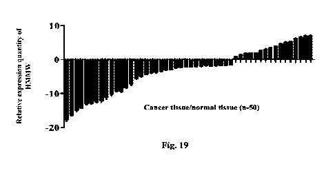
Fig. 19 shows the expression level of micropeptide HMMW in cancer
tissue/normal tissue
detected by the qPCR method.
DETAILED DESCRIPTION
The present invention will be further described below in conjunction with
specific embodiments.
Embodiment 1
In this embodiment, the encoding ability of HMMW micropeptide was detected.
Construct in vitro an overexpression vector pcDNA-HMMW with a Flag tag and
containing
cDNA (its nucleotide sequence is shown in SEQ ID NO. 10), wherein the profile
of the empty
plasmid pcDNA3.1 is shown in Fig. 1. Introduce the above plasmid into 293T
cells by 1ipo3000
lipofection reagent, collect cells 48 h after transfection, centrifuge,
discard the supernatant and
collect precipitated cells, rinse the precipitated cells with PBS twice,
centrifuge, discard the
supernatant and collect the precipitated cells. Add RIPA lysis buffer to the
collected precipitated
cells, lyse on ice for 20 min, and then centrifuging at 12,000 g for 10 min
and collect the
supernatant. Then add 1XSDS loading buffer, mix well by pipetting, then boil
up and degenerate
for 5 min. Separate total protein by 10% SDS-PAGE gel, then transfer it onto a
PVDF membrane,
block with 5% nonfat dry milk at room temperature for 2 h, incubate with Flag
primary antibody
(abeam) overnight at 4 DEG C, and wash with TBST 3 times. Incubate with the
secondary
antibody at room temperature for 1 h and wash with TBST three times. Develop
with an ECL
7
CA 03169796 2022- 8- 26
PWI0016CADOO
supersensitive chemiluminescence solution and use the Tannon imaging system to
form images
and detect whether there is a target band.
The results are shown in Fig. 2. It was found through Western blot that a
target band of
micropeptide HMMW appeared after transfection of the recombinant plasmid. It
further indicates
that the micropeptide HMMW protein has an encoding ability.
Embodiment 2
In this embodiment, micropeptides HMMW I to IX were obtained by the solid-
phase synthesis
method and tested.
The micropeptides HMMW Ito IX (their amino acid sequences are shown in SEQ ID
NO. 1 to 9)
were synthesized by the peptide solid-phase synthesis method, the synthesized
micropeptides
HMMW were separated and purified by preparative T-IPLC, and the purity of the
micropeptides
HMMW was determined by analytical RP-HPLC. In the solid-phase peptide
synthesis method,
Fmoc-wang-resin or Fmoc-CTC-resin was used as a starting material and then
protected amino
acids were used to sequentially ligate dipeptide ¨unpentacontapeptide. After
peptide ligation and
full wash, peptide cutting and post-treatment were conducted to obtain a crude
angiogenesis
inhibitor. The crude product was dissolved, purified by preparative HPLC
twice, concentrated
and freeze-dried to obtain a pure product. Finally, after the tertiary
purification, a refined
micropeptide product was obtained. This method can not only ensure the
efficiency of synthesis,
but also raise product purity. The details are as follows:
1. Peptide ligation (including ligation of dipeptide ¨
unpentacontapeptide):
Weigh an appropriate amount of Fmoc-wang-resin or Fmoc-CTC-resin, pour it into
a glass sand
core reaction column, and add an appropriate amount of C112C12 to fully expand
the resin.
a. Removal of protecting group: Add a protecting group removing liquid of
hexahydropyridine/N, N-dimethylformamide (DMF), react for a period of time,
drain the
protecting group removing liquid, wash with DMF once, and add an appropriate
amount of
the protecting group removing liquid again for reaction to remove Fmoc
protecting group;
b. Wash: Drain the protecting group removing liquid, and wash the resin
with DMF to
thoroughly remove by-products;
8
CA 03169796 2022- 8- 26
PW10016CADOO
c. Condensation: Dissolve the protecting amino acid and activator used for
peptide ligation in
DMF and condensing agent, shake up, control the temperature at about 34 DEG C,
and
fully react in the reactor;
d. Wash: Drain the reaction solution and fully wash the resin with DMF to
thoroughly remove
by-products
2. Peptide cutting:
Put the drained resin into a round-bottom flask, add a cutting fluid to fully
lyse hexatriconta-
peptide intermediate, and separate the resin from the peptide by a sand core
funnel. The
components of the cutting fluid and the volume composition of the components
are:
trifluoroacetic acid: phenol: water: thioanisole: EDT=90: 3: 3: 2: 2.
3. Post-treatment:
First add anhydrous ether to the cutting fluid to separate out the peptide,
then centrifuge, discard
the supernatant, then wash the peptide with anhydrous ether, and drain to
obtain a peptide crude
product.
4. Purification:
a. Dissolution: Weigh the crude product to prepare a 5-20 g/L
solution, and filter it with a
mixed filter membrane with a pore size of 0.45 gm.
Preparation: Conduct primary purification, secondary purification and tertiary
purification by
semi-preparative HPLC to obtain a qualified refined peptide product. Mobile
phase: A is
acetonitrile, B is 0.1% TFA aqueous solution.
Primary purification: Equilibrate the preparative column by rinsing with 10%-
90% acetonitrile
and 20%-80% buffer solution at a flow rate of 50 mL/min-100 mL/min for 10 min.
Dissolve the
filtered crude product and load it with an infusion pump.
Table 1 Elution gradient for primary purification
Time (min) Flow rate (mL/min) A% B%
Wavelength nm
0 60 10 90 220
40 60 20 80 220
9
CA 03169796 2022- 8- 26
PW10016CADOO
Collect solutions with an absorption value of greater than 200 mV at UV
wavelength 220 nm,
combine the solutions with a detected purity of greater than 95% as the peak
top, and keep it for
secondary separation and purification.
Secondary purification: After removing by rotary evaporation the organic
solvent from the peak
top received in the primary purification, load the sample by infusion pump in
form of 5-95%
acetonitrile and 15-85% buffer at a flow rate of 50-100mL/min.
Table 2 Elution gradient for secondary purification
Time (min) Flow rate (mL/min) A% B%
Wavelength nm
0 60 5 95 220
40 60 15 85 220
The solutions with absorption greater than 200 mV at UV wavelength 220 nm were
collected.
The solutions were considered qualified if their purity was greater than 98%.
b. Concentration, filtration and freeze-drying: Concentrate the qualified
solution in a rotary
evaporator under reduced pressure at 37 DEG C to remove residual solvent and
water for
injection. Finally, filter it with a 0.22 p,m filter membrane, put the
filtrate in a freeze-drying tray
and freeze-dry it with a freeze dryer to obtain a pure product.
Tertiary purification: Conduct tertiary purification of the qualified sample
with a purification of
greater than 98% obtained in the secondary purification, and use 5-95%
acetonitrile and 10-90%
buffer at a flow rate of 50-100 mL/min to prepare a refined peptide product.
Table 3 Elution gradient for tertiary purification
Time (min) Flow rate (mL/min) A% B%
Wavelength nm
0 60 5 95 220
40 60 10 90 220
Collect solutions with an absorption value of greater than 200 mV at UV
wavelength 220 nm,
combine the samples with a detected purity of greater than 95% as the
qualified refined product.
5. Purity detection
CA 03169796 2022- 8- 26
PWI0016CADOO
Collect freeze-dried purified product and detect peptide purity by analytical
RP-HPLC. Analysis
conditions: Mobile phase: ACN (+0.1% TFA) and H20 (+0.1% TFA); linear gradient
of
acetonitrile: 10%-100%; flow rate: 1 mL/min; operation time: 20 min; loading
volume: 20 pL;
detection wavelength: 220 nm.
Table 4 Purities of micropeptides HMMW Ito IX detected by RP-HPLC
Name Peak area Purity (%)
HMMW-I 1349502 98.87%
HMMW-II 5510739 95.56
HMMW-III 2578258 95.68
HMMW-W 8335415 98.32
HMMW-V 2695695 95.42
HMMW-VI 8429617 95.19
HMMW-VII 5562335 95.74
HMMW-VIII 3662672 95.66
HMMW-IX 3293152 95.56
The purities of synthesized micropeptides were determined by RP-HPLC. The
results showed
that the purities of the nine prepared micropeptides HMMW were all greater
than 95%, which
met the design requirements.
In this experiment, micropeptides HMMW I to IX were successfully synthesized
by solid-phase
synthesis method. This method has high repeatability, strong operability and
less pollution; two
types of resin, i.e.: wang resin and CTC resin, can be used in the experiment
to synthesize
peptides; in the experiment, when wang resin was used, it was more stable and
had fewer side
reactions, a better peak pattern of the process crude product and a higher
purification yield
compared with other resins, so the cost was lower; in the experiment, when CTC
resin was used,
the reaction was less affected by temperature and the reaction rate was
higher; RP-HPLC was
used to purify peptide and gradient elution was used with a better effect
compared with isocratic
elution. In the separation process, the retention time was appropriate and the
production
efficiency and the purity were high.
11
CA 03169796 2022- 8- 26
PWI0016CADOO
Embodiment 3
In this embodiment, the effects of micropeptides HMMW I to IX on the
proliferation ability of
human tumor cells were detected.
After SCC4 cells of head and neck cancer, SW579 cells of thyroid cancer, A549
cells of lung
cancer, TE13 cells of esophageal squamous cell carcinoma, MGC803 cells of
stomach cancer,
MDA-MB-231 cells of breast cancer, U0K262 cells of kidney cancer and A431
cells of skin
cancer of human were cultured in a 37 DEG C 5% CO2 incubator until the density
was 90%, they
were digested and collected by trypsin, the cells were re-suspended in a
culture solution and
counted under a microscope, the concentration of the cells was adjusted to
3.0x10⁴ cells/mL, and
the cell suspension was inoculated to 96-well plates, 100 pL per well, and
cultured in a 37 DEG
C 5% CO2 incubator overnight. After the cells were completely adherent,
different doses of
micropeptides HMMW I to IX were added as drug groups, taxol was used as a
positive control
group, and the culture solution without any drug was used as a blank control
group. They were
diluted with the culture solution till predetermined concentrations. The
diluted solutions were
added to 96-well plates, respectively, 100 pL per well, and incubated in a 37
DEG C 5% CO2
incubator for 48h. 20 1_, of 5 mg/mL MTT was added to each well of the 96-
well plates, and the
culture was continued for 4 h. The culture medium was sucked away, and 100 pL
of DMSO was
added to each well for dissolution. Absorbance was detected by ELIASA at
detection wavelength
570 nm and reference wavelength 630 nm, and proliferation inhibition (PI) was
calculated
according to Formula PI (%) =1-drug group/negative group. The test was
independently repeated
three times. The results obtained from the test were expressed with mean SD
and statistical t test
was done. *P<0.05 means significant difference, and "P<0.01 means very
significant
difference.
Table 5. Inhibitory effects of micropeptides HMMW Ito IX on proliferation of
human tumor
cells
Group Dose Tumor type (inhibition rate/%)
HMMW-I (I-IM) SCC4 SW579 A549 TE13 MGC803 MDA-MB U0K262 A431
-231
1 20.18 18.98 14.71 13.09 15.93
18.90 16.92 17.04
12
CA 03169796 2022- 8- 26
PW10016CADOO
2
32.67 24.87 19.37 17.27 19.91 23.17 24.15 20.98
4
44.89 36.23 21.87 23.98 24.09 36.99 38.19 31.09
8
58.19 42.17 37.09 36.09 38.18 45.06 43.54 41.21
16
64.29 59.02 43.92 43.53 45.10 56.19 59.21 48.02
32 79.18 68.01 58.90 61.02 68.09 69.18 72.10 65.09
1
19.58 17.90 14.09 12.45 16.23 17.38 16.23 16.44
2
31.62 26.81 18.21 16.89 18.57 22.45 23.32 22.18
4
42.34 33.19 22.45 22.68 23.18 35.57 34.29 30.29
HMMW-II
8
55.41 41.57 35.05 35.16 39.16 46.48 42.34 40.41
16
60.15 54.15 42.54 44.37 44.15 55.24 52.51 45.32
32
72.14 63.25 56.87 60.13 63.24 61.35 70.70 62.19
1 17.18 17.91 15.73 14.04 16.90
16.94 17.90 18.34
2
30.67 23.80 18.34 18.21 18.95 22.12 23.15 23.58
4
41.89 35.21 23.85 20.90 25.01 35.91 35.16 34.03
HMMW-III
8
53.19 41.12 35.01 34.03 39.13 44.05 41.53 42.11
16
60.29 55.00 41.90 44.50 47.16 52.14 57.20 49.06
32
73.18 64.00 54.84 60.09 65.05 67.13 70.16 67.19
1
17.54 18.94 15.06 14.43 15.20 16.34 17.23 15.43
2
30.32 27.21 19.23 17.59 19.37 23.25 24.12 23.28
4
41.31 35.39 24.41 23.62 24.15 36.53 36.25 34.25
HMMWT -IV
8
53.21 40.51 36.35 32.46 37.36 47.28 43.24 42.31
16
62.13 52.05 41.50 43.31 45.12 53.21 54.31 48.30
32 70.34 61.21 54.27 58.33 61.54 60.25 69.75 60.29
1 16.13 16.51 16.76 15.07 15.93
17.92 18.95 17.35
2
32.27 22.86 19.14 19.11 19.92 21.32 21.35 22.38
4
40.84 34.11 22.80 22.94 24.05 33.95 33.11 31.05
HMMW-V
8
52.09 40.10 36.31 32.53 38.11 42.35 40.43 40.31
16
61.24 54.40 42.95 41.57 46.36 50.12 55.24 47.05
32
70.28 63.05 55.54 59.39 63.00 64.33 67.26 65.39
HMMWT -VI 1 15.41 17.58 16.01 15.83 16.10
15.54 18.53 14.43
13
CA 03169796 2022- 8- 26
PWI0016CADOO
2
32.18 25.20 18.13 18.49 18.27 22.22 25.15 22.21
4
40.21 33.35 23.40 22.32 23.13 34.33 33.15 33.35
8
52.20 41.50 34.30 31.44 36.26 45.24 41.20 40.30
16
60.03 50.45 40.54 42.51 44.10 51.31 53.11 46.20
32
68.14 60.20 52.23 54.30 60.34 62.27 64.73 58.24
1 15.33 15.53 15.86 16.17 15.93
18.52 19.45 16.25
2 31.21 21.56 18.13 18.18 19.92
20.12 20.31 21.35
4
41.54 33.14 22.60 21.54 24.05 32.45 32.21 30.15
HMMW-VII
8
51.02 41.00 35.30 33.50 38.11 41.15 41.40 39.30
16
63.14 52.45 43.75 40.47 46.36 52.02 52.14 44.25
32
71.20 61.15 53.51 57.33 63.00 63.23 64.25 64.32
1 16.18 16.91 13.73 15.04 15.90
17.94 18.90 16.34
2
31.67 22.80 17.34 19.21 19.95 21.12 22.15 22.58
4
40.89 32.21 21.85 21.90 23.01 34.91 33.16 33.03
HMMW-VIII
8
51.19 40.12 32.01 33.03 35.13 43.05 40.53 41.11
16
61.29 52.00 40.90 42.50 46.16 50.14 54.20 48.06
32
70.18 60.00 53.84 61.09 63.05 64.13 71.16 66.19
1 16.33 16.53 16.86 17.17 18.93
19.52 18.45 18.25
2
33.21 22.56 19.13 19.18 21.92 23.12 22.31 23.35
4
42.54 31.14 23.60 22.54 25.05 34.45 33.21 31.15
HMMW-IX
8
50.02 45.00 36.30 35.50 39.11 42.15 43.40 39.30
16
64.14 54.45 45.75 43.47 47.36 53.02 54.14 43.25
32
72.20 63.15 54.51 58.33 64.00 62.23 65.25 61.32
87.02 82.83 84.29 79.21 81.28 83.92 83.92 85.19
Taxol
(ighnl)
The results are shown in Fig. 3 to Fig. 10. Compared with the negative control
group,
micropeptide HMMW-I at a dose of 1 to 32 1.1M could significantly inhibit the
proliferation of
SCC4 cells of head and neck cancer (Fig. 3), SW579 cells of thyroid cancer
(Fig. 4), A549 cells
of lung cancer (Fig. 5), TE13 cells of esophageal squamous cell carcinoma
(Fig. 6), MGC803
5 cells of stomach cancer (Fig. 7), MDA-MB-231 cells of breast cancer
(Fig. 8), U0K262 cells of
14
CA 03169796 2022- 8- 26
PWI0016CADOO
kidney cancer (Fig. 9), A431 cells of skin cancer (Fig. 10) of human to
various extent, and
showed a dose-dependent relation. The results are shown in Table 5. Compared
with the negative
control, micropeptides HMMW I to IX at a dose of 1 to 32 pM all could
significantly inhibit the
proliferation of SCC4 cells of head and neck cancer, SW579 cells of thyroid
cancer, A549 cells of
lung cancer, TE13 cells of esophageal squamous cell carcinoma, MGC803 cells of
stomach
cancer, MDA-MB-231 cells of breast cancer, U0K262 cells of kidney cancer, A431
cells of skin
cancer of human, and showed a dose-dependent relation. It indicates that the
peptides with more
than 85% homology to the original sequence HMMW I all have an inhibitory
effect on the
proliferation of tumor cells, and it can be considered to use micropeptides
HMMW I to IX as
candidate anti-tumor drugs.
Embodiment 4
Effects of micropeptides HMMW Ito IX on the migration ability of human tumor
cells
Inoculate SCC4 cells of head and neck cancer, SW579 cells of thyroid cancer,
A549 cells of lung
cancer, TE13 cells of esophageal squamous cell carcinoma, MGC803 cells of
stomach cancer,
MDA-MB-231 cells of breast cancer, U0K262 cells of kidney cancer, and A431
cells of skin
cancer of human to transwell cells, 100 [IL per well, and meanwhile add
micropeptides HMMW I
to IX at different doses to every cell. Add 0.6 mL of complete medium
containing 10% FBS to
the transwell cells to stimulate cell migration, and culture in 5% CO2 at 37
DEG C for 24 h.
Discard the medium in the wells, fix with 90% alcohol at room temperature for
30 min, stain with
0.1% crystal violet at room temperature for 10 min, rinse with clear water,
gently wipe off the
non-migrated cells in the upper layer with a cotton swab, observe under a
microscope and select
four fields of view to take pictures and count. Calculate the migration
inhibition rate (MIR) of the
cells according to the following formula:
MIR(%) =1 N _______________________________________ :'100%
Al control
where Ntest is the number of migrated cells in the test groups (the groups at
a dose of 1, 4 or 14
p,M in the table), N.1õ01 is the number of migrated cells in the blank control
groups (the groups at
a dose of 0 p,M in the table).
CA 03169796 2022- 8- 26
PW10016CADOO
The test was repeated independently three times. Mean SD was calculated based
on the test
results. Statistical t test was done. Here, "the test was repeated
independently three times" means
that every dose of any type of cells was tested repeatedly three times, and
then the above formula
was used to calculate the number of migrated cells (Mean SD). Value P was used
to express
statistical difference. The statistical significance of the results is a
method for estimating how true
the results are (the totality can be represented), *P<0.05 means significant
difference, and
**P<0.01 means very significant difference.
Group Dose Tumor type (inhibition rate)
(M) SCC4 SW579 A549 TE13 MGC803 MDA-MB U0K262 A431
-231
HMMW-I 1
17.85% 19.81% 35.00% 23.76% 27.61% 28.06% 15.39% 33.19%
4
42.97% 51.03% 53.95% 42.29% 53.45% 50.34% 42.38% 55.17%
16
77.00% 70.98% 73.46% 68.32% 76.80% 75.38% 71.47% 73.22%
1
18.81% 18.80% 34.01% 22.74% 25.64% 27.07% 16.29% 31.14%
HMMW-II 4
43.67% 50.13% 52.91% 40.23% 52.41% 51.24% 41.28% 53.12%
16
75.04% 71.92% 72.36% 65.31% 73.82% 73.18% 70.43% 71.12%
1
16.81% 18.83% 33.01% 22.76% 25.61% 27.06% 16.39% 32.13%
HMMW-III 4
43.94% 50.04% 54.93% 41.29% 52.45% 51.34% 41.38% 54.27%
16
74.01% 71.92% 72.42% 65.32% 74.80% 74.38% 70.47% 72.21%
1
16.84% 18.81% 33.00% 22.76% 25.60% 29.06% 16.32% 32.19%
HMMW-1V 4
43.91% 50.03% 52.91% 41.29% 52.45% 51.34% 41.38% 54.17%
16
75.04% 71.91% 71.46% 65.32% 74.80% 73.38% 70.47% 71.24%
1
18.36% 17.45% 35.01% 23.74% 27.64% 28.07% 18.29% 30.14%
HMMW-V 4
44.67% 51.13% 53.91% 42.23% 54.41% 50.24% 42.28% 54.12%
16
71.04% 71.93% 71.36% 64.31% 75.82% 74.18% 72.43% 70.12%
1
16.69% 19.81% 32.00% 21.76% 26.64% 27.06% 17.32% 33.19%
HMMW-VI 4
42.81% 52.03% 51.90% 40.21% 51.45% 50.34% 40.38% 55.17%
16
71.04% 70.91% 70.46% 66.32% 72.80% 74.38% 72.47% 70.23%
1
17.42% 20.80% 35.01% 23.64% 26.60% 28.27% 19.24% 32.10%
HMMW-VII 4
44.61% 53.13% 53.91% 41.21% 51.31% 50.14% 40.21% 52.22%
16
CA 03169796 2022- 8- 26
PWI0016CADOO
16
72.34% 70.92% 71.36% 63.30% 70.62% 72.15% 72.40% 70.11%
1
17.80% 18.81% 32.06% 23.73% 24.60% 26.14% 18.29% 33.10%
HMMW-VIII 4
42.92% 52.05% 51.90% 42.23% 53.43% 50.24% 43.48% 53.37%
16
72.35% 70.90% 73.40% 63.31% 73.81% 73.32% 71.45% 71.20%
1
18.81% 17.81% 36.00% 21.76% 25.61% 26.06% 17.39% 33.19%
HMMW-IX 4
43.97% 50.03% 52.95% 40.29% 51.45% 52.34% 43.38% 55.17%
16
74.00% 71.98% 71.46% 65.32% 74.80% 73.38% 70.47% 73.22%
Avastin 10
68.64% 59.81% 66.21% 58.19% 71.25% 62.19% 61.39% 64.63%
Table 6 Inhibitory effects of micropeptides HMMW Ito IX on the migration
ability of human
tumor cells
The results are shown in Table 6. Compared with the negative control group,
micropeptides
HMMW Ito IX at a dose of 1 to 16 RM all could significantly inhibit the
migration of SCC4 cells
of head and neck cancer, SW579 cells of thyroid cancer, A549 cells of lung
cancer, TE13 cells of
esophageal squamous cell carcinoma, MGC803 cells of stomach cancer, MDA-MB-231
cells of
breast cancer, U0K262 cells of kidney cancer, A431 cells of skin cancer of
human to various
extent, and showed a dose-dependent relation. It indicates that the peptides
with more than 85%
homology to the original sequence HMMW I all have an inhibitory effect on the
migration of
tumor cells and can be used as treatment drugs to inhibit the migration
ability of malignant tumor
cells.
Embodiment 5
Effects of micropeptides HMMW Ito IX on the invasion ability of human tumor
cells.
Dilute 10 mg/mL Matrigel with culture medium at 1:3, spread it on membranes of
transwell cells
and dry it in the air at room temperature. Use trypsin to digest and collect
SCC4 cells of head and
neck cancer, SW579 cells of thyroid cancer, A549 cells of lung cancer, TE13
cells of esophageal
squamous cell carcinoma, MGC803 cells of stomach cancer, MDA-MB-231 cells of
breast
cancer, U0K262 cells of kidney cancer, and A431 cells of skin cancer of human,
which were
cultured to the logarithmic phase, wash with PBS twice and re-suspend with a
blank culture
medium. Adjust cell concentration to 1 x10⁵ cells/mL. Inoculate the cells to
transwell cells, 100
pL per well, and meanwhile add micropeptides HMMW I to IX at different doses
to every cell.
17
CA 03169796 2022- 8- 26
PWI0016CADOO
Add 0.6 mL of complete medium containing 10% FBS to the transwell cells to
stimulate cell
invasion, and culture in 5% CO2 at 37 DEG C for 24 h. Discard the medium in
the wells, fix with
90% alcohol at room temperature for 30 min, stain with 0.1% crystal violet at
room temperature
for 10 min, rinse with clear water, gently wipe off the non-invaded cells in
the upper layer with a
cotton swab, observe under a microscope and select four fields of view to take
pictures and count.
Calculate the invasion inhibition rate (IIR) of the cells according to the
following formula:
UR(%) = 1- xICO%
N CentriVi
where Ntest is the number of invaded cells in the test groups (the groups at a
dose of 1, 4 or 16 p.M
in the table), 1\1,0.1 is the number of invaded cells in the blank control
groups (the groups at a
dose of 0 p.M in the table). The test was repeated independently three times.
Mean SD was
calculated based on the test results. Statistical t test was done. Value P was
used to express
statistical difference. The statistical significance of the results is a
method for estimating how true
the results are (the totality can be represented), *P<0.05 means significant
difference, and
"P<0.01 means very significant difference.
Table 7 Inhibitory effects of micropeptides HMMW Ito IX on the invasion
ability of human
tumor cells
Group Dose Tumor type (inhibition rate)
SCC4 SW579 A549 TEI3 MGC803 MDA-MB U0K262 A431
-231
HMMW-I 1 20.75% 19.63% 31.23% 20.43% 26.12% 27.97% 33.80% 32.74%
4 46.24% 44.76% 51.32% 42.86% 50.08% 48.95% 53.12% 51.22%
16 74.84% 64.89% 64.08% 66.51% 68.62% 70.49% 66.67% 69.37%
1 21.72% 22.13% 33.25% 21.41% 27.52% 25.93% 34.70% 31.71%
4 45.14% 45.78% 55.12% 43.46% 51.04% 47.75% 54.16% 53.41%
HMMW-II 16 73.80% 66.73% 65.09% 64.56% 66.51% 71.52% 65.64% 68.25%
1 22.75% 18.63% 32.13% 21.23% 25.11% 28.91% 34.37% 33.70%
4 48.24% 42.76% 52.30% 41.81% 53.38% 49.82% 55.15% 53.21%
HMMW-HI 16 75.84% 63.89% 66.09% 63.31% 67.61% 68.36% 67.23% 67.33%
18
CA 03169796 2022- 8- 26
PWI0016CADOO
1 22.79% 23.10% 34.22% 22.40% 23.50% 24.91% 38.40% 36.25%
4 47.34% 46.71% 56.11% 42.42% 54.21% 46.73% 58.36% 57.19%
HMMW-IV 16 74.40% 67.72% 65.24% 67.51% 63.49% 71.64% 67.62% 71.36%
1 20.34% 19.09% 31.19% 20.29% 26.53% 27.25% 33.43% 32.19%
4 46.26% 44.23% 51.27% 42.74% 50.24% 48.37% 53.27% 51.35%
HMMW-V 16 74.71% 64.14% 64.25% 66.69% 68.38% 70.50% 66.14% 69.29%
1 22.83% 23.29% 34.15% 22.25% 23.42% 24.83% 38.15% 36.83%
4 47.82% 46.63% 56.32% 42.39% 54.19% 46.57% 58.28% 57.35%
HMMW-VI 16 74.16% 67.58% 65.35% 67.47% 63.35% 71.43% 67.51% 71.16%
1 19.70% 20.11% 32.22% 25.11% 29.50% 27.91% 36.74% 34.70%
4 43.11% 43.72% 52.42% 45.36% 54.34% 49.55% 56.26% 55.51%
HMMW-VIT 16 70.84% 63.71% 64.02% 67.52% 69.57% 74.32% 67.62% 69.24%
1 23.71% 19.61% 34.03% 24.21% 26.01% 27.90% 35.27% 34.50%
4 49.14% 43.56% 55.20% 42.31% 57.18% 45.81% 54.14% 55.11%
HMMW-Vill 16 76.64% 64.79% 68.03% 61.51% 70.60% 64.26% 68.21% 69.43%
1 24.83% 25.21% 30.15% 21.25% 26.42% 27.83% 39.15% 34.83%
4 48.82% 48.60% 60.32% 40.39% 57.19% 49.57% 59.28% 54.35%
HMMW-IX 16 75.16% 62.48% 70.35% 65.47% 68.35% 75.43% 69.51% 74.16%
Avastin 10 68.41% 67.40% 61.35% 69.31% 63.48% 70.03% 72.16%
70.39%
The results are shown in Table 7. Micropeptides HMMW I to IX all can
significantly inhibit the
migration of SCC4 cells of head and neck cancer, SW579 cells of thyroid
cancer, A549 cells of
lung cancer, TE13 cells of esophageal squamous cell carcinoma, MGC803 cells of
stomach
cancer, MDA-MB-231 cells of breast cancer, U0K262 cells of kidney cancer, A431
cells of skin
cancer of human to various extent, and showed a dose-dependent relation. It
indicates that the
peptides with more than 85% homology to the original sequence HMMW I all have
an inhibitory
effect on the invasion of tumor cells and can be used as treatment drugs to
inhibit the invasion
ability of malignant tumor cells.
Embodiment 6
Effects of micropeptide HMMW on in vivo growth of human tumor cells.
(1) Massively culture SCC4 cells of head and neck cancer, SW579 cells of
thyroid cancer,
A549 cells of lung cancer, TE13 cells of esophageal squamous cell carcinoma,
MGC803 cells of
19
CA 03169796 2022- 8- 26
PWI0016CADOO
stomach cancer, MDA-MB-231 cells of breast cancer, U0K262 cells of kidney
cancer, and A431
cells of skin cancer of human, digest with a 0.25% pancreatin solution,
centrifuge the cell
suspension at 1,000 rpm for 5 min after termination of digestion, re-suspend
the cells by serum-
free DMEM culture medium, then count the cells and adjust cell concentration
to 5x10⁷ cells/ml;
(2) Inoculate each nude mouse (female mice at the age of 4-6 weeks and with a
weight of 14-
16 g were ordered and adaptively reared for 1 week in an SPF animal breeding
room) with 100 ill
of the cell suspension of the corresponding group in the left armpit, and the
number of cells
injected is 5x10⁶;
(3) After inoculation, the tumor growth at the inoculation sites of nude mice
was closely
observed. On the 7th day after inoculation, white scabs appeared at the
inoculation sites, which
could move subcutaneously after being touched. With the growth of tumor
tissue, hard tumor
masses were gradually formed at the inoculation sites. On about the 14th day,
the average volume
of the tumor tissue reached 100 mm3. BALB/c nude mice were randomly divided
into three
groups (the normal saline group was a blank control group, the micropeptide
HMMW at a dose of
10 mg/kg was a low-dose group, and the micropeptide HMMW at a dose of 15 mg/kg
was a high-
dose group), 6 mice in each group, and the animals weighed 16-18 g at the
beginning of
administration;
(4) The volume of the transplanted tumor was measured and recorded every
two days. The
calculation formula of tumor volume (TV) is shown below:
TV=0.5 x axbA2
where, a is the length of the transplanted tumor (mm), and b is the width of
the transplanted
tumor (mm).
The results are shown in Fig. 11 to Fig. 18. Compared with the normal saline
control group,
micropeptide HMMW could significantly inhibit the tummigenicity in vivo of
SCC4 cells of
head and neck cancer (Fig. 11), SW579 cells of thyroid cancer (Fig. 12), A549
cells of lung
cancer (Fig. 13), TE13 cells of esophageal squamous cell carcinoma (Fig. 14),
MGC803 cells of
stomach cancer (Fig. 15), MDA-MB-231 cells of breast cancer (Fig. 16), U0K262
cells of
kidney cancer (Fig. 17), A431 cells of skin cancer (Fig. 18) of human to
various extent and
CA 03169796 2022- 8- 26
PWI0016CADOO
showed a dose-dependent relation, so it can be considered to use micropeptide
HMMW as a new
type of antitumor peptide.
Embodiment 7
Effects of micropeptides HMMW Ito IX on in vivo growth of human tumor cells.
(1) Massively culture SCC4 cells of head and neck cancer, SW579 cells of
thyroid cancer,
A549 cells of lung cancer, TE13 cells of esophageal squamous cell carcinoma,
MGC803 cells of
stomach cancer, MDA-MB-231 cells of breast cancer, U0K262 cells of kidney
cancer, and A431
cells of skin cancer of human, digest with a 0.25% pancreatin solution,
centrifuge the cell
suspension at 1,000 rpm for 5 min after termination of digestion, re-suspend
the cells by serum-
free DMEM culture medium, then count the cells and adjust cell concentration
to 5 x10⁷ cells/ml;
(2) Inoculate each nude mouse (female mice at the age of 4-6 weeks
and with a weight of 14-
16 g were ordered and adaptively reared for 1 week in an SPF animal breeding
room) with 100 ill
of the cell suspension of the corresponding group in the left armpit, and the
number of cells
injected is 5x10⁶;
(3) After inoculation, the tumor growth at the inoculation sites of nude mice
was closely
observed. On the 7th day after inoculation, white scabs appeared at the
inoculation sites, which
could move subcutaneously after being touched. With the growth of tumor
tissue, hard tumor
masses were gradually formed at the inoculation sites. On about the 14th day,
the average volume
of the tumor tissue reached 100 mm3. BALB/c nude mice were randomly divided
into three
groups (the normal saline group was a blank control group, and each of
micropeptides HMMW I
to IX formed a group at a dose of 15 mg/kg), 6 mice in each group, and the
animals weighed 16-
18 g at the beginning of administration;
(4) The volume of the transplanted tumor was measured and recorded
every two days. The
calculation formula of tumor volume (TV) is shown below:
TV=0.5 x axbA2
where, a is the length of the transplanted tumor (mm), and b is the width of
the transplanted
tumor (mm).
21
CA 03169796 2022- 8- 26
PWI0016CADOO
Table 8 Inhibitory effects of micropeptides HMMW Ito IX on in vivo tumor
growth of human
tumor cells
Group (n=6) Tumor type (inhibition rate)
SCC4 SW579 A549 TE13 MGC803 MDA-MB-231 U0K262 A431
HMMW-I 72.56% 66.63% 69.23% 65.43% 68.12%
67.97% 63.80% 62.74%
HMMW-II 66.24% 64.76% 71.32% 62.86% 70.08%
68.95% 63.12% 71.22%
HMMW-III 70.84% 71.89% 73.08% 66.31% 69.62%
69.49% 61.67% 69.54%
HMMW-IV 71.72% 72.13% 68.25% 61.41% 67.52%
65.93% 64.70% 71.71%
HMMW-V 67.14% 70.78% 71.12% 63.46% 71.04%
67.75% 64.16% 69.41%
HMMW-VI 73.54% 66.79% 65.27% 65.56% 66.51%
71.18% 65.19% 68.25%
HMMW-VII 72.75% 68.63% 69.13% 71.23% 65.11%
68.91% 64.37% 67.70%
HMMW-VIII 68.24% 71.76% 69.30% 68.81% 69.38%
69.82% 65.15% 68.21%
HMMW-IX 71.84% 67.89% 69.09% 65.31% 68.61%
69.36% 65.23% 64.33%
The results are shown in Fig. 8. Compared with the normal saline control
group, micropeptides
HMMW I to IX could significantly inhibit the tumorigenicity in vivo of SCC4
cells of head and
neck cancer, SW579 cells of thyroid cancer, A549 cells of lung cancer, TE13
cells of esophageal
squamous cell carcinoma, MGC803 cells of stomach cancer, MDA-MB-231 cells of
breast
cancer, U0K262 cells of kidney cancer, and A431 cells of skin cancer of human
and showed a
dose-dependent relation. It indicates that the peptides with more than 85%
homology to the
original sequence HMMW I all have an inhibitory effect on the in vivo growth
of tumor cells, so
it can be considered to use micropeptides HMMW Ito IX as a new type of
antitumor peptides.
Embodiment 8
Expression of HMMW in tumor patients and normal para-carcinoma tissue.
Download RNA-seq sequencing files and clinical information of cancer tissues
and normal
tissues of 16 tumors including head and neck cancer, brain glioma, thyroid
cancer, esophageal
squamous cell carcinoma, lung cancer, liver cancer, stomach cancer, kidney
cancer, breast cancer,
ovarian cancer, cervical cancer, bladder cancer, colorectal cancer, pancreatic
cancer,
osteosarcoma and skin cancer by the TCGA standard method, and analyze the
differential
expression of micropeptide HMMW (judgment criterion: (1)1 Cancer/paracancer
expression
22
CA 03169796 2022- 8- 26
PW10016CADOO
quantity 1>2, (2)P<0.05).
Table 8 Analysis of expression quantity of micropeptide HMMW in human tumor
tissue and
normal tissue (cancer/paracancer)
Tumor type Number of cases Fold change in expression
Value P
Head and neck cancer 528 -20.895
6.13E-22
Thyroid cancer 507 -8.285 4.05E-10
Brain glioma 516 1.394
0.0359
Lung cancer 504 -12.345
1.38E-7
Esophageal squamous cell
185 -11.201 7.17E-5
carcinoma
Ovarian cancer 608 1.381 0.0683
Cervical cancer 307 0.984 0.0284
Stomach cancer 443 -6.103 0.00018
Breast cancer 1098 -8.193 2.48E-5
Bladder cancer 412 0.895 0.1935
Liver cancer 377 1.237
0.00014
Osteosarcoma 381 1.035 0.05213
Stomach cancer 291 -19.351 4.15E-8
Skin cancer 470 -15.245
0.00219
Colorectal cancer 461 0.818
0.0426
Pancreatic cancer 185 1.781
0.0503
As shown in Table 9, compared with normal tissues, the expression levels of
micropeptide
HMMW in eight tumor tissues of head and neck cancer, thyroid cancer, lung
cancer, esophageal
squamous cell carcinoma, gastric cancer, breast cancer, kidney cancer and skin
cancer of human
were significantly reduced. It indicates that the expression of micropeptide
HMMW is
significantly negatively correlated with the development of various tumors.
Embodiment 9
Expression of HMMW in clinical patients with head and neck cancer and normal
paracancer
tissues.
23
CA 03169796 2022- 8- 26
PWI0016CADOO
(1) Collection of specimens
With the informed consent of the patients, head and neck cancer and paracancer
tissue specimens
were collected during the operation, washed with normal saline, and stored in
liquid nitrogen or -
80 C refrigerator for future use.
(2) Primer design
Primer Premier5.0 was used to design primers according to the nucleotide
sequence
corresponding to the micropeptide HMMW, and the sequence is as follows:
Forward primer (the sequence is shown in SEQ ID NO. 3)
Reverse primer (the sequence is shown in SEQ ID NO. 4)
(3) Detection of HMMW expression in head and neck cancer patients and normal
paracancer
tissues by real-time quantitative PCR.
Extract and collect the total RNA in the sample according to the Trizol manual
of Life
Technologies, then quantify the purity and concentration of the extracted RNA
by the NanoDrop
ND-1000 nucleic acid quantifier, and ensure the integrity of the extracted RNA
through quality
inspection by agarose. Use TaKaRa kit PrimeScriptTM RT kit with gDNA Eraser
(Perfect Real
Time) to reversely transcribe the extracted total RNA to synthesize cDNA. Use
TaKaRa kit
SYBR Premix Ex TaqTm II (TliRNaseH Plus) to conduct qPCR reaction. The
reaction system is
shown in the table below:
Table 9 PCR reaction system
Reagent Dose (pt)
SYBR Premix Ex Taq II (TliRNaseH Plus) (2x) 12.5
PCR Forward Primer (10 111µ4) 1
PCR Reverse Primer (10 M) 1
DNA template (<100 ng) 2
Sterilized water 8.5
Total 25
After mixing the above components evenly, follow the procedure below:
Initially denature at 95
24
CA 03169796 2022- 8- 26
PW10016CADOO
DEG C for 30 s at 40 cycles; 95 DEG C for 5 s and 60 DEG C for 30 s. Judge the
specificity of
the reaction according to the melting curve and calculate the relative
expression quantity of
HMMW by the 2- ' method. The result is shown in Fig. 19. In the head and neck
cancer sample
of about 75%, the expression level of HMMW was significantly lower than that
of normal
paracancer tissue.
CA 03169796 2022- 8- 26