Note: Descriptions are shown in the official language in which they were submitted.
CA 03170020 2022-08-05
WO 2021/155830
PCT/CN2021/075388
ANTI-HPV T CELL RECEPTORS AND ENGINEERED CELLS
CLAIM OF PRIORITY
This application claims the benefit of international Application No.
PCT/CN2020/074366,
filed on February 5, 2020. The entire contents of the foregoing application
are incorporated
herein by reference.
TECHNICAL FIELD
The present disclosure relates to T cells receptors that recognize or bind to
a cancer
antigen, engineered cells, and cell-based therapies.
BACKGROUND
Cancer is one of the most widespread cellular anomalies caused by biological
and
environmental factors, such as age, gender, genetic mutations, environmental
exposure such as
UV radiation, occupational risk factors, carcinogens, asbestos, radioactive
materials, and viral
infections (e.g., HPV, EBV, HBV, HCV, HTLV-1 and KSHV) (Margaret E et al.,
"Viruses
Associated With Human Cancer," Biochimica et Biophysica Acta.1782:127-150
(2008)).
Particularly, some cancers (e.g., cervical cancer) are primarily caused by
virus (e.g., human
papilloma virus, HPV) infection (Stanley et al., "HPV: From Infection To
Cancer." Biochemical
Society Transactions: 35: part 6 (2007)).
Despite advancement in treatments such as chemotherapy, the efficacy of
various
treatments for cancers, including HPV associated cancers, is relatively poor.
Accordingly, there
exists an unmet need for effective therapies for cancers.
SUMMARY
The present disclosure is related to T cells receptors that recognize or bind
tumor antigen
human papilloma virus (HPV) E6, genetically engineered cells, and cell
therapies for treating
HPV associated cancers.
In one aspect, the disclosure is related to a T cell receptor (TCR) or antigen-
binding
fragment thereof, comprising an alpha chain comprising a variable alpha (Va)
region and a beta
chain comprising a variable beta (Vb) region. In some embodiments, the Va
region comprises a
1
CA 03170020 2022-08-05
WO 2021/155830
PCT/CN2021/075388
complementarity determining region 1 (CDR-1), a complementarity determining
region 2 (CDR-
2), and a complementarity determining region 3 (CDR-3); in some embodiments,
the Va region
CDR-1 comprises an amino acid sequence that is identical to a selected Va
region CDR-1 amino
acid sequence, the Va region CDR-2 comprises an amino acid sequence that is
identical to a
selected Va region CDR-2 amino acid sequence, and the Va region CDR-3
comprises an amino
acid sequence that is identical to a selected Va region CDR-3 amino acid
sequence; and in some
embodiments, the Vb region comprises a complementarity determining region 1
(CDR-1), a
complementarity determining region 2 (CDR-2), and a complementarity
determining region 3
(CDR-3); in some embodiments, the Vb region CDR-1 comprises an amino acid
sequence that is
identical to a selected Vb region CDR-1 amino acid sequence, the Vb region CDR-
2 comprises
an amino acid sequence that is identical to a selected Vb region CDR-2 amino
acid sequence,
and the Vb region CDR-3 comprises an amino acid sequence that is identical to
a selected Vb
region CDR-3 amino acid sequence. In some embodiments, the selected Va region
CDR-1,
CDR-2, and CDR-3 amino acid sequences and the selected Vb region CDR-1, CDR-2,
and CDR-
3 amino acid sequences are one of the following:
(1) the selected Va region CDR-1, CDR-2, and CDR-3 amino acid sequences are
set forth in
SEQ ID NOs: 5, 6, and 7, respectively, and the selected Vb region CDR-1, CDR-
2, and CDR-3
amino acid sequences are set forth in SEQ ID NOs: 8, 9, and 10, respectively;
(2) the selected Va region CDR-1, CDR-2, and CDR-3 amino acid sequences are
set forth in
SEQ ID NOs: 27, 28, and 29, respectively, and the selected Vb region CDR-1,
CDR-2, and
CDR-3 amino acid sequences are set forth in SEQ ID NOs: 30, 31, and 32,
respectively;
(3) the selected Va region CDR-1, CDR-2, and CDR-3 amino acid sequences are
set forth in
SEQ ID NOs: 33, 34, and 35, respectively, and the selected Vb region CDR-1,
CDR-2, and
CDR-3 amino acid sequences are set forth in SEQ ID NOs: 36, 37, and 38,
respectively;
(4) the selected Va region CDR-1, CDR-2, and CDR-3 amino acid sequences are
set forth in
SEQ ID NOs: 39, 40, and 41, respectively, and the selected Vb region CDR-1,
CDR-2, and
CDR-3 amino acid sequences are set forth in SEQ ID NOs: 42, 43, and 44,
respectively.
In some embodiments, the Va region comprises CDR-1, CDR-2, and CDR-3 with the
amino acid sequences set forth in SEQ ID NOs: 5, 6, and 7, respectively; and
the Vb region
.. comprises CDR-1, CDR-2, and CDR-3 with the amino acid sequences set forth
in SEQ ID NOs:
8, 9, and 10, respectively.
2
CA 03170020 2022-08-05
WO 2021/155830
PCT/CN2021/075388
In some embodiments, the Va region comprises the amino acid sequence set forth
in any
of SEQ ID NO: 1, or an amino acid sequence that has at least 80%, 85%, 90%,
91%, 92%, 93%,
94%, 95%, 96%, 97%, 98%, or 99% sequence identity thereto; and the Vb region
comprises the
amino acid sequence set forth in any of SEQ ID NO: 2, or an amino acid
sequence that has at
.. least 80%, 85%, 90%, 91%, 92%, 93%, 94%, 95%, 96%, 97%, 98%, or 99%
sequence identity
thereto.
In some embodiments, the Va region comprises CDR-1, CDR-2, and CDR-3 with the
amino acid sequences set forth in SEQ ID NOs: 27, 28, and 29, respectively;
and the Vb region
comprises CDR-1, CDR-2, and CDR-3 with the amino acid sequences set forth in
SEQ ID NOs:
.. 30, 31, and 32, respectively.
In some embodiments, the Va region comprises the amino acid sequence set forth
in any
of SEQ ID NO: 45, or an amino acid sequence that has at least 80%, 85%, 90%,
91%, 92%, 93%,
94%, 95%, 96%, 97%, 98%, or 99% sequence identity thereto; and the Vb region
comprises the
amino acid sequence set forth in any of SEQ ID NO: 46 or an amino acid
sequence that has at
.. least 80%, 85%, 90%, 91%, 92%, 93%, 94%, 95%, 96%, 97%, 98%, or 99%
sequence identity
thereto.
In some embodiments, the Va region comprises CDR-1, CDR-2, and CDR-3 with the
amino acid sequences set forth in SEQ ID NOs: 33, 34 and 35, respectively; and
the Vb region
comprises CDR-1, CDR-2, and CDR-3 with the amino acid sequences set forth in
SEQ ID NOs:
.. 36, 37, and 38, respectively.
In some embodiments, the Va region comprises the amino acid sequence set forth
in any
of SEQ ID NO: 47, or an amino acid sequence that has at least 80%, 85%, 90%,
91%, 92%, 93%,
94%, 95%, 96%, 97%, 98%, or 99% sequence identity thereto; and the Vb region
comprises the
amino acid sequence set forth in any of SEQ ID NO: 48 or an amino acid
sequence that has at
.. least 80%, 85%, 90%, 91%, 92%, 93%, 94%, 95%, 96%, 97%, 98%, or 99%
sequence identity
thereto.
In some embodiments, the Va region comprises CDR-1, CDR-2, and CDR-3 with the
amino acid sequences set forth in SEQ ID NOs: 39, 40, and 41, respectively;
and the Vb region
comprises CDR-1, CDR-2, and CDR-3 with the amino acid sequences set forth in
SEQ ID NOs:
.. 42, 43, and 44, respectively.
In some embodiments, the Va region comprises the amino acid sequence set forth
in any
3
CA 03170020 2022-08-05
WO 2021/155830
PCT/CN2021/075388
of SEQ ID NO: 49, or an amino acid sequence that has at least 80%, 85%, 90%,
91%, 92%, 93%,
94%, 95%, 96%, 97%, 98%, or 99% sequence identity thereto; and the Vb region
comprises the
amino acid sequence set forth in any of SEQ ID NO: 50 or an amino acid
sequence that has at
least 80%, 85%, 90%, 91%, 92%, 93%, 94%, 95%, 96%, 97%, 98%, or 99% sequence
identity
thereto.
In some embodiments, the alpha chain comprises a mouse alpha chain constant
region,
and the beta chain comprises a mouse beta chain constant region.
In some embodiments, the alpha chain comprises the amino acid sequence set
forth in
any of SEQ ID NO: 15, or an amino acid sequence that has at least 80%, 85%,
90%, 91%, 92%,
93%, 94%, 95%, 96%, 97%, 98%, or 99% sequence identity thereto; and the beta
chain
comprises the amino acid sequence set forth in any of SEQ ID NO: 16, or an
amino acid
sequence that has at least 80%, 85%, 90%, 91%, 92%, 93%, 94%, 95%, 96%, 97%,
98%, or 99%
sequence identity thereto.
In some embodiments, the alpha chain comprises the amino acid sequence set
forth in
any of SEQ ID NO: 51, or an amino acid sequence that has at least 80%, 85%,
90%, 91%, 92%,
93%, 94%, 95%, 96%, 97%, 98%, or 99% sequence identity thereto; and the beta
chain
comprises the amino acid sequence set forth in any of SEQ ID NO: 52, or an
amino acid
sequence that has at least 80%, 85%, 90%, 91%, 92%, 93%, 94%, 95%, 96%, 97%,
98%, or 99%
sequence identity thereto.
In some embodiments, the alpha chain comprises the amino acid sequence set
forth in
any of SEQ ID NO: 53, or an amino acid sequence that has at least 80%, 85%,
90%, 91%, 92%,
93%, 94%, 95%, 96%, 97%, 98%, or 99% sequence identity thereto; and the beta
chain
comprises the amino acid sequence set forth in any of SEQ ID NO: 54, or an
amino acid
sequence that has at least 80%, 85%, 90%, 91%, 92%, 93%, 94%, 95%, 96%, 97%,
98%, or 99%
sequence identity thereto.
In some embodiments, the alpha chain comprises the amino acid sequence set
forth in
any of SEQ ID NO: 55, or an amino acid sequence that has at least 80%, 85%,
90%, 91%, 92%,
93%, 94%, 95%, 96%, 97%, 98%, or 99% sequence identity thereto; and the beta
chain
comprises the amino acid sequence set forth in any of SEQ ID NO: 56, or an
amino acid
sequence that has at least 80%, 85%, 90%, 91%, 92%, 93%, 94%, 95%, 96%, 97%,
98%, or 99%
sequence identity thereto.
4
CA 03170020 2022-08-05
WO 2021/155830
PCT/CN2021/075388
In some embodiments, the TCR or antigen-binding fragment thereof binds to or
recognizes a peptide epitope of E6 (SEQ ID NO: 19) that is presented by a
major
histocompatibility complex (MHC) molecule.
In some embodiments, the MHC molecule is an HLA-A2 molecule.
In some embodimentsõ the TCR or antigen-binding fragment thereof, when
expressed on
the surface of a T cell, stimulates cytotoxic activity against a target cancer
cell.
In some embodiments, the target cancer cell comprises HPV DNA sequences or
expresses E6.
In one aspect, the disclosure is related to a T cell receptor (TCR) or antigen-
binding
fragment thereof, comprising an alpha chain comprising a variable alpha (Va)
region and a beta
chain comprising a variable beta (Vb) region; In some embodiments, the Va
region comprises a
complementarity determining region 1 (CDR1), a complementarity determining
region 2 (CDR2),
and a complementarity determining region 3 (CDR3), and the Vb region comprises
a CDR1, a
CDR2, and a CDR3; in some embodiments,
(1) the Va region CDR1, CDR2, and CDR3 are identical to complementarity
determining
regions 1, 2, and 3 in SEQ ID NO: 1, respectively, and the Vb region CDR1,
CDR2, and CDR3
are identical to complementarity determining regions 1, 2, and 3 in SEQ ID NO:
2, respectively;
(2) the Va region CDR1, CDR2, and CDR3 are identical to complementarity
determining
regions 1, 2, and 3 in SEQ ID NO: 45, respectively, and the Vb region CDR1,
CDR2, and CDR3
are identical to complementarity determining regions 1, 2, and 3 in SEQ ID NO:
46, respectively;
(3) the Va region CDR1, CDR2, and CDR3 are identical to complementarity
determining
regions 1, 2, and 3 in SEQ ID NO: 47, respectively, and the Vb region CDR1,
CDR2, and CDR3
are identical to complementarity determining regions 1, 2, and 3 in SEQ ID NO:
48, respectively;
or
(4) the Va region CDR1, CDR2, and CDR3 are identical to complementarity
determining
regions 1, 2, and 3 in SEQ ID NO: 49, respectively, and the Vb region CDR1,
CDR2, and CDR3
are identical to complementarity determining regions 1, 2, and 3 in SEQ ID NO:
50, respectively.
In one aspect, the disclosure is related to a vector comprising a nucleic acid
encoding the
TCR or antigen-binding fragment thereof as described herein.
In some embodiments, the vector is an expression vector, a viral vector, a
retroviral
vector, or a lentiviral vector.
5
CA 03170020 2022-08-05
WO 2021/155830
PCT/CN2021/075388
In one aspect, the disclosure is related to a vector including: a) a first
nucleic acid
sequence encoding a TCR alpha chain comprising an alpha chain variable region
of a human
anti-E6 TCR and an alpha chain constant region; and b) a second nucleic acid
sequence encoding
a TCR beta chain comprising a beta chain variable region of the human anti-E6
TCR and a beta
chain constant region.
In some embodiments, the alpha chain constant region is a human TCR alpha
chain
constant region and the beta chain constant region is a human TCR beta chain
constant region.
In some embodiments, the alpha chain constant region is a mouse TCR alpha
chain
constant region and the beta chain constant region is a mouse TCR beta chain
constant region.
In some embodiments, the first nucleic acid sequence and the second nucleic
acid
sequence are linked by a linker sequence. In some embodiments, the linker
sequence is a P2A
sequence.
In some embodiments, the first nucleic acid sequence comprises a sequence set
forth in
SEQ ID NO: 17, or a nucleic acid sequence that has at least 80%, 85%, 90%,
91%, 92%, 93%,
94%, 95%, 96%, 97%, 98%, or 99% sequence identity thereto; and the second
nucleic acid
sequence comprises a sequence set forth in SEQ ID NO: 18, or a nucleic acid
sequence that has
at least 80%, 85%, 90%, 91%, 92%, 93%, 94%, 95%, 96%, 97%, 98%, or 99%
sequence identity
thereto. In some embodiments, the first nucleic acid sequence comprises a
sequence set forth in
SEQ ID NO: 57, or a nucleic acid sequence that has at least 80%, 85%, 90%,
91%, 92%, 93%,
94%, 95%, 96%, 97%, 98%, or 99% sequence identity thereto; and the second
nucleic acid
sequence comprises a sequence set forth in SEQ ID NO: 58, or a nucleic acid
sequence that has
at least 80%, 85%, 90%, 91%, 92%, 93%, 94%, 95%, 96%, 97%, 98%, or 99%
sequence identity
thereto; In some embodiments, the first nucleic acid sequence comprises a
sequence set forth in
SEQ ID NO: 59, or a nucleic acid sequence that has at least 80%, 85%, 90%,
91%, 92%, 93%,
94%, 95%, 96%, 97%, 98%, or 99% sequence identity thereto; and the second
nucleic acid
sequence comprises a sequence set forth in SEQ ID NO: 60, or a nucleic acid
sequence that has
at least 80%, 85%, 90%, 91%, 92%, 93%, 94%, 95%, 96%, 97%, 98%, or 99%
sequence identity
thereto. In some embodiments, the first nucleic acid sequence comprises a
sequence set forth in
SEQ ID NO: 61, or a nucleic acid sequence that has at least 80%, 85%, 90%,
91%, 92%, 93%,
94%, 95%, 96%, 97%, 98%, or 99% sequence identity thereto; and the second
nucleic acid
sequence comprises a sequence set forth in SEQ ID NO: 62, or a nucleic acid
sequence that has
6
CA 03170020 2022-08-05
WO 2021/155830
PCT/CN2021/075388
at least 80%, 85%, 90%, 91%, 92%, 93%, 94%, 95%, 96%, 97%, 98%, or 99%
sequence identity
thereto.
In some embodiments, the vector as described herein further includes a third
nucleic acid
sequence encoding a checkpoint inhibitor.
In some embodiments, the checkpoint inhibitor is an antibody.
In some embodiments, the checkpoint inhibitor is an anti-PD-1 antibody scFv,
or an anti-
CTLA4 antibody scFv.
In some embodiments, the antibody comprises a heavy chain variable domain
comprising
an amino acid sequence that is at least 80%, 85%, 90%, 91%, 92%, 93%, 94%,
95%, 96%, 97%,
98%, or 99% identical to SEQ ID NO: 11; and a light chain variable domain
comprising an
amino acid sequence that is at least 80%, 85%, 90%, 91%, 92%, 93%, 94%, 95%,
96%, 97%,
98%, or 99% identical to SEQ ID NO: 12.
In some embodiments, the third nucleic acid sequence comprises a nucleic acid
sequence
that is at least 80%, 85%, 90%, 91%, 92%, 93%, 94%, 95%, 96%, 97%, 98%, or 99%
identical
to SEQ ID NO: 13; and a nucleic acid sequence that is at least 80%, 85%, 90%,
91%, 92%, 93%,
94%, 95%, 96%, 97%, 98%, or 99% identical to SEQ ID NO: 14.
In some embodiments, the vector is an expression vector, a viral vector, a
retroviral
vector, or a lentiviral vector. In some embodiments, the retroviral vector is
pIVIP71.
In some embodiments, the vector comprises (1) a nucleic acid sequence that is
at least
80%, 85%, 90%, 91%, 92%, 93%, 94%, 95%, 96%, 97%, 98%, or 99% identical to SEQ
ID NO:
20; (2) a nucleic acid sequence that is at least 80%, 85%, 90%, 91%, 92%, 93%,
94%, 95%, 96%,
97%, 98%, or 99% identical to SEQ ID NO: 63; (3) a nucleic acid sequence that
is at least 80%,
85%, 90%, 91%, 92%, 93%, 94%, 95%, 96%, 97%, 98%, or 99% identical to SEQ ID
NO: 64; or
(4) a nucleic acid sequence that is at least 80%, 85%, 90%, 91%, 92%, 93%,
94%, 95%, 96%,
97%, 98%, or 99% identical to SEQ ID NO: 65.
In one aspect, the disclosure is related to an engineered cell comprising the
vector as
described herein.
In one aspect, the disclosure is related to an engineered cell, comprising the
TCR or
antigen-binding fragment thereof as described herein.
In some embodiments, the TCR or antigen binding fragment thereof is
heterologous to
the cell.
7
CA 03170020 2022-08-05
WO 2021/155830
PCT/CN2021/075388
In some embodiments, the engineered cell is a cell line.
In some embodiments, the engineered cell is a primary cell obtained from a
subject (e.g.,
a human subject).
In some embodiments, the engineered cell is a T cell. In some embodiments, the
T-cell is
isolated from a human subject. In some embodiments, the T cell is CD8+. In
some embodiments,
the T cell is CD4+.
In one aspect, the disclosure is related to a method for producing the
engineered cell,
comprising introducing the vector as described herein into a cell in vitro or
ex vivo.
In some embodiments, the vector is a viral vector and the introducing is
carried out by
transduction.
In one aspect, the disclosure is related to a method of treating a disease or
a disorder,
comprising administering the engineered cell as described herein to a subject
having a disease or
disorder associated with HPV.
In some embodiments, the disease or disorder associated with HPV is a cancer.
In some
embodiments, the cancer is a cancer of the head and neck, uterine cervix,
oropharynx, anus, anal
canal, anorectum, vagina, vulva, or penis.
In one aspect, the disclosure is related to a method of treating a tumor in a
subject, the
method comprising administering to the subject in need thereof (a) an
engineered T cell,
comprising: a nucleic acid encoding a TCR or antigen-binding fragment thereof
that specifically
binds to an HPV antigen; and (b) a checkpoint inhibitor.
In some embodiments, the tumor is an HPV-induced tumor.
In one aspect, the disclosure is related to a TCR or antigen-binding fragment
thereof that cross
competes with the TCR or antigen-binding fragment thereof as described herein.
In one aspect, the disclosure provides a method of administering to a patient
an effective
amount of genetically engineered anti-cancer human T cells to treat a disease,
disorder or
condition in the patient, wherein the genetically engineered anti-cancer human
T cells express an
anti-tumor T-cell receptor for HPV E6 antigen. In some embodiments, the alpha
chain of an anti-
tumor T-cell receptor is encoded by the nucleotide sequence of SEQ ID NO: 3,
and the beta
chain is encoded by the nucleotide sequence of SEQ ID NO: 4. In some
embodiments, the alpha
chain of an anti-tumor T-cell receptor is encoded by the nucleotide sequence
of SEQ ID NO: 78,
and the beta chain is encoded by the nucleotide sequence of SEQ ID NO: 79. In
some
8
CA 03170020 2022-08-05
WO 2021/155830
PCT/CN2021/075388
embodiments, the alpha chain of an anti-tumor T-cell receptor is encoded by
the nucleotide
sequence of SEQ ID NO: 80, and the beta chain is encoded by the nucleotide
sequence of SEQ
ID NO: 81. In some embodiments, the alpha chain of an anti-tumor T-cell
receptor is encoded by
the nucleotide sequence of SEQ ID NO: 82, and the beta chain is encoded by the
nucleotide
sequence of SEQ ID NO: 83. In some embodiments, the alpha chain of the anti-
tumor T cell
receptor has a variable alpha (Va) region comprising an amino acid sequence of
SEQ ID NO: 1
and beta chain of the anti-tumor human T cell receptor has a variable beta
(vo) region
comprising an amino acid sequence of SEQ ID NO: 2. In some embodiments, the
alpha chain of
the anti-tumor T cell receptor has a variable alpha (Va) region comprising an
amino acid
sequence of SEQ ID NO: 45, and beta chain of the anti-tumor human T cell
receptor has a
variable beta (vo) region comprising an amino acid sequence of SEQ ID NO: 46.
In some
embodiments, the alpha chain of the anti-tumor T cell receptor has a variable
alpha (Va) region
comprising an amino acid sequence of SEQ ID NO: 47, and beta chain of the anti-
tumor human
T cell receptor has a variable beta (Vp) region comprising an amino acid
sequence of SEQ ID
NO: 48. In some embodiments, the alpha chain of the anti-tumor T cell receptor
has a variable
alpha (Va) region comprising an amino acid sequence of SEQ ID NO: 49, and beta
chain of the
anti-tumor human T cell receptor has a variable beta (Vp) region comprising an
amino acid
sequence of SEQ ID NO: 50. The disease, disorder or condition can be cancer-
related, such as
cervical cancer, head and neck cancer, oropharyngeal cancers, anal cancer,
penile cancer, vaginal
cancer and vulvar cancer.
In one aspect, the disclosure also provides a T cell receptor. In some
instances, the alpha
chain of the anti-tumor human T cell receptor has a sequence of a variable
alpha (Va) region
(SEQ ID NO: 1) and the beta chain of the anti-tumor human T cell receptor has
a sequence of
variable beta (Vp) region (SEQ ID NO: 2). In some instances, the alpha chain
of the anti-tumor
human T cell receptor has a sequence of a variable alpha (Va) region (SEQ ID
NO: 45) and the
beta chain of the anti-tumor human T cell receptor has a sequence of variable
beta (Vp) region
(SEQ ID NO: 46). In some instances, the alpha chain of the anti-tumor human T
cell receptor has
a sequence of a variable alpha (Va) region (SEQ ID NO: 47) and the beta chain
of the anti-tumor
human T cell receptor has a sequence of variable beta (Vp) region (SEQ ID NO:
48). In some
instances, the alpha chain of the anti-tumor human T cell receptor has a
sequence of a variable
alpha (Va) region (SEQ ID NO: 49) and the beta chain of the anti-tumor human T
cell receptor
9
CA 03170020 2022-08-05
WO 2021/155830
PCT/CN2021/075388
has a sequence of variable beta (vo) region (SEQ ID NO: 50). In some
instances, the variable
alpha (Va) region of the anti-tumor human T cell receptor is fused to a
constant region of a
mouse T-cell receptor alpha chain. In some instances, the variable beta (Vp)
region of the anti-
tumor human T cell receptor is fused to a constant region of a mouse T-cell
receptor beta chain.
In one aspect, the disclosure provides an engineered T cell comprising a
nucleic acid
encoding a genetically engineered antigen receptor that specifically binds to
human papilloma
virus (HPV) antigen E6.
In one aspect, the disclosure further provides a method for patient-specific T-
cell therapy,
wherein a gene is engineered into patient-specific T cells and delivered back
into the patient as a
therapeutic agent.
The present disclosure further provides a method of diagnosing a
disease/condition,
wherein the condition can include cancer, and wherein the disease can be
diagnosed by analyzing
the complex formed as a result of the contact between the T-cell receptors
with the sample from
the patient/mammal to be diagnosed, and wherein the complex can be detected by
any of the
means well-known in the art. In some embodiments, the results can be used to
determine whether
the cell therapy will be effective.
The present disclosure further provides a pharmaceutical composition
comprising an
engineered T cell receptor (TCR) or an antigen-binding fragment thereof having
antigenic
specificity for human papillomavirus (I-IPV) antigen E6 and a pharmaceutically
acceptable
carrier.
The present disclosure also provides a vector system for transfecting cells
with a chimeric
gene, wherein the vector system includes nucleic acid sequences encoding the
variable region of
the alpha chain of a human anti-E6 TCR, nucleic acid sequences encoding the
variable region of
the beta chain of same human anti-E6 TCR and a linker sequence.
As used herein, the term "about" refers to a measurable value such as an
amount, a time
duration, and the like, and encompasses variations of 20%, 10%, 5%, 1%,
0.5% or 0.1%
from the specified value.
As used herein, the term "I-IPV antigen" refers to a polypeptide molecule
derived from
human papilloma virus (I-IPV). In some embodiments, the I-IPV is HPV1, HPV2,
HPV3, HPV4,
I-IPV6, EIPV10, HPV11, HPV16, EIPV18, HPV26, HPV27, HPV28, HPV29, EIPV30,
HPV31,
HPV33, HPV34, EIPV35, EIPV39, HPV40, HPV41, HPV42, HPV43, HPV45, HPV49, HPV51,
CA 03170020 2022-08-05
WO 2021/155830
PCT/CN2021/075388
HPV52, HPV54, HPV55, HPV56, HPV57, HPV58, HPV59, HPV68, or HPV69.
Particularly, the
HPV can be a high risk HPV, for example, HPV16, HPV18, HPV31, HPV33, HPV35,
HPV39,
HPV45, HPV51, HPV52, HPV56, HPV58, HPV59, HPV68, or HPV69. In some
embodiments,
the HPV polypeptide molecule is selected from E6.
As used herein, the term "peripheral blood cells" refers to cells normally
found in the
peripheral blood including, but is not limited to, eosinophils, neutrophils, T
cells, monocytes, K
cells, granulocytes, and B cells.
As used herein, the term "genetically engineered cell" or "genetically
modified cell"
refers to a cell with a modification of a nucleic acid sequence in the cell,
including, but not
limited to, a cell having a insertion, deletion, substitution, or modification
of one or more
nucleotides in its genome, and a cell with an exogenous nucleic acid sequence
(e.g., a vector),
wherein the exogenous nucleic acid sequence is not necessarily integrated into
the genome.
As used herein, the term "cancer" or "cancer cell" refers to the cells
dividing in an
uncontrolled manner. Examples of such cells include cells having an abnormal
state or condition
characterized by rapidly proliferating cell growth. The term is meant to
include cancerous
growths, e.g., tumors; oncogenic processes, metastatic tissues, and
malignantly transformed cells,
tissues, or organs, irrespective of histopathologic type or stage of
invasiveness. The cancer cells
can form the solid tumors or the excessive tumor cells in blood (e.g.,
hematologic cancer).
Alternatively or additionally it can include all types of cancerous growths or
oncogenic processes,
metastatic tissues or malignantly transformed cells, tissues, or organs,
irrespective of
histopathologic type or stage of invasiveness. Examples of solid tumors
include malignancies,
e.g., sarcomas, adenocarcinomas, and carcinomas, of the various organ systems,
such as those
affecting liver, lung, breast, lymphoid, gastrointestinal (e.g., colon),
genitourinary tract (e.g.,
renal, urothelial cells), prostate and pharynx. Adenocarcinomas include
malignancies such as
most colon cancers, rectal cancer, renal-cell carcinoma, liver cancer, non-
small cell carcinoma of
the lung, cancer of the small intestine and cancer of the esophagus. Examples
of cancers that can
be treated by the methods described herein include e.g., bone cancer,
pancreatic cancer, skin
cancer (e.g., melanoma), cancer of the head or neck, cutaneous or intraocular
malignant
melanoma, uterine cancer, ovarian cancer, rectal cancer, cancer of the anal
region, stomach
.. cancer, testicular cancer, uterine cancer, carcinoma of the fallopian
tubes, carcinoma of the
endometrium, carcinoma of the cervix, carcinoma of the vagina, carcinoma of
the vulva,
11
CA 03170020 2022-08-05
WO 2021/155830
PCT/CN2021/075388
Hodgkin Disease, non-Hodgkin lymphoma, cancer of the esophagus, cancer of the
small
intestine, cancer of the endocrine system, cancer of the thyroid gland, cancer
of the parathyroid
gland, cancer of the adrenal gland, sarcoma of soft tissue, cancer of the
urethra, cancer of the
penis, chronic or acute leukemias including acute myeloid leukemia, chronic
myeloid leukemia,
acute lymphoblastic leukemia, chronic lymphocytic leukemia, lymphocytic
lymphoma, cancer of
the bladder, cancer of the kidney or ureter, carcinoma of the renal pelvis,
neoplasm of the central
nervous system (CNS), primary CNS lymphoma, tumor angiogenesis, spinal axis
tumor, brain
stem glioma, pituitary adenoma, Kaposi's sarcoma, epidermoid cancer, squamous
cell cancer,
and/or T-cell lymphoma.
As used herein, the term "HPV associated cancer" refers to cancers that are
associated or
caused by HPV infection.
As used herein, the term "vector" refers to a vehicle by which a
polynucleotide sequence
(e.g. a foreign gene) can be introduced into a host cell, in order to obtain
the desired gene
expression of the introduced nucleotide sequence. Cloning vectors can include
e.g., plasmids,
phages, viruses, etc. Most popular type of vector is a "plasmid", which refers
to a closed circular
double stranded DNA loop into which additional DNA segments comprising gene of
interest can
be ligated. Another type of vector is a viral vector, in which a nucleic acid
construct to be
transported is ligated into the viral genome. Viral vectors are capable of
autonomous replication
in a host cell into which they are introduced or may integrate themselves into
the genome of a
host cell and thereby are replicated along with the host genome. Moreover,
certain vectors are
capable of directing the expression of genes to which they are operatively
linked. Such vectors
are referred to herein as "recombinant expression vectors" or simply
"expression vectors". In
some embodiments, the vectors are viral vectors (e.g., replication defective
retroviruses,
adenoviruses and adeno-associated viruses).
As used herein, a "subject" is a mammal, such as a human or a non-human
animal. In
some embodiments, the subject, e.g., patient, to whom the cells, cell
populations, or
compositions are administered is a mammal, typically a primate, such as a
human. In some
embodiments, the primate is a monkey or an ape. The subject can be male or
female and can be
any suitable age, including infant, juvenile, adolescent, adult, and geriatric
subjects. In some
embodiments, the subject is a non-primate mammal, such as a dog, a cat, a
horse, a rodent, a rat,
or a mouse.
12
CA 03170020 2022-08-05
WO 2021/155830
PCT/CN2021/075388
Unless otherwise defined, all technical and scientific terms used herein have
the same
meaning as commonly understood by one of ordinary skill in the art to which
this invention
belongs. Methods and materials are described herein for use in the present
invention; other,
suitable methods and materials known in the art can also be used. The
materials, methods, and
examples are illustrative only and not intended to be limiting. All
publications, patent
applications, patents, sequences, database entries, and other references
mentioned herein are
incorporated by reference in their entirety. In case of conflict, the present
specification, including
definitions, will control.
Other features and advantages of the invention will be apparent from the
following
detailed description and figures, and from the claims.
DESCRIPTION OF THE DRAWINGS
Exemplary embodiments are illustrated in referenced figures. It is intended
that the
embodiments and figures disclosed herein are to be considered illustrative
rather than restrictive.
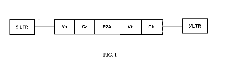
FIG. 1 is a schematic diagram showing a pMP71 retroviral vector construct. P2A
encodes
a 2A self-cleaving peptide; Va encodes the variable region of the alpha chain
of a human anti-E6
TCR; Vb encodes the variable region of the beta chain of the same human anti-
E6 TCR; Ca
encodes the constant region of the mouse TCR alpha chain; Cb encodes the
constant region of
the mouse TCR beta chain. tP indicates packaging sequences on viral RNA. 5'LTR
and 3'LTR
.. are long terminal repeats.
FIG. 2A shows the expression of TCR in non-transduced human primary T cells.
NT is a
non-transduced control. After 48 hours of culture, expression of the
recombinant TCR was
detected by staining mouse TCR beta chain. A viable CD3+ lymphocyte gating
strategy was used.
FIG. 2B shows the expression of E202 TCR in human primary T cells transduced
with
the E202 construct. After 48 hours of culture, expression of the recombinant
TCR was detected
by staining mouse TCR beta chain. A viable CD3+ lymphocyte gating strategy was
used.
FIG. 3A is a graph showing the intracellular IFN- expression of non-transduced
human
T cells upon antigen-specific stimulation. NT is a non-transduced control. The
non-transduced
human T cells were co-cultured overnight with target cells expressing the EIPV
E6 antigen at 1:1
effector-to-target ratio. The T cells were then collected and the
intracellular IFN-y expression
was determined by flow cytometry.
13
CA 03170020 2022-08-05
WO 2021/155830
PCT/CN2021/075388
FIG. 3B is a graph showing the intracellular IFN-y expression of E202 TCR-T
cells upon
antigen-specific stimulation. TCR-T cells were co-cultured overnight with
target cells expressing
the HPV E6 antigen at 1:1 effector-to-target ratio. The T cells were then
collected and the
intracellular IFN-y expression was determined by flow cytometry.
FIG. 4 is a graph showing the activation curve of TCR-T cells containing the
E202 TCR.
TCR-T cells were co-cultured overnight with different concentrations of HPV
peptide-pulsed
APCs at 1:1 effector-to-target ratio. The T cells was then collected and the
intracellular IFN-y
expression was measured to determine the EC50.
FIG. 5 is a graph showing the relation of the specific killing percentage of
target cells by
E202 TCR-T cells and E:T ratios. Target cells expressing HPV E6 antigen were
pre-stained with
CFSE and then co-cultured overnight with TCR-T cells at 1:2, 1:1, 3:1 and 10:1
effector-to-
target ratios. The cytotoxicity of T cells against target cells was measured
by 7-AAD staining.
NT is a non-transduced control.
FIG. 6A is a schematic diagram showing a pMP71 retroviral vector construct.
P2A
encodes a 2A self-cleaving peptide; Va encodes the variable region of the
alpha chain of a
human anti-HPV16 E6 TCR; Vb encodes the variable region of the beta chain of
the same
human anti-HPV16 E6 TCR; Ca encodes the constant region of the mouse TCR alpha
chain; Cb
encodes the constant region of the mouse TCR beta chain. tF indicates
packaging sequences on
viral RNA. 5'LTR and 3'LTR are long terminal repeats.
FIG. 6B is a schematic diagram showing a pMP71 retroviral vector construct
(E202P03).
P2A and T2A encodes 2A self-cleaving peptides; Va encodes the variable region
of the alpha
chain of a human anti-HPV16 E6 TCR; Vb encodes the variable region of the beta
chain of the
same human anti-HPV16 E6 TCR; Ca encodes the constant region of the mouse TCR
alpha
chain; Cb encodes the constant region of the mouse TCR beta chain; VH encodes
the variable
region of the heavy chain of an immune checkpoint inhibitor (ICI); VL encodes
the variable
region of the light chain of the immune checkpoint inhibitor (ICI). VH and VL
are linked with a
GS linker. tF indicates packaging sequences on viral RNA. 5'LTR and 3'LTR are
long terminal
repeats.
FIG. 7A shows the expression of TCR in non-transduced human primary T cells.
NT is a
non-transduced control. After 13 days of culture, expression of the
recombinant TCR was
detected by staining mouse TCR beta chain. A viable CD3+ lymphocyte gating
strategy was used.
14
CA 03170020 2022-08-05
WO 2021/155830
PCT/CN2021/075388
FIG. 7B shows the expression of E202 TCR in human primary T cells transduced
with
the E202 construct. After 13 days of culture, expression of the recombinant
TCR was detected by
staining mouse TCR beta chain. A viable CD3+ lymphocyte gating strategy was
used.
FIG. 7C shows the expression of E202P03 TCR in human primary T cells
transduced
with the E202P03 construct. After 13 days of culture, expression of the
recombinant TCR was
detected by staining mouse TCR beta chain. A viable CD3+ lymphocyte gating
strategy was used.
FIG. 8A is a graph showing the intracellular IFN- y expression of non-
transduced human
T cells upon antigen-specific stimulation. NT is a non-transduced control. The
non-transduced
human T cells were co-cultured overnight with target cells expressing the EIPV
E6 antigen at 1:1
effector-to-target ratio. The T cells were then collected and the
intracellular IFN-y expression
was determined by flow cytometry.
FIG. 8B is a graph showing the intracellular IFN-y expression of E202 TCR-T
cells upon
antigen-specific stimulation. TCR-T cells were co-cultured overnight with
target cells expressing
the EIPV E6 antigen at 1:1 effector-to-target ratio. The T cells were then
collected and the
intracellular IFN-y expression was determined by flow cytometry.
FIG. 8C is a graph showing the intracellular IFN-y expression of E202P03 TCR-T
cells
upon antigen-specific stimulation. TCR-T cells were co-cultured overnight with
target cells
expressing the EIPV E6 antigen at 1:1 effector-to-target ratio. The T cells
were then collected and
the intracellular IFN-y expression was determined by flow cytometry.
FIG. 9 is a histogram showing the IFN-y expression of E202 and E202P03 TCR-T
cells
upon antigen-specific stimulation in the cell culture supernatant. TCR-T cells
were co-cultured
overnight with target cells expressing the EIPV E6 antigen at the indicated
effector-to-target
ratios. The cell culture supernatant was then collected and the IFN-y
expression in the
supernatant was measured. NT is a non-transduced control.
FIG. 10A is a graph showing the CD107a expression of non-transduced human T
cells
upon antigen-specific stimulation. NT is a non-transduced control. The non-
transduced human T
cells were co-cultured overnight with target cells expressing the EIPV E6
antigen at 1:1 effector-
to-target ratio. The T cells were then collected and the CD107a expression was
determined in the
CD8 subpopulation by flow cytometry.
FIG. 10B is a graph showing the CD107a expression of E202 TCR-T cells upon
antigen-
specific stimulation. TCR-T cells were co-cultured overnight with target cells
expressing the
CA 03170020 2022-08-05
WO 2021/155830
PCT/CN2021/075388
1-1113V E6 antigen at 1:1 effector-to-target ratio. The T cells were then
collected and the CD107a
expression was determined in the CD8 subpopulation by flow cytometry.
FIG. 10C is a graph showing the CD expression of E202P03 TCR-T cells
upon
antigen-specific stimulation. TCR-T cells were co-cultured overnight with
target cells expressing
the EIPV E6 antigen at 1:1 effector-to-target ratio. The T cells were then
collected and the
CD107a expression was determined in the CD8 subpopulation by flow cytometry.
FIG. 10D is a graph showing the CD107a expression of non-transduced human T
cells
upon antigen-specific stimulation. NT is a non-transduced control. The non-
transduced human T
cells were co-cultured overnight with target cells expressing the EIPV E6
antigen at 1:1 effector-
to-target ratio. The T cells were then collected and the CD107a expression was
determined in the
CD4 subpopulation by flow cytometry.
FIG. 10E is a graph showing the CD107a expression of E202 TCR-T cells upon
antigen-
specific stimulation. TCR-T cells were co-cultured overnight with target cells
expressing the
EIPV E6 antigen at 1:1 effector-to-target ratio. The T cells were then
collected and the CD107a
expression was determined in the CD4 subpopulation by flow cytometry.
FIG. 1OF is a graph showing the CD107a expression of E202P03 TCR-T cells upon
antigen-specific stimulation. TCR-T cells were co-cultured overnight with
target cells expressing
the EIPV E6 antigen at 1:1 effector-to-target ratio. The T cells were then
collected and the
CD107a expression was determined in the CD4 subpopulation by flow cytometry.
FIG. 11 is a graph showing the relation of the specific killing percentage of
target cells by
E202 or E202P03 TCR-T cells and E:T ratios. Target cells expressing EIPV E6
antigen were pre-
stained with CFSE and then co-cultured overnight with TCR-T cells at 1:1, 3:1,
and 10:1
effector-to-target ratios. The cytotoxicity of T cells against target cells
was measured by 7-AAD
staining. NT is a non-transduced control.
FIG. 12 is a histogram showing the anti-PD-1 scFv expression in the cell
culture
supernatant. Either E202 or E202P03 TCR-T cells were seeded in a 24-well plate
at 3 x 10⁶/m1
for 48 hours. The cell culture supernatant was then collected and the anti-PD-
1 expression in the
supernatant was determined.
FIG. 13A shows the expression of TCR in untransduced (UT) human PBMCs.
Expression of the recombinant TCR was detected by staining mouse TCR beta
chain. A viable
CD3+ lymphocyte gating strategy was used.
16
CA 03170020 2022-08-05
WO 2021/155830
PCT/CN2021/075388
FIG. 13B shows the expression of E203 TCR in human PBMCs transduced with the
E203 construct. 5 days post transduction, expression of the recombinant TCR
was detected by
staining mouse TCR beta chain. A viable CD3+ lymphocyte gating strategy was
used.
FIG. 13C shows the expression of E204 TCR in human PBMCs transduced with the
.. E204 construct. 5 days post transduction, expression of the recombinant TCR
was detected by
staining mouse TCR beta chain. A viable CD3+ lymphocyte gating strategy was
used.
FIG. 13D shows the expression of E205 TCR in human PBMCs transduced with the
E205 construct. 5 days post transduction, expression of the recombinant TCR
was detected by
staining mouse TCR beta chain. A viable CD3+ lymphocyte gating strategy was
used.
FIG. 14A shows the intracellular IFN-y expression of non-transduced (UT) human
CD4+
T cells without antigen-specific stimulation.
FIG. 14B shows the intracellular IFN-y expression of non-transduced (UT) human
CD4+
T cells that were co-cultured overnight with Ca Ski E6/E7 cells at 1: 2
effector-to-target cell ratio.
The intracellular IFN-y expression was determined by flow cytometry.
FIG. 14C shows the intracellular IFN-y expression of non-transduced (UT) human
CD4+
T cells that were co-cultured overnight with Ca Ski cells at 1: 2 effector-to-
target cell ratio. The
intracellular IFN-y expression was determined by flow cytometry.
FIG. 14D shows the intracellular IFN-y expression of non-transduced (UT) human
CD4+
T cells that were co-cultured overnight with 293T cells at 1: 2 effector-to-
target cell ratio. The
intracellular IFN-y expression was determined by flow cytometry.
FIG. 14E shows the intracellular IFN-y expression of human CD4+ E203 TCR-T
cells
without antigen-specific stimulation.
FIG. 14F shows the intracellular IFN-y expression of human CD4+ E203 TCR-T
cells
that were co-cultured overnight with Ca Ski E6/E7 cells at 1: 2 effector-to-
target cell ratio. The
intracellular IFN-y expression was determined by flow cytometry.
FIG. 14G shows the intracellular IFN-y expression of human CD4+ E203 TCR-T
cells
that were co-cultured overnight with Ca Ski cells at 1: 2 effector-to-target
cell ratio. The
intracellular IFN-y expression was determined by flow cytometry.
FIG. 14H shows the intracellular IFN-y expression of human CD4+ E203 TCR-T
cells
.. that were co-cultured overnight with 293T cells at 1: 2 effector-to-target
cell ratio. The
intracellular IFN-y expression was determined by flow cytometry.
17
CA 03170020 2022-08-05
WO 2021/155830
PCT/CN2021/075388
FIG. 141 shows the intracellular IFN-y expression of human CD4+ E204 TCR-T
cells
without antigen-specific stimulation.
FIG. 141 shows the intracellular IFN-y expression of human CD4+ E204 TCR-T
cells
that were co-cultured overnight with Ca Ski E6/E7 cells at 1: 2 effector-to-
target cell ratio. The
intracellular IFN-y expression was determined by flow cytometry.
FIG. 14K shows the intracellular IFN-y expression of human CD4+ E204 TCR-T
cells
that were co-cultured overnight with Ca Ski cells at 1: 2 effector-to-target
cell ratio. The
intracellular IFN-y expression was determined by flow cytometry.
FIG. 14L shows the intracellular IFN-y expression of human CD4+ E204 TCR-T
cells
that were co-cultured overnight with 293T cells at 1: 2 effector-to-target
cell ratio. The
intracellular IFN-y expression was determined by flow cytometry.
FIG. 14M shows the intracellular IFN-y expression of human CD4+ E205 TCR-T
cells
without antigen-specific stimulation.
FIG. 14N shows the intracellular IFN-y expression of human CD4+ E205 TCR-T
cells
that were co-cultured overnight with Ca Ski E6/E7 cells at 1: 2 effector-to-
target cell ratio. The
intracellular IFN-y expression was determined by flow cytometry.
FIG. 140 shows the intracellular IFN-y expression of human CD4+ E205 TCR-T
cells
that were co-cultured overnight with Ca Ski cells at 1: 2 effector-to-target
cell ratio. The
intracellular IFN-y expression was determined by flow cytometry.
FIG. 14P shows the intracellular IFN-y expression of human CD4+ E205 TCR-T
cells
that were co-cultured overnight with 293T cells at 1: 2 effector-to-target
cell ratio. The
intracellular IFN-y expression was determined by flow cytometry.
FIG. 15A shows the intracellular IFN-y expression of non-transduced (UT) human
CD8+
T cells without antigen-specific stimulation.
FIG. 15B shows the intracellular IFN-y expression of non-transduced (UT) human
CD8+
T cells that were co-cultured overnight with Ca Ski E6/E7 cells at 1: 2
effector-to-target cell ratio.
The intracellular IFN-y expression was determined by flow cytometry.
FIG. 15C shows the intracellular IFN-y expression of non-transduced (UT) human
CD8+
T cells that were co-cultured overnight with Ca Ski cells at 1: 2 effector-to-
target cell ratio. The
intracellular IFN-y expression was determined by flow cytometry.
18
CA 03170020 2022-08-05
WO 2021/155830
PCT/CN2021/075388
FIG. 15D shows the intracellular IFN-y expression of non-transduced (UT) human
CD8+
T cells that were co-cultured overnight with 293T cells at 1: 2 effector-to-
target cell ratio. The
intracellular IFN-y expression was determined by flow cytometry.
FIG. 15E shows the intracellular IFN-y expression of human CD8+ E203 TCR-T
cells
without antigen-specific stimulation.
FIG. 15F shows the intracellular IFN-y expression of human CD8+ E203 TCR-T
cells
that were co-cultured overnight with Ca Ski E6/E7 cells at 1: 2 effector-to-
target cell ratio. The
intracellular IFN-y expression was determined by flow cytometry.
FIG. 15G shows the intracellular IFN-y expression of human CD8+ E203 TCR-T
cells
that were co-cultured overnight with Ca Ski cells at 1: 2 effector-to-target
cell ratio. The
intracellular IFN-y expression was determined by flow cytometry.
FIG. 15H shows the intracellular IFN-y expression of human CD8+ E203 TCR-T
cells
that were co-cultured overnight with 293T cells at 1: 2 effector-to-target
cell ratio. The
intracellular IFN-y expression was determined by flow cytometry.
FIG. 151 shows the intracellular IFN-y expression of human CD8+ E204 TCR-T
cells
without antigen-specific stimulation.
FIG. 15J shows the intracellular IFN-y expression of human CD8+ E204 TCR-T
cells
that were co-cultured overnight with Ca Ski E6/E7 cells at 1: 2 effector-to-
target cell ratio. The
intracellular IFN-y expression was determined by flow cytometry.
FIG. 15K shows the intracellular IFN-y expression of human CD8+ E204 TCR-T
cells
that were co-cultured overnight with Ca Ski cells at 1: 2 effector-to-target
cell ratio. The
intracellular IFN-y expression was determined by flow cytometry.
FIG. 15L shows the intracellular IFN-y expression of human CD8+ E204 TCR-T
cells
that were co-cultured overnight with 293T cells at 1: 2 effector-to-target
cell ratio. The
intracellular IFN-y expression was determined by flow cytometry.
FIG. 15M shows the intracellular IFN-y expression of human CD8+ E205 TCR-T
cells
without antigen-specific stimulation.
FIG. 15N shows the intracellular IFN-y expression of human CD8+ E205 TCR-T
cells
that were co-cultured overnight with Ca Ski E6/E7 cells at 1: 2 effector-to-
target cell ratio. The
intracellular IFN-y expression was determined by flow cytometry.
19
CA 03170020 2022-08-05
WO 2021/155830
PCT/CN2021/075388
FIG. 150 shows the intracellular IFN-y expression of human CD8+ E205 TCR-T
cells
that were co-cultured overnight with Ca Ski cells at 1: 2 effector-to-target
cell ratio. The
intracellular IFN-y expression was determined by flow cytometry.
FIG. 15P shows the intracellular IFN-y expression of human CD8+ E205 TCR-T
cells
that were co-cultured overnight with 293T cells at 1: 2 effector-to-target
cell ratio. The
intracellular IFN-y expression was determined by flow cytometry.
FIG. 16A shows the absolute killing efficacy of Ca Ski E6/E7 cells by
untransduced (UT),
E203, E204, and E205 TCR-T cells. CellTraceTm CFSE-labeled Ca Ski E6/E7 cells
and
CellTraceTm Violet-labeled 293T cells were mixed and co-cultured overnight
with TCR-T cells
at a 0:1, 0.4: 1,2:1, or 10:1 effector-to-target cell ratio. Beads were added
as a reference for flow
cytometry analysis.
FIG. 16B shows the competitive killing efficacy of Ca Ski E6/E7 cells by
untransduced
(UT), E203, E204, and E205 TCR-T cells. CellTraceTm CFSE-labeled Ca Ski E6/E7
cells and
CellTraceTm Violet-labeled 293T cells were mixed and co-cultured overnight
with TCR-T cells
at a 0:1, 0.4: 1,2:1, or 10:1 effector-to-target cell ratio.
FIG. 17A shows the activation curve of CD8+ TCR-T cells containing the E203
TCR.
The intracellular IFN-y expression was measured to determine the EC50.
FIG. 17B shows the activation curve of CD4+ TCR-T cells containing the E203
TCR.
The intracellular IFN-y expression was measured to determine the EC50.
FIG. 18A shows the activation curve of CD8+ TCR-T cells containing the E204
TCR.
The intracellular IFN-y expression was measured to determine the EC50.
FIG. 18B shows the activation curve of CD4+ TCR-T cells containing the E204
TCR.
The intracellular IFN-y expression was measured to determine the EC50.
FIG. 19A shows the activation curve of CD8+ TCR-T cells containing the E205
TCR.
The intracellular IFN-y expression was measured to determine the EC50.
FIG. 19B shows the activation curve of CD4+ TCR-T cells containing the E205
TCR.
The intracellular IFN-y expression was measured to determine the EC50.
FIG. 20 is table showing sequences of E202, E203, E204, and E205 TCR. CDR1a,
CDR2a, and CDR3a are CDR1, CDR2 and CDR3 of the TCR alpha chain variable
domain,
respectively. CDR1 (3, CDR2(3, and CDR3(3 are CDR1, CDR2 and CDR3 of the TCR
beta chain
variable domain, respectively. TRA VJ are the rearranged V and J segments
encoding the alpha
CA 03170020 2022-08-05
WO 2021/155830
PCT/CN2021/075388
chain variable domain of the TCR. TRB VDJ are the rearranged V, D, and J
segments encoding
the beta chain variable domain of the TCR.
FIG. 21 provides several sequences as described in the disclosure.
DETAILED DESCRIPTION
Human papilloma viruses (HPVs) are small (approximately 8000 pairs of bases)
double-
stranded DNA viruses which infect squamous epithelia and induce proliferative
lesions such as
skin warts (Xavier, "Natural History And Epidemiology Of Hpv Infection And
Cervical Cancer".
Gynecologic Oncology 110 (2008) S4¨S7; Hausen et al., "Human Papilloma
Viruses." Annu.
Rev. Microbial. 1994. 48;427-47). HPV has a well conserved genetic
organization and all the
potential open reading frames (ORFs) are located in one DNA strand, the
reading frames of
which are designated as early (E) or late (L) genes. While the early genes (El
-E8) are activated
immediately after infection, the late genes encode structural proteins
expressed in the granular
layer of the epithelium. The gene products of the early genes are involved in
controlling
replication and expression of viral DNA (Mannarini et a/."Human Papilloma
Virus (HPV) In
Head And Neck Region: Review Of Literature". Acta Otorhinolaryngol
Ita12009;29:119-126).
Chimeric Antigen Receptor (CARs) T-cell are engineered cells having an
extracellular
antigen recognition domain fused with intracellular T cell signaling and
costimulatory domains.
CARs can directly and selectively recognize cell surface tumor associated
antigens (TAAs) in a
.. major histocompatibility class (MHC)-independent manner. Despite the
documented success of
CAR T cell therapy in patients with hematologic malignancies, only modest
responses have been
observed in solid tumors. This can be attributed, in part, to the
establishment of an
immunosuppressive microenvironment in solid tumors. Such milieu involves the
upregulation of
several intrinsic inhibitory pathways mediated by increased expression of
inhibitory receptors
(IRs) in T cells reacting with their cognate ligands within the tumor (Ping et
al., "T-cell receptor-
engineered T cells for cancer treatment: current status and future
directions." Protein & cell 9.3
(2018): 254-266.).
Adoptive cell transfer (ACT) is a modality of cancer immunotherapy which has
demonstrated remarkable success in treating hematologic malignancies and
malignant melanoma.
An especially effective form of ACT, which uses gene-modified T cells
expressing a chimeric
antigen receptor (CAR) to specifically target tumor-associated-antigen (TAA),
such as CD19 and
21
CA 03170020 2022-08-05
WO 2021/155830
PCT/CN2021/075388
GD2, has displayed encouraging results in clinical trials for treating such
diseases as B cell
malignancies and neuroblastoma (Simon et al., "CAR-T cell therapy in melanoma:
A future
success story?." Experimental dermatology 27.12 (2018): 1315-1321). The use of
modified
TCRs for the treatment of different diseases has achieved significant results
over the years and
has been the focus area of a number of studies.
The present disclosure provides T-cell receptor (TCR)-engineered T cells,
which can be
used in cell therapy. The engineered T-cell receptors are capable of
recognizing the surface
antigen on the cell receptor which are otherwise not recognized by normal T-
cells. The
engineered T cells can be employed against multiple targets such as cancer
cells expressing
appropriate antigens.
Theoretically, a T cell receptor can have antigenic specificity for any HPV
antigen. The
E6 and E7 onco-proteins in HPV are necessary for malignant conversion of the
cells. The HPV
E7 protein mainly contributes to cancer development via inactivation of the
Retinoblastoma
protein, which results in constitutive cancer cell cycle activation. In some
embodiments, the
modified T cells are capable of recognizing an epitope of HPV in a MHC
dependent manner (e.g.,
the HLA-A 02:01¨restricted epitope of a high-risk serotype of HPV such as HPV-
16). In this
setting, HPV antigen positive tumor cells can be killed by engineered TCR-T
cells.
T CELL RECEPTORS AND BINDING MOLECULES
T cells are a type of lymphocyte which typically develops in the thymus gland
and plays a
central role in the immune response. It plays an important role in the
"adaptive immune
response." T cells can be distinguished from other lymphocytes by the presence
of a T-cell
receptor on the cell surface. Differentiated T cells have an important role in
controlling the
immune response. CD8+ T cells, also known as "killer cells", are cytotoxic.
Once they recognize
a target cell, they are able to directly kill the target cell (e.g., virus-
infected cells or cancer cells).
CD8+ T cells can also produce cytokines and recruit other cells (e.g.,
macrophages and natural
killer (NK) cells) to mount an immune response. CD4+ T cells, also known as
"helper cells", can
indirectly kill target cells, e.g., by facilitating maturation of B cells into
plasma cells and memory
B cells, and activation of cytotoxic T cells and macrophages. Helper T cells
become activated
when they are presented with peptide antigens by MHC class II molecules, which
are expressed
on the surface of antigen-presenting cells (APCs). Once activated, they divide
rapidly and secrete
22
CA 03170020 2022-08-05
WO 2021/155830
PCT/CN2021/075388
cytokines that regulate or assist the immune response. Regulatory T cells are
important for
tolerance, thereby preventing or inhibiting autoimmune response. The major
role of regulatory T
cells is to shut down T cell-mediated immunity toward the end of an immune
reaction and to
suppress autoreactive T cells that escaped the process of negative selection
in the thymus.
T cells play an important role in cancer immunity where antigens from the
cancer cells are
taken up and presented on the cell surface of special immune cells called
antigen-presenting cells
(APCs) so that other immune cells can recognize the antigens of interest. In
the lymph nodes, the
APCs activate the T-cells and activate them to recognize the tumor cells. The
activated T-cells
can then travel via the blood vessels to reach the tumor, infiltrate it,
recognize the cancer cells
and kill them.
The activation of T cells requires T cell receptors. A "T cell receptor" or
"TCR" is a
molecule that contains a variable a (or alpha) and b (or beta) chains (also
known as TCRa and
TCRP, respectively) or a variable g (or gamma) and d (or delta) chains (also
known as TCRy and
TCRO, respectively), or antigen-binding portions thereof, and which is capable
of specifically
binding to an antigen, e.g., a peptide antigen or peptide epitope bound to an
major
histocompatibility complex (MHC) molecule.
The present disclosure provides a T cell receptor (TCR) or antigen-binding
fragment
thereof, and binding molecules derived from TCR. In some embodiments, the TCR
is in the ab
form. TCRs that exist in af3 and yo forms are generally structurally similar,
but T cells
expressing them may have distinct anatomical locations or functions.
Generally, a TCR is found
on the surface of T cells (or T lymphocytes) where it is generally responsible
for recognizing
antigens, such as peptides bound to major histocompatibility complex (MHC)
molecules.
In some embodiments, the TCR is an intact or full-length TCR, such as a TCR
containing
the a chain and b chain. In some embodiments, the TCR is an antigen-binding
portion that is less
than a full- length TCR but that binds to a specific peptide bound in an MHC
molecule, such as
binds to an MEC-peptide complex. In some cases, an antigen-binding portion or
fragment of a
TCR can contain only a portion of the structural domains of a full-length or
intact TCR, but yet
is able to bind the peptide epitope, such as MEC-peptide complex, to which the
full TCR binds.
In some cases, an antigen-binding portion contains the variable domains of a
TCR, such as
variable a (Va or Va) chain and variable b (Vb or vp) chain of a TCR, or
antigen -binding
fragments thereof sufficient to form a binding site for binding to a specific
MHC-peptide
23
CA 03170020 2022-08-05
WO 2021/155830
PCT/CN2021/075388
complex.
The variable domains of the TCR contain complementarity determining regions
(CDRs),
which generally are the primary contributors to antigen recognition and
binding capabilities and
specificity of the peptide, MHC and/or MHC-peptide complex. In some
embodiments, a CDR of
a TCR or combination thereof forms all or substantially all of the antigen-
binding site of a given
TCR molecule. The various CDRs within a variable region of a TCR chain
generally are
separated by framework regions (FRs), which generally display less variability
among TCR
molecules as compared to the CDRs. In some embodiments, CDR3 is the main CDR
responsible
for antigen binding or specificity, or is the most important among the three
CDRs on a given
TCR variable region for antigen recognition, and/or for interaction with the
processed peptide
portion of the peptide-MHC complex. In some contexts, the CDR1 of the alpha
chain can
interact with the N-terminal part of certain antigenic peptides. In some
cases, CDR1 of the beta
chain can interact with the C-terminal part of the peptide. In some contexts,
CDR2 contributes
most strongly to or is the primary CDR responsible for the interaction with or
recognition of the
MHC portion of the MHC-peptide complex.
The a-chain and/or b-chain of a TCR also can contain a constant domain, a
transmembrane
domain and/or a short cytoplasmic tail. In some aspects, each chain (e.g.
alpha or beta) of the
TCR can possess one N-terminal immunoglobulin variable domain, one
immunoglobulin
constant domain, a transmembrane region, and a short cytoplasmic tail at the C-
terminal end. In
some embodiments, a TCR, for example via the cytoplasmic tail, is associated
with invariant
proteins of the CD3 complex involved in mediating signal transduction. In some
cases, the
structure allows the TCR to associate with other molecules like CD3 and
subunits thereof. For
example, a TCR containing constant domains with a transmembrane region may
anchor the
protein in the cell membrane and associate with invariant subunits of the CD3
signaling
apparatus or complex. The intracellular tails of CD3 signaling subunits (e.g.
CD3y, CD3, CD3e
and CD3z chains) contain one or more immunoreceptor tyrosine-based activation
motif or ITAM
and generally are involved in the signaling capacity of the TCR complex.
The exact locus of a domain or region can vary depending on the particular
structural or
homology modeling or other features used to describe a particular domain. It
is understood that
reference to amino acids, including to a specific sequence set forth as a SEQ
ID NO used to
describe domain organization of a TCR are for illustrative purposes and are
not meant to limit
24
CA 03170020 2022-08-05
WO 2021/155830
PCT/CN2021/075388
the scope of the embodiments provided. In some cases, the specific domain
(e.g. variable or
constant) can be several amino acids (such as one, two, three or four) longer
or shorter. In some
aspects, residues of a TCR are known or can be identified according to the
International
Immunogenetics Information System (IMGT) numbering system (see e.g.
www.imgt.org;
Lefranc et al. "IMGT unique numbering for immunoglobulin and T cell receptor
variable
domains and Ig superfamily V-like domains." Developmental & Comparative
Immunology 27.1
(2003): 55-77.). The structures and variations of TCR are known in the art,
and are described,
e.g., in WO 2019 /195486, which is incorporated herein by reference in its
entirety.
In some embodiments, the a chain and b chain of a TCR each further contain a
constant
domain. In some embodiments, the a chain constant domain (Ca) and b chain
constant domain
(Cb) individually are mammalian, such as is a human or a non-human constant
domain (e.g., a
mouse constant domain). In some embodiments, the constant domain is adjacent
to the cell
membrane. For example, in some cases, the extracellular portion of the TCR
formed by the two
chains contains two membrane-proximal constant domains, and two membrane-
distal variable
domains, which variable domains each contain CDRs.
In some aspects, TCRs as descried herein can contain a human constant region,
such as an
alpha chain containing a human Ca region and a beta chain containing a human
Cb regin. In
some embodiments, the TCRs are fully human. In some embodiments, the
expression and/or
activity of TCRs, such as when expressed in human cells, e.g. human T cells,
such as primary
human T cells, are not impacted by or are not substantially impacted by the
presence of an
endogenous human TCR.
In some embodiments, the engineered TCRs are expressed at similar or improved
levels on
the cell surface, exhibit the similar or greater functional activity (e.g.
cytolytic activity) and/or
exhibit similar or greater anti-tumor activity, when expressed by human cells
that contain or
express an endogenous human TCR, such as human T cells, as compared to the
level of
expression, function activity and/or anti-tumor activity of the same TCR in
similar human cells
but in which expression of the endogenous TCR has been reduced or eliminated.
In some
examples an engineered TCR as described herein, when expressed in human T
cells, is expressed
on the cell surface at a level that is at least or at least about 80%, 85%,
90%, 95%, 100%, 105%,
110%, 115% or 120% of the level of expression of the same TCR when expressed
in similar
human T cells but in which expression of the endogenous TCR has been reduced
or eliminated.
CA 03170020 2022-08-05
WO 2021/155830
PCT/CN2021/075388
In some embodiments, each of the Ca and Cb domains is human. In some
embodiments,
the Ca is encoded by the TRAC gene (IMGT nomenclature) or is a variant
thereof. In some
embodiments, the variant of a Ca contains replacement of at least one non-
native cysteine.
In some embodiments, the TCR can be a heterodimer of two chains a and b that
are linked,
such as by a disulfide bond or disulfide bonds. In some embodiments, the
constant domain of the
TCR can contain short connecting sequences in which a cysteine residue forms a
disulfide bond,
thereby linking the two chains of the TCR. In some embodiments, a TCR can have
an additional
cysteine residue in each of the a and b chains, such that the TCR contains two
disulfide bonds in
the constant domains. In some embodiments, each of the constant and variable
domains contains
disulfide bonds formed by cysteine residues.
In some embodiments, the TCR comprises CDRs, Va and/or Vb and constant region
sequences as described herein.
In some embodiments, the TCR is a dimeric TCR (dTCR). In some embodiments a
dTCR
contains a first polypeptide wherein a sequence corresponding to a provided
TCR a chain
variable region sequence is fused to the N terminus of a sequence
corresponding to a TCR a
chain constant region extracellular sequence, and a second polypeptide wherein
a sequence
corresponding to a provided TCR b chain variable region sequence is fused to
the N terminus a
sequence corresponding to a TCR b chain constant region extracellular
sequence, the first and
second polypeptides being linked by a disulfide bond.
In some embodiments, a TCR can be cell-bound or in soluble form. In some
embodiments,
the TCR is in cell-bound form expressed on the surface of a cell.
In some embodiments, the TCR is a single chain TCR (scTCR). The scTCR is a
single
amino acid strand containing an a chain and a b chain that is able to bind to
MHC-peptide
complexes. Typically, a scTCR can be generated using methods known to those of
skill in the art.
These methods are described e.g., in WO 96/13593, WO 96/18105, W099/18129, WO
04/033685, W02006/037960, W02011/044186; WO 2019 /195486; U.S. Patent No.
7,569,664;
each of which is incorporated herein by reference in its entirety.
The TCR, antigen binding fragments thereof, and TCR-derived binding molecules
can
bind or recognize a peptide epitope associated with an antigen of interest
(e.g., a cancer antigen).
In some embodiments, the antigen can be a peptide epitope expressed on the
surface of a cancer
cell and/or a cell infected with a virus, e.g., HPV. In some embodiments, the
antigen is presented
26
CA 03170020 2022-08-05
WO 2021/155830
PCT/CN2021/075388
in the context of an MEC molecule. Such binding molecules include e.g., T cell
receptors (TCRs)
and antigen-binding fragments thereof, antibodies and antigen binding
fragments thereof, and
TCR-like CAR. They exhibit antigenic specificity for binding or recognizing
such peptide
epitopes. In some aspects, engineered cells that express a provided binding
molecule, e.g. a TCR
or antigen-binding fragment, exhibit cytotoxic activity against target cells
expressing the peptide
epitope, such as cancer cells or cells that are infected with HPV.
In some aspects, the TCR, antigen binding fragments thereof, and TCR-derived
binding
molecules recognize or bind to epitopes in the context of an MEC molecule,
such as an MEC
Class I molecule or an MEC class II molecule. Both MEC Class I molecules or
MEC class II
molecules are human leukocyte antigens (HLA). They play an important component
of adaptive
immune system. The EILA expression is controlled by genes located on
chromosome 6. It
encodes cell surface molecules specialized to present antigenic peptides to
the T-cell receptor on
T cells.
In some embodiments, the TCR, antigen binding fragments thereof, and TCR-
derived
binding molecules recognize or bind to epitopes in the context of an MEC Class
I molecule. The
MEC Class I molecule is a human leukocyte antigen (HLA)-A2 molecule, including
any one or
more subtypes thereof, e.g. HLA-A*0201, *0202, *0203, *0206, or *0207. The
human leukocyte
antigen A2 (HLA-A2) is among the most common human serotypes. In some cases,
there can be
differences in the frequency of subtypes between different populations. For
example, more than
95% of the HLA-A2 positive Caucasian population is EILA- A*0201, whereas in
the Chinese
population the frequency has been reported to be approximately 23% for HLA-
A*0201, 45% for
HLA-A*0207, 8% for HLA-A*0206 and 23% for HLA-A*0203. In some embodiments, the
MEC molecule is HLA-A*0201. In some embodiments, the present disclosure
provides TCR or
antigen-binding fragment thereof that bind an HPV-EB6/HLA-A2 complex.
In some embodiments, the binding molecule, e.g., TCR or antigen-binding
fragment
thereof or TCR-derived binding molecule, is isolated or purified, or is
recombinant. In some
aspects, the binding molecule, e.g., TCR or antigen-binding fragment thereof
or TCR-derived
binding molecule, is fully human. In some embodiments, the binding molecule is
monoclonal. In
some aspects, the binding molecule is a single chain. In other embodiments,
the binding
molecule contains two chains. In some embodiments, the binding molecule, e.g.,
TCR, antigen-
binding fragment thereof or TCR-derived binding molecule, is expressed on the
surface of a cell.
27
CA 03170020 2022-08-05
WO 2021/155830
PCT/CN2021/075388
The TCR, antigen-binding fragment thereof, or TCR-derived binding molecules
can have a
Va and a Vb, or a region that is similar to Va and a region that is similar to
Vb. In some
embodiments, the Va region comprises the amino acid sequence set forth in any
of SEQ ID NO:
1, 45, 47, 49, or an amino acid sequence that has at least 80%, 85%, 90%, 91%,
92%, 93%, 94%,
95%, 96%, 97%, 98%, or 99% sequence identity thereto. In some embodiments, the
Vb region
comprises the amino acid sequence set forth in any of SEQ ID NO: 2, 46, 48,
50, or an amino
acid sequence that has at least 80%, 85%, 90%, 91%, 92%, 93%, 94%, 95%, 96%,
97%, 98%, or
99% sequence identity thereto. In some embodiments, the Va region comprises
one or more Va
CDR sequences as described herein. In some embodiments, the Vb region
comprises one or more
Vb CDR sequences as described herein.
In some embodiments, the TCR, TCR derived binding molecules, or antigen-
binding
fragment thereof, comprising an alpha chain comprising a variable alpha (Va)
region and a beta
chain comprising a variable beta (Vb) region, wherein the Va region can have
complementarity
determining regions (CDRs) 1, 2, 3, wherein the CDR1 region comprises or
consists of an amino
acid sequence that is at least 80%, 85%, 90%, or 95% identical to a selected
Va CDR1 amino
acid sequence, the CDR2 region comprises or consists of an amino acid sequence
that is at least
80%, 85%, 90%, or 95% identical to a selected Va CDR2 amino acid sequence, and
the CDR3
region comprises or consists of an amino acid sequence that is at least 80%,
85%, 90%, or 95%
identical to a selected Va CDR3 amino acid sequence, and a variable beta (Vb)
region
comprising CDRs 1, 2, 3, wherein the CDR1 region comprises or consists of an
amino acid
sequence that is at least 80%, 85%, 90%, or 95% identical to a selected Vb
CDR1 amino acid
sequence, the CDR2 region comprises or consists of an amino acid sequence that
is at least 80%,
85%, 90%, or 95% identical to a selected Vb CDR2 amino acid sequence, and the
CDR3 region
comprises or consists of an amino acid sequence that is at least 80%, 85%,
90%, or 95% identical
to a selected Vb CDR3 amino acid sequence. The selected Va CDRs 1, 2, 3 amino
acid
sequences and the selected Vb CDRs, 1, 2, 3 amino acid sequences are shown in
FIG. 20.
In some embodiments, the TCR, antigen-binding fragment thereof, or TCR derived
binding molecules described herein can contain a variable region (e.g., Va)
containing one, two,
or three of the CDRs of SEQ ID NO: 5 with zero, one or two amino acid
insertions, deletions, or
substitutions; SEQ ID NO: 6 with zero, one or two amino acid insertions,
deletions, or
substitutions; SEQ ID NO: 7 with zero, one or two amino acid insertions,
deletions, or
28
CA 03170020 2022-08-05
WO 2021/155830
PCT/CN2021/075388
substitutions.
In some embodiments, the TCR, antigen-binding fragment thereof, or TCR derived
binding molecules described herein can contain a variable region (e.g., Vb)
containing one, two,
or three of the CDRs of SEQ ID NO: 8 with zero, one or two amino acid
insertions, deletions, or
substitutions; SEQ ID NO: 9 with zero, one or two amino acid insertions,
deletions, or
substitutions; SEQ ID NO: 10 with zero, one or two amino acid insertions,
deletions, or
substitutions.
In some embodiments, the TCR, antigen-binding fragment thereof, or TCR derived
binding molecules described herein can contain a variable region (e.g., Va)
containing one, two,
or three of the CDRs of SEQ ID NO: 27 with zero, one or two amino acid
insertions, deletions,
or substitutions; SEQ ID NO: 28 with zero, one or two amino acid insertions,
deletions, or
substitutions; SEQ ID NO: 29 with zero, one or two amino acid insertions,
deletions, or
substitutions.
In some embodiments, the TCR, antigen-binding fragment thereof, or TCR derived
binding molecules described herein can contain a variable region (e.g., Vb)
containing one, two,
or three of the CDRs of SEQ ID NO: 30 with zero, one or two amino acid
insertions, deletions,
or substitutions; SEQ ID NO: 31 with zero, one or two amino acid insertions,
deletions, or
substitutions; SEQ ID NO: 32 with zero, one or two amino acid insertions,
deletions, or
substitutions.
In some embodiments, the TCR, antigen-binding fragment thereof, or TCR derived
binding molecules described herein can contain a variable region (e.g., Va)
containing one, two,
or three of the CDRs of SEQ ID NO: 33 with zero, one or two amino acid
insertions, deletions,
or substitutions; SEQ ID NO: 34 with zero, one or two amino acid insertions,
deletions, or
substitutions; SEQ ID NO: 35 with zero, one or two amino acid insertions,
deletions, or
substitutions.
In some embodiments, the TCR, antigen-binding fragment thereof, or TCR derived
binding molecules described herein can contain a variable region (e.g., Vb)
containing one, two,
or three of the CDRs of SEQ ID NO: 36 with zero, one or two amino acid
insertions, deletions,
or substitutions; SEQ ID NO: 37 with zero, one or two amino acid insertions,
deletions, or
substitutions; SEQ ID NO: 38 with zero, one or two amino acid insertions,
deletions, or
substitutions.
29
CA 03170020 2022-08-05
WO 2021/155830
PCT/CN2021/075388
In some embodiments, the TCR, antigen-binding fragment thereof, or TCR derived
binding molecules described herein can contain a variable region (e.g., Va)
containing one, two,
or three of the CDRs of SEQ ID NO: 39 with zero, one or two amino acid
insertions, deletions,
or substitutions; SEQ ID NO: 40 with zero, one or two amino acid insertions,
deletions, or
substitutions; SEQ ID NO: 41 with zero, one or two amino acid insertions,
deletions, or
substitutions.
In some embodiments, the TCR, antigen-binding fragment thereof, or TCR derived
binding molecules described herein can contain a variable region (e.g., Vb)
containing one, two,
or three of the CDRs of SEQ ID NO: 42 with zero, one or two amino acid
insertions, deletions,
.. or substitutions; SEQ ID NO: 43 with zero, one or two amino acid
insertions, deletions, or
substitutions; SEQ ID NO: 44 with zero, one or two amino acid insertions,
deletions, or
substitutions.
The present disclosure also provides TCR a (alpha) and/or b (beta) chain as
described
herein. In some embodiments, the a chain comprises the amino acid sequence set
forth in SEQ
.. ID NO: 15, 51, 53, 55, or an amino acid sequence that has at least 80%,
85%, 90%, 91%, 92%,
93%, 94%, 95%, 96%, 97%, 98%, or 99% sequence identity thereto. In some
embodiments, the b
chain comprises the amino acid sequence set forth in SEQ ID NO: 16, 52, 54,
56, or an amino
acid sequence that has at least 80%, 85%, 90%, 91%, 92%, 93%, 94%, 95%, 96%,
97%, 98%, or
99% sequence identity thereto. In some embodiments, the a chain comprises one
or more Va
CDR sequences as described herein. In some embodiments, the b chain comprises
one or more
Vb CDR sequences as described herein.
In some embodiments, the TCR may be a heterodimer of two chains a and b that
are linked,
such as by a disulfide bond or disulfide bonds. In some embodiments, the
constant domain of the
TCR may contain short connecting sequences in which a cysteine residue forms a
disulfide bond,
.. thereby linking the two chains of the TCR. In some embodiments, a TCR may
have an additional
cysteine residue in each of the a and b chains, such that the TCR contains two
disulfide bonds in
the constant domains. In some embodiments, each of the constant and variable
domains contains
disulfide bonds formed by cysteine residues.
In some embodiments, the native disulfide bonds are not present. In some
embodiments,
.. the one or more of the native cysteines (e.g. in the constant domain of the
a chain and b chain)
that form a native interchain disulfide bond are substituted to another
residue, such as to a serine
CA 03170020 2022-08-05
WO 2021/155830
PCT/CN2021/075388
or alanine. In some embodiments, an introduced disulfide bond can be formed by
mutating non
cysteine residues on the alpha and beta chains, such as in the constant domain
of the a chain and
b chain, to cysteine. Opposing cysteines in the TCR a and b chains provide a
disulfide bond that
links the constant regions of TCR a and b chains of the substituted TCR to one
another and
which is not present in a TCR comprising the unsubstituted constant region in
which the native
disulfide bonds are present, such as unsubstituted native human constant
region or the
unsubstituted native mouse constant region. In some embodiments, the presence
of non-native
cysteine residues (e.g. resulting in one or more non-native disulfide bonds)
in a recombinant
TCR can favor production of the desired recombinant TCR in a cell in which it
is introduced
over expression of a mismatched TCR pair containing a native TCR chain.
In some embodiments, the nucleic acid encoding the alpha chain and the nucleic
acid
encoding the beta chain can be connected via a linker, such as any described
elsewhere herein.
The disclosure also provides nucleic acid comprising a polynucleotide encoding
a
polypeptide comprising a TCR a chain variable region, a TCR b chain variable
region, an
immunoglobulin heavy chain variable region or an immunoglobulin light chain
variable region.
The variable region comprises CDRs as shown in FIG. 20. When the polypeptides
are paired
with corresponding polypeptide (e.g., a corresponding a chain variable region
or a corresponding
b chain variable region), the paired polypeptides bind to the antigen of
interest (e.g., HPV E6).
In some embodiments, by binding to the antigen of interest, the TCR or antigen-
binding
fragment thereof, or TCR-derived binding molecules, can activate T cells
(e.g., by activating
TCR signaling pathway). In some embodiments, the activation can upregulate
immune response,
increase expression of cytokines (e.g., IFNy) and/or CD107a, promote T-cell
proliferation and T
cell mediated killing.
In some embodiments, the TCR or antigen-binding fragment thereof, or TCR-
derived
binding molecules as described herein can increase immune response, activity
or number of T
cells by at least 10%, 20%, 30%, 40%, 50%, 60%, 70%, 80%, 90%, 100%, 2 folds,
3 folds, 5
folds, 10 folds, or 20 folds. In some embodiments, the TCR or antigen-binding
fragment thereof,
or TCR-derived binding molecules, when the antigen of interest is present, can
increase serum
concentrations of IFN-y. In some embodiments, the activation can induce at
least 10%, 20%,
30%, 40%, 50%, 60%, 70%, 80%, 90%, 1 fold, 2 folds, 5 folds, 10 folds, 100
folds, or 1000
folds increase of the serum concentrations of IFN-y. In some embodiments, the
activation can
31
CA 03170020 2022-08-05
WO 2021/155830
PCT/CN2021/075388
induce at least 10%, 20%, 30%, 40%, 50%, 60%, 70%, 80%, 90%, 1 fold, 2 folds,
3 folds, 4
folds, or 5 folds increase of specific killing of target cells. In some
embodiments, the specific
killing of target cells is determined by absolute or competitive killing
efficacy of target cells (e.g.,
APCs pulsed with a HPV peptide) using the methods described herein.
In some aspects, the provided recombinant TCRs include TCRs that are at least
partially
CD8-independent. In some aspects, the provided recombinant TCRs include TCRs
that are at
least partially CD8-dependent.
In some embodiments, the TCR or antigen-binding fragment thereof, or TCR-
derived
binding molecules as described herein specifically binds to HPV E6 epitope. In
some
embodiments, the epitope has a sequence of SEQ ID NO: 19. In some embodiments,
the epitope
has a sequence of amino acids 29-38 of HPV E6 (SEQ ID NO: 75). Binding
affinities can be
deduced from the quotient of the kinetic rate constants (KD=koff/kon). In some
embodiments, KD
is less than 1 x 10⁻⁶M, less than 1 x 10⁻⁷M, less than 1 x 10⁻⁸M, less than 1
x 10-9M, or less than
1 x 10⁻¹⁰ M. In some embodiments, the KD is less than 50nM, 30 nM, 20 nM, 15
nM, 10 nM, 9
nM, 8 nM, 7 nM, 6 nM, 5 nM, 4 nM, 3 nM, 2 nM, or 1 nM. In some embodiments, KD
is greater
than 1 x 10⁻⁷M, greater than 1 x 10⁻⁸M, greater than 1 x 10⁻⁹M, greater than 1
x 10-10 M, greater
than 1 x 10-11M, or greater than 1 x 10-12 M. General techniques for measuring
the affinity of a
binding molecule for an antigen include, e.g., ELISA, RIA, and surface plasmon
resonance
(SPR).
In some embodiments, the T cells expressing the TCR, antigen-binding fragment
thereof,
or TCR-derived binding molecules as described herein specifically binds to HPV
peptide-pulsed
APCs. In some embodiments, the T cells and the APCs are co-cultured at a 1: 1
effector-to-target
cell ratio. In some embodiments, EC50 can be determined by measuring the
intracellular IFN-y
expression. In some embodiments, the EC50 is determined in CD4+ T cell
population. In some
embodiments, the EC50 is determined in CD8+ T cell population. In some
embodiments, the
EC50 is less than 50 ng/ml, less than 45 ng/ml, less than 40 ng/ml, less than
35 ng/ml, less than
ng/ml, less than 25 ng/ml, less than 20 ng/ml, less than 15 ng/ml, less than
10 ng/ml, less than
5 ng/ml, less than 4 ng/ml, less than 3 ng/ml, less than 2 ng/ml, or less than
1 ng/ml.
In some embodiments, the TCR or antigen-binding fragment thereof, or TCR-
derived
30 binding molecules have a relatively high expression efficiency. For
example, the expression
efficiency for the TCR or antigen-binding fragment thereof, or TCR-derived
binding molecules
32
CA 03170020 2022-08-05
WO 2021/155830
PCT/CN2021/075388
described herein can be at least 10%, 20%, 30%, 40%, 50%, or 100% higher than
an reference
molecule (e.g., an endogenous TCR) under the same conditions.
In some embodiments, the binding molecule, e.g. TCR, does not exhibit cross-
reactive or
off-target binding, such as undesirable off-target binding, e.g. off-target
binding to antigens
present in healthy or normal tissues or cells.
In some embodiments, the CDRs of the Va region are encoded by sequences from a
human TRAV gene segment and a human TRAJ gene segment. In some embodiments,
the
TRAV gene segment is TRAV38-2/DV8 (e.g., TRAV38-2/DV8*01), TRAV4 (e.g.,
TRAV4*01)
or TRAV27 (TRAV27*01). In some embodiments, the TRAJ gene segment is TRAJ18
(e.g.,
TRAJ18*01), TRAJ12 (e.g., TRAJ12*01), TRAJ17 (e.g., TRAJ17*01), or TRAJ31
(e.g.,
TRAJ31*01). In some embodiments, the CDRs of the Vb region are encoded by
sequences from
a human TRBV gene segment, a human TRBD gene segment, and a human TRBJ gene
segment.
In some embodiments, the TRBV gene segment is TRBV12-4 (e.g., TRBV12-4*01) or
TRBV29-1 (e.g., TRBV29-1*01). In some embodiments, the TRBD gene segment is
TRBD2
(e.g., TRBD2*02). In some embodiments, the TRBJ gene segment is TRBJ1-1 (e.g.,
TRBJ1-
1*01).
In some embodiments, the CDRs of the Va region of a TCR are encoded by
sequences
from human TRAV38-2/DV8 (e.g., TRAV38-2/DV8*01) gene segment and human TRAJ18
(e.g., TRAJ18*01) gene segment. In some embodiments, the CDRs of the Vb region
of a TCR
are encoded by sequences from human TRBV28 (e.g., TRAV28*01) gene segment,
human
TRBD1 (e.g., TRBD1*01) gene segment, and human TRBJ1-1 (e.g., TRAJ1-1*01) gene
segment.
In some embodiments, the CDRs of the Va region of a TCR are encoded by
sequences
from human TRAV4 (e.g., TRAV4*01) gene segment and human TRAJ12 (e.g.,
TRAJ12*01)
gene segment. In some embodiments, the CDRs of the Vb region of a TCR are
encoded by
sequences from human TRBV12-4 (e.g., TRAV12-4*01) gene segment, human TRBD2
(e.g.,
TRBD2*02) gene segment, and human TRBJ1-1 (e.g., TRAJ1-1*01) gene segment.
In some embodiments, the CDRs of the Va region of a TCR are encoded by
sequences
from human TRAV4 (e.g., TRAV4*01) gene segment and human TRAJ17 (e.g.,
TRAJ17*01)
gene segment. In some embodiments, the CDRs of the Vb region of a TCR are
encoded by
sequences from human TRBV12-4 (e.g., TRAV12-4*01) gene segment, human TRBD2
(e.g.,
33
CA 03170020 2022-08-05
WO 2021/155830
PCT/CN2021/075388
TRBD2*02) gene segment, and human TRBJ1-1 (e.g., TRAJ1-1*01) gene segment.
In some embodiments, the CDRs of the Va region of a TCR are encoded by
sequences
from human TRAV27 (e.g., TRAV27*01) gene segment and human TRAJ31 (e.g.,
TRAJ31*01)
gene segment. In some embodiments, the CDRs of the Vb region of a TCR are
encoded by
sequences from human TRBV29-1 (e.g., TRAV29-1*01) gene segment, human TRBD2
(e.g.,
TRBD2*02) gene segment, and human TRBJ1-1 (e.g., TRAJ1-1*01) gene segment.
IIPV infection and cancer
Human papilloma virus (HPV) infection is one of the most common type of
sexually
transmitted viral infection in humans. In most cases the symptoms of HPV
infection are mild and
regress naturally; however, prolonged infection can result in genital warts
and cancer. Known
cancer types associated with HPV include cervical cancer, head and neck
cancer, oropharyngeal
cancers, anal cancer, penile cancer, vaginal cancer and vulvar cancer.
HPV belongs to the Papillomaviridae family that consists of small,
nonenveloped
deoxyribonucleic acid (DNA) viruses. The HPV genome consists of double-
stranded DNA and
encodes DNA sequences for six early (El, E2, E4, E5, E6, and E7) and two late
proteins (L1 and
L2). The El and E2 proteins are the early viral proteins required for
replication and translation of
virus, E2 also regulates the expression of E6 and E7, E4 and E5 participate in
viral assembly and
growth stimulation, whereas the late proteins Ll and L2 are the minor and
major capsid proteins.
There are more than 100 strains of HPV and based on their sequence they can be
divided into
alpha, beta, gamma, delta and mu. Most papillomaviruses that infect the cervix
and oropharynx
belong to the alphavirus genus. Further, these viruses can be classified into
high-risk and low-
risk HPV types depending on their oncogenic potential. Among them, HPV 16 is
considered to
have the highest ability to cause cancer.
The E6 and E7 gene products of HPV contribute to the pathogenesis of cancer.
The
HPV virus integrates into the host DNA within the nucleus and thereby
dysregulates expression
of the oncoproteins E6 and E7. Degradation of p53 is induced by E6, leading to
loss of p53
activity. Its degradation is accomplished through the formation of a complex
among p53, E6, and
E6AP (Bernard et al. "Proteasomal degradation of p53 by human papillomavirus
E6 oncoprotein
relies on the structural integrity of p53 core domain." PloS one 6.10 (2011):
e25981). In the
physiological state, p53 functions to arrest cells in the G1 phase of the cell
cycle to allow repair
34
CA 03170020 2022-08-05
WO 2021/155830
PCT/CN2021/075388
of host DNA and, under conditions of severe DNA damage, p53 can also induce
apoptosis. In
addition to inhibiting p53, E7 also binds certain cyclin-dependent kinase
inhibitors, resulting in
further loss of cell cycle control.
HPV normally infects squamous epithelial cells, which have the capacity to
proliferate,
and also obtains access to basal cells during trauma or abrasion. In basal
cells, HPV infection
induces the expression of viral genes that assist in viral replication.
Although HPV is most commonly sexually transmitted, nonsexual transmission and
occasional transmission through fomites has been known to occur. The risk
factors that can
contribute to HPV acquisition can be early onset of sexual activity, multiple
sexual partners, and
use of oral contraceptives. In addition, low socioeconomic status and smoking
habits have been
reported to increase the risks of acquiring infection. While in most cases
infection is subclinical
and is cleared by the immune system, persistent infection has been linked with
oncogenesis.
Various methods of detecting HPV infection are known in the art. HPV infection
can be
detected by target amplification, signal amplification, and probe
amplification. Target
amplification is based on the duplication of HPV DNA fragments from a target
gene sequence.
Target amplification techniques include polymerase chain reaction (PCR) of
viral genes (e.g.,
capsid Li gene), amplicor human papilloma virus test, linear array human
papilloma virus
genotyping test, papillo check, real time polymerase chain reaction, and
APTIMA human
papilloma virus assays. Signal amplification techniques utilize DNA technology
or hybrid
capture to increase DNA signals to detectable levels. These include hybrid
capture, Care human
papilloma virus test, and Cervista, a FDA-approved genotyping test that can
detect 14 high-risk
HPV types. Probe amplification methods utilize a labeled molecular probe that
can hybridize to a
specified HPV DNA sequence.
The relationship between HPV and cancer and the various HPV detection methods
are
further described in Bansal et al., "Human papillomavirus-associated cancers:
A growing global
problem," International Journal of Applied and Basic Medical Research 6.2
(2016): 84; Brianti et
al., "Review of HPV-related diseases and cancers," New Microbiol 40.2 (2017):
80-85; Chan et
al., "Human Papillomavirus Infection and Cervical Cancer: Epidemiology,
Screening, and
Vaccination¨Review of Current Perspectives," Journal of oncology 2019 (2019);
each of which
is incorporated herein by reference in its entirety.
In some aspects, the present disclosure provides methods of preventing or
reducing risk
CA 03170020 2022-08-05
WO 2021/155830
PCT/CN2021/075388
of HPV infection and HPV associated cancer in subjects that are at risk for
HPV infection. In
some aspects, the present disclosure also provides methods of preventing or
reducing risk of
developing HPV associated cancer in subjects exhibiting HPV infection.
In some embodiments, the TCR, antigen binding fragments thereof, and TCR-
derived
binding molecules can bind to antigens encoded by HPV. The HPV sub-type can be
selected
from 1-1PV1, 1-1PV2, 1-1PV3, 1-1PV4, 1-1PV6, 1-1PV10, 1-1PV11, 1-1PV16, 1-
1PV18, 1-1PV26, 1-1PV27,
1-1PV28, 1-1PV29, 1-1PV30, 1-1PV31, 1-1PV33, 1-1PV34, 1-1PV35, 1-1PV39, HPV40,
1-1PV41, HPV42,
1-1PV43, 1-1PV45, 1-1PV49, 1-1PV51, 1-1PV52, 1-1PV54, 1-1PV55, 1-1PV56, HPV57,
1-1PV58, HPV59,
1-1PV68, and 1-1PV69, among other variants. In some embodiments, the sub-type
of HPV targeted
by the binding molecule is selected from at least one high-risk HPV: e.g.,
HPV16, 1-1PV18,
1-1PV31, 1-1PV33, 1-1PV35, 1-1PV39, 1-1PV45, 1-1PV51, 1-1PV52, 1-1PV56, HPV58,
1-1PV59, HPV68,
and 1-1PV69.
In some embodiments, the HPV antigen includes but is not limited to, El, E2,
E3, E4,
E6, E7, Ll and L2 proteins. In some embodiments, the antigen is an E6 antigen.
In yet another
embodiment, the antigen is an E7 antigen. In some embodiments, the antigen is
an 1-1PV16 E6
antigen. In some embodiments, the recognized epitope is an E6 antigen peptide
and has a
sequence of SEQ ID NO: 19.
ENGINEERED CELLS
The present disclosure provides engineered cells (e.g., T cells) that comprise
TCR or
antigen-binding fragment thereof, or other similar antigen-binding molecules
as described herein.
These engineered cells can be used to treat various disorders or disease as
described herein (e.g.,
virus infection, cancers, virus-induced disorders).
In various embodiments, the cell that is engineered can be obtained from e.g.,
humans
and non-human animals. In various embodiments, the cell that is engineered can
be obtained
from bacteria, fungi, humans, rats, mice, rabbits, monkeys, pig or any other
species. Preferably,
the cell is from humans, rats or mice. More preferably, the cell is obtained
from humans. In
various embodiments, the cell that is engineered is a blood cell. Preferably,
the cell is a leukocyte
(e.g., a T cell), lymphocyte or any other suitable blood cell type. In some
embodiments, the cell
is a peripheral blood cell. In some embodiments, the cell is a T cell, B cell
or NK cell.
In some embodiments, the cell is a T cell. In some embodiments, the T cells
can express
36
CA 03170020 2022-08-05
WO 2021/155830
PCT/CN2021/075388
a cell surface receptor that recognizes a specific antigenic moiety on the
surface of a target cell.
The cell surface receptor can be a wild type or recombinant T cell receptor
(TCR), a chimeric
antigen receptor (CAR), or any other surface receptor capable of recognizing
an antigenic moiety
that is associated with the target cell. T cells can be obtained by various
methods known in the
art, e.g., in vitro culture of T cells (e.g., tumor infiltrating lymphocytes)
isolated from patients.
TCR gene-modified T cells can be obtained by transducing T cells (e.g.,
isolated from the
peripheral blood of patients), with a viral vector. In some embodiments, the T
cell is a TCR
gene-modified T cell. In some embodiments, the T cells are CD4+ T cells, CD8+
T cells, or
regulatory T cells. In some embodiments, the T cells are T helper type 1 T
cells and T helper
type 2 T cells. In some embodiments, the T cell expressing this receptor is an
43-T cell. In
alternate embodiments, the T cell expressing this receptor is a 76-T cell.
In some embodiments, the cell is an NK cell. In some embodiments, preparation
of the
engineered cells includes one or more culture and/or preparation steps. The
cells for introduction
of the binding molecule, e.g., TCR, can be isolated from a sample, such as a
biological sample,
e.g., one obtained from or derived from a subject. In some embodiments, the
subject from which
the cell is isolated is one having the disease or condition or in need of a
cell therapy or to which
cell therapy will be administered. The subject in some embodiments is a human
in need of a
particular therapeutic intervention, such as the adoptive cell therapy for
which cells are being
isolated, processed, and/or engineered.
In some embodiments, the cells are stem cells, such as multipotent and
pluripotent stem
cells, including induced pluripotent stem cells (iPSCs). The cells can be
primary cells, such as
those isolated directly from a subject and/or isolated from a subject and
frozen. In some
embodiments, the stem cells are cultured with additional differentiation
factors to obtain desired
cell types (e.g., T cells).
Different cell types can be obtained from appropriate isolation methods. The
isolation
methods include the separation of different cell types based on the expression
or presence in the
cell of one or more specific molecules, such as surface markers, e.g., surface
proteins,
intracellular markers, or nucleic acid. In some embodiments, any known method
for separation
based on such markers can be used. In some embodiments, the separation is
affinity- or
immunoaffinity-based separation. For example, the isolation in some aspects
includes separation
of cells and cell populations based on the cells' expression or expression
level of one or more
37
CA 03170020 2022-08-05
WO 2021/155830
PCT/CN2021/075388
markers, typically cell surface markers, for example, by incubation with an
antibody or binding
partner that specifically binds to such markers, followed generally by washing
steps and
separation of cells having bound the antibody or binding partner, from those
cells having not
bound to the antibody or binding partner.
Such separation steps can be based on positive selection, in which the cells
having bound
the reagents are retained for further use, and/or negative selection, in which
the cells having not
bound to the antibody or binding partner are retained. In some examples, both
fractions are
retained for further use. In some aspects, negative selection can be
particularly useful where no
antibody is available that specifically identifies a cell type in a
heterogeneous population, such
that separation is best carried out based on markers expressed by cells other
than the desired
population.
Also provided are methods, nucleic acids, compositions, and kits, for
expressing the
binding molecules, and for producing the genetically engineered cells
expressing such binding
molecules. The genetic engineering generally involves introduction of a
nucleic acid encoding
the therapeutic molecule, e.g. TCR, CAR, e.g. TCR-like CAR, polypeptides,
fusion proteins, into
the cell, such as by retroviral transduction, transfection, or transformation.
In some embodiments,
gene transfer is accomplished by first stimulating the cell, such as by
combining it with a
stimulus that induces a response such as proliferation, survival, and/or
activation, e.g., as
measured by expression of a cytokine or activation marker, followed by
transduction of the
activated cells, and expansion in culture to numbers sufficient for clinical
application.
In some embodiments, recombinant nucleic acids are transferred into cells
using
recombinant infectious virus particles, such as, e.g., vectors derived from
simian virus 40 (5V40),
adenoviruses, adeno-associated virus (AAV). In some embodiments, recombinant
nucleic acids
are transferred into T cells using recombinant lentiviral vectors or
retroviral vectors, such as
gamma-retroviral vectors. In some embodiments, the retroviral vector has a
long terminal repeat
sequence (LTR), e.g., a retroviral vector derived from the Moloney murine
leukemia virus
(MoMLV), myeloproliferative sarcoma virus (MPSV), murine embryonic stem cell
virus
(MESV), murine stem cell virus (MSCV), or spleen focus forming virus (SFFV).
Most retroviral
vectors are derived from murine retroviruses. In some embodiments, the
retroviruses include
those derived from any avian or mammalian cell source. The retroviruses
typically are
amphotropic, meaning that they are capable of infecting host cells of several
species, including
38
CA 03170020 2022-08-05
WO 2021/155830
PCT/CN2021/075388
humans. In some embodiments, the vector is a lentivirus vector. In some
embodiments,
recombinant nucleic acids are transferred into T cells via electroporation. In
some embodiments,
recombinant nucleic acids are transferred into T cells via transposition.
Other methods of
introducing and expressing genetic material in immune cells include calcium
phosphate
transfection, protoplast fusion, cationic liposome-mediated transfection;
tungsten particle-
facilitated microparticle bombardment and strontium phosphate DNA co-
precipitation. Many of
these methods are descried e.g., in W02019195486, which is incorporated herein
by reference in
its entirety.
In some aspects, development of a humanized and/or fully human recombinant TCR
presents technical challenges. For example, in some aspects, a humanized
and/or a fully human
recombinant TCR receptor, when engineered into a human T cell, may compete
with endogenous
TCR complexes and/or can form mispairings with endogenous TCRa and/or TCRb
chains,
which may, in certain aspects, reduce recombinant TCR signaling, activity,
and/or expression,
and ultimately result in reduced activity of the engineered cells. The
engineered cell can be
genetically modified. In some embodiments, the engineered cells can comprise a
genetic
disruption of a T cell receptor alpha constant (TRAC) gene and/or a T cell
receptor beta constant
(TRBC) gene. In some embodiments, the TRBC gene is one or both of a T cell
receptor beta
constant 1 (TRBCJ) or T cell receptor beta constant 2 (TRBC2) gene. In some
embodiments, the
engineered cells do not express endogenous TCR a chain and/or TRC b chain. In
some other
aspects, non-human constant domains are used, e.g., rodent (e.g., mouse)
constant domains. The
use of non-human constant domains can effectively reduce the likelihood of
mispairing.
Also provided are populations of engineered cells, compositions containing
such cells
and/or enriched for such cells, such as in which cells expressing the binding
molecule make up at
least 15%, 20%, 25%, 30%, 35%, 40%, 50%, 60%, 70%, 80%, 90%, 91 %, 92%, 93%,
94%,
95%, 96%, 97%, 98%, 99%, or more percent of the total cells in the composition
or cells of a
certain type such as T cells, CD8+ or CD4+ cells.
RECOMBINANT VECTORS
The present disclosure also provides recombinant vectors (e.g., an expression
vectors)
that include an isolated polynucleotide disclosed herein (e.g., a
polynucleotide that encodes a
polypeptide disclosed herein), host cells into which are introduced the
recombinant vectors (i.e.,
39
CA 03170020 2022-08-05
WO 2021/155830
PCT/CN2021/075388
such that the host cells contain the polynucleotide and/or a vector comprising
the polynucleotide),
and the production of recombinant polypeptides or fragments thereof by
recombinant techniques.
As used herein, a "vector" is any construct capable of delivering one or more
polynucleotide(s) of interest to a host cell when the vector is introduced to
the host cell. An
"expression vector" is capable of delivering and expressing the one or more
polynucleotide(s) of
interest as an encoded polypeptide in a host cell into which the expression
vector has been
introduced. Thus, in an expression vector, the polynucleotide of interest is
positioned for
expression in the vector by being operably linked with regulatory elements
such as a promoter,
enhancer, and/or a poly-A tail, either within the vector or in the genome of
the host cell at or near
or flanking the integration site of the polynucleotide of interest such that
the polynucleotide of
interest will be translated in the host cell introduced with the expression
vector.
A vector can be introduced into the host cell by methods known in the art,
e.g.,
electroporation, chemical transfection (e.g., DEAE-dextran), transformation,
transfection, and
infection and/or transduction (e.g., with recombinant virus). Thus, non-
limiting examples of
vectors include viral vectors (which can be used to generate recombinant
virus), naked DNA or
RNA, plasmids, cosmids, phage vectors, and DNA or RNA expression vectors
associated with
cationic condensing agents.
The present disclosure provides a recombinant vector comprising a nucleic acid
construct
suitable for genetically modifying a cell, which can be used for treatment of
pathological disease
or condition.
Any vector or vector type can be used to deliver genetic material to the cell.
These
vectors include but are not limited to plasmid vectors, viral vectors,
bacterial artificial
chromosomes (BACs), yeast artificial chromosomes (YACs), and human artificial
chromosomes
(HACs). Viral vectors can include but are not limited to recombinant
retroviral vectors,
recombinant lentiviral vectors, recombinant adenoviral vectors, foamy virus
vectors,
recombinant adeno-associated viral (AAV) vectors, hybrid vectors, and plasmid
transposons (e.g.,
sleeping beauty transposon system, and PiggyBac transposon system) or
integrase based vector
systems. Other vectors that are known in the art can also be used in
connection with the methods
described herein.
In some embodiments, the vector is a viral vector. The viral vector can be
grown in a
culture medium specific for viral vector manufacturing. Any suitable growth
media and/or
CA 03170020 2022-08-05
WO 2021/155830
PCT/CN2021/075388
supplements for growing viral vectors can be used in accordance with the
embodiments
described herein.
In some embodiments, the vector used is a recombinant retroviral vector. A
retroviral
vector is capable of directing the expression of a nucleic acid molecule of
interest. A retrovirus is
present in the RNA form in its viral capsule and forms a double-stranded DNA
intermediate
when it replicates in the host cell. Similarly, retroviral vectors are present
in both RNA and
double-stranded DNA forms. The retroviral vector also includes the DNA form
which contains a
recombinant DNA fragment and the RNA form containing a recombinant RNA
fragment. The
vectors can include at least one transcriptional promoter/enhancer, or other
elements which
control gene expression. Such vectors can also include a packaging signal,
long terminal repeats
(LTRs) or portion thereof, and positive and negative strand primer binding
sites appropriate to
the retrovirus used. Long terminal repeats (LTRs) are identical sequences of
DNA that repeat
many times (e.g., hundreds or thousands of times) found at either end of
retrotransposons or
proviral DNA formed by reverse transcription of retroviral RNA. They are used
by viruses to
insert their genetic material into the host genomes. Optionally, the vectors
can also include a
signal which directs polyadenylation, selectable markers such as Ampicillin
resistance,
Neomycin resistance, TK, hygromycin resistance, phleomycin resistance
histidinol resistance, or
DE1FR, as well as one or more restriction sites and a translation termination
sequence. For
example, such vectors can include a 5' LTR, a leading sequence, a tRNA binding
site, a
packaging signal, an origin of second strand DNA synthesis, and a 3' LTR or a
portion thereof.
Additionally, retroviral vector used herein can also refers to the recombinant
vectors created by
removal of the retroviral gag, pol, and env genes and replaced with the gene
of interest.
In some embodiments, a MP71 vector is used. A MP71 retroviral vector construct
is
generated using standard molecular biology techniques. In some embodiments,
the MP71
retroviral vector contains two genes linked by a P2A sequence: (1) the
variable region of the
alpha chain of a human anti-E6 TCR fused to the constant region of the mouse
TCR alpha chain;
(2) the variable region of the beta chain of same human anti-E6 TCR fused to
the constant region
of the mouse TCR beta chain. (FIG. 1)
In some embodiments, the vector can include an additional nucleic acid
encoding an
inhibitory protein (e.g., a checkpoint inhibitor). In various embodiments, the
cell expresses the
genetically engineered antigen receptor and the inhibitory protein. In various
embodiments, the
41
CA 03170020 2022-08-05
WO 2021/155830
PCT/CN2021/075388
inhibitory protein is constitutively expressed.
In some embodiments, the vector or construct can contain a single promoter
that drives
the expression of one or more nucleic acid molecules. In some embodiments,
such promoters can
be multicistronic (bicistronic or tricistronic). For example, in some
embodiments, transcription
.. units can be engineered as a bicistronic unit containing an IRES (internal
ribosome entry site),
which allows coexpression of gene products (e.g. encoding an alpha chain
and/or beta chain of a
TCR) by a message from a single promoter. Alternatively, in some cases, a
single promoter may
direct expression of an RNA that contains, in a single open reading frame
(ORF), two or three
genes (e.g. encoding an alpha chain and/or beta chain of a TCR) separated from
one another by
sequences encoding a self-cleavage peptide (e.g., P2A or T2A) or a protease
recognition site
(e.g., furin). The ORF thus encodes a single polyprotein, which, either during
(in the case of 2A
e.g., T2A) or after translation, is cleaved into the individual proteins. In
some cases, the peptide,
such as T2A, can cause the ribosome to skip (ribosome skipping) synthesis of a
peptide bond at
the C-terminus of a 2A element, leading to separation between the end of the
2A sequence and
.. the next peptide downstream.
Various cell lines can be used in connection with the vectors as described
herein.
Exemplary eukaryotic cells that may be used to express polypeptides include,
but are not limited
to, COS cells, including COS 7 cells; 293 cells, including 293-6E cells; CHO
cells, including
CHO-S, DG44. Lec13 CHO cells, and FUT8 CHO cells; PER.C60 cells; and NSO
cells. In
some embodiments, a particular eukaryotic host cell is selected based on its
ability to make
desired post-translational modifications to the binding molecule. For example,
in some
embodiments, CHO cells produce polypeptides that have a higher level of
sialylation than the
same polypeptide produced in 293 cells.
In one aspect, the disclosure also relates to a nucleic acid comprising a
polynucleotide
encoding a polypeptide comprising:
(1) a TCR a chain or a fragment thereof comprising an a chain
variable region (Va)
comprising complementarity determining regions (CDRs) 1, 2, and 3 comprising
the amino acid
sequences set forth in SEQ ID NOs: 5, 6, and 7, respectively, and wherein the
Va, when paired
with a b chain variable region (Vb) comprising the amino acid sequence set
forth in SEQ ID NO:
2 binds to E6;
42
CA 03170020 2022-08-05
WO 2021/155830
PCT/CN2021/075388
(2) a TCR b chain or a fragment thereof comprising a b chain variable
region (Vb)
comprising complementarity determining regions (CDRs) 1, 2, and 3 comprising
the amino acid
sequences set forth in SEQ ID NOs: 8, 9, and 10, respectively, and wherein the
Vb, when paired
with a a chain variable region (Va) comprising the amino acid sequence set
forth in SEQ ID NO:
1 binds to E6;
(3) a TCR a chain or a fragment thereof comprising an a chain variable region
(Va)
comprising complementarity determining regions (CDRs) 1, 2, and 3 comprising
the amino acid
sequences set forth in SEQ ID NOs: 27, 28, and 29, respectively, and wherein
the Va, when
paired with a b chain variable region (Vb) comprising the amino acid sequence
set forth in SEQ
ID NO: 46 binds to E6;
(4) a TCR b chain or a fragment thereof comprising a b chain variable region
(Vb)
comprising complementarity determining regions (CDRs) 1, 2, and 3 comprising
the amino acid
sequences set forth in SEQ ID NOs: 30, 31, or 32, respectively, and wherein
the Vb, when paired
with a a chain variable region (Va) comprising the amino acid sequence set
forth in SEQ ID NO:
45 binds to E6;
(5) a TCR a chain or a fragment thereof comprising an a chain variable region
(Va)
comprising complementarity determining regions (CDRs) 1, 2, and 3 comprising
the amino acid
sequences set forth in SEQ ID NOs: 33, 34, or 35, respectively, and wherein
the Va, when paired
with a b chain variable region (Vb) comprising the amino acid sequence set
forth in SEQ ID NO:
48 binds to E6;
(6) a TCR b chain or a fragment thereof comprising a b chain variable region
(Vb)
comprising complementarity determining regions (CDRs) 1, 2, and 3 comprising
the amino acid
sequences set forth in SEQ ID NOs: 36, 37, or 38, respectively, and wherein
the Vb, when paired
with a a chain variable region (Va) comprising the amino acid sequence set
forth in SEQ ID NO:
47 binds to E6;
(7) a TCR a chain or a fragment thereof comprising an a chain variable region
(Va)
comprising complementarity determining regions (CDRs) 1, 2, and 3 comprising
the amino acid
sequences set forth in SEQ ID NOs: 39, 40, or 41, respectively, and wherein
the Va, when paired
with a b chain variable region (Vb) comprising the amino acid sequence set
forth in SEQ ID NO:
50 binds to E6; or
43
CA 03170020 2022-08-05
WO 2021/155830
PCT/CN2021/075388
(8) a TCR b chain or a fragment thereof comprising a b chain variable region
(Vb)
comprising complementarity determining regions (CDRs) 1, 2, and 3 comprising
the amino acid
sequences set forth in SEQ ID NOs: 42, 43, or 44, respectively, and wherein
the Vb, when paired
with a a chain variable region (Va) comprising the amino acid sequence set
forth in SEQ ID NO:
49 binds to E6.
In some embodiments, the VH when paired with a VL specifically binds to HPV
E6, or
the VL when paired with a VH specifically binds to HPV E6. In some
embodiments, the nucleic
acid is cDNA.
In one aspect, the disclosure relates to a vector comprising one or more of
the nucleic
acids as described herein. In one aspect, the disclosure also relates to a
vector comprising two of
the nucleic acids as described herein. In some embodiments, the vector encodes
the Va region
and the Vb region that together bind to an HPV antigen.
In one aspect, the disclosure relates to a pair of vectors, wherein each
vector comprises
one of the nucleic acids as described herein, wherein together the pair of
vectors encodes the Va
region and the Vb region that together bind to an HPV antigen.
In one aspect, the disclosure relates to a cell comprising the vector or the
pair of vectors
as described herein. In some embodiments, the cell is a T cell.
In some cases, certain TCRs, may exhibit poor expression or activity in part
due to
mispairing and/or competition with endogenous TCR chains and/or other factors.
One method to
address these challenges has been to design recombinant TCRs with mouse
constant domains to
prevent mispairings with endogenous human TCR a or b chains. However, the use
of
recombinant TCRs with mouse sequences may present a risk for immune response.
In some
embodiments, a genetic disruption is introduced, e.g., by gene editing, at an
endogenous gene
encoding one or more TCR chains.
As shown in FIG. 1 the nucleic acid construct is cloned in a retroviral vector
pMP71
containing two genes linked by a P2A sequence: (1) the variable region of the
alpha chain of a
human anti-E6 TCR fused to the constant region of the mouse TCR alpha chain;
(2) the variable
region of the beta chain of same human anti-E6 TCR fused to the constant
region of the mouse
TCR beta chain. In some embodiments, the nucleic acid construct further
comprises a sequence
encoding a signal peptide.
Referring to FIG. 6B, the nucleic acid construct comprises three sequences
wherein the
44
CA 03170020 2022-08-05
WO 2021/155830
PCT/CN2021/075388
three sequences include: (a) the variable region of the alpha chain of a human
TCR fused to the
constant region of a mouse TCR alpha chain identified as "Va-Ca", wherein Va
corresponds to
the variable region of the alpha chain of a human TCR and Ca corresponds to
the constant
region of a mouse TCR alpha chain; (b) the variable region of the beta chain
of same human
TCR fused to the constant region of the mouse TCR beta chain identified as "Vb-
Cb", wherein
Vb corresponds to the variable region of the beta chain of same human TCR and
Cb corresponds
to the constant region of the mouse TCR beta chain; and, (c) the variable
regions of the heavy
and light chain of an immune checkpoint inhibitor (ICI), linked with a GS
linker (e.g., SEQ ID
NO: 76). In some embodiments, the nucleic acid construct further comprises a
sequence
encoding a signal peptide. In some embodiments, the TCR is an anti-E6 TCR. In
some
embodiments, the immune checkpoint inhibitor is an anti-PD-1 antibody scFv.
The nucleic acid
construct can further include other sequences which can assist and/or enable
in the transfection,
transduction, integration, replication, transcription, translation, expression
and/or stabilization of
the construct. In some embodiments, the nucleic acid construct comprises a
linker sequence, e.g.,
P2A and/or T2A sequences linking sequences (a), (b) and/or (c).
In some embodiments, the GS linker comprises at least or about 5, 6, 7, 8, 9,
10, 11, 12,
13, 14, 15, 16, 17, 18, 19, 20, 30, 40, or 50 amino acid residues. In some
embodiments, the GS
linker comprises at least or about 4, 5, 6, 7, 8, 9, 10, 11, 12, 13, 14, 15,
16, 20, 25, 30, or 40
glycine residues. In some embodiments, the GS linker comprises at least or
about 1, 2, 3, 4, 5, 6,
7, or 8 serine residues. In some embodiments, the GS linker comprises or
consists of both
glycine and serine residues. In some embodiments, the GS linker comprises or
consists of a
sequence that is at least or about 70%, at least or about 75%, at least or
about 80%, at least or
about 85%, at least or about 90%, at least or about 95%, at least or about
99%, or 100% identical
to GGGGSGGGGSGGGGS (SEQ ID NO: 76). In some embodiments, the GS linker
comprises
at least 1, 2, 3, 4, 5, 6, 7, or 8 repeats of GGGGS (SEQ ID NO: 77). In some
embodiments, the
GS linker has no more than 10, 11, 12, 13, 14, 15, 16, 17, 18, 19, 20, 30, 40,
or 50 amino acid
residues.
The present disclosure also provides nucleic acids that encode TCR a and/or b
chain as
described herein. In some embodiments, the nucleic acid that encodes the a
chain comprises the
sequence set forth in SEQ ID NO: 15, 51, 53, 55, or a nucleic acid sequence
that has at least 80%,
85%, 90%, 91%, 92%, 93%, 94%, 95%, 96%, 97%, 98%, or 99% sequence identity
thereto. In
CA 03170020 2022-08-05
WO 2021/155830
PCT/CN2021/075388
some embodiments, the nucleic acid that encodes the b chain comprises the
sequence set forth in
SEQ ID NO: 16, 52, 54, 56, or a sequence that has at least 80%, 85%, 90%, 91%,
92%, 93%,
94%, 95%, 96%, 97%, 98%, or 99% sequence identity thereto. In some
embodiments, the a chain
comprises one or more Va CDR sequences as described herein. In some
embodiments, the b
chain comprises one or more Vb CDR sequences as described herein. In some
embodiments, the
vector comprises a sequence that is at least 80%, 85%, 90%, 91%, 92%, 93%,
94%, 95%, 96%,
97%, 98%, or 99% identical to SEQ ID NO: 20, 63, 64, 65, 26, 66, 67, or 68.
In some embodiments, the inhibitory protein is an anti-PD-1 antibody (e.g., an
anti-PD-1
scFV).
In some embodiments, the antibody comprises a heavy chain variable domain
comprising
an amino acid sequence that is at least 80%, 85%, 90%, 91%, 92%, 93%, 94%,
95%, 96%, 97%,
98%, or 99% identical to SEQ ID NO: 11; and a light chain variable domain
comprising an
amino acid sequence that is at least 80%, 85%, 90%, 91%, 92%, 93%, 94%, 95%,
96%, 97%,
98%, or 99% identical to SEQ ID NO: 12.
In some embodiments, the vector comprises a sequence that encodes an anti-PD-1
scFV.
In some embodiments, the vector comprises a sequence that encodes an amino
acid sequence that
is at least 80%, 85%, 90%, 91%, 92%, 93%, 94%, 95%, 96%, 97%, 98%, or 99%
identical to
SEQ ID NO: 24, 69, 70, or 71. In some embodiments, the vector comprises a
sequence that is at
least 80%, 85%, 90%, 91%, 92%, 93%, 94%, 95%, 96%, 97%, 98%, or 99% identical
to SEQ ID
NO: 25, 72, 73, or 74.
The term "Linker" (L) or "linker domain" or "linker region" as used herein
refer to an
oligo- or polypeptide region from about 1 to 100 amino acids in length, which
links together any
of the domains/regions. Linkers can be composed of flexible residues like
glycine and serine so
that the adjacent protein domains are free to move relative to one another.
Longer linkers can be
.. used when it is desirable to ensure that two adjacent domains do not
sterically interfere with one
another. Linkers can be cleavable or non-cleavable. Examples of cleavable
linkers include 2A
linkers (for example P2A, T2A), 2A-like linkers or functional equivalents
thereof and
combinations thereof. In some embodiments, the linkers include the
picornaviral 2A-like linker,
CHYSEL sequences of porcine teschovirus (P2A), Thosea asigna virus (T2A) or
combinations,
variants and functional equivalents thereof. Other linkers will be apparent to
those of skill in the
art and can be used in the methods described herein.
46
CA 03170020 2022-08-05
WO 2021/155830
PCT/CN2021/075388
The present disclosure also provides a nucleic acid sequence comprising a
nucleotide
sequence encoding any of the TCRs, antigen binding fragments thereof, and/or
TCR-derived
binding molecules (including e.g., functional portions and functional variants
thereof,
polypeptides, or proteins described herein). "Nucleic acid" as used herein can
include
"polynucleotide," "oligonucleotide," and "nucleic acid molecule," and
generally means a
polymer of DNA or RNA, which can be single-stranded or double-stranded,
synthesized or
obtained from natural sources, which can contain natural, non-natural or
altered nucleotides.
Furthermore, the nucleic acid comprises complementary DNA (cDNA). It is
generally preferred
that the nucleic acid does not comprise any insertions, deletions, inversions,
and/or substitutions.
However, it can be suitable in some instances, as discussed herein, for the
nucleic acid to
comprise one or more insertions, deletions, inversions, and/or substitutions.
The nucleic acids as described herein can be constructed based on chemical
synthesis
and/or enzymatic ligation reactions using procedures known in the art. For
example, a nucleic
acid can be chemically synthesized using naturally occurring nucleotides or
variously modified
nucleotides. In some of any such embodiments, the nucleotide sequence is codon-
optimized.
The present disclosure also provides the nucleic acids comprising a nucleotide
sequence
complementary to the nucleotide sequence of any of the nucleic acids described
herein or a
nucleotide sequence which hybridizes under stringent conditions to the
nucleotide sequence of
any of the nucleic acids described herein.
In some embodiments, the nucleotide sequence encoding the alpha chain and the
nucleotide sequence encoding the beta chain are separated by a peptide
sequence that causes
ribosome skipping. In some embodiments, the peptide that causes ribosome
skipping is a P2A or
T2A peptide. In some embodiments, the nucleic acid is synthetic. In some
embodiments, the
nucleic acid is cDNA.
In some embodiments, the vector can additionally include a nucleic acid
sequence that
encodes a checkpoint inhibitor (CPI) (e.g., an inhibitory protein). In some
embodiments, the
checkpoint inhibitor is e.g., any antibody or antigen binding fragment thereof
as described herein.
In some embodiments, the antibody or antigen binding fragments thereof can
specifically bind to
PD-1, PD-L1, PD-L2, 2B4 (CD244), 4-1BB, A2aR, B7.1, B7.2, B7-H2, B7-H3, B7-H4,
B7-H6,
BTLA, butyrophilins, CD160, CD48, CTLA4, GITR, gp49B, HIFILA2, HVEM, ICOS, ILT-
2,
ILT-4, MR family receptors, LAG-3, OX-40, PIR-B, SIRPalpha (CD47), TFM-4,
TIGIT, TIM-1,
47
CA 03170020 2022-08-05
WO 2021/155830
PCT/CN2021/075388
TIM-3, TIM-4, or VISTA. In some embodiments, the inhibitory protein is a scFv
(e.g., an anti-
PD-1 scFv). In some embodiments, the anti-PD-1 scFV has a sequence that is at
least 80%, 85%,
90%, 91%, 92%, 93%, 94%, 95%, 96%, 97%, 98%, or 99% identical to SEQ ID NO:
22. The
disclosure also provides a nucleic acid sequence that encodes the anti-PD-1
scFV. In some
embodiments, the nucleic acid has a sequence that is at least 80%, 85%, 90%,
91%, 92%, 93%,
94%, 95%, 96%, 97%, 98%, or 99% identical to SEQ ID NO: 23.
In some embodiments, the vector can additionally include a nucleic acid
sequence that
encodes a bifunctional trap fusion protein. In some embodiments, the
bifunctional trap protein
targets both the PD-1 and TGF-0. In some embodiments, the bifunctional trap
protein targets
both the PD-Li and TGF-0. In some embodiments, the bifunctional fusion protein
designed to
block PD-Li and sequester TGF-0. M7824 (MSB0011395C) comprises the
extracellular domain
of human TGF-0 receptor II (TGFPRII) linked to the C-terminus of the human
anti-PD-Li scFv,
based on the human IgG1 monoclonal antibody (mAb) avelumab. In some
embodiments, the
bifunctional fusion protein comprises the extracellular domain of human TGF-0
receptor II
(TGFPRII) linked to the C-terminus of the human anti-PD-1 scFv.
In some of any such embodiments, the TCR or antigen-binding fragment thereof
is
encoded by a nucleotide sequence that has been codon-optimized. In certain
embodiments, the
alpha and/or beta chain further comprises a signal peptide. In particular
embodiments, the TCR
or antigen-binding fragment thereof is isolated or purified or is recombinant.
In some of any such
embodiments, the TCR or antigen-binding fragment is recombinant. In some of
any such
embodiments, the TCR or antigen-binding fragment thereof is human.
The disclosure also provides a nucleic acid sequence that is at least 1%, 2%,
3%, 4%, 5%,
6%, 7%, 8%, 9%, 10%, 15%, 20%, 25%, 30%, 35%, 40%, 45%, 50%, 55%, 60%, 65%,
70%,
75%, 80%, 85%, 90%, 91%, 92%, 93%, 94%, 95%, 96%, 97%, 98%, 99% identical to
any
nucleotide sequence as described herein, and an amino acid sequence that is at
least 1%, 2%, 3%,
4%, 5%, 6%, 7%, 8%, 9%, 10%, 15%, 20%, 25%, 30%, 35%, 40%, 45%, 50%, 55%, 60%,
65%,
70%, 75%, 80%, 85%, 90%, 91%, 92%, 93%, 94%, 95%, 96%, 97%, 98%, 99% identical
to any
amino acid sequence as described herein. In some embodiments, the disclosure
relates to
nucleotide sequences encoding any peptides that are described herein, or any
amino acid
sequences that are encoded by any nucleotide sequences as described herein.
In some embodiments, the nucleic acid sequence is at least or about 10, 20,
30, 40, 50, 60,
48
CA 03170020 2022-08-05
WO 2021/155830
PCT/CN2021/075388
70, 80, 90, 100, 110, 120, 130, 150, 200, 250, 300, 350, 400, 500, or 600
nucleotides. In some
embodiments, the amino acid sequence is at least or about 5, 6, 7, 8, 9, 10,
20, 30, 40, 50, 60, 70,
80, 90, 100, 110, 120, 130, 140, 150, 160, 170, 180, 190, or 200 amino acid
residues. In some
embodiments, the nucleic acid sequence is less than 10, 20, 30, 40, 50, 60,
70, 80, 90, 100, 110,
120, 130, 150, 200, 250, 300, 350, 400, 500, or 600 nucleotides. In some
embodiments, the
amino acid sequence is less than 5, 6, 7, 8, 9, 10, 20, 30, 40, 50, 60, 70,
80, 90, 100, 110, 120,
130, 140, 150, 160, 170, 180, 190, or 200 amino acid residues.
To determine the percent identity of two amino acid sequences, or of two
nucleic acid
sequences, the sequences are aligned for optimal comparison purposes (e.g.,
gaps can be
introduced in one or both of a first and a second amino acid or nucleic acid
sequence for optimal
alignment and non-homologous sequences can be disregarded for comparison
purposes. The
amino acid residues or nucleotides at corresponding amino acid positions or
nucleotide positions
are then compared. When a position in the first sequence is occupied by the
same amino acid
residue or nucleotide as the corresponding position in the second sequence,
then the molecules
are identical at that position. The percent identity between the two sequences
is a function of the
number of identical positions shared by the sequences, taking into account the
number of gaps,
and the length of each gap, which need to be introduced for optimal alignment
of the two
sequences.
Methods of generating T cell receptors and TCR-like molecules
The present disclosure also provides methods for identifying and generating T
cell
receptors that can recognize a target antigen. In some aspects, the methods
involve subjecting
biological samples containing T cells, such as primary T cells, including
those derived from
normal donors or patients having a disease or condition of interest, to
multiple rounds of antigen
exposure and assessment. In some aspects, the rounds involve the use of
artificial or engineered
antigen presenting cells, such as autologous dendritic cells or other APCs
pulsed with a desired
peptide antigen, to promote presentation on an MHC, such as a class I or II
MHC.
In some aspects, multiple rounds of antigen exposure are carried out and in
some aspects T
cells are sorted following one or more of the rounds, e.g., based on ability
to bind to the desired
antigen (such as peptide-MHC tetramers).
Sorting can be carried out by methods known in the art, e.g., flow cytometry.
Cells that
49
CA 03170020 2022-08-05
WO 2021/155830
PCT/CN2021/075388
can bind to the desired antigen (positive fraction) and cells that cannot
effectively bind to the
desired antigen (negative fraction) are analyzed, e.g., by single-cell
sequencing methods. In some
embodiments, sequencing is performed to identify, at a single-cell level, TCR
pairs present in
each sample. In some aspects, the methods can quantify the number of copies of
a given TCR
pair present in a sample, and as such can assess the abundance of a given TCR
in a given sample,
and/or enrichment thereof over another sample, such as enrichment or abundance
in the positive
(antigen-binding) fraction, e.g., over one or more rounds, for example, as
compared to the
negative fraction. Such assays can be performed to generate antigen-specific T
cell receptors
(TCRs). In some aspects, clonal T cell lines are generated and the sequences
of individual paired
TCR alpha and beta chains and abundance thereof in various populations are
determined on a
single-cell basis, using high-throughput paired TCR sequencing.
The TCR or antigen-binding fragment thereof can be further modified. In some
embodiments, the binding molecules, e.g., TCRs or antigen-binding fragments
thereof, include
one or more amino acid variations, e.g., substitutions, deletions, insertions,
and/or mutations,
compared to the sequence of a binding molecule, e.g., any TCR described
herein. Exemplary
variants include those designed to improve the binding affinity and/or other
biological properties
of the binding molecule. Amino acid sequence variants of a binding molecule
can be prepared by
introducing appropriate modifications into the nucleotide sequence encoding
the binding
molecule, or by peptide synthesis. Such modifications include, for example,
deletions from,
and/or insertions into and/or substitutions of residues within the amino acid
sequences of the
binding molecule. Any combination of deletion, insertion, and substitution can
be made to arrive
at the final construct, provided that the final construct possesses the
desired characteristics, e.g.,
specifically bind to the antigen.
Various binding molecules can be made from TCR. The binding molecules, e.g.,
TCRs or
antigen-binding fragments thereof, can include one or more amino acid
substitutions, e.g., as
compared to a binding molecule, e.g., TCR, sequence described herein and/or
compared to a
sequence of a natural repertoire, e.g., human repertoire. Sites of interest
for substitutional
mutagenesis include the CDRs, FRs and /or constant regions. Amino acid
substitutions can be
introduced into a binding molecule of interest and the products screened for a
desired activity,
e.g., retained/improved antigen affinity or avidity, decreased immunogenicity,
improved half-life,
CD8-independent binding or activity, surface expression, promotion of TCR
chain pairing and/or
CA 03170020 2022-08-05
WO 2021/155830
PCT/CN2021/075388
other improved properties or functions.
In some embodiments, one or more residues within a CDR of a parent binding
molecule,
e.g., TCR, is/are substituted. In some embodiments, the substitution is made
to revert a sequence
or position in the sequence to a germline sequence, such as a binding molecule
sequence found
in the germline (e.g., human germline), for example, to reduce the likelihood
of immunogenicity,
e.g., upon administration to a human subject.
In some embodiments, a functional variant is made from a TCR or a TCR-derived
binding
molecule. The term "functional variant," as used herein, refers to a binding
molecule having an
adequate or significant sequence identity to a parent molecule. Further, the
functional variant
retains the same biological activity as of the parent protein. The functional
variant encompasses
those variants of the TCR protein described herein (the parent TCR,
polypeptide, or protein) that
retain the ability to specifically bind to HIPV epitope for which the parent
TCR has antigenic
specificity or to which the parent polypeptide or protein specifically binds.
Furthermore the
binding region (e.g., variable domain) of the functional variant can be to a
similar extent, the
.. same extent, or to a higher extent, as the parent TCR protein. In reference
to the parent TCR,
polypeptide, or protein, the functional variant can, for instance, be at least
about 30%, 50%, 75%,
80%, 90%, 95%, 96%, 97%, 98%, 99% or more identical in amino acid sequence to
the parent
TCR, polypeptide, or protein.
Substitutions, insertions, or deletions can be made to one or more CDRs so
long as such
.. alterations do not substantially reduce the ability of the binding
molecule, e.g., TCR or antigen-
binding fragment thereof, to bind antigen. For example, conservative
alterations (e.g.,
conservative substitutions as provided herein) that do not substantially
reduce binding affinity
can be made in CDRs. Such alterations can, for example, be outside of antigen
contacting
residues in the CDRs. In certain embodiments of the variable sequences
provided herein, each
.. CDR either is unaltered, or contains no more than one, two or three amino
acid substitutions.
TCR-derived antibodies
The present disclosure also provides an antibody or antigen-binding fragment
thereof that
contains any one or more of the CDRs as described above. In some embodiments,
the antibody
or antigen-binding fragment contains variable heavy and light chain containing
a CDR1, a CDR2
and/or a CDR3 contained in the alpha chain and a CDR1, a CDR2 and/or a CDR3
contained in
51
CA 03170020 2022-08-05
WO 2021/155830
PCT/CN2021/075388
the beta chain. In some embodiments, the antibody or antigen-binding fragment
contains one or
more CDRs that are at least at or about 80%, 85%, 90, 91, 92, 93, 94, 95, 96,
97, 98, or 99%
identical to CDR sequences in FIG. 20.
In some embodiments, the antibodies and antigen binding fragments thereof,
e.g. TCR-like
.. antibodies, specifically recognize a peptide epitope (e.g., HPV antigen) in
the context of an MHC
molecule, such as an MHC class I. In some cases, the MHC class I molecule is
an HLA-A2
molecule, e.g. HLA-A2*01.
In some embodiments, the antibodies and antigen binding fragments thereof can
specifically recognize a peptide epitope (e.g., HPV antigen) in an MHC
molecule independent
manner.
In general, antibodies (also called immunoglobulins) are made up of two
classes of
polypeptide chains, light chains and heavy chains. A non-limiting antibody of
the present
disclosure can be an intact, four immunoglobulin chain antibody comprising two
heavy chains
and two light chains. The heavy chain of the antibody can be of any isotype
including IgM, IgG,
IgE, IgA, or IgD or sub-isotype including IgGl, IgG2, IgG2a, IgG2b, IgG3,
IgG4, IgEl, IgE2,
etc. The light chain can be a kappa light chain or a lambda light chain. An
antibody can comprise
two identical copies of a light chain and two identical copies of a heavy
chain. The heavy chains,
which each contain one variable domain (or variable region, VH) and multiple
constant domains
(or constant regions), bind to one another via disulfide bonding within their
constant domains to
form the "stem" of the antibody. The light chains, which each contain one
variable domain (or
variable region, VL) and one constant domain (or constant region), each bind
to one heavy chain
via disulfide binding. The variable region of each light chain is aligned with
the variable region
of the heavy chain to which it is bound. The variable regions of both the
light chains and heavy
chains contain three hypervariable regions sandwiched between more conserved
framework
regions (FR).
In some embodiments, the antibody is an intact immunoglobulin molecule (e.g.,
IgG1 ,
IgG2a, IgG2b, IgG3, IgM, IgD, IgE, IgA). The IgG subclasses (IgGl, IgG2, IgG3,
and IgG4) are
highly conserved, differ in their constant region, particularly in their
hinges and upper CH2
domains. The sequences and differences of the IgG subclasses are known in the
art, and are
described, e.g., in Vidarsson, et al, "IgG subclasses and allotypes: from
structure to effector
functions." Frontiers in immunology 5 (2014); Irani,et al. "Molecular
properties of human IgG
52
CA 03170020 2022-08-05
WO 2021/155830
PCT/CN2021/075388
subclasses and their implications for designing therapeutic monoclonal
antibodies against
infectious diseases." Molecular immunology 67.2 (2015): 171-182; Shakib,
Farouk, ed. The
human IgG subclasses: molecular analysis of structure, function and
regulation. Elsevier, 2016;
each of which is incorporated herein by reference in its entirety.
The antibody can also be an immunoglobulin molecule that is derived from any
species
(e.g., human, rodent, mouse, camelid). Antibodies disclosed herein also
include, but are not
limited to, polyclonal, monoclonal, monospecific, polyspecific antibodies, and
chimeric
antibodies that include an immunoglobulin binding domain fused to another
polypeptide. The
term "antigen binding domain" or "antigen binding fragment" is a portion of an
antibody that
retains specific binding activity of the intact antibody, i.e., any portion of
an antibody that is
capable of specific binding to an epitope on the intact antibody's target
molecule. It includes, e.g.,
Fab, Fab', F(ab')2, and variants of these fragments. Thus, in some
embodiments, an antibody or
an antigen binding fragment thereof can be, e.g., a scFv, a Fv, a Fd, a dAb, a
bispecific antibody,
a bispecific scFv, a diabody, a linear antibody, a single-chain antibody
molecule, a multi-specific
antibody formed from antibody fragments, and any polypeptide that includes a
binding domain
which is, or is homologous to, an antibody binding domain. Non-limiting
examples of antigen
binding domains include, e.g., the heavy chain and/or light chain CDRs of an
intact antibody, the
heavy and/or light chain variable regions of an intact antibody, full length
heavy or light chains
of an intact antibody, or an individual CDR from either the heavy chain or the
light chain of an
intact antibody.
In some embodiments, the antigen binding fragment can form a part of a
chimeric antigen
receptor (CAR). In some embodiments, the chimeric antigen receptor are fusions
of single-chain
variable fragments (scFv) as described herein, fused to CD3-zeta transmembrane-
and
endodomain. In some embodiments, the chimeric antigen receptor also comprises
intracellular
signaling domains from various costimulatory protein receptors (e.g., CD28,
41BB, ICOS). In
some embodiments, the chimeric antigen receptor comprises multiple signaling
domains, e.g.,
CD3z-CD28-41BB or CD3z-CD28-0X40, to increase potency. Thus, in one aspect,
the
disclosure further provides cells (e.g., T cells) that express the chimeric
antigen receptors as
described herein.
In some embodiments, the scFV comprises one heavy chain variable domain, and
one light
chain variable domain. In some embodiments, the scFV comprises two heavy chain
variable
53
CA 03170020 2022-08-05
WO 2021/155830
PCT/CN2021/075388
domains, and two light chain variable domains.
TCR -derived CAR
The antibody or antigen-binding portion thereof can be expressed on cells as
part of a
recombinant receptor, such as an antigen receptor. Among the antigen receptors
are functional
non-TCR antigen receptors, such as chimeric antigen receptors (CARs).
Generally, a CAR
containing an antibody or antigen-binding fragment that exhibits TCR-like
specificity directed
against a peptide in the context of an MHC molecule can also be referred to as
a TCR-like CAR
Thus, among the provided binding molecules, e.g., HPV binding molecules, are
antigen receptors,
such as those that include one of the provided antibodies, e.g., TCR-like
antibodies. In some
embodiments, the antigen receptors and other chimeric receptors specifically
bind to a region or
epitope of an antigen, e.g. TCR-like antibodies. Among the antigen receptors
are functional non-
TCR antigen receptors, such as chimeric antigen receptors (CARs). Also
provided are cells
expressing the CARs and uses thereof in adoptive cell therapy, such as
treatment of diseases and
disorders associated with HPV antigen expression.
TCR-like CARs that contain a non-TCR molecule that exhibits T cell receptor
specificity,
such as for a T cell epitope or peptide epitope when displayed or presented in
the context of an
MHC molecule. In some embodiments, a TCR-like CAR can contain an antibody or
antigen-
binding portion thereof, e.g., TCR-like antibody, such as described herein. In
some embodiments,
the antibody or antibody-binding portion thereof is reactive against specific
peptide epitope in
the context of an MHC molecule, wherein the antibody or antibody fragment can
differentiate
the specific peptide in the context of the MHC molecule from the MHC molecule
alone, the
specific peptide alone, and, in some cases, an irrelevant peptide in the
context of an MHC
molecule. In some embodiments, an antibody or antigen-binding portion thereof
can exhibit a
higher binding affinity than a T cell receptor.
Exemplary antigen receptors, including CARs, and methods for engineering and
introducing such receptors into cells, include those described, for example,
in US2002/131960,
US2013/287748, US2013/0149337, U.S. 6,451,995, U.S. 7,446,190, U.S. 8,252,592;
each of
which is incorporated herein by reference in its entirety.
In some embodiments, the CARs generally include an extracellular antigen (or
ligand)
binding domain, including e.g., an antibody or antigen-binding fragment
thereof specific for a
54
CA 03170020 2022-08-05
WO 2021/155830
PCT/CN2021/075388
peptide, linked to one or more intracellular signaling components, in some
aspects via linkers
and/or transmembrane domain(s). In some embodiments, such molecules can
typically mimic or
approximate a signal through a natural antigen receptor, such as a TCR, and,
optionally, a signal
through such a receptor in combination with a co-stimulatory receptor.
In some embodiments, the CAR typically includes in its extracellular portion
one or more
antigen binding molecules, such as one or more antigen-binding fragment,
domain, or portion, or
one or more antibody variable domains, and/or antibody molecules. In some
embodiments, the
CAR includes an antigen-binding portion or portions of an antibody molecule,
such as a single-
chain antibody fragment (scFv) derived from the variable heavy (VH) and
variable light (VL)
chains of a monoclonal antibody (mAh). In some embodiments, the CAR contains a
TCR-like
antibody, such as an antibody or an antigen-binding fragment (e.g., scFv) that
specifically
recognizes a peptide epitope presented on the cell surface in the context of
an MHC molecule.
In certain embodiments, the intracellular signaling domain comprises a CD28
transmembrane and signaling domain linked to a CD3 (e.g., CD3-zeta)
intracellular domain. In
some embodiments, the intracellular signaling domain comprises a chimeric CD28
and CD 137
(4-1BB, TNFRSF9) co-stimulatory domains, linked to a CD3 zeta intracellular
domain.
In some embodiments, the binding molecule can also be a genetically engineered
T cell
receptor (TCR), killer-cell immunoglobulin-like receptor (KIR), C-type lectin
receptor,
leukocyte immunoglobulin-like receptor (LILR), Type 1 cytokine receptor, Type
2 cytokine
receptor, tumor necrosis factor family, TGFP receptor, chemokine receptor, or
a member of
immunoglobulins superfamily (IgSF).
In some embodiments, the engineered cells are further modified in any number
of ways,
such that their therapeutic or prophylactic efficacy is increased. For
example, the engineered
TCR or other binding molecules expressed by the population can be conjugated
either directly or
indirectly through a linker to a targeting moiety. The practice of conjugating
binding molecules,
e.g., the CAR or TCR, to targeting moieties is known in the art, and are
described e.g., in
Wadhwa et al. "Receptor mediated glycotargeting." Journal of drug targeting
3.2 (1995): 111-
127., and U.S. Pat. No. 5,087,616; which are incorporated herein by reference
in the entirety.
METHOD FOR PREPARATION OF ENGINEERED CELLS
The present disclosure provides a method or process for preparing,
manufacturing
CA 03170020 2022-08-05
WO 2021/155830
PCT/CN2021/075388
and/or using the engineered cells for treatment of pathological diseases or
conditions.
The cells for introduction of the binding molecule, e.g., TCR, can be isolated
from a
sample, such as a biological sample, e.g., one obtained from or derived from a
subject. In some
embodiments, the subject from which the cell is isolated is one having the
disease or condition or
in need of a cell therapy or to which cell therapy will be administered. The
subject in some
embodiments is a human in need of a particular therapeutic intervention, such
as the adoptive
cell therapy for which cells are being isolated, processed, and/or engineered.
Accordingly, the cells in some embodiments are primary cells, e.g., primary
human cells.
The samples include tissue, fluid, and other samples taken directly from the
subject, as well as
samples resulting from one or more processing steps, such as separation,
centrifugation, genetic
engineering (e.g. transduction with viral vector), washing, and/or incubation.
The biological
sample can be a sample obtained directly from a biological source or a sample
that is processed.
Biological samples include, but are not limited to, body fluids, such as
blood, plasma, serum,
cerebrospinal fluid, synovial fluid, urine and sweat, tissue and organ
samples, including
processed samples derived therefrom.
In some aspects, the sample from which the cells are derived or isolated is
blood or a
blood-derived sample, or is or is derived from an apheresis or leukapheresis
product. Exemplary
samples include whole blood, peripheral blood mononuclear cells (PBMCs),
leukocytes, bone
marrow, thymus, tissue biopsy, tumor, leukemia, lymphoma, lymph node, gut
associated
lymphoid tissue, mucosa associated lymphoid tissue, spleen, other lymphoid
tissues, liver, lung,
stomach, intestine, colon, kidney, pancreas, breast, bone, prostate, cervix,
testes, ovaries, tonsil,
or other organ, and/or cells derived therefrom. Samples include, in the
context of cell therapy,
e.g., adoptive cell therapy, samples from autologous and allogeneic sources.
In some embodiments, the cells are derived from cell lines, e.g., T cell
lines. The cells in
some embodiments are obtained from a xenogeneic source, for example, from
mouse, rat, or
non-human primate.
In some embodiments, the blood cells collected from the subject are washed,
e.g., to
remove the plasma fraction and to place the cells in an appropriate buffer or
media for
subsequent processing steps. In some embodiments, the cells are washed with
phosphate
buffered saline (PBS). In some embodiments, the wash solution lacks calcium
and/or magnesium
and/or many or all divalent cations. In some aspects, a washing step is
accomplished a semi-
56
CA 03170020 2022-08-05
WO 2021/155830
PCT/CN2021/075388
automated "flow-through" centrifuge. In some aspects, a washing step is
accomplished by
tangential flow filtration (TFF). In some embodiments, the cells are
resuspended in a variety of
biocompatible buffers after washing, such as, for example, Ca 2+/Mg 2+ free
PBS. In certain
embodiments, components of a blood cell sample are removed and the cells
directly resuspended
in culture media. In some embodiments, the methods include density-based cell
separation
methods, such as the preparation of white blood cells from peripheral blood by
lysing the red
blood cells and centrifugation through a Percoll or Ficoll gradient.
In some embodiments, the method comprises one or more steps of: e.g.,
isolating the T
cells from a patient's blood; transducing the population T cells with a viral
vector including the
nucleic acid construct encoding a genetically engineered antigen receptor;
expanding the
transduced cells in vitro; and/or infusing the expanded cells into the
patient, where the
engineered T cells will seek and destroy antigen positive tumor cells. In some
embodiments, the
nucleic acid construct further includes a sequence encoding an inhibitory
protein. In some
embodiments, these engineered T cells can block PD-1/PD-L1 immunosuppression
and
strengthen the antitumor immune response. The method further comprises:
transfection of T cells
with the viral vector containing the nucleic acid construct.
In some embodiments, the methods involve introducing any vectors described
herein into
a cell in vitro or ex vivo. In some embodiments, the vector is a viral vector
and the introducing is
carried out by transduction. In some embodiments, the methods further involve
introducing into
the cell one or more agent, wherein each of the one or more agent is
independently capable of
inducing a genetic disruption of a T cell receptor alpha constant (TRAC) gene
and/or a T cell
receptor beta constant (TRBC) gene. In some embodiments, the one or more agent
is an
inhibitory nucleic acid (e.g., siRNA). In some embodiments, the one or more
agent is a fusion
protein comprising a DNA-targeting protein and a nuclease or an RNA-guided
nuclease (e.g., a
clustered regularly interspaced short palindromic nucleic acid (CRISPR)-
associated nuclease).
The transfection of T cells can be achieved by using any standard method such
as
calcium phosphate, electroporation, liposomal mediated transfer,
microinjection, biolistic particle
delivery system, or any other known methods by skilled artisan. In some
embodiments,
transfection of T cells is performed using the calcium phosphate method.
According to various embodiments described herein, the present disclosure
provides an
immunotherapy against tumors, particularly EIPV associated cancers. The
engineered T cells
57
CA 03170020 2022-08-05
WO 2021/155830
PCT/CN2021/075388
recognize a tumor associated EIPV antigen and simultaneously secrete a single-
chain antibody
(scFv) fusion protein that blocks Programmed Cell Death Protein 1 (PD-1).
These engineered T
cells demonstrate a stronger antitumor response and reduced T cell exhaustion.
It has been found
experimentally that PD-1 checkpoint blockade is more effective in the methods
described herein
because anti-PD-1 agent delivery is localized to the tumor site, thus has a
higher concentration at
the tumor site. Also, toxicity due to non-specific inflammation is reduced
because anti-PD-1
drug delivery is localized to the tumor site. The present disclosure provides
that combination of
anti-HPV TCR and anti- PD-1 antibody improves T cell activation and/or
prevents T cell
exhaustion compared to existing alternatives.
The present disclosure provides a method to create a personalized anti-tumor
immunotherapy. Genetically engineered T cells can be produced from a patient's
blood cells.
These engineered T cells are then reinfused into the patient as a cellular
therapy product. This
product can be applied to any patient who has an EIPV associated tumor,
including, but are not
limited to cervical cancer, vulvar cancer, vaginal cancer, penile cancer, anal
cancer, and
oropharyngeal cancer.
Methods of preparing engineered cells and administering these engineered cells
to a
subject are known in the art, and are described e.g., in US Pat. No.
10,174,098 and Draper et al.
"Targeting Of HPV-16+ Epithelial Cancer Cells By Tcr Gene Engineered t Cells
Directed
Against e6." Clinical Cancer Research 21.19 (2015): 4431-4439, both of which
are incorporated
by reference in their entirety.
METHODS OF TREATMENT
The methods disclosed herein can be used for various therapeutic purposes. In
one aspect,
the disclosure provides methods for treating a cancer in a subject, methods of
reducing the rate of
the increase of volume of a tumor in a subject over time, methods of reducing
the risk of
developing a metastasis, or methods of reducing the risk of developing an
additional metastasis
in a subject. In some embodiments, the treatment can halt, slow, retard, or
inhibit progression of
a cancer. In some embodiments, the treatment can result in the reduction of in
the number,
severity, and/or duration of one or more symptoms of the cancer in a subject.
In one aspect, the disclosure features methods that include administering a
therapeutically
effective amount of engineered cells expressing TCR, antigen binding fragments
thereof, and
58
CA 03170020 2022-08-05
WO 2021/155830
PCT/CN2021/075388
TCR-derived binding molecules to a subject in need thereof (e.g., a subject
having, or identified
or diagnosed as having, a cancer), e.g., an HPV-associated cancer. In some
embodiments, the
HPV-associated cancer is cervical cancer, head and neck cancer, oropharyngeal
cancers, anal
cancer, penile cancer, vaginal cancer or vulvar cancer.
In some embodiments, the subject has a solid tumor. In some embodiments, the
subject
has breast cancer (e.g., triple-negative breast cancer), carcinoid cancer,
cervical cancer,
endometrial cancer, glioma, head and neck cancer, liver cancer, lung cancer,
small cell lung
cancer, lymphoma, melanoma, ovarian cancer, pancreatic cancer, prostate
cancer, renal cancer,
colorectal cancer, gastric cancer, testicular cancer, thyroid cancer, bladder
cancer, urethral cancer,
or hematologic malignancy. In some embodiments, the cancer is unresectable
melanoma or
metastatic melanoma, non-small cell lung carcinoma (NSCLC), small cell lung
cancer (SCLC),
bladder cancer, or metastatic hormone-refractory prostate cancer.
In some embodiments, the compositions and methods disclosed herein can be used
for
treatment of patients at risk for a cancer. Patients with cancer can be
identified with various
methods known in the art.
Furthermore, the disclosure provides methods for treating infection or
infection
associated conditions in a subject. In some embodiments, the treatment can
halt, slow, retard, or
inhibit progression of the disease. These methods generally involve
administering a
therapeutically effective amount of genetic engineered cells disclosed herein
to a subject in need
thereof. In some embodiments, the disease or condition treated is an
infectious disease or
condition, such as, but not limited to, viral, retroviral, bacterial, and
protozoal infections,
immunodeficiency, Human Papilloma Virus (HPV), Cytomegalovirus (CMV), Epstein-
Barr
virus (EBV), adenovirus, BK polyomavirus.
As used herein, by an "effective amount" is meant an amount or dosage
sufficient to
effect beneficial or desired results including halting, slowing, retarding, or
inhibiting progression
of a disease, e.g., a cancer. An effective amount will vary depending upon,
e.g., an age and a
body weight of a subject to which the therapeutic agent and/or therapeutic
compositions is to be
administered, a severity of symptoms and a route of administration, and thus
administration can
be determined on an individual basis.
As used herein, the term "delaying development of a disease" refers to defer,
hinder, slow,
retard, stabilize, suppress and/or postpone development of the disease (such
as cancer). This
59
CA 03170020 2022-08-05
WO 2021/155830
PCT/CN2021/075388
delay can be of varying lengths of time, depending on the history of the
disease and/or individual
being treated. As is evident to one skilled in the art, a sufficient or
significant delay can, in effect,
encompass prevention, in that the individual does not develop the disease. For
example, a late
stage cancer, such as development of metastasis, can be delayed.
An effective amount can be administered in one or more administrations. By way
of
example, an effective amount of a composition is an amount sufficient to
ameliorate, stop,
stabilize, reverse, inhibit, slow and/or delay progression of a cancer in a
patient or is an amount
sufficient to ameliorate, stop, stabilize, reverse, slow and/or delay
proliferation of a cell (e.g., a
biopsied cell, any of the cancer cells described herein, or cell line (e.g., a
cancer cell line)) in
vitro. As is understood in the art, an effective may vary, depending on, inter
alia, patient history
as well as other factors such as the type (and/or dosage) of compositions
used.
Effective amounts and schedules for administrations may be determined
empirically, and
making such determinations is within the skill in the art. Those skilled in
the art will understand
that the dosage that must be administered will vary depending on, for example,
the mammal that
will receive the treatment, the route of administration, the particular type
of therapeutic agents
and other drugs being administered to the mammal. Guidance in selecting
appropriate doses can
be found in the literature. In addition, a treatment does not necessarily
result in the 100% or
complete treatment or prevention of a disease or a condition. There are
multiple
treatment/prevention methods available with a varying degree of therapeutic
effect which one of
ordinary skill in the art recognizes as a potentially advantageous therapeutic
mean.
In some aspects, the present disclosure also provides methods of diagnosing a
disease/condition in a mammal, wherein the TCRs, antigen binding fragments,
TCR-derived
binding molecules interact with the sample(s) obtained from a subject to form
a complex,
wherein the sample can comprise one more cells, polypeptides, proteins,
nucleic acids,
antibodies, or antigen binding portions, blood, whole cells, lysates thereof,
or a fraction of the
whole cell lysates, e.g., a nuclear or cytoplasmic fraction, a whole protein
fraction, or a nucleic
acid fraction thereof, wherein the detection of the complex is the indicative
of presence of a
condition in the mammal, wherein the condition is cancer, HPV infection, or
HPV-positive
premalignancy. Further, the detection of the complex can be in any number of
way known in the
art but not limited to, ELISA, Flow cytometery, Fluorescence in situ
hybridization (FISH),
Polymerase chain reaction (PCR), microarray, southern blotting,
electrophoresis, Phage analysis,
CA 03170020 2022-08-05
WO 2021/155830
PCT/CN2021/075388
chromatography and more. Thus, the treatment methods can further include
determining whether
a subject can benefit from a treatment as disclosed herein, e.g., by
determining whether the
subject has HPV infection or EIPV associated cancer.
In any of the methods described herein, the engineered cells and, and/or at
least one
additional therapeutic agent can be administered to the subject at least once
a week (e.g., once a
week, twice a week, three times a week, four times a week, once a day, twice a
day, or three
times a day). In some embodiments, at least two different engineered cells
(e.g., cells express
different binding molecules) are administered in the same composition (e.g., a
liquid
composition). In some embodiments, engineered cells and at least one
additional therapeutic
agent are administered in the same composition (e.g., a liquid composition).
In some
embodiments, engineered cells and the at least one additional therapeutic
agent are administered
in two different compositions. In some embodiments, the at least one
additional therapeutic agent
is administered as a pill, tablet, or capsule. In some embodiments, the at
least one additional
therapeutic agent is administered in a sustained-release oral formulation.
In some embodiments, the one or more additional therapeutic agents can be
administered
to the subject prior to, concurrently with, or after administering the
engineered cells to the
subject.
In some embodiments, one or more additional therapeutic agents can be
administered to
the subject. The additional therapeutic agent can be a checkpoint inhibitor
(CPI). In some
embodiments, the checkpoint inhibitor is an inhibitory protein, e.g., an
antibody or antigen
binding fragment thereof. The checkpoint inhibitor can inhibit or block one or
more immune
checkpoints, including e.g., PD-1, PD-L1, PD-L2, 2B4 (CD244), 4-1BB, A2aR,
B7.1, B7.2, B7-
H2, B7-H3, B7-H4, B7-H6, BTLA, butyrophilins, CD160, CD48, CTLA4, GITR, gp49B,
HIFILA2, HVEM, ICOS, ILT-2, ILT-4, MR family receptors, LAG-3, OX-40, PIR-B,
SIRPalpha
(CD47), TFM-4, TIGIT, TIM-1, TIM-3, TIM-4, VISTA and combinations thereof. In
some
embodiments, the inhibitory protein blocks PD-1 or PD-Ll. In various
embodiments, the
inhibitory protein comprises an anti-PD-1 scFv. The inhibitory protein is
capable of leading to
reduced expression of PD-1 or PD-Li and/or inhibiting upregulation of PD- 1 or
PD-Li in T
cells in the population and/or physically obstructing the formation of the PD-
1/PD-L1 complex
and subsequent signal transduction. In some embodiments, the inhibitory
protein blocks PD-1. In
some embodiments, the additional therapeutic agent is an anti-0X40 antibody,
an anti-PD-Li
61
CA 03170020 2022-08-05
WO 2021/155830
PCT/CN2021/075388
antibody, an anti-PD-L2 antibody, an anti-LAG-3 antibody, an anti-TIGIT
antibody, an anti-
BTLA antibody, an anti-CTLA-4 antibody, or an anti-GITR antibody. In some
embodiments, the
additional therapeutic agent is an anti-CTLA4 antibody (e.g., ipilimumab), an
anti-CD20
antibody (e.g., rituximab), an anti- EGFR antibody (e.g., cetuximab), an anti-
CD319 antibody
(e.g., elotuzumab), or an anti-PD1 antibody (e.g., nivolumab).
In some embodiments, the additional therapeutic agent is a bifunctional trap
fusion
protein. Bifunctional trap proteins can target both immune checkpoints and TGF-
0 negative
regulatory pathways. In addition to expression of immune checkpoints, the
tumor
microenvironment contains other immunosuppressive molecules. Of particular
interest is the
cytokine TGF-0 (TGFB), which has multiple functions in cancer. TGF-0 prevents
proliferation
and promotes differentiation and apoptosis of tumor cells early in tumor
development. However,
during tumor progression, tumor TGF-0 insensitivity arises due to the loss of
TGF-0 receptor
expression or mutation to downstream signaling elements. TGF-0 then promotes
tumor
progression through its effects on angiogenesis, induction of epithelial-to-
mesenchymal
transition (EMT), and immune suppression. High TGF-0 serum level and loss of
TGF-0 receptor
(TGFPR) expression on tumors correlates with poor prognosis. TGFP-targeted
therapies have
demonstrated limited clinical activity. In some embodiments, the bifunctional
trap protein targets
both the PD-1 and TGF-0. In some embodiments, the bifunctional trap protein
targets both the
PD-Li and TGF-0. In some embodiments, the bifunctional fusion protein designed
to block PD-
Li and sequester TGF-0. M7824 (MSB0011395C) comprises the extracellular domain
of human
TGF-0 receptor II (TGFPRII) linked to the C-terminus of the human anti-PD-Li
scFv, based on
the human IgG1 monoclonal antibody (mAb) avelumab. In some embodiments, the
bifunctional
fusion protein comprises the extracellular domain of human TGF-0 receptor II
(TGFPRII) linked
to the C-terminus of the human anti-PD-1 scFv. These bifunctional trap fusion
proteins are
described e.g., Knudson, et al. "M7824, a novel bifunctional anti-PD-Ll/TGFP
Trap fusion
protein, promotes anti-tumor efficacy as monotherapy and in combination with
vaccine."
Oncoimmunology 7.5 (2018): e1426519, which is incorporated herein by reference
in its entirety.
In some embodiments, the subject is treated by cells that express TCR or
antigen-binding
molecules as described herein and one or more bifunctional trap fusion
proteins.
In one some embodiments, the additional therapeutic agent can comprise one or
more
inhibitors selected from the group consisting of an inhibitor of B-Raf, an
EGFR inhibitor, an
62
CA 03170020 2022-08-05
WO 2021/155830
PCT/CN2021/075388
inhibitor of a MEK, an inhibitor of ERK, an inhibitor of K-Ras, an inhibitor
of c-Met, an
inhibitor of anaplastic lymphoma kinase (ALK), an inhibitor of a
phosphatidylinositol 3-kinase
(PI3K), an inhibitor of an Akt, an inhibitor of mTOR, a dual PI3K/mTOR
inhibitor, an inhibitor
of Bruton's tyrosine kinase (BTK), and an inhibitor of Isocitrate
dehydrogenase 1 (IDH1) and/or
Isocitrate dehydrogenase 2 (IDH2). In some embodiments, the additional
therapeutic agent is an
inhibitor of indoleamine 2,3-dioxygenase-1) (ID01) (e.g., epacadostat). In
some embodiments,
the additional therapeutic agent can comprise one or more inhibitors selected
from the group
consisting of an inhibitor of HER3, an inhibitor of LSD1, an inhibitor of
MDM2, an inhibitor of
BCL2, an inhibitor of CHK1, an inhibitor of activated hedgehog signaling
pathway, and an agent
that selectively degrades the estrogen receptor.
In some embodiments, the additional therapeutic agent can comprise one or more
therapeutic agents selected from the group consisting of Trabectedin, nab-
paclitaxel, Trebananib,
Pazopanib, Cediranib, Palbociclib, everolimus, fluoropyrimidine, IFL,
regorafenib, Reolysin,
Alimta, Zykadia, Sutent, temsirolimus, axitinib, everolimus, sorafenib,
Votrient, Pazopanib,
IMA-901, AGS-003, cabozantinib, Vinflunine, an Hsp90 inhibitor, Ad-GM-CSF,
Temazolomide,
IL-2, IFNa, vinblastine, Thalomid, dacarbazine, cyclophosphamide,
lenalidomide, azacytidine,
lenalidomide, bortezomid, amrubicine, carfilzomib, pralatrexate, and
enzastaurin.
In some embodiments, the additional therapeutic agent can comprise one or more
therapeutic agents selected from the group consisting of an adjuvant, a TLR
agonist, tumor
necrosis factor (TNF) alpha, IL-1, EIMGB1, an IL-10 antagonist, an IL-4
antagonist, an IL-13
antagonist, an IL-17 antagonist, an HVEM antagonist, an ICOS agonist, a
treatment targeting
CX3CL1, a treatment targeting CXCL9, a treatment targeting CXCL10, a treatment
targeting
CCL5, an LFA-1 agonist, an ICAM1 agonist, and a Selectin agonist.
In some embodiments, carboplatin, nab-paclitaxel, paclitaxel, cisplatin,
pemetrexed,
gemcitabine, FOLFOX, or FOLFIRI are administered to the subject. In some
embodiments, the
additional therapeutic agent is selected from asparaginase, busulfan,
carboplatin, cisplatin,
daunorubicin, doxorubicin, fluorouracil, gemcitabine, hydroxyurea,
methotrexate, paclitaxel,
rituximab, vinblastine, vincristine and/or combinations thereof.
COMPOSITIONS AND FORMULATIONS
The present disclosure provides compositions (including pharmaceutical and
therapeutic
63
CA 03170020 2022-08-05
WO 2021/155830
PCT/CN2021/075388
compositions) containing the engineered cells and populations thereof,
produced by the methods
disclosed herein. Also provided are methods, e.g., therapeutic methods for
administrating the
engineered T cells and compositions thereof to subjects, e.g., patients.
Compositions including the engineered T cells for administration, including
pharmaceutical compositions and formulations, such as unit dose form
compositions including
the number of cells for administration in a given dose or fraction thereof are
provided. The
pharmaceutical compositions and formulations can include one or more optional
pharmaceutically acceptable carrier or excipient. In some embodiments, the
composition
includes at least one additional therapeutic agent.
A pharmaceutically acceptable carrier refers to an ingredient in a
pharmaceutical
composition, other than an active ingredient. The pharmaceutically acceptable
carrier does not
interfere with the active ingredient and is nontoxic to a subject. A
pharmaceutically acceptable
carrier can include, but is not limited to, a buffer, excipient, stabilizer,
or preservative. The
pharmaceutical formulation refers to process in which different substances
and/or agents are
combined to produce a final medicinal product. The formulation studies involve
developing a
preparation of drug acceptable for patient. Additionally, a preparation which
is in such form as to
permit the biological activity of an active ingredient contained therein to be
effective, and which
contains no additional components which are unacceptably toxic to a subject to
which the
formulation would be administered.
In some embodiments, the choice of carrier is determined in part by the
particular cell
(e.g., T cell or NK cell) and/or by the method of administration. A variety of
suitable
formulations are available. For example, the pharmaceutical composition can
contain
preservatives. Suitable preservatives can include, for example, methylparaben,
propylparaben,
sodium benzoate, and benzalkonium chloride. In some embodiments, a mixture of
two or more
preservatives is used. The preservative or mixtures thereof are typically
present in an amount of
about 0.0001% to about 2% by weight of the total composition. Carriers are
described, e.g., by
Remington's Pharmaceutical Sciences 16th edition, Osol, A. Ed. (1980).
Pharmaceutically
acceptable carriers are generally nontoxic to recipients at the dosages and
concentrations
employed, and include, but are not limited to: buffers such as phosphate,
citrate, and other
organic acids; antioxidants including ascorbic acid and methionine;
preservatives (such as
octadecyldimethylbenzyl ammonium chloride; hexamethonium chloride;
benzalkonium chloride;
64
CA 03170020 2022-08-05
WO 2021/155830
PCT/CN2021/075388
benzethonium chloride; phenol, butyl or benzyl alcohol; alkyl parabens such as
methyl or propyl
paraben; catechol; resorcinol; cyclohexanol; 3-pentanol; and m-cresol); low
molecular weight
(less than about 10 residues) polypeptides; proteins, such as serum albumin,
gelatin, or
immunoglobulins; hydrophilic polymers such as polyvinylpyrrolidone; amino
acids such as
glycine, glutamine, asparagine, histidine, arginine, or lysine;
monosaccharides, disaccharides,
and other carbohydrates including glucose, mannose, or dextrins; chelating
agents such as EDTA;
sugars such as sucrose, mannitol, trehalose or sorbitol; salt-forming counter-
ions such as sodium;
metal complexes (e.g. Zn-protein complexes); and/or non-ionic surfactants such
as polyethylene
glycol (PEG).
Suitable buffering agents include, for example, citric acid, sodium citrate,
phosphoric
acid, potassium phosphate, and various other acids and salts. In some
embodiments, a mixture of
two or more buffering agents is used. The buffering agent or mixtures thereof
are typically
present in an amount of about 0.001% to about 4% by weight of the total
composition. Methods
for preparing administrable pharmaceutical compositions are known. Exemplary
methods are
described in more detail in, for example, Remington: The Science and Practice
of Pharmacy,
Lippincott Williams & Wilkins; 21st ed. (May 1, 2005).
The formulations can include aqueous solutions. The formulation or composition
can
also contain more than one active ingredient useful for a particular
indication, disease, or
condition being treated with the engineered cells, preferably those with
activities complementary
to the cells, where the respective activities do not adversely affect one
another. Such active
ingredients are suitably present in combination in amounts that are effective
for the purpose
intended. Thus, in some embodiments, the pharmaceutical composition can
further include other
pharmaceutically active agents or drugs, such as checkpoint inhibitors, fusion
proteins,
chemotherapeutic agents, e.g., asparaginase, busulfan, carboplatin, cisplatin,
daunorubicin,
doxorubicin, fluorouracil, gemcitabine, hydroxyurea, methotrexate, paclitaxel,
rituximab,
vinblastine, and/or vincristine.
The pharmaceutical composition in some embodiments contains the cells in
amounts
effective to treat or prevent the disease or condition, such as a
therapeutically effective or
prophylactically effective amount. Therapeutic or prophylactic efficacy in
some embodiments is
monitored by periodic assessment of treated subjects. The desired dosage can
be delivered by a
single bolus administration of the cells, by multiple bolus administrations of
the cells, or by
CA 03170020 2022-08-05
WO 2021/155830
PCT/CN2021/075388
continuous infusion administration of the cells.
The cells and compositions can be administered using standard administration
techniques, formulations, and/or devices. Administration of the cells can be
autologous or
heterologous. For example, immunoresponsive T cells or progenitors can be
obtained from one
subject, and administered to the same subject or a different, compatible
subject after genetically
modifying them in accordance with various embodiments described herein.
Peripheral blood
derived immunoresponsive T cells or their progeny (e.g., in vivo, ex vivo or
in vitro derived) can
be administered via localized injection, including catheter administration,
systemic injection,
localized injection, intravenous injection, or parenteral administration.
Usually, when
administering a therapeutic composition (e.g., a pharmaceutical composition
containing a
genetically modified immunoresponsive cell), it is generally formulated in a
unit dosage
injectable form (solution, suspension, emulsion).
Formulations disclosed herein include those for oral, intravenous,
intraperitoneal,
subcutaneous, pulmonary, transdermal, intramuscular, intranasal, buccal,
sublingual, or
suppository administration. In some embodiments, the cell populations are
administered
parenterally. The term "parenteral," as used herein, includes intravenous,
intramuscular,
subcutaneous, rectal, vaginal, and intraperitoneal administration. In some
embodiments, the cells
are administered to the subject using peripheral systemic delivery by
intravenous, intraperitoneal,
or subcutaneous injection.
The compositions in some embodiments are provided as sterile liquid
preparations, e.g.,
isotonic aqueous solutions, suspensions, emulsions, dispersions, or viscous
compositions, which
can in some aspects be buffered to a selected pH. Liquid preparations are
normally easier to
prepare than gels, other viscous compositions, and solid compositions.
Additionally, liquid
compositions are somewhat more convenient to administer, especially by
injection. Viscous
compositions, on the other hand, can be formulated within the appropriate
viscosity range to
provide longer contact periods with specific tissues. Liquid or viscous
compositions can
comprise carriers, which can be a solvent or dispersing medium containing, for
example, water,
saline, phosphate buffered saline, polyol (for example, glycerol, propylene
glycol, liquid
polyethylene glycol) and suitable mixtures thereof.
Sterile injectable solutions can be prepared by incorporating the cells in a
solvent, such
as in admixture with a suitable carrier, diluent, or excipient such as sterile
water, physiological
66
CA 03170020 2022-08-05
WO 2021/155830
PCT/CN2021/075388
saline, glucose, dextrose, or the like. The compositions can contain auxiliary
substances such as
wetting, dispersing, or emulsifying agents (e.g., methylcellulose), pH
buffering agents, gelling or
viscosity enhancing additives, preservatives, flavoring agents, and/or colors,
depending upon the
route of administration and the preparation desired. Standard texts can in
some aspects be
consulted to prepare suitable preparations.
Various additives which enhance the stability and sterility of the
compositions,
including antimicrobial preservatives, antioxidants, chelating agents, and
buffers, can be added.
Prevention of the action of microorganisms can be ensured by various
antibacterial and
antifungal agents, for example, parabens, chlorobutanol, phenol, and sorbic
acid. Prolonged
absorption of the injectable pharmaceutical form can be brought about by the
use of agents
delaying absorption, for example, aluminum monostearate and gelatin.
The formulations to be used for in vivo administration are generally sterile.
Sterility can
be readily accomplished, e.g., by filtration through sterile filtration
membranes.
The compositions or pharmaceutical compositions as described herein can be
included in
a container, pack, or dispenser together with instructions for administration.
METHODS OF ADMINISTRATION
Provided are also methods of administering the cells, populations, and
compositions,
and uses of such cells, populations, and compositions to treat or prevent
diseases, conditions, and
disorders, including cancers. In some embodiments, the methods described
herein can reduce the
risk of the developing diseases, conditions, and disorders as described
herein.
In some embodiments, the cells, populations, and compositions, described
herein are
administered to a subject or patient having a particular disease or condition
to be treated, e.g., via
adoptive cell therapy, such as adoptive T cell therapy. In some embodiments,
cells and
compositions prepared by the provided methods, such as engineered compositions
and end-of-
production compositions following incubation and/or other processing steps,
are administered to
a subject, such as a subject having or at risk for the disease or condition.
In some aspects, the
methods thereby treat, e.g., ameliorate one or more symptom of, the disease or
condition, such as
by lessening tumor burden in cancer expressing an antigen recognized by the
engineered T cells.
Methods for administration of cells for adoptive cell therapy are known and
can be used
in connection with the provided methods and compositions. For example,
adoptive T cell therapy
67
CA 03170020 2022-08-05
WO 2021/155830
PCT/CN2021/075388
methods are described, e.g., in U.S. 2003/0170238; U.S. Pat. No. 4,690,915;
Rosenberg, "Cell
transfer immunotherapy for metastatic solid cancer¨what clinicians need to
know." Nature
reviews Clinical oncology 8.10 (2011): 577; Themeli et al. "Generation of
tumor-targeted human
T lymphocytes from induced pluripotent stem cells for cancer therapy." Nature
biotechnology
31.10 (2013): 928; Tsukahara et al. "CD19 target-engineered T-cells accumulate
at tumor lesions
in human B-cell lymphoma xenograft mouse models." Biochemical and biophysical
research
communications 438.1(2013): 84-89; Davila et al. "CD19 CAR-targeted T cells
induce long-
term remission and B Cell Aplasia in an immunocompetent mouse model of B cell
acute
lymphoblastic leukemia." PloS one 8.4 (2013); each of which is incorporated
herein by reference
in its entirety.
In some embodiments, the cell therapy, e.g., adoptive T cell therapy, is
carried out by
autologous transfer, in which the T cells are isolated and/or otherwise
prepared from the subject
who is to receive the cell therapy, or from a sample derived from such a
subject. Thus, in some
aspects, the cells are derived from a subject, e.g., patient, in need of a
treatment and the cells,
following isolation and processing are administered to the same subject.
In some embodiments, the cell therapy, e.g., adoptive T cell therapy, is
carried out by
allogeneic transfer, in which the T cells are isolated and/or otherwise
prepared from a subject
other than a subject who is to receive or who ultimately receives the cell
therapy, e.g., a first
subject. In such embodiments, the cells then are administered to a different
subject, e.g., a second
subject, of the same species. In some embodiments, the first and second
subjects are genetically
identical. In some embodiments, the first and second subjects are genetically
similar. In some
embodiments, the second subject expresses the same HLA class or supertype as
the first subject.
In some embodiments, the subject has been treated with a therapeutic agent
targeting
the disease or condition, e.g. the tumor, prior to administration of the cells
or composition
containing the cells. In some aspects, the subject is refractory or non-
responsive to the other
therapeutic agent. In some embodiments, the subject has persistent or relapsed
disease, e.g.,
following treatment with another therapeutic intervention, including
chemotherapy, radiation,
and/or hematopoietic stem cell transplantation (HSCT), e.g., allogenic HSCT.
In some
embodiments, the administration effectively treats the subject despite the
subject having become
resistant to another therapy.
In some embodiments, the subject is responsive to the other therapeutic agent,
and
68
CA 03170020 2022-08-05
WO 2021/155830
PCT/CN2021/075388
treatment with the therapeutic agent reduces disease burden. In some aspects,
the subject is
initially responsive to the therapeutic agent, but exhibits a relapse of the
disease or condition
over time. In some embodiments, the subject has not relapsed. In some such
embodiments, the
subject is determined to be at risk for relapse, such as at high risk of
relapse, and thus the cells
.. are administered prophylactically, e.g., to reduce the likelihood of or
prevent relapse. In some
embodiments, the subject has not received prior treatment with another
therapeutic agent.
In some embodiments, the cells are administered at a desired dosage, which in
some
aspects includes a desired dose or number of cells or cell type(s) and/or a
desired ratio of cell
types. Thus, the dosage of cells in some embodiments is based on a total
number of cells (or
number per kg body weight) and a desired ratio of the individual populations
or sub-types, such
as the CD4+ to CD8+ ratio. In some embodiments, the dosage of cells is based
on a desired total
number (or number per kg of body weight) of cells in the individual
populations or of individual
cell types. In some embodiments, the dosage is based on a combination of such
features, such as
a desired number of total cells, desired ratio, and desired total number of
cells in the individual
populations.
In some embodiments, the populations or sub-types of cells, such as CD8+ and
CD4+ T cells, are administered at or within a tolerated difference of a
desired dose of total cells,
such as a desired dose of T cells. In some embodiments, the desired dose is a
desired number of
cells or a desired number of cells per unit of body weight of the subject to
whom the cells are
administered, e.g., cells/kg. In some embodiments, the desired dose is at or
above a minimum
number of cells or minimum number of cells per unit of body weight. In some
embodiments,
among the total cells, administered at the desired dose, the individual
populations or sub- types
are present at or near a desired output ratio (such as CD4+ to CD8+ ratio),
e.g., within a certain
tolerated difference or error of such a ratio.
In some embodiments, the cells are administered at or within a tolerated
difference of a
desired dose of one or more of the individual populations or sub-types of
cells, such as a desired
dose of CD4+ cells and/or a desired dose of CD8+ cells. In some embodiments,
the desired dose
is a desired number of cells of the sub-type or population, or a desired
number of such cells per
unit of body weight of the subject to whom the cells are administered, e.g.,
cells/kg. In some
embodiments, the desired dose is at or above a minimum number of cells of the
population or
sub-type, or minimum number of cells of the population or sub-type per unit of
body weight.
69
CA 03170020 2022-08-05
WO 2021/155830
PCT/CN2021/075388
Thus, in some embodiments, the dosage is based on a desired fixed dose of
total cells
and a desired ratio, and/or based on a desired fixed dose of one or more,
e.g., each, of the
individual sub-types or sub-populations. Thus, in some embodiments, the dosage
is based on a
desired fixed or minimum dose of T cells and a desired ratio of CD4+ to CD8+
cells, and/or is
based on a desired fixed or minimum dose of CD4+ and/or CD8+ cells.
In certain embodiments, the cells or individual populations of sub-types of
cells, are
administered to the subject at a range of about one million to about 100
billion cells, such as, e.g.,
1 million to about 50 billion cells (e.g., about 5 million cells, about 25
million cells, about 500
million cells, about 1 billion cells, about 5 billion cells, about 20 billion
cells, about 30 billion
.. cells, about 40 billion cells, or a range defined by any two of the
foregoing values), such as about
10 million to about 100 billion cells (e.g., about 20 million cells, about 30
million cells, about 40
million cells, about 60 million cells, about 70 million cells, about 80
million cells, about 90
million cells, about 10 billion cells, about 25 billion cells, about 50
billion cells, about 75
billion cells, about 90 billion cells, or a range defined by any two of the
foregoing values), and in
some cases about 100 million cells to about 50 billion cells (e.g., about 120
million cells, about
250 million cells, about 350 million cells, about 450 million cells, about 650
million cells, about
800 million cells, about 900 million cells, about 3 billion cells, about 30
billion cells, about 45
billion cells) or any value in between these ranges.
In some embodiments, the dose of total cells and/or dose of individual sub-
populations
of cells is within a range of between at or about 104 and at or about 109
cells/kilograms (kg) body
weight, such as between 105 and 106 cells/kg body weight, for example, at
least or at least about
or at or about 1 x10 5 cells/kg, 1.5x10 5 cells/kg, 2x10 5 cells/kg, or 1 x10
6 cells/kg body weight.
For example, in some embodiments, the cells are administered at, or within a
certain range of
error of, between at or about 104 and at or about 109 T cells/kilograms (kg)
body weight, such as
between 105 and 106 T cells/kg body weight, for example, at least or at least
about or at or about
lx 105 T cells/kg, 1.5x10⁵ T cells/kg, 2x10⁵ T cells/kg, or lx106 T cells/kg
body weight.
In some embodiments, the cells are administered at or within a certain range
of error
of between at or about 104 and at or about 109 CD4+ and/or CD8+
cells/kilograms (kg) body
weight, such as between 105 and 106 CD4+ and/or CD8+ cells/kg body weight, for
example, at
least or at least about or at or about 1 x10⁵ CD4+ and/or CD8+ cells/kg,
1.5x10⁵ CD4+ and/or
CD8+ cells/kg, 2x10⁵ CD4+ and/or CD8+ cells/kg, or lx106 CD4+ and/or CD8+
cells/kg body
CA 03170020 2022-08-05
WO 2021/155830
PCT/CN2021/075388
weight.
In some embodiments, the cells are administered at or within a certain range
of error
of, greater than, and/or at least about 1 x10⁶, about 2.5x10⁶, about 5x10⁶,
about 7.5 xl 06, or about
9x1 06 CD4+ cells, and/or at least about 1 x10⁶, about 2.5 x10⁶, about 5 x10⁶,
about 7.5x1 06, or
about 9x10⁶ CD8+ cells, and/or at least about 1 x10⁶, about 2.5x10⁶, about
5x10⁶, about 7.5x10⁶,
or about 9 x1 06 T cells. In some embodiments, the cells are administered at
or within a certain
range of error of between about 108 and 1012 or between about 1010 and 1011 T
cells, between
about 108 and 1 012 or between about 1010 and 1011 CD4+ cells, and/or between
about 108 and
1012 or between about 1010 and 1 011 CD8+ cells.
In some embodiments, the cells are administered at or within a tolerated range
of a
desired output ratio of multiple cell populations or sub-types, such as CD4+
and CD8+ cells or
sub- types. In some aspects, the desired ratio can be a specific ratio or can
be a range of ratios.
for example, in some embodiments, the desired ratio (e.g., ratio of CD4+ to
CD8+ cells) is
between at or about 1:5 and at or about 5:1 (or greater than about 1:5 and
less than about 5:1), or
between at or about 1:3 and at or about 3:1 (or greater than about 1:3 and
less than about 3:1),
such as between at or about 2:1 and at or about 1:5 (or greater than about 1:5
and less than about
2:1, such as at or about 5:1, 4.5:1, 4:1, 3.5:1, 3:1, 2.5:1, 2:1, 1.9:1,
1.8:1, 1.7:1, 1.6:1, 1.5:1, 1.4:1,
1.3:1, 1.2:1, 1.1:1, 1:1, 1:1.1, 1:1.2, 1:1.3, 1:1.4, 1:1.5, 1:1.6, 1:1.7,
1:1.8, 1:1.9: 1:2, 1:2.5, 1:3,
1:3.5, 1:4, 1:4.5, or 1:5. In some aspects, the tolerated difference is within
about 1%, about 2%,
about 3%, about 4% about 5%, about 10%, about 15%, about 20%, about 25%, about
30%, about
35%, about 40%, about 45%, about 50% of the desired ratio, including any value
in between
these ranges. In some aspects, the TCR described here provides improved
expression and activity,
thereby providing therapeutic effects even at a low effector to target (E:T)
ratio.
Optimal response to therapy can depend on the ability of the engineered
recombinant
receptors such as TCRs, to be consistently and reliably expressed on the
surface of the cells
and/or bind the target antigen. For example, in some cases, properties of
certain recombinant
receptors, e.g., TCRs, can affect the expression and/or activity of the
recombinant receptor, in
some cases when expressed in a cell, such as a human T cell, used in cell
therapy. In some
contexts, the level of expression of particular recombinant receptors, e.g.,
TCRs, can be low, and
activity of the engineered cells, such as human T cells, expressing such
recombinant receptors,
may be limited due to poor expression or poor signaling activity. In some
cases, consistency
71
CA 03170020 2022-08-05
WO 2021/155830
PCT/CN2021/075388
and/or efficiency of expression of the recombinant receptor, and activity of
the receptor is
limited in certain cells or certain cell populations of available therapeutic
approaches. In some
cases, a large number of engineered T cells (a high effector to target (E:T)
ratio) is required to
exhibit functional activity. In some embodiments, the desired ratio (E:T
ratio) is between at or
about 1:10 and at or about 10:1 (or greater than about 1:10 and less than
about 10:1), or between
at or about 1:1 and at or about 10:1 (or greater than about 1:1 and less than
about 5:1), such as
between at or about 2:1 and at or about 10:1. In some embodiments, the E:T
ratio is greater than
or about 1:1, 2:1, 3:1, 4:1, 5:1, 6:1, 7:1, 8:1, 9:1, or 10:1.
For the prevention or treatment of disease, the appropriate dosage may depend
on the
type of disease to be treated, the type of cells or recombinant receptors, the
severity and course
of the disease, whether the cells are administered for preventive or
therapeutic purposes,
previous therapy, the subject's clinical history and response to the cells,
and the discretion of the
attending physician. The compositions and cells are in some embodiments
suitably administered
to the subject at one time or over a series of treatments.
The cells described herein can be administered by any suitable means, for
example, by
bolus infusion, by injection, e.g., intravenous or subcutaneous injections,
intraocular injection,
periocular injection, subretinal injection, intravitreal injection, trans-
septal injection, subscleral
injection, intrachoroidal injection, intracameral injection, subconjectval
injection, subconjuntival
injection, sub-Tenon's injection, retrobulbar injection, peribulbar injection,
or posterior
juxtascleral delivery. In some embodiments, they are administered by
parenteral, intrapulmonary,
and intranasal, and, if desired for local treatment, intralesional
administration. Parenteral
infusions include intramuscular, intravenous, intraarterial, intraperitoneal,
or subcutaneous
administration. In some embodiments, a given dose is administered by a single
bolus
administration of the cells. In some embodiments, it is administered by
multiple bolus
administrations of the cells, for example, over a period of no more than 3
days, or by continuous
infusion administration of the cells.
In some embodiments, the cells are administered as part of a combination
treatment,
such as simultaneously with or sequentially with, in any order, another
therapeutic intervention,
such as an antibody or engineered cell or receptor or agent, such as a
cytotoxic or therapeutic
agent. The cells in some embodiments are co-administered with one or more
additional
therapeutic agents or in connection with another therapeutic intervention,
either simultaneously
72
CA 03170020 2022-08-05
WO 2021/155830
PCT/CN2021/075388
or sequentially in any order. In some contexts, the cells are co- administered
with another
therapy sufficiently close in time such that the cell populations enhance the
effect of one or more
additional therapeutic agents, or vice versa. In some embodiments, the cells
are administered
prior to the one or more additional therapeutic agents. In some embodiments,
the cells are
.. administered after the one or more additional therapeutic agents. In some
embodiments, the one
or more additional agents includes a cytokine, such as IL-2, for example, to
enhance persistence.
In some embodiments, the methods comprise administration of a chemotherapeutic
agent.
Following administration of the cells, the biological activity of the
engineered cell
populations in some embodiments is measured, e.g., by any of a number of known
methods.
Parameters to assess include specific binding of engineered T cells to the
antigen, in vivo, e.g.,
by imaging, or ex vivo, e.g., by ELISA or flow cytometry. In certain
embodiments, the ability of
the engineered cells to destroy target cells can be measured using any
suitable method known in
the art, such as cytotoxicity assays described in, for example, Kochenderfer
et al. "Construction
and pre-clinical evaluation of an anti-CD19 chimeric antigen receptor."
Journal of
immunotherapy (Hagerstown, Md.: 1997) 32.7 (2009): 689 and Hermans et al. "The
VITAL
assay: a versatile fluorometric technique for assessing CTL-and NKT-mediated
cytotoxicity
against multiple targets in vitro and in vivo." Journal of immunological
methods 285.1(2004):
25-40. In certain embodiments, the biological activity of the cells is
measured by assaying
expression and/or secretion of one or more cytokines, such as CD107a, IFNy, IL-
2, and TNF. In
.. some aspects the biological activity is measured by assessing clinical
outcome, such as reduction
in tumor burden or load.
DOSING SCHEDULE AND TREATMENT REGIMENS
Repeated dosing methods are provided in which a first dose of cells is given
followed
by one or more second consecutive doses. The timing and size of the multiple
doses of cells
generally are designed to increase the efficacy and/or activity and/or
function of engineered cells
as described herein, when administered to a subject in adoptive therapy
methods. In some
embodiments, the repeated dosing reduce the downregulation or inhibiting
activity that can occur
when inhibitory immune molecules, such as PD-1 and/or PD-Li are upregulated on
engineered T
cells. The methods involve administering a first dose, generally followed by
one or more
consecutive doses, with particular time frames between the different doses.
73
CA 03170020 2022-08-05
WO 2021/155830
PCT/CN2021/075388
In the context of adoptive cell therapy, administration of a given "dose"
encompasses
administration of the given amount or number of cells as a single composition
and/or single
uninterrupted administration, e.g., as a single injection or continuous
infusion, and also
encompasses administration of the given amount or number of cells as a split
dose, provided in
multiple individual compositions or infusions, over a specified period of time
(e.g., no more than
3 days). Thus, in some contexts, the first or consecutive dose is a single or
continuous
administration of the specified number of cells, given or initiated at a
single point in time. In
some contexts, however, the first or consecutive dose is administered in
multiple injections or
infusions over a limited time period (e.g., no more than three days), such as
once a day for three
days or for two days or by multiple infusions over a single day period.
The cells of the first dose are administered in a single pharmaceutical
composition.
In some embodiments, the cells of the consecutive dose are administered in a
single
pharmaceutical composition.
In some embodiments, the cells of the first dose are administered in a
plurality of
compositions, collectively containing the cells of the first dose. In some
embodiments, the cells
of the consecutive dose are administered in a plurality of compositions,
collectively containing
the cells of the consecutive dose. In some aspects, additional consecutive
doses can be
administered in a plurality of compositions over a period of no more than 3
days.
The term "split dose" refers to a dose that is split so that it is
administered over more
than one day. This type of dosing is encompassed by the present methods and is
considered to be
a single dose. Thus, in some embodiments, the first dose and/or consecutive
dose(s) can be
administered as a split dose. For example, in some embodiments, the dose can
be administered to
the subject over 2 days or over 3 days. Exemplary methods for split dosing
include administering
25% of the dose on the first day and administering the remaining 75% of the
dose on the second
day. In other embodiments, 33% of the first dose can be administered on the
first day and the
remaining 67% administered on the second day. In some aspects, 10% of the dose
is
administered on the first day, 30% of the dose is administered on the second
day, and 60% of the
dose is administered on the third day. In some embodiments, the split dose is
not spread over
more than 3 days.
With reference to a prior dose, such as a first dose, the term "consecutive
dose" refers
to a dose that is administered to the same subject after the prior, e.g.,
first, dose without any
74
CA 03170020 2022-08-05
WO 2021/155830
PCT/CN2021/075388
intervening doses having been administered to the subject in the interim.
Nonetheless, the term
does not encompass the second, third, and/or so forth, injection or infusion
in a series of
infusions or injections comprised within a single split dose. Thus, unless
otherwise specified, a
second infusion within a one, two or three-day period is not considered to be
a "consecutive"
dose as used herein. Likewise, a second, third, and so-forth in the series of
multiple doses within
a split dose also is not considered to be an "intervening" dose in the context
of the meaning of
"consecutive" dose. Thus, unless otherwise specified, a dose administered a
certain period of
time, greater than three days, after the initiation of a first or prior dose,
is considered to be a
"consecutive" dose even if the subject receives a second or subsequent
injection or infusion of
the cells following the initiation of the first dose, so long as the second or
subsequent injection or
infusion occurred within the three-day period following the initiation of the
first or prior dose.
Thus, unless otherwise specified, multiple administrations of the same cells
over a
period of up to 3 days is considered to be a single dose, and administration
of cells within 3
days of an initial administration is not considered a consecutive dose and is
not considered to be
an intervening dose for purposes of determining whether a second dose is
"consecutive" to the
first.
In some embodiments, multiple consecutive doses are given, in some aspects
using
the same timing guidelines as those with respect to the timing between the
first dose and first
consecutive dose, e.g., by administering a first and multiple consecutive
doses, with each
consecutive dose given within a period of time in which an inhibitory immune
molecule, such as
PD-1 and/or PD-L1, has been upregulated in cells in the subject from an
administered first dose.
It is within the level of a skilled artisan to empirically determine when to
provide a consecutive
dose, such as by assessing levels of PD-1 and/or PD-Li in antigen-expressing,
such as TCR-
expressing cells, from peripheral blood or other bodily fluid.
In some embodiments, the timing between the first dose and first consecutive
dose, or a
first and multiple consecutive doses, is such that each consecutive dose is
given within a period
of time is greater than about 5 days, 6 days, 7 days, 8 days, 9 days, 10 days,
11 days, 12 days, 13
days, 14 days, 15 days, 16 days, 17 days, 18 days, 19 days, 20 days, 21 days,
22 days, 23 days,
24 days, 25 days, 26 days, 27 days, 28 days or more. In some embodiments, the
consecutive dose
is given within a time period that is less than about 28 days after the
administration of the first or
immediately prior dose. The additional multiple additional consecutive dose or
doses also are
CA 03170020 2022-08-05
WO 2021/155830
PCT/CN2021/075388
referred to as subsequent dose or subsequent consecutive dose.
The size of the first and/or one or more consecutive doses of cells are
generally
designed to provide improved efficacy and/or reduced risk of toxicity. In some
aspects, a dosage
amount or size of a first dose or any consecutive dose is any dosage or amount
as described
above. In some embodiments, the number of cells in the first dose or in any
consecutive dose is
between about 0.5 x10⁶ cells/kg body weight of the subject and 5 x10⁶
cells/kg, between about
0.75 x10⁶ cells/kg and 3 x10⁶ cells/kg or between about 1 x 10⁶ cells/kg and 2
x106 cells/kg.
As used herein, "first dose" is used to describe the timing of a given dose
being prior
to the administration of a consecutive or subsequent dose. The term does not
necessarily imply
that the subject has never before received a dose of cell therapy or even that
the subject has
not before received a dose of the same cells or cells expressing the same
recombinant receptor or
targeting the same antigen.
In some embodiments, multiple doses can be administered to a subject over an
extended
period of time (e.g., over a period of at least 1 week, 2 weeks, 3 weeks, 1
month, 2 months, 3
months, 4 months, 5 months, 6 months, 7 months, 8 months, 9 months, 10 months,
11 months,
12 months, 1 year, 2 years, 3 years, 4 years, or 5 years). A skilled medical
professional may
determine the length of the treatment period using any of the methods
described herein for
diagnosing or following the effectiveness of treatment (e.g., the observation
of at least one
symptom of cancer).
In some embodiments, the engineered receptor, e.g., the TCR, expressed by the
cells in the consecutive dose contains at least one immunoreactive epitope as
the receptor, e.g.,
the TCR, expressed by the cells of the first dose. In some embodiments, the
receptor, e.g., the
TCR, expressed by the cells administered in the consecutive dose is identical
to the receptor, e.g.,
the TCR, expressed by the first dose or is substantially identical to the
receptor, e.g., the TCR,
expressed by the cells of administered in the first dose.
The receptors, such as TCRs, expressed by the cells administered to the
subject in the
various doses generally recognize or specifically bind to a molecule that is
expressed in,
associated with, and/or specific for the disease or condition or cells thereof
being treated. Upon
specific binding to the molecule, e.g., antigen, the receptor generally
delivers an
immunostimulatory signal, such as an ITAM-transduced signal, into the cell,
thereby promoting
an immune response targeted to the disease or condition. For example, in some
embodiments, the
76
CA 03170020 2022-08-05
WO 2021/155830
PCT/CN2021/075388
cells in the first dose express a TCRs that specifically binds to an antigen
expressed.
EXAMPLES
The invention is further described in the following examples, which do not
limit the
scope of the invention described in the claims.
EXAMPLE 1: Vector Construct, Cell Line and Media
For E202 TCR-T cells, an MP71 retroviral vector construct containing 2 coding
regions
was generated: (1) the variable region of the alpha chain of a human anti-E6
TCR fused to the
constant region of the mouse TCR alpha chain; (2) the variable region of the
beta chain of same
human anti-E6 TCR fused to the constant region of the mouse TCR beta chain
(FIG. 1). The
sequence is set forth in SEQ ID NO: 20. The full vector sequence is set forth
in SEQ ID NO: 26.
HEK-293T, Ca Ski, and T2 cells were purchased from American Type Culture
Collection
(ATCC). Peripheral blood mononuclear cells (PBMCs) from anonymous donors were
purchased
from Hemacare. Ca Ski E6/E7 cells were produced by retroviral transduction of
Ca Ski cells with
a vector overexpressing human E6 and E7. Cells were cultured in DMEM
(Dulbecco's Modified
Eagle Medium) + 10% FBS (Fetal Bovine Serum), or RPMI (Roswell Park Memorial
Institute
medium) + 10% FBS.
EXAMPLE 2: Retroviral vector production, T cell transduction and expansion,
and TCR
Staining
Retroviral vectors were prepared by transient transfection of HEK-293T cells
using a
standard calcium phosphate precipitation protocol. Viral supernatants were
harvested after 48
hours and used to transduce T cells. Before retroviral transduction, PBMCs
were activated for 2
days by culturing with T cell activator beads and human IL-2. For
transduction, freshly harvested
retroviral supernatant was spin-loaded onto non-tissue culture-treated 24-well
plates coated with
15 jag RetroNectin per/well (Clontech Laboratories) by centrifuging for 2
hours at 2,000 g at
32 C. Activated PBMCs were loaded onto the plates and spun at 600 g at 32 C
for 30 minutes.
T cells were incubated at 37 C and 5% CO2. Culture medium was replenished
every 2
days. All antibodies were purchased from Biolegend. Expression of the
recombinant TCR was
77
CA 03170020 2022-08-05
WO 2021/155830
PCT/CN2021/075388
detected 48 hours after transduction. Mouse TCR beta chain was stained by an
antibody,
followed by flow cytometry analysis. CD3, CD4, and CD8 staining was performed
simultaneously. The results showed that the anti-E6 TCR was abundantly
expressed in human T
cells (FIGS. 2A-2B).
EXAMPLE 3: In vitro TCR-T intracellular IF'N- y production
TCR-T cells were co-cultured with HPV peptide pulsed T2 cells at various
effector-to-
target ratios. Intracellular IFN-y expression was measured by flow cytometry
according to the
manufacturer's instructions. It was found that TCR-T cells containing the anti-
E6 TCR can be
specifically activated by target cells, which can be measured by intracellular
IFN-y expression
(FIGS. 3A-3B).
EXAMPLE 4: EC50 of E202 TCR via peptide titration
Further TCR-T cells were co-cultured overnight with different concentrations
of HPV
peptides pulsed into T2 antigen presenting cells. TCR-T cells and APCs cells
were co-cultured at
a 1:1 effector-to-target ratio. The T cells were then collected and the
intracellular IFN-y
expression was measured to determine the EC50.
As shown in FIG. 4, E202 TCR-T cells recognized APC pulsed with E6 peptide at
an
EC50 of 0.045 pg/mL.
EXAMPLE 5: in vitro specific killing of E202 TCR-T cells
For E202 TCR-T cell killing assays, Ca Ski E6/E7 cells were pre-stained with
CFSE
(Carboxyfluorescein succinimidyl ester) and then co-cultured overnight with
untransduced or
TCR transduced T cells at 1:2, 1:1, 3:1, or 10:1 effector-to-target ratio. The
cytotoxicity of T
cells against target cells was measured by Annexin V/7-AAD staining. As shown
in FIG. 5, E202
TCR-T cells killed the E6+ target cells (Ca Ski E6/E7) in a specific manner.
With higher E:T
ratio, the TCR-T cells have higher killing capacity.
EXAMPLE 6: Construct design
For E202P03 TCR-T cells, an MP71 retroviral vector construct containing 3
coding
regions was generated using standard molecular biology techniques: (1) the
variable region of
78
CA 03170020 2022-08-05
WO 2021/155830
PCT/CN2021/075388
the alpha chain of a human anti-HPV16 E6 TCR fused to the constant region of
the mouse TCR
alpha chain; (2) the variable region of the beta chain of same human anti-
HPV16 E6 TCR fused
to the constant region of the mouse TCR beta chain; (3) the variable regions
of the heavy and
light chain of an immune checkpoint inhibitor (ICI) linked with a GS linker
(FIGS. 6A-6B). The
sequence for E202P03 is set forth in SEQ ID NO: 25.
EXAMPLE 7: TCR Staining
Primary T cells were transduced with the constructs of E202 or E202P03 TCR.
Expression of the recombinant TCR was detected 13 days after transduction.
Mouse TCR beta
chain was stained by an antibody, followed by flow cytometry analysis. CD3,
CD4, and CD8
staining was performed simultaneously. The results showed that in non-
transduced primary T
cells, no recombinant TCR was detected (FIG. 7A). The anti-E6 TCR was
abundantly expressed
in human T cells transduced with the constructs of E202 or E202P03 (FIGS. 7B-
7C). All
antibodies were purchased from BioLegend. A viable CD3+ lymphocyte gating
strategy was used.
EXAMPLE 8: In vitro intracellular IFN- y production
TCR-T cells were co-cultured with EIPV peptide pulsed T2 cells at various
effector-to-
target ratios. Intracellular IFN-y expression was measured by flow cytometry
according to the
manufacturer's instructions. It was found that TCR-T cells transduced by the
constructs of E202
or E202P03 to express anti-E6 TCR can be specifically activated by target
cells, which can be
measured by intracellular IFN-y expression (FIGS. 8B-8C). Non-transduced T
cells were not
activated by target cells (FIG. 8A).
EXAMPLE 9: In vitro TCR-T IFN- y secretion
TCR-T cells were co-cultured overnight with target cells expressing the EIPV
E6 antigen
at 1:0, 1:1, 3:1, or 10:1 effector-to-target ratios. The cell culture
supernatant was then collected
and the IFN-y expression in the supernatant was measured using a human IFN-y
ELISA kit
according to the manufacturer's instructions (FIG. 9).
TCR-T cells containing the E6 TCR could be activated by target cells, as
measured by
IFN-y expression. Stimulated either by peptide-pulsed APCs or E6+ target cells
(Ca Ski E6/E7),
79
CA 03170020 2022-08-05
WO 2021/155830
PCT/CN2021/075388
the E202 and E202P03 TCR-T cells had much higher IFN-y production than non-
transduced T
cells.
EXAMPLE 10: In vitro TCR-T CD107a expression
TCR-T cells were co-cultured overnight with target cells expressing the HPV E6
antigen
at a 1:1 effector-to-target ratio. The T cells were then collected and the
CD107a expression was
measured in either CD8 (FIGS. 10A-10C) or CD4 (FIGS. 10D-10F) subpopulations
by flow
cytometry. The results showed that CD107a was expressed in the CD8
subpopulation but not in
the CD4 subpopulation.
EXAMPLE 11: In vitro specific killing of E202 or E202P03 TCR-T cells
Target cells expressing HPV E6 antigen were pre-stained with CFSE and then co-
cultured overnight with TCR-T cells at 1:1, 3:1, or 10:1 effector-to-target
ratios. The cytotoxicity
of T cells against target cells was measured by 7-AAD staining.
Both E202 and E202P03 TCR-T cells killed LMP2+ target cells (Ca Ski) in a
specific
manner. Non-transduced T cells killed target cells more weakly than the E202
and E202P03
TCR-T cells (FIG. 11). Thus, E202 and E202P03 TCR-T cells have higher killing
capacity than
the non-transduced TCR-T cells.
EXAMPLE 12: In vitro anti-PD-1 scFv expression in E202P03 TCR-T cell culture
Either E202 or E202P03 TCR-T cells were seeded in a 24-well plate at 3 x
106/m1 for 48
hours. Supernatant was then collected from the cell culture and the anti-PD-1
expression in the
supernatant was measured. The results showed that the E202P03 TCR-T cells
expressed more
anti-PD-1 scFv than the E202 TCR-T cells (FIG. 12).
EXAMPLE 13: TCR Staining of E203, E204, and E205 TCR-T cells
Retroviral vector constructs were also generated using the methods as
described herein to
express E203, E204, or E205 TCR. Human PBMCs were transduced with the
constructs of E203,
E204, or E205 TCR. Expression of the recombinant TCR was detected 5 days after
transduction.
Mouse TCR beta chain was stained by an antibody, followed by flow cytometry
analysis. CD3
staining was performed simultaneously. The results showed that in untransduced
human PBMCs,
CA 03170020 2022-08-05
WO 2021/155830
PCT/CN2021/075388
no recombinant TCR was detected (FIG. 13A). The anti-E6 TCR was abundantly
expressed in
human PBMCs transduced with the constructs of E203, E204, or E205 TCR (FIGS.
13B-13D). A
viable CD3+ lymphocyte gating strategy was used.
EXAMPLE 14: In vitro intracellular IFN- y production of E203, E204, and E205
TCR-T
cells
0.2 x 10⁶ untransduced (UT), E203, E204, or E205 TCR-T cells were co-cultured
overnight with 0.4 x 10⁶ Ca Ski E6/E7 cells (HPV E6 and E7 overexpressed Ca
Ski cells), Ca
Ski cells or 293T cells. Cells were then treated with Brefeldin A and Monensin
for 4 hours, after
which levels of intracellular IFN-y were measured by flow cytometry. Both CD4+
(FIGS. 14A-
14P) and CD8+ (FIGS. 15A-15P) T cell populations were analyzed. Specifically
for CD8+ T
cells, the results showed that TCR-T cells transduced by the constructs of
E203 or E205 to
express anti-E6 TCR can be specifically activated by Ca Ski E6/E7 cells, as
determined by
intracellular IFN-y expression (FIG. 15F and FIG. 15N). By contrast,
untransduced T cells were
not activated by Ca Ski E6/E7 cells (FIG. 15B).
EXAMPLE 15: in vitro specific killing of E203, E204, or E205 TCR-T cells
0.03 x 10⁶ Ca Ski E6/E7 cells were labeled with CellTraceTm CFSE and 0.03 x
106 293T
cells were labeled with CellTraceTm Violet. The labeled Ca Ski E6/E7 cells and
labeled 293T
cells were mixed and co-cultured overnight with untransduced (UT), E203, E204,
or E205 TCR-
T cells at 0:1, 0.4: 1, 2:1, or 10:1 effector-to-target cell ratio (4
replicates in 96-well plates).
Afterwards, the live Ca Ski E6/E7 cells and live 293T cells were quantified by
flow cytometry.
Beads were added as a reference for flow cytometry analysis. Absolute killing
efficacy was
calculated based on the ratio of the live Ca Ski E6/E7 cells over beads (FIG.
16A), and
competitive killing efficacy was calculated based on the ratio of the live Ca
Ski E6/E7 cells over
the live 293T cells (FIG. 16B). The results showed that the target cell (Ca
Ski E6/E7) killing
specificity was ranked as: E203 TCR-T cells > E205 TCR-T cells > E204 TCR-T
cells.
EXAMPLE 16: EC50 of E203 TCR via peptide titration
E203 TCR-T cells were co-cultured overnight with antigen presenting cells
(APCs) at a
1:1 effector-to-target cell ratio. The APCs were pulsed with different
concentrations of HPV
81
CA 03170020 2022-08-05
WO 2021/155830
PCT/CN2021/075388
peptides. The T cells were then collected and the intracellular IFN-y
expression was measured to
determine the EC50.
As shown in FIG. 17A, when CD8+ E203 TCR-T cells were used to recognize HPV
peptide-pulsed APCs, the EC50 was 1.045 ng/ml. As shown in FIG. 17B, when CD4+
E203
.. TCR-T cells were used to recognize HPV peptide-pulsed APCs, the EC50 was
17.62 ng/ml.
EXAMPLE 17. EC50 of E204 TCR via peptide titration
E204 TCR-T cells were co-cultured overnight with antigen presenting cells
(APCs) at a
1:1 effector-to-target cell ratio. The APCs were pulsed with different
concentrations of HPV
.. peptides. The T cells were then collected and the intracellular IFN-y
expression was measured to
determine the EC50.
As shown in FIGS. 18A-18B, E204 TCR-T cells (including CD8+ and CD4+ T cells)
did
not exhibit HPV peptide-dependent activation. EC50 was not determined for E204
TCR-T cells.
EXAMPLE 18: EC50 of E205 TCR via peptide titration
E205 TCR-T cells were co-cultured overnight with antigen presenting cells
(APCs) at a
1:1 effector-to-target cell ratio. The APCs were pulsed with different
concentrations of HPV
peptides. The T cells were then collected and the intracellular IFN-y
expression was measured to
determine the EC50.
As shown in FIG. 19A, when CD8+ E205 TCR-T cells were used to recognize HPV
peptide-pulsed APCs, the EC50 was 0.9167 ng/ml. As shown in FIG. 19B, when
CD4+ E205
TCR-T cells were used to recognize HPV peptide-pulsed APCs, the EC50 was 10.42
ng/ml.
OTHER EMBODIMENTS
It is to be understood that while the invention has been described in
conjunction with the
detailed description thereof, the foregoing description is intended to
illustrate and not limit the
scope of the invention, which is defined by the scope of the appended claims.
Other aspects,
advantages, and modifications are within the scope of the following claims.
82