Note: Descriptions are shown in the official language in which they were submitted.
WO 2011/082367
PCT/US2010/062606
Balloon Catheter Systems for Delivery of Dry Drug Delivery
Vesicles to a Vessel in the Body
Field of the Inventions
The inventions described below relate to the field of
treatment of vascular disease, and more specifically to the
field of drug eluting balloons for the treatment of
restenosis.
Background of the Inventions
In the field of vascular disease, restenosis refers to
the re-growth of tissue within a blood vessel which as been
treated with angioplasty or stent placement, such that the
blood vessel becomes occluded shortly after pre-existing
blockages are cleared. Whether blood vessels are treated with
angioplasty alone, bare metal stents or drug eluting stents,
restenosis is likely. To combat restenosis, various compounds
have been applied to treated blood vessel walls at the time of
initial treatment. These compounds includes rapamycin and
paclitaxel and various derivatives of these compounds.
Typically, these compounds are delivered to the blood vessel
wall through balloons or through a drug-eluting compound on
the stent. Drug-eluting stents appear to forestall
restenosis, and late term thrombosis is a significant
complication of drug eluting stents which must eventually be
treated, perhaps with balloon delivery of additionally
therapeutic agent. Balloon delivery through various
mechanisms has been proposed, including (1) coating balloons
with a therapeutic compound and then inflating them within a
lesion to press the therapeutic compound into contact with the
surrounding blood vessel wall and (2) passing a therapeutic
compound through the porous wall of a balloon while the
balloon is inflated within the lesion in order to infuse the
1
Date Recue/Date Received 2022-09-01
W02011/082367
PCT/US2010/062606
therapeutic compound into the blood vessel wall. For
compounds such as paclitaxel, these techniques appear useful
at least to the extent that clinical experimentation is
warranted. However, due to inherent properties of rapamycin
and its analogs or derivatives, e.g. hydrophobicity, direct
delivery of these drugs from amorphous or crystalline coatings
on the surface of an angioplasty balloon is inefficient.
Summary
The devices and methods described below provide for
effective balloon delivery of rapamycin and other hydrophobic
compounds to the wall of blood vessels. Balloon catheters,
such as those used for balloon angioplasty, are modified with
the addition of a mass of dry micelles, disposed at a suitable
location within the balloon or catheter. Immediately prior to
use, or during use, the mass of dry micelles is reconstituted
with the addition of an aqueous solution into the catheter.
The balloon is then pressurized and the reconstituted micelles
are forced out of the balloon through a porous wall of the
balloon. The dry micelle reservoir may be a powdered
lyophilized micelle reservoir or a film, and it can be
installed in the balloon catheter during manufacture of the
balloon or after manufacture. The reservoir may be installed
within the angioplasty balloon, or within a lumen in
communication with the angioplasty balloon, or in a storage
chamber at the proximal end of the catheter, either as a loose
or packed powder or as a film coating. In addition, the dry
micelles may be suspended in hydrogel or other stabilized non-
aqueous media. The dry micelles are reconstituted and
mobilized when wetted by injecting an aqueous solution into
the catheter, either during the process of preparing the
balloon catheter for use, or during actual use. The micelles
are infused into tissue surrounding the balloon when
pressurized fluid within the balloon leaks through the wall of
2
Date Recue/Date Received 2022-09-01
W02011/082367
PCT/US2010/062606
the balloon. In a more basic embodiment, a balloon catheter
can be provided with a coating of micelles, in dry,
reconstituted or original form on the outer surface of a
porous balloon wall.
Brief Description of the Drawings
Figure 1 illustrates a double-walled balloon catheter
with a reservoir of dry micelles.
Figures 2 and 3 illustrate a method of operating the
balloon catheter of Figure 1.
Figures 4, 5 and 6 illustrate a method of operating the
balloon catheter of Figure 1.
Figure 7 illustrates a double-walled balloon catheter
with the reservoir of dry micelles, in which both the inner
and outer balloons are porous balloons.
Figures 8 and 9 illustrate a balloon catheter system in
which a reservoir of micelles is disposed within a balloon
inflation lumen.
Figures 10, 11, 12 and 13 illustrate a balloon catheter
system with a proximally located micelle reservoir.
Figure 14 illustrates an alternative method of wetting
the dry micelle formulation in the system of Figures 10
through 13.
Figure 15 illustrates a balloon catheter system in which
the reservoir of micelles is disposed within a proximal
storage chamber within the catheter handle.
Figure 16 illustrates the system of Figures 10 through 13
modified by placement of the micelle storage chamber between
the three-way valve and the coiled tube chamber.
3
Date Recue/Date Received 2022-09-01
W02011/082367
PCT/US2010/062606
Detailed Description of the Inventions
Figure 1 illustrates a double-walled balloon catheter 1
with a porous walled outer balloon 2 disposed over an inner
balloon 3 (which may be porous or non-porous) mounted on the
distal end 4 of the catheter, and a reservoir 5 of dry
micelles within the balloon catheter, disposed between the
inner and outer balloons in the inter-balloon space 6. The dry
micelles may be deposited in a reservoir without substantial
additional media, or may be suspended in a dry hydrogel or
other stabilized non-aqueous media. Within the catheter body,
a first lumen 7 communicates from the proximal end 8 to the
inner balloon 3, and a second lumen 9 communicates from the
proximal end of the catheter to the space between the inner
balloon and outer balloon. The porous outer balloon may
comprise standard balloon materials such as nylons, block co-
polymers (PEBAX), urethanes, PET, PE (HMWPE, LLDPE, etc.),
with numerous pores in the size range of 100 to 5000 nm (.1 to
5 microns), and may be compliant (elastomeric and conformable
to the vessel wall) or non-compliant, while the inner balloon
may be non-porous or porous, and also may be elastomeric and
conformable to the vessel wall (or outer balloon) or non-
compliant, though at least one of the inner or outer balloons
is preferably non-compliant for devices intended for
angioplasty. For angioplasty, the balloon is preferably
nylon, about 20 microns thick (0.8 mil thick), with holes 2 to
5 microns in average diameter (measured on the inside surface
of the balloon), up to 100 holes of 5 micron diameter or up to
200 holes of 2 micron diameter (or a mix of variously sized
holes), an overall length of 20 mm and an expanded diameter of
3 mm. For other purposes, such as treatment of peripheral
blood vessels, the balloon may range from 1.5 to 28 mm in
diameter and 5 mm to 200 mm or more. The proximal end 10 of
the catheter includes the Luer fittings 11 and 12, in fluid
communication with the inner balloon and outer balloon,
4
Date Recue/Date Received 2022-09-01
W02011/082367
PCT/US2010/062606
respectively, and reservoirs 13 and 14 which are filled with a
physiologically acceptable aqueous solution such as saline,
ringers solution or PBS, contrast media (Ultravist for
example) and distension media such as dextran, or other common
pharmaceutical excipients such as polypeptides or
polysaccharides.
In use, after preparing the balloon catheter and patient,
the balloon catheter is navigated to a. target site within the
patient's vasculature and inflated in order to open an
occlusion or restriction at the target site. As illustrated
in Figures 2 and 3, the outer balloon may be pressurized to
several atmospheres of pressure, through the inflation lumen 9
aligned with space between the outer balloon and the inner
balloon. This inflation will fill the inter-balloon space 6
with aqueous solution, exerting pressure sufficient to force
an occluded target site open, while also creating an
environment in which the micelle preparation in micelle
reservoir 5 is reconstituted or and the micelles within the
preparation are mobilized. During this step, a small portion
of the micelles may be forced from the catheter, as
illustrated by the diffuse mass 15 of micelles shown outside
the outer balloon. After angioplasty (or stent deployment)
has been performed to the satisfaction of the
interventionalist, while maintaining pressure within the outer
balloon (which can be accomplished by blocking the proximal
Luer fitting with a small valve) to prevent back leakage of
the fluid in the outer balloon, the inner balloon is inflated
slowly to force the micelles and fluid out of the outer
balloon through the porous wall of the outer balloon, as shown
in Figure 3. Pressure may be maintained for a minute or two
(for coronary arteries) or for several seconds to a few
minutes (in the peripheral arteries) in embodiments in which
the balloons are non-perfusing (that is, the balloon does not
allow blood flow to flow past the balloon while inflated), and
5
Date Recue/Date Received 2022-09-01
W02011/082367
PCT/US2010/062606
even longer when the catheter system is embodied in a
perfusing balloon system, to force many of the micelles from
the reservoir 5 into the blood vessel, as represented by the
diffuse mass of micelles 15.
In an alternative method of use, the inner balloon may be
used as the balloon which is pressurized to affect the
angioplasty or stent deployment as illustrated in Figures 4, 5
and 6. In this case, as shown in Figure 4, the vascular
surgeon will inflate the inner balloon through inflation lumen
7, leaving the micelle reservoir dry and intact. After
angioplasty (or stent deployment) has been performed to the
satisfaction of the vascular surgeon or interventionalist, the
vascular surgeon will deflate the inner balloon, as shown in
Figure 5, and fill the outer balloon with sufficient aqueous
solution to reconstitute the micelle preparation and mobilize
or suspend the micelles. Some micelles may be flushed from
the outer balloon at this point. As shown Figure 6, while
maintaining pressure within the outer balloon to prevent back-
leakage of the fluid in the outer balloon, the vascular
surgeon will re-inflate the inner balloon 3 to force the
micelles and fluid out of the outer balloon through the porous
wall of the outer balloon.
Figure 7 illustrates a double-walled balloon catheter
with the reservoir of dry micelles, in which both the inner
balloon 16 and outer balloon 2 are porous balloons. Using the
catheter of Figure 7 configured with a porous inner balloon,
the inner balloon may be used as the balloon which is
pressurized to affect the angioplasty or stent deployment. In
this case, the vascular surgeon will inflate the inner balloon
3 through inflation lumen 7, and leakage of solution from the
inner balloon to the inter-balloon space 6 and the micelle
reservoir will wet and mobilize the micelles. The continued
pressurization of the inner balloon to accomplish the
angioplasty or stent expansion will result in flow of aqueous
6
Date Recue/Date Received 2022-09-01
WO 2011/082367
PCT/US2010/062606
solution through the porous inner balloon, through the space
between the balloons and through the porous wall of the outer
balloon, thus carrying micelles out of the catheter and into
contact with the blood vessel walls.
Though pre-inflation of balloon catheters is not
universally encouraged, the catheter maybe prepared, prior to
insertion into the vasculature of a patient by filling the
catheter with an aqueous solution, such as saline (or ringers
solution, contrast media (Ultravist for example) and
distension media such as dextran), and removing any excess
solution from the catheter by drawing back fluid through the
inflation port. This may include drawing a substantial amount
of the micelles from the catheter into a syringe, mixing the
aqueous solution and micelles within the syringe outside the
catheter, and re-injecting the micelle/aqueous solution
mixture into the catheter. The outer balloon may be filled
for a period of time to allow reconstitution, and then drained
through the inflation lumen (the process may result in drawing
some of the micelles into the inflation lumen). If pre-
inflation is performed by the vascular surgeon, any of the
three methods described above may be used.
Figures 8 and 9 illustrate a balloon catheter with a
micelle reservoir disposed within an inflation lumen. The
catheter 17 includes a balloon 18, which has porous walls and
is comparable to the outer balloon of Figure 1, and an
inflation lumen 19 in communication with the balloon volume 20
and an inflation port at the proximal end of the catheter.
The micelle reservoir 21 is disposed with the inflation lumen
19, coated on the walls of the lumen or disposed in an
enlarged segment of the lumen which can serve as a mixing
chamber. Although illustrated in the inflation lumen near the
distal end of the balloon, the reservoir may be located more
proximally in the inflation fluid pathway, including the
inflation lumen, the inflation pathway in the handle of the
7
Date Recue/Date Received 2022-09-01
W02011/082367
PCT/US2010/062606
catheter, or in a separate chamber attached to the proximal
handle, between the inflation lumen (or a secondary lumen) and
the inflator used to inflate the balloon. In this device, flow
of inflation fluid serves to wet and mobilize the micelles,
which are then entrained in the inflation fluid and carried
into the balloon, as shown in Figure 9, and then out through
the pores of the balloon with that portion of inflation fluid
which escapes the balloon. In this embodiment, an inner
balloon can also be provided as illustrated in Figures 4
through 6, and inflated to force much of the fluid and
entrained micelles through the walls of the balloon 18.
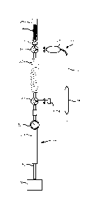
Figure 10 illustrates a balloon catheter system with a
proximally located micelle reservoir. In this configuration,
the catheter 17 includes the catheter body 22, handle 23, and
a balloon 18, which has porous walls and is comparable to the
outer balloon of Figure 1. The micelle reservoir 24 is
disposed within a micelle storage chamber 25, in fluid
communication with the balloon catheter lumen (within catheter
17) through the three-way valve 26. Opposite the micelle
storage chamber 25, the three-way valve communicates with the
coiled tube suspension chamber 27. The coiled tube suspension
chamber is disposed between the three-way valve 26 and the
balloon inflation device 28 (sometimes referred to as an
endoflator). The inflation device is a finely calibrated
syringe with a chamber 29, plunger 30 and plunger handle 31
operable to draw fluid into the chamber and force fluid from
the chamber. The inflator includes a meter 32 which
accurately displays the pressure of fluid, and the amount of
fluid, injected into the balloon catheter. The three-way
valve 26 is operable to selectively align the coiled tube
chamber, and the inflator, with the drug delivery lumen within
the catheter 17 or the micelle storage chamber 25. A second
three-way valve 33 is disposed between the coiled tube
suspension chamber 27 and the inflator 28. The inflator may
8
Date Recue/Date Received 2022-09-01
W02011/082367
PCT/US2010/062606
be filled from a fluid source connected to the second three-
way valve. A pressure relief valve 34 may be provided to
avoid over-pressurization of the system. A filter 35 may be
provided at the proximal end of the catheter, at the output of
the micelle storage chamber, at the output of the three-way
valve (between the three-way valve and the catheter body) or
between the coiled tube micelle chamber and the three-way
valve 26, to prevent any agglomeration of micelles from
passing into the catheter and ensure that only small particles
are passed into the balloon. The filter is preferably a
static .45 micron filter, but may be as small as a .1 micron
(100 nanometer). The micelle storage chamber 25 is preferable
collapsible, so that withdrawal of the micelles after
injection of reconstituting fluid is facilitated. The micelle
storage chamber may be a collapsible pouch, a cylinder with an
easily movable base, or a syringe which must be operated in
tandem with the inflator to push the reconstituted suspension
from the chamber as the inflator is used to withdraw the
suspension. The micelle storage chamber may include a relief
valve or vent to enable degassing and facilitate filling. The
micelle storage chamber 25 is preferable transparent, so that
complete reconstitution and emptying into the coiled tube
suspension chamber can be visually confirmed. The coiled tube
chamber has an inner diameter of 1 to 2 mm, and a length of
about 300 mm. Limiting the diameter to 2 mm or less severely
minimizes the mixing or osmosis of micelles into the inflator
fluid, so that the concentration of the suspension in the
coiled tube chamber is not diluted when inflator fluid is
forced into the coiled tube chamber. The coiled tube chamber
is coiled merely for compactness. The overall inner volume of
the coiled tube is preferably 1 to 2 ml volume of micelle
suspension. (The coiled tube suspension chamber and the
micelle storage chamber are thus distinguished by their
separate functions and distinct structure. The micelle
storage chamber is used to store the micelles for extended
9
Date Recue/Date Received 2022-09-01
W02011/082367
PCT/US2010/062606
periods prior to use (after manufacture, in shipping and
storage for the shelf life of the micelles formulation in its
lyophilized condition). The coiled tube suspension chamber is
used intra-operatively, to briefly store the micelles
suspension immediately prior to delivery through the catheter
and balloon, and is sized and dimensioned to limit mixing of
the suspension with the inflator fluid held in the inflator
chamber, which it abuts at the boundary of the suspension
bolus and the inflation fluid.)
Thus, Figures 10 through 13 show a balloon catheter
system for delivery of drugs or therapeutic agents to a blood
vessel from a dry reservoir stored at the proximal end of the
catheter. The balloon catheter comprises a catheter body with
a distal end adapted for insertion into the vasculature of a
patient, a porous balloon disposed on the distal end. The
proximal end of the balloon catheter has a lumen extending
from the proximal end to the balloon. The proximal end is
adapted for connection to a fluid source. The system also
includes a storage chamber with a reservoir of dry drug
delivery vesicles, and an inflator and suspension chamber in
fluid communication with an inflator. These components are
selectively aligned in fluid communication with each other
through a valve operable to selectively connect the storage
chamber to the suspension chamber or the lumen of the
catheter. The inflator is operable to fill the storage
chamber with fluid to reconstitute the dry drug delivery
vesicles into a fluid suspension of drug delivery vesicles and
draw the fluid suspension into the suspension chamber, when
the valve is positioned to connect the storage chamber to the
suspension chamber, and the inflator is operable to force the
suspension from the suspension chamber through the catheter
lumen and porous balloon to the blood vessel.
In use, the system of Figure 10 is operated in several
steps. After standard preparation of the catheter, which may
Date Recue/Date Received 2022-09-01
W02011/082367
PCT/US2010/062606
include flushing the catheter with water or saline, the
operator fills the inflator chamber with fluid, and fills the
coiled tube suspension chamber with fluid. As shown in Figure
11, the operator turns the three-way valve 26 to align the
inflator and coiled tube suspension chamber 27 with the
micelle storage chamber 25, and forces the fluid into the
micelle storage chamber 24 by operating the inflator handle.
The micelle storage chamber is depicted in a distended state,
to illustrate that it has been filled with fluid. Filling the
micelle storage chamber with fluid will reconstitute the
micelles in the micelle storage chamber and create a
suspension that can be moved into the catheter. Next, as
shown in Figure 12, the three-way valve 26 is maintained in
position to align the inflator and coiled tube suspension
chamber 27 with the micelle storage chamber 25, and the
suspension of micelles in a small bolus 36 is drawn into the
coiled tube suspension chamber 27. (The micelle storage
chamber 25 is depicted in a collapsed state, to illustrate
that its contents have been withdrawn.) Routine steps are
then taken to ensure that no gas is entrained in the micelle
suspension. Next, as shown in Figure 13, the three way valve
is manipulated to align the coiled tube suspension chamber and
inflator with the catheter lumen, and the operator pushes the
inflator handle into the inflator chamber to force additional
fluid into the coiled tube suspension chamber and through to
the catheter. The suspension that had been drawn into the
coiled tube suspension chamber 27 (Figure 12) is pushed, in a
substantially intact bolus 36, into the catheter and thus into
the balloon. If not already flushed of air, this step may
serve to flush the catheter and balloon prior to insertion
into the body and navigation into the blood vessel to be
treated. When flushed, the catheter is inserted into the
vasculature and navigated to the blood vessel to be treated.
The operator continues to pressurize the inflator, and thus
pressurize the balloon, as necessary to force the suspension,
11
Date Recue/Date Received 2022-09-01
W02011/082367
PCT/US2010/062606
and the suspended micelle formulation, through the wall of the
balloon and into body tissue surrounding the balloon. The
delivery of fluid can continue until inflation fluid (from the
inflator, which may be a contrast fluid) exits the balloon.
The inflation fluid, or a flushing fluid delivered using the
inflator, preferably includes contrast agent (iodinated
radiocontrast agents, e.g. ionic agents like diatrizoate or
metrizoate or non-ionic agents like iopamidol, iopromide, or
iodixanol) so that the arrival of the inflation fluid at the
balloon pores, and thus complete ejection of the micelle
suspension, can be visually confirmed under fluoroscopy.
The method may be modified by injecting fluid into the
micelle storage chamber from a syringe separate from the
inflator, as shown in Figure 14, which shows the micelle
reservoir 24 within the micelle storage chamber 25, catheter
17, the coiled tube suspension chamber 27, the balloon
inflation device 28 and its chamber 29, plunger 30, plunger
handle 31, meter 32 and the second three-way valve 33 as in
Figure 10, and the additional syringe 37 may be provided, and
connected to the micelle storage chamber through the four-way
valve 38. In this system, the four-way valve 38 is positioned
to align the syringe in fluid communication with the micelle
storage chamber, then the syringe is operated to fill the
micelle storage chamber with fluid and the four way valve is
then turned to align the coiled tube suspension chamber 27 to
the micelle storage chamber, and operation is thereafter
performed as described in relation to Figures 10 through 13.
Other means for filling the micelle storage chamber with
reconstituting fluid may be used, included injection through a
self-sealing membrane in the chamber wall, a needle port, or
the like.
The proximal components of the system, including the
micelle chamber, coiled tube suspension chamber, filter and
three-way valve, may be provided in a single housing to
12
Date Recue/Date Received 2022-09-01
W02011/082367
PCT/US2010/062606
facilitate handling and operation of the system. This is
illustrated in Figure 15, which shows the micelle storage
chamber 25 and coiled tube isolation reservoir 27 and,
optionally, the three-way valve 26 disposed in the handle 39.
This configuration provides a conveniently operable system
with the micelle reservoir stored within the handle of the
catheter to be used to deliver the micelle formulation to the
body through the balloon. The system is assembled after the
micelle formulation is filter sterilized and deposited in the
micelle storage chamber. After the micelle storage reservoir
is installed in the handle and sealed to the valve, the entire
catheter may be sterilized with standard ETO sterilization or
other methods that would otherwise degrade the micelle
formulation.
The system may be modified by placing the micelle storage
chamber between the three-way valve and the coiled tube
chamber, as shown in Figure 16, which shows the micelle
reservoir 24 within the micelle storage chamber 25, catheter
17, the coiled tube suspension chamber 27, the balloon
inflation device 28 and it chamber 29, plunger 30, plunger
handle 31, meter 32 and the second three-way valve 33 as in
Figure 10. In this system, the micelle storage chamber is
positioned between the three-way valve and the coiled tube
chamber. The inflator is operated to fill the micelle storage
chamber with fluid and withdraw the resulting suspension into
the coiled tube chamber, and operation is thereafter performed
as described in relation to Figures 10 through 13.
Referring again to the system of Figures 10 through 13,
the purpose of the coiled tube suspension chamber is to expose
the suspension bolus to the pneumatic force of the inflation
fluid in the inflator while minimizing mixing. Mixing can
also be prevented by replacing the coiled tube with a second
cylinder divided into chambers by a piston. In such an
embodiment, a first chamber, in fluid communication with the
13
Date Recue/Date Received 2022-09-01
W02011/082367
PCT/US2010/062606
three-way valve 26 and the micelle storage chamber 25, is
filled with reconstitution fluid, and the second chamber,
closest to the inflator, is filled with fluid and is in fluid
communication with the inflator. Operation of the inflation
to force the piston back and forth serves to force the
reconstitution fluid into the micelle storage reservoir, and
withdraw the resulting suspension into the cylinder, and
thereafter force the suspension from the first chamber into
the catheter with pneumatic pressure applied to the piston
from the inflator. To accomplish the goal of flushing all the
suspension, and also the goal of providing the contrast bloom
that confirms complete delivery, the inflator provides
inflation fluid or contrast through a bypass communication
around the cylinder to the catheter lumen.
The inflation pressure and inflation duration, in
combination with the amount of dry micelle formulation and
volume of the reconstituted micelle suspension can be
controlled to ensure a predetermined dose of micelles, and
encapsulated drug, are delivered to the body tissue
surrounding the balloon. Pressure applied by the inflator may
be two to twenty atmospheres, and the inflator is preferably
operated to apply 6 to 12 atmospheres of pressure. With
suspended micelle formulation in the suspension chamber, and
hole sizes of 2 to 5 microns in the balloon, application of 12
atmospheres for 60 seconds will deliver the entire 1 ml of the
suspended micelle formulation through the catheter and balloon
wall. The parameters may be adjusted to achieve .25 to 10 ml
over the course of 10 to 120 seconds. The dosage of drug or
therapeutic agent actually delivered can thus be controlled
and predetermined with some certainty by controlling the
amount of drug or therapeutic agent in the micelle formulation
disposed in the micelle storage chamber. For example, if it
is desired to deliver 2 mg of rapamycin to a diseased portion
of a blood vessel, the micelle reservoir containing 2 or 3 mg
14
Date Recue/Date Received 2022-09-01
W02011/082367
PCT/US2010/062606
of rapamycin can be stored in the micelle storage chamber,
reconstituting the micelles with fluid to achieve a
concentration of 2 mg/ml (that is, 1 ml if the micelle storage
chamber contains 2 mg total rapamycin), withdrawing 1 ml of
fluid into the coiled tube suspension chamber, and forcing the
entire 1 ml through the catheter and balloon into the blood
vessel walls.
The micelles used in the catheter systems described above
may be formulated and lyophilized using known procedures, or
procedures developed in the future. The micelle reservoir may
be disposed within the catheter after formulation and
lyophilization, or they may be installed in an aqueous slurry
in the catheter or a catheter component, and lyophilized
afterward, whereupon the catheter may be stored for extended
periods of time prior to shipment, and wetted just prior to
use in a patient, or when the balloon or balloons are inflated
within the body of the patient. The micelles may be loaded
with rapamycin or other therapeutic agents such as rapamycin
analogs, ABT-578, zotarolimus, everolimus, biolimus A9,
deforolimus (also referred to as ridaforolimus), temsirolimus,
tacrolimus, pimcrolimus, nitric oxide synthase, C3 exoenzyme,
RhoA inhibitors, tubulusin, A3 agonists, CB2 agonists, 17-AAG,
Hsp90 antagonists, tyrphostins, cathepsin S inhibitors,
paclitaxel, dexamethasone, ceramides, dimethyl sphingosine,
ether-linked diglycerides, ether-linked phosphatidic acids,
sphinganines, estrogens, taxol, taxol analogs, actinomycin D,
prostaglandins, vitamin A, probucol, Batimastat, Statins,
Trapidil, mitomycin C and Cytochalasin B.
The micelles used in the catheter are preferably formed
from tri-block amphiphilic co-polymers of the form A-B-A where
A is hydrophobic (PCL (Polycaprolactone)or PLGA (poly(lactic-
co-glycolic acid) for example) and B is hydrophilic (PEG, or
PEO for example), in which case the A block interacts with the
micelle core and drugs encapsulated in the core and the B
Date Recue/Date Received 2022-09-01
W02011/082367
PCT/US2010/062606
block forms the shell of the micelle. The micelles may also
be formed of tri-block amphiphilic co-polymers of the form A-
B-A where A is PLA, PDLLA, PPS, PPO, or Poly(amino acid)s and
B=PEG or PEO. Tr-block copolymers of the form B-A-B and Di-
block copolymers of the form A-B may also be used.
Additionally, the micelles may be formed with a core polymer
of PCL. The micelles are formed by nano-precipitation, and
result in micelle sizes in the range of 40-120 nm diameter.
Rapamycin or other drug particles can be loaded into the
micelles by entrapment during the initial formation of the
micelles. This will result in efficient loading of the drug
particles, and a high percentage of the drug particles in the
formulation slurry will become entrapped within the micelles.
Drug loading may be accomplished by adsorption or migration of
the drug into the micelles after formulation, though this is
not expected to be as efficient as entrapment. The systems
and methods described above can be employed to deliver other
small drug delivery vesicles or delivery vessels in addition
to micelles, particularly small dry vesicles that benefit from
reconstitution immediately prior to delivery, such as
nanoparticles and liposomes. Nanoparticles useful in the
system include e.g. PCL, PLGA, PLA, PDLLA, PPS, PPO, or
Poly(amino acid)s loaded with drugs. Liposomes can include
dry powder liposomes made by lyophilization or dry-spraying.
The various reservoirs shown in the various devices may be
protected by filling the catheter or chamber or balloon
housing the reservoir with nitrogen or inert gas.
After formulation, the micelles are freeze-dried, or
lyophilized. The micelles may survive intact, or partially
collapse into other structures. Nonetheless, upon re-wetting,
a substantial portion of the micelle population will be
mobilized intact. To enhance the survival of the micelles,
lyophilization may be performed after a lyoprotectant or cryo-
protectant, for example, sucrose, glucose, lactose, mannitol,
16
Date Recue/Date Received 2022-09-01
W02011/082367
PCT/US2010/062606
trehalose, may be added to the original micelle mixture.
After lyophilization, the mixture of the micelles,
encapsulated drug within the micelles, and the lyoprotectant
compound is particularly useful as the reservoir described
above.
The micelles used in this system and method described
above should be in the range of 40 to 250 nm (.04 to .250
micron) generally, and in the range of 60 to 120 nm when
formulated from the tri-block copolymer mentioned above (PLGA-
PEG-PLGA or PCL¨PEG¨PCL). This size will result in a balance
of efficient penetration of the micelles into the artery walls
and sufficient space within the micelles to encapsulate a
suitable amount of rapamycin or other therapeutic substance.
Use of tri-block polymers such as PLGA-PEG-PLGA will provide
micelles in the desired sized range. For micelle doses
prepared prior to loading into the catheter, polydispersity
index of the micelle population is preferably less than 0.2,
as measured by a dynamic light diffusion test. This may be
achieved by controlled formulation, filtration or
centrifugation of polydisperse population of micelles.
For reconstitution of the micelles, an aqueous solution,
typically an isotonic solution with or without additional
lyoprotectant and/or pharmaceutical excipient, is added to the
dry micelle formulation via syringe, catheter barrel, or tube.
The suspension is further mixed, if required, by physical
agitation, drawing back and forth into a syringe, or other
means.
While the devices and methods described above have been
illustrated in the context of coronary artery treatment and
restenosis, they may be used in other vessels in the body,
including the peripheral blood vessels, esophagus, ureters,
urethra, sinus, valves, etc., and may be used to deliver a
17
Date Recue/Date Received 2022-09-01
W02011/082367
PCT/US2010/062606
variety of drugs, therapeutic agents, especially hydrophobic
agents which may be encapsulated in micelles or liposomes.
While the preferred embodiments of the devices and
methods have been described in reference to the environment in
which they were developed, they are merely illustrative of the
principles of the inventions. The elements of the various
embodiments may be incorporated into each of the other species
to obtain the benefits of those elements in combination with
such other species, and the various beneficial features may be
employed in embodiments alone or in combination with each
other. Other embodiments and configurations may be devised
without departing from the spirit of the inventions and the
scope of the appended claims.
18
Date Recue/Date Received 2022-09-01