Note: Descriptions are shown in the official language in which they were submitted.
CA 03172898 2022-08-24
WO 2021/174151 PCT/US2021/020133
METHODS OF REDUCING POLYSORBATE DEGRADATION IN DRUG
FORMULATIONS
CROSS-REFERENCE TO RELATED APPLICATIONS
This application claims priority to U.S. Provisional Patent Application No.
62/982,346,
filed February 27, 2020, U.S. Provisional Patent Application No. 63/021,181,
filed May 7, 2020
and U.S. Provisional Patent Application No. 63/073,125, filed September 1,
2020, the contents of
which are incorporated herein by reference in their entirety.
FIELD
[0001] The present invention generally pertains to compositions with reduced
amount of certain
lipases, methods of making such compositions and methods of reducing
polysorbate degradation
due to the presence of such lipases. In particular, the present invention
generally pertains to
compositions and methods of making compositions with reduced presence of liver
carboxylesterase-Bl-like protein and liver carboxylesterase-l-like protein.
BACKGROUND
[0002] Among drug products, protein-based biotherapeutics are an important
class of drugs that
offer a high level of selectivity, potency and efficacy, as evidenced by the
considerable increase
in clinical trials with monoclonal antibodies (mAbs) over the past several
years. Bringing a
protein-based biotherapeutic to the clinic can be a multiyear undertaking
requiring coordinated
efforts throughout various research and development disciplines, including
discovery, process
and formulation development, analytical characterization, and pre-clinical
toxicology and
pharmacology.
[0003] One critical aspect for a clinically and commercially viable
biotherapeutic is stability of
the drug product in terms of the manufacturing process as well as shelf life.
This often
necessitates appropriate steps to help increase physical and chemical
stability of the protein-
1
CA 03172898 2022-08-24
WO 2021/174151 PCT/US2021/020133
based biotherapeutics throughout the different solution conditions and
environments necessary
for manufacturing and storage with minimal impact on product quality,
including identifying
molecules with greater inherent stability, protein engineering, and
formulation development.
Surfactants, such as, polysorbate are often used to enhance the physical
stability of a protein-
based biotherapeutic product. Over seventy percent of marketed monoclonal
antibody
therapeutics contain between 0.001% and 0.1% polysorbate, a type of
surfactant, to impart
physical stability to the protein-based biotherapeutics. Polysorbates are
susceptible to auto-
oxidation and hydrolysis, which results in free fatty acids and subsequent
fatty acid particle
formation. The degradation of polysorbate can adversely affect the drug
product quality since
polysorbate can protect against interfacial stress, such as aggregation and
adsorption. Presence
of some lipases can be a likely cause of degradation of polysorbates in a
formulation. Thus, such
lipases in drug products need to be detected, monitored and reduced.
[0004] Direct analysis of lipases can require isolation of the product in a
sufficiently large
amount for the assay, which is undesirable and has only been possible in
selected cases. Hence,
it is a challenging task to determine the workflow and analytical tests
required to characterize
lipases responsible for polysorbate degradation in a sample. In addition to
detecting the lipases
responsible for polysorbate degradation, the drug product must be obtained by
purification
methods that remove or reduce such lipases.
[0005] It will be appreciated that a need exists for methods for depleting
lipase from a
formulated drug product.
SUMMARY
[0006] Maintaining stability of drug formulations, not only during storage but
also during
manufacturing, shipment, handling and administration, is a significant
challenge. Among drug
products, protein biotherapeutics are gaining popularity due to their success
and versatility. One
of the major challenges for protein biotherapeutics development is to overcome
the limited
stability of the protein and excipients in the products, which can be affected
by the presence of
lipases (present as host-cell proteins). Evaluation of its effect on the drug
formulation and
reduction of such lipases can be an important step in drug formulation
development, followed by
2
CA 03172898 2022-08-24
WO 2021/174151 PCT/US2021/020133
methods to prepare the drug formulation so as to have reduced lipases and
increased stability
owing to the reduced lipases.
[0007] In one exemplary embodiment, the disclosure provides a method of
depleting lipase from
a sample comprising contacting the sample including lipase with a probe, said
probe capable of
binding to the lipase to form a complex and separating the complex from the
sample to thereby
deplete the lipase from the sample. In one aspect, the sample can comprise a
protein of interest.
In one aspect, the sample can comprise a polysorbate excipient. In a specific
aspect, the
polysorbate excipient can be selected from polysorbate-20, polysorbate-60,
polysorbate-80 or
combinations thereof. In yet another specific aspect, the polysorbate
excipient is polysorbate-80.
[0008] In one aspect, the lipase is liver carboxylesterase-Bl-like protein. In
another aspect, the
lipase is liver carboxylesterase-l-like protein.
In one aspect, the lipase is capable of degrading the polysorbate in the
sample. Thus, the method
of this embodiment reduces the degradation of polysorbates by depleting the
sample of the
lipase.
[0009] In one aspect, the probe can be capable of being linked to a solid
support. In a specific
aspect, the solid support can be agarose beads or magnetic beads.
[0010] In one aspect, the probe can be attached to a solid support using a
ligand. In a specific
aspect, the ligand can be an indicator, biotin molecule, a modified biotin
molecule, a nuclei, a
sequence, an epitope tag, an electron poor molecule or an electron rich
molecule.
[0011] In one aspect, the method can further comprise recovering the lipase
from the complex.
[0012] In one exemplary embodiment, the disclosure provides a method of
purifying a sample
having a protein of interest and a lipase, comprising contacting the sample
with a probe, said
probe capable of binding to the lipase to form a complex and separating the
complex from the
sample. In one aspect, the lipase is liver carboxylesterase-Bl-like protein.
In another aspect, the
lipase is liver carboxylesterase-l-like protein.
[0013] In one aspect, the sample comprises a polysorbate excipient. In a
specific aspect, the
polysorbate excipient can be selected from polysorbate-20, polysorbate-60,
polysorbate-80 or
combinations thereof. In yet another specific aspect, the polysorbate
excipient can be
polysorbate-80.
[0014] In one aspect, the probe can be capable of being linked to a solid
support. In a specific
3
CA 03172898 2022-08-24
WO 2021/174151 PCT/US2021/020133
aspect, the solid support can be agarose beads or magnetic beads.
[0015] In one aspect, the probe can be attached to a solid support using a
ligand. In a specific
aspect, the ligand can be an indicator, biotin molecule, a modified biotin
molecule, a nuclei, a
sequence, an epitope tag, an electron poor molecule or an electron rich
molecule.
[0016] In one exemplary embodiment, the disclosure provides a method of
decreasing
degradation of polysorbate in a sample, comprising contacting the sample
including lipase and
polysorbate with a probe, said probe capable of binding to the lipase to form
a complex and
separating the complex from the sample to thereby decrease degradation of
polysorbate in the
sample.
[0017] In one aspect, the lipase is liver carboxylesterase-Bl-like protein. In
another aspect, the
lipase is liver carboxylesterase-l-like protein.
[0018] In one aspect, the sample can comprise a protein of interest. In one
aspect, the sample
can comprise a polysorbate excipient. In a specific aspect, the polysorbate
excipient is selected
from polysorbate-20, polysorbate-60, polysorbate-80 or combinations thereof.
In yet another
specific aspect, the polysorbate excipient is polysorbate-80.
[0019] In one aspect, the probe can be capable of being linked to a solid
support. In a specific
aspect, the solid support can be agarose beads or magnetic beads.
[0020] In one aspect, the probe can be attached to a solid support using a
ligand. In a specific
aspect, the ligand can be an indicator, biotin molecule, a modified biotin
molecule, a nuclei, a
sequence, an epitope tag, an electron poor molecule or an electron rich
molecule.
[0021] In one exemplary embodiment, the disclosure provides a composition
comprising a
protein of interest purified from mammalian cells and a residual amount of
liver
carboxylesterase-Bl-like protein. In one aspect, the residual amount of liver
carboxylesterase-
Bl-like protein is less than about 5 ppm. In another aspect, the composition
can further comprise
a surfactant. In yet a further aspect, the surfactant can be a hydrophilic
nonionic surfactant. In
another aspect, the surfactant can be a sorbitan fatty acid ester. In a
specific aspect, the
surfactant can be a polysorbate. In another specific aspect, the concentration
of the polysorbate
in the composition can be about 0.01% w/v to about 0.2% w/v. In a further
specific aspect, the
surfactant can be a polysorbate 80. In one aspect, the mammalian cells can
include a CHO cell.
[0022] In one aspect, the liver carboxylesterase-Bl-like protein can cause
degradation of
4
CA 03172898 2022-08-24
WO 2021/174151 PCT/US2021/020133
polysorbate 80.
[0023] In one aspect, the composition can be a parenteral formulation.
[0024] In one aspect, the protein of interest can be a monoclonal antibody, a
polyclonal
antibody, a bispecific antibody, an antibody fragment, a fusion protein, or an
antibody-drug
complex. In one aspect, the concentration of the protein of interest can be
about 20 mg/mL to
about 400 mg/mL.
[0025] In one aspect, the composition can further comprise one or more
pharmaceutically
acceptable excipients. In another aspect, the composition can further comprise
a buffer selected
from a group consisting of histidine buffer, citrate buffer, alginate buffer,
and arginine buffer. In
one aspect, the composition can further comprise a tonicity modifier. In yet
another aspect, the
composition can further comprise sodium phosphate.
[0026] In one exemplary embodiment, the disclosure provides a composition
comprising a
protein of interest purified from mammalian cells and a residual amount of
liver
carboxylesterase-l-like protein. In one aspect, the residual amount of liver
carboxylesterase-1-
like protein is less than about 5 ppm. In another aspect, the composition can
further comprise a
surfactant. In yet a further aspect, the surfactant can be a hydrophilic
nonionic surfactant. In
another aspect, the surfactant can be a sorbitan fatty acid ester. In a
specific aspect, the
surfactant can be a polysorbate. In another specific aspect, the concentration
of the polysorbate
in the composition can be about 0.01 %w/v to about 0.2% w/v. In a further
specific aspect, the
surfactant can be a polysorbate 80. In one aspect, the mammalian cells can
include a CHO cell.
[0027] In one aspect, the liver carboxylesterase-l-like protein can cause
degradation of
polysorbate 80.
[0028] In one aspect, the composition can be a parenteral formulation.
[0029] In one aspect, the protein of interest can be a monoclonal antibody, a
polyclonal
antibody, a bispecific antibody, an antibody fragment, a fusion protein, or an
antibody-drug
complex. In one aspect, the concentration of the protein of interest can be
about 20 mg/mL to
about 400 mg/mL.
[0030] In one aspect, the composition can further comprise one or more
pharmaceutically
acceptable excipients. In another aspect, the composition can further comprise
a buffer selected
from a group consisting of histidine buffer, citrate buffer, alginate buffer,
and arginine buffer. In
CA 03172898 2022-08-24
WO 2021/174151 PCT/US2021/020133
one aspect, the composition can further comprise a tonicity modifier. In yet
another aspect, the
composition can further comprise sodium phosphate.
[0031] In one exemplary embodiment, the disclosure provides a method of
detecting a lipase in a
sample. In one aspect, the lipases can be liver carboxylesterase-l-like
protein or liver
carboxylesterase-Bl-like protein. In one aspect, the method of detecting a
lipase in a sample can
comprise contacting the sample with a serine hydrolase probe. In one aspect,
the method of
detecting a lipase in a sample can comprise contacting and incubating the
sample with a serine
hydrolase probe to form a complex of lipase and serine hydrolase probe. In a
further aspect, the
method of detecting a lipase in a sample can comprise filtering out the serine
hydrolase probe
that does not form the complex of lipase and serine hydrolase probe.
[0032] In one aspect, the method of detecting a lipase in a sample can further
comprise
contacting the contacting the sample with magnetic beads having an ability to
bind to the serine
hydrolase probe to such that magnetic beads are bound to the complex of lipase
and serine
hydrolase probe. The magnetic beads bound to the complex of lipase and serine
hydrolase probe
can be further removed from the sample and washed with a buffer.
[0033] In another aspect, the method can further comprise removing the
magnetic beads which
are bound to the complex of lipase and serine hydrolase probe to form a
solution of enriched
lipases.
[0034] In one aspect, the method can further comprise adding hydrolyzing agent
to the solution
to obtain digests. In a specific aspect, the hydrolyzing agent can be trypsin.
In one aspect, the
method can further comprise analyzing the digests to detect the lipases. In
one aspect, the
digests can be analyzed using a mass spectrometer. In a specific aspect, the
mass spectrometer
can be a tandem mass spectrometer. In another specific aspect, the mass
spectrometer can be
coupled to a liquid chromatography system. In yet another specific aspect, the
mass
spectrometer can be coupled to a liquid chromatography - multiple reaction
monitoring system.
[0035] In one aspect, the method can further comprise adding protein
denaturing agent to the
solution. In a specific aspect, the protein denaturing agent can be urea. In
one aspect, the
method can further comprise adding protein reducing agent to the solution. In
a specific aspect,
the protein reducing agent can be DTT (dithiothreitol). In one aspect, the
method can further
comprise adding protein alkylating agent to the solution. In a specific
aspect, the protein
6
CA 03172898 2022-08-24
WO 2021/174151 PCT/US2021/020133
alkylating agent can be iodoacetamide.
[0036] These, and other, aspects of the invention will be better appreciated
and understood when
considered in conjunction with the following description and the accompanying
drawings. The
following description, while indicating various embodiments and numerous
specific details
thereof, is given by way of illustration and not of limitation. Many
substitutions, modifications,
additions, or rearrangements may be made within the scope of the invention.
BRIEF DESCRIPTION OF THE DRAWINGS
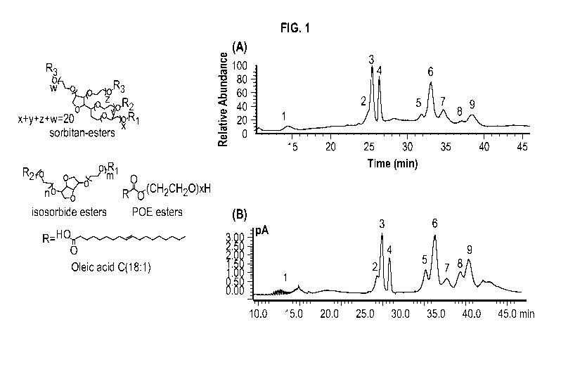
[0037] FIG. 1 shows chemical structures of major species in polysorbates.
Polysorbates are
mainly composed of fatty acid esters sharing a common sorbitan POE, isosorbide
POE or POE
head group, with oleic acid as the main fatty acid for PS80. The right panel A
shows a total ion
current (TIC) chromatogram of PS80 in mAb formulation by online 2D-LC/MS
analysis. The
identity of the labeled peaks are: (1) POE- POE isosorbide- POE sorbitan, (2)
POE sorbitan
monolinoleate, (3) POE sorbitan monooleate, (4) POE isosorbide monooleate and
POE
monooleate, (5) POE sorbitan linoleate/oleate diester, (6) POE sorbitan di-
oleate, (7) POE
isosorbide di-oleate and POE di-oleate, (8) Probably POE isosorbide/POE
linoleate/oleate diester
as mass spectra are too complicated to interpret, (9) POE sorbitan mixed
trioleate and tetraoleate.
The right panel B shows a CAD chromatogram showing the separation and
detection of PS80 in
mAb formulation by online 2D-LC/CAD analysis.
[0038] FIG. 2 shows a chromatogram of 0.1% PS80 in 50 mg/mL mAb-1 incubated at
5 C in
10mM histidine, pH 6 for 0 hours and 36 hours according to an exemplary
embodiment. Peaks
eluted between 11 to 17.5 minutes were POE, POE isosorbide and POE sorbitan.
[0039] FIG. 3 shows a chart of the percentage of PS80 remaining plotted
against incubation
time, where the original mAb-1, mAb-1 mixed with 0.125 [tM, 0.5 [tM and 2 [tM
FP probe are
indicated by filled circle with black solid line, filled diamond with red
dotted line, filled square
with orange dashed line and filled triangle with blue dotted line.
[0040] FIG. 4 shows a schematic diagram of the lipase(s) depletion experiment
according to an
exemplary embodiment. Streptavidin dynabeads magnetic beads were coupled with
7
CA 03172898 2022-08-24
WO 2021/174151 PCT/US2021/020133
desthiobiotin-FP probe and used for lipase(s) depletion. The original mAb-1
and flow through
mAb-1 as well as process control mAb-1 were incubated with 0.1% PS80 at 5 C
for 36 hours and
subjected to PS degradation measurement. The enriched lipase(s) are subjected
to digestion and
HCP analysis using mass spectrometry.
[0041] FIG. 5 shows a chart of percentage of PS80 remaining in original mAb-1,
process control
and lipase(s) depleted mAb-1, where the original mAb-1, process control and
lipase(s) depleted
mAb-1 are indicated by filled diamond with black solid line, filled square
with blue dotted line
and filled circle with orange dashed line.
[0042] FIG. 6A shows a chromatogram of 0.1% PS80 in 20 i.tg/mL commercial
rabbit liver
esterase incubated at 5 C in 10mM histidine, pH 6 for 0 hours, 1.5 hours and 8
hours according
to an exemplary embodiment.
[0043] FIG. 6B shows a chromatogram of 0.1% PS80 in 100 i.tg/mL commercial
human liver
carboxylesterases 1 incubated at 5 C in 10mM histidine, pH 6 for for 0 hours,
5 hours and 18
hours according to an exemplary embodiment.
[0044] FIG. 6C shows a chromatogram of 0.1% PS80 in 50 mg/mL mAb-1 incubated
at 5 C in
10mM histidine, pH 6 for 0 hours, 18 hours and 36 hours according to an
exemplary
embodiment.
[0045] FIG. 6D shows the sequence alignment of Liver Carboxylesterase B-1-like
(A0A061I7X9), Liver Carboxylesterase 1-like (A0A061FE2) and Human liver
carboxylesterase
(hCES-1).
DETAILED DESCRIPTION
[0046] Host cell proteins (HCPs) are a class of impurities that must be
removed from all cell-
derived protein therapeutics. The FDA does not specify a maximum acceptable
level of HCP,
but HCP concentrations in final drug product must be controlled and
reproducible from batch to
batch (FDA, 1999). A primary safety concern relates to the possibility that
HCPs can cause
antigenic effects in human patients (Satish Kumar Singh, Impact of Product-
Related Factors on
8
CA 03172898 2022-08-24
WO 2021/174151 PCT/US2021/020133
Immunogenicity of Biotherapeutics, and 100 JOURNALS OF PHARMACEUTICAL SCIENCES
354-
387 (2011)). In addition to adverse health consequences for the patient,
enzymatically active
HCPs can potentially affect product quality during processing or long-term
storage (Sharon X.
Gao et al., Fragmentation of a highly purified monoclonal antibody attributed
to residual CHO
cell protease activity, 108 BIOTECHNOLOGY AND BIOENGINEERING 977-982 (2010);
Flavie
Robert et al., Degradation of an Fc-fusion recombinant protein by host cell
proteases:
Identification of a CHO cathepsin D protease, 104 BIOTECHNOLOGY AND
BIOENGINEERING 1132-1141 (2009)). HCPs may present the greatest risk for
persisting through
purification operations into the final drug product. During long-term storage,
the critical quality
attributes of the product molecule must be maintained and degradation of
excipients in the final
product formulation must be minimized.
[0047] Several drug formulations on the market comprise polysorbate as one of
the most
commonly used nonionic surfactants in biopharmaceutical protein formulation
that can improve
protein stability and protect drug products from aggregation and denaturation
(Sylvia Kiese et
al., Shaken, Not Stirred: Mechanical Stress Testing of an IgG1 Antibody, 97
JOURNAL OF
PHARMACEUTICAL SCIENCES 4347-4366 (2008); Ariadna Martos et al., Trends on
Analytical
Characterization of Polysorbates and Their Degradation Products in
Biopharmaceutical
Formulations, 106 JOURNAL OF PHARMACEUTICAL SCIENCES 1722-1735 (2017)).
Polysorbate 20
(PS20) and polysorbate 80 (PS80) are the most commonly used nonionic
surfactants in
biopharmaceutical protein formulation that can improve protein stability and
protect drug
products from aggregation and denaturation. Typical polysorbate concentrations
in drug
products range can be between about 0.001% to about 0.1% (w/v) to provide
sufficient efforts on
protein stability.
[0048] Polysorbates, however, are liable to degradation that can drive
undesired particulate
formation in the formulated drug substances. Polysorbates are known to degrade
in two main
pathways: auto-oxidation and hydrolysis. Oxidation was found to be more likely
to take place in
PS80 due to the high content in unsaturated fatty acid ester substituents,
whereas in PS20,
oxidation was believed to take place on ether bond in polyoxyethylene chain
that is not
frequently observed (Oleg V. Borisov, Junyan A. Ji & Y. John Wang, Oxidative
Degradation
9
CA 03172898 2022-08-24
WO 2021/174151 PCT/US2021/020133
of Potysorbate Surfactants Studied by Liquid Chromatography¨Mass Spectrometry,
104 JOURNAL OF PHARMACEUTICAL SCIENCES 1005-1018 (2015); Anthony Tomlinson et
at., Polysorbate 20 Degradation in Biopharmaceutical Formulations:
Quantification of Free
Fatty Acids, Characterization of Particutates, and Insights into the
Degradation Mechanism,
12 MOLECULAR PHARMACEUTICS 3805-3815 (2015); Jia Yao et at., A Quantitative
Kinetic Study
of Potysorbate Autoxidation: The Role of Unsaturated Fatty Acid Ester
Substituents,
26 PHARMACEUTICAL RESEARCH 2303-2313 (2009)). In addition, polysorbates can
also undergo
hydrolysis by breaking the fatty acid ester bond. The particulates originating
on degradation of
polysorbates can form visible or even sub-visible which can raise the
potential for
immunogenicity in patients and may have varying effects on the drug product
quality. One such
possible impurity could be fatty acid particles that are formed during
manufacture, shipment,
storage, handling or administration of drug formulations comprising
polysorbate. The fatty acid
particles could potentially cause adverse immunogenic effects and impact shelf
life.
Additionally, the degradation of polysorbates can also cause reduction in the
total amount of
surfactant in the formulation affecting the product's stability during its
manufacturing, storage,
handling, and administration.
[0049] Typically, polysorbate degradation can only be observed in drug
products after a fairly
long storage time. However, PS80 degradation was observed in case of one
monoclonal
antibody (mAb) within 24 hours at 4 C although no obviously high concentrated
lipase was
detected, suggesting unfamiliar lipase(s) existed in this drug substance. It
is imperative to detect
and reduce concentration(s) of such lipase(s) in order to maintain the
stability of the drug
formulation.
[0050] Putative phospholipase B-like 2 (PLBD2) was the first host cell protein
that was
proposed to cause an enzymatic hydrolysis of PS20 (Nitin Dixit et at.,
Residual Host Cell
Protein Promotes Polysorbate 20 Degradation in a Sulfatase Drug Product
Leading to Free
Fatty Acid Particles, 105 JOURNAL OF PHARMACEUTICAL SCIENCES 1657-1666
(2016)). Porcine
liver esterase was reported to be able to specifically hydrolysis of
polysorbate 80 (not PS20) and
lead the formation of PS85 over time in mAb drug product (Steven R. Labrenz,
Ester Hydrolysis
of Potysorbate 80 in mAb Drug Product: Evidence in Support of the Hypothesized
Risk After the
CA 03172898 2022-08-24
WO 2021/174151 PCT/US2021/020133
Observation of Visible Particulate in mAb Formulations, 103 JOURNAL OF
PHARMACEUTICAL
SCIENCES 2268-2277 (2014)). Group XV lysosomal phospholipase A2 isomer X1
(LPLA2)
demonstrated the ability to degrade PS20 and PS80 at less than 1 ppm (Troii
Hall et
al., Polysorbates 20 and 80 Degradation by Group XV Lysosomal Phosphohpase A 2
Isomer XI
in Monoclonal Antibody Formulations, 105 JOURNAL OF PHARMACEUTICAL SCIENCES
1633-1642
(2016) and Ying Cheng et al., A Rapid High-Sensitivity Reversed¨Phase Ultra
High
Performance Liquid Chromatography Mass Spectrometry Method for Assessing
Polysorbate 20
Degradation in Protein Therapeutics, 108 JOURNAL OF PHARMACEUTICAL SCIENCES
2880-2886
(2019)).
[0051] Recently, a range of carboxyesters, including pseudomonas cepacia
lipase on immobead
150 (PCL), candida antarctica lipase B on immobead 150 (CALB), thermomyces
lanuginosus
lipase on immobead 150 (TLL), rabbit liver esterase (RLE), Candida antarctica
lipase B (CALB)
and porcine pancreatic lipase type II (PPL), were selected to study the
hydrolysis of two unique
PS20 and PS80 which contained 99% of laurate and 98% oleate esters,
respectively. Different
carboxyesters showed their unique degradation patterns, indicating that
degradation pattern can
be used to differentiate enzymes that hydrolyze polysorbates (A. C. Mcshan et
al., Hydrolysis of
Polysorbate 20 and 80 by a Range of Carboxylester Hydrolases, 70 PDA JOURNAL
OF
PHARMACEUTICAL SCIENCE AND TECHNOLOGY 332-345 (2016)). It can be essential to
evaluate
the effect of a host-cell protein co-purified with a drug product on
polysorbates to ensure
stability of the drug formulation. This can require identification of the host-
cell protein and its
ability to degrade polysorbates. Identification of host-cell proteins can be
particularly
challenging since the presence of HCPs is generally in ppm range, which makes
the isolation and
identification of the HCP difficult.
[0052] The present invention discloses improved compositions comprising
polysorbate with
reduced level of host-cell proteins that can degrade polysorbate(s), methods
for detection of such
host-cell proteins and methods for depleting such host-cell proteins.
[0053] Unless described otherwise, all technical and scientific terms used
herein have the same
meaning as commonly understood by one of ordinary skill in the art to which
this invention
belongs. Although any methods and materials similar or equivalent to those
described herein can
11
CA 03172898 2022-08-24
WO 2021/174151 PCT/US2021/020133
be used in the practice or testing, particular methods and materials are now
described. All
publications mentioned are hereby incorporated by reference.
[0054] The term "a" should be understood to mean "at least one"; and the terms
"about" and
"approximately" should be understood to permit standard variation as would be
understood by
those of ordinary skill in the art; and where ranges are provided, endpoints
are included.
[0055] In some exemplary embodiments, the disclosure provides a composition
comprising a
protein of interest, polysorbate, and a residual amount of a lipase.
[0056] As used herein, the term "composition" refers to an active
pharmaceutical agent that is
formulated together with one or more pharmaceutically acceptable vehicles.
[0057] As used herein, the term "an active pharmaceutical agent" can include a
biologically
active component of a drug product. An active pharmaceutical agent can refer
to any substance
or combination of substances used in a drug product, intended to furnish
pharmacological
activity or to otherwise have direct effect in the diagnosis, cure,
mitigation, treatment or
prevention of disease, or to have direct effect in restoring, correcting or
modifying physiological
functions in animals. Non-limiting methods to prepare an active pharmaceutical
agent can
include using fermentation process, recombinant DNA, isolation and recovery
from natural
resources, chemical synthesis, or combinations thereof
[0058] In some exemplary embodiments, the amount of active pharmaceutical
agent in the
formulation can range from about 0.01 mg/mL to about 600 mg/mL. In some
specific
embodiments, the amount of active pharmaceutical agent in the formulation can
be about 0.01
mg/mL, about 0.02 mg/mL, about 0.03 mg/mL, about 0.04 mg/mL, about 0.05 mg/mL,
about
0.06 mg/mL, about 0.07 mg/mL, about 0.08 mg/mL, about 0.09 mg/mL, about 0.1
mg/mL, about
0.2 mg/mL, about 0.3 mg/mL, about 0.4 mg/mL, about 0.5 mg/mL, about 0.6 mg/mL,
about 0.7
mg/mL, about 0.8 mg/mL, about 0.9 mg/mL, about 1 mg/mL, about 2 mg/mL, about 3
mg/mL,
about 4 mg/mL, about 5 mg/mL, about 6 mg/mL, about 7 mg/mL, about 8 mg/mL,
about 9
mg/mL, about 10 mg/mL, about 15 mg/mL, about 20 mg/mL, about 25 mg/mL, about
30
mg/mL, about 35 mg/mL, about 40 mg/mL, about 45 mg/mL, about 50 mg/mL, about
55
12
CA 03172898 2022-08-24
WO 2021/174151 PCT/US2021/020133
mg/mL, about 60 mg/mL, about 65 mg/mL, about 70 mg/mL, about 5 mg/mL, about 80
mg/mL,
about 85 mg/mL, about 90 mg/mL, about 100 mg/mL, about 110 mg/mL, about 120
mg/mL,
about 130 mg/mL, about 140 mg/mL, about 150 mg/mL, about 160 mg/mL, about 170
mg/mL,
about 180 mg/mL, about 190 mg/mL, about 200 mg/mL, about 225 mg/mL, about 250
mg/mL,
about 275 mg/mL, about 300 mg/mL, about 325 mg/mL, about 350 mg/mL, about 375
mg/mL,
about 400 mg/mL, about 425 mg/mL, about 450 mg/mL, about 475 mg/mL, about 500
mg/mL,
about 525 mg/mL, about 550 mg/mL, about 575 mg/mL, or about 600 mg/mL.
[0059] In some exemplary embodiments, pH of the composition can be greater
than about 5Ø
In one exemplary embodiment, the pH can be greater than about 5.0, greater
than about 5.5,
greater than about 6, greater than about 6.5, greater than about 7, greater
than about 7.5, greater
than about 8, or greater than about 8.5.
[0060] In some exemplary embodiments, the active pharmaceutical agent can be a
protein of
interest.
[0061] As used herein, the term "protein" or "protein of interest" can include
any amino acid
polymer having covalently linked amide bonds. Proteins comprise one or more
amino acid
polymer chains, generally known in the art as "polypeptides." "Polypeptide"
refers to a polymer
composed of amino acid residues, related naturally occurring structural
variants, and synthetic
non-naturally occurring analogs thereof linked via peptide bonds, related
naturally occurring
structural variants, and synthetic non-naturally occurring analogs thereof.
"Synthetic peptides or
polypeptides' refers to a non-naturally occurring peptide or polypeptide.
Synthetic peptides or
polypeptides can be synthesized, for example, using an automated polypeptide
synthesizer.
Various solid phase peptide synthesis methods are known to those of skill in
the art. A protein
may contain one or multiple polypeptides to form a single functioning
biomolecule. A protein
can include any of bio-therapeutic proteins, recombinant proteins used in
research or therapy,
trap proteins and other chimeric receptor Fc-fusion proteins, chimeric
proteins, antibodies,
monoclonal antibodies, polyclonal antibodies, human antibodies, and bispecific
antibodies.
Another exemplary aspect, a protein can include antibody fragments,
nanobodies, recombinant
antibody chimeras, cytokines, chemokines, peptide hormones, and the like.
Proteins may be
produced using recombinant cell-based production systems, such as the insect
bacculovirus
13
CA 03172898 2022-08-24
WO 2021/174151 PCT/US2021/020133
system, yeast systems (e.g., Pichia sp.), mammalian systems (e.g., CHO cells
and CHO
derivatives like CHO-Kl cells). For a recent review discussing biotherapeutic
proteins and their
production, see Ghaderi et at., "Production platforms for biotherapeutic
glycoproteins.
Occurrence, impact, and challenges of non-human sialylation," (Darius Ghaderi
et
al., Production platforms for biotherapeutic glycoproteins. Occurrence,
impact, and challenges
of non-human sialylation, 28 BIOTECHNOLOGY AND GENETIC ENGINEERING REVIEWS147-
176
(2012)). In some embodiments, proteins comprise modifications, adducts, and
other covalently
linked moieties. These modifications, adducts and moieties include for example
avidin,
streptavidin, biotin molecule, a modified biotin molecule, glycans (e.g., N-
acetylgalactosamine,
galactose, neuraminic acid, N-acetylglucosamine, fucose, mannose, and other
monosaccharides),
PEG, polyhistidine, FLAGtag, maltose binding protein (MBP), chitin binding
protein (CBP),
glutathione-S-transferase (GST) myc-epitope, fluorescent labels and other
dyes, and the like.
Proteins can be classified on the basis of compositions and solubility and can
thus include simple
proteins, such as, globular proteins and fibrous proteins; conjugated
proteins, such as,
nucleoproteins, glycoproteins, mucoproteins, chromoproteins, phosphoproteins,
metalloproteins,
and lipoproteins; and derived proteins, such as, primary derived proteins and
secondary derived
proteins.
[0062] In some exemplary embodiments, the protein of interest can be an
antibody, a bispecific
antibody, a multispecific antibody, antibody fragment, monoclonal antibody,
fusion protein, and
combinations thereof.
[0063] In a particular aspect, the protein of interest can aflibercept (see,
US 7,279,159, the entire
teaching of which is incorporated herein by reference).
[0064] The term "antibody," as used herein includes immunoglobulin molecules
comprising four
polypeptide chains, two heavy (H) chains and two light (L) chains inter-
connected by disulfide
bonds, as well as multimers thereof (e.g., IgM). Each heavy chain comprises a
heavy chain
variable region (abbreviated herein as HCVR or VH) and a heavy chain constant
region. The
heavy chain constant region comprises three domains, CH1, CH2 and CH3. Each
light chain
comprises a light chain variable region (abbreviated herein as LCVR or VI) and
a light chain
constant region. The light chain constant region comprises one domain (CL1).
The VH and VL
14
CA 03172898 2022-08-24
WO 2021/174151 PCT/US2021/020133
regions can be further subdivided into regions of hypervariability, termed
complementarity
determining regions (CDRs), interspersed with regions that are more conserved,
termed
framework regions (FR). Each VH and \/1_, is composed of three CDRs and four
FRs, arranged
from amino-terminus to carboxy-terminus in the following order: FR1, CDR1,
FR2, CDR2, FR3,
CDR3, and FR4. In different embodiments of the invention, the FRs of the anti-
big-ET-1
antibody (or antigen-binding portion thereof) may be identical to the human
germline sequences
or may be naturally or artificially modified. An amino acid consensus sequence
may be defined
based on a side-by-side analysis of two or more CDRs.
[0065] The term "antibody," as used herein, also includes antigen-binding
fragments of full
antibody molecules. The terms "antigen-binding portion" of an antibody,
"antigen-binding
fragment" of an antibody, and the like, as used herein, include any naturally
occurring,
enzymatically obtainable, synthetic, or genetically engineered polypeptide or
glycoprotein that
specifically binds an antigen to form a complex. Antigen-binding fragments of
an antibody may
be derived, e.g., from full antibody molecules using any suitable standard
techniques such as
proteolytic digestion or recombinant genetic engineering techniques involving
the manipulation
and expression of DNA encoding antibody variable and optionally constant
domains. Such DNA
is known and/or is readily available from, for example, commercial sources,
DNA libraries
(including, e.g., phage-antibody libraries), or can be synthesized. The DNA
may be sequenced
and manipulated chemically or by using molecular biology techniques, for
example, to arrange
one or more variable and/or constant domains into a suitable configuration, or
to introduce
codons, create cysteine residues, modify, add or delete amino acids, etc.
[0066] As used herein, an "antibody fragment" includes a portion of an intact
antibody, such as,
for example, the antigen-binding or variable region of an antibody. Examples
of antibody
fragments include, but are not limited to, a Fab fragment, a Fab' fragment, a
F(ab')2 fragment, a
scFv fragment, a Fv fragment, a dsFy diabody, a dAb fragment, a Fd' fragment,
a Fd fragment,
and an isolated complementarity determining region (CDR) region, as well as
triabodies,
tetrabodies, linear antibodies, single-chain antibody molecules, and multi
specific antibodies
formed from antibody fragments. Fv fragments are the combination of the
variable regions of
the immunoglobulin heavy and light chains, and ScFv proteins are recombinant
single chain
CA 03172898 2022-08-24
WO 2021/174151 PCT/US2021/020133
polypeptide molecules in which immunoglobulin light and heavy chain variable
regions are
connected by a peptide linker. In some exemplary embodiments, an antibody
fragment contains
sufficient amino acid sequence of the parent antibody of which it is a
fragment that it binds to the
same antigen as does the parent antibody; in some exemplary embodiments, a
fragment binds to
the antigen with a comparable affinity to that of the parent antibody and/or
competes with the
parent antibody for binding to the antigen. An antibody fragment may be
produced by any
means. For example, an antibody fragment may be enzymatically or chemically
produced by
fragmentation of an intact antibody and/or it may be recombinantly produced
from a gene
encoding the partial antibody sequence. Alternatively, or additionally, an
antibody fragment
may be wholly or partially synthetically produced. An antibody fragment may
optionally
comprise a single chain antibody fragment. Alternatively, or additionally, an
antibody fragment
may comprise multiple chains that are linked together, for example, by
disulfide linkages. An
antibody fragment may optionally comprise a multi-molecular complex. A
functional antibody
fragment typically comprises at least about 50 amino acids and more typically
comprises at least
about 200 amino acids.
[0067] The phrase "bispecific antibody" includes an antibody capable of
selectively binding two
or more epitopes. Bispecific antibodies generally comprise two different heavy
chains, with
each heavy chain specifically binding a different epitope¨either on two
different molecules
(e.g., antigens) or on the same molecule (e.g., on the same antigen). If a
bispecific antibody is
capable of selectively binding two different epitopes (a first epitope and a
second epitope), the
affinity of the first heavy chain for the first epitope will generally be at
least one to two or three
or four orders of magnitude lower than the affinity of the first heavy chain
for the second
epitope, and vice versa. The epitopes recognized by the bispecific antibody
can be on the same
or a different target (e.g., on the same or a different protein). Bispecific
antibodies can be made,
for example, by combining heavy chains that recognize different epitopes of
the same antigen.
For example, nucleic acid sequences encoding heavy chain variable sequences
that recognize
different epitopes of the same antigen can be fused to nucleic acid sequences
encoding different
heavy chain constant regions, and such sequences can be expressed in a cell
that expresses an
immunoglobulin light chain.
16
CA 03172898 2022-08-24
WO 2021/174151 PCT/US2021/020133
[0068] A typical bispecific antibody has two heavy chains each having three
heavy chain CDRs,
followed by a CH1 domain, a hinge, a CH2 domain, and a CH3 domain, and an
immunoglobulin
light chain that either does not confer antigen-binding specificity but that
can associate with each
heavy chain, or that can associate with each heavy chain and that can bind one
or more of the
epitopes bound by the heavy chain antigen-binding regions, or that can
associate with each heavy
chain and enable binding or one or both of the heavy chains to one or both
epitopes. BsAbs can
be divided into two major classes, those bearing an Fc region (IgG-like) and
those lacking an Fc
region, the latter normally being smaller than the IgG and IgG-like bispecific
molecules
comprising an Fc. The IgG-like bsAbs can have different formats, such as, but
not limited to
triomab, knobs into holes IgG (kih IgG), crossMab, orth-Fab IgG, Dual-variable
domains Ig
(DVD-Ig), Two-in-one or dual action Fab (DAF), IgG-single-chain Fv (IgG-scFv),
or Kk-bodies.
The non-IgG-like different formats include Tandem scFvs, Diabody format,
Single-chain
diabody, tandem diabodies (TandAbs), Dual-affinity retargeting molecule
(DART), DART-Fc,
nanobodies, or antibodies produced by the dock-and-lock (DNL) method (Gaowei
Fan, Zujian
Wang & minutes gju Hao, Bispecific antibodies and their applications, 8
JOURNAL OF
HEMATOLOGY & ONCOLOGY 130; Dafne Muller & Roland E. Kontermann, Bispecific
Antibodies, HANDBOOK OF THERAPEUTIC ANT1BODIES265-310 (2014)).
[0069] The methods of producing BsAbs are not limited to quadroma technology
based on the
somatic fusion of two different hybridoma cell lines, chemical conjugation,
which involves
chemical cross-linkers, and genetic approaches utilizing recombinant DNA
technology.
Examples of bsAbs include those disclosed in the following patent
applications, which are
hereby incorporated by reference: U.S. Ser. No. 12/823838, filed June 25,
2010; U.S. Ser. No.
13/ 488628, filed June 5,2012; U.S. Ser. No. 14/031075, filed September 19,
2013; U.S. Ser.
No. 14/808171, filed July 24, 2015; U.S. Ser. No. 15/713574, filed September
22, 2017; U.S.
Ser. No. 15/713569, field September 22, 2017; U.S. Ser. No. 15/386453, filed
December 21,
2016; U.S. Ser. No. 15/386443, filed December 21, 2016; U.S. Ser. No. 15/22343
filed July
29, 2016; and U.S. Ser. No. 15814095, filed November 15, 2017. Low levels of
homodimer
impurities can be present at several steps during the manufacturing of
bispecific antibodies. The
detection of such homodimer impurities can be challenging when performed using
intact mass
analysis due to low abundances of the homodimer impurities and the co-elution
of these
17
CA 03172898 2022-08-24
WO 2021/174151 PCT/US2021/020133
impurities with main species when carried out using a regular liquid
chromatographic method.
[0070] As used herein "multispecific antibody" or "Mab" refers to an antibody
with binding
specificities for at least two different antigens. While such molecules
normally will only bind
two antigens (i.e., bispecific antibodies, BsAbs), antibodies with additional
specificities such as
trispecific antibody and KIH Trispecific can also be addressed by the system
and method
disclosed herein.
[0071] The term "monoclonal antibody" as used herein is not limited to
antibodies produced
through hybridoma technology. A monoclonal antibody can be derived from a
single clone,
including any eukaryotic, prokaryotic, or phage clone, by any means available
or known in the
art. Monoclonal antibodies useful with the present disclosure can be prepared
using a wide
variety of techniques known in the art including the use of hybridoma,
recombinant, and phage
display technologies, or a combination thereof
[0072] In some exemplary embodiments, the protein of interest can have a pI in
the range of
about 4.5 to about 9Ø In one exemplary specific embodiment, the pI can be
about 4.5, about
5.0, about 5.5, about 5.6, about 5.7, about 5.8, about 5.9, about 6.0, about
6.1 about 6.2, about
6.3, about 6.4, about 6.5, about 6.6, about 6.7, about 6.8, about 6.9, about
7.0, about 7.1 about
7.2, about 7.3, about 7.4, about 7.5, about 7.6, about 7.7, about 7.8, about
7.9, about 8.0, about
8.1 about 8.2, about 8.3, about 8.4, about 8.5, about 8.6, about 8.7, about
8.8, about 8.9, or about
9Ø
[0073] In some exemplary embodiments, the types of protein of interest in the
compositions can
be at least two. In some specific embodiments, one of the at least two protein
of interest can be a
monoclonal antibody, a polyclonal antibody, a bispecific antibody, an antibody
fragment, a
fusion protein, or an antibody-drug complex. In some other specific
embodiments, concentration
of one of the at least two protein of interest can be about 20 mg/mL to about
400 mg/mL. In
some exemplary embodiments, the types of protein of interest in the
compositions are two. In
some exemplary embodiments, the types of protein of interest in the
compositions are three. In
some exemplary embodiments, the types of protein of interest in the
compositions are five.
[0074] In some exemplary embodiments, the two or more protein of interest in
the composition
18
CA 03172898 2022-08-24
WO 2021/174151 PCT/US2021/020133
can be selected from trap proteins, chimeric receptor Fc-fusion proteins,
chimeric proteins,
antibodies, monoclonal antibodies, polyclonal antibodies, human antibodies,
bispecific
antibodies, multi specific antibodies, antibody fragments, nanobodies,
recombinant antibody
chimeras, cytokines, chemokines, or peptide hormones.
[0075] In some exemplary embodiments, the composition can be a co-formulation.
[0076] In some exemplary embodiments, the protein of interest can be purified
from mammalian
cells. The mammalian cells can be of human origin or non-human origin can
include primary
epithelial cells (e.g., keratinocytes, cervical epithelial cells, bronchial
epithelial cells, tracheal
epithelial cells, kidney epithelial cells and retinal epithelial cells),
established cell lines and their
strains (e.g., 293 embryonic kidney cells, BHK cells, HeLa cervical epithelial
cells and PER-C6
retinal cells, MDBK (NBL-1) cells, 911 cells, CRFK cells, MDCK cells, CHO
cells, BeWo cells,
Chang cells, Detroit 562 cells, HeLa 229 cells, HeLa S3 cells, Hep-2 cells, KB
cells, LSI80 cells,
LS174T cells, NCI-H-548 cells, RPMI2650 cells, SW-13 cells, T24 cells, WI-28
VA13, 2RA
cells, WISH cells, BS-C-I cells, LLC-MK2 cells, Clone M-3 cells, 1-10 cells,
RAG cells,
TCMK-1 cells, Y-1 cells, LLC-PKi cells, PK(15) cells, GHi cells, GH3 cells, L2
cells, LLC-RC
256 cells, MHiCi cells, XC cells, MDOK cells, VSW cells, and TH-I, B1 cells,
BSC-1 cells, RAf
cells, RK-cells, PK-15 cells or derivatives thereof), fibroblast cells from
any tissue or organ
(including but not limited to heart, liver, kidney, colon, intestines,
esophagus, stomach, neural
tissue (brain, spinal cord), lung, vascular tissue (artery, vein, capillary),
lymphoid tissue (lymph
gland, adenoid, tonsil, bone marrow, and blood), spleen, and fibroblast and
fibroblast-like cell
lines (e.g., CHO cells, TRG-2 cells, IMR-33 cells, Don cells, GHK-21 cells,
citrullinemia cells,
Dempsey cells, Detroit 551 cells, Detroit 510 cells, Detroit 525 cells,
Detroit 529 cells, Detroit
532 cells, Detroit 539 cells, Detroit 548 cells, Detroit 573 cells, HEL 299
cells, IMR-90 cells,
MRC-5 cells, WI-38 cells, WI-26 cells, Midi cells, CHO cells, CV-1 cells, COS-
1 cells, COS-3
cells, COS-7 cells, Vero cells, DBS-FrhL-2 cells, BALB/3T3 cells, F9 cells, SV-
T2 cells, M-
MSV-BALB/3T3 cells, K-BALB cells, BLO-11 cells, NOR-10 cells, C3H/IOTI/2
cells,
HSDMiC3 cells, KLN205 cells, McCoy cells, Mouse L cells, Strain 2071 (Mouse L)
cells, L-M
strain (Mouse L) cells, L-MTK' (Mouse L) cells, NCTC clones 2472 and 2555, SCC-
PSA1 cells,
Swiss/3T3 cells, Indian muntjac cells, SIRC cells, Cn cells, and Jensen cells,
Sp2/0, NSO, NS1
19
CA 03172898 2022-08-24
WO 2021/174151 PCT/US2021/020133
cells or derivatives thereof).
[0077] In some exemplary embodiments, the composition can be stable. The
stability of a
composition can comprise evaluating the chemical stability, physical stability
or functional
stability of the active pharmaceutical agent. The formulations of the present
invention typically
exhibit high levels of stability of the active pharmaceutical agent.
[0078] In terms of protein formulations, the term "stable," as used herein
refers to the protein of
interest within the formulations being able to retain an acceptable degree of
chemical structure or
biological function after storage under exemplary conditions defined herein. A
formulation may
be stable even though the protein of interest contained therein does not
maintain 100% of its
chemical structure or biological function after storage for a defined amount
of time. Under
certain circumstances, maintenance of about 90%, about 95%, about 96%, about
97%, about
98% or about 99% of a protein's structure or function after storage for a
defined amount of time
may be regarded as "stable".
[0079] Stability can be measured, inter alia, by determining the percentage of
native protein(s)
that remain in the formulation after storage for a defined amount of time at a
defined
temperature. The percentage of native protein can be determined by, inter
alia, size exclusion
chromatography (e.g., size exclusion high performance liquid chromatography
[SE-HPLC]),
such that native means non-aggregated and non-degraded. An "acceptable degree
of stability,"
as that phrase is used herein, means that at least 90% of the native form of
the protein can be
detected in the formulation after storage for a defined amount of time at a
given temperature. In
certain embodiments, at least about 90%, 91%, 92%, 93%, 94%, 95%, 96%, 97%,
98%, 99% or
100% of the native form of the protein can be detected in the formulation
after storage for a
defined amount of time at a defined temperature. The defined amount of time
after which
stability is measured can be at least 14 days, at least 28 days, at least 1
month, at least 2 months,
at least 3 months, at least 4 months, at least 5 months, at least 6 months, at
least 7 months, at
least 8 months, at least 9 months, at least 10 months, at least 11 months, at
least 12 months, at
least 18 months, at least 24 months, or more.
[0080] Stability can be measured, inter alia, by determining the percentage of
protein that forms
CA 03172898 2022-08-24
WO 2021/174151 PCT/US2021/020133
in an aggregate within the formulation after storage for a defined amount of
time at a defined
temperature, wherein stability is inversely proportional to the percent
aggregate that is formed.
This form of stability is also referred to as "colloidal stability" herein.
The percentage of
aggregated protein can be determined by, inter al/a, size exclusion
chromatography (e.g., size
exclusion high performance liquid chromatography [SE-HPLC]). An "acceptable
degree of
stability," as that phrase is used herein, means that at most 6% of the
protein is in an aggregated
form detected in the formulation after storage for a defined amount of time at
a given
temperature. In certain embodiments an acceptable degree of stability means
that at most about
6%, 5%, 4%, 3%, 2%, 1%, 0.5%, or 0.1% of the protein can be detected in an
aggregate in the
formulation after storage for a defined amount of time at a given temperature.
The defined
amount of time after which stability is measured can be about at least 2
weeks, at least 28 days,
at least 1 month, at least 2 months, at least 3 months, at least 4 months, at
least 5 months, at least
6 months, at least 7 months, at least 8 months, at least 9 months, at least 10
months, at least 1 1
months, at least 12 months, at least 18 months, at least 24 months, or more.
The temperature at
which the pharmaceutical formulation may be stored when assessing stability
can be any
temperature from about -80 C to about 45 C, e.g., storage at about -80 C,
about -30 C, about -
20 C, about 0 C, about 4 C, about 5 C, about 25 C, about 35 C, about 37 C or
about 45 C. For
example, a pharmaceutical formulation may be deemed stable if after six months
of storage at
C, less than about 3%, 2%, 1 %, 0.5%, or 0.1 % of the protein is detected in
an aggregated
form. A pharmaceutical formulation may also be deemed stable if after six
months of storage at
about 25 C, less than about 4%, 3%, 2%, 1 %, 0.5%, or 0.1 % of the protein is
detected in an
aggregated form. A pharmaceutical formulation may also be deemed stable if
after 28 days of
storage at 45 C, less than about 6%, 5%, 4%, 3%, 2%, 1%, 0.5%, or 0.1% of the
protein is
detected in an aggregated form. A pharmaceutical formulation may also be
deemed stable if
after three months of storage at -20 C, -30 C, or -80 C less than about 3%,
2%, 1 %, 0.5%, or
0.1 % of the protein is detected in an aggregated form.
[0081] Stability can also be measured, inter al/a, by determining the
percentage of protein that
forms in an aggregate within the formulation after storage for a defined
amount of time at a
defined temperature, wherein stability is inversely proportional to the
percent aggregate that is
formed. This form of stability is also referred to as "colloidal stability"
herein. The percentage
21
CA 03172898 2022-08-24
WO 2021/174151 PCT/US2021/020133
of aggregated protein can be determined by, inter alia, size exclusion
chromatography (e.g., size
exclusion high performance liquid chromatography [SE-HPLC]). An acceptable
degree of
stability," as that phrase is used herein, means that at most about 6% of the
protein is in an
aggregated form detected in the formulation after storage for a defined amount
of time at a given
temperature. In certain embodiments an acceptable degree of stability means
that at most about
6%, 5%, 4%, 3%, 2%, 1%, 0.5%, or 0.1% of the protein can be detected in an
aggregate in the
formulation after storage for a defined amount of time at a given temperature.
The defined
amount of time after which stability is measured can be about at least 2
weeks, at least 28 days,
at least 1 month, at least 2 months, at least 3 months, at least 4 months, at
least 5 months, at least
6 months, at least 7 months, at least 8 months, at least 9 months, at least 10
months, at least 1 1
months, at least 12 months, at least 18 months, at least 24 months, or more.
The temperature at
which the pharmaceutical formulation may be stored when assessing stability
can be any
temperature from about -80 C to about 45 C, for example, storage at about -80
C, about -30 C,
about -20 C, about 0 C, about 4 -8 C, about 5 C, about 25 C, about 35 C, about
37 C or about
45 C. For example, a pharmaceutical formulation may be deemed stable if after
six months of
storage at about 5 C, less than about 3%, 2%, 1%, 0.5%, or 0.1% of the protein
is detected in an
aggregated form. A pharmaceutical formulation may also be deemed stable if
after six months of
storage at about 25 C, less than about 4%, 3%, 2%, 1%, 0.5%, or 0.1% of the
protein is detected
in an aggregated form. A pharmaceutical formulation may also be deemed stable
if after about
28 days of storage at 45 C, less than about 6%, 5%, 4%, 3%, 2%, 1%, 0.5%, or
0.1% of the
protein is detected in an aggregated form. A pharmaceutical formulation may
also be deemed
stable if after three months of storage at about -20 C, -30 C, or -80 C less
than about 3%, 2%,
1%, 0.5%, or 0.1% of the protein is detected in an aggregated form.
[0082] Stability can be also measured, inter alia, by determining the
percentage of protein that
migrates in a more acidic fraction during ion exchange ("acidic form") than in
the main fraction
of protein ("main charge form"), wherein stability is inversely proportional
to the fraction of
protein in the acidic form. While not wishing to be bound by theory,
deamidation of the protein
may cause the protein to become more negatively charged and thus more acidic
relative to the
non-deamidated protein (see, e.g., Robinson, N. (2002) "Protein Deamidation"
PNAS,
99(8):5283-5288). The percentage of "acidified" protein can be determined by,
inter alia, ion
22
CA 03172898 2022-08-24
WO 2021/174151 PCT/US2021/020133
exchange chromatography (e.g., cation exchange high performance liquid
chromatography
[CEX- HPLC]). An "acceptable degree of stability," as that phrase is used
herein, means that at
most 49% of the protein is in a more acidic form detected in the formulation
after storage for a
defined amount of time at a defined temperature. In certain exemplary
embodiments, an
acceptable degree of stability means that at most about 49%, 45%, 40%, 35%,
30%, 25%, 20%,
15%, 10%, 5%, 4%, 3%, 2%, 1%, 0.5%, or 0.1% of the protein can be detected in
an acidic form
in the formulation after storage for a defined amount of time at a given
temperature. The defined
amount of time after which stability is measured can be about at least 2
weeks, at least 28 days,
at least 1 month, at least 2 months, at least 3 months, at least 4 months, at
least 5 months, at least
6 months, at least 7 months, at least 8 months, at least 9 months, at least 10
months, at least 11
months, at least 12 months, at least 18 months, at least 24 months, or more.
[0083] The temperature at which the pharmaceutical formulation may be stored
when assessing
stability can be any temperature from about -80 C to about 45 C, e.g., storage
at about -80 C,
about -30 C, about -20 C, about 0 C, about 4 -8 C, about 5 C, about 25 C, or
about 45 C. For
example, a pharmaceutical formulation may be deemed stable if after three
months of storage at -
80 C, -30 C, or -20 C less than about 30%, 29%, 28%, 27%, 26%, 25%, 24%, 23%,
22%, 21%,
20%, 19%, 18%, 17%, 16%, 15%, 14%, 13%, 12%, 10%, 9%, 8%, 7%, 6%, 5%, 4%, 3%,
2%,
1%, 0.5% or 0.1% of the protein is in a more acidic form. A pharmaceutical
formulation may
also be deemed stable if after six months of storage at 5 C, less than about
32%, 31%, 30%,
29%, 28%, 27%, 26%, 25%, 24%, 23%, 22%, 21%, 20%, 19%, 18%, 17%, 16%, 15%,
14%,
13%, 12%, 10%, 9%, 8%, 7%, 6%, 5%, 4%, 3%, 2%, 1%, 0.5% or 0.1% of the protein
is in a
more acidic form. A pharmaceutical formulation may also be deemed stable if
after six months
of storage at 25 C, less than about 43%, 42%, 41 %, 40%, 39%, 38%, 37%, 36%,
35%, 34%,
33%, 32%, 31%, 30%, 29%, 28%, 27%, 26%, 25%, 24%, 23%, 22%, 21%, 20%, 19%,
18%,
17%, 16%, 15%, 14%, 13%, 12%, 10%, 9%, 8%, 7%, 6%, 5%, 4%, 3%, 2%, 1%, 0.5% or
0.1%
of the protein is in a more acidic form. A pharmaceutical formulation may also
be deemed stable
if after 28 days of storage at 45 C, less than about 49%, 48%, 47%, 46%, 45%,
44%, 43%, 42%,
41%, 40%, 39%, 38%, 37%, 36%, 35%, 34%, 33%, 32%, 31%, 30%, 29%, 28%, 27%,
26%,
25%, 24%, 23%, 22%, 21%, 20%, 19%, 18%, 17%, 16%, 15%, 14%, 13%, 12%, 10%, 9%,
8%,
7%, 6%, 5%, 4%, 3%, 2%, 1%, 0.5% or 0.1% of the protein can be detected in a
more acidic
23
CA 03172898 2022-08-24
WO 2021/174151 PCT/US2021/020133
form.
[0084] Other methods may be used to assess the stability of the formulations
of the present
invention such as, for example differential scanning calorimetry (DSC) to
determine thermal
stability, controlled agitation to determine mechanical stability, and
absorbance at about 350 nm
or about 405 nm to determine solution turbidities. For example, a formulation
of the present
invention may be considered stable if, after 6 or more months of storage at
about 5 C to about
25 C, the change in 0D405 of the formulation is less than about 0.05 (e.g.,
0.04, 0.03, 0.02,
0.01, or less) from the 0D405 of the formulation at time zero. Measuring the
biological activity
or binding affinity of the protein to its target may also be used to assess
stability. For example, a
formulation of the present invention may be regarded as stable if, after
storage at e.g., 5 C, 25 C,
45 C, etc. for a defined amount of time (e.g., 1 to 12 months), the protein
contained within the
formulation binds to its target with an affinity that is at least 90%, 95%, or
more of the binding
affinity of the protein prior to said storage. Binding affinity may be
determined by e.g., ELISA
or plasmon resonance. Biological activity may be determined by a protein
activity assay, such as
for example, contacting a cell that expresses the protein with the formulation
comprising the
protein. The binding of the protein to such a cell may be measured directly,
such as, for
example, via FACS analysis. Alternatively, the downstream activity of the
protein system may
be measured in the presence of the protein and compared to the activity of the
protein system in
the absence of protein.
[0085] In some exemplary embodiments, the composition can be used for the
treatment,
prevention and/or amelioration of a disease or disorder. Exemplary, non-
limiting diseases and
disorders that can be treated and/or prevented by the administration of the
pharmaceutical
formulations of the present invention include, infections; respiratory
diseases; pain resulting
from any condition associated with neurogenic, neuropathic or nociceptic pain;
genetic disorder;
congenital disorder; cancer; herpetiformis; chronic idiopathic urticarial;
scleroderma,
hypertrophic scarring; Whipple's Disease; benign prostate hyperplasia; lung
disorders, such as
mild, moderate or severe asthma, allergic reactions; Kawasaki disease, sickle
cell disease;
Churg-Strauss syndrome; Grave's disease; pre-eclampsia; Sjogren's syndrome;
autoimmune
lymphoproliferative syndrome; autoimmune hemolytic anemia; Barrett's
esophagus; autoimmune
24
CA 03172898 2022-08-24
WO 2021/174151 PCT/US2021/020133
uveitis; tuberculosis; nephrosis; arthritis, including chronic rheumatoid
arthritis; inflammatory
bowel diseases, including Crohn's disease and ulcerative colitis; systemic
lupus erythematosus;
inflammatory diseases; HIV infection; AIDS; LDL apheresis; disorders due to
PCSK9-activating
mutations (gain of function mutations, "GOF"), disorders due to heterozygous
Familial
Hypercholesterolemia (heFH); primary hypercholesterolemia; dyslipidemia;
cholestatic liver
diseases; nephrotic syndrome; hypothyroidism; obesity; atherosclerosis;
cardiovascular diseases;
neurodegenerative diseases; neonatal Onset Multisystem Inflammatory Disorder
(NOM
ID/CINCA); Muckle-Wells Syndrome (MWS); Familial Cold Autoinflammatory
Syndrome
(FCAS); familial Mediterranean fever (F1VIF); tumor necrosis factor receptor-
associated periodic
fever syndrome (TRAPS); systemic onset juvenile idiopathic arthritis (Still's
Disease); diabetes
mellitus type 1 and type 2; auto-immune diseases; motor neuron disease; eye
diseases; sexually
transmitted diseases; tuberculosis; disease or condition which is ameliorated,
inhibited, or
reduced by a VEGF antagonist; disease or condition which is ameliorated,
inhibited, or reduced
by a PD-1 inhibitor; disease or condition which is ameliorated, inhibited, or
reduced by a
Interleukin antibody; disease or condition which is ameliorated, inhibited, or
reduced by a NGF
antibody; disease or condition which is ameliorated, inhibited, or reduced by
a PCSK9 antibody;
disease or condition which is ameliorated, inhibited, or reduced by a ANGPTL
antibody; disease
or condition which is ameliorated, inhibited, or reduced by an activin
antibody; disease or
condition which is ameliorated, inhibited, or reduced by a GDF antibody;
disease or condition
which is ameliorated, inhibited, or reduced by a Fel d 1 antibody; disease or
condition which is
ameliorated, inhibited, or reduced by a CD antibody; disease or condition
which is ameliorated,
inhibited, or reduced by a C5 antibody or combinations thereof
[0086] In some exemplary embodiments, the composition can be administered to a
patient.
Administration may be via any route acceptable to those skilled in the art.
Non-limiting routes
of administration include oral, topical, or parenteral. Administration via
certain parenteral routes
may involve introducing the formulations of the present invention into the
body of a patient
through a needle or a catheter, propelled by a sterile syringe or some other
mechanical device
such as a continuous infusion system. A composition provided by the present
invention may be
administered using a syringe, injector, pump, or any other device recognized
in the art for
parenteral administration. A composition of the present invention may also be
administered as
CA 03172898 2022-08-24
WO 2021/174151 PCT/US2021/020133
an aerosol for absorption in the lung or nasal cavity. The compositions may
also be administered
for absorption through the mucus membranes, such as in buccal administration.
[0087] As used herein, "polysorbate" refers to a common excipient used in
formulation
development to protect antibodies against various physical stresses such as
agitation, freeze-thaw
processes, and air/water interfaces (Emily Ha, Wei Wang & Y. John Wang,
Peroxide formation
in polysorbate 80 and protein stability, 91 JOURNAL OF PHARMACEUTICAL SCIENCES
2252-2264
(2002); Bruce A. Kerwin, Polysorbates 20 and 80 Used in the Formulation of
Protein
Biotherapeutics: Structure and Degradation Pathways, 97 JOURNAL OF
PHARMACEUTICAL
SCIENCES 2924-2935 (2008); Hanns-Christian Mahler et al., Adsorption Behavior
of a
Surfactant and a Monoclonal Antibody to Sterilizing-Grade Filters, 99 Journal
of Pharmaceutical
Sciences 2620-2627 (2010)) and can include a non-ionic, amphipathic surfactant
composed of
fatty acid esters of polyoxyethylene-sorbitan. The esters can include
polyoxyethylene sorbitan
head group and either a saturated monolaurate side chain (polysorbate 20;
PS20) or an
unsaturated monooleate side chain (polysorbate 80; PS80). In some exemplary
embodiments,
the polysorbate can be present in the formulation in the range of 0.001% to 2%
(weight/volume).
Polysorbate can also contain a mixture of various fatty acid chains; for
example, polysorbate 80
contains oleic, palmitic, myristic and stearic fatty acids, with the
monooleate fraction making up
approximately 58% of the polydisperse mixture (Nitin Dixit et al., Residual
Host Cell Protein
Promotes Polysorbate 20 Degradation in a Sulfatase Drug Product Leading to
Free Fatty Acid
Particles, 105 JOURNAL OF PHARMACEUTICAL SCIENCES 1657-1666 (2016)). Non-
limiting
examples of polysorbates include polysorbate-20, polysorbate-40, polysorbate-
60, polysorbate-
65, and polysorbate-80.
[0088] A polysorbate can be susceptible to auto-oxidation in a pH- and
temperature-dependent
manner, and additionally, exposure to UV light can also produce instability
(Ravuri S.k. Kishore
et al., Degradation of Polysorbates 20 and 80: Studies on Thermal Autoxidation
and Hydrolysis,
100 JOURNAL OF PHARMACEUTICAL SCIENCES 721-731 (2011)), resulting in free
fatty acids in
solution along with the sorbitan head group. The free fatty acids resulting
from polysorbate can
include any aliphatic fatty acids with six to twenty carbons. Non-limiting
examples of free fatty
acids include oleic acid, palmitic acid, stearic acid, myristic acid, lauric
acid, or combinations
26
CA 03172898 2022-08-24
WO 2021/174151 PCT/US2021/020133
thereof.
[0089] In some exemplary embodiments, the polysorbate can form free fatty acid
particles. The
free fatty acid particles can be at least 5 [tm in size. Further, these fatty
acid particles can be
classified according to their size as visible (> 100 [tm), sub-visible (< 100
[tm, which can be sub-
divided into micron (1-100 [tm) and submicron (100 nm-1000 nm)) and nanometer
particles (<
100 nm) (Linda Narhi, Jeremy Schmit & Deepak Sharma, Classification of protein
aggregates,
101 JOURNAL OF PHARMACEUTICAL SCIENCES 493-498). In some exemplary
embodiments, the
fatty acid particles can be visible particles. Visible particles can be
determined by visual
inspection. In some exemplary embodiments, the fatty acid particles can be sub-
visible particles.
Subvisible particles can be monitored by the light blockage method according
to United States
Pharmacopeia (USP).
[0090] In some exemplary embodiments, the concentration of polysorbate in the
composition
can be about 0.001 %w/v, about 0.002 %w/v, about 0.003 %w/v, about 0.004 %w/v,
about 0.005
%w/v, about 0.006 %w/v, about 0.007 %w/v, about 0.008 %w/v, about 0.009 %w/v,
about 0.01
%w/v, about 0.011 %w/v, about 0.015 %w/v, about 0.02 %w/v, 0.025 %w/v, about
0.03 %w/v,
about 0.035 %w/v, about 0.04 %w/v, about 0.045 %w/v, about 0.05 %w/v, about
0.055 %w/v,
about 0.06 %w/v, about 0.065 %w/v, about 0.07 %w/v, about 0.075 %w/v, about
0.08 %w/v,
about 0.085 %w/v, about 0.09 %w/v, about 0.095 %w/v, about 0.1 %w/v, about
0.11 %w/v,
about 0.115 %w/v, about 0.12 %w/v, about 0.125 %w/v, about 0.13 %w/v, about
0.135 %w/v,
about 0.14 %w/v, about 0.145 %w/v, about 0.15 %w/v, about 0.155 %w/v, about
0.16 %w/v,
about 0.165 %w/v, about 0.17 %w/v, about 0.175 %w/v, about 0.18 %w/v, about
0.185 %w/v,
about 0.19 %w/v, about 0.195 %w/v, or about 0.2 %w/v.
[0091] In some exemplary embodiments, the polysorbate can be degraded by the
lipase(s)
present in the composition. These lipase(s) can be a process-related impurity
which can be
derived from the manufacturing process and can include the three major
categories: cell
substrate-derived, cell culture-derived and downstream derived. Cell substrate-
derived
impurities include, but are not limited to, proteins derived from the host
organism and nucleic
acid (host cell genomic, vector, or total DNA). Cell culture-derived
impurities include, but are
not limited to, inducers, antibiotics, serum, and other media components.
Downstream-derived
27
CA 03172898 2022-08-24
WO 2021/174151 PCT/US2021/020133
impurities include, but are not limited to, enzymes, chemical and biochemical
processing
reagents (e.g., cyanogen bromide, guanidine, oxidizing and reducing agents),
inorganic salts
(e.g., heavy metals, arsenic, nonmetallic ion), solvents, carriers, ligands
(e.g., monoclonal
antibodies), and other leachables.
[0092] In one aspect, the lipase can be a serine hydrolase. In a specific
aspect, then lipase can be
carboxylesterase B-1-like protein (A0A061I7X9). In another specific aspect,
the lipase can be
liver carboxylesterase 1-like protein (A0A061IFE2). In yet another specific
aspect, the lipase
can be both carboxylesterase B-1-like protein and liver carboxylesterase 1-
like protein.
[0093] The effect of lipases on degradation of polysorbate was identified by
using detecting
methods according to some exemplary embodiments.
[0094] Having identified lipases that can degrade polysorbates in certain
protein preparations, it
would be highly advantageous and desirable to have reagents, methods, and kits
for the specific,
sensitive, and quantitative determination and/or depletion of such lipase
levels, as well as to
develop methods of preparing compositions with low levels of lipases.
[0095] In some exemplary embodiments, the disclosure provides compositions
which comprises
less than about 5 ppm of carboxylesterase B-1-like protein and/or liver
carboxylesterase 1-like
protein.
[0096] In some exemplary embodiments, the residual amount of carboxylesterase
B-1-like
protein and/or liver carboxylesterase 1-like protein in the composition can be
less than about 5
ppm. In some specific exemplary embodiments, the residual amount of
carboxylesterase B-1-
like protein and/or liver carboxylesterase 1-like protein is less than about
0.01 ppm, about 0.02
ppm, about 0.03 ppm, about 0.04 ppm, about 0.05 ppm, about 0.06 ppm, 0.07 ppm,
0.08 ppm,
0.09 ppm, about 0.1 ppm, about 0.2 ppm, about 0.3 ppm, about 0.4 ppm, about
0.5 ppm, about
0.6 ppm, 0.7 ppm, 0.8 ppm, 0.9 ppm, about 1 ppm, about 2 ppm, about 3 ppm,
about 4 ppm, or
about 5 ppm.
[0097] In some exemplary embodiments, the disclosure provides various methods
of preparing a
composition having a protein of interest which comprises less than about 5 ppm
of
28
CA 03172898 2022-08-24
WO 2021/174151 PCT/US2021/020133
carboxylesterase B-1-like protein and/or liver carboxylesterase 1-like
protein.
[0098] The disclosure also provides a method of preparing a composition having
a protein of
interest with less than about 5 ppm of carboxylesterase B-1-like protein
and/or liver
carboxylesterase 1-like protein comprising forming a sample with the protein
of interest and the
lipase, contacting the sample with a probe, said probe capable of binding to
the lipase to form a
complex and separating the complex from the sample.
[0099] In some exemplary embodiments, the sample can be obtained from any step
of the
bioprocess, such as, culture cell culture fluid (CCF), harvested cell culture
fluid (HCCF), process
performance qualification (PPQ), any step in the downstream processing, drug
solution (DS), or
a drug product (DP) comprising the final formulated product. In some other
specific exemplary
embodiments, the sample can be selected from any step of the downstream
process of
clarification, chromatographic purification, viral inactivation, or
filtration. In some specific
exemplary embodiments, the drug product can be selected from manufactured drug
product in
the clinic, shipping, storage, or handling. In some other specific exemplary
embodiments, the
drug product can comprise polysorbate(s).
[0100] In some exemplary embodiments, the method of preparing a composition
having a
protein of interest with less than about 5 ppm of carboxylesterase B-1-like
protein and/or liver
carboxylesterase 1-like protein can also include further chromatographic
steps.
[0101] In some exemplary embodiments, method of preparing a composition having
a protein of
interest with less than about 5 ppm of carboxylesterase B-1-like protein
and/or liver
carboxylesterase 1-like protein can further include filtering one or all of
the following: sample,
eluate from one or more of the chromatographic steps, and/or flow-through from
one or more of
the chromatographic steps.
[0102] As used herein, "viral filtration" can include filtration using
suitable filters including, but
not limited to, Planova 2ONTM, 50 N or BioEx from Asahi Kasei Pharma,
ViresolveTM filters
from EMD Millipore, ViroSart CPV from Sartorius, or Ultipor DV20 or DVSOTM
filter from Pall
Corporation. It will be apparent to one of ordinary skill in the art to select
a suitable filter to
obtain desired filtration performance.
29
CA 03172898 2022-08-24
WO 2021/174151 PCT/US2021/020133
[0103] In some exemplary embodiments, method of preparing a composition having
a protein of
interest with less than about 5 ppm of carboxylesterase B-1-like protein
and/or liver
carboxylesterase 1-like protein can further include performing UF/DF on one or
all of the
following: sample, eluate from one or more of the chromatographic steps,
and/or flow-through
from one or more of the chromatographic steps.
[0104] As used herein, the term "ultrafiltration" or "UF" can include a
membrane filtration
process similar to reverse osmosis, using hydrostatic pressure to force water
through a semi-
permeable membrane. Ultrafiltration is described in detail in: LEOS J. ZEMAN &
ANDREW L.
ZYDNEY, MICROFILTRATION AND ULTRAFILTRATION: PRINCIPLES AND APPLICATIONS
(1996).
Filters with a pore size of smaller than 0.11.tm can be used for
ultrafiltration. By employing
filters having such small pore size, the volume of the sample can be reduced
through permeation
of the sample buffer through the filter while antibodies are retained behind
the filter.
[0105] As used herein, "diafiltration" or "DF" can include a method of using
ultrafilters to
remove and exchange salts, sugars, and non-aqueous solvents, to separate free
from bound
species, to remove low molecular-weight material, and/or to cause the rapid
change of ionic
and/or pH environments. Microsolutes are removed most efficiently by adding
solvent to the
solution being ultrafiltered at a rate approximately equal to the
ultrafiltration rate. This washes
microspecies from the solution at a constant volume, effectively manufacturing
the retained
antibody. In certain embodiments of the present invention, a diafiltration
step can be employed
to exchange the various buffers used in connection with the instant invention,
optionally prior to
further chromatography or other purification steps, as well as to remove
impurities from the
antibody preparation.
[0106] In some exemplary embodiments, the probe can be capable of being linked
on a solid
support. The solid support may be any of the well known supports or matrices
which are
currently widely used or proposed for immobilisation, separation etc. These
may take the form
of particles, sheets, gels, filters, membranes, fibres, capillaries, or
microtitre strips, tubes, plates
or wells etc. Conveniently the support may be made of glass, silica, latex or
a polymeric
material.. Particulate materials, for example, beads are generally preferred
due to their greater
binding capacity, particularly polymeric beads. A particulate solid support
used according to the
invention will comprise spherical beads. Non-magnetic polymer beads suitable
for use in the
CA 03172898 2022-08-24
WO 2021/174151 PCT/US2021/020133
method of the invention are available from Dyno Particles AS (Lillestrom,
Norway) as well as
from Qiagen, Pharmacia and Serotec.
[0107] However, to aid manipulation and separation, magnetic beads are
preferred. The term
"magnetic" as used herein means that the support is capable of having a
magnetic moment
imparted to it when placed in a magnetic field, and thus is displaceable under
the action of that
field. In other words, a support comprising magnetic particles may readily be
removed by
magnetic aggregation, which provides a quick, simple and efficient way of
separating the
particles following the nucleic acid binding step, and is a far less rigorous
method than
traditional techniques such as centrifugation which generate shear forces
which may degrade
nucleic acids. Thus, using the method of the invention, the complex formed
between the probe
and lipase may be removed by application of a magnetic field, for example,
using a permanent
magnet. It is usually sufficient to apply a magnet to the side of the vessel
containing the sample
mixture to aggregate the particles to the wall of the vessel and to pour away
the remainder of the
sample. In some specific aspects, the superparamagnetic particles can be used,
for example
those described by Sintef in EP-A-106873, as magnetic aggregation and clumping
of the
particles during reaction can be avoided, thus ensuring uniform and nucleic
acid extraction. The
well-known magnetic particles sold by Dynal AS (Oslo, Norway) as DYNABEADS,
are
particularly suited to use in the present invention. Further, beads, or other
supports, may be
prepared having different types of functionalised surface, for example
positively charged or
hydrophobic. Weakly and strongly positively charged surfaces, weakly
negatively charged
neutral surfaces and hydrophobic surfaces e.g. polyurethane-coated have been
shown to work
well.
[0108] In some exemplary embodiments, the probe can be capable of being linked
on a solid
support using a ligand. Non-limiting examples can include an indicator, biotin
molecule, a
modified biotin molecule, modified biotin molecule, a modified biotin
molecule, a nuclei, a
protein sequence, an epitope tag, an electron poor molecule or an electron
rich molecule.
Specific examples of ligands can include, but are not limited to, biotin
molecule or a modified
such as deiminobiotin molecule, desthiobiotin molecule, vicinal diols, such as
1,2-
dihydroxyethane, 1,2-dihydroxycyclohexane, etc., digoxigenin, maltose,
oligohistidine,
glutathione, 2,4-dintrobenzene, phenylarsenate, ssDNA, dsDNA, a peptide of
polypeptide, a
31
CA 03172898 2022-08-24
WO 2021/174151 PCT/US2021/020133
metal chelate, a saccharide, rhodamine or fluorescein, or any hapten to which
an antibody can be
generated. Examples of ligands and their capture reagents include but are not
limited to:
dethiobiotin or structurally modified biotin-based reagents, including
deiminobiotin molecule, a
modified biotin molecule, which bind to proteins of the avidin/streptavidin
family, which may,
for example, be used in the forms of strepavidin-Agarose, oligomeric-avidin-
Agarose, or
monomeric-avidin-Agarose; any 1,2-diol, such as 1,2-dihydroxyethane
(HO¨CH2¨CH2-0H),
and other 1,2-dihyroxyalkanes including those of cyclic alkanes, for example,
1,2-
dihydroxycyclohexane which bind to an alkyl or aryl boronic acid or boronic
acid esters, such as
phenyl-B(OH)2 or hexyl-B(OEthy1)2 which may be attached via the alkyl or aryl
group to a solid
support material, such as Agarose; maltose which binds to maltose binding
protein (as well as
any other sugar/sugar binding protein pair or more generally to any
ligand/ligand binding protein
pairs that has properties discussed above); a hapten, such as the
dinitrophenyl group, for any
antibody where the hapten binds to an anti-hapten antibody that recognizes the
hapten, for
example the dinitrophenyl group will bind to an anti-dinitrophenyl-1gG; a
ligand which binds to a
transition metal, for example, an oligomeric histidine will bind to Ni(II),
the transition metal
capture reagent may be used in the form of a resin bound chelated transition
metal, such as
nitrilotriacetic acid-chelated Ni(II) or iminodiacetic acid-chelated Ni(II);
glutathione which binds
to glutathione-S-transferase.
[0109] The disclosure also provides a method of detecting a liver
carboxylesterase-l-like protein
or liver carboxylesterase-Bl-like protein in a sample by contacting the sample
with a serine
hydrolase probe. In one aspect, the method of detecting a lipase in a sample
can comprise
contacting and incubating the sample with a serine hydrolase probe to form a
complex of lipase
and serine hydrolase probe. In a further aspect, the method of detecting a
lipase in a sample can
comprise filtering out the serine hydrolase probe that does not form the
complex of lipase and
serine hydrolase probe.
[0110] In some exemplary embodiments, the method of detecting a lipase in a
sample can further
comprise contacting the contacting the sample with magnetic beads having an
ability to bind to
the serine hydrolase probe such that magnetic beads are bound to the complex
of lipase and
serine hydrolase probe.
[0111] In some specific exemplary embodiments, the magnetic beads bound to the
complex of
32
CA 03172898 2022-08-24
WO 2021/174151 PCT/US2021/020133
lipase and serine hydrolase probe can be further removed from the sample and
washed with a
buffer.
[0112] In some exemplary embodiments, the method of detecting a lipase in a
sample can further
comprise removing the magnetic beads, which are bound to the complex of lipase
and serine
hydrolase probe to form a solution of enriched lipase.
[0113] In some exemplary embodiments, the method of detecting a lipase in a
sample can further
comprise adding hydrolyzing agent to the solution to obtain digests.
[0114] As used herein, the term "hydrolyzing agent" refers to any one or
combination of a large
number of different agents that can perform digestion of a protein. Non-
limiting examples of
hydrolyzing agents that can carry out enzymatic digestion include protease
from Aspergillus
Saitoi, elastase, subtilisin, protease XIII, pepsin, trypsin, Tryp-N,
chymotrypsin, aspergillopepsin
I, LysN protease (Lys-N), LysC endoproteinase (Lys-C), endoproteinase Asp-N
(Asp-N),
endoproteinase Arg-C (Arg-C), endoproteinase Glu-C (Glu-C) or outer membrane
protein T
(OmpT), immunoglobulin-degrading enzyme of Streptococcus pyogenes (IdeS),
thermolysin,
papain, pronase, V8 protease or biologically active fragments or homologs
thereof or
combinations thereof. Non-limiting examples of hydrolyzing agents that can
carry out non-
enzymatic digestion include the use of high temperature, microwave,
ultrasound, high pressure,
infrared, solvents (non-limiting examples are ethanol and acetonitrile),
immobilized enzyme
digestion (WIER), magnetic particle immobilized enzymes, and on-chip
immobilized enzymes.
For a recent review discussing the available techniques for protein digestion
see Switazar et at.,
"Protein Digestion: An Overview of the Available Techniques and Recent
Developments"
(Linda Switzar, Martin Giera & Wilfried M. A. Niessen, Protein Digestion: An
Overview of the
Available Techniques and Recent Developments, 12 JOURNAL OF PROTEOME RESEARCH
1067-
1077 (2013)). One or a combination of hydrolyzing agents can cleave peptide
bonds in a protein
or polypeptide, in a sequence-specific manner, generating a predictable
collection of shorter
peptides.
[0115] The ratio of hydrolyzing agent to the lipase and the time required for
digestion can be
appropriately selected to obtain a digestion of the lipase. When the enzyme to
substrate ratio is
unsuitably high, the correspondingly high digestion rate will not allow
sufficient time for the
peptides to be analyzed by mass spectrometer, and sequence coverage will be
compromised. On
33
CA 03172898 2022-08-24
WO 2021/174151 PCT/US2021/020133
the other hand, a low E/S ratio would need long digestion and thus long data
acquisition time.
The enzyme to substrate ratio can range from about 1:0.5 to about 1:200. As
used herein, the
term "digestion" refers to hydrolysis of one or more peptide bonds of a
protein. There are
several approaches to carrying out digestion of a protein in a sample using an
appropriate
hydrolyzing agent, for example, enzymatic digestion or non-enzymatic
digestion.
[0116] In some exemplary embodiments, the method of detecting a lipase in a
sample can further
comprise adding protein denaturing agent to the solution.
[0117] As used herein, "protein denaturing" can refer to a process in which
the three-
dimensional shape of a molecule is changed from its native state without
rupture of peptide
bonds. The protein denaturation can be carried out using a protein denaturing
agent. Non-
limiting examples of a protein denaturing agent include heat, high or low pH,
or exposure to
chaotropic agents. Several chaotropic agents can be used as protein denaturing
agents.
Chaotropic solutes increase the entropy of the system by interfering with
intramolecular
interactions mediated by non-covalent forces such as hydrogen bonds, van der
Waals forces, and
hydrophobic effects. Non-limiting examples for chaotropic agents include
butanol, ethanol,
guanidinium chloride, lithium perchlorate, lithium acetate, magnesium
chloride, phenol,
propanol, sodium dodecyl sulfate, thiourea, N-lauroylsarcosine, urea, and
salts thereof In a
specific aspect, the protein denaturing agent can be urea.
[0118] In some exemplary embodiments, the method of detecting a lipase in a
sample can further
comprise adding protein denaturing or reducing agent to the solution.
[0119] As used herein, the term "protein reducing agent" refers to the agent
used for reduction of
disulfide bridges in a protein. Non-limiting examples of the protein reducing
agents used to
reduce the protein are dithiothreitol (DTT), B-mercaptoethanol, Ellman's
reagent, hydroxylamine
hydrochloride, sodium cyanoborohydride, tris(2-carboxyethyl)phosphine
hydrochloride (TCEP-
HC1), or combinations thereof In one aspect, the protein reducing agent can be
DTT
(dithiothreitol).
[0120] In some exemplary embodiments, the method of detecting a lipase in a
sample can further
comprise adding protein alkylating agent to the solution.
34
CA 03172898 2022-08-24
WO 2021/174151 PCT/US2021/020133
[0121] As used herein, the term "protein alkylating agent" refers to the agent
used for alkylation
certain free amino acid residues in a protein. Non-limiting examples of the
protein alkylating
agents are iodoacetamide (IA), chloroacetamide (CAA), acrylamide (AA), N-
ethylmaleimide
(NEM), methyl methanethiosulfonate (MMTS), and 4-vinylpyridine or combinations
thereof
[0122] In some exemplary embodiments, the method of detecting a lipase in a
sample can further
comprise analyzing the digests to detect the lipases. In one aspect, the
digests can be analyzed
using a mass spectrometer. In a specific aspect, the mass spectrometer can be
a tandem mass
spectrometer. In another specific aspect, the mass spectrometer can be coupled
to a liquid
chromatography system. In yet another specific aspect, the mass spectrometer
can be coupled to
a liquid chromatography - multiple reaction monitoring system.
[0123] As used herein, the term "mass spectrometer" includes a device capable
of identifying
specific molecular species and measuring their accurate masses. The term is
meant to include
any molecular detector into which a polypeptide or peptide may be eluted for
detection and/or
characterization. A mass spectrometer can include three major parts: the ion
source, the mass
analyzer, and the detector. The role of the ion source is to create gas phase
ions. Analyte atoms,
molecules, or clusters can be transferred into gas phase and ionized either
concurrently (as in
electrospray ionization) or through separate processes. The choice of ion
source depends heavily
on the application.
[0124] As used herein, the term "tandem mass spectrometry" includes a
technique where
structural information on sample molecules is obtained by using multiple
stages of mass
selection and mass separation. A prerequisite is that the sample molecules can
be transferred
into gas phase and ionized intact and that they can be induced to fall apart
in some predictable
and controllable fashion after the first mass selection step. Multistage
MS/MS, or MS, can be
performed by first selecting and isolating a precursor ion (MS2), fragmenting
it, isolating a
primary fragment ion (MS3), fragmenting it, isolating a secondary fragment
(MS4), and so on as
long as one can obtain meaningful information, or the fragment ion signal is
detectable. Tandem
MS has been successfully performed with a wide variety of analyzer
combinations. What
analyzers to combine for a certain application can be determined by many
different factors, such
as sensitivity, selectivity, and speed, but also size, cost, and availability.
The two major
CA 03172898 2022-08-24
WO 2021/174151 PCT/US2021/020133
categories of tandem MS methods are tandem-in-space and tandem-in-time, but
there are also
hybrids where tandem-in-time analyzers are coupled in space or with tandem-in-
space analyzers.
A tandem-in-space mass spectrometer comprises an ion source, a precursor ion
activation device,
and at least two non-trapping mass analyzers. Specific m/z separation
functions can be designed
so that in one section of the instrument ions are selected, dissociated in an
intermediate region,
and the product ions are then transmitted to another analyzer for m/z
separation and data
acquisition. In tandem-in-time, mass spectrometer ions produced in the ion
source can be
trapped, isolated, fragmented, and m/z separated in the same physical device.
[0125] The peptides identified by the mass spectrometer can be used as
surrogate representatives
of the intact protein and their post translational modifications. They can be
used for protein
characterization by correlating experimental and theoretical MS/MS data, the
latter generated
from possible peptides in a protein sequence database. The characterization
includes, but is not
limited, to sequencing amino acids of the protein fragments, determining
protein sequencing,
determining protein de novo sequencing, locating post-translational
modifications, or identifying
post translational modifications, or comparability analysis, or combinations
thereof.
[0126] As used herein, the term "database" refers to bioinformatic tools which
provide the
possibility of searching the uninterpreted MS-MS spectra against all possible
sequences in the
database(s). Non-limiting examples of such tools are Mascot
(http://www.matrixscience.com),
Spectrum Mill (http://www.chem.agilent.com), PLGS (http://www.waters.com),
PEAKS
(http://www.bioinformaticssolutions.com), Proteinpilot
(http://download.appliedbiosystems.com//proteinpilot), Phenyx
(http://www.phenyx-ms.com),
Sorcerer (http://www.sagenresearch.com), OMS SA
(http://www.pubchem.ncbi.nlm.nih.gov/omssa/), X! Tandem
(http://www.thegpm.org/TANDEM/), Protein Prospector (http://www.
http://prospector.ucsfedu/prospector/mshome.htm), Byonic
(https://www.proteinmetrics.com/products/byonic) or Sequest
(http://fields.scripps.edu/sequest).
[0127] In some exemplary embodiments, the mass spectrometer can be coupled to
a liquid
chromatography system.
36
CA 03172898 2022-08-24
WO 2021/174151 PCT/US2021/020133
[0128] As used herein, the term "chromatography" refers to a process in which
a chemical
mixture carried by a liquid or gas can be separated into components as a
result of differential
distribution of the chemical entities as they flow around or over a stationary
liquid or solid phase.
Non-limiting examples of chromatography include traditional reversed-phased
(RP), ion
exchange (IEX) and normal phase chromatography (NP). Unlike RP, NP and IEX
chromatography, in which hydrophobic interaction, hydrophilic interaction and
ionic interaction
respectively are the dominant interaction modes, mixed-mode chromatography can
employ a
combination of two or more of these interaction modes. Several types of liquid
chromatography
can be used with the mass spectrometer, such as, rapid resolution liquid
chromatography
(RRLC), ultra-performance liquid chromatography (UPLC), ultra-fast liquid
chromatography
(UFLC) and nano liquid chromatography (nLC). For further details on
chromatography method
and principles, see Colin et at. (CoLIN F. POOLE ET AL., LIQUID CHROMATOGRAPHY
FUNDAMENTALS AND INSTRUMENTATION (2017)).
[0129] In some exemplary embodiments, the mass spectrometer can be coupled to
a nano liquid
chromatography. In some exemplary embodiments, the mobile phase used to elute
the protein in
liquid chromatography can be a mobile phase that can be compatible with a mass
spectrometer.
In some specific exemplary embodiments, the mobile phase can be ammonium
acetate,
ammonium bicarbonate, or ammonium formate, or combinations thereof.
[0130] In some exemplary embodiments, the mass spectrometer can be coupled to
a liquid
chromatography - multiple reaction monitoring system.
[0131] As used herein, "multiple reaction monitoring" or "MRM" refers to a
mass spectrometry-
based technique that can precisely quantify small molecules, peptides, and
proteins within
complex matrices with high sensitivity, specificity and a wide dynamic range
(Paola Picotti &
Ruedi Aebersold, Selected reaction monitoring¨based proteomics: workflows,
potential, pitfalls
and future directions, 9 NATURE METHODS 555-566 (2012)). MRM can be typically
performed
with triple quadrupole mass spectrometers wherein a precursor ion
corresponding to the selected
small molecules/ peptides is selected in the first quadrupole and a fragment
ion of the precursor
ion was selected for monitoring in the third quadrupole (Yong Seok Choi et
at., Targeted human
cerebrospinal fluid proteomics for the validation of multiple Alzheimers
disease biomarker
37
CA 03172898 2022-08-24
WO 2021/174151 PCT/US2021/020133
candidates, 930 JOURNAL OF CHROMATOGRAPHY B 129-135 (2013)).
[0132] In some exemplary embodiments, the mass spectrometer can be coupled to
a liquid
chromatography ¨ selected reaction monitoring system.
[0133] It is understood that the present invention is not limited to any of
the aforesaid,
chromatographic resin(s), excipient(s), filtration method(s), hydrolyzing
agent(s), protein
denaturing agent(s), protein alkylating agent(s), instrument(s) used for
identification, and any
chromatographic resin(s), excipient(s), filtration method(s), hydrolyzing
agent(s), protein
denaturing agent(s), protein alkylating agent(s), instrument(s) used for
identification can be
selected by any suitable means.
[0134] Various publications, including patents, patent applications, published
patent
applications, accession numbers, technical articles and scholarly articles are
cited throughout the
specification. Each of these cited references is incorporated by reference
herein in its entirety
and for all purposes,.
[0135] The present invention will be more fully understood by reference to the
following
Examples. They should not, however, be construed as limiting the scope of the
invention
EXAMPLES
Materials.
[0136] Dynabeads MyOne Streptavidin Ti was purchased from Invitrogen of Thermo
Fisher
Scientific (Waltham, MA). ActivX Desthiobiotin-FP Serine Hydrolase Probe,
Formic acid,
acetonitrile, Diothiothreitol (DTT) and 1-step ultra TMD-blotting solution
were purchased from
Thermo Fisher Scientific (Waltham, MA). Acetic acid, 10X Tris buffered saline
(TB S),
Iodoacetamide (IAM), bovine serum albumin (BSA) and urea were purchased from
Sigma-
Aldrich (St. Louis, MO). HEPES buffered saline with EDTA and 0.005% v/v
Surfactant P-20
(HBS-EP) was purchased from GE (Boston, MA). Monoclonal antibody drug
substance was
made at Regeneron Pharmaceutical Inc. Polysorbate 80 were purchased from Croda
(East
Yorkshire, UK). Rabbit rLE was purchased from Sigma Aldrich (St. Louis, MO).
Human CES-1
38
CA 03172898 2022-08-24
WO 2021/174151 PCT/US2021/020133
was purchased from Abcam (Cambridge UK). Sequencing Grade Modified Trypsin was
purchased from Promega (Madison, WI). Oasis Max column (2.1x20 mm, 30 p.m) and
Acquity
UPLC BEH C4 column (2.1x50 mm, 1.7 p.m) were purchased from Waters (Milford,
MA).
Acclaim PepMap 100 C18 analytical column (0.075x250 mm, 3 p.m) and Acclaim
PepMap 100
C18 trap column (0.075x20 mm, 3 p.m) were purchased from Thermo Fisher
Scientific
(Waltham, MA). DPBS (10x) was purchased from Gibco (Thermo Fisher Scientific,
Waltham,
MA) and Tween20 was purchased from J.T. Baker (Phillipsburg, NJ). Q-Exactive
Plus with
electrospray ionization (ESI) source was purchased from Thermo Fisher
Scientific (Waltham,
MA).
Two-Dimensional Liquid Chromatography- Charged Aerosol Detection (CAD)/ Mass
Spectrometry (MS) Method to Analyze Polysorbate Degradation.
[0137] The degradation of PS20 and PS80 in CHO cell-free media or formulated
antibody were
analyzed by two-dimensional HPLC-CAD/ MS method as previously described by
Genentech
(Yi Li et al., Characterization and Stability Study of Polysorbate 20 in
Therapeutic Monoclonal
Antibody Formulation by Multidimensional Ultrahigh-Performance Liquid
Chromatography¨
Charged Aerosol Detection¨Mass Spectrometry, 86 ANALYTICAL CHEMISTRY 5150-5157
(2014)). Polysorbates were first separated from formulated mAb using Oasis MAX
column
(2.1x20 mm, 30 [tm) pre-equilibrated with 99% solvent A (0.1% formic acid in
water) and 1%
solvent B (0.1% formic acid in acetonitrile). Post sample injection, the
equilibration gradient was
held for 1 minute, followed by a linear increase of Solvent B to 15% in 4
minutes to separate
polysorbate from mAb. The eluted polysorbates were then diverted to Acquity
BEH C4 column
(2.1x50 mm, 1.7pm) using a switch valve for reversed phase chromatography-
based separation.
At the start of the separation, solvent B was quickly increased to 20% in 1.5
minutes, then
gradually increased to 99% at 45 minutes and held for 5 minutes, followed by
an equilibration
step of 1%B for 5 minutes. The flow rate was kept at 0.1 mL/min and column
temperature at
40 C.
[0138] The 2D-LC system was set up with Thermo UltiMate 3000 and coupled with
Corona
Ultra CAD detector, operating at nitrogen pressure of 75 psi for quantitation.
Chromeleon 7 was
used for system control and data analysis. Q-Exactive Plus with ESI source was
coupled with
39
CA 03172898 2022-08-24
WO 2021/174151 PCT/US2021/020133
the 2D-LC system for characterization only. The instrument was operated in a
positive mode
with capillary voltage at 3.8kV, capillary temperature at 350 C, sheath flow
rate at 40, and aux
flow rate at 10. Full scan spectra were collected over the m/z range of 150-
2000. Thermo
Xcalibur software was used to collect and analyze MS data.
[0139] Peak area of each ester was obtained from the CAD chromatogram and
added up to
account for intact PS80. The remaining percentage of PS80 after degradation
was calculated by
comparing the sum of the peak area of monoester eluting between 25 minutes and
30 minutes at
each time point to the sum of peak areas at time zero. Relative percentage of
different order
ester or total esters can be calculated similarly.
PS80 Degradation Assay with Human CES-1, Rabbit LES and Formulated Antibody.
[0140] The effect of human CES-1 and rabbit LES on PS80 was examined by mixing
2 tL 0.1
mg/mL human CES-1 or 0.02 mg/mL rabbit LES with 2 tL 1% PS80 in 16 tL 10 mM
histidine
buffer, pH 6.0, followed with incubation at 4 C for 1.5, 8 and 18 hours,
respectively. One
aliquot (3 ilL) of each solution was diluted 25 times by adding 72 !IL 10 mM
histidine, pH 6.0,
before the LC-CAD analysis.
[0141] The hydrolysis of PS80 in formulated mAb was examined by mixing 18 tL
50 mg/mL
mAb (buffer exchange to 10 mM histidine, pH 6.0) with 2 !IL 1% PS80 then
incubated at 5 C for
18, 24 and 36 hours. One aliquot (3 ilL) of each solution was diluted 25 times
with 10mM
histidine, pH 6.0 before the LC-CAD analysis.
Inhibition of Lipases from CHO-Derived Antibodies.
[0142] ActivX Desthiobiotin-FP serine hydrolase probe were diluted in DMSO to
0.1 mM as
stock solution. Aliquoted 1.25 tL, 5 tL and 20 !IL probe stock solution each
was mixed with 5
mg mAb in 1 X PBS in a final volume of 1 mL, followed by gentle rotating at
room temperature
for 1 hour. Each mixture was then buffer exchanged into 10 mM histidine, pH
6.0 to remove the
free probes, and the mAb concentration was adjusted to 50 mg/mL. Each buffer
exchanged
sample was incubated with 0.1% PS80 at 5 C followed by LC-CAD PS80 degradation
assay.
CA 03172898 2022-08-24
WO 2021/174151 PCT/US2021/020133
Depletion of Lipases from CHO-Derived Antibodies.
[0143] Lipases depletion experiment was performed by using immobilized ActivX
Desthiobiotin-FP serine hydrolase probe. To immobilize the probe, 354, ActivX
Desthiobiotin-
FP serine hydrolase probe (0.1 mM stock solution in DMSO) was first coupled
with 2 mg
Streptavidin Dynabeads to a final volume of 1 mL in 1 X PBS by gentle rotating
at room
temperature for 2 hours. Process control sample was prepared by mixing 354,
DMSO with 2
mg Streptavidin Dynabeads to a final volume of 1 mL in 1 X and gentle rotating
at room
temperature for 2 hours. The beads were washed by 1 X PBS 3 times and then
resuspended into
800 tL 1 X PBS. 5 mg mAb sample was then added into the FP probe-coupled
Streptavidin
Dynabeads and incubated at room temperature with gentle rotation for 1 hour.
The supernatant
was buffer exchanged into 10 mM histidine, pH 6.0, and the mAb concentration
adjusted to 50
mg/mL. The buffer-exchanged supernatant samples were then incubated with 0.1%
PS80 at 5 C
followed by LC-CAD PS80 degradation assay.
Detection of Host Cell Proteins (HCPs) in CHO-Derived Antibodies with ABPP.
[0144] ActivX Desthiobiotin-FP serine hydrolase probe were diluted in DMSO to
0.1 mM as
stock solution. Aliquoted 20 tL probe stock solution was first mixed with 5 mg
mAb in 1 X PBS
to a final volume of 1 mL, followed by gentle rotating at room temperature for
1 hour. Free
probes were removed by filtration and protein was recovered by 5 M urea in
PBS. 2 mg
Streptavidin Dynabeads was added to the solution and incubated by gentle
rotating at room
temperature for 2 hours. After removing the supernatant, Dynabeads were
collected by magnet,
and washed by 5 M urea in PBS and then resuspended into 5 M urea/50 mM tris
solution with 5
mM TCEP. The proteins were denatured and reduced at 55 C for 30 minutes and
then incubated
with 10 mM iodoacetamide for 30 minutes in dark. Alkylated proteins were
diluted 5 times and
digested with 1 tg trypsin at 37 C for overnight. Dynabeads were removed by
magnet and the
supernatant with peptide mixture was acidified by 5 tL of 10% FA, desalted
using GL-TipTm
SDB desalting tip (GL science, Japan) and resuspended into 40 tL 0.1% FA. 15
tL were
transferred to Eppendorf tubes for Nano LC-MS/MS analysis and the rest were
stored at -80 C.
Negative control was performed by heating mAb sample at 80 C for 5 minutes
first to denature
all proteins to prevent host cell proteins from binding to the ActivX
Desthiobiotin-FP serine
41
CA 03172898 2022-08-24
WO 2021/174151 PCT/US2021/020133
hydrolase probe.
LC-MS/MS Analysis.
[0145] The peptide mixture was dissolved in 40 tL of 0.1% formic acid (FA) and
10 tL was
first loaded onto a 20 cm x 0.075 mm Acclaim PepMap 100 C18 trap column
(Thermo Fisher
Scientific) for desalting and later separated on a 250 mm x 0.075 mm Acclaim
PepMap 100 C18
analytical column in an UltiMate 3000 nanoLC (Thermo Fisher Scientific). The
mobile phase A
was made of 0.1% FA in ultra-pure water and mobile phase B was made of 0.1% FA
in 80%
ACN. The peptides were separated with a 150 minute linear gradient of 2%-32%
of buffer B at
flow rate of 300 nL/min. The UltiMate 3000 nanoLC was coupled with a Q-
Exactive HFX mass
spectrometer (Thermo Fisher Scientific). The mass spectrometer was operated in
the data-
dependent mode in which the 10 most intense ions were subjected to higher-
energy collisional
dissociation (HCD) fragmentation with the normalized collision energy (NCE)
27%, AGC 3e6,
max injection time 60 ms for each full MS scan (from m/z 375-1500 with
resolution of 120,000)
and AGC 1e5, max injection time 60 ms for MS/MS events (from m/z 200-2000 with
resolution
of 30,000).
mAb-1 Direct Digestion.
[0146] 100 tg of mAb-1 was dried with speed vacuum, then re-constituted with
20 tL 8 M urea
containing 10 mM DTT. The protein was denatured and reduced at 55 C for 30
minutes, and
then incubated with 6 tL of 50 mg/mL iodoacetamide for 30 minutes in dark.
Alkylated protein
was digested with 100 tL 0.1 .g/ L trypsin at 37 C for overnight. The peptide
mixture was
acidified by 5 tL of 10% TFA. The sample was diluted to 0.4 [tg/ilt and 2 tL
was injected onto
the column for LC-MS/MS analysis.
PRAT analysis of CES-B1L and CES-1L in mAb-1.
[0147] Direct digestion of samples (0.8 g) were loaded onto a 20 cm x 0.075
mm Acclaim
PepMap 100 C18 trap column (Thermo Fisher Scientific) for desalting and later
separated on a
250 mm x 0.075 mm Acclaim PepMap 100 C18 analytical column in an UltiMate 3000
nanoLC
(Thermo Fisher Scientific). The column was preequilibrated with 98% mobile
phase A (made of
42
CA 03172898 2022-08-24
WO 2021/174151 PCT/US2021/020133
0.1% formic acid in water) and 2% mobile phase B (made of 0.1% formic acid in
80% ACN) at a
flow rate of 300 nL/min. Post sample injection a linear gradient from 2% to
37% mobile phase
B was applied over 100 minutes to separate the peptides. Mass spectrometry
data were acquired
by parallel reaction monitoring (PRM) targeting 3 peptides LNVQGDTK [m/z
437.735121,
AISESGVILVPGLFTK [m/z 815.974421 and ENHAFVPTVLDGVLLPK [m/z 925.014521
from CES-1L, 3 peptides APEEILAEK [m/z 500.271521, DGASEEETNLSK [m/z
640.286121
and IRDGVLDILGDLTFGIPSVIVSR [m/z 819.13553] from CES-B1L, and 3 peptides
GPSVFPLAPCSR [644.329321, LLIYDASNRPTGIPAR [586.32833] and
STSESTAALGCLVK [712.358521 from mAb-1. In all experiments, a full mass
spectrum at
120,000 resolution relative to m/z 200 (AGC target 1e6, 60 ms maximum
injection time, m/z
350-2000) was followed by time scheduled PRM scans at 30,000 resolution (AGC
target 1e5,
100 ms maximum injection time). Higher energy collisional dissociation (HCD)
was used with
27eV normalized collision energy and an isolation window of 2 m/z for MS/MS
analysis.
Example 1. Polysorbate in mAb Formulation Detected by 2D-LC-CAD/MS
[0148] Polysorbate in formulated mAbs was separated, identified, and
quantitated by 2D-LC-
CAD/MS following slightly modified method by Yi Li et at., supra and Oleg V.
Borisov et
al., Toward Understanding Molecular Heterogeneity of Polysorbates by
Application of Liquid
Chromatography¨Mass Spectrometry with Computer-Aided Data Analysis, 83
ANALYTICAL
CHEMISTRY 3934-3942 (2011). The first dimensional LC by Oasis Max column was
designed to
remove mAb, and the second dimensional reversed phase chromatography was
implemented to
separate the remaining POE and POE esters based on their fatty acid content
and type. The PS80
species eluted in the order of POE, POE isosorbide, POE sorbitan, monoesters,
diesters, triesters
and tetraesters (FIG. 1, right panel). The structure of each ester was
elucidated by mass
spectrometry based on the chemical formula of the polymer and dioxolanylium
ion generated by
in-source fragmentation. FIG. 1 right panel A is the representative total ion
current (TIC)
chromatogram of PS80 with major peaks labeled, in the eluting order of POE-
POE isosorbide-
POE sorbitan, POE sorbitan monolinoleate, POE sorbitan monooleate, POE
isosorbide
monooleate and POE monooleate, POE sorbitan linoleate/oleate diester, POE
sorbitan di-oleate,
POE isosorbide di-oleate and POE di-oleate, Probably POE isosorbide/POE
linoleate/oleate
43
CA 03172898 2022-08-24
WO 2021/174151 PCT/US2021/020133
diester and POE sorbitan mixed trioleate and tetraoleate. It should be noted
that peak 8 in figure
1 was labeled as possible POE isosorbide/POE linoleate/oleate diester mixer as
the mass spectra
were too complicated to interpret. Quantitation of polysorbates was determined
by charged
aerosol detection (CAD) chromatography analysis (FIG. 1, right panel B).
Example 2. Rapid PS80 Degradation in mAb-1 Formulation
[0149] Rapid PS80 degradation was observed in mAb-1 during storage at 5 C for
36 hours.
Significant decreases occurred in peaks eluting between 25 and 30 minutes,
representing POE
monoesters, i.e., POE sorbitan monolinoleate, POE sorbitan monooleate, POE
isosorbide
monooleate and POE monooleate, while POEs eluting between 10 and 18 minutes
showed
significant increases (FIG. 2). There were no changes on POE di-, tri- and
tetra-esters eluting
between 32-45 minutes. This unique degradation pattern suggests that it is
more likely one
family of lipase/esterase responsible for PS80 degradation. This family of
hydrolases can only
degrade the monoester part of PS80 while leaving higher order esters
untouched. If more than
one type of hydrolase was involved in the degradation, the degradation pattern
would be more
complex.
Example 3. Inhibition of Lipases by Desthiobiotin- Fluorophosphonate (FP)
Probe Results
in a Vanished PS80 Degradation
[0150] Because most of the lipases that have been reported to degrade
polysorbates belong to the
family of serine hydrolases, we carried out inhibition experiments using the
FP probe. This
experiment enables the identification and distinction of enzymatically active
hydrolyses from
other inactive hydrolyses either in their zymogen form or with endogenous
inhibitors. The
rationale of this experiment is that if there is any active serine hydrolase,
adding its inhibitor
would stop the enzyme from functioning, in our case, degrading PS80.
Desthiobiotin-FP probe is
one of the commercially available serine hydrolase probes that contain the
reactive
fluorophosphonate group which forms covalent bond with Ser at the catalytic
center of the serine
type hydrolase and blocks its enzymatic activity. The inhibition experiment
clearly demonstrated
that by adding as little as 0.125 i.tM of the FP probe, the enzymatic activity
was completely
stopped (FIG. 3). This experiment also demonstrated that only the serine type
of lipase is
44
CA 03172898 2022-08-24
WO 2021/174151 PCT/US2021/020133
presented in the formulated drug substance as the desthiobiotin-FP probe is
specific to serine
hydrolase.
Example 4. Depletion of Lipases by Desthiobiotin-Fluorophosphonate (FP) Probe
Results
in a Diminished PS80 Degradation in formulated mAb
[0151] We then performed depletion of lipases using immobilized ActivX FP
serine hydrolase
probe. The biotin part of the probe can be captured and immobilized on
Streptavidin surface,
allowing enrichment and purification of the captured serine hydrolases. The
design of the
depletion experiment serves two goals: 1) if PS80 degradation is caused by
lipase(s) belonging to
the serine hydrolase family, depletion will result in a diminished PS80
degradation; 2) the
lipase(s) captured on the Desthiobiotin-FP probe can be further identified by
mass spectrometry
analysis.
[0152] The depletion experiment was performed as outlined in the Material and
Methods section
with depletion scheme for mAbs shown in FIG. 4. Desthiobiotin-FP probe was
coupled to
Streptavidin Dynabeads for depletion of lipases. As shown in FIG. 5, prior to
lipase depletion,
approximately 44.7% of PS80 monoester degradation in mAb-1 was observed after
18 h
incubation at 5 C. Additional 18 h incubation led to complete PS80 monoesters
loss. After lipase
depletion, less than 8% PS80 degradation was observed after either 18 h or 36
h incubation. The
depletion results demonstrated that the lipase(s) that degraded PS80 in mAb-1
was removed by
the desthiobitin-FP probe. To ensure that it is the probe rather than
streptavidin magnetic beads
that interacted with the lipases, the experiments were performed by
introducing a process control
sample. The process control sample was produced by mixing mAb-1 with
streptavidin magnetic
beads only without adding desthiobiotin-FP probe. Approximately 29% and 86% of
PS80
degradation in mAbl was observed after 18 h and 36 h 5 C incubation,
respectively, indicating
that there were certain non-specific interactions between lipases and magnetic
beads. Compared
to the FP probe, the lipase removed by the non-specific interactions was
significantly less,
therefore, the majority of the lipase was removed by specific binding between
the immobilized
FP probe and lipase.
Example 5. Liver Carboxylesterases were Identified in the FP Probe-Enriched
Fraction
CA 03172898 2022-08-24
WO 2021/174151 PCT/US2021/020133
from mAb-1
[0153] The host cell proteins captured by the desthiobiotin-FP probe were
subject to tryptic
digestion and mass spectrometry analysis as described in material and method
section. Among
the 15 host cell proteins identified (Table 1), CES-B1L and CES-1L were
identified for the first
time. By comparing with the denatured control sample (Table 2), it was
concluded that both
proteins were captured by specific binding to the FP probe. CES-1L was only
identified in the
active form but not in the denatured form suggesting that it was biologically
active in mAb-1.
CES-B1L was identified in the active form with 13 unique peptides while only 2
unique peptides
in the denatured forms, suggesting a small amount of CES-B1L protein was able
to bind non-
specifically to the magnetic beads, and the results were in line with process
control results in the
depletion experiment (FIG. 5). Nevertheless, the much higher number of unique
peptides
identified in the active forms suggests that CES-B1L is also responsible for
PS80 degradation.
For other identified host cell proteins, most can be easily determined as non-
active enzymes as
they were found in both conditions with a similar number of unique peptides,
for example,
cullin-9-like protein and ceruloplasmin. Anionic trypsin-2 was identified in
the active form of
mAb-1 as it is also a serine protease, however, it can be excluded owing to
its function as a
protease irrelevant to PS80 degradation. The other two co-captured proteins,
actin and annexin,
can also be excluded from degrading PS80 due to their lack of enzymatic
functions.
[0154] To determine the abundance of the newly identified lipases, CES-B1L and
CES-1L, PRM
analysis were performed. The concentrations of CES-B1L and CES-1L were
determined to be
9.6 and 9.0 ppm, respectively. The relatively low abundance of these two
lipases suggests that
their enzymatic activity for degrading PS80 is strong.
Table 1. Enriched host cell protein by FP probe identified in native mAb-1
Protein in mAb-1 (native) Accession # # Uniq. Peps.
Liver carboxylesterase B-1-like protein A0A061I7X9 13
Liver carboxylesterase 1-like protein A0A061IFE2 7
Peroxiredoxin-1 Q9JKY1 7
Cullin-9-like protein A0A061I1V1U7 7
Junction plakoglobin G3HLU9 6
Transthyretin G3I4M9 6
46
CA 03172898 2022-08-24
WO 2021/174151 PCT/US2021/020133
Glyceraldehyde-3-phosphate dehydrogenase A0A0611DP2 .. 3
Ceruloplasmin A0A061INJ7 3
Annexin G3IG05 3
Anionic Trypsin-2 G3HL18 3
Vitamin D-binding protein G3IHJ6 3
Ubiquitin-40S ribosomal protein S27a-like protein A0A0611Q58 3
Desmocollin-2-like protein A0A0611EGO .. 2
tyrosine-protein kinase receptor G3HG67 2
Actin, aortic smooth muscle G3HQY2 2
Table 2. Enriched host cell protein by FP probe identified in denatured mAb-1
Protein in mAb-1 (denatured) Accession # # Uniq. Peps.
Cullin-9-like protein A0A061IMU7 7
Transthyretin G3I4M9 4
Ubiquitin-40S ribosomal protein S27a-like protein A0A0611Q58
3
Glyceraldehyde-3-phosphate dehydrogenase A0A0611DP2 3
Peroxiredoxin-1 Q9JKY1 3
Vitamin D-binding protein G3IHJ6 3
Ceruloplasmin A0A061INJ7 2
tyrosine-protein kinase receptor G3HG67 2
Desmocollin-2-like protein A0A0611EGO 2
Liver carboxylesterase B-1-like protein A0A06117X9 2
Junction plakoglobin G3HLU9 1
Example 6. PS80 Degradation Pattern with human Liver Carboxylesterase-1 and
Rabbit
Liver Esterase
[0155] One common and important practice in HCP analysis is to validate the
function of lipase
activities. The inhibition and depletion experiments have provided strong
evidence that CES-
B1L and CES-1L are most likely the lipases responsible for PS80 degradation.
However,
considering the FP probe that was used is not specific to a single protein but
to a family of
proteins, it is possible that other lipase(s) presented in the drug product
that may also play a role
in the degradation pathway. A spiked-in experiment can offer the essential
verification on
whether the suspected lipases are the root cause of PS80 degradation. If the
spiked-in lipase can
generate exactly the same degradation pattern as the endogenous lipase, the
identified lipase can
then be confirmed as the key element for PS degradation. This confirmation is
usually difficult
47
CA 03172898 2022-08-24
WO 2021/174151 PCT/US2021/020133
due to the lack of available active lipases. To further confirm the role of
these two newly
identified lipases, a BLAST search was performed. The search results suggested
commercially
available rabbit liver esterase and human liver carboxylesterase 1 are
functionally similar with
each having 56.0% and 69.7% sequence homology to the first segment of CES-B1L
and CES-
1L, respectively (FIG. 6D). Both human liver carboxylesterase 1 and rabbit
liver esterase were
chosen to compare the PS80 degradation pattern as mAb-1. FIG. 6 showed that
both human and
rabbit liver esterase exhibited an equivalent degradation pattern as that
showed in mAb-1 (FIG.
6, A-C). In all three samples, the rapidly degraded components of PS80 were
monoesters, which
eluted between 25 to 30 minutes, while di-, tri- and tetra-esters which eluted
after 32 minutes
remained unchanged. The signature degradation pattern experiments by the known
lipases
verified that CES-B1L and CES-1L were responsible for PS80 degradation in mAb-
1.
48