Note: Descriptions are shown in the official language in which they were submitted.
CA 03173727 2022-06-02
WO 2021/111263
PCT/IB2020/061200
1
METHODS FOR IMPROVING CELL GROWTH WITH SPECIES-SPECIFIC OR
GENUS-SPECIFIC PROTEINS AND THE APPLICATIONS THEREOF
Technical Field
[0001] Embodiments discussed herein generally relate to improved methods
for
meat production using in vitro cell culture. Embodiments discussed herein also
generally relate to the improved methods for cell growth using growth factors.
Background
[0002] Animal meat is high in protein, and supplies all the amino
acids needed to
build the protein used to support body functions. Meat for consumption is
traditionally obtained from animals or fish that are reared on farms. However,
agriculture and aquaculture for producing animal meat require a large amount
of
energy and resources, and have a high carbon footprint. Meat produced by
agriculture or aquaculture may pose a public health risk as the production
processes may expose the meat to diseases, pollutants, and toxins. A number
of concerns such as a growing population, increasing demand for meat,
environmental concerns, limited land and water resources, biodiversity loss,
and
the negative perception associated with animal slaughter have led scientists
to
develop techniques to produce meat by alternative processes.
[0003] In vitro meat production is the process by which muscle tissue or
organ
tissue from animals are grown in laboratories using cell culture techniques to
manufacture meat and meat products. As used herein, in vitro meat and meat
products includes animal protein products as well as non-meat products
including soluble forms and solid forms. While still in an early stage of
development, in vitro meat and meat products may offer a number of advantages
over traditional meat product such as health and environmental advantages, and
benefits to animal welfare. It is a next-generation and emerging technology
that
operates as part of a wider field of cellular agriculture, or the production
of
agricultural products from cell cultures.
[0004] Cells for the production of in vitro meat may be cells (e.g., muscle
cells,
somatic cells, stem cells, etc.) taken from animal biopsies, which may then be
grown separately from the animal in culture media in a bioreactor or other
type of
sterile environment. The cells may grow into a semi-solid or solid form
mimicking an animal organ by attaching to an edible three-dimensional scaffold
that is placed in the bioreactor. The starter cells may be primary cells
directly
CA 03173727 2022-06-02
WO 2021/111263
PCT/IB2020/061200
2
obtained from the animal's tissues, or continuous cell lines. If grown under
the
right conditions in appropriate culture media, primary cells will grow and
proliferate, but only a finite number of times that is related to the telomere
length
at the end of the cell's DNA. Continuous cell lines, on the other hand, can be
cultured in vitro over an extended period. Cell biology research has
established
procedures on how to convert primary cells into immortal continuous cell
lines.
Primary cells may be transformed into continuous cell lines using viral
oncogenes, chemical treatments, or overexpression of telomerase reverse
transcriptase to prevent the telomeres from shortening.
[0005] The culture media may contain components necessary for cell
proliferation such as amino acids, salts, vitamins, growth factors, and
buffering
systems to control pH. Current methods add fetal bovine serum (FBS) to the
media prior to use as it provides vital macromolecules, growth factors, and
immune molecules. However, FBS is derived from unborn calves and, therefore,
is incompatible with the objective of being free from animal products. Growing
the cells in an animal component-free medium is an important factor considered
by scientists involved in in vitro meat production research. Some growth
factors
may be derived from human sources.
[0006] Generally, over 95% of the culture-medium cost is attributed
to the protein
components. Recombinant human growth factors (e.g. insulin, IGF-1), human
serum albumin (HSA), or fetal bovine serum (FBS) are often supplemented in
excess amounts to basal media. While human protein factors and FBS
effectively promote growth and differentiation of human cells, they are less
bioactive on cells from distant species (e.g. fish, bird). This causes a long
culturing period and low cell quality. To compensate for the low bioactivity
on
non-human cells, excessively high levels of human protein factors or FBS are
added to the growth medium, which leads to high costs.
[0007] Current in vitro meat production covers most commodity meat
types, such
as cell-based beef, pork and poultry meats. However, these types of meats
have a complex tissue organization involving multiple cell types that are
difficult
and costly to produce using current biomedical technology techniques. There is
also a lack of non-GM methods to increase the protein level and biomass yield
in
meat produced by cell culture techniques. Furthermore, as explained above,
current cell culture technologies may rely on animal components (e.g., FBS) as
a
nutrient source, as well as expensive non-food grade growth factors.
CA 03173727 2022-06-02
WO 2021/111263
PCT/IB2020/061200
3
Summary
[0008] The embodiments of the present disclosure apply methods for in
vitro
meat production for human consumption that provides a solution to the above
challenges.
[0009] It is an objective of the present invention to provide an
alternative method
to cultivate cells using species-specific or genus-specific growth factors.
This
approach not only decreases medium-cost by lowering growth factor usage, but
it also shortens the culturing time and improves cell quality by enhancing
cellular
responses. Using species-specific or genus-specific growth factors may help to
enhance cellular response (for example, growth, differentiation) and break the
maximum cellular response encountered when using non-species-specific or
non-genus-specific growth factors.
[0010] It is also an objective of the present invention to provide a
method to
evaluate the efficacies of growth factors of different species origins on
stimulating cell growth.
[0011] According to one embodiment of the present disclosure, a method for
meat production by in vitro cell culture includes isolating tissue from an
animal or
plant source and making a cell suspension of cells. The method further
includes
introducing culture medium comprising growth factor of (i) genetically same or
similar species to the cells and/or (ii) genetically same genus to the cells.
Additionally, the method further includes growing the cells on a food-grade
scaffold in a culture medium, the cells growing into a solid or semi-solid
structure
that mimics an animal organ.
[0012] According to another embodiment of the present disclosure, a method for
meat production by in vitro cell culture includes isolating tissue from a
plant or
animal source and making a cell suspension of cells, and growing the cells on
a
food-grade scaffold in a culture medium such that the cells grow into a solid
or
semi-solid structure that mimics an animal organ. The method further includes
co-culturing the cells with bioengineered cells that secrete nutrients, growth
factors, and cytokines that support the growth of the cells, wherein the
bioengineered cells are (i) genetically same or similar species to the cells
and/or
(ii) genetically same genus to the cells.
[0013] In some embodiments, the species is genetically similar to the
cells when
they are more than 90% match in DNA sequence.
CA 03173727 2022-06-02
WO 2021/111263
PCT/IB2020/061200
4
[0014] Embodiments disclosed herein apply methods for in vitro meat
production
for human consumption that provides a solution to the above challenges.
[0015] Brief Description of the Drawings
[0016] The disclosure may be better understood by reference to the
detailed
description when considered in connection with the accompanying drawings.
The components in the figures are not necessarily to scale, emphasis instead
being placed upon illustrating the principles of the disclosure.
[0017] FIG. 1 is a flowchart of a method for meat production by in
vitro cell
culture, according to one embodiment of the present disclosure.
[0018] FIG. 2 is a schematic representation of a method for post-
transcriptional
enhancement of protein expression, according to one embodiment of the present
disclosure.
[0019] FIG. 3 is a schematic representation of a method for post-
transcriptional
enhancement of collagen, type 1, alpha 1 (COL1A1) expression, according to
one embodiment of the present disclosure.
[0020] FIG. 4 is a schematic representation of a method for post-
transcriptional
enhancement of collagen, type 1, alpha 2 (COL1A2) expression, according to
one embodiment of the present disclosure.
[0021] FIG. 5 is a schematic or conceptual cross-sectional view of a
bioreactor
used for in vitro meat production having a solid phase support, according to
one
embodiment of the present disclosure.
[0022] FIG. 6 is a schematic or conceptual cross-sectional view of a
bioreactor
similar to FIG. 5 but having a second solid phase, according to one embodiment
of the present disclosure.
[0023] FIG. 7 is a chart illustrating the respective cell numbers after
treating
MCF-7 cells by different concentrations (1 pg/ml to 100 ng/m I) of recombinant
human IGF-1 (Oryzogen). Cells were harvested on day 10 for direct cell
counting.
[0024] FIG. 8 is a chart illustrating the respective relative
fluorescence after the
treatment of MCF-7 cells by 1.5 nM of recombinant human IGF-1 (from 3
different suppliers), recombinant mouse IGF-1, and recombinant fish (tuna,
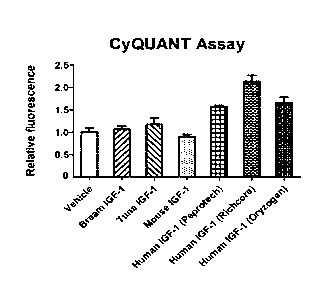
bream) IGF-1. Cells were harvested on day 7 and subjected to CyQUANT Cell
Proliferation Assay.
Detailed Description
CA 03173727 2022-06-02
WO 2021/111263
PCT/IB2020/061200
[0025] Referring now to the drawings, and with specific reference to
FIG. 1, a
method 10 for in vitro meat production is shown. As used herein, "in vitro
meat
production" refers to a cell-based meat production process or cell-based
agriculture process in which tissues from animals and/or plants are grown in
5 laboratories using cell culture techniques to manufacture meat and meat
products. At a block 12, tissue from an animal or a plant is isolated. In one
embodiment, the tissue is derived from bony fish of the class Osteichthyes
including saltwater fish such as a grouper, sea bass, or a yellow cocker. In
other
embodiments, other types of animal tissue, such as cow tissue, may be
isolated.
In some embodiments, the block 12 may involve collecting organ tissue, such as
a swim bladder, from a fish and making a cell suspension. Although the
following description primarily describes tissues derived from fish sources,
it will
be understood that the concepts may be applied to tissues derived from other
types of animal sources and/or plant sources to provide other types of in
vitro
meat and/or animal protein products, and vegetarian meat and/or protein
products.
[0026] Many of the isolated cells are adult cells, and can be made to
proliferate
continuously using various established methods in medical research (block 14).
For example, specific genes, such as Yamanaka factors, may be used to
reprogram the adult cells into stem cells, such as induced pluripotent stem
cells
(iPSCs). Alternatively, the isolated adult cells may be transformed into
continuous cell lines by telomerase reverse transcriptase overexpression. In
other embodiments, other types of cells may be isolated such as adult stem
cells
and embryonic stem cells. In this regard, it will be understood that the
methods
of the present disclosure include all sources of cell lines.
[0027] At a next block 16, the cells are grown into a solid or semi-
solid structure
mimicking an animal organ, such as a fish organ, by attaching/adhering to a
food-grade biocompatible scaffold in a sterile chamber or container, such as a
bioreactor. The sterile chamber or container may be temperature controlled,
and
may have inlets and outlets for introducing and removing substances such as
chemicals, nutrients, and cells. The food-grade biocompatible scaffold becomes
part of the final edible product, and is made of plant-based or fungi-based
materials such as, but not limited to, agarose, alginate, chitosan, mycelium,
and
konjac glucomannan. Alginate is a biopolymer naturally derived from brown
algae and is biocompatible. In addition, plant-based chitosan from fungi has
CA 03173727 2022-06-02
WO 2021/111263
PCT/IB2020/061200
6
antibacterial properties. In some embodiments, the block 16 is carried out in
the
absence of antibiotics or antimicrobial compounds in the sterile container. A
block 18 involves supplying the culture medium to the bioreactor to support
cell
survival and growth. The culture medium may be a buffered solution containing
components such as, but not limited to, inorganic salts (e.g., calcium
chloride
(CaCl2), potassium chloride (KCI), sodium chloride (NaCI), sodium bicarbonate
(NaHCO3), sodium dihydrogen phosphate (NaH2PO4), magnesium sulfate
(MgSO4), etc.), amino acids, vitamins (e.g., thiamine, riboflavin, folic acid,
etc.),
and other components such as glucose, 8-mercaptoethanol,
ethylenediaminetetraacetic acid (EDTA), and sodium pyruvate. Non-limiting
examples of growth media include, but are not limited to, Leibovitz's L-15
medium, Eagle's Minimum Essential Media (MEM), Medium 199, Dulbecco's
Modified Eagle Medium (DMEM), Ham's F12 Nutrient Mix, Ham's F10 Nutrient
Mix, MacCoy's 5A Medium, Glasgow Modified Eagle Medium (GMEM), Iscove's
Modified Dulbecco's Medium, and RPM! 1640.
[0028] According to a block 20, food-grade growth factors and cytokines are
introduced into the culture medium in the bioreactor to support cell growth
and
proliferation. The growth factors and cytokines may include, but are not
limited
to, insulin growth factor 1 (IGF-1), insulin, interleukin 6 (IL-6),
interleukin 6
receptor (IL-6R), interleukin 11 (IL-11), fibroblast growth factor (FGF),
epidermal
growth factor (EGF), and transferrin. The growth factors of (i) genetically
same
or similar species to the isolated cells and/or (ii) genetically same genus to
the
isolated cells (i.e. cells growing at block 16) are used in the present
invention. It
is found that the use of growth factors of (i) genetically same or similar
species to
the isolated cells and/or (ii) genetically same genus to the isolated cells
exert
higher bioactivities to the isolated cells compared to the use of growth
factors
and serum of genetically distant species to the isolated cells. Due to the
enhanced comparability, there is no need to supply a megadose of "suboptimal"
growth factors when culturing isolated cells using growth factors of (i)
genetically
same or similar species and/or (ii) genetically same genus. Higher bioactivity
could also help reducing the amount of growth factors needed in the culture
medium, shortening the culture period and improving cell quality. The cost of
the
culture medium could be reduced due to the decrease in the levels of growth
factors required in the culture medium for the stimulations of cell growth and
differentiation. Furthermore, the use of suboptimal growth factors limits the
CA 03173727 2022-06-02
WO 2021/111263
PCT/IB2020/061200
7
magnitude of the maximum cellular response (growth, differentiation). In some
instances, certain responses can never be reached no matter how much of the
suboptimal growth factor is supplied. Species-specific and/or genus-specific
growth factors could help overcoming these limits.
[0029] In some embodiments, the species is genetically similar to the
isolated
cells when they are more than 90% match in DNA sequence.
[0030] Species-specific or genus-specific growth factors can
effectively act on
receptors of the isolated cells. Compared to the conventional growth medium,
which is often supplemented by high levels of human protein growth factors
and/or FBS irrespective of the species origin of the isolated cell, the use of
species-specific or genus-specific growth factors is better optimized. Species-
specific or genus-specific variations in amino acid sequence and post-
translational modifications of the growth factor(s) and cell receptor(s) may
account for this phenomenon.
[0031] As will be discussed in much greater detail below, to identify which
species of a certain growth factor exerts the highest bioactivity on the
target
isolated cells, target cells are first seeded in complete medium (i.e. basal
medium + FBS). Upon reaching the target confluence (around 20%-70%), target
cells are treated by the growth factor of different species at a range of
concentrations (e.g. 1 pM-1 pM). Target cells are kept in the incubator until
reaching the desired time point(s) for the studied parameter (e.g. cell
growth,
differentiation markers, cellular products). For example, when there are
differences in cell confluence between the treatment groups (around 2-10
days),
cell growth can be measured by trypan blue exclusion, the CyQUANT assay, or
any other appropriate cell proliferation/death assays. The bioactivities of
the
growth factors of various species are compared based on their EC50 values
(half-maximal effective concentration). For cost-effectiveness, target cells
should
be cultured using the growth factor with the lowest EC50 value. However, if
the
aim is to attain the shortest culturing time or the highest cell quality,
select the
growth factor which triggers the highest maximum cellular response. The
optimal
dose of the growth factor is defined as the lowest concentration required to
elicit
the maximum cellular response.
[0032] In some embodiments, the block 20 may involve co-culturing
bioengineered cells with the isolated cells in the absence of fetal bovine
serum
(FBS). The bioengineered cells are engineered to secrete the above growth
CA 03173727 2022-06-02
WO 2021/111263
PCT/IB2020/061200
8
factors and cytokines, and supply these biomolecules to the isolated cells as
needed for growth and proliferation. As used herein, "bioengineered" cells are
not equivalent to genetically-modified cells. The bioengineered cells have a
specific gene that overexpresses one or more specific proteins. The
bioengineered cells may be fish cells, or other types of animal cells, such as
cow
cells. The bioengineering cells and the isolated cells may be genetically
similar
or identical species. Also the bioengineering cells and the isolated cells may
belong in the same genus. As non-limiting examples, bioengineered fish cells
may be co-cultured with isolated fish cells, or bioengineered cow cells may be
co-cultured with isolated cow cells. In some particular examples, the
bioengineering cells may be chicken cells or bird cells if chicken cells are
used
as the isolated cells. In yet another particular example, the bioengineering
cells
may be yellow crocker cells or other fish cells if yellow cracker cells are
used as
the isolated cells. The bioengineered cells are not present in the final meat
product. The co-culturing method of the present disclosure eliminates the need
for animal-derived fetal bovine serum (FBS) in the culture medium.
Furthermore,
the co-culturing method provides a continuous supply of food-grade specific
growth factors and cytokines to the growing isolated cells in situ, and
simplifies
and reduces the cost of the production process, wherein the growth factors are
(i) of genetically same or similar species to the isolated cells and/or (ii)
of the
same genus to the isolated cells. However, in other embodiments, FBS or other
serum may be used to supply growth factors, cytokines, and other nutrients to
support cell growth during the block 16.
[0033]
In some embodiments, the block 20 contains recombinant growth factors
of genetically same or similar species to the isolated cells. In yet some
embodiments, the recombinant growth factors of the same genus to the isolated
cells are used. The recombinant growth factors are introduced into the growth
medium. The use of such recombinant growth factors exerts higher bioactivities
to the isolated cells than growth factors and serum of distant species to the
isolated cells. Bacterial, yeast, insect, mammalian, or any other appropriate
protein expression systems may be used to produce such recombinant growth
factors. Protein purification is performed by (but not limited to) affinity
chromatography, ion-exchange chromatography, size exclusion
chromatography, or a combination of these strategies.
CA 03173727 2022-06-02
WO 2021/111263
PCT/IB2020/061200
9
[0034] In some embodiments, recombinant growth factors of Epinephelus
akaara
(fish), which is genetically similar species to the fish muscle cells or swim
bladder cells of Epinephelus awoara (fish), are used. The recombinant growth
factors for culturing Epinephelus awoara used are Epinephelus akaara's IGF-1,
insulin and/or transferrin. The concentration of such IGF-1 is ranged from
lOng/m I to 10Ong/m I. The concentration of such insulin is ranged from 1pg/m1
¨
10pg/ml. The concentration of such transferrin is ranged from 0.5pg/m1 ¨
5pg/m I.
[0035] Additionally, according to a block 22, protein expression in
the cells is
increased to increase the biomass yield in the resulting meat product. As used
herein, "biomass yield" refers to the amount of digestible material (e.g.,
proteins)
in the resulting meat product that is available for energy production upon
consumption. More specifically, the block 22 involves increasing protein
expression by altering micro RNA levels in the cells, with the manipulation of
the
cells being carried out prior to culturing. Micro RNAs are endogenous, short,
non-encoding single-stranded RNA sequences involved in regulating post-
transcriptional gene expression. The block 22 involves increasing the amount
of
up-regulating micro RNAs that increase protein expression by promoting
messenger RNA (mRNA) translation, and/or decreasing the amount of down-
regulating micro RNAs that decrease protein expression by suppressing mRNA
translation. The micro RNA levels may be increased or decreased by
introducing micro RNAs, micro RNA mimics, or micro RNA inhibitors into the
cells. The micro RNA mimics have the same function as micro RNAs, but
maybe more stable and efficient in modulating protein expression. In some
embodiments, electroporation may be used to introduce episomal vectors into
the cells that carry instructions to express specific micro RNAs.
Alternatively or
in combination with this, an adeno-associated virus may be used as a vehicle
carrying episomal instructions to express specific micro RNAs. Decreasing the
amount of targeted down-regulating micro RNAs may be achieved by introducing
inhibitors for the targeted micro RNAs into the cells by transfection. It is
noted
here that the methods of increasing protein expression/biomass yield according
to the present disclosure is carried out without modifying the genome of the
cells.
[0036] Turning to FIG. 2, a method for post-transcriptional
enhancement of
protein expression in the cell lines is schematically depicted. One or more up-
regulating micro RNAs (miRNAs) may be increased to increase mRNA
CA 03173727 2022-06-02
WO 2021/111263
PCT/IB2020/061200
translation and protein production of selected proteins. Alternatively or in
combination with this, one or more down-regulating miRNAs may be blocked
with inhibitors (anti-miRNAs) to increase mRNA translation and protein
production of selected proteins.
5 [0037] Fish swim bladder primarily includes fibroblasts and collagen
protein.
Collagen type 1 (collagen I) is a dominant protein in fish swim bladder, and
increased expression of collagen I in cultured fish swim bladder cells may
increase biomass yield. Collagen I in the fish swim bladder cells includes
collagen, type 1, alpha 1 (COL1A1) and collagen, type 1, alpha 2 (COL1A2).
10 COL1A1 and COL1A2 expression is increased by up-regulating microRNA 21
(miR-21), such that increasing levels of miR-21 increase COL1A1 and COL1A2
production in fish swim bladder cells. Additionally, COL1A1 and COL1A2
expression are decreased by down-regulating microRNA 29a (miR-29a), such
that decreasing levels of miR-29a or blocking the action of miR-29a increases
COL1A1 and COL1A2 production in fish swim bladder cells. FIGs. 3-4 show
increasing COL1A1 (FIG. 3) and COL1A2 (FIG. 4) production by increasing miR-
21 levels and by blocking the action of miR-29a with the use of inhibitors
(anti-
miR 29a). Increased COL1A1 and COL1A2 production results in increased
biomass yield in the resulting meat product. Similar strategies may be applied
to
increase relevant protein levels in other types of animal cells.
[0038] Turning to FIG. 5, an exemplary bioreactor 30 used for
culturing the
isolated cells is shown. The cells attach to and grow on a solid phase support
32
provided by a food-grade scaffold 34 which is held in a sterile chamber 36 in
the
bioreactor 30. The scaffold 34 may dictate the shape of the meat product. The
food-grade scaffold 34 is made of plant-based or fungi-based materials such
as,
but not limited to, agarose, alginate, chitosan, mycelium, and konjac
glucomannan. The solid phase support 32 may be porous so that the cells may
attach to and grow on inner surfaces of the support 32. The culture medium
supplying nutrients to the cells is introduced into the bioreactor 30 through
an
inlet 38, and is emptied from the bioreactor 30 through an outlet 40.
[0039] FIG. 6 shows a bioreactor 50 similar to the bioreactor 30 of
FIG. 5, but
further includes a second solid phase 52 separated from the solid phase
support
32 by a fine mesh 54. The second solid phase 52 may contain or support the
bioengineered cells that secrete nutrients, growth factors, and cytokines for
the
cells growing on the solid phase support 32 in situ, and may physically
separate
CA 03173727 2022-06-02
WO 2021/111263
PCT/IB2020/061200
11
the bioengineered cells from the cells on the solid phase support 32. The
second solid phase 52 is made of plant-based materials, similar to the solid
phase support 32. The mesh 54 is permeable to nutrients, growth factors, and
cytokines, but is impermeable to cells. The bioreactor 50 of FIG. 6 allows the
co-
culturing of the bioengineered cells with the growing cells. In some
embodiments, the bioreactors 30 and 50 of FIGs. 5 and 6 may be arranged in
tandem. In other embodiments, several of the bioreactors 30, several of the
bioreactors 50, or mixtures of the bioreactors 30 and 50 may be arranged in
series for scaling up the process. The bioreactor 30 may be used mainly for
biomass production, whereas the bioreactor 50 may be used for providing
nutrients, growth factors, and cytokines to the growing cells.
[0040] The in vitro meat production method of the present disclosure
provides
meat products with a simple tissue organization of one cell type. The meat
product with one cell type is easier to make, develop, and commercialize
compared to other cultured meats having multiple cell types. Alternative
embodiments of the present disclosure provide meat products with multiple cell
types. Furthermore, Applicant has discovered a strategy to increase
biomass/protein production by altering micro RNA levels or activity in the
growing cells. In one example, two key micro RNAs (miR-21 and miR-29a) are
targeted to increase the levels of the dominant protein (collagen I) found in
fish
swim bladder cells. As far as the Applicant is aware, alteration of micro RNA
levels or activity to achieve an increased protein/biomass yield in cultured
meat
products has not been used by others in the field of cultured meat
development.
Targeting micro RNAs for increased protein production may cause less stress to
the cells than known knock-in or knock-out methods. Bio-engineered cells are
co-cultured with the growing animal cells to supply the growing fish cells
with
food-grade growth factors and cytokines for cell growth and proliferation in
situ,
reducing or eliminating the need for animal-derived FBS in the culture medium.
The co-culturing technique simplifies the production process and reduces
production costs.
[0041] Furthermore, the nutrients of the cultivated meat product may
be
customized to generate a healthier food product. For example, the cultured
meat product may be customized according to diet recommendations from a
dietician to from a personal genomic test. Healthy nutrients such as high-
density
cholesterol, polyunsaturated fatty acids, and monounsaturated fatty acids in
the
CA 03173727 2022-06-02
WO 2021/111263
PCT/IB2020/061200
12
meat product may be enriched by culturing the cells in specific conditions.
Alternatively, or in combination with this, nutrients known to be damaging to
health such as low-density cholesterol and saturated fatty acids may be
reduced
by culturing the cells in specific conditions. Micronutrients, such as
vitamins and
minerals, may also be enhanced. Nutrient customization of the cultivated meat
products may be achieved in various ways such as, but not limited to, 1)
tailoring
the nutrients fed to the growing cells during cell culture, and/or 2)
controlling the
proportions of layering scaffolds with different cells.
[0042] The production of the cultivated food product is under a
clean, sterile and
highly controlled process. Thus, undesirable degradation by microorganisms
such as bacterial or fungi of the nutrients in the food product is minimized.
Undesirable taste and smell from the breakdown of nutrients by microorganisms
are also minimized. This property of cultivated food enables new uses in
cooking
and helps creates novel recipes. One such application of cultivated food is
cultivated fish maw derived from fish swim bladders. Traditional fish maw has
an
undesirable fishy taste and smell due to the degradation of amine by bacteria
in
the production process. This undesirable property limits the food ingredient
to
savory dishes served hot or warm. Cultivated fish maw produced from cell
culture technology does not have an undesirable fishy taste and smell. In
addition to hot and savory dishes, cultivated fish maw can be used in sweet
dishes, as a dessert or in a ready-to-eat format served at chilled or at
ambient
temperature.
[0043] Identification Of Species-Specific Or Genus-Specific Growth Factor
[0044] The method of identifying species-specific or genus-specific
growth
factors will be discussed in detail here. It includes two major steps, which
are the
cell growth stimulation step and measuring cell growth step.
[0045] Cell growth stimulation step
[0046] The MCF-7 human epithelial cell line is cultured in DMEM/F12
complete
medium DMEM/F12 (Thermofisher Scientific), 10% FBS (Thermofisher
Scientific), 1% Glutamax (Thermofisher Scientific), 0.2% Primocin (Invivogen)
inside a humidified incubator (34 C; 5% CO2; 95% air). Split cells at a ratio
of 1:4
to 1:8 for routine maintenance.
[0047] Upon reaching about 80% confluence, detach cells by
trypsin/EDTA
(Thermofisher Scientific). To study the effect of growth factors on cell
growth,
CA 03173727 2022-06-02
WO 2021/111263
PCT/IB2020/061200
13
cells are seeded at a density of 3 x 10⁴cells/cm2 in complete medium onto 24-
well (if cell growth is measured by cell counting) or 96-well plates (if cell
growth
is measured by the CyQUANT Cell Proliferation Assay Kit). Return the cells to
the incubator.
[0048] After 24 hours, remove the medium. Pre-adapt cells to serum-free
conditions by adding serum-free medium DMEM/F12, 0.1% human serum
albumin (Sigma Aldrich), 0.2% Primocin. Keep the cells in this medium for at
least 16 hours inside the incubator.
[0049] Prepare growth factors (e.g. IGF-1) at 10x working
concentrations in
serum-free medium for each species/genius, which growth-stimulating effect to
be examined (e.g. recombinant human IGF-1, human IGF-1-LR3, mouse IGF-1,
bream IGF-1 and tuna IGF-1). Add the 10x growth factors into the wells such
that cells will be treated by lx growth factors (e.g. add 50 pl 10x growth
factor to
well containing 450 pl serum-free medium) (e.g. 1 pM-1 pM). Return the cells
to
the incubator.
[0050] Observe the cells daily under a microscope for signs of cell
growth. When
there are obvious differences in terms of cell confluence between the
treatment
groups (usually detected between day 2¨ day 10), quantify cell growth either
by
cell counting, the CyQUANT Cell Proliferation Assay, or any other cell
proliferation/death assays.
[0051] Measuring cell growth step
[0052] There are two ways to measure cell growth, namely, trypan blue
exclusion
and CyQUANT Cell Proliferation Assay Kit.
[0053] Trypan blue exclusion
[0054] Cells should have been treated in 24-well plates. Aspirate the
culture
medium and detach cells by trypsin-EDTA.
[0055] Stop trypsin activity by adding 1 volume of the complete
medium into the
well. Ensure that all cells are detached by pipetting 3-5 times inside the
well.
[0056] Collect the cell suspension into 1.5 ml tubes. Pellet the
cells by
centrifuging the tubes at 400 x g for 5 minutes.
[0057] Remove the supernatant without disturbing the cell pellet.
Resuspend the
cell pellet in 200 pl DMEM/F12 basal medium.
[0058] Mix 10 pl of the cell suspension with 10 pl of 0.4% Trypan
Blue solution
(Thermofisher Scientific).
CA 03173727 2022-06-02
WO 2021/111263
PCT/IB2020/061200
14
[0059] After 2 minutes, add 10 pl of the cell/trypan blue mixture to
each chamber
of a Countess II FL Disposable Slide (Thermofisher Scientific) or a
hemocytometer. If using the Countess II FL system, insert the slide into the
slide
holder of a Countess II FL Automated Cell Counter (Thermofisher Scientific)
and
determine the cell concentration and A viability. If using a hemocytometer,
count
cells with a microscope.
[0060] Calculate the number of cells in each treatment group. The
number of
viable cells per well equals to viable cell concentration (cells/m1).
[0061] CyQUANT Cell Proliferation Assay Kit
[0062] The CyQUANT Cell Proliferation Assay Kit (Thermofisher Scientific)
quantifies cell growth by measuring the nucleic acid content in samples. Cells
should have been seeded onto 96-well plates, preferably in triplicate wells
per
treatment group.
[0063] Remove the culture medium as much as possible by a multichannel
pipette. Avoid scratching the well bottom with the pipette tip.
[0064] Freeze the plate in a -80 C freezer. The plate may be stored
at -80 C for
up to 4 weeks.
[0065] Thaw the plate and assay kit reagents at room temperature.
[0066] Mix the kit reagents according to Table 1 for each well.
Table 1
Components Volume per well
(1-11)
Cell lysis buffer stock solution 10 pl
Autoclaved MilliQ water 189.5 pl
CyQUANT GR stock solution 0.5 pl
Total= 200 pl
[0067] When the plate has completely thawed, add 200 pl of the CyQUANT GR
dye/lysis buffer mixture to each sample well and to three empty wells (blank).
Incubate the plate at room temperature for 5 minutes, protected from light.
CA 03173727 2022-06-02
WO 2021/111263
PCT/IB2020/061200
[0068] Using a multichannel pipette, transfer 160 pl from each well
of the 96-well
plate to the corresponding well of a black 96-well plate.
[0069] Measure the sample fluorescence using a fluorescence
microplate reader
(e.g. Molecular Devices SpectraMax iD5). Set the excitation and emission
5 wavelengths at 480 nm and 520 nm respectively.
[0070] Average the blank wells fluorescence readings. Subtract this
average
reading from all sample fluorescence readings to correct for background
fluorescence.
[0071] Calculate the mean corrected fluorescence for the vehicle
group. Express
10 the treatment group fluorescence readings as fold of control (FOC) by
dividing
the sample readings (i.e. IGF-1 groups) by the mean vehicle reading.
[0072] Example 1
[0073] IGF-1 stimulated the growth of MCF-7 cells in a dose-dependent
manner
[0074] To verify that IGF-1 stimulates the growth of human MCF-7
cells, cells
15 were treated with increasing doses of human IGF-1 (0 pg/m I ¨ 100 ng/ml)
for 10
days and processed for cell counting. As seen in FIG. 7, while 1-100 pg/ml IGF-
1
did not enhance the cell number, further increase of IGF-1 concentration (1-
100
ng/ml) promoted cell growth in a dose-dependent manner. Hence, MCF-7 cells
are suitable for evaluating the growth-stimulating activity of IGF-1.
[0075] Example 2
[0076] Human IGF-1, but not the mouse or fish IGF-1, promoted the
growth of
human MCF-7 cells
[0077] To investigate whether the growth-stimulating activity of IGF-
1 depends
on its species origin, we treated MCF-7 cells with 1.5 nM recombinant IGF-1 of
various species, i.e. human, mouse, and fish (bream, tuna). After 7 days, cell
growth was assessed by the CyQUANT Assay (FIG. 8). While human IGF-1
obtained from multiple sources consistently increased MCF-7 cell growth (-50 -
100% increase), mouse and fish IGF-1 did not (FIG. 8). These findings suggest
that human IGF-1, being the same species as the human MCF-7 cells, is more
effective than fish and mouse IGF-1 in promoting cell growth.
[0078] The present invention shows that it is more effective to apply
growth
factors and albumin of (i) genetically same or similar species or (ii) same
genus
as the cultured cell type. The usage of these growth factors or protein
factors
may be decreased while achieving the same growth rate. Species-specific
CA 03173727 2022-06-02
WO 2021/111263
PCT/IB2020/061200
16
growth factors and/or genus-specific growth factors represent a promising
direction to reduce media cost especially during large-scale cell production
for
cultivated meat and other applications using the cultivated cell mass. Using
more
bioactive growth factors can also decrease processing times and improve the
quality (e.g. texture, taste, nutritional value) of cultivated meat or cell
mass.
[0079] The above description is illustrative and is not restrictive.
Many variations
of embodiments may become apparent to those skilled in the art upon review of
the disclosure. The scope embodiments should, therefore, be determined not
with
reference to the above description, but instead should be determined with
reference to the pending claims along with their full scope or equivalents.
[0080] One or more features from any embodiment may be combined with one or
more features of any other embodiment without departing from the scope
embodiments. A recitation of "a", "an" or "the" is intended to mean "one or
more"
unless specifically indicated to the contrary. Recitation of "and/or" is
intended to
represent the most inclusive sense of the term unless specifically indicated
to the
contrary.
[0081] While the present disclosure may be embodied in many different
forms, the
drawings and discussion are presented with the understanding that the present
disclosure is an exemplification of the principles of one or more inventions
and is
not intended to limit any one embodiment to the embodiments illustrated.
[0082] The disclosure, in its broader aspects, is therefore not
limited to the
specific details, representative system and methods, and illustrative examples
shown and described above. Various modifications and variations may be made
to the above specification without departing from the scope or spirit of the
present disclosure, and it is intended that the present disclosure covers all
such
modifications and variations provided they come within the scope of the
following
claims and their equivalents.
CA 03173727 2022-06-02
WO 2021/111263
PCT/IB2020/061200
17
Exemplary Protocols
A. Development of a fish bladder cell line
1. Obtain a healthy yellow crocker, sea bass or fish of a similar category
from a local
fish market.
2. Keep the fish on ice until cell isolation.
3. Immerse the fish in 10% bleach.
4. Remove swim bladder from the fish under aseptic condition.
5. Wash the organ one or more times in hypochlorous acid.
6. Wash the organ one or more times in antibiotic medium (Leibovitz's L-15 or
DMEM
or EMEM with 400 IU/ml, penicillin, 400 pg/ml streptomycin).
7. After washing, cut the organ into small pieces (2-3 mm3).
8. Transfer the cut organ to a centrifuge tube containing 0.25% trypsin-EDTA
in PBS.
9. Incubate at room temperature with continuous shaking for 1 hour.
10. Filter the supernatant with a 100 pm mesh to remove undigested tissue.
11. Centrifuge the filtrate at 200g for 5 minutes.
12. Resuspend the cell pellet with complete medium (Leibovitz's L-15 or
DMEM or
EMEM with 200 IU/m I, penicillin, 200 pg/ml streptomycin, 10% fetal bovine
serum).
13. Seed the cell into a T25 flask.
14. Incubate at 24-28 C.
15. Remove cells that are not attached to the tissue culture flask the next
day.
16. Replace half of the medium with fresh medium every 2-3 days.
17. The cells are considered established when a complete monolayer is formed
and
the established cells are ready for subculture.
B. Development of a fish bladder cell line by tissue explant
1. Obtain a healthy yellow crocker, sea bass, or fish of a similar category
from a local
fish market.
2. Keep the fish on ice until cell isolation.
3. Immerse the fish in 10% bleach.
4. Remove swim bladder from the fish under aseptic condition.
5. Wash the organ one or more times in hypochlorous acid.
6. Wash the organ one or more times in antibiotic medium (Leibovitz's L-15 or
DMEM
or EMEM with 400 IU/ml, penicillin, 400 pg/ml streptomycin).
7. After washing, cut the organ into small pieces (1-2 mm3).
8. Place organ pieces into a 24 well plate individually containing complete
medium
(Leibovitz's L-15 or DMEM or EMEM with 200 IU/ml, penicillin, 200 pg/ml
streptomycin,
10% fetal bovine serum).
9. Incubate at 24-28 C.
10. Replace half of the medium with fresh medium every 2-3 days without
disturbing
the tissue explant.
11. Incubate the tissue explant until adherent cells are observed.
12. Remove tissue explant.
13. The cells are considered established when a complete monolayer is
formed and
the established cells are ready for subculture.
C. Development of a fish muscle cell line
1. Obtain a healthy grouper, cod, sole, halibut, flounder, or fish of a
similar category
from a local fish market.
CA 03173727 2022-06-02
WO 2021/111263
PCT/IB2020/061200
18
2. Keep the fish on ice until cell isolation.
3. Immerse the fish in 10% bleach.
4. Remove muscle from the fish under aseptic condition.
5. Wash the tissue one or more times in hypochlorous acid.
6. Wash the tissue one or more times in antibiotic medium (Leibovitz's L-15 or
DMEM
or EMEM with 400 IU/ml, penicillin, 400 pg/ml streptomycin).
7. After washing, cut the tissue into small pieces (2-3 mm3).
8. Transfer the cut tissue to a centrifuge tube containing collagenase and
dispase in
PBS.
9. Incubate at room temperature with continuous shaking for 1 hour.
10. Filter the supernatant with a 100 pm mesh to remove undigested tissue.
11. Centrifuge the filtrate at 200g for 5 minutes.
12. Resuspend the cell pellet with complete medium (Leibovitz's L-15 or
DMEM or
EMEM with 200 IU/m I, penicillin, 200 pg/ml streptomycin, 10% fetal bovine
serum).
13. Seed the cell into a T25 flask.
14. Incubate at 24-28 C.
15. Remove cells that are not attached to the tissue culture flask the next
day.
16. Replace half of the medium with fresh medium every 2-3 days.
17. The cells are considered established when a complete monolayer is formed
and the
established cells are ready for subculture.
D. Development of a fish muscle cell line from tissue explant
1. Obtain a healthy grouper, cod, sole, halibut, flounder, or fish of a
similar category
from a local fish market.
2. Keep the fish on ice until cell isolation.
3. Immerse the fish in 10% bleach.
4. Remove muscle from the fish under aseptic condition.
5. Wash the tissue one or more times in hypochlorous acid.
6. Wash the tissue one or more times in antibiotic medium (Leibovitz's L-15
or
DMEM or EMEM with 400 IU/ml, penicillin, 400 pg/ml streptomycin).
7. After washing, cut the muscle into small pieces (1-2 mm3).
8. Place muscle pieces into a 24 well plate individually containing complete
medium
(Leibovitz's L-15 or DMEM or EMEM with 200 IU/ml, penicillin, 200 pg/ml
streptomycin,
10% fetal bovine serum).
9. Incubate at 24-28 C.
10. Replace half of the medium with fresh medium every 2-3 days without
disturbing
the tissue explant.
11. Incubate the tissue explant until adherent cells are observed.
12. Remove tissue explant.
13. The cells are considered established when a complete monolayer is formed
and the
established cells are ready for subculture.
E. Adult Stem cell isolation and culture
1. Obtain a healthy grouper, cod, sole, halibut, flounder or fish 6 months
or younger
of similar category from a local fish market.
2. Keep the fish on ice until cell isolation.
3. Immerse the fish in 10% bleach.
CA 03173727 2022-06-02
WO 2021/111263
PCT/IB2020/061200
19
4. Remove muscle from the fish under aseptic conditions.
5. Wash the tissue one or more times in hypochlorous acid.
6. Wash the tissue one or more times in antibiotic medium (Leibovitz's
L-15 or
DMEM or EMEM with 400 IU/ml, penicillin, 400 pg/ml streptomycin).
7. After washing, cut the tissue into small pieces (2-3 mm3).
8. Transfer the cut tissue to a centrifuge tube containing collagenase and
dispase in
PBS.
9. Incubate at room temperature with continuous shaking for 1 hour.
10. Filter the supernatant with a 100 pm mesh to remove undigested tissue.
11. Centrifuge the filtrate at 200g for 5 minutes.
12. Resuspend the cell pellet with complete medium (Leibovitz's L-15 or DMEM
or
EMEM with 200 IU/m I, penicillin, 200 pg/ml streptomycin, 10% fetal bovine
serum,
10Ong/m1 basic fibroblast growth factor).
13. Plate the cells on an uncoated plate for 1 hour at 24-28 C.
14. Harvest the supernatant and place on a plate coated with lam inin,
gelatin, Matrigel
or similar matrix.
is. Incubate at 24-28 C.
16. After 24 hours, wash away any loosely attached and non-adherent cells.
17. Replace medium every day with complete medium (Leibovitz's L-15 or DMEM or
EMEM with 200 IU/m I, penicillin, 200 pg/ml streptomycin, 10% fetal bovine
serum,
10Ong/m1 basic fibroblast growth factor).
F. Generating and culturing iPSC
1. 2-4 days before transfection, plate cells in complete medium (L15 with
10% FBS)
in a tissue culture flask. Cells should be approximately 75-90% confluent on
the day of
transfection (Day 0).
2. Aspirate the medium from gelatin-coated 6-well plates and replace them
with 2
mL of fresh complete medium per well. Place the coated plates at 37 C until
ready for
use.
3. Thaw the Epi5TM vectors at 37 C and place them on wet ice until ready
for use.
Before use, briefly centrifuge the thawed vectors to collect them at the
bottom of the
tube.
4. Wash the cells in PBS.
5. Add 3 mL of 0.05% Trypsin/EDTA to the culture flask containing the cells.
6. Incubate the flask at room temperature for 3 minutes.
7. Add 5-8 mL of complete medium to each flask. Carefully transfer
cells into an
empty, sterile 15m L conical tube.
8. Check the viability by trypan blue dye exclusion cell viability assay
9. Centrifuge the cells at 200g for 2 min.
10. Carefully aspirate most of the supernatant and resuspend with complete
medium.
11. Seed cells on gelatin-coated dishes plate 50,000 to 100,000 cells per well
into a 6-
well plate at 30-60% confluence in 2 mL complete medium and Incubate overnight
at
24-28 C.
12. Prewarm Opti-MEM/Reduced-Serum Medium to room temperature and
prepare
Tube A and Tube B as described below.
13. Add 1.2 I_ each of the two Epi5TM Reprogramming Vector mixes (2.4
pt total) to
118 I_ Opti- MEM medium in a 1.5 mL microcentrifuge tube labeled Tube A. Add
4.8
1_11_ of P3000TM Reagent and mix well.
14. Dilute 3.6 tL Lipofectamine 3000 reagent in 121 ILLL prewarmed Opti-
MEM
CA 03173727 2022-06-02
WO 2021/111263
PCT/IB2020/061200
medium in a 1.5 mL microcentrifuge tube labeled Tube B.
15. To prepare a transfection master mix, add the contents of Tube A to Tube B
and
mix well.
16. Incubate the transfection master mix for 5 minutes at room temperature.
5 17. Mix one more time and add the entire 2504 of transfection master mix
to each
well.
18. Incubate overnight at 24-28 C.
19. 24 hours post-transfection, aspirate the medium from the plates. Add 2
mL
N2B27 Medium (L15 with IX N-2 supplement, IX B27 supplement, 100 ng/mL bFGF to
10 each well.
20. Change the N2B27 Medium every day for a total of 14 days by replacing
the
spent medium with 2 mL N2B27 Medium.
21. Aspirate the spent N2B27 Medium on Day 14 and replace it with a
complete
medium. Resume medium changes every day at 2 mL per well.
15 22. Observe the plates every other day under a microscope for the
emergence of
cell clumps, indicative of transformed cells. Within 15 to 21 days post-
transfection, the
iPSC colonies will grow to an appropriate size for transfer.
23. Colonies are distinct by Day 21 and can be picked for further culture and
expansion.
20 G. Method for subculturing cells
1. Remove and discard the culture medium.
2. Briefly rinse the cell PBS to remove all traces of serum which contains
trypsin
inhibitor.
3. Add 2-3 mL of 0.25% Trypsin-EDTA solution to the flask.
4. Incubate at room temperature for 1 min.
5. Add 5-8 mL of complete growth medium.
6. Aspirate cells by gently pipetting.
7. Add appropriate aliquots of the cell suspension to new culture flasks at a
subcultivation ratio of 1:2 to 1:3.
8. Incubate at 24-28 C.
H. Adaption to suspension culture
1. Passage monolayer culture at a frequency appropriate for the cell in
question by
trypsinization.
2. At each passage, wash cell monolayer with PBS and overlay with 0.25%
trypsin.
3. Incubate at room temperature for 5 min.
4. Inactivate the enzyme with a complete medium.
5. Harvest the cell suspension and check the viability by trypan blue dye
exclusion cell
viability assay.
6. Seed the cell suspension into another culture flask.
7. Repeat passaging until the viability of the suspended cells is equal or
more than
90%.
8. Establish a suspension culture with 50 ml complete medium in a
spinner or
shaker flask at a cell density of 0.1-0.5 million/ml.
9. Incubate the spinner or shaker flask suspension cultures in a CO2
incubator
under the same conditions of temperature, humidity, and atmosphere optimal for
CA 03173727 2022-06-02
WO 2021/111263
PCT/IB2020/061200
21
monolayer cultures.
10. Adjust the cell density to 0.1-0.5 million/ml with fresh medium every 2-3
days.
11. Check the viability by trypan blue dye exclusion cell viability assay.
12. Establish multiple parallel cultures at cell density that promote health
cell growth.
13. Increase cell density gradually to 1 million/ml using part of the culture.
14. If increasing cell density leads to cell death, discard the high-density
culture.
15. Restart high-density adaption using cell form step 12.
16. Scale up to a 3L bioreactor when cells are adapted to grow in suspension.
I. Adaption to serum-free medium (plant hydrolysate)
1. Culture cells in DMEM/F12 complete medium (1:1 mixture of DMEM medium
and Ham's F12 medium, 2-4mM glutamine, 10% FBS).
2. Prepare serum-free medium (1:1 mixture of DMEM medium and Ham' s F12
medium, 2-4mM glutamine, 20% plant hydrolysate e.g. soy, cottonseed, rapeseed,
wheat, yeast or equivalent).
3. When cells reach confluence, replace medium with adaption medium I (40%
fresh complete medium, 40% conditioned media from the passage before, 20%
serum-
free medium).
4. Check the viability by trypan blue dye exclusion cell viability assay every
2-3 days.
5. If adaption leads to cell death, discard the culture and repeat step 3.
6. When cells reach confluence, replace medium with adaption medium 11(30%
fresh complete medium, 30% conditioned media from the cells in step 1, 40%
serum-
free medium).
7. Check the viability by trypan blue dye exclusion cell viability assay every
2-3 days.
8. If adaption leads to cell death, discard the culture and repeat step 6.
9. When cells reach confluence, replace medium with adaption medium III
(20%
fresh complete medium, 20% conditioned media from the cells in step 1, 60%
serum-
free medium).
10. Check the viability by trypan blue dye exclusion cell viability assay
every 2-3 days
11. If adaption leads to cell death, discard the culture and repeat step 9.
12. When cells reach confluence, replace medium with adaption medium IV
(10%
fresh complete medium, 10% conditioned media from the cells in step 1, 80%
serum-
free medium).
13. Check the viability by trypan blue dye exclusion cell viability assay
every 2-3 days
14. If adaption leads to cell death, discard the culture and repeat step 12.
is. When cells reach confluence, replace medium with serum-free medium.
16. Check the viability by trypan blue dye exclusion cell viability assay
every 2-3 days.
17. If adaption leads to cell death, discard the culture and repeat step 15.
18. The serum-free medium usage can be increased more gradually in each
step,
i.e. an increase of 20% or less in each step.
J. Adaption to serum-free medium (chemically defined)
1. Culture cells in DMEM/F12 complete medium (1:1 mixture of DMEM medium
and Ham's F12 medium, 2-4mM glutamine, 10% FBS).
2. Prepare serum free medium (1:1 mixture of DMEM medium and Ham's F12
medium, 2-4 mM glutamine, ascorbic acid 2-phosphate 65-130 ug/ml, NaHCO3 550-
1100 ug/ml, sodium selenite 14-28 ng/ml, insulin 19-38 ug/ml, transferrin 11-
22 ug/ml,
FGF-2 100-200 ng/ml, TGF-beta 2-4 ng/ml).
CA 03173727 2022-06-02
WO 2021/111263
PCT/IB2020/061200
22
3. When cells reach confluence, replace medium with adaption medium I (40%
fresh complete medium, 40% conditioned media from the passage before, 20%
serum-
free medium).
4. Check the viability by trypan blue dye exclusion cell viability assay every
2-3 days
5. If adaption leads to cell death, discard the culture and repeat step 3
6. When cells reach confluence, replace medium with adaption medium 11(30%
fresh complete medium, 30% conditioned media from the cells in step 1, 40%
serum-
free medium).
7. Check the viability by trypan blue dye exclusion cell viability assay every
2-3 days..
8. If adaption leads to cell death, discard the culture and repeat step 6
9. When cells reach confluence, replace medium with adaption medium III
(20%
fresh complete medium, 20% conditioned media from the cells in step 1, 60%
serum-
free medium).
10. Check the viability by trypan blue dye exclusion cell viability assay
every 2-3 days.
11. If adaption leads to cell death, discard the culture and repeat step 9.
12. When cells reach confluence, replace medium with adaption medium IV
(10%
fresh complete medium, 10% conditioned media from the cells in step 1, 80%
serum-
free medium).
13. Check the viability by trypan blue dye exclusion cell viability assay
every 2-3 days.
14. If adaption leads to cell death, discard the culture and repeat step 12.
15. When cells reach confluence, replace medium with serum-free medium.
16. Check the viability by trypan blue dye exclusion cell viability assay
every 2-3 days.
17. If adaption leads to cell death, discard the culture and repeat step 15.
18. The serum-free medium usage can be increased more gradually in each step.
For
example, an increase of 20% or less in each step.
K. Post-transcriptional enhancement of protein expression
1. Culture cells in complete medium (Leibovitz's L-15 or DMEM or EMEM with 200
IU/ml, penicillin, 200 pg/ml streptomycin, 10% fetal bovine serum), or serum-
free
medium (DMEM/F12 with plant hydrolysate or chemically defined compounds).
2. Remove and discard the culture medium.
3. Briefly rinse the cell PBS to remove all traces of serum which contains
trypsin
inhibitor.
4. Add 2-3 mL of 0.25% Trypsin-EDTA solution to the flask.
5. Incubate at room temperature for 1 min.
6. Aspirate cells by gently pipetting.
7. Centrifuge cell at 200g for 2 min.
8. Resuspend cells in complete medium or serum-free medium.
9. Add 0.5 million cells to each well of a 6-well plate.
10. Incubate at 24-28 C overnight.
Transfect micro RNA oligonucleotides (miR-21, miR-29a, miR-21 mimic, miR-29a
mimic, anti-miR-21, anti-miR-29a, or equivalent) into the cell using
polyethylenimine,
liposome, electroporation, or other methods.
12. Incubate at 24-28 C overnight.
13. Transfer the cells to a multi-layer flask, spinner flask or shaker
flask in a CO2
incubator under the same conditions of temperature, humidity, and atmosphere
optimal
culture
CA 03173727 2022-06-02
WO 2021/111263
PCT/IB2020/061200
23
L. Scaffolding for cell culture (Konjac + gum)
1. Boil water with a few pieces of saffron until the color becomes pale
yellow.
2. Remove the saffron and rest the solution until warm.
3. Prepare all dry ingredient
a. Konjac-0.5-5%, preferably 3 %
b. Baking soda - 0.3-3%, preferably 2 %
c. Perfected Xanthan Gum - 0.2-2%, preferably 1.5%
4. Measure 100m1 of saffron solution.
5. Add Baking soda, Locust Bean Gum, Xanthan Gum sequentially. Stir the
mixture
well after adding each ingredient.
6. Add Konjac by sprinkling little by little on top of the solution.
Keep stirring. The
solution should become mushy.
7. Spread the konjac mixture into mold with approximately 1-15mm thickness.
8. Cover the mold with the lid and rest under room temperature for more than
30 min.
9. Put the mold in 4 C fridge for 4 hours.
10. Steam the mold under low heat for 40 minutes.
Rest the mold under room temperature for 2 hours.
12. Dehydrate the scaffold at 45-55 C for 15 minutes.
M. Scaffolding for cell culture (Alginate + Glutinous Rice Flour)
1. Weigh 0.1-2 g (0.1-2%), preferably about 1g (1%) Sodium Alginate.
2. Add 100mlwater into the blender.
3. Add the Alginate powder into the blender and blend the mixture until
dissolved.
4. Cover the container with plastic film and put the Alginate solution into
the
refrigerator overnight to eliminate the gas bubbles.
5. Weigh 1-10 g (1-10%), preferably about 5g (5%) Glutinous Rice Flour and put
in a
mold.
6. Add Alginate solution into the mold with approximately 1-15mm thickness.
7. Stir the mixture until all flour dissolves.
8. Steam the mixture under low heat for 30 min until the shape is set.
9. Cover the mold with the lid and rest under room temperature for 30 min.
10. Weigh 1% Calcium Lactate and stir to dissolve in water.
11. Immerse the scaffold with 1% Calcium Lactate solution for at least 2.5
hours to
allow the formation of the membrane around the scaffold.