Note: Descriptions are shown in the official language in which they were submitted.
WO 2021/252319
PCT/US2021/036100
PROJECTION OF DEFOCUSED
IMAGES ON THE PERIPHERAL RETINA TO TREAT REFRACTIVE ERROR
RELATED APPLICATIONS
[0001] The present PCT application claims priority to the
following provisional patent
applications 63/036,226, filed on June 8, 2020, entitled -PROJECTION OF
DEFOCUSED IMAGES ON THE PERIPHERAL RETINA TO TREAT REFRACTIVE
ERROR", 62/706,153, filed on August 3, 2020, entitled "PROJECTION OF
DEFOCUSED IMAGES ON THE PERIPHERAL RETINA TO TREAT REFRACTIVE
ERROR", 62/706,456, filed on August 18, 2020 ,entitled -PROJECTION OF
DEFOCUSED IMAGES ON THE PERIPHERAL RETINA TO TREAT REFRACTIVE
ERROR", the entire disclosures of which are incorporated herein by reference.
[0002] The subject matter of the present application is related
to PCT/US2019/043692,
filed on July 26,2019, entitled -ELECTRONIC CONTACT LENS TO DECREASE
MYOPIA PROGRESSION-, published as W02020028177A1 on February 6, 2020, the
entire disclosures of which is incorporated herein by reference.
BACKGROUND
[0003] Prior approaches to treating refractive error such as
myopia can be less than
ideal in at least some respects. Spectacle lenses, contact lenses, and
refractive surgery
can be used to treat refractive errors of the eye. However, lenses must be
worn in order to
correct the errors, and uncorrected refractive error can impact a person's
ability to achieve
and fully participate in school, sports, and other activities. Although
surgery can be
performed to decrease refractive error, surgery comes with risks, such as
infection and
degraded vision in at least some instances. Also, these approaches do not
address the
underlying changes in the length of the eye that is related to refractive
error such as
myopia.
[0004] Work in relation to the present disclosure suggests that the retina of
many
species, including human beings, responds to defocused images and is
repositioned
through scleral remodeling, in order to decrease the blur caused by the
defocus. The
mechanism of the generation of the growth signal is still under study, but one
observable
phenomenon is an increase in thickness of the choroid. A defocused image can
cause the
choroid thickness to change, which is related to the axial length of the eye.
Changes to
the axial length of the eye can alter the refractive error by changing the
position of the
- 1 -
CA 03174148 2022- 9- 28
WO 2021/252319
PCT/US2021/036100
retina in relation to the cornea. For example, an increase in axial length
increases myopia
of an eye by increasing the distance between the cornea and retina.
[0005] While the defocus of images can play a role in choroidal thickness and
changes
in the axial length of the eye, the prior approaches are less than ideally
suited to address
to refractive error of the eye related to axial length. Although
pharmaceutical treatments
have been proposed to treat myopia associated with axial length growth, these
treatments
can have less than ideal results and have not been shown to safely treat
refractive error in
at least some instances. Although light has been proposed as a stimulus to
alter the
growth of the eye, at least some of the prior devices can provide less than
ideal results.
Also, the time of treatment can be longer than would be ideal, and at least
some of the
prior approaches may be more complex than would be ideal.
[0006] Therefore, new approaches are needed to treat refractive
error of the eye that
ameliorate at least some of the above limitations of the prior approaches.
SUMMARY
[0007] The presently disclosed methods, devices and apparatus
provide improved
treatment of refractive error with decreased treatment times. In some
embodiments, the
stimulus comprises one or more of a spatial frequency distribution or a ratio
of stimulus
intensity to background illumination intensity to promote an improved
response. In some
embodiments, the stimulus is presented at an appropriate time of day to
promote the
response.
[0008] An apparatus to treat refractive error of the eye
comprises one or more optics
configured to project stimuli comprising out of focus images onto the
peripheral retina
outside the macula. While the stimuli can be configured in many ways, in some
embodiments the stimuli are arranged to decrease interference with central
vison such as
macular vision. The stimuli can be out of focus images may comprise an amount
of
defocus within a range from about 2 Diopters ("D") to about 6 D, and the range
can be
from about 3 D to about 6D. In some embodiments, the brightness of the stimuli
is
greater than a brightness of background illumination by an appropriate amount
such as at
least 3 times the background brightness. In some embodiments, each of a
plurality of
stimuli comprises a spatial frequency distribution with an amplitude profile
having
substantial spatial frequencies within a range from about range of 1X10⁻¹ to
1X101 cycles
per degree. In some embodiments, each of the stimuli is sized and shaped with
an
intensity profile distribution so as to provide spatial frequencies to promote
a response to
- 2 -
CA 03174148 2022- 9- 28
WO 2021/252319
PCT/US2021/036100
the stimuli. Each of the stimuli may comprise one or more localized intensity
peaks in
proximity to a region of decreased illumination. In some embodiments, the
region of
deceased illumination is located between a plurality of peaks, although the
region of
decreased illumination may be bounded by an annular peak.
INCORPORATION BY REFERENCE
[0009] All patents, applications, and publications referred to
and identified herein are
hereby incorporated by reference in their entirety and shall be considered
fully
incorporated by reference even though referred to elsewhere in the
application.
BRIEF DESCRIPTION OF THE DRAWINGS
[0010] A better understanding of the features, advantages and
principles of the present
disclosure will be obtained by reference to the following detailed description
that sets
forth illustrative embodiments, and the accompanying drawings of which:
[0011] FIG. lA shows a retinal stimulation device, in
accordance with some
embodiments;
[0012] FIG. 1B shows a spectacle lens based retinal stimulation
device comprising a
display and a housing to contain the electronics for operating the near eye
display, in
accordance with some embodiments;
[0013] FIG. 1C shows a spectacle lens based retinal stimulation
device as in Figure
1B, in which the eye has moved and different display elements have been
activated in
response to the eye movement, in accordance with some embodiments;
[0014] FIG. 2A shows a soft contact lens, in accordance with
some embodiments;
[0015] FIG. 2B shows soft contact lens with embedded light
sources, optics and
electronics for projecting images with defocus on the periphery of the retina
of a user, in
accordance with some embodiments;
[0016] FIG. 3 shows a system diagram of the function of the
components of the
contact lens as in FIG. 2;
[0017] FIG. 4A shows an optical configuration in which the
optical path length is
increased by folding the optical path with two mirrors, in accordance with
some
embodiments;
[0018] FIG. 4B shows the optical configuration as in FIG. 4A
projecting light into an
eye, in accordance with some embodiments;
[0019] FIG. 5A shows an optical configuration comprising a lens
to focus light onto
the retina, in accordance with some embodiments
- 3 -
CA 03174148 2022- 9- 28
WO 2021/252319
PCT/US2021/036100
[0020] FIG. 5B shows an optical configuration as in FIG. 5A
projecting light into an
eye, in accordance with some embodiments;
[0021] FIG. 6A shows a light-pipe in order to increase the
optical path length, in
accordance with some embodiments;
[0022] FIG. 6B shows an optical configuration as in FIG. 6A
projecting light into an
eye, in accordance with some embodiments;
[0023] FIG. 7 shows a plurality of stimuli and an image on a
display as seen by a user,
in accordance with some embodiments;
[0024] FIG. 8A shows stimuli on a screen to provide myopically
defocused stimuli to
the retina, in accordance with some embodiments;
[0025] FIG. 8B shows the corresponding dimensions of the
myopically defocused
stimuli on the retina in degrees, in accordance with some embodiments;
100261 FIG. 9 shows a stimulus depicting a natural scene, such
as an annular flower
pattern, in accordance with some embodiments;
[0027] FIG 10 shows image contrast and a histogram with red
(R), blue (B) and green
(G) values for the stimuli shown in FIGS. 8A to 9, in accordance with some
embodiments;
[0028] FIG. 11 shows an image suitable for modification and
incorporation as a
stimulus as described herein, in accordance with some embodiments;
[0029] FIG. 12 shows an image similar to the image of FIG. 11
that has been
processed to provide an improved stimulus, in accordance with some
embodiments:
[0030] FIG. 13 shows an image of spatial frequencies
distributions of the image of
FIG. 11, in accordance with some embodiments;
[0031] FIG. 14 shows an image of spatial frequencies
distributions of the image of
FIG. 13, which is used as the stimulus, in accordance with some embodiments;
100321 FIG. 15 shows a plot of image spatial frequency in
cycles per degree and the
log of the energy at each frequency for the stimulus images shown in FIGs. 8B
and 9, in
accordance with some embodiments;
[0033] FIG. 16 shows a system for treating refractive error of
the eye, in accordance
with some embodiments;
100341 FIG. 17 shows a method of treating refractive error of
the eye, in accordance
with some embodiments;
- 4 -
CA 03174148 2022- 9- 28
WO 2021/252319
PCT/US2021/036100
[0035] FIG. 18A depicts a stimulus with a myopic defocus of 6D
("6D stimulus") and
another stimulus with a myopic defocus of 3D ("3D Stimulus"), in accordance
with some
embodiments;
[0036] FIG. 18B depicts a stimulus with 25% coverage ("25%
stimulus) and a
stimulus with 50% coverage ("50% stimulus-) , in accordance with some
embodiments;
[0037] FIG. 18C depicts a stimulus with a brightness ratio of
0.1:1 and a stimulus with
a 1:1 brightness ratio, in accordance with some embodiments;
[0038] Fig. 18D depicts a black and white stimulus and a red
stimulus, in accordance
with some embodiments;
[0039] FIG. 19 depicts an optical system to project stimuli
onto the retina, in
accordance with some embodiments;
[0040] FIG. 20A shows the focus of the central entertainment
region and the
background pattern for the control eye, e.g. the left eye, in accordance with
some
embodiments;
[0041] FIG. 20B shows the myopic defocus of the stimulus, the
central entertainment
region and the background pattern for the tested eye, e.g. the right eye, in
accordance with
some embodiments;
[0042] FIG. 21 shows clinical results similar to the results of
Table 1, in accordance
with some embodiments;
[0043] FIG. 22 shows the aggregate data of the 5X, 10X, and 20X
luminance trials
show that the mean change in central axial length (in microns) for the test
eye was
significantly smaller than that of the control eye (p<0.025) after the one-
hour defocus
sessions, in accordance with some embodiments; and
[0044] FIG. 23 shows the average change in axial length and
choroidal thickness
(mean SEM): For the aggregate of all trials, the change in axial length for
the test eye
was significantly lower than that for the control eye after an hour of defocus
sessions, in
accordance with some embodiments.
DETAILED DESCRIPTION
[0045] The following detailed description provides a better
understanding of the
features and advantages of the inventions described in the present disclosure
in
accordance with the embodiments disclosed herein. Although the detailed
description
includes many specific embodiments, these are provided by way of example only
and
should not be construed as limiting the scope of the inventions disclosed
herein.
- 5 -
CA 03174148 2022- 9- 28
WO 2021/252319
PCT/US2021/036100
[0046] The presently disclosed methods and apparatus can be
configured in many
ways to provide retinal stimulation as described herein. The presently
disclosed methods
and apparatus are well suited for combination with many prior devices, such as
one or
more of an ophthalmic device, a TV screen, a computer screen, a virtual
reality ("VR")
display, an augmented reality ("AR-) display, a handheld device, a mobile
computing
device, a tablet computing device, a smart phone, a wearable device, a
spectacle lens
frame, a spectacle lens, a near eye display, a head-mounted display, a goggle,
a contact
lens, an implantable device, a corneal onlay, a corneal inlay, a corneal
prosthesis, or an
intraocular lens. Although specific reference is made to spectacles and
contact lenses, the
presently disclosed methods and apparatus are well suited for use with any of
the
aforementioned devices, and a person of ordinary skill in the art will readily
appreciate
how one or more of the presently disclosed components can be interchanged
among
devices, based on the teachings provided herein.
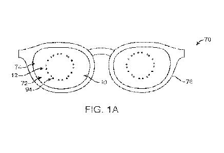
[0047] FIG. IA shows a retinal stimulation device to one or
more of decrease myopia
progression or at least partially reverse myopia progression. The device
comprises a lens
to support a plurality of light sources. The plurality of light sources can be
coupled to
one or more optical components to provide a stimulus to the retina as
described herein. In
some embodiments, the lens 10 comprises a spectacle lens 74. In some
embodiments, the
lens 10 is shaped to correct spherical and cylindrical refractive errors of
the user, to
provide corrected visual acuity to through the lens. The plurality of light
sources may
comprise one or more of projection units 12 or a display 74 such as a near eye
display.
The plurality of light sources is arranged about the central portion of the
lens so as to
provide light stimulation to an outer location of the retina such as the
peripheral retina as
described herein. In some embodiments, the light sources are located in an
approximately
annular region so as to provide stimulation to the peripheral retina. The
light sources can
be arranged in a generally annular pattern, for example in quadrants, so as to
correspond
to quadrants of the peripheral retina outside the macula. Each of the
plurality of light
sources can be configured to project a pattern anterior to the retina with an
appropriate
stimulus pattern as described herein. In some embodiments, light from the
light sources
traverses an optical axis of the eye so as to stimulate the retina at a
location on an
opposite side of the retina from the light source.
[0048] In some embodiments, the projection units 12 are
configured to emit light rays
to enter the pupil of the eye without substantial aliasing. In some
embodiments, the pupil
of the eye may be enlarged by appropriate amounts of illumination or
application of
- 6 -
CA 03174148 2022- 9- 28
WO 2021/252319
PCT/US2021/036100
mydriatic agents so that a greater area of the retinal surface is accessible
to the stimulus
projected by the projection units 12.
[0049] In some embodiments, the plurality of light sources is
configured to remain
static while the user views an object. Alternatively, the light sources can be
configured to
move in response to eye movement, for example with the selective activation of
pixels as
described herein.
[0050] Although reference is made to the plurality of light
sources supported on a lens,
the light sources can be supported on any suitable optically transmissive
substrate, such as
a beam splitter or a substantially flat optical component, and the light
sources may
comprise light sources of a pixel display such as an AR or VR display. In some
embodiments, the display 72 comprises pixels 94 which are selectively
activated to
provide a stimulus to the retina as described herein. Alternatively or in
combination, the
projection units 12 may comprise a shaped structure to provide the stimulus to
the retina
as described herein.
[0051] In some embodiments, the pixels are configured to emit a
plurality of colors, so
that the projected light can be combined to create any suitable color or hue,
such as white
light, for example.
[0052] In some embodiments, the plurality of light sources is
supported a head
mounted support, such as eyeglass frame 76 on spectacles 70.
[0053] FIGS. 1B and 1C depict spectacles 70 for the treatment
of refractive error of
the eye, such as spherical refractive error, although any suitable vision
device as
described herein can be appropriately modified in accordance with the
embodiments
disclosed herein. The plurality of light sources can be coupled to one or more
optical
components to provide a stimulus to the retina as described herein. The
spectacles 70 may
comprise one or more components of commercially available augmented reality
glasses.
The spectacle 70 may comprise one or more displays 72 for retina stimulation.
The near
eye displays 72 may be mounted to lenses 74. The lenses 74 may be spectacle
lenses
supported by eyeglass frame 76. The lens 74 may be a corrective or non-
corrective lens.
The lens 74 may be a plano lens, a spherical corrective lens, an astigmatic
correction lens,
or a prism correction lens. In some embodiments, the near eye display is
located away
from an optical zone to provide clear central vision. An optical axis may
extend along a
line of sight from an object of the patient's regard, though the lens 74 to a
fovea of the
eye. In some embodiments, the spectacle 70 comprises an eye tracker suitable
for
incorporation in accordance with the present disclosure. The near eye display
72 can be
- 7 -
CA 03174148 2022- 9- 28
WO 2021/252319
PCT/US2021/036100
programmed to selectively activate pixels 94, in order to provide peripheral
stimulation to
the retina, as described herein. In some embodiments, a layer of a plastic
substrate
bearing micro-lenses is attached to the micro-display in order to generate the
desired level
of defocus and stimulation at the retina. The selectively activatable pixels
may comprise
a groups of pixels, which can be selectively activated together, e.g. a first
group of pixels
94a, a second group of pixels 94B, a third group of pixels 94C, and a fourth
group of
pixels 94D. The groups of pixels can be arranged to provide an appropriate
eccentricity
with respect to a line of sight of the patient, so as to provide peripheral
retinal stimulation
as described herein.
[0054] In some embodiments, a near eye display 72 comprises a
combination of a
micro-display and a micro-optic. In some embodiments, the micro-optic is
configured to
collect, substantially collimate and focus the light rays emanating from the
micro-display.
In some embodiments, the micro-optic is configured to form an image anterior
to or
posterior to the retina as described herein. In some embodiments, the distance
of the near
eye display from the entrance pupil of the eye is within a range from about 10
mm to
about 30 mm, for example about 15 mm. The micro-display can be placed on a
transparent substrate, such as the front or back surface of the lens 74 of the
spectacles 70.
When the micro-display is placed on the front surface of the lens 94, then the
focus of the
micro-displays may be affected by the cylindrical correction on the back
surface of the
lens 94.
[0055] In some embodiments, the focus of the pixels in a micro-
display may vary
based on their location on the lens 74 and the refractive correction provided
by the lens in
that area. In some embodiments, the focus of the pixels may be fixed. In some
embodiments, the focus of the pixels may vary based on the sensed position of
the cornea
to account for the refraction of the cornea and the lens of the eye. In some
embodiments,
the pixels are defocused to create a defocused spot on the retina about 1 mm
in diameter.
[0056] Light emitted by the pixels 94 in the micro-display of
the near eye display can
be one or more of substantially collimated or focused before being directed to
the pupil of
the eye. In some embodiments, a micro-lens array is aligned to the pixels of
the near eye
display, so that rays from the near eye display can enter the pupil and form
an image
anterior to or posterior to the retina. In some embodiments, the width of the
near eye
display corresponds to a patient's field of view. In some embodiments, the
extent of the
near eye display may be substantially similar to the extent of the lens 74 of
the spectacles
70.
- 8 -
CA 03174148 2022- 9- 28
WO 2021/252319
PCT/US2021/036100
[0057] In some embodiments, the device provides unimpaired
central vision so that
the quality of life and quality of vision of the users are not adversely
affected. In some
embodiments, central vision comprises of a field of view of +/- 5 degrees or
greater,
preferably +/- 7.5 degrees or greater, such as +/-12.5 degrees, covering the
macula, while
foveal vision used for fixation has a field of view of +/-1.0 degrees. In some
embodiments, the defocused image is projected at an outer portion of the
retina toward
the periphery of the retina, for example within a range from 15 degrees (full
angle, or +/-
7.5 degrees) to 40 degrees (full angle, or +/- 20 degrees) eccentric to the
fovea and can be
within a range from 20 degrees to 40 degrees, for example within a range from
20 degrees
to 30 degrees. In some embodiments, the micro-display 72 does not obstruct the
central
vision field of view. In some embodiments, the pixels 94 do not obstruct the
central
vision field of view.
100581 In some embodiments, the micro-displays and optics are
configured to project
light onto outer regions of the retina sufficiently far from the fovea, that
the illumination
remains substantially fixed even with eye movement In some embodiments, the
point of
regard is monitored and the desired location of the pixels to be activated on
the micro-
display is determined, e.g. by a computations with a processor, such that an
image is
projected at the desired location on the retina, allowing sustained
stimulation at the same
retinal location. In some embodiments, the point of regard on the spectacle
plane or the
plane of the micro-display is calculated by monitoring the horizontal, the
vertical and
torsional displacement of the eye relative to the primary position.
[0059] The point of regard can be determined with a in many
ways, for example with
an eye position sensor such as a magnetic sensor or an optical sensor. In some
embodiments, a search coil embedded in the eyeglass frame is used to track eye
movements. The coil embedded in the eyeglass frame can be coupled to a
magnetic
structure placed on the eye, such as one or more of a coil on a contact lens,
a coil
implanted in the eye, a magnetic material on a contact lens, or a magnetic
material
implanted in the eye. In some embodiments, the sensor comprises an optical
sensor, such
as a position sensitive detector or an array sensor to measure a position of
the eye
optically. The optical sensor can be configured to measure a position of the
eye in many
ways, for example configured to measure a position of one or more of a corneal
reflex
from a light source, a pupil, a limbus or a sclera. The eyeglass frame may
support an
additional light source to illuminate the eye, for example to generate a
corneal reflex.
Data from the sensor can provide the location of the coaxially sighted corneal
light reflex
- 9 -
CA 03174148 2022- 9- 28
WO 2021/252319
PCT/US2021/036100
("CSCLR"), and hence the direction of the visual axis and the location of the
fovea. The
point of regard, visual axis, optical axis, nodes of the eye, and CSCLR are
described in
"Ocular axes and angles: time for better understanding", Srinivasan, S., in J
CATARACT
REFRACT SURG - VOL 42, MARCH 2016. In some embodiments, the processor, using
the eye position sensor, may be configured to adjust the optics, such as the
pixels in the
micro display to reduce movement of the stimulated locations of the retina in
response to
eye movement. In some embodiments, target locations of the peripheral images
are
computed from the location of the fovea based on the information form the eye
position
sensor and a real time ray tracing calculation provides the locations of the
pixels to be
activated in the micro-display. The time to selectively switch to a second
plurality of
pixels in response to the eye movement can be less than 100 milliseconds, for
example
less than 20 milliseconds.
100601 In some embodiments, the location of the pixels in the
micro-display to be
activated to form the outer image toward the periphery of the retina is
referenced from the
optical center of the eyeglass optics, since it is the point of regard at
primary gaze. In
some embodiments, the location of the point of regard is calculated by taking
into account
eye movement relative to the position of the eye at primary gaze and
calculating the
location of the pixels to be activated with reference to the new point of
regard. For
example, FIG. 1B shows active pixels 94 when a patient is looking level and
straight
ahead, so-called primary gaze, while FIG. 1C shows active pixels 94 when a
patient is
looking up and to the left. In such a case, the shape of the array of pixels
may be the
same, but translated up and to the left, or the shape of the array may change.
[0061] In some embodiments, the device is binocular and
comprises a micro-display
and optics for each eye of the user. The micro-display can be optically
coupled with one
or more micro-optical components, designed to substantially collimate the
illumination
generated by the pixels of the micro-display and rendered convergent, before
entering the
pupil.
[0062] In some embodiments, a display 72 is mounted on the
outer side of a spectacle
lens and aligned with the spectacle lens optic such that the near eye display
can provide a
field of view of +1-40 degrees or greater, so that the micro-display can
continue to provide
peripheral retinal stimulus for the normal range of eye movements, typically
+/-15
degrees laterally and +10 to -20 degrees vertically, including downgaze when
reading or
viewing near objects. In some embodiments, light from the micro-display is
transmitted
through the spectacle lens optic and provided with the refractive correction
of the user.
- 10 -
CA 03174148 2022- 9- 28
WO 2021/252319
PCT/US2021/036100
[0063] In some embodiments, the optical system is configured to
form the images
anterior to the retina and comprises one or more of a single micro-lens
(lenslet), a
plurality of micro-lenses (lenslet array), a compound lens, such as a Gabor
lens, a micro-
prism, or a micro-mirror, or a combination thereof In some embodiments, light
baffles
and micro-mirrors are arranged to ensure that the amount of light not captured
by the
micro-optic is substantially decreased, e.g. minimized, in order to reduce
stray light and
light escaping from the front side of the display.
[0064] In some embodiments, a pixel fill factor less than 10%
(0.1) is sufficiently
sparse to provide a clear view of the foveal and macular image. In some
embodiments,
the fill factor is in the range of 0.01 to 0.3 and can be within a range from
0.05 to 0.20.
For example, an array of pixels of pixel size 5 microns and a pixel pitch of
20 microns
leads to a fill factor of 0.06. A low fill factor may also reduce the
complexity of the
manufacturing process and reduces the cost of such micro-optic displays.
[0065] In some embodiments, the micro-optic array is designed
to be optically aligned
with the display, so that light from a single or a plurality of pixels 94 can
be collected,
collimated and focused to be directed to the pupil of the user at primary
gaze. The
density of these micro-optical elements can control the overall visibility of
the near eye
display. In some embodiments, the micro-optic has a low fill factor
(preferably equal to or
less than 0.1) so that the overall light transmission through the near eye
display will be
acceptable to users and allow the patient to view objects.
[0066] In some embodiments, the device comprises a switchable
micro-optic array that
can be switched between a plano (no optical power) state and an activated
state by
electro-optical components, utilizing for example a liquid crystal or a LC
based material
that can be switched from one refractive index to another, or one polarization
to another,
for example. In some embodiments, the micro-optic array does not scatter light
or distort
images of the real world when it is not activated.
[0067] In some embodiments, the location of the pixels in the
micro-display to be
activated to form the outer image toward the periphery of the retina is
referenced from the
optical center of the eyeglass optics, since it is the point of regard at
primary gaze. In
some embodiments, the location of the point of regard is calculated by taking
into account
eye movement relative to the position of the eye at primary gaze and
calculating the
location of the pixels to be activated with reference to the new point of
regard.
[0068] In some embodiments, a plurality of pixels is activated
to form the light source
that is imaged by the micro-optics. The optical design of the micro-optics and
its
- 11 -
CA 03174148 2022- 9- 28
WO 2021/252319
PCT/US2021/036100
separation from the micro-display can be configured to provide the focal
length of the
image delivery system, the image magnification of the image projected on the
retina and
the blur caused by diffraction, as measured as the Airy disc diameter of the
optical
delivery system.
[0069] Work in relation to the present disclosure suggests that
the retina perceives
changes in image blur caused by higher order aberrations present in the
defocused image
(in addition to the spherical defocus), including longitudinal chromatic
aberration (LCA),
higher order spherical aberration, astigmatism, etc. that are sensitive to the
sign of the
defocus. Based on the teachings provided herein a person of ordinary skill in
the art can
conduct experiments to determine whether the retina can recognize a myopic
blur from a
hyperopic blur when the depth of focus of the device is greater than or nearly
equal to the
magnitude of defocus. The device as described herein can be appropriately
configured to
provide appropriate amounts of defocus at appropriate locations, for example.
[0070] The device can be configured to provide appropriate
image magnification,
diffraction that limits the image resolution and depth of focus in relation to
the magnitude
of myopic defocus being applied and the rate of change of image blur or image
sharpness
gradient as a function of the magnitude of defocus.
[0071] In some embodiments, the near eye display is configured
to provide a clear,
substantially undistorted field of view of the foveal and macular image for
comfortable
vision. In some embodiments, the field of view of the central image is at
least +/- 5
degrees and can be more (e.g. +/-12 degrees), for example, in order to account
for
differences in interpupillary distance (IPD) of different users, for example.
Image quality
and field of view of the real image can be provided with a substantially
transparent near
eye display transparent, and by reducing the fill factor of light emitting
pixels in the
micro-display. In some embodiments, a fill factor less than 10% (0.1) is
sufficiently
sparse to provide a clear view of the foveal and macular image. In some
embodiments,
the fill factor is in the range of 0.01 to 0.3 and can be within a range from
0.05 to 0.20.
For example, an array of pixels of pixel size 5 microns and a pixel pitch of
20 microns
will lead to a fill factor of 0.06. A low fill factor may also reduce the
complexity of the
manufacturing process and reduces the cost of such micro-optic displays.
100721 In some embodiments, the micro-optic array is designed
to be optically aligned
with the display, so that light from a single or a plurality of pixels can be
collected,
collimated and focused to be directed to the pupil of the user at primary
gaze. The
population density of these micro-optical elements can control the overall
visibility of the
- 12 -
CA 03174148 2022- 9- 28
WO 2021/252319
PCT/US2021/036100
near eye display. In some embodiments, the micro-optic has a low fill factor
(preferably
equal to or less than 0.1) so that the overall light transmission through the
near eye
display will be acceptable to users.
[0073] In some embodiments the device comprises a switchable
micro-optic array that
can be switched between a plano (no optical power) state and an activated
state by
electro-optical components, utilizing for example a liquid crystal or a LC
based material
that can be switched from one refractive index to another, or one polarization
to another,
for example. In some embodiments, the micro-optic array does not scatter light
or distort
images of the real world when it is not activated.
[0074] FIGS. 2A and 2B depict a contact lens 10 comprising a
plurality of light
sources configured to project a defocused image on the retina away from the
central field
that includes the macula in order to stimulate a change in choroidal
thickness. The
plurality of light sources can be coupled to one or more optical components to
provide a
stimulus to the retina as described herein. Although reference is made to a
contact lens,
the lens 10 may comprise a lens of one or more of a projector, an ophthalmic
equipment,
a TV screen, a computer screen, an augmented realize display, a virtual
reality display, a
handheld device such as a smart phone, a wearable device such as a spectacle
lens, a near
eye display, a head-mounted display, a goggle, a contact lens, a corneal
onlay, a corneal
inlay, a corneal prosthesis, or an intraocular lens.
[0075] This contact lens 10 comprises a base or carrier contact
lens comprising
embedded electronics and optics. The base soft contact lens 10 is made of a
biocompatible material such as a hydrogel or a silicone hydrogel polymer
designed to be
comfortable for sustained wear. The contact lens comprises a maximum overall
distance
across, e.g. a diameter 13. The biocompatible material can encapsulate the
components of
the soft contact lens 10. In some embodiments, the contact lens 10 has a
central optical
zone 14 designed to cover the pupil of a user's eye under many illumination
conditions.
In some embodiments, the optical zone comprises a circular zone defined with a
radius
15. In some embodiments, a plurality of projection units 12 is located a
distance 17 from
a center of the optical zone. Each of the plurality of projection units 12
comprises a
distance across 19. In some embodiments, the distances between the projection
units are
sized to place the projection units outside the optical zone to stimulate a
peripheral region
of the retina, although the projection units can also be placed inside the
optical zone to
stimulate the peripheral retina as described herein.
- 13 -
CA 03174148 2022- 9- 28
WO 2021/252319
PCT/US2021/036100
[0076] The optical zone 14 can be appropriately sized for the
pupil of the eye and the
illumination conditions during treatment. In some embodiments, the optical
zone
comprises a diameter of 6 mm, for example when the contact lens is configured
for use
during the day. The optical zone 14 may have a of diameter within a range from
6 mm to
9 mm, for example within a range from 7.0 mm to 8.0 mm. The central optical
zone 14 is
designed to provide emmetropic correction or other suitable correction to the
user, and
may be provided with both spherical and astigmatic correction. The central
optical zone
14 is circumscribed by an outer annular zone, such as a peripheral zone 16 of
width in a
range 2.5 mm to 3.0 mm. The peripheral zone 16, sometimes referred to as the
blend
zone is primarily designed to provide a good fit to the cornea, including good
ceniration
and minimum decentration. The outer annular zone is surrounded by an outermost
edge
zone 18 of width in the range from 0.5 mm to1.0 mm. The optical zone 14 is
configured
to provide refractive correction and can be spherical, toric or multifocal in
design, for
example with a visual acuity of 20/20 or better. The outer annular zone
peripheral to the
optical zone 14 is configured to fit the corneal curvature and may comprise
rotational
stabilization zones for translational and rotational stability, while allowing
movement of
the contact lens 10 on the eye following blinks. The edge zone 18 may comprise
a
thickness within a range from 0.05 mm to 0.15 mm and may end in a wedge shape.
The
overall diameter 13 of the soft contact lens 10 can be within a range from
12.5 mm to
15.0 mm, for example within a range from 13.5 mm to 14.8 mm.
[0077] The contact lens 10 includes a plurality of embedded
projection units 12. Each
of the plurality of projection units 12 comprises a light source and one or
more optics to
focus light in front of the retina as described herein. Each of the optics may
comprise one
or more of a mirror, a plurality of mirrors, a lens, a plurality of lenses, a
diffractive optic,
a Fresnel lens, a light pipe or a wave guide. The contact lens 10 may comprise
a battery
20 and a sensor 22. The contact lens 10 may comprise a flex printed circuit
board (PCB)
24, and a processor can be mounted on the flex PCB 24. The processor can be
mounted
on the PCB 24 and coupled to the sensor 22 and the plurality of light sources
30. The soft
contact lens 10 may also comprise wireless communication circuitry and one or
more
antennae 41 for electronic communication and for inductively charging the
battery 20 of
the contact lens 10. Although reference is made to a battery 20, the contact
lens 10 may
comprise any suitable energy storage device.
[0078] The projection units 12 can be configured to provide
defocused images to the
peripheral portion of the retina as described herein and may include light
sources and
- 14 -
CA 03174148 2022- 9- 28
WO 2021/252319
PCT/US2021/036100
projection optics. In some embodiments, one or more projection optics are
configured
with the light sources to project a defocused image from the light sources
onto the
peripheral retina away from the central visual field that includes the macula
in order to
stimulate a change in choroidal thickness, such as an increase or decrease in
cordial
thickness. The one or more projection units 12 can be configured to stimulate
the retina
without degrading central vision and corresponding images formed on one or
more of the
foveal or macular regions of the retina. In some embodiments, the one or more
projection
optics do not decrease the image forming characteristics of the vision
correction optics
prescribed to correct refractive errors of the users. This configuration can
allow the user
to have good visual acuity while receiving therapy from the defocused images
as
described herein.
[0079] In some embodiments, the light from light sources of the
projection units 12
are substantially collimated and focused by one or more projection optics, as
described
herein. The function of the light sources and the projection optics is to
substantially
collimate the light emitted by the light sources and direct it at a focus that
is designed to
be in the front of or behind the retina to provide appropriate defocus to
stimulate a change
in choroidal thickness. For myopic defocus, the focused images may appear
approximately 1.5 mm to 2.5 mm in front of the peripheral retina and myopic by
about
2.0D to 5.0D, for example 2.0D to 4.0D, or preferably 2.5D to 3.5D, for
example. For
hyperopic defocus, he focused images may appear approximately 1.5 mm to 2.5 mm
behind of the peripheral retina, in order to be hyperopic by about -2.0D to -
5.0D, for
example -2.0D to -4.0D, or preferably -2.5D to -3.5D, for example.
[0080] The plurality of stimuli and the clear zone can be
arranged to allow eye
movements relative to the projection optics and clear zone, which can be well
suited for
use in embodiments where the eye moves relative to the projection optics, such
as
spectacle, AR and VR applications. In accordance with some embodiments, light
from
the projection units may be directed at an oblique angle with respect to an
optical axis of
the eye in order to enter the pupil while maintaining a clear central vision
zone that is
substantially larger than the pupil in order to provide a large field of view
of the clear
zone, e.g. a large eye box. The clear zone can be dimensioned in many ways,
and may
comprise a circular zone, an oval, a square zone or a rectangular zone. In
some
embodiments, the eye box may be 5.0 min by 4.0 mm. In some embodiments, the
clear
zone comprises an eye box may be 15 mm by 4.0 mm. A larger clear viewing zone,
e.g. a
larger eye box, allows a greater level of eye movements without the stimulus
being
- 15 -
CA 03174148 2022- 9- 28
WO 2021/252319
PCT/US2021/036100
blocked by the edge of the pupil, for example when the eye changes direction
in gaze and
the clear viewing zone defined by the eye box remains stationary. In some
embodiments,
the oblique angle of projection of the stimulus into the eye depends upon the
size of the
eye box.
[0081] In accordance with some embodiments, the lens 10 or
other suitable optical
support structure comprises projection units which include projection optics
and micro-
displays as the light source. The micro-displays may comprise an OLED (organic
light
emitting diode) or an array of micro-LEDs. Light emitted by these displays may
be
Lambertian. In some embodiments, the micro-display is optically coupled to a
micro-
optical array that substantially collimates and focuses the light emanating
from the micro-
display. The micro-display may comprise one or more miniaturized pixels. In
some
embodiments, the micro-display forms an extended array of pixels,
characterized by a
pixel size and a pixel pitch, in which the pixel size and the pixel pitch
together correspond
to a fill factor of the micro-display. As described herein, each of the pixels
may have a
size within a range from about 2 microns to about 100 microns, and the pixel
pitch may
range from 10 microns to 1.0 mm, for example. The corresponding fill factor
can range
from 0.1% to 10% or more. In some embodiments where real world viewing is
desirable,
a smaller fill factor blocks less light from the real environment and provides
a greater
level of comfort and vision. Alternatively or in combination, a greater fill
factor can
enhance the overall brightness of the stimulus and may be well suited for
applications that
do not rely on real word viewing and all around vision. In some embodiments,
the pixel
array is optically coupled with a micro-optic array in order to substantially
collimate and
focus light from the pixels.
[0082]
[0083] In accordance with some embodiments, the lens 10 or
other suitable optical
support structure comprises projection units which include projection optics
and micro-
displays as the light source. The micro-displays may comprise an OLED (organic
light
emitting diode) or an array of micro-LEDs. Light emitted by these displays may
be
Lambertian. In some embodiments, the micro-display is optically coupled to a
micro-
optical array that substantially collimates and focuses the light emanating
from the micro-
display. The micro-display may comprise one or more miniaturized pixels. In
some
embodiments, the micro-display forms an extended array of pixels,
characterized by a
pixel size and a pixel pitch, in which the pixel size and the pixel pitch
together correspond
to a fill factor of the micro-display. As described herein, each of the pixels
may have a
- 16 -
CA 03174148 2022- 9- 28
WO 2021/252319
PCT/US2021/036100
size within a range from about 2 microns to about 100 microns, and the pixel
pitch may
range from 10 microns to 1.0 mm, for example. The corresponding fill factor
can range
from 0.1% to 10%. In some embodiments, the pixel array is optically coupled
with a
micro-optic array in order to substantially collimate and focus light from the
pixels.
[0084] The images created by these displays is defocused and
may be placed
symmetrically in four quadrants of the field of view or of the eye (e.g. nasal-
inferior,
nasal-superior, temporal-inferior and temporal-superior). The micro displays
can be
located away from the optical center of the lens by a distance within a range
from 1.5 mm
to 4.0 mm, preferably 2.5 mm to 3.5 mm. The central optic of the contact lens
can be
selected to bring the user to emmetropia, and may have a diameter within a
range 3.0 to
5.0 mm. Each micro-display may be circular, rectangular or arcuate in shape
and have an
area within a range from 0.011111112 to 8.0 mm2, for example within a range
from 0.04
mm2 to 8.0 mm2, for example within a range from 1 mm2 to 8 mm2, or preferably
within
a range from 1.0 mm2 to 4.0 mm2, in some embodiments.
[0085] The micro-display can be coupled to and supported with
the body of the
correction optic such as a contact lens, or a spectacle lens, an augmented
reality (-AR")
headset, or a virtual reality ("VR") headset for example. In some embodiments,
the
micro-displays are coupled to and supported with one or more of an intraocular
lens, a
corneal prosthesis, a corneal onlay, or a corneal inlay. The optical
configurations
described herein with reference to a contact lens can be similarly used with
one or more
of an intraocular lens, a corneal prosthesis, a corneal onlay, or a corneal
inlay, for
example.
[0086] In some embodiments, the micro-displays and the micro-
optic arrays are
mounted immediately adjacent to each other on the same correction optic,
separated by a
fixed distance in order to project a bundle of rays to the pupil of the eye,
at an orientation
that it forms a defocused image at a desired location on the retina as
described herein. In
some embodiments, the one or more projection optics are mounted on or in the
one or
more correction optics, such that rays from the projection optics are
refracted through the
correction optics. The correction optics refract the rays from the projection
optics to be
convergent or divergent as helpful for clear vision, so that the micro-optical
array can
provide the desired magnitude of additional power that may be plus or minus,
depending
on the magnitude and sign of the defocus desired. The micro-display may be
monochromatic or polychromatic, for example.
- 17 -
CA 03174148 2022- 9- 28
WO 2021/252319
PCT/US2021/036100
[0087] In some embodiments, the projected defocused image can
be provided by a
micro-display comprising a screen comprising one or more of an LCD screen, a
screen
driven by OLEDS (organic light emitting diodes), TOLEDS, AMOLEDS, PMOLEDS, or
QLEDS.
[0088] FIG. 3 shows system diagram of the function of the
components of a retinal
stimulation device, such as a lens 10 as in FIGS. 1A to 2B. These components
can be
supported with the PCB 24. For example, the power source, such as a battery
20, can be
mounted on the PCB 24 and coupled to other components to provide a power
source
function 21. The sensor 22 can be configured to provide an activation function
23. The
sensor 22 can be coupled to a processor mounted on the PCB 24 to provide a
control
function 25 of the lens 10. The control function 25 may comprise a light
intensity setting
27 and a light switch 29. The processor can be configured to detect signal
from the
sensor 22 corresponding to an increase in intensity, a decrease in intensity,
or an on/off
signal from the sensor 22, for example with a coded sequence of signals from
the sensor
22. The processor is coupled to the light projection units 18 which can
comprise a light
source 30 and optics 32 to provide the projection function 31. For example,
the processor
can be coupled to the plurality of light sources 30 (e.g. projection units 12
or one or more
displays 72) to control each of the light sources 30 in response to user input
to the sensor
22.
[0089] The retinal stimulation device may comprise global
positioning system (GPS)
circuitry for determining the location of the user, and an accelerometer to
measure body
movement, such as head movement. The retinal stimulation device may comprise a
processor coupled to one or more of the GPS or the accelerometer to receive
and store
measured data. In some embodiments, the GPS along with a local clock (clock
keeping
local time) are used by a processor to compute the occurrence of diurnal
variations in
axial length of the eye of the wearer. In some embodiments, application of the
stimulus
may be made to coincide with the occurrence of maximum axial length under
diurnal
variations. The retinal stimulation device may comprise communication
circuitry, such as
wireless communication circuitry, e.g. Bluetooth or WIFI, or wired
communication
circuitry, e.g. a USB, in order to transmit data from the device to a remote
server, such as
a cloud-based data storage system. This transmission of data to the remote
server can
allow the treatment and compliance of the user to be monitored remotely. In
some
embodiments, the processor comprises a graphics processing unit (GPU). The GPU
can
- 18 -
CA 03174148 2022- 9- 28
WO 2021/252319
PCT/US2021/036100
be used to efficiently and rapidly process content from the web in order to
utilize this
content in forming the stimulus as described herein.
[0090] The methods and apparatus for retinal stimulation as
described herein can be
configured in many ways and may comprise one or more attributes to encourage a
user to
receive therapy. For example, the retinal stimulation as described herein can
be
combined with a display of a game to encourage a user to wear the treatment
device. In
some embodiments, the retinal stimulation can be combined with another
stimulus, such
as an emoji, to encourage a user to wear the device for treatment. The
components of the
system may communicate with or receive information from a game or other
stimulus to
facilitate the retinal stimulation with the game or stimulus.
[0091] Referring to FIG. 4A, the optic configuration 32
comprises a plurality of
mirrors configured to collect light emitted by the micro-displays, then direct
the light
beam to the pupil of the eye 11, in order to form an eccentric retinal image,
as shown in
FIG. 4B. The mirrors may substantially collimate the light beam or direct the
light beam
toward the retina 33 with a suitable vergence so as to focus the light beam
onto the retina
33.
[0092] Although the optic configurations shown in FIGS. 4A and
4B refer to a lens,
such as a contact lens, a similar optical configuration can be used with a
lens of one or
more of a projector, an ophthalmic equipment, a TV screen, a computer screen,
a
handheld device such as a smart phone, a wearable device such as a spectacle
lens, a near
eye display, a head-mounted display, a display mounted on a helmet, an AR
display, a
VR display, a goggle, a contact lens, a corneal onlay, a corneal inlay, a
corneal prosthesis,
or an intraocular lens. Also, although reference is made to a myopic defocus,
the defocus
may comprise a hyperopic defocus, an astigmatic defocus, or an image focused
onto the
retina, or other defocus for the correction of refractive error as described
herein, for
example.
[0093] The mirror assembly shown in FIG. 4A can be configured
to achieve a depth of
focus that is less than 1D, enabling the applied defocus of 2.0-4.0D to be
clearly
perceived by the peripheral retina 33 at the specified radial eccentricity
(e.g. within a
range from 5 degrees to 30 degrees, or from 20 degrees to 30 degrees).
100941 As shown in FIGS. 5A and 5B, another embodiment
comprises optics 32
comprising a converging or collimating lens in optical coupling with light
source 30. In
this configuration a lens 34, which may comprise a single lens, is used to
substantially
collimate the light output from the stimulation source and direct it to the
cornea 37
- 19 -
CA 03174148 2022- 9- 28
WO 2021/252319
PCT/US2021/036100
through the lens such as contact lens 10. Although reference is made to a
contact lens,
the lens may comprise a lens of one or more of a projector, an ophthalmic
equipment, a
TV screen, a computer screen, a handheld device such as a smart phone, a
wearable
device such as a spectacle lens, a near eye display, a head-mounted display, a
VR display,
and AR display a goggle, a contact lens, a corneal onlay, a corneal inlay, a
corneal
prosthesis, or an intraocular lens.
[0095] The effectiveness of the collimating lens 34 depends on
its refractive index and
should be sufficiently high in order to create a substantial difference in
refractive indices
between the lens material and the material of the contact lens 10 that
functions as the
substrate. In this example, the refractive index of the embedded lens 34 has
been assumed
to be 2.02 (e.g., refractive index of a lanthanum fluorosilicate glass LaSF5),
although
other materials may be used.
100961 Another embodiment comprises a light-pipe 36 in order to
increase the optical
path length, as shown in FIGS. 6A and 6B. The light-pipe 36 can provide an
increased
optical path length to provide appropriate image magnification, for example
from 0.5X to
8X magnification, preferably from IX to 3X magnification, and retinal image
size.
[0097] Although reference is made to a light pipe 36 on a
cornea 37 as would occur
with a contact lens, the lens combined with the light pipe 36 may comprise a
lens of one
or more of a projector, an ophthalmic equipment, a TV screen, a computer
screen, a
handheld device such as a smart phone, a wearable device such as a spectacle
lens, a near
eye display, a head-mounted display, a VR display, an AR display, a goggle, a
contact
lens, a corneal onlay, a corneal inlay, a corneal prosthesis, or an
intraocular lens.
[0098] Numerous other optical configurations may be used,
including the use of a
micro-lens array with a point source, use of diffractive optics in order to
use a thinner
lens, generation of multiple retinal images using a single point source and an
optical
processing unit.
[0099] FIG. 7 shows a plurality of stimuli 702 and an image 704
on a display 706 as
seen by a user. The stimuli 702 are located around a display 706, in which the
display
corresponds to a region of clear central vision, and the stimuli correspond to
peripheral
vision of the user, for example vision outside the macula. The plurality of
stimuli can be
imaged anterior to the retina with a myopic defocus, so as to provide a
stimulus to
increase choroidal thickness and decrease growth in the axial length of the
eye.
[0100] The stimuli can be configured in many ways as described
herein. In some
embodiments, the stimuli comprise a light pattern 708 on a dark background
710, e.g. a
- 20 -
CA 03174148 2022- 9- 28
WO 2021/252319
PCT/US2021/036100
black and white pattern. In some embodiments, the stimuli comprise a
polychromatic
pattern on a darker background, such as a white or nearly white stimulus on a
gray
background or substantially black background. In some embodiments, each of the
stimuli
comprises a dark inner region and one or more light outer regions on a dark
background,
e.g. a dark cross through a white circular region a dark background. Stimuli
may be
selected based on their global contrast factor, their polarity (e.g., white or
polychromatic
on black background, versus, black on white or polychromatic background). The
stimuli
can be configured in many ways and may comprise a plurality of repeated icons
shown on
a display. The stimuli may be arranged in a circular or annular pattern of
repeated icons.
The stimuli may comprise any suitable global contrast factor, such as a global
contrast
factor of at least 0.5, at least 0.7, or at least 0.8, for example.
[0101] FIG. 8A shows stimuli 702 on a screen 800 to provide
myopically defocused
stimuli to the retina, and FIG. 8B shows the corresponding dimensions of the
myopically
defocused stimuli on the retina in degrees. The size of the stimuli on the
display is related
to the distance between the user and the display, and the dimensions can be
changed in
accordance with the viewing distance to provide an appropriate angular
subtense to the
retina. One of ordinary skill in the art can readily perform calculations to
determine the
size of the stimuli on the display to provide appropriate angular sizing of
the defocused
projected images.
[0102] As shown in FIGs. 8A and 8B, each of the stimuli
comprises a distance across
802, e.g. 18 mm, corresponding to an angular illumination 812 on the retina,
e.g. 3.3
degrees. The stimuli are arranged on the display to provide a clear central
field of view
804 having a distance across 806, for example 70 mm across, so as to provide
an
undisrupted central field of view 804 having a distance across 814 of 15
degrees. The
plurality of stimuli comprises a maximum distance across 815, e.g. 178 mm,
which
corresponds to an angular subtense 816 of 35 degrees. The stimuli can be
arranged with
any appropriate object size in order to provide appropriate image size on the
retina.
Although reference is made to specific dimensions, any suitable dimensions can
be used,
for example by varying the distance to the eye and corresponding angular
subtense. In
some embodiments, the stimuli are arranged to provide a clear central field of
view, for
example 15 mm across, so as to provide an undisrupted central field of view of
15
degrees. In some embodiments, the plurality of stimuli comprises a maximum
distance
across, e.g. 70 mm, which corresponds to an angular subtense of 35 degrees.
- 21 -
CA 03174148 2022- 9- 28
WO 2021/252319
PCT/US2021/036100
[0103] FIG. 9 shows a stimulus 702 depicting a natural scene
900, such as an annular
flower pattern. Although a flower pattern is shown, any image can be used. The
stimulus
can be provided on a display alternatively or in combination with the stimulus
702 shown
in FIGs. 8A and 8B, for example. The dimensions and angles of the stimulus
shown in
FIG. 9 can be dimensioned similarly to the stimulus shown in FIGS. 8A and 8B.
For the
central field of view 814 shown as a dark circle may comprise a distance
across, for
example corresponding to about 15 degrees, and the maximum distance 806 across
the
annular region can be about 35 degrees, for example. Work in relation to the
present
disclosure suggests that a polychromatic natural scene, such as a flower
pattern may be
more pleasant for the user. Work in relation to the present disclosure also
suggests that in
some embodiments, the polychromatic flower scene may be less effective as a
stimulus
than an annular array of white circles on a black background with a black
cross
segmenting the circular icon, although other stimuli may be used.
[0104] FIG. 10 shows image contrast and a histogram with red
(R), blue (B) and green
(G) values for the stimuli shown in FIGS_ 8A to 9. For the circle pattern as
shown in
FIGs. 9A and 9B, the histogram shows a pixel count of approximately 3.5 x 10⁵
stimuli
pixels with an intensity value of approximately 255. Black pixels have been
excluded in
histogram to increase clarity of graphical representation (Intensity=0). For
the flower
pattern shown in FIG. 9, the blue intensity distribution shows an intensity
peak at about
50, a red peak at about 110 and a green peak at about 120, in which the counts
are below
0.5 x 10⁵.
[0105] In some embodiments, contrast is defined as separation
between the lowest and
the highest intensity of the image. The Global Contrast factor (GCF) can also
be used to
define the contrast of the stimulus images. The GCF measures the richness of
details as
perceived by a human observer. In some embodiments, the GCF of the stimulus is
determined as described in Global contrast factor-a new approach to image
contrast'
Matkovic, Kresimir et al., 2005; Computational Aesthetics in Graphics,
Visualization and
Imaging (2005); L. Neumann, M. Sbert, B. Gooch, W. Purgathofer (Editors).
[0106] The GCF values obtained are as follows:
101071 Flower: 6.46
101081 Circle pattern (b/w) : 9.94
[0109] Work in relation to the present disclosure suggests that
white Circles on black
background may be preferred over flowers in a field because of higher GCF.
- 22 -
CA 03174148 2022- 9- 28
WO 2021/252319
PCT/US2021/036100
[0110] FIG. 11 shows an image 1100 suitable for modification
and incorporation as a
stimulus as described herein. The image 1100 may comprise a processed image to
provide a suitable spatial frequency distribution as described herein. The
image may
comprise a natural image or a computer-generated image. The image can be
masked so
as to define an annular stimulus, e.g. similar to FIG. 9. FIG. 12 shows an
image 1200
similar to the image of FIG. 11 that has been processed to provide an improved
stimulus.
This processed image can be masked digitally to form an annular stimulus as
shown in
FIG. 9, with appropriate spatial frequencies and contrast.
[0111] While the image can be processed in many ways, in some embodiments an
image is processed with a digital spatial frequency filter and the contrast
adjusted so as to
provide an image with an appropriate spatial frequency distribution to
generate an
improved response of the eye. At a step in the process, the image is processed
with a
moving average filter having a length, for example a filter with a 400 pixel
length. At
another step, the RGB image is converted to a Grayscale image. At another
step, the
RGB image is adjusted according to the moving average image. At yet another
step, the
moving average filter is reapplied to the new image. In some embodiments, the
moving
average of the brightness is smoothed. For example, the initial image may have
100%
difference in brightness, and the adjusted image has a 25 % difference in
brightness.
[0112] FIG. 13 shows an image of spatial frequencies
distributions of the image of
FIG. 11.
[0113] FIG. 14 shows an image of spatial frequencies
distributions of the image of
FIG. 12, which can be used as the stimulus in FIG. 9.
[0114] FIG. 15 shows a plot of image spatial frequency in
cycles per degree and the
log of the energy at each frequency for the stimulus images shown in FIGS. 8B
and 9. In
the plot shown in FIG. 15, the average radial profile of the spatial frequency
spectrum is
shown, in which the amplitude log (arbitrary units, "au") is related to number
density of
features for a particular spatial frequency. For reference, this plot shows
the 1/f, 1/12 and
1/e-5 lines. The processed image comprising flower pattern with a circle shown
in FIG. 9
has a similar frequency dependence to the white circle pattern with black
crosses shown
in FIGS. 7 to 8B. These plots show that the flower pattern and circle pattern
both exhibit
approximately 1/f slope dependencies at intermediate (e.g. mid-range)
frequencies from
about 2 to 10 cycles per degree. In some embodiments, the stimulus comprises a
variation in intensity (energy, au) with a frequency dependence within a range
from 1/f to
- 23 -
CA 03174148 2022- 9- 28
WO 2021/252319
PCT/US2021/036100
1/f2 frequency dependency for frequencies within a range from about 2 to 10
cycles per
degree.
[0115] The stimulus can be configured in many ways with
appropriate spatial
frequency distributions, for example with a profile of spatial frequency
distributions. In
some embodiments, each of the plurality of stimuli comprises a length, edges,
and an
intensity profile distribution to generate spatial frequencies in a range of
1X10⁻¹ to
2.5X101 cycles per degree as imaged into the eye anterior or posterior to the
retina and
optionally within a range from 1X10⁻¹ to 1X101 cycles per degree. In some
embodiments,
the plurality of stimuli as imaged in the eye comprises a spatial frequency
distribution
providing a decrease in spatial frequency amplitude with an increase in
spatial frequency
for a range of spatial frequencies from about 1X10⁻¹ to about 5X10 cycles per
degree.
In some embodiments, the decrease in spatial frequency intensity is within a
range from
1/(spatial frequency) to 1/(spatial frequency)2 for the spatial frequency
amplitude in
arbitrary units. In some embodiments, the range of spatial frequencies is from
about
3X10⁻¹ to about 1.0X10] cycles per degree and an optionally within a range
from about
3X10⁻¹ to about 2.0X10 and further optionally within a range from about 3X10-
1 to about
1.0X10 .
[0116] Alternatively or in combination with the spatial
frequency properties, the
stimulus can be configured with an appropriate ratio of stimulus intensity to
background
intensity. In some embodiments, a brightness of the plurality of defocused
stimuli images
is higher than a brightness of ambient illumination by a factor of at least 3
times the
brightness of ambient illumination, optionally at least 5 times the brightness
of
background illumination, optionally within a range from 3 to 20 times the
brightness of
background illumination and further optionally within a range from 5 to 15
times the
brightness of background illumination.
101171 In some embodiments, the stimuli comprising the spatial
frequency and
intensity properties are presented with an appropriate ratio to one or more of
background
illumination or ambient illumination. In some embodiments, each of the
plurality of
stimuli as imaged in the eye is overlaid onto a substantially uniform grey
background. In
some embodiments, each of the plurality of stimuli comprises a polychromatic
icon, e.g. a
white icon, on a darker background to provide contrast, such that the icons
have an edge
profile or a total length of edges that generates features of spatial
frequency
predominantly in a range from 1X10⁻¹ cycles per degree to 2.5X101 cycles per
degree,
and optionally within a range from 1X10⁻¹ cycles per degree to 1X101 cycles
per degree.
- 24 -
CA 03174148 2022- 9- 28
WO 2021/252319
PCT/US2021/036100
[0118] FIG. 16 shows a system 1600 for treating refractive
error of the eye. The
system 1600 comprises a treatment device 1602, such as a user device
operatively
coupled to a server 1604 with a secure bi-directional communication protocol.
The server
1604 is configured to communicate with a treatment professional device 1608
with a
secure bidirectional communication protocol 1606. In some embodiments, the
server
1608 is coupled to a caregiver device 1610 with a secure bidirectional
communication
protocol 1606. In some embodiments, the system 1600 comprises a treatment
database
1612, which stores treatment parameters and results from a plurality of
treatments. The
treatment database 1602 can be configured to communicate with the server 1604
with
secure bidirectional communication protocol 1606. In some embodiments, the
treatment
system 1600 comprises one or more clinical measurement devices 1614 configured
to
communicate with the server with a secure bidirectional communication protocol
1606.
Each of the devices can be operatively coupled to another device with the
secure
bidirectional communication protocol 1606. The secure communication may
comprise
any suitable secure communication protocol that transmits encrypted data, and
the data
can be stored in any suitable encrypted format. The devices shown in FIG. 16
can be
configured to comply with HIPAA and GDPR, for example, as will be appreciated
by one
of ordinary skill in the art. The server 1604 may comprise any suitable server
such as a
cloud-based server comprising a plurality of servers, which can be at
different geographic
locations. The treatment database 1612 may comprise a component of the server,
although it is shown separately.
[0119] The treatment device 1602 can be configured in many ways
as described
herein, and may comprise a user device comprising one or more of an ophthalmic
device,
a TV screen, a computer screen, a virtual reality ("VR") display, an augmented
reality
(-AR") display, a handheld, a mobile computing device, a tablet computing
device, a
smart phone, a wearable device, a spectacle lens frame, a spectacle lens, a
near eye
display, a head-mounted display, a goggle, a contact lens, an implantable
device, a
corneal onlay, a corneal inlay, a corneal prosthesis, or an intraocular lens.
For example,
the treatment device 1602 may comprise an optical system with beam splitters
as
described herein. In some embodiments, the treatment device 1602 comprises a
user
device, such as a smart phone or tablet, for example. The display 1620 of the
user device
can be configured to provide a plurality of stimuli 702 as described herein.
In some
embodiments, the user device1602 comprises a lenslet array 1622 placed over
the
plurality of stimuli 702, so as to provide an image 1624 of the stimuli 702
anterior or
- 25 -
CA 03174148 2022- 9- 28
WO 2021/252319
PCT/US2021/036100
posterior to the retina. In some embodiments, each lenslet of the lenslet
array is aligned
with one of the plurality of stimuli. The user device can be configured with a
clear
viewing area 804 as described herein, for example without the lenslet array
extending into
the clear viewing area. The clear viewing area 804 can be configured for the
user to view
images, such as videos and allow the user to use the device in a substantially
normal
manner, for example so as to use a web browser, play video games, send and
receive texts
and emails, etc. The lenslet array 1622 can be positioned at a distance from
the pixels so
as to provide an appropriate amount of defocus as described herein. In some
embodiments, the treatment system 1600 comprises one or more clinical
measurement
devices 1614.
[0120] The treatment professional device 1608 can be configured
for the treatment
professional to receive data from the user device 1602, such as treatment
data. The
treatment data may comprise any suitable treatment data, such as duration of
treatment
each day, daily use, screen time, screen time with the stimuli activated. The
treatment
professional device 1608, can also be configured to send and receive data from
ophthalmic instruments, such as refraction data as described herein, in order
to evaluate
the efficacy of treatment. The treatment professional device 1608 can be
configured to
transmit treatment instructions to the user device 1602. The treatment
instructions may
comprise any suitable parameter as described herein, and may comprise a
duration of and
a time for treatment, for example. Work in relation to the present disclosure
suggests that
circadian rhythms may play a role in the efficacy of treatment, and the
treatment
instruction may comprise instructions for the user to perform the treatment at
a time of
day or a range of times, for example in the morning, for example at a time
where the
patient is located within a range from about 6 am to about 9 am local time.
101211 The clinical measurement device 1614 may comprise any
suitable clinical
measurement device, such as one or more of an autorefractor or an OCT system,
for
example. Alternatively or in combination, the patient records such as manifest
refraction
can be stored at the clinical site and transmitted to the server.
[0122] The caregiver device 1610, may comprise any suitable
device with a display,
such as a smartphone or table. The caregiver device 1610 can be configured to
transmit
and receive data related to the treatment of the user. The caregiver device
1610 can be
configured for a caregiver, such as a parent to monitor the treatment and
promote
compliance with a treatment protocol. For example, the server 1604 can be
configured
transmit notifications to the caregiver device 1610, such as notifications
that the user is
- 26 -
CA 03174148 2022- 9- 28
WO 2021/252319
PCT/US2021/036100
scheduled for treatment and the caregiver can interact with the user to
encourage the user
to receive treatment.
[0123] The treatment database 1612 can be configured to store
data related to
treatment. The data related to treatment may comprise treatment data and
efficacy data,
for example. The efficacy data may comprise one or more of refractive data and
axial
length data. The refractive data may comprise refractive data of the eyes of
the user, e.g.
sphere, cylinder and axis, at points in time, e.g. longitudinal data. The
axial length data
may comprise data such as OCT data collected at points in time. The treatment
data 1612
may comprise data related to stimulus parameters as described herein, and may
comprise
duration of treatment at each day, intensity of stimulus, type of stimulus and
defocus data,
for example.
[0124] In some embodiments, algorithms such as artificial
intelligence, machine
learning, neural networks or convolutional neural networks are used to process
the data to
determined improved treatment parameters, such as duration of treatment, time
of day of
treatment, defocus, shape and intensity of the stimulus, amount of defocus,
spatial
frequencies of stimulus, ratio of stimulus to ambient light, background of
stimulus, or any
other parameter related to treatment. These parameters can be adjusted to
provide
improved treatment, and can be suggested to the treatment professional on the
treatment
professional device for the treatment professional to push the instructions to
the user
device.
[0125] While the treatment device 1602 such as the user device
can be configured in
many ways, in some embodiments the device 1602 comprises a sensor 1624 to
detect one
or more of luminosity or spectral data, such as a luminance sensor or a
spectrophotometer. The sensor 1624 can be configured to measure and detect
environmental light exposure of the subject such as a wearer or user. In some
embodiments, the sensor is supported, e.g. mounted, on the treatment device as
described
herein, such as spectacles, a wearable device, or a user device.
[0126] The system of FIG. 16 is well suited for use with
clinical trials to perform the
clinical trial and generate efficacy data, for example.
101271 FIG. 17 shows a method 1700 of treating refractive error
of the eye.
101281 At a step 1705 refractive data is received. The
refractive data may comprise
any suitable refractive data such as one or more of a manifest refraction,
retinoscopy, a
cycloplegic refraction, or an autorefraction. The refractive data may comprise
one or
- 27 -
CA 03174148 2022- 9- 28
WO 2021/252319
PCT/US2021/036100
more components of the refraction at a time of measurement, such as one or
more of
sphere, cylinder or axis.
[0129] At a step 1710, axial length data is received. The axial
length data may
comprise axial length data from the treated eve or a fellow eye, and the axial
length data
may comprise OCT data, for example.
[0130] At a step 1715, a time for treatment is determined the
time for treatment may
comprise one or more of a range of times for treatment, such as a time in the
morning.
The time for treatment may be based on a circadian rhythm of the patient, for
example.
[0131] At a step 1720, treatment instructions are received from
the healthcare
provider. The treatment instructions may comprise any suitable parameter as
described
herein. For example, the treatment instructions may comprise one or more of a
duration
of treatment, an intensity of the stimuli, a shape of the stimuli, a
background of the
stimuli, a chrominance of the stimuli, a ratio of the intensity of the stimuli
to the central
viewing area, a ratio of the intensity of the stimuli to ambient illumination,
a shape profile
of the stimuli, a defocus of the stimuli, or a spatial frequency profile of
the stimuli, for
example.
[0132] At a step 1725, the user is instructed to receive
treatment. The user can be
instructed in many ways, for example with a prompt for the user to begin
treatment,
which the user can accept when the user is ready to begin treatment. The
prompt may
also comprise instructions for the user to begin treatment in an appropriate
environment,
for example an indoor environment. The prompt may provide an option for the
user to
delay treatment for an amount of time, for example for five minutes, and the
user is
prompted again at an appropriate time.
[0133] At a step 1730, the caregiver is instructed that user is
to receive treatment, for
example that it is time for the user to receive treatment. This can allow the
caregiver, e.g.
parent, to encourage the user to receive treatment.
[0134] At a step 1735, the user begins treatment. The user can
initiate the treatment in
many ways, for example with an input into the user device 1602. The input may
comprise an input to a touchscreen display, for example. Alternatively or in
combination,
the user can respond to a prompt to receive treatment.
101351 At a step 1740, the stimulus is provided to the user.
The stimulus may comprise
any suitable stimulus, for example a stimulus 702 as described herein.
- 28 -
CA 03174148 2022- 9- 28
WO 2021/252319
PCT/US2021/036100
101361 At a step 1745, the user is allowed to view central
clear area 804 on display.
The user can view the data on the central clear area while the stimulus, e.g.
stimuli, is
provided.
101371 At a step 1750, the treatment ends. The user can be
informed that the treatment
has ended. The caregiver can also be informed.
101381 At a step 1755, treatment data is transmitted to a
server 1604. The data can be
transmitted to the healthcare provider 1608 or the treatment database 1612,
for example.
101391 At a step 1760, steps are repeated as appropriate. For
example, a subsequent
treatment can be provided to the user, and the user and caregiver notified of
the
subsequent treatment. Additional refractive data can be measured.
Alternatively or in
combination, additional OCT data can be measured.
101401 Although FIG. 17 shows a method of treating refractive
error in accordance
with some embodiments, one of ordinary skill in the art will recognize many
adaptations
and variations. For example, the steps can be performed in any order, some of
the steps
omitted and some of the steps repeated. Also some of the steps may comprise
sub-steps
of other steps.
101411 Any computing device, processor or combination thereof
can be configured to
perform one or more of the steps of FIG. 17.
101421 EXPERIMENTAL STUDIES
101431 Clinical studies on human subjects have been conducted
to evaluate the
efficacy on human subjects. The study involved a clinical test instrument, in
which
subjects were presented with a stimulus, and the efficacy of various stimuli
and associated
parameters evaluated.
101441 CLINICAL STUDIES
101451 The following study parameters were evaluated in a
clinical study.
101461 1) Magnitude of myopic defocus. Values of 6D, 4.5D and
3D of myopic
defocus were evaluated. FIG. 18A depicts a stimulus 702 with a myopic defocus
of 6D
("6D stimulus") region 1802 and another region 1804 with a myopic defocus of
3D ("3D
Stimulus").
101471 2) Coverage of the retina. The coverage corresponds to
the percent of an
annulus having an inner diameter 1806 corresponding to 15 degrees (full angle)
and an
outer diameter 1808 corresponding to about 35 degrees (full angle). The
percent areas
listed below correspond to the percent coverage of this annulus. The stimuli
702 tested
included a segmented annulus 1814 with percentages of 70%, 50% and 25% of the
full
- 29 -
CA 03174148 2022- 9- 28
WO 2021/252319
PCT/US2021/036100
annulus. FIG. 18B depicts a stimulus 702 with a region 1810 with 25% coverage
("25%
stimulus) and a region 1812 with 50% coverage ("50% stimulus").
[0148] 3) Brightness over background image. The luminance of
stimulus compared to
the indoor lighting condition, cd/m2 were evaluated with ratios of 1.0, 3.0,
5.0, 10.0 and
20Ø FIG. 18C depicts a stimulus 702 having a region 1820 with a brightness
ratio of
0.1:1 and a region 1822 with a 1:1 brightness ratio.
[0149] 3) Chromaticity. Studies were conducted to determine the
effect of
monochromatic vs. white light, and the following chromaticity parameters were
tested,
white, green and red. Fig. 18D depicts stimulus 702 with a black and white
stimulus
region 1830 and a red stimulus region 1832.
[0150] 4) Variation in spatial frequency content of stimulus.
The spatial frequency
content of the stimuli was assessed determine how the stimulus pattern
influences the
efficiency of the stimulus. Various patterns were tested, including a nature
pattern as in
FIG. 9, test circles comprising disks of brighter intensity as in FIGs. 18A to
18D and
crossed dots as in FIGs. 8 to 9B.
[0151] FIG. 19 depicts an optical system 1900 to project
stimuli 702 onto the retina
33. In the studies conducted, the system 1900 comprised a bench top system.
The system
is configured to receive a left eye and a right eye of the subject for testing
purposes. The
test eye 1902 is placed in front of a first beam splitter 1906 and the control
eye 1904 is
placed in front of a second beam splitter 1908. The test eye 1902 and control
eye 1904 are
allowed to similarly view a central display 1910 in front of a passive
background 1912.
The display 1910 may comprise a clear central vision zone and show suitable
content and
may comprise a computer screen. In some embodiments, the central zone vision
comprises an entertainment region as seen by the patient though the clear
central vision
zone with entertainment shown on the display. The active stimulation system
comprises a
table mounted device with head or chin rest. The system is configured to
provide a
background image at optical infinity for both eyes, and a video for central
(foveal) vision.
A stimulus 702 is shown on a display 1920 placed in front of a lens 1922 (e.g.
an
achromatic lens) to provide an overlaid stimulus image with a myopic defocus
to the test
eye. The myopic stimulus is projected anterior to the retina of the eye. The
distance of
the displayed stimulus from the lens 1922 and optical power of the lens 1922
are
configured to provide an appropriate amount of defocus. The stimulus 702 is
overlaid
with the central display 1910 and passive background 1912 with the first beam
splitter
1906. The second beam splitter 1908 is similar to the first beam splitter and
background
- 30 -
CA 03174148 2022- 9- 28
WO 2021/252319
PCT/US2021/036100
light blocked with an occlude 1924. The beam splitters comprised a reflectance
and
transmittance ratio of 50/50 for each eye, so as to transmit 50% of the light
and to
coupling 50% to the stimulus. The stimulus was provided on a screen, such as
display
1920,at an appropriate distance from the achromatic lens.
[0152] The following parameters were adjusted as described
herein, including, the
magnitude of defocus, the coverage of the stimulus on the retina, e.g. the
retinal image
shell, dominance over the background image, e.g. contrast and brightness, and
chromaticity, e.g. wavelength distribution.
[0153] Background patterns were also considered in these
experiments. The
background pattern may comprise a uniform pattern 1930a or a patterned
background
1930b, e.g. a grid pattern. The background pattern was projected onto the
peripheral
retina with hyperopic defocus. In some embodiments, this hyperopic defocus is
provided
in order to push the point of focus of distant objects to optical infinity
rather than the
hyperfocal point. Work in relation to the present disclosure suggests that a
patterned
background may compete with the myopically defocused stimulus, and that a
uniform
background pattern may be preferred, in accordance with some embodiments.
While the
background can be presented in many ways, the background was presented as a
poster
with an appropriate test pattern.
[0154] A camera 1926 was used to observe one or more of the
eyes. In the
experiments conducted, the right eye was the test eye 1902, and the control
eye 1904 was
the left eye. Although FIG. 19 shows the left eye as the test eye and the
right eye as the
control eye, this can be readily changed by coupling the achromatic lens and
display to
the right eye and providing the occluder to the left eye. For example, the
positions of the
display with the stimulus pattern and the achromatic lens can be placed on the
right side,
and the occluder moved to the left side.
101551 FIG. 20A shows the focus of the clear central vision
region 804, e.g.
entertainment region, and the background pattern 1930 for the control eye,
e.g. the left
eye. The central region shown on the display to both eyes is presented to the
user with no
substantial refractive error, e.g. at optical infinity. The background pattern
1930 is also
presented to the user at infinity. The retinal image shell 2002 is also shown.
The
background pattern was projected onto the peripheral retina with a hyperopic
defocus,
even though the central vision is corrected for an object (the computer
display) at infinity.
[0156] FIG. 20B shows the myopic defocus of the stimulus 702,
the video being
displayed in the central clear vision region 804 and the background pattern
for the tested
-31 -
CA 03174148 2022- 9- 28
WO 2021/252319
PCT/US2021/036100
eye, e.g. the right eye. The optical configuration shows the stimulus as
described herein
imaged anterior to the retina with a myopic defocus.
[0157] These studies were conducted with a passive background
comprising
substantially uniform gray paper. The gray paper was illuminated with ceiling
mounted
adjustable lights. The brightness level was measured at 9-11 cd/m2. The active
display
region comprising a central entertainment was provided with a television
("TV"). The
brightness level of the TV was measured at 10 to 11 cd/m2.
[0158] Although the ambient illumination of the room was
measured at 500 lux to 700
lux, these values were controlled and reduced during the experimental tests.
[0159] The axial length and choroidal thickness of the eye were
measured with an
ambient illumination around 5-6 lux during measurement. The axial length and
choroidal thickness were measured with a commercially available biometer and
optical
coherence tomography ("OCT-) system, respectively.
[0160] During testing, the background illumination was 9-10
lux, and the TV screen
was 9 to 10 lux.
[0161] Testing was carried out in the morning (usually 8:30 AM
to 12:00 noon). The
study was performed on the same subjects, after a wash out period of 1 hour.
In other
words, the subjects typically came into the office at 7:30-7:45 AM, spent 30-
45 minutes
relaxing (water, bathroom break, but no sweet snacks, coffee or caffeinated
drinks), then
he/she took the first test (1 hour stimulation), went through a 60 minute wash
out session,
in which they relaxed in a room by themselves, then went through the next
stimulation
step. All axial length measurements were referenced to the axial length
measurements at
the beginning of the day.
[0162] The peripheral stimulus was provided as a variable in
these studies.
[0163] TABLE 1. Experimental results for a white stimulus on a
black background.
Brightness of Decrease in axial length upon 1-hour
stimulation
Peripheral Stimulus (Test eye - control eye)
9-10 lux (1X) <1 micron (not significant)
27-30 lux (3X) 1 micron (not significant)
45-50 lux (5X) 1-2 micron (not significant)
90-100 lux (10X) 10 micron (significant at p =0.05)
- 32 -
CA 03174148 2022- 9- 28
WO 2021/252319
PCT/US2021/036100
180-200 lux (20X) 10-12 micron (significant at p <=0.05)
[0164] These studies showed decreases in retinal thickness as
measured by OCT. The
brightness of the peripheral stimulus can be interpreted as a ratio brightness
of the
defocused stimulus to ambient illumination, e.g. the central display such as
the TV screen
or the background such as the gray paper. These studies show a decrease in
axial length
and increase in choroidal thickness for ratios of at least 3X, with
statistically significant
changes for ratios of at least 10X. These data suggest a ratio of the stimulus
to ambient
illumination within a range from 3X to at least 20X, for example within a
range from 5X
to 15X, such as 10X.
[0165] FIG. 21 shows clinical results similar to the results of
Table 1. This data shows
the effect of active stimulation of the peripheral retina with a myopically
defocus
projected image as described herein. The results were obtained with 1 hour of
stimulation
with a projected image comprising a white on black target as described herein.
The
subjects viewed a TV screen at 20 feet through a clear central zone as
described herein.
The axial length (corneal apex to retinal pigment epithelium "RPE") was
measured
without moving the subject. 192 data points were taken per measurement.
Variation in
axial length due to diurnal fluctuations was compensated for by measuring
change in the
test eye relative to the control eye in a paired manner. These results show
nominal
changes for the test eye (shown on left) versus the control eye (shown on
right) for 5X
stimulus illumination. However, for a ratio of stimulus to ambient of I OX,
the test eye
showed substantially greater decreases in axial length than the control eye.
For the 20X
stimulus illumination the difference was about 15 microns and statistically
significant
with a p value of 0.0016.
[0166] The above results were obtained with a white stimulus on
a black background
with a black cross extending through the white stimulus as shown with
reference to FIGs.
RA and RB. This stimulus comprises a spatial frequency distribution defined by
a
substantially linear relationship of amplitude to reciprocal spatial frequency
in a range of
spatial frequencies from about 0.1 to about 25 cycles per degree, for example
from about
0.3 to about 10 cycles per degree, and optionally from about 0.1 to 5 cycles
per degree.
[0167] Although the device used in these experiments comprises
a monocular
stimulation device, in some embodiments of the present disclosure the device
comprises a
binocular stimulation device.
- 33 -
CA 03174148 2022- 9- 28
WO 2021/252319
PCT/US2021/036100
[0168] ADDITIONAL CLINICAL STUDY RESULTS
[0169] The primary objective of this study was to measure the
extent of axial length
reduction and central choroidal thickening following defocus sessions under
controlled
conditions using the proposed system.
[0170] Twelve subjects with normal vision (nine males and three
females) aged 21-32
years participated in the study (seven Asian, four Caucasian, and one
Hispanic). The
spherical equivalents of the test subjects were in the range 0.00 to ¨3.50 D,
with an
average of ¨0.70 D. The subjects underwent two defocus sessions, under
photopic indoor
light conditions, with an hour of discontinuation without defocus between the
sessions.
We used a non-wearable, augmented reality-based device to project digital
defocus on the
peripheral retina as described herein. The projected annular peripheral
defocus stimulus
extended from approximately a 15-degree diameter of the visual field outwards
to a 35-
degree diameter of the right eye as described herein.
[0171] Referring again to FIGs. 8A and 8B, these figures show
the extent of the
stimulus in millimeters (FIG. 8A) and degrees (FIG. 8B).
[0172] The system described herein has readily programmable
control over important
stimulus aspects for controlling eye growth including the size, retinal
location, luminance,
chromaticity, duration of activation, and dioptric magnitude of the peripheral
defocus
stimulus.
[0173] Referring again to FIG. 7, this image shows the
subject's view through the test
eye through the apparatus as described herein.
[0174] The left eye served as a control and did not receive any
projected peripheral
defocus. A gray backdrop served as the background for the projected defocus
stimulus
past the 15-degree diameter for both eyes. The content of the central aperture
was a
colored movie displayed on an HD television positioned 4 meters away, which
served as
a fixation zone. We set the test conditions of the digital projected stimulus
to 5 times
(5X), 10 times (10X), and 20 times (20X) the luminance of the gray poster
background
and the central 15-degree window (both of which were of equal luminance). The
test
conditions for the luminance ratio were randomized for each subject. Axial
length
measurements (Haag-Streit Lenstar APS900) and optical coherence tomography
scans
(Heidelberg Spectralis SD-OCT) of the posterior pole or macula were obtained
before and
after each defocus session.
- 34 -
CA 03174148 2022- 9- 28
WO 2021/252319
PCT/US2021/036100
[0175] The 5X, 10X, and 20X luminance ratios of the gray poster
background test
conditions underwent eight, nine, and seven trials, respectively, totaling
twenty-four
trials.
[0176] FIG. 22 shows a chart 2200 of the aggregate data of the
5X, 10X, and 20X
luminance trials show that the mean change in central axial length (in
microns) for the
test eye was significantly smaller than that of the control eye (p<0.025)
after the one-hour
defocus sessions. Similarly, the combined data from all trials reveal that the
mean change
in the subfoveal choroidal thickness was significantly greater in the test
eyes than that in
the control eyes (p<0.025) after the same defocus sessions. Both p-values
correspond to
the two-tailed t-test comparisons, with a = 0.025 (Bonferroni correction).
[0177] FIG. 23 shows a chart 2300 of the average change in
axial length and choroidal
thickness (mean SEM): For the aggregate of all trials, the change in axial
length for the
test eye was significantly lower than that for the control eye after an hour
of defocus
sessions. Similarly, the relative increase in the subfoveal choroidal
thickness was also
significantly higher in the test eye compared to the control eye after the
defocus sessions
(asterisk "*" refers to a p value less than or equal to 0.025, also referred
to as "* =
p<0.025").
[0178] The exact opposite behavior was observed in these two
parameters for the
control eye. The axial length in the test eye decreased by an average of
approximately 1
micron, while it increased in the control eye by an average of about 7
microns. The
subfoveal choroidal thickness increased in the test eye by an average of about
4 microns
from the baseline, while it decreased on an average of about 2 microns in the
control eye.
The average relative effect for the test eye compared to the control eye was
approximately
an 8-micron decrease for the axial length, and an approximately 6-micron
increase for the
central choroidal thickness. The mean change in the central choroidal
thickness
measurements performed at 0.50 mm (subfoveal), 1.00 mm (parafoveal), and 1.50
mm
(perifoveal) of the retinal eccentricity were each significantly different in
the test eye
versus the control eye (p<0.025), for all comparisons made before and after
the defocus
sessions. The central choroid thinned in each region in the control eye and
thickened
significantly after an hour of projected peripheral defocus sessions, as shown
in Fig. 23.
[0179] FIG. 23 also shows relative thickening of the choroidal
layer behind the retina
(mean SEM): The choroidal layer thickened significantly after an hour of
projected
defocus across 0.50 mm (subfoveal), 1.00 mm (parafoveal), and 1.50 mm
(perifoveal) of
retinal eccentricity in the posterior pole (* = p<0.025).
- 35 -
CA 03174148 2022- 9- 28
WO 2021/252319
PCT/US2021/036100
[0180] When considering the luminance ratio of each test, the
20X condition was the
only one that showed statistical significance in the mean axial length change
between the
test and control eye (p = 0.02) (independent t-test, two-tailed, uncorrected).
Although the
20X luminance defocus stimulus condition performed more robustly than either
the 5X or
10X stimuli, an increasing trend was seen in the difference between the test
and control
eyes as stimulus luminance increased compared to the background.
[0181] Our results show a statistically significant reduction
of the axial length, and an
increase in the choroidal thickness in the test eve, after 1-hour defocus
sessions, as
compared to the control eye. In addition, the central choroidal layer
thickened
significantly after an hour of projected defocus. One of the distinct
advantages of this
augmented reality-based system over conventional or multifocal defocus systems
is that it
allows for readily programmable control over important stimulus aspects. When
testing
several different luminosity intensities of the peripheral projected stimulus,
we found an
inverse correlation between increasing luminosity and decreasing axial length
and a
positive correlation between increasing luminosity and increasing choroidal
thickness.
The 20X test condition had a greater mean axial length change in the control
eye after
defocus, as compared to the mean change for the controls in the other two
luminance test
conditions. This is most likely due to the normal variability in the control
eyes that occurs
naturally without defocus. It may also have been due to a monocular coupling
effect of
the projected defocus, the binocular effects of which are yet to be
understood. This
exploratory study successfully proves the concept of utilizing an augmented
reality-based
peripheral defocus optical system for physiologically affecting ocular
biometrics.
[0182] Our results and the versatile nature of the proposed
method show promise for
this concept of projected and programmable peripheral myopic defocus to help
in
efficiently understanding the role of the periphery in regulating eye growth
and finding
the fastest and most effective treatment strategies. Additionally, it can be
applied to
augmented reality and virtual reality devices, in-office treatments,
spectacles, and contact
lenses.
[0183] Referring again to FIGS. IA to 3, 16, and 19, the
stimulation apparatus can be
configured to stimulate the eye with dilation of the pupil. Work in relation
to the present
disclosure suggests that increased diameter of the pupil, e.g. dilation of the
pupil, can
allow increased amounts of light to be provided to the peripheral portions of
retina. The
increased diameter of the pupil can be beneficial for increasing the
eccentricity of
illumination away from the fovea in order to illuminate regions of the retina
farther from
- 36 -
CA 03174148 2022- 9- 28
WO 2021/252319
PCT/US2021/036100
the fovea, and for increasing amounts of illumination to the peripheral
regions of the
retina. Work in relation to the present disclosure suggest that the surface
area of the
stimulated regions of the retina may be related to the efficacy of the
response. Also,
constriction of the pupil can be decreased by providing the stimulus to the
peripheral
regions of the retina while maintaining substantially lower illumination
amounts of the
fovea and macula during stimulation.
[0184] While the stimulation apparatus can be configured in
many ways, in some
embodiments, one or more optics are arranged to project the plurality of
stimuli toward
the peripheral portion of the retina when a pupil of the eye has been dilated.
The pupil
can be dilated in many ways, for example with decreased amounts of light so as
to
comprises a natural pupil, or with mydriatics such as cycloplegics so as to
comprise a
phannacologically dilated pupil.
101851 While the one or more stimuli, e.g. the plurality of
stimuli, can be arranged to
illuminate the retina with the pupil dilated in many ways, in some embodiments
the one
or more stimuli are arranged to illuminate the peripheral portion of the
retina at an angle
of at least 35 degrees from a visual axis of the eye.
[0186] In some embodiments, the stimulation apparatus comprises
a sensor to measure
a size of the pupil and a processor configured with instructions to direct the
optical
stimulus toward the eye in response to the size of the pupil. This can allow
for increased
amounts of light to the peripheral regions of the retina and in some instances
a more
accurate delivery and estimation of the amount of light delivered to the
peripheral regions
of the retina. While the size of the pupil can be measured in many ways, in
some
embodiments the size of the measured pupil comprises a diameter of the pupil.
In some
embodiments, the processor is configured to adjust one or more of an intensity
or a
duration of the optical stimulus in response to the size of the pupil. For
example, a larger
diameter pupil may receive the stimulus for a shorter time or lesser
intensity, and a
smaller diameter pupil may receive the stimulus for an increased amount of
time or
increased intensity. While the sensor to measure pupil size can be configured
in any
suitable way as will be known by one of ordinary skill in the art, in some
embodiments
the sensor comprises a sensor array. The sensor may comprise a sensor array of
a camera,
for example. The camera may comprise any suitable device such the patient
mobile
device, e.g. smart phone, or a measurement sensor built into a testing and
measurement
device as described herein.
- 37 -
CA 03174148 2022- 9- 28
WO 2021/252319
PCT/US2021/036100
[0187] In some embodiments, the plurality of stimuli is
configured to allow dilation of
a natural pupil when illuminated with plurality of stimuli. Work in relation
to the present
disclosure suggests that illumination of the peripheral portions of the retina
has a less
significant effect on pupil diameter than illumination of fovea or of the
macula. In some
embodiments, plurality of stimuli is configured to constrict the pupil by no
more than one
millimeter (mm) when the stimulus is provided as compared to a diameter of the
pupil
when the stimulus has not yet been provided.
[0188] In some embodiments, the pupil comprises a stimulation
diameter when the eye
is exposed to the plurality of stimuli, and the eye comprises a photopic
diameter when the
eye is exposed to photopic viewing conditions without the plurality of
stimuli. In some
embodiments, the photopic diameter is at least one millimeter smaller than the
stimulation
diameter. In some embodiments, the photopic viewing condition comprises a
luminance
of at least 3 Candela (cd) per meter squared (m2).
[0189] In some embodiments, the stimulus is configured to
illuminate the peripheral
portion of the retina with an eccentricity of greater than 35 degrees with a
pupil of the eye
dilated by at least about 1 millimeter as compared to photopic illumination
while the
stimulus is provided to the peripheral retina with the eccentricity of greater
than 35
degrees.
[0190] In some embodiments, no more than 10% of a total amount of energy of
the
plurality of stimuli is directed to a fovea of the eye in order to decrease
constriction of the
pupil in response to the plurality of stimuli and optionally no more than 5%
of the total
amount and optionally no more than 1% of the total amount.
[0191] In some embodiments, the stimulus comprises a photopic
stimulus directed to
the peripheral regions of the retina, and illumination of one or more of the
fovea or
macula comprises one or more of mesopic or scotopic illumination in order to
decrease a
size of the pupil. The apparatus can be configured in many ways to provide
this
stimulation. In some embodiments the apparatus comprises a display configured
to
provide the one or more of the mesopic or scotopic illumination and the
plurality of
stimuli are configured in any suitable way to provide the photopic
illumination as
described herein.
101921 In some embodiments, a method of treating a refractive
error of an eye, the
method comprises dilating a pupil of the eye, and providing an optical
stimulus to a
peripheral portion of the retina decrease the refractive error of the eye. The
stimulus may
- 38 -
CA 03174148 2022- 9- 28
WO 2021/252319
PCT/US2021/036100
comprise any suitable stimulus as described herein, and may comprise a
plurality of
stimuli.
[0193] While the pupil can be dilated in many ways, in some
embodiments, the pupil
is dilated with a mydriatic. While any suitable mydriatic can be used to
pharmacologically increase the size of the pupil, in some embodiments, the
mydriatic
comprises a cycloplegic.
[0194] In some embodiments, the cycloplegic is selected from
the group consisting of
atropine, cyclopentolate, homatropine, scopolamine and tropicamide. For
example, the
cycloplegic may comprises atropine with an appropriate percentage. In some
embodiments, the percentage by weight within a range from 0.025% to 0.2% and
optionally from 0.05% to 0.1%.
[0195] In some embodiments, a size of the pupil is measured and
the optical stimulus
is directed toward the eye in response to the size of the pupil, and one or
more of an
intensity or a duration of the optical stimulus is adjusted in response to the
size of the
pupil. In some embodiments, the size of the pupil is measured with a sensor
such a
sensor array, and the sensor array comprises a sensor array of a camera, for
example.
[0196] In some embodiments, the pupil comprises natural pupil
of the eye dilated with
an appropriate amount of illumination of the peripheral retina and light from
other
sources passing through the natural pupil, such that the natural pupil is
capable of
constricting and dilating in response to illumination to the eye.
[0197] In some embodiments, the natural pupil is dilated with a
mesopic background
illumination or a scotopic background illumination.
[0198] In some embodiments, the natural pupil constricts by no
more than one
millimeter (mm) when the stimulus is provided as compared to a diameter of the
natural
pupil when the stimulus has not yet been provided.
101991 In some embodiments, the natural pupil comprises a
stimulation diameter when
the eye is exposed to the stimulus, and wherein the natural pupil comprises a
photopic
diameter when the eye is exposed to photopic viewing conditions. In some
embodiments,
and the photopic diameter is at least one millimeter smaller than the
stimulation diameter.
102001 In some embodiments, the stimulus is configured to
illuminate the peripheral
retina with an eccentricity of greater than 35 degrees with the pupil dilated
by at least
about 1 millimeter as compared to photopic illumination while the stimulus is
provided to
the peripheral retina with the eccentricity of greater than 35 degrees.
- 39 -
CA 03174148 2022- 9- 28
WO 2021/252319
PCT/US2021/036100
[0201] While the stimulus can be configured in many ways to
decrease pupil
constriction, in some embodiments, no more than 10% of a total amount of
energy of the
plurality of stimuli is directed to a fovea of the eye in order to decrease
constriction of the
pupil in response to the plurality of stimuli and optionally no more than 5%
of the total
amount and optionally no more than 1% of the total amount.
[0202] In some embodiments, the stimulus comprises a photopic
stimulus directed to
the peripheral regions of the retina and wherein illumination of one or more
of the fovea
or macula comprises one or more of mesopic or scotopic illumination in order
to decrease
a size of the pupil.
[0203] As described herein, the computing devices and systems
described and/or
illustrated herein broadly represent any type or form of computing device or
system
capable of executing computer-readable instructions, such as those contained
within the
modules described herein. In their most basic configuration, these computing
device(s)
may each comprise at least one memory device and at least one physical
processor.
[0204] The term "memory" or "memory device," as used herein,
generally represents
any type or form of volatile or non-volatile storage device or medium capable
of storing
data and/or computer-readable instructions. In one example, a memory device
may store,
load, and/or maintain one or more of the modules described herein. Examples of
memory
devices comprise, without limitation, Random Access Memory (RAM), Read Only
Memory (ROM), flash memory, Hard Disk Drives (HDDs), Solid-State Drives
(SSDs),
optical disk drives, caches, variations or combinations of one or more of the
same, or any
other suitable storage memory.
[0205] In addition, the term "processor" or "physical
processor," as used herein,
generally refers to any type or form of hardware-implemented processing unit
capable of
interpreting and/or executing computer-readable instructions. In one example,
a physical
processor may access and/or modify one or more modules stored in the above-
described
memory device. Examples of physical processors comprise, without limitation,
microprocessors, microcontrollers, Central Processing Units (CPUs), Field-
Programmable Gate Arrays (FPGAs) that implement softcore processors,
Application-
Specific Integrated Circuits (ASICs), portions of one or more of the same,
variations or
combinations of one or more of the same, or any other suitable physical
processor. The
processor may comprise a distributed processor system, e.g. running parallel
processors,
or a remote processor such as a server, and combinations thereof
- 40 -
CA 03174148 2022- 9- 28
WO 2021/252319
PCT/US2021/036100
[0206] Although illustrated as separate elements, the method
steps described and/or
illustrated herein may represent portions of a single application. In
addition, in some
embodiments one or more of these steps may represent or correspond to one or
more
software applications or programs that, when executed by a computing device,
may cause
the computing device to perform one or more tasks, such as the method step.
[0207] In addition, one or more of the devices described herein
may transform data,
physical devices, and/or representations of physical devices from one form to
another.
Additionally or alternatively, one or more of the modules recited herein may
transform a
processor, volatile memory, non-volatile memory, and/or any other portion of a
physical
computing device from one form of computing device to another form of
computing
device by executing on the computing device, storing data on the computing
device,
and/or otherwise interacting with the computing device.
102081 The term "computer-readable medium,- as used herein,
generally refers to any
form of device, carrier, or medium capable of storing or carrying computer-
readable
instructions. Examples of computer-readable media comprise, without
limitation,
transmission-type media, such as carrier waves, and non-transitory-type media,
such as
magnetic-storage media (e.g., hard disk drives, tape drives, and floppy
disks), optical-
storage media (e.g., Compact Disks (CDs), Digital Video Disks (DVDs), and BLU-
RAY
disks), electronic-storage media (e.g., solid-state drives and flash media),
and other
distribution systems.
[0209] A person of ordinary skill in the art will recognize
that any process or method
disclosed herein can be modified in many ways. The process parameters and
sequence of
the steps described and/or illustrated herein are given by way of example only
and can be
varied as desired. For example, while the steps illustrated and/or described
herein may be
shown or discussed in a particular order, these steps do not necessarily need
to be
performed in the order illustrated or discussed.
[0210] The various exemplary methods described and/or
illustrated herein may also
omit one or more of the steps described or illustrated herein or comprise
additional steps
in addition to those disclosed. Further, a step of any method as disclosed
herein can be
combined with any one or more steps of any other method as disclosed herein.
102111 The processor as described herein can be configured to
perform one or more
steps of any method disclosed herein. Alternatively or in combination, the
processor can
be configured to combine one or more steps of one or more methods as disclosed
herein.
- 41 -
CA 03174148 2022- 9- 28
WO 2021/252319
PCT/US2021/036100
[0212] Unless otherwise noted, the terms "connected to" and
"coupled to" (and their
derivatives), as used in the specification and claims, are to be construed as
permitting
both direct and indirect (i.e., via other elements or components) connection.
In addition,
the terms "a" or "an," as used in the specification and claims, are to be
construed as
meaning "at least one of- Finally, for ease of use, the terms "including- and
"having"
(and their derivatives), as used in the specification and claims, are
interchangeable with
and shall have the same meaning as the word "comprising.
[0213] The processor as disclosed herein can be configured with
instructions to
perform any one or more steps of any method as disclosed herein.
[0214] It will be understood that although the terms "first,"
"second," "third", etc. may
be used herein to describe various layers, elements, components, regions or
sections
without referring to any particular order or sequence of events. These terms
are merely
used to distinguish one layer, element, component, region or section from
another layer,
element, component, region or section. A first layer, element, component,
region or
section as described herein could be referred to as a second layer, element,
component,
region or section without departing from the teachings of the present
disclosure.
[0215] As used herein, the term "or" is used inclusively to
refer items in the alternative
and in combination.
[0216] As used herein, characters such as numerals refer to
like elements.
[0217] The present disclosure includes the following numbered
clauses:
[0218] Clause 1. An apparatus for treating refractive error of
an eye, the apparatus
comprising: a plurality of stimuli; and one or more optics to image the
plurality of stimuli
anterior or posterior to a peripheral portion of the retina to form a
plurality of defocused
images on a peripheral portion of the retina; wherein the plurality of stimuli
and the one
or more optics are arranged to reduce interference with a central vision of
the eye.
102191 Clause 2. The apparatus of clause 1 wherein said
plurality of images is
defocused by an amount within a range from 3.0D to 6.0D, optionally myopically
defocused, and optionally within a range from 3.5D to 5.0D.
[0220] Clause 3. The apparatus of clause 1 wherein a brightness
of said plurality of
defocused images is higher than a brightness of background illumination by a
factor of at
least 3, optionally at least 5 times the brightness of background
illumination, optionally
within a range from 3 to 20 times the brightness of background illumination
and further
optionally within a range from 5 to 15 times the brightness of background
illumination.
- 42 -
CA 03174148 2022- 9- 28
WO 2021/252319
PCT/US2021/036100
[0221] Clause 4. The apparatus of clause 1 wherein each of said
plurality of defocused
images comprises an intensity profile distribution, the intensity profile
distribution
comprising one or more peaks distributed around an inner portion with a
decreased
intensity relative to the one or more peaks.
[0222] Clause 5. The apparatus of clause 4, wherein the one or
more peaks comprises
a plurality of peaks and wherein the inner portion is located between the
plurality of
peaks.
[0223] Clause 6. The apparatus of clause 5, wherein the
plurality of peaks comprises
four peaks and the inner portion is located between the four peaks.
[0224] Clause 7. The apparatus of clause 6, wherein the inner
portion comprises a
cross extending between the four peaks.
[0225] Clause 8. The apparatus of clause 4, wherein the one or
more peaks comprises
an annular peak and wherein the inner portion is located within the annular
peak.
[0226] Clause 9. The apparatus of clause 1 wherein each of said
plurality of defocused
images comprises a polychromatic icon on a darker background to provide
contrast and
optionally wherein the polychromatic icon comprises a white icon and said
darker
background comprises a substantially black background.
[0227] Clause 10. The apparatus of clause 1, wherein each of
the plurality of stimuli
comprises a length, edges, and an intensity profile distribution to generate
spatial
frequencies in a range of 1X10⁻¹ to 2.5X101 cycles per degree as imaged into
the eye
anterior or posterior to the retina and optionally within a range from 1X10⁻¹
to 1X101
cycles per degree.
[0228] Clause 11. The apparatus of clause 1 wherein said
plurality of stimuli as
imaged in the eye comprises a spatial frequency distribution providing a
decrease in
spatial frequency amplitude with an increase in spatial frequency for a range
of spatial
frequencies from about 1X10⁻¹ to about 2.5X101 cycles per degree and
optionally from
1X10⁻¹ to about 5X100 cycles per degree.
[0229] Clause 12. The apparatus of clause 11, wherein the
decrease in spatial
frequency intensity is within a range from 1/(spatial frequency)0.5 to
1/(spatial
frequency)2 for the spatial frequency amplitude in arbitrary units and
optionally from
1/(spatial frequency) to 1/(spatial frequency)2 for the spatial frequency
amplitude in
arbitrary units.
[0230] Clause 13. The apparatus of clause 11, wherein the range
of spatial frequencies
is from about 3X10⁻¹ to about 1.0X101 cycles per degree and optionally within
a range
- 43 -
CA 03174148 2022- 9- 28
WO 2021/252319
PCT/US2021/036100
from about 3X10⁻¹ to about 2.0X100 and further optionally from about 3X10⁻¹ to
about
1.0X100.
[0231] Clause 14. The apparatus of clause 1, wherein the
apparatus is configured for
mono-ocular stimulation of the eye of the patient.
[0232] Clause 15. The apparatus of clause 1, wherein the
apparatus is configured for
binocular stimulation of the patient.
[0233] Clause 16. The apparatus of clause 15, further
comprising: a second plurality of
stimuli to stimulate the fellow eye of the patient; and a second one or more
optics to
image the second plurality of stimuli anterior or posterior to a peripheral
portion of a
retina of the fellow eye to form a second plurality of defocused images on the
peripheral
portion of the second retina; wherein the second plurality of stimuli and the
second one or
more optics are arranged to reduce interference with a central vision of the
fellow eye.
102341 Clause 17. The apparatus of clause 1 the plurality of
stimuli and the one or
more optics are arranged to provide a substantially uninterrupted field of
view within a
range from 10 degrees to 30 degrees, optionally from 10 degrees to 20 degrees
and
optionally within a range from 12 degrees to 18 degrees, and optionally
wherein each of
said plurality of defocused images is projected onto the retina outside the
field of view.
[0235] Clause 18. The apparatus of clause 1, wherein each of
the plurality of stimuli as
imaged in the eye is overlaid onto a substantially uniform grey background,
said each of
the plurality of stimuli comprising a white icon, such that said icons have a
total length of
edges that generate features of spatial frequency predominantly in a range
from 1X10⁻¹
cycles per degree to 2.5X101 cycles per degree and optionally within a range
from 1X10-
1 cycles per degree to 1X101 cycles per degree.
[0236] Clause 19. The apparatus of clause 1, wherein each of
the plurality of stimuli as
imaged in the eye comprises a polychromatic icon having an edge profile on a
background that generates features of spatial frequency predominantly in a
range from
1X10⁻¹ cycles per degree to 2.5X101 cycles per degree and optionally within a
range
from 1X10⁻¹ cycles per degree to 1X101 cycles per degree.
[0237] Clause 20. The apparatus of clause 1 wherein each of the
plurality of stimuli
comprises a global contrast factor greater than 0.7 and optionally greater
than 8Ø
102381 Clause 21. The apparatus of clause 1, wherein the one or
more optics comprises
one or more of a hologram, a waveguide, a mirror, a lens, a spectacle lens, or
a contact
lens.
- 44 -
CA 03174148 2022- 9- 28
WO 2021/252319
PCT/US2021/036100
[0239] Clause 22. The apparatus of clause 1, further comprising
a support to couple to
the user to support the one or more optics, the support comprising a component
of one or
more of a head mounted device, a spectacle lens, an eyeglass frames, goggles,
an AR
display, a contact lens, or a VR display.
[0240] Clause 23. The apparatus of clause 1, further comprising
a lens to correct a
refractive error of the eye.
[0241] Clause 24. The apparatus of clause 1, wherein the one or
more optics are
arranged to project the plurality of stimuli toward the peripheral portion of
the retina
when a pupil of the eye has been dilated with a mydriatic.
[0242] Clause 25. The apparatus of clause 24 wherein the
plurality of stimuli is
arranged to illuminate the peripheral portion of the retina at an angle of at
least 35 degrees
from a visual axis of the eye.
102431 Clause 26. The apparatus of clause 1, further comprises
a sensor to measure a
size of the pupil and further comprising a processor configured with
instructions to direct
the optical stimulus toward the eye in response to the size of the pupil and
optionally
wherein the size of the pupil comprises a diameter of the pupil.
[0244] Clause 27. The apparatus of clause 26, wherein the
processor is configured to
adjust one or more of an intensity or a duration of the optical stimulus in
response to the
size of the pupil.
[0245] Clause 28. The apparatus of clause 26, wherein the
sensor comprises a sensor
array and optionally wherein the sensor array comprises a sensor array of a
camera.
[0246] Clause 29. The apparatus of clause 26, wherein the
plurality of stimuli is
configured to allow dilation of a natural pupil when illuminated with
plurality of stimuli.
[0247] Clause 30. The apparatus of clause 1, wherein plurality
of stimuli is configured
to constrict the pupil by no more than one millimeter (mm) when the stimulus
is provided
as compared to a diameter of the pupil when the stimulus has not yet been
provided.
[0248] Clause 31. The apparatus of clause 1, wherein the pupil
comprises a
stimulation diameter when the eye is exposed to the plurality of stimuli and
wherein the
eye comprises a photopic diameter when the eye is exposed to photopic viewing
conditions without the plurality of stimuli, and wherein the photopic diameter
is at least
one millimeter smaller than the stimulation diameter and optionally wherein
the photopic
viewing condition comprises a luminance of at least 3 Candela (cd) per meter
squared
(m2).
- 45 -
CA 03174148 2022- 9- 28
WO 2021/252319
PCT/US2021/036100
[0249] Clause 32. The apparatus of clause 1, wherein the
stimulus is configured to
illuminate the peripheral portion of the retina with an eccentricity of
greater than 35
degrees with a pupil of the eye dilated by at least about 1 millimeter as
compared to
photopic illumination while the stimulus is provided to the peripheral retina
with the
eccentricity of greater than 35 degrees.
[0250] Clause 33. The apparatus of clause 1, wherein no more
than 10% of a total
amount of energy of the plurality of stimuli is directed to a fovea of the eye
in order to
decrease constriction of the pupil in response to the plurality of stimuli and
optionally no
more than 5% of the total amount and optionally no more than 1% of the total
amount.
[0251] Clause 34. A method of treating a refractive error of an
eye, the method
comprising: providing a stimulus to a peripheral region of a retina of the
eye, wherein the
stimulus is provided in a morning.
102521 Clause 35. The method of clause 34, wherein the stimulus
is provided by an
apparatus as in any one of the preceding clauses.
[0253] Clause 36. The method of clause 34, wherein the stimulus
is provided between
6 am and 10 am.
[0254] Clause 37. The method of clause 34, wherein the stimulus
is provided between
6 am and 10 am.
[0255] Clause 38. The method of clause 34, wherein the stimulus
is provided to the
eye on a plurality of adjacent days, in the morning, and wherein a total
treatment time on
each day comprises no more than an hour.
[0256] Clause 39. A tangible medium configured with
instructions to be executed by a
processor, the tangible medium configured to perform the method of any one of
clauses
34 to 38.
[0257] Clause 40. A patient database comprising: treatment data
corresponding to a
plurality of retinal stimulation treatments for a plurality of patients; and
efficacy data for
the plurality of patients, the efficacy data comprising refractive data for
the plurality of
treatments.
[0258] Clause 41. A method of conducting a clinical trial, the
method comprising:
providing peripheral retinal stimulation to a test eye and not to a control
eye on each day
of a plurality of days; measuring axial lengths of the test eye and the
control eye before
and after treatment on each day of a plurality of days; and comparing axial
lengths of the
test eye to axial lengths of the control eye to determine efficacy of the
peripheral retinal
stimulation.
- 46 -
CA 03174148 2022- 9- 28
WO 2021/252319
PCT/US2021/036100
[0259] Clause 42. A method of treating a refractive error of an
eye, the method
comprising: dilating a pupil of the eye; and providing an optical stimulus to
a peripheral
portion of the retina decrease the refractive error of the eye.
[0260] Clause 43. The method of clause 42, wherein the stimulus
comprises the
plurality of stimuli of any one of the preceding clauses.
[0261] Clause 44. The method of clause 42, wherein the pupil is
dilated with a
mydriatic.
[0262] Clause 45. The method of clause 44, wherein the
mydriatic comprises a
cycloplegic an optionally wherein the cycloplegic is selected from the group
consisting of
atropine, cyclopentolate, homatropine, scopolamine and tropicamide.
[0263] Clause 46. The method of clause 45, wherein the
cycloplegic comprises
atropine with a percentage by weight within a range from 0.025% to 0.2% and
optionally
from 0.05% to 0.1%.
[0264] Clause 47. The method of clause 42, wherein a size of
the pupil is measured
and the optical stimulus is directed toward the eye in response to the size of
the pupil and
optionally wherein the size of the pupil comprises a diameter of the pupil.
[0265] Clause 48. The method of clause 47, wherein one or more
of an intensity or a
duration of the optical stimulus is adjusted in response to the size of the
pupil.
[0266] Clause 49. The method of clause 47, wherein the size of
the pupil is measured
with a sensor and optionally wherein the sensor comprises a sensor array and
optionally
wherein the sensor array comprises a sensor array of a camera.
[0267] Clause 50. The method of clause 42, wherein the pupil
comprises natural pupil
of the eye dilated with an appropriate amount of illumination of the
peripheral retina and
light from other sources passing through the natural pupil and optionally
wherein the
natural pupil is capable of constricting and dilating in response to
illumination to the eye.
102681 Clause 51. The method of clause 50, wherein the natural
pupil is dilated with a
mesopic background illumination or a scotopic background illumination and
optionally
wherein the mesopic background illumination comprises an amount within a range
from
0.01 Candela per square meter (cd/m2) to 3 cd/m2.
102691 Clause 52. The method of clause 51, wherein the natural
pupil constricts by no
more than one millimeter (mm) when the stimulus is provided as compared to a
diameter
of the natural pupil when the stimulus has not yet been provided.
[0270] Clause 53. The method of clause 51, wherein the natural
pupil comprises a
stimulation diameter when the eye is exposed to the stimulus and wherein the
natural
- 47 -
CA 03174148 2022- 9- 28
WO 2021/252319
PCT/US2021/036100
pupil comprises a photopic diameter when the eye is exposed to photopic
viewing
conditions, and wherein the photopic diameter is at least one millimeter
smaller than the
stimulation diameter.
[0271] Clause 54. The method of clause 42, wherein the stimulus
is configured to
illuminate the peripheral retina with an eccentricity of greater than 35
degrees with the
pupil dilated by at least about 1 millimeter as compared to photopic
illumination while the
stimulus is provided to the peripheral retina with the eccentricity of greater
than 35
degrees.
[0272] Clause 55. The method of clause 42, wherein no more than
10% of a total
amount of energy of the plurality of stimuli is directed to a fovea of the eye
in order to
decrease constriction of the pupil in response to the plurality of stimuli and
optionally no
more than 5% of the total amount and optionally no more than 1% of the total
amount.
102731 Clause 56. The method of clause 42, wherein the stimulus
comprises a
photopic stimulus directed to the peripheral regions of the retina and wherein
illumination
of one or more of the fovea or macula comprises one or more of mesopic or
scotopic
illumination in order to decrease a size of the pupil.
[0274] Embodiments of the present disclosure have been shown
and described as set
forth herein and are provided by way of example only. One of ordinary skill in
the art will
recognize numerous adaptations, changes, variations and substitutions without
departing
from the scope of the present disclosure. Several alternatives and
combinations of the
embodiments disclosed herein may be utilized without departing from the scope
of the
present disclosure and the inventions disclosed herein. Therefore, the scope
of the
presently disclosed inventions shall be defined solely by the scope of the
appended claims
and the equivalents thereof.
- 48 -
CA 03174148 2022- 9- 28