Note: Descriptions are shown in the official language in which they were submitted.
WO 2021/237163
PCT/US2021/033763
METHODS OF FABRICATING LASER-SINTERED CARBOHYDRATE MATERIALS
AND COMPOSITIONS AND USES THEREOF
CROSS-REFERENCE TO RELATED APPLICATIONS
[0001] This Application claims priority from Application 63/029,241
filed on May 22,
2020 in the United States.
STATEMENT REGARDING FEDERALLY SPONSORED
RESEARCH OR DEVELOPMENT
[0002] This invention was made with Government Support via a National
Research
Service Award (NRSA) F31 Predoctoral Fellowship under Grant Number HL140905
awarded by the National Institutes of Health (NIH). The government may have
certain
rights in this invention.
BACKGROUND OF THE INVENTION
[0003] The ultimate goal of the field of tissue engineering is the
creation of patient-specific
artificial organs that restore function to diseased or injured patients. Over
the past two
decades, momentous advances in biomaterials and cell/tissue biology have led
to several
key clinical successes in the field. Engineered thin, avascular tissues, such
as skin,
cornea, and bladder, have been implanted in patients with excellent functional
recovery.
Large-scale solid organs, however, represent a much greater challenge to
engineer due to
their exquisitely complex internal fluidic networks (i.e., vasculature).
Convective
transport of oxygen and nutrients through the vasculature is essential for
survival and
function of cells occupying the interior of large engineered tissues. In the
absence of
nutrient transport through the vasculature, tissues thicker than several
hundred microns
will rapidly develop a necrotic core. Therefore, many researchers have devoted
substantial effort to engineering artificial tissue constructs containing open
internal voids
or channels to facilitate convective transport.
[0004] Several paradigms have emerged for the creation of tissue
scaffolds with enhanced
potential for convective transport. In a widely-used strategy, cells of
interest are seeded
in a hydrogel composed of a polymeric biomaterial along with temporary
particles or
1
CA 03174427 2022- 9- 30
WO 2021/237163
PCT/US2021/033763
dissolved gases which may be selectively removed from the hydrogel (e.g.,
salts).
Removal of the temporary material (e.g., by salt leaching or gas foaming)
yields an
interconnected network of pores. Such interconnected pore networks have also
been
formed by critical point drying and by electrospinning polymers. These porous
networks
allow convective transport of oxygen and nutrients into the tissue and provide
a wide
surface area for them to diffuse into the cell-populated hydrogel. However,
these
fabrication strategies offer no control over the scaffold architecture and
frequently make
use of harsh cytotoxic conditions or reagents. Furthemaore, the unstructured
porous
network would not be amenable to direct connection to host vasculature if
implanted in
an animal model.
[0005] Separately, needle-molding techniques have been introduced to
produce straight
channels in defined patterns within hydrogels. Polymer solutions are dispensed
around
an array of needles and then crosslinked to form a solid gel. The needles are
pulled out
after the gel solidifies to leave open channels. These needle-molded hydrogels
have
become useful tools for studying transport in vascular channels, but cannot
fully
recapitulate the complex, branched architecture of physiologic vasculature.
Another
approach to patterning vasculature, soft lithography, utilizes
photolithographic
techniques, which offer control of channel architecture at micron-scale
resolution.
However, the process involves expensive equipment and typically produces
microfluidic
vessels exclusively as linear x-, y-, or z-vectors that fail to mirror
realistic vascular
networks. Overall, neither soft lithography nor needle-molding can create
complex,
three-dimensional microfluidic networks which capture the architectural
features of
vasculature in vivo.
[0006] Additive manufacturing (AM, also known as three-dimensional
printing or 3D
printing) has been used to fabricate fluidic networks within biomaterials. To
that end,
sacrificial templating is one promising fabrication method. Using this
technique, a
defined pattern of fluidic channels may be fabricated within a bulk material
by encasing
a removable template within the bulk material. FIGs. 1A-1D are a stepwise
series of
schematics depicting sacrificial templating to form a simple vascular network.
FIG. lA
shows a template 101 fabricated via an existing AM method. In FIG. 1B,
template 101
is encased in a bulk material 103. After template 101 has been selectively
removed from
within bulk material 103, FIG. 1C depicts a simple vascular network 105 that
reflects the
2
CA 03174427 2022- 9- 30
WO 2021/237163
PCT/US2021/033763
architecture of template 101 defined by bulk material 103. After fabrication,
arrows 107
in FIG. 1D indicate fluid flow through simple vascular network 105 within bulk
material
103.
[0007] In a first method of forming the removeable template, water-
soluble carbohydrate
glass is first extruded into a self-supporting lattice and encapsulated in
biomaterial
hydrogels, as in Miller and Bellan. See J. S. Miller, et al. Rapid Casting of
Patterned
Vascular Networks for Perfusable Engineered Three-Dimensional Tissues, 11(9),
Nat.
Mater. 768-774 (2012) and L. M. Bellan, et al. Fabrication of an Artificial 3-
Dimensional Vascular Network Using Sacrificial Sugar Structures, Soft Matter.
5(7),
1354. (2009). When the 3D-printed carbohydrate glass lattice was dissolved
from within
the bulk hydrogel, the geometry of the lattice was retained as open channels
in the
hydrogel matrix. While this method is able to create a variety of two-
dimensional vessel
networks, the resulting extruded sugar structures were brittle, and the method
cannot be
fully extended to create diverse three-dimensional architectures. Furthetmore,
the
reproducibility of the final network geometry and the resolution of the
printed filaments
are both limited by the use of extrusion to form the carbohydrate glass
network.
[0008] A second method of forming the removeable template utilizes a
temporary fugitive
ink that is extruded alongside one or more polymeric scaffold materials, for
example in
Bertassoni and Kolesky. See L. E. Bertassoni, et al. Hydrogel Bioprinted
Microchannel
Networks for Vascularization of Tissue Engineering Constructs, 14(13), Lab
Chip, 2202-
2211(2014). D. B. Kolesky, 3D Bioprinting of Vascularized, Heterogeneous Cell-
Laden
Tissue Constructs, 26(19), Adv. Mater. 3124-3130 (2014). D. B. Kolesky, et al.
Three-
Dimensional Bioprinting of Thick Vascularized Tissues, Proc. Natl. Acad. Sci.
U.S.A.,
201521342 (2016). Subsequent removal of the fugitive ink creates a
corresponding void
space in the bulk material. As with carbohydrate glass printing, while it can
create
diverse 2D networks, it is not possible to print complex, arbitrary three-
dimensional
architectures using the fugitive ink extrusion technique.
BRIEF SUMMARY OF THE INVENTION
[0009] Some embodiments may be directed to a composition useful in
forming a structure
in the form of a substantially interconnected vascular network. In some
embodiments,
the composition may comprise a powder including a carbohydrate powder and an
anti-
3
CA 03174427 2022- 9- 30
WO 2021/237163
PCT/US2021/033763
caking agent, wherein the powder has a granular form, and has a specific
energy of less
than 6 millijoules per milliliter (mJ/mL).
[0010] In some embodiments, the carbohydrate powder may comprise at
least one of
dextran or isomalt.
[0011] In some embodiments, the anti-caking agent comprises at least
one of cornstarch,
silicon dioxide, or xanthan gum.
[0012] In some embodiments, the powder may be configured to be a free-
flowing powder
and the powder may have a maximum particle size of 250 micrometers (pm) or
less.
[0013] Some embodiments may he directed to a three-dimensional
structure comprising a
powder system including a carbohydrate powder and an anti-caking agent.
[0014] In some embodiments, the three-dimensional structure may further
comprise a
surface coating comprising a hydrophobic polymer.
[0015] In some embodiments, the powder system may have been fused
together into a
solid, contiguous filament using a laser.
[0016] In some embodiments, the powder system may have been fused
together into a
solid, filament network using a laser.
[0017] In some embodiments, the structure may have undergone a surface
smoothing
using a smoothing solution.
[0018] In some embodiments, a smoothed structure may have been
subsequently coated
in a hydrophobic polymer.
[0019] Some embodiments may be directed to a structure comprising a
substantially
interconnected vascular network, comprising: a matrix material through which a
structural material is disposed. In some embodiments, the structural material
may be
capable of dissolving or degrading in water. In some embodiments, the
structural
material may be formed from a powder that is fused into a solid when
irradiated with an
energy beam. In some embodiments, the powder system may have a granular form,
and
have a specific energy of less than 6 mJ/mL.
[0020] In some embodiments, the powder may have been fused
together into a solid,
contiguous filament using a laser.
4
CA 03174427 2022- 9- 30
WO 2021/237163
PCT/US2021/033763
[0021] In some embodiments, the powder may have been fused together
into a solid,
contiguous three-dimensional filament network using a laser.
[0022] In some embodiments, a surface of the structure may have
smooth topography.
[0023] In some embodiments, the powder may further comprise an
anti-caking agent.
[0024] In some embodiments, the structural material may comprise at
least one
carbohydrate.
[0025] In some embodiments, the structural material may comprise at
least one of dextran
or isomalt.
[0026] In some embodiments, the structural material may comprise at
least one of
photoresist, agarose, gelatin, carbohydrates, sucrose, glucose, fructose,
lactose, isomalt,
dextran, cellulose, methylcellulose, poly(lactic acid), or poly(ethylene
glycol).
[0027] In some embodiments, the anti-caking agent may comprise one or
more materials
that improve at least one of flow, friction characteristics, or particle
packing of the
structural material.
[0028] In some embodiments, the anti-caking agent may comprise at least
one of
cornstarch, silicon dioxide, or xanthan gum.
[0029] In some embodiments, a particle size of the powder may be 250 pm
diameter or
less.
[0030] In some embodiments, a surface of the structure may have
a hydrophobic coating.
[0031] In some embodiments, the hydrophobic coating may comprise at
least one of
polycaprolactone, poly(L-lactide), polylactic acid, poly(lactic co-glycolic
acid), collagen,
gelatin, zein, shellac, starch, wax, or petroleum jelly.
[0032] In some embodiments, the matrix material may comprise at least
one of polyamide,
poly(2-hydroxy ethyl methacrylate), poly(vinyl alcohol), polyacrylamide,
poly(ethylene
glycol), a polyurethane, collagen, agarose, albumin, alginate, chitosan,
starch, hyaluronic
acid, gelatin, fibrin, matrigel, glycerol, glycol, mannitol, inositol,
xylitol, adonitol,
glycine, arginine, biological polymeric molecules, or peptide amphiphiles, or
monomers,
dimers, or oligomers thereof.
CA 03174427 2022- 9- 30
WO 2021/237163
PCT/US2021/033763
[0033]
In some embodiments, the powder may be configured to be a free-flowing
powder.
[0034]
Some embodiments may be directed to a method of forming a
substantially
interconnected vascular network. In some embodiments, the method may include:
solidifying a powder by fusing with an energy beam to form a filament network;
backfilling the void space of the filament network with a matrix material; and
removing
the filaments to fat
_________________________________________________________________ la the
substantially interconnected vascular network comprising
fluidic channels. In some embodiments, the filament network may comprise a
plurality
of filaments and defines a void space, and the plurality of filaments may be
capable of
dissolving or degrading in water.
[0035]
In some embodiments, the matrix material may be an aqueous solution
comprising
a biomaterial.
[0036]
Some embodiments of the method may further comprise crosslinking the
biomaterial to form a hydrogel in the void space.
[0037]
In some embodiments, the powder may comprise a carbohydrate powder and
an
anti-caking agent. In some embodiments, the powder may have a granular form,
and
have a specific energy of less than 6 mJ/mL. In some embodiments, the energy
beam
may be a laser.
[0038]
Some embodiments of the method may further comprise surface smoothing
the
plurality of filaments with a smoothing solution, wherein the surface
smoothing
otherwise does not alter an architecture of the filaments.
[0039]
In some embodiments, the smoothing solution may comprise at least one
of
isomalt, dextran, sucrose, glucose, lactose, trehalose, maple syrup, or sugar
cane syrup.
[0040]
Some embodiments of the method may further comprise surface coating
the
plurality of filaments with a surface coating material, wherein the surface
coating
material does not backfill the void space and wherein the matrix material and
the surface
coating material may be different materials.
[0041]
In some embodiments, the surface coating material may comprise at
least one of
polycaprolactone, poly(L-lactide), polylactic acid, poly(lactic co-glycolic
acid), collagen,
gelatin, zein, shellac, a starch, wax, or petroleum jelly.
6
CA 03174427 2022- 9- 30
WO 2021/237163
PCT/US2021/033763
[0042] In some embodiments, the filament network may be disposed in a
three-
dimensionally branched pattern.
[0043] In some embodiments, the filament network may be disposed in an
interpenetrating
geometry.
[0044] In some embodiments, the filaments may be configured in an
unsupported
geometry during fabrication.
[0045] In some embodiments, removing the filaments may be performed by
dissolution or
degradation.
[0046] In some embodiments, the aqueous solution may further comprise a
suspension of
living cells and wherein the step of removing the filaments does not damage
the living
cells.
[0047] In some embodiments, the matrix material may be a crosslinked
biomaterial, and
the biomaterial comprises at least one of poly amide, poly(2-hydroxy ethyl
methacry late),
poly(vinyl alcohol), polyacrylamide, poly(ethylene glycol), a polyurethane,
collagen,
agarose, albumin, alginate, chitosan, starch, hyaluronic acid, gelatin,
fibrin, matrigel,
glycerol, glycol, mannitol, inositol, xylitol, adonitol, glycine, arginine,
biological
polymeric molecules, or peptide amphiphiles, or monomers, dimers, or oligomers
thereof.
[0048] In some embodiments, the powder may be configured to be a free-
flowing powder,
and the powder may have a maximum particle size of 250 _t.m or less.
[0049] Other aspects and advantages of the invention will be apparent
from the following
description and the appended claims.
BRIEF DESCRIPTION OF THE DRAWINGS
[0050] FIGs. 1A-1D are schematics of a method of the prior art.
[0051] FIGs. 2A-2F are schematics of the method, according to
some embodiments.
[0052] FIG. 3 is a graph of the specific energy of the powder,
according to embodiments
of the disclosure.
7
CA 03174427 2022- 9- 30
WO 2021/237163
PCT/US2021/033763
[0053] FIGs. 4A-4C are scanning electron micrographs of the powder,
according to
embodiments of the disclosure.
[0054] FIGs. 5A-5D are schematics of selective laser sintering,
according to embodiments
of the disclosure.
[0055] FIG. 6 is a photograph of a filament network, according to one
or more
embodiments.
[0056] FIG. 7 is a graph showing filament width vs. laser power,
according to one or more
embodiments.
[0057] FIGs. 8A and 8B are photographs of a filament network, according
to one or more
embodiments.
[0058] FIG. 9 is a stress vs. strain graph for a structural material,
according to one or more
embodiments.
[0059] FIGs. 10A and 10B are scanning electron micrographs of a
filament network
surface, according to one or more embodiments.
[0060] FIGs. 11A-11D are graphs of the impacts of surface smoothing,
according to one
or more embodiments.
[0061] FIGs. 12A-12F are photographs of interconnected vascular
networks, according
one or more embodiments.
[0062] FIGs. 13A-13E depict an interconnected vascular network,
according one or more
embodiments.
[0063] FIGs. 14A-14E depict an interconnected vascular network,
according one or more
embodiments.
[0064] Ms. 15A-15F depict an interconnected vascular network, according
one or more
embodiments.
[0065] FIGs. 16A-16F depict an interconnected vascular network,
according one or more
embodiments.
[0066] FIGs. 17A-17F depict an interconnected vascular network,
according one or more
embodiments.
8
CA 03174427 2022- 9- 30
WO 2021/237163
PCT/US2021/033763
[0067] FIGs. 18A and 18B depict an interconnected vascular network,
according one or
more embodiments.
[0068] FIGs. 19A and 19B depict an interconnected vascular network,
according one or
more embodiments.
[0069] FIGs. 20A-20D depict embodiments of a mutual tree
attraction method.
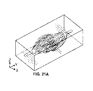
[0070] FIGs. 21A-21G depict an interconnected vascular network,
according one or more
embodiments.
[0071] Throughout the figures, similar numbers are typically
used for similar components.
DETAILED DESCRIPTION OF THE INVENTION
[0072] A key limitation of the existing AM methods is the inability to
construct complex
networks in all three dimensions within a range of materials. Liquid-phase
materials
deposited via extrusion, such as those employed in the above AM methods, are
subject
to deformation or collapse under their own weight. Furthermore. the viscosity
of the
liquids makes precisely dispensing small volumes quite challenging (as may be
required
to form omnidirectional structures in a layer-by-layer fashion). These
intrinsic
rheological limitations prohibit extrusion-based AM techniques, such as those
described
above, from forming architectures which include unsupported overhangs,
underhangs,
and/or arbitrary three-dimensional branching, all of which are hallmarks of
the
mammalian vasculature. Moreover, the 90-degree channel junctions, formed as a
result
of extruded rectilinear networks, have markedly different fluid dynamics than
the
structures found in nature. The altered fluid dynamics of networks formed
using existing
AM techniques have undesirable implications for hemodynamics, shear stresses
experienced by endothelial cells, and the overall fluidic resistance of the
network.
[0073] In contrast, the sugar alcohol isomalt was discovered to be
compatible with
selective laser sintering (SLS) and undergoes a stable melting transition to
form
contiguously fused solid filaments. Accordingly, embodiments described herein
may be
directed toward compositions of matter and methodologies that may be employed
to
generate patterned SLS, including a workflow for fully automated fabrication
of three-
dimensional structures from various carbohydrate powders.
9
CA 03174427 2022- 9- 30
WO 2021/237163
PCT/US2021/033763
[0074] Further, SLS of carbohydrates as described herein is a novel
approach to additive
manufacturing of this class of materials. The method disclosed in embodiments
herein
offers significant improvement over previous methods of fabricating
carbohydrate
structures, such as improved resolution, structural complexity,
reproducibility, and
throughput.
[0075] To that end, some embodiments may be directed to SLS patterned
structures in the
form of a solid, contiguous filament or a solid, contiguous three-dimensional
structure,
such as a three-dimensional dendritic carbohydrate lattice or solid,
contiguous filament
networks. In some embodiments, the patterned structures may be used to
template a
matrix material. In some embodiments, the patterned structures may be used to
cast
vascularized engineered tissues (e.g., the structures may be described as
matrices with
embedded perfusable vascular networks). Furthermore, some embodiments of this
disclosure may be directed toward a composition of matter and a method for
fabricating
engineered vascular networks unconstrained by the limitations of existing
extrusion
printing techniques such as those described above. Embodiments of the
composition of
matter may be compatible with SLS and undergo a stable melting transition to
form
contiguously fused solid filaments. Further, embodiments described herein may
include
a workflow for fully automated fabrication of three-dimensional structures
from various
carbohydrate powders. Finally, one or more embodiments are directed toward a
method
called Mutual Tree Attraction for computationally generating a dendritic
substantially
interconnected vascular network.
[0076] Broadly, FIGs. 2A-2F briefly depict steps for forming a
substantially
interconnected vascular network that may be used in embodiments of the method.
As in
FIGs. 2A-2F, embodiments of the method may include the steps of: solidifying
powder
granules by sintering or melting with an energy beam to form a three-
dimensional
structure to be used as a template; surface smoothing the template with a
smoothing
solution; surface coating the template with a surface coating material;
backfilling a void
space of the template with a matrix material; crosslinking the matrix
material; and
removing the template to form the patterned structure having channels shaped
like the
template. This method has been termed Selectively Laser Sintered-Carbohydrate
Sacrificial Templating (SLS-CaST). Furthermore, each of the steps briefly
depicted in
FIGs. 2A-2F will also be discussed in greater detail below.
CA 03174427 2022- 9- 30
WO 2021/237163
PCT/US2021/033763
[0077] In some embodiments, SLS may be used to form a three-dimensional
structure by
tracing a 2D pattern with an energy beam focused onto a layer of the powder.
Embodiments of the powder may include one or more carbohydrate powder(s)) and
may
fuse together to form a structural material. FIG. 2A depicts granules of a
powder 1 being
fused together using an SLS system 2 via sintering or melting with a laser 3
to form a
filament network 5. The shape drawn by laser 3 may be controlled by the
movement of
stage 6, movement of laser 3, or both.
[0078] In some embodiments, a three-dimensional structure formed of a
structural material
via SLS may be used as a template. Initially after formation, the surface of
the three-
dimensional structure may be rough. FIG. 2B depicts filament network 5 after
removal
from the 3D printing system. Void space 7 can be seen surrounding filament
network 5,
including between filaments 9. Thus, filament network 5 defines void space 7.
A surface
11 of filament network 5 is rough.
[0079] In some embodiments, the surface of the filament network may be
smoothed with
a smoothing solution. FIG. 2C depicts filament network 5 after undergoing
surface
smoothing to decrease the roughness of surface 11. Surface smoothing caused
filament
network 5 to become a smoothed structure.
[0080] In some embodiments, the surface of the filament network may be
surface coated
with a surface coating material. FIG. 2D depicts filament network 5 after a
surface
coating material has formed a coating 13 on surface 9 of filament network 5.
However,
coating 13 does not backfill void space 7.
[0081] In some embodiments, the void space surrounding the filament
network is
backfilled with a matrix material. FIG. 2E depicts a matrix material 15
surrounding
filament network 5 and backfilling void space 7. After introduction around the
filament
network, some embodiments of the matrix material may require cros slinking or
other
polymer processing to solidify within the void spaces.
[0082] In some embodiments, the filament network may be removed to form
the
substantially interconnected vascular network. The form of the final vascular
network
may include fluidic channels through the matrix material that reflect the
geometry of the
filament network. FIG. 2F depicts a substantially interconnected vascular
network 17
11
CA 03174427 2022- 9- 30
WO 2021/237163
PCT/US2021/033763
that includes fluidic channels 19 defined by coating 13 and matrix material
15. Arrows
21 indicate one potential fluid flow direction through vascular network 17.
[0083] A Powder, per One or More Embodiments
[0084] Embodiments of the powder may include one or more carbohydrate
powders.
Embodiments of the powder may further include pigment(s), anti-caking
agent(s), or
both. Some embodiments of the powder may have the ability to flow, as may be
quantified using the specific energy. Embodiments of the powder may be in
granular
form having a large number of powder granules having particular geometric
characteristics (e.g., average diameter). Each of these factors of the powder
are detailed
below.
[0085] Embodiments of the powder may include one carbohydrate powder or
a mixture of
two or more carbohydrate powders. The carbohydrate powders may include or
consist
of, for example: photoresist, agarose, gelatin, carbohydrates, sucrose,
glucose, fructose,
lactose, isomalt, dextran, cellulose, methylcellulose, poly(lactic acid),
and/or
poly(ethylene glycol).
[0086] As discussed above, prior work used sugars formed via melt
extrusion for
sacrificial templating; however, the results were brittle and the final three-
dimensional
shapes were limited. In contrast, isomalt (a sugar alcohol frequently used as
an artificial
sugar substitute) and dextran were found to be compatible with SLS and to
undergo a
stable melting transition to form contiguously fused solid filaments. Thus,
isomalt and
dextran may be used to form three-dimensional structures, including complex
three-
dimensional structures such as vascular architectures. Thus, in some
embodiments, the
powder may include one or both of isomalt and dextran powders.
[0087] In some embodiments, the powder may have the ability to flow for
dispensing
during SLS. Specific energy is one measure of powder flowability. To that end,
some
embodiments of this disclosure utilize a powder, with or without an anti-
caking agent,
having a specific energy of less than 6 millijoules per milliliter (mJ/mL)
(e.g., less than
mJ/mL or less than 4 mJ/mL). Put another way, some embodiments of this
disclosure
utilize a powder, with or without an anti-caking agent, having a specific
energy of
between 6 mJ/mL and 0.1 mJ/mL (e.g., between 5 mJ/mL and 0.1 mJ/mL, between 4
mJ/mL and 0.1 mJ/mL, or between 2 mJ/mL and 0.1 mJ/mL).
12
CA 03174427 2022- 9- 30
WO 2021/237163
PCT/US2021/033763
[0088] The addition of an anti-caking agent may effectively augment
powder flow while
preserving sintering quality. To that end, in some embodiments, one or more
anti-caking
agents may improve the characteristics of a powder compared with a similar
powder
lacking an anti-caking agent, for example, flow, flowability, friction
characteristics,
and/or particle packing of the structural material.
[0089] Thus, according to some embodiments, the powder for SLS may be a
mixture of
one or more carbohydrate powder(s) and one or more anti-caking agent(s). In
some
embodiments, the anti-caking agent may include, but is not limited to, one or
more of:
cornstarch, silicon dioxide, or xanthan gum or a mixture of two or more anti-
caking
agents.
[0090] Nylon is a standard material for SLS because it has favorable
powder rheology
properties; however, nylon may be undesirable for a particular application,
for example
because it is insufficiently biocompatible, not readily dissolvable, or
another reason.
Accordingly, FIG. 3 compares the specific energy of nylon with that of isomalt
and of a
mixture comprising 30 mass percent ( wt%) cornstarch in isomalt (referred to
hereinafter
as "isomalt + cornstarch"), according to one or more embodiments of the
disclosure. As
shown, isomalt powder granules have a higher specific energy and thus are much
more
cohesive than standard SLS materials such as nylon, which may manifest as poor
powder
distribution or uneven layers. FIG. 3 also shows that the isomalt + cornstarch
mixture
has comparable flowability to nylon. Thus, isomalt + cornstarch is able to
free-flow like
nylon. The data for isomalt and isomalt + cornstarch have a probability value
(p-value)
of p < 0.05 for a t-test with the sample size (n), n=3.
[0091] FIGs 4A-4C are scanning electron micrographs of nylon (FIG. 4A),
isomalt (FIG.
4B), and cornstarch (FIG. 4C) that illustrate the varying powder morphology
and help to
explain differences in powder flowability. Nylon is an extremely free-flowing
powder
used in conventional polymer SLS processes. As shown in FIG. 4A, nylon has a
relatively
small, smooth, and regular morphology. In contrast, isomalt powder is
irregular,
polydisperse, and jagged as shown in FIG. 4B. Consequently, isomalt powder has
higher
powder cohesion and relatively poor flowability compared with nylon. As
discussed
above, the addition of cornstarch significantly improves the flowability of
powdered
isomalt. FIG. 4C shows powdered cornstarch. The small, smooth cornstarch
particles are
13
CA 03174427 2022- 9- 30
WO 2021/237163
PCT/US2021/033763
hypothesized to intercalate between the large, jagged isomalt grains to reduce
friction and
resistance to flow.
[0092] The inclusion of a pigment may allow the powder to more
efficiently absorb the
radiation from the energy beam (e.g., laser) during SLS. Additionally,
inclusion of a
pigment may also enable successful fabrication using a visible spectrum energy
beam.
Thus, some embodiments of the powder may include one or more pigment. In some
embodiments, the pigment may be a biocompatible pigment. In some embodiments,
the
bioconapatible pigment may include one or more of: tartrazine, anthocyanin,
and Altura
Red AC (Red 40). As with the anti-caking agent discussed above, the pigment
may be
incorporated into the final three-dimensional structure upon SLS.
[0093] According to some embodiments, a particle size described herein
may be defined
as an average particle diameter, a mean particle diameter, a median particle
diameter, a
mode of the particle diameter, a weighted mean of the particle diameter, a
mean Feret
diameter, a Sauter mean diameter, a maximum of a probability density function
of the
particle diameter (using any applicable mathematic model, such as a log-normal
distribution. a Weibull distribution, a Rosin-Rammler distribution, a log-
hyperbolic
distribution, a skew log-Laplace model distribution, and additional models
derived
therein), or some other statistical metric that may be used to determine
and/or define the
diameter of the particles.
[0094] In one or more embodiments, a particle size of the powder (for
example, measured
as the mean Feret diameter or another way as discussed above) may be less than
250
micrometers ( m) diameter (e.g., less than 200 pin, less than 100 m, less
than 50 pin,
less than 10 m, etc.). Put another way, some embodiments of this disclosure
utilize a
powder with a particle size of between 250 m and 0.01 m (e.g., between 200
na and
0.01 m, between 100 rn and 0.01 m, between 50 m and 0.01 rn, or between
10 rn
and 0.01 pm).
[0095] In some embodiments, milling, grinding, mechanically separating,
and/or sieving,
may be performed to ensure the powder has an intended particle size and/or
particle size
distribution. Such particle size operations may be performed on one or more
components
of the powder separately (i.e., before mixing), after combining two or more
components
of the powder, or both. Furthermore, in some embodiments, a powder may be a
mixture
14
CA 03174427 2022- 9- 30
WO 2021/237163
PCT/US2021/033763
of powder grains of carbohydrate(s) with or without anti-caking agent(s) that
have been
milled and/or ground to reduce their grain size and/or particle side prior to
sintering.
[0096] In some embodiments, the powder may also be separated, by
mechanical or other
means, to select powder grains below a threshold size. One such method for
separating
powders by size may be sieving, but alternative methods may also be employed.
When
the powder includes two or more component powders (i.e., carbohydrate
powder(s) and/or
anti-caking agent(s)), these particle processing methods may be performed
before and/or
after mixing the component powders.
[0097] In some embodiments, the particle size in the powder may impact
a powder layer
height of a single layer, and the powder layer height may impact the final
resolution of
the three-dimensional structure. If the powder layer height of each additional
layer of
un-sintered powder is too thick (i.e., contains too much powder), the powder
may fail to
fully fuse together. Powder that is not fully fused may not be properly added
to the three-
dimension structure as it grows, thus the process may fail to form a
contiguous three-
dimensional structure. In contrast, if each newly-added layer is too thin
(i.e., does not
contain enough powder), the laser may over-sinter the previous layer, which
may
undesirably alter the final geometry.
[0098] The powder may be able to free-flow to form layers with a powder
layer height
(when in a granular form) that may be successfully formed into a structural
material via
sintering, according to one or more embodiments. In some embodiments, the
powder
layer height may be less than 250 gm (e.g., less than 225 gm, less than 210
gm, less than
200 gm, less than 175 gm, less than 150 gm, etc.). In some embodiments, the
powder
layer height may also be greater than 100 gm (e.g., greater than 110 gm,
greater than
120 gm, greater than 125 gm, greater than 150 gm). In some embodiments, the
powder
layer height may be in the range from 100 gm to 250 gm (e.g., in the range
from 100 gm
to 225 gm; in the range from 125 gm to 225 gm; in the range from 125 gm to 200
gm;
in the range from 100 gm to 200 pm; in the range from 100 gm to 175 gm, etc.).
[0099] Solidifying a Powder by Sintering or Melting, per One or
More Embodiments
[00100] SLS is a versatile form of 3D printing that may be used to form
a structural material
by tracing a two-dimensional pattern with an energy beam focused onto a layer
of a
powder that may be employed in some embodiments. Some embodiments of the
method
CA 03174427 2022- 9- 30
WO 2021/237163
PCT/US2021/033763
may use SLS to solidify a powder, for example a powder discussed above. In
some
embodiments, the powder may be sintered or melted with to form a solid of a
structural
material, which may be in the shape of a three-dimensional structure. In some
embodiments, this three-dimensional structure may be used as a template. In
some
embodiments, the energy beam used for SLS may be a laser beam. The SLS process
and
characteristics of the resulting structural material arc detailed below.
[00101] In some embodiments, the powder may become fused during SLS
(for example,
melted or sintered due to heat) to form the structural material as it absorbs
the
electromagnetic radiation transmitted via the energy beam. To that end, in
some
embodiments, the structural material is formed when the powder is irradiated
with an
energy beam. Thus, a path of the energy beam in two dimensions may cause a
region of
the powder to fuse, forming the structural material. Patterning sequential
layers in a third
dimension may fuse the structural material together in all three dimensions to
form a
three-dimensional structure. Thus, the three-dimensional structure may be
fabricated by
repeating this process in a layer-by-layer fashion, whereby an un-sintered
layer of the
power system may be spread over a previously-fused layer(s) and may be
patterned by
being exposed to the energy beam so as to add to the three-dimensional
structure.
[00102] FIGs. 5A-5D illustrates a schematic of a three-dimensional
printing process via
SLS, according to one or more embodiments. Each of these steps are briefly
discussed
below and detailed further.
[00103] FIG. 5A depicts a digital rendering 23 performed by a computer
25 of a three-
dimensional structure to be printed.
[00104] FIG. 5B depicts digital rendering 23 in computer 25 being
converted into three two-
dimensional digital slices 27. These digital slices 27 correspond to where the
laser may
solidify the powder into the three-dimensional structure during SLS.
[00105] FIG. 5C depicts powder dispensing as part of layer-by-layer
fabrication of a
filament network via SLS.
[00106] Here, an SLS system 2 has a stage 6, powder reservoir 29, and a
sieve 31. SLS
system 2 has partially formed a filament network 5. Powder reservoir 29 stores
powder
1. Sieve 31 dispenses a powder layer 33 of powder 1 onto stage 6 in
preparation for
16
CA 03174427 2022- 9- 30
WO 2021/237163
PCT/US2021/033763
fusion/melting with an energy beam. Stage 6 may be a z-stage, an x-y stage, or
an x-y-z
stage (where the z-direction is defined as the direction of flow of powder
reservoir 29
from sieve 31).
[00107] Although not depicted here, sieve 31, stage 6, or both may be
controlled by a
microelectronic controller. A microelectronic controller may control one or
more aspects
of SLS 2, for example a flow rate of powder 1 onto stage 6 or the location
powder 1 is
dispensed onto stage 6. Such a microelectronic controller may control one or
more
aspects of powder layer 33, for example, powder layer 33 thickness. Additional
details
of microcontroller(s) within SLS system 2 are discussed further.
[00108] FIG. 5D depicts laser patterning as part of layer-by-layer
fabrication of a filament
network via SLS.
[00109] Here, SLS system 2 also includes a laser 3. Laser 3
sinters/fuses powder layer 33
to add to filament network 5.
[00110] Although not depicted here, laser 3, stage 6, or both may be
controlled by a
microelectronic controller. A microelectronic controller may control one or
more aspects
of SLS 2, for example the various characteristics of laser 3 or the location
laser 3 is
sintering powder layer 33. Such a microelectronic controller may control one
or more
aspects of powder layer 33, for example, powder layer 33 thickness. Additional
details
of microcontroller(s) within SLS system 2 are discussed further.
[00111] Prior to beginning the SLS fabrication, a 3D model may be
digitally rendered using,
in some embodiments, a computer-aided design (CAD) software. Such a rendering
may
then be exported into a file, such as a ".STL" file, for example, compatible
with a
computer-aided 3D printing platform, termed an SLS system. This SLS file may
assist
in translating the 3D model from CAD into precise instructions required for
the SLS
system's various tasks outlined above. One such format, according to some
embodiments, may be a stereolithography (.stl) format.
[00112] The SLS file may also be processed by additional software which
may transfoun
the 3D model by slicing it into a series of 2D cross-sectional slices, each of
which may
be used to pattern, for example, one layer of the powder. In some embodiments,
parameters may be specified during this slicing process that govern the
behavior of the
17
CA 03174427 2022- 9- 30
WO 2021/237163
PCT/US2021/033763
SLS system during sintering. In some embodiments, a speed and/or a power of
the energy
beam and/or a height of each of the layers may be set and/or customized by one
or more
software programs. The series of 2D slices may be represented as a series of
instructions
for the SLS system, expressed in a final format that may be, in some
embodiments, the
G-code language. The series of instructions may, in some embodiments, move the
energy
beam to a specific point or along a specific path, lower the print platform,
and/or move
the powder distributor, along with any other instructions. In some
embodiments, the
series of instructions for the SLS system may be further modified by a custom-
developed
program that may optimize the instructions specifically for SLS of
carbohydrate
powder(s) to ensure proper interpretation by the SLS system and/or may allow
energy
beam power density/energy beam scan speed settings to be specified.
[00113] Production of a three-dimensional structure made of a
structural material from a
powder that includes carbohydrate powder(s), according to embodiments this
disclosure,
may be performed with an SLS system. This SLS system may be an automated,
computer-aided 3D printing platform which uses additive manufacturing to
create the
three-dimensional structures from the powder in the layer-by-layer fashion
according to
the series of instructions. To precisely control the spatial positioning of
the energy beam,
the energy beam of the SLS system may be mounted to a gantry or other such
mechanism.
According to some embodiments, the SLS system may also include a print
platform that
moves freely in a z-direction so it may, for instance, advance downwards after
a layer of
the powder has been sintered. In some embodiments, a next layer of the powder
may be
formed atop the print platform by depositing a metered amount of the un-
sintered powder
from a powder hopper and then smoothing said metered amount of the un-sintered
powder using a distributor.
[00114] The SLS process may be coordinated by a microelectronic
controller in the SLS
system, which controls the motors, valves, mechanisms, etc. responsible for
various
tasks. In some embodiments, those tasks may include, for example, any of:
positioning,
aiming, and controlling the output of the energy beam; positioning and moving
the print
platform; positioning and controlling the mechanism of the powder hopper,
positioning
and engaging the distributor; and other tasks for fusing the powder into a
solid.
18
CA 03174427 2022- 9- 30
WO 2021/237163
PCT/US2021/033763
[00115] Solidification using an SLS set may begin with a first layer of
the powder that is
manually deposited on the print platform and leveled by the user, in some
embodiments.
Once the first layer of powder is on the print platform, the series of
instructions may be
executed in succession such that the first layer of the powder may be
patterned by the
energy beam (e.g., the laser), followed by a second layer of powder that may
be deposited
from the hopper, and so forth. Alternatively, in some embodiments, the first
layer may
be mechanically metered and distributed by the SLS system such that
distributing the
first layer is a first instruction of the series of instructions.
[00116] In some embodiments, the powder may be added for the subsequent
layer by
shaking a sieve connected to a powder reservoir suspended above the stage.
Some
embodiments may employ a shaking motion (for example, of the sieve) to
dispense the
powder. Such a shaking motion may aerate the powder and/or reduce compaction
of the
powder. After dispensing the powder from the suspended reservoir, in some
embodiments, a dispensed quantity of the powder may be spread into a powder
layer
having an approximately equal layer thickness. Distribution of the dispensed
quantity of
the powder may be performed by a counter-rotating roller, for example.
Finally, in some
embodiments, excess powder (meaning, more than is required to form the powder
layer)
may be removed (for example, by a plow mechanism) and collected for
redistribution.
Excess powder may be redistributed many times (for example, tens to hundreds
of times)
without a noticeable decrease in print quality.
[00117] The SLS system may alternate between patterning a last layer of
the powder and
depositing a next layer of the powder until a final geometry has been
sintered.
[00118] The appearance and quality of a three-dimensional structure
formed via SLS may
be influenced by various factors, including: laser power density, laser
scanning speed,
and/or powder layer height. Proper control of these settings, according to one
or more
embodiments, may allow for consistent sintering of powders to create the three-
dimensional structure. Improper values (or combinations of values) of factors
such as
these may result in the final geometry differing from an intended three-
dimensional
structure by lowering the resolution of features, adding unintended features,
subtracting
intended features, failing to fully fuse powder, creating balling defects,
distorting the
19
CA 03174427 2022- 9- 30
WO 2021/237163
PCT/US2021/033763
final three-dimensional structure, adding cavities, and/or other undesired
alterations to
the intended final geometry.
[00119] One having skill in the art will realize laser power density
and laser scanning speed
may be interrelated. For instance, a low-powered laser moving with a slow
scanning
speed may still impart excessive power to a region. Excessive power may cause
over-
sintering, distortions, cavities, and/or other undesired alterations to the
intended final
geometry.
[00120] Over-sintering is when particles lying outside of the intended
pattern are fused
along with particles within the intended pattern being fused to form the
intended final
geometry. Over-sintering may occur in the z-axis (i.e., the build axis) and/or
the x-axis/y-
axis (i.e., the planar axes). In some embodiments, over-sintering may cause
excessive
fusion between successive powder layers, which may lower the resolution of the
final
geometry along the build axis and/or may add unintentional features in the
build and/or
planar axes.
[00121] When the laser power density is too high and/or the laser
scanning speed is too low,
undesired alterations to the final geometry like over-sintering may occur.
Additionally,
low laser scanning speed may cause an irregular melt pool while the powder is
sintered.
The irregular melt pool may lead to additional undesired alterations to the
intended final
geometry, such as distortions and/or cavities.
[00122] Alternatively, when the laser power density is too low and/or
the laser scan speed
is too fast, the powder may fail to foim a continuously fused final three-
dimensional
structure. These circumstances may, in some embodiments, cause the balling
defect that
may sometimes be seen in SLS: when insufficiently melted, some powder may ball
up
into disconnected spheres instead of forming the final three-dimensional
structure.
[00123] In some embodiments, for a given powder, the width of a
filament may be a
function of laser power, translation speed, or both. Thus, some embodiments
may control
the laser power, translation speed, or both to control filament dimensions.
[00124] In some embodiments, there may be an approximately linear
relationship with a
negative correlation between translation speed and filament diameter. FIGs. 6
and 7
evidence an embodiment where the relationship between translation speed and
filament
CA 03174427 2022- 9- 30
WO 2021/237163
PCT/US2021/033763
diameter is approximately linear with a negative correlation. The negative
correlation is
apparent in FIGs . 6 and 7 in that the filament width decreases as the laser
speed increases.
FIG. 6 is a photograph of a three-dimensional structure comprising multiple
filaments
fabricated at a fixed power density of 45 watts per square millimeter (W mm-
2). Upon
increasing the translation speed of each successive filament from 500 to 2,750
millimeters per minute (mm min-1), the diameter of the filaments decreased
from 800 to
400 1.trn (scale bar = 1 mm). FIG. 6 is a representative image of five images
captured
following the fabrication of each three-dimensional structure. FIG. 7
numerically graphs
the filament width versus the laser speed for those five three-dimensional
structures
(including FIG. 6). This graph shows an approximately linear relationship with
a
negative correlation between translation speed and filament diameter in a
regime
spanning 400-800 pm at a fixed power density of 45 W mm-2. The graph shows the
average filament width and the standard deviation for n = 5 print runs.
[00125] In some embodiments, the laser power density may be between in
the range from
20 to 100 W/mm, such as from 40 to 60 W/mm2 (e.g., in the range from 45 W/mm2
to
60 W/mm2, in the range from 40 W/mm2 to 55 W/mm2, in the range from 45 W/mm2
to
55 W/mm2, in the range from 40 W/mm2 to 50 W/mm2, or in the range from 50
W/mm2
to 60 W/mm2).
[00126] In some embodiments, the laser scanning speed may be in the
range from 200 to
4000 mm/min, such as from 1000 mm/min to 2000 mm/min (e.g., in the range from
1250
mm/min to 2000 mm/min, in the range from 1250 mm/min to 1750 mm/min, in the
range
from 1000 mm/min to 1750 mm/min, in the range from 1250 mm/min to 2000 mm/min,
in the range from 1500 mm/min to 2000 mm/min, or in the range from 1000 mm/min
to
1500 mm/min).
[00127] Using the SLS system, according to one or more embodiments,
three-dimensional
structures exhibiting heterogeneous three-dimensional branching, smooth
curvature, and
unsupported geometry may be fabricated. For example, FIGs. 8A and 8B (scale
bars =
mm) are photographs of three-dimensional structures formed of the structural
material
that were fabricated using SLS of a powder comprising a mixture of isomalt and
30 mass
percent (w%) cornstarch material, according to one or more embodiments. The
structures
21
CA 03174427 2022- 9- 30
WO 2021/237163
PCT/US2021/033763
of FIGs. 8A and 8B exhibit smooth curvature, hierarchical branching, and
unsupported
overhangs, all of which are architectural motifs of the mammalian vasculature.
[00128] A resolution of a final geometry of a three-dimensional
structure, such as an
interconnected vascular network produced via one or more embodiments described
herein, may be limited by a resolution of an SLS system. The resolution of the
SLS
system may be limited by factors such as optics of an energy beam (e.g., a
laser spot
size), which may define an approximate minimum diameter of a sintered filament
formed
according to one or more embodiments. Alternative optics may be utilized to
reduce the
laser spot size, which may produce the final geometry that includes smaller
filaments,
according to some embodiments. Additionally, resolution of the SLS system may
be
limited by the thickness of the powder layers, which may be limited by the
diameter of
the powder. Thus, thinner powder layers, and accordingly more finely ground
powder,
may produce the final geometry that includes smaller filaments, according to
some
embodiments.
[00129] In some embodiments, a filament may have a minimum diameter of
300 pm (for
example, 200 pm, 100 pm, 50 pm, 10 pm, or 1 pm). In some embodiments, a
filament
(formed by a single pass of the energy beam) may have a maximum diameter of 1
mm
(for example, 5 mm, 10 mm, 50 mm, 100 mm, or 500 mm).
[00130] As in the embodiments depicted in FIGs. 8A and 8B, three-
dimensional structures
formed from a powder containing an anti-caking agent may include both the anti-
caking
agent and the carbohydrate powders in the final structure. Put another way, in
some
embodiments, energy beam irradiation as occurs during SLS of a powder
including
carbohydrate powder(s) and anti-caking agent(s) may sinter and/or melt both
the
carbohydrate powder(s) and the anti-caking agent(s) during solidification into
the final
three-dimensional structure.
[00131] Sintered structural materials fabricated of isomalt or isomalt
+ cornstarch as
depicted here, according to one or more embodiments, are stiff and brittle,
with Young's
modulus on the same order of magnitude as extruded carbohydrate glass. Such
structural
materials may be robust enough to support their own weight, endure multiple
post-
proces sine steps, and/or be shipped between labs.
22
CA 03174427 2022- 9- 30
WO 2021/237163
PCT/US2021/033763
[00132] As shown in FIG. 9 and summarized in the table below, sintered
carbohydrates are
stiff and brittle under compression, making them self-supporting and amenable
to
extensive handling, multiple coaling steps, and casting inside viscous pre-
polymer
solutions.
[00133] FIG. 9 depicts a graph of stress (in megapascal (MPa)) versus
strain (in percent
elongation (%)) collected during uniaxial compression for a solid carbohydrate
cylinder
and a macroporous carbohydrate cylinder according to an embodiment of this
method.
FIG. 9 also depicts a schematic of each geometry as an inset located adjacent
to the
corresponding stress vs strain curve. Young's modulus (in MPa) was measured as
the
slope of the stress-strain curve in the linear region, yield stress (in MPa)
was measured
as the peak stress value before failure, and yield strain (%) was measured as
the
corresponding strain. The accompanying results in the table summarize n=11
solid
cylinders and 9 cylindrical lattices fabricated across three independent print
runs.
Solid Cylinder
Cylindrical Lattice
Young's Modulus (MPa) 580 200 640 280
Yield Stress (MPa) 19 9.0 30
4.9
Yield Strain (%) 6.9 1.4
2.9 0.7
L
[00134] A three-dimensional structure, solidified according to one or
more embodiments,
may be able to support its own weight and may be brittle and stiff. In some
embodiments,
a three-dimensional structure fabricated of the structural material may have a
Young's
modulus in the range from 200 MPa to 1000 MPa, (for example, in the range from
500
MPa to 1000 MPa, in the range from 500 MPa to 700 MPa), for example
approximately
600 MPa.
[00135] In some embodiments, a three-dimensional structure formed of a
structural material
may take the form of a filament network that may be formed of a plurality of
filaments,
a three-dimensionally branched pattern, an interpenetrating geometry, and/or
an
unsupported geometry.
23
CA 03174427 2022- 9- 30
WO 2021/237163
PCT/US2021/033763
[00136] The materials and/or methods, according to one or more
embodiments of this
disclosure, may be applied to fabricate a three-dimensional structure and thus
a final
geometry that may include various freeform structures and/or patterned fluidic
networks.
[00137] The structural material may be capable of dissolving and/or
disintegrating upon
interaction with a liquid, according to one or more embodiments. In some
embodiments,
the liquid may include, but is not limited to, or may utilize one or more of,
water, saline,
or phosphate buffered saline (PBS). After solidification, the three-
dimensional structure
may be ready for use as a template for sacrificial templating, according to
one or more
embodiments discussed below.
[00138] The three-dimensional structure may be environmentally stable
such that it may be
stored for multiple weeks before use in sacrificial templating, in one or more
embodiments.
[00139] Surface Smoothing, per One or More Embodiments
[00140] Following SLS according to embodiments of this disclosure, the
surfaces of a
three-dimensional structure may initially have a high surface roughness. This
surface
roughness may be due, in part, to decoration of the three-dimensional
structure with
loosely attached granules of the powder. To remove these loosely attached
granules
and/or to reduce the surface roughness, the three-dimensional structure may
undergo
surface smoothing with a smoothing solution. The smoothing solution may
include one
or more carbohydrates. The smoothing solution may include solvent(s). Contact
between the smoothing solution and the three-dimensional surface may be
tailored.
Following surface smoothing according to some embodiments, the surface
roughness of
the three-dimensional structure may be decreased due to a smoother surface
topography.
In some embodiments, surface smoothing may alter the size and/or the mass of
the three-
dimensional structure. Some embodiments of the surface smoothing may deposit
an
additional carbohydrate coating on the outside of the three-dimensional
structure.
Aspects of the smoothing solution are detailed below.
[00141] In some embodiments, the smoothing solution may include a
carbohydrate. The
carbohydrate used for the smoothing solution may include, but is not limited
to, isomalt,
dextran, sucrose, glucose, lactose, trehalose, maple syrup, and/or sugar cane
syrup, in
some embodiments. Additionally, some embodiments of the smoothing solution may
24
CA 03174427 2022- 9- 30
WO 2021/237163
PCT/US2021/033763
include two or more carbohydrates. The carbohydrate(s) used in the smoothing
solution
may or may not be in the powder used to fabricate the three-dimensional
structure to be
treated.
[00142] The smoothing solution according to some embodiments may
include one or more
solvents. The solvent may include, but is not limited to, water and/or an
organic solvent,
in one or more embodiments.
[00143] In some embodiments, the smoothing solution may have a
sufficiently high
concentration of the one or more carbohydrates included in the smoothing
solution such
that the smoothing solution does not dissolve or undesirably alter the three-
dimensional
structure.
[00144] The three-dimensional structure may be contacted with the
smoothing solution via
a washing, spraying, or dipping method, in some embodiments. Contact between
the
three-dimensional structure and the smoothing solution may continue for a
contact time.
In some embodiments, contact time and/or contact method may be tailored so the
contact
is sufficient to remove the loose granules and smooth the surface of the three-
dimensional
structure. In some embodiments, contact time and/or contact method may be
tailored so
the contact does not dissolve or undesirably alter the three-dimensional
structure.
[00145] For example, FIGs. 10A and 10B are scanning electron
micrographs of a three-
dimensional structure formed via SLS before surface smoothing (FIG. 10A) and
after
surface smoothing (FIG. 10B) according to embodiments herein.
[00146] FIG. 10A is a scanning electron micrograph of a three-
dimensional structure having
a shape of a filament formed via SLS of a powder according to one or more
embodiments
discussed previously. Upon visual inspection, FIG. 10A shows significant
surface
roughness. This surface roughness may be due to partially fused powder
granules
decorating the surface. The scale bars are 200 pm (magnified view) and 1 mm
(inset).
[00147] FIG. 10B is a scanning electron micrograph of the same filament
after surface
smoothing with a smoothing solution. Specifically, the filament depicted in
FIG. 10A
was treated with a smoothing solution consisting of a concentrated isomalt
solution (60
weight percent (wt%) isomalt in water), according to embodiments of this
disclosure.
Following surface smoothing, the filament was re-imaged as FIG. 10B. Upon
visual
CA 03174427 2022- 9- 30
WO 2021/237163
PCT/US2021/033763
inspection, FIG. 10B shows low to moderate surface roughness. As in FIG. 10A,
the
scale bars for FIG. 10B are 200 pn (magnified view) and 1 mm (inset).
[00148] In one or more embodiments, the surface smoothing may leave the
three-
dimensional structure largely intact. In one or more embodiments, surface
smoothing
may not significantly alter the overall architecture. In one or more
embodiments, surface
smoothing may not significantly bridge across regions of the three-dimensional
structure.
In one or more embodiments, the surface smoothing may decrease surface
roughness. In
one or more embodiments, the surface smoothing may remove loose granules of
the
powder from the three-dimensional structure. The above features are evidenced
by
comparing the micrographs of a filament taken before (FIG. 10A) and after
(FIG. 10B)
surface smoothing according to some embodiments. Specifically, comparing FIGs.
10A
and 10B shows surface smoothing decreasing the surface roughness without
significantly
altering the overall architecture or bridging across filaments.
[00149] In addition to the visually evident improvement of the filament
surface roughness
seen in FIGs. 10A and 10B, some embodiments of the surface smoothing may alter
the
filament in one or more of the following quantifiable ways: reducing the
surface
roughness, reducing the filament width, or increasing the filament mass. FIGs.
11A-11D
numerically depict the impacts of surface smoothing on surface roughness (Ra)
(FIG.
11A), filament width (FIG. 11B and 11C), and filament mass (FIG. 11D),
according to
embodiments of this disclosure. The data depicted in FIGs. 11A, 11B, and 11D
is the
mean standard deviation, for n=9 (FIG. 11A), n=24 (FIG. 11B), and n=7 (FIG.
11D).
For FIGs. 11A and 11B, p<0.001 for a paired t-test. For FIG. 11C, p<0.01 for a
paired
t-test.
[00150] Some embodiments of the surface smoothing may result in a
reduced average
surface roughness. To that end, FIG. 11A shows a nearly two-fold reduction of
Ra for
filaments following surface smoothing according to embodiments of this
disclosure.
[00151] Some embodiments of the surface smoothing may result in a
reduced size of the
three-dimensional structure. In some embodiments, as in FIG. 11B, such a size
change
may reflect measurements of the width of a filament before and after surface
smoothing.
To that end, FIG. 11B shows a reduction of the filament width of between 100
inn and
200 lam following surface smoothing according to embodiments of this
disclosure. Some
26
CA 03174427 2022- 9- 30
WO 2021/237163
PCT/US2021/033763
embodiments may deploy alternative metrics to quantify the size change that
may depend
on the geometry of the three-dimensional structure. Furthermore, FIG. 11C is a
graph of
the change in filament width (in pm) from surface smoothing as a function of
laser speed
(in mm/min). FIG. 11C shows that surface smoothing reduces the diameter of
filaments
by an approximately constant amount. roughly 100 pm.
[00152] Some embodiments of the surface smoothing may result in an
increased mass of
the three-dimensional structure. To that end, FIG. 11D shows an increase of
the filament
mass of between 10% and 20% following surface smoothing according to
embodiments
of this disclosure.
[00153] In some embodiments, surface smoothing may deposit an
additional carbohydrate
coating on the outside of the three-dimensional structure. This added
carbohydrate
coating may have the same or a different composition compared to the three-
dimensional
structure prior to surface smoothing.
[00154] In some embodiments, the three-dimensional structure may
undergo surface
smoothing via exposure to the smoothing solution as discussed above before
undergoing
further steps. In other embodiments, the three-dimensional structure may not
have
undergone surface smoothing before undergoing further steps. A three-
dimensional
structure, according to one or more embodiments, may or may not further
include a
smoothed surface as described above.
[00155] Surface Coating, According to One or More Embodiments
[00156] To prevent premature dissolution of a three-dimensional
structure and/or to
establish a barrier between the three-dimensional structure and cells that may
be included
in a matrix material, one or more embodiments of the method include surface
coating the
three-dimensional structure with a surface coating material. Therefore.
according to one
or more embodiments, the three-dimensional structure may further include the
surface
coating material. Such a surface coating material may coat the surface without
back-
filling the space between regions of the three-dimensional structure and/or
substantially
changing the shape of the three-dimensional structure. In one or more
embodiments, the
surface coating material may not significantly backfill the void space.
Aspects of the
surface coating are detailed below.
27
CA 03174427 2022- 9- 30
WO 2021/237163
PCT/US2021/033763
[00157] In one or more embodiments, the surface coating material may
include a polymer
solution. The polymer solution may include, but is not limited to,
polycaprolactone,
poly(L-lactide), polylactic acid, poly(lactic co-glycolic acid), an
amphiphilic co-polymer
including a member of the Pluronic family, an amphiphilic co-polymer
comprising an
ester- or ether- derivative of a poly(ethylene glycol) molecule, collagen,
gelatin, zein,
ghee, shellac, a starch, wax, or petroleum jelly. In one or more embodiments,
the surface
coating material may include one or more polymer solutions.
[00158] Some embodiments of the surface coating material may include
one or more
solvents. In one or more embodiments, the surface coating material may include
dichloromethane and/or chloroform.
[00159] In some embodiments, the surface coating may he applied, for
example, by a
dipping method (dip + solvent evaporation), among other coating techniques.
[00160] Some embodiments of the surface coating material may be a
hydrophobic material.
In some embodiments, the surface coating material may include a hydrophobic
polymer
as a surface coating. The hydrophobic polymer may include, but is not limited
to,
polycaprolactone, poly(1-lactide), polylactic acid, poly(l actic co-glycolic
acid), collagen,
gelatin, zein, shellac, a starch, wax, or petroleum jelly. The surface coating
may include
one or more hydrophobic polymer. The hydrophobic polymer may be dissolved in
one
or more solvents, such as dichloromethane and/or chloroform.
[00161] In other embodiments, the surface coating material may be an
amphiphilic material,
including both hydrophilic and hydrophobic functional groups. The amphiphilic
material
may include, but is not limited to, the amphiphilic materials included above,
such as
pluronics (block co-polymers of poly(propylene oxide) and poly(ethylene
oxide)) and
hydrophobic molecules conjugated to poly(ethylene glycol) (PEG), such as PEG-
stearate, PEG-oleate, PEG-laurate, PEG-castor oil, or PEG-myristate. The
surface
coating may include one or more amphiphilic material. The amphiphilic material
may
be dissolved in one or more solvents, such as dichloromethane (DCM) and/or
chloroform.
Such a solvent may be used for dip coating, for example.
[00162] In some embodiments, the three-dimensional structure may
undergo surface
coating via exposure to the surface coating material as discussed above before
undergoing further steps. In other embodiments, the three-dimensional
structure may not
28
CA 03174427 2022- 9- 30
WO 2021/237163
PCT/US2021/033763
have undergone surface coating before undergoing further steps. A three-
dimensional
structure, according to one or more embodiments, may or may not further
include a
surface coating as described above.
[00163] A three-dimensional structure, solidified, (optionally) surface
smoothed, and
(optionally) surface coated according to one or more embodiments, may be
brittle and
stiff, and thus may continue to be able to support its own weight. The three-
dimensional
structure may be sufficiently environmentally stable to be stored for multiple
weeks
before use, in one or more embodiments. Following solidification, (optional)
surface
smoothing, and (optional) surface coating, the three-dimensional structure may
be used
as a template for sacrificial templating, according to one or more embodiments
discussed
below.
[00164] Backfilling A Void Space, According to Embodiments
[00165] Next, a three-dimensional structure may be encased in a matrix
material by
surrounding the three-dimensional structure with a matrix material and
allowing the
matrix material to solidify. Thus, the void space of the three-dimensional
structure may
be backfilled with the matrix material. In some embodiments, this three-
dimensional
structure may be serving as a template for the SLS-CaST method. Aspects of the
back
tilling are detailed below, and examples are presented further in FIGs . 12A-
12F.
[00166] In some embodiments, the matrix material may be an orthogonal
bulk material. In
some embodiments, the matrix material may be any of a biomaterial, an
elastomer, a
plastic, a hydrogcl, a biomaterial, a silicone, or some other curable
material, or a mixture
of one or more of these materials.
[00167] Some embodiments of the matrix material may be an elastomer
and/or a plastic
which, according to some embodiments, may include polydimethylsiloxane,
polycaprolactone foam, epoxy-based matrices, or monomers, dimers, or oligomers
of any
of those materials. In one or more embodiments, the elastomer and/or plastic
may include
one or more of these elastomers and/or plastics.
[00168] Some embodiments of the matrix material may be a biomaterial
which, according
to some embodiments, may include polyamide, poly(2-hydroxy ethyl
methacrylate),
poly(vinyl alcohol), polyacrylamidc, poly(ethylene glycol), a polyurethane,
collagen,
29
CA 03174427 2022- 9- 30
WO 2021/237163
PCT/US2021/033763
agarose, albumin, alginate, chitosan, starch. hyaluronic acid, gelatin,
fibrin, matrigel,
glycerol, glycol, mannitol, inositol, xylitol, adonitol, glycine, arginine,
biological
polymeric molecules, albumin, peptide amphiphiles, or monomers, dimers, or
oligomers
thereof. In one or more embodiments, the biomaterial may include one or more
of these
biomaterials. Some embodiments of such a biomaterial may be a crosslinked
biomaterial
following solidification.
[00169] In one or more embodiments, the biomaterial may be in solution
and thus the matrix
material may further include a solvent. In some embodiments, the solvent may
be water
or saline.
[00170] In some embodiments, the matrix material may be an aqueous
solution of at least
one biomaterial. In some embodiments, this aqueous solution may further
include a
suspension of living cells.
[00171] In one or more embodiments, the vascular network may be formed
from two or
more matrix materials, each matrix material occupying a distinct region of the
void space.
For example, layers of dissimilar materials may be cast one atop another. An
example
of such embodiments is detailed further.
[00172] In one or more embodiments, the vascular network may be formed
from matrix
material(s) that include two or more types of living cells, each cell type
occupying a
distinct region of the void space. An example of such embodiments is detailed
further.
[00173] In one or more embodiments, the matrix material and the surface
coating material
may be dissimilar materials. In alternative embodiments, the matrix material
and the
surface coating material may be similar materials or the same material.
[00174] In some embodiments, the method may be compatible with cell-
laden materials
without adversely affecting the viability of encapsulated cells. Thus, some
embodiments
of the method may be employed without the use of cytotoxic reagents or
conditions, in
one or more embodiments. Further, in some embodiments, the matrix material,
the
structural material, the (optional) smoothing solution, and the (optional)
surface coating
material may be compatible with cell-laden materials without adversely
affecting the
viability of encapsulated cells. In some embodiments, the matrix material
and/or the
surface coating material may include living cells. Experiments described below
showed
CA 03174427 2022- 9- 30
WO 2021/237163
PCT/US2021/033763
that not only are cells metabolically active near patterned vessels formed via
embodiments of this disclosure, but the cells proliferate in the regions near
the vessels
over time.
[00175] Solidifying a Matrix Material, According to Embodiments
[00176] While some embodiments may employ a matrix material that
solidifies merely with
the elapsing of time and thus may not require additional treatment steps,
other
embodiments may utilize a matrix material that solidifies following additional
solidification treatment. The additional solidification treatment may include,
but are not
limited to, one or more of catalyst addition, photo curing, thermal curing,
and enzymatic
curing. The additional solidification treatment may cause the matrix material
to
crosslink, branch, network, or otherwise chemically or physically change so as
to solidify
the matrix material and/or to improve its stiffness, strength, toughness,
hardness,
resilience, and/or durability. In some embodiments, solidifying the matrix
material
(including any additional solidification treatment) may be successfully
completed when
the matrix material has a template formed of a structural material disposed or
embedded
within. Aspects of the matrix solidification are detailed below.
[00177] Some embodiments may employ a matrix material that solidifies
merely with the
elapsing of time. Some such embodiments may not require additional treatment
steps to
solidify the matrix material. The exact solidification mechanism of such an
embodiment
may include, but is not limited to, one or more of chemical reaction within
the matrix
material and solvent evaporation.
[00178] Some embodiments may employ catalyst addition for
solidification of the matrix
material. Some such embodiments may require mixing a catalyst with the matrix
material
prior to curing.
[00179] Some embodiments may employ photo curing (also called
photopolymerization)
for solidification of the matrix material. In some embodiments, the
electromagnetic
radiation used for photo curing may include visible light or ultraviolet light
(UV).
Despite the opacity of a template formed of the structural material, according
to one or
more embodiments described above, photo curing may be successfully employed
around
the template to solidify a matrix material via photopolymerization, such as
polyethylene
glycol diacrylate (PEGDA).
31
CA 03174427 2022- 9- 30
WO 2021/237163
PCT/US2021/033763
[00180] Some embodiments may employ thermal curing (also called thermal
polymerization) for solidification of the matrix material. According to one or
more
embodiments, thermally crosslinked matrix materials, like agarose, may be
compatible
with S LS -C aS T.
[00181] Some embodiments may employ enzymatic curing for solidification
of the matrix
material. Some such embodiments may require mixing an enzyme with the matrix
material prior to curing. Prior work has shown that en zy m ati c all y
polymerized silk
fibroin and fibrin gels are difficult to pattern using extrusion. However,
matrix materials
requiring enzymatic polymerization, such as silk fibroin and fibrin, may be
fully
compatible with the SLS-CaST method, according to one or more embodiments of
this
disclosure.
[00182] Other additional solidification treatments to solidify and/or
crosslink matrix
materials may also be compatible with the SLS-CaST method.
[00183] Removing A Template, According to Embodiments
[00184] Following solidification of a matrix material around a three-
dimensional structure
that may be formed of a structural material, in one or more embodiments, the
three-
dimensional structure may be removed from within the matrix material. This
removal
may create open cavities within the matrix material in the shape of the three-
dimensional
structure, forming a perfusable compartment within the matrix material. In
such a case,
the three-dimensional structure here may be serving as a template. The
structural
material may be formed via embodiments of an SLS method as described above.
[00185] Removal of the template from within the matrix material may be
performed by
dissolution and/or degradation of the structural material with a liquid,
according to one
or more embodiments. In some embodiments, the liquid may include, but is not
limited
to, or may utilize one or more of, water, saline, or phosphate buffered saline
(PBS).
[00186] In some embodiments, the step of removing the template may not
damage living
cells within the matrix material.
[00187] Incorporation of Cells within Sacrificially Templated Matrix
Materials,
According to Embodiments
32
CA 03174427 2022- 9- 30
WO 2021/237163
PCT/US2021/033763
[00188] The paradigm of sacrificial templating decouples three-
dimensional printing from
cell handling. Laser-sintered filament networks may thus be used to pattern
perfusable
vascular networks in a range of matrix materials including natural and
synthetically
derived biocompatible hydrogels, which can support cells in both the lumenal
(that is,
along the interior surface of a vessel) and parenchymal (that is, outside the
vessel,
comprising the bulk of the tissue) spaces.
[00189] In some embodiments, parenchymal cells may be incorporated
homogeneously into
the interconnected vascular network. Some such embodiments may be formed by
mixing
the cells into the matrix material prior to solidification, for example by
mixing cells into
the pre-hydrogel solution.
[00190] In some embodiments, regions of the vascular network proximate
to the flow path
may be seeded with endothelial cells. Some such embodiments may be formed by
injecting a dense cell suspension (for example, 30 x 106 cells per ml) into
the
interconnected vascular network. In some embodiments, the interconnected
vascular
network may be rotated to coat all flow paths after the cell suspension is
injected into the
interconnected vascular network. In such an embodiment, endothelial cells (for
example,
human umbilical vein endothelial cells (HUVECs)) may adhere to the lumenal
surfaces
of the vascular network and form a monolayer along each flow path.
[00191] Both healthy and diseased tissues in the body are characterized
by spatially defined
zones of cells and extracellular matrix (ECM). For example, the
musculoskeletal system
contains interfaces between bone, cartilage and tendon injured tissues may
have regions
of fibrous scar tissue and tumors within tissues have distinctive
microenvironments of
cells and ECM. Parenchymal fibroblast culture alongside lumenal endothelial
cells is a
relevant model because of the crucial role played by such stromal cells in
stabilizing the
self-assembly of endothelial cells into higher-order structures, such as a
putative capillary
network. Thus, some embodiments may include interconnected vascular networks
that
have spatially heterogeneous configurations of cells and matrix materials.
[00192] Some embodiments may include both parenchymal and endothelial
cells. In some
embodiments, parenchymal cells may be incorporated homogeneously by mixing the
cells into the matrix material prior to solidification followed by the
incorporation of
endothelial cells by flowing cell laden fluid through the vascular network
after formation.
33
CA 03174427 2022- 9- 30
WO 2021/237163
PCT/US2021/033763
In one or more embodiments, the vascular network may be formed from two or
more
matrix materials, each matrix material occupying a distinct region of the void
space. Each
of the matrix material may or may not be seeded with one or more type of
parenchymal
cells.
[00193] In an example, independent seeding of lumenal (HUVEC (GFP)) and
parenchymal
(IMR-90 fibroblast (Histone 2B (H2B)¨m0ra11ge2), 2.5 x 10⁶ per milliliter (mL-
1)) cell
populations was demonstrated for a dendritic vascular network patterned within
a gelatin
methacrylate (GelMA; 10 wt%) matrix material.
[00194] Following 11 days of perfusion culture (with ramped flow rate),
the endothelial
layer was stable, well maintained, and showed formation of putative
endothelial sprouts
growth into the fibroblast-laden parenchymal compartment. Additionally,
uniform
interstitial seeding of IMR-90 fibroblasts was seen. Partial confocal
florescence
microscopy further demonstrated HUVEC seeding across a series of channel
bifurcations
in the dendritic network. HUVECs are well-distributed across the central
channels of the
network and around the circumference of individual channels. Thus, the
endothelialized
channels demonstrated uniform coverage of endothelial cells across the various
branches
of the network and around the full circumference of the channels.
[00195] In addition to seeding cells homogeneously in the parenchymal
zone, embodiments
may include a combination of strategically sequential gel polymerizations,
discrete cell
populations, and discrete matrix materials to form a single, spatially
patterned
interconnected vascular network. Such an interconnected vascular network with
a
spatially heterogeneous configurations of cells and materials may be useful
for
applications such as interfacial tissue engineering or screening assays
performed under
perfusion culture.
[00196] For example, transitioning zones of extravascular tissue were
cast along a shared
vascular network to form a monolithic and peifusable construct. Control over
spatial
tissue patterning was demonstrated by seeding a bottom zone containing cancer
aggregates (344SQ (H2B¨mVenus)); 15,000 aggregates m1-1) with fibroblasts (IMR-
90
(H2B¨m0range2); 5 x 10⁶ m1-1) in photopolymerized GelMA (10 wt%), a middle
zone
containing fibroblasts (10 x 106 na1-1) alongside endothelial cells (HUVEC
(GFP); 5 x
106 m1-1) in GelMA (7.5 wt%) mixed with fibrin (10 mg m1-1), and a top zone
containing
34
CA 03174427 2022- 9- 30
WO 2021/237163
PCT/US2021/033763
endothelial cells (15 x 106 m1-1) with supporting fibroblasts (5 x 10⁶ m1-1)
in
enzymatically polymerized fibrin (20 mg m1-1). Images of the individual
fluorescence
channels showed well-defined interfaces between zones and multiday perfusion
through
the branched vascular network indicated channel patency and robust lamination
between
layers.
[00197] Examples of Matrix Materials in SLS-CaST, According to
Embodiments
[00198] FIGs. 12A-12F photographs depicting similarly shaped channels
within multiple
matrix materials of varying compositions, stiffnesses, and/or solidification
mechanisms
formed using an SLS-CaST method according to one or more embodiments. Here,
the
SLS-CaST procedure employed a single template design in the shape of a
filament
network in order to form channels that form an interconnected vascular network
through
each of these matrix materials. Each of the matrix materials was backfilled
and solidified
around the template, after which the template was dissolved, according to
embodiments
described above. FIGs. 12A-12F highlight the interconnected vascular network
through
each matrix material by perfusing the interconnected vascular network with
blue dye.
Perfusion through the patterned channel network, as seen in FIGs. 12A-12F, may
demonstrate channel patency and connectivity of the interconnected vascular
network
within each matrix material. Furthermore, FIGs. 12A-12F depict a variety of
solidified
matrix materials having a variety of solidification methods. Sample
fabrication details
are discussed further.
[00199] FIGs. 12A and 12B depict an interconnected vascular network
within a stiff
elastomer or a plastic, specifically polycaprolactone (PCL) foam in FIG. 12A
and
polydimethylsiloxane (PDMS) in FIG. 12B (scale bar = 5 mm), according to one
or more
embodiments. Thus, stiff elastomers or plastics may be used as matrix
materials for the
SLS-CaST method, according to embodiments of this disclosure. Channels
patterned
within stiff elastomers or plastics, according to one or more embodiments, may
have
application for imaging phantoms, as microfluidic devices, or as scaffolds for
bone tissue
engineering.
[00200] FIGs. 12C-12F depict an interconnected vascular network within
matrix materials
including an array of natural and synthetically-derived hydrogels (e.g.,
biomaterials) of
varying stiffnesses and solidification mechanisms, according to one or more
CA 03174427 2022- 9- 30
WO 2021/237163
PCT/US2021/033763
embodiments. Thus, natural or synthetically-derived hydrogels may be used as
matrix
materials for the SLS-CaST method, according to embodiments of this
disclosure. These
embodiments highlight the versatility of SLS-CaST for fabricating
interconnected
vascular network from matrix materials with a range of crosslinking
mechanisms,
mechanical properties, and cell-matrix interactions. Specifically, the
hydrogel utilized in
FIG. 12C is PEGDA, in FIG. 12D is agarosc, in FIG. 12E is silk fibroin, and in
FIG. 12F
is fibrin, according to embodiments of this disclosure.
[00201] Interconnected vascular networks within biomaterials, according
to some
embodiments, may be used as perfusable model tissues. Thus, each
interconnected
vascular network depicted in FIGs. 12C-12F was formed within the biomatcrial
without
the use of cytotoxic reagents or conditions. In one or more embodiments, the
biomaterial
through which an interconnected vascular network may be formed may further
contain a
suspension of living cells.
[00202] Furthermore, FIGs. 12A-12F depict matrix materials that
employed different
additional solidification treatments according to embodiments herein. FIG. 12A
is an
embodiment of this disclosure where the matrix material (polycaprolactone
(PCL)) was
initially prepared to include a solvent (chloroform) that solidified during an
overnight
cure largely via solvent evaporation. In FIG. 12B, the matrix material (PDMS)
was
initially prepared with an included catalyst and solidified during a 48-hour
cure according
to embodiments of this disclosure. FIG. 12C shows an embodiment of this
disclosure
where photopolymerization was successfully performed to crosslink PEGDA via
photopolymerization using incident light from various angles. FIG. 12D shows
an
embodiment of this disclosure where thermal curing was successfully employed
to
crosslink agarose via exposure to increased temperature. Finally, FIGs. 12E
and 12F
depict patterned channels formed within silk fibroin and fibrin, thus
illustrating
successful enzymatic polymerization on a matrix material that backfills around
a
template, according to embodiments of this disclosure. Because the matrix
material is
opaque, the vascular network formed using PCL FIG. 12A was imaged with micro-
computerized tomography (nCT). Contrastingly, the vascular networks formed of
various transparent matrix materials were perfused with a blue liquid and
photographed
(FIGs. 12B-12F).
36
CA 03174427 2022- 9- 30
WO 2021/237163
PCT/US2021/033763
[00203] Complex Channel Architectures Fabricated via SLS-CaST,
According to
Embodiments
[00204] One or more embodiments herein may also pattern more complex
channel
configurations encompassing some of the salient architectural motifs of
native,
mammalian vascular networks. Referring now to FIGs. 13-16, hierarchically-
branched,
interconnected vascular networks are shown according to one or more
embodiments.
Each of FIGs. 13-16 were formed using an SLS-CaST method discussed above from
a
matrix material of 2 wt% low-melt agarose, according to one or more
embodiments.
Furthermore, in some embodiments, the matrix material may define an
interconnected
vascular network having multiple, fluidly independent flow paths. FIGs. 17A-
17D depict
such an interconnected vascular network having two flow paths. Further details
of the
fabrication method for the substantially interconnected vascular networks in
FIGs. 13-17
are discussed further.
[00205] While axial tapering of channels is non-trivial for extrusion-
based printing, it may
be readily incorporated into sintered carbohydrate templates and thus realized
downstream in interconnected vascular networks formed within the matrix
materials,
according to one or more embodiments. To that end, FIGs. 13A-13E depict an
embodiment of an interconnected vascular network within a matrix material that
has
tapered channels and a smooth curvature. FIG. 13A depicts a perspective view
schematic
of the substantially interconnected vascular network including two planes
indicating the
locations of two cross-sections. The interconnected vascular network is 10 mm
by 20
mm by 4 mm with a volume of 800 microliters (pL). Cross section planes A and B
are
also indicated. FIG. 13B is a perspective view photograph of the filament
network
fabricated via SLS for use as a template. FIG. 13C is a photograph of a top
view of the
resulting interconnected vascular network showing a scale bar = 2 mm. FIG. 13D
is a
photograph of a cross section taken at plane A and FIG. 13E is a photograph of
a cross
section taken at plane B. Cross-sections of the matrix material at the
indicated planes in
FIGs. 13C and 13D show channels with a circular profile. Cross sections of the
matrix
material at the indicated planes shows channels with a circular profile that
become
smaller in diameter as they approach the center of the matrix material. This
variation can
be seen by comparing the cross section originating closer to the center of the
matrix
material (FIG. 13D) to the cross section originating further from the center
of the matrix
37
CA 03174427 2022- 9- 30
WO 2021/237163
PCT/US2021/033763
material (FIG. 13C). that become smaller in diameter as they approach the
center of the
matrix material.
[00206] With existing extruded sacrificial templates, filament
intersections require
depositing one filament atop another (i.e., in separate planes, requiring
redundant
material dispensing). Therefore, inter-vessel junctions patterned with
extruded templates
retain this stacked, log cabin morphology, which is markedly different from
native inter-
vessel junctions. In contrast, according to one or more embodiments, SLS
templates may
offer seamless branching transitions even for junctions between many adjoining
channels. Accordingly, FIGs. 14A-14E depict an embodiment of a three-
dimensional
interconnected vascular network demonstrating a smooth transition from a
single inlet
and a single outlet to four daughter branches. FIG. 14A depicts a perspective
view
schematic of such a substantially interconnected vascular network. The
interconnected
vascular network is 7 mm by 13mna by 7 mm with a volume of 650 1,1L. FIG. 14B
is a
top view photograph of the filament network fabricated via SLS for us as a
template.
FIG. 14C is a perspective view photograph of the resulting substantially
interconnected
vascular network showing a scale bar = 5 mm. FIG. 14D is a photograph
depicting the
top view of the same vascular network. FIG. 14E is a photograph of a cross
section taken
through the four daughter branches near the center of the vascular network.
FIG. 14E
clearly shows a separate, round channel for each of the four daughter
branches.
[00207] In some embodiments, SLS-CaST may be used to produce
interconnected vascular
networks with hierarchical channel branching in all three dimensions as
depicted in FIGs.
15A-F and 16A-F. In contrast, neither multiple branching iterations nor
suspended
overhanging geometry are readily produced via existing extrusion printing
processes.
Such embodiments may be useful, for example, for replicating mammalian
vasculature.
For such an embodiment, the carbohydrate templates to produce the
architectures of these
examples may require one or more of: unsupported overhangs and/or underhangs,
bifurcations, or curved channels oriented obliquely to the x-, y-, and z-axes
of the
template.
[00208] To that end, FIGs. 15A-15F depict an interconnected vascular
network in the form
of a three-dimensional hierarchical network that includes multiple branching
iterations
and suspended overhanging geometry, according to one or more embodiments.
38
CA 03174427 2022- 9- 30
WO 2021/237163
PCT/US2021/033763
Accordingly, FIGs. 15A-15F depict an embodiment of a three-dimensional
interconnected vascular network demonstrating eight daughter branching
iterations with
suspended overhanging geometry. FIG. 15A depicts a perspective view schematic
of
such a substantially interconnected vascular network. The interconnected
vascular
network is 16 mm by 25 mm by 8 mm with a volume of 3.2 milliliters (mL). FIG.
15B
and 15C arc photographs depicting a perspective view and top view,
respectively, of the
filament network fabricated via SLS for us as a template. FIG. 15D is a
perspective view
photograph of the resulting substantially interconnected vascular network
showing a
scale bar = 10 mm. FIGs. 15F and 15F are photographs depicting the top view
and a side
view, respectively, of the same vascular network.
[00209] Additionally. FIGs. 16A-16F depict an interconnected vascular
network having the
shape of a twisted three-dimensional hierarchical network, according to one or
more
embodiments. Furthermore, the vascular network of FIGs. 16A-16F has smooth
heterogeneous branch tapering across all three dimensions and a higher degree
of channel
tortuosity compared with FIGs. 15A-15D. FIG. 16A depicts a perspective view
schematic of such a substantially interconnected vascular network. The
interconnected
vascular network is 16 mm by 25 mm by 10 mm with a volume of 4 mL. FIG. 16B
and
16C are photographs depicting a perspective view and top view, respectively,
of the
filament network fabricated via SLS for us as a template. FIG. 16D is a
perspective view
photograph of the resulting substantially interconnected vascular network
showing a
scale bar = 10 mm. FIGs. 16E and 16F are photographs depicting the top view
and a side
view, respectively, of the same vascular network.
[00210] In some embodiments, a substantially interconnected vascular
network may
include multiple independent flow paths through the matrix material. Put
another way,
in some embodiments, the matrix material may define a substantially
interconnected
vascular network having two or more fluidly separated fluid paths. Such a
substantially
interconnected vascular network may be formed using a filament network that
includes
a plurality of filaments that serve as a template for multiple independent
flow paths.
[00211] Some embodiments of such a vascular network may include a
plurality of filaments
that define multiple independent flow paths that are not entangled. Put
another way, such
a vascular network may include multiple independent flow paths, where each
flow path
39
CA 03174427 2022- 9- 30
WO 2021/237163
PCT/US2021/033763
is defined by a filament network, where the filament networks may be separated
without
breaking any of the filament networks. Such an embodiment may be formed by
separately fabricating multiple filament networks via SLS and proximately
positioning
the filament networks immediately prior to solidification of the matrix
material.
[00212] Some embodiments of such a vascular network may include
multiple filament
networks that define multiple independent flow paths that are "entangled,"
meaning the
filament networks are geometrically linked and thus cannot be separated
without
breaking at least one of the filament networks. In some embodiments, forming
such a
substantially interconnected vascular network may include fabricating
multiple,
entangled filament networks via SLS and casting the vascular network in the
matrix
material. In some embodiments, the entangled filament networks may be
fabricated so
as to mirror their final arrangement in the interconnected vascular network
prior to
introduction of the matrix material.
[00213] FIG. 17A depicts a perspective view schematic of such a
substantially
interconnected vascular network having two filament networks. The
interconnected
vascular network is 18 mm by 30 mm by 8 mm with a volume of 4.3 mL. FIG. 17B
and
17C are photographs depicting a perspective view and top view, respectively,
of the
filament network fabricated via SLS for us as a template. FIG. 17D is a
perspective view
photograph of the resulting substantially interconnected vascular network
showing a
scale bar = 5 mm. FIGs. 17E and 17F are photographs depicting the top view and
a side
view, respectively, of the same vascular network, with scale bars = 5 mm.
[00214] Upon close inspection, it can clearly be seen that the two
entangled filament
networks in FIGs. 17A-F could not be fabricated separately via SLS and then
entangled
immediately prior to solidification of the matrix material. Instead, the two
entangled
filament networks of FIGs. 17A-F were co-fabricated via SLS in a form
approximately
equivalent to their final form within the solid matrix material. In
particular, FIG. 17B
shows two entangled filament networks simultaneously fabricated via SLS
according to
embodiments of this disclosure. The as-fabricated filament networks in FIG.
17B mirror
the final arrangement of the interconnected vascular network prior to
introduction or
solidification of the matrix material.
[00215] Model Tissues Fabricated via STS-CaST, According to
Embodiments
CA 03174427 2022- 9- 30
WO 2021/237163
PCT/US2021/033763
[00216] FIGs. 18A-18B and 19A-19B depict an interconnected vascular
network fabricated
via SLS-CaST from an extra cellular matrix (ECM) material that contains living
cells,
according to one or more embodiments of this disclosure. Such an embodiment
may be
used to assess the metabolic activity of cells within sacrificially-templated
model tissues.
[00217] FIG. 18A schematically depicts the vascular network and FIG.
18B schematically
depicts the vascular network after being cross-sectioned for staining and
imaging. The
interconnected vascular network is 6 mm by 15 mm by 6 mm. Prior to optical
imaging,
the vascular network is cross-sectioned into 300 pm thick slices in the y-
direction
(parallel to the flow direction), stained, and imaged via optical microscopy
as shown in
FIGs. 19A and 19B. Cellular staining was performed using 3-(4,5-
Dimethylthiazol-2-
y1)-2,5-Diphenyltetrazolium Bromide (MTT) and nuclear staining was performed
using
Nuclear GreenTM LCS1.
[00218] FIGs. 19A-19B are optical micrographs of two cell-laden
vascular networks
fabricated using ECM matrix material having a cell density of 10e6 cells per
milliliter
(cells/mL) (FIG. 19A) and 60e6 cells/mL (FIG. 19B), according to embodiments
of this
disclosure.
[00219] Each sample was images after perfusion with cellular medium at
0 days, 3 days,
and 7 days. Thus, as time elapses, FIGs. 19A-19B show development of a
characteristic
annular region of highly active cells near the channel. The insets at day 0
show the entire
cross-section with a dashed line indicating the location of the magnified
regions.
[00220] The purple signal displayed in each figure reflects a formazan
precipitate that was
formed when living cells actively metabolize the compound 3-(4,5-
Dimethylthiazol-2-
y1)-2.5-Diphenyltetrazolium Bromide (MTT).
[00221] In view of the above, one-step casting of the entire ECM, via
SLS-CaST as
described in embodiments of this disclosure, may eliminate the need for time-
intensive
cell/ECM printing, allow non-specialists in tissue fabrication to perform
experiments
with patterned vascular networks, and facilitate experiments with a wider
range of cells,
aggregates, and organoids. Indeed, cells which are intolerant to extrusion
and/or which
benefit from culture in aggregates may be successfully incorporated into
vascularized
model tissues using SLS-CaST according to one or more embodiments described
herein.
Moreover, one or more embodiments herein may decouple fabrication of vascular
41
CA 03174427 2022- 9- 30
WO 2021/237163
PCT/US2021/033763
architecture from experimental design and may offer the user unconstrained
access to the
entire remarkable palette of biomaterials developed over the past decades.
[00222] Computational Design of Artificial Vasculature, According
to Embodiments
[00223] To design biomimetic vascular networks with tunable vessel
topologies, one may
employ a mathematical model of leaf venation to computationally grow branched
vascular networks as disclosed herein. Networks modeled thusly may then be
produced
via an SLS-CaST method according to one or more embodiments. The leading
hypothesis for the formation of leaf veins is the auxin canalization theory,
which
describes the differentiation of veins as a feedback loop between flow of the
hormone
auxin and the expression of veins. Space colonization algorithms inspired by
auxin
canalization have been previously elaborated for parametric tree growth. To
better model
natural vasculature, a modeling framework called Mutual Tree Attraction was
developed
to generate branched three-dimensional networks with a single inlet and a
single outlet.
FIGs. 20A-20D illustrate the mutual tree attraction for computationally
growing dendritic
vascular networks, according to one or more embodiments. Specifically, FIGS.
20A-
20D depict a schematic of algorithmic growth of dendritic vascular networks
with a 2D
architecture via mutual tree attraction, according to one or more embodiments
of this
disclosure.
[00224] In one or more embodiments, the mutual tree attraction method,
as schematically
depicted in FIGs. 20A-20D, may include: instantiating a two-dimensional or
three-
dimensional growth domain wherein the dendritic network will be generated, and
instantiating two or more seed nodes to serve as the beginning points for
generating a
sequence of connected nodes; computing the positions of a sequence of nodes
and edges,
beginning with the seed nodes, such that they are connected in a biomimetic,
branched
tree-like morphology; forming a closed branched network (a dendritic network)
by the
connection of terminal nodes from each node tree; and forming a three-
dimensional
model of a dendritic vascular network by generating cylindrical filaments
following the
node tree. In some embodiments, the method of computationally generating
dendritic
vascular networks discussed here may generate the architecture of the
dendritic network
for an interconnected vascular network formed via an SLS-CaST method as
discussed
above.
42
CA 03174427 2022- 9- 30
WO 2021/237163
PCT/US2021/033763
[00225]
In some embodiments, virtual hormone sources stochastically may be
placed
within a two-dimensional or a three-dimensional growth domain and influence
the
growth path and branching behavior of a series of nodes which originate at the
inlet and
outlet. Thus, each seed node may become the root of a branched tree of nodes,
according
to one or more embodiments. Through a series of discrete time steps, each seed
point,
representing an inlet or outlet, becomes the root of a branched tree of nodes
that fills the
growth domain, as described elsewhere. In some embodiments, the branching
morphology may be tuned by adjusting the hormone source density and the
attractive
influence of a hoi
___________________________________________________________________ -none
source, which decays with distance. In FIG. 20A, two seed nodes
51 are located within an elliptical growth domain 53. The first inset
schematic shows an
attractive field of virtual hormone sources 55 within growth domain 53. In the
second
inset schematic, a root 57 is extended along the average vector between a node
51 (here,
the seed node) and virtual hormone sources 55, forming a second node 59. Node
59 is a
branch point formed as multiple nodes are attracted differentially to their
respective
proximate hot _____________ -none sources 55.
[00226]
To form a closed architecture, the actively growing branch tips of
each tree may
behave as additional hormone sources which attract branches of the opposite
tree. Thus,
the advancing tips of each node tree may behave as additional hormone sources
so that,
as the two growing trees approach the center of the growth domain, their
terminal
branches are mutually attracted. FIG. 20B depicts this intermediate stage
where each
tree grows to approach the other, according to some embodiments of this
disclosure.
Each node tree includes branches 67 terminated with advancing tips 69. Each
node serves
as a hormone source for nodes from the opposite tree. Thus, tip convergence
yields a
continuous branch.
[00227]
In some embodiments, while hormone sources in the growth domain may
influence
both advancing trees, tip hormone sources may influence the opposite tree
(i.e., self-
attraction may be prohibited within a tree). To that end, FIG. 20C shows the
state of a
network 71 where two trees have converged to form a closed network.
[00228]
Finally, the set of nodes and edges representing the vascular network
may be
assigned vessel diameters according to some embodiments. Also, an inlet and
outlet may
be added. Thus, a three-dimensional model of the architecture compatible with
typical
43
CA 03174427 2022- 9- 30
WO 2021/237163
PCT/US2021/033763
digital fabrication toolchains may be formed. Here, FIG. 20D shows an example
of a
final network 73 produced via this method once vessel thicknesses have been
assigned
and an inlet 75 and an outlet 77 are added.
[00229] Vessel thicknesses may be assigned independently from the
network growth step
and can be derived from Murray's Law or other theoretical relations, according
to one or
more embodiments. In one or more embodiments, the dimensions of the filaments
may
be determined through a mathematical relation, including but not limited to
Murray's
Law. The completed architectures may be referred to as dendritic networks in
recognition of their tree-like morphology, which is reminiscent of natural
flow systems
ubiquitous in biology and beyond.
[00230] Fabrication of Model Mammalian Vasculature using SLS-CaST,
According
to Embodiments
[00231] Referring now to FIGs. 21A-21G, fabrication of an
interconnected vascular
network in the shape of a filament network made of a plurality of filaments
using a
template formed via SLS to form channels within a matrix material according to
SLS-
CaST is illustrated, according to one or more embodiments. Here, FIGs. 21A-21F
displays generative dendritic architectures having extensive unsupported
branching and
filament tapering. In the resulting perforated or patterned matrix, all
channels are intact
and contiguous.
[00232] A perspective view schematic of the interconnected vascular
network can be seen
in FIG. 21A. FIG. 21B is a top-view schematic of the same vascular network
indicating
planes C and D. where plane C is closer to the inlet and plane D is closer to
the center of
the vascular network. The interconnected vascular network was computationally
modeled within a three-dimensional ellipsoidal domain, according to one or
more
embodiments.
[00233] FIG. 21C is a photograph of the template for the interconnected
vascular network
in the shape of the filament network or a dendritic vascular network schematic
shown in
FIGs. 21A and 21B (scale bar = 10 mm). The interconnected vascular network
modeled
for FIG. 21A is shown in FIG. 21C upon fabrication using SLS from a powder,
according
to one or more embodiments. The template for the interconnected vascular
network
44
CA 03174427 2022- 9- 30
WO 2021/237163
PCT/US2021/033763
features hierarchical branching between a single inlet and a single outlet
into a total of
33 daughter vessels.
[00234] FIG. 21D is a micro-Computed Tomography (laCT) cross-section of
the template
shown in FIG. 21C. FIG. 21D shows the intact nature of the 33 daughter
vessels.
[00235] FIGS. 21E-21G are photographs of the final interconnected
vascular network
formed via the sacrificial templating of the dendritic carbohydrate template
of FIGs. 21A-
21D, according to one or more embodiments. After being formed via SLS-CaST in
an
agarose hydrogel (2 wt%) according to embodiments of this disclosure, the
interconnected vascular network photographed in FIG. 21E (scale bar = 10 mm)
was
patent and was fully perfused with a blue liquid. Cross sections of the
completed vascular
network shown in in FIG. 21E and 21F (schematically shown in FIG. 21B as
planes C
and D, respectively) illustrate fully perfused channels with approximately
circular cross-
sections within the agarose hydro2e1 matrix material.
[00236] Theoretical and empirical evaluations of fluid convection
through final
interconnected vascular network of FIGs. 21E and 21F demonstrated that these
networks
can effectively distribute flow across all of the channel segments across a
range of flow
rates (Q). Perfusion of a planar dendritic network sacrificially tcmplated in
an agarose
gel yielded fluid flow through all of the branches. Fluid streams that
bifurcate exhibited
lower velocity in the daughter branches, whereas converging streams gained
velocity as
they merge. Parabolic velocity profiles increased in direct proportion to the
applied flow
rate, as expected for laminar flow of a Newtonian fluid. Moreover, simulating
convection
through this vascular network using standard computational fluid dynamics
(CFD)
recapitulated the experimentally observed velocity profiles at the same order
of
magnitude, indicating that such simulations can offer meaningful predictive
insights into
the velocity profiles and magnitudes that will develop in dendritic
architectures.
[00237] Examining the results of three individual particle image
velocimetry (PIV)
experiments, independent gel replicates exhibited reproducible maximum
velocity
magnitudes (v.) and wall shear stresses (WSS), which increased as expected in
direct
proportion to the applied flow rate (Q = -21 n - R 2vmax). Computationally
predicted values
followed the same trend, with somewhat larger magnitudes than experimental
values,
which may be explained by frictional losses in the microbore tubing used in
the
CA 03174427 2022- 9- 30
WO 2021/237163
PCT/US2021/033763
experiment. The relatively larger deviation between shear stress values
compared with
velocity values may be explained by the fact that the shear stress values are
computed at
the channel edges, where the physical particles are more sparse and more prone
to
quantification artefacts. The maximum velocities and wall shear stresses were
consistent
across channels at the same axial distance, indicating evenly distributed
fluid flow
throughout the network. Furthermore, the volumetric flow rate (and thus the
velocity
profile) through a particular segment of the network is highly dependent on
the local
radius (Q = (TrAPR4)/(8pL)), implying that small differences in the original
carbohydrate
templates may be amplified when recording the velocity in templated networks.
Finally,
while PIV is only tractable over a regime of relatively low flow rates, these
networks can
withstand (probably depending on the material) much higher flow rates on the
order of
tens or hundreds of milliliters per minute. For example, a flow rate of 120
milliliters per
minute (mL min-1) was well-tolerated by this architecture, which corresponds
theoretically to v. = 1.3 x 10⁶ microliters per minute ( L min-1) and WSS =
148 dynes
per square centimeter (dyn cm-2) for a region studied near the center of the
vascular
network. The monolithic nature of these rapidly cast gels may be directly
related to this
observed capacity to withstand high flow rates because there are no individual
layers or
extruded filaments of gel that can shear apart or delaminate. Replicate PIV
experiments
in three independent gels demonstrated reproducible fluid convection (i.e.,
vnia,õ) and wall
shear stress and followed the theoretically predicted direct proportionality
with flow rate.
[00238] A larger, more complex dendritic network than FIGs. 21A-21F was
generated
through mutual tree attraction, fabricated as a carbohydrate template, and
cast with a cell-
laden agarose gel (25e6 cell mL-1, 2 wt%). MTT staining of a section (500 um
thick)
from the center of the gel shows patent channels and uniformly distributed
metabolically
active cells at day O. Vascular proximity in the center of this dendritic
architecture was
quantified by segmenting an MTT-stained tissue section and generating a map of
distances from each point in the parenchymal space to the nearest vascular
channel or
outer surface. By representing the map as a histogram and corresponding
cumulative
distribution function, it was shown that 65% of the tissue volume in the
center of this
architecture lies within 1 mm of a vessel and 88% of the volume lies within
1.5 mm,
suggesting that this architecture can successfully support cells within the
viable distance
of ¨1 mm discussed elsewhere. While smaller vessels and intervessel distances
can be
46
CA 03174427 2022- 9- 30
WO 2021/237163
PCT/US2021/033763
readily fabricated via SLS-CaST, this example highlighted the need for
vascular network
complexity to scale with tissue volume, as well as to introduce new metrics
for describing
vascular proximity.
[00239]
Sacrificial Template for a Dendritic Vascular Network, per One or More
Embodiments
[00240]
In particular, SLS of carbohydrate(s), according to one or more
embodiments
described above, enables the fabrication of templates having architectures not
previously
accessible, including those with extensive three-dimensional branching and/or
unsupported geometries. Thus. one application for SLS of carbohydrate(s),
according to
one or more embodiments, may be to form sacrificial templates to create
interconnected
vascular networks within biocompatible matrices. Accordingly, interconnected
vascular
networks formed via SLS-CaST within biocompatible matrices may take the form
of
dendritic vascular architectures (such as those described above and depicted
in FIGs.
20A-20D and FIGs. 21A-21G), which may include these newly accessible
architectural
features in order to more accurately mimic biological structures. In view of
the above,
SLS-CaST may be an excellent methodology for forming engineered model tissues
that
may mimic native physiology.
[00241]
In one or more embodiments, a three-dimensional model of the dendritic
vascular
network may be computationally generated using the mutual tree attraction
algorithm
described above. Vessel diameters may thus be provided by Murray's Law or
another
mathematical relation, according to one or more embodiments.
Further, an
interconnected vascular network having the shape of the computationally-
generated
dendritic vascular network may be formed within a biocompatible matrix
material via
SLS-CaST, per one or more embodiments. The SLS-CaST method described here may
optionally include surface smoothing and/or surface coating.
[00242]
Sacrificial Templating of Model Tissue Vasculature with Sintered
Carbohydrates, per One or More Embodiments
[00243]
In one or more embodiments, a vascular network may be created in
engineered
tissues by forming the engineered tissue around a template of laser-sintered
carbohydrates, then selectively removing the template from within the
engineered tissue.
In one or more embodiments, a template may be solidified via sintering in the
shape of
47
CA 03174427 2022- 9- 30
WO 2021/237163
PCT/US2021/033763
the desired interconnected vascular network (e.g., a dendritic network
architecture, See
Sacrificial Template for a Dendritic Vascular Network, per One or More
Embodiments,
outlined above), then encased in a matrix material and dissolved away so that
the
interconnected vascular network may be retained as void space in the matrix
material.
The matrix material used may be any biocompatible hydrogels or other
biomaterials of
variable composition, according to one or more embodiments. Immediately after
sintering, the template may benefit from additional processing due to its
solubility in
water. Rapid dissolution of the template in the matrix material with an
aqueous
component, such as an aqueous pre-polymer, may be avoided by coating the
template in
a hydrophobic polymer (e.g., polycaprolactone or poly(lactic acid)), according
to one or
more embodiments. According to one or more embodiments, prior to coating with
the
hydrophobic polymer, treatment with a smoothing solution may also be performed
to
smooth a surface of the template. Following surface coating and/or surface
treatment
and backfilling the template with the matrix material, the template may be
dissolved out
from the matrix material, according to one or more embodiments. In some
embodiments,
the matrix material may be a hydrogel (with or without crosslinking) that
contains cells.
A system formed according to the one or more embodiments described here may be
an
engineered tissue construct or a model tissue.
[00244] Further, embodiments of this disclosure may be applied to
create vascular networks
in a matrix material containing any hydrogel, wherein that hydrogel may
contain any cell
type or combination of cell types. Some embodiments may solve the major
problem of
fabricating vascular topologies in biocompatible materials. For example,
embodiments
of this disclosure may create engineered tissue constructs that recapitulate
the internal
architecture of the kidney, liver, pancreas, or lungs and/or contain the
appropriate cell
type.
[00245] Additionally. embodiments may be used to fabricate model
systems for scientific
research, for example, studies of vascular function and/or disease. For
example, simple
vascular networks formed using one or more embodiments may serve as a platform
for
scientific research experiments. Such experiments may examine things like
vessel
formation and remodeling; angiogenesis; tumor intravasation and extravasation;
and
atherosclerosis.
48
CA 03174427 2022- 9- 30
WO 2021/237163
PCT/US2021/033763
[00246]
Furthermore, various types of model tissues formed according to one or
more
embodiments of this disclosure may aide in the evaluation of pharmaceuticals
including
novel pharmaceuticals.
[00247]
Outside of the field of biofabrication, one or more embodiments of
this disclosure
may be used for the creation of microfluidic devices. In some embodiments, a
silicone
matrix may be substituted for the hydrogel used in the biomimetic examples
discussed
above. Formation of a microfluidic device using a silicone matrix according to
embodiments of this disclosure which form channels in a stiff, long-lasting
material. One
application for a silicone-based microfluidic device, created according to
some
embodiments, may be the formation of flow phantoms for imaging studies.
[00248] Development of Open-Source SLS Hardware, According to
Embodiments
[00249]
An OpenSLS printer (described elsewhere) was modified for
compatibility with
carbohydrate materials. In brief, commercial CO2 laser cutter was modified by
replacing
the cutting bed with a custom powder-handling module and overriding the
onboard
electronics with an open-source 3D printer motherboard (RAMBo v.3.1,
Ultimachine)
running a customized version of open-source Marlin firmware
(h(tns://github.c.xmi/MarlinFirmware). The powder-handling module was designed
with
an 11 x 13 x 9 cm build volume and was fabricated primarily using laser-cut
acrylic and
3D-printed poly(lactic acid) (Ultimachine) filament. Detailed designs for the
powder-
handling module components are available at the OpenSLS repository
(https://github.com/MillerLabFTW/OpenSLS) along with the bill of materials,
wiring
diagrams and custom firmware.
[00250] 3D Model Design and Processing, According to Embodiments
[00251]
3D models were designed using OpenSCAD (http://openscad.org), Blender
(https://blender.org) or with custom generative algorithms. A custom Python
add-on for
Blender was developed to interactively, parametrically generate hierarchically
branched
architectures, which was inspired by vessel bifurcation in the natural process
of
intu s susception (h (tos://gith .co
eitab FTW/ Law s .s:u sc eoti Adcion) . The DNA
helix model in Fig. 8A was created by
jharris
(https://www.thinfliverse.comIthimz:412973) and the ash tree model in FIG. 8B
was
created by dutchmogul (http s ://ww w thin giverse,comit hing: 1079821).
Three-
49
CA 03174427 2022- 9- 30
WO 2021/237163
PCT/US2021/033763
dimensional models were sliced into G-code using open-source Slic3r software
(Slic3r.org). To assign different laser speed and power settings to different
regions of a
model, each region was assigned to a different extruder within Slic3r. After
slicing, G-
code was modified using a custom Python script to make it compatible with
OpenSLS
motor axis assignments and to incorporate the desired speed and power
parameters. G-
eode was sent to the printer via the open-source Pronterface console
(github.com/Kliment/Printrun).
[00252] Preparation and Laser Sintering of Carbohydrates, According to
Embodiments
[00253] Isomalt (Decomalt, Paris Gourmet) was ground in a blade coffee
grinder (Krups
F203), then strained through no. 35 and 60 mesh sieves (60 mesh = 250 pm grid
spacing).
[00254] Sieved isomalt powder was mixed at a 7:3 ratio by mass with
food-grade cornstarch
(Argo ), put on a lyophilizer (Labconcoe) as a precaution against ambient
moisture,
and stored at room temperature in an airtight container.
[00255] For fabrication, a thin layer of the powdered
carbohydrate mixture was manually
spread across the build platform on top of a layer of painter's tape.
[00256] The appropriate G-code file was initiated in Pronterface to
begin layer-by-layer
fabrication.
[00257] For each layer of a print, solid geometry is patterned by
selectively melting the
carbohydrate powder with a laser.
[00258] Following this patterning step, fresh powder is added for the
subsequent layer by
shaking a sieve reservoir suspended above the build volume. The shaking motion
dislodges powder into a heap on the build platform while aerating it and
preventing
compaction. Following the dispensing of fresh powder from the suspended
reservoir, the
heap of powder is spread into an even layer by a counter-rotating roller.
Finally, excess
powder is removed by a plow mechanism and collected below the powder handling
module. The powder may be recycled tens to hundreds of times without a
noticeable
drop in print quality.
CA 03174427 2022- 9- 30
WO 2021/237163
PCT/US2021/033763
[00259] Typical prints used laser translation speeds of 1,000-2,000 mm
min-1 (16-32
millimeters per second (nun s-1)) and power densities of 40-60 W mm-2 from a
40 watt
(W) laser tube (LightObjec .
[00260] All carbohydrate structures were fabricated with 1501..tm layer
height except those
shown in FIGs. 8A and 8B, which were fabricated with 250 pm layer height.
[00261] During fabrication, nitrogen gas was gently flowed through the
laser nosepiece to
prevent accumulation of carbohydrate residue on the laser optics and the laser
cutter was
continually flushed with a steady stream of nitrogen.
[00262] After fabrication, excess loose powder was removed with a brush
or compressed
air and individual filaments were cleaned with a needle.
[00263] Characterization of Carbohydrate Powders and Fabricated Parts,
According
to Embodiments
[00264] Flow properties of powdered carbohydrates were investigated
using a powder
rheometer (FT4, Freeman Technology ) using stability and variable flow rate
testing
protocol. Pure isomalt powder was compared with the isomalt + cornstarch
powder as
well as to commercial nylon powder (PA650, Advanced Laser Materials). After
powder
pre-conditioning using the manufacturer's recommended protocol, the resistance
to flow
was measured across seven cycles in an unconfined geometry with a blade tip
speed of
100 mm s-1, yielding measurements of specific energy. To compare flow
properties
between materials, specific energy (in mJ g-1) was multiplied by each powder's
conditioned bulk density (in grams per milliliter (g m1-1)) and reported as mJ
m1-1.
[00265] Scanning electron microscopy was performed to analyze the
morphology of
powdered materials and characterize the surface properties of sintered parts.
Powder
grains or pieces of sintered parts were sputter coated with 10 nanometers (nm)
gold (Desk
V Sputter Coater; Denton Vacuum()) and imaged on a Quanta 400 environmental
scanning electron microscope (ESEM) (FEI Company ).
[00266] To measure the surface roughness of sintered carbohydrates,
scanning electron
microscopy micrographs were thresholded and a custom MATLAB 0 function was
used
to extract the profile of each edge. Average surface roughness (Ra) was
computed using
the formula:
51
CA 03174427 2022- 9- 30
WO 2021/237163
PCT/US2021/033763
rL
Ra = jo Z(x) dx,
where Z(x) is the edge profile and L is the length of the edge. The width of
sintered
filaments was measured in Fiji ImageJ (National Institutes of Health) using
photographs
acquired on a stereoscopic microscope (SteREO Discovery.V8, Zeiss ) equipped
with a
digital single-lens reflex (DSLR) camera (EOS 5DSR, Canon ).
[00267] Volumetric micro-computed tomography scans of sintered
carbohydrates and PCL
foams were acquired on a SkyScan'm 1272 x-ray computerized tomography (CT)
scanner
(Bruker0), using a 50 kilovolt (kV), 200 microamp (pA) x-ray source and
exposure
duration 424 milliseconds (ms). One-hundred-and-eighty-degree scans were
performed
in 0.3 increments. Volume reconstruction and visualization were performed
using
NRecon and CTVox (Bruker0) software, respectively.
[00268] Uniaxial compression testing was conducted in accordance with a
modified
version of ASTM standard D695-02a. Samples were subjected to compressive
loading
on a mechanical testing system (858 Mini Bionix, MTS), equipped with a 10
kilonewton
(kN) load cell. Samples were compressed along their long axes (i.e., the axial
direction)
with a cross-head displacement rate of 0.5 millimeters per minute (mm min-1)
following
application of a 25 Newton (N) preload.
[00269] Carbohydrate Post-Processing and Sacrificial Templating,
According to
Embodiments
[00270] Sintered carbohydrates were post-processed by treating with a
concentrated isomalt
solution. Isomalt was dissolved in boiling deionized water (60 grams (g)
isomalt in 100
milliliters (m1) water), cooled to room temperature, filtered (Steritlip0,
0.22 pm
polyethersulfone), and stored at room temperature. Sintered carbohydrates were
submerged in and out of this solution for 10-20 seconds (s), then excess
liquid was
removed with a stream of pressurized nitrogen gas or by wicking with a
KimnwipeTM.
[00271] In some embodiments, it may be desirable to add an additional
hydrophobic coating
to the carbohydrate templates to preserve the carbohydrate architecture while
the bulk
matrix material is solidified (for example, warm agarose solutions may deform
the
carbohydrate filament before solidifying completely, especially in large
volume gels).
Hydrophobic coatings were employed for all example embodiments with agarose
gels
52
CA 03174427 2022- 9- 30
WO 2021/237163
PCT/US2021/033763
(FIGs. 12D, 13-17, and 21), as well as for the demonstration gels (FIGs. 12A-
12C and
12E-12F). Hydrophobic coating was accomplished by dipping the templates in a
solution
of PCL (CAPATM 6500, Pcrstorp ABC) or poly(lactidc-co-glycolidc) (AP062,
Akina,
Inc.) dissolved in chloroform (Alfa Aesar'm; 10-25 mg m1-1). Excess coating
was
removed with pressurized nitrogen gas and the coated template was allowed to
dry for 5
min.
[00272] In some embodiments, it may be desirable to exclude a
hydrophobic coating to the
carbohydrate templates. Uncoated templates were used for experiments with
endothelial
cell seeding and spatial patterning (FIGs. 18A-19B and 19A-19B).
[00273] Carbohydrate templates designated for sacrificial templating
were adhered to glass
slides by dispensing additional PCL solution (100 mg m11 in chloroform) along
the
interface between the template and the glass and allowing the solvent to
evaporate.
[00274] In typical sacrificial templating experiments, pre-polymer was
dispensed via
pipette around a carbohydrate template after post-processing as described
above.
[00275] In FIG. 12A, PCL foams were prepared by forming a slurry of
sodium chloride
crystals in PCL solution (40 wt% in chloroform), then dispensing this slurry
around a
carbohydrate template and allowing overnight solvent evaporation. In FIG. 12B,
PDMS
(SylgardTM 184, Dow Corning()) was prepared by dispensing a 10:1 base:catalyst
mixture around a carbohydrate template and curing for 48 hours (h).
[00276] In FIG. 12C, photopolymerized PEGDA and gelatin methacryloyl
(GelMA)
hydrogels were prepared by dispensing a mixture of PEGDA (6 kDa, 20 wt%;
synthesized as reported elsewhere) or GelMA (10 wt%; synthesized as reported
elsewhere) and photoinitiator (Irgacure 2959. 0.05 wt%; CibaC)) around a
carbohydrate
template and photopolymerizing for 30-60 s (100 megawatt per square centimeter
(mW
cm-2), 320-500 nm; Omnicurc0 S2000).
[00277] In FIG. 12D, agarose hydrogels were prepared by dispensing a
solution of low-
melt agarose (2 wt%; Gold Biotechnology ), heated to 60 C then cooled to 37-
42 C,
around a carbohydrate template, then cooling to 4 'C.
[00278] In FIG. 12E, silk fibroin hydrogels were prepared by mixing
aqueous silk solution
(4-8 wt%; a gift from Dr. David L. Kaplan, prepared as reported elsewhere)
with 10 units
53
CA 03174427 2022- 9- 30
WO 2021/237163
PCT/US2021/033763
per milliliter (U m1-1) horseradish peroxidase and 10 microliters per
milliliter ( L mL-1)
hydrogen peroxide (165 millimolar (naM); VWR Chemicals BDHO), dispensing
around
a carbohydrate template, and polymerizing for 10 minutes (min) at 37 C.
[00279] In FIG. 12F, fibrin hydrogels were prepared by dispensing a
mixture of fibrinogen
(20 milligrams per milliliter (mg m1-1); Sigma-Aldrich ) and thrombin (50 U m1-
1;
Sigma-Aldrich ) around a carbohydrate template and polymerizing for 10 min at
37 C.
[00280] For all bulk materials, carbohydrate templates were sacrificed
(dissolved away) by
incubation in deionized water or PBS, assisted by gently flushing through the
channels
with a syringe.
[00281] PCL foams were subsequently frozen and lyophilized.
[00282] Sacrificially templated channels were visualized by perfusion
either with
Microfil0 silicone injection compound or colloidal ink suspended in a gel.
Microfil0
(Flow Tech) was prepared by mixing color base, diluent, and curing agent (4,
5, and 0.45
ml, respectively), pre-curing for 30 min, then perfusing through channels over
a period
of 10 min using a syringe pump until sufficiently cured. Ink suspension was
formed by
mixing india ink (between 10 and 100 L mL-1) with PEGDA (3.4 kDa, 20 wt%) and
photoinitiator (Irgacure 2959; 0.05 wt%) or with agarose (2 wt%). This
suspension was
perfused through channels via syringe and photopolymerized (60-120 s at
various
rotation angles, 100 mW cm-2) or cooled while inside the channels.
Carbohydrate
templates and sacrificially templated materials were photographed using a DSLR
camera,
with hydrogels submerged in water for photography.
[00283] Preparation of Cell-Laden Gels, According to Embodiments
[00284] Agarose gels with HepG2 cells, according to embodiments
[00285] HcpG2 hepatoblastoma cells (ATCC) were grown in Dulbecco's
modified Eagle's
medium (DMEM, 4.5 g L-1 glucose; Corning ) supplemented with 10% fetal bovine
serum (FBS) (Atlanta BiologicalsTm), 1% penicillin/streptomycin (Life
TechnologiesTm)
and lx non-essential amino acids (Caisson Laboratories, Inc.).
[00286] Cell-laden agarose gels were prepared by resuspending the
appropriate number of
cells in complete media to half the desired gel volume.
54
CA 03174427 2022- 9- 30
WO 2021/237163
PCT/US2021/033763
[00287] This cell suspension was combined with an equal volume of
sterile low-melt
agarose (4 wt% in PBS, heated to 60 C, then cooled to 37-42 C before mixing
with
cells).
[00288] The cell suspension and agarose were vigorously mixed for
approximately 60
seconds using a positive displacement pipettor (Microman0 E. Gilsontm) prior
to
dispensing around a carbohydrate template and cooling to 4 C for 5-10
minutes.
[00289] The carbohydrate template was removed by incubation in media
for 10-15 minutes,
assisted by gently flushing through the channels with media via syringe.
[00290] Agarose gels with primary hepatocyte aggregates,
according to embodiments
[00291] Hepatocyte/norrnal human dermal fibroblasts (NHDF) aggregates
were harvested,
encapsulated in agarose gels, and catheterized in perfusion chambers as
described above
for HepG2 cells. All experiments used a final cell density of approximately
10e6
hepatocytes mL-1 and 5e6 NHDF mL-1, and acellular agarose gels ("blank") were
cast as
negative controls for albumin measurement.
[00292] For comparison of static and perfused gels, one set of gels was
perfused with
hepatocyte culture media at 20 [IL nain-1 with a multichannel peristaltic
pump.
[00293] A second set of gels was identically catheterized and a 1 tilL
syringe of hepatocyte
media was connected at the inlet and outlet to permit diffusive transport of
nutrients into
the gel.
[00294] For longitudinal albumin measurement, outflow media was
collected each from the
perfused gels, and the static gels were flushed through with fresh media to
capture
accumulated albumin.
[00295] High density dendritic gels were connected in a flow loop
configuration with
50 mL hepatocyte culture media continuously recirculated using a custom
designed
peristaltic pump at 1 mL min-1. Each day, between 10 and 20 mL media was
sterilely
removed from the flow loop for albumin measurement and replaced with an equal
volume
of fresh media.
[00296] Endothelialized GelMA gels, according to embodiments
CA 03174427 2022- 9- 30
WO 2021/237163
PCT/US2021/033763
[00297] Human Umbilical Vein Endothelial Cells (HUVEC) green-florescent
paint (GFP)-
labeled; (Angio-Proteomie; passage 4-7) were grown in complete Vasculife
media
(Lifeline Cell Technologies) supplemented with 1% penicillin/streptomycin.
IMR-90
lung fibroblasts (American Type Culture Collection) were grown in F-12K media
(American Type Culture Collection) supplemented with 10% FBS and 1%
penicillin/streptomycin.
[00298] Sacrificially-templated GelMA gels (10 wt%; planar dendritic
architecture) were
prepared with or without encapsulated 1MR-90 fibroblasts (2.5e6 cells mL-1)
and
catheterized inside custom perfusion chambers (as described in the main
Methods
section) prior to HUVEC injection.
[00299] HUVECs were re-suspended to a density of 30e6 mL-1 for seeding
and slowly
injected into the channel via the catheter. To encourage uniform HUVEC
adhesion, gels
were seeded for six hours at 37 C and rotated 90 degrees every 15 minutes.
[00300] After the seeding period, gels were perfused with complete
VascuLife0 media
(HUVEC-only gels) or a 1:1 mixture of VascuLife(D:F12-K media (HUVEC gels with
IMR-90). The flow rate was set to 5 iaL min-1 for the first 24 hours, 10 L
min-1 for the
next 24 hours, and 20 uL min-1 thereafter.
[00301] Spatially patterned gels, according to embodiments
[00302] GFP-HUVEC and IMR-90 fibroblast cells were cultured as
described above.
344SQ murine metastatic lung adenocarcinoma cells were grown in RPMI 1640
media
(Corning()) supplemented with 10% FBS and 1% penicillin/streptomycin.
[00303] IMR-90 and 344SQ were stably transduced with plasmids encoding
H2B-
mOrange2 and H2B-mVenus, respectively, using second-generation lentivirus in
accordance with Rice University Institutional Bio safety Committee oversight
on Protocol
662023.
[00304] 344SQ aggregates were formed as detailed elsewhere; briefly.
laser-ablated PDMS
microwells were passivated with a Pluronic F-127 solution (5 wt%) and seeded
with 200
cells per well using gentle centrifugation (200 x g). After overnight
aggregation at 37
C, aggregates were harvested with gentle pipetting and loose cells were
discarded by
filtering through a 40 um cell strainer.
56
CA 03174427 2022- 9- 30
WO 2021/237163
PCT/US2021/033763
[00305] Carbohydrate templates were prepared for casting of spatially
patterned gels by
adhering one open face to a glass slide and stretching Parafilme over the
opposite open
face, leaving a small gap through which pre-polymers could be pipet led. The
appropriate
combinations of cells or beads (10 !_im green, red, and blue fluorescent
polystyrene beads;
MagSphere) were suspended in the appropriate hydrogel pre-polymers and
pipetted
sequentially into the volume of the carbohydrate template.
[00306] GelMA-containing gel regions were partially photopolymerized (5
second light
exposure) immediately after pipetting and photopolymerization was completed
(30
second exposure each side) after the full construct was cast.
[00307] Following photopolymerization, gels were incubated at 37 'V to
completely
crosslink fibrin gels. Gels were catheterized in perfusion chambers as
described above
and perfused with complete VascuLife media (5 itiL min').
[00308] Preparation and Perfusion of Cell-Laden Gels, According
to Embodiments
[00309] Gels sacrificially templated as described above were prepared
for multiday
perfusion culture by catheterization inside a custom designed perfusion
chamber cassette.
[00310] In general, the volume of the perfusion chamber was formed
either from non-
flowing PDMS (SE 1700; Dow Corning ) extruded directly onto a glass slide
using a
custom pneumatic extrusion printer (https://github.com/MillerLabFTW/ShopBot-
PDMS-Printer) or from 3D-printed poly(lactic acid) with a molded PDMS gasket
insert.
[00311] Each gel was placed inside a perfusion chamber and catheterized
with intravenous
cannula catheters (between 14 and 22 gauge, depending on vascular network
architecture) or flexible blunt luer tips between 15 and 20 gauge (Nordson
Electron
Fusion Devices).
[00312] In experiments with agarose gels, to prevent the catheters from
damaging or
slipping out of the channels, the gel was immobilized by dispensing additional
acellular
agarose to fill the entire perfusion chamber. After this additional agarose
solidified, the
perfusion cassette was assembled by capping the chamber with a glass slide and
tightening screws to secure the assembly.
[00313] Endothelialized gels and gels with simple vascular networks
were connected in a
straight flow path where complete medium was pumped from a syringe or media
bag,
57
CA 03174427 2022- 9- 30
WO 2021/237163
PCT/US2021/033763
through a sterile filter (0.22 pm polyethersulfone), through the gel, and into
a waste
container. The various components of the flow path were connected using
silicone
microborc tubing (Tygon0, Cole-Parmer0).
[00314] Medium was pumped at a flow rate of 25 L min-1 using either
syringe pumps (NE-
300; New Era Pump SystemsTM) or a multichannel peristaltic pump (IsmatecTM,
Cole-
Parmer(D).
[00315] Dendritic cell-laden gels were connected in a flow loop
configuration. For HepG2
flow loop perfusion (Fig. 6), 150 ml complete medium was continuously cycled
with a
peristaltic pump (Ismatec m) at 500 L min-1 between the gel and a PYREX
bottle
flushed with air.
[00316] For flow loop perfusion with longitudinal functional
monitoring, a flow loop design
was adopted that allowed recirculating media to be sampled and replenished. An
approximately constant volume of 50 mL was recirculated using a custom
peristaltic
pump; 20 mL was removed each day through a one-way sampling valve and this
volume
was replenished from a connected intravenous bag.
[00317] Flow-Loop Perfusion of Gels Patterned with Dendritic Vascular
Networks,
According to Embodiments
[00318] Gels with dendritic networks were perfused in a flow loop using
a peristaltic pump
at a flow rate of approximately 500 !AL min-1. Orienting the gel vertically
improved the
perfusion of channels throughout the architecture as compared to horizontally-
oriented
gels. A gradient in phenol red color across the gel shows the relative
metabolite exchange
available throughout the gel. Gels were initially cast using phenol red-free
media and
perfused with media containing phenol red. Just after beginning perfusion, the
perfusion
media is visible in the channels but diffusion of nutrients and phenol red
into the
interstitial space is negligible. After 2 days, the components of the
perfusion media are
able to diffuse throughout the entire gel.
[00319] In gels with metabolically active cells, red color is observed
in the vicinity of the
perfused channel where CO2 can exchange into the channels and be carried away.
In
contrast, CO2 and metabolic wastes accumulate far from the channels,
acidifying the gel
and yielding a yellow color. The gel containing no live cells confirmed that
the perfusion
58
CA 03174427 2022- 9- 30
WO 2021/237163
PCT/US2021/033763
media will diffuse everywhere in the gel after 2 days, validating that the
phenol red
gradient is due to differential metabolite exchange rather than incomplete
diffusion of the
phenol red.
[00320] For flow-loop perfusion with longitudinal media sampling, a
more advanced
apparatus was designed that has a one-way sampling valve and an IV bag
containing
media which can be added into the flow loop to replenish nutrients.
[00321] Immunostaining and Fluorescence Imaging, According to
Embodiments
[00322] Wide-field fluorescence imaging of endothelialized and
spatially patterned gels
was performed on a Ti-E inverted microscope (Nikon ) equipped with ZylaTM 4.2
sCMOS camera (Andor0 Technology Limited), using a motorized stage to acquire
large
area scans.
[00323] Gel Sectioning and Imaging, According to Embodiments
[00324] Before staining, gels were removed from culture and live
sectioned into slices on a
vibrating microtome (VT1000S, LeicaTm). For sectioning, gels were adhered to a
metal
stage with cyanoacrylate glue and submerged in room temperature DMEM
(sectioning
medium: serum-free, phenol red-free, 1% penicillin/streptomycin). Immediately
after
sectioning, gel slices were stained with MTT solution to measure metabolic
output.
[00325] To prepare MTT stock solution, MTT (Bio Basic) was dissolved in
PBS (5 mg
m1-1), sterile filtered and stored at ¨20 C. MTT staining solution was
prepared by
mixing equal volumes of thawed stock solution with sectioning medium and added
to gel
slices until they were entirely submerged.
[00326] After 30 min (or 60 min for hypothermic perfusion experiments),
MTT staining
solution was aspirated and gel slices were fixed in paraformaldehyde (4%;
Electron
Microscopy Sciences, Inc.) for 30 min.
[00327] After fixing, slices were incubated in nuclear stain (3 IaL mL-
1) Nuclear GrccnTM
LCS1; Abeam) and then washed in PBS (3 x 30 min) before imaging.
[00328] Stained gel slices were imaged on a stereoscopic microscope
(SteREO
Discovery.V8; Zeissmt) equipped with a DSLR camera (EOS 5DSR; Canon ).
59
CA 03174427 2022- 9- 30
WO 2021/237163
PCT/US2021/033763
[00329] While the invention has been described with respect to a
limited number of
embodiments, those skilled in the art, having benefit of this disclosure, will
appreciate
that other embodiments can be devised which do not depart from the scope of
the
invention as disclosed herein. Accordingly, the scope of the invention should
be limited
only by the attached claims.
CA 03174427 2022- 9- 30