Note: Descriptions are shown in the official language in which they were submitted.
CA 03174681 2022-09-06
WO 2021/188481 PCT/US2021/022475
-1-
BIOPSY SITE MARKERS WITH NON-MIGRATION FEATURES
PRIORITY
[00001] This application claims priority to U.S. Provisional Application
Serial No. 62/990,571,
entitled "Non-Migrating Biopsy Site Identifiers," filed on March 17, 2020, the
disclosure of which is incorporated by reference herein.
BACKGROUND
[00002] A number of patients will have breast biopsies because of irregular
mammograms and
palpable abnormalities. Biopsies can include surgical excisional biopsies and
stereotactic and ultrasound guided needle breast biopsies. In the case of
image directed
biopsy, the radiologist or other physician may take a small sample of the
irregular tissue
for laboratory analysis. If the biopsy proves to be malignant, additional
surgery (e.g., a
lumpectomy or a mastectomy) may be required. In the case of needle biopsies,
the
patient may return to the radiologist a day or more later, and the biopsy site
(the site of
the lesion) may need to be relocated in preparation for the surgery. An
imaging system,
such as ultrasound, magnetic resonance imaging (MM) or x-ray may be used to
locate
the biopsy site. In order to assist the relocation of the biopsy site, a
marker may be
placed at the time of the biopsy.
[00003] The use of markers used after breast biopsies to mark the location
where the biopsied
tissue was removed is described in the following US Patents: US 6,083,524,
"Polymerizable biodegradable polymers including carbonate or dioxanone
linkages,"
issued July 4, 2000; US 6,162,241, "Hemostatic tissue sealants," issued
December 4,
2000; US 6,270,464, "Biopsy localization method and device," issued August 7,
2001;
US 6,356,782, "Subcutaneous cavity marking device and method," issued March
12,
2002; US 6,605,294, "Methods of using in situ hydration of hydrogel articles
for
sealing or augmentation of tissue or vessels," issued August 12, 2003; US
8,600,481,
"Subcutaneous cavity marking device," issued December 3, 2013 and US
8,939,910,
CA 03174681 2022-09-06
WO 2021/188481 PCT/US2021/022475
-2-
"Method for enhancing ultrasound visibility of hyperechoic materials", issued
January
27, 2015. All of these US Patents are incorporated by reference in their
entirety.
[00004] Once a marker is placed at a biopsy site, the marker can later be
relocated to identify
the biopsy site in subsequent follow-up procedures. In some contexts, a placed
marker
may not completely correspond to the biopsy site when the marker is relocated.
For
instance, the marker may migrate to another nearby location during the
intervening
time between the biopsy procedure and subsequent follow up procedures.
Migration
of the biopsy site marker can cause difficulties when identifying the biopsy
site during
subsequent follow-up procedures. Accordingly, it may be desirable to
incorporate
features into a marker to maintain the marker in a fixed position over time.
[00005] While several systems and methods have been made and used for marking
a biopsy
site, it is believed that no one prior to the inventor has made or used the
invention
described in the appended claims.
BRIEF DESCRIPTION OF THE DRAWINGS
[00006] While the specification concludes with claims which particularly point
out and
distinctly claim the invention, it is believed the present invention will be
better
understood from the following description of certain examples taken in
conjunction
with the accompanying drawings, in which like reference numerals identify the
same
elements. In the drawings some components or portions of components are shown
in
phantom as depicted by broken lines.
[00007] FIGS. 1A, 1B, and 1C show exemplary aspects of placement of a biopsy
site marker,
in accordance with aspects of the present disclosure;
[00008] FIG. 2 depicts a perspective view of an exemplary marker delivery
device;
[00009] FIG. 3 depicts a side cross-sectional view of the marker delivery
device of FIG. 2;
CA 03174681 2022-09-06
WO 2021/188481 PCT/US2021/022475
-3-
[00010] FIG. 4 depicts a cross-sectional view of a marker being deployed from
the distal portion
of the marker delivery device of FIG. 1 and through a lateral aperture in a
biopsy needle
to mark a biopsy site;
[00011] FIG. 5A depicts a top plan view of an exemplary alternative marker for
use with the
marker delivery device of FIG. 2, a carrier of the marker in a dehydrated
state;
[00012] FIG. 5B depicts another top plan view of the marker of FIG. 5A, the
carrier of the
marker in a partially hydrated state;
[00013] FIG. 6A depicts a top plan view of another exemplary alternative
marker for use with
the marker delivery device of FIG. 2, a marker element of the marker in a
straight
configuration;
[00014] FIG. 6B depicts another top plan view of the marker of FIG. 6A, the
marker element
of the marker in a bent configuration;
[00015] FIG. 7A depicts a top plan view of yet another exemplary alternative
marker for use
with the marker delivery device of FIG. 2;
[00016] FIG. 7B depicts a partial perspective view of the marker of FIG. 7A;
[00017] FIG. 8 depicts a top plan view of still another exemplary alternative
marker for use
with the marker delivery device of FIG. 2;
[00018] FIG. 9 depicts a front elevational view of the marker of FIG. 8;
[00019] FIG. 10 depicts a top plan view of still another exemplary alternative
marker for use
with the marker delivery device of FIG. 2;
[00020] FIG. 11 depicts a top plan view of still another exemplary alternative
marker for use
with the marker delivery device of FIG. 2;
[00021] FIG. 12 depicts a top plan view of still another exemplary alternative
marker for use
with the marker delivery device of FIG. 2; and
CA 03174681 2022-09-06
WO 2021/188481 PCT/US2021/022475
-4-
[00022] FIG. 13 depicts a front elevational view of the marker of FIG. 12.
[00023] The drawings are not intended to be limiting in any way, and it is
contemplated that
various embodiments of the invention may be carried out in a variety of other
ways,
including those not necessarily depicted in the drawings. The accompanying
drawings
incorporated in and forming a part of the specification illustrate several
aspects of the
present invention, and together with the description serve to explain the
principles of
the invention; it being understood, however, that this invention is not
limited to the
precise arrangements shown.
DETAILED DESCRIPTION
[00024] The following description of certain examples of the invention should
not be used to
limit the scope of the present invention.
Other examples, features, aspects,
embodiments, and advantages of the invention will become apparent to those
skilled in
the art from the following description, which is by way of illustration, one
of the best
modes contemplated for carrying out the invention. As will be realized, the
invention
is capable of other different and obvious aspects, all without departing from
the
invention. Accordingly, the drawings and descriptions should be regarded as
illustrative in nature and not restrictive.
[00025] It may be beneficial to be able to mark the location or margins of a
lesion, whether
temporarily or permanently, prior to or immediately after removing or sampling
it.
Marking prior to removal may help to ensure that the entire lesion is excised,
if desired.
Alternatively, if the lesion were inadvertently removed in its entirety,
marking the
biopsy site immediately after the procedure would enable reestablishment of
its
location for future identification.
[00026] Once a marker is positioned at a biopsy site, it may be desirable for
the marker to remain
visible under ultrasound. It may also be desirable to make the marker readily
identifiable relative to other structural features of a patient. For instance,
it may be
desirable for the marker to be distinguishable under ultrasound visualization
from
microcalcifications to avoid inadvertently characterizing the marker as a
CA 03174681 2022-09-06
WO 2021/188481 PCT/US2021/022475
-5-
mi crocal ci fi cati on during sub sequent ultrasonic examinations. Generally,
microcalcifications are used in the field to identify suspicious lesions or
masses. Thus,
it is generally desirable for the ultrasound view to be distinguishable as a
marker and
not inadvertently identified as a new mass.
[00027] I. Exemplary Marker
[00028] Aspects presented herein relate to devices and procedures for
manufacturing a marker
for percutaneously marking a biopsy cavity (10) having surrounding tissue
(30), as
shown in FIGS. 1A-1C. For instance, as seen in FIG. 1A, a marker (100) may be
initially placed in the biopsy cavity (10) to facilitate relocation of the
biopsy site.
Marker (100) may comprise a carrier (120) and a marker element (12). Carrier
(120)
generally includes a bioabsorbable marker material (122). Thus, carrier (120)
is
generally configured for absorption into a patient after placement of marker
(100)
within the biopsy cavity (10). In some examples, carrier (120) can include a
plurality
of microbubbles to enhance visualization of carrier (120) under ultrasound. As
will be
described in greater detail below, marker material (122) is generally
bioabsorbable such
that marker material (122) may be generally absorbed into the patient's tissue
over
time. In the present example, marker material (122) comprises a hydrogel that
is
initially in a dehydrated state. Although a hydrogel is used in the present
example, it
should be understood that in other examples marker material (122) may comprise
other
known bioabsorbable materials.
[00029] In the present example, marker (100) further includes a marker element
(12) that is
generally not bioabsorbable. Marker element (12) may comprise a radiopaque or
echogenic marker embedded within the bioabsorbable marker material (122) of
carrier
(120). For instance, marker element (12) may comprise metal, hard plastic, or
other
radiopaque or hyperechoic materials known to those of ordinary skill in the
art in view
of the teachings herein. In other examples, marker (100) may be formed without
a
marker element (12). In still other examples, marker (100) may be formed with
only
marker element (12) such that carrier (120) is omitted and marker element (12)
is in a
CA 03174681 2022-09-06
WO 2021/188481 PCT/US2021/022475
-6-
"bare" form. In other words, in some examples marker (100) is formed of only
carrier
(120) as a bare clip.
[00030] Marker material (122) is generally expandable once disposed within a
patient at a
biopsy site. As shown in FIGS. 1B and 1C, the initially dehydrated marker
material
(122) may absorb fluid from the surrounding tissue (30) into which it is
inserted. In
response to this absorption of fluid, maker material (122) may swell, thereby
permitting
carrier (120) to fill a cavity formed at a biopsy site by removal of tissue
samples during
a biopsy procedure. Biodegradable materials may be particularly suitable in
applications where it is desired that natural tissue growth be permitted to
completely or
partially replace the implanted material over time. Accordingly,
biocompatibility is
ensured and the natural mechanical parameters of the tissue are substantially
restored
to those of the pre-damaged condition.
[00031] Marker (100) may be inserted into the body either surgically via an
opening in the body
cavity (30), or through a minimally invasive procedure using such devices as a
catheter,
introducer or similar type insertion device. Marker (100) may be delivered
immediately after removal of the tissue specimen using the same device used to
remove
the tissue specimen itself. Follow-up noninvasive detection techniques, such
as x-ray
mammography or ultrasound may then be used by the physician to identify,
locate, and
monitor the biopsy cavity site over a period of time via marker (100).
[00032] Marker (100) of the present example is large enough to be readily
visible to a clinician
under x-ray or ultrasonic viewing, for example; yet small enough to be able to
be
percutaneously deployed into the biopsy cavity and to not cause any
difficulties with
the patient. Although examples are described in connection with treatment and
diagnosis of breast tissue, aspects presented herein may be used for markers
in any
internal, tissue, e.g., in breast tissue, lung tissue, prostate tissue, lymph
gland tissue,
etc.
[00033] The hydration of the marker material (122) of carrier (120) by the
natural moisture of
the tissue surrounding it causes expansion of the polymer and thus minimizes
the risk
CA 03174681 2022-09-06
WO 2021/188481 PCT/US2021/022475
-7-
of migration. The growing hydrogel-based marker material (122) centers marker
(100)
in the biopsy cavity as it grows. As the hydrogel expands, naturally present
moisture
from the surrounding tissue, the hydration enables increasing sound through
transmission, appears more and more hypoechoic and is easy to visualize on
follow up
ultrasound studies.
[00034] The hydrated hydrogel marker material (122) of carrier (120) may also
be used to frame
permanent marker (12). The hypoechoic nature of the hydrated marker material
(122)
enables ultrasound visibility of the permanent marker (12) within the hydrogel
hydrated
marker material (122) because the permanent marker (12) is outlined as a
specular
reflector within a hypoechoic hydrated marker having a water-like
nonreflective
substrate.
[00035] II. Exemplary Marker Delivery Device
[00036] In some examples it may be desirable to deploy marker (100) described
above within
the body cavity (30) using certain marker delivery devices. For instance,
FIGS. 2 and
3 show an exemplary marker delivery device (150) which includes an elongate
outer
cannula (162) having a marker exit, such as side opening (164) formed adjacent
to, but
spaced proximally from, the distal end of the cannula (162).
[00037] A grip (166) can be provided at the proximal end of cannula (162). A
push rod (168)
can be provided, with push rod (168) extending coaxially in cannula (162) such
that
push rod (168) is configured to translate within cannula (162) to displace one
or more
markers through side opening (164) (see FIG. 3). Rod (168) may have sufficient
rigidity
in compression to push a marker from an internal lumen (165) of cannula (162)
out
through opening (164), yet be relatively flexible in bending. A plunger (170)
is coupled
at the proximal end of rod (168) for forcing rod (168) distally in cannula
(162) to deploy
a marker out of cannula (162).
[00038] A user may grasp grip (166) with two fingers, and may push on plunger
(170) using
the thumb on the same hand, so that marker delivery device (160) is operated
by a user's
CA 03174681 2022-09-06
WO 2021/188481 PCT/US2021/022475
-8-
single hand. A spring (not shown) or other feature may be provided about rod
(168) to
bias rod (168) proximally relative to grip (166) and cannula (162).
[00039] FIG. 3 shows a cross-sectional view of a distal portion of the marker
delivery device
(160). As can be seen, a biopsy marker (300) similar to marker (100) described
above
is disposed within internal lumen (165) of cannula (162). In the present
example,
marker (300) comprise a biodegradable or otherwise resorbable marker material
(306),
such as a generally cylindrically shaped body of collagen, hydrogel, or etc.,
and a
metallic, generally radiopaque permanent marker or marker element (310) (shown
in
phantom) disposed within or otherwise carried by marker material (306).
[00040] Cannula (162) may be formed of any suitable metallic or non-metallic
material. In some
versions, cannula (162) is formed of a thin-walled hollow tube formed of a
suitable
medical grade plastic or polymer. One suitable material is a thermoplastic
elastomer,
such as Polyether block amide (PEBA), such as is known under the tradename
PEBAX.
Cannula (162) may be formed of PEBAX, and may be substantially transparent to
visible light and X-ray.
[00041] Side opening (164) may be formed by cutting away a portion of the wall
of cannula
(162). Side opening (164) communicates with an internal lumen (165) of cannula
(162).
Side opening (164) may extend axially (in a direction parallel to the axis of
lumen
(165)) from a proximal opening end (164A) to a distal opening end (164B), as
illustrated in FIG. 3.
[00042] In the present example, distal tip (172) extends from the distal end
of cannula (162)
and is rounded as shown in FIG. 3. Referring to FIG. 3, the distal end of
cannula (162)
is closed by a unitary endpiece (171), with a portion of endpiece (171)
extending into
internal lumen (165) of cannula (162). Endpiece (171) may be a molded or cast
component. Endpiece (171) comprises a tip (172), a ramp (210) having a ramp
surface
(212), and a marker engaging element (240). Ramp surface (212) aids in
directing
marker (300) from internal lumen (165) through side opening (164). Marker
engaging
CA 03174681 2022-09-06
WO 2021/188481 PCT/US2021/022475
-9-
element (240) helps to retain marker (300) in internal lumen (165) until the
user intends
to deploy marker (300).
[00043] Marker engaging element (240) is disposed within internal lumen (165),
and at least a
portion of marker engaging element (240) is disposed distally of proximal end
(164A)
of side opening (164). Marker engaging element (240) extends along a portion
of the
floor of cannula (162) under opening (164) such that marker engaging element
(240) is
positioned to reinforce the portion of cannula (162) in which opening (164) is
formed.
For instance, by positioning marker engaging element (240) underneath opening
(164),
as shown in FIG. 3, element (240) helps to stiffen cannula (162) in the region
where
wall of cannula (162) is cut to form opening (164). As shown in FIG. 3, marker
engaging element (240) extends from the proximal most portion of ramp surface
(212),
and does not extend proximally of side opening (164), though in other
embodiments, a
portion of element (240) may extend proximally of opening (164).
[00044] As shown in FIG. 3, marker engaging element (240) is in the form of a
step having a
generally uniform thickness (T) along element's (240) axial length, except
that element
(240) has a tapered proximal end (242). Tapered proximal end (242) forms an
included
angle with the longitudinal axis of lumen (165) (included angle with a
horizontal line
in FIG. 3) of about 45 degrees, while ramp surface (212) forms an included
angle with
the longitudinal axis of about 30 degrees. Of course, any number of other
suitable
angles may be used.
[00045] As shown in FIG. 3, an upwardly facing surface (244) (surface facing
opening (164))
of marker engaging element (240) extends distally to contact ramp surface
(212), so
that there is not a space or gap between surface (244) and ramp surface (212).
Such an
arrangement is advantageous to reduce the possibility that marker (300), upon
moving
past marker engaging element (240), may become lodged between marker
engagement
element (240) and ramp (212). In some versions, marker engaging element (240),
ramp
(210), and/or tip (172) are formed of, or include, a material that is
relatively more
radiopaque than the wall of cannula (162). For instance, where element (240),
ramp
(210), and tip (172) are formed as an integral endpiece (171), endpiece (171)
may
CA 03174681 2022-09-06
WO 2021/188481 PCT/US2021/022475
-10-
include a radiopaque additive, such as barium sulfate. For instance, endpiece
(171) may
be a component molded of PEBAX, with about 20 percent by weight barium sulfate
added to the molten PEBAX mold composition. The relatively more radiopaque
marker
engaging element (240), ramp (210), and tip (22) may be useful in
distinguishing the
position of those components using radiographic imaging. Also, where ramp
(210)
and/or step of engaging element (240) are positioned in association with
opening (164),
the addition of a radiopaque material can help identify the position of
opening (164),
and the position of marker (300) relative to opening (164) before, during, or
after
deployment of marker (300).
[00046] Referring to FIG. 4, marker delivery device (160) is used to deploy a
marker (300) to
mark a biopsy location within a patient. In FIG. 4, a cannular biopsy needle
(400) is
shown having a closed distal end with piercing tip (402) and a lateral tissue
receiving
aperture (414). Marker delivery device (160) is introduced to a biopsy site
through
biopsy needle (400), which may be the same needle (400) used to collect a
tissue sample
from the biopsy site. Biopsy needle (400) may be of the type used with single
insertion,
multiple sample vacuum assisted biopsy devices. Several such biopsy devices
are
disclosed in the various patents and patent applications that have been
referred to and
incorporated by reference herein, though other biopsy devices may be used.
[00047] FIG. 4 shows the distal end of marker delivery device (160) disposed
within needle
(400). Needle (400) may be positioned in tissue, and a biopsy sample may be
obtained
through lateral aperture (414), thereby providing a biopsy cavity adjacent
lateral
aperture (414). Then, after the tissue sample has been obtained and
transferred
proximally through needle (400), and without removing needle (400) from the
patient's
tissue, marker delivery device (160) is inserted into a proximal opening in
needle (400).
In FIG. 4, needle (400) and marker delivery device (160) are positioned such
that
opening (164) of cannula (162) and lateral aperture (414) of needle (400) are
substantially aligned axially and circumferentially. Then, with marker
delivery device
(160) and needle (400) so positioned at the biopsy site, push rod (168) is
advanced to
CA 03174681 2022-09-06
WO 2021/188481 PCT/US2021/022475
-11-
deploy marker (300) up ramp surface (212), through opening (164), and then
through
lateral aperture (414), into the biopsy cavity.
[00048] III. Exemplary Biopsy Site Markers for Limited Migration
[00049] In some examples it may be desirable to include certain features
within a marker similar
to marker (100) to reduce a risk of the marker to migrate when placed within
tissue.
For instance, some markers may be prone to migration after placement of a
biopsy site
due to movement of tissue in the intervening time between marker placement and
subsequent follow-up procedures. As a result, such markers may introduce
challenges
with identifying the biopsy site during subsequent follow-up procedures.
Accordingly,
it may be desirable to incorporate features into a marker similar to marker
(100) to
maintain the marker in a fixed position within tissue over time. Although
several
examples are described herein that incorporate the features outlined above, it
should be
understood that various alternative combinations can be used without departing
from
the basic principles described herein.
[00050] A. Exemplary Biopsy Site Marker with Multi-Modal Anchoring
[00051] FIGS. 5A and 5B show an exemplary marker (500) that is generally
configured to
anchor to tissue upon delivery at a biopsy site to limit migration of marker
(500) relative
to an initial placement in tissue. Marker (500) is further generally
configured to respond
to one or more conditions at a biopsy site to increase anchoring over time,
further
contributing to limiting the migration of marker (500).
[00052] As with marker (100) described above, marker (500) of the present
example includes a
carrier (520) and a marker element (512). As with carrier (120) described
above, carrier
(520) of the present example generally includes a bioabsorbable marker
material (522).
Thus, carrier (520) is generally configured for absorption into a patient
after placement
of marker (500) within a biopsy cavity such as biopsy cavity (10) described
above.
Carrier (520) of the present example defines a generally cylindrical shape,
although a
variety of other shapes may be used. As similarly described above, some
examples of
CA 03174681 2022-09-06
WO 2021/188481 PCT/US2021/022475
-12-
carrier (520) may include a plurality of microbubbles to enhance visualization
of carrier
(520) under ultrasound.
[00053] Marker material (522) of the present example comprise a hydrogel or
other suitable
materials. Hydrogel materials are generally configured to absorb into a
patient's tissue
over time. Thus, marker material (522) is generally non-permanent.
Additionally,
hydrogel is generally configured to expand or swell when placed within tissue.
As will
be described in greater detail below, hydrogel may be dehydrated and/or cured
prior to
being deployed at a biopsy site or within a biopsy cavity. Once the hydrogel
contacts
tissue, the hydrogel may absorb moisture from the tissue and expand or swell
as the
moisture in the hydrogel increases. In some examples, the hydrogel may also be
manipulated during dehydration and/or curing to control expansion of the
hydrogel in
accordance with various expansion profiles (e.g., limit longitudinal
expansion, limit
transverse expansion, and/or etc.). Although maker material (522) is described
herein
as being hydrogel, it should be understood that in other examples marker
material (522)
may comprise other suitable materials or include various combinations of
suitable
materials with or without hydrogel.
[00054] As with marker element (12) described above, marker element (512) of
the present
example is at least partially disposed within a portion of carrier (520).
However unlike
marker element (12), one or more portions of marker element (512) is disposed
outside
of carrier (520). As will be described in greater detail below, this
configuration of
marker element (512) is generally configured to promote anchoring of marker
(500)
within tissue.
[00055] Marker element (512) of the present example includes a primary anchor
(530)
(alternatively referred to as a "harpoon"), one or more secondary anchors
(534)
(alternatively referred to as an "outrigger"), and a coil (536) connecting or
joining the
primary anchor (530) and the one or more secondary anchors (534). As will be
described in greater detail below, anchors (530, 534) are generally configured
to engage
tissue to anchor marker (500) within tissue.
CA 03174681 2022-09-06
WO 2021/188481 PCT/US2021/022475
-13-
[00056] Primary anchor (530) of the present example is generally configured to
provide initial
engagement and anchoring with tissue. To promote such engagement, primary
anchor
(530) includes a barb (532) disposed on a distal end of primary anchor (530).
Similar
to a fishhook or other structure, barb (532) is configured to penetrate tissue
when forced
in one direction (e.g., distally), but to catch or stick to tissue when forced
in an opposite
direction (e.g., proximally). As such, it should be understood that barb (532)
may
include a sharp distal end and an angled proximally oriented projection from
the sharp
distal end. Although the present example is shown as including a single barb
(532), it
should be understood that in other examples, multiple barbs (532) and or
proximally
oriented projections may be incorporated into primary anchor (530) along the
length of
primary anchor (530).
[00057] Primary anchor (530) extends distally from coil (536). At least a
portion of primary
anchor (530) extends outside of carrier (520) such that a portion of primary
anchor
(530) is configured to engage tissue. As will be described in greater detail
below, the
particular length of extension of primary anchor (530) is generally related to
a
predetermined expansion of the hydrogel of carrier (520). For instance, the
extension
of primary anchor (530) is generally of a sufficient length so that barb (532)
remains
engaged with tissue even after complete expansion of carrier (520) within
tissue.
[00058] Although the present example is shown as including a single primary
anchor (530),
multiple primary anchors (530) may be used in other examples. For instance, in
some
examples two primary anchors (530) may extend distally from coil (536) at an
angle
relative to a longitudinal axis defined by carrier (520). In other examples,
one primary
anchor (530) may extend distally as shown, while another primary anchor (530)
may
extend proximally from coil (536). In yet other examples, multiple primary
anchors
(530) may extend distally from coil (536) proximally, distally, or both.
[00059] One or more secondary anchors (534) extend laterally from coil (536)
from the inside
of carrier (520) to the outside of carrier (520). One or more secondary
anchors (534)
are together configured to provide additional anchoring of marker (500) within
tissue.
As will be described in greater detail below, such anchoring may increase over
time
CA 03174681 2022-09-06
WO 2021/188481 PCT/US2021/022475
-14-
once marker (500) is placed within tissue as each secondary anchor (534) is
configured
to be responsive to expansion of carrier (520).
[00060] Each secondary anchor (534) includes an elongate wire rod-shaped
construction. Each
secondary anchor (534) further extends outwardly from coil (536). The
extension of
each secondary anchor (534) is shown as being lateral or away from the
longitudinal
axis defined by carrier (520). Additionally, each secondary anchor (534) is
shown as
being at an angle relative to the longitudinal axis defined by carrier (520)
such that each
secondary anchor (534) also extends proximally (or away from the extension of
primary anchor (530). In this orientation, each secondary anchor (534) is
configured to
permit movement of marker (500) in one direction (e.g., distal), yet prevent
movement
of marker (500) in another direction (e.g., proximal).
[00061] Each secondary anchor (534) is configured to have generally spring-
like characteristics.
For instance, each secondary anchor (534) may be flexible enough to bend and
thereby
permit movement in one direction (e.g., distal), yet be rigid enough to
prevent
movement of marker (500) in the opposite direction (e.g., proximal). Such
properties
may be facilitated by the particular material of each secondary anchor (534),
the
dimensions of each secondary anchor (534) (e.g., diameter), or a combination
of both.
[00062] Marker element (512) of the present example is shown as including two
secondary
anchors (534) with one secondary anchor (534) protruding from each side of
carrier
(520). In other examples, marker element (512) may include any suitable number
of
secondary anchors (534). For instance, in some examples marker element (512)
may
include a plurality of secondary anchor (534) extending from each side of
carrier (520).
In other examples, the number of secondary anchors (534) may be asymmetrical
with
one secondary anchor (534) extending from one side of carrier (520) and
multiple
secondary anchors (534) extending from another side of carrier (520).
[00063] As noted above, coil (536) joins or connects primary anchor (530) and
each secondary
anchor (536). Coil (536) includes one or more loops of wire material to
enhance
visualization of marker element (512) under x-ray visualization at a variety
of angles
CA 03174681 2022-09-06
WO 2021/188481 PCT/US2021/022475
-15-
relative to the x-ray source and detector. Additionally, the one or more loops
of coil
(536) may be configured to anchor marker element (512) within carrier (520) to
thereby
provide a mechanical ground for primary anchor (530) and each secondary anchor
(536).
[00064] Coil (536) of the present example is integral with primary anchor
(530) and each
secondary anchor (536). However, in other examples coil (536) may be a
separate
component with primary anchor (530) and/or each secondary anchor (536) being
connected, secured, and/or fastened to coil (536). Regardless, in some
examples coil
(536) may additionally be configured to provide at least some resiliency to
each
secondary anchor (536). With the integral construction, coil (536), primary
anchor
(530), and each secondary anchor (536) include a single common material such
as
metal. By way of example only, merely exemplary suitable materials for coil
(536),
primary anchor (530), and each secondary anchor (536) may include
biocompatible
alloys such as nitinol, stainless steel, titanium, and/or etc.
[00065] FIGS. 5A and 5B together show an exemplary use of marker (500). For
instance, FIG.
5A shows marker (500) in an initial dehydrated configuration. Such a
configuration
may correspond to marker (500) being loaded into marker delivery device
similar to
marker delivery device (150) described above. Such a configuration may also
correspond to the condition of marker (500) immediately after deployment at a
biopsy
site.
[00066] In the initial dehydrated configuration, marker (500) may be inserted
into a biopsy site
using marker delivery device (150) or any other suitable means. During
insertion, the
sharp tip defined by barb (532) of primary anchor (530) may penetrate into
tissue. This
penetration sets the adjacent protrusion of barb (532) to set the axial
position of marker
(500) and limit proximal movement of marker (500) back through the cavity used
for
deployment of marker (500). Secondary anchors (534) may likewise promote
insertion
into tissue by bending or otherwise moving in response to distal movement of
marker
(500) through tissue. Secondary anchors (534) may also limit proximal movement
of
CA 03174681 2022-09-06
WO 2021/188481 PCT/US2021/022475
-16-
marker (500) back through the cavity used for deployment of marker (500) due
to the
proximal orientation of each secondary anchor (534).
[00067] After marker (500) is deployed in tissue, marker material (522) may
absorb fluid from
the surrounding tissue. This absorption will lead to an expansion or swelling
of carrier
(520) overtime as shown in FIG. 5B. This expansion or swelling may cause
corresponding movement of each secondary anchor (534), thereby increasing the
angle
of each secondary anchor (534) relative to the longitudinal axis of carrier
(520). As the
angle of each secondary anchor (534) increases, the fixation of marker (500)
within the
tissue may increase via each secondary anchor (534). Although at least some
movement
of each secondary anchor (534) may be facilitated by expansion of marker
material
(522), it should be understood that in some examples at least some movement
may be
contributed by resiliency in either secondary anchors (534) themselves, or
resiliency
provided by coil (536).
[00068] B. Exemplary Biopsy Site Marker with Bending Member
[00069] FIGS. 6A and 6B show an exemplary marker (600) that is generally
configured to bend
at one or more points to anchor to tissue upon delivery at a biopsy site and
limit
migration of marker (600) relative to an initial placement in tissue. As with
marker
(100) described above, marker (600) of the present example includes a carrier
(620)
and a marker element (612). As with carrier (120) described above, carrier
(620) of the
present example generally includes a bioabsorbable marker material (622).
Thus,
carrier (620) is generally configured for absorption into a patient after
placement of
marker (600) within a biopsy cavity such as biopsy cavity (10) described
above. Carrier
(620) of the present example defines a generally cylindrical shape, although a
variety
of other shapes may be used. As similarly described above, some examples of
carrier
(620) may include a plurality of microbubbles to enhance visualization of
carrier (620)
under ultrasound.
[00070] Marker material (622) of the present example comprise a hydrogel or
other suitable
materials. Hydrogel materials are generally configured to absorb into a
patient's tissue
CA 03174681 2022-09-06
WO 2021/188481 PCT/US2021/022475
-17-
over time. Thus, marker material (622) is generally non-permanent.
Additionally,
hydrogel is generally configured to expand or swell when placed within tissue.
As will
be described in greater detail below, hydrogel may be dehydrated and/or cured
prior to
being deployed at a biopsy site or within a biopsy cavity. Once the hydrogel
contacts
tissue, the hydrogel may absorb moisture from the tissue and expand or swell
as the
moisture in the hydrogel increases. In some examples, the hydrogel may also be
manipulated during dehydration and/or curing to control expansion of the
hydrogel in
accordance with various expansion profiles (e.g., limit longitudinal
expansion, limit
transverse expansion, and/or etc.). Although maker material (622) is described
herein
as being hydrogel, it should be understood that in other examples marker
material (622)
may comprise other suitable materials or include various combinations of
suitable
materials with or without hydrogel.
[00071] Unlike carrier (120) described above, carrier (620) is divided into
two portions ¨ a
primary element (624) and a secondary element (626). As will be described in
greater
detail below, secondary element (626) is generally configured to move relative
to
primary element (624) to enhance anchoring in tissue via the combination of
primary
element (624) and secondary element (626). Both primary element (624) and
secondary
element (626) are shown in the present example as having a similar cylindrical
shape.
However, it should be understood that in other examples primary element (624)
and
secondary element (626) may have dissimilar shapes.
[00072] As with marker element (12) described above, marker element (612) of
the present
example is at least partially disposed within a portion of carrier (620).
However unlike
marker element (12), one or more portions of marker element (612) is disposed
outside
of carrier (620). For instance, marker element (612) extends from primary
element
(624) to secondary element (626), exposing a portion of marker element (612)
between
primary element (624) and secondary element (626). As will be described in
greater
detail below, this configuration of marker element (612) is generally
configured to
promote anchoring of marker (600) within tissue via movement of secondary
element
(626) relative to primary element (624).
CA 03174681 2022-09-06
WO 2021/188481 PCT/US2021/022475
-18-
[00073] Marker element (612) includes a spring (630) (alternatively referred
to as a "resilient
member," "drive member," and/or "driver"), a primary coil (632), and a
secondary coil
(634). Spring (630) disposed between primary coil (632) and secondary coil
(634).
Although spring (630) is shown in the present example as being centered
between
primary coil (632) and secondary coil (634), it should be understood that in
some
examples spring (630) may be disposed off-center relative to primary coil
(632) and
secondary coil (634).
[00074] Regardless of the particular position of spring (630), spring (630) as
being positioned
outside of both primary element (624) and secondary element (626) of carrier
(620).
This configuration is generally desirable to permit movement of secondary
element
(626) relative to primary element (624) about an axis defined by spring (630).
Thus,
spring (630) is generally configured to drive movement of secondary element
(626)
and/or primary element (624).
[00075] Spring (630) may take a variety of forms suitable for driving movement
of secondary
element (626) relative to primary element (624). In the present example,
spring (630)
is shown as a coil or torsion spring. Such a configuration may also be
desirable to
enhance the visibility of marker element (612) under x-ray by including one or
more
overlapping coils. However, other suitable configurations may be used. For
instance,
in some examples spring (630) may include a shape memory material such as
nitinol.
Spring (630) may then transition from a first relatively straight shape to a
second bent
shape in response to a temperature increase from surrounding tissue.
[00076] Primary coil (632) and secondary coil (634) are disposed on opposing
ends of marker
element (612). Primary coil (632) is disposed within primary element (624) of
carrier
(620). Meanwhile, secondary coil (634) is disposed within secondary element
(626) of
carrier (620). Both coils (632, 634) define a distinctive geometric pattern
that may be
visible under x-ray and/or ultrasound. For instance, in some examples, coils
(632, 634)
may include one or more loops of wire material to enhance visualization of
marker
element (612) under x-ray visualization at a variety of angles relative to the
x-ray source
and detector. Additionally, the one or more loops of each coil (632, 634) may
be
CA 03174681 2022-09-06
WO 2021/188481 PCT/US2021/022475
-19-
configured to anchor marker element (612) within primary element
(624)/secondary
element (626) of carrier (620) to thereby provide a mechanical ground for
marker
element (612). In other examples, coils (632, 634) may be of a ribbon or sheet
configuration bent at one or more points to provide enhanced visualization
under x-ray
and/or ultrasound. In any of the above-described configurations for each coil
(632,
634), such coils (632, 634) may include one or more openings and/or bores to
further
enhance visualization. Furthermore, each coil (632, 634) does not necessarily
be of
identical configurations. Indeed, in some examples it may be desirable to have
at least
some variation between the configuration of each coil (632, 634) to more
readily
identify a particular end of marker (600).
[00077] Each coil (632, 634) of the present example is integral with the rest
of marker element
(612). Such a configuration may be desirable to promote ease of manufacturing
by, for
example, having to bend only a single wire. However, in other examples each
coil (632,
634) may be a separate component with other portions of marker element (612)
being
connected, secured, and/or fastened to each coil (632, 634). With the integral
construction, marker element (612) may include a single common material such
as
metal. By way of example only, merely exemplary suitable materials for each
coil (632,
634) and other components of marker element (612) may include biocompatible
alloys
such as nitinol, stainless steel, titanium, and/or etc.
[00078] FIGS. 6A and 6B together show an exemplary use of marker (600). For
instance, FIG.
6A shows marker (600) in an initial straight configuration. Such a
configuration may
correspond to marker (600) being loaded into a marker delivery device similar
to
marker delivery device (150) described above. While marker (600) is in the
straight
configuration, marker (600) is generally configured for deployment at a biopsy
site
using the marker delivery device.
[00079] Once marker (600) is deployed at a biopsy site, marker (600) is
configured to
automatically transition to a bent configuration as shown in FIG. 6B. As can
be seen,
spring (630) is configured to drive movement of secondary element (626)
relative to
primary element (624) about an axis defined by spring (630). This transition
results in
CA 03174681 2022-09-06
WO 2021/188481 PCT/US2021/022475
-20-
marker (600) being of a more irregular shape and thus more likely to anchor
within
tissue at a biopsy site. In the present example, a rotation of approximately
90 is shown.
However, it should be understood that in other examples various other
rotations may
be used. By way of example only, one range of suitable rotation angles may
include
approximately 70 to approximately 100 .
[00080] As noted above, spring (630) may be configured to drive movement of
secondary
element (626) in a variety of ways. In the present example, such movement is
accomplished by spring (630) being resiliently biased to rotate secondary
element (626)
from the position shown in FIG. 6A to the position shown in FIG. 6B. In other
examples, spring (630) may include a shape memory alloy. Such an alloy may be
sensitive to the temperature of the surrounding tissue and may therefore drive
movement of secondary element (626) slowly over time as spring (630) warms
from
ambient temperature to the temperature of tissue. Such a configuration may be
desirable
to promote movement of secondary element (626) that is at least partially
contemporaneous with expansion and/or swelling of carrier (620).
[00081] C. Exemplary Biopsy Site Marker with Plurality of Anchoring Elements
[00082] FIGS. 7A and 7B show an exemplary marker (700) that is generally
configured to
anchor to tissue using anchors oriented across multiple planes to limit
migration of
marker (700) relative to an initial placement in tissue. As with marker (100)
described
above, marker (700) of the present example includes a carrier (720) and a
marker
element (712). As with carrier (120) described above, carrier (720) of the
present
example generally includes a bioabsorbable marker material (722). Thus,
carrier (720)
is generally configured for absorption into a patient after placement of
marker (700)
within a biopsy cavity such as biopsy cavity (10) described above. Carrier
(720) of the
present example defines a generally cylindrical shape, although a variety of
other
shapes may be used. As similarly described above, some examples of carrier
(720) may
include a plurality of microbubbles to enhance visualization of carrier (720)
under
ultrasound.
CA 03174681 2022-09-06
WO 2021/188481 PCT/US2021/022475
-21-
[00083] Marker material (722) of the present example comprise a hydrogel or
other suitable
materials. Hydrogel materials are generally configured to absorb into a
patient's tissue
over time. Thus, marker material (722) is generally non-permanent.
Additionally,
hydrogel is generally configured to expand or swell when placed within tissue.
As will
be described in greater detail below, hydrogel may be dehydrated and/or cured
prior to
being deployed at a biopsy site or within a biopsy cavity. Once the hydrogel
contacts
tissue, the hydrogel may absorb moisture from the tissue and expand or swell
as the
moisture in the hydrogel increases. In some examples, the hydrogel may also be
manipulated during dehydration and/or curing to control expansion of the
hydrogel in
accordance with various expansion profiles (e.g., limit longitudinal
expansion, limit
transverse expansion, and/or etc.). Although maker material (722) is described
herein
as being hydrogel, it should be understood that in other examples marker
material (722)
may comprise other suitable materials or include various combinations of
suitable
materials with or without hydrogel.
[00084] Marker element (712) includes a braded portion (730) and a plurality
of anchor portions
(732) (alternatively referred to as "outriggers") extending distally from
braided portion
(730). In the present example, braided portion (730) is disposed entirely
within carrier
(720). In other examples, at least a portion of braided portion (730) may
extend outside
of carrier (720). Braided portion (730) is defined by a plurality of wires
braided together
in a repeating pattern. Various suitable repeating patterns may be used.
Generally, a
suitable repeating pattern may be configured to provide a distinctive pattern
to enhance
visualization under x-ray and/or ultrasound visualization.
[00085] Anchor portions (732) extend distally from braided portion (730). In
the present
example, braided portion (730) includes three wires with anchor portions (732)
being
formed by three corresponding unbraided wires. Alternatively, in other
examples, any
suitable number of wires may be used such as two, four, five, or six. Each
anchor
portion (732) is configured to project outwardly from a distal end of braided
portion
(730) at a different angle from each other anchor portion (732) to cross
multiple
CA 03174681 2022-09-06
WO 2021/188481 PCT/US2021/022475
-22-
different planes. In this configuration, each anchor portion (732) is
configured to
engage tissue across multiple planes rather than across a single plane.
[00086] FIGS. 7A and 7B show an exemplary use of marker (700). For instance,
FIG. 7A shows
marker (700) in a configuration that in some examples may correspond to the
configuration after deployment at a biopsy site using a marker delivery device
similar
to marker delivery device (150) described above. In this position, anchor
portions (732)
are generally more compacted or closer together than when in a fully anchored
configuration. Although anchor portions (732) are shown as still having some
space
between each other, it should be understood that in other uses anchor portions
(732)
may be placed closer together for the purpose of deployment. For instance, in
some
uses, anchor portions (732) may be pressed together to form a generally
straight distal
projection. This configuration may be desirable to facilitate smoother
deployment
using a marker delivery device similar to marker delivery device (150)
described above.
In other uses, such a straight configuration of anchor portions (732) may be
further
facilitated by braiding anchor portions (732) in a pattern similar to the
pattern of
braided portion (730). In such a configuration, such braiding of anchor
portions (732)
may be relatively loose to promote subsequent spreading of anchor portions
(732)
relative to each other.
[00087] After deployment, anchor portions (732) spread in multiple different
directions as
shown in FIG. 7B. As a result of this spread, each anchor portion (732)
intermeshes
with tissue at a biopsy site across multiple different planes. As a result,
anchor portions
(732) are together configured to anchor marker (700) within tissue across
multiple
directions (e.g., laterally and longitudinally).
[00088] D. Exemplary Biopsy Site Marker with Multi-Planar Anchoring Elements
[00089] FIGS. 8 and 9 show an exemplary marker (800) that is generally
configured to anchor
to tissue using anchors aligned along multiple planes to limit migration of
marker (800)
relative to an initial placement in tissue. As with marker (100) described
above, marker
(800) of the present example includes a carrier (820) and a marker element
(812). As
CA 03174681 2022-09-06
WO 2021/188481 PCT/US2021/022475
-23-
with carrier (120) described above, carrier (820) of the present example
generally
includes a bioabsorbable marker material (822). Thus, carrier (820) is
generally
configured for absorption into a patient after placement of marker (800)
within a biopsy
cavity such as biopsy cavity (10) described above. Carrier (820) of the
present example
defines a generally cylindrical shape, although a variety of other shapes may
be used.
As similarly described above, some examples of carrier (820) may include a
plurality
of microbubbles to enhance visualization of carrier (820) under ultrasound.
[00090] Marker material (822) of the present example comprise a hydrogel or
other suitable
materials. Hydrogel materials are generally configured to absorb into a
patient's tissue
over time. Thus, marker material (822) is generally non-permanent.
Additionally,
hydrogel is generally configured to expand or swell when placed within tissue.
As will
be described in greater detail below, hydrogel may be dehydrated and/or cured
prior to
being deployed at a biopsy site or within a biopsy cavity. Once the hydrogel
contacts
tissue, the hydrogel may absorb moisture from the tissue and expand or swell
as the
moisture in the hydrogel increases. In some examples, the hydrogel may also be
manipulated during dehydration and/or curing to control expansion of the
hydrogel in
accordance with various expansion profiles (e.g., limit longitudinal
expansion, limit
transverse expansion, and/or etc.). Although maker material (822) is described
herein
as being hydrogel, it should be understood that in other examples marker
material (822)
may comprise other suitable materials or include various combinations of
suitable
materials with or without hydrogel.
[00091] Marker element (812) includes a primary coil (830) and a plurality of
anchors (832,
838, 844, 850) (alternatively referred to as "outriggers") extending outwardly
from
primary coil (830). In the present example, primary coil (830) is disposed
entirely
within carrier (820). Primary coil (830) is defined by one or more wire coils.
Generally,
the combination of the one or more wire coils forming primary coil (830) are
configured to provide a distinctive pattern to enhance visualization under x-
ray and/or
ultrasound visualization. Although primary coil (830) is shown as having a
particular
CA 03174681 2022-09-06
WO 2021/188481 PCT/US2021/022475
-24-
orientation within carrier (820) in the present example, it should be
understood that
primary coil (830) may have a variety of alternative orientations in other
examples.
[00092] Each anchor (832, 838, 844, 850) extends proximally or distally away
from primary
coil (830) to protrude from carrier (820). Each anchor (832, 838, 844, 850)
includes a
corresponding secondary coil (834, 840, 846, 852) and spring (836, 842, 848,
854).
Each secondary coil (834, 840, 846, 852) is generally configured as one or
more wire
loops and is configured to promote tissue in-growth to provide enhanced
anchoring of
marker (800). Each spring (836, 842, 848, 854) is positioned along a length of
each
anchor (832, 838, 844, 850) between primary coil (830) and a respective
secondary coil
(834, 840, 846, 852). As will be described in greater detail below, each
spring (836,
842, 848, 854) is configured to bias a respective secondary coil (834, 840,
846, 852)
outwardly and into tissue. As such, each spring (836, 842, 848, 854) is
positioned along
the length of each anchor (832, 838, 844, 850) outside of carrier (820).
Although
anchors (832, 838, 844, 850) of the present example are shown as being
substantially
similar to each other, it should be understood that in other examples, anchors
(832, 838,
844, 850) may have variation in structure. For instance, in some examples, one
or more
anchors (832, 838, 844, 850) may include multiple springs, multiple coils,
and/or
different geometric profiles. Additionally, in some examples, anchors (832,
838, 844,
850) may be of varying lengths with one anchor (832, 838, 844, 850) being
longer or
shorter than one or more other anchors (832, 838, 844, 850). Various suitable
combinations of such features will be apparent to those of ordinary skill in
the art in
view of the teachings herein.
[00093] The present example includes a first anchor (832) and a second anchor
(838) extending
distally from carrier (820) and a third anchor (844) and a fourth anchor (850)
extending
proximally from carrier (820). As best shown in FIG. 9, first anchor (832) and
second
anchor (838) are laterally offset relative to each other. Similarly, third
anchor (844) and
fourth anchor (850) are also laterally offset relative to each other. In some
instances,
such a lateral offset may be undesirable due to an asymmetrical anchoring
profile. For
instance, the lateral offset may promote rolling or some movement of the
cylindrical
CA 03174681 2022-09-06
WO 2021/188481 PCT/US2021/022475
-25-
shape of marker (800). However, in the present example, anchors (832, 838,
844, 850)
are positioned balance any asymmetry. For instance, first anchor (832) and
third anchor
(844) may be aligned along a common plane. Similarly, second anchor (838) and
fourth
anchor (850) may be aligned another common plane offset from the common plane
of
first anchor (832) and third anchor (844). As a result, marker (800) may have
a more
balanced anchoring profile with enhanced anchoring provided by the number of
anchors (832, 838, 844, 850).
[00094] In an exemplary use, marker (800) may initially be disposed in a
tubular structure
similar to outer cannula (162) of marker delivery device (150) for deployment
at a
biopsy site. To facilitate confinement within the tubular structure, anchors
(832, 838,
844, 850) may bend about springs (836, 842, 848, 854) to conform to the inner
diameter
of the tubular structure.
[00095] During deployment of marker (800), marker (800) may be ejected from
the tubular
structure as similarly described above with respect to marker delivery device
(150).
Once marker (800) is released from the tubular structure, the resilient bias
of springs
(836, 842, 848, 854) may cause anchors (832, 838, 844, 850) to expand. As a
result,
each secondary coil (834, 840, 846, 852) of each anchor (832, 838, 844, 850)
may be
forced into adjacent tissue to anchor marker (800) at the biopsy site.
Overtime, some
tissue in-growth may occur with respect to each secondary coil (834, 840, 846,
852)
further increasing anchoring of marker (800) over time.
[00096] FIG. 10 shows an exemplary marker (900) that is substantially similar
to marker (800)
described above. For instance, like with marker (800), marker (900) of the
present
example includes a carrier (920) and a marker element (912). Carrier (920) of
the
present example is substantially similar to carrier (820) described above. For
instance,
carrier (920) is generally configured for absorption into a patent after
placement of
marker (900) within a biopsy cavity. Similarly, carrier (920) may include a
hydrogel or
other suitable material configured to expand upon hydration and absorb into a
patient's
tissue over time.
CA 03174681 2022-09-06
WO 2021/188481 PCT/US2021/022475
-26-
[00097] Marker element (912) is substantially similar to marker element (812)
described above.
For instance, like with marker element (812), marker element (912) of the
present
example includes a primary coil (930) with a plurality of anchors (932, 938,
944, 950)
extending away from primary coil. Likewise, each anchor (932, 938, 944, 950)
includes
a secondary coil (934, 940, 946, 952) and a spring (936, 942, 948, 950), with
spring
(936, 942, 948, 950) being disposed between primary coil (930) and secondary
coil
(934, 340, 946, 952). As with springs (836, 842, 848, 850) described above,
springs
(936, 942, 948, 950) of the present example are disposed outside of carrier
(920) and
are resiliently biased to promote engagement of each secondary coil (934, 940,
946,
952) with tissue.
[00098] Unlike anchors (832, 838, 844, 850) described above, anchors (932,
938, 944, 950) of
the present example all extend distally away from primary coil (930). In other
words,
each anchor (932, 938, 944, 950) extends in the same direction relative to the
other
anchors (932, 938, 944, 950). To accommodate such a relationship, the present
example
includes anchors (932, 938, 944, 950). For instance, in the present example
the inside
anchors (944, 950) have a longer length relative to the outside anchors (932,
938) to
provide adequate clearance between all anchors (932, 938, 944, 950). Although
certain
specific lengths for anchors (932, 938, 944, 950) are shown in the present
example, in
other examples, various alternative lengths may be used. Alternatively, the
orientation
of each anchor (932, 938, 944, 950) may be modified to provide clearance
between
anchors (932, 938, 944, 950) rather than having varying length.
[00099] Although not shown, it should be understood that anchors (932, 938,
944, 950) of the
present example may be laterally offset relative to other anchors (932, 938,
944, 950)
as similarly described above with respect to anchors (832, 838, 844, 850). For
instance,
in some examples an outside anchor (932, 938) may be laterally aligned with an
adjacent inside anchor (944, 950) such that the outer anchor (932, 938) and
adjacent
inside anchor (944, 950) extend along a common plane. Meanwhile, outside
anchors
(944, 950) may be laterally offset relative to each other. As similarly
described above
CA 03174681 2022-09-06
WO 2021/188481 PCT/US2021/022475
-27-
with respect to anchors (832, 838, 844, 850), such a lateral offset may be
desirable to
provide multiple anchoring points oriented along two or more separate planes.
[000100] In an exemplary use, marker (900) may be used similarly as described
above with
respect to marker (800). For instance, marker (900) may initially be disposed
in a
tubular structure similar to outer cannula (162) of marker delivery device
(150) for
deployment at a biopsy site. To facilitate confinement within the tubular
structure,
anchors (932, 938, 944, 950) may bend about springs (936, 942, 948, 954) to
conform
to the inner diameter of the tubular structure.
[000101] During deployment of marker (900), marker (900) may be ejected from
the tubular
structure as similarly described above with respect to marker delivery device
(150).
Once marker (900) is released from the tubular structure, the resilient bias
of springs
(936, 942, 948, 954) may cause anchors (932, 938, 944, 950) to expand. As a
result,
each secondary coil (934, 940, 946, 952) of each anchor (932, 938, 944, 950)
may be
forced into adjacent tissue to anchor marker (900) at the biopsy site.
Overtime, some
tissue in-growth may occur with respect to each secondary coil (934, 940, 946,
952)
further increasing anchoring of marker (900) over time.
[000102] E. Exemplary Biopsy Site Marker with Nitinol Tube
[000103] FIG. 11 shows an exemplary marker (1000) that is generally configured
to
automatically change shape upon delivery at a biopsy site and limit migration
of marker
(1000) relative to an initial placement in tissue. As with marker (100)
described above,
marker (1000) of the present example includes a carrier (1020) and a marker
element
(1012). As with carrier (120) described above, carrier (1020) of the present
example
generally includes a bioabsorbable marker material (1022). Thus, carrier
(1020) is
generally configured for absorption into a patient after placement of marker
(1000)
within a biopsy cavity such as biopsy cavity (10) described above. Carrier
(1020) of
the present example defines a generally cylindrical shape, although a variety
of other
shapes may be used. As similarly described above, some examples of carrier
(1020)
CA 03174681 2022-09-06
WO 2021/188481 PCT/US2021/022475
-28-
may include a plurality of microbubbles to enhance visualization of carrier
(1020)
under ultrasound.
[000104] Marker material (1022) of the present example comprise a hydrogel or
other suitable
materials. Hydrogel materials are generally configured to absorb into a
patient's tissue
over time. Thus, marker material (1022) is generally non-permanent.
Additionally,
hydrogel is generally configured to expand or swell when placed within tissue.
As will
be described in greater detail below, hydrogel may be dehydrated and/or cured
prior to
being deployed at a biopsy site or within a biopsy cavity. Once the hydrogel
contacts
tissue, the hydrogel may absorb moisture from the tissue and expand or swell
as the
moisture in the hydrogel increases. In some examples, the hydrogel may also be
manipulated during dehydration and/or curing to control expansion of the
hydrogel in
accordance with various expansion profiles (e.g., limit longitudinal
expansion, limit
transverse expansion, and/or etc.). Although maker material (1022) is
described herein
as being hydrogel, it should be understood that in other examples marker
material
(1022) may comprise other suitable materials or include various combinations
of
suitable materials with or without hydrogel.
[000105] Unlike carrier (120) described above, carrier (1020) is divided into
two portions ¨ a
distal element (1024) and a proximal element (1026). As will be described in
greater
detail below, distal element (1024) and proximal element (1026) are generally
spaced
from each other by a predetermined distance to permit movement of a portion of
marker
(1000) relative to distal element (1024) and proximal element (1026). Thus,
distal
element (1024) and proximal element (1026) together form a generally
cylindrical
shape, with the cylindrical shape being interrupted by the space between
distal element
(1024) and proximal element (1026). Both distal element (1024) and proximal
element
(1026) are shown in the present example as having a similar cylindrical shape.
However, it should be understood that in other examples distal element (1024)
and
proximal element (1026) may have dissimilar shapes.
[000106] As with marker element (12) described above, marker element (1012) of
the present
example is at least partially disposed within a portion of carrier (1020).
However unlike
CA 03174681 2022-09-06
WO 2021/188481 PCT/US2021/022475
-29-
marker element (12), one or more portions of marker element (1012) is disposed
outside of carrier (1020). For instance, marker element (1012) extends from
distal
element (1024) to proximal element (1026), exposing a portion of marker
element
(1012) between distal element (1024) and proximal element (1026). As will be
described in greater detail below, this configuration of marker element (1012)
is
generally configured to promote anchoring of marker (1000) within tissue via
movement of one or more of marker element (1012), distal element (1026),
and/or
proximal element (1024).
[000107] Marker element (1012) includes an anchor tube (1030) (alternatively
referred to as a
"tube," "expansion member," and/or "cross member"), a distal connector (1032),
and
a proximal connector (1034). Anchor tube (1030) is disposed between distal
connector
(1032) and proximal connector (1034). Anchor tube (1030) of the present
example
comprises a plurality of nitinol wires oriented relative to each other to form
a tubular
structure. As will be discussed in greater detail below, this configuration of
anchor tube
(1030) is configured to permit expansion of anchor tube (1030) in response to
heat from
surrounding tissue so that the wires forming anchor tube (1030) engage the
surrounding
tissue, thereby anchoring marker (1000). Although anchor tube (1030) is formed
of a
plurality of wires in the present example, it should be understood that in
other examples
anchor tube (1030) may take on a variety of forms using a variety of
materials. For
instance, in some examples anchor tube (1030) may be of a tubular sheet
material with
holes and/or slots extending through the sheet. In such examples, anchor tube
(1030)
may include nitinol, other biocompatible shape-memory alloys, or biocompatible
non-
shape-memory alloys.
[000108] Distal connector (1032) is disposed on one end of anchor tube (1030)
with proximal
connector (1034) being disposed on the other end of anchor tube (1030). Each
connector (1032, 1034) is at least partially disposed within an element (1024,
1026) of
carrier (1020). Thus, each connector (1032, 1034) provides a mechanical ground
between anchor tube (1030) and each element (1024, 1026) of carrier (1020).
Additionally, each connector (1032, 1034) may be configured to enhance
visualization
CA 03174681 2022-09-06
WO 2021/188481 PCT/US2021/022475
-30-
of marker (1000) under ultrasound. For instance, in some examples, each
connector
(1032, 1034) may include one or more wire coils configured to provide a
distinctive
pattern under x-ray and/or ultrasonic visualization. In other examples, each
connection
(1032, 1034) may include a ribbon or sheet material bent or twisted at several
locations
to provide a distinctive pattern under x-ray and/or ultrasonic visualization.
[000109] In some examples, each connector (1032, 1034) may be integral with
anchor tube
(1030). Thus, in such examples, each connector (1032, 1034) and anchor tube
(1030)
may comprise the same material as anchor tube (1030). In other examples, each
connector (1032, 1034) may comprise a different material relative to anchor
tube
(1030). In such examples, each connector (1032, 1034) may be integral with
anchor
tube (1030) using overmolding, forging or other similar processes.
Alternatively, each
connector (1032, 1034) may be separate from anchor tube (1030) and connected
thereto
using one or more fasteners, adhesives, and/or other suitable mechanical
couplings.
[000110] In an exemplary use, marker (1000) may be in an initial configuration
with anchor tube
(1030) in a generally tubular configuration similar to the configuration shown
in FIG.
11. Such a configuration may correspond to marker (1000) being loaded into a
marker
delivery device similar to marker delivery device (150) described above. While
marker
(1000) is in the initial configuration, marker (1000) is generally configured
for
deployment at a biopsy site using the marker delivery device.
[000111] Once marker (1000) is deployed at a biopsy site, marker (1000) is
configured to
automatically transition to an expanded configuration. During this transition,
anchor
tube (1030) gradually absorbs heat from surrounding tissue. This heat
absorption
activates the shape-memory properties of anchor tube (1030). In the present
example,
each wire making up anchor tube (1030) may be configured with a curved pattern
larger
than the radius defined by carrier (1020) upon activation of the shape-memory
property.
As a result, anchor tube (1030) will expand upon activation of the shape-
memory
property, with each wire forming anchor tube (1030) engaging the surrounding
tissue
with increasing force. Once the expansion is complete, anchor tube (1030) may
be
configured to anchor marker (1000) within tissue.
CA 03174681 2022-09-06
WO 2021/188481 PCT/US2021/022475
-31-
[000112] F. Exemplary Biopsy Site Marker with Anchors Having a Multi-Spring
Configuration
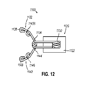
[000113] FIGS. 12 and 13 show an exemplary marker (1100) that is generally
configured to
anchor to tissue using anchors aligned along multiple planes to limit
migration of
marker (1100) relative to an initial placement in tissue. As with marker (100)
described
above, marker (1100) of the present example includes a carrier (1120) and a
marker
element (1112). As with carrier (120) described above, carrier (1120) of the
present
example generally includes a bioabsorbable marker material (1122). Thus,
carrier
(1120) is generally configured for absorption into a patient after placement
of marker
(1100) within a biopsy cavity such as biopsy cavity (10) described above.
Carrier
(1120) of the present example defines a generally cylindrical shape, although
a variety
of other shapes may be used. As similarly described above, some examples of
carrier
(1120) may include a plurality of microbubbles to enhance visualization of
carrier
(1120) under ultrasound.
[000114] Marker material (1122) of the present example comprise a hydrogel or
other suitable
materials. Hydrogel materials are generally configured to absorb into a
patient's tissue
over time. Thus, marker material (1122) is generally non-permanent.
Additionally,
hydrogel is generally configured to expand or swell when placed within tissue.
As will
be described in greater detail below, hydrogel may be dehydrated and/or cured
prior to
being deployed at a biopsy site or within a biopsy cavity. Once the hydrogel
contacts
tissue, the hydrogel may absorb moisture from the tissue and expand or swell
as the
moisture in the hydrogel increases. In some examples, the hydrogel may also be
manipulated during dehydration and/or curing to control expansion of the
hydrogel in
accordance with various expansion profiles (e.g., limit longitudinal
expansion, limit
transverse expansion, and/or etc.). Although maker material (1122) is
described herein
as being hydrogel, it should be understood that in other examples marker
material
(1122) may comprise other suitable materials or include various combinations
of
suitable materials with or without hydrogel.
[000115] Marker element (1112) includes a primary coil (1130) and a plurality
of anchors (1132,
1142) (alternatively referred to as "outriggers") extending outwardly from
primary coil
CA 03174681 2022-09-06
WO 2021/188481 PCT/US2021/022475
-32-
(1130). In the present example, primary coil (1130) is disposed entirely
within carrier
(1120). Primary coil (1130) is defined by one or more wire coils. Generally,
the
combination of the one or more wire coils forming primary coil (1130) are
configured
to provide a distinctive pattern to enhance visualization under x-ray and/or
ultrasound
visualization. Although primary coil (1130) is shown as having a particular
orientation
within carrier (1120) in the present example, it should be understood that
primary coil
(1130) may have a variety of alternative orientations in other examples.
[000116] Each anchor (1132, 1142) extends distally away from primary coil
(1130) to protrude
from a portion of carrier (1120). Each anchor (1132, 1142) includes a
corresponding
first spring (1134, 1144), second spring (1136, 1146), and secondary coil
(1138, 1148).
Each secondary coil (1138, 1148) is generally configured as one or more wire
loops
and is configured to promote tissue in-growth to provide enhanced anchoring of
marker
(1100). Each first spring (1134, 1144) and second spring (1136, 1146) is
positioned
along a length of each anchor (1132, 1142) between primary coil (1130) and a
respective secondary coil (1138, 1148). As will be described in greater detail
below,
each of first spring (1134, 1144) and second spring (1136, 1146) are
configured to bias
a respective secondary coil (1138, 1148) outwardly and into tissue. As such,
each first
spring (1134, 1144) and second spring (1136, 1146) is positioned along the
length of
each anchor (1132, 1142) outside of carrier (1120).
[000117] Although anchors (1132, 1142) of the present example are shown as
being substantially
similar to each other, it should be understood that in other examples, anchors
(1132,
1142) may have variation in structure. For instance, in some examples, one or
more
anchors (1132, 1142) may include additional springs, multiple coils, and/or
different
geometric profiles. Additionally, in some examples, anchors (1132, 1142) may
be of
varying lengths with one anchor (1132, 1142) being longer or shorter than one
or more
other anchors (1132, 1142). Various suitable combinations of such features
will be
apparent to those of ordinary skill in the art in view of the teachings
herein.
[000118] The present example includes a first anchor (1132) and a second
anchor (1142)
extending distally from carrier (1120). As best shown in FIG. 13, first anchor
(1132)
CA 03174681 2022-09-06
WO 2021/188481 PCT/US2021/022475
-33-
and second anchor (1142) are laterally offset relative to each other. Such a
lateral offset
may be desirable to promote anchoring along multiple offset planes. For
instance, first
anchor (1132) in the present example is configured to provide anchoring along
one
plane, while second anchor (1142) is configured to provide anchoring along
another
plane laterally offset from the plane associated with first anchor (1132).
[000119] In an exemplary use, marker (1100) may initially be disposed in a
tubular structure
similar to outer cannula (162) of marker delivery device (150) for deployment
at a
biopsy site. To facilitate confinement within the tubular structure, anchors
(1132, 1142)
may bend about first springs (1134, 1144) and/or second springs (1136, 1146)
to
conform to the inner diameter of the tubular structure.
[000120] During deployment of marker (1100), marker (1100) may be ejected from
the tubular
structure as similarly described above with respect to marker delivery device
(150).
Once marker (1100) is released from the tubular structure, the resilient bias
of springs
(1134, 1136, 1144, 1146) may cause anchors (1132, 1142) to expand, thereby
increasing the lateral profile or span of marker (1100). As a result, each
secondary coil
(1138, 1148) of each anchor (1132, 1142) may be forced into adjacent tissue to
anchor
marker (1100) at the biopsy site. Overtime, some tissue in-growth may occur
with
respect to each secondary coil (1138, 1148) further increasing anchoring of
marker
(1100) over time.
[000121] IV. Exemplary Combinations
[000122] The following examples relate to various non-exhaustive ways in which
the teachings
herein may be combined or applied. It should be understood that the following
examples are not intended to restrict the coverage of any claims that may be
presented
at any time in this application or in subsequent filings of this application.
No disclaimer
is intended. The following examples are being provided for nothing more than
merely
illustrative purposes. It is contemplated that the various teachings herein
may be
arranged and applied in numerous other ways. It is also contemplated that some
variations may omit certain features referred to in the below examples.
Therefore, none
CA 03174681 2022-09-06
WO 2021/188481 PCT/US2021/022475
-34-
of the aspects or features referred to below should be deemed critical unless
otherwise
explicitly indicated as such at a later date by the inventors or by a
successor in interest
to the inventors. If any claims are presented in this application or in
subsequent filings
related to this application that include additional features beyond those
referred to
below, those additional features shall not be presumed to have been added for
any
reason relating to patentability.
[000123] Example 1
[000124] A biopsy site marker, comprising: a carrier; and a marker element
including a primary
coil, a first anchor and a second anchor, the primary coil being disposed
within the
carrier, at least a portion of the first anchor and the second anchor
extending laterally
and outwardly away from the primary coil and from opposite sides of the
carrier, the
first anchor and second anchor being configured to move relative to the
primary coil to
engage tissue at a biopsy site.
[000125] Example 2
[000126] The marker of Example 1, the first anchor and the second anchor
extending laterally
from the primary coil at an angle relative to a longitudinal axis defined by
the carrier.
[000127] Example 3
[000128] The marker of Examples 1 or 2, the carrier including a hydrogel
marker material.
[000129] Example 4
[000130] The marker of Example 3, the hydrogel marker material being
configured to expand in
the presence of moisture, the first anchor and second anchor being configured
to
respond to expansion of the hydrogel marker material to increase engagement of
the
first anchor and the second anchor with tissue.
[000131] Example 5
CA 03174681 2022-09-06
WO 2021/188481 PCT/US2021/022475
-35-
[000132] The marker of any one or more of Examples 1 through 4, the marker
element further
including a third anchor, the third anchor extending distally from the primary
coil, a
portion of the third anchor being disposed outside of the carrier.
[000133] Example 6
[000134] The marker of Example 5, the third anchor including one or more
barbs, the one or
more barbs being configured to penetrate and catch tissue.
[000135] Example 7
[000136] The marker of Example 6, the first anchor and the second anchor each
including a
secondary coil, the secondary coil being disposed on an outer end of each of
the first
anchor and the second anchor.
[000137] Example 8
[000138] The marker of any one or more of Examples 1 through 7, the primary
coil being
resiliently biased to drive the first anchor and the second anchor toward a
predetermined position.
[000139] Example 9
[000140] The marker of Example 8, the predetermined position of the first
anchor and the second
anchor being disposed distally of an initial position defined by the first
anchor and the
second anchor.
[000141] Example 10
[000142] The marker of Example 9, the marker being configured for deployment
from a cannula
when the first anchor and the second anchor are disposed in the initial
position.
[000143] Example 11
[000144] The marker of any one or more of Examples 1 through 11, the first
anchor or the second
anchor defining a spring disposed outside of the carrier.
CA 03174681 2022-09-06
WO 2021/188481 PCT/US2021/022475
-36-
[000145] Example 12
[000146] The marker of Example 11, the spring being configured to drive a
portion of the first
anchor or the second anchor into tissue.
[000147] Example 13
[000148] The marker of any one or more of Examples 1 through 12, the first
anchor and the
second anchor being connected to the primary coil at a first position and a
second
position, respectively, the first position being laterally offset relative to
the second
position.
[000149] Example 14
[000150] The marker of any one or more of Examples 1 through 13, the first
anchor and the
second anchor including a shape-memory alloy, the first anchor and the second
anchor
being configured to engage tissue when exposed to heat thereby activating a
shape-
memory property of the shape-memory alloy.
[000151] Example 15
[000152] The marker of Example 14, the first anchor and the second anchor
being configured to
form a predetermined curve when exposed to heat.
[000153] Example 16
[000154] A biopsy site marker, the biopsy site marker comprising: a carrier,
the carrier having a
first element and a second element; and a marker element extending between the
first
element and the second element of the carrier, the marker element having a
resilient
portion, the resilient portion being configured to transition the marker
element between
a pre-deployment state and a deployment state, the resilient portion being
further
configured to move the second element of the carrier relative to the first
element when
transitioning the marker element from between the pre-deployment state and the
deployment state.
CA 03174681 2022-09-06
WO 2021/188481 PCT/US2021/022475
-37-
[000155] Example 17
[000156] The biopsy site marker of Example 16, the resilient portion including
a spring.
[000157] Example 18
[000158] The biopsy site marker of Examples 16 or 17, the resilient portion
being configured to
move the second element of the carrier about 90 relative to the fist element
of the
carrier.
[000159] Example 19
[000160] The biopsy site marker of any one or more of Examples 16 through 18,
the marker
element further including a first coil and a second coil, the first coil and
the second coil
being disposed on opposing ends of the marker element, the first coil being
disposed
within the first element of the carrier, the second coil being disposed within
the second
element of the carrier.
[000161] Example 20
[000162] A biopsy site marker, the biopsy site marker comprising: a carrier;
and a marker
element, the marker element including a braided portion disposed within the
carrier and
a plurality of anchor portions extending outside of the carrier from the
braided portion.
[000163] Example 21
[000164] The biopsy site marker of Example 20, each anchor portion of the
plurality of anchor
portions being configured to move from each adjacent anchor portion to anchor
the
marker to tissue.
[000165] Example 22
[000166] The biopsy site marker of Examples 20 or 22, the carrier including a
hydrogel marker
material configured to expand in the presence of moisture, the hydrogel marker
material
CA 03174681 2022-09-06
WO 2021/188481 PCT/US2021/022475
-38-
being configured to drive movement of the plurality of anchor portions via
expansion
of the hydrogel marker material in the presence of moisture.
[000167] Example 23
[000168] A biopsy site marker, the biopsy site marker comprising: a carrier;
and a marker
element, the marker element including a body defining a coil shape, a first
anchor, and
a second anchor, the body being at least partially disposed within the
carrier, the first
anchor including a first spring and a second spring disposed between the body
and an
outer end of the first anchor, the second anchor including a third spring and
a fourth
spring disposed between the body and an outer end of the second anchor.
[000169] Example 24
[000170] The biopsy site marker of Example 23, the first anchor including a
secondary coil
disposed on the outer end of the first anchor, the second anchor including a
secondary
coil disposed on the outer end of the second anchor.
[000171] Example 25
[000172] The biopsy site marker of Examples 23 or 24, the first spring, the
second spring, the
third spring, and the fourth spring being configured to increase the span of
the biopsy
site marker across a lateral dimension.
[000173] Example 26
[000174] The biopsy site marker of any one or more of Examples 23 through 24,
the marker
element further including a third anchor and a fourth anchor, the first anchor
and the
second anchor extending distally from the body, the third anchor and the
fourth anchor
extending proximally from the body.
[000175] Example 27
CA 03174681 2022-09-06
WO 2021/188481 PCT/US2021/022475
-39-
[000176] The biopsy site marker of Example 26, the third anchor including a
fifth spring disposed
between the body and an outer end of the third anchor, the fourth anchor
including a
sixth spring disposed between the body and an outer end of the fourth anchor.
[000177] Example 28
[000178] The biopsy site marker of Example 26, the third anchor including a
fifth spring and a
sixth spring disposed between the body and an outer end of the third anchor,
the fourth
anchor including a seventh spring and an eighth spring disposed between the
body and
an outer end of the fourth anchor.
[000179] V. Conclusion
[000180] It should be appreciated that any patent, publication, or other
disclosure material, in
whole or in part, that is said to be incorporated by reference herein is
incorporated
herein only to the extent that the incorporated material does not conflict
with existing
definitions, statements, or other disclosure material set forth in this
disclosure. As such,
and to the extent necessary, the disclosure as explicitly set forth herein
supersedes any
conflicting material incorporated herein by reference. Any material, or
portion thereof,
that is said to be incorporated by reference herein, but which conflicts with
existing
definitions, statements, or other disclosure material set forth herein will
only be
incorporated to the extent that no conflict arises between that incorporated
material and
the existing disclosure material.
[000181] Having shown and described various embodiments of the present
invention, further
adaptations of the methods and systems described herein may be accomplished by
appropriate modifications by one of ordinary skill in the art without
departing from the
scope of the present invention. Several of such potential modifications have
been
mentioned, and others will be apparent to those skilled in the art. For
instance, the
examples, embodiments, geometrics, materials, dimensions, ratios, steps, and
the like
discussed above are illustrative and are not required. Accordingly, the scope
of the
present invention should be considered in terms of the following claims and is
CA 03174681 2022-09-06
WO 2021/188481 PCT/US2021/022475
-40-
understood not to be limited to the details of structure and operation shown
and
described in the specification and drawings.