Note: Descriptions are shown in the official language in which they were submitted.
CA 03175140 2022-09-12
WO 2021/183839 PCT/US2021/022029
NOVEL ANTI-LILRB4 ANTIBODIES AND DERIVATIVE PRODUCTS
CROSS-REFERENCE TO RELATED APPLICATIONS
[001] This application claims priority to U.S. provisional patent
application no.
62/988,892, filed March 12, 2020, the disclosure of which is incorporated
herein by reference.
SEQUENCE LISTING
[002] The sequence listing that is contained in the file named "066564-
8013W001 ST25", which is 151 KB (as measured in Microsoft Windows) and was
created on
March 12, 2021, is filed herewith by electronic submission and is incorporated
by reference
herein.
FIELD OF THE INVENTION
[003] The present disclosure relates generally to the fields of medicine,
oncology, and
immunology. More particular, the disclosure relates to antibodies that bind to
LILRB4.
BACKGROUND
[004] Human Leukocyte Immunoglobulin-Like Receptor subfamily B member 4
(LILRB4), also known as Immunoglobulin-like transcript 3 (ILT3 or ILT-3),
Leukocyte
Immunoglobulin-like Receptor 5 (LIR5 or LIR-5), and CD85k or CD85K, is a type
I membrane
protein that contains cytoplasmic immunoreceptor tyrosine-based inhibition
motif (ITIM) and
involves in negative regulation of immune cell activation. LILRB4 is expressed
on monocytes,
macrophages and dendritic cells and can inhibit innate immunity in a cell-
autonomous manner
as well as suppress T cell activation through an indirect mechanism. LILRB4 is
a specific
marker for monocytic acute myeloid leukemia (AML) including refractory and
relapsed disease.
It has been shown that LILRB4 supports tumor cell infiltration into tissues
and suppresses T
cell activity via a signaling pathway that involves APOE, LILRB4, SHP-2, uPAR
and ARG1
in AML cells (Deng M. et al., Nature (2018) 562:605-09). There is a
significant need for novel
anti-LILRB4 antibodies.
BRIEF SUMMARY OF THE INVENTION
[005] The present disclosure provides anti-LILRB4 antibodies and antigen-
binding
fragment thereof, amino acid and nucleotide sequences thereof, anti-LILRB4
chimeric antigen
receptors, and uses thereof.
1
CA 03175140 2022-09-12
WO 2021/183839 PCT/US2021/022029
[006] In one aspect, the present disclosure provides an isolated anti-
LILRB4 antibody
or an antigen-binding fragment thereof In some embodiments, the anti-LILRB4
antibody or
an antigen-binding fragment comprises: (a) a heavy chain variable region
comprising a heavy
chain complementarity determining region (HC-CDR) 1 having an amino acid
sequence of
SEQ ID NO: 5, an HC-CDR2 having an amino acid sequence of SEQ ID NO: 6 and an
HC-
CDR3 having an amino acid sequence of SEQ ID NO: 7; and (b) a light chain
variable region
comprising a light chain complementarity determining region (LC-CDR) 1 having
an amino
acid sequence of SEQ ID NO: 8 with a mutation at amino acid residues NS, an LC-
CDR2
having an amino acid sequence of SEQ ID NO: 9 and an LC-CDR3 having an amino
acid
sequence of SEQ ID NO: 10.
[007] In certain embodiments, the LC-CDR1 has an amino acid sequence of SEQ
ID
NO: 28.
[008] In certain embodiments, the heavy chain variable region has an amino
acid
sequence at least about 90% identical to SEQ ID NO: 1; and wherein the light
chain variable
region has an amino acid sequence at least about 90% identical to SEQ ID NO:
27.
[009] In certain embodiments, the heavy chain variable region has an amino
acid
sequence of SEQ ID NO: 1; and wherein the light chain variable region has an
amino acid
sequence of SEQ ID NO: 27.
[0010] In certain embodiments, the antibody or the antigen-binding
fragment further
comprises an immunoglobulin constant region, optionally a constant region of
Ig, or optionally
a constant region of human IgG.
[0011] In certain embodiments, the antibody described herein is of the
IgGl, IgG2,
IgG3 or IgG4 isotype.
[0012] In certain embodiments, the antibody or the antigen-binding
fragment is
humanized.
[0013] In certain embodiments, the antigen-binding fragment is a
camelized single
domain antibody, a diabody, a ds (disulfide-stabilized) diabody or ds diabody,
a scFv, a scFv
dimer, a BsFv, a dsFv, a (dsFv)2, a dsFv-dsFv', an Fv fragment, a Fab, a Fab',
a F(a1302, a
bispecific antibody, a nanobody, a domain antibody, or a bivalent antibody.
[0014] In certain embodiments, the anti-LILRB4 antibody described herein
is a
bispecific antibody. In some embodiments, the anti-LILRB4 bispecific antibody
is against a T-
2
CA 03175140 2022-09-12
WO 2021/183839 PCT/US2021/022029
cell receptor such as CD3. In some embodiments, the anti-LILRB4 bispecific
antibody is
against an NK-cell receptor such as CD16A.
[0015] Therefore, the present disclosure in another aspect provides a
bispecific
antibody or antigen-binding fragment capable of binding to LILRB4 and CD3.
[0016] In certain embodiments, the bispecific antibody or antigen-binding
fragment
provided herein comprises: (a) a first antigen-binding region comprising a
first light chain
variable (VL) domain and a first heavy chain variable (VH) domain; and (b) a
second antigen-
binding region comprising a second VL domain and a second VH domain, wherein
the first
antigen-binding region is capable of binding to LILRB4 and the second antigen-
binding region
is capable of binding to CD3, or vice versa.
[0017] In certain embodiments, the first VL domain and the first heavy
chain variable
domain link to a first pair of constant domains, respectively, and wherein the
second VL domain
and the second VH domain link to a second pair of constant domains,
respectively.
[0018] In certain embodiments, the first VL domain links to a first light
chain constant
(CL) domain, and the first VH domain links to a first heavy chain constant
domain 1 (CH1). In
certain embodiments, the first VL domain links to a first CH1 domain, and the
VH domain links
to a second CL domain.
[0019] In certain embodiments, the second VL domain links to a second CL
domain, and
the second VH domain links to a second CH1 domain. In certain embodiments, the
second VL
domain links to a second CH1 domain, and the second VH domain links to a
second CL domain.
In certain embodiments, the second VL domain links to a T cell receptor (TCR)
a chain constant
domain, and the second VH domain links to a TCR 0 chain constant domain. In
certain
embodiments, the second VL domain links to a TCR 0 chain constant domain, and
the second
VH domain links to a TCR a chain constant domain.
[0020] In certain embodiments, the first antigen-binding region and/or
the second
antigen-binding region is a single chain variable fragment (scFv).
[0021] In certain embodiments, the antibody or antigen-binding fragment
provided
herein further comprises a third antigen-binding region comprising a third VL
domain and a
third VH domain, wherein the third antigen-binding region is capable of
binding to LILRB4 or
CD3.
[0022] In certain embodiments, the third VL domain and the third heavy
chain variable
domain link to a first pair of constant domains, respectively. In certain
embodiments, the third
3
CA 03175140 2022-09-12
WO 2021/183839 PCT/US2021/022029
VL domain links to a third CL domain, and the third VH domain links to a third
CH1 domain. In
certain embodiments, the third VL domain links to a third CH1 domain, and the
third VH domain
links to a third CL domain. In certain embodiments, the third VL domain links
to a second TCR
a chain constant domain, and the third VH domain links to a second TCR 0 chain
constant
domain. In certain embodiments, the third VL domain links to a second TCR 0
chain constant
domain, and the third VH domain links to a second TCR a chain constant domain.
[0023] In certain embodiments, the TCR a chain constant domain has an
amino acid
sequence of SEQ ID NO: 89. In certain embodiments, the TCR a chain constant
domain has a
591A mutation of SEQ ID NO: 89.
[0024] In certain embodiments, the antibody or the antigen-binding
fragment is linked
to one or more conjugate moieties. In certain embodiments, the conjugate
moiety comprises a
clearance-modifying agent, a toxin, a detectable label, a chemotherapeutic
agent, or
purification moiety.
[0025] In another aspect, the present disclosure provides a
pharmaceutical composition
comprising the antibody or antigen-binding fragment thereof described herein,
and a
pharmaceutically acceptable carrier.
[0026] In another aspect, the present disclosure provides an isolated
polynucleotide
encoding the antibody or antigen-binding fragment thereof described herein.
[0027] In another aspect, the present disclosure provides a vector
comprising the
isolated polynucleotide described herein.
[0028] In another aspect, the present disclosure provides a host cell
comprising the
vector described herein. In certain embodiments, the host cell is a mammalian
cell, e.g., a CHO
cell.
[0029] In another aspect, the present disclosure provides a hybridoma
encoding or
producing the anti-LILRB4 antibody as provided herein.
[0030] In another aspect, the present disclosure provides a method of
expressing the
antibody or antigen-binding fragment thereof described herein. In some
embodiments, the
method comprises culturing the host cell described herein under the condition
at which the
vector described herein is expressed.
[0031] In another aspect, the present disclosure provides a method of
treating or
ameliorating the effect of a cancer in a subject. In some embodiments, the
method comprises
4
CA 03175140 2022-09-12
WO 2021/183839 PCT/US2021/022029
administering to the subject a therapeutically effective amount of the
antibody or antigen-
binding fragment thereof described herein or the pharmaceutical composition
described herein.
[0032] The method may reduce or eradicate the tumor burden in the
subject, may
reduce the number of tumor cells, may reduce tumor size, may reduce tumor
infiltration, may
reduce tumor metastasis, may eradicate the tumor in the subject. The cancer
may be a solid
tumor or hematologic malignancy.
[0033] In certain embodiments, the cancer is adrenal cancer, bile duct
carcinoma, bone
cancer, brain cancer, breast cancer, cervical cancer, choriocarcinoma, colon
cancer, colorectal
cancer, esophageal cancer, eye cancer, gastric cancer, gastroesophageal
cancer, glioblastoma,
head and neck cancer, kidney cancer, liver cancer, lung cancer, non-small cell
lung cancer,
bronchioloalveolar cell lung cancer, mesothelioma, squamous cell carcinoma,
melanoma,
merkel cell cancer, nasopharyngeal carcinoma, neuroblastoma, oral cancer,
ovarian cancer,
pancreatic cancer, penile cancer, prostate cancer, renal cell cancer,
retinoblastoma, sarcoma,
skin cancer, testicular cancer, thymic carcinoma, thyroid cancer, uterine
cancer, and vaginal
cancer.
[0034] In some embodiments, the cancer is a metastatic, recurrent or drug-
resistant
cancer.
[0035] In some embodiments, said cancers are hematologic malignancies
including
acute lymphocytic/lymphoblastic leukemia (ALL), acute myeloid leukemia (AML),
B-cell
leukemia, blastic plasmacytoid dendritic cell neoplasm (BPDCN), chronic
lymphoblastic
leukemia (CLL), chronic myelomonocytic leukemia (CMML), chronic myelocytic
leukemia
(CIVIL), diffuse large B-cell lymphoma (DLBCL), extranodal NK/T-cell lymphoma,
hairy cell
leukemia, HEIV8-associated primary effusion lymphoma, plasmablastic lymphoma,
pre-B
acute lymphocytic leukemia (Pre-B ALL), primary CNS lymphoma, primary
mediastinal large
B-cell lymphoma, T-cell/histiocyte-rich B-cell lymphoma, heavy chain disease,
Hodgkin's
lymphoma, non-Hodgkin's lymphoma, Waldenstrom's macroglobulinemia, multiple
myeloma
(MM), myelodysplastic syndromes (MDS), myeloproliferative neoplasms, and
polycythemia
vera.
[0036] In certain embodiments, said hematologic malignancies include
subsets or
subtypes of acute myeloid leukemia (AML), acute promyelocytic leukemia (APL)
or M3 AML,
acute myelomonocytic leukemia or M4 AML, acute monocytic/monoblastic leukemia
or M5
AML, and acute myeloblastic leukemia.
CA 03175140 2022-09-12
WO 2021/183839 PCT/US2021/022029
[0037] In certain embodiments, said hematologic malignancies include
acute myeloid
leukemia (AML) that is resistant to venetoclax, or venetoclax in combination
with
azacytidine/azacitidine, that is relapsed after treatment with
azacytidine/azacitidine and/or
venetoclax, that is resistant to venetoclax in combination with decitabine or
is relapsed after
treatment with azacytidine/azacitidine and decitabine.
[0038] In certain embodiments, the antibody or an antigen-binding
fragment thereof is
administered intravenously, intra-arterially, intra-tumorally, or
subcutaneously.
[0039] In certain embodiments, the method further comprises administering
to the
subject one or more drugs selected from the group consisting of a
topoisomerase inhibitor, an
anthracycline topoisomerase inhibitor, an anthracycline, a daunorubicin, a
nucleoside
metabolic inhibitor, a cytarabine, a hypomethylating agent, a low dose
cytarabine (LDAC), a
combination of daunorubicin and cytarabine, a daunorubicin and cytarabine
liposome for
injection, Vyxeos , an azacytidine, Vidaza , a decitabine, an all-trans-
retinoic acid (ATRA),
an arsenic, an arsenic trioxide, a histamine dihydrochloride, Ceplene , an
interleukin-2, an
aldesleukin, Proleukin , a gemtuzumab ozogamicin, Mylotarg , an FLT-3
inhibitor, a
midostaurin, Rydapt , a clofarabine, a farnesyl transferase inhibitor, a
decitabine, an IDH1
inhibitor, an ivosidenib, Tibsovo , an IDH2 inhibitor, an enasidenib, Idhifa ,
a smoothened
(SMO) inhibitor, a glasdegib, an arginase inhibitor, an DO inhibitor, an
epacadostat, a BCL-2
inihbitor, a venetoclax, Venclexta , a platinum complex derivative,
oxaliplatin, a kinase
inhibitor, a tyrosine kinase inhibitor, a PI3 kinase inhibitor, a BTK
inhibitor, an ibrutinib,
IMBRUVICA , an acalabrutinib, CALQUENCE , a zanubrutinib, a PD-1 antibody, a
PD-Li
antibody, a CTLA-4 antibody, a LAG3 antibody, an ICOS antibody, a TIGIT
antibody, a TEVI3
antibody, a CD40 antibody, a 4-1BB antibody, a CD47 antibody, a SIRPla
antibody or fusions
protein, a CD70 antibody, and CLL1 antibody, a CD123 antibody, an antagonist
of E-selectin,
an antibody binding to a tumor antigen, an antibody binding to a T-cell
surface marker, an
antibody binding to a myeloid cell or NK cell surface marker, an alkylating
agent, a nitrosourea
agent, an antimetabolite, an antitumor antibiotic, an alkaloid derived from a
plant, a hormone
therapy medicine, a hormone antagonist, an aromatase inhibitor, and a P-
glycoprotein inhibitor.
[0040] In certain embodiments, the method further comprises administering
to the
subject initially a monotherapy of anti-LILRB4 antibodies for a period of time
followed by
addition of one or more drugs selected from the group consisting of an
azacytidine, Vidaza ,
a BCL-2 inihbitor, a venetoclax, and Venclexta .
6
CA 03175140 2022-09-12
WO 2021/183839 PCT/US2021/022029
[0041] In yet another aspect, the present disclosure provides a method
for detecting a
cancer cell or cancer stem cell in a sample or subject. In certain
embodiments, the method
comprises: (a) contacting a subject or a sample from the subject with the
antibody or an antigen-
binding fragment thereof described herein; and (b) detecting binding of said
antibody to a
cancer cell or cancer stem cell in said subject or sample.
[0042] In some embodiments, the sample is a body fluid or biopsy. In some
embodiments, the sample is blood, sputum, tears, saliva, mucous, serum, urine
or feces.
[0043] In some embodiments, the detection comprises immunohistochemistry,
flow
cytometry, immunoassays (including ELISA, RIA etc.) or Western blot.
[0044] In some embodiments, the method further comprises performing steps
(a) and
(b) a second time or additional times and determining a change in detection
levels as compared
to the first time.
[0045] The anti-LILRB4 antibody or an antigen binding fragment thereof
may further
comprise a label, such as a peptide tag, an enzyme, a magnetic particle, a
chromophore, a
fluorescent molecule, a chemo-luminescent molecule, or a dye. The isolated
monoclonal
antibody or an antigen binding fragment thereof may be conjugated to a
liposome or
nanoparticle.
[0046] In another aspect, the present disclosure provides use of the
antibody or antigen-
binding fragment thereof described herein in the manufacture of a medicament
for treating
cancer in a subject.
[0047] In another aspect, the present disclosure provides a kit
comprising the antibody
or antigen-binding fragment thereof described herein, useful in detecting
LILRB4.
[0048] In another aspect, the present disclosure provides an anti-LILRB4
chimeric
antigen receptor (CAR) protein. In some embodiments, the CAR protein
comprises: (a) a heavy
chain variable region comprising an HC-CDR1 having an amino acid sequence of
SEQ ID NO:
5, an HC-CDR2 having an amino acid sequence of SEQ ID NO: 6 and an HC-CDR3
having
an amino acid sequence of SEQ ID NO: 7; and (b) a light chain variable region
comprising an
LC-CDR1 having an amino acid sequence of SEQ ID NO: 8 with a mutation at amino
acid
residues NS, an LC-CDR2 having an amino acid sequence of SEQ ID NO: 9 and an
LC-CDR3
having an amino acid sequence of SEQ ID NO: 10. In some embodiments, the LC-
CDR1 has
an amino acid sequence of SEQ ID NO: 28.
7
CA 03175140 2022-09-12
WO 2021/183839 PCT/US2021/022029
[0049] In some embodiments, the heavy chain variable region of the LILRB4
CAR
protein has an amino acid sequence at least about 90% identical to SEQ ID NO:
1; and the light
chain variable region of the CAR protein has an amino acid sequence at least
about 90%
identical to SEQ ID NO: 27. In some embodiments, the heavy chain variable
region has an
amino acid sequence of SEQ ID NO: 1; and wherein the light chain variable
region has an
amino acid sequence of SEQ ID NO: 27. In some embodiments, the CAR protein
comprises a
single-chain variable fragment (scFv) having an amino acid sequence at least
85%, 90%, 95%
or 99% identical to SEQ ID NO: 66 or SEQ ID NO: 68. In some embodiments, the
CAR protein
has a scFv having an amino acid sequence identical to SEQ ID NO: 66 or SEQ ID
NO: 68.
[0050] In another aspect, the present disclosure provides a
polynucleotide molecule
encoding a CAR protein described herein. In some embodiments, the
polynucleotide molecule
further comprises a promoter active in eukaryotic cells. In some embodiments,
the
polynucleotide molecule is an expression vector.
[0051] In another aspect, the present disclosure provides an engineered
cell comprising
the polynucleotide molecule encoding a CAR protein described herein. In some
embodiments,
the cell is a T cell, an NK cell or a macrophage.
[0052] In another aspect, the present disclosure provides a method of
treating or
ameliorating cancer in a subject in need thereof comprising administering to
the subject an
effective amount of a cell therapy comprising one or more cells comprising the
polynucleotide
molecule encoding a CAR protein described herein. In some embodiments, the
method further
comprises administering to said human subject a second cancer therapy. In some
embodiments,
the second cancer therapy is chemotherapy, immunotherapy, radiotherapy,
hormone therapy or
surgery. In some embodiments, the second cancer therapy is administered at the
same time as
the cell therapy. In some embodiments, said second cancer therapy is
administered before or
after the cell therapy. In some embodiments, the method further comprises
administering to
said human subject a second administration of an effective amount of one or
more cells
comprising the polynucleotide molecule encoding a CAR protein described
herein.
[0053] In some embodiments, said cell therapy is administered local to
cancer site,
regional to a cancer site, or systemically.
[0054] In some embodiments, said cancer is hematologic malignancies
including acute
lymphocytic/lymphoblastic leukemia (ALL), acute myeloid leukemia (AML), B-cell
leukemia,
blastic plasmacytoid dendritic cell neoplasm (BPDCN), chronic lymphoblastic
leukemia (CLL),
chronic myelomonocytic leukemia (CMML), chronic myelocytic leukemia (CML),
diffuse
8
CA 03175140 2022-09-12
WO 2021/183839 PCT/US2021/022029
large B-cell lymphoma (DLBCL), extranodal NK/T-cell lymphoma, follicular
lymphoma,
hairy cell leukemia, HEIV8-associated primary effusion lymphoma, plasmablastic
lymphoma,
pre-B acute lymphocytic leukemia (Pre-B ALL), primary CNS lymphoma, primary
mediastinal
large B-cell lymphoma, T-cell/histiocyte-rich B-cell lymphoma, heavy chain
disease,
Hodgkin's lymphoma, non-Hodgkin's lymphoma, Waldenstrom's macroglobulinemia,
multiple myeloma (MM), myelodysplastic syndromes (MD S), myeloproliferative
neoplasms,
and polycythemia vera.
[0055] In certain embodiments, said hematologic malignancies include
subsets or
subtypes of acute myeloid leukemia (AML), acute promyelocytic leukemia (APL)
or M3 AML,
acute myelomonocytic leukemia or M4 AML, acute monocytic/monoblastic leukemia
or M5
AML, and acute myeloblastic leukemia.
[0056] In still an additional aspect, there is provided a method of
treating or
ameliorating the effect of an autoimmune disease in a subject, the method
comprising
administering to the subject a therapeutically effective amount of the
antibody or an antigen-
binding fragment thereof as defined herein. The antibody or an antigen-binding
fragment
thereof may be administered intravenously, intra-arterially,
intraperitoneally, or
subcutaneously. The method may further comprise administering to the subject
one or more
drugs selected from the group consisting of a steroid or an NSAID. The
autoimmune disease
may be Guillain-Barre syndrome, Chronic inflammatory demyelinating
polyneuropathy,
ankylosing spondylitis, psoriatic arthritis, enteropathic arthritis, reactive
arthritis,
undifferentiated spondyloarthropathy, juvenile spondyloarthropathy, Behcet's
disease,
enthesitis, ulcerative colitis, Crohn's disease, irritable bowel syndrome,
inflammatory bowel
disease, fibromyalgia, chronic fatigue syndrome, pain conditions associated
with systemic
inflammatory disease, systemic lupus erythematosus, Sjogren's syndrome,
rheumatoid arthritis,
juvenile rheumatoid arthritis, juvenile onset diabetes mellitus (also known as
Type I diabetes
mellitus), Wegener's granulomatosis, polymyositis, dermatomyositis, inclusion
body myositis,
multiple endocrine failure, Schmidt's syndrome, autoimmune uveitis, Addison's
disease,
Grave's Disease, Hashimoto's thyroiditis, autoimmune thyroid disease,
pernicious anemia,
gastric atrophy, chronic hepatitis, lupoid hepatitis, atherosclerosis,
multiple sclerosis,
amyotrophic lateral sclerosis, hypoparathyroidism, Dressler's syndrome,
myasthenia gravis,
Eaton-Lambert syndrome, autoimmune thrombocytopenia, idiopathic
thrombocytopenic
purpura, hemolytic anemia, pemphigus vulgaris, pemphigus, dermatitis
herpetiformis, alopecia,
scleroderma, progressive systemic sclerosis, CREST syndrome (calcinosis,
Raynaud's
phenomenon, esophageal dysmotility, sclerodactyly, and telangtasia), adult
onset diabetes
9
CA 03175140 2022-09-12
WO 2021/183839 PCT/US2021/022029
mellitus (also known as Type II diabetes mellitus), mixed connective tissue
disease,
polyarteritis nodosa, systemic necrotizing vasculitis, glomerulonephritis,
atopic dermatitis,
atopic rhinitis, Goodpasture's syndrome, Chagas' disease, sarcoidosis,
rheumatic fever, asthma,
anti-phospholipidsyndrome, erythema multiforme, Cushing's syndrome, autoimmune
chronic
active hepatitis, allergic disease, allergic encephalomyelitis, transfusion
reaction, leprosy,
malaria, leshmaniasis, trypanosomiasis, Takayasu's arteritis, polymyalgia
rheumatica, temporal
arteritis, shistosomiasis, giant cell arteritis, eczema, lymphomatoid
granulomatosis, Kawasaki's
disease, endophthalmitis, psoriasis, erythroblastosis fetalis, eosinophilic
faciitis, Shulman's
syndrome, Felty's syndrome, Fuchs cyclitis, IgA nephropathy, Henoch-Schonlein
purpura,
graft versus host disease, transplantation rejection, tularemia, periodic
fever syndromes,
pyogenic arthritis, Familial Mediterranean Fever, TNF-receptor associated
periodic syndrome
(TRAPS), Muckle-Wells syndrome, or hyper-IgD syndrome.
BRIEF DESCFRIPTION OF FIGURES
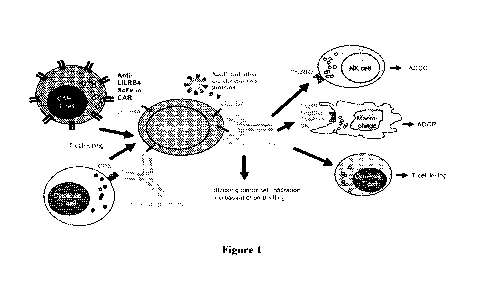
[0057] Figure 1 shows the schematics of the mechanisms of action of anti-
LILRB4
antibodies and derivative products.
[0058] Figure 2 shows the comparison of icIEF results of H7K3 (Molecule
A) and
H7K3m5 (Molecule B) at 40oC for 2W or 4W.
[0059] Figure 3 shows recognition of human endogenous LILRB4 on THP-1
cells by
the anti-LILRB4 antibody H7K3m5.
[0060] Figure 4 shows ADCC of THP-1-GFP cells by wild type (WT) or
afucosylated
(afu) H7K3m5.
[0061] Figure 5 shows LILRB4 expression on human monocytes and
plasmacytoid
dendritic cells (pDC).
[0062] Figure 6 shows up-regulation of LILRB4 expression by IL-10 and
IFNa
treatment in human monocytes.
[0063] Figure 7 shows LILRB4 level down-regulated and uPAR level up-
regulated on
human monocytes stimulated by LPS.
[0064] Figure 8A shows LILRB4 expression on in vitro human monocytes
differentiated macrophages. Figure 8B shows copy number of LILRB4 on in vitro
monocyte-
derived human macrophages.
[0065] Figure 9 shows copy number of LILRB4 on in vitro differentiated
MDSCs.
CA 03175140 2022-09-12
WO 2021/183839 PCT/US2021/022029
[0066] Figures 10A-10B show copy number of LILRB4 on human monocyte-
derived
dendritic cells (DCs). LILRB4 level is seen in the following order from high
to low:
Tolerogenic DC > Activated DC > Immature DC
[0067] Figure 11 shows the comparison of LILRB4 mRNA expression levels
between
solid tumor samples from the TCGA RNA sequencing database with high and low
signals for
macrophage infiltration, based on a macrophage gene expression "signature"
generarated
through computational biology approaoches. The total number of samples for
each tumor type
that was included in the analysis is indicated in parentheses. The results
shown here are in part
based upon RNA sequencing data generated by the TCGA Research Network.
[0068] Figures 12A-12B illustrate the representative flow cytometry data
showing that
H7K3m5 specifically binds monocytic myeloid cells infiltrated into solid tumor
microenvironment (TME), as well as peripheral blood monocytic myeloid cells
from solid
tumor patients. Dark grey-filled histogram: sample incubated with H7K3m5;
light grey-filled
histogram: sample incubated with human IgG1 isotype control. Figure 12A.
Binding signal of
H7K3m5 or its isotype control on various myeloid cell subsets infiltrating the
TME. Figure
12B. Binding signal of H7K3m5 or its isotype control on various myeloid cell
subsets from
peripheral blood.
[0069] Figures 13A-13D show no H7K3m5 mediated monocyte killing in
autologous
ADCC using fresh PBMCs. Freshly isolated PBMCs from healthy donors were
incubated
overnight in the presence of serially titrated H7K3m5, isotype control human
IgGl, or
rituximab as positive control. Monocytes and B cells were identified and
counted as
CD14+CD19- and CD19+CD14- by flow cytometry. Figure 13A and 13C show lack of
monocyte killing by PBMCs from two different donors and Figure 13B and 13D
show
corresponding B cell killing as positive control.
[0070] Figures 14A-14D show autologous ADCC of normal monocytes by wild
type
(WT) or afucosylated (afu) H7K3m5. Figure 14A and 14C show representative
monocyte
killing through ADCC by PBMCs from two different donors. ADCC on monocyte by
afucosylated H7K3m5 was observed in donor 024 but activity was minimal in
donor 13. No
ADCC was observed by wild type H7K3m5. Figure 14B and 14D show corresponding B
cell
killing as positive control.
[0071] Figures 15A-15D show autologous ADCC of pDC or monocytes by wild
type
(WT) or afucosylated (afu) H7K3m5-mediated autologous ADCC. ADCC against pDCs
was
observed by both wild type and afucosylated H7K3m5 in two donors. In the
meantime,
11
CA 03175140 2022-09-12
WO 2021/183839 PCT/US2021/022029
monocytes may be killed only with afucosylated H7K3m5, depending on donors.
Moreover,
the afucosylated H7K3m5 showed much stronger ADCC activity than the wild-type
towards
pDC or monocytes (Figures 15A-15B).
[0072] Figures 16A-16B show dose-dependent ADCC of CD33+ MDSC cells with
purified NK cells in the presence of H7K3m5, which has no ADCC effect on
monocytes at the
same dose levels. H7K3m5 showed ADCC activity against the AML cell line THP-1
in the
same experiment (Figure 16B).
[0073] Figures 17A-17B show ADCP of THP-1-GFP cells by anti-LILRB4. THP-1-
GFP cells were co-cultured with in-vitro differentiated macrophages for 24
hours in the
presence of serially titrated wild type H7K3m5 or isotype control human IgGl.
THP-1-GFP
cells and macrophages were identified and quantified as GFP+ and CD163+CD206+,
respectively. Percent of THP-1 cell killing were calculated from absolute
count of GFP+ cells
or from GFP+% cells. Figure 17A and 17B show ADCP of THP-1-GFP cells by wild
type
H7K3m5 from macrophages differentiated from two separate heathy donors.
[0074] Figure 18 shows in vitro T cell cytotoxicity against THP-1-GFP
cells with anti-
LILRB4. THP-1-GFP cells were co-cultured with purified naive T cells. Anti-
LILRB4
H7K3m5 can induce T-cell cytotoxicity against AML cells. Effector pan T cells
were from 3
different healthy donors. The curves are plotted as mean SD. The EC50 values
are in nano
molar units.
[0075] Figure 19 shows representative cytokine production profile in the
supernatant
from the in vitro T-cell cytotoxicity assay samples. Anti-LILRB4 induced T-
cell cytotoxicity
against THP-1 cells is reflected by the elevation of cytokines in the co-
culture. The curves are
plotted as mean SD. The ECso values are in nano molar units. An EC50 value
cannot be
derived for IL-6.
[0076] Figure 20 shows the evaluation of T-cell activation by flow
cytometry. A-B:
surface staining of T cell activation markers CD69 (A) and CD25 (B). C-E:
Intracellular
cytokine staining of co-cultured T cells and THP-1 cells by flow cytometry. C:
Cells producing
both IFNy and TNFa; D: Cells producing IFNy but not TNFa; E: Cells producing
TNFa but
not IFNy.
[0077] Figure 21 shows increased T cell activation markers and MHC
expression on
THP-1 cells upon H7K3m5 treatment. D428 = donor 428. MFI = geometric mean
fluorescence
intensity.
12
CA 03175140 2022-09-12
WO 2021/183839 PCT/US2021/022029
[0078] Figure 22 shows surface expression of activation markers on THP-1
AML cells
by flow cytometry. MFI=geometric mean fluorescence intensity.
[0079] Figure 23 shows H7K3m5 is efficacious in AML xenograft model. This
experiment evaluated the growth kinetics of THP-1.1uc cells and determined the
efficacy of
H7K3m5 in the THP-1.1uc human AML xenograft model in female NSG mice using bio-
imaging. 1x10⁶ THP-1.1uc cells were intravenously implanted to JAX female NSG
mice via
tail vein. Whole body bioluminescent imaging was conducted on Day 1 (4-6 hrs
after cell
injection) prior to animal randomization and single-dose intravenous
administration of the
vehicle control or H7K3m5 (1 mg/kg). On Days 7, 14, 17 and 21, whole body
bioluminescent
imaging data were collected for the control and treated animals.
[0080] Figure 24 illustrates the flow cytometric data showing that H7K3m5
potentiates
maturation/activation of monocyte-derived dendritic cells (Mo-DC) in response
to Toll-Like
Receptor (TLR) signaling. H7K3m5 enhanced the expression of activation markers
(CD86,
HLA-DR) while decreasing the expression of the tolerogenic marker CD209. Each
line
represents result from a different healthy donor. The fraction of donors in
which H7K3m5
produced the desired pro-inflammatory effect is indicated in parentheses.
*p<0.05 (paired t
test).
[0081] Figure 25 illustrates the flow cytometry data showing that H7K3m5
enhances
the expression of activation markers (CD86 and HLA-DR) on the surface of
mature monocyte-
derived DC (Mo-DC) upon a mixed leukocyte reaction with allogeneic T cells.
The effect of
H7K3m5 was evaluated in the absence (-CD4OL) or presence (+CD4OL) of CD40
ligand to
study the effect of H7K3m5 on immature and mature Mo-DC, respectively. Each
line
represents the result from a different healthy donor (n=3 donors).
[0082] Figure 26 illustrates the ELISA data showing that H7K3m5 enhances
IL-12
production in an allogeneic mixed leukocyte reaction of T cells and mature
monocyte-derived
DC (Mo-DC). The effect of H7K3m5 was evaluated in the absence (-CD4OL) or
presence
(+CD4OL) of CD40 ligand to study the effect of H7K3m5 on immature and mature
Mo-DC,
respectively. Data are presented as mean SEM and data for each donor is also
shown as
individual data points (n=2-3 donors).
[0083] Figure 27 illustrates the ELISA data showing that H7K3m5 enhances
IFN-y
production in an allogeneic mixed leukocyte reaction of T cells and monocyte-
derived DC
(Mo-DC). The effect of H7K3m5 was evaluated in the absence (-CD4OL) or
presence
(+CD4OL) of CD40 ligand to study the effect of H7K3m5 on immature and mature
Mo-DC,
13
CA 03175140 2022-09-12
WO 2021/183839 PCT/US2021/022029
respectively. Data are presented as mean SEM and data for each donor is also
shown as
individual data points (n=3 donors).
[0084] Figures 28A and 28B show the schematic representation of
configurations of
LILRB4/CD3 bispecific antibodies.
[0085] Figures 29A and 29B show the binding to normal monocytes and AML
cell
line THP-1 by CD3 and LILRB4 bispecific antibodies as measured by FACS.
Similar binding
affinity trends were observed across different anti-LILRB4 mono-specific and
bispecific
antibodies between monocytes and THP-1.
[0086] Figures 30A and 30B show T cell-mediated cytotoxicity of
bispecific
CD3/LILRB4 antibodies on monocytes (Figure 22A) and THP-1-luc-GFP cells
(Figure 22B).
[0087] Figures 31A and 31B show the autologous killing of monocytes by
LILRB4xCD3 bi-specifics (Figure 31A) and autologous killing of B cells by
Rituxan as a
control (Figure 31B).
[0088] Figure 32 shows the binding affinity comparison using H7K3m5 full-
length
IgG and ScFv proteins in flow cytometry assays against human primary monocytes
and human
leukemia cell line THP-1.
[0089] Figure 33 shows the schematic representation of the DNA construct
for
expressing the anti-LILRB4 CAR proteins. The DNA construct was based on 2'
generation
CAR constructs containing CD28 or 4-1BB costimulatory domain with CD3zeta
activation
domain. The scFv was derived from anti-LILRB4 monoclonal antibody H7K3m5. The
5' and
3' homologous arms are homologous sequences upstream and downstream of the
Cas9 DNA
cleavage site in the TRAC gene (based on gRNA design). Promoter and leader
peptide are
elements for gene expression and extracellular translocation. 5V40 poly-A tail
was included
for improving transcript stability and translation.
[0090] Figure 34 shows the efficient generation of LILRB4 CAR-T cells
using
CRISPR knock-out and knock-in method. Human primary T cells were transfected
with
CRISPR-Cas9 RNP complexes designed to inactivate the TCR alpha (TRAC) locus,
with or
without DNA template for homologous recombination-based knock in. Following
transfection,
cells were expanded in culture for 2 weeks. Anti-LILRB4 CAR-T cells were
identified by
binding to LILRB4-Fc fusion protein (ACRObiosystems CDK-H5259) and anti-Fc
antibody
(Biolegend B278652, negative control). Successful TCR alpha (TRAC)
inactivation (Knock-
Out or KO) was measured by anti-CD3 staining (anti-CD3 PE, BD 555333). ATC,
activated T
14
CA 03175140 2022-09-12
WO 2021/183839 PCT/US2021/022029
cells; KO, TCR alpha (TRAC) inactivated T cells; RB4 CD28, T cells expressing
anti-LILRB4
CAR with a CD28 costimulatory domain; RB4 41BB, T cells expressing anti-LILRB4
CAR
with a 4-1BB costimulatory domain.
[0091] Figure 35 shows the proliferation of the TCR alpha (TRAC)
inactivated T cells
(KO) and anti-LILRB4 CAR (or control CAR) knocked-in T cells. After knocking
out
TCRalpha, the cells were grown in complete Optimizer medium with IL-2 300
IU/ml and
without anti-CD3/28 added. Fold expansion was plotted by dividing the total T
cell number on
days (as indicated) with the starting culture number. Anti-LILRB4 CAR-T cells
had
significantly higher fold of expansion in comparison to the control CAR-T
cells. ATC,
activated T cells; KO, TCR alpha (TRAC) inactivated T cells; ctrl CD28, T
cells expressing a
control CAR with a CD28 costimulatory domain; ctrl 41BB, T cells expressing a
control CAR
with a 4-1BB costimulatory domain; RB4 CD28, T cells expressing Anti-LILRB4
CAR with
a CD28 costimulatory domain; RB4 41BB, T cells expressing Anti-LILRB4 CAR with
a 4-
1BB costimulatory domain.
[0092] Figures 36A-3611 show the antigen-dependent activation of CAR-T
culture. 1
ug/ml recombinant control antigen or LILRB4 antigen were coated on 96 well
plates overnight
in PBS buffer. Plates were washed twice with PBS buffer. 1X10⁵ CAR-T cells in
culture media
(without any cytokine added) were added to each well and incubated for 72
hours. Cell culture
supernatant was collected for cytokine release measurement by Luminex assay.
ATC, activated
T cells; KO, TCR alpha (TRAC) inactivated T cells; antiRB4 CD28CART, T cells
expressing
Anti-LILRB4 CAR with a CD28 costimulatory domain; antiRB4 41BB CART, T cells
expressing anti-LILRB4 CAR with a 4-1BB costimulatory domain; control
CD28CART, T
cells expressing a control CAR with a CD28 costimulatory domain; control
41BBCART, T
cells expressing a control CAR with a 4-1BB costimulatory domain.
[0093] Figures 37A-37C show the characterization of CAR-T cells after 2
weeks
expansion. Frozen CAR-T cells in liquid nitrogen storage were thawed and kept
in culture for
2-3 days before flow cytometry analysis. Antibodies used were anti-CD8 APC Cy7
(BD561945), anti-PD1 PE (BD560908) and anti-TB/13 BV421 (BD565562). Anti-
LILRB4
CAR-T cells were identified by binding to LILRB4-Fc fusion protein
(ACRObiosystems CDK-
H5259) and anti-Fc antibody (Biolegend B278652).
[0094] Figure 38 shows the cytotoxicity of anti-LILRB4 CAR-T cells. CHO
K1 RB4
cells were seeded at different density (6X10⁴, 2X10⁴ or 7X10³) for 12 hours,
1X10⁵ CAR-T
cells were added and cytotoxicity were measured by removing the supernatant
CAR-T cells
CA 03175140 2022-09-12
WO 2021/183839 PCT/US2021/022029
and wash the plate 2 times with PBS. Total viable adherent CHO K1 RB4 cells
were measured
by Promega CTG2.0 luminescence kit. And the % cytotoxicity were calculated by
dividing the
Luminescent signal of each condition with the same E:T ratio activated T cell
control.
[0095] Figures 39A-39B show the schematics of a phase 1 first-in-human
clinical trial.
Figure 39A is a schematic of the "window" design for dose escalation. Figure
39B is a
schematic of anti-LILRB4 monotherapy. Figures 39C-39D are the schematics of
potential
combination studies of anti-LILRB4 antibody with azacytidine and/or
venetoclax. Other
potential combinations with anti-LILRB4 would follow the same or a similar
schema. AZA,
azacytidine; VEN, venetoclax; C1D1, Cycle 1 Day 1; Cycle 2 Day 1; DLT, dose
limiting
toxicity; MTD1, maximum tolerated dose of anti-LILRB4 monotherapy; MTD2,
maximum
tolerated dose of anti-LILRB4 in combination with azacytidine. Anti-LILRB4 is
administered
as monotherapy or in combination with other agents every 14 days until
progression of disease
or death.
DETAILED DESCRIPTION OF THE INVENTION
[0096] The following description of the disclosure is merely intended to
illustrate
various embodiments of the disclosure. As such, the specific modifications
discussed are not
to be construed as limitations on the scope of the disclosure. It will be
apparent to one skilled
in the art that various equivalents, changes, and modifications may be made
without departing
from the scope of the disclosure, and it is understood that such equivalent
embodiments are to
be included herein. All references cited herein, including publications,
patents and patent
applications are incorporated herein by reference in their entirety.
[0097] I. Definitions
[0098] It is to be understood that both the foregoing general description
and the
following detailed description are exemplary and explanatory only and are not
restrictive of the
invention as claimed. In this application, the use of the singular includes
the plural unless
specifically stated otherwise. In this disclosure, the term "or" is used to
mean "and/or" unless
explicitly indicated to refer to alternatives only or the alternatives are
mutually exclusive. As
used herein "another" may mean at least a second or more. Furthermore, the use
of the term
"including", as well as other forms, such as "includes" and "included", is not
limiting. Also,
terms such as "element" or "component" encompass both element or component
comprising
one unit and elements or components that comprise more than one subunit unless
specifically
16
CA 03175140 2022-09-12
WO 2021/183839 PCT/US2021/022029
stated otherwise. Also, the use of the term "portion" can include part of a
moiety or the entire
moiety.
[0099] As used herein, the singular forms "a", "an" and "the" include
plural references
unless the context clearly dictates otherwise.
[00100] The term "antibody" as used herein includes any immunoglobulin,
monoclonal
antibody, polyclonal antibody, multivalent antibody, bivalent antibody,
monovalent antibody,
multi-specific antibody, or bispecific antibody that binds to a specific
antigen. A native intact
antibody comprises two heavy (H) chains and two light (L) chains. Mammalian
heavy chains
are classified as alpha, delta, epsilon, gamma, and mu, each heavy chain
consists of a variable
domain (VH) and a constant region including a first, second, and third
constant domain (CHi,
CH2, CH3, respectively); mammalian light chains are classified as X, or lc,
while each light chain
consists of a variable domain (VI) and a constant domain (CO. A typical IgG
antibody has a
"Y" shape, with the stem of the Y typically consisting of the second and third
constant domains
of two heavy chains bound together via disulfide bonding. Each arm of the Y
includes the
variable domain and first constant domain of a single heavy chain bound to the
variable and
constant domains of a single light chain. The variable domains of the light
and heavy chains
are responsible for antigen binding. The variable domains in both chains
generally contain three
highly variable loops called the complementarity determining regions (CDRs)
(light chain
CDRs including LCDR1, LCDR2, and LCDR3, heavy chain CDRs including HCDR1,
HCDR2,
HCDR3). CDR boundaries for the antibodies and antigen-binding fragments
disclosed herein
may be defined or identified by the conventions of Kabat, IMGT, Chothia, or Al-
Lazikani (Al-
Lazikani, B., Chothia, C., Lesk, A. M., J. Mol. Biol., 273(4), 927 (1997);
Chothia, C. et at., J
Mol Biol. (1985) 186(3):651-63; Chothia, C. and Lesk, A.M., J.Mol.Biol. (1987)
196:901;
Chothia, C. et al., Nature (1989) 342(6252):877-83; Marie-Paule Lefranc et
al., Developmental
and Comparative Immunology (2003) 27: 55-77; Marie-Paule Lefranc et al.,
Immunome
Research (2005) 1(3); Marie-Paule Lefranc, Molecular Biology of B cells
(second edition),
chapter 26, 481-514, (2015)). The three CDRs are interposed between flanking
stretches known
as framework regions (FRs), which are more highly conserved than the CDRs and
form a
scaffold to support the hypervariable loops. The constant domains of the heavy
and light chains
are not involved in antigen-binding but exhibit various effector functions.
Antibodies are
assigned to classes based on the amino acid sequence of the constant region of
their heavy
chain. The five major classes or isotypes of antibodies are IgA, IgD, IgE,
IgG, and IgM, which
are characterized by the presence of alpha, delta, epsilon, gamma, and mu
heavy chains,
respectively. Several of the major antibody classes are divided into
subclasses such as IgG1
17
CA 03175140 2022-09-12
WO 2021/183839 PCT/US2021/022029
(gamma I heavy chain), IgG2 (gamma2 heavy chain), IgG3 (gamma3 heavy chain),
IgG4
(gamma4 heavy chain), IgAl (alphal heavy chain), or IgA2 (a1pha2 heavy chain).
[00101] The term "antigen" refers to a substance capable of inducing
adaptive immune
responses. Specifically, an antigen is a substance specifically bound by
antibodies or T
lymphocyte antigen receptors. Antigens are usually proteins and
polysaccharides, less
frequently also lipids. Suitable antigens include without limitation parts of
bacteria (coats,
capsules, cell walls, flagella, fimbrai, and toxins), viruses, and other
microorganisms. Antigens
also include tumor antigens, e.g., antigens generated by mutations in tumors.
As used herein,
antigens also include immunogens and haptens.
[00102] The term "antigen-binding fragment" as used herein refers to an
antibody
fragment formed from a portion of an antibody comprising one or more CDRs, or
any other
antibody fragment that binds to an antigen but does not comprise an intact
native antibody
structure. Examples of antigen-binding fragment include, without limitation, a
diabody, a Fab,
a Fab', a F(al302, an Fv fragment, a disulfide stabilized Fv fragment (dsFv),
a (dsFv)2, a
bispecific dsFy (dsFv-dsFv'), a disulfide stabilized diabody (ds diabody), a
single-chain
antibody molecule (scFv), an scFv dimer (bivalent diabody), a bispecific
antibody, a
multispecific antibody, a camelized single domain antibody, a nanobody, a
domain antibody,
and a bivalent domain antibody. An antigen-binding fragment is capable of
binding to the same
antigen to which the parent antibody binds.
[00103] A "Fab fragment" comprises one light chain and the CHI and
variable domains
of one heavy chain. The heavy chain of a Fab molecule cannot form a disulfide
bond with
another heavy chain molecule.
[00104] A "Fab' fragment" comprises one light chain and a portion of one
heavy chain
that contains the VH domain and the CH1 domain and also the region between the
CH1 and CH2
domains, such that an interchain disulfide bond can be formed between the two
heavy chains
of two Fab' fragments to form an F(ab1)2 molecule.
[00105] A "F(ab1)2 fragment" contains two light chains and two heavy
chains containing
a portion of the constant region between the CHI and CH2 domains, such that an
interchain
disulfide bond is formed between the two heavy chains. A F(ab1)2 fragment thus
is composed
of two Fab' fragments that are held together by a disulfide bond between the
two heavy chains.
18
CA 03175140 2022-09-12
WO 2021/183839 PCT/US2021/022029
[00106] "Fv" with regard to an antibody refers to the smallest fragment of
the antibody
to bear the complete antigen-binding site. An Fv fragment consists of the
variable domain of a
single light chain bound to the variable domain of a single heavy chain.
[00107] "Single-chain Fv antibody" or "scFv" refers to an engineered
antibody
consisting of a light chain variable domain and a heavy chain variable domain
connected to
one another directly or via a peptide linker sequence (Huston JS et at., Proc
Natl Acad Sci USA
(1988) 85:5879).
[00108] An "Fc" region comprises two heavy chain fragments comprising the
CH2 and
CH3 domains of an antibody. The two heavy chain fragments are held together by
two or more
disulfide bonds and by hydrophobic interactions of the CH3 domains. The Fc
region of the
antibody is responsible for various effector functions such as antibody-
dependent cell-mediated
cytotoxicity (ADCC), and complement dependent cytotoxicity (CDC), but does not
function in
antigen binding.
[00109] "Single-chain Fv-Fc antibody" or "scFv-Fc" refers to an engineered
antibody
consisting of a scFv connected to the Fc region of an antibody.
[00110] A "dsFv" refers to a disulfide-stabilized Fv fragment that the
linkage between
the variable domain of a single light chain and the variable domain of a
single heavy chain is a
disulfide bond. In some embodiments, a "(dsFv)2" or "(dsFv-dsFv')" comprises
three peptide
chains: two VH domains linked by a peptide linker (e.g., a long flexible
linker) and bound to
two VL domains, respectively, via disulfide bridges. In some embodiments, dsFv-
dsFv' is
bispecific in which each disulfide paired heavy and light chain has a
different antigen
specificity.
[00111] "Camelized single domain antibody," "heavy chain antibody," or
"HCAb"
refers to an antibody that contains two VH domains and no light chains
(Riechmann L. and
Muyldermans S., J Immunol Methods. Dec 10;231(1-2):25-38 (1999); Muyldermans
S., J
Biotechnol. Jun;74(4):277-302 (2001); W094/04678; W094/25591; U.S. Patent No.
6,005,079). Heavy chain antibodies were originally derived from Camelidae
(camels,
dromedaries, and llamas). Although devoid of light chains, camelized
antibodies have an
authentic antigen-binding repertoire (Hamers-Casterman C. et at., Nature
(1993) 363:446-8;
Nguyen VK. et at., Immunogenetics (2002) 54:39-47; Nguyen VK. et at.,
Immunology (2003)
109:93-101). The variable domain of a heavy chain antibody (VHH domain)
represents the
smallest known antigen-binding unit generated by adaptive immune responses
(Koch-Nolte F.
et al., FASEB J. (2007) 21:3490-8).
19
CA 03175140 2022-09-12
WO 2021/183839 PCT/US2021/022029
[00112] A "nanobody" refers to an antibody fragment that consists of a VHH
domain
from a heavy chain antibody and two constant domains, CH2 and CH3.
[00113] "Diabodies" or "dAbs" include small antibody fragments with two
antigen-
binding sites, wherein the fragments comprise a VH domain connected to a VL
domain in the
same polypeptide chain (VH-VL or VL-VH) (see, e.g., Holliger P. et al., Proc
Natl Acad Sci U S
A. Jul 15;90(14):6444-8 (1993); EP404097; W093/11161). By using a linker that
is too short
to allow pairing between the two domains on the same chain, the domains are
forced to pair
with the complementary domains of another chain, thereby creating two antigen-
binding sites.
The antigen¨binding sites may target the same or different antigens (or
epitopes). In certain
embodiments, a "bispecific ds diabody" is a diabody target two different
antigens (or epitopes).
[00114] In certain embodiments, an "scFv dimer" is divalent (or bivalent)
single-chain
variable fragments (di-scFvs, bi-scFvs) that can be engineered by linking two
scFvs. A bivalent
diabody or bivalent scFv (BsFv, di-scFvs, bi-scFvs) comprising VH-VL (linked
by a peptide
linker) dimerized with another VH-VL moiety such that VH's of one moiety
coordinate with the
VL's of the other moiety and form two binding sites which can target the same
antigens (or
epitopes) or different antigens (or epitopes). In other embodiments, an "scFv
dimer" is a
bispecific diabody comprising Vf1-VL2 (linked by a peptide linker) associated
with VL1-VH2
(also linked by a peptide linker) such that VH1 and VIA coordinate and VH2 and
VL2 coordinate
and each coordinated pair has a different antigen specificity.
[00115] A "domain antibody" refers to an antibody fragment containing only
the
variable domain of a heavy chain or the variable domain of a light chain. In
certain instances,
two or more VH domains are covalently joined with a peptide linker to create a
bivalent or
multivalent domain antibody. The two VH domains of a bivalent domain antibody
may target
the same or different antigens.
[00116] A "bispecific" antibody refers to an artificial antibody which has
fragments
derived from two different monoclonal antibodies and is capable of binding to
two different
epitopes. The two epitopes may present on the same antigen, or they may
present on two
different antigens.
[00117] "Cancer" as used herein refers to any medical condition
characterized by
malignant cell growth or neoplasm, abnormal proliferation, infiltration or
metastasis, and
includes both solid tumors and non-solid cancers (hematologic malignancies)
such as leukemia.
As used herein "solid tumor" refers to a solid mass of neoplastic and/or
malignant cells.
Examples of cancer or tumors include hematological malignancies, oral
carcinomas (for
CA 03175140 2022-09-12
WO 2021/183839 PCT/US2021/022029
example of the lip, tongue or pharynx), digestive organs (for example
esophagus, stomach,
small intestine, colon, large intestine, or rectum), peritoneum, liver and
biliary passages,
pancreas, respiratory system such as larynx or lung (small cell and non-small
cell), bone,
connective tissue, skin (e.g., melanoma), breast, reproductive organs
(fallopian tube, uterus,
cervix, testicles, ovary, or prostate), urinary tract (e.g., bladder or
kidney), brain and endocrine
glands such as the thyroid. In certain embodiments, the cancer is selected
from ovarian cancer,
breast cancer, head and neck cancer, renal cancer, bladder cancer,
hepatocellular cancer, and
colorectal cancer. In certain embodiments, the cancer is selected from a
lymphoma, Hodgkin's
lymphoma, non-Hodgkin's lymphoma and B-cell lymphoma.
[00118] The term "chimeric" as used herein, means an antibody or antigen-
binding
fragment, having a portion of heavy and/or light chain derived from one
species, and the rest
of the heavy and/or light chain derived from a different species. In an
illustrative example, a
chimeric antibody may comprise a constant region derived from human and a
variable region
from a non-human animal, such as from mouse or rabbit. In some embodiments,
the non-human
animal is a mammal, for example, a mouse, a rat, a rabbit, a goat, a sheep, a
guinea pig, or a
hamster.
[00119] The term "specific binding" or "specifically binds" as used herein
refers to a
non-random binding reaction between two molecules, such as for example between
an antibody
and an antigen. In certain embodiments, the antibodies or antigen-binding
fragments provided
herein specifically bind to human LILRB4 with a binding affinity (KD) of <10-
6M (e.g.,<5x10-
7 M, <2X10⁻⁷ M, <10-7 M, <5x10⁻⁸ M, <2x10⁻⁸ M, <10-8 M, <5x10⁻⁹ M, <4x10⁻⁹M,
<3x10-
9M,<2x10⁻⁹ M, or <10-9 M). KD used herein refers to the ratio of the
dissociation rate to the
association rate (kofflkon), which may be determined by using any conventional
method known
in the art, including but are not limited to surface plasmon resonance method,
microscale
thermophoresis method, HPLC-MS method and flow cytometry (such as FACS)
method. In
certain embodiments, the KD value can be appropriately determined by using
flow cytometry.
[00120] The ability to "block binding" or to "compete for the same
epitope" as used
herein refers to the ability of an antibody or antigen-binding fragment to
inhibit the binding
interaction between two molecules (e.g. human LILRB4 and an anti-LILRB4
antibody) to any
detectable degree. In certain embodiments, an antibody or antigen-binding
fragment that blocks
binding between two molecules inhibits the binding interaction between the two
molecules by
at least 85%, or at least 90%. In certain embodiments, this inhibition may be
greater than 85%,
or greater than 90%.
21
CA 03175140 2022-09-12
WO 2021/183839 PCT/US2021/022029
[00121] Those skilled in the art will recognize that it is possible to
determine, without
undue experimentation, if a given antibody binds to the same epitope as the
antibody of present
disclosure by ascertaining whether the former prevents the latter from binding
to a LILRB4
antigen polypeptide. If the given antibody competes with the antibody of
present disclosure, as
shown by a decrease in binding by the antibody of present disclosure to the
LILRB4 antigen
polypeptide, then the two antibodies bind to the same, or a closely related,
epitope. Or if the
binding of a given antibody to the LILRB4 antigen polypeptide was inhibited by
the antibody
of present disclosure, then the two antibodies bind to the same, or a closely
related, epitope.
[00122] The term "chimeric antigen receptor" or "CAR", as used herein,
refer to
engineered receptors that are capable of grafting a desired specificity to an
antigen into immune
effector cells, such as T cells, NK cells and macrophages. Typically, a CAR
protein comprises
an extracellular domain that introduces the desired specificity, a
transmembrane domain and
an intracellular domain that transmits a signal to the immune effector cells
when the immune
effector cells bind to the antigen. In certain embodiments, the extracellular
domain comprises
a leader peptide, an antigen recognition region and a spacer region. In
certain embodiments,
the antigen recognition region is derived from an antibody that specifically
binds to the antigen.
In certain embodiments, the antigen recognition region is a single¨chain
variable fragment
(scFv) derived from the antibody. In certain embodiments, the single¨chain
variable fragment
(scFv) is derived from a humanized antibody. In certain embodiment, the single-
chain variable
fragment comprises a heavy chain variable region fused to a light chain
variable region through
a flexible linker.
[00123] A "conservative substitution" with reference to amino acid
sequence refers to
replacing an amino acid residue with a different amino acid residue having a
side chain with
similar physiochemical properties. For example, conservative substitutions can
be made among
amino acid residues with hydrophobic side chains (e.g. Met, Ala, Val, Leu, and
Ile), among
residues with neutral hydrophilic side chains (e.g. Cys, Ser, Thr, Asn and
Gln), among residues
with acidic side chains (e.g. Asp, Glu), among amino acids with basic side
chains (e.g. His,
Lys, and Arg), or among residues with aromatic side chains (e.g. Trp, Tyr, and
Phe). As known
in the art, conservative substitution usually does not cause significant
change in the protein
conformational structure, and therefore could retain the biological activity
of a protein.
[00124] "Effector functions" as used herein refer to biological activities
attributable to
the binding of Fc region of an antibody to its effectors such as Cl complex
and Fc receptor.
Exemplary effector functions include: complement dependent cytotoxicity (CDC)
induced by
22
CA 03175140 2022-09-12
WO 2021/183839 PCT/US2021/022029
interaction of antibodies and Cl q on the Cl complex; antibody-dependent cell-
mediated
cytotoxicity (ADCC) induced by binding of Fc region of an antibody to Fc
receptor on an
effector cell; and phagocytosis.
[00125] The term "epitope" as used herein refers to the specific group of
atoms or amino
acids on an antigen to which an antibody binds. Two antibodies may bind the
same or a closely
related epitope within an antigen if they exhibit competitive binding for the
antigen. For
example, if an antibody or antigen-binding fragment blocks binding of a
reference antibody to
the antigen by at least 85%, or at least 90%, or at least 95%, then the
antibody or antigen-
binding fragment may be considered to bind the same/closely related epitope as
the reference
antibody.
[00126] The term "homologue" and "homologous" as used herein are
interchangeable
and refer to nucleic acid sequences (or its complementary strand) or amino
acid sequences that
have sequence identity of at least 80% (e.g., at least 85%, 88%, 90%, 91%,
92%, 93%, 94%,
95%, 96%, 97%, 98%, 99%) to another sequences when optimally aligned.
[00127] The phrase "host cell" as used herein refers to a cell into which
an exogenous
polynucleotide and/or a vector has been introduced.
[00128] The term "humanized" as used herein means that the antibody or
antigen-
binding fragment comprises CDRs derived from non-human animals, FR regions
derived from
human, and when applicable, the constant regions derived from human.
[00129] An "isolated" substance has been altered by the hand of man from
the natural
state. If an "isolated" composition or substance occurs in nature, it has been
changed or
removed from its original environment, or both. For example, a polynucleotide
or a polypeptide
naturally present in a living animal is not "isolated," but the same
polynucleotide or polypeptide
is "isolated" if it has been sufficiently separated from the coexisting
materials of its natural
state so as to exist in a substantially pure state. An "isolated nucleic acid
sequence" refers to
the sequence of an isolated nucleic acid molecule. In certain embodiments, an
"isolated
antibody or antigen-binding fragment thereof' refers to the antibody or
antigen-binding
fragments having a purity of at least 60%, 70%, 75%, 80%, 81%, 82%, 83%, 84%,
85%, 86%,
87%, 88%, 89%, 90%, 91%, 92%, 93%, 94%, 95%, 96%, 97%, 98%, 99% as determined
by
electrophoretic methods (such as SDS-PAGE, isoelectric focusing, capillary
electrophoresis),
or chromatographic methods (such as ion exchange chromatography or reverse
phase HPLC).
23
CA 03175140 2022-09-12
WO 2021/183839 PCT/US2021/022029
[00130] A "leader peptide" refers to a peptide having a length of about 5-
30 amino acids
that is present at the N-terminus of newly synthesized proteins that form part
of the secretory
pathway. Proteins of the secretory pathway include, but are not limited to
proteins that reside
either inside certain organelles (the endoplasmic reticulum, Golgi or
endosomes), are secreted
from the cell, or are inserted into a cellular membrane. In some embodiments,
the leader peptide
forms part of the transmembrane domain of a protein.
[00131] "LILRB4" as used herein, refers to LILRB4 derived from any
vertebrate source,
including mammals such as primates (e.g., humans, monkeys) and rodents (e.g.,
mice and rats).
Exemplary sequence of human LILRB4 includes GenBank SEQ Reference No.
NP 001265355, AAH26309, ABM83015, ABM86208, AIC55892. The term "LILRB4" as
used herein is intended to encompass any form of human LILRB4, for example, 1)
native
unprocessed LILRB4 molecule, "full-length" LILRB4 chain or naturally occurring
variants of
LILRB4, including, for example, splice variants or allelic variants; 2) any
form of LILRB4 that
results from processing in the cell; or 3) full length, a fragment (e.g., a
truncated form, an
extracellular/transmembrane domain) or a modified form (e.g. a mutated form, a
glycosylated/PEGylated, a His-tag/immunofluorescence fused form) of LILRB4
subunit
generated through recombinant method.
[00132] The term "anti-LILRB4 antibody" refers to an antibody that is
capable of
specifically binding to LILRB4 (e.g. human or monkey LILRB4).
[00133] A "LILRB4-related" disease or condition as used herein refers to
any disease or
condition caused by, exacerbated by, or otherwise linked to increased or
decreased expression
or activities of LILRB4. In some embodiments, the LILRB4 related condition is
immune-
related disorder, such as, for example, cancer, autoimmune disease,
inflammatory disease or
infectious disease.
[00134] The term "link" as used herein refers to the association via
intramolecular
interaction, e.g., covalent bonds, metallic bonds, and/or ionic bonding, or
inter-molecular
interaction, e.g., hydrogen bond or noncovalent bonds.
[00135] The term "operably linked" refers to an arrangement of elements
wherein the
components so described are configured so as to perform their usual function.
Thus, a given
signal peptide that is operably linked to a polypeptide directs the secretion
of the polypeptide
from a cell. In the case of a promoter, a promoter that is operably linked to
a coding sequence
will direct the expression of the coding sequence. The promoter or other
control elements need
not be contiguous with the coding sequence, so long as they function to direct
the expression
24
CA 03175140 2022-09-12
WO 2021/183839 PCT/US2021/022029
thereof. For example, intervening untranslated yet transcribed sequences can
be present
between the promoter sequence and the coding sequence and the promoter
sequence can still
be considered "operably linked" to the coding sequence.
[00136]
"Percent (%) sequence identity" with respect to amino acid sequence (or
nucleic
acid sequence) is defined as the percentage of amino acid (or nucleic acid)
residues in a
candidate sequence that are identical to the amino acid (or nucleic acid)
residues in a reference
sequence, after aligning the sequences and, if necessary, introducing gaps, to
achieve the
maximum number of identical amino acids (or nucleic acids). Conservative
substitution of the
amino acid residues may or may not be considered as identical residues.
Alignment for
purposes of determining percent amino acid (or nucleic acid) sequence identity
can be achieved,
for example, using publicly available tools such as BLASTN, BLASTp (available
on the
website of U.S. National Center for Biotechnology Information (NCBI), see
also, Altschul S.F.
et al., J. Mol. Biol. (1990) 215:403-410; Stephen F. et al., Nucleic Acids
Res. (1997) 25:3389-
3402), ClustalW2 (available on the website of European Bioinformatics
Institute, see also,
Higgins D.G. et al., Methods in Enzymology (1996) 266:383-402; Larkin M.A. et
al.,
Bioinformatics (2007) 23:2947-8), and ALIGN or Megalign (DNASTAR) software.
Those
skilled in the art may use the default parameters provided by the tool, or may
customize the
parameters as appropriate for the alignment, such as for example, by selecting
a suitable
algorithm.
[00137] The
term "polynucleotide" or "nucleic acid" includes both single-stranded and
double-stranded nucleotide polymers. The nucleotides comprising the
polynucleotide can be
ribonucleotides or deoxyribonucleotides or a modified form of either type of
nucleotide. Said
modifications include base modifications such as bromouridine and inosine
derivatives, ribose
modifications such as 2',31-dideoxyribose, and internucleotide linkage
modifications such as
phosphorothioate, phosphorodithioate,
phosphoroselenoate, phosphorodiselenoate,
phosphoroanilothioate, phoshoraniladate and phosphoroamidate.
[00138] The
term "polypeptide" or "protein" means a string of at least two amino acids
linked to one another by peptide bonds. Polypeptides and proteins may include
moieties in
addition to amino acids (e.g., may be glycosylated) and/or may be otherwise
processed or
modified. Those of ordinary skill in the art will appreciate that a
"polypeptide" or "protein"
can be a complete polypeptide chain as produced by a cell (with or without a
signal sequence),
or can be a functional portion thereof. Those of ordinary skill will further
appreciate that a
polypeptide or protein can sometimes include more than one polypeptide chain,
for example
CA 03175140 2022-09-12
WO 2021/183839 PCT/US2021/022029
linked by one or more disulfide bonds or associated by other means. The term
also includes
amino acid polymers in which one or more amino acids are chemical analogs of a
corresponding naturally-occurring amino acid and polymers.
[00139] The term "pharmaceutically acceptable" indicates that the
designated carrier,
vehicle, diluent, excipient(s), and/or salt is generally chemically and/or
physically compatible
with the other ingredients comprising the formulation, and physiologically
compatible with the
recipient thereof.
[00140] As used herein, the term "subject" refers to a human or any non-
human animal
(e.g., mouse, rat, rabbit, dog, cat, cattle, swine, sheep, horse or primate).
A human includes pre-
and post-natal forms. In many embodiments, a subject is a human being. A
subject can be a
patient, which refers to a human presenting to a medical provider for
diagnosis or treatment of
a disease. The term "subject" is used herein interchangeably with "individual"
or "patient." A
subject can be afflicted with or is susceptible to a disease or disorder but
may or may not display
symptoms of the disease or disorder.
[00141] The term "therapeutically effective amount" or "effective dosage"
as used
herein refers to the dosage or concentration of a drug effective to treat a
disease or condition.
For example, with regard to the use of the monoclonal antibodies or antigen-
binding fragments
thereof disclosed herein to treat cancer, a therapeutically effective amount
is the dosage or
concentration of the monoclonal antibody or antigen-binding fragment thereof
capable of
reducing the tumor volume, eradicating all or part of a tumor, inhibiting or
slowing tumor
growth or cancer cell infiltration into other organs, inhibiting growth or
proliferation of cells
mediating a cancerous condition, inhibiting or slowing tumor cell metastasis,
ameliorating any
symptom or marker associated with a tumor or cancerous condition, preventing
or delaying the
development of a tumor or cancerous condition, or some combination thereof.
[00142] "Treating" or "treatment" of a condition as used herein includes
preventing or
alleviating a condition, slowing the onset or rate of development of a
condition, reducing the
risk of developing a condition, preventing or delaying the development of
symptoms associated
with a condition, reducing or ending symptoms associated with a condition,
generating a
complete or partial regression of a condition, curing a condition, or some
combination thereof.
[00143] The term "vector" as used herein refers to a vehicle into which a
polynucleotide
encoding a protein may be operably inserted so as to bring about the
expression of that protein.
A vector may be used to transform, transduce, or transfect a host cell so as
to bring about
expression of the genetic element it carries within the host cell. Examples of
vectors include
26
CA 03175140 2022-09-12
WO 2021/183839 PCT/US2021/022029
plasmids, phagemids, cosmids, artificial chromosomes such as yeast artificial
chromosome
(YAC), bacterial artificial chromosome (BAC), or P1-derived artificial
chromosome (PAC),
bacteriophages such as lambda phage or M13 phage, and animal viruses.
Categories of animal
viruses used as vectors include retrovirus (including lentivirus), adenovirus,
adeno-associated
virus, herpesvirus (e.g., herpes simplex virus), poxvirus, baculovirus,
papillomavirus, and
papovavirus (e.g., SV40). A vector may contain a variety of elements for
controlling expression,
including promoter sequences, transcription initiation sequences, enhancer
sequences,
selectable elements, and reporter genes. In addition, the vector may contain
an origin of
replication. A vector may also include materials to aid in its entry into the
cell, including but
not limited to a viral particle, a liposome, or a protein coating. A vector
can be an expression
vector or a cloning vector. The present disclosure provides vectors (e.g.,
expression vectors)
containing the nucleic acid sequence provided herein encoding the antibody or
antigen-binding
fragment thereof, at least one promoter (e.g., SV40, CMV, EF-1a) operably
linked to the
nucleic acid sequence, and at least one selection marker. Examples of vectors
include, but are
not limited to, retrovirus (including lentivirus), adenovirus, adeno-
associated virus, herpesvirus
(e.g., herpes simplex virus), poxvirus, baculovirus, papillomavirus,
papovavirus (e.g., SV40),
lambda phage, and M13 phage, plasmid pcDNA3.3, p1V1D18-T, pOptivec, pCMV,
pEGFP,
pIRES, pQD-Hyg-GSeu, pALTER, pBAD, pcDNA, pCal, pL, pET, pGEMEX, pGEX, pCI,
pEGFT, pSV2, pFUSE, pVITRO, pVIVO, pMAL, pMONO, pSELECT, pUNO, pDUO, Psg5L,
pBABE, pWPXL, pBI, p15TV-L, pPro18, pTD, pRS10, pLexA, pACT2.2, pCMV-
SCRIPT®, pCDM8, pCDNA1.1/amp, pcDNA3.1, pRc/RSV, PCR 2.1, pEF-1, pFB,
pSG5,
pXT1, pCDEF3, pSVSPORT, pEF-Bos etc.
[00144] II. Anti-LILRB4 Antibody and Antigen-binding Fragment
[00145] The present disclosure in one aspect provides an anti-LILRB4
antibody and
antigen-binding fragment thereof that has a high binding affinity to LILRB4.
In some
embodiments, when bound to LILRB4, such antibodies modulate the activation of
LILRB4. In
certain embodiments, the antibody or antigen-binding fragment, when bound to
LILRB4,
suppresses activation of LILRB4. In certain embodiments, the antibody or
antigen-binding
fragment, when bound to LILRB4, can specifically interfere with, block or
reduce the
interaction between ApoE and LILRB4. In certain embodiments, the antibody or
antigen-
binding fragment provided herein is capable of inhibiting ApoE-mediated
activity of LILRB4.
27
CA 03175140 2022-09-12
WO 2021/183839 PCT/US2021/022029
In certain embodiments, the antibodies or antigen-binding fragments provided
herein
specifically or selectively bind to human LILRB4.
[00146] Binding affinity of the antibody and antigen-binding fragment
provided herein
can be represented by KD value, which represents the ratio of dissociation
rate to association
rate (kadkon) when the binding between the antigen and antigen-binding
molecule reaches
equilibrium. The antigen-binding affinity (e.g., KD) can be appropriately
determined using
suitable methods known in the art, including, for example, bio-layer
interferometry.
[00147] Binding of the antibodies to human LILRB4 can also be represented
by "half
maximal effective concentration" (EC50) value, which refers to the
concentration of an antibody
where 50% of its maximal effect (e.g., binding or inhibition etc.) is
observed. The ECso value
can be measured by methods known in the art, for example, sandwich assay such
as ELISA,
Western Blot, flow cytometry assay, and other binding assays.
[00148] Specific anti-LILRB4 antibodies
[00149] The present disclosure in one aspect provides an anti-LILRB4
antibody and
antigen-binding fragment thereof comprising one or more (e.g. 1, 2, 3, 4, 5,
or 6) CDR
sequences of an anti-LILRB4 antibody disclosed herein. CDRs are known to be
responsible for
antigen binding, however, it has been found that not all of the 6 CDRs are
indispensable or
unchangeable. In other words, it is possible to replace or change or modify
one or more CDRs
in anti-LILRB4 antibody disclosed herein, yet substantially retain the
specific binding affinity
to LILRB4.
[00150] In certain embodiments, the LILRB4 antibody is derived from the
antibody
H7K3 having heavy chain variable region sequence of SEQ ID NO: 1 and light
chain variable
region sequence of SEQ ID NO: 3. In particular embodiments, the anti-LILRB4
antibody has
enhanced stability compared to H7K3, yet substantially retain the specific
binding affinity to
LILRB4.
[00151] In certain embodiments, the LILRB4 antibody has a heavy chain
variable region
comprising a heavy chain complementarity determining region (HC-CDR) 1 having
an amino
acid sequence of SEQ ID NO: 5, an HC CDR2 having an amino acid sequence of SEQ
ID NO:
6 and an HC CDR3 having an amino acid sequence of SEQ ID NO: 7. In certain
embodiments,
the HC-CDR3 has an amino acid sequence of SEQ ID NO: 7 with a mutation at
amino acid
residue W.
28
CA 03175140 2022-09-12
WO 2021/183839 PCT/US2021/022029
[00152] In certain embodiments, the LILRB4 antibody has a light chain
variable region
comprising a light chain complementarity determining region (LC-CDR) 1 having
an amino
acid sequence of SEQ ID NO: 8, an LC-CDR2 having an amino acid sequence of SEQ
ID NO:
9 and an LC-CDR3 having an amino acid sequence of SEQ ID NO: 10. In certain
embodiments,
the light chain variable region comprises an LC-CDR1 has an amino acid
sequence of SEQ ID
NO: 8 with a mutation at amino acid residues NS, an LC-CDR2 having an amino
acid sequence
of SEQ ID NO: 9 and an LC-CDR3 having an amino acid sequence of SEQ ID NO: 10.
In
certain embodiments, the LC-CDR1 has an amino acid sequence selected from SEQ
ID NOs:
20, 22, 24, 26, 28, 30, 32, 34,
[00153] In certain embodiments, the LILRB4 antibody has a CDR sequence as
listed in
Table 1 below.
[00154] Table 1. CDR sequences of the anti-LILRB4 antibodies
Antibody CDR1 CDR2 CDR3
VH/VL #
SEQ ID NO: 5 SEQ ID NO: 6 SEQ ID NO: 7
H7
GFSLSSSYWIS WIGSIDSGSVGITYYATWVKG ARHGDNWALDL
SEQ ID NO: 8 SEQ ID NO: 9 SEQ ID NO: 10
K3
RASQSINSWLAWY LLIYKASTLAS QHGYIRGDLDNV
SEQ ID NO: 5 SEQ ID NO: 6 SEQ ID NO: 12
H7m1
GFSLSSSYWIS WIGSIDSGSVGITYYATWVKG ARHGDNVALDL
SEQ ID NO: 5 SEQ ID NO: 6 SEQ ID NO: 14
H7m2
GFSLSSSYWIS WIGSIDSGSVGITYYATWVKG ARHGDNYALDL
SEQ ID NO: 5 SEQ ID NO: 6 SEQ ID NO: 16
H7m3
GFSLSSSYWIS WIGSIDSGSVGITYYATWVKG ARHGDNFALDL
SEQ ID NO: 5 SEQ ID NO: 6 SEQ ID NO: 18
H7m4
GFSLSSSYWIS WIGSIDSGSVGITYYATWVKG ARHGDNQALDL
SEQ ID NO: 20 SEQ ID NO: 9 SEQ ID NO: 10
K3m1
RASQSIVSWLAWY LLIYKASTLAS QHGYIRGDLDNV
SEQ ID NO: 22 SEQ ID NO: 9 SEQ ID NO: 10
K3m2
RASQSIDSWLAWY LLIYKASTLAS QHGYIRGDLDNV
SEQ ID NO: 24 SEQ ID NO: 9 SEQ ID NO: 10
K3m3
RASQSIESWLAWY LLIYKASTLAS QHGYIRGDLDNV
SEQ ID NO: 26 SEQ ID NO: 9 SEQ ID NO: 10
K3m4
RASQSIQSWLAWY LLIYKASTLAS QHGYIRGDLDNV
SEQ ID NO: 28 SEQ ID NO: 9 SEQ ID NO: 10
Kim'
RASQSISSWLAWY LLIYKASTLAS QHGYIRGDLDNV
SEQ ID NO: 30 SEQ ID NO: 9 SEQ ID NO: 10
K3m6
RASQSITSWLAWY LLIYKASTLAS QHGYIRGDLDNV
SEQ ID NO: 32 SEQ ID NO: 9 SEQ ID NO: 10
K3m7
RASQSINQWLAWY LLIYKASTLAS QHGYIRGDLDNV
SEQ ID NO: 34 SEQ ID NO: 9 SEQ ID NO: 10
K3m8
RASQSINVWLAWY LLIYKASTLAS QHGYIRGDLDNV
29
CA 03175140 2022-09-12
WO 2021/183839 PCT/US2021/022029
[00155] The heavy chain and light chain variable region amino acid
sequences of the
anti -LILRB 4 antibodies above are provided below.
117 (SEQ ID NO: 1)
EVQLVESGGGLVQPGGSLRLSCAASGFSLSSSYWI SWVRQAPGKGLEWI GS IDSGSVGITYYATWVKG
RFT I S RDNS KNTLYLQMNSLRAE DTAVYYCARHGDNWALDLWGQGTLVTVS S
K3 (SEQ ID NO: 3)
DIQMTQSPSTLSASVGDRVT I TCRASQ S INSWLAWYQQKPGKAPKLL TY KASTLASGVP SRFSGSGSG
TEFTLT I SSLQPDDFATYYCQHGY I RGDLDNVEGGGT KVE 1K
H7m1 (SEQ ID NO: 11)
EVQLVESGGGLVQPGGSLRLSCAASGFSLSSSYWI SWVRQAPGKGLEWI GS IDSGSVGITYYATWVKG
RFT I S RDNS KNTLYLQMNSLRAE DTAVYYCARHGDNVALDLWGQGTLVTVS S
H7m2 (SEQ ID NO: 13)
EVQLVESGGGLVQPGGSLRLSCAASGFSLSSSYWI SWVRQAPGKGLEWI GS IDSGSVGITYYATWVKG
RFT I S RDNS KNTLYLQMNSLRAE DTAVYYCARHGDNYALDLWGQGTLVTVS S
H7m3 (SEQ ID NO: 15)
EVQLVESGGGLVQPGGSLRLSCAASGFSLSSSYWI SWVRQAPGKGLEWI GS IDSGSVGITYYATWVKG
RFT I S RDNS KNTLYLQMNSLRAE DTAVYYCARHGDNFALDLWGQGTLVTVS S
H7m4 (SEQ ID NO: 17)
EVQLVESGGGLVQPGGSLRLSCAASGFSLSSSYWI SWVRQAPGKGLEWI GS IDSGSVGITYYATWVKG
RFT I S RDNS KNTLYLQMNSLRAE DTAVYYCARHGDNQALDLWGQGTLVTVS S
K3m1 (SEQ ID NO: 19)
DIQMTQSPSTLSASVGDRVT I TCRASQ S IVSWLAWYQQKPGKAPKLL TY KASTLASGVP SRFSGSGSG
TEFTLT I SSLQPDDFATYYCQHGY I RGDLDNVEGGGT KVE 1K
K3m2 (SEQ ID NO: 21)
DIQMTQSPSTLSASVGDRVT I TCRASQ S I DSWLAWYQQKPGKAPKLL TY KASTLASGVP SRFSGSGSG
TEFTLT I SSLQPDDFATYYCQHGY I RGDLDNVEGGGT KVE 1K
K3m3 (SEQ ID NO: 23)
DIQMTQSPSTLSASVGDRVT I TCRASQ S I E SWLAWYQQKPGKAPKLL TY KASTLASGVP SRFSGSGSG
TEFTLT I SSLQPDDFATYYCQHGY I RGDLDNVEGGGT KVE 1K
K3m4 (SEQ ID NO: 25)
CA 03175140 2022-09-12
WO 2021/183839 PCT/US2021/022029
DIQMTQSPSTLSASVGDRVTITCRASQSIQSWLAWYQQKPGKAPKLLIYKASTLASGVPSRFSGSGSG
TEFTLT I SSLQPDDFATYYCQHGY IRGDLDNVFGGGTKVEIK
K3m5 (SEQ ID NO: 27)
DIQMTQSPSTLSASVGDRVTITCRASQSI SSWLAWYQQKPGKAPKLLIYKASTLASGVPSRFSGSGSG
TEFTLT I SSLQPDDFATYYCQHGY IRGDLDNVFGGGTKVEIK
K3m6 (SEQ ID NO: 29)
DIQMTQSPSTLSASVGDRVTITCRASQSITSWLAWYQQKPGKAPKLLIYKASTLASGVPSRFSGSGSG
TEFTLT I SSLQPDDFATYYCQHGY IRGDLDNVFGGGTKVEIK
K4m7 (SEQ ID NO: 31)
DIQMTQSPSTLSASVGDRVTITCRASQSINQWLAWYQQKPGKAPKLLIYKASTLASGVPSRFSGSGSG
TEFTLT I SSLQPDDFATYYCQHGY IRGDLDNVFGGGTKVEIK
K4m8 (SEQ ID NO: 33)
DIQMTQSPSTLSASVGDRVTITCRASQSINVWLAWYQQKPGKAPKLLIYKASTLASGVPSRFSGSGSG
TEFTLT I SSLQPDDFATYYCQHGY IRGDLDNVFGGGTKVEIK
[00156] In certain embodiments, the antibodies and antigen-binding
fragments thereof
provided herein comprise suitable framework region (FR) sequences, as long as
the antibodies
and antigen-binding fragments thereof can specifically bind to LILRB4. The CDR
sequences
provided in Table 1 can be grafted to any suitable FR sequences of any
suitable species such
as mouse, human, rat, rabbit, among others, using suitable methods known in
the art such as
recombinant techniques.
[00157] In certain embodiments, the antibodies and antigen-binding
fragments thereof
provided herein are humanized. A humanized antibody or antigen-binding
fragment is
desirable in its reduced immunogenicity in human. A humanized antibody is
chimeric in its
variable regions, as non-human CDR sequences are grafted to human or
substantially human
FR sequences. Humanization of an antibody or antigen-binding fragment can be
essentially
performed by substituting the non-human (such as murine) CDR genes for the
corresponding
human CDR genes in a human immunoglobulin gene (see, for example, Jones et
al., Nature
(1986) 321:522-525; Riechmann et al., Nature (1988) 332:323-327; Verhoeyen et
al., Science
(1988) 239:1534-1536).
[00158] Suitable human heavy chain and light chain variable domains can be
selected to
achieve this purpose using methods known in the art. In an illustrative
example, "best-fit"
31
CA 03175140 2022-09-12
WO 2021/183839 PCT/US2021/022029
approach can be used, where a non-human (e.g. rodent) antibody variable domain
sequence is
screened or BLASTed against a database of known human variable domain
sequences, and the
human sequence closest to the non-human query sequence is identified and used
as the human
scaffold for grafting the non-human CDR sequences (see, for example, Sims et
al., J. Immunol.
(1993) 151:2296; Chothia et al., J. Mot. Biol. (1987) 196:901). Alternatively,
a framework
derived from the consensus sequence of all human antibodies may be used for
the grafting of
the non-human CDRs (see, for example, Carter et at. Proc. Natl. Acad. Sci. USA
(1992)
89:4285; Presta et al., J. Immunol. (1993) 151:2623).
[00159] In certain embodiments, the humanized antibodies or antigen-
binding fragments
provided herein are composed of substantially all human sequences except for
the CDR
sequences which are non-human. In some embodiments, the variable region FRs,
and constant
regions if present, are entirely or substantially from human immunoglobulin
sequences. The
human FR sequences and human constant region sequences may be derived
different human
immunoglobulin genes, for example, FR sequences derived from one human
antibody and
constant region from another human antibody.
[00160] In certain embodiments, the humanized antibodies and antigen-
binding
fragment thereof provided herein comprise a heavy chain FR sequence of H7
and/or a light
chain FR sequence of K3. In some embodiments, the FR regions derived from
human may
comprise the same amino acid sequence as the human immunoglobulin from which
it is derived.
In some embodiments, one or more amino acid residues of the human FR are
substituted with
the corresponding residues from the parent non-human antibody. This may be
desirable in
certain embodiments to make the humanized antibody or its fragment closely
approximate the
non-human parent antibody structure. In certain embodiments, the humanized
antibody or
antigen-binding fragment provided herein comprises no more than 10, 9, 8, 7,
6, 5, 4, 3, 2, or
1 amino acid residue substitutions in each of the human FR sequences, or no
more than 10, 9,
8, 7, 6, 5, 4, 3, 2, or 1 amino acid residue substitutions in all the FRs of a
heavy or a light chain
variable domain. In some embodiments, such change in amino acid residue could
be present in
heavy chain FR regions only, in light chain FR regions only, or in both
chains.
[00161] In certain embodiments, the antibodies and antigen-binding
fragments thereof
provided herein comprise a heavy chain variable domain sequence selected from
the group
consisting of SEQ ID NOs: 1, 11, 13, 15, and 17. In certain embodiments, the
antibodies and
antigen-binding fragments thereof provided herein comprise a light chain
variable domain
32
CA 03175140 2022-09-12
WO 2021/183839 PCT/US2021/022029
sequence selected from the group consisting of SEQ ID NOs: 3, 19, 21, 23, 25,
27, 29, 31, and
33.
[00162] In some embodiments, the anti-LILRB4 antibodies and the antigen-
binding
fragments provided herein comprise all or a portion of the heavy chain
variable domain and/or
all or a portion of the light chain variable domain. In one embodiment, the
anti-LILRB4
antibodies and the antigen-binding fragments provided herein is a single
domain antibody
which consists of all or a portion of the heavy chain variable domain provided
herein. More
information of such a single domain antibody is available in the art (see,
e.g., U.S. Pat. No.
6,248,516).
[00163] In certain embodiments, the anti-LILRB4 antibodies and the
fragments thereof
provided herein further comprise an immunoglobulin constant region. In some
embodiments,
an immunoglobulin constant region comprises a heavy chain and/or a light chain
constant
region. The heavy chain constant region comprises CH1, hinge, and/or CH2-CH3
regions. In
certain embodiments, the heavy chain constant region comprises an Fc region.
In certain
embodiments, the light chain constant region comprises CI< or C.
[00164] The antibodies or antigen-binding fragments thereof provided
herein can be a
monoclonal antibody, polyclonal antibody, humanized antibody, chimeric
antibody,
recombinant antibody, bispecific antibody, labeled antibody, bivalent
antibody, or anti-
idiotypic antibody. A recombinant antibody is an antibody prepared in vitro
using recombinant
methods rather than in animals.
[00165] Antibody Variants
[00166] The antibodies and antigen-binding fragments thereof provided
herein also
encompass various variants thereof In certain embodiments, the antibodies and
antigen-
binding fragments thereof encompasses various types of variants of an
exemplary antibody
provided herein.
[00167] In certain embodiments, the antibody variants comprise one or more
modifications or substitutions in one or more CDR sequences as provided in
Table 1, one or
more variable region sequences (but not in any of the CDR sequences) provided
herein, and/or
the constant region (e.g., Fc region). Such variants retain specific binding
affinity to LILRB4
of their parent antibodies, but have one or more desirable properties
conferred by the
modification(s) or substitution(s). For example, the antibody variants may
have improved
antigen-binding affinity, improved glycosylation pattern, reduced risk of
glycosylation,
33
CA 03175140 2022-09-12
WO 2021/183839 PCT/US2021/022029
reduced deamidation or deamination, improved or increased effector
function(s), reduced or
depleted effector function(s), improved FcRn receptor binding, increased
pharmacokinetic
half-life, pH sensitivity, and/or compatibility to conjugation (e.g., one or
more introduced
cysteine residues).
[00168] The parent antibody sequence may be screened to identify suitable
or preferred
residues to be modified or substituted, using methods known in the art, for
example "alanine
scanning mutagenesis" (see, for example, Cunningham and Wells (1989) Science,
244:1081-
1085). Briefly, target residues (e.g., charged residues such as Arg, Asp, His,
Lys, and Glu) can
be identified and replaced by a neutral or negatively charged amino acid
(e.g., alanine or
polyalanine), and the modified antibodies are produced and screened for the
interested property.
If substitution at a particular amino acid location demonstrates an interested
functional change,
then the position can be identified as a potential residue for modification or
substitution. The
potential residues may be further assessed by substituting with a different
type of residue (e.g.
cysteine residue, positively charged residue, etc.).
[00169] Affinity variant
[00170] Affinity variant may contain modifications or substitutions in one
or more CDR
sequences, one or more FR sequences, or the heavy or light chain variable
region sequences
provided herein. The affinity variants retain specific binding affinity to
LILRB4 of the parent
antibody, or even have improved LILRB4 specific binding affinity over the
parent antibody.
[00171] Various methods known in the art can be used to achieve this
purpose. For
example, a library of antibody variants (such as Fab or scFv variants) can be
generated and
expressed with phage display technology, and then screened for the binding
affinity to human
LILRB4. For another example, computer software can be used to virtually
simulate the binding
of the antibodies to human LILRB4, and identify the amino acid residues on the
antibodies
which form the binding interface. Such residues may be either avoided in the
substitution so as
to prevent reduction in binding affinity or targeted for substitution to
provide for a stronger
binding.
[00172] In certain embodiments, the humanized antibody or antigen-binding
fragment
provided herein comprises one or more amino acid residue substitutions in one
or more CDR
sequences, and/or one or more FR sequences. In certain embodiments, an
affinity variant
comprises no more than 10, 9, 8, 7, 6, 5, 4, 3, 2, or 1 substitution in the
CDR sequences and/or
FR sequences in total.
34
CA 03175140 2022-09-12
WO 2021/183839 PCT/US2021/022029
[00173] In certain embodiments, the anti-LILRB4 antibodies and antigen-
binding
fragments thereof comprise 1, 2, or 3 CDR sequences having at least 80% (e.g.
at least 85%,
88%, 90%, 91%, 92%, 93%, 94%, 95%, 96%, 97%, 98%, 99%) sequence identity to
that (or
those) listed in Table 1, and in the meantime retain the binding affinity to
LILRB4 at a level
similar to or even higher than its parent antibody.
[00174] In certain embodiments, the anti-LILRB4 antibodies and antigen-
binding
fragments thereof comprise one or more variable region sequences having at
least 80% (e.g. at
least 85%, 88%, 90%, 91%, 92%, 93%, 94%, 95%, 96%, 97%, 98%, 99%) sequence
identity
to that (or those) provided herein, and in the meantime retain the binding
affinity to LILRB4
at a level similar to or even higher than its parent antibody. In some
embodiments, the
substitutions, insertions, or deletions occur in regions outside the CDRs
(e.g., in the FRs).
[00175] Glycosylation variant
[00176] In still another embodiment, the antibody comprises a particular
glycosylation
pattern. For example, an aglycosylated antibody can be made (i.e., the
antibody lacks
glycosylation). The glycosylation pattern of an antibody may be altered to,
for example,
increase the affinity or avidity of the antibody for an antigen. Such
modifications can be
accomplished by, for example, altering one or more of the glycosylation sites
within the
antibody sequence. For example, one or more amino acid substitutions can be
made that result
removal of one or more of the variable region framework glycosylation sites to
thereby
eliminate glycosylation at that site. Such aglycosylation may increase the
affinity or avidity of
the antibody for antigen. See, e.g., U.S. Patents 5,714,350 and 6,350,861.
[00177] An antibody may also be made in which the glycosylation pattern
includes
hypofucosylated or afucosylated glycans, such as a hypofucosylated antibodies
or afucosylated
antibodies have reduced amounts of fucosyl residues on the glycan. The
antibodies may also
include glycans having an increased amount of bisecting GlcNac structures.
Such altered
glycosylation patterns have been demonstrated to increase the ADCC ability of
antibodies.
Such modifications can be accomplished by, for example, expressing the
antibodies in a host
cell in which the glycosylation pathway was been genetically engineered to
produce
glycoproteins with particular glycosylation patterns. These cells have been
described in the art
and can be used as host cells in which to express recombinant antibodies of
the invention to
thereby produce an antibody with altered glycosylation. For example, the cell
lines Ms704,
Ms705, and Ms709 lack the fucosyltransferase gene, FUT8 (a (1,6)-
fucosyltransferase), such
that antibodies expressed in the Ms704, Ms705, and Ms709 cell lines lack
fucose on their
CA 03175140 2022-09-12
WO 2021/183839 PCT/US2021/022029
carbohydrates. The Ms704, Ms705, and Ms709 FUT8-/- cell lines were created by
the targeted
disruption of the FUT8 gene in CHO/DG44 cells using two replacement vectors
(see U.S.
Patent Publication No. 20040110704. As another example, EP 1 176 195 describes
a cell line
with a functionally disrupted FUT8 gene, which encodes a fucosyl transferase,
such that
antibodies expressed in such a cell line exhibit hypofucosylation by reducing
or eliminating the
a-1,6 bond-related enzyme. EP 1 176 195 also describes cell lines which have a
low enzyme
activity for adding fucose to the N-acetylglucosamine that binds to the Fc
region of the antibody
or does not have the enzyme activity, for example the rat myeloma cell line
YB2/0 (ATCC
CRL 1662). PCT Publication WO 2003/035835 describes a variant CHO cell line,
Lec13 cells,
with reduced ability to attach fucose to Asn(297)-linked carbohydrates, also
resulting in
hypofucosylation of antibodies expressed in that host cell. Antibodies with a
modified
glycosylation profile can also be produced in chicken eggs, as described in
PCT Publication
WO 06/089231. Alternatively, antibodies with a modified glycosylation profile
can be
produced in plant cells, such as Lemna (US Patent 7,632,983). Methods for
production of
antibodies in a plant system are disclosed in the U.S. Patents 6,998,267 and
7,388,081. PCT
Publication W01999/054342 describes cell lines engineered to express
glycoprotein-
modifying glycosyl transferases (e.g., f3(1,4)-N-acetylglucosaminyltransferase
III (GnTIII))
such that antibodies expressed in the engineered cell lines exhibit increased
bisecting GlcNac
structures which results in increased ADCC activity of the antibodies.
Hypofucosylation is also
called afucosylation when fucosylation is minimal on antibodies.
[00178] Alternatively, the fucose residues of the antibodies can be
cleaved off using a
fucosidase enzyme; e.g., the fucosidase a-L-fucosidase removes fucosyl
residues from
antibodies. Antibodies disclosed herein further include those produced in
lower eukaryote host
cells, in particular fungal host cells such as yeast and filamentous fungi
have been genetically
engineered to produce glycoproteins that have mammalian- or human-like
glycosylation
patterns. A particular advantage of these genetically modified host cells over
currently used
mammalian cell lines is the ability to control the glycosylation profile of
glycoproteins that are
produced in the cells such that compositions of glycoproteins can be produced
wherein a
particular N-glycan structure predominates (see, e.g., U.S. Patents 7,029,872
and 7,449,308).
These genetically modified host cells have been used to produce antibodies
that have
predominantly particular N-glycan structures.
[00179] In addition, since fungi such as yeast or filamentous fungi lack
the ability to
produce fucosylated glycoproteins, antibodies produced in such cells will lack
fucose unless
the cells are further modified to include the enzymatic pathway for producing
fucosylated
36
CA 03175140 2022-09-12
WO 2021/183839 PCT/US2021/022029
glycoproteins (See for example, PCT Publication W02008112092). In particular
embodiments,
the antibodies disclosed herein further include those produced in lower
eukaryotic host cells
and which comprise fucosylated and nonfucosylated hybrid and complex N-
glycans, including
bisected and multiantennary species, including but not limited to N-glycans
such as GlcNAc(1-
4)Man3 GlcNAc2; Gal(1-4)G1cNAc(1-4)Man3G1cNAc2; NANA(1-4)Gal(1-4)G1cNAc(1-
4)Man3G1cNAc2. In particular embodiments, the antibody compositions provided
herein may
comprise antibodies having at least one hybrid N-glycan selected from the
group consisting of
GlcNAcMan5G1cNAc2; GalG1cNAcMan5G1cNAc2; and NANAGalG1cNAcMan5G1cNAc2.
In particular aspects, the hybrid N-glycan is the predominant N-glycan species
in the
composition. In further aspects, the hybrid N-glycan is a particular N-glycan
species that
comprises about 30%, 40%, 50%, 60%, 70%, 80%, 90%, 95%, 97%, 98%, 99%, or 100%
of
the hybrid N-glycans in the composition.
[00180] In
particular embodiments, the antibody compositions provided herein comprise
antibodies having at least one complex N-glycan selected from the group
consisting of
GlcNAcMan3 GlcNAc2; Gal GlcNAcMan3 GlcNAc2; NANAGal GlcNAcMan3 GlcNAc2;
GlcNAc2Man3 GlcNAc2; Gal GlcNAc2Man3 GlcNAc2; Gal2G1cNAc2Man3G1cNAc2;
NANAGal2G1cNAc2Man3G1cNAc2; and NANA2Gal2G1cNAc2Man3G1cNAc2. In particular
aspects, the complex N-glycan is the predominant N-glycan species in the
composition. In
further aspects, the complex N-glycan is a particular N-glycan species that
comprises about
30%, 40%, 50%, 60%, 70%, 80%, 90%, 95%, 97%, 98%, 99%, or 100% of the complex
N-
glycans in the composition. In particular embodiments, the N-glycan is
fusosylated. In general,
the fucose is in an a1,3-linkage with the GlcNAc at the reducing end of the N-
glycan, an a1,6-
linkage with the GlcNAc at the reducing end of the N-glycan, an a1,2-linkage
with the Gal at
the non-reducing end of the N-glycan, an a1,3-linkage with the GlcNac at the
non-reducing
end of the N-glycan, or an a1,4-linkage with a GlcNAc at the non-reducing end
of the N-glycan.
[00181]
Therefore, in particular aspects of the above the glycoprotein compositions,
the
glycoform is in an a1,3-linkage or a1,6-linkage fucose to produce a glycoform
selected from
the group consisting of Man5G1cNAc2(Fuc), GlcNAcMan5G1cNAc2(Fuc),
Man3 GlcNAc2(Fuc), GlcNAcMan3 GlcNAc2(Fuc),
GlcNAc2Man3 GlcNAc2(Fuc),
Gal GlcNAc2Man3 GlcNAc2(Fuc),
Gal2G1cNAc2Man3G1cNAc2(Fuc),
NANAGal2G1cNAc2Man3G1cNAc2(Fuc), and NANA2Gal2G1cNAc2Man3G1cNAc2(Fuc);
in an a1,3-linkage or a1,4-linkage fucose to produce a glycoform selected from
the group
consisting of GlcNAc(Fuc)Man5G1cNAc2, GlcNAc(Fuc)Man3G1cNAc2, GlcNAc2(Fuc1-
2)Man3 GlcNAc2, .. Gal GlcNAc2(Fucl-2)Man3 GlcNAc2, .. Gal2G1cNAc2(Fuc1-
37
CA 03175140 2022-09-12
WO 2021/183839 PCT/US2021/022029
2)Man3 GlcNAc2, NANAGal2G1cNAc2(Fuc 1 -2)Man3 GlcNAc2, and
NANA2Gal2G1cNAc2(Fucl-2)Man3G1cNAc2; or in an a1,2-linkage fucose to produce a
glycoform selected from the group consisting of Gal(Fuc)G1cNAc2Man3G1cNAc2,
Gal2(Fuc 1 -2)G1cNAc2Man3 GlcNAc2, NANAGal2(Fuc 1 -2)G1cNAc2Man3 GlcNAc2, and
NANA2Gal2(Fuc 1 -2)G1cNAc2Man3 GlcNAc2.
[00182] In
further aspects, the antibodies comprise high mannose N-glycans, including
but not limited to, Man8G1cNAc2, Man7G1cNAc2, Man6G1cNAc2, Man5G1cNAc2,
Man4G1cNAc2, or N-glycans that consist of the Man3G1cNAc2 N-glycan structure.
In further
aspects of the above, the complex N-glycans further include fucosylated and
non-fucosylated
(or afucosylated) bisected and multiantennary species. As used herein, the
terms "N-glycan"
and "glycoform" are used interchangeably and refer to an N-linked
oligosaccharide, for
example, one that is attached by an asparagine-N-acetylglucosamine linkage to
an asparagine
residue of a polypeptide. N-linked glycoproteins contain an N-
acetylglucosamine residue
linked to the amide nitrogen of an asparagine residue in the protein.
[00183] The
anti-LILRB4 antibodies and antigen-binding fragments provided herein
also encompass a glycosylation variant, which can be obtained to either
increase or decrease
the extent of glycosylation of the antibody or antigen binding fragment.
[00184] The
antibody or antigen binding fragment thereof may comprise one or more
amino acid residues with a side chain to which a carbohydrate moiety (e.g. an
oligosaccharide
structure) can be attached. Glycosylation of antibodies is typically either N-
linked or 0-linked.
N-linked refers to the attachment of the carbohydrate moiety to the side chain
of an asparagine
residue, for example, an asparagine residue in a tripeptide sequence such as
asparagine-X-
serine and asparagine-X-threonine, where X is any amino acid except proline. 0-
linked
glycosylation refers to the attachment of one of the sugars N-
aceylgalactosamine, galactose, or
xylose to a hydroxyamino acid, most commonly to serine or threonine. Removal
of a native
glycosylation site can be conveniently accomplished, for example, by altering
the amino acid
sequence such that one of the above-described tripeptide sequences (for N-
linked glycosylation
sites) or serine or threonine residues (for 0-linked glycosylation sites)
present in the sequence
in the is substituted. A new glycosylation site can be created in a similar
way by introducing
such a tripeptide sequence or serine or threonine residue.
[00185] One
type of glycosylation modification is made with antibody-producing cells
deficient for certain enzymatic pathways that are responsible for site-
specific glycosylation,
including fucosylation. For example, an antibody lacking fucosylation
(referred to as
38
CA 03175140 2022-09-12
WO 2021/183839 PCT/US2021/022029
afucosylated antibody) generally has enhanced ADCC activity. With afucosylated
H7K3m5,
killing of normal monocytes via ADCC was observed in 25-50% of tested PBMC
donors
(Figures 14A-14D, and Figures 15C-15D). In addition, as shown in Figures 15A-
15B, both
afucosylated and wild type H7K3m5 resulted in killing of pDCs via autologous
ADCC. In the
meantime, monocytes may be killed only with afucosylated H7K3m5, depending on
donors,
and not with wild-type H7K3m5 (Figures 15C-15D).
[00186] Cysteine-engineered variant
[00187] The anti-LILRB4 antibodies and antigen-binding fragments provided
herein
also encompass a cysteine-engineered variant, which comprises one or more
introduced free
cysteine amino acid residues.
[00188] A free cysteine residue is one which is not part of a disulfide
bridge. A cysteine-
engineered variant is useful for conjugation with for example, a cytotoxic
and/or imaging
compound, a label, or a radioisoptype among others, at the site of the
engineered cysteine,
through for example a maleimide or haloacetyl. Methods for engineering
antibodies or antigen-
binding fragments to introduce free cysteine residues are known in the art,
see, for example,
W02006/034488.
[00189] Fc Variant
[00190] The antibodies disclosed herein can also be engineered to include
modifications
within the Fc region, typically to alter one or more functional properties of
the antibody, such
as serum half-life, complement fixation, Fc receptor binding, and/or effector
function (e.g.,
antigen-dependent cellular cytotoxicity). Furthermore, the antibodies
disclosed herein can be
chemically modified (e.g., one or more chemical moieties can be attached to
the antibody) or
be modified to alter its glycosylation, again to alter one or more functional
properties of the
antibody. Each of these embodiments is described in further detail below. The
numbering of
residues in the Fc region is that of the EU index of Kabat. The antibodies
disclosed herein also
include antibodies with modified (or blocked) Fc regions to provide altered
effector functions.
See, e.g., U.S. Patent 5,624,821; W02003/086310; U52004/0002587;
U52005/0152894;
U52005/0249723; W02006/019447. Such modification can be used to enhance or
suppress
various reactions of the immune system, with possible beneficial effects in
diagnosis and
therapy. Alterations of the Fc region include amino acid changes
(substitutions, deletions and
insertions), glycosylation or deglycosylation, and adding multiple Fc. Changes
to the Fc can
also alter the half-life of antibodies in therapeutic antibodies, enabling
less frequent dosing and
39
CA 03175140 2022-09-12
WO 2021/183839 PCT/US2021/022029
thus increased convenience and decreased use of material. This mutation has
been reported to
abolish the heterogeneity of inter-heavy chain disulfide bridges in the hinge
region.
[00191] In one embodiment, the hinge region of CH1 is modified such that
the number
of cysteine residues in the hinge region is increased or decreased. This
approach is described
further in U.S. Patent 5,677,425. The number of cysteine residues in the hinge
region of CH1
is altered, for example, to facilitate assembly of the light and heavy chains
or to increase or
decrease the stability of the antibody. In another embodiment, the antibody is
modified to
increase its biological half-life. Various approaches are possible. For
example, one or more of
the following mutations can be introduced: T252L, T254S, T256F, as described
in U.S. Patent
6,277,375. Alternatively, to increase the biological half-life, the antibody
can be altered within
the CH1 or CL region to contain a salvage receptor binding epitope taken from
two loops of a
CH2 domain of an Fc region of an IgG, as described in U.S. Patents 5,869,046
and 6,121,022.
In yet other embodiments, the Fc region is altered by replacing at least one
amino acid residue
with a different amino acid residue to alter the effector function(s) of the
antibodies. For
example, one or more amino acids selected from amino acid residues 234, 235,
236, 237, 297,
318, 320 and 322 can be replaced with a different amino acid residue such that
the antibody
has an altered affinity for an effector ligand but retains the antigen binding
ability of the parent
antibody. The effector ligand to which affinity is altered can be, for
example, an Fc receptor or
the Cl component of complement. This approach is described in further detail
in U.S. Patents
5,624,821 and 5,648,260.
[00192] In another example, one or more amino acid residues within amino
acid
positions 231 and 239 are altered to thereby alter the ability of the antibody
to fix complement.
This approach is described further in PCT Publication W01994/029351. In yet
another
example, the Fc region is modified to increase or decrease the ability of the
antibodies to
mediate antibody dependent cellular cytotoxicity (ADCC) and/or to increase or
decrease the
affinity of the antibodies for an Fcy receptor by modifying one or more amino
acids at the
following positions: 238, 239, 243, 248, 249, 252, 254, 255, 256, 258, 264,
265, 267, 268, 269,
270, 272, 276, 278, 280, 283, 285, 286, 289, 290, 292, 293, 294, 295, 296,
298, 301, 303, 305,
307, 309, 312, 315, 320, 322, 324, 326, 327, 329, 330, 331, 333, 334, 335,
337, 338, 340, 360,
373, 376, 378, 382, 388, 389, 398, 414, 416, 419, 430, 434, 435, 437, 438 or
439. This approach
is described further in PCT Publication WO 2000/042072. Moreover, the binding
sites on
human IgG1 for FcyR1, FcyRII, FcyRIII and FcRn have been mapped and variants
with
improved binding have been described. Specific mutations at positions 256,
290, 298, 333, 334
and 339 were shown to improve binding to FcyRIII. Additionally, the following
combination
CA 03175140 2022-09-12
WO 2021/183839 PCT/US2021/022029
mutants were shown to improve FcyRIII binding: T256A/S298A, S298A/E333A,
S298A/K224A and S298A/E333A/K334A.
[00193] In one embodiment, the Fe region is modified to decrease the
ability of the
antibodies to mediate effector function and/or to increase anti-inflammatory
properties by
modifying residues 243 and 264. In one embodiment, the Fe region of the
antibody is modified
by changing the residues at positions 243 and 264 to alanine. In one
embodiment, the Fe region
is modified to decrease the ability of the antibody to mediate effector
function and/or to
increase anti-inflammatory properties by modifying residues 243, 264, 267 and
328.
[00194] In one embodiment, the Fe region is modified to abolish the
ability of the
antibodies to mediate effector function by modifying residues 234, 235 and 329
to alanine or
glycine (L234A-L235A-P329G).
[00195] The anti-LILRB4 antibodies and antigen-binding fragments provided
herein
also encompass an Fe variant, which comprises one or more amino acid residue
modifications
or substitutions at its Fe region and/or hinge region.
[00196] In certain embodiments, the anti-LILRB4 antibodies or antigen-
binding
fragments comprise one or more amino acid substitution(s) that improves pH-
dependent
binding to neonatal Fe receptor (FcRn). Such a variant can have an extended
pharmacokinetic
half-life, as it binds to FcRn at acidic pH which allows it to escape from
degradation in the
lysosome and then be translocated and released out of the cell. Methods of
engineering an
antibody and antigen-binding fragment thereof to improve binding affinity with
FcRn are well-
known in the art, see, for example, Vaughn, D. et al., Structure, 6(1): 63-73,
1998; Kontermann,
R. et al., Antibody Engineering, Volume 1, Chapter 27: Engineering of the Fe
region for
improved PK, published by Springer, 2010; Yeung, Y. et al., Cancer Research
(2010) 70: 3269-
3277; and Hinton, P. et al., J. Immunology (2006) 176:346-356.
[00197] In certain embodiments, the anti-LILRB4 antibodies or antigen-
binding
fragments comprise one or more amino acid substitution(s) that alters the
antibody-dependent
cellular cytotoxicity (ADCC). Certain amino acid residues at CH2 domain of the
Fe region can
be substituted to provide for enhanced ADCC activity. Alternatively, or
additionally,
carbohydrate structures on the antibody can be changed to enhance ADCC
activity. Methods
of altering ADCC activity by antibody engineering have been described in the
art, see for
example, Shields RL. et al., J Biol Chem. (2001) 276(9): 6591-604; Idusogie
EE. et al., J
Immunol. (2000) 164(8):4178-84; Steurer W. et al., J Immunol. (1995) 155(3):
1165- 74;
Idusogie EE. et al., J Immunol. (2001) 166(4): 2571-5; Lazar GA. et al., PNAS
(2006) 103(11):
41
CA 03175140 2022-09-12
WO 2021/183839 PCT/US2021/022029
4005-4010; Ryan MC. et al., Mol. Cancer Ther. (2007) 6: 3009-3018; Richards
JO. et al., Mol
Cancer Ther. (2008) 7(8): 2517-27; Shields R. L. etal., J. Biol. Chem, 2002,
277: 26733-26740;
Shinkawa T. et al., J. Biol. Chem (2003) 278: 3466-3473.
[00198] In certain embodiments, the anti-LILRB4 antibodies or antigen-
binding
fragments comprise one or more amino acid substitution(s) that alters
Complement Dependent
Cytotoxicity (CDC), for example, by improving or diminishing Clq binding
and/or CDC (see,
for example, W099/51642; Duncan & Winter Nature 322:738-40 (1988); U.S. Pat.
No.
5,648,260; U.S. Pat. No. 5,624,821); and W01994/029351 concerning other
examples of Fe
region variants.
[00199] In certain embodiments, the anti-LILRB4 antibodies or antigen-
binding
fragments comprise one or more amino acid substitution(s) in the interface of
the Fc region to
facilitate and/or promote heterodimerization. These modifications comprise
introduction of a
protuberance into a first Fc polypeptide and a cavity into a second Fc
polypeptide, wherein the
protuberance can be positioned in the cavity so as to promote interaction of
the first and second
Fc polypeptides to form a heterodimer or a complex. Methods of generating
antibodies with
these modifications are known in the art, e.g., as described in U.S. Pat. No.
5,731,168.
[00200] Antigen-binding fragments
[00201] Provided herein are also anti-LILRB4 antigen-binding fragments.
Various types
of antigen-binding fragments are known in the art and can be developed based
on the anti-
LILRB4 antibodies provided herein, including for example, the exemplary
antibodies whose
CDR and variable sequences are provided herein, and their different variants
(such as affinity
variants, glycosylation variants, Fc variants, cysteine-engineered variants
and so on).
[00202] In certain embodiments, an anti-LILRB4 antigen-binding fragment
provided
herein is a camelized single domain antibody, a diabody, a single chain FIT
fragment (scFv), an
scFv dimer, a BsFy, a dsFy, a (dsFv)2, a dsFy-dsFy', an FIT fragment, a Fab, a
Fab', a F(ab')2, a
bispecific antibody, a ds diabody, a nanobody, a domain antibody, a single
domain antibody,
or a bivalent domain antibody.
[00203] A Single Chain Variable Fragment (scFv) is a fusion of the
variable regions of
the heavy and light chains of immunoglobulins, linked together with a short
(usually serine,
glycine) linker. This chimeric molecule retains the specificity of the
original immunoglobulin,
despite removal of the constant regions and the introduction of a linker
peptide. This
modification usually leaves the specificity unaltered. These molecules were
created historically
42
CA 03175140 2022-09-12
WO 2021/183839 PCT/US2021/022029
to facilitate phage display where it is highly convenient to express the
antigen binding domain
as a single peptide. Alternatively, scFv can be created directly from
subcloned heavy and light
chains derived from a hybridoma. Single chain variable fragments lack the
constant Fc region
found in complete antibody molecules, and thus, the common binding sites
(e.g., protein A/G)
used to purify antibodies. These fragments can often be purified/immobilized
using Protein L
since Protein L interacts with the variable region of kappa light chains.
[00204] Flexible linkers generally are comprised of helix- and turn-
promoting amino
acid residues such as alaine, serine and glycine. However, other residues can
function as well.
Tang et al. (1996) used phage display as a means of rapidly selecting tailored
linkers for single-
chain antibodies (scFvs) from protein linker libraries. A random linker
library was constructed
in which the genes for the heavy and light chain variable domains were linked
by a segment
encoding an 18-amino acid polypeptide of variable composition. The scFv
repertoire (approx.
x 106 different members) was displayed on filamentous phage and subjected to
affinity
selection with hapten. The population of selected variants exhibited
significant increases in
binding activity but retained considerable sequence diversity. Screening 1054
individual
variants subsequently yielded a catalytically active scFv that was produced
efficiently in
soluble form. Sequence analysis revealed a conserved proline in the linker two
residues after
the VH C terminus and an abundance of arginines and prolines at other
positions as the only
common features of the selected tethers.
[00205] The recombinant antibodies of the present disclosure may also
involve
sequences or moieties that permit dimerization or multimerization of the
receptors. Such
sequences include those derived from IgA, which permit formation of multimers
in conjunction
with the J-chain. Another multimerization domain is the Gal4 dimerization
domain. In other
embodiments, the chains may be modified with agents such as biotin/avidin,
which permit the
combination of two antibodies.
[00206] In a separate embodiment, a single-chain antibody can be created
by joining
receptor light and heavy chains using a non-peptide linker or chemical unit.
Generally, the light
and heavy chains will be produced in distinct cells, purified, and
subsequently linked together
in an appropriate fashion (i.e., the N-terminus of the heavy chain being
attached to the C-
terminus of the light chain via an appropriate chemical bridge).
[00207] Cross-linking reagents are used to form molecular bridges that tie
functional
groups of two different molecules, e.g., a stablizing and coagulating agent.
However, it is
contemplated that dimers or multimers of the same analog or heteromeric
complexes comprised
43
CA 03175140 2022-09-12
WO 2021/183839 PCT/US2021/022029
of different analogs can be created. To link two different compounds in a step-
wise manner,
hetero-bifunctional cross-linkers can be used that eliminate unwanted
homopolymer formation.
[00208] An exemplary hetero-bifunctional cross-linker contains two
reactive groups:
one reacting with primary amine group (e.g., Nhydroxy succinimide) and the
other reacting
with a thiol group (e.g., pyridyl disulfide, maleimides, halogens, etc.).
Through the primary
amine reactive group, the cross-linker may react with the lysine residue(s) of
one protein (e.g.,
the selected antibody or fragment) and through the thiol reactive group, the
cross-linker,
already tied up to the first protein, reacts with the cysteine residue (free
sulfhydryl group) of
the other protein (e.g., the selective agent).
[00209] It is preferred that a cross-linker having reasonable stability in
blood will be
employed. Numerous types of disulfide-bond containing linkers are known that
can be
successfully employed to conjugate targeting and therapeutic/preventative
agents. Linkers that
contain a disulfide bond that is sterically hindered may prove to give greater
stability in vivo,
preventing release of the targeting peptide prior to reaching the site of
action. These linkers are
thus one group of linking agents.
[00210] Another cross-linking reagent is SMPT, which is a bifunctional
cross-linker
containing a disulfide bond that is "sterically hindered" by an adjacent
benzene ring and methyl
groups. It is believed that steric hindrance of the disulfide bond serves a
function of protecting
the bond from attack by thiolate anions such as glutathione which can be
present in tissues and
blood, and thereby help in preventing decoupling of the conjugate prior to the
delivery of the
attached agent to the target site.
[00211] The SMPT cross-linking reagent, as with many other known cross-
linking
reagents, lends the ability to cross-link functional groups such as the SH of
cysteine or primary
amines (e.g., the epsilon amino group of lysine). Another possible type of
cross-linker includes
the hetero-bifunctional photoreactive phenylazides containing a cleavable
disulfide bond such
as sulfosuccinimidy1-2-(p-azido salicylamido) ethyl-1,3'-dithiopropionate. The
N-hydroxy-
succinimidyl group reacts with primary amino groups and the phenylazide (upon
photolysis)
reacts non-selectively with any amino acid residue.
[00212] In addition to hindered cross-linkers, non-hindered linkers also
can be employed
in accordance herewith. Other useful cross-linkers, not considered to contain
or generate a
protected disulfide, include SATA, SPDP and 2-iminothiolane (Wawrzynczak &
Thorpe,
1987). The use of such cross-linkers is well understood in the art. Another
embodiment
involves the use of flexible linkers.
44
CA 03175140 2022-09-12
WO 2021/183839 PCT/US2021/022029
[00213] U.S. Patent 4,680,338 describes bifunctional linkers useful for
producing
conjugates of ligands with amine-containing polymers and/or proteins,
especially for forming
antibody conjugates with chelators, drugs, enzymes, detectable labels and the
like. U.S. Patents
5,141,648 and 5,563,250 disclose cleavable conjugates containing a labile bond
that is
cleavable under a variety of mild conditions. This linker is particularly
useful in that the agent
of interest may be bonded directly to the linker, with cleavage resulting in
release of the active
agent. Particular uses include adding a free amino or free sulfhydryl group to
a protein, such as
an antibody, or a drug.
[00214] U.S. Patent 5,856,456 provides peptide linkers for use in
connecting
polypeptide constituents to make fusion proteins, e.g., single chain
antibodies. The linker is up
to about 50 amino acids in length, contains at least one occurrence of a
charged amino acid
(preferably arginine or lysine) followed by a proline, and is characterized by
greater stability
and reduced aggregation. U.S. Patent 5,880,270 discloses aminooxy-containing
linkers useful
in a variety of immunodiagnostic and separative techniques.
[00215] Various techniques can be used for the production of such antigen-
binding
fragments. Illustrative methods include, enzymatic digestion of intact
antibodies (see, e.g.,
Morimoto et al., Journal of Biochemical and Biophysical Methods (1992) 24:107-
117; and
Brennan et al., Science (1985) 229:81), recombinant expression by host cells
such as E. Coli
(e.g. for Fab, Fv and ScFv antibody fragments), screening from a phase display
library as
discussed above (e.g. for ScFv), and chemical coupling of two Fab'-SH
fragments to form
F(ab)2 fragments (Carter et al., Bio/Technology (1992) 10:163-167). Other
techniques for the
production of antibody fragments will be apparent to a skilled practitioner.
[00216] In certain embodiments, the antigen-binding fragment is a scFv.
Generation of
scFv is described in, for example, WO 93/16185; U.S. Pat. Nos. 5,571,894; and
5,587,458.
scFv may be fused to an effector protein at either the amino or the carboxyl
terminus to provide
for a fusion protein (see, for example, Antibody Engineering, ed. Borrebaeck).
[00217] Bispecific Antibodies
[00218] In certain embodiments, the LILRB4 antibody disclosed herein is a
bispecific
antibody. In certain embodiments, the LILRB4 bispecific antibody can be used
to treat
hematologic and solid malignancies via redirecting the cytotoxic T-cells or NK-
cells toward
cancer cells by engaging both antigens on the T-cells or NK-cells and LILRB4
on the cancer
cells. In some embodiments, the anti-LILRB4 bispecific antibody is against a T-
cell receptor
CA 03175140 2022-09-12
WO 2021/183839 PCT/US2021/022029
such as CD3. In some embodiments, the anti-LILRB4 bispecific antibody is
against an NK-
cell receptor such as CD16A.
[00219] It is appreciated that the anti-LILRB4 bispecific antibodies of
the present
disclosure can have various forms and structures, which can be understood by
the exemplary
embodiments of the bispecific antibody specifically binds to LILRB4 and CD3
(LILRB4/CD3
bispecific) as illustrated in Figures 28A and 28B.
[00220] As illustrated in Figure 28A, in an exemplary embodiment of the
invention,
the LILRB4/CD3 bispecific antibody is Y shaped and comprises two arms. One arm
of the
antibody, formed by a portion of a first heavy chain polypeptide and a first
light chain
polypeptide, comprises a pair of heavy chain variable domain (VH1) and light
chain variable
domain (VL1), which form an antigen-binding site capable of specifically
binding to LILRB4.
The other arm of the antibody, formed by a portion of a second heavy chain
polypeptide and
a second light chain polypeptide, comprises a second pair of heavy chain
variable domains
(VH2) and light chain variable domain (VL2). The VH2 and VL2 form a second
antigen-
binding site capable of specifically binding to CD3. In this configuration,
each bispecific
antibody comprises a single copy of the antigen binding site against LILRB4
and a single
copy of the antigen binding site against CD3, which is referred to as 1+1
[00221] Referring to the embodiment 4-3ab in Figure 28A, the first heavy
chain
polypeptide comprises from N to C terminus VH1¨ CH1¨ CH2¨ CH3, wherein CH1,
CH2, and
CH3 refers to heavy chain constant domain 1, 2 and 3; the first light chain
polypeptide
comprises from N to C terminus VL1¨ CL, wherein CL refers to a light chain
constant domain;
the second heavy chain polypeptide comprises from N to C terminus
VH2¨TCRP¨CH2¨CH3;
and the second light chain polypeptide comprises from N to C terminus
VL2¨TCRa. The use
of TCRa and TCRf3 constant domain in the bispecific antibody enables correct
association of
the light chains and their cognate heavy chain, leading to higher yield of
desired bispecific
antibody against LILRB4 and CD3 (see, e.g., W02019057122A1). In certain
embodiments,
the TCRa domain has an amino acid sequence of SEQ ID NO: 89, the TCRf3 domain
has an
amino acid sequence of SEQ ID NO: 90.
[00222] Alternatively, as illustrated in the 4ab-3 embodiment in Figure
28A, the first
heavy chain polypeptide comprises from N to C terminus VH1¨TCRf3¨CH2¨CH3; the
first
light chain polypeptide comprises from N to C terminus VL1¨TCRa; the second
heavy chain
polypeptide comprises from N to C terminus VH2¨CH1¨CH2¨CH3; and the second
light chain
polypeptide comprises from N to C terminus VL2-
46
CA 03175140 2022-09-12
WO 2021/183839 PCT/US2021/022029
[00223] As illustrated in Figure 28A, the stem of the Y shaped antibody
comprises a Fe
region consisting of the second and the third constant domains (CH2 and CH3)
of the first and
second heavy chain polypeptides bound together via disulfide bonding. In
certain embodiments,
the Fe region is engineered with knobs in holes (KiH) technology (Ridgway JB
et al, Protein
Eng (1996) 9:617-21; Atwell S et al, J Mol Blot (1997) 270:26-35; Merchant et
al, Nature
Biotech (1998) 16, 677-681), which prevents homodimerization of the heavy
chain
polypeptides. In short, each of the constant region of the two heavy chain
polypeptides is
mutated to create either a knob or a hole, which pairs to promote
heterodimerization. The
design of the LILRB4/CD3 bispecific antibody using KiH technology enable
heterodimerization of the heavy chains and correct association of the light
chains and their
cognate heavy chain, leading to higher yield of desired bispecific antibody
against LILRB4 and
CD3.
[00224] In certain embodiments, the configuration of the bispecific
antibody comprises
two copies of the antigen binding sites against LILRB4 and a single copy of
the antigen
binding site against CD3. This configuration is referred to as 2+1 and
illustrated in Figure
28A. In such configuration, the LILRB4/CD3 bispecific antibody comprises two
pairs of
heavy chain/light chain polypeptides, which forms a first antigen-binding
region and a second
antigen-binding region that bind to LILRB4 and CD3, respectively. Distinctive
from the 1+1
configuration, in the 2+1 configuration, one heavy chain polypeptide comprises
a third heavy
chain variable domain (VH3) cognate to a third light variable domain in a
third light chain
polypeptide, forming a third antigen-binding region binding to LILRB4.
[00225] Referring to the embodiment 44-3ab in Figure 28A, in an exemplary
embodiment of the invention, the LILRB4/CD3 bispecific antibody, which is Y
shaped and
comprises two arms, comprises a first heavy chain polypeptide, a first light
chain
polypeptide, a second heavy chain polypeptide, a second light chain
polypeptide and a third
light chain polypeptide. The first heavy chain polypeptide comprises from N to
C terminus
VH3¨ CH1¨L¨ VH1¨ CH1¨CH2¨CH3, wherein L is a linker (e.g., (G4S)2); the first
light chain
polypeptide comprises from N to C terminus VL1¨ CL; the second heavy chain
polypeptide
comprises from N to C terminus VH2¨ TCRP¨CH2¨CH3; and the second light chain
polypeptide comprises from N to C terminus VL2¨ TCRa; the third light chain
polypeptide
comprises from N to C terminus VL3¨ CL. On one arm of the antibody, the VH1
and VL1 form
a first antigen-binding site against LILRB4, while the VH3 and VL3 form a
second antigen-
binding site against LILRB4. On the other arm of the antibody, the VH2 and VL2
form an
47
CA 03175140 2022-09-12
WO 2021/183839 PCT/US2021/022029
antigen-binding site capable of specifically binding to CD3. In certain
embodiment, the third
light chain polypeptide VL3¨ CL may be identical to the first light chain
polypeptide VL1¨ CL
[00226] Referring to the embodiment 4ab4ab-3 in Figure 28A, in an
exemplary
embodiment of the invention, the LILRB4/CD3 bispecific antibody comprises a
first heavy
chain polypeptide, a first light chain polypeptide, a second heavy chain
polypeptide, a second
light chain polypeptide and a third light chain polypeptide. The first heavy
chain polypeptide
comprises from N to C terminus VH3¨ TCRf3 ¨L¨ VH1¨ TCRP¨CH2¨CH3, wherein L is
a
linker (e.g., (G4S)2); the first light chain polypeptide comprises from N to C
terminus VL1¨
TCRa; the second heavy chain polypeptide comprises from N to C terminus VH2¨
CH1¨CH2¨
CH3; and the second light chain polypeptide comprises from N to C terminus
VL2¨ CL; the
third light chain polypeptide comprises from N to C terminus VL3¨ TCRa. On one
arm of the
antibody, the VH1 and VL1 form a first antigen-binding site against LILRB4,
while the VH3
and VL3 form a second antigen-binding site against LILRB4. On the other arm of
the
antibody, the VH2 and VL2 form an antigen-binding site capable of specifically
binding to
CD3. In certain embodiment, the third light chain polypeptide VL3¨ TCRa may be
identical
to the first light chain polypeptide VL1¨ TCRa
[00227] Referring to the embodiment 43ab-4 in Figure 28A, in an exemplary
embodiment of the invention, the LILRB4/CD3 bispecific antibody comprises a
first heavy
chain polypeptide, a first light chain polypeptide, a second heavy chain
polypeptide, a second
light chain polypeptide and a third light chain polypeptide. The first heavy
chain polypeptide
comprises from N to C terminus VH3¨ CH1 ¨L¨ VH2¨ TCRP¨CH2¨CH3, wherein L is a
linker
(e.g., (G4S)2); the first light chain polypeptide comprises from N to C
terminus VL2¨ TCRa;
the second heavy chain polypeptide comprises from N to C terminus VH1¨
CH1¨CH2¨CH3;
and the second light chain polypeptide comprises from N to C terminus VL1¨ CL;
the third
light chain polypeptide comprises from N to C terminus VL3¨ CL. On one arm of
the
antibody, the VH1 and VL1 form a first antigen-binding site against LILRB4. On
the other
arm of the antibody, the VH3 and VL3 form a second antigen-binding site
against LILRB4,
while the VH2 and VL2 form an antigen-binding site capable of specifically
binding to CD3.
In certain embodiment, the third light chain polypeptide VL3¨ CL may be
identical to the first
light chain polypeptide VL1¨ CL
[00228] Referring to the embodiment 4ab3-4ab in Figure 28A, in an
exemplary
embodiment of the invention, the LILRB4/CD3 bispecific antibody comprises a
first heavy
chain polypeptide, a first light chain polypeptide, a second heavy chain
polypeptide, a second
light chain polypeptide and a third light chain polypeptide. The first heavy
chain polypeptide
48
CA 03175140 2022-09-12
WO 2021/183839 PCT/US2021/022029
comprises from N to C terminus VH3¨ TCRf3 ¨L¨ VH2¨ CH1¨CH2¨CH3, wherein L is a
linker
(e.g., (G4S)2); the first light chain polypeptide comprises from N to C
terminus VL2¨ CL; the
second heavy chain polypeptide comprises from N to C terminus VH1¨ TCRf3
¨CH2¨CH3;
and the second light chain polypeptide comprises from N to C terminus VL1¨
TCRa; the third
light chain polypeptide comprises from N to C terminus VL3¨ TCRa. On one arm
of the
antibody, the VH1 and VL1 form a first antigen-binding site against LILRB4. On
the other
arm of the antibody, the VH3 and VL3 form a second antigen-binding site
against LILRB4,
while the VH2 and VL2 form an antigen-binding site against CD3. In certain
embodiment, the
third light chain polypeptide VL3¨ TCRa may be identical to the first light
chain polypeptide
VL1¨ TCRa
[00229] In certain embodiments, the antigen-binding site directed to CD3
is generated
based on the anti-CD3 antibodies known in the art, e.g., the anti-CD3
antibodies described in
W02019057099, SP34 (Pessano et al EMBO J(1985) 4, 337-334), OKT3 (Ortho,
Raritan, NJ;
Van Wauwe et al, J Immunol (1984) 133, 129-32), M291 (Protein Design
Laboratories,
Fremont, CA), BC3 (Fred Hutchinson Cancer Research Center, Seattle, WA), TR66
(Novus
Biologicals, Centennial, CO) and BMA030 (Walker C et al., Eur Jlmmunol. (1907)
17:1611-
8).
[00230] In certain embodiments, the LILRB4/CD3 bispecific antibody can be
engineered to improve homogeneity and manufacturability. In some embodiments,
as
illustrated in Figure 28B, the TCRa can be mutated at S91A of SEQ ID NO: 89 to
remove 0-
glycan modification site. In some embodiment, a Q1E mutation can be made at
the N-
terminal of the heavy chain or light polypeptide to prevent N-terminal pyro-Q
formation.
[00231] In certain embodiments, anti-LILRB4 bispecific antibodies could be
constructed in many other ways as reviewed by Konterman et al 2017, 9 182-212.
In particular
anti-LILRB4 bispecific antibodies could be constructed as covalent antibody
conjugates,
asymmetric F(ab')2, CovX-bodies, mouse/rat chimeric IgGs, la-bodies with
common heavy
chains, tandem single-chain variable domains (scFv), BiTEs, triplebodies,
diabodies, tandem
domain antibodies, scFv fusions with CH1/CL domains, Fab-scFv bibodies or
tribodies, Fab-
Fv fusion, Fab-single domain antibody (sdAb)/VHH fusions, orthogonal Fab-Fab,
scFv2-
albumin/toxin fusions, single-chain diabody-albumin/toxin fusion, tandem scFv
albumin/toxin
fusions, dock-and-lock (DNL) Fab3, DNL-Fab2-scFv, DNL-Fab-IgG fusions, ImmTAC
TCR-
scFv fusions, IgG with different heavy chains and different or common light
chains, IgG-scFv
fusions to the heavy or light chain N or C terminus, IgG single-chain Fab
(scFab) fusions to
the heavy or light chain N- or C-terminus, single-chain IgG (scIgG) with scFv
fusions, dual-
49
CA 03175140 2022-09-12
WO 2021/183839 PCT/US2021/022029
variable domain (DVD) bispecific antibodies, asymmetric scFv-Fc, tandem-scFv-
Fc fusions,
dual affinity re-tarting antibodies (DART) with or without Fc fusion,
asymmetric Fab-scFv-Fc
fusions, scFv-CH3 fusions, TriFabs, IgG tandem scFv fusions, IgG-crossFab
fusions, tandem
Fab-IgG fusions with orthogonal Fabs, didiabody-Fc fusion, single-chain
diabody Fc-fusion,
Fab-scFv-Fc fusions, scFv4-Fc fusion, scFv2-Fcab, Di-diabody, single-chain
diabody CH3
fusion, IgE/M CH2 fusions, F(ab')2 fusions, CH1/CK fusions, two-in-one dual
action Fabs
(DAF), or DutaMab, DNL-Fab2-IgG fusion.
[00232] Heterodimizeration of heavy chains for bispecific antibodies which
contain Fc
domains can be accomplished by a number of means including but not limited to
knob-in-holes
(Ridgway et al PEDS 1996; Atwell et al J Mol Biol 1997; Merchant et al Nat
Biotechnol 1998),
HA-TF mutations (Moore et al Mabs 2011), ZW1 (Von Kreudenstein et al MAbs
2013), CH3
charge pairs (Gunasekaran et al J Biol Chem 2010), IgG1 hinge/CH3 charge pairs
(Strop et al
J Mol Biol 2012), IgG2 hinge/CH3 charge pairs (Strop et al J Mol Biol 2012),
EW-RVT
mutations with or without an engineered disulfide (Choi et al Mol Cancer Ther
2013; Choi et
al Mol Immunol 2015), biclonic (Geuijen et al J Clinical Onc 2014), DuoBody
(Labrijn et al
Proc Natl Acad Sci USA 2013), SEEDbody IgG/A chimera (David et al Protein Eng
Des Sel
2010), BEAT (Moretti et al BMC Proceedings 2013), 7.8.60 or 29.8.34 (Leaver-
Fey et al
Structure 2016). Correct pairing of different light chains can be accomplished
by a variety of
methods including, but not limited to CrossMab (Schaefer et al Cancer Cell
2011), orthologonal
Fab (Lewis et al Nat Biotechnol 2014), T-cell receptor fusions (Wu et al MAbs
2015), CR3
(Golay et al J Immunol 2016), 1VIUT4 (Golay et al J Immunol 2016), DuetMab
(Mazor et al
MAbs 2015,7, 377-89; Mazor et al MAbs 2015, 7, 461-669).
[00233] In certain embodiments, bispecific antibodies could be targeted to
LILRB4 and
another target including, but not limited to CD3, CD2, CD16a, NKp46, CD137,
0X40, PD-1,
PD-L1, CD40, CTLA4, LAG3, TIM3, CD47.
[00234] Coniu2ates
[00235] In some embodiments, the anti-LILRB4 antibodies and antigen-
binding
fragments thereof further comprise a conjugate moiety. The conjugate moiety
can be linked to
the antibodies and antigen-binding fragments thereof. A conjugate moiety is a
proteinaceous
or non-proteinaceous moiety that can be attached to the antibody or antigen-
binding fragment
thereof. It is contemplated that a variety of conjugate moieties may be linked
to the antibodies
or antigen-binding fragments provided herein (see, for example, "Conjugate
Vaccines",
Contributions to Microbiology and Immunology, J. M. Cruse and R. E. Lewis, Jr.
(eds.), Carger
CA 03175140 2022-09-12
WO 2021/183839 PCT/US2021/022029
Press, New York, (1989)). These conjugate moieties may be linked to the
antibodies or antigen-
binding fragments by covalent binding, affinity binding, intercalation,
coordinate binding,
complexation, association, blending, or addition, among other methods.
[00236] In certain embodiments, the antibodies and antigen-binding
fragments disclosed
herein may be engineered to contain specific sites outside the epitope binding
portion that may
be utilized for binding to one or more conjugate moieties. For example, such a
site may include
one or more reactive amino acid residues, such as for example cysteine or
histidine residues,
to facilitate covalent linkage to a conjugate moiety.
[00237] In certain embodiments, the antibodies may be linked to a
conjugate moiety
indirectly, or through another conjugate moiety. For example, the antibody or
antigen-binding
fragments may be conjugated to biotin, then indirectly conjugated to a second
conjugate that is
conjugated to avidin. The conjugate can be a clearance-modifying agent, a
toxin (e.g., a
chemotherapeutic agent), a detectable label (e.g., a radioactive isotope, a
lanthanide, a
luminescent label, a fluorescent label, or an enzyme-substrate label), or
purification moiety.
[00238] A "toxin" can be any agent that is detrimental to cells or that
can damage or kill
cells. Examples of toxin include, without limitation, taxol, cytochalasin B,
gramicidin D,
ethidium bromide, emetine, mitomycin, etoposide, tenoposide, vincristine,
monomethyl
auristatin E (IV:MAE), monomethyl auristatin F (MMAF), mertansine, emtansine,
DM1,
maytansinoid DM1, vinblastine, colchicine, doxorubicin, daunorubicin,
dihydroxy anthracin
dione, mitoxantrone, mithramycin, actinomycin D, 1-dehydrotestosterone,
glucocorticoids,
procaine, tetracaine, lidocaine, propranolol, puromycin and analogs thereof,
antimetabolites
(e.g., methotrexate, 6-mercaptopurine, 6-thioguanine, cytarabine, 5-
fluorouracil decarbazine),
alkylating agents (e.g., mechlorethamine, thioepa chlorambucil, melphalan,
carmustine (B SNU)
and lomustine (CCNU), cyclothosphamide, busulfan, dibromomannitol,
streptozotocin,
mitomycin C, and cis-dichlorodiamine platinum (II) (DDP) cisplatin),
anthracyclines (e.g.,
daunorubicin (formerly daunomycin) and doxorubicin), antibiotics (e.g.,
dactinomycin
(formerly actinomycin), bleomycin, mithramycin, and anthramycin (AMC)), anti-
mitotic
agents (e.g., vincristine and vinblastine), a topoisomerase inhibitor, and a
tubulin-binders.
[00239] Examples of detectable label may include a fluorescent labels
(e.g. fluorescein,
rhodamine, dansyl, phycoerythrin, or Texas Red), enzyme-substrate labels (e.g.
horseradish
peroxidase, alkaline phosphatase, luceriferases, glucoamylase, lysozyme,
saccharide oxidases
or 0-D-galactosidase), radioisotopes (e.g. 1231, 1241, 1251, 1311, 35s, 3H,
"In, 112In, 14C, 64cti, 67cti,
86y, 88y, 90y, 177Lu, 211At, 186-rse,
188Re, 153sm, 212-r=15=1,
and 32P, other lanthanides), luminescent
51
CA 03175140 2022-09-12
WO 2021/183839 PCT/US2021/022029
labels, chromophoric moiety, digoxigenin, biotin/avidin, a DNA molecule or
gold for detection.
[00240] In certain embodiments, the conjugate moiety can be a clearance-
modifying
agent which helps increase half-life of the antibody. Illustrative examples
include water-soluble
polymers, such as PEG, carboxymethylcellulose, dextran, polyvinyl alcohol,
polyvinyl
pyrrolidone, copolymers of ethylene glycol/propylene glycol, and the like. The
polymer may
be of any molecular weight, and may be branched or unbranched. The number of
polymers
attached to the antibody may vary, and if more than one polymers are attached,
they can be the
same or different molecules.
[00241] In certain embodiments, the conjugate moiety can be a purification
moiety such
as a magnetic bead.
[00242] In certain embodiments, the antibodies and antigen-binding
fragments thereof
provided herein is used for a base for a conjugate.
[00243] Polynucleotides and Recombinant Methods
[00244] The present disclosure provides isolated polynucleotides that
encode the anti-
LILRB4 antibodies and antigen-binding fragments thereof. In certain
embodiments, the
isolated polynucleotides comprise one or more nucleotide sequences as shown in
SEQ ID NOs:
2, 4, and 35, which encodes the variable region of the exemplary antibodies
provided herein.
DNA encoding the monoclonal antibody is readily isolated and sequenced using
conventional
procedures (e.g., by using oligonucleotide probes that are capable of binding
specifically to
genes encoding the heavy and light chains of the antibody). The encoding DNA
may also be
obtained by synthetic methods.
[00245] The isolated polynucleotide that encodes the anti-LILRB4
antibodies and
antigen-binding fragments can be inserted into a vector for further cloning
(amplification of
the DNA) or for expression, using recombinant techniques known in the art.
Many vectors are
available. The vector components generally include, but are not limited to,
one or more of the
following: a signal sequence, an origin of replication, one or more marker
genes, an enhancer
element, a promoter (e.g. 5V40, CMV, EF-1a), and a transcription termination
sequence.
[00246] The present disclosure provides vectors (e.g., expression vectors)
containing the
nucleic acid sequence provided herein encoding the antibodies or antigen-
binding fragments,
at least one promoter (e.g., 5V40, CMV, EF-1a) operably linked to the nucleic
acid sequence,
and at least one selection marker. Examples of vectors include, but are not
limited to, retrovirus
(including lentivirus), adenovirus, adeno-associated virus, herpesvirus (e.g.,
herpes simplex
52
CA 03175140 2022-09-12
WO 2021/183839 PCT/US2021/022029
virus), poxvirus, baculovirus, papillomavirus, papovavirus (e.g., SV40),
lambda phage, and
M13 phage, plasmid pcDNA3.3, p1V1D18-T, pOptivec, pCMV, pEGFP, pIRES, pQD-Hyg-
GSeu, pALTER, pBAD, pcDNA, pCal, pL, pET, pGEMEX, pGEX, pCI, pEGFT, pSV2,
pFUSE, pVITRO, pVIVO, pMAL, pMONO, pSELECT, pUNO, pDUO, Psg5L, pBABE,
pWPXL, pBI, p15TV-L, pPro18, pTD, pRS10, pLexA, pACT2.2, pCMV-SCRIPT®,
pCDM8, pCDNA1.1/amp, pcDNA3.1, pRc/RSV, PCR 2.1, pEF-1, pFB, pSG5, pXT1,
pCDEF3, pSVSPORT, pEF-Bos etc.
[00247] Vectors comprising the polynucleotide sequence encoding the
antibody or
antigen-binding fragment can be introduced to a host cell for cloning or gene
expression.
Suitable host cells for cloning or expressing the DNA in the vectors herein
are the prokaryote,
yeast, or higher eukaryote cells described above. Suitable prokaryotes for
this purpose include
eubacteria, such as Gram-negative or Gram-positive organisms, for example,
Enterobacteriaceae such as Escherichia, e.g., E. coil, Enterobacter, Erwin/a,
Klebsiella,
Proteus, Salmonella, e.g., Salmonella typhimurium, Serratia, e.g., Serratia
marcescans, and
Shigella, as well as Bacilli such as B. subtilis and B. licheniformis,
Pseudomonas such as P.
aeruginosa, and Streptomyces.
[00248] In addition to prokaryotes, eukaryotic microbes such as
filamentous fungi or
yeast are suitable cloning or expression hosts for anti-LILRB4 antibody-
encoding vectors.
Saccharomyces cerevisiae, or common baker's yeast, is the most commonly used
among lower
eukaryotic host microorganisms. However, a number of other genera, species,
and strains are
commonly available and useful herein, such as Schizosaccharomyces pombe;
Kluyveromyces
hosts such as, e.g., K. lactis, K fragilis (ATCC 12,424), K bulgaricus (ATCC
16,045), K.
wickeramii (ATCC 24,178), K waltii (ATCC 56,500), K. drosophilarum (ATCC
36,906), K.
thermotolerans, and K marxianus; yarrowia (EP 402,226); Pichia pastoris (EP
183,070);
Candida; Trichoderma reesia (EP 244,234); Neurospora crassa; Schwanniomyces
such as
Schwanniomyces occidentalis; and filamentous fungi such as, e.g., Neurospora,
Penicillium,
Tolypocladium, and Aspergillus hosts such as A. nidulans and A. niger. .
[00249] Suitable host cells for the expression of glycosylated antibodies
or antigen-
fragment provided here are derived from multicellular organisms. Examples of
invertebrate
cells include plant and insect cells. Numerous baculoviral strains and
variants and
corresponding permissive insect host cells from hosts such as Spodoptera
frupperda
(caterpillar), Aedes aegypti (mosquito), Aedes albopictus (mosquito),
Drosophila
melanogaster (fruiffly), and Bombyx mori have been identified. A variety of
viral strains for
53
CA 03175140 2022-09-12
WO 2021/183839 PCT/US2021/022029
transfection are publicly available, e.g., the L-1 variant ofAutographa
californica NPV and the
Bm-5 strain of Bombyx mori NPV, and such viruses may be used as the virus
herein according
to the present invention, particularly for transfection of Spodoptera
frugiperda cells. Plant cell
cultures of cotton, corn, potato, soybean, petunia, tomato, and tobacco can
also be utilized as
hosts.
[00250] However, interest has been greatest in vertebrate cells, and
propagation of
vertebrate cells in culture (tissue culture) has become a routine procedure.
Examples of useful
mammalian host cell lines are monkey kidney CV1 line transformed by SV40 (COS-
7, ATCC
CRL 1651); human embryonic kidney line (293 or 293 cells subcloned for growth
in suspension
culture, Graham et al., J. Gen Virol. (1977) 36:59); baby hamster kidney cells
(BHK, ATCC
CCL 10); Chinese Hamster Ovary cells (CHO), CHO cells deficient in
dihydrofolate reductase
(DHFR) activity, CHO-DHFR (Urlaub et al., Proc. Natl. Acad. Sci. USA (1980)
77:4216);
mouse sertoli cells (TM4, Mather, Biol. Reprod. (1980) 23:243-251); monkey
kidney cells
(CV1 ATCC CCL 70); African green monkey kidney cells (VERO-76, ATCC CRL-1587);
human cervical carcinoma cells (HELA, ATCC CCL 2); canine kidney cells (MDCK,
ATCC
CCL 34); buffalo rat liver cells (BRL 3A, ATCC CRL 1442); human lung cells
(W138, ATCC
CCL 75); human liver cells (Hep G2, HB 8065); mouse mammary tumor (MMT 060562,
ATCC CCL51); TRI cells (Mather et al., Annals N.Y. Acad. Sci. (1982) 383:44-
68); MRC 5
cells; F54 cells; and a human hepatoma line (Hep G2). In some preferable
embodiments, the
host cell is 293F cell.
[00251] Host cells are transformed with the above-described expression or
cloning
vectors for anti-LILRB4 antibody production and cultured in conventional
nutrient media
modified as appropriate for inducing promoters, selecting transformants, or
amplifying the
genes encoding the desired sequences. In another embodiment, the antibody may
be produced
by homologous recombination known in the art.
[00252] The host cells used to produce the antibodies or antigen-binding
fragments
provided herein may be cultured in a variety of media. Commercially available
media such as
Ham's F10 (Sigma), Minimal Essential Medium (MEM) (Sigma), RPMI-1640 (Sigma),
and
Dulbecco's Modified Eagle's Medium (DMEM), Sigma) are suitable for culturing
the host cells.
In addition, any of the media described in Ham et al., Meth. Enz. 58:44
(1979), Barnes et al.,
Anal. Biochem. (1980) 102:255, U.S. Pat. No. 4,767,704; 4,657,866; 4,927,762;
4,560,655; or
5,122,469; WO 90/03430; WO 87/00195; or U.S. Pat. Re. 30,985 may be used as
culture media
for the host cells. Any of these media may be supplemented as necessary with
hormones and/or
54
CA 03175140 2022-09-12
WO 2021/183839 PCT/US2021/022029
other growth factors (such as insulin, transferrin, or epidermal growth
factor), salts (such as
sodium chloride, calcium, magnesium, and phosphate), buffers (such as HEPES),
nucleotides
(such as adenosine and thymidine), antibiotics (such as GENTAMYCINTm drug),
trace
elements (defined as inorganic compounds usually present at final
concentrations in the
micromolar range), and glucose or an equivalent energy source. Any other
necessary
supplements may also be included at appropriate concentrations that would be
known to those
skilled in the art. The culture conditions, such as temperature, pH, and the
like, are those
previously used with the host cell selected for expression, and will be
apparent to the ordinarily
skilled artisan.
[00253] When using recombinant techniques, the antibody can be produced
intracellularly, in the periplasmic space, or directly secreted into the
medium. If the antibody
is produced intracellularly, as a first step, the particulate debris, either
host cells or lysed
fragments, is removed, for example, by centrifugation or ultrafiltration.
Carter et al.,
Bio/Technology (1992) 10:163-167 describe a procedure for isolating antibodies
which are
secreted to the periplasmic space of E. colt. Briefly, cell paste is thawed in
the presence of
sodium acetate (pH 3.5), EDTA, and phenylmethylsulfonylfluoride (PMSF) over
about 30 min.
Cell debris can be removed by centrifugation. Where the antibody is secreted
into the medium,
supernatants from such expression systems are generally first concentrated
using a
commercially available protein concentration filter, for example, an Amicon or
Millipore
Pellicon ultrafiltration unit. A protease inhibitor such as PMSF may be
included in any of the
foregoing steps to inhibit proteolysis and antibiotics may be included to
prevent the growth of
adventitious contaminants.
[00254] The anti-LILRB4 antibodies and antigen-binding fragments thereof
prepared
from the cells can be purified using, for example, hydroxylapatite
chromatography, gel
electrophoresis, dialysis, DEAE-cellulose ion exchange chromatography,
ammonium sulfate
precipitation, salting out, and affinity chromatography, with affinity
chromatography being the
preferred purification technique.
[00255] In certain embodiments, Protein A immobilized on a solid phase is
used for
immunoaffinity purification of the antibody and antigen-binding fragment
thereof The
suitability of protein A as an affinity ligand depends on the species and
isotype of any
immunoglobulin Fc domain that is present in the antibody. Protein A can be
used to purify
antibodies that are based on human gammal, gamma2, or gamma4 heavy chains
(Lindmark et
al., J. Immunol. Meth. (1983) 62:1-13). Protein G is recommended for all mouse
isotypes and
CA 03175140 2022-09-12
WO 2021/183839 PCT/US2021/022029
for human gamma3 (Guss etal., EMBO J. (1986)5:1567-75). The matrix to which
the affinity
ligand is attached is most often agarose, but other matrices are available.
Mechanically stable
matrices such as controlled pore glass or poly(styrenedivinyl)benzene allow
for faster flow
rates and shorter processing times than can be achieved with agarose. Where
the antibody
comprises a CH3 domain, the Bakerbond ABXTm resin (J. T. Baker, Phillipsburg,
N.J.) is useful
for purification. Other techniques for protein purification such as
fractionation on an ion-
exchange column, ethanol precipitation, Reverse Phase HPLC, chromatography on
silica,
chromatography on heparin SEPHAROSETM chromatography on an anion or cation
exchange
resin (such as a polyaspartic acid column), chromatofocusing, SDS-PAGE, and
ammonium
sulfate precipitation are also available depending on the antibody to be
recovered.
[00256] Following any preliminary purification step(s), the mixture
comprising the
antibody of interest and contaminants may be subjected to low pH hydrophobic
interaction
chromatography using an elution buffer at a pH between about 2.5-4.5,
preferably performed
at low salt concentrations (e.g., from about 0-0.25M salt).
[00257] Purification
[00258] In certain embodiments, the antibodies of the present disclosure
may be purified.
The term "purified," as used herein, is intended to refer to a composition,
isolatable from other
components, wherein the protein is purified to any degree relative to its
naturally-obtainable
state. A purified protein therefore also refers to a protein, free from the
environment in which
it may naturally occur. Where the term "substantially purified" is used, this
designation will
refer to a composition in which the protein or peptide forms the major
component of the
composition, such as constituting about 50%, about 60%, about 70%, about 80%,
about 90%,
about 95% or more of the proteins in the composition.
[00259] Protein purification techniques are well known to those of skill
in the art. These
techniques involve, at one level, the crude fractionation of the cellular
milieu to polypeptide
and non-polypeptide fractions. Having separated the polypeptide from other
proteins, the
polypeptide of interest may be further purified using chromatographic and
electrophoretic
techniques to achieve partial or complete purification (or purification to
homogeneity).
Analytical methods particularly suited to the preparation of a pure peptide
are ion-exchange
chromatography, exclusion chromatography; polyacrylamide gel electrophoresis;
isoelectric
focusing. Other methods for protein purification include, precipitation with
ammonium sulfate,
PEG, antibodies and the like or by heat denaturation, followed by
centrifugation; gel filtration,
56
CA 03175140 2022-09-12
WO 2021/183839 PCT/US2021/022029
reverse phase, hydroxylapatite and affinity chromatography; and combinations
of such and
other techniques.
[00260] In purifying an antibody of the present disclosure, it may be
desirable to express
the polypeptide in a prokaryotic or eukaryotic expression system and extract
the protein using
denaturing conditions. The polypeptide may be purified from other cellular
components using
an affinity column, which binds to a tagged portion of the polypeptide. As is
generally known
in the art, it is believed that the order of conducting the various
purification steps may be
changed, or that certain steps may be omitted, and still result in a suitable
method for the
preparation of a substantially purified protein or peptide.
[00261] Commonly, complete antibodies are fractionated utilizing agents
(i.e., protein
A) that bind the Fc portion of the antibody. Alternatively, antigens may be
used to
simultaneously purify and select appropriate antibodies. Such methods often
utilize the
selection agent bound to a support, such as a column, filter or bead. The
antibodies are bound
to a support, contaminants removed (e.g., washed away), and the antibodies
released by
applying conditions (salt, heat, etc.).
[00262] Various methods for quantifying the degree of purification of the
protein or
peptide will be known to those of skill in the art in light of the present
disclosure. These include,
for example, determining the specific activity of an active fraction, or
assessing the number of
polypeptides within a fraction by SDS/PAGE analysis. Another method for
assessing the purity
of a fraction is to calculate the specific activity of the fraction, to
compare it to the specific
activity of the initial extract, and to thus calculate the degree of purity.
The actual units used to
represent the amount of activity will, of course, be dependent upon the
particular assay
technique chosen to follow the purification and whether or not the expressed
protein or peptide
exhibits a detectable activity.
[00263] It is known that the migration of a polypeptide can vary,
sometimes significantly,
with different conditions of SDS/PAGE (Capaldi et at., 1977). It will
therefore be appreciated
that under differing electrophoresis conditions, the apparent molecular
weights of purified or
partially purified expression products may vary.
[00264] III. Pharmaceutical Composition
57
CA 03175140 2022-09-12
WO 2021/183839 PCT/US2021/022029
[00265] The present disclosure further provides pharmaceutical
compositions
comprising the anti-LILRB4 antibodies or antigen-binding fragments thereof and
one or more
pharmaceutically acceptable carriers.
[00266] Pharmaceutical acceptable carriers for use in the pharmaceutical
compositions
disclosed herein may include, for example, pharmaceutically acceptable liquid,
gel, or solid
carriers, aqueous vehicles, nonaqueous vehicles, antimicrobial agents,
isotonic agents, buffers,
antioxidants, anesthetics, suspending/dispending agents, sequestering or
chelating agents,
diluents, adjuvants, excipients, or non-toxic auxiliary substances, other
components known in
the art, or various combinations thereof.
[00267] Suitable components may include, for example, antioxidants,
fillers, binders,
disintegrants, buffers, preservatives, lubricants, flavorings, thickeners,
coloring agents,
emulsifiers or stabilizers such as sugars and cyclodextrins. Suitable
antioxidants may include,
for example, methionine, ascorbic acid, EDTA, sodium thiosulfate, platinum,
catalase, citric
acid, cysteine, thioglycerol, thioglycolic acid, thiosorbitol, butylated
hydroxanisol, butylated
hydroxytoluene, and/or propyl gallate. As disclosed herein, inclusion of one
or more
antioxidants such as methionine in a composition comprising an antibody or
antigen-binding
fragment and conjugates as provided herein decreases oxidation of the antibody
or antigen-
binding fragment. This reduction in oxidation prevents or reduces loss of
binding affinity,
thereby improving antibody stability and maximizing shelf-life. Therefore, in
certain
embodiments compositions are provided that comprise one or more antibodies or
antigen-
binding fragments as disclosed herein and one or more antioxidants such as
methionine. Further
provided are methods for preventing oxidation of, extending the shelf-life of,
and/or improving
the efficacy of an antibody or antigen-binding fragment as provided herein by
mixing the
antibody or antigen-binding fragment with one or more antioxidants such as
methionine.
[00268] To further illustrate, pharmaceutical acceptable carriers may
include, for
example, aqueous vehicles such as sodium chloride injection, Ringer's
injection, isotonic
dextrose injection, sterile water injection, or dextrose and lactated Ringer's
injection,
nonaqueous vehicles such as fixed oils of vegetable origin, cottonseed oil,
corn oil, sesame oil,
or peanut oil, antimicrobial agents at bacteriostatic or fungistatic
concentrations, isotonic
agents such as sodium chloride or dextrose, buffers such as phosphate or
citrate buffers,
antioxidants such as sodium bisulfate, local anesthetics such as procaine
hydrochloride,
suspending and dispersing agents such as sodium carboxymethylcelluose,
hydroxypropyl
methylcellulose, or polyvinylpyrrolidone, emulsifying agents such as
Polysorbate 80
58
CA 03175140 2022-09-12
WO 2021/183839 PCT/US2021/022029
(TWEEN-80), sequestering or chelating agents such as EDTA
(ethylenediaminetetraacetic acid)
or EGTA (ethylene glycol tetraacetic acid), ethyl alcohol, polyethylene
glycol, propylene
glycol, sodium hydroxide, hydrochloric acid, citric acid, or lactic acid.
Antimicrobial agents
utilized as carriers may be added to pharmaceutical compositions in multiple-
dose containers
that include phenols or cresols, mercurials, benzyl alcohol, chlorobutanol,
methyl and propyl
p-hydroxybenzoic acid esters, thimerosal, benzalkonium chloride and
benzethonium chloride.
Suitable excipients may include, for example, water, saline, dextrose,
glycerol, or ethanol.
Suitable non-toxic auxiliary substances may include, for example, wetting or
emulsifying
agents, pH buffering agents, stabilizers, solubility enhancers, or agents such
as sodium acetate,
sorbitan monolaurate, triethanolamine oleate, or cyclodextrin.
[00269] The pharmaceutical compositions can be a liquid solution,
suspension, emulsion,
pill, capsule, tablet, sustained release formulation, or powder. Oral
formulations can include
standard carriers such as pharmaceutical grades of mannitol, lactose, starch,
magnesium
stearate, polyvinyl pyrollidone, sodium saccharine, cellulose, magnesium
carbonate, etc.
[00270] In certain embodiments, the pharmaceutical compositions are
formulated into
an injectable composition. The injectable pharmaceutical compositions may be
prepared in any
conventional form, such as for example liquid solution, suspension, emulsion,
or solid forms
suitable for generating liquid solution, suspension, or emulsion. Preparations
for injection may
include sterile and/or non-pyretic solutions ready for injection, sterile dry
soluble products,
such as lyophilized powders, ready to be combined with a solvent just prior to
use, including
hypodermic tablets, sterile suspensions ready for injection, sterile dry
insoluble products ready
to be combined with a vehicle just prior to use, and sterile and/or non-
pyretic emulsions. The
solutions may be either aqueous or nonaqueous.
[00271] In certain embodiments, unit-dose parenteral preparations are
packaged in an
ampoule, a vial or a syringe with a needle. All preparations for parenteral
administration should
be sterile and not pyretic, as is known and practiced in the art.
[00272] In certain embodiments, a sterile, lyophilized powder is prepared
by dissolving
an antibody or antigen-binding fragment as disclosed herein in a suitable
solvent. The solvent
may contain an excipient which improves the stability or other pharmacological
components
of the powder or reconstituted solution, prepared from the powder. Excipients
that may be used
include, but are not limited to, water, dextrose, sorbital, fructose, corn
syrup, xylitol, glycerin,
glucose, sucrose or other suitable agents. The solvent may contain a buffer,
such as citrate,
sodium or potassium phosphate or other such buffer known to those of skill in
the art at, in one
59
CA 03175140 2022-09-12
WO 2021/183839 PCT/US2021/022029
embodiment, about neutral pH. Subsequent sterile filtration of the solution
followed by
lyophilization under standard conditions known to those of skill in the art
provides a desirable
formulation. In one embodiment, the resulting solution will be apportioned
into vials for
lyophilization. Each vial can contain a single dosage or multiple dosages of
the anti-LILRB4
antibody or antigen-binding fragment thereof or composition thereof
Overfilling vials with a
small amount above that needed for a dose or set of doses (e.g., about 10%) is
acceptable so as
to facilitate accurate sample withdrawal and accurate dosing. The lyophilized
powder can be
stored under appropriate conditions, such as at about 4 C to room
temperature.
[00273] Reconstitution of a lyophilized powder with water for injection
provides a
formulation for use in parenteral administration. In one embodiment, for
reconstitution the
sterile and/or non-pyretic water or other liquid suitable carrier is added to
lyophilized powder.
The precise amount depends upon the selected therapy being given, and can be
empirically
determined.
[00274] IV. Methods of Use of Anti-LILRB4 Antibodies
[00275] The present disclosure also provides therapeutic methods
comprising:
administering a therapeutically effective amount of the antibody or antigen-
binding fragment
as provided herein to a subject in need thereof, thereby treating or
preventing a LILRB4-related
condition or a disorder. In some embodiment, the LILRB4-related condition or a
disorder is
cancer, autoimmune disease, inflammatory disease, or infectious disease.
[00276] Examples of cancer can be generally categorized into solid tumors
and
hematologic malignancies. Solid tumors include but are not limited to, non-
small cell lung
cancer (squamous/non-squamous), small cell lung cancer, renal cell cancer,
colorectal cancer,
colon cancer, ovarian cancer, breast cancer (including basal breast carcinoma,
ductal carcinoma
and lobular breast carcinoma), pancreatic cancer, gastric carcinoma, bladder
cancer,
esophageal cancer, mesothelioma, melanoma, head and neck cancer, thyroid
cancer, sarcoma,
prostate cancer, glioblastoma, cervical cancer, thymic carcinoma, melanoma,
multiple
myeloma, mycoses fungoides, Merkel cell cancer, hepatocellular carcinoma
(HCC),
fibrosarcoma, myxosarcoma, liposarcoma, chondrosarcoma, osteogenic sarcoma,
and other
sarcomas, synovioma/synovial sarcoma, mesothelioma, Ewing's sarcoma,
leiomyosarcoma,
rhabdomyosarcoma, basal cell carcinoma, adenocarcinoma, sweat gland carcinoma,
medullary
thyroid carcinoma, papillary thyroid carcinoma, pheochromocytoma, sebaceous
gland
carcinoma, papillary carcinoma, papillary adenocarcinomas, medullary
carcinoma,
CA 03175140 2022-09-12
WO 2021/183839 PCT/US2021/022029
bronchogenic carcinoma, hepatoma, bile duct carcinoma, choriocarcinoma, Wilms'
tumor,
cervical cancer, testicular tumor, seminoma, mast cell derived tumors, EBV-
positive and -
negative PTLD, nasopharyngeal carcinoma, spinal axis tumor, brain stem glioma,
astrocytoma,
medulloblastoma, craniopharyogioma, ependymoma, pinealoma, hemangioblastoma,
acoustic
neuroma, oligodendroglioma, meningioma, melanoma, neuroblastoma and
retinoblastoma.
[00277] Solid tumors are characterized by multiple biologic hallmarks
including
sustaining proliferative signaling, evading growth suppressors, resisting cell
death, enabling
replicative immortality, inducing angiogenesis, activating invasion and
metastasis, tumor
promoting inflammation, avoiding immune destruction, genomic instability and
mutation, and
deregulating cellular energetics. Treatment efforts have evolved from
cytotoxic
chemotherapies targeting rapidly dividing cells to small molecules inhibiting
select signaling
pathways to monoclonal antibodies targeting surface proteins. More recently
the concept of
cancer immunotherapy to reinvigorate endogenous immunity or cellular therapies
utilizing
synthetic immunity have shown promise. Despite these advances, most patients
with advanced
solid tumors still do not survive long-term. The use of immune checkpoint
inhibitors such as
anti-CTLA-4 or anti-PD-1/PD-L1 have led to long-term progression-free and
overall survival
in a minority of patients.
[00278] Newer immunotherapy approaches targeting different aspects of
immune
biology and different tumor-infiltrating cells are needed to improve outcomes,
such as those
targeting LILRB4 as an inhibitory receptor expressed on subset of myeloid
cells, including
myeloid derived suppressor cells (MDSC). These myeloid cells are described
functionally as
myeloid derived suppressive cells because their immune suppressive/anti-
inflammatory
phenotype can inhibit the activation, proliferation and cytotoxic activity of
tumor antigen-
specific T cells.
[00279] In some embodiments, depleting MDSC may revert the suppressive
effect on
tumor antigen-specific T cells for solid tumor treatment.
[00280] In some embodiments, blocking LILRB4 on myeloid cells could also
unlock its
inhibitory effects on antigen presentation cells (APCs), including dendritic
cells or myeloid
leukemia cells which express LILRB4. Increased antigen presenting activity can
be observed
by certain cytokines produced APCs, and can leads to T cell activation,
cytotoxicity and T cell
cytokine production.
[00281] Hematologic malignancies include but are not limited to acute
lymphocytic/lymphoblastic leukemia (ALL), acute myeloid leukemia (AML), B-cell
leukemia,
61
CA 03175140 2022-09-12
WO 2021/183839 PCT/US2021/022029
blastic plasmacytoid dendritic cell neoplasm (BPDCN), chronic lymphoblastic
leukemia (CLL),
chronic lymphocytic leukemia (CLL), chronic myeloid leukemia (CML), chronic
myelomonocytic leukemia (CMML), classical Hodgkin lymphoma (CHL), diffuse
large B-cell
lymphoma (DLBCL), extranodal NK/T-cell lymphoma, hairy cell leukemia, heavy
chain
disease, HEIV8-associated primary effusion lymphoma, lymphoid malignancy,
multiple
myeloma (MM), myelodysplasia, myelodysplastic syndrome (MD S), non-Hodgkin's
lymphoma, plasmablastic lymphoma, pre-B acute lymphocytic leukemia (Pre-B
ALL), primary
CNS lymphoma, primary mediastinal large B-cell lymphoma, T-cell/histiocyte-
rich B-cell
lymphoma, myeloproliferative neoplasms, and Waldenstrom's macroglobulinemia.
[00282] Autoimmune or inflammatory diseases include, but are not limited
to, Acquired
Immunodeficiency Syndrome (AIDS, which is a viral disease with an autoimmune
component),
alopecia areata, ankylosing spondylitis, antiphospholipid syndrome, autoimmune
Addison's
disease, autoimmune hemolytic anemia, autoimmune hepatitis, autoimmune inner
ear disease
(AIED), autoimmune lymphoproliferative syndrome (ALPS), autoimmune
thrombocytopenic
purpura (ATP), Behcet's disease, cardiomyopathy, celiac sprue-dermatitis
hepetiformis;
chronic fatigue immune dysfunction syndrome (CFIDS), chronic inflammatory
demyelinating
polyneuropathy (CIPD), cicatricial pemphigold, cold agglutinin disease, CREST
syndrome,
Crohn's disease, Degos' disease, dermatomyositis-juvenile, discoid lupus,
essential mixed
cryoglobulinemia, fibromyalgia-fibromyositis, Graves' disease, Guillain-Barre
syndrome,
Hashimoto's thyroiditis, idiopathic pulmonary fibrosis, idiopathic
thrombocytopenia purpura
(ITP), IgA nephropathy, insulin-dependent diabetes mellitus, juvenile chronic
arthritis (Still's
disease), juvenile rheumatoid arthritis, Meniere's disease, mixed connective
tissue disease,
multiple sclerosis, myasthenia gravis, pernicious anemia, polyarteritis
nodosa, polychondritis,
polyglandular syndromes, polymyalgia rheumatica, polymyositis and
dermatomyositis,
primary agammaglobulinemia, primary biliary cirrhosis, psoriasis, psoriatic
arthritis,
Raynaud's phenomena, Reiter's syndrome, rheumatic fever, rheumatoid arthritis,
sarcoidosis,
scleroderma, systemic scleroderma, progressive systemic sclerosis (PSS),
systemic sclerosis
(SS), Sjogren's syndrome, stiff-man syndrome, systemic lupus erythematosus
(SLE), Takayasu
arteritis, temporal arteritis/giant cell arteritis, inflammatory bowel disease
(IBD), ulcerative
colitis, Cohn's disease, intestinal mucosal inflammation, wasting disease
associated with colitis,
uveitis, vitiligo and Wegener's granulomatosis, Alzheimer's disease, asthma,
atopic allergy,
allergy, atherosclerosis, bronchial asthma, eczema, glomerulonephritis, graft
vs. host disease,
hemolytic anemias, osteoarthritis, sepsis, stroke, transplantation of tissue
and organs, vasculitis,
diabetic retinopathy, ventilator induced lung injury, viral infections,
autoimmune diabetes and
62
CA 03175140 2022-09-12
WO 2021/183839 PCT/US2021/022029
the like. Inflammatory disorders, include, for example, chronic and acute
inflammatory
disorders.
[00283] Infectious disease include, but are not limited to, fungus
infection,
parasite/protozoan infection or chronic viral infection, for example, malaria,
coccidioiodmycosis immitis, histoplasmosis, onychomycosis, aspergillosis,
blastomycosis,
candidiasis albicans, paracoccidiodomycosis, microsporidiosis, Acanthamoeba
keratitis,
Amoebiasis, Ascariasis, Babesiosis, Balantidiasis, Baylisascariasis, Chagas
disease,
Clonorchiasis, Cochliomyia, Cryptosporidiosis, Diphyllobothriasis,
Dracunculiasis,
Echinococcosis, Elephantiasis, Enterobiasis, Fascioliasis, Fasciolopsiasis,
Filariasis, Giardiasis,
Gnathostomiasis, Hymenolepiasis, Isosporiasis, Katayama fever, Leishmaniasis,
Lyme disease,
Metagonimiasis, Myiasis, Onchocerciasis, Pediculosis, Scabies,
Schistosomiasis, Sleeping
sickness, Strongyloidiasis, Taeniasis, Toxocariasis, Toxoplasmosis,
Trichinosis, Trichuriasis,
Trypanosomiasis, helminth infection, infection of hepatitis B (HBV), hepatitis
C (HCV),
herpes virus, Epstein-Barr virus, HIV, cytomegalovirus, herpes simplex virus
type I, herpes
simplex virus type II, human papilloma virus, adenovirus, human
immunodeficiency virus I,
human immunodeficiency virus II, Kaposi West sarcoma associated herpes virus
epidemics,
thin ring virus (Torquetenovirus), human T lymphotrophic virus I, human T
lymphotrophic
virus II, varicella zoster, JC virus or BK virus.
[00284] The therapeutically effective amount of an antibody or antigen-
binding
fragment as provided herein will depend on various factors known in the art,
such as for
example body weight, age, past medical history, present medications, state of
health of the
subject and potential for cross-reaction, allergies, sensitivities and adverse
side-effects, as well
as the administration route and extent of disease development. Dosages may be
proportionally
reduced or increased by one of ordinary skill in the art (e.g., physician or
veterinarian) as
indicated by these and other circumstances or requirements.
[00285] In certain embodiments, the antibody or antigen-binding fragment
as provided
herein may be administered at a therapeutically effective dosage of about
0.0001 mg/kg to
about 100 mg/kg. In certain of these embodiments, the antibody or antigen-
binding fragment
is administered at a dosage of about 50 mg/kg or less, and in certain of these
embodiments the
dosage is 10 mg/kg or less, 5 mg/kg or less, 3 mg/kg or less, 1 mg/kg or less,
0.5 mg/kg or less,
or 0.1 mg/kg or less. In certain embodiments, the administration dosage may
change over the
course of treatment. For example, in certain embodiments the initial
administration dosage may
63
CA 03175140 2022-09-12
WO 2021/183839 PCT/US2021/022029
be higher than subsequent administration dosages. In certain embodiments, the
administration
dosage may vary over the course of treatment depending on the reaction of the
subject.
[00286] Dosage regimens may be adjusted to provide the optimum desired
response (e.g.,
a therapeutic response). For example, a single dose may be administered, or
several divided
doses may be administered over time.
[00287] The antibodies and antigen-binding fragments disclosed herein may
be
administered by any route known in the art, such as for example parenteral
(e.g., subcutaneous,
intraperitoneal, intravenous, including intravenous infusion, intramuscular,
or intradermal
injection) or non-parenteral (e.g., oral, intranasal, intraocular, sublingual,
rectal, or topical)
routes.
[00288] In some embodiments, the antibodies or antigen-binding fragments
disclosed
herein may be administered alone or in combination with one or more additional
therapeutic
means or agents. For example, the antibodies or antigen-binding fragments
disclosed herein
may be administered in combination with another therapeutic agent, for
example, a
chemotherapeutic agent or an anti-cancer drug.
[00289] In certain of these embodiments, an antibody or antigen-binding
fragment as
disclosed herein that is administered in combination with one or more
additional therapeutic
agents may be administered simultaneously with the one or more additional
therapeutic agents,
and in certain of these embodiments the antibody or antigen-binding fragment
and the
additional therapeutic agent(s) may be administered as part of the same
pharmaceutical
composition. However, an antibody or antigen-binding fragment administered "in
combination"
with another therapeutic agent does not have to be administered simultaneously
with or in the
same composition as the agent. An antibody or antigen-binding fragment
administered prior to
or after another agent is considered to be administered "in combination" with
that agent as the
phrase is used herein, even if the antibody or antigen-binding fragment and
second agent are
administered via different routes. Where possible, additional therapeutic
agents administered
in combination with the antibodies or antigen-binding fragments disclosed
herein are
administered according to the schedule listed in the product information sheet
of the additional
therapeutic agent, or according to the Prescriber' s Digital Reference
(available online only at
pdr.net) or protocols well known in the art.
[00290] Particular agents contemplated for combination therapy with
antibodies of the
present disclosure include chemotherapy. Chemotherapy may include cytarabine
(ara-C) and
an anthracycline (most often daunorubicin), high-dose cytarabine alone, all-
trans-retinoic acid
64
CA 03175140 2022-09-12
WO 2021/183839 PCT/US2021/022029
(ATRA) in addition to induction chemotherapy, usually an anthracycline,
histamine
dihydrochloride (Ceplene) and interleukin 2 (Proleukin) after the completion
of consolidation
therapy, gemtuzumab ozogamicin (Mylotarg) for patients aged more than 60 years
with
relapsed AML who are not candidates for high-dose chemotherapy, clofarabine,
as well as
targeted therapies, such as kinase inhibitors, farnesyl transferase
inhibitors, decitabine, and
inhibitors of MDR1 (multidrug-resistance protein), or arsenic trioxide or
relapsed acute
promyelocytic leukemia (APL).
[00291] In certain embodiments, the agents for combination therapy are one
or more
drugs selected from the group consisting of a topoisomerase inhibitor, an
anthracycline
topoisomerase inhibitor, an anthracycline, a daunorubicin, a nucleoside
metabolic inhibitor, a
cytarabine, a hypomethylating agent, a low dose cytarabine (LDAC), a
combination of
daunorubicin and cytarabine, a daunorubicin and cytarabine liposome for
injection, Vyxeos ,
an azacytidine/azacitidine, Vidaza , a decitabine, an all-trans-retinoic acid
(ATRA), an arsenic,
an arsenic trioxide, a histamine dihydrochloride, Ceplene , an interleukin-2,
an aldesleukin,
Proleukin , a gemtuzumab ozogamicin, Mylotarg , an FLT-3 inhibitor, a
midostaurin,
Rydapt , a clofarabine, a farnesyl transferase inhibitor, a decitabine, an
IDH1 inhibitor, an
ivosidenib, Tibsovo , an IDH2 inhibitor, an enasidenib, Idhifa , a smoothened
(SMO)
inhibitor, a glasdegib, an arginase inhibitor, an DO inhibitor, an
epacadostat, a BCL-2 inihbitor,
a venetoclax, Venclexta , a platinum complex derivative, oxaliplatin, a kinase
inhibitor, a
tyrosine kinase inhibitor, a PI3 kinase inhibitor, a BTK inhibitor, an
ibrutinib, IMBRUVICA ,
an acalabrutinib, CALQUENCE , a zanubrutinib, a PD-1 antibody, a PD-Li
antibody, a
CTLA-4 antibody, a LAG3 antibody, an ICOS antibody, a TIGIT antibody, a TIM3
antibody,
a CD40 antibody, a 4-1BB antibody, a CD47 antibody, a SIRP la antibody or
fusions protein,
an antagonist of E-selectin, an antibody binding to a tumor antigen, an
antibody binding to a
T-cell surface marker, an antibody binding to a myeloid cell or NK cell
surface marker, an
alkylating agent, a nitrosourea agent, an antimetabolite, an antitumor
antibiotic, an alkaloid
derived from a plant, a hormone therapy medicine, a hormone antagonist, an
aromatase
inhibitor, and a P-glycoprotein inhibitor.
[00292] In certain embodiments, the LILRB4-related condition or a disorder
is acute
myeloid leukemia (AML), Acute Myelomonocytic Leukemia (FAB M4) subtype and
Acute
Monoblastic/Monocytic Leukemia (FAB M5) subtype.
[00293] In certain embodiments, AML that is resistant or refractory to
standard of care
treatment such as venetoclax and azacytidine/azacitidine.
CA 03175140 2022-09-12
WO 2021/183839 PCT/US2021/022029
[00294] AML is an aggressive malignancy with poor prognosis. In the World
Health
Organization (WHO) classification, acute myelomonocytic leukemia (M4 in the
French-
American-British [FAB] classification) and acute monoblastic/monocytic
leukemia (M5 in the
FAB classification) are subtypes of AML, Not Otherwise Specified (AML, NOS).
[00295] Acute myelomonocytic leukemia in the WHO system (FAB M4) has
blasts
consisting of myeloblasts, monoblasts, and promonocytes (total of 20%), and
20% to 79%
of monocytic lineage. Acute monoblastic/monocytic leukemia (FAB M5) has
monoblasts/promonocytes 20%, and 80% of marrow cells with monocytic features.
Acute
myelomonocytic leukemia (M4) and acute monocytic leukemia (M5) account for
approximately 20% and 10% of all cases of AML, respectively (Ganzel et al.,
2016). AML
patients with a significant monocytic component are more likely to have
evidence of
extramedullary disease (Ganzel et al., 2016) and hyperleukocytosis (defined as
>100 x 103/4,
white blood cells in the peripheral blood) (Rollig and Ehninger, 2015).
[00296] LILRB4 is expressed on AML cells with monocytic differentiation
(Deng et al.,
2018; Dobrowolska et al., 2013) and CMML (Chien et al., 2019). The expression
level of
LILRB4 may be equivalent to normal monocytes or up to 10-fold higher on AML
blasts with
monocytic differentiation (Deng et al., 2018). Moreover, both functional and
immunophenotypic studies suggest that LILRB4 is expressed by leukemic stem
cells from
monocytic AML (Deng et al., 2018).
[00297] In 2008, AZA was approved by the European Medicines Agency (EMA)
for the
treatment of AML patients with 20% to 30% bone marrow (BM) blasts, older than
64 years
and who are ineligible for hematopoietic stem cell transplant (HSCT).
Hematologists at
specialized centers started treating AML patients with >30% BM blasts with AZA
as early as
2007, indicating that the physicians were convinced they were doing the best
for their patients.
This assumption was based on the significant improvement of overall survival
(OS) obtained
in the AZA-MDS-001 trial and the Cancer and Leukemia Group B protocols, in
which 32%
and 38% of the trial population had AML with 20% to 30% BM blasts,
respectively.
[00298] In 2010, the international phase 3 randomized AZA-AML-001 clinical
trial
testing AZA versus conventional care regimens (CCR) (intensive chemotherapy,
low-dose
cytarabine or best supportive care as preselected by the treating physician)
in AML patients
older than 65 years with newly-diagnosed AML, >30% BM blasts and 15 x 109/L
white
blood cell (WBC) count was initiated. In this trial, a clinically meaningful
improvement in OS
for AZA versus CCR (10.4 vs. 6.5 months; p = 0.1009) was reported.
Additionally, the overall
66
CA 03175140 2022-09-12
WO 2021/183839 PCT/US2021/022029
response (complete remission [CR] + complete remission with incomplete blood
count
recovery [CRi]) rates were comparable in the AZA (27.8%) and CCR (25.1%) arms
(P =
0.5384) and the EMA approval of AZA was expanded on 30 October 2015 to include
AML
patients with >30% BM blasts.
[00299] In certain embodiments, the LILRB4-related condition or a disorder
is Chronic
Myelomonocytic Leukemia (CMML).
[00300] CMML diagnostic classification is based on clinical examination,
morphology,
cytogenetics, and, whenever possible, flow cytometry and molecular biology
should be
integrated to classify patients according to WHO 2016 categories. WHO
classification (Arber
et al., 2016) includes CMML-0 for cases with <2% blasts in peripheral blood
and <5% blasts
in BM; CMML-1 for cases with 2% to 4% blasts in peripheral blood and/or 5% to
9% blasts in
BM; and CMML-2 for cases with 5% to 19% blasts in peripheral blood, 10% to 19%
in BM,
and/or when Auer rods are present.
[00301] The distinction between "dysplastic" CMML and "proliferative" CMML
initially proposed by the FAB classification based on a WBC cutoff of 13 x
109/L remains
useful, as their clinical features differ (cytopenia vs. organomegaly, high
WBC, and
constitutional symptoms), and consequently, their clinical management.
[00302] Extramedullary leukemia, apart from splenomegaly and hepatomegaly,
mainly
includes specific serous effusions (pleural and less often pericardial or
peritoneal) and specific
cutaneous infiltration, all associated with worse prognosis.
[00303] VIDAZA (azacitidine) is a nucleoside metabolic inhibitor
indicated for the
treatment of patients with the following FAB myelodysplastic syndrome (MDS)
subtypes:
Refractory anemia or refractory anemia with ringed sideroblasts (if
accompanied by
neutropenia or thrombocytopenia or requiring transfusions), refractory anemia
with excess
blasts, refractory anemia with excess blasts in transformation, and chronic
myelomonocytic
leukemia (CMML).
[00304] The present disclosure further provides methods of using the anti-
LILRB4
antibodies or antigen-binding fragments thereof to detect presence or amount
of LILRB4 in a
sample, comprising contacting the sample with the antibody or antigen-binding
fragment
thereof, and determining the presence or the amount of LILRB4 in the sample.
The method of
detecting LILRB4 using an anti-LILRB4 antibody includes, without limitation,
ELISA,
Western-blot, flow cytometry and FACS.
67
CA 03175140 2022-09-12
WO 2021/183839 PCT/US2021/022029
[00305] In some embodiments, the present disclosure provides methods of
diagnosing
a LILRB4 related disease or condition in a subject, comprising: a) contacting
a sample obtained
from the subject with the antibody or antigen-binding fragment thereof
provided herein; b)
determining presence or amount of LILRB4 in the sample; and c) correlating the
existence of
the LILRB4 to the LILRB4 related disease or condition in the subject.
[00306] In some embodiments, the present disclosure provides kits
comprising the
antibody or antigen-binding fragment thereof provided herein, optionally
conjugated with a
detectable moiety. The kits may be useful in detection of LILRB4 or diagnosis
of LILRB4
related disease.
[00307] In some embodiments, the present disclosure also provides use of
the antibody
or antigen-binding fragment thereof provided herein in the manufacture of a
medicament for
treating a LILRB4 related disease or condition in a subject, in the
manufacture of a diagnostic
reagent for diagnosing a LILRB4 related disease or condition.
[00308] V. Chimeric Antigen Receptors
[00309] The present disclosure in another aspect provides a chimeric
antigen receptor
(CAR) protein that binds LILRB4 (LILRB4 CAR protein). In certain embodiments,
the CAR
protein comprises an antigen recognition region, i.e., an antibody or antigen-
binding fragment
that recognizes LILRB4 as described herein, and other membrane and
intracellular components.
In some embodiments, the LILRB4 CAR protein comprises a LILRB4 antigen
recognition
region, a transmembrane domain and an intracellular co-stimulatory signal
domain. In certain
embodiments, the single chain LILRB4 CAR protein also comprises a leader
peptide, a spacer
region and an intracellular T cell signaling domain.
[00310] In certain embodiments, the antigen recognition region comprises
multiple
polypeptide chains.
[00311] In some embodiments, the CAR protein comprises a first polypeptide
including
an antibody heavy chain variable domain and a polypeptide including an
antibody light chain
variable domain, wherein the first or the second polypeptide further includes
a transmembrane
domain, and wherein the antibody heavy chain variable domain and the antibody
light chain
variable domain together form an antigen recognition region.
[00312] In some embodiments, the CAR protein comprises a first polypeptide
including
an antibody heavy chain variable domain and a second polypeptide including an
antibody light
68
CA 03175140 2022-09-12
WO 2021/183839 PCT/US2021/022029
chain variable domain and an antibody light chain constant domain, wherein the
first
polypeptide further includes a transmembrane domain, and wherein the antibody
heavy chain
variable domain, the antibody light chain variable domain and the antibody
light chain constant
domain together form an antigen recognition region. In some embodiments, the
first portion
further includes an intracellular co-stimulatory signaling domain and a CD3C
intracellular T
cell signaling domain.
[00313] In some embodiments, the CAR protein comprises a first polypeptide
including
an antibody heavy chain variable domain and an antibody heavy chain constant
domain, and a
second polypeptide including an antibody light chain variable domain, wherein
the first
polypeptide further includes a transmembrane domain, and wherein the antibody
heavy chain
variable domain, the antibody heavy chain constant domain, and the antibody
light chain
variable domain together form an antigen recognition region. In some
embodiments, the first
portion further includes an intracellular co-stimulatory signaling domain and
a CD3C
intracellular T cell signaling domain.
[00314] In some embodiments, the CAR protein comprises a first polypeptide
including
an antibody heavy chain variable domain and a second polypeptide including an
antibody light
chain variable domain, wherein the second polypeptide further includes a
transmembrane
domain, and wherein the antibody heavy chain variable domain, the antibody
light chain
variable domain and the antibody light chain constant domain together form an
antigen
recognition region. In some embodiments, the second portion further includes
an intracellular
co-stimulatory signaling domain and a CD3C intracellular T cell signaling
domain.
[00315] In some embodiments, the CAR protein comprises a first polypeptide
including
an antibody heavy chain variable domain and an antibody heavy chain constant
domain, and a
second polypeptide including an antibody light chain variable domain, wherein
the second
polypeptide further includes a transmembrane domain, and wherein the antibody
heavy chain
variable domain, the antibody heavy chain constant domain, and the antibody
light chain
variable domain together form an antigen recognition region. In some
embodiments, the second
portion further includes an intracellular co-stimulatory signaling domain and
a CD3C
intracellular T cell signaling domain.
[00316] In certain embodiments, the CAR protein is a single chain
polypeptide that
comprises an anti-LILRB4 scFv as described herein, i.e., an anti-LILRB4 heavy
chain variable
domain and an anti-LILRB4 light chain variable domain, which are linked by a
linker domain.
In one embodiment, the CAR protein includes from the N-terminus to the C-
terminus: a leader
69
CA 03175140 2022-09-12
WO 2021/183839 PCT/US2021/022029
peptide, an anti-LILRB4 heavy chain variable domain, a linker domain, an anti-
LILRB4 light
chain variable domain, a hinge region, a transmembrane domain, an
intracellular co-
stimulatory signal domain. In one embodiment, the CAR protein includes from
the N-terminus
to the C-terminus: a leader peptide, an anti-LILRB4 light chain variable
domain, a linker
domain, an anti-LILRB4 heavy chain variable domain, a hinge region, a
transmembrane
domain, an intracellular co-stimulatory signal domain. In some embodiments,
the CAR protein
further includes a CD3C intracellular T cell signaling domain.
[00317] In certain embodiment, the linker domain generally is comprised of
helix- and
turn-promoting amino acid residues such as alanine, serine and glycine.
However, other
residues can function as well. In some embodiment, the linker domain is
inserted between the
VH and VL of the scFv. In some embodiments, the linker domain is between the
transmembrane domain and the intracellular co-stimulatory signaling domain. In
some
embodiments, the linker domain is between the intracellular T cell signaling
domain and the
intracellular co-stimulatory signaling domain. In some embodiments, the linker
domain
comprises the sequence GGGGSGGGGSGGGGS (SEQ ID NO: 70).
[00318] In some embodiments, the transmembrane domain is a CD8a
transmembrane
domain which has at least 90%, 95%, 96%, 97%, 98%, 99% or 100% amino acid
sequence
identity compared to a naturally occurring CD8a transmembrane domain
polypeptide (SEQ ID
NO: 71). In some embodiments, the CD8a transmembrane domain is encoded by the
nucleic
acid sequence of SEQ ID NO: 72.
[00319] In some embodiments, the transmembrane domain is a CD28
transmembrane
domain which has at least 90%, 95%, 96%, 97%, 98%, 99% or 100% amino acid
sequence
identity compared to a naturally occurring CD28 transmembrane domain
polypeptide (SEQ ID
NO: 73). In some embodiments, the CD28 transmembrane domain is encoded by the
nucleic
acid sequence of SEQ ID NO: 74.
[00320] The intracellular co-stimulatory signaling domain includes amino
acid
sequences capable of providing co-stimulatory signaling in response to binding
of an antigen
to the CAR. In some embodiments, the signaling of the co-stimulatory signaling
domain results
in the production of cytokines and proliferation of the T cell or NK cell
expressing the same.
In some embodiments, the intracellular co-stimulatory signaling domain is a
CD28 intracellular
co-stimulatory signaling domain, a 4-1BB intracellular co-stimulatory
signaling domain, an
ICOS intracellular co-stimulatory signaling domain, an OX-40 intracellular co-
stimulatory
signaling domain or any combination thereof In some embodiments, the CD28 co-
stimulating
CA 03175140 2022-09-12
WO 2021/183839 PCT/US2021/022029
domain has the polypeptide sequence of SEQ ID NO: 75. In some embodiments, the
CD28
intracellular co-stimulatory signaling domain is encoded by the nucleic acid
sequence of SEQ
ID NO: 76. In some embodiments, the 4-1BB intracellular co-stimulatory
signaling domain has
the polypeptide sequence of SEQ ID NO: 77. In some embodiments, the 4-1BB
intracellular
co-stimulatory signaling domain is encoded by the nucleic acid sequence of SEQ
ID NO: 78.
[00321] A "hinge region" as provided herein is a polypeptide connecting
the antigen-
binding region with the transmembrane domain. In some embodiments, the hinge
region
connects a heavy chain variable region with the transmembrane domain. In some
embodiments,
the hinge region connects a heavy chain constant region with the transmembrane
domain. In
some embodiments, the hinge region connects a light chain variable region with
the
transmembrane domain. In some embodiments, the hinge region connects a light
chain constant
region with the transmembrane domain. In some embodiments, the binding
affinity of the
antigen-binding region to an antigen is increased compared to the absence of
the hinge region.
In some embodiments, the steric hindrance between an antigen-binding region
and an antigen
is decreased in the presence of the hinge region. In some embodiments, the
hinge region is a
CD8a hinge region. In some embodiments, the hinge region is a CD28 hinge
region.
[00322] In some embodiments, the intracellular T cell signaling domain
includes the
signaling domain of the zeta (C) chain of the human CD3 complex, i.e., a CD3C
intracellular T
cell signaling domain. In some embodiments, the intracellular T cell signaling
domain is the
protein CD3zIso1 with the amino acid sequence of SEQ ID No: 79. In some
embodiments, the
intracellular T cell signaling domain is the protein CD3zIso3 with the amino
acid sequence of
SEQ ID No: 80, encoded by the nucleic acid sequence of SEQ ID NO: 81.
[00323] In one example, the CAR protein is a single chain polypeptide that
includes
from the N-terminus to the C-terminus: a CD8a leader peptide, an anti-LILRB4
scFv, a CD8a
hinge region, a CD8a transmembrane domain (or a CD28 transmembrane domain), a
4-1BB
intracellular co-stimulatory signaling domain (or a CD28 intracellular co-
stimulatory signaling
domain, or a CD28 intracellular co-stimulatory signaling domain followed by a
4-1BB
intracellular co-stimulatory signaling domain) and a CD3C intracellular T cell
signaling domain
in one of two isoforms (CD3zIso1 or CD3zIso3).
[00324] In certain embodiments, the LILRB4 CAR protein provided herein
demonstrates a high affinity to LILRB4. In certain embodiments, the CAR
protein provided
herein has a binding affinity to LILRB4 (EC50 as measured by ELISA) of less
than 1 nM, 0.9
nM, 0.8 nM, 0.7 nM, 0.6 nM, 0.5 nM, 0.4 nM, 0.3 nM, 0.2 nM, 0.1 nM, 0.09 nM,
0.08 nM,
71
CA 03175140 2022-09-12
WO 2021/183839 PCT/US2021/022029
0.07 nM, 0.06 nM or 0.05 nM. For the purposes of this application, ELISA EC50
values may
be determined as follows. LILRB-4 extracellular domain protein (with 6 HIS tag
at the C-
terminus) was produced recombinantly in HEK293 cells and coated onto a high
binding 96-
well clear plate (Corning-Costar, Fisher Scientific) at 1 g/m1 concentration
(100 l/well) at
4 C for 14 to 16 hours. The coated plates were washed with PBS, pH 7.4,
briefly and blocked
with 200 l/well of 5% non-fat milk in PBS for 2 hours at 37 C. Serial
dilutions of the testing
monoclonal antibodies (IgGs or scFvs fragments), starting from 10 g/m1 and 3-
fold titration
down for 12 steps, were added to the 96-well plate for binding by incubating
45 minutes at
37 C with a cover on the assay plate. Then the plates were washed with PBS
containing Tween
20 (0.05% concentration) for 3 times and PBS one time. Secondary antibody of
anti-human or
anti-rabbit, or other species IgG specific antibodies with HRP conjugate
(Jackson
ImmunoResearch) was added for incubation at room temperature for 1 hour per
manufacturer's
suggested dilution. Detection was conducted by adding HRP substrate, TMB
(ThermoFisher)
for 10 minutes, and stopped by adding 50 l/well of 2N H2504. The plates were
read for
absorbance at 450 nm using a plate reader (SpectraMax M4, Molecular Devices).
Data were
collected and graphed using a 4-parameter fitting curve with GrapPad Prism 7
software for
EC50 calculation.
[00325] In another aspect, the present disclosure provides a
polynucleotide molecule
encoding a CAR protein described herein. In some embodiments, the
polynucleotide molecule
further comprises a promoter active in eukaryotic cells. In some embodiments,
the promoter is
the JeT promoter. The JeT promoter is a recombinant promoter with
transcriptional activity
comparable to a number of strong mammalian promoters. The JeT promoter
consists of five
key elements: (1) a TATA box; (2) a transcription initiation site (Inr); (3) a
CAT consensus
sequence in conjunction with (4) a CArG element and finally, (5) four Spl
transcription binding
sites (GGGCGG) arranged in two tandems (US 2002/0098547 Al). In some
embodiments, the
polynucleotide molecule is an expression vector. In some embodiment, the
vector is generated
based on pLVX-EFlalpha-IRES-ZsGreen from Clontech, or pSIN-EFlalpha-IRES-
Puromycin
or pSIN-EF 1 alpha. In one example, the polynucleotide molecule of the present
disclosure
comprises the following elements sequentially: (1) JeT promoter; (2) sequence
encoding a
CD8-alpha leader; (3) sequence encoding a heavy chain variable region; (4)
sequence encoding
a linker; (5) sequence encoding a light chain variable region; (6) sequence
encoding a CD8
hinge and TM domain; (7) sequence encoding a 4-1BB co-stimulatory domain; (8)
sequence
encoding a CD3-zeta activation domain. In one example, the elements described
above are
72
CA 03175140 2022-09-12
WO 2021/183839 PCT/US2021/022029
flanked by 5' and 3' homologous arms that facilitate the insertion of the
polynucleotide
molecule to a target locus, e.g., T cell receptor alpha constant (TRAC) locus.
[00326] VI. Engineered Cells Expressing Anti-LILRB4 CAR Protein
[00327] In another aspect, the present disclosure provides engineered
immune cells
which express a CAR protein described herein. The immune cells may be T cells
(e.g.,
regulatory T cells, CD4+ T cells, CD8+ T cells, or gamma-delta T cells),
Natural Killer (NK)
cells, invariant NK cells, NKT cells, or macrophages. Also provided herein are
methods of
producing and engineering the immune cells as well as methods of using and
administering the
cells for adoptive cell therapy, in which case the cells may be autologous or
allogeneic. Thus,
the engineered immune cells may be used as immunotherapy, such as to target
cancer cells.
[00328] Expressing the CAR protein allows the engineered immune cells to
bind to a
target cell, such as a cancer cell, by recognizing an antigen present on the
target cell. Upon
binding to the target cell, the engineered immune cell becomes activated, then
proceed to
proliferate and become cytotoxic, eventually destroys the target cell. CAR-T
cell
immunotherapy has demonstrated success in clinical trials and been approved by
U.S. FDA to
treat refractory B-cell acute lymphoblastic leukemia and B-cell non-Hodgkin
lymphoma
(Hartmann J et al., EMBO Mol Med (2017) 9:1183-97). CAR NK cells and CAR
macrophages
have been developed recently as immunotherapy options in addition to CAR-T
cells (Kloess S
et al., Transfusion Medicine and Hemotherapy (2019) 46:4-13; Klichinsky M et
al., AACR
Annual Meeting 2017, Abstract 4575). Therefore, in certain embodiments of the
present
disclosure, the immune cells that express the CAR protein described herein are
T cells, NK
cells or macrophages.
[00329] The immune cells may be isolated from subjects, particularly human
subjects.
The immune cells can be obtained from a subject of interest, such as a subject
suspected of
having a particular disease or condition, a subject suspected of having a
predisposition to a
particular disease or condition, a subject who is undergoing therapy for a
particular disease or
condition, a subject who is a healthy volunteer or healthy donor, or from
blood bank. Immune
cells can be collected from any location in which they reside in the subject
including, but not
limited to, blood, cord blood, spleen, thymus, lymph nodes, and bone marrow.
The isolated
immune cells may be used directly, or they can be stored for a period of time,
such as by
freezing.
73
CA 03175140 2022-09-12
WO 2021/183839 PCT/US2021/022029
[00330] The immune cells may be enriched/purified from any tissue where
they reside
including, but not limited to, blood (including blood collected by blood banks
or cord blood
banks), spleen, bone marrow, tissues removed and/or exposed during surgical
procedures, and
tissues obtained via biopsy procedures. Tissues/organs from which the immune
cells are
enriched, isolated, and/or purified may be isolated from both living and non-
living subjects,
wherein the non-living subjects are organ donors. In particular embodiments,
the immune cells
are isolated from blood, such as peripheral blood or cord blood. In some
aspects, immune cells
isolated from cord blood have enhanced immunomodulation capacity, such as
measured by
CD4- or CD8-positive T cell suppression. In specific aspects, the immune cells
are isolated
from pooled blood, particularly pooled cord blood, for enhanced
immunomodulation capacity.
The pooled blood may be from 2 or more sources, such as 3, 4, 5, 6, 7, 8, 9,
10 or more sources
(e.g., donor subjects).
[00331] The population of immune cells can be obtained from a subject in
need of
therapy or suffering from a disease associated with reduced immune cell
activity. Thus, the
cells will be autologous to the subject in need of therapy. Alternatively, the
population of
immune cells can be obtained from a donor, preferably a histocompatibility
matched donor.
The immune cell population can be harvested from the peripheral blood, cord
blood, bone
marrow, spleen, or any other organ/tissue in which immune cells reside in said
subject or donor.
The immune cells can be isolated from a pool of subjects and/or donors, such
as from pooled
cord blood.
[00332] When the population of immune cells is obtained from a donor
distinct from the
subject, the donor is preferably allogeneic, provided the cells obtained are
subject-compatible
in that they can be introduced into the subject. Allogeneic donor cells may or
may not be
human-leukocyte-antigen (HLA)-compatible. To be rendered subject-compatible,
allogeneic
cells can be treated to reduce immunogenicity.
[00333] The immune cells can be genetically engineered to express the CARs
using
suitable methods of modification are known in the art. See, for instance,
Sambrook and Ausubel,
CURRENT PROTOCOLS IN MOLECULAR BIOLOGY, Greene Publishing Associates and John
Wiley
& Sons, NY, 1994. In some embodiments, the immune cells comprise one or more
nucleic
acids introduced via genetic engineering that encode one or more CAR proteins.
In certain
embodiments, the nucleic acids encoding the CAR proteins are inserted in the
genome of the
immune cells using gene editing methods, e.g. CRISPR/Cas technology. In one
example, the
74
CA 03175140 2022-09-12
WO 2021/183839 PCT/US2021/022029
nucleic acids encoding the CAR proteins are inserted at the T-cell receptor
alpha constant
(TRAC) locus (see, e.g., Eyquem Jet al., Nature (2017) 543:113-117).
[00334] Also provided are methods for immunotherapy comprising
administering an
effective amount of the immune cells of the present disclosure. In some
embodiments, a
medical disease or disorder is treated by transfer of a population of immune
cells described
herein that elicits an immune response. In certain embodiments, the medical
disease or disorder
is a cancer. In certain embodiments, the medical disease or disorder is an
autoimmune or
inflammatory disease.
[00335] The following examples are provided to better illustrate the
claimed invention
and are not to be interpreted as limiting the scope of the invention. All
specific compositions,
materials, and methods described below, in whole or in part, fall within the
scope of the present
invention. These specific compositions, materials, and methods are not
intended to limit the
invention, but merely to illustrate specific embodiments falling within the
scope of the
invention. One skilled in the art may develop equivalent compositions,
materials, and methods
without the exercise of inventive capacity and without departing from the
scope of the invention.
It will be understood that many variations can be made in the procedures
herein described while
still remaining within the bounds of the present invention. It is the
intention of the inventors
that such variations are included within the scope of the invention.
EXAMPLE 1
[00336] Materials and Methods
[00337] SEC: The procedure of size exclusion chromatography (SEC) is as
follows.
Dilute sample to 10.0 mg/mL with mobile phase before SEC analysis if the
sample
concentration is above 10.0 mg/mL. 100 [ig of sample was injected into column.
The equipment
used was the Agilent 1260 HPLC system with a TSKgel G3000SWXL column (7.8x300
mm,
[tm particle size) and a UV detector (detection wavelength: 280 nm). Mobile
phase was 50
mM phosphate buffer with 300 mM Sodium Chloride (pH 6.8 0.1). An isocratic
gradient was
applied for 20 min at a flow rate of 1.0 mL/min.
[00338] Imaged Capillary Isoelectric Focusing (icIEF): 20 [EL of the
reference standard
or sample (diluted to 1.0 mg/mL) was mixed individually with ¨80 [EL of a
master mixture,
which is composed of 0.5 [EL of pI 7.40 marker, 0.5 [EL of pI 9.77 marker, 1.0
[EL of Pharmalyte
3-10, 3.0 [EL of Pharmalyte 8-10.5, 35 [EL of 1% methyl cellulose (MC), 37.5
[EL of 8 M urea
solution, 0.07 [EL of acetic acid and 2.5 [EL of ultrapure water. The loading
mixture was then
CA 03175140 2022-09-12
WO 2021/183839 PCT/US2021/022029
analyzed with iCE3 Capillary Isoelectric Focusing Analyzer equipped with a FC-
COATED
whole-column detection capillary. The focusing was carried out by two steps:
(1) 1.5 kV for 1
minute; (2) 3 kV for 8 minutes, and the auto-sampler tray was maintained at 15
C. Absorbance
detection took place at 280 nm. After the analysis, the raw data were
processed with Empower
3.
[00339] EC50 FACS: Stable CHO-K 1 cells expressing either human LILRB4 or
flag-
tagged cynomolgus monkey LILRB4 were used to determine binding capacity of
stability
samples of H7K3 antibody. CHO-K 1 cells (1x10⁵) were stained with serially
titrated stability
samples followed by secondary staining with fluorescent-conjugated anti-human
IgG
(Biolegend). Geometric mean fluorescence (MFI) was measured and ECso were
calculated by
Prism.
[00340] EC50 ELISA: Human LILRB4 ECD-his recombinant protein (Sino
Biological)
or cynomolgus monkey LILRB4 ECD-his recombinant protein (ACRObiosystems) was
coated
onto EIA/RIA plates (Corning) overnight at 4 C. After blocking for 2 hours at
37 C with 5%
non-fat milk, 100 [IL of serial diluted anti-LILRB4 antibodies were added into
the well and
incubated for 45 min at 37 C. Subsequently, the plates were washed with PBS-
Tween 20
(0.05%) for three times, and PBS one time before incubated for 35 min with HRP-
conjugated
anti-hFc antibody (Jackson ImmunoResearch Laboratories) at room temperature.
Signals were
developed with TMB substrates (Sigma), stopped by the addition of 2 M sulfuric
acid before
read at 450 nm with a plate reader (Molecular Devices). ECso was calculated
based on OD4so
measurement using Prism (GraphPad).
[00341] ApoE inhibition (IC50): A mouse T hybridoma cell line expressing
LILRB4
extracellularly linked with the nuclear factor of activated T cells (NFAT)-GFP
reporter system
was used to screen ligand blocking capacity of H7K3 variants. When LILRB4-
reporter cells (2
x 104 per well) were co-cultured with immobilized recombinant APOE3 protein
(Novoprotein
Cat#CI02) at 10 ug/mL, GFP expression was induced by binding of APOE to
receptor LILRB4
and the GFP signal was quantified using flow cytometry. In the presence of
serially titrated
LILRB4 blocking antibody H7K3, the GFP expression was reduced in a dose-
dependent
manner and ICso was calculated by flow cytometry signals.
[00342] Autologous ADCC: PBMCs were freshly isolated from healthy donors
and
cultured overnight in the presence of rhIL-2 at 50 ng/mL and serially titrated
antibodies. Cells
were stained with fluorescent conjugated anti-CD14, anti-CD19, anti-CD303, and
anti-CD123
antibodies, and acquired by FACS Celesta to count live monocytes, pDC and B
cells. Dead
76
CA 03175140 2022-09-12
WO 2021/183839 PCT/US2021/022029
cells were excluded by adding 7-AAD. Percent of killing was calculated as 100
¨ [(# of cells
with antibody treatment) / (# of cells without antibody treatment)].
[00343] ADCC of THP-1-GFP cells: THP-1-GFP cells and freshly isolated
PBMCs
were co-cultured (E:T ratio = 50:1) overnight in the presence of rhIL-2 at 50
ng/mL and serially
titrated antibodies. Live THP-1-GFP cell count was obtained by gating on GFP+
and 7-AAD-
cells. Percent of killing was calculated using the same formulation of
autologous ADCC.
[00344] ADCC of MDSC: MDSC and NK cells from two different healthy donors
were
prepared. MDSC was generated by co-culturing PBMCs with SKMEL5 cells in the
presence
of GM-CSF at 40 ng/mL for 8 days and purified using CD33+ microbeads. IL-2
(100 ng/mL)
was added for NK cell activation. MDSC:NK = 1:2.5 (duplicates) with 50,000 of
MDSC and
125,000 of NK cells were co-cultured for 21 hrs. After incubation of 21 hrs,
cells were stained
with CD14-FITC. THP-1-luc-GFP cells were used as positive control in the same
setting.
Percent of killing was calculated using the same formulation of autologous
ADCC.
[00345] ADCP: Human monocytes were isolated from PBMCs from healthy donors
using negative selection (Miltenyi Biotec) and cultured in X-vivo 10 + 10% FBS
medium for
7 days in the presence of 50 ng/mL M-CSF (R&D system). In the last 24 hrs, 50
ng/mL of
interferon gamma was added to prime macrophages. THP-1-GFP cells (2.5 x 10⁴)
were co-
cultured with macrophages (E:T=5:1) for 24 hrs in the presence of serially
titrated anti-LILRB4
antibody, stained with RPE-conjugated anti-CD163 and anti-CD206. Live THP-1
cells were
acquired by flow cytometry by gating on GFP+ cells. Percent of killing was
calculated based
on both absolute count of THP-1 cells and GFP+%.
[00346] T-cell mediated cytotoxicity: A FACS based approach was used to
determine
the ability of anti-LILRB4 to mediate tumor cell killing by naive T cells.
Human buffy coats
were obtained from heathy donors and peripheral blood mononuclear cells (PBMC)
were
isolated from buffy coats by Ficoll Paque Plus (GE Healthcare Catalog No. 17-
1440-03)
density gradient cell separation. Pan T cells were further isolated from PBMCs
using a human
Pan T cell isolation kit (Miltenyi Biotec Catalog No. 130-096-535). 4x10⁵
freshly isolated
human pan T cells were used as effector cells and lx105 THP-1-GFP were used as
target cells
in a 4:1 ratio. Human pan T cells, THP-1-GFP cells, and increasing
concentrations of anti-
LILRB4 antibody or isotype control human IgG1 (BioXcell Catalog No. BE0297)
were mixed
in 200 IAL total in RPMI 1640 (Gibco Catalog No. 61870-036) + 10% heat-
inactivated FBS
(Gibco Catalog No. 10082-147) in U-shaped 96-well plate and incubated for 48
hrs at 37 C.
At the end of incubation, 40 uL of supernatant was collected for cytokine
Luminex assay. 7-
77
CA 03175140 2022-09-12
WO 2021/183839 PCT/US2021/022029
AAD (BD Pharmingen Catalog No. 559925) was added to cells and 100 uL of cells
was
acquired by FACS Celesta and the percentage of GFP-positive cells were
measured. Flow
cytometry data were analyzed using Flowjo software (Flowjo LLC) and cell
cytotoxicity was
calculated as: percent of cytotoxicity = 100 ¨ ([TINT] x100), where T and NT
are the
percentages of GFP+ cells treated with or without test antibodies,
respectively.
[00347] Cytokine assays by Luminex: Cell supernatants were tested using a
custom 15
plex panel kit (R&D Systems). In order to perform the DA Bead assay using the
wall less plate
and the reagents of the 15 plex kit, the protocol of the R&D Systems kit was
slightly modified.
First, the DA Bead wall less plate was blocked for 30 minutes at room
temperature with 10 [EL
1% bovine serum albumin (BSA) in PBS. The DA Bead plate was subsequently
washed once
using the automatic washing station LT MX (Curiox Biosystems) with 0.1% BSA
0.05%
Tween 20 in PBS (wash buffer). Each well received 7.5 [EL of premixed magnetic
beads. The
appropriate wells then received 7.5 [EL of diluted samples, standards or
blank. The DA Bead
plate was then vortexed for approximately 10 seconds on an analog microplate
Genie Shaker
(Scientific Industries Inc., Bohemia, NY) at an intensity scale of 4. DA Bead
plate was placed
on a 3 mm span orbital shaker (Orbit 300, Labnet, Edison, NJ) and shaken for
120 minutes at
350 revolutions per minute (rpm) (0.2 x g) at room temperature. DA Bead plate
was then washed
3 times with the LT MX washing station. Each used well received 10 [EL of
detection antibody
diluent. The DA Bead was subsequently placed for approximately 10 seconds on
an analog
microplate genie shaker, as described above, and incubated on the orbital
shaker for 60 minutes
at 350 rpm at room temperature. The DA Bead plate was then washed 3 times
using the LT
MX station. Each well received 10 [EL of streptavidin phycoerythrin diluent.
The DA Bead
plate was placed for approximately 10 seconds on the Genie Shaker, as
described above, and
incubated for 30 minutes on the orbital shaker at 350 rpm at room temperature.
The DA Bead
plate was washed 3 times in the LT MX station. The beads were resuspended with
a total
volume of 65 [EL wash buffer and transferred to a skirted PCR plate and read
in a Luminex
reader for data acquisition (MAGPIX, a dual laser flow-based detection
instrument, Luminex).
[00348] FACS analysis of co-cultured T-cells and THP-1 cells: Human buffy
coats were
obtained from heathy donors and peripheral blood mononuclear cells (PBMC) were
isolated
from buffy coats by Ficoll Paque Plus (GE Healthcare Catalog No. 17-1440-03)
density
gradient cell separation. Pan T cells were further isolated from frozen PBMCs
by negative
depletion using a human Pan T cell isolation kit (Miltenyi Biotec Catalog No.
130-096-535).
8x10⁵ purified human pan T cells were used as effector cells and 1x10⁵ THP-1-
GFP were used
as target cells in an 8:1 ratio. Human pan T cells, THP-1-GFP cells, and anti-
LILRB4 antibody
78
CA 03175140 2022-09-12
WO 2021/183839 PCT/US2021/022029
or isotype control human IgG1 (BioXcell Catalog No. BE0297) were mixed in 200
IAL total in
X-vivo 10 (Lonza Catalog No. 04-380Q) + 10% heat-inactivated fetal bovine
serum (FBS;
Gibco Catalog No. 10082-147) in U-shaped 96-well plate and incubated for 48
hrs at 37 C.
The final concentration of anti-LILRB4 antibody or human IgG1 was 3 lag/mL (20
nM). To
measure intracellular TNFa and IFNy production, protein transport inhibitor
(BD Biosciences
Catalog No. 555029) was added in the last 11 hrs of incubation. At the end of
incubation, the
cells were spun down out of the medium containing protein transport inhibitor
and incubated
with human IgG (Sigma Aldrich Catalog No. 14506) at room temperature for 10
minutes to
block Fc receptors. The cells were then stained for surface antigens with
directly conjugated
anti-CD4 (Catalog No. 564975) and anti-CD8 (Catalog No. 563256), fixed and
permeabilized
using Fixation/Permeabilization kit (Catalog NO. 555028), and stained with
anti-IFNy
(Catalog No. 554552) and anti-TNFa (Catalog No. 554514) or isotype control
antibodies
(Catalog NO. 554681, 555749). The antibodies against surface and intracellular
antigens are
from Becton, Dickinson and Company.
[00349] FACS analysis of T-cell and THP-1 cell activation: Surface
expression of T-cell
activation markers was evaluated with directly conjugated anti-CD4 (Catalog
No. 564975),
anti-CD8 (Catalog No. 563256), anti-CD69 (Catalog No. 555533), and anti-CD25
(Catalog No.
555432) antibodies from BD Biosciences. Surface expression of THP-1 cell
markers was
assessed with directly conjugated anti-HLA-DR (Catalog No. 559866), anti-CD80
(Catalog No.
563084), anti-CD83 (Catalog No. 565336), anti-CD86 (Catalog No. 562432), anti-
CD205
(Catalog No. 558156), anti-CD87 (Catalog No. 743096) from BD Biosciences, and
anti-CD40
(Catalog No. 334310), anti-HLA-A, B, C (Catalog No. 311406), anti-LILRB4
(Catalog No.
333008) from Biolegend. Cells were acquired by FACS Celesta after 7-
aminoactinomycin D
(7AAD; BD Pharmingen Catalog No. 559925) was added to cells. Flow cytometry
data were
analyzed using Flowjo software (Flowjo LLC) and graphed by Prism GraphPad
software.
[00350] PCR fragments for CAR-encoding fragments: PCR were performed with
PRIMESTAR DNA Polymerase (Takara Bio R010B) using Venti thermocycler using the
follow conditions: 98 C 30s, (98 C 10s, 64 C 5s 72 C 30s) x 35 cycles, 72 C
7min. The
PCR products were furthur purified using PCR clean up kit (Macherey-Nagel
740609.250) and
the eluted DNA were further washed/concentrated by ethanol precipitation.
[00351] Gene targeting: The inventors used TransAct to activate T cells
first, then wash
off TransAct before electroporation. 72 h after TransAct activation of PMBC,
the CD3/CD28
beads were magnetically removed, and the T cells were transfected by 5 ug and
100 pmo1/1
79
CA 03175140 2022-09-12
WO 2021/183839 PCT/US2021/022029
RNA duplex using neon transfection system (thermo fisher, 10 ul tip). 4 x 10⁵
cells were mixed
with the RNP complex and 2.5 ug PCR fragment encoding CAR. Following
electroporation
cells were diluted into culture medium and incubated at 37 C, 7% CO2.
Subsequently, edited
cells were cultured using standard conditions (37 C and expanded in T-cell
growth medium,
replenished as needed to maintain a density of ¨1 x 10⁶ cells per ml every 2
to 3 days).
[00352] TCRalpha KO T cells: Human primary T cells were transfected with
CRISPR-
Cas9 RNP complexes including a guide RNA targeting the 5' end of the first
exon of TRAC
and supplied with DNA template for homologous recombination-based knock in.
After
knocking out TCRalpha, the cells were grown in complete Optimizer medium with
IL-2 300
IU/ml and without anti-CD3/28 added. Following transfection, cells were
expanded in culture
for 2 weeks.
[00353] Flow cytometry assays of CAR-T transduction: LILRB4 CAR-T were
identified
by binding to LILRB4-Fc fusion protein (ACRObiosystems CDK-H5259) and anti-Fc
antibody
(Biolegend B278652). Success in knock-out of the endogenous TCR alpha (TRAC)
locus (KO)
was measured by anti-CD3 staining (anti-CD3 PE, BD 555333).
[00354] Antigen stimulation assays: lug/ml recombinant control antigen or
LILRB4
antigen were coated on 96 well plate overnight in PBS buffer. Plate were
washed twice with
PBS buffer. 1X10⁵ CAR-T cells in culture media (without any cytokine added)
were added to
each well and incubated for 72 hours. Cell culture supernatant were collected
for cytokine
release measurement by Luminex assay (R&D Systems FCSTM-18).
[00355] CAR-T cell mediated cytotoxicity assays: CHO K1 RB4 cells were
seeded at
different density (6X10⁴, 2X10⁴ or 7X10³) for 12 hours. 1X10⁵ CAR-T cells were
added and
cytotoxicity were measured by removing the supernatant CAR-T cells and washing
the plate 2
times with PBS. Total viable adherent CHO K1 RB4 cells were measured by
Promega CTG2.0
luminescence kit, and the % cytotoxicity was calculated by dividing the
Luminescent signal of
each condition with the same E:T ratio activated T cell control.
[00356] Phase 1 design: During the Part 1A monotherapy escalation phase
(single dose
on Day 1) patients will be enrolled into sequential cohorts of increasing
doses of anti-LILRB4
monotherapy. The goal of Part 1A is to determine the maximum tolerated dose
(MTD) of anti-
LILRB4 monotherapy (MTD1). DLTs for MTD1 will be evaluated during the first 14
days of
treatment (prior to the first dose of anti-LILRB4 in combination with
azacytidine/azacitidine),
i.e., the first dose-interval for anti-LILRB4. The initial dose-escalation
begins with an
accelerated titration design followed by a standard escalation phase that will
use a 3+3 design.
CA 03175140 2022-09-12
WO 2021/183839 PCT/US2021/022029
Part 1 will include both relapsed and/or refractory myelomonocytic (M4) and
monocytic/monoblastic (M5) AML patients and chronic myelomonocytic leukemia
(CMML)
patients as described above. This 2-week monotherapy lead-in ("window") of
anti-LILRB4
enables the studying of the effects of a monoclonal antibody that specifically
targets LILRB4
as monotherapy. During Part 1B (starting on Day 15), patients without DLTs
during Part 1A
will receive the same dose of anti-LILRB4 that was administered in Part 1A in
combination
with a standard dose of azacytidine/azacitidine (75 mg/m2 subcutaneously for 7
days every 28
days). The MTD of anti-LILRB4 in combination with azacytidine/azacitidine
(MTD2) will be
determined in the 28-day DLT window, consisting of the 14 days of monotherapy
and 14 days
of the combination treatment. The overall DLT period of Part 1 (Part 1A and
Part 1B combined)
is 28 days. Subsequent cycles will be anti-LILRB4 in combination with
azacytidine/azacitidine.
EXAMPLE 2
[00357] The inventors previously identified a rabbit anti-LILRB4 antibody
named B4-
193 that has a high binding affinity to human LILRB4 and can inhibit cancer
development in
a xenograft AML mouse model. (see US Provisional Application No. 62/730,715,
the
disclosure of which is incorporated herein in its entirety)
[00358] The inventors generated a humanized anti-LILRB4 antibody that
contains the
same CDRs as B4-193. The humanized anti-LILRB4 antibody, named H7K3, has a
heavy
chain variable region sequence of SEQ ID NO: 1 and a light chain variable
region sequence of
SEQ ID NO: 3.
[00359] The in sit/co analysis of the H7K3 indicated that it has a
potential oxidation site
at the amino acid residue W in the heavy chain CDR3 and a potential
deamidation site at the
amino acid residues NS in the light chain CDR1, which may potentially decrease
the stability
of the antibody.
[00360] The inventors then assessed the stability of the antibody H7K3 at
40 C in PBS
or a formulation buffer, the results of which are summarized in the Tables 2
and 3. The results
indicate that the antibody H7K3 is unstable.
[00361] Table 2. Stability of 117K3 in PBS
SEC icIEF FACS FACS
(HMW/MP/LMW) (Acidic/MP/Basic) Human EC50 nM Cyno EC50 nM
H7K3 0.6501 1.185
81
CA 03175140 2022-09-12
WO 2021/183839 PCT/US2021/022029
To 3.7/96.3/0 20.9/69.5/9.6 0.7337 1.205
2w 40 C 4.3/93.8/1.9 88.8/9.4/1.8 0.8954 1.706
4w 40 C 3.6/93.2/3.2 94.9/4.3/0.7 0.6956 1.72
Changes at Very slight change Dramatic increase No change in Decrease in
40 C in acidic species binding binding
[00362] Table 3. Stability of 117K3 in Formulation buffer
SEC icIEF FACS FACS
(HMW/MP/LMW) (Acidic/MP/Basic) Human EC50 nM Cyno EC50 nM
H7K3 0.6501 1.185
To 1.0/99.0/0 17.1/72.3/10.6 0.5574 0.8835
2w 40 C 1.0/98.6/0.4 52.8/39.2/8.0 1.424 3.806
4w 40 C 1.2/98.0/0.4 71.7/23.1/5.2 2.144 6.051
Changes at Very slight change Dramatic increase Decrease in Log decrease in
40 C in acidic species binding binding
EXAMPLE 3
[00363] To reengineer H7K3 to correct deamination and oxidation
liabilities, the
inventors generated a series of variants having mutations at the potential
oxidation site and
deamination site. The sequences of the H7K3 variants are summarized in the
Table 4.
[00364] Table 4. 117K3 variants
Variant Mutation Heavy Chain Light chain
variable sequence variable sequence
1 H7m1 (W/V) SEQ ID NO: 11 SEQ ID
NO: 3
2 H7m2 (W/Y) SEQ ID NO: 13 SEQ ID
NO: 3
3 H7m3 (W/F) SEQ ID NO: 15 SEQ ID
NO: 3
4 H7m4 (W/Q) SEQ ID NO: 17 SEQ ID
NO: 3
K3m1 (N/V) SEQ ID NO: 1 SEQ ID NO: 19
82
CA 03175140 2022-09-12
WO 2021/183839 PCT/US2021/022029
6 K3m2 (N/D) SEQ ID NO: 1 SEQ ID NO: 21
7 K3m3 (N/E) SEQ ID NO: 1 SEQ ID NO: 23
8 K3m4 (N/Q) SEQ ID NO: 1 SEQ ID NO: 25
9 K3m5 (N/S) SEQ ID NO: 1 SEQ ID NO: 27
K3m6 (N/T) SEQ ID NO: 1 SEQ ID NO: 29
11 K3m7 (S/Q) SEQ ID NO: 1 SEQ ID NO: 31
12 K3m8 (S/V) SEQ ID NO: 1 SEQ ID NO: 33
[00365] The
inventors tested the binding affinity of the H7K3 variants via FACS using
CHO-stable cells expressing human and cyno LILRB4. The results are summarized
in the
Table 5 below.
[00366] Table 5. Binding affinity of
117K3 variants
Variant Mutation EC50 (nM) EC50
(nM)
Human Cyno
5 K3m1 (NN) 0.4626 1.233
8 K3m4 (N/Q) 0.4796 0.9889
9 K3m5 (N/S) 0.4152 1.125
11 K3m7 (S/Q) 0.4552 0.9741
12 K3m8 (SN) 0.402 0.9331
WT K3 0.5431 1.333
[00367] The
inventors further tested the stability of the H7K3 variants. Oxidized species
was observed (confirmed by mapping results) in H7K3 after incubation at 40 C
for 4W
compared to the control sample without incubation at 40 C, as summarized in
the Table 6
below. No obvious oxidation was observed in H7K3m5 after incubation at 40 C
for 4 weeks.
It can be concluded that H7K3m5 is more stable than H7K3.
[00368]
Table 6. Comparison in Deglycosylated Reduced Mass (DRM) results of
117K3 and H7K3m5
Theoretical Measured Difference
Molecule Buffer Condition Subunit Modification
Mass (Da) Mass (Da) (ppm)
LC: partial reduced 23601.3 23601.0 -
12.7
mM Control HC: partial reduced,
Hi sti dine- 48969.3 48969.9
12.3
deglycosylated, -K
HC1, pH
LC: partial reduced 23601.3 23601.7
16.9
H7K3 6.0, 7.0% Incubation LC: partial reduced, with
sucrose, NA 23583.9
NA
0.02% at 40 C modification
PS80 for 4W LC: partial reduced, Oxide 23617.3
23617.7 16.9
LC: partial reduced, 2*Oxide 23633.3 23633.8 21.2
83
CA 03175140 2022-09-12
WO 2021/183839 PCT/US2021/022029
HC: partial reduced,
deglycosylated, -K, 2*Oxide, NA 48992.4 NA
with unknown modification
LC: partial reduced 23574.3 23574.1 -
8.5
Control HC: partial reduced,
48969.3 48969.6 6.1
deglycosylated, -K
H7K3m5
Incubation LC: partial reduced 23574.3 23574.0 -12.7
at 40 C HC: partial reduced,
48969.3 48969.6 6.1
for 4W deglycosylated, -K
[00369] The
inventors further tested the stability of the H7K3 variants. About -50%
decrease in main peak were observed in H7K3 after incubation at 40 C for 4
weeks compared
to the control sample without incubation at 40 C. About 16% decrease in main
peak % were
observed in H7K3m5 after incubation at 40 C for 4 weeks compared to the
control. It can be
concluded that H7K3m5 is more stable than H7K3.
[00370] Table 7. Comparison in icIEF results of 117K3 and H7K3m5
pI of Area%
Buffer
Molecule Sample Composition
main Acidic Main Basic
peak peaks peak peaks
Control 8.8 17.1 72.3 10.6
40 C 8.8 52.8 39.2 8.0
H7K3 for 2W
20 mM
Histidine-
for 4W 40C
HC1, 7% 8.8 71.7 23.1 5.2
sucrose,
0.02% PS80,
Control 8.8 26.3 62.5 11.1
pH 6.0
40 C for 8.8 34.3 52.4 13.3
H7K3m5
40 C for 8.8 41.6 46.4 11.9
4W
[00371] No
significant oxidation was detected in H7K3m5 under the light exposure.
Consistency between DRM and peptide mapping results was obtained for H7K3m5
with light
exposure. The results indicated that H7K3m5 showed acceptable stability under
oxidizing
environment (light).
84
CA 03175140 2022-09-12
WO 2021/183839 PCT/US2021/022029
[00372] For H7K3m5 after incubation at 40 C for 4 weeks, the DRM results
indicated
there is no significant oxidation. H7K3m5 appears more stable than H7K3
regarding DRM and
icIEF results. icIEF is capable of monitoring the oxidation and deamidation
even with low ratio.
EXAMPLE 4
[00373] This example illustrates the biological function of H7K3m5.
[00374] The inventors first assessed whether H7K3m5 binds to the LILRB4
expressed
on cell surface. As shown in Figure 3, H7K3m5 recognized LILRB4 expressed on
THP-1-
GFP cells. H7K3m5 is capable of inducing ADCC activity against THP-1 cells in
the presence
of PBMC in vitro, as shown in Figure 4. Notably, afucosylated H7K3m5 showed
enhanced
cell killing compared to wild-type H7K3m5, with EC50 enhanced by more than 100-
fold. As
shown in Figure 5, H7K3m5 also binds to LILRB4 expressed on human monocytes
and
plasmacytoid dendritic cells (pDC).
[00375] As shown in Figure 6, LILRB4 expression in human monocytes can be
up-
regulated by IL-10 and IFNa treatment. Human monocytes were isolated from
human PBMC
by negative selection, and stimulated by 50 ng/ml IL-10 plus 1500 U IFNa in
RPMI medium
with 10% FBS and 1xL-glutamin for 24 hours. Cells were stained by CD14-PE and
anti-
LILRB4-APC at 2 ug/ml.
[00376] On the other hand, human monocytes treated by LPS down-regulated
LILRB4
expression and increased uPAR expression level (Figure 7).
[00377] As shown in Figures 8A and 8B, LILRB4 is expressed on in vitro
differentiated
human macrophages. The copy number of LILRB4 on in vitro monocyte-derived
human
macrophages is very high, close to 150,000 copies/cell.
[00378] As shown in Figure 9, LILRB4 expression is greatly increased in
myeloid-
derived suppressor cells (MDSC). The copy number of LILRB4 on in vitro
differentiated
MDSCs can be as high as 200,000 copies/cell.
[00379] As shown in Figures 10A-10B, LILRB4 is also highly expressed on
human
monocyte-derived dendritic cells (DCs). LILRB4 level is seen in the following
order from high
to low: Tolerogenic DC > Activated DC > Immature DC.
[00380] Table 8. Comparison of copy numbers of LILRB4 on different types
of
human primary cells and AML cell line
CA 03175140 2022-09-12
WO 2021/183839 PCT/US2021/022029
Range of LILRB4 Average of LILRB4
ype Cell t
copy number/cell copy number/cell
Human monocyte from
2355 ¨ 7886 5061
healthy donors (n=12)
In vitro differentiated
134,760; 143,574 139,167
MD SC s (n=2)
In vitro monocyte-derived
226,367; 185,079 205,723
macrophages (n=2)
AML cell line THP-1
(n=1) 57,211 57,211
[00381] The inventors then compared the LILRB4 mRNA expression levels
between
solid tumor samples with high and low signal for macrophage infiltration. RNA
sequencing
data from the TCGA database was analyzed using computational biology and
statistical
approaches to identify samples presenting coodinated up-regulated expression
of multiple
transcripts primarily expressed by tumor-associated macrophages (collectively
defining a
macrophage gene expression "signature") within each tumor type. Samples
presenting high
signal for the macrophage gene expression "signature" were classified as
highly infiltrated by
tumor-associated macrophages. The expression levels of LILRB4 transcript were
then
compared between those tumor samples and the remaining samples in which a
macrophage
gene expression "signature" was not apparent (grouped as low macrophage
infiltrated samples).
As shown in Figure 11, higher LILRB4 mRNA expression levels are correlated to
the
macrophage infiltration.
[00382] The inventors then determined the type of cells bound by LILRB4
antibody
(H7K3m5) in solid tumors. Tissue samples from solid tumors were dissociated
into single cells
using mechanical methods and PBS-10 mM EDTA. In some cases, a peripheral blood
sample
was also obtained from same donor as tumor tissue sample and processed for
flow cytometric
analysis using standard methods. The resultant cells were incubated with
H7K3m5 and an
antibody cocktail for markers of myeloid cells at 4 C, using standard methods,
and the stained
samples were analyzed by flow cytometry. The gating in tumor samples were as
follows.
Myeloid dendritic cells (DC, CD11b+CD15-CD14-HLA-DR+CD1 1 c+), HLA-DRHi tumor-
associated macrophages (TAM)/monocytes (CD11b+CD15-CD14+HLA-DR+), HLA-DRL
TAM/monocytic myeloid-derived suppressor cells (M-MDSC, CD11b+CD15-CD14+HLA-
DR-), PMN-MDSC (CD11b+CD15+CD14-). The gating in the peripheral blood were as
follows:
myeloid DC (CD11b+CD14-CD110, monocytes (CD11b+CD14+HLA-DRHi), M-MD SC
86
CA 03175140 2022-09-12
WO 2021/183839 PCT/US2021/022029
(CD11b+CD14+HLA-DRL ), PMN-MD SC (CD11b+CD15+CD14-HLA-DR-Lox-1). As
shown in Figures12A-12B, the results of these experiments demonstrate that
H7K3m5 binds
monocytic (but not granulocytic) myeloid cells in the tumor microenvironment
and in the
periphery. This finding is consistent with the expression pattern of LILRB4,
which is restricted
to myeloid cells of the monocytic lineage.
[00383] Although LILRB4 is expressed on primary normal monocytes, binding
of
H7K3m5 did not result in monocyte killing via ADCC (Figures 13A-13D). With
afucosylated
H7K3m5, killing of normal monocytes via ADCC was observed in 25-50% of tested
PBMC
donors (Figures 14A-14D, and Figures 15C-15D). In addition, as shown in
Figures 15A-15B,
both afucosylated and wild type H7K3m5 resulted in killing of pDCs via
autologous ADCC.
In the meantime, monocytes may be killed only with afucosylated H7K3m5,
depending on
donors (Figures 15C-15D).
[00384] As shown in Figures 16A and 16B, anti-LILRB4 antibody such as
H7K3m5
depleted in vitro-derived (tumor cell conditioned) myeloid-derived suppressor
cells (MDSCs)
via ADCC. This is a potential mechanism of action for anti-LILRB4 in the
treatment of solid
tumors.
[00385] Besides ADCC, wild type H7K3m5 also demonstrated cell killing via
ADCP
towards THP-1 cells (Figures 17A-17B).
[00386] Anti-LILRB4 antibody also enhances T-cell mediated cytotoxicity
against
tumor cells. As shown in Figure 18, anti-LILRB4 can induce T-cell cytotoxicity
against AML
cell line THP-1-GFP whereas no T cell cytotoxicity was observed with isotype
control antibody.
H7K3m5 treatment induced naïve T cells to kill THP-1 AML cells in a dose-
dependent manner.
The mean EC50 was 0.208 0.125 nM (31.2 18.8 ng/mL) (n=3).
[00387] Cytokine measurement by a multiplexing Luminex assay from the
supernatant
of cytotoxicity assay samples demonstrated a dose-dependent increase of IFNy
and TNFa in
H7K3m5 treated samples compared to control samples (Figure 19). The cytokine
profile
changes were consistent with increased cell killing activity measured by THP-1
cell
quantification (Figure 18). In addition to increased TNFa and IFNy levels
which are likely
produced by activated cytotoxic T cells, a dose-dependent increase of MCP-1
and IL-1Ra
known produced by monocytes and macrophages were also observed, which are
likely from
activated THP-1 cells. IL-6, IL-8, IL-10 levels were also increased in respond
to H7K3m5
treatment in comparison to controls. These cytokines are likely from both
activated T cells and
THP-1 cells.
87
CA 03175140 2022-09-12
WO 2021/183839 PCT/US2021/022029
[00388] T-cell activation markers were assessed by flow cytometry (see
Figure 20). A
2-fold higher percentage of CD69-expressing T cells was observed with H7K3m5
treatment
(Figure 20A). Here, 4.7% of CD4+ T cells and 23.6% of CD8+ T cells treated
with H7K3m5
expressed CD69 while only 2.6% of CD4+ T cells and 12.2% of CD8+ T cells
treated with
isotype control expressed CD69. There was no increase of CD69+ T cells by
H7K3m5
treatment when T cells were cultured alone. H7K3m5 had only mild effect on
CD25+ T-cell
increase (Figure 20B).
[00389] Cytokine production in the co-cultured T cells and THP-1 AML cells
was also
assessed by intracellular staining for flow cytometry. Compared to isotype
control, H7K3m5
treatment increased IFNy and TNFa producing CD4+ T cells and CD8+ T cells by
2.7-fold
and 34-fold, respectively (Figure 20C). IFNy-producing CD4+ and CD8+ T cells
were
increased by 2-fold and 4-fold with H7K3m5 treatment, and IFNy was not
produced by THP-
1 cells (Figure 20D). On the contrary, TNFa-producing THP-1 cells was
increased by 3-fold
with H7K3m5 treatment when co-cultured with T cells (Figure 18E). Without T
cells, H7K3m5
treatment on THP-1 cells alone did not result in an increase of TNFa-producing
THP-1 cells.
These data indicated that IFNy was only produced by activated T cells and TNFa
was produced
by both activated T cells and THP-1 AML cells.
[00390] Taking together, H7K3m5 enhanced antigen presentation of THP-1
cells and
activated T cells to produce IFNy and TNFa.
[00391] The proposed mechanism of enhancement of T-cell mediated
cytotoxicity
consists of blockade of LILRB4 inhibitory receptor signaling with the anti-
LILRB4 blocking
antibody, reduction of arginase production by THP-1 cells, production of
cytokines by THP-1
cells (Figures 19 and 20) and enhanced antigen presentation ability of THP-1
(Figure 21). As
shown in Figure 21, THP-1 AML cells and naïve T cells were co-cultured at 37 C
for 48 hrs
at E:T ratio of 4:1 in the presence of H7K3m5 or human IgG1 at 1.5 ug/mL (10
nM). T-cell
and THP-1 cell activation markers were assessed by flow cytometry (Figure 21).
On THP-1
AML cells, a mild up-regulation of costimulatory molecule CD83 was detected in
all
experiments. Each experiment used pan T cells from a different donor (n=4). A
mild up-
regulation of MHC class II molecule HLADR (n=3), MHC class I molecule (n=1),
and another
costimulatory molecule CD86 (n=2) was also detected on THP-1 cells. These data
indicate that
H7K3m5 treatment enhances antigen-presentation activity of THP-1. T-cell
activation marker
CD69 was induced by H7K3m5 treatment moderately (n=1), consistent with
increased
88
CA 03175140 2022-09-12
WO 2021/183839 PCT/US2021/022029
cytotoxic activity. Taking together, the enhanced antigen presentation of THP-
1 may trigger
T-cell activation and lead to enhanced cytotoxic activity.
[00392] When THP-1 cells are co-cultured with naive T cells in the
presence of H7K3m5,
H7K3m5 activates T-cell cytotoxicity against THP-1 (Figure 20). To further
understand the
changes to both cell types in this activity, naive T cells and THP-1 AML cells
were co-cultured
at 37 C for 48 hrs at an E:T ratio of 8:1 in the presence of H7K3m5 or human
IgG1 at 3 ug/mL
(20 nM). THP-1 activation was evaluated by flow cytometry (Figure 22).
Expression of HLA
class I (A, B, C) and Class II (HLA-DR), co-stimulatory molecule CD40, CD86,
CD80, and
CD83 was induced by H7K3m5 treatment, indicating that H7K3m5 enhanced antigen-
presentation function of THP-1 cells. On the contrary, expression of
inhibitory molecules
CD205 and LILRB4 was not reduced by H7K3m5. Interestingly and unexpected,
H7K3m5
treatment also increased expression of uPAR, a NF-kB target downstream of
LILRB4, which
is highly expressed in monocytic AML cells and well known to promote cancer
invasion,
metastasis, survival and angiogenesis (Deng et al 2018).
[00393] When coculturing THP-1 AML cells with naive T cells, no T-cell
activation and
cytotoxicity was observed. This is likely due to compromised antigen
presentation function of
THP-1 AML cells that have high LILRB4 expression. When H7K3m5 was added to co-
cultured THP-1 and naive T cells, enhanced antigen presentation function of
THP-1 cells was
observed exemplified by increased HLA-DR and CD83 expression. Activation of
naive T cells
were correspondingly observed, in particular cytotoxic activation against THP-
1 cells. This is
corroborated by the increased secretion of cytokines (i.e., TNFa and IFNy) in
tissue culture
media. These data demonstrate that H7K3m5 is able to activate T cell
cytotoxicity against THP-
1 AML cells.
[00394] Cytotoxicity of normal monocytes by naive T cells mediated by
H7K3m5 was
also evaluated in this in vitro system but no killing of normal monocytes was
observed (data
not shown). LILRB4 density on normal monocytes is 10-fold lower than that on
THP-1 AML
cells, more importantly, the antigen presentation capacity of normal monocytes
is expected to
be significantly lower than that of tumor AML cells due to lack of tumor
associated antigens.
Although the inventors found that patient AML cells may express LILRB4 at the
similar or
high levels as that on normal monocytes, it is plausible that AML blasts may
be phenotypically
more like THP-1 cells and can be killed by activated T cells when treated with
H7K3m5.
[00395] The inventors then tested the effect of H7K3m5 on the dendritic
cells
differentiated from monocytes (Mo-DC). Classical monocytes were isolated from
healthy
89
CA 03175140 2022-09-12
WO 2021/183839 PCT/US2021/022029
donor PBMC and differentiated into immature dendritic cells with DC media
(StemXVivo, 50
[tg/mL gentamycin, 50 ng/mL GM-CSF and 35 ng/mL IL-4) for 6 days. The immature
monocyte-derived dendritic cells (Mo-DC) were then incubated with antibodies
(100 nM) in
the presence of 100 ng/mL LPS (TLR4 agonist) to induce dendritic cell
maturation and
activation. After 2 days, the cells were analyzed by flow cytometry for
expression of cell
surface markers. As shown in Figure 24, the increased expression of HLA-DR and
of the co-
stimulatory molecule CD86 and the decreased expression of the tolerogenic
marker CD209
indicate that H7K3m5 enhances the antigen-presentation and pro-inflammatory
capacity of
Mo-DC in response to TLR signaling.
[00396] Immature dendritic cells were differentiated from monocytes (Mo-
DC) isolated
from PBMC of healthy donors with DC media (StemXVivo, 50 [tg/mL gentamycin,
100 ng/mL
GM-C SF and 35 ng/mL IL-4) for 5 days. On day 5, Mo-DC were supplemented with
fresh DC
media and treated with 30 [tg/mL H7K3m5 or its isotype control in the absence
or presence of
[tg/mL CD40 ligand for an additional 2 days. On day 7, T cells were isolated
from PBMC of
healthy, unrelated donors and suspended in fresh media containing the cytokine
cocktail and
30 [tg/mL H7K3m5. Cultures of T cells only and Mo-DC only were included as
controls. At
the end of 4 days, IFN-y and IL-12 levels in media supernatant were measured
by ELISA,
whereas the cell surface phenotype of Mo-DC was analyzed by flow cytometry.
The results of
the experiments illustrated in Figures 24-27 show that the pro-inflammatory
effect of H7K3m5
on Mo-DC is more pronounced if the latter have been matured with CD40 ligand.
EXAMPLE 5
[00397] This example illustrates the generation of LILRB4/CD3 bispecific
antibodies
based on the heavy and light chain variable domain sequences of H7K3m5 and the
antibodies
disclosed in W02019057099. Heavy chain heterodimerization was controlled by
knob and hole
mutations (Merchant et al Nature Biotech 1998, 16, 677-681) stabilized by
engineered disulfide
bonds (Carter J Immunol Methods 2001, 248, 7-15). Correct light chain pairing
was controlled
by replacing light chain Ckappa or heavy chain CH1 with human T-cell receptor
alpha (TRAC)
and beta (TRBC) constant domains as described previously (W02019057122A1).
Mutations
were also introduced into the human IgG1 constant domains to reduce effector
function,
improve stability, and increase productivity in CHO (Alegre et al
Transplantation 1994, 57
1537-43 and Hu et al Biotechnol Prog 2017, 33, 786-794).
[00398] Expression and purification of LILRB4/CD3 bispecific antibodies
CA 03175140 2022-09-12
WO 2021/183839 PCT/US2021/022029
[00399] Figure 28A shows the schematic representation of 6 first
generation
LILRB4/CD3 bispecific antibodies in 1+1 or 2+1 configurations. The bispecific
antibodies in
the 1+1 configurations (4-3ab and 4ab-3) were engineered to bind to a single
copy of CD3
epsilon and LILRB4. The bispecific antibodies in the 2+1 (44-4ab, 4ab4ab-3,
43ab-4, and 4ab3-
4ab) configurations were engineered to bind to a single copy of CD3 epsilon
and 2 copies of
LILRB4. The 44-3ab and 4ab4ab-3 configurations have both LILRB4 binding
moieties in
tandem on one arm of the bispecific. The 43ab-4 and 4ab3-4ab configurations
have LILRB4
binding moieties on both arms of the bispecific. The polypeptide chains for
each first-
generation bispecific antibodies and the amino acid sequences thereof are
listed in Table 9.
[00400] Table 9. Sequences for first-generation LILRB4/CD3 bispecific
antibodies
Antibody Bispecific Polypeptide Configuration SEQ ID
No. Name Names NO.
1 4-3ab 2 VH(CD3)¨TCRP¨CH2¨CH3(KiHa) 92
3 VH(LILRB4)¨ CH1¨CH2¨CH3(KiHb) 94
VL(CD3)¨TCRa 101
11 VL(LILRB4)¨ CL 103
2 4ab-3 1 VH(CD3)¨ CH1¨CH2¨CH3(KiHa) 91
5 VH(LILRB4)¨TCRI3 ¨CH2¨CH3(KiHb) 96
9 VL(CD3)¨CL 100
12 VL(LILRB4)¨TCRa 104
3 44-3ab 2 VH(CD3)¨TCRP¨CH2¨CH3(KiHa) 92
4 VH(LILRB4)¨CH1¨L¨VH(LILRB4)¨ 95
CH1¨CH2¨CH3(KiHb)
10 VL(CD3)¨TCRa 101
11 VL(LILRB4)¨CL 103
4 4ab4ab-3 1 VH(CD3)¨ CH1¨CH2¨CH3(KiHa) 91
6 VH(LILRB4)¨TCRP¨L¨VH(LILRB4)¨ 97
TCRO ¨CH2¨CH3(KiHb)
9 VL(CD3)¨CL 100
12 VL(LILRB4)¨TCRa 104
5 43ab-4 8 VH(LILRB4)¨CH1¨L¨VH(CD3)¨TCRI3¨ 99
CH2¨CH3(KiHa)
3 VH(LILRB4)¨CH1¨CH2¨CH3(KiHb) 94
10 VL(CD3)¨TCRa 101
11 VL(LILRB4)¨CL 103
6 4ab3-4ab 7 VH(LILRB4)¨ TCRI3¨L¨VH(CD3)¨CH1¨ 98
CH2¨CH3(KiHa)
5 VH(LILRB4)¨ TCRI3¨CH2¨CH3(KiHb) 96
9 VL(CD3)¨CL 100
91
CA 03175140 2022-09-12
WO 2021/183839 PCT/US2021/022029
12 VL(LILRB4)¨TCRa 104
[00401] To generate the first generation the bispecific antibodies, DNA
encoding the
first-generation bispecific antibodies were cloned into mammalian expression
vectors after
gene synthesis. Bispecific antibodies were then expressed by transiently
transfecting
appropriate mix of vectors into Expi293 cells and expressing in 100 mL scale.
All samples
were first purified from supernatants by protein A affinity chromatography.
Bispecific
antibodies No. 1-3 were further purified by size-exclusion chromatography
(SEC) while
bispecific antibodies 4-6 were purified by anion exchange chromatography
(AEX). All samples
were analyzed by SDS-PAGE and analytical SEC for purity. The results are shown
in Table
10. Aliquots were deglycosylated with PNGase F (MEDNA Bio M3103) and were
characterized by mass spectrometry using a Water Acquity UPLC coupled to a
Xevo G2-XS
QTOF using an Acquity UPLC protein BEH SEC column. The results are shown in
Table 11.
[00402] Table 10. Yields and purities of first-generation bispecific
antibodies
Bispecific Antibody Yield from 100 mL Expi239 (mg) Purity (SEC)
4-3 ab 11.4 99.5%
4ab-3 6.1 100%
44-3 ab 5.8 96.5%
4ab4ab-3 5.1 96.0%
43 ab -4 2.8 98.4%
4ab3-4ab 2.2 97.8%
[00403] Table 11. Mass spectrometry data for first generation bispecific
antibodies
Bispecific Molecular Weight Molecular Weight Significant impurities
Antibody Calculated (Da) Measured (Da)
4-3 ab 148,640 148,626 Hole-hole homodimer, 0-
glycan (+948), LC mispairing,
pyro-Q (-17)
4ab-3 148,640 148,266 Hole-hole homodimer, 0-
glycan (+948), pyro-Q (-17)
4ab4ab-3 199,311 199,298 0-glycan (+948)
4ab3-4ab 199,227 199,230 None
44-3 ab 196,423 196,410 Pyro-Q (-17)
43 ab -4 196,423 196,427 None
[00404] Second generation bispecific antibodies were designed to improve
homogeneity
and manufacturability. Specifically, this was done by mutating 591A in the
TCRa domain to
remove 0-glycan modification or making a Q1E mutation to prevent N-terminal
pyro-Q
92
CA 03175140 2022-09-12
WO 2021/183839 PCT/US2021/022029
formation. The polypeptide chains for each first-generation bispecific
antibodies and the amino
acid sequences thereof are listed in Table 12. DNA encoding the second-
generation bispecific
antibodies and controls were cloned into mammalian expression vectors after
gene synthesis
or standard molecular biology protocols starting from first-generation
bispecific antibodies.
Bispecific antibodies were then expressed using appropriate vectors
transiently transfected into
CHO-K 1 cells and expressed at 1 L scale for 14 days using a fed-batch
protocol. Bispecific
antibodies were purified from supernatant from all samples using by a protein
A column and
polished by SEC. All samples were analyzed by SDS-PAGE and analytical SEC for
purity.
The results are shown in Table 13. Aliquots of samples were deglycosylated and
were
characterized by mass spectrometry, which showed that the samples lacked
impurities of first-
generation bispecific antibodies (see Table 14).
[00405] Table 12. Sequences for Second Generation LILRB4/CD3 Bispecific
Antibodies
Name Description Polypeptide Configuration SEQ
ID
NO.
4ab3-4ab Bispecific 1 7 VH(LILRB4)¨ TCRO¨L¨ 98
591A VH(CD3)¨ CH1¨CH2¨CH3(KiHa)
VH(LILRB4)¨TCRP ¨CH2¨ 96
CH3(KiHb)
9 VL(CD3)¨CL 100
12 591A VL(LILRB4)¨TCRa(591A) 105
4-3ab Q1E Bispecific 2 2 Q1E VH(CD3)¨TCRP¨CH2¨CH3(KiHa) 93
S91A (Q1E)
3 VH(LILRB4)¨ CH 1¨CH2¨ 94
CH3(KiHb)
591A VL(CD3)¨TCRa(591A) 102
11 VL(LILRB4)¨CL 103
anti- Control 1 13 LALA VH(LILRB4)¨ CH 1¨CH2¨CH3 106
LILRB4 11 VL(LILRB4)¨CL 103
IgG1 LALA
anti-CD3 Control 2 1 VH(CD3)¨ CH1¨CH2¨CH3(KiHa) 91
IgG1 LALA 9 VL(CD3)¨CL 100
14 CH2¨CH3(KiHb) 107
[00406] Table 13. Yields and purities of second-generation bispecific
antibodies
Name Yield from 1L CHO- Purity (SEC)
K1 (mg)
4ab3-4ab 591A 164.82 98.6%
4-3ab Q1E 591A 205.56 98.4%
anti-LILRB4 IgG1 416.50 99.6%
LALA
93
CA 03175140 2022-09-12
WO 2021/183839 PCT/US2021/022029
anti-CD3 IgG1 LALA 38.52 99.6%
[00407] Table 14. Mass spectrometry data for second generation bispecific
antibodies
Name Calculated (Da) Measured (Da) Significant impurities
4ab3-4ab 591A 199192.82 199190.7 None
4-3ab Q1E S91A 148623.13 148621.8 None
anti-LILRB4 IgG1 145027.37 145027.8 None
LALA
anti-CD3 IgG1 98523.88 98529.8 Pyro-Q
LALA
[00408] In vitro cytotoxicity assay
[00409] A FACS based approach was used to determine the ability of the
LILRB4/CD3
bispecific antibodies to mediate target cell killing by T cells. In vitro
cytotoxicity of THP-1
and MV4-11 cells: Human AML cell line THP-1 and MV-4-11 cells were engineered
to express
green fluorescent protein (GFP). Human buffy coats were obtained from heathy
donors
collected by Stanford Blood Center. Human peripheral blood mononuclear cells
(PBMC) were
isolated from buffy coats by Ficoll Paque Plus (GE Healthcare Catalog No. 17-
1440-03)
density gradient cell separation. Pan T cells were further isolated from PBMCs
using a human
Pan T cell isolation kit (Miltenyi Biotec Catalog No. 130-096-535). 5x10⁵
freshly isolated
human pan T cells were used as effector cells and lx105 THP-1-GFP were used as
target cells
in a 5:1 ratio. In the MV4-11 cell killing assay, 9x10⁵ freshly isolated human
pan T cells were
used as effector cells and lx105 MV4-11-GFP were used as target cells in a 9:1
ratio. Human
pan T cells, THP-1-GFP or MV4-11-GFP cells, and increasing concentrations of
10-202 or
isotype control human IgG1 (BioXcell Catalog No. BE0297) were mixed in 200
1_, total in
RPMI 1640 (Gibco Catalog No. 61870-036) + 10% heat-inactivated fetal bovine
serum (FBS;
Gibco Catalog No. 10082-147) in U-shaped 96-well plate and incubated for 48
hrs at 37 C. At
the end of incubation, 7-aminoactinomycin D (7-AAD; BD Pharmingen Catalog No.
559925)
was added to cells and 100 uL of cells was acquired by FACS Celesta and the
percentage of
GFP-positive cells were measured. Flow cytometry data were analyzed using
Flowjo software
(Flowjo LLC) and cell cytotoxicity was calculated as: percent of cytotoxicity
= 100 ¨ ([T/NT]
x100), where T and NT are the percentages of GFP+ cells treated with or
without test antibodies,
respectively.
94
CA 03175140 2022-09-12
WO 2021/183839 PCT/US2021/022029
[00410] Autologous killing of human normal monocytes: PBMCs at 1 x 10⁶ and
increased concentrations of LILRB4/CD3 bispecific antibodies or controls were
mixed in
200 tL total in RPMI+10% heat-inactivated FBS and 50 ng/ml of IL-2 (R&D
systems Catalog
No. 202-IL/CF) in a U-shaped 96-well plate and incubated for 48 hours at 37 C.
PBMCs
incubated with increasing concentrations of rituximab (Biogen/Genentech) were
used as assay
controls. At the end of incubation, cells were washed and incubated with 5
of Fc receptor
blocker (human immunoglobulin G [IgG] at 10 mg/mL, Sigma Aldrich Catalog No.
14506) at
room temperature for 10 minutes, followed by incubation on ice for 30 minutes
with 100 tL
of fluorescent conjugated CD14 (Clone M5E2, Catalog No.555397) and CD19 (Clone
HIB19,
Catalog No.555415) from BD Biosciences. Monocytes were identified as CD14-
positive cells,
while B cells were identified as CD19-positive cells.
[00411] As shown in Figures 29A-29B, similar binding affinity trends were
observed
across different anti-LILRB4 mono-specific and bispecific antibodies between
monocytes and
THP-1. As shown in Figures 30A-30B, T cell-mediated cytotoxicity of bispecific
LILRB4/CD3 antibodies on monocytes and THP-1-luc-GFP cells are observed. As
shown in
Figures 31A-31B, autologous killing of monocytes by LILRB4/CD3 bi-specifics
(Figure 31A)
and autologous killing of B cells by Rituxan as a control (Figure 31B) were
observed.
[00412] Cell killing curve generation and ECso calculation was performed
by Prism
GraphPad software using non-linear sigmoidal dose-response curve fit. The
average SD were
calculated by Excel. Results are reported in Tables 15 and 16.
[00413] Table 15. T-cell cytotoxicity of first generation bispecifics
Name THP-1 THP-1 Monocyte EC50 (nM) Monocyte (max %)
ECso (max %)
Donor Donor Donor 3 Donor Donor Donor
(nM) 1 2 1 2 3
4-3 ab 0.00036 98 n/a ** 0.0094 no 43 41 13
activity
4ab-3 0.000084 98.6 n/a ** 0.029 0.00925 43 34 32.5
44-3 ab 0.00043 96.4 n/a ** 0.0034 no 39 30 0
activity
4ab4ab- <0.00001* 98.3 0.0013 0.00032 0.0029 55 59 54.5
3
43 ab -4 <0.00001* 98.7 0.00019 no 0.000084 45 0 42
activity
4ab3- <0.00001* 98.1 0.00043 0.00093 0.0016 29 44 44
4ab
*estimated ECso
CA 03175140 2022-09-12
WO 2021/183839 PCT/US2021/022029
**EC50 cannot be measured accurately; 4-3ab, 4ab-3, and 44-3ab have weaker
potency than
4ab4ab-3, 43ab-4, and 4ab3-4ab
[00414] Table 16. T-cell cytotoxicity of second-generation bispecific
antibodies
Name THP-1 MV4-11 Monocyte B-cell
EC50 (nM) EC50 (nM) EC50 (nM) EC50 (nM)
4ab3-4ab 0.00050 0.000034 0.0059 no activity
4-3 ab 0.0043 0.0025 0.026 no activity
anti-LILRB4 no activity no activity no activity no
activity
IgG1 LALA
anti-CD3 not tested no activity no activity no activity
IgG1 LALA
Rituxan not tested not tested no activity 0.043
[00415] Surface Plasmon Resonance (Biacore)
[00416] Binding affinities of first-generation bispecific antibodies to
recombinant
CD3e-CD3d heterodimer protein (Acro Biosystems) or LILRB4 recombinant protein
(Sino
Biological) were measured by surface plasmon resonance using a Biacore 8K
instrument.
Briefly, CD3e-CD3d or LILRB4 protein was immobilized on a CMS chip with EDC
and NHS
according to standard protocols. Bispecific antibody analytes were then
injected at 6
concentration (1.25, 2.5, 5, 10, 20, and 40 nM) or 7 concentrations (20, 2.5,
5, 10, 20, 40, and
80 nM) in lx HBX-EP+ (10 mM HEPES, 150 mM NaCl, 3 mM EDTA, 0.05% Tween 20, pH
7.4), for LILRB4 and CD3e-CD3d, respectively. The chip was regenerated using
10 mM
glycine pH 1.5 as the regeneration buffer. The sensorgrams for reference
channel Fcl and
buffer channel were subtracted from the test sensorgrams and the experimental
data was fitted
by 1:1 binding model. See Table 17 for affinities.
[00417] Table 17. Affinities of first generation bispecifics to recombinant
protein
and cells
Bispecific Biacore affinity Biacore Affinity to FACS EC50 to FACS EC50 to
to CD3e-CD3d LILRB4 (KD, nM) CD3+ Jurkat LILRB4+
(KD,nM) cells THP-1 cells
4-3 ab 49.6 0.19 NC 1.3
4ab-3 33.4 0.27 NC 0.77
44-3 ab 42.1 0.23 NC 0.45
4ab4ab-3 26.8 0.26 NC 0.55
43 ab -4 47.0 0.29 NC 0.33
4ab3-4ab 57.3 0.26 NC 0.43
[00418] Bio-Layer Interferometry (Gator Bio)
96
CA 03175140 2022-09-12
WO 2021/183839 PCT/US2021/022029
[00419] Binding affinities of second-generation bispecific antibodies were
measured to
recombinant human CD3e (Acro, cat # CDE-H5223), cyno CD3e (Acro, cat # CDE-
05226),
human LILRB4 (Sino Biological, cat# 16742-H08H), and cyno LILRB4 (Acro, cat#
CDK-
05227) by Bio-Layer Interferometry (BLI) using an instrument from Gator Bio.
Bispecific
antibodies were captured using an immobilized anti-human Fc antibody (HFC)
probe (Gator
Bio, cat# PL168-160003) at 5 ug/mL load. Binding affinities were measured
using 6
concentrations (0.6 to 159 nM, 0.7 to 164 nM, 0.8 to 200 nM, and 2.5 to 595
nM), for human
CD3e, cyno CD3e, human LILRB4, and cyno LILRB4, respectively. Binding affinity
constants
were determined using a 1:1 fitting model (Global Fit) with Gator's data
analysis software
1.6.1.1203, and the KD was calculated using the ratio Kdis/Kon. See Table 18
for affinities.
[00420] Table 18. Affinities of second-generation bispecific antibodies to
recombinant proteins and cells
Bispecific FACS Affinity Affinity to FACS Affinity Affinity
EC50 to to human cyno EC50 to to human to cyno
CD3+ CD3e CD3e LILRB4+ LILRB4 LILRB4
Jurkat (KID, nM) (KID, nM) THP-1 (KID, (KID,
cells cells nM) nM)
(estimated, (nM)
nM)
4ab3-4ab 256.7 0.07 0.26 0.7 0.89 8.82
S91A
4-3 ab 853.6 0.10 0.07 1.3 0.84 9.53
Q1E/S91A
[00421] Flow cytometry
[00422] Binding of first and second generation bispecifics to CD3 or
LILRB4 was
measured by FACS on Jurkat cells or THP-1 cells. To measure binding of
bispecifics to THP-
1 cells, THP-1 cells were first incubated with human Fc blocker (BD Pharmingen
Catelog No.
564220) at 10 ug per one million cells at room temperature for 10 minutes,
followed by
incubated with serially diluted bispecifics for 30 minutes on ice. Cells were
washed twice with
BSA staining buffer (BD Pharmingen CatelogNo. 554657) and incubated with
secondary
Alexa 647 conjugated anti-human IgG Fc monoclonal antibody (Biolegend Catelog
No.409306)
at 5 ug/mL for 30 minutes on ice. After final wash, 7-AAD was applied to
exclude dead cells.
To measure binding of first generation bispecifics to Jurkat cells, each
bispecific was diluted
in buffer including 1% BSA (5-fold dilution series, 400 nM highest
concentration) and
incubated with cells for 30 minutes at 4 C. Then bispecifics were stained
with Alexa 647 anti-
human IgG Fc at 5 ug/mL at 4 C for 0.5 hours before analysis. Binding for CD3
was clearly
97
CA 03175140 2022-09-12
WO 2021/183839 PCT/US2021/022029
observed on the cells compared to an isotype control, but the EC50 could not
be calculated or
only estimated since the binding curve did not reach a plateau. See Tables 17
and 18 for EC50
values.
EXAMPLE 6
[00423] This example illustrates the generation of CAR-T cells that
express a CAR
protein based on the heavy chain and light chain variable region sequences of
H7K3m5.
[00424] Two different configurations of single chain Fv (scFv) derived
from anti-
LILRB4 monoclonal antibody H7K3m5 were tested for binding to human primary
monocytes
and human leukemia cell THP-1 using flow cytometry. As shown in Figure 32, the
V1Vh
configuration maintains the binding affinity as H7K3m5 whereas the VhVl
configuration lost
some binding affinity. Therefore, V1Vh is selected for the CAR constructs.
[00425] As illustrated in Figure 33, the DNA construct for expressing the
anti-LILRB4
CAR proteins is a 2' generation CAR constructs containing CD28 or 4-1BB
costimulatory
domains with CD3zeta activation domain. The scFv is derived from anti-LILRB4
monoclonal
antibody H7K3m5. The 5' and 3' homologous arms are homologous sequences
upstream and
downstream of the Cas9 DNA cleavage site in TRAC gene (based on gRNA design).
Promoter
and leader peptide are necessary elements for gene expression and
extracellular translocation.
Here the inventors used JeT promoter to control the expression of scFv (Eyquem
Jet al. Nature
(2017) 543:113-117). 5V40 poly-A tail is included for improving transcript
stability and
translation.
[00426] Human primary T cells were transfected with CRISPR-Cas9 RNP
complexes
including a guide RNA targeting the 5' end of the first exon of TRAC and
supplied with the
DNA construct for homologous recombination-based knock in. After knocking out
TCRalpha,
the cells were grown in complete Optimizer medium with IL-2 300 IU/ml and
without anti-
CD3/28 added. Following transfection, cells were expanded in culture for 2
weeks. Anti-
LILRB4 CAR-T cells were identified by binding to LILRB4-Fc fusion protein
(ACRObiosystems CDK-H5259) and anti-Fc antibody (Biolegend B278652). Success
in
knock-out of the endogenous TCR alpha (TRAC) locus (KO) was measured by anti-
CD3
staining (anti-CD3 PE, BD 555333). As shown in Figure 34, efficient generation
of anti-
LILRB4 CAR-T cells was confirmed.
[00427] As shown in Figure 35, the TCR alpha (TRAC) inactivated T cells
(KO) and
anti-LILRB4 CAR (or control CAR) knocked-in T cells undergo cell proliferation
and
98
CA 03175140 2022-09-12
WO 2021/183839 PCT/US2021/022029
expansion in vitro. Anti-LILRB4 CAR-T cells had significantly higher fold of
expansion in
comparison to the control CAR-T cells.
[00428] To test the antigen-dependent activation of CAR-T culture, lug/ml
recombinant
control antigen or LILRB4 antigen were coated on 96 well plate overnight in
PBS buffer. Plates
were washed twice with PBS buffer. 1X10⁵ CAR-T cells in culture media (without
any
cytokine added) were added to each well and incubated for 72 hours. Cell
culture supernatant
were collected for cytokine release measurement by Luminex assay. As shown in
Figures 36A-
3611, the release of cytokine IL-8, IFNy, IL-10, IL- lra, IL-2, IL-6, TNFa and
ILlalpha by the
anti-LILRB4 CAR-T cells depends on the presence of LILRB4.
[00429] Figures 37A-37C show the characterization of CAR-T cells after 2
weeks
expansion. Frozen CAR-T cells in liquid nitrogen storage were thawed and kept
in culture for
2-3 days before flow cytometry analysis. Antibodies used were anti-CD8 APC Cy7
(BD561945), anti-PD1 PE (BD560908) anti-TIM3 BV421 (BD565562). LILRB4 CAR-T
cells
were identified by binding to LILRB4-Fc fusion protein (ACRObiosystems CDK-
H5259) and
anti-Fc antibody (Biolegend B278652). As shown in Figure 37A, expressing anti-
LILRB4 CD28 slightly decreased the percentage of CD8+ T cells (from 61.8% to
41.8%) and
did not substantially change the expression of PD-1 and TIM3 on the cells. As
shown in Figure
37B, expressing anti-LILRB4 4-1BB decreased the percentage of CD8+ T cells
from 48.7%
to 3.61% and increased the expression of PD-1. As shown in Figure 37C, knock-
out (KO)
TCR alpha expression at TRAC locus, where the CAR construct was inserted, did
not
substantially change the percentage of CD8+ T cells or the expression of PD-1
or TIM3.
[00430] To test the cytotoxicity of the anti-LILRB4 CAR-T cells, CHO K1
RB4 cells
were seeded at different density (6X10⁴, 2X10⁴ or 7X10³) for 12 hours, 1X10⁵
CAR-T cells
were added and cytotoxicity were measured by removing the supernatant CAR-T
cells and
wash the plate 2 times with PBS. Total viable adherent CHO K1 RB4 cells were
measured by
Promega CTG2.0 luminescence kit, and the % cytotoxicity were calculated by
dividing the
Luminescent signal of each condition with the same E:T ratio activated T cell
control. As
shown in Figure 38, anti-LILRB4 CAR-T cells (44) but not anti-CD19 CAR-T cells
(94) killed
CHO K1 RB4 cells.
EXAMPLE 7
[00431] This example illustrates the design of phase 1 First-in-Human
trial.
99
CA 03175140 2022-09-12
WO 2021/183839 PCT/US2021/022029
[00432] As shown in Figure 39A, a "window" design comprising of a 2-week
monotherapy lead-in ("window") of anti-LILRB4 enables the studying of the
effects of a
monoclonal antibody that specifically targets LILRB4 as monotherapy.
[00433] During the Part 1A monotherapy escalation phase (single dose on
Day 1)
patients will be enrolled into sequential cohorts of increasing doses of anti-
LILRB4
monotherapy. The goal of Part 1A is to determine the MTD of anti-LILRB4
monotherapy
(MTD1). DLTs for MTD1 will be evaluated during the first 14 days of treatment
(prior to the
first dose of anti-LILRB4 in combination with azacytidine/azacitidine), i.e.,
the first dose-
interval for anti-LILRB4. The initial dose-escalation begins with an
accelerated titration design
followed by a standard escalation phase that will use a 3+3 design. Part 1
will include both
relapsed and/or refractory myelomonocytic (M4) and monocytic/monoblastic (M5)
AML
patients and chronic myelomonocytic leukemia (CMML) patients.
[00434] During Part 1B (starting on Day 15), patients without DLTs during
Part 1A will
receive the same dose of anti-LILRB4 that was administered in Part 1A in
combination with a
standard dose of azacytidine/azacitidine (75 mg/m2 subcutaneously for 7 days
every 28 days).
The MTD of anti-LILRB4 in combination with azacytidine/azacitidine (MTD2) will
be
determined in the 28-day DLT window, consisting of the 14 days of monotherapy
and 14 days
of the combination treatment.
[00435] The overall DLT period of Part 1 (Part 1A and Part 1B combined) is
28 days.
This could also be easily changed to 42 days, with the first 14 days for
monotherapy DLT and
the last 28 days (with addition of azacytidine/azacitidine) for combotherapy
DLT.
[00436] Subsequent cycles will be anti-LILRB4 in combination with
azacytidine/azacitidine.
[00437] As shown in Figure 39B and Figure 39C, once the MTD and/or RP2D
for the
anti-LILRB4 + azacytidine/azacitidine combination has been identified and
approval given by
the Safety Review Committee (SRC), enrollment in one of two expansion arms
(Figure 39B
and Figure 39C) will commence in relapsed and/or refractory
monoblastic/monocytic
leukemia patients.
[00438] As shown in Figure 39B, this study will enroll a monotherapy
cohort anti-
LILRB4 in patients with relapsed/refractory AML with monocytic
differentiation. Figure 39B
can also be the design for a First-in-Human phase 1 trial in case the "window"
designed is not
accepted.
100
CA 03175140 2022-09-12
WO 2021/183839 PCT/US2021/022029
[00439] As shown in Figure 39C, this study will enroll a combination
cohort of anti-
LILRB4 + azacytidine/azacitidine in patients with relapsed/refractory AML with
monocytic
differentiation.
[00440] As shown in Figure 39D, this study will enroll a combination
cohort of anti-
LILRB4 + azacytidine/azacitidine + venetoclax in patients with
relapsed/refractory AML with
monocytic differentiation and in patients with newly diagnosed AML with
monocytic
differentiation.
* * * * * * * * * * * * *
[00441] All of the compositions and methods disclosed and claimed herein
can be made
and executed without undue experimentation in light of the present disclosure.
While the
compositions and methods of this invention have been described in terms of
preferred
embodiments, it will be apparent to those of skill in the art that variations
may be applied to
the methods and in the steps or in the sequence of steps of the method
described herein without
departing from the concept, spirit and scope of the invention. More
specifically, it will be
apparent that certain agents which are both chemically and physiologically
related may be
substituted for the agents described herein while the same or similar results
would be achieved.
All such similar substitutes and modifications apparent to those skilled in
the art are deemed to
be within the spirit, scope and concept of the invention as defined by the
appended claims.
101