Note: Descriptions are shown in the official language in which they were submitted.
CA 03175283 2022-09-14
WO 2022/172086 PCT/IB2022/000064
- 1 -
SUPRACHOROIDAL INJECTION DEVICE
BACKGROUND
[0001] The human eye comprises several layers. The white outer layer is
the sclera,
which surrounds the choroid layer. The region between the sclera and the
choroid layer
may be referred to as the suprachoroidal space, though the sclera and choroid
layer may
be in direct apposition with each other. The retina is interior to the choroid
layer. The
sclera contains collagen and elastic fiber, providing protection to the
choroid and retina.
The choroid layer includes vasculature providing oxygen and nourishment to the
retina.
The retina comprises light sensitive tissue, including rods and cones. The
region between
the choroid and the retina may be referred to as the subretinal space, though
the choroid
and the retina may be in direct apposition with each other. The vitreous humor
is a gel-
like tissue contained in the largest interior region of the eye (i.e., the
vitreous chamber),
interior to the retina.
[0002] In some scenarios, it may be desirable to dispense a therapeutic
agent to a
patient's eye to treat one or more ocular conditions. Such ocular conditions
may include,
by way of example only, macular degeneration, retinitis pigmentosa, diabetic
retinopathy,
and/or other ocular conditions. The dispensed therapeutic agent may comprise
various
kinds of drugs including but not limited to small molecules, large molecules,
cells, and/or
gene therapies, etc. As described in U.S. Pat. No. 10,226,379, entitled
"Method and
Apparatus for Subretinal Administration of Therapeutic Agent," issued March
12, 2019,
the disclosure of which is incorporated by reference herein, in its entirety,
a therapeutic
agent may be administered to the subretinal space (i.e., the interstitial
region between the
choroid and the retina). Alternatively, a therapeutic agent may be
administered to the
suprachoroidal space (i.e., the interstitial region between the sclera and the
choroid) or to
the vitreous region of the eye.
[0003] While a variety of surgical methods and instruments have been made
and used to
treat an eye, it is believed that no one prior to the inventors has made or
used the
CA 03175283 2022-09-14
WO 2022/172086 PCT/IB2022/000064
- 2 -
invention described in the appended claims.
BRIEF DESCRIPTION OF THE DRAWINGS
[0004] While the specification concludes with claims which particularly
point out and
distinctly claim this technology, it is believed this technology will be
better understood
from the following description of certain examples taken in conjunction with
the
accompanying drawings, in which like reference numerals identify the same
elements and
in which:
[0005] FIG. 1 depicts a perspective view of an example of an instrument
for delivery of a
therapeutic agent to a suprachoroidal space;
[0006] FIG. 2 depicts a perspective view of a head of the instrument of
FIG. 1;
[0007] FIG. 3 depicts another perspective view of the head of FIG. 2;
[0008] FIG. 4 depicts a front elevation view of the head of FIG. 2;
[0009] FIG. 5 depicts a side cross-sectional view of the head of FIG. 2,
taken along 5-5
of FIG. 4;
[00010] FIG. 6 depicts a side elevation view of the instrument of FIG. 1,
with the head of
FIG. 2 engaged with a schematic representation of an eye of a patient;
[00011] FIG. 7 depicts a side cross-sectional view of the head of FIG. 2
engaged with a
schematic representation of an eye of a patient;
[00012] FIG. 8A depicts a side cross-sectional view of another example of a
head that may
be incorporated into the instrument of FIG. 1, with the head engaging a
schematic
representation of an eye of a patient, and with a needle in a retracted
position;
[00013] FIG. 8B depicts a side-cross-sectional view of the head of FIG. 8A
engaging a
schematic representation of an eye of a patient, and with the needle in an
advanced
position;
[00014] FIG. 9A depicts a side cross-sectional view of another example of a
head that may
be incorporated into the instrument of FIG. 1, with the head engaging a
schematic
CA 03175283 2022-09-14
WO 2022/172086 PCT/IB2022/000064
- 3 -
representation of an eye of a patient, and with a needle in a retracted
position;
[00015] FIG. 9B depicts a side-cross-sectional view of the head of FIG. 9A
engaging a
schematic representation of an eye of a patient, and with the needle in an
advanced
position;
[00016] FIG. 10 depicts a side elevation view of another example of a head
that may be
incorporated into the instrument of FIG. 1, with a needle protruding distally
from the
head;
[00017] FIG. 11 depicts a side elevation view of the needle of FIG. 10
disposed in a
schematic representation of an eye of a patient, and with the needle in
several alternative
use positions;
[00018] FIG. 12 depicts a schematic view of another example of a head that
may be
incorporated into the instrument of FIG. 1;
[00019] FIG. 13 depicts a plot of voltage as a function of needle depth
associated with an
example of use of the head of FIG. 12;
[00020] FIG. 14 depicts a side elevation view of another example of a head
that may be
incorporated into the instrument of FIG. 1, with a needle of the head disposed
in layers of
an eye of a patient;
[00021] FIG. 15 depicts a side elevation view of another example of a head
that may be
incorporated into the instrument of FIG. 1, with a needle of the head disposed
in layers of
an eye of a patient;
[00022] FIG. 16 depicts a perspective view of another example of an
instrument for
delivery of a therapeutic agent to a suprachoroidal space;
[00023] FIG. 17 depicts a side cross-sectional view of a head of the
instrument of FIG. 16
engaged with a schematic representation of an eye of a patient;
[00024] FIG. 18A depicts a schematic view of a first stage of a procedure
where the
instrument of FIG. 16 is used to deliver therapeutic agent to a suprachoroidal
space;
CA 03175283 2022-09-14
WO 2022/172086 PCT/IB2022/000064
-4-
1000251 FIG. 18B depicts a schematic view of a second stage of a procedure
where the
instrument of FIG. 16 is used to deliver therapeutic agent to a suprachoroidal
space;
[00026] FIG. 18C depicts a schematic view of a third stage of a procedure
where the
instrument of FIG. 16 is used to deliver therapeutic agent to a suprachoroidal
space;
[00027] FIG. 19 depicts a side cross-sectional view of the head of the
instrument of FIG.
16 engaged with a schematic representation of an eye of a patient, with a
needle disposed
in a thin sclera layer; and
[00028] FIG. 20 depicts a side cross-sectional view of the head of the
instrument of FIG.
16 engaged with a schematic representation of an eye of a patient, with a
needle disposed
in a thick sclera layer.
[00029] The drawings are not intended to be limiting in any way, and it is
contemplated
that various embodiments of the technology may be carried out in a variety of
other ways,
including those not necessarily depicted in the drawings. The accompanying
drawings
incorporated in and forming a part of the specification illustrate several
aspects of the
present technology, and together with the description serve to explain the
principles of
the technology; it being understood, however, that this technology is not
limited to the
precise arrangements shown.
DETAILED DESCRIPTION
[00030] The following description of certain examples of the technology
should not be
used to limit its scope. Other examples, features, aspects, embodiments, and
advantages
of the technology will become apparent to those skilled in the art from the
following
description, which is by way of illustration, one of the best modes
contemplated for
carrying out the technology. As will be realized, the technology described
herein is
capable of other different and obvious aspects, all without departing from the
technology.
Accordingly, the drawings and descriptions should be regarded as illustrative
in nature
and not restrictive.
[00031] It is further understood that any one or more of the teachings,
expressions,
embodiments, examples, etc. described herein may be combined with any one or
more of
CA 03175283 2022-09-14
WO 2022/172086 PCT/IB2022/000064
- 5 -
the other teachings, expressions, embodiments, examples, etc. that are
described herein.
The following-described teachings, expressions, embodiments, examples, etc.
should
therefore not be viewed in isolation relative to each other. Various suitable
ways in
which the teachings herein may be combined will be readily apparent to those
skilled in
the art in view of the teachings herein. Such modifications and variations are
intended to
be included within the scope of the claims.
[00032] For clarity of disclosure, the terms "proximal" and "distal" are
defined herein
relative to a surgeon or other operator grasping a surgical instrument having
a distal
surgical end effector. The term "proximal" refers the position of an element
closer to the
surgeon or other operator and the term "distal" refers to the position of an
element closer
to the surgical end effector of the surgical instrument and further away from
the surgeon
or other operator.
[00033] I. Suprachoroidal Administration of Therapeutic Agent
[00034] As noted above, there may be scenarios where it is desirable to
administer a
therapeutic agent to an eye of a patient. By way of example only, this may be
done to
treat one or more ocular conditions such as macular degeneration, retinitis
pigmentosa,
diabetic retinopathy, other retinal disease, and/or other ocular conditions.
The dispensed
therapeutic agent may comprise various kinds of drugs including but not
limited to small
molecules, large molecules, cells, and/or gene therapies, etc.
[00035] Another variable in dispensing a therapeutic agent to an eye of a
patient is the
precise location in the eye in which to deliver the therapeutic agent. In
determining the
appropriate location, it may be necessary to identify where to insert a needle
into the eye
(e.g., somewhere between the limbus and the equator of the eye), as the needle
insertion
site may affect the efficacy of delivery and also the risk of trauma posed by
the needle to
various structures within the eye. Another potentially critical factor in
determining the
appropriate location for delivering a therapeutic agent to the eye may include
the depth of
insertion of the needle into the eye. For instance, it may be desirable to
insert the needle
such that the needle will deliver the therapeutic agent to the suprachoroidal
space, to the
subretinal space, to the vitreous region, or elsewhere in the eye. This
delivery location
CA 03175283 2022-09-14
WO 2022/172086 PCT/IB2022/000064
- 6 -
and corresponding needle insertion depth may also affect the efficacy of
delivery and the
risk of trauma posed by the needle to various structures within the eye.
[00036]
Given the relatively small size of anatomical structures within the eye, the
thinness of the layers in the eye, and the procedural sensitivity to the
instrument reaching
the desired delivery location, it may be desirable to provide a delivery
instrument that is
configured to consistently promote reliable delivery of therapeutic agents to
the
appropriate location in the eye. In other words, it may be desirable to
provide a delivery
instrument that is less sensitive to the expertise and technique of an
operator who might
otherwise attempt to deliver the therapeutic agent using a conventional
delivery
instrument such as a conventional syringe. The following description provides
several
examples of delivery instruments that may be used to deliver a therapeutic
agent to a
precise target location in a patient's eye with consistency and reliability,
reducing the
amount of operator skill that might otherwise be necessary to reliably reach
the target
location with a conventional delivery instrument. While the examples described
below
provide delivery at a particular region between the limbus and equator of the
eye, and to
the suprachoroidal space of the eye, the instrument may be modified to provide
delivery
at any other suitable location in the eye.
[00037] A.
Example of Instrument with Fixed Position Needle and Integral
Plunger
[00038]
FIGS. 1-7 depict one example of an instrument (10) that may be used to deliver
a
therapeutic agent to a target location in an eye of a patient. Instrument (10)
of this
example includes a plunger (20) that is slidably disposed with respect to a
body (30).
Plunger (20) includes a shaft (22). A piston (24) is located at a distal end
of shaft (22)
while a plunger (26) is located at the proximal end of shaft (22). Body (30)
includes a
barrel (32) defining a hollow interior region (34) and a hilt (36). Piston
(24) is slidably
positioned in hollow interior region (34) such that a liquid may be contained
in the
portion of interior region (34) that is distal to piston (24); and such that
the liquid may be
dispensed from interior region (34) by distal advancement of piston (24)
relative to barrel
(32). Plunger (20) and body (30) may thus be operated like a syringe, with
piston (24)
and hollow interior region (34) cooperating to define a fluid reservoir.
CA 03175283 2022-09-14
WO 2022/172086 PCT/IB2022/000064
-7-
1000391 Instrument (10) of the present example further includes a head
(100) at the distal
end of barrel (32). In some versions, head (100) may be integrally formed with
barrel
(32). In some other versions, head (100) may be secured to barrel (32) via a
conventional
luer fitting, via some other kind of threaded fitting, via a snap fitting, or
in any other
suitable fashion as will be apparent to those skilled in the art in view of
the teachings
herein. As best seen in FIGS. 2-5, head (100) of the present example includes
a body
(102) with a distal face (110) that is bounded by three distal edges (120,
122, 124) with
three corners (130, 132, 134). Each distal edge (120, 122, 124) has a
curvature that is
concave along a longitudinal dimension and along a lateral dimension. In this
context,
the term "longitudinal dimension" refers to the dimension along which the
central
longitudinal axis of body (30) extends; while "lateral dimension" refers to a
dimension
that is perpendicular to the central longitudinal axis of body (30). Distal
face (110) also
has a concave curvature along the longitudinal and lateral dimensions. As
described in
greater detail below, the curvature of upper distal edge (120) is configured
to complement
the curvature of a limbus (L) of a patient's eye (E); while the curvature of
distal face
(110) is configured to complement the curvature of the exterior surface of the
patient's
eye (E).
[00040] As best seen in FIG. 4, corners (130, 132, 134) are generally
rounded such that
corners (130, 132, 134) are atraumatic. Thus, when distal face (110) is urged
against a
patient's eye (E) as described in greater detail below, corners (130, 132,
134) will not
inflict trauma on the patient's eye (E).
[00041] A needle (190) protrudes distally from distal face (110) in this
example. As best
seen in FIG. 5, needle (190) is disposed in an internal passageway (140)
formed in head
(100). A sharp distal tip (192) of needle (190) protrudes distally relative to
distal face
(110). The sharpness of distal tip (192) is provided by a bevel in this
example. By way
of example only, the angle of the bevel at distal tip may range from
approximately 5
degrees to approximately 45 degrees; or may be approximately 15 degrees. In
some
versions, this bevel angle of distal tip (192) may complement the angle of the
layers of
tissue (S, Ch, R) in the eye (E) relative to the longitudinal axis of needle
(190), such that
distal tip (192) remains parallel with the boundaries of those layers as
needle (190) is
CA 03175283 2022-09-14
WO 2022/172086 PCT/IB2022/000064
- 8 -
inserted into the eye (E). Such a bevel angle may prevent or minimize
intrusion of distal
tip (192) into the choroid (Ch). In some other versions, the bevel angle of
distal tip (192)
may be selected such that distal tip (192) is obliquely oriented relative to
the boundaries
of the layers of tissue (S, Ch, R) in the eye (E). Such a bevel angle may
increase the
likelihood of therapeutic agent reaching the suprachoroidal space (SCS) in the
event that
a portion of distal tip (192) remains in the sclera (S) and/or a portion of
distal tip (192)
overshoots the suprachoroidal space (SCS) and is disposed in the choroid (Ch).
[00042] A proximal end (196) of needle (190) is positioned in passageway
(140), with a
proximal portion (144) of passageway (140) extending proximally from proximal
end
(196) of needle (190) to hollow interior region (34) of barrel (32). A lumen
(194) of
needle (190) is in fluid communication with proximal portion (144) of
passageway (140),
such that fluid (e.g., including therapeutic agent) contained within hollow
interior region
(34) of barrel (32) may be expelled out through distal tip (192) of needle
(190). In the
present example, needle (190) is fixedly secured within passageway (140), such
that
needle (190) does not translate longitudinally within passageway (140). In
some other
versions, examples of which are described in greater detail below, needle
(190) is
operable to translate longitudinally within passageway (140) (e.g., in
response to an
operator input).
[00043] In the present example, proximal portion (144) of passageway (140)
is straight,
such that proximal portion (144) of passageway (140) extends along the
longitudinal axis
(LA) of head (100) (FIG. 7). In the present example, the longitudinal axis
(LA) of head
(100) is coaxially aligned with the longitudinal axis of body (30). A distal
portion (142)
of passageway (140) extends along a curve. With needle (190) being disposed in
distal
portion (142) of passageway (140), this curve of distal portion (142) imparts
a
corresponding curve to needle (190). As shown in FIG. 7 and as will be
described in
greater detail below, this curve of distal portion (142) and needle (190)
provides an exit
axis (EA) for needle (190) that is obliquely oriented relative to the
longitudinal axis (LA).
In this context, the exit axis (EA) is the axis along which distal tip (192)
is oriented. The
angle (0) defined between the exit axis (EA) of needle (190) and the
longitudinal axis
(LA) may be referred to as an exit angle. By way of example only, the exit
angle (0)
CA 03175283 2022-09-14
WO 2022/172086 PCT/IB2022/000064
- 9 -
may range from approximately 50 degrees to approximately 89 degrees; or may be
approximately 85 degrees. While the exit angle (0) is fixed in the present
example, other
versions may allow the operator to selectively vary the exit angle (0), as
will be
described in greater detail below. In some scenarios, the longitudinal axis
(LA) passes
through the center of the eye (E) when distal face (110) is fully seated on
the eye (E).
[00044] FIGS. 6-7 show an example of instrument (10) in use. As shown in
FIG. 6,
instrument (10) is positioned such that distal face (110) abuts the outer
surface (i.e.,
conjunctiva) of a patient's eye (E). The operator positions and orients head
(100) such
that upper distal edge (120) is positioned along the outer curvature of the
limbus (L) of
the patient's eye (E), with lower corner (134) being oriented toward the
equator (Eq) of
the patient's eye (E). By indexing off of the limbus (L), upper distal edge
(120) may be
used to ensure that needle (190) enters the eye (E) at the appropriate
location between the
limbus (L) and the equator (Eq) on a reliable, consistent basis. In other
words, with
upper distal edge (120) serving as a guide, the operator does not need to
utilize calipers or
other instrumentation in order to identify and mark the appropriate needle
(190) insertion
location relative to the limbus (L). With upper distal edge (120) indexed
along the
limbus (L), the operator may urge head (100) against the eye (E) until distal
face (110) is
fully seated against the eye (E).
[00045] As distal face (110) is fully seated against the eye (E), as shown
in FIG. 7, needle
(190) penetrates the sclera (S) of the eye (E) and at least a portion of
distal tip (192) is
positioned in the suprachoroidal space (SCS). With at least a portion of
distal tip (192)
positioned in the suprachoroidal space (SCS), the operator may actuate plunger
(20)
distally relative to body (30), thereby expelling the therapeutic agent
contained in hollow
interior region (34) of barrel (32) into the suprachoroidal space (SCS) via
needle (190).
[00046] In some versions, at least a portion of distal tip (192) reaches
the choroid (Ch)
(e.g., contacting the choroid (Ch) without necessarily piercing the choroid
(Ch)) when
distal face (110) is fully seated against the eye (E). In such scenarios, the
therapeutic
agent contained in hollow interior region (34) of barrel (32) may nevertheless
still reach
the suprachoroidal space (SCS) when the operator actuates plunger (20)
distally relative
to body (30). The bevel angle of distal tip (192) may assist in directing
fluid that is
CA 03175283 2022-09-14
WO 2022/172086 PCT/IB2022/000064
- 10 -
expelled from needle (190) into the suprachoroidal space (SCS), even if a
portion of
distal tip (192) is in contact with the choroid (Ch) at the time the fluid is
expelled.
Moreover, the exit angle (0) of needle (190), as provided by distal portion
(142) of
passageway (140) of head (100), is also configured to promote distal tip (192)
reaching
the suprachoroidal space (SCS) when distal face (110) is fully seated against
the eye (E).
The length of the portion of needle (190) that is exposed relative to distal
face (110) also
provides positioning of distal tip (192) in the suprachoroidal space (SCS)
when distal
face (110) is fully seated against the eye (E). Thus, the successful delivery
of therapeutic
agent to the suprachoroidal space (SCS) is influenced by a combination of the
bevel angle
of distal tip (192), the exit angle (0) of needle (190), and length of the
portion of needle
(190) that is exposed relative to distal face (110).
[00047] In the present example, distal tip (192) never penetrates the
retina (R) and never
reaches the vitreous chamber (V). Thus, distal tip (192) penetrates no further
than the
choroid (Ch). In some other variations, at least a portion of distal tip (192)
penetrates the
retina (R). Moreover, distal tip (192) may reach the vitreous chamber (V) in
some
vari ati ons.
[00048] In the procedure described above, the fluid is delivered to the
anterior region of
the eye (E). In some scenarios, after the fluid is delivered to the anterior
region of the eye
(E), at least some of the fluid (e.g., including a therapeutic agent) may
eventually disperse
through the suprachoroidal space (SCS) toward the posterior region of the eye
(E). This
may be beneficial when treating ocular conditions associated with the
posterior region of
the eye (E) (e.g., macular degeneration, etc.).
[00049] While plunger (20) is described herein as being utilized to drive
fluid from hollow
interior region (34) out through needle (190), any other suitable kind of
fluid driving
feature(s) may be used in addition to, or in lieu of, plunger (20) to serve
such purposes.
[00050] B. Example of Instrument with Translating Needle and
Sensor
[00051] In some instances, it may be desirable to provide a variation of
instrument (10)
where needle (190) is contained within head (100) until distal face (110) is
fully seated
against the eye (E), such that needle (190) will only be advanced distally
through the
CA 03175283 2022-09-14
WO 2022/172086 PCT/IB2022/000064
- 11 -
sclera (S) after distal face (110) is fully seated against the eye (E). To
that end, FIGS.
8A-8B depicts an alternative example of a head (200) that may be integrated
into
instrument (10) as a substitute for head (100). Head (200) of this example may
be
configured and operable just like head (100) except for the differences
described below.
Head (200) of this example includes a distal face (210) that is configured
just like distal
face (110); and a passageway (240) extending from interior region (34) of
barrel (32) to
distal face (210). Head (200) of this example also includes a needle (290)
with a sharp
distal tip (292).
[00052] Unlike needle (190) of head (100), needle (290) of head (200) is
configured to
translate longitudinally relative to passageway (240). To provide such
translation, needle
(290) is coupled with an actuator (250). By way of example only, actuator
(250) may
include a slider, a dial, a knob, a lever, or any other suitable kind of
actuator as will be
apparent to those skilled in the art in view of the teachings herein. While
actuator (250)
is shown as being integrated into head (200), actuator (250) may instead be
positioned at
any other suitable location, including but not limited to body (30), etc.
Regardless of the
location of actuator (250), actuator (250) may be coupled with needle (250) in
any
suitable fashion as will be apparent to those skilled in the art in view of
the teachings
herein.
[00053] When actuator (250) is in a pre-actuated state as shown in FIG. 8A,
distal tip
(292) of needle (290) is positioned proximally in relation to distal face
(210). This
retraction of needle (290) into head (200) may help protect distal tip (292)
from
inadvertent damage; and may protect the operator and patient from inadvertent
trauma
that might otherwise be provided by distal tip (292). Head (200) may remain in
this state
up until the point at which distal face (210) is fully seated against the eye
(E) as shown in
FIG. 8A. Once distal face (210) is fully seated against the eye (E), the
operator may
manipulate actuator (250) to reach an actuated state as shown in FIG. 8B. In
this actuated
state, needle (290) has penetrated the sclera (S) and distal tip (292) is
positioned in the
suprachoroidal space (SCS). With distal tip (292) positioned in the
suprachoroidal space
(SCS), the operator may actuate plunger (20) distally relative to body (30),
thereby
expelling the therapeutic agent contained in hollow interior region (34) of
barrel (32) into
CA 03175283 2022-09-14
WO 2022/172086 PCT/IB2022/000064
- 12 -
the suprachoroidal space (SCS) via needle (290) as described above.
[00054] During the transition from the state shown in FIG. 8A to the state
shown in FIG.
8B, the curved distal portion (242) of passageway (242) may guide needle (290)
to
achieve a desired exit angle (0), as also described above. In addition to
curved distal
portion (242) of passageway (242) guiding needle (290) to achieve a desired
exit angle
(0), or as an alternative to curved distal portion (242) of passageway (242)
guiding
needle (290) to achieve a desired exit angle (0), needle (290) may include a
resilient pre-
formed bend that assists needle (290) in achieving the desired exit angle (0)
as the distal
portion of needle (290) is exposed relative to distal face (210) in the
actuated state.
[00055] As with needle (190) of head (100), the successful delivery of
therapeutic agent to
the suprachoroidal space (SCS) via needle (290) of head (200) is influenced by
a
combination of the bevel angle of distal tip (292), the exit angle (0) of
needle (290), and
length of the portion of needle (290) that is exposed relative to distal face
(210). In some
versions, actuator (250) is configured to arrest distal advancement of needle
(290) at a
point where actuator (250) will prevent distal tip (292) from penetrating the
retina (R)
and/or reaching the vitreous chamber (V). In some versions where actuator
(250) is
operated manually, actuator (250) may provide the operator with tactile
feedback that
will enable the operator to feel when distal tip (292) has reached the
suprachoroidal space
(SCS), such that the operator may arrest actuation of actuator (250) once the
operator
feels (via actuator (250)) distal tip (292) reaching the suprachoroidal space
(SCS). For
instance, as distal tip (292) traverses the sclera (S), the operator may feel
substantial
resistance from the relatively tough tissue of the sclera (S); but then a
sudden reduction in
resistance once distal tip (292) has fully penetrated the sclera (S) and
reached the
suprachoroidal space (SCS). Some versions may provide a mechanism that
amplifies the
tactile feedback felt through actuator (250) as distal tip (292) traverses the
sclera (S) and
ultimately reaches the suprachoroidal space (SCS). Such tactile feedback
amplification
mechanisms may include one or more gears and/or other components as will be
apparent
to those skilled in the art in view of the teachings herein.
[00056] In addition to, or as an alternative to, providing tactile feedback
through actuator
(250), a version of instrument (10) with head (200) may include one or more
components
CA 03175283 2022-09-14
WO 2022/172086 PCT/IB2022/000064
- 13 -
that are operable to sense the depth position of needle (290) within the eye
(E), to thereby
determine when distal tip (292) has reached the suprachoroidal space (SCS). To
that end,
in the example shown in FIGS. 8A-8B, a strain sensor (260) is in communication
with
needle (290). Strain sensor (260) is operable to sense the strain on needle
(290) as needle
is advanced distally from the position shown in FIG. 8A to the position shown
in FIG.
8B. Strain sensor (260) is thus operable to generate a signal that is
indicative of when
distal tip (292) has reached the suprachoroidal space (SCS), as the strain in
needle (290)
will suddenly drop once distal tip (292) reaches the suprachoroidal space
(292). Various
suitable forms that strain sensor (260) may take will be apparent to those
skilled in the art
in view of the teachings herein.
[00057] In the present example, strain sensor (260) is in communication
with a processing
module (262), which is also in communication with a response module (264).
Processing
module (262) is operable to process signals from strain sensor (260) and drive
response
module (264) based on the signals from strain sensor (260). By way of example
only,
processing module (262) may include a microprocessor, an application specific
integrated
circuit (ASIC), and/or any other suitable components as will be apparent to
those skilled
in the art in view of the teachings herein. Response module (264) is
configured to
provide one or more responses in response to a command signal that is issued
by
processing module (262) based on strain sensor (260) indicating that distal
tip (292) has
reached the suprachoroidal space (SCS). Strain sensor (260), processing module
(262),
and response module (264) may positioned at any suitable location(s) within
instrument
(10), including but not limited to head (200) and/or body (30).
[00058] In some versions, response module (264) is operable to provide user
feedback to
the operator to indicate that distal tip (292) has reached the suprachoroidal
space (SCS).
For instance, response module (264) may include a tactile feedback feature
that provides
haptic feedback (e.g., a vibration, etc.) to the operator via actuator (250)
and/or via body
(30), etc., to indicate that distal tip (292) has reached the suprachoroidal
space (SCS). In
addition, or in the alternative, response module (264) may illuminate a light
or provide
some other form of visual feedback to indicate that distal tip (292) has
reached the
suprachoroidal space (SC S). In addition, or in the alternative, response
module (264)
CA 03175283 2022-09-14
WO 2022/172086 PCT/IB2022/000064
- 14 -
may emit an audible tone or provide some other form of audible feedback to
indicate that
distal tip (292) has reached the suprachoroidal space (SCS).
Various suitable
components that may be integrated into response module (264) to provide
tactile, visual,
and/or audible feedback to the operator to indicate that distal tip (292) has
reached the
suprachoroidal space (SCS) will be apparent to those skilled in the art in
view of the
teachings herein.
[00059]
In addition to providing user feedback, or in the alternative to providing
user
feedback, response module (264) may affect communication of fluid from
interior region
(34) of barrel (32) to needle (290) based on whether distal tip (292) has
reached the
suprachoroidal space (SCS). For instance, response module (264) may include a
valve
interposed between interior region (34) of barrel (32) and needle (290), with
the valve
remaining in a closed state until a strain sensor (260) detects distal tip
(292) reaching the
suprachoroidal space (SCS). With the valve in the closed state, fluid may not
be
communicated from interior region (34) of barrel (32) to needle (290). Once
strain
sensor (260) detects that distal tip (292) has reached the suprachoroidal
space (SCS),
processing module (262) may send a command signal to response module (264) to
open
the valve, thereby enabling fluid to be communicated from interior region (34)
of barrel
(32) to needle (290). In some versions having a valve as part of response
module (264),
the valve may provide misalignment between a fluid channel from interior
region (34) of
barrel (32) leading to needle (290), such that the misalignment provides a
closed state for
the valve. Once strain sensor (260) detects distal tip (292) reaching the
suprachoroidal
space (SCS), processing module (262) may activate the valve of response module
(264)
to provide alignment between the fluid channel from interior region (34) of
barrel (32)
and needle (290), such that the alignment provides an open state for the
valve.
[00060]
In addition, or in the alternative, response module (264) may include a pump
or
other feature that is operable to actively drive fluid from interior region
(34) of barrel
(32) to needle (290) in response to a signal indicating that distal tip (292)
has reached the
suprachoroidal space (SCS). In such versions, plunger (20) may be omitted.
Various
suitable components that may be integrated into response module (264) to
provide the
above-describe fluid communication effects will be apparent to those skilled
in the art in
CA 03175283 2022-09-14
WO 2022/172086 PCT/IB2022/000064
- 15 -
view of the teachings herein. Similarly, other suitable kinds of responses
that may be
carried out by response module (264), and components that may be incorporated
into
response module (264) for carrying out such other responses, will be apparent
to those
skilled in the art in view of the teachings herein.
[00061]
While strain sensor (260), processing module (262), and response module (264)
are described above in the context of head (200), such components are optional
and may
be omitted in some versions of instrument (10) that include head (200).
Similarly, the
other variations of instrument (10) that are described herein, including but
not limited to
the variations including head (100, 300, 400, 500, 700) may include strain
sensor (260),
processing module (262), and response module (264), if desired.
[00062] C. Example of Instrument with Translating Pre-Curved
Needle
[00063]
In the above-described examples of heads (100, 200), the obliquely oriented
exit
axis (EA) of needle (190, 290) is provided by the curved distal portion (142,
242) of
passageway (140, 240). In such versions, needle (190, 290) may in fact be
resiliently
biased to assume a straight configuration, with the curved distal portion
(142, 242) of
passageway (140, 240) imparting lateral stress on needle (190, 290) in order
to deform
needle (190, 290) to achieve the oblique exit angle (0) from distal face (110,
210) of
head (100, 200). In some other variations, the needle itself may be
resiliently biased to
have a bent distal end, such that the needle is pre-curved. An example of such
a variation
is shown in FIGS. 9A-9B.
[00064]
FIGS. 9A-9B show an alternative example of a head (300) that may be integrated
into instrument (10) as a substitute for head (100). Head (300) of this
example may be
configured and operable just like head (100) except for the differences
described below.
Head (300) of this example includes a distal face (310) and a passageway (340)
extending
from interior region (34) of barrel (32) to distal face (310). Head (300) of
this example
also includes a needle (390) with a sharp distal tip (392). Needle (390) is
configured to
translate longitudinally relative to passageway (340). To provide such
translation, needle
(390) is coupled with an actuator (350). By way of example only, actuator
(350) may be
configured and operable like actuator (250) described above.
Other suitable
CA 03175283 2022-09-14
WO 2022/172086 PCT/IB2022/000064
- 16 -
configurations and operabilities for actuator (350) will be apparent to those
skilled in the
art in view of the teachings herein.
[00065] In the present example, distal face (310) is substantially flat,
such that distal face
(310) is not contoured to complement the curvature of the eye (E). In some
other
versions, distal face (310) is in fact contoured to complement the curvature
of the eye (E).
Distal face (310) may thus be configured and operable similar to distal face
(110, 210) of
head (110, 210). In versions where distal face (310) is flat, the operator may
pivot head
(300) at the interface between distal face (310) and the eye (E) to
effectively adjust the
angle at which needle (390) enters the eye (E). Regardless of whether distal
face (310) is
flat or contoured, distal face (310) may include an upper distal edge or other
guidance
feature that is configured to facilitate indexing of head (300) relative to
the limbus (L) of
the eye (E) to thereby ensure that needle (390) enters the eye at the desired
position
between the limbus (L) and the equator (Eq).
[00066] The entire length of passageway (340) is straight in this example.
In some
versions, the entire length of passageway (340) is coaxial with the central
longitudinal
axis of head (300) and/or body (30). In some versions where the entire length
of
passageway (340) is straight, the resilient bias in the distal portion (394)
of needle (390)
is enough to achieve the desired oblique exit axis (EA) as distal portion
(394) of needle
(390) exits distal face (310) (FIG. 9B). In some other versions, a distal
portion of
passageway (340) is curved or otherwise bent. For instance, a distal portion
of
passageway (340) may be configured similar to distal portion (142, 242) of
passageway
(140, 240). In such versions, the resilient bias in the distal portion (394)
of needle (390),
together with deformation induced by a curved distal portion of passageway
(340), may
provide the desired oblique exit axis (EA) as distal portion (394) of needle
(390) exits
distal face (310).
[00067] When actuator (350) is in a pre-actuated state as shown in FIG. 9A,
distal tip
(392) of needle (390) is positioned proximally in relation to distal face
(310). This
retraction of needle (390) into head (300) may help protect distal tip (392)
from
inadvertent damage; and may protect the operator and patient from inadvertent
trauma
that might otherwise be provided by distal tip (392). With passageway (340)
being
CA 03175283 2022-09-14
WO 2022/172086 PCT/IB2022/000064
- 17 -
straight in this example, and with distal portion (394) being resiliently
biased to achieve a
curved configuration, passageway (340) may hold distal portion (394) in a
stressed state
while actuator (350) is in a pre-actuated state as shown in FIG. 9A. Head
(300) may
remain in this state up until the point at which distal face (310) is placed
in contact with
the eye (E) as shown in FIG. 9A.
[00068] Once distal face (310) is in sufficient contact with the eye (E),
the operator may
manipulate actuator (350) to reach an actuated state as shown in FIG. 9B.
During such
actuation, as the distal portion (394) exits distal face (310), the resilient
bias of distal
portion (394) will cause the length of distal portion (394) that is exposed
relative to distal
face (310) to bend, thereby causing distal tip (392) to deflect laterally
along an obliquely
oriented exit axis (EA). During this actuation, needle (390) penetrates the
sclera (S) and
distal tip (392) is eventually positioned in the suprachoroidal space (SCS).
In some
versions, the pre-formed bend in distal portion (394) of needle (390) may be
carefully
configured to promote the bevel angle of distal tip (392) remaining
substantially parallel
with the boundaries of the layers of tissue (S, Ch, R) in the eye (E).
Regardless of the
configuration of distal tip (392), the pre-formed bend in distal portion (394)
of needle
(390) may be carefully configured to promote entry of distal tip (392) into
the
suprachoroidal space (SCS) at an optimal angle. With distal tip (392)
positioned in the
suprachoroidal space (SCS), the operator may actuate plunger (20) distally
relative to
body (30), thereby expelling the therapeutic agent contained in hollow
interior region
(34) of barrel (32) into the suprachoroidal space (SCS) via needle (390) as
described
above.
[00069] In some versions of head (300), needle (390) is formed of nitinol.
Alternatively,
any other suitable material or combination of materials may be used to form
needle (390).
Other suitable material(s) that may be used to form needle (390) will be
apparent to those
skilled in the art in view of the teachings herein.
[00070] D. Example of Instrument with Adjustable Angle Needle
[00071] In the examples described above, a curved distal portion (142, 242)
of
passageway (140, 240) and/or a pre-formed bend in distal portion (394) of
needle (390) is
CA 03175283 2022-09-14
WO 2022/172086 PCT/IB2022/000064
- 18 -
used to achieve a desired exit angle (0) for needle (190, 290, 390). In the
example of
heads (100, 200), the curvature of curved distal portion (142, 242) of
passageway (140,
240) may be fixed, such that the operator may be unable to make adjustments to
the exit
angle (0) if such adjustments are necessary to reach the suprachoroidal space
(SCS) of a
particular patient at hand. Similarly, in the example of head (300), the
curvature of the
pre-formed bend in distal portion (394) of needle (390) may be fixed, such
that the
operator may be unable to make adjustments to the exit angle (0) if such
adjustments are
necessary to reach the suprachoroidal space (SCS) of a particular patient at
hand. It may
therefore be desirable to provide a variation of instrument (10) that allows
the operator to
adjust the exit angle (0) of a needle if such adjustments are necessary to
reach the
suprachoroidal space (SCS) of a particular patient at hand.
[00072] FIGS. 10 depicts an alternative example of a head (400) that may be
integrated
into instrument (10) as a substitute for head (100). Head (400) of this
example may be
configured and operable just like head (100) except for the differences
described below.
Head (400) of this example includes a distal face (410) and an adjustment
member (470)
rotatably supported by a body (402) of head (400). Distal face (410) is shown
as being
flat in the present example, though other versions of distal face (410) may be
contoured
to complement the curvature of the eye (E) (e.g., similar to distal face (110,
210). As will
be described in greater detail below, a needle (490) passes through adjustment
member
(470), with adjustment member (470) being operable to adjust the exit angle
(0) of
needle (490). In some versions, the longitudinal position of needle (490) is
fixed relative
to the body (402) of head (400) (e.g., similar to the relationship between
needle (190) and
body (102) of head (100)). In some other versions, an actuator (e.g., similar
to actuator
(290, 390)) is operable to drive longitudinal translation of needle (490)
relative to body
(402) of head (400).
[00073] Adjustment member (470) includes a wheel (472) with an axle (474).
Wheel
(472) is pivotably coupled with body (402) via axle (474) such that wheel
(472) is
rotatable relative to body (402) about the rotation axis defined by axle
(474). A portion
of wheel (472) is exposed relative to body (402) to thereby enable an operator
to engage
wheel (472) with a finger or thumb and thereby rotate wheel (472) relative to
body (402).
CA 03175283 2022-09-14
WO 2022/172086 PCT/IB2022/000064
- 19 -
Wheel (472) further defines a passageway (476). A needle (490) is disposed in
passageway (476). In the present example, passageway (476) is positioned such
that
passageway (476) perpendicularly intersects the rotation axis defined by axle
(474). In
other versions, passageway (476) may be offset in relation to the rotation
axis defined by
axle (474) such that the rotation axis defined by axle (474) does not
intersect passageway
(476).
[00074] As shown in FIG. 11, when the operator rotates wheel (472) relative
to body
(402), the operator may effectively reposition needle (490) at various exit
angles (0),
thereby achieving a variety of alternative use positions (490a, 490b, 490c).
In alternative
use position (490c), distal tip (492c) is positioned entirely in the sclera
(S), such that
therapeutic agent expelled via distal tip (492c) may not necessarily reach the
suprachoroidal space (SCS). In alternative use position (490b), distal tip
(492b) is closer
to reaching the suprachoroidal space (SCS), but is still positioned entirely
in the sclera
(S), such that therapeutic agent expelled via distal tip (492b) may again not
necessarily
reach the suprachoroidal space (SCS). However, in alternative use position
(490a), distal
tip (492a) is within the suprachoroidal space (SCS), such that therapeutic
agent expelled
via distal tip (492a) will effectively reach the suprachoroidal space (SCS).
[00075] During use of an instrument (10) incorporating head (400), the
operator may
advance distal face (410) into engagement with the eye (410), with adjustment
member
(470) set to provide a first exit angle (0) for needle (490). In versions
where the
longitudinal position of needle (490) is fixed relative to body (402), the
operator may
advance distal tip (492) into the eye (E) simply by urging distal face (410)
into contact
with the eye (E). In versions where an actuator is used to longitudinally
translate needle
(490) relative to body (402), the operator may first bring distal face (410)
into contact
with the eye (E), then operate the actuator to advance distal tip (492) into
the eye (E). In
either case, the operator may rely on any suitable form of feedback to
determine whether
the advanced distal tip (492) successfully reached the suprachoroidal space
(SCS). By
way of example only, the operator may rely on tactile feedback via the hand
grasping
instrument (10). Alternatively, the operator may rely on feedback from another
feature
like response module (264). Other suitable ways in which the operator may
determine
CA 03175283 2022-09-14
WO 2022/172086 PCT/IB2022/000064
- 20 -
whether the advanced distal tip (492) successfully reached the suprachoroidal
space
(SCS) will be apparent to those skilled in the art in view of the teachings
herein.
[00076] If the operator determines that the advanced distal tip (492) has
not successfully
reached the suprachoroidal space (SCS), the operator may retract needle (490)
from the
eye (E) and then manipulate wheel (472) to adjust the exit angle (0). To
assist in this
adjustment process, wheel (472) of the present example includes indicia (478)
and body
(478) defines a window (404) through which indicia (478) may be visually
observed.
Indicia (478) may indicate the exit angle (0) associated with the current
angular position
of wheel (472). Wheel (472) and body (402) may further include detents or
other
features that assist in releasably maintaining wheel (472) at certain discrete
angular
positions associated with corresponding exit angles (0). Once the operator has
adjusted
the exit angle (0) by manipulating wheel (472), the operator may again advance
distal tip
(492) into the eye (E) at the adjusted exit angle (0). The above-described
process may be
repeated until the operator determines that distal tip (492) has successfully
reached the
suprachoroidal space (SCS). Once the operator determines that distal tip (492)
has
successfully reached the suprachoroidal space (SCS), the operator may
administer the
therapeutic agent to the suprachoroidal space (SCS) via the needle as
described above.
[00077] While a wheel (472) is described above as an example of a structure
that may be
used to provide adjustability of the exit angle (0) of a needle (490), any
other suitable
kind of structures may be used. Other suitable structures that may be used to
provide
adjustability of the exit angle (0) of a needle (490) will be apparent to
those skilled in the
art in view of the teachings herein. While adjustment member (470) is
described herein
in the context of head (400), adjustment member (470) or variations thereof
may be
readily incorporated into any of the other various heads (100, 200, 300, 500,
800)
described herein.
[00078] E. Example of Instrument with Voltage Sensing
[00079] As described above in the context of head (200), a strain sensor
(260) may be
used to detect the position of needle (290) within the layers of tissue (S,
Ch, R) in the eye
(E) as a function of the stress encountered by needle (290) during advancement
of needle
CA 03175283 2022-09-14
WO 2022/172086 PCT/IB2022/000064
-21 -
(290) into the eye (E). FIG. 12 depicts another example of how an instrument
may detect
the position of needle (290) within the layers of tissue (S, Ch, R) in the eye
(E). In
particular, FIG. 12 depicts a head (500) that may be integrated into
instrument (10) as a
substitute for head (100). Head (500) of this example may be configured and
operable
just like head (100) except for the differences described below. Head (500) of
this
example includes a needle (590), a set of distal electrodes (504), a
processing module
(580), and a response module (582). Needle (590) is formed of an electrically
conductive
material such that needle (590) is operable to serve as an electrode (e.g., at
a different
polarity from electrodes (504)). Processing module (580) is in electrical
communication
with needle (590) and electrodes (504).
[00080] Electrodes (504) are positioned and configured to contact tissue of
the eye (E) as
head (500) is urged into contact with the eye (E). In some versions,
electrodes (504)
include one or more electrodes (504) that are spaced apart from each other in
an
angularly spaced array about the longitudinal axis of body (502) or needle
(590). In some
other versions, electrodes (504) are provided in the form of one or more
annular
electrodes that is/are coaxially positioned about the longitudinal axis of
body (502) or
needle (590). Other suitable forms that electrodes (504) may take will be
apparent to
those skilled in the art in view of the teachings herein.
[00081] When needle (590) and electrodes (504) are simultaneously in
contact with tissue,
processing module (580) may detect an electrical potential or voltage in the
tissue. FIG.
13 shows a plot (600) depicting an example of how such voltage may vary based
on the
position of distal tip (592) of needle (590) within the layers of tissue (S,
Ch, R) in the eye
(E). During initial insertion of needle (590) into the eye (E), a first
portion (602) of the
plot (600) shows a relatively low voltage as distal tip (592) traverses the
sclera (S). As
distal tip (592) reaches the suprachoroidal space (SCS), the plot (600) shows
a sharp
increase (604) in voltage, with the final portion (606) of plot (600) showing
a relatively
high voltage with distal tip (592) in the suprachoroidal space (SCS).
[00082] In some versions, needle (590) is configured to translate
longitudinally relative
body (502) such that distal tip (592) is retracted proximally in body (502)
when distal
electrodes (504) make initial contact with the eye (E). Once distal electrodes
(504) are in
CA 03175283 2022-09-14
WO 2022/172086 PCT/IB2022/000064
- 22 -
contact with the eye (E), needle (590) may be advanced distally into the eye
(E) as
described in other examples herein, with processing module (580) tracking the
voltage
value as needle (590) is advanced distally.
[00083] When processing module (580) detects a voltage associated with
distal tip (592)
successfully reaching the suprachoroidal space (SCS) (e.g., consistent with
the final
portion (606) of plot (600) in FIG. 13), processing module (580) may activate
response
module (582) to provide a corresponding response. Response module (582) may be
configured and operable like response module (264) described above. Thus,
response
module (582) may provide tactile, visual, and/or audible feedback to the
operator to
indicate that distal tip (592) has reached the suprachoroidal space (SCS); may
open a
valve, activate a pump, and/or provide other fluid communication effects in
response to
distal tip (592) reaching the suprachoroidal space (SCS); and/or provide any
other
suitable kind(s) of response(s) as will be apparent to those skilled in the
art in view of the
teachings herein.
[00084] While electrodes (504), needle (590), and processing module (580)
are described
herein as being used to track changes in voltage as a function of the depth of
insertion of
needle (590) into the eye (E), these components may instead be used to track
any other
suitable electrical parameter (e.g., impedance or electrical resistance, etc.)
as a function
of the depth of insertion of needle (590) into the eye (E), as will be
apparent to those
skilled in the art in view of the teachings herein. While electrodes (504),
processing
module (580), and response module (582) are described herein in the context of
head
(500), these components or variations thereof may be readily incorporated into
any of the
other various heads (100, 200, 300, 400, 800) described herein.
[00085] F. Example of Instrument with Sclera Tensioning Feature
[00086] In some scenarios, an operator may inadvertently press the head of
an instrument
against an eye (E) with too much force, which may cause deformation in the
layers of the
eye (E). An example of such a scenario is shown in FIG. 14. As shown in FIG.
14, a
head (700) of an instrument includes a needle (790) projecting distally from a
body (702),
with a distal face (710) being pressed into the eye (E) with substantial
force. This
CA 03175283 2022-09-14
WO 2022/172086 PCT/IB2022/000064
- 23 -
pressure imposed by distal face (710) has caused the sclera (S) to deform and
thereby
press against the choroid (Ch), thereby compressing the suprachoroidal space
(SC).
While distal tip (792) of needle (790) may still be successfully positioned in
the
suprachoroidal space (SC S) and thereby delivery therapeutic agent to the
suprachoroidal
space (SC S), the compressed state of the suprachoroidal space (SC S) may
adversely
impact the ability of the therapeutic agent to be received and dispersed
within the
suprachoroidal space (SCS).
[00087] Thus, with a configuration of head (700) similar to that shown in
FIG. 14, the
success of the procedure may be substantially dependent on the skill of the
operator,
based on the extent to which the operator can avoid creating the conditions
depicted in
FIG. 14 by pressing head (700) into the eye (E) with too much force. It may
therefore be
desirable to provide an alternative configuration for head (700) that is less
sensitive to the
skill of the operator, such that the suprachoroidal space (SCS) is less likely
to be
compressed even if the operator presses the head into the eye (E) with
substantial force.
FIG. 15 depicts an example of such an alternative.
[00088] In particular, FIG. 15 depicts a head (800) that may be integrated
into instrument
(10) as a substitute for head (100). Head (800) of this example may be
configured and
operable just like head (100) except for the differences described below. Head
(800) of
this example includes a needle (890) extending distally from a distal surface
(810), a first
distal projection (830), and a second distal projection (840). Distal
projections (830, 840)
are laterally offset from needle (890). Each distal projection (830, 840)
includes an eye
engaging surface (832, 842). Distal surface (810) is located proximally in
relation to eye
engaging surfaces (832, 842). Thus, when head (800) is pressed against an eye
(E) as
shown in FIG. 15, eye engaging surfaces (832, 842) engage the sclera (S) while
distal
surface (810) remains recessed relative to the sclera (S), with a gap (824)
being defined
between distal surface (810) and the sclera (S). With eye engaging surfaces
(832, 842)
being laterally offset from needle (890), the pressure applied to the eye (E)
by head (800)
is laterally offset from the point at which needle (890) is inserted into the
eye (E).
Moreover, eye engaging surfaces (832, 842) maintain tension in the sclera (S);
and the
pressure imposed by head (800) on the eye (E) is distributed over a larger
surface area of
CA 03175283 2022-09-14
WO 2022/172086 PCT/IB2022/000064
- 24 -
the eye (E) as compared to the pressure from head (700). Thus, the sclera (S)
is less
likely to deform in the region where needle (890) enters the sclera (S); and
head (800)
does not tend to compress the suprachoroidal space (SCS) in the region where
distal tip
(892) of needle (890) dispenses therapeutic agent to the suprachoroidal space
(SCS).
This may improve the ability of the therapeutic agent to be received and
dispersed within
the suprachoroidal space (SCS), as compared to the scenario with head (700)
depicted in
FIG. 14.
[00089]
While head (800) of the present example is described as including two distal
projections (830, 840), head (800) may include any other suitable number of
distal
projections (830, 840). For instance, head (800) may include three or more
distal
projections that are angularly spaced apart from each other about a central
longitudinal
axis of head (800) and/or needle (890). As another example, distal projections
(830, 840)
may be substituted or supplemented with a single distal projection that
continuously
spans a full circumference about needle (890), such that the single distal
projection and
distal surface (810) together define a cup-like shape. Other suitable
configurations and
variations will be apparent to those skilled in the art in view of the
teachings herein.
While distal projections (830, 840) are described herein in the context of
head (800),
distal projections (830, 840) or variations thereof may be readily
incorporated into any of
the other various heads (100, 200, 300, 400, 500) described herein.
[00090] G.
Example of Instrument with Fixed Position Needle and Flexible
Conduit for Syringe Coupling
[00091]
In some scenarios, it may be desirable to utilize a separate syringe to
deliver fluid
via an instrument to a suprachoroidal space (SCS), where the delivery
instrument is
coupled with the syringe via a flexible conduit. This may enable the operator
to grasp the
delivery instrument with one hand while grasping the syringe with another
hand.
Alternatively, this may enable a first operator to grasp the delivery
instrument with one
hand and another operator (e.g., an assistant) to grasp the syringe. In either
scenario, it
may be easier for the delivery instrument operator to maintain the position,
orientation,
and stabilization of the delivery instrument if the operator does not need to
use the same
hand that is grasping the delivery instrument to perform other functions
(e.g., driving a
CA 03175283 2022-09-14
WO 2022/172086 PCT/IB2022/000064
- 25 -
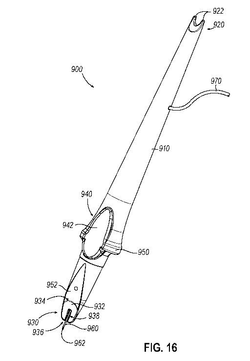
plunger to deliver fluid, etc.). FIGS. 16-18C show an example of a delivery
instrument
(900) that may be grasped and manipulated by a single hand of an operator
while another
hand of the same operator (or the hand of another operator) grasps and
operates a syringe
(980).
[00092] As shown in FIG. 16, instrument (900) of this example includes a
shaft (910) that
is configured for grasping with a single hand of the operator (e.g., via a
pencil grip). A
proximal end of shaft (910) includes an integral marking assembly (920), which
includes
a pair of proximally extending marking prongs (922) that are laterally spaced
apart from
each other. Prongs (922) of this example are blunt or otherwise atraumatic,
such that
prongs (922) may be pressed against a patient's eye (E) without piercing the
sclera (S).
The distal end of shaft (910) includes a head (930) that is used to engage an
eye (E) of a
patient to thereby deliver fluid (e.g., therapeutic agent, etc.) to the eye
(E) as described
herein. Head (930) includes a concave distal face (932). Distal face (932) is
oriented
generally transversely relative to the longitudinal axis of shaft (910).
Distal face (932) is
bounded by an upper edge region (934) and a lower edge region (936), with
regions (934,
936) together defining an outer perimeter of distal face (932). Similar to
distal face (110)
of head (100) described above, distal face (932) of head (930) has a concave
curvature
along longitudinal and lateral dimensions. The curvature of distal face (932)
is
configured to complement the curvature of the exterior surface of the
patient's eye (E).
[00093] Instrument (900) of the present example further includes a
laterally facing
guidance feature (940) and a laterally extending grip feature (950) along an
intermediate
region of shaft (910). Laterally facing guidance feature (940) includes a
concave face
(942) having a curvature that is configured to complement the curvature of a
limbus (L)
of a patient's eye (E). As described in greater detail below, this
configuration may enable
concave face (942) to be used to visually facilitate alignment of instrument
(900) relative
to the eye (E). Grip feature (950) is configured to promote grasping of
instrument (900)
with a single hand, particularly using a pencil grip. Alternatively,
instrument (900) may
be grasped in any other suitable fashion. A laterally facing surface (952)
extends
between concave face (942) of laterally facing guidance feature (940) and
upper edge
region (934) of distal face (932). Surface (952) is obliquely oriented
relative to the
CA 03175283 2022-09-14
WO 2022/172086 PCT/IB2022/000064
- 26 -
longitudinal axis of shaft (910) and has a concave curvature. Surface (952) is
configured
to avoid or minimize contact with the cornea of the eye (E) during use of
instrument
(900), as will be described in greater detail below.
[00094] A needle (960) extends distally from head (930). A portion of
needle (960) is
positioned in a laterally presented recess or trough (938) that is formed in
distal face
(932). Needle (960) has a sharp, beveled tip (962) that is exposed relative to
head (930).
Needle (960) may comprise stainless steel and/or any other suitable
material(s). In the
present example, needle (960) is resiliently flexible, such that the region of
needle (960)
that is exposed relative to head (930) may deform laterally (e.g., as
described below with
reference to FIG. 19). In some scenarios, a portion of needle (960) may flex
laterally out
of trough (938) and away from trough (938). Trough (938) may thus provide
clearance
for lateral deformation of needle (960). Needle (960) may be approximately 30
gauge or
any other suitable size. In this example, needle (960) is straight and extends
along a
needle axis (NA) that is parallel with the longitudinal axis of shaft (912).
In some
versions, the needle axis (NA) is coaxial with the longitudinal axis of shaft
(912). In
some other versions, the needle axis is laterally offset from (yet still
parallel with) the
longitudinal axis of shaft (912). In the present example, the position of
needle (960)
relative to shaft (910) is fixed, such that needle (960) does not translate
longitudinally
relative to shaft (910). In some other versions, needle (960) is translatable
relative to
shaft (910) (e.g., between a proximal retracted position and a distal exposed
position).
[00095] A flexible conduit (970) extends from shaft (910) and provides a
path for fluid
communication with the lumen of needle (960) as will be described in greater
detail
below. Flexible conduit (970) may comprise a transparent flexible tube or may
take any
other suitable form. The interior of shaft (910) may define a lumen providing
a pathway
for fluid communication from flexible conduit (970) to needle (960). In some
variations,
flexible conduit (970) is received such a lumen and connects with a hub at the
proximal
end of needle (960). In some other variations, flexible conduit (970) distally
terminates
in the lumen in shaft (910) and needle (960) proximally terminates in shaft
(910), such
that fluid is communicated from flexible conduit (970) to needle (960) via the
lumen.
Alternatively, flexible conduit (970) may be fluidically coupled with needle
(960) in any
CA 03175283 2022-09-14
WO 2022/172086 PCT/IB2022/000064
- 27 -
other suitable fashion.
[00096] As shown in FIG. 17, instrument (900) may be positioned in relation
to an eye (E)
such that distal face (932) of head (930) is fully seated against the exterior
surface of the
eye (E). Needle (960) enters the sclera (S) at an entry point (EP) that is a
predetermined
distance from the limbus (L) of the eye (E). By way of example only, this
predetermined
distance from the limbus (L) to the entry point (EP) ranges from approximately
3.0 mm
to approximately 4.0 mm; or may be approximately 3.5 mm.
[00097] In some scenarios, this predetermined distance from the limbus (L)
to the entry
point (EP) is achieved by the operator aligning upper edge region (934) of
head (930)
along the limbus (L), such that upper edge region (934) serves as an indexing
feature. In
addition, or in the alternative, this predetermined distance may also be
achieved using
marking prongs (922). The spacing between marking prongs (922) may be selected
to
indicate the appropriate distance between the limbus (L) and the entry point
(EP), such
that marking assembly (920) may be used like calipers. In some such scenarios,
the
operator may first press marking prongs (922) against a pad having
biocompatible ink to
thereby ink the prongs (922); then press the inked marking prongs (922)
against the eye
(E) to thereby mark the entry point (EP) with ink. In some other scenarios,
the operator
may press marking prongs (922) against the eye (E), without ink on marking
prongs
(922), with sufficient force to cause a marking indentation in the sclera (S)
(without
piercing the sclera (S)). Alternatively, marking prongs (922) may be used in
any other
suitable fashion. Alternatively, still, the entry point (EP) may be marked in
any other
suitable fashion.
[00098] Still referring to the arrangement shown in FIG. 17, due to the
structural
configuration and arrangement of distal face (932) and needle (960), the
needle axis (NA)
defines a predetermined angle (0) with a tangent line (TL) at the entry point
(EP). The
tangent line (TL) is tangent to the exterior surface of the eye (E) at the
entry point (EP).
In the present example, this predetermined angle (0) is oblique. By way of
example
only, the angle (0) may range from approximately 0 degrees to approximately 40
degrees; or may be approximately 5 degrees. While the angle (0) is fixed in
the present
example, other versions may allow the operator to selectively vary the angle
(0), as
CA 03175283 2022-09-14
WO 2022/172086 PCT/IB2022/000064
- 28 -
described in various examples provided above.
[00099] With distal face (932) of head (930) fully seated against the
exterior surface of the
eye (E), needle (960) passes fully through the sclera (S) such that tip (962)
is positioned
in the suprachoroidal space (SCS). The length of needle (960) that is exposed
relative to
distal face (932), in view of the angle (0), is selected to allow tip (962) to
reach the
suprachoroidal space (SCS) without penetrating the choroid (Ch). However, in
some
scenarios, tip (962) may incidentally engage the choroid (Ch) without passing
fully
through the choroid (Ch) and reaching the subretinal space between the choroid
(Ch) and
the retina (R). By way of example only, the length of needle (960) that is
exposed
relative to distal face (932) may range from approximately 0.5 mm to
approximately 5.0
mm; or may be approximately 1.25 mm. The length of needle (960) that is
exposed
relative to distal face (932), in combination with the oblique angle (0) at
which needle
(960) enters the eye (E), may reduce the risk of needle (960) undesirably
traversing the
choroid (Ch). In other words, providing an oblique angle (0) for entry of
needle (960)
may provide less risk of undesirable choroid (Ch) penetration as compared to
the risk of
undesirable choroid (Ch) penetration that may be presented by a perpendicular
angle (0)
for entry of needle (960). Similarly, the length of needle (960) that is
exposed relative to
distal face (932), in combination with the oblique angle (0) at which needle
(960) enters
the eye (E), may reduce the risk of needle (960) only reaching an insertion
depth where
tip (962) remains in the sclera (S), without reaching the suprachoroidal space
(SCS). The
configuration and orientation of needle (960) may thus accommodate different
sclera (S)
thicknesses in different patients, with little to no risk of needle (960)
being inserted to a
depth that is too shallow (i.e., not reaching the suprachoroidal space (SCS))
or too deep
(i.e., passing completely through the choroid (Ch) and perhaps even the retina
(R)).
[000100] In the present example, the bevel angle of tip (962) is selected
to promote
communication of fluid out from tip (962) into the suprachoroidal space (SCS).
By way
of example only, this bevel angle may range from approximately 8 degrees to
approximately 20 degrees; or may be approximately 14 degrees. Tip (962) may
have a
tri-bevel configuration or any other suitable configuration. The bevel of tip
(962) in the
present example is angularly oriented to face the choroid (C), to thereby
promote
CA 03175283 2022-09-14
WO 2022/172086 PCT/IB2022/000064
- 29 -
communication of fluid to the suprachoroidal space (SCS) as the fluid exits
tip (962)
when tip is positioned at or near the suprachoroidal space (SCS).
[000101] FIGS. 18A-18C show instrument (900) coupled with a syringe (980)
for use in a
procedure to deliver fluid (e.g., therapeutic agent, etc.) to an eye (E).
Syringe (980) of
this example includes a conventional syringe, such that syringe (980) includes
a barrel
(982) containing fluid, a distal luer fitting (984), and a plunger (986) that
is slidably
disposed in barrel (982). Conduit (970) includes a luer fitting (972) that
removably
couples with luer fitting (984) of syringe (980). An operator may thus drive
fluid from
barrel (982) of syringe (980) into conduit (970), and thereby drive the fluid
to needle
(960) of instrument (900), by advancing plunger (986) distally relative to
barrel (982).
[000102] As shown in FIG. 18A, the operator may initially position
instrument (900)
relative to the eye (E) by positioning distal tip (962) of needle (960) at the
desired entry
point (EP). As noted above, marking assembly (920) may be used like calipers
to mark
the entry point (EP) relative to the limbus (L) before the operator positions
distal tip
(962) of needle (960) at the entry point (EP). At the stage shown in FIG. 18A,
the
operator may refrain from fully advancing needle (960) through the sclera (S).
For
instance, the operator may just initially penetrate the sclera (S) with distal
tip (962) at this
stage, or minimally contact the sclera (S) with distal tip (962), to maintain
the position of
distal tip (962) at the entry point (EP) before fully advancing distal tip
(962) into the eye
(E) as described below. At this stage, head (930) is still spaced away from
the eye (E),
such that the only portion of instrument (900) contacting the eye (E) is
needle (960).
[000103] Once the operator has appropriately positioned distal tip (962) at
the entry point
(E), the operator may then pivot instrument (900) relative to the eye (E),
with the
interface of needle (960) and the entry point (EP) providing the pivot point
for such
pivotal motion. The operator may pivot instrument (900), while visually
observing
instrument (900) along the length of shaft (910) from the proximal end, until
instrument
(900) reaches a point where upper edge region (934) of distal face (932) is
generally
aligned with the limbus (L). As noted above, concave face (942) of guidance
feature
(940) may facilitate visual observation of upper edge region (934) of distal
face (932) in
relation to the limbus (L) during this pivotal movement. Upon reaching the
appropriate
CA 03175283 2022-09-14
WO 2022/172086 PCT/IB2022/000064
- 30 -
pivotal alignment of instrument (900) in relation to the eye (E), the operator
may
complete distal advancement of instrument (900) relative to the eye (E) until
distal face
(932) is fully seated against the eye (E), as shown in FIG. 18B. With distal
face (932)
fully seated against the eye (E), needle (960) may be positioned such that
distal tip (962)
is located precisely at (or substantially at) the suprachoroidal space (SCS)
as shown in
FIG. 17. In some scenarios, as described below with reference to FIG. 19, the
sclera (S)
may deform during the transition from the state shown in FIG. 18A to the state
shown in
FIG. 18B. In some other scenarios, as described below with reference to FIG.
20, the
distal portion of needle (960) may deform during the transition from the state
shown in
FIG. 18A to the state shown in FIG. 18B. In still other scenarios, both the
sclera (S) and
the distal portion of needle (960) may deform during the transition from the
state shown
in FIG. 18A to the state shown in FIG. 18B.
[000104] With distal tip (962) located precisely at (or substantially at)
the suprachoroidal
space (SCS) as shown in FIG. 17, the operator may then advance plunger (986)
distally
relative to barrel (982), as shown in FIG. 18C. Such advancement of plunger
(986) may
drive fluid from barrel (982) through flexible conduit (970), and further
through needle
(960), such that the fluid ultimately exits distal tip (962) into the
suprachoroidal space
(SC S).
[000105] In the present example, since the needle axis (NA) is oriented at
an angle (0) that
is not perpendicular to the suprachoroidal space (SCS), the fluid delivered
via distal tip
(962) may diffuse more easily within the suprachoroidal space (SCS) than the
fluid might
otherwise diffuse in a scenario where the fluid is delivered via a needle that
is oriented
perpendicular to the suprachoroidal space (SCS). In other words, the oblique
angle (0)
of the needle axis (NA) may promote diffusion of the delivered fluid within
the
suprachoroidal space (SCS). The oblique angle (0) of the needle axis (NA) may
also
reduce a risk of the choroid (C) being acutely stretched at the injection
site, which might
otherwise occur in scenarios where the fluid is delivered via a needle that is
oriented
perpendicular to the suprachoroidal space (SCS). Moreover, the oblique angle
(0) of the
needle axis (NA) may reduce the risk of the retina (R) being undesirably
penetrated by
distal tip (962).
CA 03175283 2022-09-14
WO 2022/172086 PCT/IB2022/000064
-31-
10001061 While instrument (900) is described above as being coupled with
syringe (980)
via conduit (970), instrument (900) may instead be coupled with any other
suitable kind
of fluid delivery device or system. For instance, instrument (900) may instead
be coupled
with an automated fluid delivery system. Instrument (900) may also incorporate
any of
the various other features described herein, including but not limited to the
integral
plunger (20) of instrument (10), actuator (250) of head (200) and associated
components,
adjustment member (470) of head (400), electrodes (504) of head (500) and
associated
components, and/or distal projections (830, 840) of head (800).
[000107] In the present example, distal tip (962) never penetrates the
retina (R) and never
reaches the vitreous chamber (V). Thus, distal tip (962) penetrates no further
than the
suprachoroidal space (SCS) or the choroid (Ch). In some other variations, at
least a
portion of distal tip (962) penetrates the retina (R). Moreover, distal tip
(962) may reach
the vitreous chamber (V) in some variations.
[000108] The configuration of instrument (900) as described above with
reference to FIGS.
16-17, and the operation of instrument as described above with reference to
FIGS. 18A-
18C, may facilitate use of instrument (900) in eyes (E) having different
sclera (S)
thicknesses. In other words, instrument (900) may be just as suitable for use
in an eye
(E) having a thick sclera (S) as it is for use in an eye (E) having a thin
sclera (S). This is
illustrated in FIGS. 19-20. In particular, FIG. 19 shows instrument (900)
being used in
an eye (E) having a relatively thin sclera (S). In such an eye (E), when
instrument (900)
is pivoted from the position shown in FIG. 18A to the position shown in FIG.
18B, the
resilience of needle (960) may overcome the resilience of the sclera (S), such
that the
distal end of needle (960) may cause some degree of deformation of the
adjacent region
of the sclera (S), without causing deformation of needle (960). Tip (962) of
needle (960)
may still reach, and remain in, the suprachoroidal space (SCS) to administer a
therapeutic
agent, etc., to the suprachoroidal space (SCS). As shown in FIG. 19, tip (962)
does not
traverse the choroid (Ch) during the procedure. In some cases, tip (962) does
not even
engage the choroid (Ch) during the procedure.
[000109] FIG. 20 shows instrument (900) being used in an eye (E) having a
relatively thick
sclera (S). In such an eye (E), when instrument (900) is pivoted from the
position shown
CA 03175283 2022-09-14
WO 2022/172086 PCT/IB2022/000064
- 32 -
in FIG. 18A to the position shown in FIG. 18B, the resilience of the sclera
(S) may
overcome the resilience of needle (960), such that the sclera (S) may cause
the distal end
of needle (960) to flexibly deform, without causing deformation of sclera (S).
Tip (962)
of needle (960) may still reach, and remain in, the suprachoroidal space (SCS)
to
administer a therapeutic agent, etc., to the suprachoroidal space (SCS). As
shown in FIG.
20, tip (962) does not traverse the choroid (Ch) during the procedure. In some
cases, tip
(962) does not even engage the choroid (Ch) during the procedure.
[000110] While FIG. 19 shows deformation of the sclera (S) without
deformation of needle
(960), and FIG. 20 shows deformation of needle (960) without deformation of
the sclera
(S), there may be scenarios (e.g., where the sclera (S) has a thickness
somewhere between
the thickness shown in FIG. 19 and the thickness shown in FIG. 20) where the
sclera (S)
and needle (960) both deform to some degree. Again, regardless of whether the
sclera
(S) deforms, needle (960) deforms, or both the sclera (S) and needle (960)
deform, tip
(962) of needle (960) may still reach, and remain in, the suprachoroidal space
(SCS) to
administer a therapeutic agent, etc., to the suprachoroidal space (SCS).
Moreover,
regardless of whether the sclera (S) deforms, needle (960) deforms, or both
the sclera (S)
and needle (960) deform, tip (962) of needle (960) may avoid imparting undue
trauma to
the choroid (Ch). Thus, needle (960) may reach, and remain in, the
suprachoroidal space
(SCS) to administer a therapeutic agent, and avoid imparting undue trauma to
the choroid
(Ch), regardless of the thickness of the sclera (S). The length of needle
(960) that is
exposed relative to distal face (932), in combination with the oblique angle
(0) at which
needle (960) enters the eye (E), in further combination with the resilient
flexibility of
needle (960) may further reduce the risk of tip (962) failing to reach the
suprachoroidal
space (SCS) (e.g., where tip (962) may otherwise fail to fully traverse the
sclera (S)).
[000111] II. Examples of Combinations
[000112] The following examples relate to various non-exhaustive ways in
which the
teachings herein may be combined or applied. It should be understood that the
following
examples are not intended to restrict the coverage of any claims that may be
presented at
any time in this application or in subsequent filings of this application. No
disclaimer is
intended. The following examples are being provided for nothing more than
merely
CA 03175283 2022-09-14
WO 2022/172086 PCT/IB2022/000064
- 33 -
illustrative purposes. It is contemplated that the various teachings herein
may be
arranged and applied in numerous other ways. It is also contemplated that some
variations may omit certain features referred to in the below examples.
Therefore, none
of the aspects or features referred to below should be deemed critical unless
otherwise
explicitly indicated as such at a later date by the inventors or by a
successor in interest to
the inventors. If any claims are presented in this application or in
subsequent filings
related to this application that include additional features beyond those
referred to below,
those additional features shall not be presumed to have been added for any
reason relating
to patentability.
[000113] Example 1
[000114] An apparatus, comprising: (a) a body defining a fluid reservoir,
the body having a
distal end; and (b) a head at the distal end of the body, the head including:
(i) a distal
face, the distal face being configured to engage an exterior surface of a
patient's eye at an
anterior region of the patient's eye, (ii) a needle, the needle being
configured to extend
distally from the distal face, the needle having a length sufficient to extend
through a
sclera layer of the patient's eye and thereby position at least a portion of a
distal tip of the
needle in a suprachoroidal space of the patient's eye at an anterior region of
the patient's
eye, the needle being in fluid communication with the fluid reservoir such
that the needle
is operable to deliver fluid from the fluid reservoir into the suprachoroidal
space of the
patient's eye, and (iii) an indexing feature, the indexing feature being
configured to
complement a landmark of the patient's eye to thereby position the needle at a
predetermined location in relation to the landmark of the patient's eye.
[000115] Example 2
[000116] The apparatus of Example 1, the distal face having a three-
dimensional concave
curvature such that the distal face is configured to complement a curvature of
the
patient's eye.
[000117] Example 3
[000118] The apparatus of any one or more of Examples 1 through 2, the
distal face
defining a longitudinal axis, the needle being configured to extend distally
from the distal
CA 03175283 2022-09-14
WO 2022/172086 PCT/IB2022/000064
- 34 -
face along an exit axis that is obliquely oriented in relation to the
longitudinal axis.
[000119] Example 4
[000120] The apparatus of Example 3, the exit axis being configured to
define an exit angle
with the longitudinal axis, the exit angle ranging from approximately 30
degrees to
approximately 0 degrees.
[000121] Example 5
[000122] The apparatus of any one or more of Examples 1 through 4, further
comprising a
plunger slidably coupled with the body, the plunger being operable to drive
fluid from the
fluid reservoir out through the needle.
[000123] Example 6
[000124] The apparatus of any one or more of Examples 1 through 5, the
distal face having
three distal edges joined by corners.
[000125] Example 7
[000126] The apparatus of Example 6, at least one of the distal edges
having a concave
curvature.
[000127] Example 8
[000128] The apparatus of any one or more of Examples 6 through 7, the
indexing feature
being formed by at least one of the distal edges.
[000129] Example 9
[000130] The apparatus of any one or more of Examples 6 through 8, the
corners being
rounded.
[000131] Example 10
[000132] The apparatus of any one or more of Examples 1 through 9, the
indexing feature
including a distal edge of the distal face.
[000133] Example 11
CA 03175283 2022-09-14
WO 2022/172086 PCT/IB2022/000064
- 35 -
[000134] The apparatus of any one or more of Examples 1 through 10, the
indexing feature
being configured to complement a limbus of the patient's eye.
[000135] Example 12
[000136] The apparatus of Example 11, the indexing feature having a
curvature configured
to complement a curvature of the limbus of the patient's eye.
[000137] Example 13
[000138] The apparatus of any one or more of Examples 1 through 12, the
needle being
longitudinally fixed relative to the distal face.
[000139] Example 14
[000140] The apparatus of any one or more of Examples 1 through 13, the
needle extending
along a curve.
[000141] Example 15
[000142] The apparatus of any one or more of Examples 1 through 14, the
needle having a
beveled distal tip, the beveled distal tip defining a bevel angle.
[000143] Example 16
[000144] The apparatus of Example 15, the bevel angle being configured to
be parallel with
boundaries of layers of the patient's eye as the needle is inserted into the
patient's eye.
[000145] Example 17
[000146] The apparatus of any one or more of Examples 1 through 16, the
needle being
operable to translate relative to the distal face.
[000147] Example 18
[000148] The apparatus of Example 17, the needle being configured to
translate between a
proximal position and a distal position, the needle in the proximal position
being
positioned such that a distal tip of the needle is positioned proximally in
relation to the
distal face, the needle in the distal position being positioned such that a
distal tip of the
CA 03175283 2022-09-14
WO 2022/172086 PCT/IB2022/000064
- 36 -
needle is positioned distally in relation to the distal face.
[000149] Example 19
[000150] The apparatus of any one or more of Examples 17 through 18,
further comprising
an actuator, the actuator being operable to drive translation of the needle
relative to the
distal face.
[000151] Example 20
[000152] The apparatus of Example 19, the actuator comprising a slider.
[000153] Example 21
[000154] The apparatus of any one or more of Examples 19 through 20, the
actuator being
positioned on the head.
[000155] Example 22
[000156] The apparatus of any one or more of Examples 1 through 21, further
comprising a
strain sensor, the strain sensor being operable to sense strain encountered by
the needle as
the needle penetrates the eye of the patient.
[000157] Example 23
[000158] The apparatus of Example 22, further comprising a response module,
the response
module being operable to provide a response based on a signal from the strain
sensor.
[000159] Example 24
[000160] The apparatus of Example 23, the response module being configured
to provide a
response based on a signal from the strain sensor indicating that a distal tip
of the needle
has reached a suprachoroidal space in the eye of the patient.
[000161] Example 25
[000162] The apparatus of any one or more of Examples 23 through 24, the
response
module being configured to provide a response including one or more of
tactile, visual, or
audible feedback to an operator.
CA 03175283 2022-09-14
WO 2022/172086 PCT/IB2022/000064
- 37 -
[000163] Example 26
[000164] The apparatus of any one or more of Examples 23 through 25, the
response
module being configured to provide a response including selectively permitting
fluid
communication from the fluid reservoir to the needle.
[000165] Example 27
[000166] The apparatus of Example 26, further comprising a valve interposed
between the
fluid reservoir and the needle, the response module being configured to
selectively open
the valve.
[000167] Example 28
[000168] The apparatus of any one or more of Examples 26 through 27,
further comprising
a pump operable to drive fluid from the fluid reservoir to the needle, the
response module
being configured to activate the pump.
[000169] Example 29
[000170] The apparatus of any one or more of Examples 1 through 28, the
head defining a
passageway containing at least a portion of the needle, a distal portion of
the passageway
defining a curve, the needle extending along the curve of the distal portion
of the
passageway.
[000171] Example 30
[000172] The apparatus of Example 29, the head further defining a
longitudinal axis, the
curve of the distal portion of the passageway orienting a distal portion of
the needle along
an exit axis that is obliquely oriented relative to the longitudinal axis of
the head.
[000173] Example 31
[000174] The apparatus of any one or more of Examples 1 through 30, the
needle including
a distal portion that is resiliently biased to extend along a curve.
[000175] Example 32
CA 03175283 2022-09-14
WO 2022/172086 PCT/IB2022/000064
- 38 -
[000176] The apparatus of Example 31, the needle being configured to
translate between a
proximal position and a distal position, the needle in the proximal position
being
positioned such that a distal tip of the needle is positioned proximally in
relation to the
distal face, the needle in the distal position being positioned such that a
distal tip of the
needle is positioned distally in relation to the distal face.
[000177] Example 33
[000178] The apparatus of Example 32, the head defining a passageway
containing at least
a portion of the needle, the needle being configured to translate along the
passageway
between the distal and proximal positions.
[000179] Example 34
[000180] The apparatus of Example 33, the passageway having a straight
configuration, the
passageway being configured to deform the distal portion of the needle into a
straight
configuration when the needle is in the proximal position.
[000181] Example 35
[000182] The apparatus of Example 34, the resilient bias of the distal
portion of the needle
being configured to urge the distal portion of the needle along a curve as the
distal
portion is exposed relative to the distal face during movement of the needle
from the
proximal position to the distal position.
[000183] Example 36
[000184] The apparatus of any one or more of Examples 1 through 35, further
comprising
an adjustment member, the head defining a longitudinal axis, the adjustment
member
being operable to adjust a needle exit axis in relation to the longitudinal
axis to thereby
adjust an oblique needle exit angle defined between the longitudinal axis and
the needle
exit axis.
[000185] Example 37
[000186] The apparatus of Example 36, the adjustment member being
integrated into the
head.
CA 03175283 2022-09-14
WO 2022/172086 PCT/IB2022/000064
- 39 -
[000187] Example 38
[000188] The apparatus of any one or more of Examples 36 through 37, the
adjustment
member including a wheel, the wheel being rotatable to adjust the needle exit
angle.
[000189] Example 39
[000190] The apparatus of Example 38, the adjustment member further
comprising an axle
defining a rotation axis, the wheel being rotatable about the rotation axis.
[000191] Example 40
[000192] The apparatus of Example 39, the wheel defining a needle
passageway, the needle
extending along the needle passageway.
[000193] Example 41
[000194] The apparatus of Example 40, the needle passageway being
positioned such that
the rotation axis passes through the needle passageway.
[000195] Example 42
[000196] The apparatus of any one or more of Examples 36 through 41, the
adjustment
member including indicia, the indicia being configured to provide visual
feedback
indicating the needle exit angle.
[000197] Example 43
[000198] The apparatus of any one or more of Examples 1 through 42, further
comprising
electrodes and a processing module, the electrodes being configured to contact
tissue, the
processing module being configured to detect a position of the needle in the
patient's eye
based on signals from the electrodes.
[000199] Example 44
[000200] The apparatus of Example 43, the processing module being
configured to detect a
position of the needle in the patient's eye based on an electrical potential
picked up by the
electrodes.
CA 03175283 2022-09-14
WO 2022/172086 PCT/IB2022/000064
- 40 -
[000201] Example 45
[000202] The apparatus of any one or more of Examples 43 through 44, the
electrodes
including at least one electrode on the head, the at least one electrode on
the head being
configured to operate at a first polarity.
[000203] Example 46
[000204] The apparatus of Example 45, the needle forming another one of the
electrodes,
the needle being configured to operate at a second polarity.
[000205] Example 47
[000206] The apparatus of any one or more of Examples 1 through 46, the
head further
including at least one distal projection, the at least one distal projection
being laterally
offset from the needle, the distal face being proximally offset from the
distal projection
such that the distal projection and the distal face are configured to define a
gap, the
needle being configured to extend through the gap.
[000207] Example 48
[000208] The apparatus of Example 47, the at least one distal projection
comprising a
plurality of distal projections positioned in an angularly spaced apart array
about the
needle.
[000209] Example 49
[000210] The apparatus of any one or more of Examples 47 through 48, the at
least one
distal projection comprising a distal projection that continuously spans a
full
circumference about the needle.
[000211] Example 50
[000212] The apparatus of any one or more of Examples 47 through 49, the at
least one
distal projection being configured to engage the eye at one or more
corresponding
positions that are laterally spaced away from a location where the needle
penetrates the
eye, such that the at least one distal projection is configured to maintain
tension in a
CA 03175283 2022-09-14
WO 2022/172086 PCT/IB2022/000064
-41 -
sclera layer of the eye in the location where the needle penetrates the eye.
[000213] Example 51
[000214] An apparatus, comprising: (a) a body defining a fluid reservoir,
the body having a
distal end; and (b) a head at the distal end of the body, the head including:
(i) a distal
face, the distal face being configured to engage an exterior surface of a
patient's eye at an
anterior region of the patient's eye, the distal face having a three-
dimensional concave
curvature such that the distal face is configured to complement a curvature of
the
patient's eye, and (ii) a needle, the needle being configured to extend
distally from the
distal face, the needle having a length sufficient to extend through a sclera
layer of the
patient's eye and thereby position at least a portion of a distal tip of the
needle in a
suprachoroidal space of the patient's eye at an anterior region of the
patient's eye, the
needle being in fluid communication with the fluid reservoir such that the
needle is
operable to deliver fluid from the fluid reservoir into the suprachoroidal
space of the
patient's eye.
[000215] Example 52
[000216] The apparatus of Example 51, further comprising an indexing
feature, the
indexing feature being configured to complement a landmark of the patient's
eye to
thereby position the needle at a predetermined location in relation to the
landmark of the
patient's eye.
[000217] Example 53
[000218] The apparatus of Example 52, the distal face having three distal
edges joined by
corners.
[000219] Example 54
[000220] The apparatus of Example 53, at least one of the distal edges
having a concave
curvature.
[000221] Example 55
[000222] The apparatus of any one or more of Examples 53 through 54, the
indexing
CA 03175283 2022-09-14
WO 2022/172086 PCT/IB2022/000064
- 42 -
feature being formed by at least one of the distal edges.
[000223] Example 56
[000224] The apparatus of any one or more of Examples 52 through 55, the
indexing
feature including a distal edge of the distal face.
[000225] Example 57
[000226] The apparatus of any one or more of Examples 52 through 56, the
indexing
feature being configured to complement a limbus of the patient's eye.
[000227] Example 58
[000228] The apparatus of Example 57, the indexing feature having a
curvature configured
to complement a curvature of the limbus of the patient's eye.
[000229] Example 59
[000230] The apparatus of any one or more of Examples 51 through 58, the
distal face
defining a longitudinal axis, the needle being configured to extend distally
from the distal
face along an exit axis that is obliquely oriented in relation to the
longitudinal axis.
[000231] Example 60
[000232] The apparatus of Example 59, the exit axis being configured to
define an exit
angle with the longitudinal axis, the exit angle ranging from approximately 30
degrees to
approximately 0 degrees.
[000233] Example 61
[000234] The apparatus of any one or more of Examples 51 through 60,
further comprising
a plunger slidably coupled with the body, the plunger being operable to drive
fluid from
the fluid reservoir out through the needle.
[000235] Example 62
[000236] The apparatus of any one or more of Examples 51 through 61, the
distal face
having three distal edges joined by corners.
CA 03175283 2022-09-14
WO 2022/172086 PCT/IB2022/000064
- 43 -
[000237] Example 63
[000238] The apparatus of Example 62, at least one of the distal edges
having a concave
curvature.
[000239] Example 64
[000240] The apparatus of any one or more of Examples 62 through 63, the
corners being
rounded.
[000241] Example 65
[000242] The apparatus of any one or more of Examples 51 through 64, the
needle being
longitudinally fixed relative to the distal face.
[000243] Example 66
[000244] The apparatus of any one or more of Examples 51 through 65, the
needle
extending along a curve.
[000245] Example 67
[000246] The apparatus of any one or more of Examples 51 through 66, the
needle having a
beveled distal tip, the beveled distal tip defining a bevel angle.
[000247] Example 68
[000248] The apparatus of Example 67, the bevel angle being configured to
be parallel with
boundaries of layers of the patient's eye as the needle is inserted into the
patient's eye.
[000249] Example 69
[000250] The apparatus of any one or more of Examples 51 through 68, the
needle being
operable to translate relative to the distal face.
[000251] Example 70
[000252] The apparatus of Example 69, the needle being configured to
translate between a
proximal position and a distal position, the needle in the proximal position
being
positioned such that a distal tip of the needle is positioned proximally in
relation to the
CA 03175283 2022-09-14
WO 2022/172086 PCT/IB2022/000064
- 44 -
distal face, the needle in the distal position being positioned such that a
distal tip of the
needle is positioned distally in relation to the distal face.
[000253] Example 71
[000254] The apparatus of any one or more of Examples 69 through 70,
further comprising
an actuator, the actuator being operable to drive translation of the needle
relative to the
distal face.
[000255] Example 72
[000256] The apparatus of Example 71, the actuator comprising a slider.
[000257] Example 73
[000258] The apparatus of any one or more of Examples 71 through 72, the
actuator being
positioned on the head.
[000259] Example 74
[000260] The apparatus of any one or more of Examples 51 through 73,
further comprising
a strain sensor, the strain sensor being operable to sense strain encountered
by the needle
as the needle penetrates the eye of the patient.
[000261] Example 75
[000262] The apparatus of Example 74, further comprising a response module,
the response
module being operable to provide a response based on a signal from the strain
sensor.
[000263] Example 76
[000264] The apparatus of Example 75, the response module being configured
to provide a
response based on a signal from the strain sensor indicating that a distal tip
of the needle
has reached a suprachoroidal space in the eye of the patient.
[000265] Example 77
[000266] The apparatus of any one or more of Examples 75 through 76, the
response
module being configured to provide a response including one or more of
tactile, visual, or
CA 03175283 2022-09-14
WO 2022/172086 PCT/IB2022/000064
- 45 -
audible feedback to an operator.
[000267] Example 78
[000268] The apparatus of any one or more of Examples 75 through 77, the
response
module being configured to provide a response including selectively permitting
fluid
communication from the fluid reservoir to the needle.
[000269] Example 79
[000270] The apparatus of Example 78, further comprising a valve interposed
between the
fluid reservoir and the needle, the response module being configured to
selectively open
the valve.
[000271] Example 80
[000272] The apparatus of any one or more of Examples 78 through 79,
further comprising
a pump operable to drive fluid from the fluid reservoir to the needle, the
response module
being configured to activate the pump.
[000273] Example 81
[000274] The apparatus of any one or more of Examples 51 through 80, the
head defining a
passageway containing at least a portion of the needle, a distal portion of
the passageway
defining a curve, the needle extending along the curve of the distal portion
of the
passageway.
[000275] Example 82
[000276] The apparatus of Example 81, the head further defining a
longitudinal axis, the
curve of the distal portion of the passageway orienting a distal portion of
the needle along
an exit axis that is obliquely oriented relative to the longitudinal axis of
the head.
[000277] Example 83
[000278] The apparatus of any one or more of Examples 51 through 82, the
needle
including a distal portion that is resiliently biased to extend along a curve.
CA 03175283 2022-09-14
WO 2022/172086 PCT/IB2022/000064
- 46 -
[000279] Example 84
[000280] The apparatus of Example 83, the needle being configured to
translate between a
proximal position and a distal position, the needle in the proximal position
being
positioned such that a distal tip of the needle is positioned proximally in
relation to the
distal face, the needle in the distal position being positioned such that a
distal tip of the
needle is positioned distally in relation to the distal face.
[000281] Example 85
[000282] The apparatus of Example 84, the head defining a passageway
containing at least
a portion of the needle, the needle being configured to translate along the
passageway
between the distal and proximal positions.
[000283] Example 86
[000284] The apparatus of Example 85, the passageway having a straight
configuration, the
passageway being configured to deform the distal portion of the needle into a
straight
configuration when the needle is in the proximal position.
[000285] Example 87
[000286] The apparatus of Example 86, the resilient bias of the distal
portion of the needle
being configured to urge the distal portion of the needle along a curve as the
distal
portion is exposed relative to the distal face during movement of the needle
from the
proximal position to the distal position.
[000287] Example 88
[000288] The apparatus of any one or more of Examples 51 through 87,
further comprising
an adjustment member, the head defining a longitudinal axis, the adjustment
member
being operable to adjust a needle exit axis in relation to the longitudinal
axis to thereby
adjust an oblique needle exit angle defined between the longitudinal axis and
the needle
exit axis.
[000289] Example 89
CA 03175283 2022-09-14
WO 2022/172086 PCT/IB2022/000064
- 47 -
[000290] The apparatus of Example 88, the adjustment member being
integrated into the
head.
[000291] Example 90
[000292] The apparatus of any one or more of Examples 88 through 89, the
adjustment
member including a wheel, the wheel being rotatable to adjust the needle exit
angle.
[000293] Example 91
[000294] The apparatus of Example 90, the adjustment member further
comprising an axle
defining a rotation axis, the wheel being rotatable about the rotation axis.
[000295] Example 92
[000296] The apparatus of Example 91, the wheel defining a needle
passageway, the needle
extending along the needle passageway.
[000297] Example 93
[000298] The apparatus of Example 92, the needle passageway being
positioned such that
the rotation axis passes through the needle passageway.
[000299] Example 94
[000300] The apparatus of any one or more of Examples 88 through 93, the
adjustment
member including indicia, the indicia being configured to provide visual
feedback
indicating the needle exit angle.
[000301] Example 95
[000302] The apparatus of any one or more of Examples 51 through 94,
further comprising
electrodes and a processing module, the electrodes being configured to contact
tissue, the
processing module being configured to detect a position of the needle in the
patient's eye
based on signals from the electrodes.
[000303] Example 96
[000304] The apparatus of Example 95, the processing module being
configured to detect a
CA 03175283 2022-09-14
WO 2022/172086 PCT/IB2022/000064
- 48 -
position of the needle in the patient's eye based on an electrical potential
picked up by the
electrodes.
[000305] Example 97
[000306] The apparatus of any one or more of Examples 95 through 96, the
electrodes
including at least one electrode on the head, the at least one electrode on
the head being
configured to operate at a first polarity.
[000307] Example 98
[000308] The apparatus of Example 97, the needle forming another one of the
electrodes,
the needle being configured to operate at a second polarity.
[000309] Example 99
[000310] The apparatus of any one or more of Examples 51 through 98, the
head further
including at least one distal projection, the at least one distal projection
being laterally
offset from the needle, the distal face being proximally offset from the
distal projection
such that the distal projection and the distal face are configured to define a
gap, the
needle being configured to extend through the gap.
[000311] Example 100
[000312] The apparatus of Example 99, the at least one distal projection
comprising a
plurality of distal projections positioned in an angularly spaced apart array
about the
needle.
[000313] Example 101
[000314] The apparatus of any one or more of Examples 99 through 100, the
at least one
distal projection comprising a distal projection that continuously spans a
full
circumference about the needle.
[000315] Example 102
[000316] The apparatus of any one or more of Examples 99 through 101, the
at least one
distal projection being configured to engage the eye at one or more
corresponding
CA 03175283 2022-09-14
WO 2022/172086 PCT/IB2022/000064
- 49 -
positions that are laterally spaced away from a location where the needle
penetrates the
eye, such that the at least one distal projection is configured to maintain
tension in a
sclera layer of the eye in the location where the needle penetrates the eye.
[000317] Example 103
[000318] An apparatus, comprising: (a) a body defining a fluid reservoir,
the body having a
distal end; and (b) a head at the distal end of the body, the head including:
(i) a distal
face, the distal face being configured to engage an exterior surface of a
patient's eye at an
anterior region of the patient's eye, the distal face defining a longitudinal
axis, and (ii) a
needle, the needle being configured to extend distally from the distal face
along an exit
axis that is obliquely oriented in relation to the longitudinal axis, the
needle having a
length sufficient to extend through a sclera layer of the patient's eye and
thereby position
at least a portion of a distal tip of the needle in a suprachoroidal space of
the patient's eye
at an anterior region of the patient's eye, the needle being in fluid
communication with
the fluid reservoir such that the needle is operable to deliver fluid from the
fluid reservoir
into the suprachoroidal space of the patient's eye.
[000319] Example 104
[000320] The apparatus of Example 103, further comprising an indexing
feature, the
indexing feature being configured to complement a landmark of the patient's
eye to
thereby position the needle at a predetermined location in relation to the
landmark of the
patient's eye.
[000321] Example 105
[000322] The apparatus of Example 104, the distal face having three distal
edges joined by
corners.
[000323] Example 106
[000324] The apparatus of Example 105, at least one of the distal edges
having a concave
curvature.
[000325] Example 107
CA 03175283 2022-09-14
WO 2022/172086 PCT/IB2022/000064
- 50 -
[000326] The apparatus of any one or more of Examples 105 through 106, the
indexing
feature being formed by at least one of the distal edges.
[000327] Example 108
[000328] The apparatus of any one or more of Examples 104 through 107, the
indexing
feature including a distal edge of the distal face.
[000329] Example 109
[000330] The apparatus of any one or more of Examples 104 through 108, the
indexing
feature being configured to complement a limbus of the patient's eye.
[000331] Example 110
[000332] The apparatus of Example 109, the indexing feature having a
curvature
configured to complement a curvature of the limbus of the patient's eye.
[000333] Example 111
[000334] The apparatus of any one or more of Examples 103 through 110, the
exit axis
being configured to define an exit angle with the longitudinal axis, the exit
angle ranging
from approximately 30 degrees to approximately 0 degrees.
[000335] Example 112
[000336] The apparatus of any one or more of Examples 103 through 111,
further
comprising a plunger slidably coupled with the body, the plunger being
operable to drive
fluid from the fluid reservoir out through the needle.
[000337] Example 113
[000338] The apparatus of any one or more of Examples 103 through 112, the
distal face
having three distal edges joined by corners.
[000339] Example 114
[000340] The apparatus of Example 113, at least one of the distal edges
having a concave
curvature.
CA 03175283 2022-09-14
WO 2022/172086 PCT/IB2022/000064
-51 -
[000341] Example 115
[000342] The apparatus of any one or more of Examples 113 through 114, the
corners
being rounded.
[000343] Example 116
[000344] The apparatus of any one or more of Examples 103 through 115, the
needle being
longitudinally fixed relative to the distal face.
[000345] Example 117
[000346] The apparatus of any one or more of Examples 103 through 116, the
needle
extending along a curve.
[000347] Example 118
[000348] The apparatus of any one or more of Examples 103 through 117, the
needle
having a beveled distal tip, the beveled distal tip defining a bevel angle.
[000349] Example 119
[000350] The apparatus of Example 118, the bevel angle being configured to
be parallel
with boundaries of layers of the patient's eye as the needle is inserted into
the patient's
eye.
[000351] Example 120
[000352] The apparatus of any one or more of Examples 103 through 119, the
needle being
operable to translate relative to the distal face.
[000353] Example 121
[000354] The apparatus of Example 120, the needle being configured to
translate between a
proximal position and a distal position, the needle in the proximal position
being
positioned such that a distal tip of the needle is positioned proximally in
relation to the
distal face, the needle in the distal position being positioned such that a
distal tip of the
needle is positioned distally in relation to the distal face.
CA 03175283 2022-09-14
WO 2022/172086 PCT/IB2022/000064
- 52 -
[000355] Example 122
[000356] The apparatus of any one or more of Examples 120 through 121,
further
comprising an actuator, the actuator being operable to drive translation of
the needle
relative to the distal face.
[000357] Example 123
[000358] The apparatus of Example 122, the actuator comprising a slider.
[000359] Example 124
[000360] The apparatus of any one or more of Examples 122 through 123, the
actuator
being positioned on the head.
[000361] Example 125
[000362] The apparatus of any one or more of Examples 103 through 124,
further
comprising a strain sensor, the strain sensor being operable to sense strain
encountered by
the needle as the needle penetrates the eye of the patient.
[000363] Example 126
[000364] The apparatus of Example 125, further comprising a response
module, the
response module being operable to provide a response based on a signal from
the strain
sensor.
[000365] Example 127
[000366] The apparatus of Example 126, the response module being configured
to provide
a response based on a signal from the strain sensor indicating that a distal
tip of the
needle has reached a suprachoroidal space in the eye of the patient.
[000367] Example 128
[000368] The apparatus of any one or more of Examples 126 through 127, the
response
module being configured to provide a response including one or more of
tactile, visual, or
audible feedback to an operator.
CA 03175283 2022-09-14
WO 2022/172086 PCT/IB2022/000064
- 53 -
[000369] Example 129
[000370] The apparatus of any one or more of Examples 126 through 128, the
response
module being configured to provide a response including selectively permitting
fluid
communication from the fluid reservoir to the needle.
[000371] Example 130
[000372] The apparatus of Example 129, further comprising a valve
interposed between the
fluid reservoir and the needle, the response module being configured to
selectively open
the valve.
[000373] Example 131
[000374] The apparatus of any one or more of Examples 129 through 130,
further
comprising a pump operable to drive fluid from the fluid reservoir to the
needle, the
response module being configured to activate the pump.
[000375] Example 132
[000376] The apparatus of any one or more of Examples 103 through 131, the
head
defining a passageway containing at least a portion of the needle, a distal
portion of the
passageway defining a curve, the needle extending along the curve of the
distal portion of
the passageway.
[000377] Example 133
[000378] The apparatus of Example 132, the head further defining a
longitudinal axis, the
curve of the distal portion of the passageway orienting a distal portion of
the needle along
the exit axis that is obliquely oriented relative to the longitudinal axis of
the head.
[000379] Example 134
[000380] The apparatus of any one or more of Examples 103 through 133, the
needle
including a distal portion that is resiliently biased to extend along a curve.
[000381] Example 135
CA 03175283 2022-09-14
WO 2022/172086 PCT/IB2022/000064
- 54 -
[000382] The apparatus of Example 134, the needle being configured to
translate between a
proximal position and a distal position, the needle in the proximal position
being
positioned such that a distal tip of the needle is positioned proximally in
relation to the
distal face, the needle in the distal position being positioned such that a
distal tip of the
needle is positioned distally in relation to the distal face.
[000383] Example 136
[000384] The apparatus of Example 135, the head defining a passageway
containing at least
a portion of the needle, the needle being configured to translate along the
passageway
between the distal and proximal positions.
[000385] Example 137
[000386] The apparatus of Example 136, the passageway having a straight
configuration,
the passageway being configured to deform the distal portion of the needle
into a straight
configuration when the needle is in the proximal position.
[000387] Example 138
[000388] The apparatus of Example 137, the resilient bias of the distal
portion of the needle
being configured to urge the distal portion of the needle along a curve as the
distal
portion is exposed relative to the distal face during movement of the needle
from the
proximal position to the distal position.
[000389] Example 139
[000390] The apparatus of any one or more of Examples 103 through 138,
further
comprising an adjustment member, the head defining a longitudinal axis, the
adjustment
member being operable to adjust a needle exit axis in relation to the
longitudinal axis to
thereby adjust an oblique needle exit angle defined between the longitudinal
axis and the
needle exit axis.
[000391] Example 140
[000392] The apparatus of Example 139, the adjustment member being
integrated into the
head.
CA 03175283 2022-09-14
WO 2022/172086 PCT/IB2022/000064
- 55 -
[000393] Example 141
[000394] The apparatus of any one or more of Examples 139 through 140, the
adjustment
member including a wheel, the wheel being rotatable to adjust the needle exit
angle.
[000395] Example 142
[000396] The apparatus of Example 141, the adjustment member further
comprising an axle
defining a rotation axis, the wheel being rotatable about the rotation axis.
[000397] Example 143
[000398] The apparatus of Example 142, the wheel defining a needle
passageway, the
needle extending along the needle passageway.
[000399] Example 144
[000400] The apparatus of Example 143, the needle passageway being
positioned such that
the rotation axis passes through the needle passageway.
[000401] Example 145
[000402] The apparatus of any one or more of Examples 139 through 144, the
adjustment
member including indicia, the indicia being configured to provide visual
feedback
indicating the needle exit angle.
[000403] Example 146
[000404] The apparatus of any one or more of Examples 103 through 145,
further
comprising electrodes and a processing module, the electrodes being configured
to
contact tissue, the processing module being configured to detect a position of
the needle
in the patient's eye based on signals from the electrodes.
[000405] Example 147
[000406] The apparatus of Example 146, the processing module being
configured to detect
a position of the needle in the patient's eye based on an electrical potential
picked up by
the electrodes.
CA 03175283 2022-09-14
WO 2022/172086 PCT/IB2022/000064
- 56 -
[000407] Example 148
[000408] The apparatus of any one or more of Examples 146 through 147, the
electrodes
including at least one electrode on the head, the at least one electrode on
the head being
configured to operate at a first polarity.
[000409] Example 149
[000410] The apparatus of Example 148, the needle forming another one of
the electrodes,
the needle being configured to operate at a second polarity.
[000411] Example 150
[000412] The apparatus of any one or more of Examples 103 through 149, the
head further
including at least one distal projection, the at least one distal projection
being laterally
offset from the needle, the distal face being proximally offset from the
distal projection
such that the distal projection and the distal face are configured to define a
gap, the
needle being configured to extend through the gap.
[000413] Example 151
[000414] The apparatus of Example 150, the at least one distal projection
comprising a
plurality of distal projections positioned in an angularly spaced apart array
about the
needle.
[000415] Example 152
[000416] The apparatus of any one or more of Examples 150 through 151, the
at least one
distal projection comprising a distal projection that continuously spans a
full
circumference about the needle.
[000417] Example 153
[000418] The apparatus of any one or more of Examples 150 through 152, the
at least one
distal projection being configured to engage the eye at one or more
corresponding
positions that are laterally spaced away from a location where the needle
penetrates the
eye, such that the at least one distal projection is configured to maintain
tension in a
CA 03175283 2022-09-14
WO 2022/172086 PCT/IB2022/000064
- 57 -
sclera layer of the eye in the location where the needle penetrates the eye.
[000419] Example 154
[000420] The apparatus of any one or more of Examples 103 through 153, the
distal face
having a three-dimensional concave curvature such that the distal face is
configured to
complement a curvature of the patient's eye.
[000421] Example 155
[000422] An apparatus, comprising: (a) a shaft having: (i) a proximal end,
(ii) a distal end,
and (iii) a fluid pathway; and (b) a head at the distal end of the shaft, the
head including:
(i) a distal face, the distal face being configured to engage an exterior
surface of a
patient's eye at an anterior region of the patient's eye, the distal face
having a concave
curvature contoured to complement a curvature of the patient's eye, and (ii) a
needle, the
needle being configured to extend distally from the distal face, the needle
having a length
sufficient to extend through a sclera layer of the patient's eye and thereby
position at least
a portion of a distal tip of the needle in a suprachoroidal space of the
patient's eye at an
anterior region of the patient's eye while the distal face is seated against
the patient's eye,
the needle being in fluid communication with the fluid pathway such that the
needle is
operable to deliver fluid from the fluid pathway into the suprachoroidal space
of the
patient's eye.
[000423] Example 156
[000424] The apparatus of Example 155, the head further including an
indexing feature, the
indexing feature being configured to complement a landmark of the patient's
eye to
thereby position the needle at a predetermined location in relation to the
landmark of the
patient's eye.
[000425] Example 157
[000426] The apparatus of Example 156, the distal face having an edge, the
edge defining
the indexing feature.
[000427] Example 158
CA 03175283 2022-09-14
WO 2022/172086 PCT/IB2022/000064
- 58 -
[000428] The apparatus of any of Examples 156 through 157, the indexing
feature being
configured to complement a limbus of the patient's eye.
[000429] Example 159
[000430] The apparatus of Example 158, the indexing feature having a
curvature
configured to complement a curvature of the limbus of the patient's eye.
[000431] Example 160
[000432] The apparatus of any of Examples 155 through 159, the needle being
longitudinally fixed relative to the distal face.
[000433] Example 161
[000434] The apparatus of any of Examples 155 through 160, the distal face
having a three-
dimensional concave curvature.
[000435] Example 162
[000436] The apparatus of any of Examples 155 through 161, the shaft
defining a
longitudinal axis, the distal face being oriented transversely relative to the
longitudinal
axis.
[000437] Example 163
[000438] The apparatus of any of Examples 155 through 162, the shaft
defining a
longitudinal axis, the needle extending along an exit axis that is parallel
with the
longitudinal axis of the shaft.
[000439] Example 164
[000440] The apparatus of any of Examples 155 through 163, the needle
having a straight
configuration.
[000441] Example 165
[000442] The apparatus of any of Examples 155 through 164, the needle
having a beveled
distal tip, the beveled distal tip defining a bevel angle.
CA 03175283 2022-09-14
WO 2022/172086 PCT/IB2022/000064
- 59 -
[000443] Example 166
[000444] The apparatus of Example 165, the bevel angle being configured to
be parallel
with boundaries of layers of the patient's eye as the needle is inserted into
the patient's
eye.
[000445] Example 167
[000446] The apparatus of any of Examples 155 through 166, the fluid
pathway comprising
a flexible conduit.
[000447] Example 168
[000448] The apparatus of Example 167, further comprising a syringe coupled
with the
flexible conduit.
[000449] Example 169
[000450] The apparatus of Example 168, the syringe being spaced apart from
the shaft via
the flexible conduit.
[000451] Example 170
[000452] The apparatus of any of Examples 155 through 169, the shaft
further including a
laterally facing guidance feature, the laterally facing guidance feature being
configured to
complement a landmark of the patient's eye to thereby position the needle at a
predetermined orientation in relation to the patient's eye.
[000453] Example 171
[000454] The apparatus of Example 170, the laterally facing guidance
feature including a
concave face configured to complement the curvature of the landmark of the
patient's
eye.
[000455] Example 172
[000456] The apparatus of Example 171, the landmark including a limbus of
the patient's
eye.
CA 03175283 2022-09-14
WO 2022/172086 PCT/IB2022/000064
- 60 -
[000457] Example 173
[000458] The apparatus of any of Examples 155 through 172, the shaft
further including a
marking assembly, the marking assembly being configured to mark a needle entry
point
in relation to a landmark of the patient's eye.
[000459] Example 174
[000460] The apparatus of Example 173, the marking assembly being
positioned at the
proximal end of the shaft.
[000461] Example 175
[000462] The apparatus of any of Examples 173 through 174, the marking
assembly
including two prongs fixedly spaced apart from each other by a predetermined
distance.
[000463] Example 176
[000464] The apparatus of Example 175, each prong of the two prongs having
an
atraumatic configuration.
[000465] Example 177
[000466] The apparatus of any of Examples 173 through 176, the marking
assembly being
configured to mark a needle entry point in relation to a limbus of the
patient's eye.
[000467] III. Miscellaneous
[000468] To the extent that several examples herein are described in the
context of a
cannula guide being positioned near an already-formed scleral incision (23),
it should be
understood that other kinds of procedures may be employed. For instance, in
some
variations of the procedures described herein, the cannula guide may be
secured to the
eye (20) first; and then the scleral incision (23) may be formed after the
cannula guide
has been secured to the eye (20). Other suitable steps and sequences that may
be carried
out in procedures that include a combination of a scleral incision (23) and a
cannula
guide will be apparent to those skilled in the art in view of the teachings
herein.
[000469] It should be understood that any of the versions of the
instruments described
CA 03175283 2022-09-14
WO 2022/172086 PCT/IB2022/000064
- 61 -
herein may include various other features in addition to or in lieu of those
described
above. By way of example only, any of the devices herein may also include one
or more
of the various features disclosed in any of the various references that are
incorporated by
reference herein.
[000470] It should be understood that any one or more of the teachings,
expressions,
embodiments, examples, etc. described herein may be combined with any one or
more of
the other teachings, expressions, embodiments, examples, etc. that are
described herein.
The above-described teachings, expressions, embodiments, examples, etc. should
therefore not be viewed in isolation relative to each other. Various suitable
ways in
which the teachings herein may be combined will be readily apparent to those
skilled in
the art in view of the teachings herein. Such modifications and variations are
intended to
be included within the scope of the claims.
[000471] It should be appreciated that any patent, publication, or other
disclosure material,
in whole or in part, that is said to be incorporated by reference herein is
incorporated
herein only to the extent that the incorporated material does not conflict
with existing
definitions, statements, or other disclosure material set forth in this
disclosure. As such,
and to the extent necessary, the disclosure as explicitly set forth herein
supersedes any
conflicting material incorporated herein by reference. Any material, or
portion thereof,
that is said to be incorporated by reference herein, but which conflicts with
existing
definitions, statements, or other disclosure material set forth herein will
only be
incorporated to the extent that no conflict arises between that incorporated
material and
the existing disclosure material.
[000472] Versions described above may be designed to be disposed of after a
single use, or
they can be designed to be used multiple times. Versions may, in either or
both cases, be
reconditioned for reuse after at least one use. Reconditioning may include any
combination of the steps of disassembly of the device, followed by cleaning or
replacement of particular pieces, and subsequent reassembly. In particular,
some
versions of the device may be disassembled, and any number of the particular
pieces or
parts of the device may be selectively replaced or removed in any combination.
Upon
cleaning and/or replacement of particular parts, some versions of the device
may be
CA 03175283 2022-09-14
WO 2022/172086 PCT/IB2022/000064
- 62 -
reassembled for subsequent use either at a reconditioning facility, or by an
operator
immediately prior to a procedure. Those skilled in the art will appreciate
that
reconditioning of a device may utilize a variety of techniques for
disassembly,
cleaning/replacement, and reassembly. Use of such techniques, and the
resulting
reconditioned device, are all within the scope of the present application.
[000473] By way of example only, versions described herein may be
sterilized before
and/or after a procedure. In one sterilization technique, the device is placed
in a closed
and sealed container, such as a plastic or TYVEK bag. The container and device
may
then be placed in a field of radiation that can penetrate the container, such
as gamma
radiation, x-rays, or high-energy electrons. The radiation may kill bacteria
on the device
and in the container. The sterilized device may then be stored in the sterile
container for
later use. A device may also be sterilized using any other technique known in
the art,
including but not limited to beta or gamma radiation, ethylene oxide, or
steam.
[000474] Having shown and described various embodiments of the present
invention,
further adaptations of the methods and systems described herein may be
accomplished by
appropriate modifications by one of ordinary skill in the art without
departing from the
scope of the present invention. Several of such potential modifications have
been
mentioned, and others will be apparent to those skilled in the art. For
instance, the
examples, embodiments, geometrics, materials, dimensions, ratios, steps, and
the like
discussed above are illustrative and are not required. Accordingly, the scope
of the
present invention should be considered in terms of the following claims and is
understood
not to be limited to the details of structure and operation shown and
described in the
specification and drawings.