Note: Descriptions are shown in the official language in which they were submitted.
WO 2021/219396 1
PCT/EP2021/059857
SENSORY PERCEPTION SURGICAL SYSTEM FOR ROBOT-ASSISTED
LAPAROSCOPIC SURGERY
FIELD OF THE INVENTION
The present invention generally relates to the field of robot-assisted
surgery.
Particularly, the invention relates to a sensory perception surgical system
for robot-
assisted laparoscopic surgery which allows detecting the properties of a
patient's
tissue, particularly the contact force exerted on the tissue, through the
measurement of
electrical impedance.
BACKGROUND OF THE INVENTION
Current robot-assisted laparoscopic surgery techniques allow carrying out high-
precision interventions, providing relevant advantages, particularly in
surgeries of
certain complexity, for example in surgeries where it is difficult to access
the operation
site. Nevertheless, current robot-assisted laparoscopy surgery techniques
present the
drawback of the surgeon not perceiving the forces exerted on the anatomical
elements
of the patient.
Robotic arms are used in a robot-assisted laparoscopic surgery for actuating
specific
tools which allow performing the intervention effectively, and for introducing
and
guiding a camera which allows viewing the operative field. These robotic arms
are
remotely controlled by a surgeon by means of a control panel provided with a
screen
that allows the surgeon to monitor the scene. Likewise, besides improving
surgical
precision, the use of computers associated with robotic arm control also
allows
introducing controls that provide greater safety to the patient.
In recent years, significant efforts have been made in the field of research
to enable
providing sensory return to the surgeon making up for the loss of tactile
sensation
when the intervention is a manual intervention.
Patent application US 2011046659-A1 describes a minimally invasive surgical
tool
including a sensor that generates a signal in response to an interaction with
the
surgical tool. The tool further includes a haptic feedback system that
generates
vibrations to obtain a haptic effect in response to the signal.
CA 03175843 2022- 10- 17
WO 2021/219396 2
PCT/EP2021/059857
On the other hand, patent US8613230-B2 discloses a system which allows
measuring
forces from a sensor installed in the outer part of the cannula of the
surgical tool, and
receives the forces in the axis of penetration Z through a mechanical
transmission
sheath. The system described in this patent only allows perceiving said forces
exerted
on the axis of penetration Z, and not the forces derived from lateral
contacts.
To enable perceiving forces exerted not only in the direction of the axis of
penetration
Z, elastic elements which allow measuring three-dimensional deformations by
means
of interferometry using optical sensors have been used, like in the case of
patent
EP2595587-B1. In this case, 3 or 4 optic fibers which allow projecting
modulated light
on a reflector located on an elastic support are used and the force vector
applied on
the forceps is obtained by means of interferometry from the outer part of the
cannula.
Patent CA 2870343 presents an alternative to the use of elastic elements
integrated on
the cannula. To that end, a sensor with 6 degrees of freedom is used, which
sensor
allows supplying the forces and torques produced between the outer distal end
of the
tool and the end where the tool is held with the robotic arm which supports
same. The
system includes a computer system which allows calculating, by means of matrix
calculus, the forces applied on the distal end based on the kinematics of the
tool-trocar
assembly and the 6 pieces of data supplied by the sensor.
W02016153561 discloses a medical instrument that comprises an elongate body
having a proximal end and a distal end and a pair of electrodes or electrode
portions
(for example, a split-tip electrode assembly). The system is configured to
perform
contact sensing and/or ablation confirmation based on electrical measurements
obtained while energy of different frequencies are applied to the pair of
electrodes or
electrode portions. The contact sensing systems and methods may calibrate
network
parameter measurements to compensate for a hardware unit in a network
parameter
measurement circuit or to account for differences in cables, instrumentation
or
hardware used.
US10595745-B2 discloses devices and methods for measuring a contact force on a
catheter. The catheter includes a proximal segment, a distal segment, and an
elastic
segment extending from the proximal segment to the distal segment. The distal
segment includes a plurality of tip electrodes including at least three radial
electrodes
disposed about a circumference of the distal segment. The radial electrodes
are
configured to output electrical signals indicative of a contact vector of the
contact force.
CA 03175843 2022- 10- 17
3
WO 2021/219396
PCT/EP2021/059857
The elastic segment includes a force sensing device configured to output an
electrical
signal indicative of a magnitude of an axial component of the contact force,
wherein the
contact force is determined by scaling the magnitude of the axial component of
the
contact force by the contact vector. In this document, the measurement of the
contact
force is done mechanically, not electrically as in present invention.
US2003100892-A1 discloses a robotic surgical tool that includes an elongate
shaft
having a working end and a shaft axis, and a pair of linking arms each having
a
proximal end and a distal end. The proximal end is pivotally mounted on the
working
end of the shaft to rotate around a first pitch axis to produce rotation in
first pitch. A
wrist member has a proximal portion pivotally connected to the distal end of
the linking
arm to rotate around a second pitch axis to produce rotation in second pitch.
An end
effector is pivotally mounted on a distal portion of the wrist member to
rotate around a
wrist axis of the wrist member to produce rotation in distal roll. The wrist
axis extends
between the proximal portion and the distal portion of the wrist member. The
elongate
shaft is rotatable around the shaft axis to produce rotation in proximal roll.
At about 900
pitch, the wrist axis is generally perpendicular to the shaft axis. The
proximal roll
around the shaft axis and the distal roll around the wrist axis do not
overlap. The use of
the linking arms allows the end effector to be bent back beyond 90 pitch. The
ability to
operate the end effector at about 90 pitch and to bend back the end effector
renders
the wrist mechanism more versatile and adaptable to accessing hard to reach
locations, particularly with small entry points such as those involving
spinal, neural, or
rectal surgical sites.
In another line of work known as Vison-Based Force Sensing (VBFS), the actual
images captured by the laparoscopic camera are used to view the tissue
deformation
caused by contact with the forceps.
In any case, sensory return has practically not been used in robot-assisted
laparoscopic surgery due to technique limitations, the imprecision of
different
developed systems, or the difficulties it entails, particularly the space
occupied by the
sensorization on the cannula of the tool.
New surgical systems for robot-assisted laparoscopic surgery, which allow
detecting
the properties of a tissue/tissues and quantifying the sensory return of the
contact force
exerted on the tissue/tissues during a surgical intervention performed
remotely, are
therefore required.
CA 03175843 2022- 10- 17
4
WO 2021/219396
PCT/EP2021/059857
DESCRIPTION OF THE INVENTION
To that end, the embodiments of the present invention provide a sensory
perception
surgical system for robot-assisted laparoscopic surgery comprising: an
electrosurgical
forceps coupled to a surgical tool, an impedance measurement circuit and an
electrocautery radiofrequency signal generator electrically coupled to the
impedance
measurement circuit and operable for supplying energy, as both monopolar and
bipolar
energy, to the electrosurgical forceps. The impedance measurement circuit
includes a
measurement sensor for measuring a signal indicative of a magnitude
corresponding to
the value of a contact impedance between the electrosurgical forceps and a
patient's
tissue; an oscillator for providing a power signal to the measurement sensor;
a first
electrical circuit and a second electronic circuit. The first electrical
circuit includes one
or more resistors and a voltage limiter for protecting the measurement sensor
and the
oscillator that are connected to the electrosurgical forceps by means of a
power cable
of the surgical tool. The second electronic circuit comprises a first switch
circuit for
commutating between the connection and the disconnection of a power cabling of
the
electrocautery radiofrequency signal generator with respect to the cable of
the surgical
tool, and a second switch circuit for commutating between the connection and
the
disconnection of the electrocautery radiofrequency signal generator and the
measurement sensor.
Likewise, the proposed system includes at least one processor operatively
connected
to the electrocautery radiofrequency signal generator and to the impedance
measurement circuit for receiving said signal measured by the measurement
sensor
and converting same into a force vector. Particularly, the modulus of the
force vector is
a function of the measured contact impedance and the argument is defined by
the
trajectory the surgical tool follows in the moment of contact.
Therefore, the mentioned processor allows obtaining the vectorial reaction
force on the
operator's controls, both in magnitude and in orientation, based on the
measured
magnitude of the contact impedance, which varies according to the force being
exerted, and on the monitoring of the trajectory being followed.
In one embodiment, the proposed system also includes a radiofrequency detector
with
at least one capacitive or inductive sensor arranged on the mentioned power
cabling
for automatically commutating the first and second switch circuits while
supplying
energy.
CA 03175843 2022- 10- 17
5
WO 2021/219396
PCT/EP2021/059857
In one embodiment, the energy supplied by the electrocautery radiofrequency
signal
generator is monopolar. In this case, the first switch circuit is formed by
one relay and
the second switch circuit is formed by another relay. Alternatively, when the
supplied
energy is bipolar, the first switch circuit is formed by at least two relays
and the second
switch circuit is also formed by at least two relays.
The system may further include a control unit comprising control elements
operatively
connected to the impedance measurement circuit and/or to the electrocautery
radiofrequency signal generator for the control thereof. For example, the
control
elements may include pedals and/or actuators/push buttons.
The processor may be included in the control unit or in a remote computation
device
and operatively connected to the control unit, the electrocautery
radiofrequency signal
generator, and/or the impedance measurement circuit by means of a cable or
wireless
connection.
In one embodiment, the electrosurgical forceps are coupled to the surgical
tool using a
set of pulleys and cables which allow the opening or closing, as well as the
mobility, of
the forceps. At least one of the pulleys is arranged on the articulation shaft
thereof.
Likewise, the set of pulleys is arranged on three parallel shafts arranged in
a
diametrical position with respect to the surgical tool and to a body of the
electrosurgical
forceps.
Other embodiments of the invention disclosed herein also include a computer-
implemented method and/or computer program products for performing the steps
and
operations performed by the mentioned processor. More particularly, a computer
program product is an embodiment having a computer system-readable medium
including code instructions coded therein which, when executed in at least one
processor of the computer system, cause the processor to perform the
operations
indicated herein as embodiments of the invention.
In one embodiment, the anatomy of the surroundings of the tissue/tissues is
modeled
based on the force vector estimated by the processor. To that end, the surface
is
progressively modeled by means of defining polygonal surfaces, such as for
example
triangles that are being formed by joining adjacent contact points obtained
during the
operation/intervention.
CA 03175843 2022- 10- 17
WO 2021/219396 6
PCT/EP2021/059857
Therefore, the present invention allows determining the force vector based on
the
measurement of a magnitude of the contact impedance between the forceps and
the
patient's tissues and on the trajectory taken, and it also allows constructing
a three-
dimensional model of the surgical environment.
One advantage provided by the present invention is that it does not introduce
any
additional sensor on the electrosurgical forceps, which allows being able to
use the
same conductors used for carrying out electrocauterization or
electrocoagulation, for
example.
BRIEF DESCRIPTION OF THE DRAWINGS
The foregoing and other features and advantages will be better understood
based on
the following detailed description of several merely illustrative and non-
limiting
embodiments in reference to the attached drawings in which:
Fig. 1 illustrates a surgical system for robot-assisted laparoscopic surgery
for detecting
the properties of a tissue, according to an embodiment of the present
invention.
Figs. 2A-2C schematically illustrate different connection configurations of an
electrocautery radiofrequency signal generator for working in monopolar mode
(Fig.
2A) or bipolar mode (Figs. 2B and 2C).
Fig. 3 illustrates in more detail the architecture of the system proposed for
obtaining the
contact impedance and the associated force vector, according to an embodiment
of the
present invention.
Fig. 4 illustrates another embodiment of the architecture of the system
proposed for
obtaining the contact impedance and the associated force vector.
Figs. 5A and 5B show different views of the electrosurgical forceps coupled to
the
surgical tool. Fig. 5A shows a perspective view of a distal end of the
surgical tool
showing rotations G1 and G2 of the articulations thereof and axial rotation G3
of the
surgical tool assembly. Fig. 5B shows the pulleys for the transmission of
movements
G1 and G2 and the arrangement of the actuator cables which also allow the
opening or
closing of the electrosurgical forceps through rotation G1.
Figs. 6A-6D illustrate different views showing the path of the cables which
transmit
energy to the electrosurgical forceps for detecting contact with the tissue,
where said
CA 03175843 2022- 10- 17
WO 2021/219396 7
PCT/EP2021/059857
path must be compatible with the limited space available between the different
pulleys,
and also allows carrying out rotations G1, G2, and G3.
Figs. 7A-70 graphically depict the calculated force vector and the
construction of the
triangles for modeling the anatomy of the environment, according to an
embodiment of
the present invention.
DETAILED DESCRIPTION OF THE INVENTION
The present invention provides a sensory perception surgical system for robot-
assisted
laparoscopic surgery and a method allowing obtaining the sensory return of the
force
exerted by a surgeon on a patient's tissue/tissues during a surgical
intervention
performed remotely based on an estimate of the force vector exerted by
detecting the
contact impedance with the tissue/tissues and on the trajectory taken.
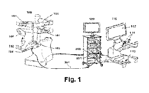
With reference to Fig. 1, said figure shows an embodiment of the proposed
system 1.
In this embodiment, the system 1 comprises a robot-assisted system 100; a
control unit
110; a laparoscopy tower 120 housing an electrocautery radiofrequency signal
generator 300 and an impedance measurement circuit 301.
The robot-assisted system 100 is provided with robotic arms 101 which allow
moving
surgical tools 102, as well as a laparoscopic camera 103. The control unit 110
includes
actuators/push buttons 111 and pedals 113 with which the surgeon can
handle/control
the robot-assisted system 100, the electrocautery radiofrequency signal
generator 300,
as well as the impedance measurement circuit 301. The control unit 110 also
has a
display screen 112.
The electrocautery radiofrequency signal generator 300, which can be any
standard
electrocauterization signal generator, is electrically connected to the
impedance
measurement circuit 301 by means of a power cable 314 and is operable for
supplying
energy to the electrosurgical forceps 104 (see Figs. 2A-20, for example)
coupled to the
surgical tools 102. The impedance measurement circuit 301 is electrically
connected to
the electrosurgical forceps 104 by means of another power cable 304. The power
cable
304 is formed by two conductor cables 304a, 304b (see Fig. 6D), the path of
which is
compatible with the kinematics of the electrosurgical forceps 102, allowing
the
movements thereof in three rotations/axes (orientation and elevation
movements, as
CA 03175843 2022- 10- 17
WO 2021/219396 8
PCT/EP2021/059857
well as opening and/or closing movements) to enable detecting contact with the
tissue/tissues.
The electrocautery radiofrequency signal generator 300 can be electrically
monopolar
when the return circuit is the patient him/herself or the saline medium used
(Fig. 2A), or
it can be electrically bipolar (Figs. 2B and 2C) if the current flows between
the terminal
element 250 (see Figs. 5A-5B) of the electrosurgical forceps 104.
Fig. 2A shows a monopolar configuration. The impedance measurement circuit 301
only houses one cable, i.e., the outgoing power cable. The incoming cable,
marked
with the arrow, passes outside the impedance measurement circuit 301. Fig. 2B
shows
a first bipolar configuration. The double incoming and outgoing cable with two
polarities, marked with arrows, exits the electrocautery radiofrequency signal
generator
300 and passes through the impedance measurement circuit 301, wherein the
return is
taken back through a conducting cannula. Fig. 2C shows a second bipolar
configuration. The cable with two conductor wires exiting the electrocautery
radiofrequency signal generator 300 pass through the impedance measurement
circuit
301 and travel along the inside thereof, one to each part of the
electrosurgical forceps
104.
Now with reference to Fig. 3, said figure shows another embodiment of the
proposed
system 1, comprising in this case the electrosurgical forceps 104 coupled to
the
surgical tool 102 of the robot-assisted system 100; the impedance measurement
circuit
301 for measuring the contact impedance with the environment 303 of the
tissue/tissues; the electrocautery radiofrequency signal generator 300; and a
computer
system or device 311 formed by at least one processor for estimating the
applied
forces based on the measurement of the impedance.
The difficulty entailed by use of the electrocautery radiofrequency signal
generator 300
to enable also measuring contact impedance lies in the fact that
radiofrequency pulses
having a very high voltage of between about 1000 and 3000 volts are used to
enable
carrying out electrocoagulation and electrocauterization. For this reason, the
use or
inclusion of the impedance measurement circuit 301 in the proposed system 1
makes
the measurement of the impedance at a low voltage and current compatible with
the
high electrocoagulation and electrocauterization energy at a high voltage.
CA 03175843 2022- 10- 17
9
WO 2021/219396
PCT/EP2021/059857
To achieve the mentioned compatibility, the impedance measurement circuit 301
includes a measurement sensor 310, particularly a low-voltage measurement
sensor,
for measuring the magnitude corresponding to the value of the contact
impedance; an
electronic module comprising two switch circuits 305, 306 for the
connection/disconnection of the power cabling 314 with respect to the power
cable
304, and for the connection/disconnection of the electrocautery radiofrequency
signal
generator 300 and the measurement sensor 310, respectively.
Likewise, the impedance measurement circuit 301 also includes an oscillator
209 to
enable measuring the impedance without applying any current, however weak it
may
be, with a continuous component, on the patient. The oscillator 209 provides a
signal
having a low voltage, for example 6 V, and a medium frequency, for example 20
KHz,
which is applied in a monopolar or bipolar manner to the surgical tool 102
through the
second switch circuit 306, the contacts of which are usually kept closed. Said
low
voltage is normally not applied to the electrocautery radiofrequency signal
generator
300 since the contact of the first switch circuit 305 is usually open.
In the embodiment of Fig. 3, each of the switch circuits 305, 306 comprises
two relays
Al, A2, Bl, B2. This configuration is particularly useful when the energy
supplied by
the electrocautery radiofrequency signal generator 300 is bipolar. In other
embodiments not illustrated in this case, and particularly when the energy
supplied by
the electrocautery radiofrequency signal generator 300 is monopolar, each of
the
switch circuits 305, 306 only includes one relay Al, Bl.
In operation, when the surgeon applies the energy for carrying out
electrocoagulation
or electrocauterization, the contact of relay Al, or relays Al, A2 of the
first switch circuit
305 must be closed, while at the same time the contact of relay Bl, or relays
Bl, B2 of
the second switch circuit 306 must be open. To that end, the system 1 also
particularly
includes a radiofrequency detector 313 having a capacitive or inductive sensor
312 on
the power cable 314, which allows automatically commutating the first and
second
switch circuits 305, 306 while energy is being applied. Alternatively, this
function may
be performed by introducing the actuation signal of the pedals 113 connected
to the
electrocautery radiofrequency signal generator 300.
In the example of Fig. 3 and for the purpose of preventing damage in the
electrocautery
radiofrequency signal generator 300 and/or in the impedance measurement
circuit 301,
CA 03175843 2022- 10- 17
WO 2021/219396 10
PCT/EP2021/059857
for example as a result of surges in the commutation of the relays, the system
1 is
particularly protected with resistors 307 and a voltage limiter 308.
The signal/magnitude corresponding to the value of the impedance obtained by
the
measurement sensor 310 is treated by the processor 311 for conversion into a
force
vector, in which the force magnitude is given by the value of the impedance
being
measured and the argument of the vector is defined by the direction in space
of the
trajectory that the surgical tool 102 follows in the moment of contact and is
controlled
by the control unit 110 which is connected to the processor 311 through a
communication channel 321.
Fig. 4 shows another embodiment of the proposed system 1. In this case, the
system 1
is formed by the electrosurgical forceps 104 coupled to the surgical tool 102;
the
impedance measurement circuit 301 for measuring the contact impedance with the
environment 303 of the tissue/tissues; and the computer system or device 311
comprising at least one processor. The impedance measurement circuit 301
includes
the measurement sensor 310, the oscillator 309, and an electronic circuit
formed by the
resistors 307 and the voltage limiter 308. Compatibility with high external
voltages like
in the case of using the electrocautery radiofrequency signal generator 300 is
therefore
permitted.
Each surgical tool 102 (see Figs. 5A and 5B) is made up of a cannula 201
supporting a
first articulated element or body 202 which can carry out rotation G1 with
respect to the
end of the cannula 201 about shaft 204 actuated by a drum 207. The body 202
supports the terminal element 250 of the electrosurgical forceps 104 the
orientation of
which can be varied by carrying out rotation G2 with respect to the body 202
about
shaft 206 actuated by means of drums 208 and 209.
Likewise, cables C1a, C1b, C2, 03, 04, and 05 and a set of pulleys 210, 211,
212,
213, 220, 221, 222, 223, 230, 231 allow transmitting the movement from drive
means
to which each surgical tool 102 is connected, and are adapted to enable
carrying out
rotation G1 about shaft 204, which entails a mechanical complexity that
hinders the
introduction of the electrical cables 304a and 304b. This mechanical
complexity is of
great relevance since the electrical conductors for measuring the impedance
must
share the smaller space available with the two cables C1a and C1b which
transmit
rotational movement G1 to the drum 207, and the four cables C2, 03, 04, and 05
CA 03175843 2022- 10- 17
WO 2021/219396 11
PCT/EP2021/059857
which transmit the orientation and opening or closing of the electrosurgical
forceps 104
by means of drums 208 and 209 (Fig. 5B).
To allow rotation G1 the mentioned set of pulleys 210, 211, 212, 213, 220,
221, 222,
223, 230, 231 is used, in which at least one, preferably all, of said pulleys
is/are
arranged on the articulation shaft thereof (Fig. 6A). Particularly, as
observed in Fig. 6B,
a set of pulleys is arranged for the four cables C2, C3, C4, and C5 which move
the
electrosurgical forceps 104, mounted on three parallel shafts 203, 204, and
205 in a
diametrical position with respect to the cannula 201 and to the body 202 (Fig.
5B). The
central shaft 204 joins the cannula 201 and the body 202, which allows
carrying out
rotation G1, and supports the 4 pulleys 220, 221, 222, and 223 joining the two
pairs of
antagonist cables transmitting the movement of the forceps, whereas the two
shafts
203 and 205 support accompanying pulleys.
This arrangement of pulleys on three consecutive shafts for each cable that
must go
through articulation G1 offers a clear advantage over other embodiments, given
that
besides allowing the generation of a guided cable passage between consecutive
pulleys, like in the case of pulleys 210 and 220 which create passage 214 (see
Fig.
5C), constituting a secure guiding of the movement of each cable, two free
spaces are
created on pulleys 230 and 231 allowing the passage of the necessary
electrical cable
304a and 304b to enable measuring the impedance.
The fact that all the pulleys are arranged on the central plane of the cannula
201 and of
the body 202 allows the pulleys to have the largest possible diameter without
exceeding the maximum gauge of the cannula 201. Likewise, with the 4 + 4 + 2
pulleys
required for the transmission of movements having the largest possible
diameter, the
present invention allows reducing the radius of curvature of the different
cables on the
pulleys, improving the durability and reliability of the surgical tool 102.
The electrical
cable 304a and 304b going through the free spaces on the pulleys 230 is
integral with
cables 02 and 03, assuring that that it does not support any mechanical force
when
deflexion of the electrosurgical forceps 104 on axis G2 occurs (Fig. 6D).
The embodiments of the present invention also provide a sensory perception
method
for estimating or calculating the reaction force vector that must be perceived
by the
surgeon or the operator in the control unit 110, through the push
buttons/actuators 111
and/or pedals 113, based on the value/magnitude of the obtained impedance.
CA 03175843 2022- 10- 17
WO 2021/219396 12
PCT/EP2021/059857
Figs. 7A-7C graphically illustrate an example of the foregoing. Given that
there is no
force sensor on the surgical tool 102 which allows the direct measurement of
the
contact force 410 (Fig. 7A), it is estimated indirectly by the processor 311
as a force
vector. The force vector 411 is estimated as a reflected vector of the contact
force 410,
the modulus of which is equal to the modulus of the contact force 410, whereas
the
argument thereof is defined by being on the same plane 416, and which is
defined by
the two passage points 414 and 415 before the perceived contact point, the
normal 412
of the contact surface 413, and an angle of reflection 418 that is equal to
the angle
incidence 417.
The contact surface 413 which allows carrying out positioning calculations in
space of
the reflected vector is not known. Therefore, the proposed method obtains an
approximation of the configuration of the surface of the anatomical elements
of the
environment by performing modeling 400 in a three-dimensional space. To that
end,
the method comprises generating a triangulation 402 (i.e., generating a series
of
triangles 403) from the contact points 404 that are perceived throughout the
operation,
by means of joining same. Each new perceived contact point 404 (Fig. 9C)
causes a
triangle 403 to be broken down into new triangles 405 and 406. In this manner,
the
environment modeling resolution which allows obtaining the argument of the
force
vector 411, which is applied as a reaction force on the controls of the
control unit 110
and generates the sensory return to the surgeon/operator, progressively
increases.
The proposed invention can be implemented in hardware, software, firmware, or
any
combination thereof. If it is implemented in software, the functions can be
stored in or
coded as one or more instructions or code in a computer-readable medium.
The scope of the present invention is defined in the attached claims.
CA 03175843 2022- 10- 17