Note: Descriptions are shown in the official language in which they were submitted.
Description
Title of Invention
NOVEL PROTEIN CONJUGATE, AND USE THEREOF FOR PREVENTING OR
TREATING NONALCOHOLIC STEATOHEPATITIS, OBESITY AND DIABETES
Technical Field
The present invention relates to a protein conjugate comprising polyubiquitin,
a
carrier linked to the polyubiquitin, and two or more biomolecules linked to
the polyubiquitin
or the carrier. In addition, the present invention relates to a pharmaceutical
composition for
preventing or treating non-alcoholic steatohepatitis, fatty liver, liver
fibrosis, cirrhosis, liver
cancer, obesity, and diabetes, comprising the protein conjugate that comprises
two or more
biomolecules.
Background Art
Non-alcoholic fatty liver disease (NAFLD) is a type of disease that shows
similar
histological manifestation to alcoholic hepatitis even with little or no
alcohol intake, and is a
type of metabolic syndrome that covers non-alcoholic fatty liver (NAFL), non-
alcoholic
steatohepatitis (NASH), cirrhosis, and liver cancer (hepatocellular
carcinomas). Non-
alcoholic fatty liver disease is on the rise as the population with obesity
and diabetes
increases, and the annual incidence rate in Korea is about 16%.
Non-alcoholic steatohepatitis (NASH) is mainly characterized by abnormal fat
accumulation or deposition in the liver (hepatic steatosis), liver
inflammation and liver
damage or liver tissue damage (fibrosis). The worldwide prevalence of non-
alcoholic
steatohepatitis is 2-4% (3-12% of adults in the United States). It is well
known that non-
alcoholic steatohepatitis exhibits a faster histological progression and can
progress to
cirrhosis, whereas simple steatosis exhibits a slow histological progression.
About 5-10% of
those diagnosed with fatty liver develop steatohepatitis (Metabolism Clinical
and
Experimental 65 (2016) 1038-1048).
Currently, there is no therapeutic agent on the market for treating non-
alcoholic
steatohepatitis, and due to the absence of a therapeutic agent therefor, other
therapeutic agents
for metabolic syndrome such as abdominal obesity, hyperlipidemia, and
diabetes, for
example, insulin resistance improving drugs, antioxidants (for example,
Vitamins C and E),
dyslipidemia therapeutic agents, hepatoprotective agents, and the like are
used, but these are
hardly considered to be therapeutic agents for directly treating non-alcoholic
steatohepatitis.
1
CA 03175852 2022- 10- 17
Since non-alcoholic steatohepatitis has the nature of a complex disease, the
licensing
authorities in the United States, Europe, and the like have set strict drug
approval
requirements for therapeutic agents therefor. For this reason, recently, a
number of therapeutic
agents have failed in the clinical development stage, and accordingly,
therapeutic agents
based on a multiple agonist for simultaneously improving various indicators
are emerging.
Non-alcoholic steatohepatitis therapeutic agents based on a multiple agonist,
which are
currently in clinical trials, includes MEDI0382 from AstraZeneca, a GLP-
1/glucagon (GCG)
dual agonist, LY3298176 from Eli Lilly, a GIP/GLP-1 dual agonist, HM15211 from
Hanmi
Pharm.Co.,Ltd., a glucagon/GIP/GLP-1 triple agonist, and the like.
Obesity refers to a state of excess body fat, and can be defined as a health
risk
condition due to excess body fat that causes several diseases, including heart
disease. The
World Health Organization (WHO) has reported that there are more than 106
million
overweight adults worldwide, at least 4 million are obese, and more than 2 in
3 Americans are
overweight or obese (Low et al, 2009; Cooke and Bloom, 2006). Obese people
have an
increased risk of several diseases, including type 2 diabetes, hyperlipidemia,
arthritis and
apnea, as compared to those having normal body weight, and obesity is known to
cause
several types of cancer and heart disease.
Diabetes mellitus is a metabolic disease in which a high blood glucose level
persists
for a long time due to insufficient insulin secretion or insulin resistance.
As the blood glucose
level in the body persists for a long time, glycation products invade the
retina, kidneys,
nerves, or large and small blood vessels throughout the body, causing chronic
complications.
Since diabetes complications are more dangerous than diabetes mellitus itself,
the biggest
goal in diabetes treatment today is to suppress the occurrence or progression
of diabetes
complications. Representative complications of diabetes include diabetic
retinopathy, diabetic
cataract, diabetic nephropathy, diabetic neuropathy, diabetic heart disease,
diabetic
osteoporosis, diabetic atherosclerosis, and the like.
In the development of therapeutic agents based on a multiple agonist, due to
the
problem of reduced activity due to structural limitations, currently there are
no studies on
therapeutic agents based on a higher than quadruple agonist for non-alcoholic
steatohepatitis,
fatty liver, liver fibrosis, cirrhosis, liver cancer, obesity, or diabetes.
Accordingly, the present
inventors have worked tirelessly to develop a therapeutic agent based on a
quadruple
agonist/antagonist for non-alcoholic steatohepatitis, fatty liver, liver
fibrosis, cirrhosis, liver
cancer, obesity, and diabetes. As a result, the present invention was
completed using a
polyubiquitin structure.
2
CA 03175852 2022- 10- 17
Prior Art Document
Patent Document
(Patent Document 0001) Korean Patent Application Publication No. 10-2016-
0032699
(Patent Document 0002) Korean Patent No. 10-2034607
Detailed Description of Invention
Technical Problem
An object of the present invention is to provide a protein conjugate
comprising
polyubiquitin, a carrier linked to the polyubiquitin, and two or more
biomolecules linked to
the polyubiquitin or the carrier.
In addition, the present invention provides a pharmaceutical composition for
preventing or treating non-alcoholic steatohepatitis (NASH), fatty liver,
liver fibrosis,
cirrhosis, liver cancer, obesity, or diabetes, comprising the protein
conjugate that comprises
two or more biomolecules. Specifically, another object of the present
invention is to provide a
pharmaceutical composition for preventing or treating non-alcoholic
steatohepatitis, fatty
liver, liver fibrosis, cirrhosis, liver cancer, obesity, or diabetes,
comprising a GCG/GLP-
1/FGF21/GIP or GCG/GLP-1/FGF21/IL-1RA acceptor quadruple agonist/antagonist.
Solution to Problem
The present invention provides a protein conjugate, characterized in that the
protein
conjugate comprises: polyubiquitin, a carrier linked directly or by a linker
to the
polyubiquitin, and a biomolecule linked directly or by a linker to the
polyubiquitin or the
carrier, wherein the polyubiquitin is composed of (i) an acceptor ubiquitin
containing one or
more unsubstituted lysines in which lysines of the ubiquitin may be
substituted with arginine
or alanine, and (ii) a donor ubiquitin in which all lysines of the ubiquitin
are substituted with
arginine or alanine, and the biomolecule is two or more selected from the
group consisting of
GCG, GLP-1, FGF21, GIP and IL-1 RA; analogues thereof; a GCG and GLP-1 dual
acceptor
agonist; and a GLP-1 and GIP dual acceptor agonist.
In one embodiment, the GCG and analogue thereof may be selected from the group
consisting of proteins composed of the amino acid sequences of SEQ ID NOs: 1
to 3, and
preferably, the GCG analogue may be a protein composed of the amino acid
sequence of SEQ
ID NO: 2.
In one embodiment, the GLP-1 and analogue thereof may be selected from the
group
consisting of proteins composed of the amino acid sequences of SEQ ID NOs: 4
to 14, and
3
CA 03175852 2022- 10- 17
preferably, the GLP-1 analogue may be a protein composed of the amino acid
sequence of
SEQ ID NO: 12.
In one embodiment, the GCG and GLP-1 dual acceptor agonist may be selected
from
the group consisting of proteins composed of the amino acid sequences of SEQ
ID NOs: 15
and 16.
In one embodiment, the FGF21 and analogue thereof may be selected from the
group
consisting of proteins composed of the amino acid sequences of SEQ ID NOs: 17
to 21.
Preferably, the FGF21 analogue may be a protein composed of the amino acid
sequence of
SEQ ID NO: 20.
In one embodiment, the GIP and analogue thereof may be selected from the group
consisting of proteins composed of the amino acid sequences of SEQ ID NOs: 22
to 26.
Preferably, the GIP and analogue thereof may be a protein composed of the
amino acid
sequence of SEQ ID NO: 24.
In one embodiment, the IL-1 RA and analogue thereof may be selected from the
group consisting of proteins composed of the amino acid sequences of SEQ ID
NOs: 27 and
28. Preferably, the IL-1RA may be a protein composed of the amino acid
sequence of SEQ ID
NO: 27.
In one embodiment, the polyubiquitin may be composed of an acceptor ubiquitin
in
which the 6th, 11th, 27th, 29th, 33rd, and 48th lysines from the N-terminus of
the ubiquitin
are substituted with arginine and a donor ubiquitin in which all lysines of
the ubiquitin are
substituted with arginine. Preferably, the polyubiquitin may be composed of an
acceptor
ubiquitin composed of the amino acid sequence of SEQ ID NO: 30 and a donor
ubiquitin
composed of the amino acid sequence of SEQ ID NO: 31.
In one embodiment, the linker may be a polypeptide composed of an amino acid
sequence in which 1 to 30 repeats of GGGGS, EAAAK or VPPPPP are combined.
Preferably,
the linker may be a polypeptide composed of the amino acid sequence of SEQ ID
NO: 29.
In one embodiment, the carrier may be selected from the group consisting of
albumin, antibody fragment, scFc (single chain Fc), scFc dimer (single chain
Fc-dimer),
transferrin, XTEN (genetic fusion of non-exact repeat peptide sequence), CTP
(carboxy-
terminal peptide), PAS (proline-alanine-serine polymer), ELK (elastin-like
peptide), HAP
(homo-amino acid polymer), GLK (gelatin-like protein), PEG (polyethylene
glycol), and
fatty acid. Preferably, the carrier may be albumin.
The present invention provides a protein conjugate represented by the
following
formula:
4
CA 03175852 2022- 10- 17
X-L-Y-L-Ub(A)-L-A-L-Z
I
W-L-Ub(D)
in the above formula, W, X, Y and Z may be each a biomolecule selected from
the
group consisting of a GCG analogue composed of the amino acid sequence of SEQ
ID NO: 2,
a GLP-1 analogue composed of the amino acid sequence of SEQ ID NO: 12, an
FGF21
analogue composed of the amino acid sequence of SEQ ID NO: 20, a GIP analogue
composed of the amino acid sequence of SEQ ID NO: 24, and IL-1RA composed of
the
amino acid sequence of SEQ ID NO: 27, Ub(A) may be an acceptor ubiquitin
composed of
the amino acid sequence of SEQ ID NO: 30, Ub(D) may be a donor ubiquitin
composed of
the amino acid sequence of SEQ ID NO: 31, L may be each independently absent
or a linker,
A may be a carrier, and Ub(A) and Ub(D) may be linked by a covalent bond.
In one embodiment, X may be a GCG analogue composed of the amino acid
sequence of SEQ ID NO: 2, Y may be a GLP-1 analogue composed of the amino acid
sequence of SEQ ID NO: 12, Z may be an FGF21 analogue composed of the amino
acid
sequence of SEQ ID NO: 20, and W may be a GIP analogue composed of the amino
acid
sequence of SEQ ID NO: 24 or IL-1RA composed of the amino acid sequence of SEQ
ID
NO: 27.
In one embodiment, the linker may be a polypeptide composed of the amino acid
sequence of SEQ ID NO: 29, and the carrier may be albumin.
The present invention provides a pharmaceutical composition for preventing or
treating non-alcoholic steatohepatitis (NASH), fatty liver, liver fibrosis,
cirrhosis, liver
cancer, obesity, or diabetes, comprising the protein conjugate.
The present invention provides a method for preventing or treating non-
alcoholic
steatohepatitis (NASH), fatty liver, liver fibrosis, cirrhosis, liver cancer,
obesity, or diabetes,
comprising administering the composition to a subject other than a human.
In addition, the present invention provides a method for preparing a protein
conjugate, characterized in that the method comprises the steps of:
(i) preparing an acceptor protein represented by the following formula;
X-L-Y-L-Ub(A)-L-A-L-Z
(ii) preparing a donor protein represented by the following formula; and
W-L-Ub(D)
(iii) linking Ub(A) in the acceptor protein and Ub(D) in the donor protein,
wherein W, X, Y and Z are each a biomolecule selected from the group
consisting of
CA 03175852 2022- 10- 17
a GCG analogue composed of the amino acid sequence of SEQ ID NO: 2, a GLP-1
analogue
composed of the amino acid sequence of SEQ ID NO: 12, an FGF21 analogue
composed of
the amino acid sequence of SEQ ID NO: 20, a GIP analogue composed of the amino
acid
sequence of SEQ ID NO: 24, and IL-1RA composed of the amino acid sequence of
SEQ ID
NO: 27, Ub(A) is an acceptor ubiquitin composed of the amino acid sequence of
SEQ ID
NO: 30, Ub(D) is a donor ubiquitin composed of the amino acid sequence of SEQ
ID NO: 31,
L is each independently absent or a linker, and A is a carrier.
Effects of Invention
The novel protein conjugate of the present invention comprises two or more
biomolecules, and inhibits steatosis, inflammation, and fibrosis in the liver,
thereby having an
excellent effect on preventing or treating non-alcoholic steatohepatitis
(NASH), fatty liver,
liver fibrosis, cirrhosis, or liver cancer, and thus, can be also usefully
used for the prevention
or treatment of obesity or diabetes. In addition, the protein conjugate can be
maintained for a
long time in the body to exert its effect for a long time, and has excellent
safety by using
polyubiquitin and a carrier harmless to the human body.
Brief Description of Drawings
Figure 1 illustrates the results obtained by confirming the purification of an
acceptor
protein by SDS-PAGE.
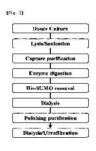
Figure 2 is a schematic diagram showing the purification process of a donor
protein.
Figures 3 and 4 illustrate the results obtained by confirming the purification
of a
donor protein through a Ni column by SDS-PAGE.
Figures 5 and 6 illustrate the results obtained by confirming the purification
of a
donor protein through His-SUMO removal by SDS-PAGE.
Figures 7 and 8 illustrate the results of polishing purification of a donor
protein.
Figure 9 is a schematic diagram showing a process of preparing a protein
conjugate
through conjugation of an acceptor protein and a donor protein.
Figures 10 and 11 illustrate the results obtained by confirming conjugation of
an
acceptor protein and a donor protein by SDS-PAGE.
Figure 12 illustrates the results obtained by confirming the Ni purification
of a
protein conjugate by chromatography.
Figures 13 and 14 illustrate the results obtained by confirming the
purification of a
protein conjugate by SDS-PAGE.
Figure 15 illustrates the results of SDS-PAGE of a final protein conjugate.
6
CA 03175852 2022- 10- 17
Figures 16 to 18 are graphs showing the results of the cAMP accumulation
assay.
Figure 19 is a graph showing the results of the FGFR/KLB functional assay.
Figure 20 is a graph showing the results of the NF-KB reporter assay.
Figures 21 and 22 are graphs showing the results obtained by measuring alanine
aminotransferase (ALT) and aspartate aminotransferase (AST) in mouse blood in
an in-vivo
efficacy test.
Figures 23 to 26 illustrate the results obtained by observing steatosis and
inflammation (lobular inflammation) in the mouse liver tissue in an in-vivo
efficacy test and
the scores according to the NAS evaluation criteria.
Figures 27 and 28 are graphs showing the results obtained by measuring TGT-0
and
triglyceride concentrations in the mouse liver tissue in an in-vivo efficacy
test.
Figures 29 and 30 are graphs showing the results obtained by measuring alanine
aminotransferase (ALT) and aspartate aminotransferase (AST) in mouse blood in
an in-vivo
efficacy test.
Figures 31 to 34 illustrate the results obtained by observing steatosis and
inflammation (lobular inflammation) in the mouse liver tissue in an in-vivo
efficacy test and
the scores according to the NAS evaluation criteria.
Figures 35 and 36 are graphs showing the results obtained by measuring TGT-0
and
triglyceride concentrations in the mouse liver tissue in an in-vivo efficacy
test.
Figures 37 to 41 are graphs showing the changes in triglyceride (TG), free
fatty acid
(NEFA), total cholesterol (T-Chol), low-density lipoprotein cholesterol (LDL-
cholesterol)
and high-density lipoprotein cholesterol (HDL-cholesterol) in the serum of
diet-induced
obesity mice in an in-vivo efficacy test.
Figures 42 and 43 are graphs showing the changes in non-fasting blood glucose
of
type 2 diabetes mice in an in-vivo efficacy test.
Figures 44 and 45 are graphs showing the changes in body weight of type 2
diabetes
mice in an in-vivo efficacy test.
Figures 46 and 47 are graphs showing the changes in food consumption of type 2
diabetes mice in an in-vivo efficacy test.
Figures 48 and 49 are graphs showing the changes in water consumption of type
2
diabetes mice in an in-vivo efficacy test.
Figures 50 to 52 illustrate the results of the in silico immunogenicity test
in MHC
Class I.
Figures 53 to 55 illustrate the results of the in silico immunogenicity test
in MHC
Class II.
7
CA 03175852 2022- 10- 17
Best Mode for Carrying out the Invention
Hereinafter, with reference to the accompanying drawings, embodiments and
examples of the present invention will be described in detail so that those of
ordinary skill in
the art to which the present invention pertains can easily practice the
present invention.
However, the present application may be embodied in various forms and is not
limited to the
embodiments and examples described herein.
Throughout the present specification, when a certain part "includes" a certain
component, it means that other components may be further included, rather than
excluding
other components, unless otherwise stated.
As used herein, the term "prevention" refers to any action of inhibiting or
delaying
the onset of a disease by administration of a composition, and "treatment"
refers to any action
in which symptoms of a subject suspected of and suffering from a disease are
improved or
beneficially changed by administration of a composition.
As used herein, the term "subject" is a mammal, preferably a human, but may be
an
animal including a companion animal (for example, dog, cat, etc.), a domestic
animal (for
example, cow, sheep, pig, horse, etc.) and a laboratory animal (for example,
rat, mouse,
guinea pig, etc.).
The pharmaceutical composition of the present invention may be administered
parenterally or orally depending on a desired method, and the dosage may vary
depending on
the patient's body weight, age, sex, health status, diet, administration time,
administration
method, excretion rate, the severity of the disease, and the like. In
addition, the
therapeutically effective amount of the composition may vary depending on the
administration method, the target site, and the condition of the patient, and
when used in the
human body, the dosage should be determined as an appropriate amount in
consideration of
both safety and efficiency.
As used herein, the term "GCG" may refer to a wild type glucagon (native GCG),
which is a protein composed of the amino acid sequence of SEQ ID NO: 1.
As used herein, the term "GCG analogue" means that some amino acids of a wild
type glucagon protein are substituted. Preferably, it may refer to a protein
composed of the
amino acid sequence of SEQ ID NO: 2 or 3. More preferably, it may refer to a
protein
composed of the amino acid sequence of SEQ ID NO: 2 in which the 16th to 20th
amino
acids from the N-terminus of the wild type glucagon protein are substituted
from SRRAQ
into ERRAK, the 23rd to 24th amino acids are substituted from VQ into IE, and
the 27th to
29th amino acids are substituted from MNT into LSA.
8
CA 03175852 2022- 10- 17
As used herein, the term "GLP-1" may refer to a wild type GLP-1 (native GLP-
1),
which is a protein composed of the amino acid sequence of SEQ ID NO: 4.
As used herein, the term "GLP-1 analogue" means that some amino acids of a
wild
type GLP-1 protein are substituted. Preferably, it may refer to one selected
from the group
consisting of a protein composed of the amino acid sequences of SEQ ID NOs: 5
to 14. More
preferably, it may refer to a protein composed of the amino acid sequence of
SEQ ID NO: 12
in which the 2th amino acid from the N-terminus of the wild type GLP-1 protein
is
substituted from A into G, the 16th amino acid is substituted from G into E,
and the 30th
amino acid is substituted from R into GG.
As used herein, the term "FGF21" may refer to a wild type FGF21 (native
FGF21),
which is a protein composed of the amino acid sequence of SEQ ID NO: 17.
As used herein, the term "FGF21 analogue" means that some amino acids of a
wild
type FGF21 protein are substituted. Preferably, it may refer to one selected
from the group
consisting of a protein composed of the amino acid sequences of SEQ ID NOs: 18
to 21.
More preferably, it may refer to a protein composed of the amino acid sequence
of SEQ ID
NO: 20 in which the 19th amino acid from the N-terminus of the wild type FGF21
protein is
substituted from R into V, the 98th to 100th amino acids are substituted from
LLL into DLK,
the 167th to 170th amino acids are substituted from SMVG into RLVE, the 174th
amino acid
is substituted from G into L, and the 179th to 181th amino acids are
substituted from YAS
into FE.
As used herein, the term "GIP" may refer to a wild type GIP (native GIP),
which is a
protein composed of the amino acid sequence of SEQ ID NO: 22.
As used herein, the term "GIP analogue" means that some amino acids of a wild
type
GIP protein are substituted. Preferably, it may refer to one selected from the
group consisting
of a protein composed of the amino acid sequences of SEQ ID NOs: 23 to 26.
More
preferably, it may refer to a protein composed of the amino acid sequence of
SEQ ID NO: 24
in which the 2th amino acid from the N-terminus of the wild type GIP protein
is substituted
from A into S.
As used herein, the term "IL-1 RA" may refer to a wild type IL-1 RA (native IL-
1 RA),
which is a protein composed of the amino acid sequence of SEQ ID NO: 27.
As used herein, the term "IL-1RA analogue" means that some amino acids of a
wild
type IL-1RA protein are substituted. Preferably, it may refer to a protein
composed of the
amino acid sequence of SEQ ID NO: 28.
Hereinafter, the present invention will be described in more detail through
the
examples, but the following examples are for illustrative purposes only and
are not intended
9
CA 03175852 2022- 10- 17
to limit the scope of the present invention.
Acceptor protein
Hereinafter, the acceptor protein prepared in the examples is a polypeptide
represented by the following formula:
X-L-Y-L-Ub(A)-L-A-L-Z
in the above formula,
X is a GCG analogue composed of the amino acid sequence of SEQ ID NO: 2, Y is
a
GLP-1 analogue composed of the amino acid sequence of SEQ ID NO: 12, and Z is
an
FGF21 analogue composed of the amino acid sequence of SEQ ID NO: 20,
Ub(A) is an acceptor ubiquitin composed of the amino acid sequence of SEQ ID
NO:
30,
L is a linker composed of the amino acid sequence of SEQ ID NO: 29, and
A is albumin composed of the amino acid sequence of SEQ ID NO: 32.
SEQ ID NOs of the amino acid sequences of each protein and signal peptide
constituting the acceptor protein are shown in Table 1.
[Table 1]
Protein Amino acid sequence
GCG analogue SEQ ID NO: 2
GLP-1 analogue SEQ ID NO: 12
FGF21 analogue SEQ ID NO: 20
Linker SEQ ID NO: 29
Ub(A) SEQ ID NO: 30
Albumin SEQ ID NO: 32
HSA SEQ ID NO: 33
IgGx SEQ ID NO: 34
Donor protein
Hereinafter, the donor protein prepared in the examples is a polypeptide
represented
by the following formula:
W-L-Ub(D)
in the above formula,
W is a GIP analogue composed of the amino acid sequence of SEQ ID NO: 24 or IL-
1RA composed of the amino acid sequence of SEQ ID NO: 27,
L is a linker composed of the sequence of SEQ ID NO: 29, and
CA 03175852 2022- 10- 17
Ub(D) is a donor ubiquitin composed of the sequence of SEQ ID NO: 31.
SEQ ID NO of the amino acid sequence of each protein constituting the donor
protein is shown in Table 2.
[Table 2]
Protein Amino acid sequence
GIP SEQ ID NO: 24
IL-1RA SEQ ID NO: 27
Linker SEQ ID NO: 29
1 Ub(D) 1 SEQ ID NO: 31 1
As used herein, the terms "Donor-191" and "D-191" refer to a donor protein
comprising a GIP analogue.
As used herein, the terms "Donor-192" and "D-192" refer to a donor protein
comprising IL-1RA.
As used herein, the terms "RD-191" and "C-191" refer to a protein conjugate in
which an acceptor protein and a donor protein comprising a GIP analogue are
linked.
As used herein, the terms "RD-192" and "C-192" refer to a protein conjugate in
which an acceptor protein and a donor protein comprising IL-1RA are linked.
[Example 1]
Preparation of plasmid DNA for acceptor protein expression
The acceptor gene sequence, which is a gene encoding the acceptor protein, was
synthesized, and then cloned into pcDNA3.1(+), an expression vector of a
simple structure
having a CMV promoter, an ampicillin resistance gene, and the like.
Fast cloning was performed to clone a signal peptide that allows the protein
to be
secreted out of the cell into a plasmid into which the acceptor gene is
inserted. Vector PCR
was performed using the plasmid DNA into which the acceptor gene was inserted
as a
template so that a signal peptide could be inserted into 5' of the acceptor
protein, and
insertion PCR was performed using human serum albumin (HSA) composed of the
sequence
of SEQ ID NO: 33 and the IgGx signal peptide composed of the sequence of SEQ
ID NO: 34
as a template so that each signal peptide could be inserted into 5' of the
acceptor protein. The
PCR reaction was performed using Phusion High-Fidelity DNA polymerase (Thermo
Fisher,
Cat. No.: F530). In the course of the primary denaturation at 98 C for 3
minutes, the
secondary denaturation at 98 C for 10 seconds, the primer conjugation at 60
C for 30
seconds, and the elongation reaction at 72 C for 3 minutes, the processes
from the secondary
11
CA 03175852 2022- 10- 17
denaturation to the elongation reaction were repeated 18 times, and then the
final elongation
reaction was conducted at 72 C for 5 minutes.
After the PCR reaction was completed, it was confirmed whether the target gene
is
amplified through agarose gel electrophoresis. Thereafter, 10 g1_, of each of
the vector PCR
product and the insertion PCR product was added 1:1 to a PCR tube, and then
0.5 g1_, of DpnI
was added. The tube was treated at 37 C for 1 hour to remove the template DNA
and ligated.
The ligated PCR product was added to DH5a competent cells and transformed by
heat shock
treatment at 42 C for 1 minute, and plated on LB solid medium containing
ampicillin, and
then stationary cultured at 37 C for at least 16 hours to obtain colonies. A
single colony was
taken and inoculated into 5 mL of LB medium containing ampicillin, and then
cultured at
37 C and 220 rpm for 16 hours. The culture solution was centrifuged at 3500
rpm for 20
minutes to obtain E. coli wet cells, and then Si, S2, and S3 solutions from
the DNA
extraction kit (COSMO Genetech, Cat. No.: CMP0112) were added to break the
cell wall,
and a turbid DNA solution in which proteins and DNA were separated was
obtained. The
plasmid DNA was purified from the obtained turbid DNA solution using the
purification
column from the DNA extraction kit (COSMO Genetech, Cat. No.: CMP0112). COSMO
Genetech was requested to perform gene sequencing for the plasmid DNA, and two
vectors
were obtained in which HSA composed of the sequence of SEQ ID NO: 33 or the
IgGx signal
peptide composed of the sequence of SEQ ID NO: 34 were inserted into 5' of the
acceptor
protein.
The vector with the identified gene sequence was added to DH5a competent cells
and transformed by heat shock treatment at 42 C for 1 minute, and plated on
LB solid
medium containing ampicillin, and then stationary cultured at 37 C for at
least 16 hours to
obtain colonies. A single colony was taken and inoculated into 5 mL of LB
medium, and then
cultured at 37 C and 220 rpm for 16 hours. A part of this culture solution
was again
inoculated into 200 mL of LB medium containing ampicillin, and then cultured
at 37 C and
220 rpm for 16 hours. The culture solution was centrifuged at 3500 rpm for 30
minutes to
obtain E. coli wet cells, and then P1, P2, and P3 solutions from the DNA
extraction kit
(QIAGEN, Cat. No.: 12263) were added to break the cell wall, and a turbid DNA
solution in
which proteins and DNA were separated was obtained. The plasmid DNA pellet was
obtained
from the obtained turbid DNA solution using the purification column from the
DNA
extraction kit (QIAGEN, Cat. No.: 12263). The pellet was dissolved in cell
culture water
(Sigma Aldrich, W3500) and then filtered through a 0.22 gm filter. The
extracted plasmid
DNA was used for protein expression after measuring the DNA concentration and
purity
using a nanodrop device (IMPLEN, Nanodrop NP-80).
12
CA 03175852 2022- 10- 17
[Example 2]
Expression of acceptor protein in ExpiCHO-S cells
24 hours before the transfection process, ExpiCHO-S (Gibco, Cat. No. : A29127,
Lot : 1974423) cells were inoculated at 4x10⁶ cells/mL and prepared, and
mounted on an
orbital shaker in an incubator at 37 C, 80 % humidity or more, 8 % CO2 and
cultured at 95
rpm (50 mm shaking diameter) conditions. After 24 hours, the cells were
filtered through a 40
gm nylon filter (BD Falcon, Cat. No.: 352340) to remove clumps, and then the
cell viability
and the number of cells were measured. The filtered cells were diluted with
ExpiCHO-S
expression medium (ExpiCHO Expression Media, Gibco, Cat. No. : A29100-01) to a
final
concentration of 6x10⁶ cells/mL, and the 200 mL of the final cells was added
in a 1 L flask.
120 pg of DNA was diluted in 8 mL of OptiPRO SFM (Gibco, Cat. No.: 12309-019)
medium and 640 pL of ExpiFectamine CHO Reagent (Gibco, Cat. No. : 100033022)
was
diluted in 7.4 mL of OptiPRO SFM (Gibco, Cat. No.: 12309-019) medium,
respectively, and
then mixed. The mixed solution was reacted at room temperature for 3 minutes,
and then
dispensed into the inoculated flask to 6x10⁶ cells/mL and transfected. It was
mounted on an
orbital shaker in an incubator at 37 C, 80 % humidity or more, 8 % CO2 and
cultured at 95
rpm (50 mm shaking diameter) conditions for 18 hours. After 18 hours, 1200 L
of
ExpiFectamine CHO Enhancer (Gibco, Cat. No.: 100033019) and 48 mL of ExpiCHO
Feed
(Gibco, Cat. No. : A29101-01) were added, and mounted on an orbital shaker in
an incubator
at 37 C, 80 % humidity or more, 8 % CO2, and cultured at 95 rpm conditions
for 7-8 days.
After the culture was completed, centrifugation was performed at 3500 rpm
conditions for at
least 30 minutes to obtain only the acceptor protein expression culture
solution except for the
cell pellet. The obtained culture solution was filtered through two filters
(Satorius stedim,
Cat. No.: DH-ST-29MDL20MC5FFV) (Satorius stedim, Cat. No.: DH-ST-5441307G400B)
to remove impurities.
80 L of the prepared culture solution was taken, and 20 L of 5 X reducing
sample
loading dye was added and was mixed, and the mixture was allowed to be stood
at 95 C for
minutes. In order to compare the expression levels on the gel, 80 pl_ of
bovine serum
albumin, which was diluted to 62.5, 125, and 250 mg/mL, and 20 pl_ of 5 X
reducing sample
loading dye were added and mixed, and the mixture was allowed to be stood at
95 C for 5
minutes. The prepared sample and the marker protein for size checking were
loaded on a 10 %
Tris-Glycine gel. The gel was stained with Coomassie brilliant blue R while
gently shaking,
and the concentration of the acceptor protein was relatively quantified based
on the bovine
serum albumin protein band.
13
CA 03175852 2022- 10- 17
[Example 3]
Purification of acceptor protein
The culture solution of the acceptor protein expressed in Example 2 was loaded
on
Blue sepharose HP resin (GE, Cat. No.: 17-0413-01) column equilibrated with an
equilibrium
buffer (20 mM sodium phosphate, pH 7.0). The impurities was removed through
pre-elution
(20 mM sodium phosphate, pH 7.0, 0.15 M KC1), and the acceptor protein was
recovered
using an elution buffer (20 mM sodium phosphate, pH 7.0, 0.6 M KC1). The
recovered
acceptor protein was subjected to dialysis with 20 mM sodium phosphate, pH 7.0
buffer to
remove salts, and ultrafiltration was performed so that the final sample was 5
mg/mL. The
results of acceptor protein purification through SDS-PAGE are shown in Figure
1.
[Example 4]
Expression of donor protein in E. con cells
The donor gene sequence encoding the donor protein was transformed into pET2 1
a
vector with His-SUMO tag, and then the plasmid into which the donor gene was
inserted was
added to BL21(DE3) competent cells and transformed by heat shock treatment at
42 C for 1
minute, and plated on LB solid medium containing ampicillin, and then
stationary cultured at
37 C for at least 16 hours to obtain colonies. A single colony was taken and
inoculated into
50 mL of LB medium, and then seed culture was performed at 37 C and 220 rpm
for 16
hours.
In the case of the donor protein (Donor-191, D-191) containing GIP as a
biomolecule,
the main culture was performed by inoculating the seed culture solution into 1
L of LB
medium containing ampicillin at a ratio of 1:100. The culture was performed at
37 C and
220 rpm for 2 hours. Thereafter, when the OD600nm reached 0.6, 0.25 M IPTG was
added
and cultured at 16 C and 220 rpm for 20 hours. After the culture was
completed,
centrifugation was performed at 3500 rpm for 30 minutes to obtain E. coli wet
cells.
In the case of the donor protein (Donor-192, D-192) containing IL-1RA as a
biomolecule, the main culture was performed by inoculating the seed culture
solution into 1 L
of LB medium containing ampicillin at a ratio of 1:100. Autoinduction was
performed at
37 C and 220 rpm for 24 hours. After the culture was completed,
centrifugation was
performed at 3500 rpm for 30 minutes to obtain E. coli wet cells.
[Example 5]
14
CA 03175852 2022- 10- 17
Purification of donor protein in E. con cells
The cultured donor protein was purified by the following method, and the
purification process is shown in Figure 2.
Lysis / sonication
The wet cells obtained by culture were resuspended using a lysis buffer (20 mM
sodium phosphate, pH 7.0, 0.5 M NaCl 0.02 M imidazole, 0.1 mM PMSF). Lysis
samples
were placed on ice, and sonication was performed under conditions of Pules
on/off= 5 sec/3
sec and 45% amplitude for 15 minutes. Lysate was obtained by centrifugation at
14,000 rpm
for 30 minutes to recover only the supernatant.
Capture purification
The lysate was loaded on Ni-sepharose resin (QIAGEN, Cat. No.: 30250). After
the
sample loading was completed, the non-specific protein was sufficiently washed
and removed
using a binding buffer (20 mM sodium phosphate, pH 7.0, 0.5 M NaCl, 0.02 M
imidazole).
Thereafter, the Hig-SUMO tagged donor protein was recovered using an elution
buffer (20
mM sodium phosphate, pH 7.0, 0.5 M NaCl, 0.25 M imidazole). The recovered His-
SUMO
tagged donor protein was subjected to dialysis with 20 mM sodium phosphate, pH
7.0 buffer
to remove salts and imidazole. The results obtained by confirming the
purification of the
donor protein through a Ni column by SDS-PAGE are shown in Figures 3 and 4.
SENP1 enzyme digestion
The His-SUMO tagged donor protein and SENP1 were subjected to SENP1 enzyme
digestion at a ratio of 100 mg : 1 mg. The concentration of the protein
recovered by Ni-
purification was quantified, and SNEP1 (in-house) corresponding to 1/100 w/w
of the amount
(mg) of the His-SUMO tagged donor protein was mixed. The reaction mixture was
allowed to
be stood at room temperature (15 to 25 C) for 1 hour.
His-SUMO removal
The reaction mixture was loaded on Ni-sepharose resin (QIAGEN, Cat. No.:
30250)
equilibrated with an equilibrium buffer (20 mM sodium phosphate, pH 7.0, 0.5 M
NaCl, 0.02
M imidazole). Sample loading was performed, and the donor protein in which the
His-SUMO
tag was cleaved was allowed to flow through. After the sample loading was
completed, the
remaining donor protein was recovered using an equilibrium buffer (20 mM
sodium
phosphate, pH 7.0, 0.5 M NaCl, 0.02 M imidazole), and the recovered donor
protein was
subjected to dialysis with 20 mM sodium phosphate, pH 7.0 buffer to remove
salts and
imidazole. Ultrafiltration was performed so that the final donor product was
10 mg/mL. The
results obtained by confirming the His-SUMO removal purification by SDS-PAGE
are shown
in Figures 5 and 6.
CA 03175852 2022- 10- 17
Purification
The donor recovered in the previous step was loaded on Capto Q ImpRes (GE,
Cat.
No.: 17-5470-15) column equilibrated with an equilibrium buffer (20 mM sodium
phosphate,
pH 7.0 buffer). The donor protein was allowed to flow through. The recovered
proteins were
concentrated to a high concentration. The results of polishing purification of
the donor
protein are shown in Figures 7 and 8.
[Example 6]
Conjugation
As shown in Figure 9, conjugation was performed using the acceptor protein
produced in Example 3 and the donor protein produced in Example 5. A mixture
of the
conditions shown in Table 3 below was prepared, and the conjugation reaction
was performed
at 30 C for 4 hours.
[Table 3]
UBC13
Reducing
Acceptor Donor ATP mUBA1 Buffer
Vol.
/MMS2
agent
50mM Tris
0.05 mM 100
Condition 10 M 15 M 4 mM 1 M 10 M pH7.6,
TCEP mL
2.5mM MgCl2
vg of the acceptor was loaded on 8 % SDS-PAGE, and the degree of conjugation
for the reaction was confirmed. The results are shown in Figures 10 and 11.
[Example 7]
Enzyme removal with Ni-sepharose
In order to recover only the conjugate sample, the reaction mixture was loaded
on
Ni-sepharose resin (QIAGEN, Cat. No.: 30250) equilibrated with an equilibrium
buffer (20
mM sodium phosphate, pH 8.0, 0.15 M NaCl). After the sample loading was
completed,
impurities were removed using an equilibrium buffer (20 mM sodium phosphate,
pH 8.0, 0.5
M NaCl). Thereafter, the conjugate was recovered using an elution buffer (25
mM Tris, pH
8.0, 0.15 M NaCl, 0.01 M imidazole). The recovered protein conjugate (C-191
and C-192)
was subjected to dialysis with 20 mM sodium phosphate, pH 7.0 buffer to remove
salts and
imidazole. The results obtained by confirming the Ni purification by
chromatography are
shown in Figure 12.
16
CA 03175852 2022- 10- 17
[Example 8]
Purification
The conjugate recovered in the previous step was loaded on Capto Q ImpRes (GE,
Cat. No.: 17-5470-15) column equilibrated with an equilibrium buffer (25 mM
sodium
phosphate, pH 7.0 buffer). The conjugate was recovered using an elution buffer
(25 mM
sodium phosphate, pH 7.0, 158 mM NaCl buffer). The recovered protein conjugate
was
subjected to dialysis with 20 mM sodium phosphate, pH 7.0 buffer to remove
salts and
imidazole. Ultrafiltration was performed so that the final conjugate product
was 5 mg/mL.
The results are shown in Figures 13 and 14.
[Example 9]
Formulation
The protein conjugate recovered from AEX was subjected to dialysis with a
formulation buffer (4.6 mM histidine, 5.7 mM Tris, pH 7.5, 10 mM arginine, 0.1
g/mL
trehalose) to remove salts and imidazole. Ultrafiltration was performed so
that the final
conjugate product was 5 mg/mL. The results are shown in Figure 15.
[Test Example 1]
cAMP accumulation assay
In order to test the protein conjugates C-191 and C-192, which are GCG/GLP-
1/FGF21/GIP or GCG/GLP-1/FGF21/IL-1RA acceptor quadruple (tetra)
agonists/antagonists
prepared in Examples 1 to 9, for the activity level of the GLP-1, GCG, and GIP
agonists at
the cellular level (in vitro), the cAMP accumulation assay was performed in
the cell line in
which the GLP-1 acceptor, the GIP acceptor, and the GCG acceptor were
overexpressed
transiently or stably, respectively, using Cisbio cAMP Gs Dynamic kit
#62AM4PEC, as
follows.
Cell preparation (Transient)
The HEK293 cells were incubated for 2 to 3 days in an incubator at 37 C and 5%
CO2 so that the concentration of HEK293 cells in a T-75 flask was 70 to 80%.
The medium
was removed and treated with 2 mL of TryPLE Express, and then incubated in an
incubator at
37 C and 5% CO2 for 3 to 5 minutes, and the cells were detached. It was
diluted by adding 6
mL of the culture medium (MEM, 10 % FBS, 1 % Anti-anti), and transferred to a
15 mL tube,
and then centrifuged at 1000 rpm for 3 minutes. The supernatant was discarded
and released
in 5 mL of the medium. The number of cells was counted, and the concentration
was allowed
17
CA 03175852 2022- 10- 17
to be 3 X 10⁵ cells/mL. 2 mL was dispensed into a 6-well plate and incubated
in an incubator
at 37 C and 5% CO2 for 24 hours. The cultured medium was removed, and 1.7 mL
of the
medium without an antibiotic medium was dispensed. FuGENE6 and each acceptor
plasmid
were added to Opti-MEM in an appropriate amount and cultured at room
temperature for 5
minutes, respectively. Each was mixed and cultured at room temperature for 15
minutes. The
mixed solution was dispensed into the corresponding well and incubated in an
incubator at
37 C and 5% CO2 for 24 hours. The medium was removed, and the cells were
washed with 2
mL of the pre-warmed PBS. 0.5 mL of Accutase per well was dispensed and
incubated in an
incubator at 37 C and 5% CO2 for 5 minutes, and the cells were detached. At
this time, it was
checked under a microscope whether the cells were completely detached, and the
plate was
struck to prevent the cells from detaching. 2.5 mL of the 37 C pre-warmed
assay buffer
(0.5% BSA, 2 mM IBMX in PBS) per well was added, transferred to a 15 mL tube,
and
centrifuged at 1,000 rpm for 3 minutes, and then again released in 2 mL of the
assay buffer.
The number of cells was counted, and the concentration was allowed to be
400,000 cells/mL
in the assay buffer.
Cell preparation (Stable)
The cells in which the GLP-1 acceptor (Genscript, Cat. No. M00451), the GIP
acceptor (Genscript, Cat. No. M00486), and the GCG acceptor (Genscript, Cat.
No. M00345)
were overexpressed were cultured so that the concentration of the cells in a T-
75 flask was 70
to 80%. The medium was removed, and the cells were washed with 2 mL of the 37
C pre-
warmed PBS. 1 mL of Accutase was dispensed and incubated in an incubator at 37
C and 5%
CO2 for 5 minutes, and the cells were detached. At this time, it was checked
under a
microscope whether the cells were completely detached, and the plate was
struck to prevent
the cells from detaching. 3 mL of the 37 C pre-warmed assay buffer per well
was added,
transferred to a 15 mL tube, and centrifuged at 1,000 rpm for 3 minutes, and
then again
released in 2 mL of the assay buffer. The number of cells was counted, and the
concentration
was allowed to be 400,000 cells/mL in the assay buffer.
Procedure
L of the prepared cells was dispensed into a 96-well low volume white plate. 5
L
of each of the reference and sample (2X) prepared by 4-fold serial dilution
was dispensed in
duplicate and incubated in an incubator at 5% CO2 for 30 minutes. At this
time, 5 L of the
assay buffer was added to the control and blank wells. 5 L of 1 X cAMP-d2
solution was
dispensed. At this time, 5 L of Lysis & Detection buffer was added to the
blank well. 5 L of
1X Anti-cAMP-Cryptate solution was dispensed and cultured for 1 hour at room
temperature
in a state where light was blocked, and then the fluorescence was measured
with a plate
18
CA 03175852 2022- 10- 17
reader.
Measurement and analysis
The fluorescence of the sample in the plate was measured with a Synergy Neo2
instrument (Excitation wavelength: 330 nm, Emission wavelength : 665 nm and
620 nm).
The HTRF ratio was calculated as follows.
HTRF Ratio = signai 665 run
x
signal 620 rim 1 1314
In addition, the Delta ratio (AR ) was calculated as follows.
AR = Ratio sample - Ratio blank = Signal - background fluorescence
The EC50 values were obtained as HTRF ratio plot values using GraphPad Prism 8
(curve-fitting of the log (agonist) vs. normalized response - variable slope
equation).
HTRF ratio plots for GLP-1R (GLP-1 acceptor), GIPR (GIP acceptor) and GCGR
(GCG acceptor) are shown in Figures 16 to 18, and the EC50 values were
calculated and are
shown in Table 4 below.
[Table 4]
EC50 GLP- 1R GIPR GCGR
(PM) 1 2 Mean SD 1 2 Mean SD
1 2 Mean SD
Liraglutide 40.7 36.7 38.7 2.83 - - - - - - -
-
Dulaglutide 1.23 0.9 1.07 0.23 - - - - - - -
-
GIP(1-39) - - - - 15.4
14.1 14.8 0.92 - .. - .. - .. -
GCG - - - - - - - - 39.3 30.4 34.9
6.29
C-191
25.4 36.5 31.0 7.85 110 85 97.5 17.7 4487 3832 4160 463
C-192 13.2 15.9 14.6 1.91 - - - - 286 251 269
24.7
As shown in Table 4, it was confirmed that C-191 had activity on each of the
GLP-1
acceptor, the GIP acceptor, and the GCG acceptor. In addition, it was
confirmed that C-192
also had activity on each of the GLP-1 acceptor and the GCG acceptor.
In particular, it was confirmed that in terms of activity on the GLP-1
acceptor, C-191
and C-192 exhibited more excellent activity than liraglutide used as a control
group. In
addition, it was confirmed that C-192 had more excellent activity by about 2
times than C-
191 in terms of activity on the GLP-1 acceptor, and had more excellent
activity by about 15
19
CA 03175852 2022- 10- 17
times than C-191 in terms of activity on the GCG acceptor.
[Test Example 2]
FGFRVICLB functional assay
In order to test the protein conjugates C-191 and C-192, which are GCG/GLP-
1/FGF21/GIP or GCG/GLP-1/FGF21/IL-1RA acceptor quadruple (tetra)
agonists/antagonists
prepared in Examples 1 to 9, for the activity level of the FGF21 agonist at
the cellular level
(in vitro), the FGFR1/KLB functional assay was performed in the cell line
(Discover X, Cat.
No. 93-118C3) in which FGFR1/KLB was overexpressed using PathHunter Detection
Kit
(Discover X, Cat. No. 93-0001), as follows.
Cell seeding
The cells were cultured to 70-80 % full in a T-75 flask. The medium was
removed
and washed with 5 mL of the pre-warmed PBS. PBS was removed, and 2 mL of
AssayComplete cell detachment reagent was added, and then incubated in an
incubator at
37 C and 5% CO2 for 3 minutes, and the attached cells were detached. It was
mixed with 6
mL of AssayComplete cell plating 0 reagent, and the mixture was transferred to
a 15 mL tube,
centrifuged at 1,000 rpm for 3 minutes, and then again released in 5 mL of
cell plating
reagent. The number of cells was counted, and the concentration was allowed to
be 500,000
cells/mL. It was transferred to a reservoir, and 40 [IL per well was dispensed
into a white 96-
well half-area cell culture plate using a multi-channel pipette (20,000
cells/well). Only 40 [IL
of cell plating reagent was dispensed into the blank well. The cells were
incubated in an
incubator at 37 C and 5% CO2 for 24 hours to allow to be attached.
Procedure
[IL of each of rhFGF21 (5X) and test sample (5X) prepared by 4-fold serial
dilution was dispensed in duplicate and cultured at room temperature for 4
hours. 10 [IL of
AssayComplete cell plating 0 reagent was added to the control and blank wells.
After the
culture was completed, the medium was removed, and 50 [IL of AssayComplete
cell plating 0
reagent was added. 30 ilL of Detection reagent was dispensed and reacted in a
state where
light was blocked at room temperature for 1 hour, and then the luminescence
was measured
with a plate reader.
Measurement and analysis
The luminescence of the sample in the plate was measured with a Synergy Neo2
instrument.
The Delta RLU (relative luminescence unit) (ARLU) was calculated as follows.
ARLU = RLU sample - RLU blank = Signal - background luminescence
CA 03175852 2022- 10- 17
In addition, the fold induction relative to the control group was calculated
as follows.
ARLUsample
Fold induction =
ARLucontrot
The EC50 values were obtained based on fold induction values by each
concentration
using GraphPad Prism 8, as follows (curve-fitting of the log (agonist) vs.
normalized
response - variable slope equation).
The plots are shown in Figure 19, and the EC50 values were calculated and are
shown in Table 5 below.
[Table 5]
EC50 (nM) rhFGF21 C-191 C-192
1 0.75 92.2 42.2
2 0.88 45.1 40.5
Mean 0.82 68.7 41.4
SD 0.09 33.3 1.20
As shown in Table 5 above, it was confirmed that both C-191 and C-192 had
activity
on the FGFR1/KLB acceptor. In addition, it was confirmed that in terms of
activity on the
FGFR1/KLB acceptor, C-192 had more excellent activity by about 40% than C-191.
[Test Example 3]
NF-x13 reporter gene luciferase assay (reporter gene assay)
In order to test the protein conjugate C-192, which is a GCG/GLP-1/FGF21/IL-
1RA
acceptor quadruple (tetra) agonist/antagonist prepared in Examples 1 to 9, for
the ability of
IL-1 RA to inhibit NF- KB activity by IL-113 at the cellular level (in vitro),
the luciferase assay
was performed in the NF-KB reporter (Luc) cell line (BPS Bioscience, Cat. No.
60650), as
follows.
Cell seeding
The cells were cultured to 70-80 % full in a T-75 flask. The medium was
removed
and washed once with 5 mL of the pre-warmed PBS. PBS was removed, and 1 mL of
TryPLE
Express was added, and incubated for 5 minutes in an incubator at 37 C and 5%
CO2, and the
cells were detached. It was mixed with 4 mL of assay medium (10 % FBS, 1%
antibiotic in
DMEM), and the mixture was transferred to a 15 mL tube, centrifuged at 1,000
rpm for 3
minutes, and then again released in 5 mL of assay medium. The number of cells
was counted,
and the concentration was allowed to be 500,000 cells/mL. It was transferred
to a reservoir,
21
CA 03175852 2022- 10- 17
and 40 L per well was dispensed into a white 96-well half-area cell culture
plate using a
multi-channel pipette (20,000 cells/well). Only 40 L of assay medium was
dispensed into
the blank well. The cells were incubated in an incubator at 37 C and 5% CO2
for 24 hours to
allow to be attached.
Procedure
L of each of the reference (10X) and sample (10X) prepared by 4-fold serial
dilution was dispensed in duplicate. Thereafter, 5 L of 50 pM IL-113 (10X)
was dispensed
and incubated in an incubator at 5% CO2 for 4 hours. 50 L of ONE-Glo reagent
(Promega,
Cat. No. E6120) was each dispensed and cultured at room temperature for 5
minutes, and
then the luminescence was measured with a Synergy Neo2 instrument. At this
time, before
treatment with ONE-Glo reagent, the plate was removed from the incubator for
15 minutes
and allowed to reach room temperature.
The Delta RLU (relative luminescence unit) (ARLU) was calculated as follows.
ARLU = RLU sample - RLU blank = Signal - background luminescence
The IC50 values were obtained as ARLU by each concentration using GraphPad
Prism 8, as follows (curve-fitting of the log (antagonist) vs. normalized
response - variable
slope equation).
The plots are shown in Figure 20, and the IC50 values were calculated and are
shown
in Table 6 below.
[Table 6]
IC50 (pM) rhIL- 1RA C-192
1 39.9 561
2 51.5 594
3 46.6 708
4 51.9 568
Mean 47.5 607
SD 4.85
68.4
As shown in Table 6 above, it was confirmed that C-192 inhibited the activity
of NF-
KB by IL-113.
[Test Example 4]
In-vivo efficacy test: Efficacy evaluation against non-alcoholic
steatohepatitis
The in vivo efficacy of the protein conjugate C-192, which is a GCG/GLP-
22
CA 03175852 2022- 10- 17
1/FGF21/1L-1RA acceptor quadruple (tetra) agonist/antagonist prepared in
Examples 1 to 9,
against non-alcoholic steatohepatitis was verified in C57BL/6J mice in which
fatty liver was
induced by feeding MCD (Methionine and Choline Deficient L-Amino Acid Diet).
The MCD
mouse model is the most typically used method for efficacy testing against non-
alcoholic
steatohepatitis, and it is known that the histological appearance of the
induced liver tissue
from week 2 to week 4 is histologically similar to that of human non-alcoholic
steatohepatitis. There are many precedent cases in which a number of
pharmaceutical
companies and academia conducted tests with various substances.
Fatty liver was induced by feeding MCD to male C57BL/6J mice for 8 weeks, and
then the mice were divided into 6 groups shown in Table 7 below, and drug
administration
was performed for 4 weeks. As a comparative substance, two currently
commercially
available drugs, liraglutide (Saxenda) and dulaglutide (Trulycity), were used.
The
administered composition was repeatedly administered subcutaneously using a
disposable
syringe according to each administration cycle. During the 8-week diet period,
body weight
was measured once a week, and group separation was performed significantly by
the body
weight after 8 weeks. The normal control group was fed with MCS (Methionine
and Choline
Sufficient L-Amino Acid Diet).
[Table 7]
Number of
Administered Administered Administration
No Group Diet
administrati
composition dose cycle
on
Normal control
1 MC S - - - -
group
Negative control
2 MCD Excipient - - -
group
Experimental
3 MCD C-192 5 nmol/kg 1 time/2 days 14 times
group A
Experimental
4 MCD C-192 40 nmol/kg 1 time/2 days 14 times
group B
Comparative
MCD Liraglutide 50 nmol/kg 2 times/1 day 56 times
group A
Comparative
6 MCD Dulaglutide 2 nmol/kg 1 time/2 days 14 times
group B
During the 4-week administration period, general symptoms and behavior were
observed, and body weight was measured once a week after the start of
administration and on
the day of tissue extraction. Blood collection and liver tissue extraction
were performed on
23
CA 03175852 2022- 10- 17
the day of the end of observation, and blood was subjected to a hematological-
biochemical
test, and the extracted liver tissue was subjected to a histopathological
test.
Alanine aminotransferase (ALT), which is used as the most basic indicator of
liver
disease, was measured through a hematological-biochemical test, and the
results are shown in
Table 8 below and Figure 21.
[Table 8]
No Group ALT
1 Normal control group 18.3
2 Negative control group 230.6
3 Experimental group A 178.3
4 Experimental group B 126.3
Comparative group A (liraglutide administration
223.0
group)
Comparative group B (dulaglutide administration
6 220.5
group)
As shown in Table 8 above, it was found that the ALT value was hardly reduced
in
the group administered with liraglutide and the group administered with
dulaglutide
(Comparative groups A and B), but the ALT value was significantly reduced in
the group
administered with C-192.
Since ALT indicates the degree of liver damage, it can be seen that when the
protein
conjugate of the present invention is administered, the degree of liver damage
is reduced.
In addition, the ALT/AST values are shown in Table 9 below and Figure 22.
[Table 9]
No Group ALT/AST
value
1 Normal control group 1.77
2 Negative control group 0.98
3 Experimental group A 1.35
4 Experimental group B 1.53
Comparative group A (liraglutide administration
5 0.99
group)
Comparative group B (dulaglutide administration
6 0.79
group)
24
CA 03175852 2022- 10- 17
As shown in Table 9 above, it was confirmed that the ALT/AST value was less
than
1.0 in the group administered with liraglutide and the group administered with
dulaglutide
(Comparative groups A and B), but it was found that the ALT/AST value was
greater than 1.0
in the group administered with C-192.
Since the ALT/AST value is often less than 1 in liver disease, it can be seen
that
when the protein conjugate of the present invention is administered, the
degree of liver
damage is reduced.
In addition, histopathological test was performed by observation through H&E
and
Masson's trichrome staining, and the results are shown in Figure 23. In the
case of non-
alcoholic steatohepatitis, steatosis and inflammation in lobules occurs, and
inflammatory cells
can be identified by tissue staining. As shown in Figure 23, it can be seen
that the group
administered with the protein conjugate at 40 nmol/kg (Experimental group B)
showed
improvements in fat and inflammatory cells to that of the normal control
group, compared to
the negative control group.
In addition, based on the evaluation criteria shown in Table 10 below,
steatosis score,
lobular inflammation score, and NAS (NAFLD activity score) were evaluated.
[Table 10]
Item Definition
Score
Low to medium power evaluation of
parenchymal involvement by steatosis
<5% 0
Steatosis
5-33% 1
>33-66% 2
>66% 3
Overall assessment of all inflammatory foci
No foci 0
Lobular inflammation <2 foci per 200Yfield 1
2-4 foci per 200Yfield 2
>4 foci per 200Yfield 3
None 0
Ballooning Few balloon cells 1
Many cells/prominet ballooning 2
The evaluation results are shown in Tables 11 to 13 below and Figures 24 to
26.
CA 03175852 2022- 10- 17
[Table 11]
No Group Steatosis score
(Steatosis)
1 Normal control group 0
2 Negative control group 2.1
3 Experimental group A 1.7
4 Experimental group B 0.9
Comparative group A (liraglutide administration
1.6
group)
Comparative group B (dulaglutide administration
6 1.9
group)
[Table 12]
Lobular inflammation score
No Group
(Lobular inflammation)
1 Normal control group 0.3
2 Negative control group 1.3
3 Experimental group A 1.2
4 Experimental group B 0.2
Comparative group A (liraglutide administration
5 0.5
group)
Comparative group B (dulaglutide administration
6 1.5
group)
[Table 13]
No Group
NAS (NAFLD Activity Score)
1 Normal control group 0.3
2 Negative control group 3.4
3 Experimental group A 2.9
4 Experimental group B 1.2
Comparative group A (liraglutide administration
5 2.2
group)
Comparative group B (dulaglutide administration
6 3.4
group)
26
CA 03175852 2022- 10- 17
As shown in Tables 11 to 13 above, all groups administered with the protein
conjugate of the present invention exhibited excellent results, and in
particular, the group
administered with the protein conjugate at 40 nmol/kg (Experimental group B)
showed
significantly more excellent evaluation results than the group administered
with liraglutide
and the group administered with dulaglutide.
The liver tissue was analyzed by ELISA for TGF-13, a marker of liver fibrosis,
and
the results are shown in Table 14 below and Figure 27.
[Table 14]
No Group Hepatic TGF-
13
1 Normal control group 74.40
2 Negative control group 164.10
3 Experimental group A 135.50
4 Experimental group B 98.70
Comparative group A (liraglutide administration
136.40
group)
Comparative group B (dulaglutide administration
6 159.60
group)
As shown in Table 14 above, the group administered with the protein conjugate
of
the present invention exhibited the reduced TGF-13 value, and in particular,
the group
administered with the protein conjugate at 40 nmol/kg (Experimental group B)
exhibited the
value almost similar to that of the normal control group.
In addition, triglyceride accumulation in the liver tissue was analyzed using
the
Triglyceride Assay Kit, and the results are shown in Table 15 below and Figure
28. The
amount of triglyceride in the liver tissue was hardly reduced in the group
administered with
liraglutide and the group administered with dulaglutide, but the group
administered with the
protein conjugate of the present invention exhibited the value almost similar
to that of the
normal control group.
[Table 15]
No Group Hepatic
Triglyceride
1 Normal control group 17.76
2 Negative control group 40.31
3 Experimental group A 25.69
27
CA 03175852 2022- 10- 17
4 Experimental group B 24.43
Comparative group A (liraglutide administration
40.69
group)
Comparative group B (dulaglutide administration
6 40.78
group)
As a result, it was found that the group administered with the protein
conjugate C-
192 of the present invention exhibited a significantly more excellent effect
in all experiments
compared to the group administered with liraglutide and the group administered
with
dulaglutide.
[Test Example 5]
In-vivo efficacy test: Efficacy evaluation against non-alcoholic
steatohepatitis
The in vivo efficacy of the protein conjugate C-191, which is a GCG/GLP-
1/FGF21/GIP acceptor quadruple (tetra) agonist/antagonist prepared in Examples
1 to 9,
against non-alcoholic steatohepatitis was verified in C57BL/6J mice in which
fatty liver was
induced by feeding MCD (Methionine and Choline Sufficient L-Amino Acid Diet).
The mice were divided into 4 groups shown in Table 16 below, and the
experiment
was conducted in the same manner as in Test Example 4 above. As a comparative
substance,
the currently commercially available dulaglutide (Trulycity) was used.
[Table 16]
Administered Administered Administration Number of
No Group Diet
composition dose cycle
administration
Normal
1 MCS - - - -
control group
Negative
2 MCD Excipient - - -
control group
Experimental
3 MCD C-191 10 nmol/kg 1 time/2 days 14 times
group A
Comparative
4 MCD Dulaglutide 2 nmol/kg 1 time/2 days 14 times
group B
During the 4-week administration period, general symptoms and behavior were
observed, and body weight was measured once a week after the start of
administration and on
28
CA 03175852 2022- 10- 17
the day of tissue extraction. Blood collection and liver tissue extraction
were performed on
the day of the end of observation, and blood was subjected to a hematological-
biochemical
test, and the extracted liver tissue was subjected to a histopathological
test.
Alanine aminotransferase (ALT), which is used as the most basic indicator of
liver
disease, was measured through a hematological-biochemical test, and the
results are shown in
Table 17 below and Figure 29.
[Table 17]
No Group ALT
1 Normal control group 19.1
2 Negative control group 312.4
3 Experimental group A 151.9
Comparative group A (dulaglutide administration
4 246.8
group)
As shown in Table 17 above, it was confirmed that the ALT value was hardly
reduced in the group administered with dulaglutide, but the ALT value was
significantly
reduced in the group administered with C-191.
Since ALT indicates the degree of liver damage, it can be seen that when the
protein
conjugate of the present invention is administered, the degree of liver damage
is reduced.
In addition, the AST/ALT values are shown in Table 18 below and Figure 30.
[Table 18]
No Group AST/ALT value
1 Normal control group 1.84
2 Negative control group 0.82
3 Experimental group A 0.98
Comparative group A (dulaglutide administration
4 0.76
group)
As shown in Table 18 above, it was confirmed that the ALT/AST value was 0.76
in
the group administered with dulaglutide, but it was confirmed that the ALT/AST
value was
0.98 in the group administered with C-191, which is higher than that of the
group
administered with dulaglutide.
Since the ALT/AST value is often less than 1 in liver disease, it can be seen
that
when the protein conjugate of the present invention is administered, the
degree of liver
29
CA 03175852 2022- 10- 17
damage is reduced.
In addition, histopathological test was performed by observation through H&E
and
Masson's trichrome staining, and the results are shown in Figure 31. In the
case of non-
alcoholic steatohepatitis, steatosis and inflammation in lobules occurs, and
inflammatory cells
can be identified by tissue staining. As shown in Figure 31, it can be seen
that the group
administered with the protein conjugate at 10 nmol/kg showed improvements in
fat and
inflammatory cells to that of the normal control group, compared to the
negative control
group.
In addition, based on the evaluation criteria shown in Table 10 above,
steatosis score,
lobular inflammation score, and NAS (NAFLD activity score) were evaluated.
The evaluation results are shown in Tables 19 to 21 below and Figures 32 to
34.
[Table 19]
No Group Steatosis score
(Steatosis)
1 Normal control group 0
2 Negative control group 2.4
3 Experimental group A 1.4
Comparative group A (dulaglutide administration
4 1.8
group)
[Table 20]
Lobular inflammation score
No Group
(Lobular inflammation)
1 Normal control group 0.2
2 Negative control group 1.6
3 Experimental group A 1.0
Comparative group A (dulaglutide administration
4 1.6
group)
[Table 21]
No Group NAS (NAFLD Activity
Score)
1 normal control group 0.2
2 negative control group 4.0
3 Experimental group A 2.4
CA 03175852 2022- 10- 17
Comparative group A (dulaglutide administration
6 3.4
group)
As shown in Tables 19 to 21 above, all groups administered with the protein
conjugate of the present invention exhibited excellent results, and showed
significantly more
excellent evaluation results than the group administered with dulaglutide.
The liver tissue was analyzed by ELISA for TGF-13, a marker of liver fibrosis,
and
the results are shown in Table 22 below and Figure 35.
[Table 22]
No Group Hepatic TGF-
13
1 Normal control group 77.3
2 Negative control group 177.9
3 Experimental group A 117.4
Comparative group A (dulaglutide administration
4 165.3
group)
As shown in Table 22 above, it was confirmed that the group administered with
the
protein conjugate of the present invention exhibited the reduced TGF-13 value,
and the value
was significantly reduced compared to the group administered with dulaglutide.
In addition, triglyceride accumulation in the liver tissue was analyzed using
the
Triglyceride Assay Kit, and the results are shown in Table 23 below and Figure
36. The
amount of triglyceride in the liver tissue was hardly reduced in the group
administered with
dulaglutide, but the group administered with the protein conjugate of the
present invention
exhibited the value almost similar to that of the normal control group.
[Table 23]
No Group Hepatic
Triglyceride
1 Normal control group 17.13
2 Negative control group 40.09
3 Experimental group A 24.02
Comparative group A (dulaglutide administration
6 36.08
group)
As a result, it can be seen that the group administered with the protein
conjugate C-
191 of the present invention exhibited a significantly more excellent effect
in all experiments
31
CA 03175852 2022- 10- 17
compared to the group administered with dulaglutide.
[Test Example 6]
In-vivo efficacy test: Efficacy evaluation against obesity
In order to confirm the effect of the protein conjugate C-192, which is a
GCG/GLP-
1/FGF21/IL-1RA acceptor quadruple (tetra) agonist/antagonist prepared in
Examples 1 to 9,
to reduce blood lipid level, diet-induced obesity mice were used to perform
the evaluation. In
order to induce obesity by a dietary method, 5-week-old C57BL/6J mice were
purchased
from Central Lab. Animal Inc., and after the acclimatization period of about 7
days was
completed, the western diet was fed for 16 weeks to induce obese mice. As
shown in Table 24
below, animals without abnormalities were selected with reference to the
average body
weight and body weight change, and group separation was performed by 6 animals
per group
so that the average body weight of each group was equal. For the route of
administration,
subcutaneous administration was carried out in the same way as the planned
clinical
application route, and the administration frequency was 1 time/2 days for 8
weeks, i.e., a total
of 28 times.
[Table 24]
Average body Administered
AdministeredAdministration Number of
No Group Diet
weight(g) composition dose cycle
administration
Solid feed for
Normal control
1 27.1 1.3 experimental Excipient
- 1 time/2 days 28 times
group
animals
Negative
2 44.7 2.3 RD western diet Excipient - 1 time/2 days 28 times
control group
Experimental
3 43.9 1.1 RD western diet C-192 10 nmol/kg 1 time/2 days 28
times
group A
Experimental
4 42.1 4.1 RD western diet C-192 30 nmol/kg 1 time/2 days 28
times
group B
Experimental
41.7 4.4 RD western diet C-192 60 nmol/kg 1 time/2 days 28 times
group C
For the administered composition, an excipient was administered to the normal
control group and the negative control group, and C-192 was administered at a
dose of 10, 30
and 60 nmol/kg to the experimental group. During the 8-week administration
period, general
symptoms such as appearance, behavior and excretion were observed once a day.
On the day
of the end of administration, blood was collected from the abdominal vena
cava, put in an
SST tube, and centrifuged at 3,000 rpm for 15 minutes to separate the serum,
and then a
32
CA 03175852 2022- 10- 17
hematological-biochemical analyzer (7180, HITACHI, Japan) was used to measure
the
biomarkers shown in Table 25 below.
[Table 25]
Lipid-related biomarker Unit Measurement method
Triglyceride
mg/dL
GPO-HMMPS Glycerol blanking
(TG)
Free fatty acid
Eq/L ACS=ACOD
(Non esterified fatty acid, NEFA)
Total cholesterol
mg/dL
Cholesterol oxidase-HMMPS
(T-Chol)
Low-density lipoprotein cholesterol
mg/dL
Selective protection enzymatic
(LDL-cholesterol)
High-density lipoprotein cholesterol
mg/dL Direct
(HDL-cholesterol)
In order to evaluate the effect of C-192 on lipid improvement, triglyceride
(TG), free
fatty acid (NEFA), total cholesterol (T-Chol), low-density lipoprotein
cholesterol (LDL), and
high-density lipoprotein cholesterol (HDL) in the serum were measured, and the
results are
shown in Table 26 below and Figures 37 to 41.
[Table 26]
TG NEFA T-Chol LDL
HDL
No Group
(mg/dL) ( Eq/L) (mg/dL) (mg/dL) (mg/dL)
Normal 14.7 1.2 105.3 2.9 62.8
1 control
group
Negative 26.0 1.5 241.8 21.7 99.2
2 control
group
3 Experimental 24.2 1.3 227.0 19.9
102.8
group A
4 Experimental 18.0 1.1 162.0 13.6
74.7
group B
Experimental 16.0 1.2 167.2 12.7
79.8
group C
33
CA 03175852 2022- 10- 17
As shown in Table 26 above and Figures 37 to 41, it was confirmed that the
levels of
lipid-related biomarkers in the blood were reduced in the group administered
with C-192. In
particular, it was confirmed that in the case of the group administered with C-
192 at 60
nmol/kg (Experimental group C), triglyceride (TG) was reduced by 38.5%, free
fatty acid
(NEFA) was reduced by 20%, total cholesterol (T-Chol) was reduced by 30.9%,
low-density
lipoprotein cholesterol (LDL-Chol) was reduced by 41.5%, and high-density
lipoprotein
cholesterol (HDL-Chol) was reduced by 19.6% compared to the negative control
group. In
addition, there was a statistically significant difference in all biomarkers
except high-density
lipoprotein cholesterol when compared to the negative control group.
Therefore, it can be
seen that the protein conjugate of the present invention reduces blood lipids
in diet-induced
obese mice.
[Test Example 7]
In-vivo efficacy test: Efficacy evaluation against diabetes
In order to confirm the anti-diabetic efficacy of the protein conjugate C-192,
which is
a GCG/GLP-1/FGF21/IL-1RA acceptor quadruple (tetra) agonist/antagonist
prepared in
Examples 1 to 9, db/db mice, which are type 2 diabetes model mice, were used
to perform the
evaluation. The db/db mouse is an animal model in which obesity and type 2
diabetes
mellitus are induced because the db/db mouse has a mutation in Lepr, a leptin
acceptor gene
on chromosome 4, resulting in no signal transduction of leptin, a hormone
secreted by
adipocytes. C57BL/6J mice were used as normal animals, and 7-week-old male
mice were
purchased from Japan SLC and subjected to quarantine and acclimatization for 7
days to
obtain 8-week-old mice, which were used as experimental animals.
As shown in Table 27 below, the composition of the experimental group was
classified into the normal control group, the negative control group, the
group administered
with a test substance, and the positive control group. After quarantine and
acclimatization
were completed, group separation was performed by 5 animals per group so that
the body
weight and the blood glucose were equal. For the route of administration,
subcutaneous
administration was carried out in the same way as the planned clinical
application route, and
administration was carried out for 8 days. The administration frequency was 1
time/1 day for
the normal control group, the negative control group and the experimental
group, and 1
time/2 days for the positive control group.
[Table 27]
No Lineage and Group Administered Administered
Administration Number of
34
CA 03175852 2022- 10- 17
Species composition dose cycle
administration
C57BL/6J Normal control
1 Excipient - 1
time/1 day 8 times
mouse group
Negative
2 Excipient - 1 time/1 day 8 times
control group
Experimental
3 C-192 10
nmol/kg 1 time/1 day 8 times
group A
db/db mouse
Experimental
4 C-192 60 nmol/kg 1 time/1 day 8 times
group B
Positive
Semaglutide 10 nmol/kg 1 time/2 days 4 times
control group
For the administered composition, an excipient was administered to the normal
control group and the negative control group, and C-192 was administered at a
dose of 10 and
60 nmol/kg to the experimental group. During the 8-day administration period,
general
symptoms were observed once a day. In order to confirm the anti-diabetic
effect in db/db
mice, blood glucose, body weight, food, and water were measured. For the blood
glucose,
non-fasting blood glucose was measured, and blood was collected from the tail
vein 1 time/2
days before administration, and the blood glucose was measured using a blood
glucose meter
(G-Doctor). In order to confirm changes in body weight, food, and water,
measurements were
carried out on the 1st day of administration, 4th day of administration, and
8th day of
administration, and the results are shown in Table 28 below and Figures 42 to
49.
[Table 28]
Glucose Weight Food
Water
No Group
(mg/dL) (g) (g)
(mL)
Normal control
1 161.0 23.6 15.4 33.1
group
Negative control
2 561.9 33.2 26.6 97.2
group
Experimental
3 409.6 33.7 20.4 61.5
group A
Experimental
4 216.8 30.6 16.7 48.6
group B
5 Positive control 444.5 32.3 21.9
41.3
CA 03175852 2022- 10- 17
group
As shown in Table 28 above and Figures 42 to 49, it was confirmed that blood
glucose, food consumption, and water consumption were reduced in the group
administered
with C-192. In particular, in the case of the group administered with C-192 at
60 nmol/kg
(Experimental group B), it was confirmed that the blood glucose level on the
9th day was
significantly reduced by 61.4 %, the body weight on the 8th day was
significantly reduced by
7.8 %, the food consumption was significantly reduced by 37.1 %, and the water
consumption was significantly reduced by 50.0 % compared to the negative
control group.
Therefore, it can be seen that the protein conjugate of the present invention
exhibits an anti-
diabetic effect.
[Test Example 8]
In silico immunogenicity analysis
The in silico immunogenicity analysis was performed for each acceptor and
donor
constituting the protein conjugates C-191 and C-192, which are GCG/GLP-
1/FGF21/GIP or
GCG/GLP-1/FGF21/IL-1RA acceptor quadruple (tetra) agonists/antagonists
prepared in
Examples 1 to 9. Immunogenicity refers to a property that induces an immune
response when
a drug is administered into the human body, and is a factor that affects not
only drug efficacy
but also safety. The in silico analysis was performed using an immunogenicity-
inducing
sequence database and computer analysis program, and the in vitro
immunogenicity
evaluation was performed using human-derived PBMCs.
As a result, as shown in Figures 50 to 55, the level of induction of
immunogenicity
was predicted with respect to the 27 HLA types shown in Table 29 below, which
are the most
frequent worldwide.
[Table 29]
Segue Segue
nce HLA type nce HLA type
No. No.
1 HLA-DRB1*01:01 15 HLA-DRB5*01:01
2 HLA-DRB1*03:01 16 HLA-DQA1*05:01/DQB1*02:01
3 HLA-DRB1*04:01 17 HLA-DQA1*05:01/DQB1*03:01
4 HLA-DRB1*04:05 18 HLA-DQA1*03:01/DQB1*03:02
HLA-DRB1*07:01 19 HLA-DQA1*04:01/DQB1*04:02
6 HLA-DRB1*08:02 20 HLA-DQA1*01:01/DQB1*05:01
36
CA 03175852 2022- 10- 17
7 HLA-DRB1*09:01 21 HLA-DQA1*01:02/DQB1*06:02
8 HLA-DRB1*11:01 22 HLA-DPA1*02:01/DPB1*01:01
9 HLA-DRB1*12:01 23 HLA-DPA1*01:03/DPB1*02:01
HLA-DRB1*13:02 24 HLA-DPA1*01/DPB1*04:01
11 HLA-DRB1*15:01 25 HLA-DPA1*03:01/DPB1*04:02
12 HLA-DRB3*01:01 26 HLA-DPA1*02:01/DPB1*05:01
13 HLA-DRB3*02:02 27 HLA-DPA1*02:01/DPB1*14:01
14 HLA-DRB4*01:01
The protein sequences of each acceptor and donor were predicted to have a low
probability of binding to the antigen binding sites of MHC I and MHC II for
each of the 27
HLA types. It was predicted that safety was high as each acceptor and donor
had low
immunogenicity.
[Test Example 9]
API Screening
GLP-1 acceptor agonist screening
In order to test the activity levels of the native GLP-1 and the GLP-1
analogue at the
cellular level (in vitro), the cAMP accumulation assay was performed in the
same manner as
in Test Example 1.
The EC50 values of the GLP-1 acceptor single agonists were calculated and are
shown in Table 30 below.
[Table 30]
Item Cell line EC50 (pM) Comments
SEQ ID NO
Transient 43.0 10.6 n=11
Liraglutide
Stable 50.3 21.9 n=8
Dulaglutide Stable 1.45 0.45 n=6
GLP-1(7-36)_native Transient 4.00 n=1
SEQ ID NO: 4
GLP#1 Transient 159 n=1
SEQ ID NO: 5
GLP#3 Transient 2.25 0.19 n=2
SEQ ID NO: 6
GLP#4 Transient 3.19 0.74 n=2
SEQ ID NO: 7
GLP#5 Transient 7.52 n=1
SEQ ID NO: 8
GLP#6 Transient 1.50 0.06 n=2
SEQ ID NO: 9
GLP#7 Transient 23.0 n=1
SEQ ID NO: 10
GLP#8 Transient 1.95 0.03 n=2
SEQ ID NO: 11
Transient 0.71 0.37 n=3
GLP#9
SEQ ID NO: 12
Stable 2.08 1.60 n=4
37
CA 03175852 2022- 10- 17
GLP#10 Transient 8.26 4.02 n=2
SEQ ID NO: 13
GLP#11 Transient 6.05 n=1
SEQ ID NO: 14
GLP#12 Transient 559 n=1
-
GLP#13 Transient 31.9 7.28 n=2
-
GLP#14 Transient >5000 n=1
-
GLP#15 Transient >5000 n=1
-
The EC50 values of the GLP-1/GCG acceptor dual agonists were calculated and
are
shown in Table 31 below.
[Table 31]
Item Cell line EC50 (pM) Comments
SEQ ID NO
Transient 52.0 27.4 n=2
GCG#2
SEQ ID NO: 2
Stable 10.1 1.23 n=2
Transient 43.9 28.3 n=2
GCG#3
SEQ ID NO: 3
Stable 25.6 26.9 n=3
GLP/GCG#1 Transient 588 n=1
-
GLP/GCG#2 Transient 201 n=1
-
GLP/GCG#3 Transient 13 n=1
SEQ ID NO: 15
GLP/GCG#4 Transient 15.0 7.00 n=2
-
GLP/GCG#5 Transient 165 n=1
-
GLP/GCG#6 Transient 129 n=1
-
GLP/GCG#7 Transient 3860 n=1
-
GLP/GCG#8 Transient 12.5 n=1
-
GLP/GCG#9 Transient 1589 n=1
-
GLP/GCG#10 Transient 9742 n=1
-
GLP/GCG#11 Transient >5000 n=1
-
GLP/GCG#12 Transient 80.1 n=1
-
GLP/GCG#13 Transient 604 n=1
-
GLP/GCG#14 Transient 221 n=1
-
GLP/GCG#15 Transient 16 n=1
SEQ ID NO: 16
The EC50 values of the GLP-1/GIP acceptor dual agonists were calculated and
are
shown in Table 32 below.
[Table 32]
Item Cell line EC50 (pM)
Comments SEQ ID NO
GLP/GIP#2 Transient 57.8 n=1
-
GLP/GIP#3 Transient >5000 n=1
-
GLP/GIP#4 Transient >5000 n=1
-
38
CA 03175852 2022- 10- 17
GIP acceptor agonist screening
In order to test the activity levels of the native GIP and the GIP analogue at
the
cellular level (in vitro), the cAMP accumulation assay was performed in the
same manner as
in Test Example 1.
The EC50 values of the GIP acceptor single agonists and the GLP-1/GIP acceptor
dual agonists were calculated and are shown in Table 33 below.
[Table 33]
Item Cell line EC50 (pM) Comments
SEQ ID NO
Transient 8.93 5.77 n=4
GIP native-Ub SEQ ID NO: 22
Stable 35.0 n=1
GIP#1 Transient 17.5 10.6 n=3 SEQ
ID NO: 23
Transient 6.63 3.87 n=3
GIP#2 SEQ ID NO: 24
Stable 12.0 5.73 n=2
GIP#3 Transient 171 n=1
-
GIP#4 Transient >5000 n=1
-
GIP#5 Transient 81.9 n=1 SEQ
ID NO: 25
GIP#6 Transient 398 n=1
-
GIP#7 Transient 76.9 n=1 SEQ
ID NO: 26
GIP#8 Transient 560 n=1
-
GLP/GIP#2 Transient 128 n=1
-
GLP/GIP#3 Transient 566 n=1
-
GLP/GIP#4 Transient 1283 n=1
-
GCG acceptor agonist screening
In order to test the activity levels of the native GCG and the GCG analogue at
the
cellular level (in vitro), the cAMP accumulation assay was performed in the
same manner as
in Test Example 1.
The EC50 values of the GCG acceptor single agonists were calculated and are
shown
in Table 34 below.
[Table 34]
Item Cell line EC50 (pM) Comments
SEQ ID NO
Transient 41.7 29.9 n=3
GCG native-Ub SEQ
ID NO: 1
Stable 35.3 32.7 n=2
GCG#1 Transient >5000 n=1
-
Transient 41.7 5.51 n=3
GCG#2 SEQ
ID NO: 2
Stable 68.5 41.8 n=2
39
CA 03175852 2022- 10- 17
Transient 47.8 21.9 n=3
GCG#3
SEQ ID NO: 3
Stable 21.1 12.3 n=3
GCG#4 Transient >5000 n=1
-
GCG#5 Transient 661 193 n=3
-
GCG#6 Transient 8400 n=1
-
GCG#7 Transient 956 n=1
-
GCG#8 Transient 761 73.5 n=2
-
GCG#9 Transient 1073 n=1
-
GCG#10 Transient >5000 n=1
-
GCG#11 Transient >5000 n=1
-
The EC50 values of the GLP-1/GCG acceptor dual agonists were calculated and
are
shown in Table 35 below.
[Table 35]
Item Cell line EC50 (pM) Comments
SEQ ID NO
GLP/GCG#1 Transient >5000 n=1
-
GLP/GCG#2 Transient >5000 n=1
-
GLP/GCG#3 Transient >5000 n=1
SEQ ID NO: 15
GLP/GCG#4 Transient >5000 n=1
-
GLP/GCG#5 Transient >5000 n=1
-
GLP/GCG#6 Transient >6250 n=1
-
GLP/GCG#7 Transient >5000 n=1
-
GLP/GCG#8 Transient >5000 n=1
-
GLP/GCG#9 Transient >5000 n=1
-
GLP/GCG#10 Transient >5000 n=1
-
GLP/GCG#11 Transient >5000 n=1
-
GLP/GCG#12 Transient 321 n=1
-
GLP/GCG#13 Transient >5000 n=1
-
GLP/GCG#14 Transient >5000 n=1
-
GLP/GCG#15 Transient 424 n=1
SEQ ID NO: 16
FGF21 acceptor agonist screening
In order to test the activity levels of the native FGF21 and the FGF21
analogue at the
cellular level (in vitro), the FGFR1/KLB functional assay was performed in the
same manner
as in Test Example 2.
The EC50 values of the FGF21 acceptor single agonists were calculated and are
shown in Table 36 below.
[Table 36]
CA 03175852 2022- 10- 17
Item EC50 (nM) Comments SEQ
ID NO
rhFGF21 (Native) 1.17 0.56 n=4
SEQ ID NO: 17
FGF#1 21.0 0.49 n=2
SEQ ID NO: 18
FGF#5 22.3 0.71 n=2
SEQ ID NO: 19
FGF#7 17.7 2.55 n=2
SEQ ID NO: 20
FGF#9 19.1 0.49 n=2
SEQ ID NO: 21
FGF#11 >5000 n=1 -
FGF#12 >5000 n=1 -
FGF#13 >5000 n=1 -
FGF#14 >5000 n=1 -
FGF#15 >5000 n=1 -
IL-1RA acceptor antagonist screening
In order to test the ability of the native IL-1RA and the IL-1RA analogue to
inhibit
NF-KB activity by IL-113 at the cellular level (in vitro), the NF-K13 reporter
gene luciferase
assay was performed in the same manner as in Test Example 3.
The IC50 values of the IL-1RA acceptor single antagonists were calculated and
are
shown in Table 37 below.
[Table 37]
Item IC50 (pM) Comments SEQ
ID NO
rhIL-1RA (Native) 68.6 7.71 n=2
SEQ ID NO: 27
IL-1RA-Ub (Donor) 236 43.8 n=2
SEQ ID NO: 28
As a result of screening the above various agonists and antagonists, the
biomolecules
having excellent effects were selected to prepare the protein conjugate of the
present
invention. As a result, it was confirmed that the protein conjugate of the
present invention has
excellent effects in the prevention and treatment of non-alcoholic
steatohepatitis (NASH),
fatty liver, liver fibrosis, cirrhosis, liver cancer, obesity, or diabetes,
and also has excellent
stability.
41
CA 03175852 2022- 10- 17