Note: Descriptions are shown in the official language in which they were submitted.
WO 2021/214619
PCT/IB2021/053191
1
DENTAL IMPLANT TO BE PLACED IN THE POSTERIOR UPPER JAW
FIELD OF THE INVENTION
The present invention relates to the technical sector relating to
implantology, i.e.
the technique with which it is possible to restore, by application of a dental
implant
(or endosseus implant), a fixed dentition in those who, for various motives or
reasons, have suffered the loss of one or more of their natural teeth.
In particular, the present invention relates to a dental implant which, owing
to its
special conformation, is specifically designed to be implanted in the upper
rear
maxilla bone, following a lifting of the maxillary sinus with the crestal
approach.
o DESCRIPTION OF THE PRIOR ART
The area of the upper rear maxilla is one of the most delicate and difficult-
to-treat
areas for application of dental implants.
By its nature, the upper rear maxilla bone has a slim thickness, if compare
for
example with the mandibular bone (lower maxilla bone).
Further, as a consequence of a loss of a tooth (for example a premolar or
molar) in
a rear area of the upper maxilla bone, the thickness of the upper maxilla bone
tends to be further reduced due to the bone atrophy consequent to resorption
of
the alveolar crest.
The reduction of the thickness of the upper rear maxilla bone leads, as a
consequence, to an increase in the volume of the maxillary sinus with a
lowering
of the sinus membrane (or Schneiderian membrane), i.e. the mucose membrane
which internally clads the paranasal cavity of the maxillary sinus.
This reduction of the upper rear maxilla bone leads, as a consequence, to the
impossibility of directly implanting a dental implant for the support of a
prosthetic
replacement (artificial tooth) and thus being able to replenish the patient's
set of
teeth.
CA 03176097 2022- 10- 19
WO 2021/214619
PCT/1B2021/053191
2
For example, the reduction of the upper rear maxilla bone can result in the
bone
having an extremely small thickness, between about 2-3 mm and about 6-7 mm.
In these cases, where the vertical thickness of the upper rear maxilla bone
has
undergone a reduction in thickness such as to prevent direct application of a
dental implant, implantological methods include carrying out a lifting of the
maxillary sinus, i.e. a raising of the sinus membrane, with the insertion of
bone
implant material, so as to regenerate a bone thickness that is sufficient to
enable a
subsequent application of a dental implant.
A first known method, illustrated in figure 1A, so as to carry out a lifting
of the
maxillary sinus (SM) with a bone implant, includes carrying out an opening
(AL) for
lateral access by means of lateral antrostomy, i.e. the opening of a bone
window in
the lateral wall (vestibular) (PL) of the maxillary sinus (SM).
Once the lateral opening (AL) has been made, a certain amount of bone implant
material (I) can be inserted, with a consequent lifting of the maxillary sinus
(SM)
and therefore a lifting of the sinus membrane (M), which operation will enable
increasing the vertical thickness of the upper rear maxilla bone (0).
Once the bone implant (I) has been inserted, it is possible to proceed with
the
insertion of a dental implant (D).
This type of method has, however, considerable drawbacks, for example
complications can arise that might be serious, such as an infection of the
implant
with consequent sinusitis or pansinusitis, oedema and post-operative pain for
the
patients.
Further, the transformation times (remodelling) for the implant and
osteointegration
of inserted dental implants that are rather long, even up to 12 months
following the
operation, and therefore a considerable passage of time is necessary before
CA 03176097 2022- 10- 19
WO 2021/214619
PCT/1B2021/053191
3
application of the prosthetic element, i.e. the artificial tooth.
A further known method is illustrated in figure 1B.
This method includes carrying out an increase in the thickness of the upper
rear
maxilla bone (0) in a vertical direction, with a lifting of the maxillary
sinus (SM) by
the crestal approach.
This method is less invasive with respect to the method that includes the
lateral
approach, as the carrying out of an opening of a lateral window of the
maxillary
sinus is not required, thus reducing the risks and the post-operative
complications.
It includes, once the gum (G) has been opened, carrying out the perforation of
the
upper rear maxilla bone (0) and the bone floor (PO) of the maxillary sinus
(SM)
using special instruments (for example burrs or scalpels) without perforating
or
damaging the sinus membrane (M), which clads the bone floor (PO) of the
maxillary sinus (SM).
Therefore it is possible to proceed using other suitable instruments (of a
manual or
mechanical type) with the detaching and lifting of the maxillary sinus (SM)
from the
bone floor (PO) of the sinus membrane (M), without perforating it, and
therefore
with a subsequent insertion of particulate bone implant material (I) inside
the hole
(F) made, up to beneath the lifted sinus membrane (M), in order to increase
the
bone thickness in which to insert and apply the dental implant (D).
The known dental implants (D), used for implantation into the upper rear
maxilla
bone, following the lifting of the maxillary sinus by crestal approach with
the
method now described, all have substantially the shape illustrated in figures
1C
and 1D.
These dental implants (D) comprise a body (C) having a shank shape, for
example
a truncoconical or cylindrical shape, having a main part (CP) and, on opposite
CA 03176097 2022- 10- 19
WO 2021/214619
PCT/1B2021/053191
4
sides to the main part (CP), a first proximal portion (Cl), or crestal, and a
second
distal part (C2), or apical.
In the proximal part (Cl) (crestal) there is a housing (S) for inserting and
positioning the prosthetic element (artificial tooth) (not illustrated).
The shank-shaped body (C) also comprises a threading (FA) which extends with a
spiral shape, about the body (C), starting from the proximal part (Cl)
(crestal) and
which, crossing the main part (CP), reaches the distal part (C2) (apical).
In order to be able to guarantee an optimal mechanical seal and
notwithstanding
the upper rear maxilla bone (0), this threading (FA) can have a slim
thickness, and
must be especially sharp, with very pronounced cutting edges.
This dental implant (ID) must be inserted, by the distal part (apical) (02)
thereof, in
the hole (F) made by crestal approach in the upper rear maxilla bone (0), and
then
can be screwed in, in such a way that the cutting edges of the spiral
threading
(FA) penetrate and grip in the bone (0), in order to guarantee mechanical
stability,
until it is completely inserted in the hole (F) so that the proximal part
(crestal) (Cl)
is at the opening of the hole (F) on the gum (G) (see figure 1B).
During the screwing-in, the distal part (C2) (apical) of the dental implant
(D)
crosses the zone in which the bone implant (I) has been inserted and can reach
into contact with the sinus membrane (M) (see figure 1B once more).
This step of insertion of the dental implant (D) is very delicate, as the
sharp
threading (FA) present in the apical distal part (C2) of the dental implant
(D),
reaching into contact with the sinus membrane (M), might perforate it,
jeopardising
the success of the surgical operation.
If this event were to occur, the material used for the bone implant (I) might
penetrate into the cavity of the maxillary sinus (SM), with the serious danger
and
CA 03176097 2022- 10- 19
WO 2021/214619
PCT/1B2021/053191
risk of onset of very significant infections, sinusitis or pansinusitis.
Further, during the screwing-in of the dental implant (D) into the hole (F),
the spiral
fashion threading (FA) with the cutting edges, present in the upper portion
(CP1)
of the main part (CP) of the body (C) of the dental implant (D) which lies
beneath
5 the apical distal part (C2), penetrates into the material of the bone
implant (I)
previously inserted in the hole (F).
The presence of a sharp threading (FA) with cutting edges in the upper portion
(CP1) of the main part (CP) of the body (C) which penetrates, during the
screwing-
in of the dental implant (D), into the bone implant material (I) can lead to a
dislocation of this material, a circumstance that can lead, as a consequence,
to an
incorrect osteointegration of the dental implant.
The aim of the present invention is therefore to provide a new dental implant
to be
implanted into the upper rear maxilla bone following the lifting of the sinus
membrane (M) of the maxillary sinus (SM) by the crestal approach, able to
obviate
the drawbacks present in the prior-art dental implants described in the
foregoing.
SUMMARY OF THE INVENTION
In particular, an aim of the present invention is to provide a novel dental
implant
having a shape such as to be inserted and screwed into a hole, made by crestal
approach in a point of the upper rear maxilla bone with a lifting of the sinus
membrane and the insertion of a bone implant material, and to guarantee an
excellent mechanical grip, at the part of the residual maxilla bone, and at
the same
time prevent any damage to the sinus membrane of the maxillary sinus as well
as
any dislocation of the bone implant material.
The above aims are attained according to the claims.
BRIEF DESCRIPTION OF THE DRAWINGS
The features of preferred, but not exclusive, embodiments of the dental
implant of
CA 03176097 2022- 10- 19
WO 2021/214619
PCT/1B2021/053191
6
the present invention will be described in the following with reference to the
appended tables of drawings, in which:
- figure 1A, already mentioned in the foregoing, schematically illustrates
the
implantological technique of a dental implant of the prior art following the
making
of an opening of a lateral window in the upper rear maxilla bone;
- figure 1B, already mentioned in the foregoing, schematically illustrates
a dental
implant of the prior art implanted in the upper rear maxilla bone following
the lifting,
with the crestal approach, of the sinus membrane of the maxillary sinus and
the
insertion of a bone implant;
- figures 10 and 1D illustrate, in relative schematic perspective views, two
dental
implants of the prior art which are implanted in the upper rear maxilla bone
following the lifting of the maxillary sinus with the crestal approach;
- figures 2A, 2B, 20 and 2D illustrate preferred embodiments of the dental
implant
of the present invention;
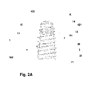
- figure 3A is a larger-scale view of the detail denoted by letter K of figure
2A;
- figure 3B illustrates the detail denoted by H of figure 2B in larger
scale;
- figures from 4A to 4F schematically illustrate a possible operating
sequence for
the lifting of the sinus membrane with the crestal approach, with the
insertion of a
bone implant material, and the subsequent application of a dental implant
according to the present invention.
DESCRIPTION OF THE PREFERRED EMBODIMENTS
With reference to the accompanying tables of drawings, reference numeral (100)
denotes the dental implant of the present invention, in its entirety and in
the
preferred but not exclusive embodiments thereof.
The dental implant (100) is specially conformed to be specifically destined
for
implantation in the upper rear maxilla bone (0) when the relative thickness is
too
CA 03176097 2022- 10- 19
WO 2021/214619
PCT/1B2021/053191
7
small to proceed to a direct application of a dental implant, and a
preparatory lifting
of the sinus membrane (M) of the maxillary sinus (SM) by the crestal approach
is
required, with the insertion of a bone implant material (I).
The dental implant (100) of the invention comprises a body (1), which has a
shank
shape, and which comprises a main portion (10) and, on opposite sides with
respect to the main portion (10), a first crestal proximal portion, (11), and
a second
apical distal part, (12).
The dental implant (100) further comprises a threading (2) having a spiral
shape
with cutting edges (21).
The special characteristics of the dental implant (100) of the present
invention
consist in the fact that (see figures from 2A to 2D, and figures 3A and 3B):
the threading (2) having a spiral shape with cutting edges (21) extends in
spiral
fashion only about and along the main portion (10) of the body (1) terminating
before the second apical distal part (12);
the second apical distal part (12) is conformed in such a way as to comprise
an
external surface (14) free of projecting or cutting parts, and so that the
external
surface (14), at least partially, annularly has a groove (15) or a series of
grooves
(16).
In substance, then, the threading (2) having a spiral shape winds only about
the
main portion (10) of the body (1), while the second apical distal part, (12)
has an
external surface (14), substantially smooth, without projections,
protuberances or
projecting cutting profiles, but which has only a groove or series of grooves,
i.e.
furrows or recesses, in any case free of sharp or cutting edges.
Owing to these special characteristics, the dental implant (100) of the
invention
obviates the drawbacks present in dental implants of known type and enables
CA 03176097 2022- 10- 19
WO 2021/214619
PCT/1B2021/053191
8
obtaining the following advantages, as identified in figures from 4A to 4F.
These figures illustrate an operating sequence for implanting the dental
implant
(100) of the invention in a hole (F) made by crestal approach in a point of
the
upper rear maxilla bone (0) in which an artificial tooth is to be positioned
following
the loss of a natural tooth (premolar, or molar) with a consequent reduction
of the
vertical thickness of the maxilla bone.
Figures 4A and 4B illustrate the step of making of a hole (F), with the
crestal
approach, in the upper rear maxilla bone (0) following the opening of the gum
(G).
To follow up such a hole (F) by the crestal approach, a special instrument (U)
is
used (of known type, of the burr or scalpel type) to perforate the upper
maxilla
bone (0) up to reaching the bone floor (PO) of the maxillary sinus (SM)
without
perforating the sinus membrane (M).
Using a special instrument (U1) (of known type, mechanical or manual), which
is
inserted in the previously-bored hole (F), it is possible to proceed to the
lifting of
the sinus membrane (M), creating a volume for insertion of bone implant
material
(I), thus completing the lifting of the maxillary sinus (SM) and increasing
the total
thickness of bone support (natural bone plus bone implant material) in which
to
implant the dental implant (100) (see figures 4C and 4D).
At this point, it is possible to insert the second apical distal part (12) of
the dental
implant (100) of the invention into the hole (F) (figure 4E) and screw the
dental
implant (100) into the hole in such a way that the cutting edges (21) of the
threading (2) present along the main portion (10) of the body (1) gain a grip,
by
penetrating into the upper rear maxilla bone (0), causing the whole dental
implant
(100) to insert into the hole (F), positioning it with the first crestal
proximal portion
(11) at the external end of the hole (F), at the position in which the gum (G)
will be
CA 03176097 2022- 10- 19
WO 2021/214619
PCT/1B2021/053191
9
repaired and in which the prosthetic element (artificial tooth) will be
applied in a
special seat (not illustrated as of known type) present in the a first crestal
proximal
portion (11) (figure 4F).
As can be seen in figure 4F, the second apical distal part (12) is positioned
at the
position of the bone implant material (I) and, possibly, even in contact with
the
sinus membrane (M).
During the step of screwing-in the dental implant (100) into the hole (F), the
second apical distal part (12), not having any projection or cutting part in
the
relative lateral surface (14), even if it were to reach into contact with the
sinus
membrane (12), would not cause any lesion or tearing of the sinus membrane
(M).
In other words, during the screwing-in and complete insertion of the dental
implant
(100) of the invention in the hole (F), the sinus membrane (M) may be reached
and contacted by the second apical distal end of the implant but as there is
no
projecting or cutting part on the external surface (14) of the second apical
distal
end, the sinus membrane (M) will be protected against any tearing or abrasion.
In substance, the second apical distal end (12) of the dental implant (100) of
the
present invention is, due to its special conformation, completely atraumatic,
enabling conservation of the integrity of the sinus membrane (M) even should,
following the complete insertion of the dental implant (100) in the hole (F)
made in
the upper rear maxilla bone (0), the second apical distal end (12) reach into
contact with the sinus membrane (M).
Further, as is once more clearly visible from figure 4F, the second apical
distal part
(12) of the dental implant (100) will be positioned at the bone implant
material (I),
previously inserted in the hole (F) following the lifting of the maxillary
sinus (SM),
i.e. the sinus membrane (M).
CA 03176097 2022- 10- 19
WO 2021/214619
PCT/1B2021/053191
Owing to the absence of projecting or cutting parts in the external surface
(14) of
the second apical distal part (12), the screwing-in of the second apical
distal part
(12) into the bone implant material (I) will not lead to any dislocation of
the
material.
5 Further, the presence of one or more grooves in the external surface (14)
of the
second apical distal part (12) will facilitate a greater adhesion of the bone
implant
material (I) to the dental implant (100), facilitating a more effective and
rapid
osteointegration.
Lastly, the threading (2) with cutting edges (21), which extends only along
the
10 main portion (10) of the body (1) of the dental implant (100), will
substantially be
positioned at the maxilla bone (0), i.e. at the residual natural and original
portion
of bone, facilitating an excellent adhesion and mechanical fixing of the
dental
implant.
It is clear, therefore, from the above description, and from what is
illustrated in the
figures, in particular in figures 4R and 4F, how the dental implant (100) of
the
present invention obviates, in an extremely effective way, the drawbacks
present
in the dental implants of the prior art, and advantageously obviates the
drawbacks
and complications cause by the use thereof.
Other further advantageous characteristics and preferred aspects of the dental
implant (100) of the invention are described in the following; these
characteristics
can exist singly or in any combination with one another.
In the appended figures, some preferred combinations are illustrated, though
the
dental implant of the present invention can in any case have other
combinations or
further accessory characteristics, all falling within the scope of the present
invention, as set out in claim 1.
CA 03176097 2022- 10- 19
WO 2021/214619
PCT/1B2021/053191
11
The dental implant (100) can preferably be realised in such a way as to have
an
overall length (H) comprised between 6 and 20 mm, with the main part (10) of
the
body (1) having a length (H1) for example comprised between 3 and 13 mm, and
with the second apical distal part (12) having a length (H2) for example for
example comprised between 0.5 and 10 mm.
The second apical distal part (12) is preferably conformed in such a way as to
comprise an end portion (120) having a spherical cap shape.
The second apical distal part (12) can be conformed in such a way as to
comprise
an cylindrically-shaped portion (121) and an end portion (120) having a
spherical
cap shape. For example a cylindrically-shaped portion (121) having a height
comprised between 0 and 10 mm and an end portion (120) having a spherical cap
shape having a height comprised between 0.5 and 3 mm.
According to preferred embodiments, for example illustrated in figures 2B, 2C,
3B,
the second apical distal part, (12) is conformed so that the relative external
surface
(14) annularly has a groove (15) which extends with a spiral shape along the
external surface (14).
For example, the spiral-shaped groove (15) extends at least along the
cylindrically-
shaped portion (121).
The spiral-shaped groove (15) preferably forms a spiral having a constant or
variable pitch (p); for example it can have a constant pitch (p) comprised
between
0.1 mm and 1.5 mm.
The spiral-shaped groove (15) can preferably have a depth comprised between
0.01 mm and 1.5 mm.
In other preferred embodiments, for example illustrated in figures 2A, 2D and
3A,
the second apical distal part (12) is conformed in such a way that the
relative
CA 03176097 2022- 10- 19
WO 2021/214619
PCT/1B2021/053191
12
external surface (14) annularly has a series of grooves (16) having a circular
or
semi-circular shape, being coaxial and reciprocally arranged superposed and
distanced by a constant or variable distance (z); for example with a constant
distance (z) comprised between 0.01 mm and 1.5 mm.
For example, the apical distal part, (12) is conformed in such a way that the
series
of grooves (16) having a circular or semi-circular shape are arranged at least
along the cylindrically-shaped portion (121).
The grooves (16) can preferably have a depth comprised between 0.01 mm and
1.5 mm.
The grooves (16) can all be circumferentially formed, all being semi-
circumferential, or having an arc of circumference shape, or some of which
being
circumferentially formed and others having a semi-circumferential or an arc of
circumference shape.
The second apical distal part, (12) can possibly be conformed in such a way
that
the relative external surface (14) can be affected by a plurality of grooves
having a
spiral shape intervalled from one another along the height of the second
apical
distal part (12) or an alternation between grooves having a circumferential
shape,
a semi-circumferential or an arc of circumference shape, and grooves having a
spiral shape, or even grooves of another other shape or progression, as long
as
they are free of projecting or cutting edges.
According to the special preferred but not exclusive embodiments illustrated
in the
figures of the drawings, the dental implant (100) can be realised in such a
way that
the body (1) further comprises an intermediate portion (13), which is arranged
between the main portion (10) and the apical distal part (12).
In these cases, the dental implant (100), further comprises a second threading
(3)
CA 03176097 2022- 10- 19
WO 2021/214619
PCT/1B2021/053191
13
which winds in a spiral fashion only about the intermediate portion (13), the
second threading (3) being conformed in such a way as to have rounded and not
cutting edges (131).
The intermediate portion (13), once the dental implant (100) has been
completely
inserted in the hole (F) made in the upper rear maxilla bone (C) by the
crestal
approach, may be positioned at the bone implant material (I) (see figure 4F).
Owing to the presence of the second threading (3) on the intermediate part
(13),
the screwing-In and insertion of the dental implant (100) will be facilitated,
as will
the fixing thereof to the bone implant material (I).
At the same time, the presence of rounded and non-cutting edges (131) on the
second threading (3) in the intermediate part (13), will prevent, or at least
reduce,
any possible dislocation of the bone implant material during the screwing-in
of the
dental implant (100).
It is newly specified, as previously, that the presence of the intermediate
portion
(13) is not an essential aspect, as the dental implant (100) of the invention
can be
made without the intermediate portion (13), i.e. with only the main portion
(10) and
the second apical distal part (12) at an end of the main portion (10).
The presence of the intermediate portion (13), between the main portion (10)
and
the second apical distal part (12) might enable obtaining further preferred
advantages, as illustrated in the foregoing.
The main portion (10) of the body (1) is preferably conformed so as to have a
truncoconical shape, for example with a first smaller diameter comprised
between
3 and 7 mm, and with a second bigger diameter comprised between 0.3 and 2
mm, or have a cylindrical shape, for example with a diameter comprised between
3 and 7 MM.
CA 03176097 2022- 10- 19
WO 2021/214619
PCT/1B2021/053191
14
In a case where the intermediate portion (13) is present, it can be conformed
so as
to have a cylindrical or truncoconical shape, for example with a height
comprised
between 0 and 10 mm, and a diameter comprised between a minimum value of
about 1.7 mm and a maximum value of about 7 mm.
The apical distal part (12) is preferably realised in such a way as to
comprise a
longitudinal recess (130), performing a function of material discharge during
the
screwing-in of the dental implant (100) (visible for example in figure 2D).
The main part (10) of the body (1) is preferably realised in such a way as to
comprise a furrow (160), also having the function of a discharge, arranged
substantially longitudinally to the main part (10) and transversally to a part
of the
threads of the threading (2) having a spiral shape (see figure 20).
CA 03176097 2022- 10- 19