Note: Descriptions are shown in the official language in which they were submitted.
1
SYSTEMS AND METHODS FOR DETECTING VASCULAR ACCESS
DISCONNECTION
RELATED APPLICATIONS
This application claims priority to U.S. Provisional Patent Application Serial
No.
62/121,980, entitled "Hemodialysis System," filed February 27, 2015 and to
U.S.
Provisional Patent Application Serial No. 62/003,346, entitled "Hemodialysis
System,"
filed May 27, 2014.
FIELD OF INVENTION
The present invention generally relates to hemodialysis and similar dialysis
systems,
e.g., systems able to treat blood or other bodily fluids extracorporeally.
BACKGROUND
Many factors make hemodialysis inefficient, difficult, and expensive. These
factors
include the complexity of hemodialysis, the safety concerns related to
hemodialysis, and the
very large amount of dialysate needed for hemodialysis. Moreover, hemodialysis
is
typically performed in a dialysis center requiring skilled technicians.
Therefore any
increase in the ease and efficiency of the dialysis process could have an
impact on treatment
cost or patient outcome.
SUMMARY OF INVENTION
Aspects of the invention generally relate to hemodialysis and similar dialysis
systems. Illustrative embodiments described herein involve, in some cases,
interrelated
products, alternative solutions to a particular problem, and/or a plurality of
different uses of
one or more systems and/or articles. Although the various systems and methods
described
herein are described in relation to hemodialysis, it should be understood that
the various
systems and method described herein are applicable to other dialysis systems
and/or in any
extracorporeal system able to treat blood or other bodily fluids, such as
hemofiltration,
hemodiafiltration, etc.
In one aspect of the invention, a method for detecting an access
disconnection, the
method includes measuring the electrical impedance from a venous line to an
arterial line
via a vascular access site, determining an electrical quantity from the
measured electrical
Date Regue/Date Received 2022-09-27
2
impedance, comparing the electrical quantity to a first predetermined
threshold, initiating a
counter when the electrical quantity crosses a first threshold, and declaring
an access
disconnection if the counter reaches a predetermined value before the
electrical quantity
crosses a second threshold. The counter may count units of time, blood volume
pumped to
the vascular access site or the number of strokes of a blood pump. The
electrical quantity
may be raw or filtered value of the impedance between the probes, the time
derivative of the
impedance, or the difference between a first filtered value of the impedance
with a first time
constant and a second filtered value of the impedance with a second time
constant that is
longer than the first time constant. The method for detecting an access
disconnect may
determine the electrical quantity from the measured impedance only while a
blood pump is
flowing fluid through the arterial line or the venous line. Further, a
controller in
communication with the blood pump, the occluder and the user interface may in
response to
the ADS algorithm declaring an access disconnect, stop the action of the blood
pump, close
the occluder and/or signal the user. The controller in the event of a declared
access
disconeect may ask the user to verify the position of arterial and venous
needles at the
vascular access site and then allow the user to select resume therapy or end
therapy.
In another aspect of the invention, method for detecting an access
disconnection, the method
includes measuring the electrical impedance from a venous line to an arterial
line via an
vascular access site at regular intervals, determining an electrical quantity
from the
measured electrical impedance, completing the stroke of a pump delivering
blood to the
patient, reducing the driving force on the pump plunger to a lower value,
declaring an
access disconnection when the electrical quantity exceeds a first
predetermined threshold.
The electrical quantity may the raw or filtered electrical impedance or the
time derivative of
the impedance or the difference between a first filtered value of the
impedance with a first
time constant and a second filtered value of the impedance with a second time
constant that
is longer than the first time constant.
In another aspect of the invention, a method for detecting an access
disconnection,
the method includes measuring the electrical impedance from a venous line to
an arterial
line via an vascular access site, determining an electrical quantity from the
measured
electrical impedance, comparing the electrical quantity to a first
predetermined threshold,
setting a provisional flag when the electrical quantity crosses a first
threshold, clearing the
provisional flag when the electrical quantity crosses a second threshold, and
declaring an
Date Recue/Date Received 2022-09-27
3
access disconnection when the provisional flag is consistently set for more
than a given
period.
In another aspect of the invention, a system for detecting an access
disconnection,
the system includes a venous line and arterial line each connected to a blood
pump at one
end and to an vascular access site on a patient at the other end, a circuit
capacitively
coupled to blood in the venous line and the arterial line capable of measuring
the electrical
impedance through part of the venous line, part of the arterial line and
through the vascular
access site, and a controller in communication with the blood pump and the
circuit which,
determines an electrical quantity from the measured electrical impedance,
compares the electrical quantity to a first predetermined threshold, initiates
a counter when
the electrical quantity crosses a first threshold, and declares an access
disconnection if the
counter reaches a predetermined value before the electrical quantity crosses a
second
threshold.
A system controller can be configured to detect dislodgment of a catheter or
needle
in a vascular access comprising a first and second catheter or needle in a
blood vessel,
fistula or graft. The system comprises a first line fluidly connecting the
first catheter or
needle to an inlet of a pump; a second line fluidly connecting the second
catheter or needle
to an outlet of the pump; a first connector connecting the first line to the
first catheter or
needle; a second connector connecting the second line to the second catheter
or needle, each
connector having an electrode in fluid communication with a fluid-carrying
lumen of its
connector; an electronic circuit electrically connected to the electrodes of
the first and
second connectors, and configured to measure electrical impedance of fluid
between the
first connector and the second connecter via a conductive path through the
blood vessel,
fistula or graft; and a controller configured to receive a series of sampled
electrical
impedance values from the electronic circuit, and to process the electrical
impedance values
as a signal. Operation of the pump may comprise extracorporeal circulation of
a portion of a
user's blood.
In an embodiment, the controller can be configured to sample and filter or
smooth
the signal using a first time constant, yielding a first filtered signal;
sample and filter or
smooth the signal using a second longer time constant, yielding a second
filtered signal;
provisionally set a disconnection flag and initiate a counter if at a point in
time the
difference between the first filtered signal and the second filtered signal is
greater than a
first threshold value; clear the disconnection flag if the difference between
the first filtered
Date Recue/Date Received 2022-09-27
4
signal and the second filtered signal decreases to less than a second lower
threshold value
before the counter has reached a pre-determined count; and declare a vascular
disconnection
if the disconnection flag is not cleared before the counter has reached the
pre-determined
count.
Optionally, the declaration may cause the controller to activate one or more
mechanical line occluders to stop a flow of fluid in the first and second
lines, stop the pump,
or notify a user of the occurrence of a possible vascular disconnection.
Notification of the
user may comprise requesting that the user verify the position of the first
and second
catheters or needles at the vascular access. The controller may be configured
to receive from
the user a command to resume operation of the pump or to discontinue further
operation of
the pump. The controller may be configured to raise the first threshold value
if a plurality of
declarations of a vascular disconnection are each followed by a user command
to resume
operation of the pump. The controller may continue to process the electrical
impedance
values if a declaration of a vascular disconnection is made and the mechanical
line
occluders are activated, and the controller may be configured to confirm a
vascular
disconnection if the difference between the first filtered signal and the
second filtered signal
exceeds a third threshold value that is greater than the first threshold
value.
The counter may count units of time, the pre-determined count being a pre-
determined time interval; may count units of blood volume pumped to the
vascular access,
the pre-determined count being a pre-determined volume of blood; or may count
strokes of
the pump, the pre-determined count being a pre-determined number of strokes.
The signal may be a time derivative of the electrical impedance values.
The controller may stop processing the electrical impedance values if the pump
stops pumping fluid through the first and second lines.
In an embodiment, the controller may conduct any of all of the above processes
without filtering the signal data, or by using a filtered version of the
signal data. The
controller may conduct any or all of the above processes by using a difference
between a
first filtered signal using a first time constant and a second filtered signal
using a second
longer time constant. Alternatively, the processed signal may be a ratio
between the first
filtered signal and the second filtered signal, comparing the ratio to first,
second and/or third
values to set provisional flags or to initiate or terminate a counter. The
controller may
conduct any or all of the above processes without using a counter or setting a
provisional
disconnection flag.
Date Recue/Date Received 2022-09-27
5
The controller may perform a signal test to determine whether a dislodgment
event
has been obscured by a conductive pathway between the electrodes outside of
the blood
vessel, fistula or graft. The controller may sample and filter or smooth the
signal using a
first time constant, yielding a first filtered signal; sample and filter or
smooth the signal
using a second longer time constant, yielding a second filtered signal;
initiate a counter and
set a provisional disconnection flag if a difference between the first
filtered signal and the
second filtered signal exceeds a first threshold value; temporarily clear the
provisional
disconnection flag if the difference between the first filtered signal and the
second filtered
signal drops below a second lower threshold value before the counter reaches a
preset
count; command an actuator of the pump to apply a force to a pumping chamber
of the
pump to complete a fluid delivery stroke to the first or second catheter or
needle; command
the actuator to apply a reduced force to the pumping chamber; and declare an
access
disconnection if the difference between the first filtered signal and the
second filtered signal
exceeds a third threshold value that is equal to or greater than the first
threshold value.
In an embodiment, the controller may be able to detect a transition from a
blood-
filled blood tubing set to a dialysate-filled blood tubing set during a
rinseback procedure. A
delayed or incomplete transition may be an indication, for example, of an
occlusion at or
distal to the connectors. The controller may be configured to measure the
signal or a
filtered form of the signal as dialys ate is pumped through the dialyzer to
the blood tubing
set; determine whether the signal or a filtered form of the signal has a first
value
approximately equal to an expected value of the signal for blood in the first
and second fluid
lines, or has a second value approximately equal to an expected value of the
signal for
dialysate solution in the first and second fluid lines; determine a point in
time when the
signal or a filtered form of the signal changes from the first value to the
second value; and
provide a first notification to a user if the controller detects a change from
the first value to
the second value, or provide a second notification to the user if the
controller detects a
change from the first value that is less than approximately the second value
within a pre-
determined period of time.
Other advantages and novel features of the present invention will become
apparent
from the following detailed description of various non-limiting embodiments of
the
invention when considered in conjunction with the accompanying figures. In
cases where
the present specification and a document referred to herein include
conflicting and/or
inconsistent disclosure, the present specification shall control. If two or
more documents
Date Recue/Date Received 2022-09-27
6
referred to herein include conflicting and/or inconsistent disclosure with
respect to each
other, then the document having the later effective date shall control.
BRIEF DESCRIPTION OF THE DRAWINGS
Aspects of the invention are described with reference to illustrative
embodiments,
which are described with reference to the drawings in which like numerals
reference like
elements, and wherein:
FIG. 1 is a schematic representation of fluid handling components of a
hemodialysis
system in an illustrative embodiment;
FIG. 2 shows a schematic fluid flow diagram for the dialysis system of FIG. 1;
FIG. 3 is a schematic fluid flow diagram for the blood flow circuit of the
FIG. 2
embodiment;
FIG. 4 is a schematic fluid flow diagram for the balancing circuit of the FIG.
2
embodiment;
FIG. 5 is a schematic fluid flow diagram for the directing circuit of the FIG.
2
embodiment;
FIG. 5A is a schematic fluid flow diagram illustrating a flow path for a drain
assembly in an illustrative embodiment;
FIG. 6 is a schematic fluid flow diagram for the mixing circuit of the FIG. 2
embodiment;
FIG. 7 is a right front perspective view of a hemodialysis system in an
illustrative
embodiment;
FIG. 7a is perspective view of selected components of a power unit in an
illustrative
embodiment;
FIG. 7b is a schematic view of an air dehumidifier arrangement in an
illustrative
embodiment;
FIG. 7c is a perspective view of a dehumidifier arrangement in the FIG. 7a
embodiment;
FIG. 8 is a left rear perspective view of the hemodialysis system of FIG. 7;
FIG. 9 is a front view of the hemodialysis system of FIG. 7;
FIG. 10 is a right front perspective view of the view of the hemodialysis
system of
FIG. 7 with the doors in a first open position;
FIG. 11 is a top view of the hemodialysis system of FIG. 10;
Date Recue/Date Received 2022-09-27
7
FIG. 12 is a front view of the hemodialysis system of FIG. 10;
FIG. 13 is a right side view of the hemodialysis system of FIG. 10;
FIG. 14 is a right front perspective view of the view of the hemodialysis
system of
FIG. 7 with the doors in a second open position;
FIG. 15 is a top view of the hemodialysis system of FIG. 14;
FIG. 16 is a front view of the hemodialysis system of FIG. 14;
FIG. 17 is a front view of the hemodialysis system of FIG. 7 with the doors in
an
open position exposing a front panel of the system;
FIG. 17a is an exploded perspective view of a control port assembly arranged
to
interface with a blood pump assembly in an illustrative embodiment;
FIG. 17b is a cross sectional side view of the FIG. 17a embodiment with an
engaged
blood pump assembly;
FIG. 17C shows a perspective view of a control port assembly with a pair of
blood
pump cassette latching and ejection assemblies in an illustrative embodiment;
FIG. 17D shows an isolated view of a latching assembly with an ejection member
in
a retracted position in an illustrative embodiment;
FIG. 17E shows an isolated view of the latching assembly of FIG. 17D with an
ejection member in an extended position in an illustrative embodiment;
FIG. 17F shows a front view of a blood pump cassette in a retained condition
on a
panel of a dialysis unit in an illustrative embodiment;
FIG. 17G shows a cross-sectional view along the line 17G-17G in FIG. 17F;FIG.
17H shows a cross-sectional view along the line 17H-17H in FIG. 17F;
FIG. 171 shows a front view of a blood pump cassette in an ejecting condition
in an
illustrative embodiment;
FIG. 17J shows a cross-sectional view along the line 17J-17J in FIG. 171;
FIG. 17K shows a cross-sectional view along the line 17K-17K in FIG. 171;
FIG. 18 is a front view of a blood circuit assembly for use with the system of
FIG. 7;
FIG. 18a is a perspective view of a blood pump having a medication holder in
an
illustrative embodiment;
FIG. 19 right perspective view of a organizing tray for the blood circuit
assembly of
FIG. 18;
FIG. 20 is a left rear perspective view of the blood circuit assembly of FIG.
18;
Date Recue/Date Received 2022-09-27
8
FIG. 20A is an front exploded view of an alternate embodiment of a blood pump
cassette;
FIG. 20 B is a rear exploded view of the blood pump cassette of FIG. 20A;
FIG. 20C is a front view of a bottom plate or back plate of the blood pump
cassette
of FIG. 20A;
FIG. 20D is a back view of a bottom plate or back plate of the blood pump
cassette
of FIG. 20A;
FIG. 21 shows a left front perspective view of the front panel of the system
of FIG.
7;
FIG. 21A shows a front view of an alternate embodiment of a front panel
assembly
in an illustrative embodiment;
FIG. 21B shows the front panel assembly of FIG. 21A with the top and middle
plate
components of the blood pump cassette removed for clarity in an illustrative
embodiment;
FIG. 22 shows a front view of the front panel of the system of FIG. 7;
FIG. 23 shows a front view of the front panel of the system of FIG. 7 with a
pair of
mounting features for the dialyzer;
FIG. 24 shows a side view of a dialyzer with quick-connect fittings attached
to the
dialys ate inlet/outlet ports of the dialyzer;
FIG. 25 shows a right perspective view of a reagent supply for use with the
system
of FIG. 7;
FIG. 26 shows a perspective view of an E-prong connector for the reagent
supply of
FIG. 25 and a corresponding connection point at the front panel of the
hemodialysis system;
FIG. 27 shows a perspective view of a pair of blood line connectors for the
blood
circuit assembly and a corresponding connection point at the front panel of
the hemodialysis
system;
FIG. 28 shows a side view of a blood line connector and connection point of
FIG. 27
FIG. 29 is a perspective view of a blood circuit assembly in an alternate
embodiment; and
FIG. 30 is a close up view of a portion of the blood circuit assembly of FIG.
29.
FIG. 31 shows an exemplary modular drain cassette in an illustrative
embodiment;
FIG. 32 shows the drain cassette of FIG. 31 in an exploded view with an
escutcheon
positioned anterior to a front wall of the drain cassette in an illustrative
embodiment;
Date Recue/Date Received 2022-09-27
9
FIG. 33 shows a perspective view of the front wall of the drain cassette of
FIG. 31 in
an illustrative embodiment;
FIG. 34 shows a main housing of the drain cassette of FIG. 31 with the front
wall
removed for clarity purposes in an illustrative embodiment;
FIG. 35 shows a rear, perspective view of the drain cassette of FIG. 31 in an
illustrative embodiment;
FIG. 36 shows a front panel in which a drain cassette has been dismounted in
an
illustrative embodiment;
FIG. 37 is a schematic representation of a conductivity circuit in an
illustrative
embodiment;
FIG. 38 is a diagram of the electrical waveforms processed by the circuit of
FIG. 37;
FIG. 39 is a representative graph of the noise/error sensitivity of the
circuit of FIG.
37 plotted against the ratio of unknown/reference resistance in the circuit;
FIG. 40 is a schematic representation of an exemplary blood flow circuit of a
hemodialysis system;
FIG. 41A is a side view of a connector that may be used in the blood flow
circuit of
FIG. 40;
FIG. 41B is a cross-sectional view of the connector of FIG. 41A;
FIG. 42 is a cross-sectional view of the connector of FIGS. 41A and 41B, with
an
attached wire and flexible tubing;
FIG. 43A is a perspective view of an alternate embodiment of a connector that
may
be used in the blood flow circuit of FIG. 40;
FIG. 43B is a top view of the connector of FIG. 43A;
FIG. 43C is a cross-sectional view of the connector of FIG. 43B;
FIGS. 44A-D are various cross-sectional views of a flexible tube incorporating
a
conductive wire;
FIG. 45 is a perspective view of a flexible double-lumen tube having a fluid-
carrying lumen and a wire-carrying lumen;
FIG. 46 is a cross-sectional view of a connector similar to the connector of
FIGS.
43A-C, with an attached wire and tubing;
FIG. 47 is a plan view of an extracorporeal blood flow circuit used in a
representative hemodialysis system;
Date Regue/Date Received 2022-09-27
10
FIG. 48 is a perspective view of a hemodialysis apparatus configured to
receive and
operate the extracorporeal blood flow circuit of FIG. 47; and
FIG. 49 is a representative plot of the resistance measured by the
conductivity circuit
of FIG. 37 under various conditions;
FIG. 50 shows an exploded, perspective view of an occlusion assembly from a
front
angle in accordance with an embodiment of the present disclosure;
FIG. 51 shows an exploded, perspective view of the occlusion assembly of FIG.
50
from a back angle;
FIG. 52 shows a front, perspective view of the occlusion assembly of FIG. 50
with
the door open and the button pressed to illustrate loading of a tube;
FIG. 53 shows a close-up perspective view of the occlusion assembly of FIG.
50,
showing the door engaging a switch when the door is closed;
FIG. 54 shows the front of the occlusion assembly of FIG. 50 without the door
and
frame to illustrate the arms fully occluding flexible tubes;
FIG. 55 shows the front of the occlusion assembly of FIG. 50 without the door
and
frame to illustrate the arms in a non-occluding position;
FIG. 56 is a rear/top perspective view of the occlusion assembly of FIG. 50
with an
actuator arm in a fully retracted position;
FIG. 57 is a rear perspective view of the occlusion assembly of FIG. 50 with
an
actuator arm in a fully extended position;
FIG. 58 shows a side perspective view of several working parts of the
occlusion
assembly of FIG. 50 in a non-occluding state;
FIG. 59 shows a side perspective view of several working parts of the
occlusion
assembly of FIG. 50 in an occluding state;
FIG. 60 shows a side, cross-sectional view of an actuator of the occlusion
assembly
of FIG. 50, illustrating a location for a main spring for the assembly; and
FIG. 61 shows the occlusion assembly of FIG. 50 mounted in a front panel
assembly
of a hemodialysis apparatus in accordance with an embodiment of the present
disclosure.
FIG. 62 shows raw and processed signals from the Access Disconnect Sensor
system and pumping pressures for a non-dislodgement event.
FIG. 63 shows raw and processed signals from the Access Disconnect Sensor
system and pumping pressures for an access disconnect event.
Date Recue/Date Received 2022-09-27
11
FIG. 64 shows raw and processed signals from the Access Disconnect Sensor
system, pumping pressures and the ADS Signal Test for an access disconnect
event.
FIG. 65 shows raw and processed signals from the Access Disconnect Sensor
system, pumping pressures and the ADS Signal Test for an access disconnect
event, with
longer duration half cycles than shown in FIGS. 62-64.
DETAILED DESCRIPTION
Various aspects of the invention are generally directed to new systems for
hemodialysis and the like, such as hemofiltration systems, hemodiafiltration
systems,
plasmapheresis systems, etc. Accordingly, although the various systems and
methods
described herein are described in relation to hemodialysis, it should be
understood that the
various systems and method described herein are applicable to other dialysis
systems and/or
in any extracorporeal system able to treat blood or other bodily fluids, such
as plasma.
As discussed below, a hemodialysis system typically includes a blood flow path
and
a dialysate flow path. It should be noted that within such flow paths, the
flow of fluid is not
necessarily linear, and there may be any number of "branches" within the flow
path that a
fluid can flow from an inlet of the flow path to an outlet of the flow path.
Examples of such
branching are discussed in detail below. In the blood flow path, blood is
drawn from a
patient, and is passed through a dialyzer, before being returned to the
patient. The blood is
treated by the dialyzer, and waste molecules (e.g., urea, creatinine, etc.)
and water are
passed from the blood, through a semi-permeable membrane in the dialyzer, into
a dialysate
solution that passes through the dialyzer by the dialysate flow path. In
various
embodiments, blood may be drawn from the patient from two lines (e.g., an
arterial line and
a venous line, i.e., "dual needle" flow), or in some cases, blood may be drawn
from the
patient and returned through the same or catheter needle (e.g., the two lines
or lumens may
both be present within the same needle, i.e., a form of "dual lumen" flow). In
still other
embodiments, a "Y" site or "T" site is used, where blood is drawn from the
patient and
returned to the patient through one patient connection having two branches
(one being the
fluid path for the drawn blood, the second the fluid path for the return
blood, i.e., a form of
"single needle" flow). The patient may be any subject in need of hemodialysis
or similar
treatments, including non-human subjects, such as dogs, cats, monkeys, and the
like, as well
as humans.
Date Regue/Date Received 2022-09-27
12
In the dialysate flow path, fresh dialysate is prepared and is passed through
the
dialyzer to treat the blood from the blood flow path. The dialysate may also
be equalized
for blood treatment within the dialyzer (i.e., the pressure between the
dialysate and the
blood are equalized), often exactly, or in some embodiments, at least within
about 1% or
about 2% of the pressure of the blood.. In some cases, it may be desirable to
maintain a
greater pressure difference (either positive or negative) between the blood
flow path and
dialysate flow path. After passing through the dialyzer, the used dialysate,
containing waste
molecules (as discussed below), is discarded in some fashion. The dialysate in
some cases
may be re-circulated in a "multi-pass" arrangement, which may be beneficial in
capturing
larger molecules having low mobility across the dialyzer. In some cases, the
dialysate is
heated prior to treatment of the blood within the dialyzer using an
appropriate heater, such
as an electrical resistive heater. The dialysate may also be filtered to
remove contaminants,
infectious organisms, debris, and the like, for instance, using an
ultrafilter. The ultrafilter
may have a pore size chosen to prevent species such as these from passing
therethrough.
For instance, the pore size may be less than about 0.3 micrometers, less than
about 0.2
micrometers, less than about 0.1 micrometers, or less than about 0.05
micrometers, etc. The
dialysate is used to draw waste molecules (e.g., urea, creatinine, ions such
as potassium,
phosphate, etc.) and water from the blood into the dialysate through osmosis
or convective
transport, and dialysate solutions are well-known to those of ordinary skill
in the art.
The dialysate typically contains various ions such as sodium, chloride,
bicarbonate,
potassium and calcium that are similar in concentration to that of normal
blood. In some
cases, the bicarbonate, may be at a concentration somewhat higher than found
in normal
blood. Typically, the dialysate is prepared by mixing water from a water
supply with one or
more ingredients: an "acid" (which may contain various species such as acetic
acid,
dextrose, NaCl, CaC1, KC1, MgCl, etc.), sodium bicarbonate (NaHCO3), and/or
sodium
chloride (NaCl). The preparation of dialysate, including using the appropriate
concentrations of salts, osmolarity, pH, and the like, is well-known to those
of ordinary skill
in the art. As discussed in detail below, the dialysate need not be prepared
at the same rate
that the dialysate is used to treat the blood. For instance, the dialysate can
be made
concurrently or prior to dialysis, and stored within a dialysate storage
vessel or the like.
Within the dialyzer, the dialysate and the blood typically are separated by a
semi-
permeable membrane. Typically, the semipermeable membrane is formed from a
polymer
such as cellulose, polyarylethersulfone, polyamide, polyvinylpyrrolidone,
polycarbonate,
Date Regue/Date Received 2022-09-27
13
polyacrylonitrile, or the like, which allows the transport of ions or small
molecules (e.g.,
urea, water, etc.), but does not allow bulk transport or convection during
treatment of the
blood. In some cases (such as high-flux dialyzers), even larger molecules,
such as beta-2-
microglobulin, may pass through the membrane. In some cases, for example, ions
and
molecules may pass through the dialyzer by convective flow if a hydrostatic
pressure
difference exists across the semi-permeable membrane.
It should be noted that, as used herein, "fluid" means anything having fluidic
properties, including but not limited to, gases such as air, and liquids such
as water, aqueous
solution, blood, dialysate, etc.
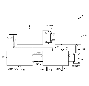
FIG. 1 shows a schematic block diagram of fluid circuitry for a hemodialysis
system
that incorporates various aspects of the invention. In this illustrative
embodiment, the
dialysis system 5 includes a blood flow circuit 141 that draws blood from a
patient, passes
the blood through a dialyzer 14, and returns the treated blood to the patient.
A balancing
circuit or an internal dialysate circuit 143 receives dialysate from an
ultrafilter 73, passes the
dialysate through the dialyzer 14, and receives used dialysate from the
dialyzer 14. A
directing circuit or an external dialysate circuit 142 provides fresh
dialysate to the ultrafilter
73, and receives used dialysate from the internal dialysate circuit 143 (which
may be
directed to a drain 31). The directing circuit 142 can also receive water from
a water supply
30 and pass it to a mixing circuit 25. The mixing circuit 25 forms dialysate
using water
from the directing circuit 142 and reagent ingredients 49, such as citric
acid, salt and a
bicarbonate, that may be received from a renewable source. The mixing circuit
25 may
prepare dialysate, for example, on an as-needed basis, during and/or in
advance of dialysis.
New dialysate prepared by the mixing circuit 25 may be provided to the
directing circuit
142, which may provide the dialysate to the ultrafilter 73, as described
above. The directing
circuit 142 may include a heater to heat the dialysate to a suitable
temperature and/or to heat
fluid in the system for disinfection. Conduits 67 (shown in dotted line) may
be connected
between the blood flow circuit 141 and the directing circuit 142, e.g., for
disinfection of the
hemodialysis system.
FIG. 2 is a schematic diagram showing a more detailed circuit arrangement for
the
dialysis system 5 shown in FIG. 1. It should be understood, of course, that
FIG. 2 is only
one possible embodiment of the general hemodialysis system of FIG. 1, and in
other
embodiments, other fluid circuits, modules, flow paths, layouts, etc. are
possible. Examples
of such systems are discussed in more detail below, and also can be found in
the following:
Date Regue/Date Received 2022-09-27
14
U.S. Application 12/072,908, filed February 27, 2008, U.S. Provisional
Application
60/903,582, filed February 27, 2007, U.S. Provisional Application 60/904,024,
filed
February 27, 2007, U.S. Patent Application 11/871,680, filed October 12, 2007,
U.S. Patent
Application 11/871,712, filed October 12, 2007, U.S. Patent Application
11/871,787, filed
October 12, 2007, U.S. Patent Application 11/871,793, filed October 12, 2007,
or U.S.
Patent Application 11/871,803, filed October 12, 2007.
The blood flow circuit 141 includes an anticoagulant supply 11 and a blood
flow
pump 13 which pumps blood from a patient through a dialyzer 14 and returns the
blood to
the patient. The anticoagulant supply 11, although shown in the path of blood
flowing
towards the dialyzer, may be instead located in another suitable location.
e.g., any location
upstream or downstream from blood flow pump 13. The balancing circuit 143
includes two
dialysate pumps 15, which pump dialysate into the dialyzer 14, and a bypass
pump 35. The
flow of blood through the blood flow circuit 141 in some cases, is
synchronized with the
flow of dialysate in the dialysate flow path. In an embodiment, the flow of
dialysate into
and out of the dialyzer 14 and the balancing circuit 143 is balanced
volumewise using
balancing chambers in the balancing circuit 143. The directing circuit 142
includes a
dialysate pump 159, which pumps dialysate from a dialysate tank 169 through a
heater 72
and/or the ultrafilter 73 to the balancing circuit 143. The directing circuit
142 also receives
waste fluid from balancing circuit 143 and directs it to a drain 31. In some
cases, the blood
flow circuit 141 can be connected via conduits 67 to the directing circuit
142, e.g., for
disinfection, as discussed below. Dialysate in the dialysate tank 169 is
provided by the
mixing circuit 25, which produces the dialysate using water from a water
supply 30
provided via the directing circuit 142 and dialysate ingredients 49 (e.g.,
bicarbonate and
acid). A series of mixing pumps 180, 183, 184 are used to mix the various
components and
produce the dialysate.
FIG. 3 shows a close-up view of the blood flow circuit 141 in this
illustrative
embodiment. Under normal operation, blood flows from a patient through
arterial line 203
via blood flow pump 13 to the dialyzer 14 (the direction of flow during normal
dialysis is
indicated by arrows 205; in some modes of operation, however, the flow may be
in different
directions, as discussed below). Optionally, an anticoagulant may be
introduced into the
blood via anticoagulant pump 80 from an anticoagulant supply. After passing
through
dialyzer 14 and undergoing dialysis, the blood returns to the patient through
venous line
Date Regue/Date Received 2022-09-27
15
204, optionally passing through an air trap and/or a blood sample port 19. The
pump 13
may include, for instance, pumps 23 that are actuated by a control fluid.
For example, in one embodiment, the blood flow pump 13 may comprise two (or
more) pod pumps 23. Each pod pump, in this particular example, may include a
rigid
chamber with a flexible diaphragm or membrane dividing each chamber into a
pumping
compartment and control compartment. There may be four entry/exit valves for
these
compartments, two for the pumping compartment and two for the control
compartment.
The valves for the control compartment of the chambers may be two-way
proportional
valves, one connected to a first control fluid source (e.g., a high pressure
air source), and the
other connected to a second control fluid source (e.g., a low pressure air
source) or a
vacuum source. The fluid valves can be opened and closed to direct fluid flow
when the
pod pumps 23 are operating. Non-limiting examples of pod pumps are described
in U.S.
Provisional Application 60/792,073, filed April 14, 2006, or in U.S. Patent
Application
11/787,212, filed April 13, 2007. If more than one pod pump is present, the
pod pumps
may be operated in any suitable fashion, e.g., synchronously, asynchronously,
in-phase, out-
of-phase, etc. For instance, in some embodiments, the two-pump pumps can be
cycled out
of phase to affect the pumping cycle, e.g., one pump chamber fills while the
second pump
chamber empties. A phase relationship anywhere between 0 (the pod pumps fill
and empty
in unison) and 180 (one pod pump fills as the other empties) can be selected
in order to
impart any desired pumping cycle. A phase relationship of 180 may yield
continuous flow
into and out of the set of pod pumps. This is useful, for instance, when
continuous flow is
desired, e.g., for use with dual needle or dual lumen catheter flow. Setting a
phase
relationship of 0 , however, may be useful in some cases for single
needle/single lumen
flow or in other cases. In a 0 relationship, the pod pumps will first fill
from the needle,
then deliver blood through the blood flow path and back to the patient using
the same
needle. In addition, running at phases between 0 and 180 can be used in some
cases, to
achieve a push/pull relationship (hemodiafiltration or continuous back flush)
across the
dialyzer.
An anticoagulant (e.g., heparin, or any other suitable anticoagulant) may be
contained within a vial 11 (or other anticoagulant supply, such as a tube or a
bag), and blood
flow circuit 141 may include a spike 201 (which, in one embodiment, is a
needle) that can
pierce the seal of the vial. The spike 201 may be formed from plastic,
stainless steel, or
another suitable material, and may be a sterilizable material in some cases,
e.g., the material
Date Regue/Date Received 2022-09-27
16
may be able to withstand sufficiently high temperatures and/or radiation so as
to sterilize the
material.
An anticoagulant pump 80, which can act as a metering chamber in some cases,
can
be used to control the flow of anticoagulant into the blood circuit. The
anticoagulant pump
80 may be a pod pump or a membrane-based metering pump, and/or may be actuated
by a
control fluid, such as air. For example, the anticoagulant pump 80 may include
a rigid
chamber with a flexible diaphragm dividing the chamber into a pumping
compartment and a
control compartment. One valve for the control compartment of the chamber may
be
connected to a first control fluid source (e.g., a high pressure air source),
and the other valve
connected to a second control fluid source (e.g., a low pressure air source)
or a vacuum
source. Valves for the pumping compartment of the chamber can be opened and
closed in
coordination with the control compartment, thus controlling the flow of
anticoagulant into
the blood. In one set of embodiments, air provided through a filter 81 may
also be
introduced into the blood flow path by the anticoagulant pump 80, e.g., to
provide air into
the vial 11 after or before anticoagulant is withdrawn from the vial.
Fluid Management System ("FMS") measurements may be used to measure the
volume of fluid pumped through a pump chamber during a stroke of the membrane,
or to
detect air in the pumping chamber. FMS methods are described in U.S. Patent
Nos.
4,808,161; 4,826,482; 4,976,162; 5,088,515; and 5,350,357. In one illustrative
embodiment, the volume of liquid delivered by an anticoagulant pump, a
dialysate pump, or
other membrane-based fluid pump is determined using an FMS algorithm in which
changes
in chamber pressure are used to calculate a volume measurement at the end of a
fill stroke
and at the end of a delivery stroke. The difference between the computed
volumes at the
end of fill and delivery strokes may be used to determine the actual stroke
volume. This
actual stroke volume can be compared to an expected stroke volume for the
particular sized
chamber. If the actual and expected volumes are significantly different, the
stroke has not
properly completed and an error message can be generated.
The blood flow circuit 141 may also include an air trap 19 to remove air
bubbles that
may be present within the blood flow path. In some cases, the air trap 19 is
able to separate
any air that may be present from the blood due to gravity, and /or may include
a port for
sampling blood.
FIG. 4 shows a close-up view of the balancing circuit 143 in the FIG. 2
embodiment.
In the balancing circuit 143, dialysate flows from the optional ultrafilter 73
into a dialysate
Date Recue/Date Received 2022-09-27
17
pump 15. In this embodiment, the dialysate pump 15 includes two pod pumps 161,
162,
two balancing chambers 341, 342, and a pump 35 for bypassing the balancing
chambers
341, 342. The balancing chambers 341, 342 may be constructed such that they
are formed
from a rigid chamber with a flexible diaphragm dividing the chamber into two
separate fluid
compartments, so that entry of fluid into one compartment can be used to force
fluid out of
the other compartment and vice versa. Non-limiting examples of pumps that can
be used as
pod pumps and/or balancing chambers are described in U.S. Provisional
Application
60/792,073, filed April 14, 2006, or in U.S. Patent Application 11/787,212,
filed April 13,
2007.
In one embodiment, balancing of flow in the internal dialysate circuit works
as
follows. A set of pneumatically operated valves 211, 212, 213, 241, 242 has
its operation
synchronized and controlled together, where valves 211, 212, 213 are ganged
and valves
241 and 242 are ganged, and a second set of pneumatically operated valves 221,
222, 223,
231, 232 similarly have its operation synchronized and controlled together,
where valves
221, 222, 223 are ganged, and valves 231 and 232 are ganged. At a first point
of time, the
first set of valves 211, 212, 213, 241, 242 is opened while the second set of
valves 221, 222,
223, 231, 232 is closed. Fresh dialysate flows into balancing chamber 341
while used
dialysate flows from dialyzer 14 into pod pump 161. Fresh dialysate does not
flow into
balancing chamber 342 since valve 221 is closed. As fresh dialysate flows into
balancing
chamber 341, used dialysate within balancing chamber 341 is forced out and
exits balancing
circuit 143 (the used dialysate cannot enter pod pump 161 since valve 223 is
closed).
Simultaneously, pod pump 162 forces used dialysate present within the pod pump
into
balancing chamber 342 (through valve 213, which is open; valves 242 and 222
are closed,
ensuring that the used dialysate flows into balancing chamber 342). This
causes fresh
dialysate contained within balancing chamber 342 to exit the balancing circuit
143 into
dialyzer 14. Also, pod pump 161 draws in used dialysate from dialyzer 14 into
pod pump
161.
Once pod pump 161 and balancing chamber 341 have filled with dialysate, the
first
set of valves 211, 212, 213, 241, 242 is closed and the second set of valves
221, 222, 223,
231, 232 is opened. Fresh dialysate flows into balancing chamber 342 instead
of balancing
chamber 341, as valve 212 is closed while valve 221 is now open. As fresh
dialysate flows
into balancing chamber 342, used dialysate within the chamber is forced out
and exits
balancing circuit, since valve 213 is now closed. Also, pod pump 162 now draws
used
Date Regue/Date Received 2022-09-27
18
dialysate from the dialyzer into the pod pump, while used dialysate is
prevented from
flowing into pod pump 161 as valve 232 is now closed and valve 222 is now
open. Pod
pump 161 forces used dialysate contained within the pod pump (from the
previous step) into
balancing chamber 341, since valves 232 and 211 are closed and valve 223 is
open. This
causes fresh dialysate contained within balancing chamber 341 to be directed
into the
dialyzer 14 (since valve 241 is now open while valve 212 is now closed). At
the end of this
step, pod pump 162 and balancing chamber 342 have filled with dialysate. This
puts the
state of the system back into the configuration at the beginning of this
description, and the
cycle is thus able to repeat, ensuring a constant flow of dialysate to and
from the dialyzer
14. In an embodiment, the fluid (e.g. pneumatic) pressures on the control side
of the
balancing chamber valves are monitored to ensure they are functioning (e.g.,
opening and
closing) properly.
As a specific example, a vacuum (e.g., 4 p.s.i. of vacuum) can be applied to
the port
for the first set of valves, causing those valves to open, while positive
pressure (e.g., 20
p.s.i. of air pressure) is applied to the second set of valves, causing those
valves to close (or
vice versa). The pod pumps each urge dialysate into one of the volumes in one
of the
balancing chambers 341, 342. By forcing dialysate into a volume of a balancing
chamber,
an equal amount of dialysate is squeezed by the diaphragm out of the other
volume in the
balancing chamber. In each balancing chamber, one volume is occupied by fresh
dialysate
heading towards the dialyzer and the other volume is occupied by used
dialysate heading
from the dialyzer. Thus, the volumes of dialysate entering and leaving the
dialyzer are kept
substantially equal.
The bypass pump 35 can direct the flow of dialysate from the dialyzer 14
through
balancing circuit 143 without passing through either of pod pumps 161 or 162.
In this
embodiment, the bypass pump 35 is a pod pump, similar to those described
above, with a
rigid chamber and a flexible diaphragm dividing each chamber into a fluid
compartment and
a control compartment. This pump may be the same or different from the other
pod pumps
and/or metering pumps described above. When control fluid is used to actuate
the bypass
pump 35, the additional drop in pressure on the exiting (spent) dialysate side
of the dialyzer
causes additional ultrafiltration of fluid from the blood in the dialyzer.
This may cause a
net efflux of fluid from the patient's blood, through the dialyzer, and
ultimately to drain.
Such a bypass may be useful, for example, in reducing the amount of fluid a
patient has,
which is often increased due to the patient's inability to excrete excess
fluid (primarily
Date Recue/Date Received 2022-09-27
19
water) through the kidneys. As shown in FIG. 4, the bypass pump 35 may be
controlled by
a control fluid (e.g., air), irrespective of the operation of pod pumps 161
and 162. This
configuration may allow for easier control of net fluid removal from a
patient, without
having to operate the inside dialysate pumps either out of balance or out of
phase with the
blood pumps in order to achieve such fluid withdrawal from the patient.
To achieve balanced flow across the dialyzer, the blood flow pump, the pumps
of
the balancing circuit, and the pumps of the directing circuit (discussed
below) may be
operated to work together to ensure that flow into the dialyzer is generally
equal to flow out
of the dialyzer. If ultrafiltration is required, the ultrafiltration pump (if
one is present) may
be run independently of some or all of the other blood and/or dialys ate pumps
to achieve the
desired ultrafiltration rate.
To prevent outgassing of the dialysate, the pumps of the balancing circuit may
be
kept at pressures above atmospheric pressure. In contrast, however, the blood
flow pump
and the directing circuit pumps use pressures below atmosphere to pull the
diaphragm
towards the chamber wall to complete a fill stroke. Because of the potential
of fluid
transfer across the semi-permeable membrane of the dialyzer and because the
pumps of the
balancing circuit run at positive pressures, the balancing circuit pumps may
be able to use
information from the blood flow pump(s) in order to synchronize the delivery
strokes of the
balancing circuit chambers to the dialyzer with the delivery strokes of the
blood pumps.
In one set of embodiments, when running in such a balanced mode, if there is
no
delivery pressure from the blood flow pump, the balancing circuit pump
diaphragm will
push fluid across the dialyzer into the blood and the alternate pod of the
balancing circuit
will not completely fill. For this reason, the blood flow pump reports when it
is actively
delivering a stroke. When the blood flow pump is delivering a stroke the
inside dialys ate
pump operates. When the blood flow pump is not delivering blood, the valves
that control
the flow from the dialyzer to the inside dialysate pumps (and other balancing
valves ganged
together with these valves, as previously discussed) may be closed to prevent
any fluid
transfer from occurring from the dialysate side to the blood side. During the
time the blood
flow pump is not delivering, the inside dialysate pumps are effectively
frozen, and the
inside dialysate pump delivery stroke resumes once the blood flow pump starts
delivering
again. The inside dialysate pump fill pressure can be set to a minimal
positive value to
ensure that the pump operates above atmosphere at minimal impedance. Also, the
inside
dialysate pump delivery pressure can be set to the blood flow pump pressure to
generally
Date Recue/Date Received 2022-09-27
20
match pressures on either side of the dialyzer, minimizing flow across the
dialyzer during
delivery strokes of the inside dialysate pump.
In another embodiment, the inside dialysate pump delivers dialysate to the
dialyzer
at a pressure slightly above the pressure at which blood is delivered to the
dialyzer. This
ensures that a full balance chamber of clean dialysate gets delivered to the
dialyzer. On the
return side, the inside dialysate pump can fill with spent dialysate from the
dialyzer at a
slightly lower pressure than the outlet pressure on the blood side of the
dialyzer, ensuring
that the receiving dialysate pump chamber can fill. This in turn ensures that
there is enough
dialysate available to complete a full stroke in the balancing chamber. Flows
across the
semi-permeable membrane caused by these differential pressures will tend to
cancel each
other; and the pumping algorithm otherwise attempts to match the average
pressures on the
dialysate and blood sides of the dialyzer.
It is generally beneficial to keep the blood flow as continuous as possible
during
therapy, as stagnant blood flow can result in blood clots. In addition, when
the delivery
flow rate on the blood flow pump is discontinuous, the balancing pump may
pause its stroke
more frequently, which can result in discontinuous and/or low dialysate flow
rates.
However, the flow through the blood flow pump can be discontinuous for various
reasons.
For instance, pressure may be limited within the blood flow pump, e.g., to
+600 mmHg
and/or -350 mmHg to provide safe pumping pressures for the patient. For
instance, during
dual needle flow, the two pod pumps of the blood flow pump can be programmed
to run
180 out of phase with one another. If there were no limits on pressure, this
phasing could
always be achieved. However to provide safe blood flow for the patient these
pressures are
limited. If the impedance is high on the fill stroke (due to a small needle,
very viscous
blood, poor patient access, etc.), the negative pressure limit may be reached
and the fill flow
rate will be slower then the desired fill flow rate. Thus the delivery stroke
must wait for the
previous fill stroke to finish, resulting in a pause in the delivery flow rate
of the blood flow
pump. Similarly, during single needle flow, the blood flow pump may be run at
0 phase,
where the two blood flow pump pod pumps are simultaneously emptied and filled.
When
both pod pumps are filled, the volumes of the two pod pumps are delivered. In
an
embodiment, the sequence of activation causes a first pod pump and then a
second pod
pump to fill, followed by the first pod pump emptying and then the second pod
pump
emptying. Thus the flow in single needle or single lumen arrangement may be
discontinuous.
Date Recue/Date Received 2022-09-27
21
One method to control the pressure saturation limits would be to limit the
desired
flow rate to the slowest of the fill and deliver strokes. Although this would
result in slower
blood delivery flow rates, the flow rate would still be known and would be
more
continuous, which would allow for more accurate and continuous dialysate flow
rates.
Another method to make the blood flow rate more continuous in single needle
operation
would be to use maximum pressures to fill the pods so the fill time would be
minimized.
The desired deliver time could then be set to be the total desired stroke time
minus the time
that the fill stroke took. However, the less continuous the blood flow, the
more the dialysate
flow rate may have to be adjusted upward during blood delivery to the dialyzer
to make up
for the time that the dialysate pump is stopped when the blood flow pump is
filling. If this
is done with the correct timing, an average dialysate flow rate taken over
several strokes can
still match the desired dialysate flow rate.
FIG. 5 shows a close up of the directing circuit 142 in the FIG. 2 embodiment.
In
this embodiment, the directing circuit 142 can provide dialysate from a
dialysate tank 169
via a dialysate pump 159 to a heater 72 and the ultrafilter 73. The heater 72
may be used to
warm the dialysate to body temperature, and/or a temperature such that the
blood in the
blood flow circuit is heated by the dialysate, and the blood returning to the
patient is at body
temperature or higher. In some cases, the heater 72 may be connected to a
control system
such that dialysate that is incorrectly heated (i.e., the dialysate is too hot
or too cold) may be
recycled (e.g., back to the dialysate tank 169) or sent to drain instead of
being passed to the
dialyzer. The heater 72 may also be used, in some embodiments, for
disinfection or
sterilization purposes. For instance, water may be passed through the
hemodialysis system
and heated using the heater such that the water is heated to a temperature
able to cause
disinfection or sterilization to occur, e.g., temperatures of at least about
70 C, at least about
80 C, at least about 90 C, at least about 100 C, at least about 110 C, etc.
The flow of dialysate through the directing circuit 142 may be controlled (at
least in
part) by operation of the dialysate pump 159. In addition, the dialysate pump
159 may
control flow through the balancing circuit 143. For instance, as discussed
above, fresh
dialysate from the directing circuit 142 flows into balancing chambers 341 and
342 of
balancing circuit 143. The dialysate pump 159 may be used as a driving force
to cause the
fresh dialysate to flow into these balancing chambers. In one set of
embodiments, dialysate
pump 159 includes a pod pump, e.g., similar to those described above.
Date Recue/Date Received 2022-09-27
22
The dialysate may also be filtered to remove contaminants, infectious
organisms,
pathogens, pyrogens, debris, and the like, for instance, using an ultrafilter
73. The ultrafilter
73 may be positioned in any suitable location in the dialysate flow path, for
instance,
between the directing circuit and the balancing circuit, e.g., as shown,
and/or the ultrafilter
73 may be incorporated into the directing circuit or the balancing circuit. If
an ultrafilter is
used, its pore size may be chosen to prevent species such as these from
passing through the
filter.
In some cases, the ultrafilter 73 may be operated such that waste from the
filter (e.g.,
the retentate stream) is passed to a waste stream, such as waste line 39 in
FIG. 5. In some
cases, the amount of dialysate flowing into the retentate stream may be
controlled. For
instance, if the retentate is too cold (i.e., heater 72 is not working, or
heater 72 is not heating
the dialysate to a sufficient temperature, the entire dialysate stream (or at
least a portion of
the dialysate) may be diverted to waste line 39, and optionally, recycled to
dialysate tank
169 using line 48. Flow from the filter 73 may also be monitored for several
reasons, e.g.,
using temperature sensors (e.g., sensors 251 and 252), conductivity sensors
(for confirming
dialysate concentration, e.g., sensor 253), or the like. An example of such
sensors is
discussed below; further non-limiting examples can be seen in a U.S. Patent
Application
12/038,474, filed February 27, 2008.
The ultrafilter and the dialyzer may provide redundant screening methods for
the
removal of contaminants, infectious organisms, pathogens, pyrogens, debris,
and the like.
Accordingly, any contaminant would have to pass through both the ultrafilter
and the
dialyzer before reaching a patient's blood. Even in the event that either the
ultrafilter or
dialyzer integrity fails, the other may still be able to maintain dialysate
sterility and prevent
contaminants from reaching the patient's blood.
The directing circuit 142 may also be able to route used dialysate coming from
a
balancing circuit to a drain, e.g., through waste line 39 to drain 31. The
drain may be, for
example, a municipal drain or a separate container for containing the waste
(e.g., used
dialysate) to be properly disposed of. In some cases, one or more check or
"one-way"
valves (e.g., check valves 215 and 216) may be used to control flow of waste
from the
directing circuit 142 and from the system 5. Also, in certain instances, a
blood leak sensor
(e.g., sensor 258) may be used to determine if blood is leaking through the
dialyzer 14 into
the dialysate flow path. In addition, a liquid sensor can be positioned in a
collection pan at
Date Regue/Date Received 2022-09-27
23
the bottom of the hemodialysis unit to indicate leakage of either blood or
dialysate, or both,
from any of the fluid circuits.
The directing circuit 142 may receive water from a water supply 30, e.g., from
a
container of water such as a bag, and/or from a device able to produce water,
e.g., a reverse
osmosis device. In some cases, the water entering the system is set at a
certain purity, e.g.,
having ion concentrations below certain values. The water entering into the
directing
circuit 142 may be passed on to various locations, e.g., to a mixing circuit
25 for producing
fresh dialysate and/or to waste line 39. In some cases, valves to the drain 31
and various
recycle lines are opened, and conduits 67 may be connected between directing
circuit 142
and blood flow circuit 141, such that water is able to flow continuously
around the system.
If heater 72 is also activated, the water passing through the system will be
continuously
heated, e.g., to a temperature sufficient to disinfect the system.
FIG. 6 shows a close-up view of the mixing circuit 25 in the illustrative
embodiment
of FIG. 2. Water from the directing circuit 142 flows into the mixing circuit
25 due to
action of a pump 180. In this embodiment, the pump 180 includes one or more
pod pumps,
similar to those described above. In some cases, a portion of the water is
directed to reagent
ingredients 49, e.g., for use in transporting the ingredients, such as the
bicarbonate 28,
through the mixing circuit 25. In some cases, sodium chloride and/or the
sodium
bicarbonate 28 may be provided in a powdered or granular form, which is mixed
with water
provided by the pump 180. Bicarbonate from bicarbonate source 28 is delivered
via
bicarbonate pump 183 to a mixing line 186, which also receives water from the
directing
circuit 142. Acid from an acid source 29 (which may be in a liquid form) is
also pumped
via an acid pump 184 to the mixing line 186. The ingredients 49 (water,
bicarbonate, acid,
NaCl, etc.) are mixed in mixing chamber 189 to produce dialysate, which then
flows out of
mixing circuit 25 to the directing circuit 142. Conductivity sensors 178 and
179 are
positioned along mixing line 186 to ensure that as each ingredient is added to
the mixing
line, it is added at proper concentrations. The volumes delivered by the water
pump 180
and/or the other pumps may be directly related to the conductivity
measurements, so the
volumetric measurements may be used as a cross-check on the composition of the
dialysate
that is produced. This may ensure that the dialysate composition remains safe
even if a
conductivity measurement becomes inaccurate during a therapy.
FIG. 7 shows a perspective view of a hemodialysis system 5 that incorporates
various aspects of the invention. In accordance with one aspect of the
invention, the system
Date Recue/Date Received 2022-09-27
24
includes a dialysis unit 51 and a power unit module 52 that are shown joined
together. In
this embodiment, the dialysis unit 51 has a housing that contains suitable
components for
performing hemodialysis, such as a dialyzer, one or more pumps to circulate
blood through
the dialyzer, a source of dialysate, and one or more pumps to circulate the
dialysate through
the dialyzer. For example, the dialysis unit 51 may include the mixing circuit
25, blood
flow circuit 141, the balancing circuit 143 and the directing circuit 142 as
described above.
The dialysis unit 51 may also include all blood circuit connections and
dialysate fluidic
connections needed for operation of the system 5. Patient access and other
connections may
be revealed by opening side-by-side vertical doors 53 via a handle 54 at a
front side of the
dialysis unit 51 housing. In this embodiment, the dialysis unit 51 includes a
control
interface 55 (attached to the housing by a flexible cable in this embodiment)
that a user may
use to control operation of the dialysis unit 51. The control interface 55 may
include a
display screen with a touch sensitive overlay to allow touch control and
interaction with a
graphical user interface presented on the screen. The control interface 55 may
also include
other features, such as push buttons, a speaker, a microphone for receiving
voice
commands, a digital camera, and so on. The back side of the control interface
55 may
include a retractable "kick-stand" (not shown) that allows the control
interface 55 to be
positioned on top of the dialysis unit 51 housing. Deploying the retractable
"kick-stand"
permits the control interface 55 to be placed in a near-vertical position to
allow proper
viewing of the display screen. In other embodiments, control interface 55 may
comprise a
tablet-style computer or hand-held electronic communications device, either of
which may
communicate wirelessly with a controller housed within dialysis unit 51.
Examples of
wireless communications means may include Bluetooth technology or wireless
local area
network technology such as Wi-Fi .
The power unit 52 housing may contain suitable components for providing
operating
power to the dialysis unit 51, e.g., pneumatic pressure/vacuum to power the
pumps, valves
and other components of the dialysis unit 51. "Pneumatic," as used herein,
means using air
or other gas to move a flexible diaphragm or other member. (It should be noted
that air is
used by way of example only, and in other embodiments, other control fluids,
such as
nitrogen (N2), CO2, water, an oil, etc., may be used). As discussed above, the
pumps and
valves of the dialysis unit 51 may operate on pneumatic power, and thus the
power unit 52
may provide one or more pneumatic sources for use by the dialysis unit 51. In
this way, the
dialysis unit 51 need not necessarily be arranged to generate and/or store the
necessary
Date Recue/Date Received 2022-09-27
25
pneumatic power needed, but instead may rely on the power unit module 52. The
power
unit 52 may include one or more pneumatic pumps to generate desired air
pressure and/or
vacuum, one or more accumulators or other devices to store pneumatic power,
valves,
conduits and/or other devices to control flow of pneumatic power in the power
unit 52, as
well as a controller having suitable components, such as a programmed general
purpose
data processor, memory, sensors (e.g., to detect pressure, temperature, etc.),
relays,
actuators, and so on.
In one embodiment, the pneumatic power (e.g., air under suitable
pressure/vacuum)
may be supplied by the power unit 52 to the dialysis unit 51 via one or more
supply tanks or
other pressure sources. For instance, if two tanks are used in the power unit
52, one supply
tank may be a positive pressure reservoir, and in one embodiment, has a set
point of 750
mmHg (gauge pressure) (1 mmHg is about 133.3 pascals). The other supply tank
can be a
vacuum or negative pressure reservoir, and in one embodiment, has a set point
of -450
mmHg (gauge pressure). This pressure difference may be used, for instance,
between the
supply tanks and the required pod pump pressure to allow for accurate control
of the
variable valves to the pod pumps. The supply pressure limits can be set based
on maximum
pressures that can be set for the patient blood flow pump plus some margin to
provide
enough of a pressure difference for control of the variable valves. Thus, in
some cases, the
two tanks may be used to supply pressures and control fluids for all of the
dialysis unit 51
functions.
In one embodiment, the power unit 52 may include two independent compressors
to
service the supply tanks. Pressure in the tanks can be controlled using any
suitable
technique, for instance, with a simple "bang-bang" controller (a controller
that exists in two
states, i.e., in an on or open state, and an off or closed state), or with
more sophisticated
control mechanisms, depending on the embodiment. As an example of a bang-bang
controller, for the positive tank, if the actual pressure is less than a set
point, the compressor
servicing the positive tank is turned on. If the actual pressure is greater
than a set point, the
compressor servicing the positive tank is turned off. The same logic may be
applied to the
vacuum tank and control of the vacuum compressor with the exception that the
sign of the
set point term is reversed. If the pressure tanks are not being regulated, the
compressor is
turned off and the valves are closed.
Tighter control of the pressure tanks can be achieved by reducing the size of
the
hysteresis band, however this may result in higher cycling frequencies of the
compressor. If
Date Recue/Date Received 2022-09-27
26
very tight control of these reservoirs is required, the bang-bang controller
could be replaced
with a proportional-integral-derivative ("PID") controller and using pulse
width modulation
("PWM") signals on the compressors. Other methods of control are also
possible.
Other pressure sources may be used in other embodiments, and in some cases,
more
than one positive pressure source and/or more than one negative pressure
source may be
used. For instance, more than one positive pressure source may be used that
provides
different positive pressures (e.g., 1000 mmHg and 700 mmHg), which may be used
to
minimize leakage. For example, high positive pressure can be used to control
valves,
whereas lower positive pressures can be used to control pumps. This limits the
amount of
pressure that can potentially be sent to the dialyzer or to the patient, and
helps to keep
actuation of the pumps from overcoming the pressures applied to adjacent
valves. A non-
limiting example of a negative pressure is -400 mmHg. In some cases, the
negative
pressure source may be a vacuum pump, while the positive pressure pump may be
an air
compressor.
In an embodiment, power unit 52 comprises a housing that may contain
components
as shown in FIG. 7a. In this example, a pump and pneumatic storage assembly is
arranged
to fit within power unit 52, and comprises a positive pressure pump 60, a
negative pressure
or vacuum pump 61, a high-positive pressure reservoir 62, a lower-positive
pressure
reservoir 63, a negative pressure reservoir 64, and a dehumidification or
'chiller' unit 65.
The high-positive pressure reservoir 62, for example, may store air at
pressures of about
1000 ¨ 1100 or more mmHg, and the lower-positive pressure reservoir 63, for
example,
may store air at pressures of about 700 ¨ 850 mmHg. The pressurized air
generated by
positive pressure pump 60 may be used to fill reservoir 63 by interposing a
pressure
regulator (not shown) between the outlet of pump 60 and the inlet of reservoir
63.
Chiller 65, or another suitable dehumidifier, may be interposed between the
outlet of
positive pressure pump 60 and the inlet of the one or more positive pressure
reservoirs 62
and/or 63. De-humidification of the pressurized air may prevent water
condensation inside
pneumatic lines or manifold passages and valves driven by the positive
pressure reservoirs
62 and/or 63. As shown schematically in FIG. 7b, the chiller 65 may include a
metal coil
conduit 66 through which air from compressor 60 is passed, and in which water
may be
condensed from the compressed air. A cooling element 67 may separate the
compressed air
coils from a heat exchanger 68, through which ambient air may be drawn, warmed
and
exhausted by fan 69. The heat exchanger rejects heat to the ambient
environment, and a
Date Recue/Date Received 2022-09-27
27
water trap 70 separates the condensed water from the compressed air. The dried
compressed air is then available for storage in reservoir 62 (or via a
pressure regulator for
storage in low pressure reservoir 63), or for delivery to downstream devices
71 such as a
valved pneumatic manifold. Cooling element 67 may be a commercially available
electrically powered Peltier device such as device model C1-34-1604 from
Tellurex, Inc.
FIG. 7c shows an example of how chiller 65 may be arranged and configured to
fit within
the confines of power unit 52.
Moreover, the power unit 52 may be selectively connectable to the dialysis
unit 51,
e.g., to allow different power units 52 to be interchanged. For example, the
dialysis unit 51
may be arranged to work with different types of power units 52, such as power
units 52 that
use electrical power to generate the pneumatic power supply, as well as power
units 52 that
use stored pneumatic power (e.g., pressurized air stored in one or more high
pressure tanks).
Thus, a power unit 52 may be interchanged for another unit 52, in case of
failure or other
requirements. For example, it may be desired to use the system 5 in an area
where noise
generation is unacceptable, such as when nearby people are sleeping. In this
case, it may be
desirable to use a power unit 52 that uses stored pneumatic power, rather than
a unit 52 that
generates pneumatic power by running pumps or other noise generating
equipment. As
shown in FIG. 8, the power unit 52 may be disconnected from the dialysis unit
51 by
manipulating a handle 521. For example, turning the handle 521 may unlock the
power unit
52 from the dialysis unit 51, disengaging not only mechanical connections
between the
housings, but also power and/or communications connections between the two. An
interface (not shown) between the dialysis unit 51 and the power unit 52 may
permit the
units to exchange pneumatic power (from the power unit 52 to the dialysis unit
51) as well
as electrical power, control communications, and other. The dialysis unit 51
may have
connection points for electrical power (e.g., standard 115V, 15amp power found
in most
home power outlets), external communication (such as Ethernet, or any other
suitable
connection suitable for communication), a water supply, and so on. The
dialysis unit 51
may provide electrical power or other connections to the power unit 52, if
desired.
The dialysis unit 51 may include a controller to control flow of control fluid
for
various components of the system 5, as well as perform other desired
functions. In some
cases, the control fluid may be held at different pressures within the various
tubes or
conduits. For instance, some of the control fluid may be held at positive
pressure (i.e.,
greater than atmospheric pressure), while some of the control fluid may be
held at negative
Date Recue/Date Received 2022-09-27
28
pressures (less than atmospheric pressure). In addition, in certain
embodiments, the
controller may have components that are kept separate from the various liquid
circuits. This
configuration has a number of advantages. For example, in one embodiment, the
liquid
circuits in the dialysis unit 51 may be heated to disinfection temperatures
and/or exposed to
relatively high temperatures or other harsh conditions (e.g., radiation) to
effect disinfection,
while electronic components of the controller may not be exposed to such harsh
conditions,
and may even be kept separate by an insulating wall (e.g., a "firewall") or
the like. That is,
the dialysis unit housing may have two or more compartments, e.g., one
compartment with
electronic and other components that may be sensitive to heat or other
conditions, and
another compartment with liquid circuit components that are heated or
otherwise treated for
disinfection.
Thus, in some embodiments, the system 5 may include a "cold" section (which is
not heated), and a "hot" section, portions of which may be heated, e.g., for
disinfection
purposes. The cold section may be insulated from the hot section through
insulation. In
one embodiment, the insulation may be molded foam insulation, but in other
embodiments
can be any type of insulation, including but not limited to a spray
insulation, an air space,
insulation cut from sheets, etc. In one embodiment, the cold section includes
a circulation
system, e.g., a fan and/or a grid to allow air to flow in and out of the cold
box. In some
cases, the insulation may be extended to cover access points to the "hot"
section, e.g., doors,
ports, gaskets, and the like. For instance, when the "hot" section is sealed,
the insulation
may completely surround the "hot" section in some cases.
Non-limiting examples of components that may be present within the "cold"
section
include power supplies, electronics, power cables, pneumatic controls, or the
like. In some
cases, at least some of the fluids going to and from the "hot" section may
pass through the
"cold" section; however, in other cases, the fluids may pass to the "hot"
section without
passing through the "cold" section.
Non-limiting examples of components that may be present within the "hot"
section
include cassettes (if present), fluid lines, temperature and conductivity
sensors, blood leak
sensors, heaters, other sensors, switches, emergency lights, or the like. In
some cases, some
electrical components may also be included in the "hot" section. These
include, but are not
limited to, a heater. In one embodiment, the heater can be used to heat the
hot box itself, in
addition to fluid. In some embodiments, the heater 72 heats the entire "hot"
section to reach
a desired temperature.
Date Regue/Date Received 2022-09-27
29
In accordance with an aspect of the invention, the dialysis unit 51 housing
may
include vertical side-by-side doors that can be opened to expose all
mechanical interface
points for blood flow circuitry and connections for dialysate circuitry, i.e.,
all connection
points for patient blood connections and acid/bicarbonate connections, that
must be made by
a user to use the dialysis unit 51. FIG. 9 shows a front view of the dialysis
unit 51 with the
vertical side-by-side doors 53 in a closed state. In this arrangement, the
doors 53 may block
access to connection points for patient blood connections and acid/bicarbonate
connections
as well as seal the interior of the unit housing so as to allow heat retention
suitable for
disinfection. The seal provided by the doors 53 may be hermetic, preventing or
substantially resisting any air exchange between the housing interior and an
exterior
environment, or may be of a somewhat lesser quality yet still allow for
disinfection.
In this embodiment, the doors 53 are connected to the dialysis unit 51 housing
by a
dual hinge arrangement such that the doors 53 can be opened to two different
states of
opening. FIGs. 10-13 show the doors 53 in a first state of opening. In this
state, the doors
53 expose all user-made connections for the blood circuit connections and for
the dialyzer
circuitry, including the dialyzer 14 itself and for reagent materials, such as
consumable
acid/bicarbonate materials. This position also exposes several other features,
such as
holders 531 for an acid/bicarbonate container (not shown) and hooks 532 that
may be used
to hold any suitable item, such as the control interface 55, which may be hung
by its handle
on one of the hooks 532. (See also FIG. 7 which shows a hook 532 on the front
of the left
door 53 which may be folded out to receive the control interface 55 or other
item.) The
holders 531 in this embodiment may be folded down from their position shown in
the
figures (i.e., folded up and into recesses in the doors 53) so as to extend
horizontally from
the doors 53. The holders 531 have a "C" shaped receiving section to receive
and hold an
acid/bicarbonate container, but of course could be shaped or otherwise
arranged in any
suitable way.
FIGs. 14-16 show the doors 53 in a second state of opening in which a hinge
plate
533 for each door 53 is pivoted outward and away from the dialysis unit
housing 51. The
hinge plates 533, which in this embodiment extend vertically along almost the
entire height
of the dialysis unit housing 51, are pivotally attached to the doors 53 at a
first, outer end,
and are pivotally attached at a second inner end to the dialysis unit housing
51. (Of course,
it should be understood that the hinge plates 533 could be arranged and/or
positioned
differently, e.g., at the top and bottom of the doors 53 as is found in many
refrigerator door
Date Recue/Date Received 2022-09-27
30
arrangements, each plates 533 may include two or more portions that are
vertically
separated from each other, etc.) Magnets 534 attached to the hinge plates 533
may interact
with corresponding magnets (or other suitable components, such as a steel
elements)
attached to the dialysis unit housing 51 so as to attract the hinge plates 533
toward the
dialysis unit housing 51, thus tending to keep the hinge plates 533 in the
position shown in
FIGs. 10-13. (Of course, the magnets 534 could be positioned on the unit
housing, and the
hinge plates 533 could have suitable elements (such as pieces of steel) that
are attracted to
the magnets 534.) The doors 53 in this embodiment also include magnets
attached near the
hinge plates 533 so that when the doors 53 are opened to the first state as
shown in FIGs 10-
13, the magnets interact with corresponding magnets in the hinge plates 533 to
help keep
the doors 53 in an open position relative to the hinge plate 533. These
magnets will also
help maintain the relative position of the doors 53 and the hinge plates 533
when the hinge
plates 533 are opened to the second state shown in FIGs. 13-16.
Although magnets are used in this illustrative embodiment as part of a
retainer
member to help the doors 53 and/or hinge plates 533 stay in a particular state
of opening or
closing, other arrangements for a retainer member are possible. For example,
the hinge
connection between the doors 53 and the hinge plates 533 and/or the connection
between
the hinge plates 533 and the housing 51 may include a detent arrangement that
serves to
resiliently hold the door 53 or hinge plate 533 in a particular position
relative to the other
part (the hinge plate or housing, respectively). In another embodiment, one or
more springs
may be used to help maintain the doors 53 in an open position relative to the
hinge plates
533. In yet another embodiment, the hinge plates 533 may have a friction or
interference fit
with a portion of the housing 51 that tends to maintain the hinge plates 533
in the closed
position (adjacent the housing). Accordingly, a retainer member that functions
to help
maintain a door 53 in a particular position relative to its hinge plate 533,
and/or that
functions to help maintain a hinge plate 533 in a particular position relative
to the housing
51, may take any one of a number of possible arrangements.
In accordance with another aspect of the invention, opening of the doors to
the
dialysis unit housing may reveal all of the user-made connections for blood
circuit
connections and dialysate fluidic connections needed for operation of the
system 5. For
example, as shown in FIG. 17, with the doors 53 in an open position (either
the first or
second state of opening) a front panel 511 of the dialysis unit 51 may be
exposed. In this
embodiment, the front panel 511 carries several items or connection points
that must be
Date Recue/Date Received 2022-09-27
31
accessed by a user. For example, the dialyzer 14, which must be periodically
replaced, is
mounted to the front panel 511. The dialyzer 14 must be connected not only to
the blood
flow circuit 141, but also the balancing circuit 143. Also, a connection point
512 for an
acid/bicarbonate source 49 is located at a lower end of the front panel 511.
It is at this
connection point 512 that a user may connect a source of consumable reagent
ingredients 49
used by the dialysis unit 51 in making dialysate. An occluder 513 is also
mounted on the
front panel 511. The occluder 513 receives tubes of the blood flow circuit and
controls the
open/closed state of the tubes based on system operation. The function of the
occluder 513
is discussed in more detail in U.S. Application 12/198,947, filed August 27,
2008 (under
Attorney Docket Number D0570.70020US00 (G28)) and below. In short, the
occluder 513
allows flow through the arterial and venous lines of the blood flow circuit
unless there is a
system problem, such as a leak, pump failure, overpressure situation, etc. In
such case, the
occluder 513 automatically closes the blood lines to prevent all flow to or
from the patient.
Also exposed on the front panel 511 are blood line connection points 514 for
connecting the
arterial and venous blood lines 203, 204 of the blood flow circuit 141 with
the directing
circuit 142 (as explained above with reference to FIGs. 2 and 3, the blood
flow circuit 141
may be connected to the directing circuit 142). This connection is normally
made at the end
of treatment to allow the system to clean and disinfect the blood flow circuit
141. The front
panel 511 also has a set of control ports 515 that mate with corresponding
control ports on
the blood pump portion of the blood flow circuit 141. The control ports 515
provide
controlled levels of air pressure and/or vacuum to control the open/closed
state of valves
and to power the pumps of the blood flow circuit 141.
In another aspect of the invention, FIG. 17a shows a perspective view of a
control
port assembly 615 onto which a blood pump assembly 13 may be mounted, and with
which
the fluidic control ports of the blood pump assembly 13 can connect. Shown,
for example,
are control ports 616 for controlling the actuation of valves on a blood pump
assembly 13,
and control ports 617 for controlling the actuation of pumps on a blood pump
assembly 13.
In order to secure a blood pump assembly 13 onto control port assembly 615, a
latch
member or other engagement device may be provided at one or more sides of, or
within,
control port assembly 615, or at a portion of front panel assembly 511
adjacent to, or within,
the location of the control port assembly 615. (In the example shown, control
port assembly
615 may be reversibly mounted onto front panel assembly 511 via retaining tabs
619).
Alternately, or in addition, a disengagement or other ejection feature for a
blood circuit
Date Recue/Date Received 2022-09-27
32
assembly may be provided to help with removal of a blood pump assembly or
other parts of
a blood circuit assembly from the front panel 511. For example, a pair of
cassette latching
and ejection assemblies may be mounted on opposite sides of the control port
assembly 615.
In the FIG. 17a embodiment, a blood circuit assembly engagement device
includes latch or
retainer members 618a and 618b pivotably mounted to the sides of control port
assembly
615. Preferably, the pivotal connections (e.g., pivotal connection 620) of
latch members
618a and 618b are biased by a suitably disposed spring to urge latch members
618a and
618b to rotate toward each other and toward the surface of control port
assembly 615, so
that they can maintain contact with the edges or other parts of a blood pump
assembly 13
(shown in cross-section in FIG. 17b) mounted on the control port assembly 615.
This is
more clearly shown in FIG. 17b, which is a top, sectional view of control port
assembly
615, onto which is mounted a blood pump assembly 13. Latch member 618b is
shown in
FIG. 17b in its normally biased position, securing the outer edge of blood
pump assembly
13 in connection with control port assembly 615. Latch member 618a, on the
other hand, is
shown in a partially retracted position, allowing blood pump assembly 13 to be
partially
separated from control port assembly 615. In a fully retracted position (not
shown), latch
member 618 a or 618b clears the front edge of blood pump assembly 13, allowing
it either
to be removed from or installed or mounted onto control port assembly 615.
As shown in FIG. 17a and 17b, in addition to a latch or retainer member 618a
and
618b that may help to hold blood pump assembly 13 onto control port assembly
615, a
separation assist member (or ejector element or member) 622a or 622b may also
be
included to assist a user in separating blood pump assembly 13 from control
port assembly
615, and lifting it away from control port assembly 615. The separation assist
member 622a
or 622b may be pivotably mounted on the front panel assembly 511 in a location
suitable
for a contacting portion 624a or 624h of the separation assist member 622a and
622b to
contact an edge of the undersurface 113a of blood pump assembly 13 to help
lift it off the
control port assembly 615 when the separation assist member 622a or 622b is
rotated in an
outward fashion. The engagement device may include an actuator to actuate the
retainer
members 618 and/or the ejector elements 622, such as a thumb- or finger-
contacting
element 626a or 626b that can be pressed laterally by a user to pivot
separation assist
member 622a or 622b outward to engage contacting portion 624a or 624h with the
undersurface 113a of blood pump assembly 13. Preferably, a spring 628 may be
included
near the pivotal connection of separation assist member 622a or 622b, and
suitably disposed
Date Recue/Date Received 2022-09-27
33
to bias separation assist member 622a or 622b to urge contacting portion 624a
or 624h away
from contact with the undersurface 113a of blood pump assembly 13. That way,
no
intrinsic force from separation assist member 622a or 622b is acting to push
blood pump
assembly 13 away from control port assembly 615. In another preferred
embodiment,
separation assist member 622a or 622b may be pivotably mounted to latch member
618a or
618b, as shown in FIG. 17a. In this embodiment, a user may engage separation
assist
member 622a or 622b with the undersurface 113a of blood pump assembly 13, and
simultaneously disengage latch member 618a and 618b from contact with the
front edge or
surface of blood pump assembly 13 by means of a single outward push of thumb-
or finger-
contacting element 626a or 626b. Thus, with the outward push of one or more
actuators,
such as a single element 626a or 626b, blood pump assembly 13 may be
alternately seated
and secured onto control port assembly 615, or separated from control port
assembly 615,
facilitating the installation and/or removal of blood pump assembly 13.
Fig. 17C shows another embodiment of a blood circuit assembly engagement
device,
that in this embodiment includes a pair of blood pump cassette retainer and
ejector
elements. In this embodiment, cassette retainer element 630 includes a
contacting member
632 that makes contact with an ejector (or separation assist) element 634. In
a retracted
state, ejector element 634 is positioned in a recessed area 636 of the blood
pump pod recess
638 in the control port assembly 640. As retainer elements 630 are pivoted
outward
(direction of arrows in Fig. 17C), contacting member 632 presses against a
proximal end
642 of the ejector element 634, whereupon ejector element 634 rotates about
pivot axis 644,
causing a distal end 646 of ejector element 634 to lift out of recess 636 to
engage the rigid
back wall of the actuation chamber of a mounted pump cassette, which is
positioned within
the blood pump pod recess 638. Figs. 17D and 17E show isolated views of the
engagement
device, with a ejector element 634 in retracted (Fig. 17D) and extended (Fig.
17E) positions.
In Fig. 17D, retainer element 630 is in a retaining position, with retention
elements 648
rotated inward toward the center of control port assembly 640, and ejector
element 634 in a
recessed position with proximal portion 642 elevated and distal portion 646
depressed. In
Fig. 17E, retainer element 630 is in a release position, with retention
elements 648 rotated
outward away from the center of control port assembly 640, and ejector element
634 in a
raised position with proximal portion 642 lowered by contacting member 632 and
distal
portion 646 raised out of recess 636 to eject a cassette mounted in control
port assembly
640. Thumb rest (actuator) 650 is shaped to conveniently allow a user to apply
an outward
Date Recue/Date Received 2022-09-27
34
force to release a cassette by applying one thumb on each of the opposing
latching members
630 in a complete assembly as shown in Fig. 17C. In an embodiment, retainer
element 630
rotates about an axis formed by pinions 652, equipped with springs 654 biased
in a latching
or retaining direction to help keep a cassette securely mounted on control
port assembly
640. Fig. 17F shows a front view of a blood pump cassette 1000 (which is part
of a blood
circuit assembly) mounted to a panel of a dialysis unit, such as an exposed
front panel 511.
FIGs. 17G and 17H show cross-sectional views of blood pump cassette 1000 along
the lines
17G-17G and 17H-17H, respectively, with the cassette 1000 properly seated on
control port
assembly 640. FIG. 17G shows the relationship between contacting members 632,
ejector
elements 634, and the rigid back walls 658 of the pump actuation chambers of
cassette
1000. Ejector elements 634 are shown to be in fully retracted positions in
their respective
recessed areas 636 to allow pump cassette 1000 to be fully seated. FIG. 17H
shows the
relationship between retention elements 648 and the front plate 656 of
cassette 1000. In this
case, retention elements 648 are brought into apposition with the front plate
656, securing
cassette 1000 onto control port assembly 640.
FIG. 171 shows a front view of the blood pump cassette from FIG. 17F in the
process of being disengaged from the panel 511 of a dialysis unit. FIGs. 17J
and 17K show
cross-sectional views of blood pump cassette 1000 with the cassette 1000
partially lifted
from its engagement with control port assembly 640. FIG. 17J shows the
relationship
between contacting members 632, ejector elements 634, and the rigid back walls
658 of the
pump actuation chambers of cassette 1000. In this case, the distal ends 646 of
ejector
elements 634 are contacting and elevating cassette 1000 from its fully seated
position in
control port assembly 640. FIG. 17K shows the relationship between retention
elements
648 and the front plate 656 of cassette 1000. In this case, the front plate
656 has been
elevated above the retaining surface of retainer elements 648.
Also exposed on the front panel 511 in Fig. 17 is a user control panel 510.
The user
control panel 510 includes one or more buttons permitting the user to bypass
the graphical
user interface on control interface 55, providing an alternate method to
control certain
functions (e.g., critical functions) during hemodialysis. This may be
important, for
example, if the control interface 55 should ever fail during a dialysis
treatment session.
Non-limiting examples of critical functions can include a "stop dialysis" or
"pause dialysis"
command and an "infuse dialysate solution" command.
Date Recue/Date Received 2022-09-27
35
FIG. 17 does not show the arterial and venous lines 203, 204 for the blood
flow
circuit 141 because in this embodiment and in accordance with another aspect
of the
invention, the blood flow circuit 141 is formed as a blood circuit assembly
that is removable
from the front panel 511 of the dialysis unit 51, and the blood circuit
assembly is not
mounted on the front panel 511 in FIG. 17. FIG. 18 shows a front view of the
blood circuit
assembly 17 in this embodiment along with the dialyzer 14. The blood circuit
assembly 17
includes various components discussed above, for example with reference to
FIG. 3, that are
mounted to a blood circuit organizing tray 171. The arterial and venous lines
203 and 204
(e.g., including lengths of flexible silicone tubing) are terminated with
blood line connectors
that, in one aspect of the invention, are arranged to provide a plug-in or
press-in connection
with the blood line connection points 514 as well as provide a screw-type
connection used
with standard patient access points (e.g., luer type patient access
connectors). The arterial
line 203 leads to an inlet at the top of the blood pump 13, which includes two
pod pumps
23, valves and other components for controlling blood flow. Associated with
the blood
pump 13 are an air filter 81, an anticoagulant pump 80 (not shown), and an
anticoagulant
supply 11 (such as a vial of heparin). (Details regarding the blood pump 13 in
this
illustrative embodiment may be found in U.S. Patent Application Serial No.
11/871,680,
filed October 12, 2007, entitled "Pumping Cassette"; U.S. Patent Application
Serial No.
11/871,712, filed October 12, 2007, entitled "Pumping Cassette"; U.S. Patent
Application
Serial No. 11/871,787, filed October 12, 2007, entitled "Pumping Cassette";
U.S. Patent
Application Serial No. 11/871,793, filed October 12, 2007, entitled "Pumping
Cassette";
and U.S. Patent Application Serial No. 11/871,803, filed October 12, 2007,
entitled
"Cassette System Integrated Apparatus.") Blood output from the blood pump 13
(the outlet
is located at a bottom of the pump 13) flows to an inlet of the dialyzer 14
(at the top of the
dialyzer 14), and out of the dialyzer (the dialyzer blood outlet is located at
the bottom of the
dialyzer 14) to the inlet of the air trap 19. The outlet of the air trap 19 is
connected to the
venous blood line 204. Connections to the inlet and outlet blood ports of the
dialyzer 14 are
made with typical screw-type connections.
Fig. 18a shows a perspective view of a blood pump 13 with an alternative
embodiment of a vial receptacle or vial holder 1206 for holding or cradling a
vial of
medication 11 (such as, e.g., an anticoagulant) onto a hollow spike 1208 that
is in fluid
communication with pump 80 (schematically shown in Fig. 3) of the blood pump
13. In
this embodiment, flexible upper arms 1210 serve to hold the body of vial 11 in
place, and
Date Regue/Date Received 2022-09-27
36
can flex to accommodate vials of various sizes. Lower arms 1212 help to align
the inverted
top of vial 11 with spike 1208 in order to prevent vial 11 from being spiked
at an angle with
respect to the inverted top of vial 11. Spiking the top of vial 11 in a
substantially
perpendicular manner may help to avoid any leaking of fluid from within vial
11 around the
outside of spike 1208.
In accordance with another aspect of the invention, the air trap 19 is placed
in the
blood flow path after the blood exits the dialyzer and before it is returned
to the patient. In
an embodiment, air trap 19 can have a spherical or spheroid-shape container
(i.e., a
container having an approximately spherical inner wall), and have its inlet
port located near
the top and offset from the vertical axis of the container, and an outlet at a
bottom of the
container. (The vertical axis of the container is arranged in a vertical
direction passing
through the top and bottom "poles" of the approximately spherical container.)
With the
inlet port offset from the vertical axis (in this case set back toward the
tray 171), blood is
introduced into the container in a direction that is approximately
perpendicular to the
vertical axis of the container and that is approximately tangential to the
spherical inner wall
of the container. The curved shape of the inside wall of the trap can thus
direct the blood to
circulate along the inside wall as the blood gravitates to the bottom of the
container (e.g., in
a spiral like fashion), facilitating the removal of air bubbles from the
blood. Air present in
the blood exiting the outlet of the dialyzer 14 will enter at the top of the
air trap 19 and
remain at the top of the container as blood flows out the outlet at the bottom
and to the
venous blood line 204. By locating the inlet port near the top of trap 19, it
is also possible
to circulate blood through the trap with minimal or no air present within the
container (as a
"run-full" air trap. The ability to avoid an air-blood interface for routine
circulation of
blood in the trap can be advantageous. Placing the inlet port at or near the
top of the
container also allows most or all of the air present in the trap to be removed
from the trap by
reversing the flow of fluid through the blood tubing (i.e. from the bottom to
the top of the
trap 19, exiting through the inlet port of the trap 19).
In an embodiment, a self-sealing port, such as a self-sealing stopper with a
split
septum or membrane, or another arrangement, is located at the top of the trap,
allowing the
withdrawal of air from the container (e.g., by syringe). The blood-side
surface of the self-
sealing membrane can be situated nearly flush with the top of the interior of
the trap, in
order to facilitate cleaning of the self-sealing port during disinfection,
e.g., by reversing
flow through the air trap using a dialysate or other cleaning fluid. Also, the
inlet, outlet and
Date Recue/Date Received 2022-09-27
37
internal wall of the container and the self-sealing port may be arranged to
substantially
eliminate stagnation regions, i.e., allow for few or no regions where blood
can stagnate or
clot. The self-sealing port can also serve as a blood sampling site, and/or to
allow the
introduction of liquids, drugs or other compounds into the blood circuit. A
sealed rubber-
type stopper can be used if access with a needle is contemplated. Using a self-
sealing
stopper with split septum permits sampling and fluid delivery using a
needleless system.
FIG. 19 shows the organizing tray 171 for the blood circuit assembly 17
without the
various blood circuit assembly 17 components mounted. In accordance with one
aspect of
the invention, the organizing tray 171 includes handles 172 (in this
embodiment, finger
pulls) that a user can grip when mounting/dismounting the blood circuit
assembly 17 to the
front panel 511. Inward of the handles 172 are openings 173 that allow spring
tabs on the
front panel 511 to pass through and engage with the organizing tray 171 and/or
the blood
pump 13 cassette to hold the blood circuit assembly 17 in place on the front
panel 511. In
accordance with another aspect of the invention, the organizing tray 171
includes blood line
engagement members 174 that each have a C-shaped recess or other hole through
which a
corresponding blood line 203, 204 passes. (In this context, a "hole" includes
a recess like
that shown in FIG. 19, a throughbore that has a continuous wall, e.g., as may
be made by a
drill, or other suitable opening.) As described in more detail below, the
blood line
engagement members 174 are used when mounting the blood lines 203, 204 in the
occluder
513. In short, when mounting the blood lines 203, 204 in the occluder 513, the
blood lines
203, 204 must be pulled and stretched downwardly (so as to reduce the outside
diameter of
the line) while being pushed horizontally into slots for the occluder 513. The
blood line
engagement members 174 function to both resist downward pulling on the blood
lines 203,
204 (e.g., each line 203, 204 may include a stop ring above the respective
engagement
member 174 that cannot be pulled through the recess in the engagement member
174) as
well as permit the user to press inwardly on the engagement member 174 to seat
the lines
203, 204 in the occluder slots. The engagement members 174 are formed
integrally with the
organizing tray 171 so that a "living hinge" or relatively flexible portion of
the organizing
tray is positioned between the engagement member 174 and the main body of the
organizing tray 171. This arrangement allows the engagement members 174 to be
pushed
inwardly relative to the organizing tray 171 as the connection portion between
the
engagement members 174 and the organizing tray main body flexes.
Date Recue/Date Received 2022-09-27
38
FIG. 20 shows a rear view of the blood circuit assembly 17 with the organizing
tray
171 removed. This view shows the rear side of the blood pump 13 section with
control
ports exposed. These control ports mate with corresponding ports 515 on the
front panel
511 (see FIG. 17) so that pneumatic control (e.g., suitable air pressure or
vacuum) can be
applied to the pumps and valves to control their operation and flow through
the blood circuit
assembly 17. FIG. 20 also shows the offset of the inlet port of the air trap
19, i.e., the inlet
port at the top of the air trap 19 is arranged to the rear of the vertical
axis of the generally
spherical container portion of the air trap 19.
FIGS. 20A and 20B show exploded, perspective views of an alternative
embodiment
of a blood pump cassette 1000. FIG. 20A shows a front-perspective, exploded
view of the
cassette 1000 having a back (actuation side) plate 1001 that includes a tubing
organizer
formed with the back plate on a single molded piece of material. FIG. 20B
shows a back-
perspective, exploded view of the cassette 1000 of FIG 20A. The cassette 1000
shown in
FIGS. 20A-20D may be used in place of cassette 13 of FIG. 18A and organizing
tray 171 of
FIG. 19, combining many of the features of these components and substantially
reducing the
cost and complexity of manufacturing and assembling them.
The cassette 1000 includes a back plate 1001 that forms rigid outer walls of
the
actuation chambers of various valves and pumps, a mid plate 1002 that holds
various valve
and pump diaphragms and helps to define various flow paths in cassette 1000,
and a front
plate 1003 that forms rigid outer walls of some of the fluid chambers of the
various valves
and pumps of cassette 1000. The cassette 1000 optionally further includes a
protective
cover 1004 that is attachable to the front side of back plate 1001. The
protective cover 1004
may include a holding arm for holding a vial that may be used for later
mounting onto vial
holder 1037. The protective cover 1004 can temporarily hold either an empty or
full vial
prior to inserting the vial into a vial holder 1037 for use during a
procedure. That is, a vial
may be coupled to a vial holder 1037 having a hollow spike that places the
vial in vial
holder 1037 in fluid communication with a fluid port 1038 in the front plate
1003. The vial
may be filled, for example with anticoagulant medication for use during
dialysis, or it may
be empty and available for use during cleaning and disinfection procedures
either before or
after a dialysis treatment.
The cassette 1000 includes blood flow pumps 1013 and 1014 for moving liquid
through the fluid flow side of the cassette 1000. That is, the cassette 1000
includes a left
pump 1013 and a right pump 1014 for pumping fluid, which may be blood in the
case of a
Date Recue/Date Received 2022-09-27
39
hemodialysis apparatus. The pumps 1013 and 1014 (also referred to herein as
pod pumps)
may be actuated by a control fluid, such as air, a liquid, a gas, or other
fluid that enters
cassette 1000 through ports on back plate 1001. The left pod pump 1013
includes a rigid
chamber wall 1005 formed on the front (or top) plate 1003, a rigid chamber
wall 1008
formed on the back (or bottom) plate 1001, a hole 1006 formed on the middle
plate 1002,
and a flexible membrane 1007 that can flex between the rigid chamber walls
1013 and
1008. The space between the rigid chamber wall 1013 and the flexible member
1007
defines the fluid or blood side (i.e., fluid chamber) of the left pump 1013
and the space
between the flexible membrane 1007 and the rigid chamber wall 1008 defines the
pneumatic side (i.e., control chamber) of the left pump 1013. Likewise, the
right pod pump
1014 includes a rigid chamber wall 1009 formed on the top plate 1003, a rigid
chamber wall
1012 formed on the bottom plate 1001, a hole 1010 formed on the middle plate
1002, and a
flexible membrane 1011 that can flex between the rigid chamber walls 1009 and
1012. The
space between the rigid chamber wall 1009 and the flexible member 1011 defines
the fluid
or blood side (i.e., fluid chamber) of the right pump 1009 and the space
between the flexible
membrane 1011 and the rigid chamber wall 1012 defines the pneumatic side
(i.e., control
chamber) of the right pump 1014.
Each of the pod pumps 1013 and 1014 may include a pair of membrane-based
entry/exit valves having fluid flow compartments formed from the top plate
1003 and
control compartments formed from the bottom plate 1001. The valves may be
actuated by
the application of positive or negative fluid (e.g., pneumatic) pressure on
individual flexible
membranes via control ports on the bottom plate 1001. The fluid valves can be
opened and
closed to direct fluid flow when the pod pumps are pumping. Depending on how
the valve
actuations are sequenced in relation to the actuation of their associated
pump, fluid may be
pumped either in a forward direction, or in a backward direction. Non-limiting
examples of
pod pumps are described in U.S. patent application Ser. No. 11/787,212, filed
Apr. 13,
2007, entitled "Fluid Pumping Systems, Devices and Methods,". The pod pumps
1013 and
1014 may be operated in any suitable fashion, e.g., synchronously,
asynchronously, in-
phase, out-of-phase, etc., with fluid flow in either direction.
For hemodialysis applications, in some cases, an anticoagulant (e.g., heparin,
or any
other anticoagulant known to those of ordinary skill in the art) may be mixed
with the blood
within blood flow cassette 1000. For example, the anticoagulant may be
contained within a
vial (or other anticoagulant supply, such as a tube or a bag), and blood flow
cassette 1000
Date Recue/Date Received 2022-09-27
40
may be able to receive the anticoagulant vial with a vial holder 1037 (which,
in one
embodiment, includes a needle or hollow spike) that can pierce the seal of the
vial. The
spike may be formed from plastic, stainless steel, or another suitable
material, and may be a
sterilizable material in some cases, e.g., the material may be able to
withstand sufficiently
high temperatures and/or chemical exposure so as to sterilize the material. As
an example,
the spike may be used to pierce the seal of the vial, such that anticoagulant
can flow into
blood flow cassette 1000 to be mixed with the blood in the blood flow path. In
other cases,
the vial may be filled or partially filled with water or dialysate during
cleaning, disinfecting
or priming operations.
A third pump 1015, which can act as a metering pump in some cases, in cassette
1000 can be used to control the flow of medication from an attached vial (such
as
anticoagulant) into a fluid path within the cassette 1000. Metering pump 1015
may be of the
same or of a different design from the pumps 1013 and 1014. For example,
metering pump
1015 may be a pod pump and may be actuated by a control fluid, such as air.
For example,
as is shown in FIGS. 20A-20D, the metering pump 1015 may include a rigid
chamber wall
1015 formed within the back plate 1001, a rigid chamber wall 1018 formed on
the mid plate
1002 (see Fig. 20B), and a flexible diaphragm 1015 dividing the pod into a
fluid
compartment or chamber and a control compartment or chamber. Valves 1028,
1029, 1030
may be connected to fluid flow paths joining in various combinations fluid
port 1038, vent
port 1019, a fluid flow path leading to or from a first or second pump (such
as pump 1013),
and a fluid flow path leading to or from metering pump 1015. The flow of
medication (e.g.,
anticoagulant) or other fluid from an attached vial into a main fluid flow
path in the cassette
1000 may thus be controlled by metering pump 1015; and periodically, air may
be
introduced from vent port 1019 by metering pump 1015 into an attached vial
through port
1038 to equalize pressure within an attached vial with ambient pressure as
medication or
other fluid is withdrawn from the vial.
The cassette 1000 may also include an air vent coupled to a port 1019. Air may
be
introduced into the flow path of metering pump 1015 to equalize pressure in an
attached vial
with ambient pressure. In this case, valve 1029 closes flow between metering
pump 1015
and the main flow path of the first 1013 (or second 1014) pump. In some cases,
metering
pump 1015 may also introduce air into the main flow path of the first 1013 or
second 1014
pumps in order to allow a system controller to control the emptying of the
blood or liquid
carrying components of the system.
Date Recue/Date Received 2022-09-27
41
The pod pumps 1013 and 1014 include raised flow path 1020 and 1021 on the
chambers 1005 and 1009, respectively. The raised flow paths 1020 and 1021
allow fluid to
continue to flow through the pod pumps 1013 and 1014 after the diaphragms
(i.e., flexible
membranes) 1007 and 1011 reach the end of a stroke.
The cassette 1000 includes several valves 1022, 1023, 1024 and 1025 formed
within
the back plate 1001. The actuation (or pneumatic) side of the valves 1022 -
1025 and 1028 -
1030 are formed from bottom plate 1001, and have corresponding actuation ports
for the
entry or egress of control (e.g. pneumatic) fluid. Several diaphragms 1026 and
1027
installed on midplate 1002 complete the valves, while diaphragms 1007, 1011
and 1016
complete the pod pumps 1013, 1014 and metering pump 1015. The metering pump
1015 is
completed by diaphragm 1016. In a preferred embodiment, the valves are
actuated
pneumatically, and as the valve diaphragm is pulled away from the adjacent
holes in
midplate 1002, liquid is drawn in, and as the diaphragm is pushed toward the
holes, liquid is
pushed through. The fluid flow is directed by the appropriate sequencing of
the opening and
closing of the valves 1022- 1025, and 1028 - 1030.
The metering pump 1015 includes three passageways connected to the fluid
chamber 1018 defined in the mid plate 1002. One passageway allows air from
vent 1019 to
be pulled into the metering pump 1015, a second passageway allows the air to
be pushed to
the spike/source container connected to vial holder 1037, and also alternately
draws liquid
from the source container or vial, and the third passageway allows the liquid
from the
source container to be pushed by the metering pump 1015 to a main fluid line
connected to
first pump 1013 (or pump 1014 in an alternate embodiment). Valves 1028, 1029,
and 1030
determine whether the metering pump 1015 moves fluid or air, and in which
direction.
Referring next to FIG.20C, the inner view of the bottom plate 1100 is shown.
The
inside view of the pod pumps 1008 and 1012, the metering pump 1015, and the
valves
1022, 1023, 1028, 1025, 1029, 1030, and 1024 actuation/air chambers are shown.
The pod
pumps 1008 and 1012, the metering pump 1015 and the valves 1022, 1023, 1028,
1025,
1029, 1030, and 1024 are actuated by a pneumatic air source. Referring now to
FIG. 20D,
the outer side of the bottom plate 1100 is shown. The source of control fluid
(e.g. air under
positive or negative pressure) is connected to this side of the cassette. In
one embodiment,
tubes connect to various ports 1031. In other embodiments, the ports 1031 are
arranged to
plug into a control port assembly (e.g., control port assembly 615 in Fig.
17A) on the front
panel of dialysis unit 51 (e.g., front panel 511 in Fig. 17).
Date Recue/Date Received 2022-09-27
42
Referring now to FIGS. 20A-20D, the bottom plate 1001 includes various
organizer
features integrated thereon. The bottom plate 1001 includes an air trap
retaining member
1032 having tube guides 1033 and 1034 defined on the bottom plate 1001. The
tube guides
1033 and 1034 guide a tube to and from an air trap disposed within the air
trap retaining
member 1032. The bottom plate 1001 also includes additional tube guides 1035
and 1039.
The bottom plate 1001 also defines a receiving portion 1036 to receive an
electrical
connector that may be used in an arrangement to monitor for disconnection of
the arterial or
venous lines from a patient during therapy. FIG. 21 shows a perspective view
of the front
panel 511 of the dialysis unit 51 with the blood circuit assembly 17 mounted
to the front
panel 511 without the organizing tray 171. (Normally, the blood circuit
assembly 17 would
include the organizing tray 171, but the tray 171 is not shown in the example
so as to more
clearly show components at the front panel 511.) On opposite sides of the
blood pump 13
cassette, the front panel 511 has spring tabs 516 that extend forwardly and
resiliently engage
with the blood pump cassette and/or the organizing tray 171 to retain the
blood circuit
assembly 17 in place. The tabs 516 may include a barb or other feature to help
retain the
blood circuit assembly 17 in place. The spring tabs 516 may be flexed
outwardly to release
their hold on the blood circuit assembly 17, allowing its removal. However, in
the absence
of an outwardly directed force on the spring tabs 516, the tabs 516 will
remain engaged with
the blood circuit assembly 17. FIG. 22 shows a front view of the front panel
511 with the
organizing tray 171 of the blood circuit assembly 17 included. To remove the
blood circuit
assembly 17 from the front panel 511, a user may place index fingers behind
the handles
172 while simultaneously placing thumbs on the inner side of the spring tabs
516 (the sides
nearest the blood pumps 23) and flexing the spring tabs 516 outwardly and away
from the
pumps 23. This causes the spring tabs 516 to release the blood circuit
assembly 17, e.g.,
disengagement of barbs on the tabs 516 from the blood pump 13 and/or the
organizing tray
171. Of course, to remove the blood circuit assembly 17, other connections
must be
removed, including connections to the dialyzer 14 and the blood line
connection points 514,
as well as removal of the lines 203, 204 from the occluder 513. When mounting
the blood
circuit assembly 17 to the front panel 511, the organizing tray 171 may be
grasped at the
handles 172 and properly aligned, e.g., so that the spring tabs 516 are
aligned to pass
through the openings 173 and the control ports of the blood pump 13 cassette
are aligned
with the corresponding ports 515 on the front panel 511. The blood circuit
assembly 17
may then be simply pushed into place, so that the spring tabs 516 engage with
the
Date Recue/Date Received 2022-09-27
43
organizing tray 171 and/or the blood pump cassette. Other connections can then
be made,
such as connections to the dialyzer 14, mounting of the blood lines 203,204
with the
occluder 513, etc.
FIG. 21 also shows the slots 517 that hold the blood lines 203, 204 for
leading into
the occluder 513. The slots 517 define a channel that is slightly smaller than
the outside
diameter of the blood lines 203, 204 so that the lines 203, 204 tend to remain
in the slots
517 after placement in the slots. This helps to ensure proper association of
the lines with
the occluder 513. Once the blood circuit assembly 17 is mounted on the spring
tabs 516, the
user may then engage the blood lines 203, 204 with the slots 517 by stretching
the lines 203,
204 downward (with the engagement members 174 on the organizing tray 171
engaging the
stop ring or other feature on the respective line 203, 204 and resisting the
downward pull)
and pushing the lines 203, 204 into a corresponding slot. The lines 203, 204
can be pushed
into place by pressing inwardly on the engagement members 174, which as
described above,
are flexible and bend inwardly relative to the organizing tray 171. The lines
203, 204 can
then be routed through the occluder 513.
In accordance with another aspect of the invention, the front panel 511
includes a
blood line wrap feature around the periphery of the front panel 511. In this
illustrative
embodiment, the front panel 511 includes flanged portions 518 along the top
edge and at
lower corners of the front panel 511. This allows a user to wrap the blood
lines 203, 204
around the periphery of the front panel 511 by placing the lines 203, 204 in a
channel
defined by the flanged portions 518. The lines 203, 204 may be wrapped in a
clockwise
direction, starting from a point near the bottom of the dialyzer 14, and
ending at a point near
the lower right corner of the front panel 511. The blood lines 203, 204 may
then be
connected at the blood line connection points 514, e.g., to allow disinfecting
fluid to be
circulated through the blood lines 203, 204. As a result, the blood lines 203,
204 can be
neatly retained on the front panel 511, allowing easy access to other
components on the
front panel 511 and allowing the user to close the doors 53 with minimal
concern for
pinching the blood lines 203, 204 between the doors 53 and the dialyzer unit
housing 51.
Alternatively, the blood lines 203, 204 may be first connected at the blood
line connection
points 514, and then wrapped in a clockwise direction, starting from a point
near the bottom
of the dialyzer 14, and ending at a point near the lower right corner of the
front panel 511.
This ensures that the blood lines are properly distributed along the flanged
portions 518 to
reach the connection points 514. Vertical fences 519 may also be provided
along the left
Date Recue/Date Received 2022-09-27
44
and right sides of the front panel 511 to help keep the blood lines 203, 204
in a desired
position and away from the hinge plates 533 and other possible pinch points.
In another aspect, as shown in FIG. 21A, an alternate embodiment of a front
panel
assembly 811 may include a modular drain assembly (or drain cassette) 815
having
connection points 814 into which the arterial and venous blood lines may be
connected. As
shown in Fig. 5A, the drain cassette 815 includes a common pathway to a drain
line 31 for
both the arterial and venous blood lines during priming, cleaning and
disinfecting
operations. Water, dialysate solution or another fluid may be introduced into
the blood
pathways of dialysis system 5 through the semi-permeable membrane of dialyzer
14 in
order to expel air from the blood pathways and to prime the blood pathways, or
in order to
clean and disinfect the blood pathways. The drain cassette 815 may optionally
include a
valve in one or both arterial or venous blood pathways. In an embodiment, an
electronically
controlled valve 831 in or near the modular drain cassette 815 in the venous
line may permit
the blood pumps on the blood pump cassette 13 to sequentially fill or clear
the arterial line
while the valve 831 in the venous line is closed, and then fill or clear the
venous line upon
opening of the valve. In this method, any air or contaminants in the arterial
line are forced
to the drain outlet of the drain cassette 815, rather than into the venous
tubing. Alternately,
the valve 831 may be arranged to control flow between the arterial line and
the drain, e.g.,
so contents in the venous line can be forced to the drain outlet rather than
into the arterial
line. The drain cassette 815 may also optionally include conductivity and/or
temperature
sensors 834, 835. A temperature sensor may be used, for example to monitor the
temperature of the fluid circulating through the blood lines during heat
disinfection.
Conductivity sensors may be used to monitor the conductivity of water or
dialysate solution
being circulated through the blood lines during tests of the urea or sodium
clearance of a
dialyzer, for example. An electronically controlled drain control valve 207
may be placed
either at the drain outlet of drain cassette 815, or it may be positioned
external to the drain
cassette 815 (as shown in Fig. 5A). Drain control valve 207 may be useful, for
example,
when heated water or chemical disinfectant is being circulated within the
blood circuit
components of dialysis unit 51. The drain cassette 815 may be constructed for
ease of
connection to and disconnection from the front panel 511 or 811 of dialysis
unit 51. A
single handle-operated latch (such as a bayonet connection, for example,) may
be included
which secures the drain cassette 815 onto the front panel by a turn of the
handle.
Date Recue/Date Received 2022-09-27
45
Fig. 21A also shows an alternate embodiment of a blood pump cassette and
organizing tray assembly. In some embodiments, the organizing tray 822 may be
incorporated in the pneumatic actuation plate (or back plate) of the blood
pump cassette
824. Fig. 21B shows the front panel assembly 811 with the top and middle plate
components of blood pump cassette 824 removed for clarity. In this example,
the
organizing tray 822 and the back plate 816 of blood pump cassette 824 have
been combined
into a single molded piece. In this example, the air trap 819 is supported by
an extension of
the organizing tray 822 and is located in a vertically more elevated position
than in the
embodiment shown in Fig. 19 and Fig. 29. Moving the air trap to a higher
position relative
to the occluder 813 or the air-in-line detectors 823 may increase the ability
of the blood
pump in a reverse-flow procedure to draw any air bubbles present in the venous
tubing into
the air trap 819. For example, an inlet of the air trap 819 may be supported
by the
organizing tray 822 at a position above an outlet of the air trap when the
blood circuit
assembly is mounted to a dialysis unit. In addition or alternately, the inlet
and/or outlet of
the air trap may be supported by the organizing tray at a position above a
highest point of
flexible tubing that extends from the outlet of the air trap to the occluder
position. Such an
arrangement may help expel any air in the venous tubing into the air trap 819.
In another aspect of the invention, a modular drain cassette may be included,
having
the function of monitoring and draining fluid (such as water or dialysate
solution) flowing
through the blood circuit of the dialysis unit 51 ¨ the blood circuit
including the blood
pumps, the blood flow compartments of the dialyzer, the air trap and the
arterial and venous
blood tubing. As shown in FIG. 5A, when the arterial and venous blood tubing
is not
connected to a patient, it may be connected to a drain chamber/air trap 4703,
which
ultimately leads to a drain line 31. This connection allows for the
circulation of heated
water, for example, for cleaning and disinfection of the blood circuit
components, for
determination of dialyzer clearance characteristics, or for priming of the
blood circuit with
dialysate solution. In one aspect of the invention, a drain cassette 815 may
comprise a drain
chamber/air trap 4703, a valve 831 on one or both of the arterial and venous
blood lines, a
check valve 836 in the drain line, and temperature and conductivity sensors
834, 835 into
one modular component that can be readily connected to or disconnected from
the front
panel of dialysis unit 51. As shown in Fig. 21A, in an embodiment, the
arterial and venous
blood lines may be connected to the drain cassette 815 via connection points
814 on front
Date Recue/Date Received 2022-09-27
46
panel 811. The drain cassette 815 may include a channel or chamber which
merges fluid
flow from the venous and arterial blood lines, exiting via a common outlet to
a drain line 31.
As noted previously, the drain cassette 815 may optionally include a valve 831
in the
venous path (or, alternatively in the arterial path, or both paths). In a
preferred embodiment,
the valve 831 is a pneumatically operated membrane valve, which is actuated by
an
electromechanical valve plumbed to a pneumatic pressure source and under the
control of
an electronic controller. The drain cassette 815 may also optionally include
conductivity
and thermal probes 834, 835 in the fluid flow channel or chamber within the
housing of the
cassette 815. In a preferred embodiment, the drain outlet, the pneumatic
control port and
the electrical connections for the conductivity and thermal sensors comprise
paired
connectors, one member of each pair rigidly attached to the housing of the
drain cassette
815, and the other member of each pair rigidly attached to the front panel 811
of dialysis
unit 51 in order to allow a user to mount or dismount drain cassette 815
quickly and easily
from front panel 811. As with the other blood circuit components of the front
panel 511 or
811 (including dialyzer 14, blood pump cassette 13 or 824, air trap 19 or 819,
and arterial
and venous blood lines), drain cassette 815 may be configured to be readily
dismountable
from dialysis unit 51.
Fig. 31 shows an exemplary modular drain cassette 815. In this view, the
escutcheon 825 of the drain cassette 815 includes markings identifying the
arterial and
venous line connection points 814. A handle 821 anterior to the escutcheon 825
may be
grasped with a single hand and turned to engage or disengage the drain
cassette 815 from
the front panel 811. Blood line connectors 802 for each of the arterial and
venous blood
lines are shown engaged within their respective connection ports or points 814
on the drain
cassette 815.
Fig. 32 shows drain cassette 815 in an exploded view, with escutcheon 825
anterior
to the front wall 826 of the drain cassette 815. In this example, front wall
826 sealingly
forms a front wall for the common channel or chamber 827 of the housing 828 of
drain
cassette 815. A common outlet 829 to a drain line from the channel 827 is
equipped with a
fluid connector 830 mounted on the back wall of housing 828, which optionally
may
include a one-way check valve (e.g., such as a duckbill valve) to prevent
fluid within the
drain line from re-entering the channel 827. A mating connector 830a is
mounted on front
panel 811, and is connected to a fluid line ultimately leading to drain.
Outlet 829 is
preferably positioned higher than either fluid connection points 814a and
814b, in order to
Date Recue/Date Received 2022-09-27
47
trap and ultimately expel to drain any air that may be present in the arterial
or venous blood
lines when connected to drain cassette 815. In this regard, the fluid channel
827 may have a
U shape, with the venous and arterial blood line connectors 802 fluidly
coupling with a
respective connection port 814a, 814b at ends of the U shape, and the drain
outlet port 829
located at the bend of the U shape. A valve 831 may be present on one or both
fluid
channel portions of channel 827 leading from connection points 814a and 814b.
Thus, the
valve may controllably open and close fluid communication in the channel 827
between the
connection ports 814 and the drain outlet port 829. In embodiments where only
one valve
831 is provided in the channel 827, flow between one connection port 814 and
the outlet
drain port 829 may be controlled by the valve while fluid communication
between the other
connection port 814 and the drain outlet port 829 may be permanently open. In
the
illustrated example, a pneumatically actuated membrane valve 831 mounted on
the back of
housing 828 is positioned over the portion of the channel 827a leading from
venous blood
line connection point 814a. A mating pneumatic connector 831a mounted on the
front panel
811 supplies valve 831 with positive or negative pneumatic pressure to actuate
the valve, a
pneumatic pressure line extending to front panel 811 from a pneumatic pressure
distribution
module or manifold located in a rear portion of dialysis unit 51. Both
connectors 830 and
831 may be constructed to form radial sealing engagements (e.g., using
elastomeric 0-rings)
with mating connectors 830a and 831a on the front panel 811 in order to allow
for drain
cassette 815 to be plugged into or unplugged from front panel 811 with
relative ease.
Similarly, an electrical connector 833 may be mounted on the back wall of
housing 828 to
make electrical connections outside of channel 827 with temperature and/or
conductivity
probes positioned within channel 827. Electrical connector 833 may be
constructed to form
a keyed connection with a mating electrical connector 833a on front panel 811
in order to
facilitate engagement and disengagement of the connector when drain cassette
815 is
installed or removed from front panel 811. In some embodiments, the
connections of the
outlet drain port connector 830, the valve control port connector 831 and the
electrical
connector 833 to respective connectors on the panel 511 may be made
essentially
simultaneously and/or in a single operation, e.g., by pushing the drain
cassette 815 into
place on the panel 511.
Fig. 33 shows a perspective view of drain cassette front wall 826. In which
electrical connections are illustrated between probes 834 and 835 and
connector 833. In this
example, probe 834 comprises a thermistor and one of a pair of conductivity
sensors,
Date Recue/Date Received 2022-09-27
48
extending into channel 827 to detect both fluid temperature and conductivity.
Probe 835
similarly extends into channel 827 as the second probe in a pair of
conductivity sensors
extending into channel 827.
Fig. 34 shows the main housing 828 of drain cassette 815, the front wall 826
having
been removed for clarity. Thermal and/or conductivity probes 834 and 835 are
shown to
illustrate their positioning in a portion 827b of fluid flow channel 827.
(Each probe,
although sealingly installed on front wall 826, has an elongated element that
penetrates
through front wall 826 to reside in some portion of fluid channel 827).
Electrical connector
833 is shown to be positioned in an area of housing 828 that is outside
channel 827. In an
embodiment, a check valve, such as a duckbill valve 836, may be mounted within
drain
connector 830 (shown in Fig. 32).
Fig. 35 shows a rear perspective view of drain cassette 815. Male fluidic
connector
830 is arranged to connect to a mating connector 830a on front panel 811,
which is
connected to a drain line. Male pneumatic connector 831 is arranged to connect
to a mating
connector 831a on front panel 811, which is connected to a pneumatic pressure
line. Male
electrical connector 833 is arranged to connect to a mating connector 833a on
front panel
811, which carries electrical connections from thermal and/or conductivity
sensors in
housing 828 to a system controller in a rear portion of dialysis unit 51.
Latch member 837,
connected to handle 821, is arranged to insert into a keyhole of front panel
811 in order to
engage and lock drain cassette 815 onto front panel 811.
Fig. 36 shows front panel 811 in which drain cassette 815 has been dismounted.
Drain cassette recess 838 is arranged to accept drain cassette 815. The user
need only align
drain connector 830, pneumatic valve connector 831 and electrical connector
833 on drain
cassette 815 with their counterpart connectors 830a, 831a and 833a on front
panel 811 and
push the cassette 815 into place to make the needed pneumatic and electrical
connections.
Latch member 837 of handle 821 on drain cassette 815 is inserted into keyhole
837a, and
handle 821 may be turned 1/4 or V2 turn to lock drain cassette 815 into recess
838, resulting in
an arrangement of the front panel as shown in Fig. 21B.
The modular features of drain cassette 815 advantageously allow a user to
easily
mount and dismount substantially all of the blood-bearing components of the
dialysis
system (except possibly for distal portions of drain line 31). Thus, the
dialysis unit 51 may
be made available for use by more than one individual by simply swapping out
the blood
bearing components (e.g., a blood circuit assembly and drain cassette), each
set of which is
Date Recue/Date Received 2022-09-27
49
assigned to each individual user. The microbiological barriers afforded by the
dialyzer
semi-permeable membrane, by an ultrafilter for incoming water or dialysate
within the
dialysate-side circuit, and by the dialysate-side disinfection procedures
between each use of
the dialysis unit 51 allow for the dialysate-side components to be reusable
among different
users. Having a modular drain cassette 815 along with the other modular blood
circuit
components allows the dialysis unit 51 to be used as conveniently in a multi-
user clinic
setting as in a single-user home setting.
In accordance with another aspect of the invention, the front panel 511 of the
dialysis unit 51 (or other suitable component) may be arranged to accommodate
a variety of
differently sized and/or shaped dialyzer units 14. Different patients, and in
some cases even
the same patient over time, may be prescribed different dialyzers so as to
provide different
treatment conditions. Thus, the dialysis unit 51 is preferably arranged to
operate with
multiple different types of dialyzers 14. In many cases, different dialyzers
14 have different
dimensions, such as the overall diameter and/or length of the dialyzer unit.
In this
illustrative embodiment as shown in FIG. 23, the front panel 511 includes a
dialyzer mount
with a pair of "keyhole" features 520 that are arranged to engage with a
respective dialysate
quick-connect fitting on the dialyzer 14. Each keyhole feature 520 includes an
upper
insertion area 520a sized to receive a portion of the quick-connect fitting
and a lower
flanged portion 520b that has a width that is smaller than an overall diameter
of the quick-
connect fitting and that engages with a grooved area of the quick-connect
fitting. So as to
aid in understanding of these features, FIG. 24 shows a dialyzer 14 with quick
connect
fittings 14a attached at dialysate inlet and outlet ports of the dialyzer 14.
(Blood inlet and
outlet ports are located at the extreme top and bottom of the dialyzer 14
shown in FIG. 24.)
The quick connect fittings 14a shown are of a standard type, and most, if not
all, dialyzers
14 have dialysate inlet/outlet ports that are arranged to engage with the
standard quick
connect fittings 14a. The quick connect fittings 14a each include a slide
element 14b that is
moved to the right (as shown in FIG. 24) relative to a base 14c to allow the
fitting 14a to be
engaged with a dialysate port on the dialyzer 14. When the slide element 14b
is moved to
allow the fitting 14a to be attached to the dialyzer 14, a groove 14d is
closed. However,
once the fitting 14a is properly seated on the inlet/outlet port of the
dialyzer 14, the slide
element 14b may be released, allowing a spring (not shown) to move the slide
to the left as
shown in FIG. 24, reestablishing the groove 14d to the condition shown in FIG.
24. Thus,
Date Recue/Date Received 2022-09-27
50
when the quick connect fitting 14a is properly engaged with the dialyzer 14,
the groove 14d
will be present as shown in FIG. 24.
To mount the dialyzer 14 to the keyhole features 520, the quick connect
fittings 14a
may be partially inserted into the upper insertion area 520a of the top and
bottom keyhole
features, respectively, so that the groove 14d of each fitting 14a is aligned
with a flange of
the lower flanged portion 520b of the keyhole features 520. (Note that the
upper insertion
area 520 of the bottom keyhole feature 520 may be made longer than that shown
in FIG. 23
to allow the accommodation of a wider range of dialyzer lengths.) With the
grooves 14d
aligned with the flanges, the dialyzer 14 may be lowered so that the quick
connect fittings
14a are fully received into the lower flanged portions 520b of the keyhole
features 520.
In accordance with another aspect of the invention, one or both of the keyhole
features 520 may be adjustable so that the weight of the dialyzer 14 is shared
by both lower
flanged portions 520b of the keyhole features 520. For example, in this
illustrative
embodiment, the bottom keyhole feature 520 has part of the lower flanged
portion 520b
adjustable in vertical position relative to the top keyhole feature 520. In
this way, the
portion of the lower flanged portion 520b may be adjusted in vertical position
so that, with
the top quick connect fitting 14a supported by the flanged portion 520b of the
top keyhole
feature 520, the movable portion of the flanged portion 520b of the bottom
keyhole feature
can be moved, e.g., upwardly, so that the bottom quick connect fitting 14a is
also supported
by the flanged portion 520b. Thus, the weight of the dialyzer 14 can be shared
by both
keyhole features 520. The flanged portion 520b may be made adjustable in any
suitable
way. In this embodiment, the flanged portion 520b has a "U" shaped member 520c
that is
vertically slidable along the vertical flanges and can be fixed in place by
tightening a set of
thumb screws. The "U" shaped member 520c may engage the quick connect fitting
14a so
that the "U" shaped member 520c supports the weight (at least in part) of the
dialyzer 14.
Although in the embodiment above, the dialyzer 14 is supported by keyhole
features
in the front panel 511, a support arrangement for the dialyzer may be
configured in other
ways. For example, the upper insertion area 520a is not necessarily required.
Instead, only
flange portions (e.g., in the shape of a "U" shaped flange having opposed
flange portions)
may be provided to engage the dialyzer quick connect fittings. The flange
portions may be
offset from the front surface of the front panel 511 to provide clearance for
the fitting and
allow the flange portions to engage with the grooves of the quick connect
fittings. Also, the
flange portions need not be provided in a vertical orientation as shown, but
instead may be
Date Recue/Date Received 2022-09-27
51
oriented at an angle to the vertical, e.g., in a horizontal arrangement. The
flange portions
may have a detent, catch, or other feature to help maintain the dialyzer in
place as well.
In accordance with another aspect of the invention, a bicarbonate, acid and/or
other
reagent supply device may be selectively associated with the dialysis unit. As
described
above, the dialysis unit 51 requires a supply of certain chemicals to generate
dialysate
and/or other materials needed for system operation. FIG. 25 shows a reagent
supply 49
used to provide acid, bicarbonate and/or other materials to the dialysis unit
52. (FIG. 21
shows the reagent supply 49 attached to the acid/bicarbonate connection point
512 on the
front panel 511.) The reagent supply 49 in this illustrative embodiment
includes an E-prong
connector 491 that is arranged to mate with the acid/bicarbonate connection
point 512. As
with other connections made by the user at the front panel 511, e.g.,
including the blood line
connections at the connection point 514, the mating connectors may be color
coded or
otherwise marked to help ensure proper connections are made. For example, the
E-prong
connector 491 and the acid/bicarbonate connection point 512 may be colored
orange, while
the arterial line 203 and its mating connection at the connection point 514
may be colored
red, and the venous line 204 and its mating connection at the connection point
514 are
colored blue. Leading from the E-prong connector 491 are a bicarbonate supply
line 492, a
water supply line 493 and an acid supply line 494. (See FIG. 6 and the
accompanying
description regarding the function of these lines.) The water supply line 493
provides water
to a bicarbonate supply 28 (which in this embodiment is a 750g Altracart
Bicarbonate
cartridge (#500750A) sold by Baxter International Inc. that includes a
powdered
bicarbonate material, but may be any suitable supply), which provides
bicarbonate to the
dialysis unit 51 via the bicarbonate supply line 492. In this embodiment, the
acid supply
line 494 leads to an acid bag spike 495, which may be used to pierce and draw
a suitable
acid from a IV-type bag or other container. In this embodiment, the acid bag
spike 495
includes a spike member 495a and a pair of spring clips 495b. The spring clips
495b are
joined together at center portions by a connecting bar such that the spring
clips 495b and the
connecting bar form an "H" shape and allow the spring clips 495b to be pivoted
relative to
each other when proximal ends of the spring clips 495b are squeezed toward
each other.
The spring clips 495b may be arranged to engage with a connector element on an
acid bag
(or other acid supply, not shown) so that the spike member 495a remains
engaged with the
bag until a user disengages the clips 495b. For example, distal ends of the
clips 495b may
include barbs that engage with the acid supply, and the clips may be
disengaged from the
Date Recue/Date Received 2022-09-27
52
acid supply by squeezing proximal ends of the clips 495b together to disengage
the barb
elements at the distal ends of the clips 495b from the acid supply. The acid
bag spike 495
may also include a valve 495c (in this case, a pinch clamp) to open/close the
line of the acid
bag spike 495. In accordance with one aspect of the invention, the acid bag
spike 495 may
be replaced (disconnected from the acid supply line 494 at a cap connector
496) with
another component, such as an acid jug straw (not shown) or other arrangement.
When
used with a jug straw, the cap connector 496 may be engaged with an acid jug
opening such
that the cap connector 496 covers the opening, like a cap. Alternatively, the
jug straw can
terminate in a spike, which then has the ability to penetrate a self-sealing
(e.g. rubber)
membrane covering the opening of the acid jug. Thus, different types of
components may
be attached to the acid supply line 494 depending on the acid supply
arrangement (such as a
jug, bottle, bag, or other).
FIG. 26 shows a close up view of the E-prong connector 491 and the
corresponding
connection point 512 at the front panel 511. The E-prong connector 491 has
three parallel
prongs (corresponding to the bicarbonate and acid supply lines 492 and 494 and
the water
supply line 493) that that engage with corresponding receiving holes in the
connection point
512. The E-prong connector 491 and receiving holes in the connection point 512
are
arranged so that a center lumen (the water supply line 493) is arranged above,
or otherwise
out of, a common plane of the two outer lumens (the bicarbonate and acid
supply lines 492
and 494). In this way, it is ensured that the bicarbonate and acid supply
lines 492 and 494
are properly connected since the E-prong connector 491 cannot be engaged with
the
connection point 512 unless appropriately oriented. The E-prong connector 491
includes a
pair of spring tabs 491a that can be engaged with corresponding slots 512a in
the connection
point 512, e.g., when the prongs are properly seated in receiving holes of the
connection
point 512. With the tabs 491a engaged in the slots 512a, the E-prong connector
491 cannot
be easily removed from the connection point 512, helping reduce the likelihood
of an
accidental disconnection. The E-prong connector 491 may be disconnected by
pressing the
tabs 491a toward each other so that barbs at the distal ends of the tabs 491a
disengage from
the slots 512a. The connection point 512 has similar spring tabs 512b which
allow the
connection point 512 to be connected to and disconnected from the front panel
511.
In accordance with another aspect of the invention, a disinfect connector (not
shown) engages with connection point 512 for use during a disinfection
procedure. The
disinfect connector has three parallel prongs having a similar orientation as
the E-prong
Date Recue/Date Received 2022-09-27
53
connector 491, so that the prongs may engage with the receiving holes in
connection point
512. The channels in the prongs of the disinfect connector terminate within a
common
chamber within the disinfect connector. Thus, during a disinfect procedure,
the bicarbonate
flow line, acid flow line and water flow line are all interconnected,
permitting disinfection
of each of these flow lines during the disinfect procedure. (This is shown as
a dashed
inverted "T" line at 49 in Fig. 6).
In accordance with another aspect of the invention, the blood lines 203, 204
are
equipped with a connector that enables two types of connections to be made.
One type of
connection is a plug-in or press-in connection by which the connector can be
pushed into a
receiving lumen and a leakfree connection made without requiring rotation of
the connector
or the receiving lumen. A second type of connection is a screw-type connection
by which a
leakfree connection can be made by a threaded engagement of the connector with
a
complementary element. For example, FIGs. 27 and 28 show a perspective view
and a side
view of a blood line connector 202 that is used with the blood lines 203, 204
and that can
engage with the blood line connection point 514 on the front panel 511. The
connector 202
includes a tube connection end 202a that connects to the corresponding blood
line 203, 204,
and a patient access connection end 202b that is arranged to connect to both a
patient access
as well as the connection point 514 to establish a leakfree connection. At the
patient access
connection end 202b, the connector 202 includes a frustoconical member 202c
that has an
internally threaded portion arranged to engage with an externally threaded
patient access.
For example, the frustoconical member 202c may be part of a male-type luer
connector that
includes the central tube 202e extending from the center of the frustoconical
member 202c.
When making the luer connection, the tube 202e may extend into a female luer
connector at
the patient access and the threaded portion on the interior of the
frustoconical member 202c
may engage with a thread on the female luer connector of the patient access
(whether
arterial or venous). Such luer connections are standard when connecting blood
lines to a
patient access. However, the connector 202 may also be engaged with the
connection point
514 by simply pushing the patient access connection end 202b into a receiving
hole of the
connection point 514. When making this connection, the exterior of the
frustoconical
member 202c may engage with a suitable seat, or other surface or element in
the connection
point 514 (such as a valve seat, 0-ring, or other) so that a seal is formed
between the
frustoconical member 202c and the connection point 514. The central tube 202e
may also,
or instead, be used to engage with the connection point 514 to establish a
suitable seal.
Date Recue/Date Received 2022-09-27
54
Locking arms 202d that extend rearwardly from the frustoconical member 202c
may engage
with holes 514a in the connection point 514 (e.g., barbed portions on the arms
202d may
engage with the holes 514a) to help maintain the connector 202 in the
receiving hole of the
connection point 514. The connector 202 may be released by pressing the arms
202d
toward each other (e.g., by pressing on finger depression portions at the
distal ends of the
arms 202d), thereby disengaging the barbs from the holes 514a, and withdrawing
the
connector 202. Note that the connection point 514 may include spring tabs 514b
to allow
the connection point 514 to be selectively engaged/disengaged at the front
panel 511. The
connectors 202 may be made in any suitable way, such as by molding of plastic
as a single
unitary part.
FIG. 29 shows a perspective view of a blood circuit assembly 17 in an
alternate
embodiment. This embodiment is different from that shown in FIGs. 18 and 19 in
a few
ways. For example, in this embodiment, the blood lines 203 and 204 have a
cross section
having a shape similar to a "figure 8" in which one portion of the "figure 8"
includes a
lumen to carry blood or other fluid, and another portion of the "figure 8"
carries a
conductor. That is, the blood lines 203 and 204 include a lumen through which
blood and
other fluids may flow, and another lumen through which an electrical conductor
may pass.
Further detail regarding this and other arrangement is provided below with
reference to
FIGs. 37-49. As also discussed in more detail below, the electrical conductor
may be used
to detect disconnection of a blood line 203, 204 from a patient or other
connection point, or
interruption of vascular access of one or both of a pair of catheters inserted
in a blood vessel
or fistula. Additionally, the organizing tray 171 in FIG. 29 is different from
that shown in
FIG. 19 in that the engagement members 174 may include a slot or hole that the
blood lines
203, 204 are engaged with, but in this embodiment, the engagement members 174
need not
engage the blood lines 203, 204 so as to resist pulling of the lines 203, 204
downwardly,
e.g., for mounting the lines in an occluder. Instead, in this embodiment, the
blood lines 203,
204 may be allowed to move freely with respect to the engagement members 174.
Another
modification in the embodiment is that the engagement members 174 include a
push plate
that spans across both lines 203, 204. This is in contrast to the arrangement
in FIG. 19
where each line 203, 204 is engaged by engagement members 174 that are
independent of
each other. The arrangement in FIG. 29 may provide an advantage in some
embodiments
that allows a user to engage the lines 203, 204 with respect to slots 517 that
lead to an
occluder in an single operation. (See FIG. 22) In one embodiment, the slots
517 may each
Date Recue/Date Received 2022-09-27
55
be associated with an air detector that operates to detect whether there are
air bubbles in the
lines 203, 204 (e.g., by optical detection or other so that air in a line 203
or 204 can be
detected by a respective air detector in one of the slots 517). Thus, the
engagement
members 174 may function to associate the lines with an air detector or other
feature in
addition to, or instead of, an occluder or other arrangement that positions
the lines 203, 204
in a desired way. In this embodiment, the engagement features 174 include
slots arranged
on an underside of the push plate that engage with a narrower portion of the
lines 203, 204
(e.g., the portion that carries the electrical conductor) so as to position
the conductor near
the push plate. This may help position the lines 203, 204 in the slots 517 in
such a way that
the conductor does not interfere with an air detector operating to detect air
in the lines 203,
204. As mentioned above, the slots on the push plate that engage with the
lines 203, 204
may engage the lines so that the lines do not rotate relative to the push
plate, but are allowed
to move along their length relative to the push plate. FIG. 30 shows a closeup
view of a
portion of the blood circuit assembly of FIG. 29 and illustrates how a portion
of the
organizing tray 171 may be arranged to at least partially conform to the shape
of a blood
line 203, 204 held by the tray 171. Similar to the engagement members 174, the
tray 171
portions that engage with the lines 203, 204 may be arranged to orient the
lines 203, 204 so
that the conductor portion of the line faces outwardly. This may help properly
position the
lines 203, 204 for the engagement members 174 or other portions of the
assembly 17.
It should be understood that any and all of the aspects of invention described
herein
may be combined with or otherwise incorporated with any of the other aspects
of invention
and/or embodiments described. For example, a dialysis system incorporating one
or more
aspects of invention described herein may include a line disconnection or
interruption
function like that described in connection with FIGs. 37-49. Such a
disconnection function
may include features such as 1) an electrical circuit or other suitable
circuitry to detect a
change in voltage, resistance or other characteristic indicative of a
disconnection of a blood
line 203, 204 with respect to an associated connector, 2) positioning of
detection electrodes
suitably near a patient or other reference, 3) one or more connector
arrangements, 4) blood
line tubing arrangements or other suitable arrangements in which a blood line
carries both a
fluid flow lumen and an electrically conductive feature, and so on. For
example, in one
aspect of the invention, a blood circuit assembly may include blood lines, one
or more blood
pumps, an air trap and electrical circuitry components suitable for use in
detecting
disconnection/connection of one or more blood lines on an organizing tray.
Such an
Date Recue/Date Received 2022-09-27
56
arrangement may allow a user to make several different connections, whether
fluidic,
pneumatic and/or electrical, in a relatively uncomplicated and straightforward
way.
Accordingly, aspects of the invention relate generally to systems and methods
to
detect disconnection of an indwelling vascular line being used in a dialysis
treatment, such
as a catheter or needle, or its attached tubing. If not quickly detected, a
disconnection can
lead to rapid exsanguination, particularly when the blood in the catheter or
tubing is under
positive pressure. Examples of circumstances involving positive intravascular
pressure
include the positive pressure associated with an artery or arterio-venous
fistula, or the
positive pressure associated with an extracorporeal blood pump circuit. In
hemodialysis, for
example, a blood pump can generate blood flow rates of 400-500 ml/min, making
rapid,
reliable disconnect detection particularly desirable. Indeed any medical
treatment involving
relatively high flow or high pressure extracorporeal circulation (such as, for
example,
hemoperfusion or cardiopulmonary bypass) can be made safer by having an
effective
system to monitor the integrity of the arterial (withdrawal) and venous
(return) blood lines.
In hemodialysis, for example, extracorporeal blood circulation can be
accomplished
with vascular access using either a single indwelling catheter, or two
separate indwelling
catheters. In a single catheter system, blood is alternately withdrawn from
and returned to
the body via the same cannula. A disconnection in this system can be quickly
detected by
placing an air monitor in the line at or near the pump inlet, because air will
be drawn into
the line from the disconnection site during the blood withdrawal phase of the
pumping. On
the other hand, in a two-catheter system, blood is typically continuously
withdrawn from the
body via one catheter inserted in a blood vessel or fistula, and returned to
the body via the
second catheter inserted in the same vessel some distance from the first
catheter, or in a
separate blood vessel altogether. In the two-catheter system, it is also
possible to monitor
for catheter or tubing dislodgement in the blood withdrawal or 'arterial'
segment by using a
sensor to detect the presence of air being entrained into the arterial tubing
as blood is
withdrawn from the blood vessel under negative pump pressure and/or positive
fistula
pressure. However, air-in-line detection cannot reliably detect a
disconnection of the venous
(return) segment of the extracorporeal circuit. In this case, if the blood-
withdrawal path
remains intact, air will not be introduced into the line. Thus it is
particularly important to be
able to detect a disruption in the continuity of the return line from the
extracorporeal pump
to the vascular access site.
Date Recue/Date Received 2022-09-27
57
In one aspect, the invention comprises a system for detecting whether a
vascular
access device, such as a needle, cannula, catheter, etc. becomes disconnected
or dislodged
from a blood vessel or vascular graft. In another aspect, the system is
configured to detect
by electrical conductivity or impedance whether the vascular access device is
occluded.
The system includes a fluid delivery device that provides for the flow of a
liquid through a
tube or conduit into the blood vessel via an indwelling needle or catheter at
a first site on the
blood vessel or graft. The fluid may be an electrolyte solution or other
solution suitable for
intravenous infusion, or it may be blood or blood components. An electrode is
disposed to
be in contact or fluid communication with the lumen of the conduit, and a
second electrode
is disposed to be in fluid communication with blood within the blood vessel or
graft via a
second on the blood vessel or graft. An electronic circuit is connected to the
first and second
electrodes, and configured to deliver a control signal to the first and second
electrodes in
order to measure the electrical resistance of the fluid between the first and
second
electrodes, such that at least one of the electrodes is located closer to the
blood vessel or
graft than to the fluid delivery device. In some embodiments the electrode is
located at
about 50-70% of the distance from the fluid delivery device to the blood
vessel or graft. In
other embodiments, the electrode is located at about 70-90% or more of the
distance from
the fluid delivery device to the blood vessel or graft. The fluid delivery
device can include a
pump, either for blood or for other therapeutic or diagnostic fluid. The fluid
delivery device
can be part of a hemodialysis blood flow circuit, which may or may not include
a blood
pump, a dialyzer cartridge, or an air trap and associated tubing. The second
electrode may
be placed in contact with the lumen of a second conduit or tube that is in
fluid
communication with the blood vessel or graft at the second site. The second
conduit may
form part of a fluid flow path from the blood vessel or graft to the fluid
delivery device. The
fluid in the second conduit may be blood being delivered to an extracorporeal
blood flow
circuit.
The system may comprise a first and second connector connecting a pair of
vascular
access catheters accessing a blood vessel segment or vascular graft segment at
two different
sites. The first and second connectors may each connect to a flexible tube
leading to the
fluid delivery device. Each connector may include an electrode that is exposed
to the lumen
of the connector. A wire may be attached to each connector, the wire being
connectable on
its other end to the electronic circuit. The flexible tubes may be double
lumen tubes having
a first lumen for carrying fluid and a second lumen for carrying a wire. The
wires of each
Date Recue/Date Received 2022-09-27
58
tube may be connected on the other end of the tube to a connector for
connection to the
electronic circuit.
The electronic circuit or an associated microprocessor may be configured to
convert
the voltages measured across terminals connected to the electrodes by the
electronic circuit
into resistance values. The system may comprise a controller configured to
receive a signal
from the electronic circuit or microprocessor, the signal representing the
electrical
resistance between the electrodes, the controller being programmed to trigger
an alert signal
when the electrical resistance value exceeds a pre-determined threshold. The
alert signal
may be an audible or visual signal to the person whose blood vessel is being
accessed, and
optionally an alert signal may include an electrical command to a tubing
occluder apparatus.
The tubing occluder apparatus may be actuated to mechanically occlude one or
more of the
tubes leading from the vascular access sites. The tubing occluder may operate
in a number
of ways, such as, for example electromechanically, hydraulically, or
pneumatically.
In another aspect, the invention comprises an apparatus for monitoring the
continuity between a vascular access device and a blood vessel or vascular
graft segment,
comprising, a first and second vascular connector, the first connector being
attached on a
proximal end to a distal end of a fluid-carrying lumen of a first double-lumen
tube, and the
second connector being attached on a proximal end to a distal end of a fluid-
carrying lumen
of a second double-lumen tube. The first connector comprises a first electrode
in contact
with a lumen of the first connector and electrically connected to a wire
within a wire-
carrying lumen of the first double-lumen tube, and the second connector
comprises a second
electrode in contact with a lumen of the second connector and electrically
connected to a
wire within a wire-carrying lumen of the second double-lumen tube. The wire
within the
first double-lumen tube and the wire within the second double-lumen tube are
each
connected to an electrical connector at a proximal end of the double-lumen
tubes. The distal
end of each connector may be configured with a locking feature to provide a
reversible, air-
tight connection between the connector and a mating connector of a vascular
catheter. The
proximal end of the double-lumen tubes can be connected to a blood pump on an
arterial
side, and an air trap on a venous side; and in a hemodialysis system, the
blood pump and air
trap may each be reversibly connectable to a dialyzer cartridge.
In another aspect, the invention comprises a vascular connector comprising a
proximal fluid connection end, a distal fluid connection end, and an electrode
configured to
electrically connect a fluid-carrying lumen of the connector with a wire
external to the
Date Recue/Date Received 2022-09-27
59
vascular connector. The proximal end of the connector may be configured to
connect with a
flexible tube, and the distal end of the connector may be configured to
connect with a
mating connector of a vascular catheter. The electrode may be installed in a
conduit on the
connector that connects the lumen of the connector to the exterior of the
connector. The
electrode may be lodged into the conduit in a manner to provide an air-tight
seal between
the lumen and the exterior of the connector. An elastomeric member such as an
0-ring may
be installed between the electrode and the conduit to contribute to the air-
tight seal.
In another aspect, the invention comprises an electrical circuit for measuring
the
resistance of a liquid between a first and second electrode, the first
electrode connected to a
first terminal of the electrical circuit, and the second electrode connected
to a second
terminal of the electrical circuit, comprising a capacitor Cl connected on a
first end to the
first terminal and a capacitor C2 connected on a first end to the second
terminal; a known
reference resistance Rref connected on a first end to a second end of
capacitor Cl; switching
means for connecting either (a) a first reference voltage V+ to a second end
of Rref, and a
lower second reference voltage V- to a second end of C2 to form a first switch
configuration
or; (b) the first reference voltage V+ to the second end of C2 and the lower
second reference
voltage V- to the second end of Rref to form a second switch configuration;
and measuring
means for measuring a voltage Vsense at the connection between Cl and Rref;
such that the
electrical circuit is configured to determine the value of the resistance of
the liquid based on
the known reference resistance Rref and the observed voltage Vsense for each
of the first
and second switch configurations. The resistance Rref may be chosen to be a
value that
permits conductivity measurement of an electrolyte solution or other solution
suitable for
intravenous infusion. The electrolyte solution may include dialysate solution.
The resistance
Rref may also be chosen to permit measurement of the resistance of a volume of
blood
between the first and second electrodes.
Conductivity Circuit
An exemplary electrical circuit shown in FIG. 37 can be used to measure the
electrical conductivity or resistance of a subject fluid. In one embodiment,
the fluid may be
an electrolyte solution or dialysate fluid, and the circuit may ultimately
provide a
measurement of the conductivity of the fluid to ensure its compatibility for
intravascular
administration. In addition to monitoring the concentration of dissolved
solutes in the fluid,
the electrical circuit can also monitor for any interruption in the continuity
of the fluid
Date Recue/Date Received 2022-09-27
60
between the electrodes connected to the circuit. For example, it can be used
to monitor an
intravenous fluid line for the presence of air bubbles, or for the presence of
a contaminating
substance. In another embodiment, the fluid may be blood, and a change in the
measured
electrical resistance of a blood flow path (for example, in a conduit) may be
used to indicate
if a discontinuity occurs between the blood flow path and measuring
electrodes. For
example, the blood flow path may comprise a column of blood between two
electrodes that
includes indwelling needles or catheters in a segment of a blood vessel,
arterio-venous
fistula or graft. Vascular access disconnection can result in the introduction
of air into the
blood flow path, causing a change in the resistivity of the blood column
between the
electrodes. The electrical circuit can be readily modified (depending on its
application) to
adjust for the difference between the impedance of a blood flow path and that
of dialysate
fluid.
The circuit shown in FIG. 37 may be used to measure an unknown resistance Rx
of a
subject media 1 using inexpensive electronic components, particularly where
the unknown
resistance involves a conductive path through an electrolytic fluid. A
switching network 2
comprising a pair of multiplexers allows the connection of nodes VA and to
reference
voltages V+ and V-. The subject media 1 having unknown resistance Rx is
connected to
terminals VTA and VTB 3, and forms a voltage divider with reference resistor
Rref 4. To
make a conductivity measurement, alternating voltages can be presented to the
subject
media 1 via switching network 2 to the voltage divider created by the known
reference
resistor Rref 4 (680ohms, for example, in the case of dialysate fluid) and the
unknown
resistance Rx of the subject media 1. The midpoint of the voltage divider is
measured. The
signal Vsense at point 8 is buffered by amplifier 10 to make the input signal
Vin of the
analog-to-digital converter (ADC) 111. Vsense switches between two values as
the voltage
divider is driven first one way and then the other way. This signal is valid
only for a short
period of time after switching because the fluid in the conductivity cell 1 is
AC coupled into
the circuit through capacitors Cl and C2 6. Thus DC-blocking capacitors Cl and
C2 6 may
be used to prevent DC currents from passing through the unknown resistance
(which may
include a conductive path through electrolytic fluid or blood). In an
embodiment, series
capacitors C can each comprise two capacitors in parallel, one having a value,
e.g., of 0.1
uF, and the other having a value, e.g., of 10 uF. Series resistors 7 may be
used to reduce
exposure by the switch network and other sense circuitry to noise and surge
voltages. ADC
Date Recue/Date Received 2022-09-27
61
111 can take multiple samples of the signal as the circuit is switched between
the two
configurations.
The switching network 2 can be driven by a pair of alternating binary control
signals
131, 144 that connect VA to V+ and VB to V- during one half-cycle, and VB to
V+ and VA
to V- during the other half-cycle. The binary control signals 131, 144 may be
characterized
by the duration of the cycle (T) or the frequency of the signal (f = 1/T), The
binary control
signals 131, 144 may be further characterized by an active period in which the
signals are
alternating as shown in FIG. 38 between high and low values and an inactive
period in
which both signals are off. In one embodiment, the active period consists of a
first control
signals supplying 3 high half-cycles, while the second control signal supplies
2 high half-
cycles. Applying the binary control signals 131, 141, to a circuit similar to
the circuit in
FIG. 37 produces a waveform at the Vsense node 58 that is similar to the
waveform 20
shown in FIG. 38. In other embodiments, the number of high half-cycles for
each control
signal 131, 144 during the active period may be any integer number of high
half-cycles for
signal 131 alternating with any integer of high half-cycles for signal 144.
Alternatively,
during the active period the control signal 131 may produce one high half-
cycle alternated
with one high half-cycle in control signal 144.
In this embodiment, Vref is 4 volts, resulting in a Vsense amplitude of less
than 4
volts, as shown in FIG. 38. A voltage divider 8 creates the voltages V+ and V-
that are near
the positive reference voltage Vref and near ground, respectively. In one
embodiment, RI
can have a value of 10 ohms, and R2 can have a value of 2K ohms When both
multiplexers
of switching network 2 are commanded to zero, the circuit is at rest and the
lower voltage is
presented to terminals VTA and VTB 3. When VA is high and VB is low, the
higher
voltage is presented to the reference resistor Rref 4 and the lower voltage is
presented to the
subject media 1 having unknown resistance Rx. When VB is high and VA is low,
the higher
voltage is presented to the subject media 1 having unknown resistance Rx and
the lower
voltage is presented to the reference resistor Rref 4.
A change in voltage AVsense before and after each square wave edge, can be
shown
to depend only on the reference resistance Rref 4, the unknown resistance Rx
of subject
media 1, and any series resistance (including, e.g., Rs 7), and is generally
independent of
series capacitance Cl or C2 6, since during this short time period the
capacitor acts as an
incremental short circuit. In particular,
Date Recue/Date Received 2022-09-27
62
Aa=AVsense/(V+-V-)=(Ry-Rref-Rth)/(Ry+Rref+Rth)=(p-1)/(p+1)
where Ry=Rx+2Rs+Rth, where Rth=source series resistance from multiplexer 2 and
voltage divider 8, and p=Ry/(Rref+Rth). (Source series resistance Rth, can be
derived as the
sum of the resistance of multiplexer 2 and the Thevenin equivalent resistance
of the voltage
divider 8. For example, for R1=10 ohms, R2=2K ohms, then
Rth=RI.parallel.(R1+R2)=9.95 ohms). Thus, if Ry is a short circuit, then p=0
and Aa=-1.
The sense node's change in voltage AVsense is then equal to the voltage change
at VB
which has an amplitude opposite to the drive node at VA. If Ry is an open
circuit, then p=G0
and Aa=1. The sense node's change in voltage AVsense is then equal to the
voltage change
at the drive node VA. Accordingly, if this change in voltage is measured, the
preceding
equations can be solved for the unknown resistance Rx:
Rx=p(Rref+Rth)-2Rs-Rth, where p=(1+Aa)/(1-Act)
As shown in FIG. 37, a low-pass filter 9 can be formed by resistor Rf and
capacitor
Cf, to filter out high-frequency noise. In one exemplary arrangement, Rf can
have a value of
1K ohms, and Cf can have a value of 0.001 uF. Buffer amplifier 10 and analog-
to-digital
converter (ADC) 1 1 I can then measure the sensed voltage for a computer or
digital signal
processor (not shown).
The reference voltages V+ and V- may be advantageously derived from a voltage
divider 8 so that V+ is close to the reference voltage Vref of the ADC 111,
and V- is close
to the ground reference voltage of the ADC 111. For example, for R1=10ohms,
R2=2
Kohms, and Vref=4.0V, then V+=3.980V, and V-=0.020V. This places both voltages
within
but near the edges of the active sensing region of the ADC 111, where they can
be used for
calibration (discussed below). Switch SW1 12 may be used to help calibrate the
load
resistance sensing.
Several improvements may decrease errors related to variations of component
values. First, a calibration step can be introduced where VA is switched to V+
for a
relatively long period of time, until settles and is approximately equal to
V+, at which point
ADC 111 can take a measurement of Vsense. A second calibration step can
involve
switching VA to V- for a relatively long period of time, until Vsense settles
and is
Date Recue/Date Received 2022-09-27
63
approximately equal to V-, at which point ADC 111 can take another measurement
of
Vsense. This allows the ADC 111 to measure both V+ and V-.
Secondly, as shown in FIG. 38, while the square wave is switching, ADC 111
readings before and after both edges of the switching waveform may be used to
compute the
dimensionless quantity Act:
Acc=AVsense/(V+-V-)=[(V2-V1)+(V3-V4)1/2(V+-V-)
As a result, both edges of the waveform can be used to measure AVsense=[(V2-
VI)+(V3-V4)]/2, so that asymmetric responses to the circuit are likely to be
canceled out.
Alternatively, an average voltage at about the midpoint of the waveform may be
used; so
that, for example, Aa=AVsense/(V+-V-)=[(V7-V6) (V7-V8)1/2(V+- V-), and
AVsense=[(V7-V6)+(V7-V8)]/2. In addition, only differential measurements of
the input
signal Vin of the ADC 111 can be used. Thus, any offset errors of the buffer
amplifier 10
and ADC 111 can be canceled out. Also, Act is a ratiometric quantity based on
measurements using the same signal path. Thus, any gain errors of the ADC 111
can also be
canceled out.
The reference resistor Rref 4 may be optimally chosen to be equal to the
geometric
mean of the endpoints of the desired range of unknown resistances, taking
series resistances
Rs 7 into account. For example, if Rs=100ohms and Rx varies from 100ohms to
3000ohms,
then Ry=Rx+2R, varies from 300ohms to 3200 ohms, and Rref should be
approximately the
square root of (3000hm5*32000hm5)=9800hm5. To measure an unknown resistance in
the
range of 100 k-300 k ohms (as in, for example, a column of blood extending
from one
electrode to another via an arterio-venous fistula), the reference resistor
Rref 4 can be
changed to approximately 200 k ohms and the filter capacitor Rf of low pass
filter 9 at the
input to the buffering amplifier 10 can be removed completely.
Because a voltage divider's output is a nonlinear function of its resistance
ratio,
errors or noise in readings from the ADC 111 produce their lowest fractional
error
(sensitivity) in the resultant calculation of Ry when it is equal to Rref, and
the sensitivity
increases the more Ry diverges from the reference resistance Rref.
Specifically, it can be
shown that the sensitivity in resistance ratio is as follows:
Sp=(1/p)-6 p/Ma=2/[(1+Aa)(1-Aa)]=2/[1 -(Act)2]
Date Regue/Date Received 2022-09-27
64
When Ry=Rref, p=1, Aa=0 and Sp=2. Thus, for a change in Aa of 0.001 (0.1% of
the ADC full-scale) around this point, the calculated resistance Ry changes by
0.002 or
0.2%. The sensitivity increases as p diverges from 1, as shown in Table 1.
TABLE 1
Aa Sp
1 0 2
2, 0.5 ±0.333 2.25
4,0.25 ±0.6 3.13
5.83, 0.172 ±0.707 4
10, 0.1 ±0.818 6.05
20, 0.05 ±0.905 11.03
FIG. 39 shows that the noise/error sensitivity doubles at about a 6:1 ratio of
unknown/reference resistance, and triples at a 10:1 ratio. Resistance
measurements outside
this range may suffer in their increased sensitivity to noise and error.
For calibration purposes, a switch SW1 12 can be used to make resistance
measurements to calibrate out a point at Rx=0. Preferably this switch 12
should be placed
across the terminals VTA and VTB 3, or as close to the terminals as feasible,
which would
give a true zero-point calibration. In practice, however, locating the switch
12 close to the
terminals VTA and VTB 3 may make the switch 12 prone to external noise and
surge
voltages, and may introduce DC leakage current into the subject media 1.
The series capacitances Cl and C2 6, and the use of square waves are important
for
unknown resistances that include an electrolytic conductive path. There are at
least two
reasons for this. First, it may be important in many applications to prevent
DC current from
flowing through an electrolyte solution or a bodily fluid having similar
properties; otherwise
electroplating and/or electrolysis of electrodes at the terminals VTA and VTB
3 can occur.
In this circuit, the capacitors Cl and C2 6 block DC currents. Furthermore,
because the
capacitors may allow very small currents to flow (microamps or less), using an
alternating
square wave voltage may help to limit the average current further.
Date Recue/Date Received 2022-09-27
65
Secondly, in the event that a small electrochemical DC voltage is induced in
the
subject media 1 (for example, the electrodes in a fluid path may oxidize over
time at
different rates), this DC voltage can be blocked by the capacitors Cl and C2
6. Because the
method for calculating resistance takes differential measurements, any
residual DC voltage
may be canceled out through the process of calculating the unknown resistance
Rx of
subject medial.
The applied voltage and duration of the high half-cycles during the active
period are
selected to saturate capacitive elements between the voltages VA and VB,
whereby the
determined impedance is equal to the pure resistance component of the unknown
impedance
Rx. Further, the period between active periods may be selected to limit the
leakage current
to which the patient may be exposed.
Referring now to the circuit in FIG. 37 and the waveform plots in FIG. 38. The
unknown resistor Rx in FIG. 37 may have a complex impedance consisting of a
pure
resistance and a capacitive resistance. The pure resistance is the resistance
to the flow of
DC current, whereas capacitive resistance is the resistance to alternating
current. In some
embodiments, the electrical lines, between the capacitors Cl, C2 and the
terminals VTA,
VTB, may be capacitively coupled. The capacitive coupling provides a
resistance in
parallel to the unknown impedance Rx which lowers the measured voltage signal
Vsense
and thus the measured impedance. In applications, such as measuring the
conductivity of
the dialysate or detecting a disconnected vascular access, the pure resistance
portion of the
complex impedance Rx is of greater interest.
In cases where capacitive elements exist either in series with the unknown
resistance
Rx or in parallel with the unknown resistance, the measured voltage signal
Vsense and thus
the measured impedance will depend on the voltage and frequency of the signal
applied at
VA and VB in Fig. 37. In one embodiment, the binary voltage signals 131, 144
operate at a
sufficient low frequency during the active phase that the capacitive elements
in series with
or in parallel with the unknown impedance Rx are fully charged or saturated.
The resulting
Vsense waveform 20 reaches a stabilized value during a half cycle so that V7
is
approximately equal to V3. The resistance calculated from a stabilized Vsense
is
minimally affected by the capacitive elements and the resulting measured
resistance reflects
primarily the pure resistive element of Rx. The frequency of the binary
voltage signals
131, 144 that produces a measurement unaffected by capacitive elements in the
unknown
Date Recue/Date Received 2022-09-27
66
impedance Rx may be determined based on measurements or calculations of the
capacitance
or may be determined empirically.
In one embodiment, a controller varies the frequency of the binary voltage
signals
131, 144 to determine the capacitance-rejecting frequency below which
capacitive elements
do not affect the measurement of the unknown resistance Rx. The controller may
start the
search for a frequency to minimize capacitive elements by starting with a high
frequency
and decreasing the frequency of the voltage signals 131, 144, thereby
extending the duration
of the high half-cycle, while monitoring the resulting Vsense waveform 20. The
controller
may continue to reduce the frequency of the voltage signals until the
controller detects that
the Vsense waveform 20 has reached steady state by the end of the half-cycle.
In one
embodiment, steady state may be defined as the Vsense voltage V7 at the middle
of the half
cycle at which it is greater than a predetermined fraction of the final
voltage V3 at the end
of the half cycle. In one embodiment, the Vsense waveform 20 has reached
steady state
with when V7 is greater than about 75% of V3. In another embodiment the Vsense
waveform 20 has reached steady state with when V7 is greater than about 90% of
V3.
Alternatively, the Vsense waveform 20 may be declared to have reached steady
state when
the rate of change of V3 is less a predetermined threshold.
Alternatively, the controller may start the frequency search with a low
frequency
value and increase the frequency until the Vsense waveform 20 is no longer at
steady state
by the end of the half cycle.
The controller may determine the capacitance-rejecting frequency for the
binary
control signals 131, 144 at the beginning of therapy and then use that
frequency through-out
the rest of the therapy. The determination of the capacitance-rejecting
frequency may occur
after a predetermined volume of blood has been has been pumped or a
predetermined
number of blood-pump strokes have occurred.
In another embodiment, the capacitance-rejecting frequency may be determined
periodically to assure that any capacitance between the wires in the arterial
blood circuit
tubing 108 and the venous catheter tubing connector 128 (Fig. 40) tubes has
not changed.
In one embodiment, the capacitance-rejecting frequency is determined every 50
strokes of
the blood pump.
In one embodiment, the inactive period of the binary voltage signals may be
extended to limit the current leakage from the circuit in FIG. 37. The active
period may
have a short duration and comprise only a few cycles at which point the
circuit is turned off
Date Recue/Date Received 2022-09-27
67
for a relatively much longer period of time. For example, the active period
may consist of 6
pulses each having a 420 microsecond duration with the active period occurring
every 80
milliseconds.
Vascular Disconnect Detector
With the appropriate modifications of a conductivity measurement circuit such
as
the one described above, it is possible to detect the conductivity and changes
in the
conductivity of blood. More specifically, it is possible to detect the change
that occurs in the
conductivity of a volume of blood when air enters the volume. This situation
can occur, for
example, when an intravascular access site becomes dislodged in an
extracorporeal blood
circuit.
The circuit shown in FIG. 37 can be used to measure the resistance of a volume
of
fluid in a conductivity cell or conduit 1. For measurements of Rx of a
conductivity cell 1
representing the resistance or conductivity of a volume of dialysate solution,
a convenient
value for the reference resistor Rref 4 can be chosen to be approximately 680
ohms. For
measurements of Rx of a conduit 1 representing the resistance or conductivity
of a column
of blood extending from a first cannula or needle, through an arterio-venous
fistula, to a
second cannula or needle, a convenient value for the reference resistor Rref 4
can be chosen
to be approximately 200 k ohms.
The advantages of using this circuit to monitor the continuity of a column of
a
bodily fluid such as blood or plasma include the following: Capacitive
coupling to the
conductivity cell or conduit 1 blocks DC current which could cause plating and
corrosion of
electrodes at terminals VTA and VTB; Voltages and current levels are very low
and
decoupled for patient safety; Current only flows briefly while the measurement
is being
taken. No current flows between measurements.
With the lower reference resistor Rref 4 value (e.g. 680 ohms), this circuit
is
appropriately configured for dialysate conductivity measurements. With a much
higher
reference resistor Rref 4 value (e.g. 200 k ohms) this circuit is
appropriately configured for
measuring the resistance between an arterial needle and a venous needle to
detect vascular
needle dislodgement from an arterio-venous fistula.
Electrode Placement
The continuity of a fluid column leading from a fluid delivery apparatus to a
patient's blood vessel or vascular graft can be monitored using the electronic
circuit
described above. The fluid being delivered may include blood or any
electrolyte solution,
Date Recue/Date Received 2022-09-27
68
including dialysate fluid. Although the following discussion will involve a
hemodialysis
system, the same principles of operation of the invention can apply to any
device that is
configured to deliver a fluid to a patient via a vascular access. In an
embodiment illustrated
by FIG. 40, the conductivity of a volume of blood or other fluid within a
fluid flow circuit
100 of a hemodialysis machine 200 can be monitored electronically, using
electrodes on
each end of the volume that make direct contact with the blood or other fluid.
Using an
electrical circuit such as the one shown in FIG. 37, one electrode can be
connected to the
VTA terminal, and the other electrode can be connected to the VTB terminal of
the circuit.
The voltages applied to the electrodes by the circuit can be sufficiently
small (e.g., about 4
volts or less), sufficiently brief, and with DC voltages sufficiently
decoupled so as to
prevent any harm to the patient. In this example, a fluid flow circuit 100 is
shown, including
an arterial access needle 102, an arterial catheter tubing 104, an arterial
catheter tubing
connector 106, arterial blood circuit tubing 108, a transition 110 between the
blood circuit
tubing 108 and hemodialysis machine 200, a blood pump inlet line 112, a blood
pump 13, a
blood pump outlet line 116, a dialyzer 14, a dialyzer outlet line 120, air
trap 122, a transition
124 between hemodialysis machine 200 and venous blood circuit tubing 126, a
venous
catheter tubing connector 128, a venous catheter tubing 130, a venous access
needle 132,
and the intraluminal volume of that portion of the patient's blood vessel or
fistula 134 that
lies between the arterial access needle 102, and the venous access needle 132.
It should be
noted that the invention described herein also encompasses circumstances in
which the
arterial access needle may reside in one blood vessel of a patient, while the
venous access
needle may reside in a separate blood vessel some distance away from the
arterial access
site. Furthermore, the circuit described above may be used to monitor the
integrity of a
vascular access in a fluid delivery system that does not have the venous
return line shown in
FIG. 40. In that case, for example, an electrode at location B could be paired
with an
electrode in contact with fluid in a dead-end line communicating with a second
needle or
cannula accessing the blood vessel or vascular graft. In another example, an
indwelling
hollow cannula or solid trocar in the vascular segment can be equipped with a
conductive
wire which could then serve as the second electrode in the monitoring system.
The vascular
segment being accessed may be a surgically constructed arterio-venous fistula,
and may also
include an artificial conduit such as a GoreTex vascular graft. The term
'arterial' is used
herein to denote the portion of the blood flow circuit that conducts blood
away from the
patient and toward the hemodialysis machine 200. The term 'venous is used to
denote the
Date Recue/Date Received 2022-09-27
69
portion of the blood flow circuit that conducts blood away from the
hemodialysis machine
200 and back toward the patient. The term 'access needle' is used to denote a
needle or
catheter device that penetrates the patient's vascular segment or fistula. In
different
embodiments it may be permanently fused or reversibly connected to a
corresponding
catheter tubing 104, 130.
The continuity of any segment of the fluid flow circuit 100 can be monitored
by
positioning two electrodes in contact with the fluid on either side of the
fluid and blood-
containing segment of interest. In order to monitor for a disconnection of the
arterial access
needle 102, or the arterial catheter tubing 104, or the venous access needle
132 or venous
catheter tubing 130, one electrode can be placed in continuity with the lumen
of the venous
side of the blood flow circuit, while a second electrode is placed in
continuity with the
lumen of the arterial side of the blood flow circuit. In one embodiment, the
two electrodes
can be positioned on or near the dialysis machine 200, with an electrode in
contact with
blood upstream of blood pump 110, and a second electrode in contact with blood
downstream of the dialyzer 14 and/or air trap 122. For example, the electrodes
can be
incorporated into transition locations 110 and 124.
In another embodiment, one of the electrodes can be positioned to be in
contact with
the fluid in the fluid flow circuit 100 at a point that is closer to the
vascular access site 134
than it is to the equipment (e.g. a dialysis machine) used to deliver fluid
flow to the accessed
blood vessel or vascular graft. In a preferred embodiment, both electrodes can
be positioned
to be nearer to the patient's blood vessel or vascular graft than the
equipment associated
with the dialysis machine 200. This may further reduce electrical interference
associated
with the dialysis machine 200. An electrode A can be conveniently placed at or
near the
arterial catheter tubing connector 106 and a second electrode B can be
conveniently placed
at or near the venous catheter tubing connector 128. In this arrangement, the
electrical
continuity pathway from the first electrode through the patient's vascular
access to the
second electrode is much shorter--and the electrical resistance lower--than
the pathway
extending back toward the dialysis machine 200. In some cases, the access
catheters 104
and 130 can be as short as about a foot, whereas the arterial and venous
tubings 108 and 126
can be about six feet long. Because of the electrical conductive properties of
the fluid in the
circuit, the electrical resistance associated with the pathway incorporating
tubing 108 and
126, and components of the dialysis machine 200, can be many times greater
than the
Date Recue/Date Received 2022-09-27
70
electrical resistance associated with the pathway through the patient's blood
vessel or fistula
134.
Electrical interference associated with the dialysis machine 200 is thus
reduced, and
a change in electrical resistance due to an access-related disconnection can
more easily be
detected. Preferably, the electrodes A and B are positioned to be more than
50% of the
distance from the dialysis machine to the patient. More preferably (and more
conveniently),
the electrodes A and B are located near the last disengageable fluid
connection before
reaching the patient. In one embodiment of a hemodialysis system, the blood
tubing 108 and
126 is approximately 6 feet in length, and the arterial and venous catheter
tubes 104, 130 are
about two feet or less in length. A convenient location for electrodes A and B
would then be
at the arterial line and venous line connectors 106, 128 (which can be, e.g.
Luer type
connectors or modifications thereof) that connect the arterial and venous
blood circuit tubes
108, 126 with the arterial and venous catheter tubes 104, 130.
Connector Electrodes
As shown in FIGS. 41A and 41B, in one embodiment, a blood line connector for
the
blood circuit of a hemodialysis system may incorporate electrodes that can
make contact
with any liquid within the lumen of the connector. In one aspect, the
electrode can comprise
an annular conductive cap 310 placed at the tube-connection or proximal end
302 of any
suitable connector, such as, for example connector 300. The electrode is
preferably
constructed from a durable and non-corrosive material, such as, for example,
stainless steel.
The distal coupling end 304 of connector 300 can be constructed to make a
sealing
engagement with a corresponding Luer-type connector of an arterial or venous
catheter, for
example. The inner annular surface 312 of the cap 310--in part or in whole--
can make
contact with any liquid present within the lumen 314 of the connector. As
shown in FIG.
41B, an 0-ring 316 or a suitable sealant can be placed between the cap
electrode 310 and
the proximal end 302 of the connector to maintain a fluid-tight connection
between the
connector and any flexible tubing attached to the connector.
An elastomeric 0-ring may be particularly useful in hemodialysis or other
extracorporeal systems in which the blood-carrying components are subjected to
disinfection or sterilization using heated liquids. The thermal coefficients
of expansion of
the plastic components of a connector may be sufficiently different from that
of an
incorporated metal electrode that a permanent seal may not be preserved after
one or more
sterilization or disinfection procedures. Adding an elastomeric component such
as an 0-ring
Date Recue/Date Received 2022-09-27
71
at the junction between an electrode and the connector seat on which it is
positioned may
preserve the seal by accommodating the different rates of expansion and
contraction
between the electrode and the connector.
As shown in FIG. 42, in one embodiment, a conductive electrode 310
(constructed
of, e.g., stainless steel) can be incorporated into a portion of a connector
300 (either at its
proximal end 302, or alternatively at its distal connecting end 304), over
which the end of a
flexible tubing 318 can be placed. In this embodiment, the electrode 310 is
generally
cylindrical, and has a taper 320 on a proximal end to permit an easier slip-
fit attachment of
the end of a segment of flexible tubing 318 over the outside surface of the
electrode 310. As
shown in FIG. 42, the internal surface of the electrode 310 has an internal
ledge 322 that
allows the electrode cap 310 to slip over and abut a proximal end 302 of
connector 300.
Connector 300 can be constructed of any suitable hard material, including
metal or more
typically a plastic material. The ledge 322 helps to ensure that a smaller
diameter inner
surface 312 of electrode 310 is properly positioned to make contact with any
liquid (e.g.
blood) that passes through the lumen 314 of connector 300. The connections
between
connector 300 and electrode 310, and electrode 310 and the termination of an
overlying
flexible tubing 318 can be made air tight or permanent with any suitable
adhesive
compatible with the compositions of the components.
To ensure a more secure seal to prevent blood leakage between the connector
and
electrode, and to limit the area under the electrode where blood elements may
migrate and
become lodged, an 0-ring 316 can be incorporated into the inner surface of
electrode 310
near the electrode internal ledge 320. This is seen in enlarged detail in FIG.
42. In this
example, the 0-ring 316 seals between the stainless steel electrode 310 and
the distal end
302 of connector 300. A barb element 324 on the proximal end 302 of connector
300 can be
incorporated in the connector design in order to hold the stretched end of the
flexible tubing
318 onto the proximal end 302 of connector 300. In an embodiment, the
electrode 310 is
held in place by the portion of the flexible tube that is stretched over both
the electrode 310
and the barb 324 of connector 300.
A wire 326 can be soldered, welded or otherwise secured onto the outer surface
of
electrode 310, and can travel under the overlying stretched tubing 318 until
exiting more
distally along the connector 300. The wire can thus conduct electrical signals
to and from
the electrode 310 as the internal surface 312 makes contact with the
intraluminal fluid (e.g.
blood). In the example shown, wire 326 is soldered to a distal portion of
electrode 310 and
Date Recue/Date Received 2022-09-27
72
travels under tubing 318, to emerge at the abutment of tubing 318 with a
corresponding stop
326 of connector 300.
In another embodiment as shown in FIGS. 43A-43C, a connector 400 as described
in U.S. Patent Application Publication No. 2010/0056975 has been modified so
that a mid-
portion 406 of the connector 400 can incorporate an electrode. Placement of
the electrode
along the mid-portion 406 of the connector 400 avoids having to alter the
distal coupling
end 404 of the connector, and avoids any alteration of the interaction between
the
termination of the flexible tubing and the proximal end 402 of the connector.
In this
example, the blood line connector 400 is constructed to make two different
types of sealing
connections on its distal coupling end 404, including an internal screw-type
connection 405
for a Luer-type connector of a patient access line, and an external press-in
type connection
407 with a dialysis machine port for recirculation of priming and disinfecting
fluid through
the blood carrying components of a dialysis system. The press-in feature 407
is formed
having a frustoconical shape on the outside surface of the distal end 404 of
the connector
400, while the Luer-compatible screw-type feature 405 is formed on the
corresponding
internal surface of the distal end 404 of the connector 400. The outside
surface of the
frustoconical member is constructed to make sealing engagement with the seat
of a mating
connector of a dialysis machine 200 or other device. A pair of locking arms
408 extending
proximally from the distal coupling end 404 of the connector 400 can each have
a barbed
portion 409 to engage a corresponding locking feature on a mating connector on
the dialysis
machine, and a finger depression portion 410 to aid in disengaging the barbed
portions 409
from the dialysis machine. The barbed portion 409 helps to lock the
frustoconical member
in sealing engagement with its mating connector on the dialysis machine when
making a
press-in type of connection. The distal ends of the locking arms can be
constructed to attach
to the connector via a flange 411 located proximal to the frustoconical
portion 407 of the
connector 400. The connector 400 has a proximal tubing attachment end 402 to
sealingly
engage a flexible tube. The tubing attachment end 402 may have one or more
barb features
412 to help prevent disengagement of the end of a flexible tube from the
connector 400.
FIG. 43B shows a side view of connector 400, bringing into view an access
feature
or port 420 that can permit placement of an electrode in direct communication
with the
lumen of connector 400. In other embodiments, the access feature may house an
elastomeric
stopper--with or without a septum--to permit sampling of fluid from within the
lumen 414
Date Recue/Date Received 2022-09-27
73
of connector 400 using a syringe with a sharp or blunt needle. Alternatively,
the feature may
serve as a port to allow connection of another fluid line to the lumen 414 of
connector 400.
In yet another embodiment, the mid-portion 406 of connector 400 may have two
access ports, as shown in the cross-sectional view of FIG. 43C. A fluid access
port 420a can
serve as a sampling port, and an electrode port 420b can serve as an electrode
cradle. An
elastomeric stopper 422 within sampling port 420a can be shaped to extend to
the lumen
414 of connector 400, simultaneously permitting sampling of fluid in the lumen
414 with a
needle, while maintaining an air-tight seal. Alternatively, a Luer-type
connector having a
septated cap or seal can be incorporated into the port, which is capable of
connecting with a
syringe or catheter having a mating Luer-type connector. An electrode port
420b can serve
as a seat or cradle for an electrode 424. In can be press-fit or cemented into
position, and
sealed with an adhesive, or with an 0-ring 416 as shown. A wire 426 can be
soldered,
welded or otherwise secured onto the outer surface of electrode 424, and can
travel
proximally toward dialysis machine 200 with the arterial tubing 108 or venous
tubing 126 to
which connector 400 is attached.
In any of the above electrode embodiments, the electrodes may be replaced by a
suitably sized thermistor, or combination of a thermistor and electrical
conductor, for the
additional purpose of monitoring the temperature of the fluid passing through
connector
300, 400 or variants thereof.
Wire Assembly
In one embodiment, the wires carrying electrical signals to or from a pair of
electrodes on connectors 106, 128 (one on the arterial side and one on the
venous side of the
blood flow circuit) can travel separate and apart from the blood tubing 108,
126 back
toward dialysis machine 200, where they ultimately terminate and connect to, a
conductivity
detecting circuit, such as the conductivity circuit shown in FIG. 37. The
conductivity circuit,
in turn, provides an appropriately configured signal to a processor on the
dialysis machine
to determine whether a change in fluid conductivity consistent with an access
disconnection
has occurred. If so, the processor can trigger an alarm condition, or can
initiate a shut-down
of blood pump 13, and trigger a mechanical occlusion of blood tubing 108
and/or 126, for
example.
Wires that extend together or separately between the dialysis machine and the
patient are at risk of getting tangled, broken or becoming disconnected.
Therefore,
preferably, each wire 326 or 426 can be attached, fused, or otherwise
incorporated into its
Date Recue/Date Received 2022-09-27
74
associated tubing 108, 128. Incorporating a wire into its associated tubing
provides a
convenient way of protecting the wires and connections, and simplifying the
interface
between the patient and the dialysis apparatus. Exemplary methods of achieving
this are
shown in FIGS. 44A-44D. In a preferred embodiment, the tubing is comprised of
a flexible
material (e.g., silicone) that can be formed in an extrusion process. As shown
in FIG. 44A, a
loose wire mesh may be embedded in the flexible silicone tubing as it is
formed and
extruded, similar to fiber reinforcement of flexible tubing. As shown in FIG.
41A, a wire
mesh 500 can be embedded within the wall of the flexible tubing 502 during
extrusion, in a
manner similar to the construction of a fiber-reinforced tube. As shown in
FIG. 44B, an
insulated wire 504 can be joined to the external surface of its adjacent
tubing 506, either
during a secondary extrusion process, or a process in which the two structures
are joined by
an adhesive, for example. As shown in FIG. 44C, a second extrusion producing a
secondary
concentric layer of tubing material 508 can be made to capture a wire running
along the
external surface of the tubing after the primary extrusion. As shown in FIG.
44D, the tubing
502 during formation can also be co-extruded with a wire 504 embedded in the
wall of the
tubing.
In some of the above methods, the resulting tube-wire combination may have a
tendency to curl because of the difference in thermal coefficients of
expansion between the
wire and the silicone material of the tubing. As the material cools after
extrusion, the
silicone may capture the embedded wire tightly, causing the cooled tube-wire
bundle to
curl. In a preferred embodiment, the wire lumen of the extrusion die is
constructed to be
large enough to accommodate a cross-sectional area significantly larger than
the cross-
sectional area of the wire to be embedded. Then as the silicone cools, the
passageway
surrounding the wire does not shrink to the point of tightly encasing the
wire. A co-
extrusion process incorporating an insulated wire can generate a tube-wire
bundle as shown
in FIG. 45. In this example, flexible tubing 502 is a co-extrusion of a fluid-
carrying lumen
601 and a wire-carrying lumen 602. Preferably, the wire 501 is multi-stranded
for flexibility
and durability, and is coated or sheathed in a durable, flexible synthetic
insulating material
503, such as, for example, PTFE. A PTFE-based sheath 503 of the stranded wire
501 can
sustain the high temperatures associated with the silicone tubing extrusion
process, so that
its integrity is maintained along the section 504 of the wire that ultimately
exits the tubing
for connection either to the dialysis machine 200 or the patient line
connectors 106, 128. A
coating or sheathing may also help prevent the wire from adhering to the side
walls of the
Date Recue/Date Received 2022-09-27
75
wire-carrying lumen after extrusion and during cooling. In another embodiment,
the
sheathing 503 may be eliminated and the wire 301 is bare inside the wire-
carrying lumen
602. FIG. 46 shows a cross-sectional view of an exemplary connector-wire-
tubing
assembly. The proximal tubing connection end of a connector 400 is shown with
the end of
a double-lumen tubing 502 attached. The fluid-carrying lumen 601 is press-fit
and/or
cemented to the proximal end of connector 400, allowing for fluid flow through
the central
lumen 414 of connector 400. Stranded wire 501 is soldered or otherwise
attached to
electrode 424, which is in conductive contact with any fluid present within
the lumen 414 of
connector 400. The non-connecting portion of the wire 501 that travels outside
tubing 502 is
preferably sheathed in an insulating synthetic coating, such as, for example,
PTFE.
Optionally, this portion of both the exposed and sheathed wire may also be
sealed with a
sealant, such as RTV. The sheathed wire 503 enters the wire-carrying lumen 602
of tubing
502 near its termination onto connector 400. The wire/tubing bundle then makes
its way
toward the dialysis machine 200, where the wire emerges from the tubing to
make a
connection to a conductivity circuit such as the one shown in FIG. 37.
FIG. 47 shows an exemplary extracorporeal circuit 210 that may be used as a
removable, replaceable unit in a hemodialysis apparatus 220 as shown in FIG.
48. In this
embodiment, the extracorporeal circuit comprises a blood pump cassette 13,
dialyzer 14,
venous return air trap 122, arterial blood tubing 108, venous blood tubing
126, arterial
catheter connector 106, and venous catheter connector 128. The arterial 106
and venous 128
connectors may be of a type similar to the connector 300 shown in FIGS. 41A
and 41B, or
similar to the connector 400 shown in FIGS. 43A-43C, or variants thereof. The
arterial 108
and venous 126 blood tubes may be of a type shown in FIGS. 44A-44D, or FIG.
45. Wires
forming terminal connections to electrodes on connectors 106 and 128 may exit
arterial 108
and venous 126 tubes as segments 504A and 504B to make a connection with a
connector
that ultimately passes the connection through on the dialysis apparatus to
terminals
associated with a conductivity circuit such as that shown in FIG. 37. In the
embodiment
shown, the connector 526 is mounted to a support structure 214 for the blood
pump 13 and
air trap 122. The segments 504A, 504B, shown in Fig. 47, may be insulated. In
another
example, the segments 504A, 504B may be bare, but covered with a shield 1004
(FIG 20A)
that connects to the bottom plate 1001 (FIG 20A). The placement of the wire
501 within the
arterial and venous tubes 108, 126 and the relative location of the arterial
tube 108 to the
venous tube 126 can create a capacitive conductance between the wires 501 in
each of the
Date Recue/Date Received 2022-09-27
76
tubes 108, 126. This capacitive conductance may serve as an additional
conductive path
between the terminals VTA and VTB 3 (FIG. 37) and in parallel with the purely
resistive
impedance through the blood columns of the catheter tubes 104, 130 and the
fistula 134
(FIG. 40). The capacitive conductance between the wires 501 within the
arterial and venous
tubes 108, 126 will vary with the distance between the tubes. The Vsense
measurement
made with a circuit similar to FIG. 37 can be made insensitive to the position
of the arterial
and venous tubes 108, 126 be selecting a frequency of the binary voltage
signals 131, 144
low enough to saturate the capacitance between the wires 501 within the
arterial and venous
tubes 108, 126. In an exemplary embodiment, the binary voltage signals are
each operated
at a 50% duty cycle at a frequency of about 2174 Hz during periodic active
phases. The
active phase may be set to occur every 80 milliseconds.
FIG. 48 shows an exemplary hemodialysis apparatus 220 that is configured to
receive the extracorporeal circuit 210 shown in FIG. 47. In this illustration,
the dialyzer 14
is already mounted onto the apparatus 220. A base unit 227 receives the
control ports of a
mating blood pump cassette 13. Sets of raceways or tracks 225 help to organize
the pair of
arterial 108 and venous 126 blood tubes when not extended out and connected
with a
patient. A connector 224 receives and passes through the connections made
between wire
segments 504A and 504B and connector 526 to the terminal connections of a
conductivity
circuit such as that shown in FIG. 1. A tubing occluder 226 is positioned to
receive venous
blood tube 126 after it exits air trap 122, and arterial blood tube 108 before
it reaches blood
pump cassette 13. The occluder 226 may be actuated pneumatically or
electromechanically,
for example, whenever an alarm condition occurs that requires cessation of
extracorporeal
blood flow. A set of arms of occluder 226 can be configured to rotate against
the walls of
the flexible tubes, constricting or stopping fluid flow within them. Thus, a
controller
installed within apparatus 220 can receive a signal from a conductivity
circuit similar to
FIG. 37, the signal representing the electrical resistance of the column of
fluid or blood
between the electrodes mounted on connectors 106 and 128. Because the
connectors are
positioned much closer fluidically to the patient's blood vessel or fistula
134 than to the
blood pump 13, dialyzer 14 and air trap 122, the signal associated with the
fluid path
through the blood vessel or fistula 134 can discriminate between an intact and
an interrupted
column of blood or fluid between the connectors 106, 128 and the patient's
blood vessel or
fistula 134. The controller can be programmed to respond to an electrical
resistance detected
by the conductivity circuit found to exceed a pre-determined value. Depending
on the
Date Recue/Date Received 2022-09-27
77
circumstances, the controller may then trigger an alarm to alert the patient
to a possible
disconnection of blood flow, and may also optionally command the occluder 226
to cease
extracorporeal flow to and from the patient.
Operation of the Disconnect Detection Circuit
FIG. 49 shows test results utilizing the disconnect detection circuit
described above
and shown in FIG. 37. In this case, a hemodialysis blood circuit and apparatus
was
employed that is similar to that disclosed in U.S. Patent Application
Publication Nos.
2009/01 14582 and 2010/0056975. The extracorporeal circuit 210 shown in FIG.
47,
comprises a blood pump 13, dialyzer 14, air trap 122, venous blood circuit
tubing 126, and
arterial blood circuit tubing 108. Extracorporeal circuit 210 mates to a
hemodialysis
apparatus 220 similar to the one shown in FIG. 48. The blood flow circuit
tested included a
pair of membrane-based blood pumps arranged on a blood pump cassette 13 shown
in FIG.
47, a dialyzer 14, a venous return air trap 122, an arterial blood tubing set
108, a venous
blood tubing set 126, arterial and venous connectors 106 and 128, and catheter
tubing sets
104, 130 connected to vascular access needles 102, 132 as shown in FIG. 40.
The needles
102, 132 were placed in a container holding anticoagulated bovine blood. The
blood tubing
set 108 and 126 was approximately six feet long, and the catheter tubing sets
104 and 130
were approximately two feet long or less. The needles were alternately
manually placed in
or withdrawn from the container during blood flow to simulate disconnection of
a needle
from a fistula or blood vessel. Periods A, C and F in FIG. 49 represent the
times during
which the needles were submerged in the blood in the container. The electrical
resistance
measured by the disconnect detection circuit shown in FIG. 37 during these
periods
averaged between 120,000 and 130,000 ohms. Periods B and E in FIG. 49
represent the
times during which the venous return needle 132 (under positive pressure from
the blood
pumps) was withdrawn several centimeters above the surface of the blood within
the
container, forming a stream of blood mixed with air as the blood exited the
venous return
needle and entered the container of blood below. The electrical resistance
measured during
these periods averaged between 140,000 and 150,000 ohms. Period D represents
the time
during which one of the needles was completely removed from the container,
creating a
fully open electrical circuit. The electrical resistance measured during this
period averaged
between about 160,000 and 180,000 ohms. Thus a controller can be readily
programmed to
distinguish the difference in the monitored resistance of the electrical
circuit between an
Date Recue/Date Received 2022-09-27
78
uninterrupted and an interrupted flow of blood. These results showed that an
interruption of
the continuity of the blood between the arterial 102 and venous 132 needles
can reliably
produce a detectible change in the measured electrical resistance between two
electrodes
when placed relatively closer to the arterial and venous access sites than to
the blood
processing components 13, 14 and 122 of the extracorporeal blood circuit.
Furthermore,
even a partial interruption of the continuity of blood flow (as in the
streaming of blood
through air) can be reliably detected, albeit with a smaller change in the
measured electrical
resistance.
ADS algorithm
The operation of the Access Disconnect Sensor (ADS) may be further understood
by
referring to FIGs. 40, 48. The controller installed within hemodialysis
apparatus 220 (FIG.
48) can control the position of the occluder 226 and the operation of the
blood pump
through the base unit 227 to minimize loss of blood upon detecting an access
disconnection.
Referring now to FIG. 40, an access disconnection or needle dislodgment may be
deemed
to occur when either the venous needle 132 or the arterial needle 102 is
removed from the
vascular access site, if either is partially dislodged from the vascular
access site, or even if
either is experiencing an obstruction to fluid flow to or from the vascular
access site. More
generally, use of the term 'access disconnect' is understood to include any
condition in
which the electrical impedance or conductivity between two electrodes in a
fluid path from
a first catheter (or cannula), through the vessel or fistula comprising the
vascular access, to a
second catheter (or cannula) has been altered through detection algorithms to
be described
below. The vascular access site refers to the vein, or fistula or shunt 134
where the needles
102, 132 or catheter from the dialysis machine 200 enters the body to access
the patient's
blood. The removal of either the venous or arterial needle 102, 132 from the
vascular
access site may result from a number of actions including but not limited to:
loosening of
tape that may have been applied over the needles 102,132, or tubing proximal
to the
needles; inadvertently pulling lines 104,108, 126, or 130 upon movement of the
patient's
body or limb; or action by a patient to remove the needle 102, 132 or catheter
from the
vascular access site; etc.
The controller may detect an access disconnection based on one or more inputs
including but not limited to the signal of a conductivity circuit similar to
FIG. 37, pressure
information from one or more sensors monitoring the operation of the blood
pump, or the
Date Recue/Date Received 2022-09-27
79
commanded position of the valves on the blood pump and controller commanded
pumping
operation. The Data Out signal from a conductivity circuit similar to that
shown in FIG. 37
that is connected to the patient as described above may be referred to as the
Access
Disconnect Sensor signal or ADS signal. In one embodiment, the ADS signal is
the
electrical impedance between the probes in the connectors 106, 128 shown in
FIG. 40. In
another embodiment, the ADS signal is a filtered value of the Data Out signal
in FIG. 37 or
measured electrical impedance between the probes in the connectors 106, 128 in
FIG 40.
Other electrical quantities may be calculated from the measured electrical
impedance or
from the ADS signal, including but not limited to: filtered values of the
impedance at a
variety of time constants; time derivative of the impedance: averaged values
of the
impedance; peak values; peak values over a moving window of data; minimum
values over
a moving window of data, or averaged values over a moving window of data.
Referring again to FIG. 40, in an embodiment, upon detecting a needle
dislodgment
or access disconnection, the controller commands a freeze state, stopping the
blood pump
blood 13 and/or closing the occluder 226 and signaling the patient. In the
case of an access
disconnection the controller signals the patient or user to check the
condition and/or
positioning of their needles 102, 132. Once this is completed, the patient may
be given the
option to resume treatment or stop treatment. The patient may be allowed to
resume
treatment if they confirm that the needles are properly positioned. If the
patient chooses to
resume treatment, the controller will open the occluder 226, restart the blood
pump 13 and
other components of the hemodialysis apparatus 220 as needed to restart
therapy. If the
patient chooses to end treatment without reestablishing vascular access, the
controller may
direct the patient to disconnect from the machine and the controller will
initiate end of
treatment procedures without returning the blood in the extracorporeal circuit
100 to the
patient. In one embodiment, the controller may communicate to the patient via
the control
interface 55 (FIG. 7)
In an example, the controller runs a software sub-routine or function referred
to here
as the ADS algorithm that identifies an access disconnection based on the ADS
signal and
other inputs that may be generated by other sensors, or by other software
components in the
controller. The controller, upon receiving an access disconnection signal from
the ADS
algorithm, will control the blood pump, occluder and/or control interface to
minimize loss
of blood and allow the patient to select the next action for the hemodialysis
machine 200.
In other embodiments, a separate machine-level controller may be programmed to
track
Date Recue/Date Received 2022-09-27
80
and/or filter the ADS signals, set signal thresholds, timing or pump stroke
counters, flags or
triggering events, and transmit one or more triggering signals to a higher
level controller
(e.g. therapy controller and/or user interface controller) as needed to
initiate a suspension of
pumping operations, occlusion of blood lines, a user notification, or a user
command.
As noted above, an access disconnection will break the conductive path between
the
probes and generate a high ADS signal. The ADS algorithm preferably identifies
an access
disconnection based on the ADS signal, and ignores other high ADS signals due
to a variety
of non-dislodgement events. Referring now to FIG. 40, non-dislodgement events
may
include but are not limited to an air bubble in either of the needle lines
104, 130, a kinked,
pinched or occluded needle line 104, 130, a compressed vein between the two
needles 102,
132, or electrically grounding the patient. The ADS algorithm may be able to
discriminate
between a spurious ADS signal and one that is likely to represent an access
disconnection
through one or more software sub-routines, functions or classes that process
the ADS signal
and other information received from the controller. Higher order functions in
the controller
software may then control the blood pump, occluder and/or control interface to
minimize
loss of blood and allow the patient to select the next action for the
hemodialysis machine
200.
The ADS algorithm is preferably insensitive to a number of physical conditions
that
may change the ADS signal, including but not limited to: changes in the
hematocrit level
during treatment, changes in the hematocrit level from day to day and from
patient to
patient, differences in the vein, fistula or access due to differences in
patient characteristics,
or the type of needle used, The ADS algorithm preferably rejects false needle
dislodgment
signals due to such changes. The ADS algorithm may detect needle dislodgements
and
differentiate other events causing a high ADS signal using one or more multi-
step methods.
One embodiment of the ADS algorithm includes a first step in which a potential
needle
disconnect is recognized based on a first value derived from the measured
electrical
impedance between the probe on the venous line and the probe on the arterial
line exceeding
a first threshold value, triggering the initiation of a counter. In the second
step, a second
value derived from the measured impedance is monitored as the counter is
incremented. If
the second derived value drops below a second threshold value, the counter is
stopped. In
the third step, a needle dislodgement or access disconnection is declared if
the counter
reaches a third threshold value and the second derived value remains above the
second
threshold value.
Date Recue/Date Received 2022-09-27
81
In an alternative embodiment, the multi-step ADS algorithm may comprise the
following steps. In the first step, a potential needle disconnect is
recognized based on a first
value derived from the measured electrical impedance between the probe on the
venous line
and the probe on the arterial line. If the first value exceeds or crosses a
first threshold value,
a counter is initiated. In the second step, a second value derived from the
measured
impedance is monitored as the counter is incremented. If the second derived
value drops
below or crosses a second threshold value, the counter is stopped. In the
third step, an
occlusion is declared and the blood lines are occluded if the counter reaches
a third
threshold value and the second derived value has not crossed the second
threshold value. In
the fourth step, the occlusion declaration is replaced by needle dislodgment
declaration, if a
third value derived from the measured electrical impedance crosses a fourth
predetermined
threshold value.
In an alternative embodiment, the multi-step ADS algorithm may comprise the
following steps. In the first step, a potential needle disconnect is
recognized in based on a
first value derived from the measured electrical impedance between the probe
on the venous
line and the probe on the arterial line exceeding or crossing a first
threshold value, and a
counter is initiated. In the second step, a second value derived from the
measured
impedance is monitored as the counter is incremented. If the second derived
value drops
below or crosses a second threshold value, the counter is stopped. In the
third step, if the
second value crosses the second threshold value, then the blood pump is paused
and all the
valves are closed except the outlet valve from the pump chamber delivering
blood. That
pump chamber is fully delivered and then the delivery pressure is reduced to
near-
atmospheric pressure. In a fourth step, a needle dislodgement or access
disconnection is
declared if a third value derived from the measured electrical impedance
between the probe
on the venous line and the probe on the arterial line exceeds or crosses a
third threshold
value related to the first threshold value.
The ADS algorithm can be implemented in several ways. The embodiments will be
described with reference to test data plotted in FIGS. 62-64. In these tests,
the venous
needle and arterial needle were placed in a common beaker of bovine blood and
a simulated
dialysis therapy was initiated. FIG. 62 plots the results for a test in which
the venous line
was occluded for several seconds and then unoccluded, which temporarily raised
the ADS
signal level. FIG. 63 plots the results for a test in which the venous line is
removed from
the beaker, simulating a needle disconnection. FIG. 62 presents plots of the
ADS related
Date Recue/Date Received 2022-09-27
82
signals, blood pump pressures and software flags that may be part of the
calculations in the
ADS algorithm. The upper part of the plot in FIG. 62 plots the ADS signal and
a plurality
of derived signals, along with thresholds used in the ADS algorithm. The
signals are
plotted in resistance units of k-ohms. The state of one or more software flags
are graphed
at the bottom of the plot in FIG 62. The software flags are binary values or
boolean values
stored in memory that are either off or on, which may be represented as being
equal to 0 or
1 respectively. The blood pumping pressures (mm Hg) are located between the
plots of
software flags and the ADS derived signals in FIG. 62. In this example, the
two blood
pumps alternate pulling blood from the arterial line by applying a negative
pressure and
delivering blood to the venous line by applying a positive pressure. The
pressure of the first
blood pump is plotted as the thick line 1232 in units of mmHg. The pressure of
the second
blood pump is plotted as the thin line 1234 in units of mmHg. The nearly
vertical lines
represent end of stroke for each blood pump pod. The ADS signals, pumping
pressures and
flags are plotted against an index of measurements. In the plotted example the
index is
updated at 20Hz, so horizontal axis values can be converted into temporal
units of seconds
by dividing the values by 20.
In FIG. 62, the ADS signal 1210 rises sharply from approximately 135 Kohms to
a
value of approximately 180 Kohms at time element 1236. The ADS signal 1210
returns to
approximately 130 at time element 1238.
In one embodiment, the ADS algorithm starts a counter when the ADS signal 1210
crosses a first predetermined threshold 1211, and the counter continues to
increment until
the ADS signal crosses a second predetermined threshold 1213. The ADS
algorithm
declares an access disconnection if the counter reaches a predetermined value.
The counter
may be reset to zero when the ADS signal crosses the second threshold 1213 or
an access
disconnection is declared. In one example, the counter increments by time and
the ADS
algorithm declares an access disconnection when the counter exceeds a
predetermined
amount of time. In another example, the counter increments by blood volume and
the ADS
algorithm declares an access disconnection when the counter exceeds a
predetermined
volume of blood. In another example, the counter increments by blood pump
strokes and
the ADS algorithm declares an access disconnection when the counter exceeds a
predetermined number of blood pump strokes. In one example, the ADS controller
will
declare a needle dislodgment if the ADS signal exceeds 180 Kohms and remains
above 175
Date Recue/Date Received 2022-09-27
83
Kohms during a plurality of blood pump strokes, or, for example, when more
than three
blood pump strokes are completed.
An example of a high-ADS event that does not trigger an access disconnection
signal is shown in the FIG. 62, in which the action of the blood pump is shown
by the pump
pressures 1232, 1234. It can be seen that one blood pump stroke is completed
after the ADS
signal 1210 exceeds the first threshold 1211 at time element 1236 and a second
blood pump
stroke is started, but is not completed before the ADS signal 1210 drops below
the second
predetermined threshold 1213 at time element 1238.
An example of a high-ADS event that does trigger an access disconnection
signal is
shown in the FIG. 63, in which the ADS signal exceeds the first threshold 1211
at time
element 1236. Three blood pump strokes are completed by both pumps combined as
evidenced by the pump pressures 1232, 1234, by time element 1239, at which
time an
access disconnection signal 1220 is triggered by the ADS algorithm. Upon
triggering of the
access disconnection signal, the controller sets a 'frozen' flag 1225 and
enters a frozen state,
during which the blood pumps are stopped and the occluder is closed (i.e.
occluding the
fluid lines). (As noted above, these functions may be performed by a single
physical
controller employing a plurality of software-base subroutines, or may be
performed by two
or more physical controllers interacting to coordinate the functions triggered
by flags or
counters). The occluder may be closed immediately and the controller may
record the
percent-stroke-completion time so that the blood pump may be allowed to resume
the
current stroke upon restart of pumping operations. At time element 1245, the
user
commands a resumption of therapy, plotted as 1230, that commands the
controller to open
the occluder and restart the blood pumps.
In an embodiment, the ADS algorithm may include programming a controller to
ignore ADS signals while the 'frozen' flag is set and any time the blood pump
is not
moving blood or other fluids through the venous line or arterial line.
In another embodiment, the ADS algorithm sets a provisional disconnect flag
based
on the ADS signal, starts a counter, and then declares an access disconnection
if the
provisional flag is not cleared before the counter reaches a predetermined
value. As
described above the counter in one example may increment time and the
predetermined
value is a period of time. In another example, the counter measures blood flow
and the
predetermined value is a volume of blood pumped. In another example, the
counter
increments blood pump strokes and the predetermined value is a number of blood
pump
Date Recue/Date Received 2022-09-27
84
strokes. In one example, the ADS algorithm sets the provisional flag if the
ADS signal
exceeds a first predetermined threshold. In an exemplary embodiment, that
first threshold is
set at about 180 Kohms. The ADS algorithm will remove or clear the provisional
flag if the
ADS signal drops below a second threshold. For example, the second threshold
may be set
to about 175 Kohms. In an exemplary embodiment, the ADS algorithm will declare
an
access disconnection to higher software levels in the system or system
controller if the
provisional flag is set for a duration of three or more blood pump strokes
(although the
threshold number of pump strokes can be set to a different number, if
desired).
An example of an embodiment comprising the provisional flag reacting to a high
ADS signal event that is not an access disconnection is plotted in FIG. 62.
The provisional
flag 1218 is set at time element 1236 when the ADS signal 1210 exceeds the
first threshold
1211 at time element 1236. At time element 1238, the provisional flag 1218 is
cleared
when the ADS signal 1218 drops below the second threshold 1213. An access
disconnect is
not signaled by the ADS algorithm in FIG. 62 because the provisional flag 1218
was cleared
before three blood pump strokes were completed.
Applying this same embodiment comprising the provisional flag to an actual
access
disconnection results in the plot shown in FIG. 63. At time element 1236, the
provisional
flag is set when the ADS signal 1210 exceeds the first threshold 1211. The ADS
signal
1210 remains above the second threshold 1213 and the provisional flag 1218
remains set
through the time representing three completed strokes by the pumps combined,
as plotted by
the blood pump pressures 1232, 1234. At the completion of the third stroke at
time element
1239, the ADS algorithm signals an access disconnection and sets the
disconnection flag
1220. Upon receiving the access disconnection signal, the controller sets a
'frozen' flag
1225 and enters a frozen state, during which the blood pumps are stopped and
the occluder
is closed. The occluder may be closed and the pumps stopped immediately; or
the blood
pump, if pulling from the arterial line may be allowed to complete the current
stroke in
order to be in a start position to restart pumping. At time element 1245, the
user commands
a resume, plotted as 1230, that commands the controller to open the occluder
and restart the
blood pumps.
In another embodiment, the ADS algorithm sets the provisional flag if the ADS
signal shows a sharp increase as could be expected in the case of an access
disconnection,
and clears the provisional flag when the ADS signal drops below a value
calculated from
the ADS signal when the provisional flag is set. In one example, the ADS
algorithm sets
Date Recue/Date Received 2022-09-27
85
the provisional flag if the time derivative of the ADS signal exceeds a first
predetermined
value. In this example the ADS algorithm records the ADS signal when the
provisional flag
is set as ADS-entry. The provisional flag is cleared only when the ADS signal
drops below
ADS-exit which is a predetermined function of ADS-entry. In a further example,
the
provisional flag may be cleared when ADS signal drops below ADS-exit and the
ADS
derivative drops below a second predetermined value.
Unfiltered ADS signal data may not be able to provide adequate discrimination
between a signal change due to a vascular access disconnect event and other
incidents (such
as, e.g., signal noise, arm movement, variations in blood composition and
conductivity,
signal drift during the course of a therapy, or small occlusions developing at
the catheter or
fistula sites). The baseline signal may also vary from patient to patient, may
depend on the
anatomy or quality of the fistula or graft, or may vary based on its location
on the body.
Preferably, an access disconnect algorithm should not require setting
individualized
parameters based on a number of these variables. Merely filtering the raw
signal data may
not be enough to resolve the issue of detecting a disconnect event in a
reliable and timely
manner independent of patient-specific variables. One step toward providing a
more
reliable detection algorithm can involve the use of provisional flags and
timers to eliminate
the erroneous declaration of a disconnect event due to a short-lived 'noise'
event. To
address the effects that longer lasting variables may have on the algorithm,
it may be useful
to compare the signal data with its filtered counterpart. In one embodiment, a
difference
may be taken between the raw signal and its filtered counterpart, the
filtering being
sufficient to isolate a pre-existing bias or a drift over time of the baseline
signal.
Alternatively, a mildly filtered signal can be compared to a more heavily
filtered version of
the same signal ¨ a difference between the signal filtered with a first time
constant and the
signal filtered with a second longer time constant. If a difference is taken
between the two
values, a threshold impedance can be set at which a triggering event can be
declared. The
threshold impedance value can be programmed to change in proportion to a
change in value
of the more heavily filtered version of the signal. If a ratio between the two
values is taken,
then a threshold ratio can be set at which a triggering event is declared.
Again referring to FIGs. 62, 63, in another embodiment (designated as the
delta
ADS embodiment), the ADS algorithm compares two filtered values of the ADS
signal
1210 that are filtered with different time constants. In the delta ADS
embodiment, the
controller looks for a rapid increase in the ADS signal as compared to the
longer term
Date Recue/Date Received 2022-09-27
86
average value of the ADS signal by monitoring the difference between an ADS
value
filtered with a short time constant (lightly filtered) and an ADS value
filtered with a longer
time constant (more heavily filtered). In the delta ADS embodiment, the
controller evaluates
a rapid increase in the ADS signal as indicative of a needle dislodgement or
access
disconnect event. The delta ADS embodiment may be less sensitive to
differences in the
baseline impedance that varies from patient to patient, day to day or during a
treatment.
The baseline electrical impedance may change from therapy to therapy for a
number of
reasons, including but not limited to different hematocrit levels, different
vascular access
locations, and different needles. The baseline electrical impedance can change
during a
treatment due to changes in the needle position, variation in the hematocrit
level, or various
other causes. At least some of the thresholds in the delta ADS embodiment are
differences
between the filtered values (slowADS, medADS), so that changes in the absolute
value or
baseline value of the ADS signal (e.g., due to signal drift or other factors)
are less likely to
trigger a false positive detection value. In another embodiment, the
controller can take the
ratio between the two filtered values, and set the provisional flag based on a
pre-determined
value for the ratio.
The delta ADS embodiment of the ADS algorithm calculates a value ¨ deltaADS
1216 ¨ that is the difference between the faster filtered (or more lightly
filtered) ADS
(medADS) 1212 and the slower filtered (or more heavily filtered) ADS value
(slowADS)
1214. A provisional flag 1218 is set when the deltaADS value 1216 is greater
than a third
predetermined threshold value 1215. (The third predermined threshold value can
be
adjusted upward or downward in proportion to the amount that the slow-filtered
ADS value
increases or decreases, for example if there is a signal drift). The values of
medADS 1212
and slowADS 1214 may be recorded when the provisional flag is set as medADS-
entry
1212A and slowADS-entry 1214A. An ADS-exit value 1217 may be calculated as a
predetermined function of medADS-entry 1212A and slowADS-entry 1214A. The
provisional flag is cleared when the medADS value 1212 value drops below ADS-
exit
1217. In one example, a provisional flag is only cleared when both (1) the
medADS value
1212 drops below ADS-exit 1217 and (2) deltaADS 1216 is below a fourth
predetermined
value (not shown).
In some examples of the delta ADS embodiment, the slowADS and/or the medADS
values may be reset by the controller after particular pump events. The
slowADS and
medADS values may be reset to improve detection and/or to reduce false
detection of
Date Recue/Date Received 2022-09-27
87
access disconnect in particular situations. In one example, the medADS value
is set equal to
the slowADS value anytime the blood pump resumes from a freeze state to
minimize false
detection values.
In another example of the delta ADS embodiment, after a Temp Disconnect State,
both the slowADS and the MedADS values are reset to the unfiltered ADS value
when the
blood pump resumes operation. In the Temp Disconnect state, the user may
temporarily
disconnect the BTS lines 108, 126 (FIG. 40) from the needle lines 104, 130 and
join the
BTS lines 108, 126 to each other to allow the blood pump 13 to flow blood
through both of
the BTS lines. The Temp Disconnect state ends after the user has reattached
the needle
lines 104, 130 to the BTS lines 108, 126.
In a further modification of the delta ADS embodiment, the slowADS value may
be
reset to the medADS value while the blood pump 13 is operating in order to
improve
detection of needle dislodgements. The first step of this embodiment detects
potential
dislodgements when the medADS is greater than the slowADS by a predetermined
amount
as described above. In certain situations the ADS signal drops quickly and the
slowADS
value responds more slowly and is temporarily greater than the medADS value.
In order to
maintain the ability to detect potential needle dislodgements, the controller
of the ADS
algorithm resets the the slowADS value to the medADS value when the slowADS
value is
greater than the medADS by a predetermined amount. In certain conditions
during a
therapy, the ADS signal may rapidly shift to a steady higher value. A rapid
and persistent
shift of the average or baseline ADS signal may cause repeated false detects
of access
disconnects. In one example of an embodiment, the slowADS value may be reset
to the
medADS value when resuming therapy after a freeze state caused by repeated
detection of
an access disconnect or an occlusion. In one example, the slowADS value is
reset to the
medADS when the user elects to resume therapy after the third detection of an
access
disconnect or occlusion within the same therapy session.
In one example, the slowADS value is reset to the medADS value when the user
elects to resume therapy after the third detection of an occlusion within the
same therapy
session. In this example, an occlusion counter is incremented each time
therapy is resumed
after a freeze state caused by an occlusion in the BTS or needle lines. The
occlusion
counter is set to zero at the start of therapy and may be reset to zero if the
controller under
the ADS algorithm detects an access disconnect. The counter is also reset to
zero when the
slowADS value is reset to the medADS value.
Date Recue/Date Received 2022-09-27
88
Referring now to FIGS. 62, 63. In an example implementation, the medADS value
1212 is the first order filtered value of the ADS signal 1210 with a time
constant of 1
second. The slowADS value 1214 is the first order filtered value of the ADS
signal 1210
with a time constant of 20 seconds. The provisional flag is set when deltaADS
1216
exceeds a third predetermined value of 16 Kohms. The ADS-exit 1217, for
example, can be
set equal to 7/8*medADS-entry + 1/8*slowADS-entry. The fourth predetermined
threshold
to clear the provisional flag can be set to 2 Kohms.
An example of an embodiment comprising the two filtered values of the ADS
signal
reacting to a high ADS signal event that is not an access disconnection is
plotted in FIG. 62.
The provisional flag 1218 is set at time element 1236, when the deltaADS 1216
exceeds the
third threshold 1215, but the medADS value 1212 drops below the ADS-exit value
1217
before three complete strokes have occurred as evidenced by the pump pressures
1232,
1234.
Applying this same embodiment comprising the two filtered ADS values and the
provisional flag to an actual access disconnection results in a plot as shown
in FIG. 63. The
deltaADS 1216 exceeds the third threshold 1215 at time element 1236 and the
provisional
flag 1218 is set. The medADS value 1212 remains high through the completion of
the next
three blood pump strokes as plotted by 1232, 1234. At the completion of the
third stroke at
time element 1239, the ADS algorithm signals an access disconnection and sets
the
disconnection flag 1220. Upon receiving the access disconnection signal, the
controller
sets a 'frozen' flag 1225 and enters a frozen state, during which the blood
pumps are
stopped the occluder is closed. At time 1245, the user commands a resume,
plotted as
1230, that commands the controller to open the occluder and restart the blood
pumps.
In one embodiment, the ADS algorithm declares an access disconnection when the
ADS signal drops below a low predetermined threshold for more than a
predetermined
period of time or while more than a predetermined amount of blood is pumped or
while
more than a predetermined number of blood pump strokes occur. In one example,
the
provisional flag is set when the ADS signal drops below a first low threshold
and only
clears when the ADS signal rises above a second low threshold. The ADS
algorithm
declares an access disconnection, if the provisional flag is set for more than
a predetermined
period of time or while more than a predetermined amount of blood is pumped,
or while
more than a predetermined number of blood pump strokes occur. In one example
the first
Date Recue/Date Received 2022-09-27
89
low threshold may be set to 20 k-ohms and the second low threshold may be set
to 25 k-
ohms.
In another embodiment, the ADS algorithm declares an access disconnection when
an ADS Signal Test fails. The ADS Signal Test comprises monitoring the ADS
signal
while executing a pump delay operation. The pump delay operation may include:
completing the stroke of the delivering pump pod, then pausing both blood pump
pods and
the inner dialysate circuit; closing all the valves on the blood pump and
preferably the
valves between the inner dialysate circuit and the dialyzer; leaving open the
outlet valve of
the delivering pump pod; then fully delivering blood from the delivering pod
by applying a
first predetermined pressure for a first predetermined time; lastly, reducing
the applied
pressure on the pump plunger or diaphragm to a lower second pressure that is
near, but
greater than atmospheric pressure and holding that second pressure for a
second
predetermined period of time. In one embodiment, the second pressure applied
to the pump
plunger or pump diaphragm of a pod pump is near atmospheric in order to apply
near zero
force on the plunger and fluid in the pump chamber. The ADS algorithm will
signal an
access disconnection immediately if the provisional flag is set, while the
second pressure is
applied. The provisional flag may be set if the ADS signal meets any of the
following
conditions including: ADS signal above a first threshold; derivative of ADS
signal above a
second threshold; deltaADS signal above a third threshold. The controller will
take one or
more actions upon the ADS algorithm signaling an access disconnection
including but not
limited to closing the occluder, stopping the blood pump, signaling the user.
The controller
may signal the user to inspect the placement of the needles and may allow the
user to
resume treatment if the needles are properly inserted.
In one embodiment, the ADS algorithm executes the ADS Signal Test only when
the
provisional flag has been set and cleared without signaling an access
disconnection or an
occlusion. In this embodiment the ADS algorithm uses the ADS Signal Test to
identify
needle dislodgments, where either the venous or arterial needle has been
removed from
vascular access site, but reestablished a conductive path to the other needle
outside of the
vein or fistula of the vascular access site. In one experiment with the
arterial and venous
needles in a simulated fistula and in which the venous needle was pulled out
of a simulated
fistula, the ADS signal initially rose, then returned to a lower value as the
blood flow from
the dislodged venous needle contacted the arterial needle and reestablished a
conductive
path. The ADS Signal Test stopped the blood flow, and the resulting high
resistance
Date Recue/Date Received 2022-09-27
90
through the blood caused a high ADS signal, which the ADS algorithm detected
and
signaled as an access disconnection. A similar algorithm can be used in an in-
vivo setting.
The ADS signal test can be used at any time to identify a needle dislodgement
based
on other detected conditions (e.g., air-in-line detection), or through a pre-
programmed
periodic monitoring protocol during a therapy. Any event that creates an
electrical
discontinuity between the arterial and venous needles can be detected by the
ADS signal
test. For example, if a conductive path is re-established between a dislodged
needle and its
counterpart via a collection of externally pooled blood or other fluid, the
introduction of a
small air bubble at the distal end of either needle can create an electrical
discontinuity
sufficient for the controller to recognize that a vascular disconnect has
actually occurred.
In a compliant blood circuit, the forward momentum of a column of blood in the
venous line
may be enough to cause a small air bubble to enter the tip of the dislodged
needle. Such an
air bubble may also enter the distal end of the needle, for example, during a
pump delay
operation.
An example of the applying the ADS Signal Test after setting and clearing a
provisional flag is presented in FIG. 64. In the test plotted in FIG. 64, the
venous needle
and arterial needle are initially placed in a first beaker of bovine blood and
a simulated
dialysis therapy is initiated. The venous line then is removed from the first
beaker and
placed in a second beaker so that the venous needle is not touching blood pool
at the bottom
of the second beaker; and lastly a metal wire electrically connects the blood
in the two
beakers. The ADS signal temporarily rises as the venous needle is removed from
the first
beaker. Once the venous needle is located in the second beaker, the blood
stream from the
venous needle, blood pool and wire reestablish an electrical connection back
to the arterial
needle as long as blood flows through the venous needle. Using the ADS Signal
test, the
controller stops the blood flow through the venous needle and looks for a rise
in the ADS
signal to detect needle dislodgement.
The ADS signal rises rapidly and sets the provisional flag 1218 at time
element
1236. The provisional flag may be set by the ADS signal 1210 exceeding the
first threshold
1211 or by deltaADS 1216 exceeding the third predetermined threshold 1215. At
time
element 1238, the ADS signal 1210 drops and clears the provisional flag 1218
based on the
ADS signal 1210 dropping below the second predetermined threshold 1213, or the
medADS
1212 dropping below ADS-exit. The ADS Signal Test begins by applying high
pressure
1232A to the blood pump pod that was delivering blood when the provisional
flag 1218
Date Recue/Date Received 2022-09-27
91
cleared at time element 1238. After a period of time, the pressure applied to
the delivering
blood pump pod was reduced to approximately atmospheric pressure 1232B at time
1243.
The provisional flag 1216 was reset and an access disconnection 1220 was
signaled at time
element 1243 because the ADS signal 1210B exceeded the first threshold 1211
and or
deltaADS 1216 exceeded the third threshold 1215.
The ADS algorithm may combine some or all of the above thresholds to set the
provisional flag and the corresponding tests to clear the flag. Similarly, an
access
disconnection may be signaled for any of criteria described above. Referring
now to FIG
63, in one embodiment, the provisional flag will be set if any of the
following conditions
occur: the ADS signal 1210 exceeds a first threshold 1212; deltaADS 1216
exceeds a third
threshold 1215, the ADS signal drops below a low threshold (not shown) or the
derivative
of the ADS signal exceeds a fifth predetermined threshold. The provisional
flag may be
cleared based on conditions that correspond to conditions that set the flag in
the first place.
For example, if the flag was set by the ADS signal exceeding the first
threshold 1211, then
the flag only clears when the ADS signal drops below the second threshold
1213, or if the
flag was set by deltaADS 1216 exceeding the third threshold, then the flag
only clears when
the medADS 1212 drops below ADS-exit 1217 (see, e.g., Fig. 62). In another
example the
flag may be cleared by requiring one or more conditions described above. The
ADS
algorithm signals the higher software levels or the rest of the controller
that a needle has
dislodged if the provisional flag has been continuously set for a period of
time, while a
quantity of blood has been pumped or if a number of blood pump strokes have
occurred.
The measured resistance values reported in Figs. 49, 62-64 were made when the
binary digital signals 131, 144 in Figs. 37, 38 were alternating at a
frequency of
approximately 35 kHz. The frequencies of the digital signals 131, 144 were
sufficiently
high to allow capacitive coupling between the wires in the arterial and venous
lines 108,
126 (Fig. 40). The parallel capacitive circuit reduced the measured resistance
values
throughout the tests, but most significantly during the open circuit
conditions, when the
venous needle was removed from the first beaker.
In contrast, the measured resistances plotted in Fig. 65 are from an
experiment in
which the duration of the high half-cycles (Fig. 38) are sixteen times longer
than the half-
cycles in the experiments plotted in Figs. 40, 62-64. In the experiment
plotted in Fig. 65 the
duration of the half cycle is approximately a quarter millisecond. In terms of
frequency the
Date Recue/Date Received 2022-09-27
92
binary control signals 131, 144 alternate at a frequency of 2174 Hz during the
active phase
in the test that is plotted in Fig. 65.
Referring now to FIG. 38, the signals 131 and 144 in one example comprise
pulses
each having a duration of 420 microseconds. The pulses occur in sets of 6
pulses that repeat
every 80,000 microseconds. Between the sets of pulses, the signals 131, 144
are both low.
The periods of low signal between the pulses may limit the amount of current
leakage that
reaches the patient.
The blood flow circuit tested included a pair of membrane-based blood pumps
arranged on a blood pump cassette 13 shown in FIG. 47, a dialyzer 14, a venous
return air
trap 122, an arterial blood tubing set 108, a venous blood tubing set 126,
arterial and venous
connectors 106 and 128, and catheter tubing sets 104, 130 connected to
vascular access
needles 102, 132 as shown in FIG. 40. The probes 3 of the circuit in Fig. 37
are mounted in
the arterial and venous connectors 106,128. The needles 102, 132 were placed
in a
container holding anticoagulated bovine blood. The blood tubing set 108 and
126 was
approximately six feet long, and the catheter tubing sets 104 and 130 were
approximately
two feet long or less. The needles were alternately manually placed in or
withdrawn from
the container during blood flow to simulate disconnection of a needle from a
fistula or blood
vessel. The period before 730 on the horizontal axis in FIG. 65 represents the
times during
which the needles were submerged in the blood in the container.
Continuing to refer to FIG. 65, the electrical resistance or ADS signal 1210
during
these periods averaged 100k ohms. At approximately 735 seconds, one of the
needles was
completely removed from the container, creating a fully open electrical
circuit. The
electrical resistance measured increased to approximately 670 Kohms. The
controller set a
provisional flag 1218 based on the large change in the ADS value 1210. The ADS
value
remained high for the next three pump strokes and a needle dislodgement was
declared at
approximately 748 seconds and the disconnect flag 1225 was set. The thick line
1232 plots
one of the blood pump pod pressures that range from (-)500 to 500 mmHg. Each
transition
between -500 and 500 mmHg represents a stroke of one of the blood pumps. Blood
pump
operation is frozen when the disconnect flag 1225 is set and the occluder 226
in Fig. 40 is
closed. Continuing to refer to FIG. 40, closing the occluder 226 blocks the
conductive
path from one electrode A at connector 106 through the blood set tubing 108,
126, the
blood pump 13, blood pump lines 112, 116, dialyzer 14, dialyzer line 120 and
air trap 122
probe B at connector 128. As described previously the conductive path through
the blood
Date Recue/Date Received 2022-09-27
93
set tubing and cassette is a parallel path to conductance through the access
site. In cases in
which at least one fistula needle is dislodged from the vascular access, the
ADS value may
become approximately equal to the resistance through the blood tubing set and
pump such
as the ADS value 1210 between 735 and 748 seconds in FIG. 65. The conductive
path
through the blood pump is interrupted when the occluder is closed at time 748,
so the
measured ADS value 1210 rises sharply to values well above 1000 Kohms. In one
embodiment, the controller distinguishes between an occlusion or an air bubble
in the blood
tubing 104, 130 (Fig. 40) and a dislodgement of one of the needles 132, 102
from the
vascular access site 134 in a two-step method. When the controller detects a
needle
dislodgement based on the ADS signal, the blood pump is frozen and the
occluder 226 is
closed. The controller initially declares an occlusion and displays the
occlusion alert to the
user. A needle dislodgement or Access Disconnect is not declared and the user
is not
alerted to a needle dislodgement until the ADS signal or a filtered value of
the ADS signal
exceeds a predetermined threshold. In one example, the controller does not
declare a
needle dislodgment until the ADS signal exceeds 1000 k ohms. One possible
theory among
others is that an occlusion or air bubble in the blood tubing 108, 126 will
block the
conductive path through the blood pump and raise the measured resistance
between the
probes at the fittings 106, 128 by removing one of the conductive path through
the blood
pump 13. In the case of an occlusion in the blood lines, 108, 126, closing the
occluder does
not change the conductive paths as the occlusion or air bubble had already
broken the
conductive path through the blood pump 13, while the conductive path through
the vascular
access 134 remains intact. In this case, closing the occluder does not change
the ADS
signal. Conversely, if one of the needles 102, 132 has pulled out of the
vascular access site
134, then the only remaining conductive path between the probes is through the
blood pump
13, and closing the occluder 226 closes that conductive path so the ADS signal
rises
sharply. In one embodiment, the ADS algorithm starts a counter when a quantity
based on
the ADS signal 1210 crosses a first threshold 1211 and the counter continues
to increment
until the ADS signal crosses a second threshold 1213. (See, e.g., Figs. 62-
64). The ADS
algorithm declares an access disconnection if the counter reaches a
predetermined value.
The counter may be reset to zero when the ADS signal crosses the second
threshold 1213 or
an access disconnection is declared. In this embodiment the first and second
thresholds are
calculated based on the measured ADS signal 1210. The first and second
thresholds may
increase as the ADS signal increases. In one example, first threshold has a
minimum value
Date Recue/Date Received 2022-09-27
94
for ADS signals below a predetermined low value and a maximum value for ADS
signals
above a predetermined high value. Between the predetermined high and low ADS
values,
the first threshold changes proportionally to changes in the ADS value. The
second
threshold may depend on the ADS values in a similarly proportional manner.
In one example, the controller compares the difference of two filtered values
of the
ADS signal 1210 to the thresholds, in which two values are filtered with
different time
constants. The ADS algorithm may calculate a value deltaADS 1216 that is the
difference
between the faster filtered ADS (medADS) 1212 and the slower filtered ADS
value
(slowADS) 1214. A provisional flag 1218 is set when the deltaADS value 1216 is
greater
than a first threshold. In this example, the first and second thresholds are
functions of the
slowADS value 1214. In one example, the first threshold is 14 Kohms for
slowADS values
below 60 Kohms. The first threshold is 51Kohms for slowADS values above 170
Kohms.
The first threshold increases proportionally with the slowADS value for
slowADS values
between 60 and 170 Kohms. The second threshold may be a fixed fraction of the
first
threshold. Alternatively, the second threshold may be a fixed value less than
the first
threshold.
In some embodiments, the first and second thresholds are increased during
defined
periods of operation to avoid false detections of needle dislodgements due to
noise in the
ADS signal. In one example, the first and second thresholds are increased by a
fixed
amount until a predetermined amount of blood has been pumped by the blood pump
13
(Fig. 40). For example, the first and second thresholds may be increased by
about 150%
during the first 25 blood pump strokes.
In an embodiment of the blood pump delay test, as described above, the third
threshold may be larger by a predetermined factor than the first threshold. In
one example
of the blood pump delay test, the provisional flag is first set when an
electrical quantity
based on the ADS signal exceeds a first threshold. The blood pump test may be
initiated
when the provisional flag is cleared before a needle dislodgement is declared.
The blood
pump test stops the blood pump and forces all the possible blood from the pod
by applying
the maximum allowed pressure to the pumping pod. Next the pumping pressure is
reduced
the near zero and after a delay an electrical quantity is compared to a third
threshold. In one
example, the third threshold is a fixed factor greater than the first
threshold. In an example,
the third threshold may be about 150% of the first threshold. In an example,
the delay
before comparing the electrical quantity to the third threshold is about 10
seconds.
Date Recue/Date Received 2022-09-27
95
In an embodiment to avoid false detections while the blood pump may not be
moving fluid toward the patient, the controller avoids calculating an
electrical quantity
based on the ADS signal and does not evaluate or compare the ADS signal or a
quantity
based on the ADS signal to a first threshold. In one example, the controller
does not
evaluate the ADS value for a plurality of strokes after the blood pump
restarts from a freeze
condition. The blood pump pressure may start low enough that no blood flows
for the first
few strokes of the pump. In one example, the controller does not evaluate the
ADS signal
for the first 2 strokes after resuming from a frozen condition. In another
example, during a
solution infusion the blood pump is paused while the outer dialysate pump
pushes dialysate
toward the patient. The controller does not evaluate the ADS signal while the
blood pump is
paused.
Optionally, the controller evaluates the electrical resistance through the
needle lines
104, 130 (Fig. 40) and the vascular access site 134 in order to ensure that a
needle
dislodgment can be detected. When the electrical resistance through the needle
lines 104,
130 and the vascular access 134 approaches the resistance value of a dislodged
needle, the
ADS algorithm may not detect the disconnection. In order to ensure the
controller's ability
to detect dislodged needles, the ADS algorithm measures the resistance of the
needle lines
and vascular access and compares it to a predetermined maximum allowed
resistance. If the
measured resistance exceeds the predetermined maximum allowed resistance, the
controller
may inform the user that that the ADS system may not function properly. The
user may be
given the option to proceed without the protection of the ADS system, or
alternatively be
given the choice to end therapy.
In one example, the controller allows the ADS algorithm to operate for a
period of
time sufficient to ensure that the needle lines are full of the patient's
blood, then stops the
blood pump 13 and closes the occluder 226 before measuring the patient's
resistance
through the needle lines and vascular access site. If the measured resistance
is equal to or
less than a predetermined maximum allowed resistance, the controller will
restart the
therapy. If the measured resistance is greater than the maximum allowed
resistance, the
therapy may be terminated or the user may be alerted that the ADS system is
not active and
allowed to choose to continue the therapy without the ADS system. In one
example, blood
pump executes 10 pump strokes before measuring the resistance through the
needle lines
and vascular access. In one example the measured allowed resistance is about
800 Kohms.
In an embodiment, the ADS algorithm confirms the functionality of the ADS
system by
Date Recue/Date Received 2022-09-27
96
evaluating the ADS signal during one or more machine operations before
starting therapy or
dialyzing the patient. In one example, the ADS algorithm confirms that the ADS
signal is
above a predetermined minimum value while the blood pump is primed with
dialysate and
the occluder is open. In another example, the ADS algorithm confirms that the
magnitude
of the ADS signal changes substantially during the process of connecting the
BTS lines 108,
126 (FIG. 40) to the needle lines 104, 130. In this example, the highest ADS
signal during
the connection process is compared to the lowest ADS value before the first
stroke of the
blood pump 13 is completed. If the difference between the highest and lowest
ADS value is
equal to or less than a predetermined value, the therapy will be paused and
the controller
will enter a freeze state. If the patient resumes the therapy, the ADS
algorithm will
complete one or more blood pump strokes and compare the lowest ADS value
during those
strokes to the highest ADS value. If the difference between the highest and
lowest ADS
value is equal to or less than a predetermined value, the therapy will be
paused and the
controller will reenter a freeze state.
Rinseback Occlusion Detection
Referring now to FIGS. 5, 5A, the ADS controller may also detect occlusions in
the
venous line 204 during the rinseback process. The rinseback process occurs at
the end of
therapy and returns blood from the blood pump 13 and di alyzer 14 to the
patient. The
rinseback process normally includes using the outer dialysate pump 160 and
blood pump 13
to push dialysate across the dialyzer 14 and flush the blood remaining in the
blood pump 13
and dialyzer 14 toward the patient through the venous line 204. The standard
occlusion
detection algorithm may not be able to detect an occlusion during this
process.
Referring now to FIG. 40, at the end of therapy and before the rinseback
operation
starts, the BTS lines 108, 126 and needle lines 104, 130 are fully primed with
the patient's
blood. As the blood is flushed out and returned to the patient, it is slowly
replaced with
dialysate and the blood hematocrit decreases in the BTS and needle lines 104,
108, 126,
130. The decreasing hematocrit may lead to a change in the electrical
impedance between
the ADS probes mounted in the fittings 106, 128 and measured by the ADS
sensing circuit
similar to the circuit in FIG. 37. If the blood tubing on the venous side of
the pump
including lines 120, 126, 130 is occluded, then the flow of dialysate is
reduced or stopped
Date Recue/Date Received 2022-09-27
97
and the reduction of the hematocrit level in the tubing is attenuated. The
attenuated change
in the hematocrit corresponds with an attenuated reduction in the ADS signal
(i.e. signal
impedance or a filtered value of the signal impedance). In this way an
occlusion in the
venous lines 130, 126, 120 or dialyzer 14 may be detected by a reduction in
the change of
the ADS signal during the rinseback process.
In one embodiment, the controller records the ADS signal at the start of the
rinseback process and compares it to the ADS signal at the end of the
rinseback process.
The controller declares an occlusion if the ADS signal at the end of the
rinseback process is
equal to or greater than a predetermined percentage of the ADS signal at the
start of the
rinseback process. In one example, the predetermined percentage is less than
100%. In
another example, the predetermined percentage is 99%. In another example, the
predetermined percentage comprises a range of values ¨ e.g., 93% to 97%.
In one example, the controller records a high-rinseback-ADS value as the
highest
medADS value during the first 12 seconds of the rinse back process. After the
rinse back
process is completed, the controller records an end-rinseback-ADS value as the
medADS
value at the end of the rinseback process. The controller declares an
occlusion if the end-
rinseback-ADS value is not less than 97% of the high-rinseback-ADS value.
Occluder
As mentioned above, an occluder, such as the occluder 513 in FIG. 17, can be
used
to control flow through lines of a blood circuit assembly, e.g., at a point
between a patient
connection of the blood lines 203, 204 and other portions of the assembly.
Below, various
aspects of the invention relating to an occluder, which may be employed alone
or in any
suitable combination with other features described herein, are described,
along with one or
more specific embodiments.
In accordance with one aspect of the disclosed invention, an occlusion
assembly for
compressing at least one flexible tube, for example a pair of flexible tubes
is described. The
occlusion assembly includes a tube occluder comprising a mechanism configured
to occlude
fluid flow within one or more flexible tubes, and in certain embodiments one
or more pairs
of flexible tubes. In certain embodiments, the tube occluder of the occlusion
assembly
comprises at least one occluding member, and in a specific embodiment
comprises an
Date Recue/Date Received 2022-09-27
98
occluding member for each section of tubing placed within the assembly. In
certain such
embodiments, each occluding member is pressed or otherwise forced or urged
into an
occluding position by an element that slides along a side of the occluding
member, causing
the occluding member to pivot at its proximal end and to translate toward the
tubing at its
distal end. In an embodiment, the element is positioned between two occluding
members
and acts to spread the distal ends of the occluding members away from each
other as they
press against their respective tubes. In a preferred option, a main spring
urges the spreading
element toward the distal ends of the occluding elements into an occluding
position. The
spreading element may be moved against the biasing force of the main spring
into a non-
occluding position near the proximal ends of the occluding elements either
manually
through a button and linkage assembly coupled to the spreading element, or by
control of a
controller activating an actuator that is also coupled to the spreading
element. A hinged
door may be configured to cover the occluding elements and their respective
sections of
tubing. Activation of the actuator may be prevented if the door is not
properly closed over
the occluding elements. Optionally, a retention element to hold the spreading
element in a
non-occluding position may be enabled when the door is in an open position.
Enabling the
retention element allows the spreader to be held in a non-occluding position
without
continued application of force by a user on the button or by continued
activation of the
actuator. The retention element may be disabled when the door is closed, so
that the
spreading element may be free to be moved into and out of an occluding
position, either
manually or via the actuator.
FIGs. 50 and 51 show exploded, perspective views of an occlusion assembly 700
in
accordance with an embodiment of the present disclosure. FIG. 50 shows an
exploded,
perspective view of the occlusion assembly 700 from a front angle and FIG. 51
shows an
exploded, perspective view of the occlusion assembly 700 from a back angle.
The occlusion assembly 700 receives a pair of tubes 705 and is configured to
occlude the tubes 705 using a pinching action at approximately the same level
along the
length of assembly 700. The pinching action reduces the size of an inner fluid
pathway of
each tube 705 to restrict the flow of fluid therethrough. The occlusion
assembly 700 may be
used with an infusion pump, in a dialysis machine, in hemodialysis, in
peritoneal dialysis, in
hemofiltration, in hemodiafiltration, in intestinal dialysis, and the like.
Date Recue/Date Received 2022-09-27
99
The occlusion assembly 700 includes a frame 701. In some embodiments, the
frame
701 includes tabs or snaps 709 for securing the frame to corresponding slots
on a front panel
of a blood filtration device, such as a hemodialysis apparatus.
The frame 701 includes anvils or blocks 702 and 703 against which a tube 705
is
compressed by the occluding ends 713 of a pair of occluding arms 710 and 711,
and a tube
guide 704 to position each tube 705 against blocks 702 and 703. The tube guide
704 and
blocks 702 and 703 are configured to each position a tube 705 in a
predetermined position
adjacent to each of the blocks 702 and 703. The occlusion assembly 700 also
includes a
door 706 which is pivotally mounted to the frame 701. The door 706 can shut
against the
frame 701 to secure the tubes 705 between each of the blocks 702 and 703 and
the tube
guide 704. The door 706 includes a latch 707 co-molded with the door 706 via a
resilient,
flexible base portion ( e.g., via a living hinge) 708 to secure the door 706
to the frame 701
in a closed position. However, the latch 707 could be arranged in other
suitable ways, such
as including a latch element that is adhered, welded, bolted or otherwise
attached to the door
706. As shown in FIGs. 50, 52 and 53, the latch 707 may be pressed laterally
to release a
catch 740 from engagement with a corresponding slot 741 on frame 701 to open
the door
706.
The occlusion assembly 700 includes two arms 710 and 711. The first arm 710
includes a pivoting end 712 and an occluding end 713; likewise, the second arm
711
includes a pivoting end 714 and an occluding end 715. The two arms 710 and 711
operate
together to occlude the tubes 705 when a button 716 is released and door 706
is closed, or
when an actuator 717 is deactivated.
FIG. 52 shows a front, perspective view of the occlusion assembly 700 with the
door
706 open and the button 716 pressed to illustrate release of occluding arms
710 and 711 to
permit loading and unloading of the tubes 705 in accordance with an embodiment
of the
present disclosure. FIG. 54 shows the front of the occlusion assembly 700 of
FIG. 50
without the door 706 and frame 701 to illustrate the arms 710 and 711 fully
occluding the
tubes 705a, b in accordance with an embodiment of the present disclosure. As
shown in
FIG. 54, a wedge element or spreader 722 contacts the facing sides of
occluding arms 710
and 711, which under spring force can apply pressure to occluding arms 710 and
711 to
press the occluding ends 713 and 715 of occluding arms 710 and 711 against a
portion of
tubes 705a, b. A user may release the occluding arms 710 and 711 by pressing
button 716,
which causes spreader 722 to withdraw away from occluding arms 710 and 711,
releasing
Date Recue/Date Received 2022-09-27
100
the pressure of spreader 722 being applied to the distal ends of occluding
arms 710 and 711.
In some aspects, the manual actuator (e.g. button 716) acts as an override
mechanism to an
automated actuator (such as, for example, a pneumatically operated
piston/cylinder
apparatus) connected to a tubing occluder element (e.g., the spreader 722).
The manual
actuator is operatively coupled to the tubing occluder to cause essentially
linear motion of at
least a portion of the tubing occluder, moving the occluding member from an
occluding
position to a non-occluding position upon manual operation of the override
mechanism by a
user.
Similarly, activation of an actuator may release occluding arms 710 and 711 by
causing spreader 722 to withdraw away from the occluding ends 713, 715 of
occluding arms
710 and 711. In one embodiment, as shown in FIG. 50, spreader 722 may be
formed of, co-
molded with, attached to or otherwise connected to a carriage assembly 723,
which in turn
is connected to an actuating arm of the actuator (see, e.g., FIGs. 56 and 57).
The actuator
may comprise, for example, a motor and gear assembly (e.g., rack and pinion
assembly or
worm-type gear assembly), a solenoid, a hydraulic cylinder or a pneumatic
cylinder, among
others. In a preferred embodiment, the actuator comprises a pneumatic cylinder
717 that
causes an actuating arm comprising a piston arm 742 to extend linearly against
a spring
force (which in an embodiment may be a coil spring 745 within cylinder 717 as
shown in
FIG. 60). As shown in FIG. 60, in a perspective side view of a pneumatically
operated
linear actuator 717, piston arm 742 is connected to carriage 723. When
activated by
pneumatic pressure, actuator 717 extends piston arm 742 and moves carriage 723
and
attached spreader 722 in a direction that withdraws spreader 722 from
engagement with the
distal ends 713, 715 of the occluding arms 710 and 711. (For clarity,
occluding arm 711,
frame 701, door 706, block 703 and tube guide 704, among other elements, have
been
removed from FIGs. 58-60). Preferably, a main spring that is either external
or internal to
cylinder/actuator 717 may apply a biasing force to piston arm 742 or carriage
723 to cause
spreader 722 to move occluding arms 710 and 711 to an occluding position. In
the event of
a loss of power or pneumatic pressure, the occluding arms 710 and 711 will
default to an
occluding mode, preventing the flow of fluid through tubes 705. As illustrated
in a cross-
sectional view of occlusion assembly 700 in FIG. 60, in an embodiment, a coil
spring 745
may be placed within the cylinder 743 to provide a biasing force against which
piston 744
may move piston arm 742 under pneumatic pressure. Pneumatic pressure may be
supplied
Date Recue/Date Received 2022-09-27
101
to linear actuator 717 from a pressure source (e.g., a tank pressurized by a
pump) regulated
by an intervening electromechanical valve under control of an electronic
controller.
As shown in FIGs. 54 and 59, when the linear actuator 717 is fully retracted,
the
carriage 723 carries spreader 722 along the facing sides of the occluder arms
710 and 711 to
rotate them into an occluding position. The first arm 710 pivots about its
pivoting end 712
to cause the occluding end 713 to press against first tube 705a that is
restrained by block
702 (see FIG. 54). The second arm 711 pivots about its pivoting end 714 such
that the
occluding end 715 can press against second tube 705b which is restrained by
block 703 .
FIGs. 55 and 58 show occlusion assembly 700 in a non-occluding state (frame
701,
door 706. Blocks 702, 703, and other elements removed for clarity). When the
button 716
is pressed or the linear actuator 717 is activated, the carriage 723 and
attached spreader 722
move distally away from the actuator 717, allowing occluder arms 710 and 711
to rotate
about pivot points 712 and 714 into a non-occluding position. The elastic
resilience of the
tubes 705a, b may cause the arms 710 and 711 to pivot towards each other. In
some
embodiments of the present disclosure, small magnets (not explicitly shown)
embedded in
the arms 710 and 711 pull the arms 710 and 711 towards each other to
facilitate the
retraction of the occluding ends 713 and 715 away from the tubes 705. In other
embodiments, small springs (not shown) may bias occluding arms 710 and 711 to
pivot
toward each other, the spring constants being weak enough to be overcome by
the main
spring (e.g., spring 745) biasing carriage 723 or spreader 722 into retracted
(occluding)
positions.
FIG. 53 shows a perspective side view of the occlusion assembly 700 of FIG. 50
(frame 701 removed for clarity) showing the door 706 engaging a switch 720
when the door
706 is closed in accordance with an embodiment of the present disclosure. As
shown in
FIG. 53, the hinge portion 708 of latch 707 is coupled to an engagement member
or catch
740 that can snap into a cooperating slot 741 of the frame 701 (see, e.g.,
FIGs. 50 and 53).
As the door 706 is closed, a portion of the catch 740 of latch 707 of the door
706 engages a
spring-loaded switch 720, which in an embodiment includes a spring arm 737 of
the switch
720.
Engagement of switch 720 by closure of door 706 signals an electronic
controller
(not shown) that the door 706 is properly closed, and that linear actuator 717
may be
activated to release occluders 710 and 711 to allow fluid to flow through
tubes 705. The
door 706 closure signal may also cause the controller to perform other
functions, such as,
Date Recue/Date Received 2022-09-27
102
for example, instructing a pump coupled to the tubes 705 to begin pumping
fluid within
tubes 705.
FIG. 56 shows the back of the occlusion assembly 700 of FIG. 50 with the
linear
actuator 717 in a fully retracted position (i.e., in the occluding position)
in accordance with
an embodiment of the present disclosure. FIG. 56 shows the back side of the
occlusion
assembly 700 in the same configuration as shown for the front view of
occlusion assembly
700 in FIG. 54. FIG. 56 shows several working parts of the occlusion assembly
700 of FIG.
50 to illustrate the operation of the actuator 717 and carriage 723 in
accordance with an
embodiment of the present disclosure. The carriage 723 moves with the
extension or
retraction of the piston arm 742 or with the actuation of the button 716. The
carriage 723
includes guides 724 co-molded with or otherwise attached to the carriage 723.
The guides
724 guide the carriage 723 as it moves via actuation of the piston arm 742 or
with the
actuation of the button 716. The guides 724 interface with tracks 725 of the
frame 701 (see,
e.g., FIG. 51).
In an optional embodiment, when door 706 is open, actuation of button 716 by a
user or activation of actuator 717 by a controller causes carriage 723 and
spreader 722 to
move into a non-occluding position, and a retaining element or assembly allows
the non-
occluding position to be held without further force being applied either by
the user or by the
actuator 717. In an exemplary embodiment shown in FIG. 56, the carriage 723
may
incorporate a latching pin 726 to cooperate with a slot or hole in a retention
member 718.
The retention member 718 includes a surface 727 positioned to be contacted by
pins 738
located on the inside of door 706 when it is closed (see, e.g., FIGs. 51 and
52). Through
holes 739 allow pins 738 to contact a portion of retention member 718 to
displace it in a
rearward direction. In the illustrated embodiment, pins 738 contact front
plate 727 of
retention member 718. Retention member 718 also includes a surface having a
slot or hole
729 positioned to receive the head of a latching pin 726, which in the
illustrated
embodiment comprises a horizontal plate 728 defining a receiving p0rti0n729.
Retention
member 718 is arranged to slide within grooves or guides of the frame 701 (not
shown) in
response to contact by the pins 738 when the door 706 is closed or opened
(see, e.g. FIG.
51). A spring 730 mounted on the frame 701 may be biased to urge the retention
member
718 forward to a stop feature (not shown) on the frame 701 so that opening the
door 706
allows the retention member 718 to slide forward, re-aligning the receiving
portion 729 in
relation to the latching pin 726. When the door 706 is closed (see FIG. 50 or
51), the pins
Date Recue/Date Received 2022-09-27
103
738 on the door 706 press against the front plate 727 which compresses the
spring 730 such
that the receiving portion 729 of the horizontal plate 728 is positioned
directly over the
latching pin 726. Upon alignment of the receiving portion 729 with the
latching pin 726, the
area of the receiving portion 729 is large enough to allow the latching pin
726 to be released
by the retention member 718, thereby allowing the carriage 723 to be subject
to the spring
force of the main spring 745 in the actuator 717. If pneumatic pressure is not
then being
applied to the actuator 717, the carriage 723 is then free to move into an
occluding position.
The retention member 718 in the disabled state (i.e., inoperative state)
allows the latching
pin 726 to move freely through the receiving portion 729 as the carriage 723
moves between
the fully extended position and the fully retracted position.
FIG. 57 is a rear view of the occlusion assembly 700 with the actuator 717
activated,
and the piston arm 742 in an extended position to place the occluding arms
710, 711 in a
non-occluding state. In this view, the head of the latching pin 726 is noted
to be above the
plane of the horizontal plate 728 of the retention member 718, and the
recessed region 731
of the latching pin 726 is noted to be aligned with the receiving portion 729
of the retention
member 718. In this illustration, door 706 is in a closed position, implying
that the
receiving portion 729 is in a sufficiently rearward position to prevent the
latching pin 726
from being latched into the retention member 718.
When the door 706 is sufficiently opened, the pins 738 of the door 706 do not
press
against the front plate 727 and the spring 730 applies a force on the front
plate 727 such that
the receiving portion 729 of the retention member 718 is positioned to allow
the latching pin
726 to engage an edge of the receiving portion 729 and latch to the retention
member 718.
The latching pin 726 moves into the receiving portion 729 pulling the front
plate 727
rearward against the force of the spring 730 when the receiving portion 729 is
positioned to
latch to the latching pin 726. When the head of latching pin 726 moves
sufficiently through
the receiving portion 729, a recessed region 731 below the head of latching
pin 726
becomes co-aligned with the horizontal plate 728 which moves as the edge of
the receiving
portion 729 moves into the recessed region 731 under the force of the spring
730 as applied
to the front plate 727. When the pins 738 of the door 706 sufficiently engage
the front plate
727, the receiving portion 729 is positioned to release the latching pin 726
from the latch
718. Thus, when the door 706 is open, the carriage 723 and spreader 722 can be
held in a
non-occluding position without the continuous application of force by the
actuator 717 or by
a user pressing against the button 716. This permits a user to load and unload
tubing from
Date Recue/Date Received 2022-09-27
104
occlusion assembly 700 without simultaneously having to apply force on the
button 716.
However, upon the closing of the door 706, the retention member 718 is no
longer
operative, and in the absence of continued application of force by either the
actuator 717 or
through the button 716, the carriage 723 and spreader 722 will move into a
position to cause
the occluding arms 710 and 711 to rotate to an occluding position.
FIGs. 58 and 59 show a side perspective view of several working parts of the
occlusion assembly 700 of FIG. 50, with frame 701, blocks 702, 703, tube guide
704, door
706, occluding arm 711 and other parts removed for clarity. In FIG. 58, the
piston arm 742
is fully extended in accordance with an embodiment of the present disclosure.
FIG. 58
shows the latching pin 726 latched onto the retention member 718. That is,
assuming that
door 706 is in an open position, the horizontal plate 728 is positioned by the
force of spring
730 to engage the recessed region 731 of the latching pin 726.
FIG. 59 shows a side, perspective view of the occlusion assembly 700 of FIG.
50
with the piston arm 742 in a fully retracted position, with certain elements
removed as in
FIG. 58 for clarity. In this example, the latching pin 726 is shown to be
completely
disengaged from the retention member 718; and in the absence of an activating
force on the
actuator 717 or a pressing force on the button 716, the piston arm 742,
carriage 723 and
spreader 722 are free to retract under the force of a main spring 745 (see
FIG. 60) biased
against the extension of piston arm 742. The spreader 722 then moves toward
the occluding
ends 713, 715 of the occluding arms 710, 711. In an embodiment, as shown in
FIGs. 58 and
59, the button 716 pivots about a pivot 732 to raise a lever arm 733 when the
button 716 is
pressed. The lever arm 733 is pivotally connected to a connecting member 734
via a
proximal pivot 735. The connecting member 734 in turn is pivotally connected
to the
carriage 723 via a distal pivot 736. When the button 716 is pressed or the
piston arm 742
moves the carriage 723 toward the retention member 718, the connecting member
734
moves with the carriage 723, rotating the button 716 about the pivot 732 as
shown in FIG.
58.
FIG. 61 shows the occlusion assembly 700 of FIG. 50 used in a front-panel
assembly 911 of a dialysis system in accordance with an embodiment of the
present
disclosure. The occlusion assembly 700 occludes flexible tubes 901, 902
through which
blood flows to and from a patient. The right side tube 902 carries blood from
a patient into
a blood pump assembly 1000 (an arterial blood line) and the left side tube 901
carries blood
from a dialyzer 14 back to the patient after passing through an air trap 19 (a
venous blood
Date Recue/Date Received 2022-09-27
105
line). The occlusion assembly 700 can occlude the flow of blood through both
of these
patient tubes 901, 902 simultaneously.
As discussed in detail above, the tubes 901, 902 are connected to a blood pump
cassette or assembly 1000, which is a modular unit that may be mounted onto
and
dismounted from the front-panel 911. Both of the patient tubes 901, 902 may be
provided
as an assembly with the blood pump cassette 1000 and air trap 19, and may be
loaded into
the occlusion assembly 700 when the blood-pump cassette 1000 is mounted onto
the front-
panel 911. In this embodiment, the occlusion assembly 700 forms a permanent
part of the
front panel 911.
When the occlusion assembly 700 is in the non-occluding state, pumps located
on
blood pump cassette 1000 may be activated to pump blood from a patient through
the right
tube 902, up through the blood pumps and through a dialyzer 14. Blood
processed by the
dialyzer 14 then returns to the patient via tube 901 after first passing
through an air trap 19
and an air-in-line detector 823.
While several embodiments of the present invention have been described and
illustrated herein, those of ordinary skill in the art will readily envision a
variety of other
means and/or structures for performing the functions and/or obtaining the
results and/or one
or more of the advantages described herein, and each of such variations and/or
modifications is deemed to be within the scope of the present invention. More
generally,
those skilled in the art will readily appreciate that all parameters,
dimensions, materials, and
configurations described herein are meant to be exemplary and that the actual
parameters,
dimensions, materials, and/or configurations will depend upon the specific
application or
applications for which the teachings of the present invention is/are used.
Those skilled in
the art will recognize, or be able to ascertain using no more than routine
experimentation,
many equivalents to the specific embodiments of the invention described
herein. It is,
therefore, to be understood that the foregoing embodiments are presented by
way of
example only and that, within the scope of the appended claims and equivalents
thereto, the
invention may be practiced otherwise than as specifically described and
claimed.
Date Recue/Date Received 2022-09-27
106
The indefinite articles "a" and "an," as used herein in the specification and
in the
claims, unless clearly indicated to the contrary, should be understood to mean
"at least one."
Date Regue/Date Received 2022-09-27