Note: Descriptions are shown in the official language in which they were submitted.
CA 03177050 2022-09-26
WO 2021/202897
PCT/US2021/025413
METHODS AND SYSTEMS FOR DETECTION OF PATHOGENS
RELATED APPLICATIONS
This application claims priority to U.S. provisional patent application No.
63/004,143
filed April 2,2020, and U.S. provisional patent application No. 63/058,172
filed July 29,
2020. The disclosures of U.S. provisional patent application NOs: 63/004,143
and
63/058,172 are incorporated by reference in their entireties herein.
SEQUENCE LISTING
The instant application contains a Sequence Listing which has been submitted
electronically in ASCII format and is hereby incorporated by reference in its
entirety. Said
ASCII copy, created on March 30, 2021, is named 057618-1235025 SL.txt and is
3,388 bytes in size.
FIELD
Disclosed are methods, compositions and systems related to testing for
pathogens,
including viral pathogens such as SARS-CoV-2 and variants thereof
BACKGROUND
SARS-CoV-2 is an enveloped, single-stranded RNA virus of the family
Coronayiridae, genus Beta coronavirus. All coronaviruses share similarities in
the
organization and expression of their genome, which encodes 16 nonstructural
proteins and
the 4 structural proteins: spike (S), envelope (E), membrane (M), and
nucleocapsid (N).
Viruses of this family are of zoonotic origin. They cause disease with
symptoms ranging
from those of a mild common cold to more severe ones such as the Severe Acute
Respiratory
Syndrome (S.ARS), Middle East Respiratory- Syndrome (NIERS) and Coronavirus
Disease
2019 (COVID-19). Other corona-viruses known to infect people include 229E,
NL63, 0C43
and I-IKU . The latter are ubiquitous and infection typically causes common
cold or flu-like
symptoms (Su S, Wong G, Shi W, et al., Epidemiology, Genetic Recombination,
and
Pathogenesis of Coronaviruses, Trends Microbiol 2016;24(6):490-502; Zhu N,
Zhang D,
Wang W, et al., A Novel Coronavirus from Patients with Pneumonia in China,
2019. N Engl
J Med 2020;382(8):727-733).
The SARS-CoV-2 virus can cause a serious or life-threatening disease or
condition,
including severe respiratory illness, to humans infected by this virus. On
February 11, 2020,
the virus tentatively named 2019-nCoV was formally designated as Severe acute
respiratory
1
CA 03177050 2022-09-26
WO 2021/202897
PCT/US2021/025413
syndrome coronavirus 2 (SARS-CoV-2). Also on February 11, 2020, the disease
caused by
SARS-CoV-2 was formally designated as Coronavirus Disease 2019 (COVID-19). On
February 4, 2020, the Secretary of the Department of Health and Human Services
(HHS)
determined that there is a public health emergency that has a significant
potential to affect
national security or the health and security of United States citizens living
abroad, and that
involves the virus that causes COVID-19. Additionally, new variants of SARS-
CoV-2 have
been detected. Thus, there is a need for the development of methods and
systems for the
detection of pathogens such as SARS-CoV-2 and variants thereof
SUMMARY
Disclosed are systems and methods for detection of pathogens such as SARS-CoV-
2
and variants thereof The methods and systems may be embodied in a variety of
ways.
In certain embodiments, the method may comprise a method to detect the
presence or
absence of a pathogen in a sample from a subject comprising: obtaining a
sample from the
subject; treating the sample to inactivate any pathogen present in the sample;
optionally,
treating the sample to concentrate any pathogen present in the sample;
treating the heat-
inactivated and optionally concentrated sample to isolate a pathogen-specific
nucleic acid
from the sample; and detecting the presence or absence of the isolated
pathogen-specific
nucleic acid. In certain embodiments, the sample may be heated to inactivate
the pathogen.
Additionally and/or alternatively, a protease may be added to the sample to
inactivate the
pathogen.
In an embodiment, the pathogen is SARS-CoV-2. For example, in certain
embodiments, the method may comprise a method to detect SARS-CoV-2 in a sample
from a
subject comprising: obtaining a sample from the subject; isolating SARS-CoV-2
RNA from
the sample; optionally, treating the sample to inactivate the virus;
generating copy DNA
(cDNA) from the SARS-CoV-2 RNA; amplifying at least one target sequence of the
SARS-
CoV-2 cDNA; and detecting the amplified SARS-CoV-2 sequences. In certain
embodiments,
the at least one target sequence of the SARS-CoV-2 comprises at least part of
the nucleocapsid
gene.
Also disclosed are systems for performing the methods herein. For example, the
system may comprise a station or stations for performing various steps of the
methods. In
certain embodiments, a station may comprise a robotic station for performing
the step or steps.
Additionally, the system may comprise a computer-program product tangibly
embodied in a
2
CA 03177050 2022-09-26
WO 2021/202897
PCT/US2021/025413
non-transitory machine-readable storage medium, including instructions
configured to run the
system or any part of the system and/or perform a step or steps of the methods
of any of the
disclosed embodiments.
BRIEF DESCRIPTION OF THE DRAWINGS
The disclosed method and systems may be better understood by reference to the
following non-limiting figures.
FIG. 1 shows a method for the detection of a pathogen in accordance with an
embodiment of the disclosure.
FIG. 2 shows an alternate method for the detection of SAR-CoV-2 in accordance
with
an embodiment of the disclosure.
FIG. 3 shows a system for the detection of a pathogen in accordance with an
embodiment of the disclosure.
FIG. 4 shows an exemplary computing device in accordance with various
embodiments of the disclosure.
DETAILED DESCRIPTION
The ensuing description provides preferred exemplary embodiments only, and is
not
intended to limit the scope, applicability or configuration of the disclosure.
Rather, the
ensuing description of the preferred exemplary embodiments will provide those
skilled in the
art with an enabling description for implementing various embodiments. It is
understood that
various changes may be made in the function and arrangement of elements
without departing
from the spirit and scope as set forth in the appended claims.
Specific details are given in the following description to provide a thorough
understanding of the embodiments. However, it will be understood that the
embodiments may
be practiced without these specific details. For example, method steps, or
parts of a system,
including circuits, systems, networks, processes, and other components may be
shown as
components in block diagram form in order not to obscure the embodiments in
unnecessary
detail.
Definitions
The present disclosure now will be described more fully hereinafter. The
disclosure
may be embodied in many different forms and should not be construed as limited
to the
aspects set forth herein; rather, these aspects are provided so that this
disclosure will satisfy
applicable legal requirements. Unless defined otherwise, all technical and
scientific terms
3
CA 03177050 2022-09-26
WO 2021/202897
PCT/US2021/025413
used herein have the same meaning as is commonly understood by one of ordinary
skill in the
art to which this disclosure belongs. All patents, applications, published
applications and
other publications referred to herein are incorporated by reference in their
entireties. If a
definition set forth in this section is contrary to or otherwise inconsistent
with a definition set
forth in the patents, applications, published applications and other
publications that are herein
incorporated by reference, the definition set forth in this section or as used
elsewhere herein
prevails over the definition that is incorporated herein by reference.
When introducing elements of the present disclosure or the embodiment(s)
thereof,
the articles "a", "an", "the" and "said" are intended to mean that there are
one or more of the
elements. The terms "comprising", "including" and "having" are intended to be
inclusive
and mean that there may be additional elements other than the listed elements.
It is
understood that aspects and embodiments of the disclosure described herein
include
"consisting" and/or "consisting essentially of" aspects and embodiments.
The term "and/or" when used in a list of two or more items, means that any one
of the
listed items can be employed by itself or in combination with any one or more
of the listed
items. For example, the expression "A and/or B" is intended to mean either or
both of A and
B, i.e., A alone, B alone or A and B in combination. The expression "A, B
and/or C" is
intended to mean A alone, B alone, C alone, A and B in combination, A and C in
combination, B and C in combination or A, B, and C in combination.
Various aspects of this disclosure may be presented in a range format. It
should be
understood that the description in range format is merely for convenience and
brevity and
should not be construed as an inflexible limitation on the scope of the
disclosure.
Accordingly, the description of a range should be considered to have
specifically disclosed all
the possible sub-ranges as well as individual numerical values within that
range. For
example, description of a range such as from 1 to 6 should be considered to
have specifically
disclosed sub-ranges such as from 1 to 3, from 1 to 4, from 1 to 5, from 2 to
4, from 2 to 6,
from 3 to 6 etc., as well as individual numbers within that range, for
example, 1, 2, 3, 4, 5,
and 6. This applies regardless of the breadth of the range.
As used herein, the terms "substantially," "approximately" and "about" are
defined
as being largely but not necessarily wholly what is specified (and include
wholly what is
specified) as understood by one of ordinary skill in the art. In any disclosed
embodiment, the
term "substantially," "approximately," or "about" may be substituted with
"within [a
4
CA 03177050 2022-09-26
WO 2021/202897
PCT/US2021/025413
percentage] of" what is specified, where the percentage includes 0.1, 1, 5,
and 10 percent. As
used herein, when an action is "based on" something, this means the action is
based at least in
part on at least a part of the something.
"Sample" or "patient sample" or "biological sample" or "specimen" are used
interchangeably herein. Samples may include upper and lower respiratory
specimens. Such
specimens (samples) may include nasopharyngeal or oropharyngeal swabs, sputum,
lower
respiratory tract aspirates, bronchoalveolar lavage, and nasopharyngeal
washes/aspirates or
nasal aspirates. Other non-limiting examples of samples include, a tissue
sample (e.g.,
biopsies), blood or a blood product (e.g., serum, plasma, or the like), cell-
free DNA, urine, a
liquid biopsy sample, or combinations thereof The term "blood" encompasses
whole blood,
blood product or any fraction of blood, such as serum, plasma, buffy coat, or
the like as
conventionally defined.
As used herein, the term "subject" or "individual" refers to a human or any
non-
human animal. A subject or individual can be a patient, which refers to a
human presenting
to a medical provider for diagnosis or treatment of a disease, and in some
cases, wherein the
disease may be any infection by a pathogen. Also, as used herein, the terms
"individual,"
"subject" or "patient" includes all warm-blooded animals.
As used herein, a "pathogen-specific nucleic acid" or "pathogen nucleic acid"
is a
nucleic acid molecule that is not normally present in the subject but is a
sequence found in
the pathogen genome. For example, a "SARS-CoV-2 specific nucleic acid" or
"SARS-CoV
nucleic acid" is not normally found in the human genome (or in samples from a
human
subject) but is a sequence derived from the SARS-CoV-2 genome.
As used herein "SARS-CoV-2" or the "SARS-CoV-2 virus" includes all genetic
variants of the virus including those that can cause the disease of COVID-19.
As used herein, the term "nucleic acid" refers to a polynucleotide such as
deoxyribonucleic acid (DNA) or ribonucleic acid (RNA). The term is used to
include single-
stranded nucleic acids, double-stranded nucleic acids, mRNA, and RNA and DNA
made
from nucleotide or nucleoside analogues.
As used herein a "detectable moiety" is a chemical moiety that allows for
molecule
that is attached to be quantitatively measured. In certain embodiments,
certain molecules
(e.g., nucleic acid probes) used in accordance with and/or provided by the
invention comprise
one or more detectable entities or moieties, i.e., such molecules are
"labeled" with such
5
CA 03177050 2022-09-26
WO 2021/202897
PCT/US2021/025413
entities or moieties. Any of a wide variety of detectable agents can be used
in the practice of
the disclosure. Suitable detectable agents include, but are not limited to:
various ligands,
radionucleotides; fluorescent dyes; chemiluminescent agents (such as, for
example,
acridinum esters, stabilized dioxetanes, and the like); bioluminescent agents;
spectrally
resolvable inorganic fluorescent semiconductors nanocrystals (i.e., quantum
dots);
microparticles; metal nanoparticles (e.g., gold, silver, copper, platinum,
etc.); nanoclusters;
paramagnetic metal ions; enzymes; colorimetric labels (such as, for example,
dyes, colloidal
gold, and the like); biotin; dioxigenin; haptens; and proteins for which
antisera or monoclonal
antibodies are available.
In certain embodiments, a detectable moiety is a fluorescent dye. Numerous
known
fluorescent dyes of a wide variety of chemical structures and physical
characteristics are
suitable for use in the practice of the disclosure. A fluorescent detectable
moiety can be
stimulated by a laser with the emitted light captured by a detector. The
detector can be a
charge-coupled device (CCD) or a confocal microscope, which records its
intensity.
Suitable fluorescent dyes include, but are not limited to, fluorescein and
fluorescein
dyes (e.g., fluorescein isothiocyanine or FITC, naphthofluorescein, 4',5'-
dichloro-2',7'-
dimethoxyfluorescein, 6-carboxyfluorescein or FAM, etc.), hexachloro-
fluorescein (HEX),
carbocyanine, merocyanine, styryl dyes, oxonol dyes, phycoerythrin,
erythrosin, eosin,
rhodamine dyes (e.g., carboxytetramethylrhodamine or TAMRA, carboxyrhodamine
6G,
carboxy-X-rhodamine (ROX), lissamine rhodamine B, rhodamine 6G, rhodamine
Green,
rhodamine Red, tetramethylrhodamine (TMR), etc.), coumarin and coumarin dyes
(e.g.,
methoxycoumarin, dialkylaminocoumarin, hydroxycoumarin, aminomethylcoumarin
(AMCA), etc.), Q-DOTS, Oregon Green Dyes (e.g., Oregon Green 488, Oregon Green
500,
Oregon Green 514., etc.), Texas Red, Texas Red-X, SPECTRUM RED, SPECTRUM
GREEN, cyanine dyes (e.g., CY-3, CY-5, CY-3.5, CY5.5, etc.), ALEXA FLUOR dyes
(e.g.,
ALEXA FLUOR 350, ALEXA FLUOR 488, ALEXA FLUOR 532, ALEXA FLUOR 546,
ALEXA FLUOR 568, ALEXA FLUOR 594, ALEXA FLUOR 633, ALEXA FLUOR 660,
ALEXA FLUOR 680, etc.), BODIPY dyes (e.g., BODIPY FL, BODIPY R6G, BODIPY
TMR, BODIPY TR, BODIPY 530/550, BODIPY 558/568, BODIPY 564/570, BODIPY
576/589, BODIPY 581/591, BODIPY 630/650, BODIPY 650/665, etc.), IRDyes (e.g.,
IRD40, IRD 700, IRD 800, etc.), and the like. Favorable properties of
fluorescent labeling
agents include high molar absorption coefficient, high fluorescence quantum
yield, and
6
CA 03177050 2022-09-26
WO 2021/202897
PCT/US2021/025413
photostability. In some embodiments, labeling fluorophores exhibit absorption
and emission
wavelengths in the visible (i.e., between 400 and 750 nm) rather than in the
ultraviolet range
of the spectrum (i.e., lower than 400 nm).
A detectable moiety may include more than one chemical entity such as in
fluorescent
resonance energy transfer (FRET). Resonance transfer results an overall
enhancement of the
emission intensity. To achieve resonance energy transfer, the first
fluorescent molecule (the
"donor" fluor) absorbs light and transfers it through the resonance of excited
electrons to the
second fluorescent molecule (the "acceptor" fluor). In one approach, both the
donor and
acceptor dyes can be linked together and attached to the oligo primer.
Donor/acceptor pairs
of dyes that can be used include, for example,
fluorescein/tetramethylrohdamine,
IAEDANS/fluroescein, EDANS/DABCYL, fluorescein/fluorescein, BODIPY FL/BODIPY
FL, and Fluorescein/ QSY 7 dye. Many of these dyes also are commercially
available, for
instance, from Molecular Probes Inc. (Eugene, Oreg.). Suitable donor
fluorophores include
6- carboxyfluorescein (FAM), tetrachloro-6-carboxyfluorescein (TET), 2'-chloro-
7'-phenyl-
1,4- dichloro-6-carboxyfluorescein (VIC), and the like.
Or, suitable fluorescent quencher molecules may be used. As used herein,
fluorescent
quenching refers to any process that decreases the fluorescence of a molecule
such as black
hole quenchers commercially available from Biosearch Technologies. Such
quenchers
include, but ar not limited to, BHQO, BHQ1, BHQ3, and BHQ4. Different quencher
dyes are
suitable for use with specific fluorophores, including FAM, TET, JOE, HEX,
Oregon
Green , TAMRA, ROX, Cyanine-3, Cyanine-3.5, Cyanine-5 and Cyanine-5.5 (e.g.,
CY-3,
CY-5, CY-3.5, CY5.5, etc).
In certain embodiments, a detectable moiety is an enzyme. Examples of suitable
enzymes include, but are not limited to, those used in an enzyme-linked
immunosoren assay
(ELISA), e.g., horseradish peroxidase, beta-galactosidase, luciferase,
alkaline phosphatase,
etc. Other examples include beta-glucuronidase, beta-D-glucosidase, urease,
glucose
oxidase, etc. An enzyme may be conjugated to a molecule using a linker group
such as a
carbodiimide, a diisocyanate, a glutaraldehyde, and the like.
In certain embodiments, a detectable moiety is a radioactive isotope. For
example, a
molecule may be isotopically-labeled (i.e., may contain one or more atoms that
have been
replaced by an atom having an atomic mass or mass number different from the
atomic mass
or mass number usually found in nature) or an isotope may be attached to the
molecule. Non-
7
CA 03177050 2022-09-26
WO 2021/202897
PCT/US2021/025413
limiting examples of isotopes that can be incorporated into molecules include
isotopes of
hydrogen, carbon, fluorine, phosphorous, copper, gallium, yttrium, technetium,
indium,
iodine, rhenium, thallium, bismuth, astatine, samarium, and lutetium (i.e.,
3H, 13C, 14C, 18F,
19F, 32P, 35S, 64Cu, 67Cu, 67Ga, 90Y, 99mTc, 111In, 1251, 1231, 1291, 1311,
1351, 186Re,
187Re, 201T1, 212Bi, 213Bi, 211At, 153Sm, 177Lu).
Methods For Patho2en Detection
Disclosed are systems and methods for detection of pathogens such as SARS-CoV-
2.
The methods and systems may be embodied in a variety of ways.
In certain embodiments, the method may comprise a method to detect the
presence or
absence of a pathogen in a sample from a subject comprising: obtaining a
sample from the
subject; treating the sample to inactivate any pathogen present in the sample;
optionally,
treating the sample to concentrate any pathogen present in the sample;
treating the heat-
inactivated and optionally concentrated sample to isolate a nucleic acid from
the sample; and
detecting the presence or absence of the isolated pathogen-specific nucleic
acid.
A variety of sample types may be used. In certain embodiments, the sample
comprises
a specimen from either the upper or lower respiratory system. For example, the
sample may
be a nasopharyngeal or oropharyngeal swab, sputum, a lower respiratory tract
aspirate, a
bronchoalveolar lavage, or a nasopharyngeal wash/aspirate or nasal aspirate.
Or, other types of
samples may be used.
In certain embodiments, the step of detecting further comprises amplification
of
sequences specific to the pathogen. For example, where the pathogen is a
virus, detection
may comprise amplification of sequences specific to the virus. In certain
embodiments, where
the pathogen is an RNA virus, detection may comprise generating copy DNA
(cDNA)
sequences specific to the virus followed by amplification e.g., by polymerase
chain reaction
(PCR) amplification of sequences specific to the virus. For example, the
method may
comprise: isolating RNA from the inactivated sample; generating copy DNA
(cDNA) from the
RNA isolated from the inactivated sample; amplifying at least one specific
target sequence of
the cDNA; and detecting presence or absence of amplified sequences.
In certain embodiments, the step of detecting further comprises amplification
of a
control gene that is present in the subject, but not the pathogen. For
example, the control gene
may be the human RNase P (RP) gene or another human gene such as a
housekeeping gene
involved in basic cell maintenance.
8
CA 03177050 2022-09-26
WO 2021/202897
PCT/US2021/025413
In some embodiments, the sample is heated to inactivate the pathogen. In
alternate
embodiments, the sample is heated to at least 60 degrees C, or to at least 65
degrees C, or to at
least 70 degrees C, or to at least 75 degrees C for a designated time. The
sample may be
heated for at least 10 minutes, or at least 20 minutes, or at least 30
minutes, or at least 40
minutes or at least 50 minutes or for 1 hour or more. In an embodiment, the
sample may be
heated at 65 degrees C for about 30 minutes. In certain embodiments, the
sample is treated
with a protease to inactivate the pathogen. In an embodiment, a protease, e.g.
proteinase K is
also added to the samples prior to heat-inactivation. Or, another protease may
be used.
In some embodiments, for the analysis of viral RNA, the step of isolating
viral RNA
comprises nucleic acid extraction. Additionally and/or alternatively, the
samples may be
subjected to methods to first concentrate the pathogen. For example, for
isolation of viral
particles, the samples may be subjected to concentration (e.g., purification)
of the virus using
a matrix designed to bind viral particles (e.g., Nanotrap0 Virus Capture Kit
(Ceres
Nanosciences, Inc.). Using such a matrix, elution of viral RNA from the
concentrated viral
particles may be performed at a temperature of about 90-99 degrees C, for at
least 3 minutes.
In an embodiment, elution may be performed at 95 degrees C for at least 5
minutes. The
nucleic acid (e.g., RNA or DNA) may then be isolated from the sample.
Or, other methods of purification may be used. For example, nucleic acid may
be
isolated using a protease (e.g., proteinase K) in an extraction buffer (e.g.,
HEPES buffer),
EDTA, and a detergent (e.g., lithium lauryl sulfate) with or without added non-
pathogen
DNA (e.g., salmon sperm) and incubating at about 60-65 degrees C for about 1
hour,
followed by extraction in phenol-chloroform-isoamyl alcohol and ethanol
precipitation. Or,
extraction in the presence of guanidinium isothiocyanate or other chaotropic
agents may be
performed.
In an embodiment, the pathogen is SARS-CoV-2. Thus, in certain embodiments,
the
method may comprise a method to detect SARS-CoV-2 in a sample from a subject
comprising: obtaining a sample from the subject; isolating SARS-CoV-2 RNA from
the
sample; generating copy DNA (cDNA) from the SARS-CoV-2 RNA; amplifying at
least one
target sequence of the SARS-CoV-2 cDNA; and detecting the amplified SARS-CoV-2
sequences. In an embodiment, the at least one target sequence of the SARS-CoV-
2 cDNA
comprises at least part of the SARS-CoV-2 nucleocapsid (N) gene.
9
CA 03177050 2022-09-26
WO 2021/202897
PCT/US2021/025413
The method may employ quantitative reverse transcriptase (RT) PCR. For
example, in
certain embodiments, the step of amplifying at least one specific target
sequence of the
pathogen may comprise hybridizing a probe to the at least one specific target
sequence such that
during the extension phase of the amplification a 5'¨>3' nuclease activity of
Taq polymerase
degrades the bound probe causing a reporter dye on the probe to separate from
a quencher dye
on the probe during amplification and thereby generating a fluorescent signal.
For detection of
SARS-CoV-2, the step of amplifying at least one target sequence of SARS-CoV-2
may
comprise hybridizing a probe to the at least one specific target sequence of
the SARS-CoV-2
cDNA such that during the extension phase of the amplification the 5' nuclease
activity of Taq
polymerase degrades the bound probe causing a reporter dye on the probe to
separate from a
quencher dye on the probe and thereby generating a fluorescent signal. A
variety of reporter
and/or quenching dyes known in the art may be used. In certain embodiments,
the reporter dye
is FAM. Additionally and/or alternatively, the quencher dye may be BHQ1. For
quantitative
PCR, the fluorescence intensity may then monitored throughout amplification,
e.g., at each
PCR cycle or at select time points.
A variety of primers and probes specific to the pathogen may be used. For
example,
for SARS-CoV-2, the step of amplifying at least one specific target sequence
of SARS-CoV-
2 comprises multiplex RT-PCR using primers and probes for the COVID-19 Ni, N2
and N3
targets and primers. In certain embodiments, the amplifying at least one
specific target
sequence of the SARS-CoV-2 nucleocapsid (N) gene present in the cDNA comprises
the use
of at least one primer and/or probe having the sequence of any one of SEQ ID
NOs: 1-9 as
disclosed herein.
In certain embodiments, the step of amplifying further comprises amplification
of a
control gene that is present in the subject, but not the virus. For example,
the control gene
may be the human RNase P (RP) gene or another gene such as a housekeeping
gene. In
certain embodiments, the primers (SEQ ID NOs: 10 and 11) and probe of SEQ ID
NO: 12 are
used for detection of the RP gene.
In certain embodiments, the assay is performed as a multiplex assay. For
example, for
detection of SARS-CoV-2, the assay may be performed with three SARS-CoV-2
primers and
probes and the RP primers and probes. Thus, in certain embodiments, primers
for the SARS-
CoV-2 Ni gene are SEQ ID NOs: 1 and 2, and the internal probe is SEQ ID NO: 3.
Also in
certain embodiments, primers for the SARS-CoV-2 N2 gene are SEQ ID NOs: 4 and
5, and
CA 03177050 2022-09-26
WO 2021/202897
PCT/US2021/025413
the internal probe is SEQ ID NO: 6. Also in certain embodiments, primers for
the SARS-CoV-
2 N3 gene are SEQ ID NOs: 7 and 8, and the internal probe is SEQ ID NO: 9.
Also in certain
embodiments, primers for the RP gene are SEQ ID NOs: 10 and 11, and the
internal probe is
SEQ ID NO: 12. The TaqMan probes may be labeled at the 5'-end with the
reporter
molecule 6-carboxyfluorescein (FAM) and with the quencher, Black Hole Quencher
1 (BHQ-
1) (Biosearch Technologies, Inc., Novato, CA) at the 3'-end. Variations on
these primers and
probes may be used to detect SARS-CoV-2 variants.
Fluorescence intensity may be monitored at each PCR cycle. The methods may be
automated. For example, in certain embodiments, an Applied Biosystems
QuantStudio7 Flex
(Q57) instrument with software version 1.3 may be used to monitor fluorescence
intensity
during the PCR amplification. Or, other instruments and computer software for
monitoring
quantitative PCR may be used.
An embodiment of a method (102) of the disclosure is illustrated in FIG. 1.
Thus, in
an embodiment, a sample is obtained from a subject (104). In certain
embodiments, the
sample may be a nasopharyngeal or oropharyngeal swab, sputum, a lower
respiratory tract
aspirate, a bronchoalveolar lavage, or a nasopharyngeal wash/aspirate or nasal
aspirate. Or,
other types of samples may be used.
Next, the sample may be treated with heat to inactivate pathogens present in
the
sample (106). In alternate embodiments, the sample is heated to at least 60
degrees C, or to at
least 65 degrees C, or to at least 70 degrees C or to at least 75 degrees C
for a designated time.
The sample may be heated for at least 10 minutes, or at least 20 minutes, or
at least 30
minutes, or at least 40 minutes or at least 50 minutes or for 1 hour or more.
In an
embodiment, the sample may be heated at 65 degrees C for about 30 minutes. In
an
embodiment, a protease, e.g. proteinase K or another protease, may be added to
inactivate the
pathogen. In certain embodiments, the protease is also added to the samples
prior to addition
of heat for heat-inactivation.
Also optionally, any pathogens present in the sample may be partially purified
(e.g.,
concentrated) from the rest of the sample (108). For example, for isolation of
viral particles,
the samples may be subjected to concentration (e.g., purification) of the
virus using a matrix
designed to bind viral particles (e.g., Nanotrap0 Virus Capture Kit, Ceres
Nanosciences,
Inc.). Or, other methods of purification may be used. Using such a matrix,
elution of viral
RNA from the concentrated viral particles may performed at a temperature of 90-
99 degrees C
11
CA 03177050 2022-09-26
WO 2021/202897
PCT/US2021/025413
for at least 3 minutes. In an embodiment, elution may be performed at 95
degrees C for at
least 5 minutes. The nucleic acid (e.g., RNA or DNA) may then be isolated from
the sample
(110). At this point the presence and/or amount of pathogen-specific nucleic
acid may be
determined (112). In certain embodiments, where the pathogen nucleic acid is
RNA, cDNA
of at least a portion of the RNA may be generated.
For detection of pathogen nucleic acid, sequences specific to the pathogen may
be
amplified for subsequent detection. For example, in one embodiment,
quantitative (i.e., real-
time) PCR amplification, using primers specific to nucleic acid sequences in
the pathogen
and an internal probe may be used. In an embodiment, the internal probe may be
labeled
with a reporter and a quencher dye such that amplification allows the 5'¨>3'
exonuclease
activity of Taq polymerase to release the reporter dye, thereby allowing
amplification to be
monitored. Any reporter and quencher dyes in the art may be used. In certain
embodiments,
the reporter day is FAM and the quencher dye is BHQ1. Or, other methods of
detection, such
as allele-specie PCR amplification, digital PCR or nucleic acid sequencing may
be used. In
certain embodiments, the step of detecting further comprises amplification of
a control gene
that is present in the subject, but not the virus (112). For example, the
control gene may be the
human RNase P (RP) gene or another gene such as a housekeeping gene.
At this point, results may be reported to the subject, or his or her health
care provider,
or other medical professional (114).
The method may be automated. For example, in an embodiment the cDNA is
amplified using Applied Biosystems QuantStudio7 Flex (QS7) instrument with
software
version 1.3. Or, other amplification systems may be used.
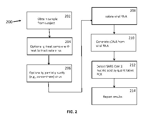
An embodiment of a method of the disclosure for detection of SARS-CoV-2 (200)
is
shown in FIG. 2. Thus, in an embodiment, a sample is obtained from a subject
(202). In
certain embodiments, the sample may be a nasopharyngeal or oropharyngeal swab,
sputum, a
lower respiratory tract aspirate, a bronchoalveolar lavage, or a
nasopharyngeal wash/aspirate or
nasal aspirate. Or, other types of samples may be used.
The sample may optionally be treated with heat to inactivate SARS-CoV-2 virus
present in the sample (204). In alternate embodiments, the sample is heated to
at least 60
degrees C, or at least degrees 65 C, or at least 70 degrees C, or at least 75
degrees C for a
designated time. The sample may be heated for at least 10 minutes, or at least
20 minutes, or at
least 30 minutes, or at least 40 minutes, or at least 50 minutes or for 1 hour
or more. In an
12
CA 03177050 2022-09-26
WO 2021/202897
PCT/US2021/025413
embodiment, the sample may be heated at 65 degrees C for about 30 minutes.
Thus, in an
embodiment, the sample may be heated to inactivate SARS-CoV-2 virus. In an
embodiment, a
protease, e.g. proteinase K or another protease, is also added to inactivate
the virus. The
protease may be added prior to the addition of heat so as to function during
heat-inactivation
of the virus.
Also optionally, the SARS-CoV-2 virus present in the sample may be partially
purified
(e.g., concentrated) from the rest of the sample (206). For example, for
isolation of viral
particles, the samples may be subjected to concentration (e.g., purification)
of the virus using
a matrix designed to bind viral particles (e.g., Nanotrap0 Virus Capture Kit,
Ceres
Nanosciences, Inc.). Or, other methods of purification may be used. Using such
a matrix,
elution of viral RNA from the concentrated viral particles may be performed at
a temperature
in the range of 90-99 degrees C for at least 3 minutes. In an embodiment,
elution may be
performed at 95 degrees C for at least five minutes or under similar
conditions.
At this point, SARS-CoV-2 RNA may be isolated from the sample (208), and used
to
generate copy DNA (cDNA) (210). The cDNA may be used to determine the presence
and/or
amount of SARS-CoV-2 RNA sequences in the sample. For example, in one
embodiment
quantitative PCR amplification is used (212). The quantitative PCR may be
performed by
amplifying at least one specific target sequence of the SARS-CoV-2
nucleocapsid (N) gene
present in the cDNA; and detecting the amplified SARS-CoV-2 nucleocapsid (N)
gene
sequences. In an embodiment, an internal probe that can bind to the cDNA or
amplification
products may be labeled with a reporter and a quencher dye such that
amplification allows
the 5' exonuclease activity of Taq polymerase to release the reporter dye,
allowing
amplification to be monitored. Any reporter and quencher dyes in the art may
be used. In
certain embodiments, the reporter day is FAM and the quencher dye is BHQ1. In
certain
embodiments, primers and probes of Table 2 (SEQ ID NOs: 1-9) are used for
detection of
SARS-CoV-2. In certain embodiments, the step of amplifying further comprises
amplification
of a control gene that is present in the subject, but not the virus. For
example, the control gene
may be the human RNase P (RP) gene or another gene. In certain embodiments,
the primers
(SEQ ID NOs: 10-11) and probe of SEQ ID NO: 12 are used for detection of the
RP gene. The
results may be reported to the subject, or his or her health care provider, or
other medical
professional (214).
13
CA 03177050 2022-09-26
WO 2021/202897
PCT/US2021/025413
The methods and systems may be optimized to produce results in less than 1
day. For
example, for the analysis of SARS-CoV-2 as disclosed herein, the method may
take less than 10
hours, or less than 8 hours, or less than 6 hours or less than 4 hours, or
less than 3 hours, or less
than 2 hours, or less than one hour. Also, as noted above, in an embodiment,
the samples may be
subjected to viral concentration and/or heat inactivation, and/or protease
treatment prior to
extraction of the viral nucleic acids. In an embodiment, heat-inactivation
allows for improved
processing of samples and/or protects laboratory personnel. This can improve
throughput, allow
processing of multiple samples (e.g., 400 samples) in about 40 minutes or
less. For example, in
certain embodiments, the disclosed methods may be performed robotically.
Robotic
processing may allow for a large number of samples to be analyzed with a
shorter turn-
around than a completely manually performed method. In a robotic embodiment,
samples
may be robotically extracted from sample collection apparatus (e.g. a tube,
vial, sample
carrier etc.) for further analysis via any of the methods disclosed herein. In
a further robotic
embodiment, extracted samples may be placed in reaction vessel (e.g. tube,
vial, etc.) for a
PCR reaction according to any of the disclosed methods. Additionally and/or
alternatively,
the PCR reaction may be performed robotically.
Compositions and Kits
Also disclosed herein are compositions and/or kits for performing any of the
disclosed
methods or running any of the disclosed systems. In an embodiment, the
compositions and/or
kits comprise reagents for detecting the presence or absence of a pathogen in
a sample from a
subject. The composition and/or kit may comprise reagents or components for
obtaining a
sample from the subject, such as nasal swabs, buffer solutions, storage
solutions and the like.
The composition and/or kit may further comprise reagents or components for
treating the
sample with heat to inactivate any pathogen present in the sample. The
composition and/or kit
may further comprise reagents or components for treating the sample with a
protease. The
composition and/or kit may further comprise reagents or components for
partially purifying
the pathogen as discussed in detail herein. The reagents and/or components may
be
individually packaged. Also, the composition and/or kit may further comprise
instructions for
use.
The compositions and/or kit may further comprise reagents or components for
detecting the presence or absence of the isolated pathogen-specific nucleic
acid. For example,
14
CA 03177050 2022-09-26
WO 2021/202897
PCT/US2021/025413
in some embodiments, the composition and/or kit may further comprise reagents
or
components for generating copy DNA (cDNA) from pathogen-specific (and/or
control) RNA.
Additionally and/or alternatively, the composition and/or kit may further
comprise
reagents or components for quantitative PCR amplification, using primers
specific to nucleic
acid sequences in the pathogen and an internal probe In certain embodiments,
the internal
probe may be labeled with a reporter and a quencher dye such that
amplification allows the
5'¨>3' exonuclease activity of Taq polymerase release of the reporter dye to
be monitored.
Any reporter and quencher dyes in the art may be used. In certain embodiments,
the reporter
day is FAM and the quencher dye is BHQ1.
In some embodiments, and for detection of SARS-CoV-2, the composition and/or
kit
may further comprise reagents or components for amplifying at least one
specific target
sequence of the SARS-CoV-2 nucleocapsid (N) gene present in the cDNA; and
detecting the
amplified SARS-CoV-2 nucleocapsid (N) gene sequences.
A variety of primers and probes specific to SARS-CoV-2 may be included in the
compositions and/or kits of the disclosure. In certain embodiments, the
compositions and/or
kit may comprise primers and/or probes for the SARS-CoV-2 Ni, N2 and N3
targets. In
certain embodiments, amplifying at least one specific target sequence of the
SARS-CoV-2
nucleocapsid (N) gene present in the cDNA comprises the use of at least one
primer and/or
probe having the sequence of any one of SEQ ID NOs: 1-9 as disclosed herein.
Also, in
certain embodiments, the compositions and/or kit may comprise reagents (e.g.,
primers and
probes) for amplification of a control gene that is present in the subject,
but not the virus. For
example, the control gene may be the human RNase P (RP) gene or another gene.
For
detection of RP sequences, the reagents (e.g., primers and probes) for
amplification of the RP
gene may comprise primers of SEQ ID NOs: 10 and 11, and a probe of SEQ ID NO:
12. In
certain embodiments, the primers and/or probes are labeled with a detectable
moiety such as
the detectable moieties disclosed herein.
The compositions and/or kits may be used to reverse transcribe RNA to cDNA for
subsequent amplification using quantitative PCR. For analysis of SARS-CoV-2,
the RT-PCR
may comprise a multiplex reaction with the SARS-CoV-2 primers and probes
and/or primers
and probes for an internal (i.e., non-SARS-CoV-2) control such as the human RP
gene. Or,
other combinations of primers and probes may be used for other pathogens. In
certain
embodiments of the disclosed compositions and/or kits, the assay is performed
as a multiplex
CA 03177050 2022-09-26
WO 2021/202897
PCT/US2021/025413
assay with three sets of SARS-CoV-2 primers and probes for the nucleocapsid
gene and one
set of primers and a probe for the RP gene. Thus, in certain embodiments,
primers for the
SARS-CoV-2 Ni gene are SEQ ID NOs: 1 and 2, and the internal probe is SEQ ID
NO: 3.
Also, in certain embodiments, primers for the SARS-CoV-2 N2 gene are SEQ ID
NOs: 4 and
5, and the internal probe is SEQ ID NO: 6. Also, in certain embodiments,
primers for the
SARS-CoV-2 N3 gene are SEQ ID NOs: 7 and 8, and the internal probe is SEQ ID
NO: 9.
Also, in certain embodiments, primers for the RP gene are SEQ ID NOs: 10 and
11, and the
internal probe is SEQ ID NO: 12. The TaqMan probes may be labeled at the 5'-
end with the
reporter molecule 6-carboxyfluorescein (FAM) and with the quencher, Black Hole
Quencher
1 (BHQ-1) (Biosearch Technologies, Inc., Novato, CA) at the 3'-end. Or other
dyes may be
used. Or, the primers may be labeled with any of the detectable moieties
disclosed herein for
detection of amplification products without the need for an internal probe.
Also, variations on
these primers and probes may be used to detect SARS-CoV-2 variants.
In certain embodiments, the compositions and/or kits may include at least one
of the
following controls:
Internal Control - RNase P (RP) control in clinical samples. The RP primer and
probe
set may be included in each run to test for human RP, which controls for
specimen quality and
demonstrates that nucleic acid was generated by the extraction process. Or,
another internal
control may be used.
Positive Template Control - contains in vitro transcribed template (e.g., SARS-
CoV-2)
RNA with genomic regions targeted by the method. The positive control may be
used to
monitor for failures of rRT-PCR reagents and reaction conditions. Or, another
positive
template control may be used.
Negative Extraction Control (NEC) ¨ In an embodiment, this may be a previously
characterized negative patient sample. The NEC may be used as an extraction
control and
positive control for the internal (e.g. RP) primer and probe set.
No Template (Negative) Control - Nuclease-free, molecular-grade water may be
used
to monitor non-specific amplification, cross-contamination during experimental
setup, and
nucleic acid contamination of reagents.
Systems
Also disclosed are systems for performing the methods herein. For example, the
system may comprise a station or stations for performing various steps of the
methods. In
16
CA 03177050 2022-09-26
WO 2021/202897
PCT/US2021/025413
certain embodiments, a station may comprise a robotic station for performing a
step or steps of
the method.
FIG. 3 illustrates one embodiment of a system (300) for pathogen detection
Thus, the
system may comprise a station for obtaining and/or receiving a sample from a
subject (302).
For example, in many cases samples may be obtained from the subject (e.g., by
a medical
professional, care-giver or even the subject themselves) at a site remote from
the testing area
and sent to the testing area. The sample may be a nasopharyngeal or
oropharyngeal swab,
sputum, a lower respiratory tract aspirate, a bronchoalveolar lavage, or a
nasopharyngeal
wash/aspirate or nasal aspirate. Or, other types of samples may be used.
The system may, in certain embodiments, have a station for treating the sample
to
inactivate pathogens present in the sample (304). In certain embodiments, the
sample is
heated to inactivate the pathogen. In alternate embodiments, the sample is
heated to at least 60
degrees C, or at least 65 degrees C, or at least 70 degrees C, or at least 75
degrees C for a
designated time. The sample may be heated for at least 10 minutes, or at least
20 minutes, or at
least 30 minutes, or at least 40 minutes, or at least 50 minutes or for 1 hour
or more. In an
embodiment, the sample may be heated at 65 degrees C for about 30 minutes. In
certain
embodiments, the system may have a station for adding a protease, e.g.
proteinase K or
another protease. This station (not shown in FIG. 3) may be prior to, after,
or part of the
station for heat-inactivation.
Also optionally, the system may have a station to partially purify (e.g.,
concentrate)
any pathogens present in the sample (306). For example, for isolation of viral
particles, the
samples may be subjected to concentration (e.g., purification) of the virus
using a matrix
designed to bind viral particles (e.g., Nanotrap0 Virus Capture Kit, Ceres
Nanosciences,
Inc.). Or, other methods of purification may be used. Using such a matrix,
elution of viral
RNA from the concentrated viral particles may be performed a temperature of 90-
99 degrees
C for at least 3 minutes. In an embodiment, elution may be performed at 95
degrees C for
about 5 minutes.
The system may also have a station for isolating nucleic acid (e.g., RNA or
DNA)
from the sample (308). Where the nucleic acid is RNA, the system may have a
station for
generating cDNA from the RNA (310). Also, the system may have a station for
determining
the presence and/or amount of pathogen-specific nucleic acid in the sample
(312). For
example, in one embodiment quantitative PCR amplification, using primers
specific to
17
CA 03177050 2022-09-26
WO 2021/202897
PCT/US2021/025413
nucleic acid sequences in the pathogen and an internal probe may be used as
disclosed herein.
The system may also include a station to report results to the subject, or his
or her health care
provider, or other medical professional (314).
The disclosure contemplates that certain of the stations as illustrated may be
combined
as a single station. For example, and not to be limiting, the stations for RNA
isolation, cDNA
preparation and quantitative PCR may be combined as a single station. Or, the
stations for
heat-inactivation and partial purification of the pathogen may be combined.
Also, as illustrated
in FIG. 3, any of the stations may be automated, robotically controlled,
and/or controlled at
least in part by a computer (400) and/or programmable software. Thus, the
system may
comprise a computer-program product tangibly embodied in a non-transitory
machine-
readable storage medium, including instructions configured to run the system
or any part of
the system and/or perform a step or steps of the methods of any of the
disclosed
embodiments. In some embodiments, a system is provided that includes one or
more data
processors and a non-transitory computer readable storage medium containing
instructions
which, when executed on the one or more data processors, cause the one or more
data
processors to perform part or all of one or more methods or processes
disclosed herein.
For example, disclosed is a system comprising one or more data processors, and
a
non-transitory computer readable storage medium containing instructions which,
when
executed on the one or more data processors, cause the one or more data
processors to
perform actions to direct at least one of the steps of obtaining a sample from
the subject;
treating the sample to inactivate any pathogen present in the sample;
optionally, treating the
sample to concentrate any pathogen present in the sample; treating the
inactivated and
optionally concentrated sample to isolate a pathogen-specific nucleic acid
from the sample;
and detecting the presence or absence of the isolated pathogen-specific
nucleic acid.
Also disclosed is a computer-program product tangibly embodied in a non-
transitory
machine-readable storage medium, including instructions configured to run the
systems
and/or perform a step or steps of the methods of any of the disclosed
embodiments. For
example, in certain embodiments, the computer-program product tangibly
embodied in a non-
transitory machine-readable storage medium, includes instructions configured
to cause one or
more data processors to perform actions to direct at least one of the steps of
obtaining a
sample from the subject; treating the sample to inactivate any pathogen
present in the sample;
optionally, treating the sample to concentrate any pathogen present in the
sample; treating the
18
CA 03177050 2022-09-26
WO 2021/202897
PCT/US2021/025413
inactivated and optionally concentrated sample to isolate a pathogen-specific
nucleic acid
from the sample; and detecting the presence or absence of the isolated
pathogen-specific
nucleic acid.
The systems and computer products may perform any of the methods disclosed
herein. One or more embodiments described herein can be implemented using
programmatic
modules, engines, or components. A programmatic module, engine, or component
can
include a program, a sub-routine, a portion of a program, or a software
component or a
hardware component capable of performing one or more stated tasks or
functions. As used
herein, a module or component can exist on a hardware component independently
of other
modules or components. Alternatively, a module or component can be a shared
element or
process of other modules, programs or machines.
FIG. 4 shows a block diagram of a analysis system (400) used for detection
and/or
quantification of a pathogen.. As illustrated in FIG. 4, modules, engines, or
components (e.g.,
program, code, or instructions) executable by one or more processors may be
used to
implement the various subsystems of an analyzer system according to various
embodiments.
The modules, engines, or components may be stored on a non-transitory computer
medium.
As needed, one or more of the modules, engines, or components may be loaded
into system
memory (e.g., RAM) and executed by one or more processors of the analyzer
system. In the
example depicted in FIG. 4, modules, engines, or components are shown for
implementing
the methods of the disclosure.
Thus, FIG. 4 illustrates an example computing device (400) suitable for use
with
systems and the methods according to this disclosure. The example computing
device (400)
includes a processor (405) which is in communication with the memory (410) and
other
components of the computing device (400) using one or more communications
buses (415).
The processor (405) is configured to execute processor-executable instructions
stored in the
memory (410) to perform one or more methods or operate one or more stations
for detecting
pathogen levels according to different examples, such as those in FIGS. 1-3 or
disclosed
elsewhere herein. In this example, the memory (410) may store processor-
executable
instructions (425) that can analyze (420) RT-PCR results for SARS-CoV-2 or
other
pathogens, as discussed herein.
The computing device 400 in this example may also include one or more user
input
devices (430), such as a keyboard, mouse, touchscreen, microphone, etc., to
accept user
19
CA 03177050 2022-09-26
WO 2021/202897
PCT/US2021/025413
input. The computing device (400) may also include a display (435) to provide
visual output
to a user such as a user interface. The computing device (400) may also
include a
communications interface (440). In some examples, the communications interface
(440) may
enable communications using one or more networks, including a local area
network
("LAN"); wide area network ("WAN"), such as the Internet; metropolitan area
network
("MAN"); point-to-point or peer-to-peer connection; etc. Communication with
other devices
may be accomplished using any suitable networking protocol. For example, one
suitable
networking protocol may include the Internet Protocol ("IP"), Transmission
Control Protocol
("TCP"), User Datagram Protocol ("UDP"), or combinations thereof, such as
TCP/IP or
UDP/IP.
Detection of SARS-CoV-2
An embodiment of the disclosed SARS-CoV-2 RT-PCR test comprises a real-time
reverse transcription polymerase chain reaction (rRT-PCR) test for the
qualitative detection of
nucleic acid from SARS-CoV-2 in upper and lower respiratory specimens (such as
nasopharyngeal or oropharyngeal swabs, sputum, lower respiratory tract
aspirates,
bronchoalveolar lavage, and nasopharyngeal wash/aspirate or nasal aspirate)
collected from
individuals suspected of being infected with SARS-CoV-2.
In an embodiment, results are presented for the identification of SARS-CoV-2
RNA.
SARS-CoV-2 RNA is generally detectable in respiratory specimens during the
acute phase of
infection. Positive results are indicative of the presence of SARS-CoV-2 RNA.
In some
embodiments, clinical correlation with patient history and other diagnostic
information may be
used to determine patient infection status. In an embodiment, positive results
do not rule out
bacterial infection or co-infection with other viruses. The agent detected may
not be the
definite cause of disease. Also, in an embodiment, negative results may not
preclude SARS-
CoV-2 infection and should not be used as the sole basis for patient
management decisions.
Negative results should be combined with clinical observations, patient
history, and
epidemiological information.
As disclosed herein, in some embodiments the method employs the step of heat-
inactivation of the pathogen. In an embodiment, after heat inactivation of the
virus, SARS-
CoV-2 nucleic acid is isolated, extracted and purified from upper and lower
respiratory
specimens (such as nasopharyngeal or oropharyngeal swabs, sputum, lower
respiratory tract
aspirates, bronchoalveolar lavage, andnasopharyngeal wash/aspirate or nasal
aspirate). In some
CA 03177050 2022-09-26
WO 2021/202897
PCT/US2021/025413
embodiments, the samples are subjected to viral concentration methods and/or
heat extraction
of the viral nucleic acids. The purified nucleic acid can then be reverse
transcribed into cDNA
followed by PCR amplification and detection with the a one-step (multiplex) RT-
PCR as
disclosed in detail herein.
In certain embodiments, the method utilizes at least one of the following
controls:
Internal Control - RNase P (RP) control in clinical samples. The RP primer and
probe
set is included in each run to test for human RP, which controls for specimen
quality and
demonstrates that nucleic acid was generated by the extraction process. Or,
another internal
control may be used.
Positive Template Control - contains in vitro transcribed template (e.g., SARS-
CoV-2)
RNA with genomic regions targeted by the method. The positive control may be
used to
monitor for failures of rRT-PCR reagents and reaction conditions.
Negative Extraction Control (NEC) ¨ In an embodiment, this may be a previously
characterized negative patient sample. Used as an extraction control and
positive control for
the RP primer and probe set.
No Template (Negative) Control - Nuclease-free, molecular-grade water used to
monitor non-specific amplification, cross-contamination during experimental
setup, and
nucleic acid contamination of reagents.
EXAMPLES
Example 1
Samples were aliquoted (e.g., into 96 well plates) and then the virus
inactivated by
heating at 65 degrees C for 30 minutes in the presence of proteinase K. The
COVID-19 RT-
PCR test can be used with the Roche MagNA Pure-96 (MP96) using MagNA Pure 96
DNA
and Viral NA Small Volume Kit and Applied Biosystems QuantStudio7 Flex (Q57)
instrument with software version 1.3. The primers and probes are those
recommended by the
Center for Disease Control (CDC). Tables 1 and 2 provide primers and probes
and reagents
that may be used. The assay was performed as a multiplex assay with all three
SARS-CoV-2
primers and probes and the RP primers and probes. TaqMan0 probes were labeled
at the 5'-
end with the reporter molecule 6-carboxyfluorescein (FAM) and with the
quencher, Black
Hole Quencher 1 (BHQ-1) (Biosearch Technologies, Inc., Novato, CA) at the 3'-
end. Y =
pyrimidine. In an embodiment, oligonucleotide sequences may be altered for
detection of
SARS-CoV-2 variants.
21
CA 03177050 2022-09-26
WO 2021/202897 PCT/US2021/025413
Table 1 - Reagents
Reagent Manufacturer Catalog #
DNA and Viral Small Volume Kit (3x192 purifications) Roche
06543588001
TaqPath 1-Step RT-PCR Master Mix, GC (2000 reactions) ThermoFisher A15300
COVID-19 N1 -F Primer IDT Custom
COVID-19 N1-R Primer IDT Custom
COVID-19 N1 -P Probe IDT Custom
COVID-19 N2-F Primer IDT Custom
COVID-19 N2-R Primer IDT Custom
COVID-19 N2-P Probe IDT Custom
COVID-19 N3-F Primer IDT Custom
COVID-19 N3-R Primer IDT Custom
COVID-19 N3-P Probe IDT Custom
RP-F Primer IDT Custom
RP-R Primer IDT Custom
RP-P Probe IDT Custom
COVID-19 N Positive Control IDT Custom
Hs RPP30 Internal Extraction Control IDT Custom
Table 2¨ CDC COVID-19 Primers and Probes
Name Description Sequence (5' ¨> 3') SEQ ID
Label Working
NO: Conc.
2019- 2019-nCoV N1 5'-GAC CCC AAA ATC AGC 1 None 20 RM
nCoV Ni-F Forward Primer GAA AT-3'
2019- 2019-nCoV N1 5'-TCT GGT TAC TGC CAG 2 None 20 RM
nCoV Nl-R Reverse Primer TTG AAT CTG-3'
2019- 2019-nCoV N1 5'-FAM-ACC CCG CAT TAC 3 FAM, 5 RM
nCoV Ni-P Probe GTT TGG TGG ACC-BHQ1-3' BHQ-1
2019- 2019-nCoV N2 5'-TTA CAA ACA TTG GCC 4 None 20 RM
nCoV N2-F Forward Primer GCA AA-3'
2019- 2019-nCoV N2 5'-GCG CGA CAT TCC GAA 5 None 20 RM
nCoV N2-R Reverse Primer GAA-3'
2019- 2019-nCoV N2 5'-FAM-ACA ATT TGC CCC 6 FAM, 5 RM
nCoV N2-P Probe CAG CGC TTC AG-BHQ1-3' BHQ-1
2019- 2019-nCoV N3 5'-GGG AGC CTT GAA TAC 7 None 20 RM
nCoV N3-F Forward Primer ACC AAA A-3'
2019- 2019-nCoV N3 5'-TGT AGC ACG ATT GCA 8 None 20 RM
nCoV N3-R Reverse Primer GCA TTG-3'
2019- 2019-nCoV N3 5'-FAM-AYC ACA TTG GCA 9 FAM, 5 RM
nCoV N3-P Probe CCC GCA ATC CTG-BHQ1-3' BHQ-1
RP-F RNAse P 5'-AGA TTT GGA CCT GCG 10 None 20 RM
Forward Primer AGC G-3'
RP-R RNAse P 5'-GAG CGG CTG TCT CCA 11 None 20 RM
Reverse Primer CAA GT-3'
RP-P RNAse P Probe 5'-FAM ¨ TTC TGA CCT GAA 12 FAM, 5
RM
GGC TCT GCG CG ¨ BHQ-1-3' BHQ-1
CONTROLS TO BE USED WITH THE COVID-19 RT-PCR
22
CA 03177050 2022-09-26
WO 2021/202897
PCT/US2021/025413
1) A negative (no template) control is used to eliminate the possibility of
sample
contamination on the assay run and is used on every assay plate. This control
is molecular
grade, nuclease-free water.
2) A positive template (COVID-19 N P) control is used to verify that the
assay run is
performing as intended and is used on every assay plate starting at master mix
addition at a
concentration of 50 copies/uL. The positive control is made of in vitro
transcribed and purified
viral RNA target that contains one copy each of N1, N2, and N3. The positive
template control
does not include RNase P target and result as "undetermined" for that marker.
3) An internal (Hs RPP30) control targeting RNase P is used to verify that
nucleic acid
is present in every sample and is used for every sample processed. This also
serves as the
extraction control to ensure that samples resulting as negative contain
nucleic acid for testing.
4) A negative extraction (NEC) control is a previously characterized
negative patient
sample. It serves both as a negative extraction control to monitor for any
cross-contamination
that occurs during the extraction process, as well as an extraction control to
validate extraction
reagents and successful RNA extraction.
INTERPRETATION OF RESULTS
All test controls are examined prior to interpretation of patient results. If
the controls
are not valid, the patient results either cannot or generally will not be
interpreted.
1) COVID-19 RT-PCR test Controls ¨ Positive. Negative. and Internal:
Negative (no template control) ¨ negative for all targets detected (Ct Not
Detected).
Positive (COVID-19 N P) ¨ positive for all targets detected (Ct < 40) Internal
extraction (Hs RPP30) ¨ negative for SARS-CoV-2 targets (Ct Not Detected),
positive for
RNase P (RP) target (Ct < 40).
Negative extraction (NEC) ¨ negative for SARS-CoV-2 targets (Ct Not Detected),
positive for RNase P (RP) target (Ct < 40).
If any control does not perform as described above, run is considered invalid
and all
specimens are repeated from extraction step.
2) Examination and Interpretation of Patient Specimen Results:
RP ¨ all clinical samples should yield positive results for RP target at < 40
Ct.
Samples that fail to show detection of RP and all three SARS-CoV-2 targets
within this range
should be repeated from extraction step. If sample detects any of the SARS-CoV-
2 targets,
the lack of amplification of RP target can be valid (Table 3).
23
CA 03177050 2022-09-26
WO 2021/202897
PCT/US2021/025413
Table 3 - COVID-19 RT-PCR test results interpretation
SARS- SARS- SARS- iase P Result Report Actions
CoV-2 CoV-2 CoV-2 Interpretation
Ni N2 N3
+/- SARS-CoV-2 POSITIVE Report results to
sender
Detected and appropriate public
health authorities.
If only one or both +/- +/- SARS-CoV-2 POSITIVE Report results to
sender
targets are positive Detected and appropriate
health
authorities.
+/- SARS-CoV-2 PRESUMPTIVE Sample is repeated
once.
Presumptive POSITIVE If the repeated result
Positive remains
"PRESUMPTIVE
POSITIVE", additional
confirmatory testing may
be conducted, if it is
necessary to differentiate
between SARS-CoV-2
and other SARS-like
viruses for
epidemiological purposes
or clinical management.
+ SARS-CoV-2 NEGATIVE Report
results to sender.
Not Detected
- Invalid Result INVALID Sample
is repeated once.
I f a second failure
occurs, it is reported to
sender as invalid and
recommend recollection
if patient is still
clinically indicated.
PERFORMANCE EVALUATION
1) Analytical Sensitivity:
Limit ofDetection (LoD):
The LoD study established the lowest concentration of SARS-CoV-2 (genome
copies(cp)/4) that can be detected by the COVID-19 RT-PCR test at least 95% of
the time.
The preliminary LoD was established by testing 10-fold dilutions of SARS-CoV-2
synthetic
RNA. The preliminary LoD was confirmed by testing 20 replicates of 2-fold
dilutions (50
cp/uL, 25 cp/uL, 12.5 cp/uL, 6.25 cp/uL, 3.125 cp/uL, and 1.25 cp/4). The
samples of 2-
fold dilutions were prepared by spiking the quantified live SARS-CoV-2 into
negative
24
CA 03177050 2022-09-26
WO 2021/202897
PCT/US2021/025413
respiratory clinical matrices (NP swabs and BAL). The study results showed
that the LoD of
the COVID-19 RT-PCR test is 6.25 cp/uL (19/20 positive).
2) Analytical Specificity:
Cross-reactivity of the COVID-19 RT-PCR test was evaluated using both in
silico
analysis and by testing whole organisms or purified nucleic acid from a panel
of organisms
listed in the table below. The empirical testing showed that all targets were
negative for all
tested microorganisms except for the SARS coronavirus which is expected to
react with N3
target (target for the universal detection of SARS-like viruses) of the COVID-
19 RT-PCR test
(Table 4; ND = not detected).
Table 4- CROSS-REACTIVITY TEST RESULTS
Sample Name Ni CT N2 CT N3 CT Source (Concentration)
Adenovirus 11 N/D N/D N/D ATCC VR-12D (1e^6)
Adenovirus 5 N/D N/D N/D ATCC VR-5D; Adenoid 75
(1.5e^6)
Bordetella pertussis N/D N/D N/D Patient Sample ( le^5)
Chlamydophila pneumoniae N/D N/D N/D ATCC 53592D; AR-39 (5e^6)
Enterovirus 70 N/D N/D N/D ATCC VR-836; J670-71 ( le^6)
Haemophilus influenzae N/D N/D N/D ATCC 51907D ( le^6)
Human coronavirus N/D N/D N/D ATCC VR-740; 229E ( le^6)
Human coronavirus N/D N/D N/D ATCC VR-3263 SD; NL63 (7e^5)
Human coronavirus N/D N/D N/D ATCC VR-3262SD; HKU1 (6e^5)
Human coronavirus N/D N/D N/D Patient Sample; 0C43 (1e^5)
Human metapneumovirus N/D N/D N/D ATCC VR-32505D (6e^5)
Human parainfluenza vims 1 N/D N/D N/D ATCC VR-94D; C35 (2e^7)
Human parainfluenza vims 2 N/D N/D N/D ATCC VR-92D; Greer (2e^7)
Human parainfluenza virus 3 N/D N/D N/D ATCC VR-1782; ATCC-2011-5
Human parainfluenza virus 4b N/D N/D N/D ATCC VR-1377; CH
19503
Human respiratory syncytial virus Nip N/D N/D ATCC VR-1580; 18537
Human rhinovirus 61 N/D N/D N/D ATCC VR-1171; 6669-CV39
N/D N/D N/D ATCC VR-1679D; H3N2, A/Hong
Influenza A Kong/8/68 (2e1'6)
Influenza B N/D N/D N/D ATCC VR-1735D; B/Taiwan/2/62
(3e^6)
Legionella pneumophila N/D N/D N/D ATCC 33152D-5; Philadelphia-
1 (1.5e1'6)
Middle East Respiratory N/D N/D N/D
Syndrome coronavirus ATCC VR-32485D; MERS (6e1'5)
Mycobacterium tuberculosis N/D N/D N/D ATCC 25177; H37Ra
Mycoplasma pneumoniae N/D N/D N/D ATCC 15531D; FH of Eaton
Agent (3e1'6)
Severe Acute Respiratory N/D N/D
Syndrome coronavirus 30.768 BET NR-3882; SARS
Streptococcus pneumoniae N/D N/D Nip ATCC 33400D-5 (3e1'6)
Streptococcus pyogenes N/D N/D N/D ATCC 12344D-5; Ti (3e1'6)
CA 03177050 2022-09-26
WO 2021/202897 PCT/US2021/025413
BLAST analysis showed no homology with primers and probes of the COVID-19 RT-
PCR test for the organisms listed in Table 5.
Table 5 - In silico analysis:
% Homology % Homology % Homology
Pathogen Strain GenBank Test Test
Test
Acc# Forward Reverse Primer Probe
Primer
Candida alb/cans All All 0 0 0
0
Neisseria meningitidis All All 0 0
Pseudomonas
All All 0 0 0
aeruginosa
Staphylococcus aureus All All 0 0 0
3) Clinical Evaluation:
A contrived clinical study was performed to evaluate the performance of the
COVID-
19 RT-PCR test. A total of 100 individual clinical respiratory samples, 50 NP
(nasopharyngeal) swabs and 50 BALs (bronchoalveolar lavage), were used in this
study. 100
negatives and 80 contrived positives were tested. Negative samples include 50
NP swabs and
50 BALs. Positive samples were comprised of 40 NP swabs and 40 BALs spiked
with
quantitated live SARS-CoV-2. 10 samples each were spiked at 8x, 4x, 2x, and lx
LoD. In one
contrived BAL sample, prepared at LoD, N3 target was not determined. The
positive and
negative percent agreements between the COVID-19 RT-PCR test and the expected
results in
NP swabs and BALs are shown in Tables 6 and 7.
Table 6 - Clinical performance of the COVID-19 RT-PCR test with NP swabs
SARS-CoV-2 Number Ni target N2 target N3
target
concentration of NP % Positive %
Positive % Positive
swabs (95% CIs) (95% CIs) (95% CIs)
lx LoD 10 100% 100% 100%
(72.25 ¨ 100) (72.25 ¨ 100) (72.25 ¨ 100)
2x LoD 10 100% 100% 100%
COVID-19 RT- (72.25 ¨ 100) (72.25 ¨ 100) (72.25 ¨
100)
26
CA 03177050 2022-09-26
WO 2021/202897
PCT/US2021/025413
PCR test 4x LoD 10 100% 100% 100%
(72.25 ¨ 100) (72.25 ¨ 100) (72.25 ¨ 100)
8x LoD 10 100% 100% 100%
(72.25 ¨ 100) (72.25 ¨ 100) (72.25 ¨ 100)
Negative 50 0 (NA) 0 (NA) 0 (NA)
NA = Not available
Performance of the COVID-19 RT-PCR test against the expected results are:
Positive Percent Agreement: 40/40 = 100% (95% CI: 91.24% - 100%)
Negative Percent Agreement: 50/50 = 100% (95% CI: 92.87% - 100%)
Table 7 - Clinical performance of the COVID-19 RT-PCR test with BAL specimens
SARS-CoV-2 Number Ni target N2 target N3 target
concentration of NP % Positive % Positive % Positive
swabs (95% CIs) (95% CIs) (95% CIs)
lx LoD 10 100% 100% 90%*
(72.25 ¨ 100) (72.25 ¨ 100) (59.59 ¨
98.22)
COVID-19 RT- 2x LoD 10 100% 100% 100%
PCR test (72.25¨ 100) (72.25¨ 100) (72.25¨
100)
4x LoD 10 100% 100% 100%
(72.25 ¨ 100) (72.25 ¨ 100) (72.25 ¨ 100)
8x LoD 10 100% 100% 100%
(72.25 ¨ 100) (72.25 ¨ 100) (72.25 ¨ 100)
Negative 50 0 (NA) 0 (NA) 0 (NA)
NA = Not available
*One BAL sample had failed detection of N3 target. Since the SARS-CoV-2
specific targets,
Ni and N2 were detected, the overall result for this sample was "POSITIVE".
Performance of the COVID-19 RT-PCR test against the expected results are:
Positive Percent Agreement: 40/40 = 100% (95% CI: 91.24% - 100%)
Negative Percent Agreement: 50/50 = 100% (95% CI: 92.87% - 100%)
Additionally, five positive and five negative patient samples were sent to the
North
Carolina Department of Health (NCDOH) and tested on the CDC assay under an
EUA. All
results were concordant (Table 8).
Table 8
27
CA 03177050 2022-09-26
WO 2021/202897
PCT/US2021/025413
Sample COVID-19 RT-PCR test NCSLPH Result CDC
assay under and EUA
1 Not detected Not detected
2 Not detected Not detected
3 Positive Presumptive Positive
4 Not detected Not detected
Positive Presumptive Positive
6 Positive Presumptive Positive
7 Not detected Not detected
8 Positive Presumptive Positive
9 Not detected Not detected
Positive Presumptive Positive
Example 2
In certain embodiments, the method may be performed by concentrating the viral
5 particles followed by subsequent extraction of the viral RNA from the
viral particles. Once
the RNA is extracted, amplification may be performed using the primers and
methods
detailed in Example 1.
Briefly, samples (nasal swab) are received and a portion aliquoted into
individual
wells in a plate (e.g., 96 well microtiter plate). After inactivation of viral
proteins by
10 treatment with proteinase K and heating at 65 degrees C for 30 minutes
(as performed in
Example 1), the samples are subjected to concentration (e.g., purification) of
the virus using a
matrix designed to bind viral particles (e.g., Nanotrap0 Virus Capture Kit,
Ceres
Nanosciences, Inc.). Briefly, the heat-inactivated viral particles are mixed
with the virus
capture beads (e.g. Nanotrap0) as recommended by the manufacturer. The beads
may then
be magnetically concentrated, media removed and the beads washed in phosphate-
buffered
saline (PBS). After magnetic concentration, elution buffer is added and the
viral particles
attached to the bead are then incubated at 95 degrees C for the required time
(e.g., 5 min) in
the elution buffer. The beads are then magnetically concentrated and the viral
RNA is
removed and transferred to another plate for RT-PCR as described in Example 1.
The process
may be automated using a Hamilton robot. Use of heated extraction of the virus
allows for
processing of 400 samples to be completed in 40 minutes as compared to 4 hours
using the
purification described in Example 1.
28
CA 03177050 2022-09-26
WO 2021/202897
PCT/US2021/025413
Example 3 - Embodiments
Various non-limiting embodiments are provided below.
Al. A method to detect SARS-CoV-2 in a sample from a subject
comprising:
obtaining a sample from the subject;
isolating SARS-CoV-2 RNA from the sample;
generating copy DNA (cDNA) from the SARS-CoV-2 RNA;
amplifying at least one target sequence of the SARS-CoV-2 cDNA; and
detecting the amplified SARS-CoV-2 sequences.
A2. The method of any one of the previous or subsequent method embodiments,
wherein
the sample is treated to inactivate the virus.
A3. The method of any one of the previous or subsequent method embodiments,
wherein
the sample is heated to inactivate the virus
A4. The method of any one of the previous or subsequent method embodiments,
wherein
the sample is heated to at least 60 degrees C, or to at least 65 degrees C, or
to at least 70
degrees C, or to at least 75 degrees C for a designated time.
A4.1 The method of any one of the previous or subsequent method embodiments,
wherein
the sample is heated for at least 10 minutes, or at least 20 minutes, or at
least 30 minutes, or at
least 40 minutes, or at least 50 minutes or for 1 hour or more.
A4.2 The method of any one of the previous or subsequent method embodiments,
wherein
the sample is heated at 65 degrees C for about 30 minutes to inactivate the
virus.
AS. The method of any one of the previous or subsequent method
embodiments, wherein
the sample is treated with a protease to inactivate the virus.
A6. The method of any one of the previous or subsequent method
embodiments, wherein
the sample is treated with a proteinase K.
A7. The method of any one of the previous or subsequent method embodiments,
wherein
the step of isolating SARS-CoV-2 RNA comprises concentrating viral particles
followed by
elution of the SARS-CoV-2 RNA from the concentrated viral particles.
A8. The method of any one of the previous or subsequent method
embodiments, wherein
the elution of viral RNA from the concentrated viral particles is performed at
95 degrees C for
at least 5 minutes.
29
CA 03177050 2022-09-26
WO 2021/202897
PCT/US2021/025413
A9. The method of any one of the previous or subsequent method embodiments,
wherein
the step of amplifying at least one target sequence of the SARS-CoV-2
comprises quantitative
RT-PCR.
A10. The method of any one of the previous or subsequent method embodiments,
wherein
the at least one target sequence of SARS-CoV-2 comprises at least part of the
SARS-CoV-2
nucleocapsid (N) gene.
All. The method of any one of the previous or subsequent method embodiments,
wherein
the at least one target sequence of SARS-CoV-2 comprises the SARS-CoV-2 N1, N2
and N3
sequences.
Al2. The method of any one of the previous or subsequent method embodiments,
wherein
the step of amplifying at least one target sequence of SARS-CoV-2 comprises
multiplex RT-
PCR using primers and probes for SARS-CoV-2 N1, N2 and N3 sequences.
A13. The method of any one of the previous or subsequent method embodiments,
wherein
the step of amplifying at least one specific target sequence of the SARS-CoV-2
cDNA
comprises hybridizing a probe to the at least one specific target sequence,
such that during the
extension phase of amplification, a 5' nuclease activity of Taq polymerase
degrades the bound
probe, causing a reporter dye on the probe to separate from a quencher dye on
the probe,
generating a fluorescent signal.
A14. The method of any one of the previous or subsequent method embodiments,
wherein
the reporter dye is FAM.
A15. The method of any one of the previous or subsequent method embodiments,
wherein
the quencher dye is BHQ1.
A16. The method of any one of the previous or subsequent method embodiments,
wherein
the step of amplifying further comprises amplification of a nucleic acid
sequence from a
control gene that is present in the subject, but not the virus.
A17. The method of any one of the previous or subsequent method embodiments,
wherein
the control gene is the human RNase P (RP) gene.
A18. The method of any one of the previous or subsequent method embodiments,
wherein
the step of amplifying at least one target sequence of SARS-CoV-2 comprises
the use of at
least one primer and/or probe having the sequence of any one of SEQ ID NOs: 1-
9.
CA 03177050 2022-09-26
WO 2021/202897
PCT/US2021/025413
A19. The method of any one of the previous or subsequent method embodiments,
wherein
the step of amplifying a nucleic acid sequence from the RP gene comprises the
use of at least
one primer and/or probe having the sequence of any one of SEQ ID NOs: 10-12.
A20. The method of any one of the previous or subsequent method embodiments,
wherein
the sample comprises a specimen from either the upper or lower respiratory
system.
A21. The method of any one of the previous or subsequent method embodiments,
wherein
the sample comprises at least one of a nasopharyngeal swab, an oropharyngeal
swab, sputum,
a lower respiratory tract aspirate, a bronchoalveolar lavage, a nasopharyngeal
wash or aspirate,
or a nasal aspirate.
Bl. A method to detect the presence or absence of a pathogen in a sample
from a subject
comprising:
obtaining a sample from the subject;
treating the sample to inactivate any pathogen present in the sample;
optionally, treating the sample to concentrate any pathogen present in the
sample;
treating the inactivated and optionally concentrated sample to isolate a
pathogen-
specific nucleic acid from the sample; and
detecting the presence or absence of the isolated pathogen-specific nucleic
acid.
B2. The method of any one of the previous or subsequent method embodiments,
further
comprising:
isolating RNA from the inactivated sample;
generating copy DNA (cDNA) from the RNA isolated from the inactivated sample;
amplifying at least one specific target sequence of the cDNA; and
detecting presence or absence of amplified sequences.
B3. The method of any one of the previous or subsequent method embodiments,
wherein
the pathogen is SARS-CoV-2.
B4. The method of any one of the previous or subsequent method embodiments,
wherein
the sample is heated to inactivate the pathogen.
B5. The method of any one of the previous or subsequent method embodiments,
wherein
the sample is heated to at least 60 degrees C, or to at least 65 degrees C, or
to at least 70
degrees C, or to at least 75 degrees C for a designated time.
31
CA 03177050 2022-09-26
WO 2021/202897
PCT/US2021/025413
B5.1 The method of any one of the previous or subsequent method embodiments,
wherein
the sample is heated for at least 10 minutes, or at least 20 minutes, or at
least 30 minutes, or at
least 40 minutes, or at least 50 minutes or for 1 hour or more.
B5.2 The method of any one of the previous or subsequent method embodiments,
wherein
the sample is heated at 65 degrees C for about 30 minutes to inactivate the
pathogen.
B6. The method of any one of the previous or subsequent method embodiments,
wherein
the sample is treated with a protease to inactivate the pathogen.
B7. The method of any one of the previous or subsequent method embodiments,
wherein
the sample is treated with a proteinase K.
B8. The method of any one of the previous or subsequent method embodiments,
wherein
the step of isolating pathogen nucleic acid comprises concentrating the
inactivated pathogen
followed by elution of the pathogen nucleic acid.
B9. The method of any one of the previous or subsequent method embodiments,
wherein
the elution of pathogen nucleic acid from the concentrated pathogen is
performed at 95
degrees C for at least 5 minutes.
B10. The method of any one of the previous or subsequent method embodiments,
wherein
the step of amplifying at least one target sequence of the pathogen nucleic
acid comprises
quantitative PCR.
B11. The method of any one of the previous or subsequent method embodiments,
wherein
the at least one target sequence of SARS-CoV-2 comprises at least part of the
SARS-CoV-2
nucleocapsid (N) gene.
B12. The method of any one of the previous or subsequent method embodiments,
wherein
the at least one target sequence of SARS-CoV-2 comprises SARS-CoV-2 Ni, N2 and
N3
sequences.
B13. The method of any one of the previous or subsequent method embodiments,
wherein
the step of amplifying at least one target sequence of the pathogen comprises
multiplex RT-
PCR using primers and probes for a pathogen target sequence or sequences.
B14. The method of any one of the previous or subsequent method embodiments,
wherein
the step of amplifying at least one target sequence of the pathogen comprises
hybridizing a
probe to the at least one pathogen target sequence such that during the
extension phase of
amplification, a 5' nuclease activity of Taq polymerase degrades the bound
probe, causing a
32
CA 03177050 2022-09-26
WO 2021/202897
PCT/US2021/025413
reporter dye on the probe to separate from a quencher dye on the probe,
generating a
fluorescent signal.
B15. The method of any one of the previous or subsequent method embodiments,
wherein
the reporter dye is FAM.
B16. The method of any one of the previous or subsequent method embodiments,
wherein
the quencher dye is BHQ1.
B17. The method of any one of the previous or subsequent method embodiments,
wherein
the step of amplifying further comprises amplification of a nucleic acid
sequence from a
control gene that is present in the subject, but not the virus.
B18. The method of any one of the previous or subsequent method embodiments,
wherein
the control gene is the human RNase P (RP) gene.
B19. The method of any one of the previous or subsequent method embodiments,
wherein
the step of amplifying at least one target sequence of SARS-CoV-2 comprises
the use of at
least one primer and/or probe having the sequence of any one of SEQ ID NOs: 1-
9.
B20. The method of any one of the previous or subsequent method embodiments,
wherein
the step of amplifying a nucleic acid sequence from the RP gene comprises the
use of at least
one primer and/or probe having the sequence of any one of SEQ ID NOs: 10-12.
B21. The method of any one of the previous or subsequent method embodiments,
wherein
the sample comprises a specimen from either the upper or lower respiratory
system.
B22. The method of any one of the previous or subsequent method embodiments,
wherein
the sample comprises at least one of a nasopharyngeal swab, an oropharyngeal
swab, sputum,
a lower respiratory tract aspirate, a bronchoalveolar lavage, a nasopharyngeal
wash or aspirate,
or a nasal aspirate.
Cl. A system for performing a step or any of the steps of the previous
embodiments or for
using any of the compositions and/or kits of the subsequent kit or composition
embodiments.
C2. A system to detect the presence or absence of a pathogen in a sample
from a subject
comprising: at least one station to inactivate the pathogen and a station to
detect the presence
or absence of the pathogen-specific nucleic acid.
C3. The system of any of the previous or subsequent system embodiments
comprising a
station to receive or obtain a sample from a subject.
C4. The system of any of the previous or subsequent system embodiments
comprising a
station to purify or partially purify the pathogen from a sample from a
subject.
33
CA 03177050 2022-09-26
WO 2021/202897
PCT/US2021/025413
C5. The system of any of the previous or subsequent system embodiments
comprising a
station to isolate nucleic acid from the pathogen.
C6. The system of any of the previous or subsequent system embodiments
comprising a
station to report results.
C7. The system of any of the previous or subsequent system embodiments,
wherein the
pathogen is SARS-CoV-2.
C8. The system of any of the previous or subsequent system embodiments,
wherein the
sample is heated to inactivate the pathogen.
C9. The system of any one of the previous or subsequent system embodiments,
wherein the
sample is heated to at least 60 degrees C, or to at least 65 degrees C, or to
at least 70 degrees
C, or to at least 75 degrees C for a designated time.
C9.1 The system of any of the previous or subsequent system embodiments,
wherein the
sample is heated for at least 10 minutes, or at least 20 minutes, or at least
30 minutes, or at
least 40 minutes, or at least 50 minutes or for 1 hour or more.
C9.2 The system of any of the previous or subsequent system embodiments,
wherein the
sample is heated at 65 degrees C for about 30 minutes to inactivate the
pathogen.
C10. The system of any of the previous or subsequent system embodiments,
wherein the
sample is treated with a protease to inactivate the pathogen.
C 1 1. The system of any of the previous or subsequent system embodiments,
wherein the
sample is treated with a proteinase K.
C12. The system of any of the previous or subsequent system embodiments,
wherein the
step of isolating pathogen nucleic acid comprises concentrating the
inactivated pathogen
followed by elution of the pathogen nucleic acid.
C13. The system of any of the previous or subsequent system embodiments,
wherein the
elution of pathogen nucleic acid from the concentrated pathogen is performed
at 95 degrees C
for at least 5 minutes.
C14. The system of any of the previous or subsequent system embodiments,
wherein the
step of amplifying at least one target sequence of the pathogen nucleic acid
comprises
quantitative PCR.
C15. The system of any of the previous or subsequent system embodiments,
wherein the at
least one target sequence of SARS-CoV-2 comprises at least part of the SARS-
CoV-2
nucleocapsid (N) gene.
34
CA 03177050 2022-09-26
WO 2021/202897
PCT/US2021/025413
C16. The system of any of the previous or subsequent system embodiments,
wherein the at
least one target sequence of SARS-CoV-2 comprises SARS-CoV-2 Ni, N2 and N3
sequences.
C17. The system of any of the previous or subsequent system embodiments,
wherein the
step of amplifying at least one target sequence of the pathogen comprises
multiplex RT-PCR
using primers and probes for a pathogen target sequence or sequences.
C18. The system of any of the previous or subsequent system embodiments,
wherein the step
of amplifying at least one target sequence of the pathogen comprises
hybridizing a probe to the
at least one pathogen target sequence such that during the extension phase of
amplification, a 5'
nuclease activity of Taq polymerase degrades the bound probe, causing a
reporter dye on the
probe to separate from a quencher dye on the probe, generating a fluorescent
signal.
C19. The system of any of the previous or subsequent system embodiments,
wherein the
reporter dye is FAM.
C20. The system of any of the previous or subsequent system embodiments
wherein the
quencher dye is BHQ1.
C21. The system of any of the previous or subsequent system embodiments,
wherein the
step of amplifying further comprises amplification of a nucleic acid sequence
from a control
gene that is present in the subject, but not the virus.
C22. The system of any of the previous or subsequent system embodiments,
wherein the
control gene is the human RNase P (RP) gene.
C23. The system of any of the previous or subsequent system embodiments,
wherein the
step of amplifying at least one target sequence of SARS-CoV-2 comprises the
use of at least
one primer and/or probe having the sequence of any one of SEQ ID NOs: 1-9.
C24. The system of any of the previous or subsequent system embodiments,
wherein the
step of amplifying a nucleic acid sequence from the RP gene comprises the use
of at least one
primer and/or probe having the sequence of any one of SEQ ID NOs: 10-12.
C25. The system of any of the previous or subsequent system embodiments
wherein the
sample comprises a specimen from either the upper or lower respiratory system.
C26. The system of any of the previous or subsequent system embodiments,
wherein the
sample comprises at least one of a nasopharyngeal swab, an oropharyngeal swab,
sputum, a
lower respiratory tract aspirate, a bronchoalveolar lavage, a nasopharyngeal
wash or aspirate,
or a nasal aspirate.
CA 03177050 2022-09-26
WO 2021/202897
PCT/US2021/025413
C27. The system of any of the previous or subsequent system embodiments,
wherein at least
one station is automated and/or controlled by a computer.
Dl. A computer-program product tangibly embodied in a non-transitory
machine-readable
storage medium, including instructions configured to perform any step of the
methods or run
any of the stations of the systems of the previous embodiments.
D2. A computer-program product tangibly embodied in a non-transitory
machine-readable
storage medium, including instructions configured to detect the presence or
absence of a
pathogen-specific nucleic acid.
D3. A computer-program product tangibly embodied in a non-transitory
machine-readable
storage medium of any of the previous or subsequent embodiments, including
instructions
configured to detect the presence or absence of SARS-CoV-2 nucleic acid.
El. A composition comprising reagents to perform any of the methods of
the previous
embodiments.
E2. The composition of any of the previous or subsequent composition
embodiments
comprising reagents detect the presence or absence of SARS-CoV-2.
E3. The composition of any of the previous or subsequent composition
embodiments,
comprising at least one primer and/or probe having the sequence of any one of
SEQ ID NOs:
1-9.
E4. The composition of any of the previous or subsequent composition
embodiments,
comprising at least one primer and/or probe having the sequence of any one of
SEQ ID NOs:
10-12.
E5. The composition of any of the previous or subsequent composition
embodiments,
comprising reagents for obtaining a sample from a subject.
E6. The composition of any of the previous or subsequent composition
embodiments,
comprising a protease for inactivating the pathogen.
E7. The composition of any of the previous or subsequent composition
embodiments,
comprising reagents to purify or partially purify the pathogen from other
components in the
sample.
E8. The composition of any of the previous or subsequent composition
embodiments,
comprising reagents for detecting the pathogen.
E9. The composition of any of the previous or subsequent composition
embodiments,
comprising reagents for generating cDNA from RNA.
36
CA 03177050 2022-09-26
WO 2021/202897
PCT/US2021/025413
E10. The composition of any of the previous or subsequent composition
embodiments,
comprising reagents for quantitative PCR.
Eli. The composition of any of the previous or subsequent composition
embodiments,
wherein probes for quantitative PCR are labeled with a reporter dye and a
quencher dye.
E12. The composition of any of the previous or subsequent composition
embodiments,
wherein the probes for quantitative PCR are labeled with FAM and/or BHQ1.
E13. The composition of any of the previous or subsequent composition
embodiments,
comprising instructions for use of any of the reagents.
E14. The composition of any of the previous or subsequent composition
embodiments,
wherein the reagents or components thereof are packaged in individual
containers.
Fl. A kit comprising reagents to perform any of the methods of the
previous
embodiments.
F2. The kit of any of the previous or subsequent kit embodiments,
comprising reagents
detect the presence or absence of SARS-CoV-2.
F3. The kit of any of the previous or subsequent kit embodiments,
comprising at least one
primer and/or probe having the sequence of any one of SEQ ID NOs: 1-9.
F4. The kit of any of the previous or subsequent kit embodiments,
comprising at least one
primer and/or probe having the sequence of any one of SEQ ID NOs: 10-12.
F5. The kit of any of the previous or subsequent kit embodiments,
comprising reagents or
components for obtaining a sample from a subject.
F6. The kit of any of the previous or subsequent kit embodiments,
comprising a protease
for inactivating the pathogen.
F7. The kit of any of the previous or subsequent kit embodiments,
comprising reagents to
purify or partially purify the pathogen from other components in the sample.
F8. The kit of any of the previous or subsequent kit embodiments,
comprising reagents for
detecting the pathogen.
F9. The kit of any of the previous or subsequent kit embodiments,
comprising reagents for
generating cDNA from RNA.
F10. The kit of any of the previous or subsequent kit embodiments, comprising
reagents for
quantitative PCR.
F11. The kit of any of the previous or subsequent kit embodiments, wherein
probes for
quantitative PCR are labeled with a reporter dye and a quencher dye.
37
CA 03177050 2022-09-26
WO 2021/202897
PCT/US2021/025413
F12. The kit of any of the previous or subsequent kit embodiments, wherein the
probes for
quantitative PCR are labeled with FAM and/or BHQ1.
F13. The kit of any of the previous or subsequent kit embodiments, comprising
instructions
for use of any of the reagents.
F14. The kit of any of the previous or subsequent kit embodiments, wherein the
reagents or
components thereof are packaged in individual containers.
Additional Considerations
Specific details are given in the above description to provide a thorough
understanding of the embodiments. However, it is understood that the
embodiments can be
practiced without these specific details. For example, circuits can be shown
in block diagrams
in order not to obscure the embodiments in unnecessary detail. In other
instances, well-
known circuits, processes, algorithms, structures, and techniques can be shown
without
unnecessary detail in order to avoid obscuring the embodiments.
Implementation of the techniques, blocks, steps and means described above can
be
done in various ways. For example, these techniques, blocks, steps and means
can be
implemented in hardware, software, or a combination thereof For a hardware
implementation, the processing units can be implemented within one or more
application
specific integrated circuits (ASICs), digital signal processors (DSPs),
digital signal
processing devices (DSPDs), programmable logic devices (PLDs), field
programmable gate
arrays (FPGAs), processors, controllers, micro-controllers, microprocessors,
other electronic
units designed to perform the functions described above, and/or a combination
thereof
Also, it is noted that the embodiments can be described as a process which is
depicted as a flowchart, a flow diagram, a data flow diagram, a structure
diagram, or a block
diagram. Although a flowchart can describe the operations as a sequential
process, many of
the operations can be performed in parallel or concurrently. In addition, the
order of the
operations can be re-arranged. A process is terminated when its operations are
completed, but
could have additional steps not included in the figure. A process can
correspond to a method,
a function, a procedure, a subroutine, a subprogram, etc. When a process
corresponds to a
function, its termination corresponds to a return of the function to the
calling function or the
main function.
38
CA 03177050 2022-09-26
WO 2021/202897
PCT/US2021/025413
Furthermore, embodiments can be implemented by hardware, software, scripting
languages, firmware, middleware, microcode, hardware description languages,
and/or any
combination thereof When implemented in software, firmware, middleware,
scripting
language, and/or microcode, the program code or code segments to perform the
necessary
tasks can be stored in a machine readable medium such as a storage medium. A
code segment
or machine-executable instruction can represent a procedure, a function, a
subprogram, a
program, a routine, a subroutine, a module, a software package, a script, a
class, or any
combination of instructions, data structures, and/or program statements. A
code segment can
be coupled to another code segment or a hardware circuit by passing and/or
receiving
information, data, arguments, parameters, and/or memory contents. Information,
arguments,
parameters, data, etc. can be passed, forwarded, or transmitted via any
suitable means
including memory sharing, message passing, ticket passing, network
transmission, etc.
For a firmware and/or software implementation, the methodologies can be
implemented with modules (e.g., procedures, functions, and so on) that perform
the functions
described herein. Any machine-readable medium tangibly embodying instructions
can be
used in implementing the methodologies described herein. For example, software
codes can
be stored in a memory. Memory can be implemented within the processor or
external to the
processor. As used herein the term "memory" refers to any type of long term,
short term,
volatile, nonvolatile, or other storage medium and is not to be limited to any
particular type of
memory or number of memories, or type of media upon which memory is stored.
Moreover, as disclosed herein, the term "storage medium", "storage" or
"memory"
can represent one or more memories for storing data, including read only
memory (ROM),
random access memory (RAM), magnetic RAM, core memory, magnetic disk storage
mediums, optical storage mediums, flash memory devices and/or other machine
readable
mediums for storing information. The term "machine-readable medium" includes,
but is not
limited to portable or fixed storage devices, optical storage devices,
wireless channels, and/or
various other storage mediums capable of storing that contain or carry
instruction(s) and/or
data.
While the principles of the disclosure have been described above in connection
with
specific apparatuses and methods, it is to be clearly understood that this
description is made
only by way of example and not as limitation on the scope of the disclosure.
39