Note: Descriptions are shown in the official language in which they were submitted.
A METHOD OF REGISTERING INERTIAL MEASUREMENT UNITS IN AN
OPERATING ROOM
[0001] This application stems from Canadian Patent Application No. 2,933,235
filed on
December 9, 2014.
RELATED ART
Field of the Invention
[0002] The present disclosure is directed to various aspects of orthopedics
including bone
and tissue reconstruction, patient-specific and mass customized orthopedic
implants,
gender and ethnic specific orthopedic implants, cutting guides, trauma plates,
bone graft
cutting and placement guides, patient-specific instruments, utilization of
inertial
measurement units for anatomical tracking for kinematics and pathology, and
utilization of
inertial measurement units for navigation during orthopedic surgical
procedures.
BACKGROUND
[0003] Reconstruction of a deformed anatomy or a partial anatomy is one of the
complex
problems facing healthcare providers. Loss of anatomy may be the result of
birth
conditions, tumors, diseases, personal injuries, or failure of previous
surgeries. As part of
providing treatment for various ailments, healthcare providers may find it
advantageous to
reconstruct an anatomy or construct an anatomy to facilitate treatment for
various
conditions that may include, without limitation, broken/shattered bones, bone
degeneration, orthopedic implant revision, joint degeneration, and custom
instrumentation
design. For example, prior art hip reconstruction solution requires mirroring
of the healthy
patient anatomy which may not be an accurate reflection of the healthy anatomy
due to
naturally occurring asymmetry.
INTRODUCTION TO THE INVENTION
[0003] It is a first aspect of the present invention to provide a surgical
navigation system
comprising a signal receiver communicatively coupled to a primary processor,
the primary
processor programmed to utilize a sequential Monte Carlo algorithm to
calculate changes
1
Date Regue/Date Received 2022-09-29
in three dimensional position of an inertial measurement unit mounted to a
surgical tool,
the processor communicatively coupled to a first memory storing tool data
unique to each
of a plurality of surgical tools, and a second memory storing a model data
sufficient to
construct a three dimensional model of an anatomical feature, the primary
processor
communicatively coupled to a display providing visual feedback regarding the
three
dimensional position of the surgical tool with respect to the anatomical
feature.
[0004] In a more detailed embodiment of the first aspect, the surgical
navigation system
further includes a reference inertial measurement unit communicatively coupled
to a first
on-board processor and a first wireless transmitter to transmit data to the
primary processor,
the reference inertial measurement unit configured to be attached to the
anatomical feature,
where the first on-board processor directs transmission of data from the
reference inertial
measurement unit to the first wireless transmitter, where the inertial
measurement unit
mounted to the surgical tool comprises a utility inertial measurement unit
communicatively
coupled to a second on-board processor and a second wireless transmitter, the
second on-
board processor configured to be mounted to one of the plurality of surgical
tools, and
where the primary processor is communicatively coupled to a primary received
configured
to receive data from the first wireless transmitter and data from the second
wireless
transmitter. In yet another more detailed embodiment, the second on-board
processor
directs communication via the second wireless transmitter of an identity of
the surgical tool
to which the utility inertial measurement unit is mounted. In a further
detailed
embodiment, the inertial measurement unit includes at least three
accelerometers and three
magnetometers, each of the at least three accelerometers outputs data relative
to three axes
for a total of no less than nine accelerometer data streams, each of at least
three
magnetometers outputs data relative to three axes for a total of no less than
nine
magnetometer data streams, the primary processor utilizes the nine
accelerometer data
streams and the nine magnetometer data streams to calculate changes in three
dimensional
position of the inertial measurement unit mounted to the surgical tool. In
still a further
detailed embodiment, the model data stored in the second memory includes a
three
dimensional virtual model of the anatomical feature, the tool data stored in
the first memory
includes three dimensional virtual models of the plurality of surgical tools,
the display
2
Date Regue/Date Received 2022-09-29
displays the three dimensional virtual model of the anatomical feature, the
display displays
a three dimensional virtual model of the surgical tool, the primary processor
is operative to
utilize data from the reference inertial measurement unit to reposition the
three dimensional
virtual model of the anatomical feature, and the primary processor is
operative to utilize
data from the utility inertial measurement unit to reposition the three
dimensional virtual
model of the surgical tool. In a more detailed embodiment, the primary
processor is
operative to utilize data from the inertial measurement unit to reposition the
three
dimensional virtual model of the surgical tool with respect to a three
dimensional virtual
model of the anatomical feature in real-time. In a more detailed embodiment,
the sequential
Monte Carlo algorithm includes a von Mises-Fisher density algorithm component.
In
another more detailed embodiment, the tool data stored in the first memory
includes
positional data indicating the relative distances between an end effector of
the surgical tool
and a mounting location on the surgical device for the inertial measurement
unit, and the
surgical tool includes at least one of a reamer, a cup positioned, an
impacter, a drill, a saw,
and a cutting guide. In yet another more detailed embodiment, the inertial
measurement
unit includes at least three magnetometers, and the display is at least one of
coupled to the
surgical tool or coupled to the primary processor.
[0005] It is a second aspect of the present invention to provide a calibration
system, for an
inertial measurement unit including a magnetometer and an accelerometer,
comprising: (a)
a primary platform rotationally repositionable with respect to an intermediate
platform
along a first axis; (b) a final platform rotationally repositionable with
respect to the
intermediate platform along a second axis, the second axis being perpendicular
to the first
axis, the final platform including a retainer configured to mount to an
inertial measurement
unit; and, (c) a processor and associated software configured to
communicatively couple
to the inertial measurement unit, the software operative to utilize data
output from a
magnetometer associated with the inertial measurement unit while the primary
platform is
rotated with respect to the intermediate platform and while the final platform
is rotated
with respect to the intermediate platform and record a data set resembling an
ellipsoid, the
software operative to fit a sphere to the data set and generate magnetometer
correction
3
Date Regue/Date Received 2022-09-29
calculations to account for distortions in a local magnetic field, thereby
normalizing future
data output from the magnetometer.
[0006] In a more detailed embodiment of the second aspect, the primary
platform is
stationary. In yet another more detailed embodiment, the primary platform at
least partially
houses a motor configured to cause rotation of the intermediate platform with
respect to
the primary platform. In a further detailed embodiment, the software is
operative to utilize
a first set of data output from an accelerometer associated with the inertial
measurement
unit while the inertial measurement unit is at a first stationary position and
operative to
utilize a second set of data output from the accelerometer at a second
stationary position
different from the first stationary position to generate accelerometer
correction calculations
to normalizing future data output from the accelerometer. In still a further
detailed
embodiment, the first stationary position corresponds to the primary platform
being at a
first fixed position with respect to the intermediate platform and the final
platform is at a
second fixed position with respect to the intermediate platform, and the
second stationary
position corresponds to at least one of the primary platform being at a third
fixed position
with respect to the intermediate platform and the final platform is at a
fourth fixed position
with respect to the intermediate platform. In a more detailed embodiment, the
final
platform includes a plurality of retainers, where each of the plurality of
retainers is
configured to mount to at least one of a plurality of inertial measurement
units.
[0007] It is a third aspect of the present invention to provide a method of
calibrating an
inertial measurement unit including a magnetometer, the method comprising: (a)
rotating
a first inertial measurement unit, which includes a first inertial measurement
unit, about a
first rotational axis and a second rotational axis, the first rotational axis
being perpendicular
to the second rotational axis, while concurrently receiving raw local magnetic
field data
from the first magnetometer; (b) applying a uniform calculation to the raw
local magnetic
field data to calculate a distortion in a local magnetic field; and, (c)
normalizing the raw
local magnetic field data received from the magnetometer by accounting for a
calculated
distortion in the local magnetic field to provide refined local magnetic field
data.
4
Date Regue/Date Received 2022-09-29
[0008] In a more detailed embodiment of the third aspect, the first inertial
measurement
unit includes a first accelerometer, the method further comprises: (i) holding
stationary the
first inertial measurement unit in a first three dimensional position while
concurrently
receiving raw accelerometer data from the first accelerometer; (ii) holding
stationary the
first inertial measurement unit in a second three dimensional position while
concurrently
receiving raw accelerometer data from the first accelerometer, the second
three
dimensional position being different than the first three dimensional
position; and, (iii)
normalizing data received from the first accelerometer to reflect zero
acceleration when
the first accelerometer is stationary. In yet another more detailed
embodiment, the first
inertial measurement unit includes a second accelerometer, the method further
comprises:
(i) holding stationary the second inertial measurement unit, as the first
accelerometer is
held stationary, in a third three dimensional position while concurrently
receiving raw
accelerometer data from the second accelerometer; (ii) holding stationary the
second
inertial measurement unit, as the first accelerometer is held stationary, in a
fourth three
dimensional position while concurrently receiving raw accelerometer data from
the second
accelerometer, the fourth three dimensional position being different than the
third three
dimensional position; and, (iii) normalizing data received from the second
accelerometer
to reflect zero acceleration when the second accelerometer is stationary. In a
further
detailed embodiment, the raw local magnetic field data is representative of an
ellipsoid in
three dimensions, and the refined local magnetic field data is representative
of a sphere in
three dimensions. In still a further detailed embodiment, the uniform
calculation includes
fitting a sphere to the raw local magnetic field data, and normalizing the raw
local magnetic
field data includes subtracting the calculated distortion from the raw local
magnetic field
data to provide refined local magnetic field data. In a more detailed
embodiment, the
method further comprises a second inertial measurement unit having its own
first
accelerometer. In a more detailed embodiment, the second inertial measurement
unit has
its own first accelerometer.
[0009] It is a fourth aspect of the present invention to provide a method of
identifying a
surgical tool when coupled to an inertial measurement unit, the method
comprising: (a)
mounting an inertial measurement unit to one of a plurality of surgical tools,
each of the
Date Regue/Date Received 2022-09-29
plurality of surgical tools having a unique interface; and, (b) reading the
unique interface
to transmit a signal to a processor communicatively coupled to the inertial
measurement
unit to identify one of the plurality of surgical tools responsive to reading
the unique
interface.
[0010] In a more detailed embodiment of the fourth aspect, the inertial
measurement unit
is operatively coupled to a plurality of switches, the unique interface
engages at least one
of the plurality of switches, and the step of reading the unique interface
includes a
determination by the processor as to which of the plurality of switches have
been engaged
by the unique interface. In yet another more detailed embodiment, the
processor is coupled
to the inertial measurement unit, and the processor and inertial measurement
unit are
housed within a common housing. In a further detailed embodiment, the
processor is
remote from the inertial measurement unit, and the processor and inertial
measurement unit
are not housed within a common housing.
[0011] It is a fifth aspect of the present invention to provide a method of
conducting
surgical navigation comprising: (a) utilizing a plurality of inertial
measurement units to
generate acceleration data and magnetic data; (b) calibrating the plurality of
inertial
measurement units in proximity to a surgical procedure location; (c)
registering relative
locations of a first and second inertial measurement units comprising the
plurality of
inertial measurement units, where registering relative locations includes
mounting the first
inertial measurement unit to a registration tool that uniquely engages a
patient's anatomy
in a particular location and orientation, and where registering the relative
locations includes
mounting the second inertial measurement unit to the patient; (d) attaching
the first inertial
measurement unit to a surgical tool post registration; (e) repositioning the
surgical tool and
the first inertial measurement unit toward a surgical site associated with the
patient's
anatomy; and, (f) providing visual feedback regarding at least one of a
location and an
orientation of the surgical tool when at least one of the patient's anatomy is
not visible or
an operative end of the surgical tool is not visible.
[0012] It is a sixth aspect of the present invention to provide a method of
conducting
surgical navigation comprising: (a) utilizing a plurality of inertial
measurement units to
6
Date Regue/Date Received 2022-09-29
generate acceleration data and magnetic data; (b) calibrating the plurality of
inertial
measurement units in proximity to a surgical procedure location; (c)
registering relative
locations of a first and second inertial measurement units comprising the
plurality of
inertial measurement units, where registering relative locations includes
mounting the first
inertial measurement unit to a registration tool that uniquely engages a
patient's anatomy
in a particular location and orientation, and where registering the relative
locations includes
mounting the second inertial measurement unit to the patient; (d) attaching
the first inertial
measurement unit to a surgical tool post registration; (e) repositioning the
surgical tool and
the first inertial measurement unit toward a surgical site associated with the
patient's
anatomy; and, (f) providing visual feedback regarding a location and an
orientation of the
surgical tool with respect to a predetermined surgical plan, where the
predetermined
surgical plan identifies at least one of a permissible range of locations and
a permissible
range of orientations the surgical tool may occupy.
[0013] It is a seventh aspect of the present invention to provide a method of
generating a
trauma plate for a particular bone, the method comprising: (a) accessing a
database
comprising a plurality of three dimensional bone models of a particular bone;
(b) assessing
features comprising at least one of longitudinal contours and cross-sectional
contours for
each of the plurality of three dimensional bone models, where the longitudinal
contours are
taken along a dominant dimension of the plurality of three dimensional bone
models; (c)
clustering the plurality of three dimensional bone models based upon the
assessed features
to generate a plurality of clusters, where the plurality of clusters is
numerically less than
ten percent of the plurality of three dimensional bone models; and, (d)
generating a trauma
plate for each of the plurality of clusters.
[0014] In a more detailed embodiment of the seventh aspect, generating a
trauma plate for
each of the plurality of clusters includes selection of fixation locations to
avoid soft tissue
attachments to the particular bone. In yet another more detailed embodiment,
the plurality
of three dimensional bone models include at least one commonality, wherein the
commonality comprises at least one of sex, ethnicity, age range, and height
range. In a
further detailed embodiment, generating the trauma plate for each of the
plurality of
7
Date Regue/Date Received 2022-09-29
clusters includes incorporating at least one of a mean longitudinal contour
and a mean
cross-sectional contour for that particular cluster.
[0015] It is an eighth aspect of the present invention to provide a method of
generating a
patient-specific trauma plate for a particular bone, the method comprising:
(a) obtaining
patient-specific image data for a particular bone having been injured or
degenerated; (b)
using the patient-specific image data to analyze at least one of those
portions of the
particular bone absent and those portions of the particular bone present; (c)
generating a
patient-specific virtual bone model of the particular bone in a unified state
that includes
bone not visible in the patient-specific image data; (d) assessing the
contours of the patient-
specific virtual bone model; and, (e) generating a patient-specific trauma
plate using the
patient-specific virtual bone model.
[0016] It is a ninth aspect of the present invention to provide a method of
kinematically
tracking motion of a patient's anatomy using inertial measurement units, the
method
comprising: (a) mounting a first inertial measurement unit to an exterior of a
patient's first
anatomical feature of interest; (b) mounting a second inertial measurement
unit to an
exterior of a patient's second anatomical feature of interest; (c) registering
a position of the
patient's first anatomical feature with a virtual model of the patient's first
anatomical
feature of interest using the first inertial measurement unit; (d) registering
a position of the
patient's second anatomical feature with a virtual model of the patient's
second anatomical
feature of interest using the second inertial measurement unit; (e)
dynamically correlating
the position of the patient's first anatomical feature of interest with a
virtual model of the
first anatomical feature using the first inertial measurement unit; and, (f)
dynamically
correlating the position of the patient's second anatomical feature of
interest with a virtual
model of the second anatomical feature using the second inertial measurement
unit.
BRIEF DESCRIPTION OF THE DRAWINGS
[0017] FIG. 1 is a schematic diagram of an overall process of generating mass
customized
and patient-specific molds from a partial anatomy.
8
Date Regue/Date Received 2022-09-29
[0018] FIG. 2 is a schematic diagram detailing how to add a new anatomical
structure to a
statistical atlas in order to generate correspondence.
[0019] FIG. 3 is a multi-resolution 3D registration algorithm overview
corresponding to
the multi-resolution 3D registration in FIG. 2.
[0020] FIG. 4 is a multi-scale registration of feature points using multi-
scale features.
[0021] FIG. 5 is a low level break down of multi-resolution registration as
outlined in FIG.
3.
[0022] FIG. 6 is a graphical representation of capturing variation in
population upon
generation of correspondence
[0023] FIG. 7 is a schematic diagram of a full bone reconstruction process
using partial,
deformed or shattered anatomy.
[0024] FIG. 8 is a schematic diagram of a defect classification process for
generation of
defect templates.
[0025] FIG. 9 is a graphical example of existing AAOS classifications for
acetabular
defects.
[0026] FIG. 10 is a graphical example of existing Paprosky acetabular defect
classifications.
[0027] FIG. 11 is a graphical example of existing Paprosky acetabular defect
subclassifications.
[0028] FIG. 12 is a table and associated drawings showing the results of
reconstruction on
a pelvis for different defects, which is an exemplary application and
validation of the full
bone reconstruction depicted in FIG. 7.
[0029] FIG. 13 is a distance map for the mean RMS error of reconstruction on a
pelvis for
different defects, which validates the accuracy of the full bone
reconstruction depicted in
FIG. 7.
9
Date Regue/Date Received 2022-09-29
[0030] FIG. 14 is a three dimensional model representation of a patient with
severe pelvis
discontinuity on the left. On the right is an example of the three dimensional
model of the
patient's pelvis shown on the left.
[0031] FIG. 15 is a comparison of the reconstructed left model and the
original patient
model, as well as right and left anatomy.
[0032] FIG. 16 is a distance map between a reconstructed model and a mirror
image of the
pelvis model reconstructed.
[0033] FIG. 17 is a patient with complete pelvis discontinuity and results of
reconstruction
with RMS error of 1.8 mm.
[0034] FIG. 18 are the results of reconstruction on partial skulls and mean
distance map
for reconstruction error.
[0035] FIG. 19 are the results of reconstruction of a shattered femur.
[0036] FIG. 20 is a schematic diagram of the process of creating a patient-
specific
reconstructive implant.
[0037] FIG. 21 is a schematic diagram of the process for implant generation
depicted in
FIG. 20.
[0038] FIG. 22 is a process flow diagram showing various steps for
reconstruction of
patient full anatomy from partial anatomy and generation of patient specific
cup implant
for pelvis discontinuity.
[0039] FIG. 23 is a graphical representation of a patient-specific placement
guide for a
patient-specific acetabular implant.
[0040] FIG. 24 comprises images studying the relationship between the three
attachment
sites of an implant and the cup orientation for mass customization.
[0041] FIG. 25 comprises images showing the sequence for mass customization of
acetabular cages in accordance with the instant disclosure.
Date Regue/Date Received 2022-09-29
[0042] FIG. 26 is a schematic diagram for a method for manufacturing a mass
produced
custom acetabular component using a modular design.
[0043] FIG. 27 is a schematic diagram of a process for generating a patient-
specific hip
stem for reconstructive surgeries.
[0044] FIG. 28 is a schematic diagram of a process for mass customized implant
generation.
[0045] FIG. 29 is a schematic diagram depicting a process for using a
statistical atlas for
generation of both mass customized and patient-specific hip implants.
[0046] FIG. 30 is a schematic diagram depicting a process for using a
statistical atlas for
generation of both mass customized and patient-specific hip implant.
[0047] FIG. 31 is a schematic diagram depicting an outline of a process for
designing
population specific hip stem components.
[0048] FIG. 32 is a graphical representation showing where the proximal femur
landmarks
are located.
[0049] FIG. 33 is a 3D model of a femur showing canal waist in the middle of
the femur
and femur waist along the length of the femur.
[0050] FIG. 34 is a graphical representation showing where the proximal femur
axes are
located.
[0051] FIG. 35 is a graphical representation showing where the neck center
calculation is
located.
[0052] FIG. 36 is a graphical representation of two points used to define a
femur proximal
anatomical axis.
[0053] FIG. 37 is a graphical representation of 3D proximal femur
measurements.
[0054] FIG. 38 shows an exemplary Doff ratio, which is generally in 2D (from
XR).
11
Date Regue/Date Received 2022-09-29
[0055] FIG. 39 is a graphical representation of the B/A ratio at the IM
Isthmus.
[0056] FIG. 40 is a graphical representation of 1M canal measurements.
[0057] FIG. 41 is a contour and a fitted circle.
[0058] FIG. 42 is a graphical representation of the measurements taken to
obtain the IM
canal femur radii ratio.
[0059] FIG. 43 depicts two femur models showing the effect of the change in
the radii
ratio, with the one on the left having a radii ratio of 0.69, and the one on
the right having a
radii ratio of 0.38.
[0060] FIG. 44 is a plot of BMD versus RRFW for males and females, as well as
the best
line fit for each data set (male, female).
[0061] FIG. 45 is a graphical representation of medial contours, neck axis and
head point
of a proximal femur before alignment.
[0062] FIG. 46 is a graphical representation of an anatomical axis alignment
with the Z-
direction.
[0063] FIG. 47 is a graphical representation of medial contours aligned using
the femoral
neck pivot point.
[0064] FIG. 48 is a graphical representation of different models generated
using
interpolation between models to show the smoothness of interpolation.
[0065] FIG. 49 is a graphical and pictorial representation of three
dimensional mapping of
bone density.
[0066] FIG. 50 is an X-ray depiction shown the IM width at 3 levels, and the
proximal
axis, head offset and femur head.
[0067] FIG. 51 is a plot of proximal angle versus head offset.
12
Date Regue/Date Received 2022-09-29
[0068] FIG. 52 is a plot of proximal angle versus head height.
[0069] FIG. 53 is a plot of head offset versus head height.
[0070] FIG. 54 is a proximal angle histogram.
[0071] FIG. 55 is a plot depicting clusters of females and males for head
offset and calcar
diameter.
[0072] FIG. 56 is a plot depicting clusters of females and males for head
offset and
proximal angle.
[0073] FIG. 57 is a head offset histogram.
[0074] FIG. 58 is an 1M sizes histogram.
[0075] FIG. 59 is a graphical representation of female measurements with
respect to a
proximal femur.
[0076] FIG. 60 is a graphical representation of male measurements with respect
to a
proximal femur.
[0077] FIG. 61 is a graphical representation of female measurements with
respect to the
greater trochanter height.
[0078] FIG. 62 is a graphical representation of male measurements with respect
to the
greater trochanter height.
[0079] FIG. 63 is a graphical representation and table showing intramedullary
canal shape
differences between males and females.
[0080] FIG. 64 depicts a female femur and intramedullary canal representative
of normal
bone density and quality.
[0081] FIG. 65 depicts a female femur and intramedullary canal representative
of less than
normal bone density and quality.
13
Date Regue/Date Received 2022-09-29
[0082] FIG. 66 depicts a female femur and intramedullary canal representative
of
osteoporosis.
[0083] FIG. 67 is a chart comprising an interpolated dataset headoffsets
histogram.
[0084] FIG. 68 is a chart comprising a canal size dataset histogram.
[0085] FIG. 69 depicts medial contours and head centers distribution for
various femur
groups.
[0086] FIG. 70 is a plot showing headoffset distribution for a particular size
femur group.
[0087] FIG. 71 is a table reflecting anteversion angle measurements for males
and females.
[0088] FIG. 72 is a picture depicting how anterior-posterior height is
measured.
[0089] FIG. 73 is a plot of heat height versus anterior-posterior head height
for a femur
relative to its pivot point for males and females, which includes a linear
best fit through
each data set (male, female).
[0090] FIG. 74 is a plot of heat height versus anterior-posterior head height
for a femur
relative to its anatomical axis mid-point for males and females, which
includes a linear best
fit through each data set (male, female).
[0091] FIG. 75 is a graphical depiction of parameters utilized for creation of
hip stem
implant families on a gender and/or ethnicity basis in accordance with the
instant
disclosure.
[0092] FIG. 75. Mass custom implant shape parameters for a femoral stem
component
extracted from clustering.
[0093] FIG. 76 depicts a primary hip stem in both assembled and exploded
views.
[0094] FIG. 77 depicts a revision hip stem in both assembled and exploded
views.
14
Date Regue/Date Received 2022-09-29
[0095] FIG. 78 is a graphical representation of surface points and utilization
of a plane to
isolate an acetabular cup geometry in accordance with the instant disclosure.
[0096] FIG. 79 graphically depicts a plurality of virtual, 3D acetabular cup
anatomical
templates created in accordance with the instant disclosure.
[0097] FIG. 80 graphically depicts an anatomical acetabular cup and femoral
stem ball
shape exhibiting multiple cup radii.
[0098] FIG. 81 is a two dimensional depiction of curvature matching between
the
acetabular cup and femoral head.
[0099] FIG. 82 is a graphical depiction of mapped contours of a pelvis used to
cross-
sectionally analyze the acetabular cup.
[0100] FIG. 83 is a graphical depiction of automatic detection of the
transverse acetabular
ligament pursuant to the instant disclosure a as method for determining
acetabular implant
cup orientation.
[0101] FIG. 84 is a graphical depiction of the sequence for extracting porous
shapes and
sizes to match a patient's bone anatomy from micro computerized tomography
scans of the
patient.
[0102] FIG. 85 is an exemplary process diagram for creating pet specific
implants and
cutting guides in accordance with the instant disclosure.
[0103] FIG. 86 is an exemplary process diagram for creating mass customized
orthopedic
implants for pets using statistical atlases in accordance with the instant
disclosure.
[0104] FIG. 87 is an exemplary process diagram for generating patient specific
cutting and
placement devices for implant systems in accordance with the instant
disclosure.
[0105] FIG. 88 is an exemplary process diagram for non-rigid registration from
FIG. 87
and creation of patient specific three dimensional pelvis and proximal femur
models from
X-rays in accordance with the instant disclosure.
Date Regue/Date Received 2022-09-29
[0106] FIG. 89 are pictures and multiple X-ray views used for reconstruction
of pelvis and
proximal femur in accordance with the instant disclosure.
[0107] FIG. 90 is an exemplary process diagram for automatic segmentation of
pelvis and
proximal femur from MRI and CT scans, as described in FIG. 87.
[0108] FIG. 91 is an exemplary process diagram for automatic segmentation of
complex
and shattered anatomy from MRI or CT scans, as outlined in FIG. 87.
[0109] FIG. 92 is an exemplary process diagram for virtual templating both an
acetabular
cup and a femoral stem used with a hip replacement procedure.
[0110] FIG. 93 is an exemplary process diagram for automatic femoral stem
placement
using distal fixation, which is a specific example of the general process
outlined in FIG.
92.
[0111] FIG. 94 is an exemplary process diagram for automatic femoral stem
placement
using press fit and three contacts, which is a specific example of the general
process
outlined in FIG. 92.
[0112] FIG. 95 is a graphical depiction of automatic pelvis landmarking in
accordance with
the instant disclosure.
[0113] FIG. 96 is a graphical depiction of automatic cup orientation and
placement in
accordance with the instant disclosure.
[0114] FIG. 97 comprises a series of X-rays overlaid with measurement and
calculation
data for acetabular cup and femoral stem placement evaluation in accordance
with the
instant disclosure.
[0115] FIG. 98 is a graphical depiction of an assessment of acetabular cup and
femoral
stem placement to ensure overall limb length restoration and orientation in
accordance with
the instant disclosure.
16
Date Regue/Date Received 2022-09-29
[0116] FIG. 99 is a screenshot of a preplanning interface for evaluating and
modifying
implant placement and sizing in accordance with the instant disclosure.
[0117] FIG. 100 comprises a series of sequential images depicting an exemplary
process
for using patient specific tools for resection and placement of a femoral
stem.
[0118] FIG. 101 comprises a series of sequential images depicting an exemplary
process
for using patient specific guide for reaming and placement of an acetabular
cup.
[0119] FIG. 102 depicts a series of 3D virtual maps of acetabulums that may be
used for
generating patient specific tools and locking mechanism in accordance with the
instant
disclosure.
[0120] FIG. 103 is an exemplary process diagram for using inertial measurement
units as
part of surgical navigation during a hip replacement procedure.
[0121] FIG. 104 is a series of sequential images depicting an exemplary
process for using
inertial measurement units as part of surgical navigation during a hip
replacement
procedure.
[0122] FIG. 105 is a series of sequential images depicting an exemplary
process for using
inertial measurement units as part of surgical navigation specific to the
femur during a hip
replacement procedure.
[0123] FIG. 106 graphically depicts an exemplary tool and process for
calibrating the
position of an inertial measurement unit for use in later surgical navigation
specific to the
pelvis during a hip replacement procedure.
[0124] FIG. 107 is an exemplary process flow diagram for preparing to use and
using
inertial measurement units during a surgical procedure, as well as using
inertial
measurement after completion of the surgical procedure to evaluate the
surgical outcome.
[0125] FIG. 108 depicts a series of images showing an inertial measurement
unit
pod/housing mounted to various tools as part of facilitating surgical
navigation during a
surgical procedure.
17
Date Regue/Date Received 2022-09-29
[0126] FIG. 109 depicts a series of images showing an inertial measurement
unit (IMU)
pod/housing, a picture of calibrating the IMU as to position with respect to
the patient's
anatomy; a picture showing utilization of the IMU pod to surgically navigate a
reamer, and
finally a picture showing utilization of the IMU pod to surgically navigate an
acetabular
cup impacter.
[0127] FIG. 110 depicts a picture in picture showing utilization of the IMU
pod with an
acetabular cup impacter as well as a graphical interface (inset picture)
showing a model of
the patient's anatomy (in this case, a pelvis) and the distal end of the
impacter color coded
to confirm that the orientation of the impacter is consistent with the
surgical pre-planning
orientation.
[0128] FIG. 111 is a picture of an IMU utilized in accordance with the instant
disclosure
along with a reference ruler for characterizing the relative dimensions of the
IMU.
[0129] FIG. 112 is an exemplary process flow diagram for creating trauma
plates and
fixation devices for a given population in accordance with the instant
disclosure.
[0130] FIG. 113 is a graphical image from a mean bone showing localized points
on the
bone surface that are localized across a population of bones in a statistical
atlas in order to
define the shape of a bone or trauma plate.
[0131] FIG. 114 is a graphical image of a bone showing propagation of plate
loci on an
entire population, here shown on a single instance.
[0132] FIG. 115 is a graphical image showing extraction of bone/trauma plate
midline
curve post propagation of plate loci.
[0133] FIG. 116 is a graphical depiction of the results of computing 3D radii
of curvature
(parameters) for a trauma plate midline curve.
[0134] FIG. 117 is a graphical depiction showing how the length of the trauma
plate is
calculated post propagation of plate loci.
18
Date Regue/Date Received 2022-09-29
[0135] FIG. 118 is a graphical depiction showing how the mid-plate width of
the trauma
plate is calculated post propagation of plate loci.
[0136] FIG. 119 is a graphical depiction showing how the plate cross sectional
radii of the
trauma plate is calculated post propagation of plate loci.
[0137] FIG. 120 showing plots of plate size data utilized to determine the
optimal number
of clusters.
[0138] FIG. 121 includes 2D and 3D plots of plate size data utilized to
generate clusters
(identified in FIG. 111 as "Clustering").
[0139] FIG. 122 depicts numerous images reflecting parameterization of plate
sizes
(identified in FIG. 111 as "Parameterized Curves" and "Generate Models").
[0140] FIG. 123 is an exemplary image showing a bone/trauma plate for a
particular cluster
being fit to one of the bone models from the cluster to evaluate
conformity/fitting to the
population.
[0141] FIG. 124 is a 3D surface distance map reflecting the spacing between
the underside
of the bone/trauma plate surface and the surface of the bone model selected
for evaluating
plate fit.
[0142] FIG. 125 depicts validation of designed plate on cadaver to avoid
muscle and
ligament impingement.
[0143] FIG. 126 is an exemplary diagram reflecting the interaction between
elements of
an exemplary patient-fit clavicle trauma system in accordance with the instant
disclosure.
[0144] FIG. 127 is an exemplary process flow diagram for the pre-planning
element
depicted in FIG. 126.
[0145] FIG. 128 is an exemplary process flow diagram for the intra-operative
guidance
depicted in FIG. 126, in this case using fluoroscopy.
19
Date Regue/Date Received 2022-09-29
[0146] FIG. 129 is a fluoroscopic image of a clavicle adjacent to an
illustration of a clavicle
from a top view with partial surrounding anatomy.
[0147] FIG. 130 is an exemplary process flow diagram for the intra-operative
guidance
depicted in FIG. 126, in this case using ultrasound.
[0148] FIG. 131 is a graphical representation matched to X-rays or
fluoroscopic images
taken during a range of motion, as well as a plot showing a post-operative
evaluation of
shoulder kinematics using one or more inertial measurement units.
[0149] FIG. 132 is a pair of three dimensional illustrations of a clavicle
with surrounding
anatomy.
[0150] FIG. 133 depicts two different views of a clavicle bone model and
points along the
bone model utilized to identify the clavicle midline curvature.
[0151] FIG. 134 depicts a clavicle bone model and locations on the bone model
where
muscle is attached.
[0152] FIG. 135 depicts a series of surfaces maps of male and female mean
clavicle models
across a given population and the degree of shape differences across each
population.
[0153] FIG. 136 is a pair of three dimensional illustrations of a clavicle
correlating contour
differences with the muscle attachment sites.
[0154] FIG. 137 is a series of cross-sections of a clavicles taken across male
and female
populations that shows the contour differences in the clavicle at the various
muscle
attachment sites.
[0155] FIG. 138 is a series of cross-sections of a clavicles taken across male
and female
populations that shows the contour differences in the clavicle along the
length of the
clavicle.
Date Regue/Date Received 2022-09-29
[0156] FIG. 139 depicts left and right clavicle models generated responsive to
population
data in a statistical atlas reflecting morphological differences between left
and right
clavicles.
[0157] FIG. 140 depicts a clavicle bone model to which is fit a superior
lateral plate (left),
plate midline curve (center), and midline plate curvature showing radius of
curvature
(right) in accordance with the instant disclosure.
[0158] FIG. 141 is a chart depicting superior lateral plate clusters for
clavicle male and
female populations and Table 1 includes data relating to the same.
[0159] FIG. 142 depicts a clavicle bone model to which is fit an anterior mid-
shaft 7h plate
(left), plate midline curve (center), and midline plate curvature showing
single radius of
curvature (right) in accordance with the instant disclosure.
[0160] FIG. 143 is a chart depicting anterior mid-shaft7h plate clusters for
clavicle male
and female populations and Table 2 includes data relating to the same.
[0161] FIG. 144 depicts a clavicle bone model to which is fit a superior mid-
shaft plate
(left), plate midline curve (center), and midline plate curvature showing
differing radii of
curvature (right) in accordance with the instant disclosure.
[0162] FIG. 145 is a chart depicting superior mid-shaft plate clusters for
clavicle male and
female populations and Table 3 includes data relating to the same.
[0163] FIG. 146 depicts a clavicle bone model to which is fit an anterior
lateral plate (left),
plate midline curve (center), and midline plate curvature showing differing
radii of
curvature (right) in accordance with the instant disclosure.
[0164] FIG. 147 is a chart depicting anterior lateral plate clusters for
clavicle male and
female populations and Table 4 includes data relating to the same.
[0165] FIG. 148 depicts a clavicle bone model to which is fit an anterior mid-
shaft long
plate (left), plate midline curve (center), and midline plate curvature
showing differing radii
of curvature (right) in accordance with the instant disclosure.
21
Date Regue/Date Received 2022-09-29
[0166] FIG. 149 is a chart depicting anterior mid-shaft plate clusters for
clavicle male and
female populations and Table 5 includes data relating to the same.
[0167] FIG. 150 is an exemplary process flow diagram for generating customized
plate
placement guides for trauma reconstructive surgeries in accordance with the
instant
disclosure.
[0168] FIG. 151 is an exemplary process flow diagram for generating customized
cutting
and placement guide for reconstructive surgeries using bone grafts in
accordance with the
instant disclosure.
[0169] FIG. 152 is an exemplary process flow diagram for generating trauma
plate
templates and placement tools in accordance with the instant disclosure.
[0170] FIG. 153 is an exemplary process flow diagram for generating hip
revision cage
templates and placement tools in accordance with the instant disclosure.
[0171] FIG. 154 is an exemplary process flow diagram for soft tissue and
kinematic
tracking of body anatomy using inertial measurement units in accordance with
the instant
disclosure.
[0172] FIG. 155 comprises a pair of screen shots showing a kinematic software
interface
that identifies soft tissue locations on bone models and tracks soft tissue
deformity in
accordance with the instant disclosure.
[0173] FIG. 156 comprises bone models of the femur, tibia, and fibula
depicting points on
the respective bone models where ligaments (MCL, LCL) are attached, where the
points
are color coded to identify points of higher or lower likelihood of ligament
attachment.
[0174] FIG. 157 comprises a bone model of the distal femur depicting points on
the
respective bone models where ligaments (ACL, PCL) are attached, where the
points are
color coded to identify points of higher or lower likelihood of ligament
attachment.
22
Date Regue/Date Received 2022-09-29
[0175] FIG. 158 comprises a bone model of the proximal tibia depicting points
on the
respective bone models where ligaments (ACL, PCL) are attached, where the
points are
color coded to identify points of higher or lower likelihood of ligament
attachment.
[0176] FIG. 159 depicts front, rear, and two side views of a 3D virtual model
of a knee
joint that includes ligament attachment in accordance with the instant
disclosure.
[0177] FIG. 160 depicts utilizing fluoroscopic images to model kinematic
motion of the
fully assembled knee joint model of FIG. 159.
[0178] FIG. 161 includes a depiction of a distal femur bone model and a
proximal tibia
bone model reflecting real-time tracking of anatomical axes in accordance with
the instant
disclosure.
[0179] FIG. 162 includes a knee joint model through a range of motion and
reconstructing
the helical axes.
[0180] FIG. 163 includes a knee joint bone model depicting the anatomical axes
in the
coronal plane.
[0181] FIG. 164 is an exemplary illustration of a clinical examination of a
knee joint using
inertial measurement units to record motion data in accordance with the
instant disclosure.
[0182] FIG. 165 is an exemplary illustration of a clinical examination of a
knee joint using
inertial measurement units to record motion data in accordance with the
instant disclosure.
[0183] FIG. 166 is an exemplary illustration of a clinical examination of a
knee joint using
inertial measurement units to record motion data in accordance with the
instant disclosure.
[0184] FIG. 167 is an exemplary illustration of a clinical examination of a
knee joint using
inertial measurement units to record motion data in accordance with the
instant disclosure.
[0185] FIG. 168 is an exemplary illustration of a clinical examination of a
knee joint using
inertial measurement units to record motion data in accordance with the
instant disclosure.
23
Date Regue/Date Received 2022-09-29
[0186] FIG. 169 is an exemplary illustration of a clinical examination of a
knee joint using
inertial measurement units to record motion data in accordance with the
instant disclosure.
[0187] FIG. 170 is an exemplary illustration of a clinical examination of a
knee joint using
inertial measurement units to record motion data in accordance with the
instant disclosure.
[0188] FIG. 171 is an exemplary illustration of a clinical examination of a
knee joint using
inertial measurement units to record motion data in accordance with the
instant disclosure.
[0189] FIG. 172 is an exemplary illustration of a clinical examination of a
knee joint using
inertial measurement units to record motion data in accordance with the
instant disclosure.
[0190] FIG. 173 includes a series of photographs, a first of which shows a
patient donning
a pair of inertial measurement unit (IMU) packages, a second of which shows
the relative
size of the IMU package to an individual IMU, and the third of which shows the
relative
size of an individual IMU to a U.S. currency quarter.
[0191] FIG. 174 is a screenshot of a user interface in accordance with the
instant disclosure
that is depicting a proximal tibia model that is dynamically updated based
upon input
received from an inertial measurement unit in order to provide feedback on
load
distribution when the patient's knee joint is taken through a range of motion.
[0192] FIG. 175 is a photograph of the rear, lower back of a patient showing
separate
inertial measurement units (IMU) placed over the Li and L5 vertebrae for
tracking relative
motion of each vertebra through a range of motion, as well as an ancillary
diagram showing
that each IMU is able to output data indicative of motion across three axes.
[0193] FIG. 176 comprises a series of photographs showing the patient and IMUs
of FIG.
175 while the patient is moving through a range of motion.
[0194] FIG. 177 is a graphical depiction representative of a process for
determining the
relative orientation of at least two bodies using inertial measurement unit
data in
accordance with the instant disclosure.
24
Date Regue/Date Received 2022-09-29
[0195] FIG. 178 comprises a pair of plots showing the absolute change in
orientation of
anatomical axes relevant to the spine (in particular Li, L5) during lateral
bending activities
of a patient, where the "(A)" data plot is representative of a health patient,
and the "(B)"
data plot is representative of a patient exhibiting spinal degeneration.
[0196] FIG. 179 comprises a pair of images, one a tope view of a proximal
tibia, and the
second an elevated perspective view of the proximal tibia, shown with a
surgical navigation
tool utilized to normalize IMUs to ensure proper orientation and location of a
tibial implant
component.
[0197] FIG. 180 is an elevated perspective view of an exemplary inertial
measurement unit
calibration device in accordance with the instant disclosure.
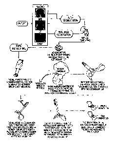
[0198] FIG. 181 shows local magnetic field maps (isometric, front, and top
views)
generated from data output from an inertial measurement unit before
calibration (top series
of three plots resembling an ellipsoid), and local magnetic field maps
(isometric, front, and
top views) generated from data output from an inertial measurement unit after
calibration
(bottom series of three plots resembling a sphere).
[0199] FIG. 182 comprises a series of diagrams showing exemplary locations of
magnetometers associated with an inertial measurement unit (A), what the
detected
magnetic field from the magnetometers should reflect if normalized to account
for
distortion(s) (B), and the result of a local distortion in the magnetic field
upon the
magnetometers if no normalization is carried out.
[0200] FIG. 183 is a series of projections for varied surgical tools each
having a unique
top surface in order to allow an inertial measurement unit processor to
intelligently identify
the surgical tool to which the IMU is mounted.
[0201] FIG. 184 is an outline drawings representative of a 1MU housing and
depicting the
interaction between one of the projections of FIG. 183 and a bottom cavity of
the IMU
housing.
Date Regue/Date Received 2022-09-29
[0202] FIG. 185 is an exemplary process flow diagram for preparing a proximal
humerus
and implanting a humeral component as part of a shoulder replacement procedure
by using
inertial measurement units in accordance with the instant disclosure.
[0203] FIG. 186 is an exemplary process flow diagram for preparing a scapular
socket and
implanting a glenoid cavity cup as part of a shoulder replacement procedure by
using
inertial measurement units in accordance with the instant disclosure.
[0204] FIG. 187 is an exemplary process flow diagram for preparing a proximal
humerus
and implanting a humeral component as part of a reverse shoulder replacement
procedure
by using inertial measurement units in accordance with the instant disclosure.
[0205] FIG. 188 is an exemplary process flow diagram for preparing a scapular
socket
and implanting a glenoid ball as part of a reverse shoulder replacement
procedure by
using inertial measurement units in accordance with the instant disclosure.
DETAILED DESCRIPTION
[0206] The exemplary embodiments of the present disclosure are described and
illustrated
below to encompass various aspects of orthopedics including bone and tissue
reconstruction, patient-specific and mass customized orthopedic implants,
gender and
ethnic specific orthopedic implants, cutting guides, trauma plates, bone graft
cutting and
placement guides, and patient-specific instruments. Of course, it will be
apparent to those
of ordinary skill in the art that the embodiments discussed below are
exemplary in nature
and may be reconfigured without departing from the scope and spirit of the
present
invention. However, for clarity and precision, the exemplary embodiments as
discussed
below may include optional steps, methods, and features that one of ordinary
skill should
recognize as not being a requisite to fall within the scope of the present
invention.
Full Anatomy Reconstruction
[0207] Referring to FIGS. 1-8, reconstruction of a deformed anatomy or a
partial anatomy
is one of the complex problems facing healthcare providers. Loss of anatomy
may be the
26
Date Regue/Date Received 2022-09-29
result of birth conditions, tumors, diseases, personal injuries, or failure of
previous
surgeries. As part of providing treatment for various ailments, healthcare
providers may
find it advantageous to reconstruct an anatomy or construct an anatomy to
facilitate
treatment for various conditions that may include, without limitation,
broken/shattered
bones, bone degeneration, orthopedic implant revision, joint degeneration, and
custom
instrumentation design. For example, prior art hip reconstruction solution
requires
mirroring of the healthy patient anatomy which may not be an accurate
reflection of the
healthy anatomy due to naturally occurring asymmetry, as shown in FIG 15 ¨ 19.
[0208] The present disclosure provides a system and methods for bone and
tissue
reconstruction. In order to carry out this reconstruction, the system and
associated methods
utilizes anatomical images representative of one or more persons. These images
are
processed to create a virtual three dimensional (3D) tissue model or a series
of virtual 3D
tissue models mimicking the proper anatomy in question. Thereafter, the system
and
associated methods are utilized to create a mold and/or other devices (e.g.,
fixation devices,
grafting devices, patient-specific implants, patient-specific surgical guides)
for use with
reconstructive surgery.
[0209] As represented in FIG. 1, an overview of the exemplary system flow
begins with
receiving input data representative of an anatomy. This anatomy may comprise a
partial
anatomy in the case of tissue degeneration or tissue absence resulting from
genetics, or this
anatomy may comprise a deformed anatomy resulting from genetics or
environmental
conditions, or this anatomy may comprise a shattered tissue resulting from one
or more
anatomy breaks. Input anatomical data comprises two dimensional (2D) images or
three
dimensional (3D) surface representations of the anatomy in question that may,
for example,
be in the form of a surface model or point cloud. In circumstances where 2D
images are
utilized, these 2D images are utilized to construct a 3D virtual surface
representation of the
anatomy in question. Those skilled in the art are familiar with utilizing 2D
images of
anatomy to construct a 3D surface representation. Accordingly, a detailed
explanation of
this process has been omitted in furtherance of brevity. By way of example,
input
anatomical data may comprise one or more of X-rays, computed tomography (CT)
scans,
27
Date Regue/Date Received 2022-09-29
magnetic resonance images (MRIs), or any other imaging data from which a 3D
surface
representation of the tissue in question may be generated.
[0210] Referring to FIG. 50 and Table I, in the context of X-ray images used
to construct
a virtual 3D bone model, it has been discovered that bone rotation during
imaging plays an
important role in correctly constructing the model. In other words, if one
attempts to
compile X-ray images in circumstances where bone rotation has occurred between
images,
the X-ray images need to be normalized to account for this bone rotation.
[0211] By way of example, in the context of a proximal femur, it has been
discovered that
bone rotation of six and fifteen degrees results in significant changes to the
measurements
extracted from X-ray images. By way of example, these measurements include,
without
limitation, proximal angle, head offset, and intramedullary canal width. As
reflected in
Table I, for the same femur, that was X-ray imaged at zero degrees (i.e., a
starting point
established by the initial X-ray), six degrees of rotation, and fifteen
degrees of rotation
exhibited differences proximal angle, head offset, and intramedullary canal
width as
measured using pixels, where each pixel size was approximately 0.29
millimeters. In
particular, proximal angle increased with increasing rotation, as did head
offset, but the
same was not true for intramedullary width. In this exemplary table, three
transverse planes
were spaced apart along the longitudinal axis, where each plane corresponded
to a location
where the width of the intramedullary canal was measured. As reflected in
Table I, the
widths of the intramedullary canal for the same location change depending upon
the angle
of rotation. Consequently, as will be discussed in more detail hereafter, when
constructing
a 3D virtual model of a bone using X-rays, one must account for rotational
deviation to the
extent bone rotation occurs during imaging.
[0212] It should be understood, however, that the foregoing is an exemplary
description of
anatomies that may be used with the exemplary system and methods and,
therefore, is in
no way intended to limit other anatomies from being used with the present
system pursuant
to the disclosed methods. As used herein, tissue includes bone, muscle,
ligaments, tendons,
and any other definite kind of structural material with a specific function in
a multicellular
organism. Consequently, when the exemplary system and methods are discussed in
the
28
Date Regue/Date Received 2022-09-29
context of bone, those skilled in the art should realize the applicability of
the system and
methods to other tissue.
[0213] Referring back to FIG. 1, the anatomy data input to the system is
directed to three
modules, two of which involve processing of the anatomy data (full bone
reconstruction
module, patient-specific module), while a third (abnormal database module)
catalogues the
anatomy data as part of a database. A first of the processing modules, the
full bone
reconstruction module, processes the input anatomy data with data received
from the
statistical atlas module to generate a virtual, 3D model of the bone(s) in
question. This 3D
model is a full, normal reconstruction of the bone(s) in question. A second of
the
processing modules, the patient-specific module, processes the input anatomy
data with
data received from the full bone reconstruction module to generate one or more
molds,
fixation systems, graft shaping tools, and renderings, in addition to one or
more final
orthopedic implants. A rendering refers to visualization of reconstructed
anatomy for
feedback regarding expected surgical outcome. More specifically, the patient-
specific
module is adapted to generate fully customized devices, designed to precisely
fit patient-
specific anatomy, despite severe deviation of the patient's anatomy from
normal.
Moreover, the patient-specific module utilizes the virtual 3D reconstructed
bone model
from the full bone reconstruction module to automatically identify anatomical
regions and
features for device design parameters (e.g., fitting region and/or shape). In
this fashion,
patient-specific data is used to define design parameters so that the output
instrument and
any implant precisely fits the specific anatomy of the patient. Exemplary
utilizations of
the patient-specific module will be discussed in greater detail hereafter. In
order to
understand the functions and processes of the system in further detail, the
following is an
explanation of the modules of the system starting with the statistical atlas
module.
[0214] As shown in FIG. 1 and 2, the statistical atlas module logs virtual, 3D
models of
one or more anatomies (e.g., bones) to capture the inherent anatomical
variability in a given
population. In exemplary form, the atlas logs mathematical representations of
anatomical
features of the one or more anatomies represented as a mean representation and
variations
about the mean representation. By representing the anatomical features as
mathematical
29
Date Regue/Date Received 2022-09-29
representations, the statistical atlas allows automated measurements of
anatomies and, as
will be discussed in more detail hereafter, reconstruction of missing
anatomies.
[0215] In order to extract anatomical variations across a common anatomy,
input anatomy
data is compared to a common frame of reference across a population, commonly
referred
to as a template 3D model or anatomical 3D template model. This template 3D
model is
visually represented on a graphic display as a 3D model that can be rotated
and otherwise
visually manipulated, but comprises a mathematical representation of
anatomical surface
features/ representations for all anatomies across the statistical atlas for
the tissue in
question (i.e., for a given bone all properties of the bone are shared across
the population
of the statistical atlas, which is generated from the template 3D model). The
template 3D
model can be a combination of multiple anatomical representations or a single
representative instance and may represent the lowest entropy state of the
statistical atlas.
For each anatomy to be added to the statistical atlas (i.e., input anatomy
data), an
anatomical 3D model is created and both the anatomical 3D model and the
template 3D
model are subjected to a normalization process.
[0216] During the normalization process, the anatomical 3D model is normalized
relative
to the scale of the template 3D model. The normalization process may involve
scaling one
or both of the anatomical 3D model and the template 3D model to have a common
unit
scale. After normalization of the anatomical 3D model and the template 3D
model, the
normalized anatomical 3D model and template 3D model are rendered scale
invariant, so
that shape features can be utilized independent of scale (meaning size in this
case). After
normalization is complete, both 3D models are processed via a scale space
mapping and
feature extraction sequence.
[0217] Scale space mapping and feature extraction is essentially a multi-
resolution feature
extraction process. In particular, this process extracts shape-specific
features at multiple
feature scales. Initially, a plurality of anatomical features is selected,
each representing
features present at a different scale space. Thereafter, for each scale space
representation
of the selected anatomical feature, model specific features are extracted.
These extracted
features are used to draw out robust (as to noise) registration parameters
between the
Date Regue/Date Received 2022-09-29
template 3D model and the anatomical 3D model. Subsequent to this multi-
resolution
feature extraction process, the extracted data is processed via a multi-
resolution 3D
registration process.
[0218] Referring to FIGS. 2-5, the multi-resolution 3D registration process
uses the scale
space extracted features to carry out an affine registration calculation
between the
anatomical 3D model and template 3D model in order to register the two models.
In
particular, the anatomical 3D model and template 3D model are processed via a
rigid
registration process. As represented in FIG. 5, this rigid registration
process is operative
to align the anatomical 3D model and template 3D model to ensure both models
are in the
same space and with no pose singularity. In order to align the 3D models, the
centroids
associated with each model are aligned. In addition, the principle axes for
each 3D model
are aligned so that the major direction of both 3D models is the same.
Finally, the pose
difference between the 3D models is minimized by carrying out an iterative
closest point
calculation.
[0219] Post rigid registration, the 3D models are registered using a
similarity registration
process. This process involves aligning the template 3D model and the
anatomical 3D
model in normal scale iteratively by calculating a similarity transform that
best aligns the
normal scale features (i.e., ridges) for both the template 3D model and the
anatomical 3D
model. The iterative similarity alignment algorithm is a variant of iterative
closest point.
Within each iteration rotation, translation and scale are calculated between
point pairs until
convergence. Pair matching or correspondence between the two set of points is
evaluated
using distance query calculated using Kd-tree, or some other space
partitioning data
structure. In particular, the ridges for both models are utilized to carry out
a calculate
matching point pairs process. In this exemplary description, ridges refers to
points on a
3D model where a single principle curvature has extrema along its curvature
lines. As part
of the calculate matching point pairs process, points are identified on ridges
of the 3D
models that match one another. Next, the ridges of both 3D models are
subjected to a
similarity transformation calculation process where rotation, translation, and
scale are
calculated that best align the ridges of both models. A transform points
process follows,
which is operative to apply the calculated rotation, translation, and scale to
the template
31
Date Regue/Date Received 2022-09-29
3D model ridges. Thereafter, the root mean square error or distance error
between each
matched point set is calculated, followed by calculation of the change in
relative root mean
square error or distance error from the previous process. If the change in
relative root mean
square error or distance error is within a predetermined threshold, then a
transformation
process occurs to apply the final rotation, translation, and scale to the
template 3D model.
[0220] An articulated registration process follows the similarity registration
process and
receives input data from a scale space features process. In the scale space
feature process,
feature are extracted from the template 3D model and the anatomical 3D model
in different
scale spaces. Each scale space is defined by convolving the original
anatomical 3D model
with Gaussian smoothing function.
[0221] The purpose of the articulated registration process is to match "n"
scale space
features of the template 3D model with "m" scale space features calculated on
the
anatomical 3D model. The difference between the number of detected features on
the
template 3D model and the anatomical 3D model is due to anatomical variation.
This
difference in a number of detected features may result in many relationships
between the
template 3D model and the anatomical 3D model. Therefore, a two-way, mutual
feature
matching is performed to accommodate such variation and achieve accurate
matching
between all mutual features. Specifically, feature sets are computed on the
template 3D
model in scale space. In this exemplary process, feature sets are connected
sets of points
that represent a prominent anatomical structure (e.g., acetabular cup in the
pelvis, spine
process in the lumbar). Likewise, feature sets are computed on the anatomical
3D model
in scale space. A matching feature pair process matches the feature sets
computed on the
template 3D model to the feature sets on the anatomical 3D model using shape
descriptors
(e.g., curvature, shape index, etc.). The result of this process is an "n-m"
mapping of
feature sets between the template 3D model and the anatomical 3D model. If
necessary, a
regrouping process is carried out to regroup the matched feature sets into a
single feature
set (e.g., if acetabular cup was detected as two pieces, this process would
regroup the two
pieces into one single feature set). Thereafter, a calculation process is
carried out to
calculate the correspondence between each point in matched feature sets on the
template
3D model and the anatomical 3D model. An affine calculation transformation
process
32
Date Regue/Date Received 2022-09-29
follows in order to calculate the rotation, translation, and shear that
transform each matched
feature set on the template 3D model to its corresponding feature set on the
anatomical 3D
model. Thereafter, the template 3D model is transformed using the calculated
affine
transformation parameters (i.e., rotation, translation, and shear). Finally, a
rigid alignment
process is carried out to align each matched feature set on the template 3D
model and the
anatomical 3D model.
[0222] A non-rigid registration process, occurring after the articulated
registration process
and the normal scale features process, involves matching all surface vertices
on the
template 3D model to vertices on the anatomical 3D model and calculating
initial
correspondence. This correspondence is then used to calculate deformation
fields that
move each vertex on the template 3D model to the matched point on the
anatomical 3D
model. Matching is done between vertices within the same class (i.e., scale
space feature
vertex; normal scale feature vertex, or non-feature vertex). In the context of
the normal
scale features process, shape features are calculated on the template 3D model
and the
anatomical 3D model in the original scale space (ridges), meaning the original
input model.
[0223] Specifically, as part of the non-rigid registration process, the scale
space features
are calculated on the template 3D model (TMssf) and on the anatomical 3D model
(NMssf).
Each set of features on the template 3D model and on the anatomical 3D model
are grown
using "k" neighbor points. An alignment process is applied to the template 3D
model scale
space features to match its corresponding feature on the anatomical 3D model.
Given two
point clouds, reference (X) and moving (Y), the goal is to iteratively align
the two point
clouds to minimize overall error metric, under constraint of a minimum
relative root mean
squared error and maximum angle threshold. A realignment process is carried
out to align
feature sets on the template 3D model with the matching sets on the anatomical
3D model
using iterative closest point in normal scale. Post realignment, the point
correspondence
between points in each feature set on the template 3D model with the matched
feature set
on the anatomical 3D model is calculated. The matched point on the anatomical
3D model
should have a surface normal direction close to the template 3D model point.
The output
is forwarded to the calculate deformation fields step.
33
Date Regue/Date Received 2022-09-29
[0224] Parallel to the scale space features calculation course, template 3D
model (TMnfp)
and anatomical 3D model (NMnfp) non-feature points or the remaining set of
points on the
template 3D model surface that does not belong to either scale space features
or normal
scale features are processed pursuant to a correspondence calculation to
calculate the point
correspondence between non-feature points on the template 3D model and non-
feature
points on the anatomical 3D model. The matched point(s) on the new model
should have a
surface normal direction close to the template model point. The output is
forwarded to the
calculate deformation fields step.
[0225] Also parallel to the scale space features calculation course, normal
scale features
(i.e., ridges) on the template 3D model (TM nsf) are aligned with the normal
scale features
(i.e., ridges) on the anatomical 3D model (NM nsf) using AICP. AICP is a
variant of the
iterative closest point calculation where in each iteration translation,
rotation, and scale are
calculated between matched point sets. After the alignment process, a
correspondence
process is carried out.
[0226] The outputs from scale space features calculation course, the
correspondence
course, and the alignment course are subjected to a deformation process where
the
deformation field is calculated to move each point on the template 3D model to
its matched
point on the anatomical 3D model.
[0227] The output of the non-rigid registration process is a subjected to a
relaxation process
in order to move the vertices of the template 3D model mesh closer to surface
of the
anatomical 3D model after the multi-resolution registration step and smooth
the output
model. In particular, the template 3D model in normal space (TM ns) and the
anatomical
3D model in normal space (NM ns) are processed via a correspondence
calculation to
compute the closest vertices on template 3D model to the anatomical 3D model
using a
normal constrained spherical search algorithm. This calculation, using the
closest vertices
for both models, generates a correspondence vector from each vertex in the
template 3D
model and its matched vertices in anatomical 3D model, which may result in
more than
one match point from the anatomical 3D model. Using the matched points for
each vertex
on the template 3D model, the weighted mean of the matched points on the
anatomical 3D
34
Date Regue/Date Received 2022-09-29
model is calculated based on the Euclidian distance from the point and matched
points. At
this point, the template 3D model is updated using the weighted average so as
to move each
point on template 3D model using the calculated weighted average distance.
After the
computed weights process, a relaxation process is carried out for every point
on template
model in order to find the closest point on the anatomical 3D model surface
and move it to
that point. Finally, a smoothing operation is performed on the deformed
template 3D
model to remove noise. The resultant registered 3D models (i.e., template and
anatomical
3D models) are then subjected to a free form deformation process.
[0228] The free form deformation process morphs the surface of the template 3D
model
with the surface of the anatomical 3D model. More specifically, the surface of
the template
3D model is iteratively moved on a weighted point-to-point basis using
mutually matched
points on both the template 3D model surface and the anatomical 3D model
surface.
[0229] Referencing FIGS. 2 and 6, after the free form deformation process, the
anatomical 3D model is subjected to a correspondence calculation process to
determine the
deviation between the anatomical 3D model and the moiphed template 3D model.
This
correspondence calculation process refines the template 3D model from the free
form
deformation step to perform a final match of the selected landmark locations
on the
template deformed 3D model and the deformed anatomical 3D model. In this
fashion, the
correspondence calculation process calculates and records the variation in
size and shape
between the 3D models, which is recorded as deviation about the mean model.
The output
of this correspondence calculation process is the addition of a normalized
anatomical 3D
model and a revised template 3D model having been updated to account for the
variations
in the anatomical 3D model. In other words, the output of the process outlined
in FIG. 2
is the normalized anatomical 3D model having been modified to have properties
(e.g., point
correspondence) consistent with the revised template 3D model to facilitate
full anatomical
reconstruction (e.g., full bone reconstruction).
[0230] Referring to FIGS. 1 and 7, inputs from the statistical atlas module
and anatomy
data are directed to a full anatomy reconstruction module. By way of example,
the anatomy
in question may be a bone or multiple bones. It should be noted, however, that
anatomies
Date Regue/Date Received 2022-09-29
other than bone may be reconstructed using the exemplary hardware, processes,
and
techniques described herein. In exemplary form, the full anatomy
reconstruction module
may receive input data as to a partial, deformed, or shattered pelvis. Input
anatomical data
comprises two dimensional (2D) images or three dimensional (3D) surface
representations
of the anatomy in question that may, for example, be in the form of a surface
model or
point cloud. In circumstances where 2D images are utilized, these 2D images
are utilized
to construct a 3D surface representation of the anatomy in question. Those
skilled in the
art are familiar with utilizing 2D images of anatomy to construct a 3D surface
representation. Accordingly, a detailed explanation of this process has been
omitted in
furtherance of brevity. By way of example, input anatomical data may comprise
one or
more of X-rays, computed tomography (CT) scans, magnetic resonance images
(MRIs), or
any other imaging data from which a 3D surface representation may be
generated. As will
be discussed in more detail hereafter, this input anatomical data may be used,
without
limitation, for: (1) a starting point for identifying the closest statistical
atlas 3D bone model;
(2) registration using a set of 3D surface vertices; and, (3) a final
relaxation step of
reconstruction output.
[0231] As depicted in FIG. 7, the input anatomical data (e.g., bone model of
the patient) is
utilized to identify the anatomical model (e.g., bone model) in the
statistical atlas that most
closely resembles the anatomy of the patient in question. This step is
depicted in FIG. 3 as
finding the closest bone in the atlas. In order to initially identify a bone
model in the
statistical atlas that most closely resembles the patient's bone model, the
patient's bone
model is compared to the bone models in the statistical atlas using one or
more similarity
metrics. The result of the initial similarity metric(s) is the selection of a
bone model from
the statistical atlas that is used as an "initial guess" for a subsequent
registration step. The
registration step registers the patient bone model with the selected atlas
bone model (i.e.,
the initial guess bone model) so that the output is a patient bone model that
is aligned with
the atlas bone model. Subsequent to the registration step, the shape
parameters for aligned
"initial guess" are optimized so that the shape matches the patient bone
shape.
[0232] Shape parameters, in this case from the statistical atlas, are
optimized so that the
region of non-deformed or existing bone is used to minimize the error between
the
36
Date Regue/Date Received 2022-09-29
reconstruction and patient bone model. Changing shape parameter values allows
for
representation of different anatomical shapes. This process is repeated, at
different scale
spaces, until convergence of the reconstructed shape is achieved (possibly
measured as
relative surface change between iterations or as a maximum number of allowed
iterations).
[0233] A relaxation step is performed to morph the optimized tissue to best
match the
original patient 3D tissue model. Consistent with the exemplary case, the
missing anatomy
from the reconstructed pelvis model that is output from the convergence step
is applied to
the patient-specific 3D pelvis model, thereby creating a patient-specific 3D
model of the
patient's reconstructed pelvis. More specifically, surface points on the
reconstructed pelvis
model are relaxed (i.e., morphed) directly onto the patient-specific 3D pelvis
model to best
match the reconstructed shape to the patient-specific shape. The output of
this step is a
fully reconstructed, patient-specific 3D tissue model representing what should
be the
normal/complete anatomy of the patient.
[0234] Referencing FIG. 1, the abnormal database is utilized as a data input
and training
for the defect classification module. In particular, the abnormal database
contains data
specific to an abnormal anatomical feature that includes an anatomical surface
representation and related clinical and demographic data.
[0235] Referencing FIGS. 1 and 8, the fully reconstructed, patient-specific 3D
tissue model
representing the normal/complete tissue and input anatomical data (i.e., 3D
surface
representation or data from which a 3D surface representation may be
generated)
representing abnormal/incomplete tissue from the abnormal database are input
to the defect
classification module. This anatomical data from the abnormal database may be
a partial
anatomy in the case of tissue degeneration or tissue absence resulting from
genetics, or this
anatomy may be a deformed anatomy resulting from genetics or environmental
conditions
(e.g., surgical revisions, diseases, etc.), or this anatomy may be a shattered
tissue resulting
from one or more anatomy breaks. By way of example, input anatomical data may
comprise one or more of X-rays, computed tomography (CT) scans, magnetic
resonance
images (MRIs), or any other imaging data from which a 3D surface
representation may be
generated.
37
Date Regue/Date Received 2022-09-29
[0236] The defect classification module pulls a plurality of abnormal 3D
surface
representations from abnormal database coupled with the normal 3D
representation of the
anatomy in question to create a quantitative defect classification system.
This defect
classification system is used to create "templates" of each defect class or
cluster. More
generally, the defect classification module classifies the anatomical
deficiency into classes
which consist of closely related deficiencies (referring to those with similar
shape, clinical,
appearance, or other characteristics) to facilitate the generation of
healthcare solutions
which address these deficiencies. The instant defect classification module
uses software
and hardware to classify the defects automatically as a means to eliminate or
reduce
discrepancies between pre-operative data and intra-operative observer
visualization.
Traditionally, pre-operative radiographs have been taken as a means to
qualitatively
analyze the extent of anatomical reconstruction necessary, but this resulted
in pre-operative
planning that was hit-or-miss at best. Currently, intra-operative observers
make the final
determination of the extent of anatomy deficiency and many times conclude that
the pre-
operative planning relying on radiographs was defective or incomplete. As a
result, the
instant defect classification module improves upon current classification
systems by
reducing interobserver and intraobserver variation related to defect
classification and
providing quantitative metrics for classifying new defect instances.
[0237] As part of the defect classification module, the module may take as an
input one or
more classification types to be used as an initial state. For example, in the
context of a
pelvis, the defect classification module may use as input defect features
corresponding to
the American Academy of Orthopaedic Surgeons (AAOS) D' Antonio et al. bone
defect
classification structure. This structure includes four different classes as
follows: (1) Type
I, corresponding to segmental bone loss; (2) Type II, corresponding to
cavitary bone loss;
(3) Type III, corresponding to combined segmental and cavitary bone loss; and,
(4) Type
IV, corresponding to pelvis discontinuity. Alternatively, the defect
classification module
may be programmed with the Paprosky bone defect classification structure,
depicted
graphically for the pelvis in FIG. 10. This structure includes three different
classes as
follows: (1) Type I, corresponding to supportive rim with no bone lysis; (2)
Type II,
corresponding to distorted hemispheres with intact supportive columns and less
than two
38
Date Regue/Date Received 2022-09-29
centimeters of superomedial or lateral migration; and, (3) Type III,
corresponding to
superior migration greater than two centimeters and sever ischial lysis with
Kohler's line
broken or intact. Moreover, the defect classification module may be programmed
with the
Modified Paprosky bone defect classification structure. This structure
includes six
different classes as follows: (1) Type 1, corresponding to supportive rim with
no
component migration; (2) Type 2A, corresponding to distorted hemisphere but
superior
migration less than three centimeters; (3) Type 2B, corresponding to greater
hemisphere
distortion having less than 1/3 rim circumference and the dome remaining
supportive; (4)
Type 2C, corresponding to an intact rim, migration medial to Kohler's line,
and the dome
remains supportive; (5) Type 3A, corresponding to superior migration, greater
than three
centimeters and severe ischial lysis with intact Kohler's line; and, (6) Type
3B,
corresponding to superior migration, greater than three centimeters and severe
ischial lysis
with broken Kohler's line and rim defect greater than half the circumference.
Using the
output classification types and parameters, the defect classification module
compares the
anatomical data to that of the reconstructed data to discern which of the
classification types
the anatomical data most closely resembles, thereby corresponding to the
resulting
assigned classification.
[0238] As an initial step, the add to statistical atlas step involves
generating
correspondence between normal atlas 3D bone model and the abnormal 3D bone
model.
More specifically, the 3D bone models are compared to discern what bone in the
normal
3D model is not present in the abnormal 3D model. In exemplary form, the
missing/abnormal bone is identified by comparing points on the surface of each
3D bone
model and generating a list of the discrete points on the surface of the
normal 3D bone
model that are not present on the abnormal 3D bone model. The system may also
record
and list (i.e., identify) those surface points in common between the two
models or
summarily note that unless recorded as points being absent on the abnormal 3D
bone
model, all other points are present in common in both bone models (i.e., on
both the normal
and abnormal bone models). Accordingly, the output of this step is the
abnormal 3D bone
model with statistical atlas correspondence and a list of features (points)
from the normal
39
Date Regue/Date Received 2022-09-29
atlas 3D bone model indicating if that feature (point) is present or missing
in the abnormal
3D bone model.
[0239] After generating correspondence between the normal atlas 3D bone model
(generated from the full bone reconstruction module) and the abnormal 3D bone
model
(generated from the input anatomical data), the missing/abnormal regions from
the
abnormal 3D bone model are localized on the normal atlas 3D bone model. In
other words,
the normal atlas 3D bone model is compared to the abnormal 3D bone model to
identify
and record bone missing from the abnormal 3D bone model that is present in the
normal
atlas 3D bone model. Localization may be carried out in a multitude of
fashions including,
without limitation, curvature comparison, surface area comparisons, and point
cloud area
comparisons. Ultimately, in exemplary form, the missing/abnormal bone is
localized as a
set of bounding points identifying the geometrical bounds of the
missing/abnormal
region(s).
[0240] Using the bounding points, the defect classification module extracts
features from
the missing/abnormal region(s) using input clinical data. In exemplary form,
the extracted
features may include shape information, volumetric information, or any other
information
used to describe the overall characteristics of the defective (i.e., missing
or abnormal) area.
These features may be refined based on existing clinical data, such as on-
going defect
classification data or patient clinical information not necessarily related to
the anatomical
feature (demographics, disease history, etc.). The output of this step is a
mathematical
descriptor representative of the defective area(s) that are used in a
subsequent step to group
similar tissue (e.g., bone) deformities.
[0241] The mathematical descriptor is clustered or grouped based upon a
statistical
analysis. In particular, the descriptor is statistically analyzed and compared
to other
descriptors from other patients/cadavers to identify unique defect classes
within a given
population. Obviously, this classification is premised upon multiple
descriptors from
multiple patients/cadavers that refine the classifications and identifications
of discrete
groups as the number of patients/cadavers grows. The output from this
statistical analysis
Date Regue/Date Received 2022-09-29
is a set of defect classes that are used to classify new input anatomical data
and determines
the number of templates.
[0242] The output of the defect classification module is directed to a
template module. In
exemplary form, the template module includes data that is specific as to each
of the defect
classifications identified by the defect classification module. By way of
example, each
template for a given defect classification includes surface representations of
the defective
bone, location(s) of the defect(s), and measurements relating to the defective
bone. This
template data may be in the form of surface shape data, point cloud
representations, one or
more curvature profiles, dimensional data, and physical quantity data. Outputs
from the
template module and the statistical atlas are utilized by a mass customization
module to
design, test, and allow fabrication of mass customized implants, fixation
devices,
instruments or molds. Exemplary utilizations of the mass customization module
will be
discussed in greater detail hereafter.
Patient-Specific Reconstruction Implants
[0243] Referring to FIGS. 1 and 20, an exemplary process and system are
described for
generating patient-specific orthopedic implant guides and associated patient-
specific
orthopedic implants for patients afflicted with partial, deformed, and/or
shattered
anatomies. For purposes of the exemplary discussion, a total hip arthroplasty
procedure
will be described for a patient with a partial anatomy. It should be
understood, however,
that the exemplary process and system are applicable to any orthopedic implant
amenable
to patient-specific customization in instances where incomplete or deformed
anatomy is
present. For example, the exemplary process and system are applicable to
shoulder
replacements and knee replacements where bone degeneration (partial anatomy),
bone
deformation, or shattered bones are present. Consequently, though a hip
implant is
discussed hereafter, those skilled in the art will understand the
applicability of the system
and process to other orthopedic implants, guides, tools, etc. for use with
original orthopedic
or orthopedic revision surgeries.
[0244] Pelvis discontinuity is a distinct form of bone loss most often
associated with total
hip arthroplasty (THA), in which osteolysis or acetabular fractures can cause
the superior
41
Date Regue/Date Received 2022-09-29
aspect of the pelvis to become separated from the inferior portion. The amount
and severity
of bone loss and the potential for biological in-growth of the implant are
some of the factors
that can affect the choice of treatment for a particular patient. In the case
of severe bone
loss and loss of pelvic integrity, a custom tri-flange cup may be used. First
introduced in
1992, this implant has several advantages over existing cages. It can provide
stability to
pelvic discontinuity, eliminate the need for structural grafting and
intraoperative
contouring of cages, and promote osseointegration of the construct to the
surrounding bone.
[0245] Regardless of the context, whether partial, deformed, and/or shattered
anatomies of
the patient are at issue, the exemplary system and process for generating
patient-specific
implants and/or guides utilizes the foregoing exemplary process and system of
3D bone
model reconstruction (see FIGS. 1-7 and the foregoing exemplary discussion of
the same)
to generate a three dimensional model of the patient's reconstructed anatomy.
More
specifically, in the context of total hip arthroplasty where pelvis
discontinuity is involved,
the exemplary patient-specific system utilizes the patient pelvis data to
generate a 3D
model of the patient's complete pelvis, which is side specific (right or
left). Consequently,
a discussion of the system and process for utilizing patient anatomy data for
a partial
anatomy and generating a 3D reconstructed model of the patient's anatomy is
omitted in
furtherance of brevity. Accordingly, a description of the process and system
for generating
patient-specific orthopedic implant guides and associated patient-specific
orthopedic
implants for patients afflicted with partial, deformed, and/or shattered
anatomies will be
described post formation of the three dimensional reconstructed model.
[0246] Referring specifically to FIGS. 20-22 and 27, after the patient-
specific
reconstructed 3D bone model of the pelvis and femur are generated, both the
incomplete
patient-specific 3D bone model (for pelvis and femur) and the reconstructed 3D
bone
model (for pelvis and femur) are utilized to create the patient-specific
orthopedic implant
and a patient-specific placement guide for the implant and/or its fasteners.
In particular,
the extract defect shape step includes generating correspondence between the
patient-
specific 3D model and the reconstructed 3D model (correspondence between
pelvis
models, and correspondence between femur models, but not between one femur
model and
a pelvis model). More specifically, the 3D models are compared to discern what
bone in
42
Date Regue/Date Received 2022-09-29
the reconstructed 3D model is not present in the patient-specific 3D model. In
exemplary
form, the missing/abnormal bone is identified by comparing points on the
surface of each
3D model and generating a list of the discrete points on the surface of the
reconstructed 3D
model that are not present on the patient-specific 3D model. The system may
also record
and list (i.e., identify) those surface points in common between the two
models or
summarily note that unless recorded as points being absent on the patient-
specific 3D
model, all other points are present in common in both models (i.e., on both
the
reconstructed and patient-specific 3D models).
[0247] Referring to FIG. 21, after generating correspondence between the
reconstructed
3D model (generated from the full bone reconstruction module) and the patient-
specific
3D model (generated from the input anatomical data), the missing/abnormal
regions from
the patient-specific 3D model are localized on the reconstructed 3D model. In
other words,
the reconstructed 3D model is compared to the patient-specific 3D model to
identify and
record bone missing from the patient-specific 3D model that is present in the
reconstructed
3D model. Localization may be carried out in a multitude of fashions
including, without
limitation, curvature comparison, surface area comparisons, and point cloud
area
comparisons. Ultimately, in exemplary form, the missing/abnormal bone is
localized and
the output comprises two lists: (a) a first list identifying vertices
corresponding to bone of
the reconstructed 3D model that is absent or deformed in the patient-specific
3D model;
and, (b) a second list identifying vertices corresponding to bone of the
reconstructed 3D
model that is also present and normal in the patient-specific 3D model.
[0248] Referencing FIGS. 21, 22, and 27, following the extract defect shape
step, an
implant loci step is performed. The two vertices lists from the extract defect
shape step
and a 3D model of a normal bone (e.g., pelvis, femur, etc.) from the
statistical atlas (see
FIGS. 1 and 2, as well as the foregoing exemplary discussion of the same) are
input to
discern the fixation locations for a femoral or pelvic implant. More
specifically, the
fixation locations (i.e., implant loci) are automatically selected so that
each is positioned
where a patient has residual bone. Conversely, the fixation locations are not
selected in
defect areas of the patient's residual bone. In this manner, the fixation
locations are chosen
43
Date Regue/Date Received 2022-09-29
independent of the ultimate implant design/shape. The selection of fixation
locations may
be automated using shape information and statistical atlas locations.
[0249] As show in FIG. 21, after the implant loci step, the next step is to
generate patient-
specific implant parameters. In order to complete this step, an implant
parameterized
template is input that defines the implant by a set number of parameters that
are sufficient
to define the underlying shape of the implant. By way of example, in the case
of a pelvis
reconstruction to replace/augment an absent or degenerative acetabulum, the
implant
parameterized template includes angle parameters for the orientation of the
replacement
acetabular cup and depth parameters to accommodate for dimensions of the
femoral head.
Other parameters for an acetabular implant may include, without limitation,
the acetabular
cup diameter, face orientation, flange locations and shapes, location and
orientation of
fixation screws. In the case of porous implants, the location and structural
characteristics
of the porosity should be included. By way of example, in the case of a
femoral
reconstruction to replace/augment an absent or degenerative femur, the implant
parameterized template includes angle parameters for the orientation of the
replacement
femoral head, neck length, head offset, proximal angle, and cross-sectional
analysis of the
exterior femur and intercondylar channel. Those skilled in the art will
understand that the
parameters chosen to define the underlying shape of the implant will vary
depending upon
the anatomy being replaced or supplemented. Consequently, an exhaustive
listing of
parameters that are sufficient to define the underlying shape of an implant is
impractical.
Nevertheless, as depicted in FIGS. 22 for example, the reconstructed 3D pelvis
model may
be utilized to obtain the radius of the acetabular cup, identification of
pelvic bone
comprising the acetabular cup circumferential upper ridge, and identification
of the
orientation of the acetabular cup with respect to the residual pelvis.
Moreover, the
parameters may be refined taking into account the implant loci so that the
implant
best/better fits the patient-specific anatomy.
[0250] Subsequent to finalizing the set number of parameters that are
sufficient to define
the underlying shape of the implant, the design of the implant is undertaken.
More
specifically, an initial iteration of the overall implant surface model is
constructed. This
initial iteration of the overall implant surface model is defined by a
combination of patient-
44
Date Regue/Date Received 2022-09-29
specific contours and estimated contours for the implanted region. The
estimated contours
are determined from the reconstructed 3D bone model, missing anatomical bone,
and
features extracted from the reconstructed 3D bone model. These features and
the location
of the implant site, which can be automatically determined, are used to
determine the
overall implant shape, as depicted for example in FIG. 22 for an acetabular
cup implant.
[0251] Referring back to FIG. 20, the initial iteration of the overall implant
surface model
is processed pursuant to a custom (i.e., patient-specific) planning sequence.
This custom
planning sequence may involve inputs from a surgeon and an engineer as part of
an iterative
review and design process. In particular, the surgeon and/or engineer may view
the overall
implant surface model and the reconstructed 3D bone model to determine if
changes are
needed to the overall implant surface model. This review may result in
iterations of the
overall implant surface model until agreement is reached between the engineer
and
surgeon. The output from this step is the surface model for the final implant,
which may
be in the form of CAD files, CNC machine encoding, or rapid manufacturing
instructions
to create the final implant or a tangible model.
[0252] Referring to FIGS. 20, 22, and 23, contemporaneous with or after the
design of the
patient-specific orthopedic implant is the design of a patient specific
placement guide. In
the context of an acetabular cup implant, as discussed in exemplary form
above, one or
more surgical instruments can be designed and fabricated to assist in placing
the patient-
specific acetabular cup. Having designed the patient-specific implant to have
a size and
shape to match that of the residual bone, the contours and shape of the
patient-specific
implant may be utilized and incorporated as part of the placement guide.
[0253] In exemplary form, the acetabular placement guide comprises three
flanges that are
configured to contact the ilium, ischium, and pubis surfaces, where the three
flanges are
interconnected via a ring. Moreover, the flanges of the placement guide may
take on the
identical shape, size, and contour of the acetabular cup implant so that the
placement guide
will take on the identical position as planned for the acetabular cup implant.
In other words,
the acetabular placement guide is shaped as the negative imprint of the
patient anatomy
(ilium, ischium, and pubis partial surfaces), just as the acetabular cup
implant is, so that
Date Regue/Date Received 2022-09-29
the placement guide fits on the patient anatomy exactly. But the implant guide
differs from
the implant significantly in that it includes one or more fixation holes
configured to guide
drilling for holes and/or placement of fasteners. In exemplary form, the
placement guide
includes holes sized and oriented, based on image analysis (e.g., microCT), to
ensure
proper orientation of any drill bit or other guide (e.g., a dowel) that will
be utilized when
securing the acetabular cup implant to the residual pelvis. The number of
holes and
orientation varies depending upon the residual bone, which impacts the shaped
of the
acetabular cup implant too. FIG. 23 depicts an example of a patient-specific
placement
guide for use in a total hip arthroplasty procedure. In another instance, the
guide can be
made so that it fits into the implant and guides only the direction of the
fixation screws. In
this form, the guide is shaped as the negative of the implant, so that it can
be placed directly
over the implant. Nevertheless, the incorporation of at least part of the
patient-specific
reconstructed implant size, shape, and contour is a theme that carries over
regardless of the
intended bone to which the patient-specific implant will be coupled.
[0254] Utilizing the exemplary system and method described herein can provide
a wealth
of information that can result in higher orthopedic placement accuracy, better
anatomical
integration, and the ability to pre-operatively measure true angles and plane
orientation via
the reconstructed three dimensional model.
Creation of Customized Implants Using Massively Customizable Components
[0255] Referring to FIG. 26, an exemplary process and system are described for
generating
customized orthopedic implants using massively customizable components. For
purposes
of the exemplary discussion, a total hip arthroplasty procedure will be
described for a
patient with severe acetabular defects. It should be understood, however, that
the
exemplary process and system are applicable to any orthopedic implant amenable
to mass
customization in instances where incomplete anatomy is present.
[0256] Severe acetabular defects require specialized procedures and implant
components
to repair. One approach is the custom triflange, which a fully custom implant
consisting
of an acetabular cup and three flanges that are attached to the ilium,
ischium, and pubis. In
contrast to the exemplary process and system, prior art triflange implants
comprise a single
46
Date Regue/Date Received 2022-09-29
complex component, which is cumbersome to manufacture and requires that the
entire
implant be redesigned for every case (i.e., completely patient-specific). The
exemplary
process and system generates a custom triflange implant that makes use of
massively
customizable components in addition to fully custom components in a modular
way to
allow custom fitting and porosity.
[0257] A preplanning step in accordance with the exemplary process is
performed to
determine the orientation of the three flanges relative to the cup, the flange
contact
locations, and the acetabular cup orientation and size. This preplanning step
is conducted
in accordance with the "Patient-specific Implants" discussion immediately
preceding this
section. By way of example, specific locations of implant fixation are
determined pursuant
to an implant loci step and using its prefatory data inputs as discussed in
the immediately
preceding section. By way of recall, as part of this implant loci step, the
two vertices lists
from the extract defect shape step and a 3D model of a normal pelvis from the
statistical
atlas (see FIGS. 1 and 2, as well as the foregoing exemplary discussion of the
same) are
input to discern the fixation locations for the custom triflange. More
specifically, the
fixation locations (i.e., implant loci) are selected so that each is
positioned where a patient
has residual bone. In other words, the fixation locations are not selected in
defect areas of
the patient's residual pelvis. In this manner, the fixation locations are
chosen independent
of the ultimate implant design/shape.
[0258] After determining the fixation locations, the triflange components
(i.e., flanges) are
designed using the "Patient-specific Implants" discussion immediately
preceding this
section. The flanges are designed to be oriented relative to the replacement
acetabular cup
so that the cup orientation provides acceptable joint functionality.
Additionally, the contact
surfaces of the flanges are contoured to match the patient's pelvis anatomy in
that the
contact surfaces of the triflanges are shaped as a "negative" of the pelvis's
bony surface.
The exemplary process of FIG. 23 utilizes the final step of the process
depicted in FIG. 17
to rapid prototype the flanges (or use conventional computer numerical control
(CNC)
equipment). After the flanges are fabricated, further machining or steps may
be performed
to provide cavities within which porous material may be added to the
triflanges.
47
Date Regue/Date Received 2022-09-29
[0259] One portion of the triflange system that does not need to be a custom
component is
the acetabular cup component. In this exemplary process, a family of
acetabular cups is
initially manufactured and provides the foundation on which to build the
triflange system.
These "blank" cups are retained in inventory for use as needed. If a
particular porosity for
the cup is desired, mechanical features are added to the cup that allows press
fitting of
porous material into the cup. Alternatively, if a particular porosity for the
cup is desired,
the cup may be coated using one or more porous coatings.
[0260] After the blank cup is formed and any porosity issues are addressed as
discussed
above, the cup is rendered patient-specific by machining the cup to accept the
flanges. In
particular, using the virtual model of the flanges, the system software
constructs virtual
locking mechanisms for the flanges, which are transformed into machine coding
so that the
locking mechanisms are machined into the cup. These locking mechanisms allow
the cup
to be fastened to the flanges so that when the flanges are mounted to the
patient's residual
bone, the cup is properly oriented with respect to the residual pelvis. This
machining may
use conventional CNC) equipment to form the locking mechanisms into the blank
cups.
[0261] Subsequent to fabrication of the locking mechanisms as part of the
blank cup, the
flanges are mounted to the cup using the interface between the locking
mechanisms. The
triflange assembly (i.e., final implant) is subjected to an annealing process
to promote
strong bonding between the components. Post annealing of the triflange
implant, a
sterilization process occurs followed by appropriate packaging to ensure a
sterile
environment for the triflange implant.
Creation of Mass Customized Implants
[0262] Referring to FIG. 28, an exemplary process and system are described for
generating
mass customized orthopedic implant guides and associated mass customized
orthopedic
implants for patients afflicted with partial, deformed, and/or shattered
anatomies. For
purposes of the exemplary discussion, a total hip arthroplasty procedure will
be described
for a patient needing primary joint replacement. It should be understood,
however, that the
exemplary process and system are applicable to any orthopedic implant and
guides
amenable to mass customization in instances where incomplete anatomy is
present. For
48
Date Regue/Date Received 2022-09-29
example, the exemplary process and system are applicable to shoulder
replacements and
knee replacements where bone degeneration (partial anatomy), bone deformation,
or
shattered bones are present. Consequently, though a hip implant is discussed
hereafter,
those skilled in the art will understand the applicability of the system and
process to other
orthopedic implants, guides, tools, etc. for use with primary orthopedic or
orthopedic
revision surgeries.
[0263] The exemplary process utilizes input data from a macro perspective and
a micro
perspective. In particular, the macro perspective involves determination of
the overall
geometric shape of the orthopedic implant and corresponding anatomy.
Conversely, the
micro perspective involves accounting for the shape and structure of
cancellous bone and
its porosity.
[0264] The macro perspective includes a database communicating with a
statistical atlas
module that logs virtual, 3D models of one or more anatomies (e.g., bones) to
capture the
inherent anatomical variability in a given population. In exemplary form, the
atlas logs
mathematical representations of anatomical features of the one or more
anatomies
represented as a mean representation and variations about the mean
representation for a
given anatomical population. Reference is had to FIG. 2 and the foregoing
discussion of
the statistical atlas and how one adds anatomy to the statistical atlas of a
given population.
Outputs from the statistical atlas are directed to an automatic landmarking
module and to
a surface/shape analysis module.
[0265] The automatic landmarking module utilizes inputs from the statistical
atlas (e.g.,
regions likely to contain a specific landmark) and local geometrical analyses
to calculate
anatomical landmarks for each instance of anatomy within the statistical
atlas. This
calculation is specific to each landmark. The approximate shape of the region
is known,
for example, and the location of the landmark being searched for is known
relative to the
local shape characteristics. For example, locating the medial epicondylar
point of the distal
femur is accomplished by refining the search based on the approximate location
of medial
epicondylar points within the statistical atlas. Accordingly, it is known that
the medial
epicondylar point is the most medial point within this search window, so a
search for the
49
Date Regue/Date Received 2022-09-29
most medial point is performed as to each bone model within the medial
epicondylar region
defined in the statistical atlas, with the output of the search being
identified as the medial
epicondylar point landmark. After the anatomical landmarks are automatically
calculated
for each virtual, 3D model within the statistical atlas population, the
virtual, 3D models of
the statistical atlas are directed to a feature extraction module, along with
shape/surface
analysis outputs.
[0266] The shape/surface outputs come from a shape/surface module also
receiving inputs
from the statistical atlas. In the context of the shape/surface module, the
virtual, 3D models
within the statistical atlas population are analyzed for shape/surface
features that are not
encompassed by the automatic landmarking. In other words, features
corresponding to the
overall 3D shape of the anatomy, but not belonging to features defined in the
previous
automatic landmarking step are calculated as well. For example, curvature data
is
calculated for the virtual 3D models.
[0267] Outputs from the surface/shape analysis module and the automatic
landmarking
module are directed to a feature extraction module. Using a combination of
landmarks
and shape features, mathematical descriptors (i.e. curvature, dimensions)
relevant to
implant design are calculated for each instance in the atlas. These
descriptors are used as
input to a clustering process.
[0268] The mathematical descriptor is clustered or grouped based upon a
statistical
analysis. In particular, the descriptor is statistically analyzed and compared
to other
descriptors from the remaining anatomy population to identify groups (of
anatomies)
having similar features within the population. Obviously, this clustering is
premised upon
multiple descriptors from multiple anatomies across the population. As new
instances are
presented to the clustering, which were not present in the initial clustering,
the output
clusters are refined to better represent the new population. The output from
this statistical
analysis is a finite number of implants (including implant families and sizes)
covering all
or the vast majority of the anatomical population.
[0269] For each cluster, a parameterization module extracts the mathematical
descriptors
within the cluster. The mathematical descriptors form the parameters (e.g.,
CAD design
Date Regue/Date Received 2022-09-29
parameters) for the eventual implant model. The extracted mathematical
descriptors are
fed into an implant surface generation module. This module is responsible for
converting
the mathematical descriptors into surface descriptors to generate a 3D,
virtual model of the
anatomy for each cluster. The 3D, virtual model complements the micro
perspective prior
to stress testing and implant manufacturing.
[0270] On the micro perspective, for each anatomy of a given population, data
is obtained
indicative of structural integrity. In exemplary form, this data for a bone
may comprise
microCT data providing structural information as to the cancellous bone. More
specifically, the microCT data may comprise images of the bone in question
(multiple
microCT images for multiple bones across a population). These images are
thereafter
segmented via the extract trabecular bone structure module in order to extract
the three
dimensional geometry of the cancellous bones and create virtual, 3D models for
each bone
within the population. The resulting 3D virtual models are input to a pore
size and shape
module. As depicted graphically in FIG. 84, the 3D virtual models include
porous size and
shape information, which is evaluated by the pore size and shape module to
determine pore
size and size for the cancellous bone. This evaluation is useful to analyze
the porous size
and shape of the bone within the intramedullary canal so that the stem of the
femoral
implant can be treated with a coating or otherwise processed to exhibit a
porous exterior to
promote integration between the residual bone of the femur and the femoral
implant. The
output from this module, in combination with the 3D virtual model output from
the implant
surface generation module, is directed to a virtual stress testing module.
[0271] The stress testing module combines implant porosity data from the pore
size and
shape module and implant shape data from the implant surface generation module
to define
the final implant shape model and properties. For example, the shape and
properties
include providing a porous coating for the final implant model that roughly
matches the
cancellous bone porosity for the bone in question. Once the shape and
properties are
incorporated, the final implant model undergoes virtual stress testing (finite-
element and
mechanical analysis) to verify the functional quality of the model. To the
extent the
functional quality is unacceptable, the parameters defining the implant shape
and porosity
are modified until acceptable performance is achieved. Presuming the final
implant model
51
Date Regue/Date Received 2022-09-29
satisfies the stress testing criteria, the final implant model is utilized to
generate machine
instructions necessary to convert the virtual model into a tangible implant
(that may be
further refined by manufacturing processes known to those skilled in the art).
In exemplary
form, the machine instructions may include rapid manufacturing machine
instructions to
fabricate the final implant through a rapid prototyping process (to properly
capture porous
structure) or a combination of traditional manufacturing and rapid
prototyping.
Creation of Gender/Ethnic Specific Hip Implants
[0272] Referring to FIGS. 29-84, an exemplary process and system are described
for
generating gender and/or ethnic specific implants. For purposes of the
exemplary
discussion, a total hip arthroplasty procedure will be described for a patient
with requiring
primary joint replacement. It should be understood, however, that the
exemplary process
and system are applicable to any orthopedic implant amenable to customization.
For
example, the exemplary process and system are applicable to shoulder
replacements and
knee replacements and other primary joint replacement procedures.
Consequently, though
a hip implant is discussed hereafter, those skilled in the art will understand
the applicability
of the system and process to other orthopedic implants, guides, tools, etc.
for use with
original orthopedic or orthopedic revision surgeries.
[0273] The hip joint is composed of the head of the femur and the acetabulum
of the pelvis.
The hip joint anatomy makes it one of the most stable joints in the body. The
stability is
provided by a rigid ball and socket configuration. The femoral head is almost
spherical in
its articular portion that forms two-thirds of a sphere. Data has shown that
the diameter of
the femoral head is smaller for females than males. In the normal hip, the
center of the
femoral head is assumed to coincide exactly with the center of the acetabulum
and this
assumption is used as the basis for the design of most hip systems. However,
the native
acetabulum is not deep enough to cover all of the native femoral head. The
almost rounded
part of the femoral head is spheroidal rather than spherical because the
uppermost part is
flattened slightly. This spheroidal shape causes the load to be distributed in
a ring-like
pattern around the superior pole.
52
Date Regue/Date Received 2022-09-29
[0274] The geometrical center of the femoral head is traversed by three axes
of the joint:
the horizontal axis; the vertical axis; and, the anterior/posterior axis. The
femoral head is
supported by the neck of the femur, which joints the shaft. The axis of the
femoral neck is
obliquely set and runs superiorly medially and anteriorly. The angle of the
inclination of
the femoral neck to the shaft in the frontal plane is the neck shaft angle. In
most adults,
this angle varies between 90 to 135 degrees and is important because it
determines the
effectiveness of the hip abductors, the length of the limb, and the forces
imposed on the
hip joint.
[0275] An angle of inclination greater than 125 degrees is called coxa valga,
whereas an
angle of inclination less than 125 degrees is called coxa vara. Angles of
inclination greater
than 125 degrees coincide with lengthened limbs, reduced effectiveness of the
hip
abductors, increased load on the femoral head, and increased stress on the
femoral neck.
In a case of coxa vara, angles of inclination less than 125 degrees coincide
with shortened
the limbs, increased effectiveness of the hip abductors, decreased load on the
femoral head,
and decreased stress on the femoral neck. The femoral neck forms an acute
angle with the
transverse axis of the femoral condyles. This angle faces medially and
anteriorly and is
called angle of anteversion. In adult humans, this angle averages
approximately 7.5
degrees.
[0276] The acetabulum lies on the lateral aspect of the hip where the ilium,
ischium, and
pubis meet. These three separate bones join into the formation of the
acetabulum, with the
ilium and ischium contributing approximately two-fifths each and the pubis one-
fifth of
the acetabulum. The acetabulum is not a deep enough socket to cover all of the
femoral
head and has both articulating and non-articulating portions. However, the
acetabular
labrum deepens the socket to increase stability. Together with labrum, the
acetabulum
covers slightly more than 50% of the femoral head. Only the sides of the
acetabulum are
lined with articular cartilage, which is interrupted inferiorly by the deep
acetabular notch.
The center part of the acetabular cavity is deeper than the articular
cartilage and is
nonarticular. This center part is called the acetabular fossae and is
separated from the
interface of the pelvic bone by a thin plate. The acetabular fossae is a
region unique for
every patient and is used in creating patient-specific guide for reaming and
placement of
53
Date Regue/Date Received 2022-09-29
the acetabular cup component. Additionally, variation of anatomical features
further
warrant the need for population specific implant designs.
[0277] Some of the problems associated with prior art use of cementless
components can
be attributed to the wide variation in size, shape, and orientation of the
femoral canal. One
of the challenges to orthopedic implant design of the femoral stem is large
variation in the
mediolateral and anteroposterior dimensions. There is also significant
variation in the ratio
of the proximal to distal canal size. The different combination of various
arcs, taper angles,
curves, and offsets in the normal population is staggering. However, that is
not the only
problem.
[0278] Ancestral differences in femora morphology and a lack of definite
standards for
modern populations makes designing the proper hip implant system problematic.
For
example, significant differences in anterior curvature, torsion, and cross-
sectional shape
exist between American Indians, American blacks, and American whites.
Differences
between Asian and Western populations in the femora are found in the anterior
bow of the
femora, where Chinese are more anteriorly bowed and externally rotated with
smaller
intramedullary canals and smaller distal condyles than Caucasian femora.
Likewise,
Caucasian femora are larger than Japanese femora in terms of length distal
condyle
dimensions. Ethnic differences also exist in the proximal femur mineral bone
density
(BMD) and hip axis length between American blacks and whites. The combined
effects
of higher BMD, shorter hip axis length, and shorter intertrochanteric width
may explain
the lower prevalence of osteoporotic fractures in American black women
compared to their
white counterparts. Similarly, elderly Asian and American black men were found
to have
thicker cortices and higher BMD than white and Hispanic men, which may
contribute to
greater bone strength in these ethnic groups. In general, American blacks have
thicker
bone cortices, narrower endosteal diameters, and greater BMD than American
whites.
[0279] Combining the femur and the pelvic ancestral (and ethnic) differences
becomes
even more challenging to primary hip systems. Revision surgery creates more
complexity.
Added to these normal anatomic and ethnic variations, the difficulties faced
by the surgeon
who performs revision operation are compounded by: (a) distortion of the
femoral canal
54
Date Regue/Date Received 2022-09-29
caused by bone loss around the originally placed prostheses; and, (b)
iatrogenic defects
produced by the removal of the components and cement.
[0280] All of the foregoing factors have led a number of hip surgeons to look
for ways to
improve design of uncemented femoral prostheses. In total hip replacement
(primary or
revision), the ideal is to establish an optimal fit between the femoral ball
and acetabular
cup. The femoral stem neck should have a cruciform cross section to reduce
stiffness. The
stem length should be such that the stem has parallel contact with the walls
of the femur
over two to three internal canal diameters. The proximal one third of the stem
is porous
coated or hydroxylapatite (HA) coated. The stem is cylindrical (i.e. not
tapered) to control
bending loads and to allow transmission of all rotational and axial loads
proximally. The
femoral head position should reproduce the patient's own head center, unless
it is
abnormal.
[0281] One way to attempt to satisfy these goals is to manufacture femoral
prostheses
individually for each patient. In other words, make a prosthesis that is
specific to a
particular patient rather than trying to reshape the patient's bone to fit a
readymade
prosthesis.
[0282] There are some common design rules for patient-specific (or mass
customization)
primary and revision hip replacements. Among these design rules are: (1) the
hip stem
should be collarless (except in revision) to allow uniform distribution of
load to the femur;
(2) the hip stem should have a modified rhomboidal cross section to maximize
fit/fill, but
should maintain rotational stability; (3) the hip stem should be bowed when
necessary to
conform to patient's bone; (4) the hip stem should be inserted along a curved
path, with no
gaps between the prosthesis and the bone; (5) the hip stem neck should have
cruciform
cross section to reduce stiffness; (6) the hip stem length should be such that
the stem has
parallel contact with the walls of the femur over two to three internal canal
diameters; (7)
the proximal one third of the hip stem is porous coated or hydroxylapatite
(HA) coated; (8)
the hip stem is cylindrical (i.e. not tapered) to control bending loads and to
allow
transmission of all rotational and axial loads proximally; (9) the femoral
head position of
the hip stem should reproduce the patient's own head center, unless it is
abnormal.
Date Regue/Date Received 2022-09-29
[0283] The following is an exemplary process and system for generating mass
customized
orthopedic implant for patients needing primary joint replacement taking into
account the
gender and/or ethnicity of the patient population. For purposes of the
exemplary
discussion, a total hip arthroplasty procedure will be described for a patient
with a partial
anatomy. It should be understood, however, that the exemplary process and
system are
applicable to any orthopedic implant amenable to mass customization in
instances where
incomplete anatomy is present. For example, the exemplary process and system
are
applicable to shoulder replacements and knee replacements where bone
degeneration
(partial anatomy), bone deformation, or shattered bones are present.
Consequently, though
a femoral component of a hip implant is discussed hereafter, those skilled in
the art will
understand the applicability of the system and process to other orthopedic
implants, guides,
tools, etc. for use with original orthopedic or orthopedic revision surgeries.
[0284] Referring to FIG. 29, an overall process flow is depicted for using a
statistical atlas
for generation of both mass customized and patient-specific hip implants.
Initially, the
process includes the statistical atlas including several instances of one or
more bones being
analyzed. In the exemplary context of a hip implant, the statistical atlas
includes several
instances of bone models for the pelvis bone and the femur bone. An
articulating surface
geometry analysis is conducted at least for the acetabular component (i.e.,
acetabulum) and
the proximal femoral component (i.e., femoral head). In particular, the
articulating surface
geometry analysis involves calculation of landmarks, measurements, and shape
features on
each bone from a given population of the statistical atlas. In addition, the
articulating
surface geometry analysis includes generating quantitative values, such as
statistics,
representative of the calculations. From these calculations, a distribution of
the
calculations is plotted and parsed based the distribution. For a bell-shaped
distribution, for
example, it may be observed that approximately ninety percent (90%) of the
population is
grouped so that a non-patient-specific implant (e.g., a mass customized
implant) may be
designed and adequately fit this grouping, thereby reducing the costs for
patients compared
with patient-specific implants. For the remaining ten percent (10%) of the
population, a
patient-specific implant may be a better approach.
56
Date Regue/Date Received 2022-09-29
[0285] In the context of a mass customized implant, the statistical atlas may
be utilized to
quantitatively assess how many different groups (i.e., different implants) are
able to
encompass the overwhelming majority of a given population. These quantitative
assessments may result in clusters of data indicating the general parameters
for a basic
implant design that, while not patient-specific, would be more specific than
an off-the-shelf
alternative.
[0286] In the context of a patient-specific implant, the statistical atlas may
be utilized to
quantitatively assess what a normal bone embodies and differences between the
patient's
bone and a normal bone. More specifically, the statistical atlas may include
curvature data
that is associated with a mean or template bone model. This template bone
model can then
be used to extrapolate what the form of the patient's correct bone would be
and craft the
implant and surgical instruments used to carry out the implant procedure.
[0287] FIG. 30 graphically summarizes the utilization of a statistical atlas
in designing
mass customized and patient-specific hip implants. In the context of the
implant box,
reference is had back to FIGS. 20 and 21 and the associated discussion for
these figures.
Similarly, in the context of the planner box, reference is had back to FIG. 20
and the
associated discussion of the custom planning interface. Finally, in the
context of the
patient-specific guides box, reference is had back to FIG. 22 and the
associated discussion
for this figure.
[0288] As depicted in FIG. 31, a flow chart is depicted for an exemplary
process that may
be utilized to design and fabricate gender and/or ethnic specific hip
implants. In particular,
the process includes utilization of a statistical atlas containing various
specimens of a
proximal femur (i.e., femur including femoral head) that have been identified
by associated
data as being from either a male or a female and the ethnicity of the person
from which the
bone pertains. Moreover, the statistical atlas module logs virtual, 3D models
of one or
more anatomies (e.g., bones) to capture the inherent anatomical variability in
a given
gender and/or ethnic population. In exemplary form, the atlas logs
mathematical
representations of anatomical features of the one or more anatomies
represented as a mean
representation and variations about the mean representation for a given
anatomical
57
Date Regue/Date Received 2022-09-29
population that may have a common gender and/or ethnicity (or grouped to have
one of a
plurality of ethnicities for which anatomical commonalities exist). Reference
is had to FIG.
2 and the foregoing discussion of the statistical atlas and how one adds
anatomy to the
statistical atlas for a given population. Outputs from the statistical atlas
are directed to an
automatic landmarking module and to a surface/shape analysis module.
[0289] Referring to FIGS. 31-43, the automatic landmarking module utilizes
inputs from
the statistical atlas (e.g., regions likely to contain a specific landmark)
and local
geometrical analyses to calculate anatomical landmarks for each instance of
anatomy
within the statistical atlas. By way of example, various proximal femur
landmarks are
calculated for each 3D virtual model of a femur that include, without
limitation: (1) femoral
head center, which is the center point of a femoral head approximated by a
sphere; (2)
greater trochanter point, which is the point on the greater trochanter having
the minimum
distance to the plane passing through the neck shaft point perpendicular to
the anatomical
neck center line; (3) osteotomy point, which is the point fifteen millimeters
from the end
of the lesser trochanter (approximately thirty millimeters from the lesser
trochanter point);
(4) neck shaft point, which is the point on the head sphere whose tangential
plane encloses
the minimum femoral neck cross-sectional area; (5) femur waist, which is the
cross-section
with the smallest diameter along the femur shaft; (6) intramedullary canal
waist, which is
the cross-section with the smallest diameter along the intramedullary canal;
(7) femoral
neck pivot point, which is the point on the femoral anatomical axis that forms
with the
femoral head center and the distal end of the femoral anatomical axis an angle
equal to the
femoral neck angle; and, (8) lesser trochanter point, which is the point on
the lesser
trochanter region that most protrudes outward. By way of further example,
various
proximal femur axes are calculated for each 3D virtual model of a femur using
the
identified anatomical landmarks that include, without limitation: (a) femoral
neck
anatomical axis, which is coaxial with a line connecting the femur head center
with the
femur neck center; (b) femoral neck axis, which is coaxial with a line joining
the femur
head center point and the femoral neck pivot point; and, (c) femoral
anatomical axis, which
is coaxial with a line connecting two points lying at a distance twenty-three
percent and
forty percent of the total femur length starting from the proximal end of the
femur. By way
58
Date Regue/Date Received 2022-09-29
of yet further example, various proximal femur measurements are calculated for
each 3D
virtual model of a femur using the identified anatomical landmarks and axes
that include,
without limitation: (i) proximal angle, which is the 3D angle between femoral
anatomical
axis and femoral neck anatomical axis; (ii) head offset, which is the
horizontal distance
between the femoral anatomical axis and the femoral head center; (iii) head
height, which
is the vertical distance between the lesser trochanter point (referenced
previously) and
femoral head center; (iv) greater trochantor to head center distance, which is
the distance
between the head center and the greater trochanter point (referenced
previously); (v) neck
length, which is the distance between the head center and the neck-pivot point
(referenced
previously); (vi) the head radius, which is the radius of the sphere fitted to
femoral head;
(vii) neck diameter, which is the diameter of the circle fitted to the neck
cross section at
plane normal to femoral neck anatomical axis and passing through neck center
point
(referenced previously); (viii) femoral neck anteversion transepicondylar
angle, which is
the angle between the transepicondylar axis and femoral neck axis; (ix)
femoral neck
anteversion posteriorcondylar angle, which is the angle between the
posteriorcondylar axis
and femoral neck axis; (x) LPFA, which is the angle between mechanical axis
and vector
pointing to the greater trochanter; (xi) calcar index area, which is defined
by the equation:
(Z-X)/Z, where Z is the femur area at 10 centimeters below the mid lesser
trochanter point
and X is the intramedullary canal area at 10 centimeters below the mid lesser
trochanter
point; (xii) canal calcar ratio area, which is the ratio between the
intramedullary canal area
at 3 centimeters below the mid-lesser trochanter level to the intramedullary
canal area at
centimeters below the mid-lesser trochanter; (xiii) XYR area, which is the
ratio between
the intramedullary canal area at 3 centimeters below the mid-lesser trochanter
to the
intramedullary canal area at 10 centimeters below the mid-lesser trochanter;
(xiv)
minor/major axes ratio, which is the ratio between the minor axis and major
axis of a fitted
ellipse to the intramedullary canal cross-section at the narrowest point on
intramedullary
canal; and, (xv) femur radii to intramedullary canal radii ratio, which is the
ratio of circle
radii, using circles best fit to the circumference of the outer circumference
of the femur and
intramedullary canal within a plane normal to the femoral anatomical axis
(this ratio
reflects the thickness of the cortical bone and, accordingly, cortical bone
loss in cases of
osteoporosis).
59
Date Regue/Date Received 2022-09-29
[0290] Referencing FIGS. 31 and 45-47, using the output from the automatic
landmarking
module, parameters for the femoral stem are assessed for a given population.
In particular,
regardless of whether the population is grouped based upon ethnicity, gender,
or a
combination of the two, the medial contour, neck angle, and head offset are
assessed.
[0291] In the case of the medial contour, this contour with respect to the
intramedullary
canal for each femur within the population is generated by intersecting the
intramedullary
canal with a plane extending through the femoral pivot point and having a
normal axis
perpendicular to both the femoral anatomical axis and the neck axis (vectors
cross product).
After the contours are generated for each femur within the population, the
population is
subdivided into groups using intramedullary canal size. When subdivided, the
contours
may be out of plane, so an alignment process is carried out to align all the
contours with
respect to a common plane (e.g., an X-Z plane). The alignment process includes
aligning
the axis which is normal to both the femoral neck axis and anatomical axis to
the Y axis
then aligning the anatomical axis to the Z axis. In this fashion, all contours
are translated
relative to a specific point in order for the contours to have a common
coordinate frame.
[0292] After the contours have a common coordinate frame, the femoral neck
point is
utilized to verify that the points of the contours are in plane. In
particular, the femoral neck
point is a consistent point that reflects real anatomy and guarantees the
points on the
contours are in plane. By verifying the points of the contour are in plane,
alignment
variability between population femurs can be significantly reduced, which
facilitates
utilization of the contours for head offset and implant angle design.
[0293] Referring to FIG. 48, the statistical atlas may also be useful to
interpolate between
normal and osteoporotic bones. When designing and sizing a femoral stem, one
of the key
considerations is intramedullary canal dimensions. In instances of normal
bone, with
respect to the femur, the intramedullary canal is significantly narrower than
compared to a
femur exhibiting osteoporosis. This narrower intramedullary canal dimension is
the result,
at least in part, of bone thicknesses (measured transverse to the dominant
axis of the femur)
decreasing, which correspondingly results in receding of the interior surface
of the femur
delineating the intramedullary channel. In this method, a synthetic population
is created
Date Regue/Date Received 2022-09-29
by interpolating between healthy and severely osteoporotic bone thicknesses
and
generating virtual 3D models having said thicknesses. This dataset thusly
contains bones
corresponding to different stages of osteoporosis. This dataset can now be
used as an input
to implant stem design.
[0294] In exemplary form, the statistical atlas includes a population of
normal, non-
osteoporotic bones and osteoporotic bones, in this case the bone is a femur.
Each of these
normal femurs of the atlas is quantified and represented as a 3D virtual
model, in
accordance with the process described herein for adding bones to a statistical
atlas.
Likewise, each of the osteoporotic bones of the atlas is quantified and
represented as a 3D
virtual model, in accordance with the process described herein for adding
bones to a
statistical atlas. As part of the 3D models for normal and osteoporotic bones,
intramedullary canal dimensions are recorded along the longitudinal length of
the femur.
Using atlas point correspondence, the intramedullary canal is identified on
the atlas bones
as spanning a fixed percentage of the overall bone length (say 5%) proximal to
the lesser
trochanter and a second fixed percentage (say 2%) proximal to the distal
cortex point.
Additionally, points on the external bone surface falling within these
proximal and distal
bounds are used for determining bone thickness, defined as the distance from
the external
point to the nearest point on the IM canal.
[0295] In the context of a proximal femur, FIGS. 51-62 confirm that gender
differences
exist across any ethnic population. As depicted in FIGS. 59 and 60, the
template 3D model
of the statistical atlas for a proximal femur of a woman exhibits statistical
significant
measurements when compared to the template 3D model of a proximal femur for a
male.
In particular, the head offset is approximately 9.3% less for females than for
males. In
current implants head offset increases with stem size, which is acceptable in
normal female
cases. But a problem arises when accounting for head offset in cases of
osteoporosis and
osteopinia where the bone loss leads to increase of intramedullary canal size,
which means
larger stem size and larger offset. Similarly, the neck diameter and head
radius are
approximately 11.2% less for females than for males. And the neck length is
approximately 9.5% less for females than for males. In addition, the proximal
angle is
approximately 0.2% less for females than for males. Finally, the femoral head
height is
61
Date Regue/Date Received 2022-09-29
approximately 13.3% less for females than for males. Consequently, the gender
bone data
confirms that simply scaling a generic, femoral implant (i.e., gender neutral)
will not
account for differences in bone geometries and, hence, a gender based femoral
implant is
needed.
[0296] Referring to FIGS. 63-68, not only do the dimensions of the proximal
femur widely
vary across gender lines, but so too does the cross-sectional shape of the
femur along the
length of the intramedullary canal. In particular, across a given population
within a
statistical atlas of male and female femurs, males have intramedullary canal
cross-sections
that are closer to circular than females. More specifically, females have
intramedullary
canal cross-sections that are 8.98% more eccentric than for males. As will be
discussed in
more detail hereafter, this gender specific data comprises part of the feature
extraction data
that is plotted to arrive at clusters from which the number and general shape
parameters
are extracted to arrive at the gender specific femoral implants.
[0297] As depicted in FIGS. 72-74, the statistical atlas includes calculations
that
correspond to measurements across a given population of femurs (divided by
gender) as to
the head center offset in the anterior-posterior (AP) direction. In exemplary
form, AP
direction was determined by a vector that points anteriorly perpendicular to
both the
mechanical axis and the posterior condylar axis. Offset was measured between
the femoral
head center and two reference points, with the first reference point being the
midpoint of
the anatomical axis, and the second reference point being the femur neck pivot
point. In
summary, AP head height relative to the neck pivot point and anatomical axis
midpoint did
not exhibit significant differences between male and female femurs. Again,
this gender
specific data comprises part of the feature extraction data that is plotted to
arrive at clusters
from which the number and general shape parameters are extracted to arrive at
the gender
specific femoral implants.
[0298] Referring back to FIGS. 28 and 31, the head center offset, cross-
sectional shape
data of the intramedullary canal, and medial contour data for the femurs
within the
statistical atlas population comprise part of the extracted feature data that
is plotted to
discern the number of clusters present across a given population (one that is
gender
62
Date Regue/Date Received 2022-09-29
specific, a second that is ethnic specific presuming the statistical atlas
includes data as to
the ethnicity associated with each bone) in order to design a gender and/or
ethnic specific,
mass customized implant consistent with the flow chart and associated
discussion for FIG.
28. The identified clusters that are gender and/or ethnic specific are
utilized to extract the
parameters necessary to design a mass customized femoral implant.
[0299] Referring to FIG. 76, an exemplary mass-customized femoral component in
accordance with the instant disclosure is depicted. In particular, the mass-
customized
femoral component comprises four primary elements that include a ball, neck,
proximal
stem, and distal stem. Each of the primary elements includes an
interchangeable interface
to allow interchangeable balls, necks, and stems with the other
interchangeable elements.
In this fashion, if a larger femoral ball is needed, only the femoral ball
would be exchanged.
Likewise, if a greater neck offset was desired, the neck element would be
exchanged for a
different neck element providing the requisite offset, while retaining the
other three
elements if appropriate. In this manner, the femoral component can, within
certain limits,
be customized to fit the patient without necessarily sacrificing the fit or
kinematics that
would otherwise be surrendered by using a one-size-fits-all implant.
Accordingly, all of
the femoral elements can be exchanged for other mass customized elements to
better suit
the patient anatomy.
[0300] In this exemplary embodiment, the neck is configured to rotate about
the axis of
the proximal stem so that the rotational orientation of the neck with respect
to the proximal
stem may be adjusted intraoperativly. In particular, preoperative measurements
may
establish the planned rotational position of the neck with respect to the
proximal stem.
Nevertheless, intraoperative considerations such as in-vivo kinematic testing
may result in
the surgeon changing the pre-operative rotational orientation to provide
improved
kinematics or avoidance of a particular impingement. By way of example, the
neck
includes a cylindrical stud having an inset circumferential groove having a
textured surface.
This cylindrical stud is received within an axial cylindrical channel of the
proximal stem.
In addition to this cylindrical channel, a second channel intersects the
cylindrical channel
and is shaped to receive a plate having a semi-circular groove that is also
textured and
configured to engage the textured surface of the inset circumferential groove.
A pair of
63
Date Regue/Date Received 2022-09-29
screws fastened to the proximal stem pushes the plate into engagement with the
cylindrical
stud so that eventually, rotational motion of the cylindrical stud with
respect to the proximal
stem is no longer possible. Accordingly, when this fixed engagement is
reached, the screws
may be loosened to allow rotational motion between the cylindrical stud and
the proximal
stem, such as would be necessary to make rotational adjustments
intraoperatively.
[0301] Engagement between the neck and ball may be conventional, whereas
engagement
between the proximal stem and the distal stem is unconventional. In
particular, the
proximal stem includes a distal shank that is threaded and engaged to be
threadably
received within a threaded opening extending into the distal stem.
Accordingly, the
proximal stem is mounted to the distal stem by rotation of the proximal stem
with respect
to the distal stem so that the threads of the shank engage the threads of the
distal stem
opening. Rotation of the proximal stem with respect to the distal stem is
concluded when
the proximal stem abuts the distal stem. However, if rotational adjustment is
necessary
between the proximal stem and the distal stem, washers may be utilized to
provide a spacer
corresponding to the correct rotational adjustment. By way of further example,
if greater
rotational adjustment is required, the washer will be greater in thickness,
whereas a thinner
washer will provide correspondingly less rotational adjustment.
[0302] Each of the primary elements may be fabricated in predetermined
alternatives that
account for size and contour variations within a given gender and/or
ethnicity. In this
fashion, the alternatives of the primary elements may be mixed and matched to
approximate a patient-specific implant that more closely configures to the
anatomy of the
patient than conventional mass customized femoral components, but at a
fraction of the
cost and process utilized to generate a patient-specific femoral implant.
[0303] FIG. 77 depicts a further alternate exemplary mass-customized femoral
component
in accordance with the instant disclosure is depicted. In particular, the mass-
customized
femoral component comprises five primary elements that include a ball, neck,
proximal
stem, intermediate stem, and distal stem. Each of the primary elements
includes an
interchangeable interface to allow interchangeable balls, necks, and stems
with the other
interchangeable elements. Those skilled in the art will understand that by
increasing the
64
Date Regue/Date Received 2022-09-29
number of elements of the mass-customized femoral component, akin to stacking
slices of
the patient's natural femur to reproduce this bone, one can increasingly
approach the fit of
a patient-specific implant by using mass-customized elements.
[0304] Similar to the anatomical differences between genders and ethnicities
for the
proximal femur, FIGS. 78-83 confirm that gender and ethnic differences exist
across a
general pelvis population within a statistical atlas. Referring back to FIG.
28, a series of
mass customized acetabular cup implants are designed and fabricated by using
statistical
atlas data (i.e., pelvis population) grouped based upon at least one of gender
and ethnicity.
The grouped atlas data is subjected to an automatic landmarking process and a
surface/shape analysis process to isolate the geometry of the acetabular cup
within the
population, as depicted graphically in FIG. 78. In addition, as depicted
graphically in
FIGS. 82 and 83, the landmarking (for location of acetabular ligament) and
contour
analysis (for evaluating the contours of the acetabular cup) processes lead to
feature
extraction, from which the anatomical cup implant surfaces are ultimately
generated, as
shown in FIG. 79. This analysis shows that the acetabular cup and femoral head
are not
composed of a single radius of curvature, but several radii, as shown in FIG
80 and 81.
Creation of Animal-Specific Implants
[0305] Referring to FIG. 85, an exemplary system and methods for designing and
fabricating an animal-specific (i.e., patient-specific for an animal) implant
and associated
instrumentation is similar to the process depicted and explained previously
with respect to
FIG. 20. As a prefatory matter, images of the animal anatomy are taken and
automatically
segmented to yield a virtual 3D bone model. Though graphically depicted as CT
scan
images, it should be understood that other imaging modalities besides CT may
be utilized
such as, without limitation, MRI, ultrasound, and X-ray. The virtual 3D bone
model of the
affected anatomy is loaded into the statistical atlas, in accordance with the
previous
exemplary disclosure. Thereafter, inputs from the statistical atlas are
utilized to reconstruct
the bone(s) and create a reconstructed virtual 3D bone model. Bone landmarks
are
calculated on the surface of the reconstructed virtual 3D bone model to allow
determination
of the correct implant size. Geometry of affected bone is then mapped and
converted to
Date Regue/Date Received 2022-09-29
parametric form, which is then used to create an animal-specific implant that
mimics the
residual anatomical geometry. In addition to the animal-specific implant,
animal-specific
instrumentation is fabricated and utilized for preparation of the animal's
residual bone and
placement of the animal-specific implant.
[0306] Referring to FIG. 86, an exemplary system and methods for designing and
fabricating a mass customized animal implant is similar to the process
depicted and
explained previously with respect to FIG. 28. As a prefatory matter, 3D animal
bone
models from the statistical atlas pertinent to the bone(s) in question are
subjected to an
automatic landmarking and surface/shape analysis. The automatic landmarking
process
uses information stored in the atlas (e.g., regions likely to contain a
specific landmark) and
local geometrical analyses to automatically calculate anatomical landmarks for
each 3D
animal bone model. For each animal bone in question within the statistical
atlas, the
shape/surface analysis directly extracts features the surface geometry of the
3D virtual
animal bone models. Thereafter, each of the 3D animal bone models have a
feature
extraction process carried out thereon that uses a combination of landmarks
and shape
features to calculate features relevant to implant design. These features are
used as inputs
to a clustering process, where the animal bone population is divided into
groups having
similar features using a predetermined clustering methodology. Each resulting
cluster
represents those instances used to define the shape and size of a single
animal implant. A
parameterization process follows for each cluster center (implant size) in
order to extract
the parameters for an overall implant model (e.g., computer aided design (CAD)
parameters). Thereafter, using the extracted parameters, the overall implant
surface and
size are generated for each cluster. Depending upon the cluster the animal
patient falls
into, the mass-customized implant is selected from the requisite group and
implanted.
Creation of Patient-Specific Cutting Guides
[0307] Referring to FIGS. 87-102, an exemplary process and system are
described for
integration of multidimensional medical imaging, computer aided design (CAD),
and
computer graphics features for designing patient-specific cutting guides. For
purposes of
exemplary explanation only, the patient-specific cutting guides are described
in the context
66
Date Regue/Date Received 2022-09-29
of a total hip arthroplasty procedure. Nevertheless, those skilled in the art
will realize that
the exemplary process and system are applicable to any surgical procedure for
which
cutting guides may be utilized.
[0308] As represented in FIG. 87, an overview of the exemplary system flow
begins with
receiving input data representative of an anatomy. Input anatomical data
comprises two
dimensional (2D) images or three dimensional (3D) surface representations of
the anatomy
in question that may, for example, be in the form of a surface model or point
cloud. In
circumstances where 2D images are utilized, these 2D images are utilized to
construct a
3D surface representation of the anatomy in question. Those skilled in the art
are familiar
with utilizing 2D images of anatomy to construct a 3D surface representation.
Accordingly, a detailed explanation of this process has been omitted in
furtherance of
brevity. By way of example, input anatomical data may comprise one or more of
X-rays
(taken from at least two views), computed tomography (CT) scans, magnetic
resonance
images (MRIs), or any other imaging data from which a 3D surface
representation may be
generated. In exemplary form, the anatomy comprises a pelvis and a femur.
[0309] It should be understood, however, that the following is an exemplary
description of
anatomies that may be used with the exemplary system and in no way is intended
to limit
other anatomies from being used with the present system. As used herein,
tissue includes
bone, muscle, ligaments, tendons, and any other definite kind of structural
material with a
specific function in a multicellular organism. Consequently, when the
exemplary system
and methods are discussed in the context of bones involved with the hip joint,
those skilled
in the art will realize the applicability of the system and methods to other
tissue.
[0310] The femur and pelvis input anatomy data of the system is directed to
one of two
modules depending upon the type of input data. In the case of X-ray data, the
2D X-ray
images are input to a non-rigid module in order to extract 3d bone contours.
If the input
data is in the form of CT scans or MRI images, these scans/images are directed
to an auto
segmentation module where the scans/images are automatically segmented to
extract the
3D bone contours (and 3D cartilage contours).
67
Date Regue/Date Received 2022-09-29
[0311] Referring to FIG. 88, the non-rigid module uses the multiple X-ray
images taken
from at least two different views are subjected to one or more pre-processing
steps. These
steps may include one or more of the following: noise reduction and image
enhancement.
The resultant pre-processed X-ray images are subjected to a calibration step
in order to
register the X-ray images. Preferably, the X-ray images have been taken in the
presence
of a fixed position calibration device so that the X-ray images are registered
with respect
to this fixed position calibration device. But when no fixed position
calibration device is
present in the X-ray images, the images may nonetheless be calibrated using
common
detected features across multiple images. From this calibration process, the
output is the
position of the anatomy relative to the imager, which is identified by the
"Pose" reference
in FIG. 88.
[0312] The resultant pre-processed X-ray images are subjected to a feature
extraction step.
This feature extraction step comprises one or more computations of image
features utilizing
the pre-processed X-ray images. By way of example, these computations may
include
gradient features, contours, textural components, or any other image derived
feature. In
this exemplary process, the feature extraction step outputs the outline of the
anatomy (e.g.,
bone shape) as represented by the "Contour" reference in FIG. 88, as well as
image features
as represented by the "Texture" reference, derived from the X-ray images. Both
the
outlined anatomy and image feature data is directed to a non-rigid
registration step.
[0313] The non-rigid registration step registers the outputs from the feature
extraction step
and the calibration step to a 3D template model of the anatomy in question
from a statistical
atlas. By way of example, the 3D template model is generated responsive to non-
linear
principal components from an anatomical database comprising part of the
statistical atlas.
During the non-rigid registration step, the 3D template model has its shape
parameters
(non-linear principal components) optimized to match the shape parameters of
the X-ray
images resulting from the pose, contour, and texture data. The output from the
non-rigid
registration step is a 3D patient-specific bone model, which is directed to a
virtual
templating module, similar to the 3D patient-specific bone model output from
the auto
segmentation module for CT scans or MRI images.
68
Date Regue/Date Received 2022-09-29
[0314] Referencing FIG. 91, the auto segmentation process is initialized by
taking the CT
scans or MRI images, for example, and carrying out an automatic segmentation
sequence.
With specific reference to FIG. 90, the automatic segmentation sequence
includes aligning
the scans/images with respect to a base or starting 3D model of the anatomy in
question.
After alignment of the scans/images to the base 3D model, the scans/images are
processed
via an initial deformation process to calculate the normal vectors, determine
locations of
the profile points, linearly interpolate the intensity values, filter the
resulting profiles using
a Savitsky-Golay filter, generate a gradient of the profiles, weigh the
profiles using a
Gaussian weight profile equation, determine the maximum profiles, and use
these
maximum profiles to deform the base 3D model. The resulting deformed 3D model
is
projected onto the template 3D model from a statistical atlas for the anatomy
in question.
Using the parameters of the template 3D model, the deformed 3D model is
further
deformed in a secondary deformation process to resemble features unique o the
template
3D model. After this latter deformation process, the deformed 3D model is
compared to
the scans/images to discern whether significant differences exist.
[0315] In circumstances where significant differences exist between the
deformed 3D
model and the scans/images, the deformed 3D model and the scans/images are
again
subjected to the initial deformation process followed by the secondary
deformation
process. This looping process is continued until the deformed 3D model is
within a
predetermined tolerance(s) for differences between the deformed 3D model and
the
scans/images.
[0316] After the deformed 3D model has been determined to exhibit less than
significant
differences with respect to the previous iteration or a maximum number of
iterations is
achieved, the surface edges of the deformed 3D model as smoothed, followed by
a higher
resolution remeshing step to further smooth the surfaces to create a smoothed
3D model.
This smoothed 3D model is subjected to an initial deformation sequence
(identical to the
foregoing initial deformation process prior to surface smoothing) to generate
a 3D
segmented bone model.
69
Date Regue/Date Received 2022-09-29
[0317] Referring back to FIG. 91, the 3D segmented bone model is processed to
generate
contours. In particular, the intersection of the 3D segmented bone model and
the
scans/images are calculated, which result in binary contours at each
image/scan plane.
[0318] The 3D segmented bone model is also processed to generate a statistical
3D model
of the bone appearance that is patient-specific. In particular, the appearance
of the bone
and any anatomical abnormality is modeled based on image information present
in within
the contours and external to the contours.
[0319] The bone contours are thereafter reviewed by a user of the segmentation
system.
This user may be a segmentation expert or infrequent user of the segmentation
system that
notices one or more areas of the 3D model that do not correlate with the
segmented regions.
This lack of correlation may exist in the context of a missing region or a
region that is
clearly inaccurate. Upon identification of one or more erroneous regions, the
user may
select a "seed point" on the model indicating the center of the area where the
erroneous
region exists, or manually outlines the missing regions. The software of the
system uses
the seed point to add or subtract from the contour local to the seed point
using the initial
scans/images of the anatomy from CT or MRI. For example, a user could select a
region
where an osteophyte should be present and the software will compare the
scans/images to
the region on the 3D model in order to add the osteophyte to the segmentation
sequence.
Any changes made to the 3D model are ultimately reviewed by the user and
verified or
undone. This review and revision sequence may be repeated as many times as
necessary
to account for anatomical differences between the scans/images and the 3D
model. When
the user is satisfied with the 3D model, the resulting model may be manually
manipulated
to remove bridges and touch up areas of the model as necessary prior to being
output to the
virtual templating module.
[0320] As shown in FIGS. 87 and 92, the virtual templating module receives 3D
patient-
specific models from either or both the auto segmentation module and the non-
rigid
registration module. In the context of a hip joint, the 3D patient-specific
models include
the pelvis and the femur, which are both input to an automatic landmarking
process. This
automatic landmarking step calculates anatomical landmarks relevant to implant
placement
Date Regue/Date Received 2022-09-29
on the femur and pelvis 3D models using regions from similar anatomy present
in a
statistical atlas and local geometrical searches.
[0321] In the context of automatic placement of the femoral stem using distal
fixation, as
shown in FIG. 93, the automatic landmarking includes definition of axes on the
femur and
the implant. With respect to the femur, the anatomical femoral axis (AFA) is
calculated,
followed by the proximal anatomical axis (PAA). The proximal neck angle (PNA)
is then
calculated, which is defined as the angle between the AFA and PNA. With
respect to the
femoral implant, the implant axis is along the length of the implant stem and
the implant
neck axis is along the length of the implant neck. Similar to the PNA of the
femur, the
implant angle is defined as the angle between the implant axis and the implant
neck axis.
The implant is then chosen which has an implant angle that is closest to the
PNA. The
implant fitting angle (IFA) is then defined as the intersection of the
proximal anatomical
axis with a vector drawn from the femoral head center at the chosen implant
angle.
[0322] When using automatic placement of the femoral stem using distal
fixation and the
calculated anatomical landmarks, as shown in FIG. 93, an implant sizing step
determines/estimates for the appropriate implant sizes for femoral components.
The
implant size is chosen by comparing the width of the implant to the width of
the
intramedullary canal and selecting the implant with the most similar width to
the
intramedullary canal. Thereafter, the system moves forward to an implant
placement step.
[0323] In the implant placement step for a distal fixation femoral stem, based
on surgeon
preferred surgical technique and previously calculated anatomical landmarks,
the initial
implant position is determined/chosen for all relevant implanted components. A
resection
plane is created to simulate the proximal femur osteotomy and the implant fit
is assessed.
Fit assessment is conducted by analyzing the cross sections of the aligned
implant and
femur intramedullary canal at varying levels along the implant axis. The
implant is aligned
to the femur by aligning the implant axis to the anatomic femur axis then
translating the
implant so that the neck of the implant is in the general location of the
proximal femur
neck. The implant is then rotated about the anatomic femur axis to achieve
desired
anteversion.
71
Date Regue/Date Received 2022-09-29
[0324] As part of this implant placement step, an iterative scheme is utilized
that includes
using an initial "educated guess" as to implant placement as part of a
kinematic simulation
to evaluate the placement of the "educated guess." In exemplary form, the
kinematic
simulation takes the implant (based upon the placement of the implant chosen)
through a
range of motion using estimated or measured joint kinematics. Consequently,
the
kinematic simulation may be used to determine impingement locations and
estimate the
resulting range of motion of the implant post implantation. In cases where the
kinematic
simulation results in unsatisfactory data (e.g., unsatisfactory range of
motion,
unsatisfactory mimicking of natural kinematics, etc.), another location for
implant
placement may be utilized, followed by a kinematic analysis, to further refine
the implant
placement until reaching a satisfactory result.
After the implant position is
determined/chosen for all relevant implanted components, the template data is
forwarded
to a jig generation module.
[0325] In the context of automatic placement of the femoral stem using press
fit and three
contacts, as shown in FIG. 94, the automatic landmarking includes definition
of axes on
the femur and the implant. With respect to the femur, the anatomical femoral
axis (AFA)
is calculated, followed by the proximal anatomical axis (PAA). The proximal
neck angle
(PNA) is then calculated, which is defined as the angle between the AFA and
PNA. With
respect to the femoral implant, the implant axis is along the length of the
implant stem and
the implant neck axis is along the length of the implant neck. Similar to the
PNA of the
femur, the implant angle is defined as the angle between the implant axis and
the implant
neck axis. The implant is then chosen which has an implant angle that is
closest to the PNA.
The implant fitting angle (IFA) is then defined as the intersection of the
proximal
anatomical axis with a vector drawn from the femoral head center at the chosen
implant
angle.
[0326] When using automatic placement of the femoral stem using press fit,
three contacts,
and the calculated anatomical landmarks, as shown in FIG. 94, an implant
sizing step
determines/estimates for the appropriate implant sizes for pelvis and femoral
components.
The implant size is chosen by aligning the implant to the femur by aligning
the implant
axis to the anatomic femur axis. The implant is then rotated to align its neck
axis with the
72
Date Regue/Date Received 2022-09-29
femoral neck axis. The implant is then translated to be in an anatomically
proper position
within the proximal femur. Thereafter, the system moves forward to an implant
placement
step.
[0327] In the implant placement step for a press fit femoral stem, based on
surgeon
preferred surgical technique and previously calculated anatomical landmarks,
the initial
implant position is determined/chosen for all relevant implanted components. A
resection
plane is created to simulate the proximal femur osteotomy and the implant fit
is assessed.
Fit assessment is conducted by analyzing a contour of the implant and femur
intramedullary
canal. The contour is created by intersecting the intramedullary canal with a
plane normal
to both anatomical axis and femoral neck axis, passing through the point of
intersection of
the anatomical axis and femur neck axis, producing a contour. When the implant
and
intramedullary canal contours are generated, only the implants with widths
less than the
intramedullary canal width at the same location are kept, resulting in many
possible correct
implant sizes. The group of possible sizes is reduced through two strategies
reducing mean
square distance error between the implant and the intramedullary canal. The
first strategy
minimizes the mean square error (MSE) or other mathematical error metric of
the distance
between both medial and lateral sides of the implant and the intramedullary
canal. The
second strategy minimizes the MSE of the distance between the lateral side of
the implant
and the intramedullary canal.
[0328] As part of this implant placement step, an iterative scheme is utilized
that includes
using an initial "educated guess" as to implant placement as part of a
kinematic simulation
to evaluate the placement of the "educated guess." In exemplary form, the
kinematic
simulation takes the implant (based upon the placement of the implant chosen)
through a
range of motion using estimated or measured joint kinematics. Consequently,
the
kinematic simulation may be used to determine impingement locations and
estimate the
resulting range of motion of the implant post implantation. In cases where the
kinematic
simulation results in unsatisfactory data (e.g., unsatisfactory range of
motion,
unsatisfactory mimicking of natural kinematics, etc.), another location for
implant
placement may be utilized, followed by a kinematic analysis, to further refine
the implant
placement until reaching a satisfactory result.
After the implant position is
73
Date Regue/Date Received 2022-09-29
determined/chosen for all relevant implanted components, the template data is
forwarded
to a jig generation module.
[0329] Referring back to FIG. 87, the jig generation module generates a
patient-specific
guide model. More specifically, from the template data and associated planning
parameters, the shape and placement of a patient-specific implant is known
with respect to
the patient's residual bone. Consequently, the virtual templating module,
using the patient-
specific 3D bone model, calculates the position of the implant with respect to
the patient's
residual bone and, thus, provides the jig generation module with information
as to how
much of the patient's residual bone is intended to be retained. Consistent
with this bone
retention data, the jig generation module utilizes the bone retention data to
assign one or
more bone cuts to reduce the patient's current bone to the residual bone
necessary to accept
the implant as planned. Using the intended bone cut(s), the jig generation
module generates
a virtual 3D model of a cutting guide/jig having a shape configured to mate
with the
patient's bone in a single location and orientation. In other words, the 3D
model of the
cutting jig is created as a "negative" of the anatomical surface of the
patient's residual bone
so that the tangible cutting guide precisely matches the patient anatomy. In
this fashion,
any guesswork associated with positioning of the cutting jig is eliminated.
After the jig
generation module generates the virtual 3D model of the cutting jig, the
module outputs
machine code necessary for a rapid prototyping machine, CNC machine, or
similar device
to fabricate a tangible cutting guide. By way of example, the exemplary
cutting jig for
resection of the femoral head and neck comprises a hollow slot that forms an
associated
guide to constrain a cutting blade within a certain range of motion and
maintains the cutting
blade at a predetermined orientation that replicates the virtual cuts from the
surgical
planning and templating modules. The jig generation module is also utilized to
create a
placement jig for the femoral stem.
[0330] Referring to FIG. 100, subsequent to resecting the femoral head and
neck,
intramedullary reaming followed by femoral stem insertion takes place. In
order to prepare
the femur for insertion of the femoral implant, reaming of the intramedullary
canal needs
to take place along an orientation consistent with the orientation of the
femoral implant. If
the reaming is offset, the orientation of the femoral implant may be
compromised. To
74
Date Regue/Date Received 2022-09-29
address this concern, the jig generation module generates a virtual guide that
is a "negative"
of the anatomical surface of the patient's residual or resected bone so that a
rapid
prototyping machine, CNC machine, or similar device can fabricate the cutting
guide that
precisely matches the patient anatomy. By way of example, the reaming jig may
include
an axial guide along which the reamer may longitudinally traverse. Using this
reaming jig,
the surgeon performing the reaming operation is ensured of reaming in the
proper
orientation.
[0331] The intramedullary canal may receive the femoral stem. Again, to ensure
the
femoral stem is properly positioned both from a rotational perspective and an
angular
perspective within the intramedullary canal, the jig generation module
generates a femoral
stem placement guide. By way of example, the femoral stem placement guide
concurrently
is a "negative" of the anatomical surface of the patient's residual or
resected bone as well
as the top of the femoral stem. In this manner, the placement guide slides
over the femoral
shaft (portion of femoral stem that the femoral ball is connected to) and
concurrently
includes a unique shape to interface with the patient's residual or resected
bone so that only
a single orientation of the femoral stem is possible with respect to the
patient's femur,
thereby ensuring proper implantation of the femoral stem consistent with pre-
operative
planning. It should be noted, however, that while the exemplary jigs have been
described
in the context of a primary hip implant, those skilled in the art should
understand that the
foregoing exemplary process and system are not limited to primary hip implants
or limited
to hip implant or revision surgical procedures. Instead, the process and
system are
applicable to any hip implants in addition to surgical procedures involving
other areas of
the body including, without limitation, knee, ankle, shoulder, spine, head,
and elbow.
[0332] As depicted in FIG. 101, in the context of the acetabulum, the jig
generation module
may generate instructions for fabricating reaming and acetabular implant
placement guides
for the acetabular cup. In particular, from the template data and associated
planning
parameters, the shape and placement of a patient-specific acetabular implant
is known with
respect to the patient's residual pelvis. Consequently, the virtual templating
module, using
the patient-specific 3D acetabulum model, calculates the size and position of
the acetabular
cup implant with respect to the patient's residual bone and, thus, provides
the jig generation
Date Regue/Date Received 2022-09-29
module with information as to how much of the patient's residual pelvis is
intended to be
retained and the desired implant orientation. Consistent with this bone
retention data, the
jig generation module utilizes the bone retention data to assign one or more
bone
cuts/reaming to reduce the patient's current pelvis to the residual bone
necessary to accept
the acetabular implant as planned. Using the intended bone cut(s), the jig
generation
module generates a virtual 3D model of a cutting guide/jig having a shape
configured to
mate with two portions of the patient's pelvis via only one orientation. In
other words, the
3D model of the cutting jig is created as a "negative" of the anatomical
surface of the
patient's pelvis so that the tangible reaming guide precisely matches the
patient anatomy.
In this fashion, any guesswork associated with positioning of the reaming jig
is eliminated.
After the jig generation module generates the virtual 3D model of the reaming
jig, the
module outputs machine code necessary for a rapid prototyping machine, CNC
machine,
or similar device to fabricate a tangible reaming jig. By way of example, the
exemplary
acetabular component jig for reaming the acetabulum comprises a four-piece
structure,
where a first piece is configured to be received in the native acetabulum and
temporarily
mount to the second piece until the second piece is secured to the pelvis
using the first
piece as a placement guide. After the second piece is fastened to the pelvis,
the first piece
may be removed. Thereafter, the third piece includes a cylindrical or
partially cylindrical
component that uniquely interfaces with the second piece to ensure the reamer
can
longitudinally traverse with respect to the third piece, but its orientation
is fixed using a
combination of the first and third pieces. Following reaming, the reamer is
removed and
the third piece is removed from the first piece. The acetabular cup implant is
mounted to
the reamed acetabulum using a forth piece. In particular, the fourth piece is
shaped
uniquely to engage the first piece in only a single orientation, while at the
same time being
formed to be received within the interior of the acetabular cup implant. After
the implant
cup is positioned, both the first and fourth pieces are removed. It should
also be noted that
additional jigs may be created for drilling one or more holes into the pelvis
to seat the
acetabular implant, where each drilling jig is mounted in succession to the
first piece in
order to verify the orientation of the drill bit.
76
Date Regue/Date Received 2022-09-29
Surgical Navigation
[0333] Referring to FIGS. 103-111, an alternate exemplary system and process
are
depicted for using one or more inertial measurement units (IMUs) to facilitate
surgical
navigation to accurately position orthopedic surgical tools and orthopedic
implants during
a surgical procedure. This first alternate exemplary embodiment is described
in the context
of performing a total hip arthroplasty procedure. Nevertheless, the methods,
systems, and
processes described hereafter are applicable to any other surgical procedure
for which
guidance of surgical tools and implants is useful.
[0334] As depicted schematically, the initial steps of utilizing patient
images (whether X-
ray, CT, MRI, etc.) and performing segmentation or registration to arrive at
virtual
templates of the patient's anatomy and appropriate implant size, shape, and
placement
parallels that previously described with reference to FIGS. 87, 88, 90-92.
What differs
somewhat are the modules and processes utilized downstream from the virtual
templating
module.
[0335] Downstream from the virtual templating module is an initialization
model
generation module. This module receives template data and associated planning
parameters (i.e., the shape and placement of a patient-specific acetabular
implant is known
with respect to the patient's residual pelvis, as well as the shape and
placement of a patient-
specific femoral implant with respect to the patient's residual femur). Using
this patient-
specific information, the initialization model generation module fabricates a
3D virtual
model of an initialization device for the patient's native acetabular cup and
a 3D virtual
model of an initialization device for the femoral implant. In other words, the
3D model of
the acetabular initialization device is created as a "negative" of the
anatomical surface of
the patient's acetabulum so that the tangible initialization device precisely
matches the
patient's acetabulum. Similarly, the 3D model of the femoral stem
initialization device is
created as a "negative" of the anatomical surface of the patient's residual
femur and femoral
implant so that the tangible initialization device precisely matches the
patient's residual
femur and femoral implant at only a single location and single orientation. In
addition to
generating these initialization devices, the initialization model generation
module also
77
Date Regue/Date Received 2022-09-29
generates machine codes necessary for a rapid prototyping machine, CNC
machine, or
similar device to fabricate the tangible acetabular initialization device and
femoral
initialization device. The tangible acetabular initialization device and
femoral initialization
device are fabricated and mounted to (or formed concurrently or integrally
with) or integral
with surgical navigation tools configured to have at least one MU 1002.
[0336] IMUs 1002, capable of reporting orientation and translational data, are
combined
with (e.g., mounted to) the surgical tools to assist in surgical navigation,
which includes
positioning surgical equipment and implant devices.
These IMUs 1002 are
communicatively coupled (wired or wireless) to a software system that receives
output data
from the IMUs indicating relative velocity and time that allows the software
to calculate
the IMU's current position and orientation, or the IMU 1002 calculates and
sends the
position and orientation of the surgical instrument, which will be discussed
in more detail
hereafter, the position and orientation of the surgical instrument associated
with the IMU.
In this exemplary description, each MU 1002 includes three gyroscopes, three
accelerometers, and three Hall-effect magnetometers (set of three, tri-axial
gyroscopes,
accelerometers, magnetometers) that may be integrated into a single circuit
board or
comprised of separate boards of one or more sensors (e.gõ gyroscope,
accelerometer,
magnetometer) in order to output data concerning three directions
perpendicular to one
another (e.g., X, Y, Z directions). In this manner, each IMU 1002 is operative
to generate
21 voltage or numerical outputs from the three gyroscopes, three
accelerometers, and three
Hall-effect magnetometers. In exemplary form, each MU 1002 includes a sensor
board
and a processing board, with a sensor board including an integrated sensing
module
consisting of a three accelerometers, three gyroscopic sensors and three
magnetometers
(LSM9DS, ST-Microelectronics) and two integrated sensing modules consisting of
three
accelerometers, and three magnetometers (LSM303, ST-Microelectronics). In
particular,
the IMUs 1002 each include angular momentum sensors measuring rotational
changes in
space for at least three axes: pitch (up and down), yaw (left and right) and
roll (clockwise
or counter-clockwise rotation). More specifically, each integrated sensing
module
consisting magnetometer is positioned at a different location on the circuit
board, with each
magnetometer assigned to output a voltage proportional to the applied magnetic
field and
78
Date Regue/Date Received 2022-09-29
also sense polarity direction of a magnetic field at a point in space for each
of the three
directions within a three dimensional coordinate system. For example, the
first
magnetometer outputs voltage proportional to the applied magnetic field and
polarity
direction of the magnetic field in the X-direction, Y-direction, and Z-
direction at a first
location, while the second magnetometer outputs voltage proportional to the
applied
magnetic field and polarity direction of the magnetic field in the X-
direction, Y-direction,
and Z-direction at a second location, and the third magnetometer outputs
voltage
proportional to the applied magnetic field and polarity direction of the
magnetic field in
the X-direction, Y-direction, and Z-direction at a third location. By using
these three sets
of magnetometers, the heading orientation of the IMU may be determined in
addition to
detection of local magnetic field fluctuation. Each magnetometer uses the
magnetic field
as reference and determines the orientation deviation from magnetic north. But
the local
magnetic field can, however, be distorted by ferrous or magnetic material,
commonly
referred to as hard and soft iron distortion. Soft iron distortion examples
are materials that
have low magnetic permeability, such as carbon steel, stainless steel, etc.
Hard iron
distortion is caused by permanent magnets. These distortions create a non-
uniform field
(see FIG. 182C), which affects the accuracy of the algorithm used to process
the
magnetometer outputs and resolve the heading orientation. Consequently, as
discuss in
more detail hereafter, a calibration algorithm is utilized to calibrate the
magnetometers to
restore uniformity in the detected magnetic field. Each IMU 1002 may be
powered by a
replaceable or rechargeable energy storage device such as, without limitation,
a CR2032
coin cell battery and a 200mAh rechargeable Li ion battery.
[0337] The integrated sensing modules in IMU 1002 may include a configurable
signal
conditioning circuit and analog to digital converter (ADC), which produces the
numerical
outputs for the sensors. The IMU 1002 may use sensors with voltage outputs,
where an
external signal conditioning circuit, which may be an offset amplifier that is
configured to
condition sensor outputs to an input range of a multi-channel 24 bit analog-to-
digital
converter (ADC) (ADS1258, Texas Instrument). The IMU 1002 further includes an
integrated processing module that includes a microcontroller and a wireless
transmitting
module (CC2541, Texas Instrument). Alternatively, the IMU 1002 may use
separate low
79
Date Regue/Date Received 2022-09-29
power microcontroller (MSP430F2274, Texas Instrument) as the processor and a
compact
wireless transmitting module (A2500R24A, Anaren) for communication. The
processor
may be integrated as part of each IMU 1002 or separate from each IMU, but
communicatively coupled thereto. This processor may be Bluetooth compatible
and
provide for wired or wireless communication with respect to the gyroscopes,
accelerometers, and magnetometers, as well as provide for wired or wireless
communication between the processor and a signal receiver.
[0338] Each MU 1002 is communicatively coupled to a signal receiver, which
uses a pre-
determined device identification number to process the received data from
multiple Mils.
The data rate is approximately 100 Hz for a single IMU and decreases as more
Mils join
the shared network. The software of the signal receiver receives signals from
the IMUs
1002 in real-time and continually calculates the IMU's current position based
upon the
received IMU data. Specifically, the acceleration measurements output from the
IMU are
integrated with respect to time to calculate the current velocity of the IMU
in each of the
three axes. The calculated velocity for each axis is integrated over time to
calculate the
current position. But in order to obtain useful positional data, a frame of
reference must
be established, which includes calibrating each MU.
[0339] The present disclosure includes a novel system and method for
calibrating one or
more Mils for use in surgical navigation. Prior patent references to utilizing
Mils as
purported aids in surgical navigation have suffered from inoperability for
numerous
reasons. Among these reasons include IMU placement with respect to metallic
surgical
instruments as well as an absence of calibrating the IMUs. More specifically,
in the context
of IMUs incorporating magnetometers, local calibration of the magnetometers is
imperative for operative tracking of surgical instruments and related
orthopedic
components.
[0340] Referring to FIG. 180, in accordance with the instant disclosure, a
novel calibration
tool 1000 is utilized to calibrate one or more IMUs 1002 that may incorporate
magnetometers. In exemplary form, the calibration tool 1000 includes a
stationary base
1006 within which is housed a controller 1008, a motor 1012, gearing 1016, a
drive shaft
Date Regue/Date Received 2022-09-29
1020, and a power supply 1024. The drive shaft 1020 is mounted to a portion of
the gearing
1016, as is the motor 1012, so that the motor is operative to drive the
gearing and rotate the
drive shaft. In particular, the motor 1012 comprises an electric motor having
a single drive
shaft to which is mounted a drive gear of the gearing 1016. This drive gear
engages a
secondary gear, which is mounted to the drive shaft 1020, so that rotational
motion of the
motor 1012 is converted into rotational motion of the drive shaft 1020.
[0341] In this exemplary configuration, the stationary base 1006 includes a
circular
exterior that partially defines a hollow interior that accommodates the motor
1012, the
gearing 1016, the controller 1008, the power supply 1024, and a portion of the
drive shaft
1020. By way of example, a central vertical axis extends through the
stationary base 1006
that is coaxial with a central axis of the drive shaft 1020. This coaxial
alignment reduces
vibration occurring as a result of rotation of the drive shaft 1020 with
respect to the
stationary base 1006. Rotation of the drive shaft 1020 is operative to rotate
an outer stage
1030 with respect to the stationary base 1006.
[0342] In exemplary form, a ring-shaped bearing plate 1034 interposes the top
of the
stationary base 1006 and the bottom of the outer stage 1030. Both the
stationary base 1006
and the bearing plate 1034 include corresponding axial openings that allow
throughput of
a portion of the drive shaft 1020. An end of the drive shaft 1020 proximate
the outer stage
1030 is mounted to a slip ring 1038, which is in turn mounted to the outer
stage. In this
fashion, rotation of the drive shaft 1020 with respect to the stationary base
1006 causes the
outer stage 1030 to rotate around the central vertical axis. As will be
discussed in more
detail hereafter, the IMUs 1002 are calibrated in part by rotating the IMUs
around the
central vertical axis.
[0343] In this exemplary embodiment, the outer stage 1030 includes a block U-
shaped
profile with corresponding opposed fork appendages 1042. Each appendage 1042
is
mounted to a roller bearing assembly 1046 that receives and is pivotally
mounted to a
center shaft 1050. Each center shaft 1050 is concurrently mounted to opposing
lateral sides
of an inner platform 1054 that sits between the fork appendages 1042. The
inner platform
1054 includes a block U-shaped profile, which fits within the corresponding
opposed fork
81
Date Regue/Date Received 2022-09-29
appendages 1042, that includes a base having a plurality of upstanding
projections 1058.
As will be discussed in more detail hereafter, the upstanding projections 1058
are each
configured to engage a corresponding recess associated with each IMU 1002 to
fix the
position of the IMU with respect to a portion of the calibration tool 1000.
Each center shaft
1050 is longitudinally aligned along a central axis and is mounted to the
inner platform
1054 so that rotation of the center shafts corresponds with rotation of the
inner platform
1054 with respect to the outer stage 1030.
[0344] In order to rotate the inner platform 1054 with respect to the outer
stage 1030, the
calibration tool includes a pulley 1060 mounted to one of the center
shafts1050. In
particular, one of the center shafts 1050 is longer than the other in order to
accommodate
mounting of the pulley 1060 and corresponding rotation of the pulley by way of
a drive
belt 1064 concurrently engaging an electric motor 1068. In this exemplary
embodiment,
an output shaft of the electric motor 1068 is mounted to its own pulley 1072,
which engages
the drive belt 1064 to ultimately rotate the pulley 1060 and correspondingly
rotates the
inner platform 1054 with respect to the outer stage 1030 (about the
longitudinally aligned
central axis of the center shafts 1050) when the electric motor is powered.
The electric
motor 1068 is mounted to a motor mount 1076 extending from an underneath side
of the
outer stage 1030 below one of the fork appendages 1042. As will be discussed
in more
detail hereafter, the IMUs 1002 are calibrated in part by rotating the inner
platform 1054
with respect to the outer stage 1030, which thus rotates the IMUs with respect
to the
longitudinal central axis, which is perpendicular to the central vertical
axis. Those skilled
in the art should understand that a third rotational axis may be introduced to
rotate the
IMUs about an axis that is perpendicular to both the longitudinal central axis
and the
longitudinal vertical axis. An exemplary calibration sequence for calibrating
one or more
IMUs 1002 using the calibration tool 1000 will hereafter be described.
[0345] In exemplary form, the IMUs 1002 are preferably calibrated in close
proximity to
the location of ultimate use in surgical navigation. This may be within an
operating room
and, more specifically, adjacent a patient bed upon which the patient will or
is lying.
Calibration of the IMUs is location specific so that calibration of the IMUs
farther away
from the location of intended use may result in meaningful variance in the
magnetic fields
82
Date Regue/Date Received 2022-09-29
at the location of calibration and the area of use (i.e., the surgical area).
Consequently, it
is preferably to calibrate the Mils 1002 near the area of use.
[0346] Using the novel calibration tool 1000, each IMU 1002 is mounted to one
of the
upstanding projections 1058 of the inner platform 1054. By way of example,
each IMU
1002 is mounted to a housing having a shaped periphery delineating an open
bottom. The
shaped periphery of the IMU 1002 housing is configured to outline the
perimeter of the
upstanding projections 1058 so that the IMU housing can be snap-fit over a
corresponding
upstanding projection in order to maintain engagement of the IMU housing and
the inner
platform 1054 during a calibration sequence. By way of example, the IMU
housing may
have an oblong, triangular, rectangular, or other sided periphery that engages
a
corresponding upstanding projection 1058. By way of exemplary discussion and
illustration, the MU housing has a rectangular opening delineated by a
constant vertical
cross-section, which is slightly larger than the rectangular cross-section of
the upstanding
projection 1058. In exemplary form, the calibration tool 1000 includes four
upstanding
projections 1058 to allow for calibration of four IMUs 1002 simultaneously.
But, it should
be noted that, more or less than four upstanding projections 1058 may be
included as part
of the inner platform 1054 to provide for calibration of one or more IMUs at
the same time.
[0347] The goal of the calibration sequence is to establish zero with respect
to the
accelerometers (i.e., meaning at a stationary location, the accelerometers
provide data
consistent with zero acceleration) and to map the local magnetic field and to
normalize the
output of the magnetometers to account for directional variance and the amount
of
distortion of the detected magnetic field. In order to calibrate the
accelerometers of the
IMUs 1002, the inner platform 1054 remains stationary with respect to the
outer stage
1030, which also remains stationary with respect to the stationary base 1006.
Multiple
readings are taken from all accelerometers with the inner platform 1054 at a
first fixed,
stationary position with respect to the outer stage 1030. Thereafter, the
inner stage is
moved to a second fixed, stationary position with respect to the outer stage
1030 and a
second set of multiple readings are taken from all accelerometers. The outputs
from the
accelerometers at the multiple, fixed positions are recorded, on an
accelerometer specific
basis, and utilized to establish a zero acceleration reading for the
applicable accelerometer.
83
Date Regue/Date Received 2022-09-29
In addition to establishing zero with respect to the accelerometers, the
calibration sequence
also maps the local magnetic field and normalizes the output of the
magnetometers to
account for directional variance and the amount of distortion of the detected
magnetic field.
[0348] In order to map the local magnetic field for each magnetometer
(presuming multiple
magnetometers for each IMU 1002 positioned in different locations), the inner
platform
1054 is rotated about the center shafts 1050 and about the central axis with
respect to the
outer stage 1030, in addition to the outer stage 1030 being rotated about the
drive shaft
1020 and about the central vertical axis with respect to the stationary base
1006. Output
data from each magnetometer is recorded while the inner platform 1054 is
rotated about
two axes perpendicular to one another. Repositioning of the each magnetometer
about the
two perpendicular axes generates a point cloud or map of the three dimensional
local
magnetic field sensed by each magnetometer. FIGS. _______________________
(calibration figs. 1-3) depict an
exemplary local magnetic field mapped from isometric, front, and top views
based upon
data received from a magnetometer while being concurrently rotated in two
axes. As is
reflected in the local magnetic field map, the local map embodies an
ellipsoid. This
ellipsoid shape is the result of distortions in the local magnetic field
caused by the presence
of ferrous or magnetic material, commonly referred to as hard and soft iron
distortion. Soft
iron distortion examples are materials that have low magnetic permeability,
such as carbon
steel, stainless steel, etc. Hard iron distortion is caused by material such
as permanent
magnets.
[0349] It is presumed that but for distortions in the local magnetic field,
the local magnetic
field map would be spherical. Consequently, the calibration sequence is
operative to
collect sufficient data point to describe the local magnetic field in
different orientations by
either the calibration tool 1000 or manual manipulation of the IMU. A
calibration algorithm
calculates the correction factors to map the distorted elliptic local magnetic
field into a
uniform spherical field.
[0350] Referencing FIGS. 182A to 182C, the multiple magnetometers positioned
in
different locations with respect to one another as part of an IMU 1002 is used
to detect
local magnetic after the calibration is complete. Absent any distortion in the
magnetic
84
Date Regue/Date Received 2022-09-29
field, each of the magnetometers should provide data indicative of the exact
same direction,
such as polar north. But distortions in the local magnetic field, such as the
presence of
ferrous or magnetic materials (e.g. surgical instruments), causes the
magnetometers to
provide different data as to the direction of polar north. In other words, if
the outputs from
the magnetometers are not uniform to reflect polar north, a distortion has
occurred and the
IMU 1002 may temporary disable the tracking algorithm from using the
magnetometer
data. It may also alert the user that distortion has been detected.
[0351] Referring to FIGS. 183 and 184, the exemplary surgical tools that
receive an IMU
1002 include an electrical switch pattern or grid that is unique for each
instrument. More
specifically, each surgical tool includes a projection having a top surface
that is
predominantly planar, but for one or more cylindrical cavities. In exemplary
form, each
IMU 1002 includes a housing defining a bottom opening that is configured to
receive the
surgical tool projection. Within this bottom opening are four switches that
each includes a
biased cylindrical button so that when the button is depressed, the switch is
closed and
sends a corresponding signal to the IMU 1002 processor. Conversely, when the
button is
not depressed, the switch remains open and no corresponding signal of switch
closure is
sent to the IMU 1002 processor. In this fashion, the processor determines
which switches
are open and which switches are closed and uses this information to identify
which surgical
tool the IMU 1002 is mounted to.
[0352] As part of identifying the surgical tool, zero to four of the switches
may be
depressed depending upon the top surface topography of the projection. As
depicted
graphically, a projection of a surgical tool is received within the IMU 1002
housing bottom
opening so that the top surface of the projection is pushed adjacent the
switches. It should
be noted that the projection and bottom opening in the IMU 1002 housing are
configures
so that the projection is received within the bottom opening in only a single
rotational
orientation, thereby limiting the chance of misalignment between the
projection and
switches that might otherwise lead to a misidentification of the surgical
tool.
[0353] In particular, as depicted in FIG. 183, the calibration adapter
surgical tool includes
a single cylindrical cavity positioned near the front right corner of the
projection (opposite
Date Regue/Date Received 2022-09-29
the shaved corner) in order to provide a unique configuration. Accordingly,
when the
projection of the calibration adapter surgical tool is received within the
bottom opening of
the IMU 1002 housing, only a single switch of the 2&2 grid of switches is
activated nearest
the front right corner of the IMU 1002 housing, which tells the MU 1002
processor that
the IMU 1002 is mounted to the calibration adapter surgical tool. In contrast,
the patient
anatomical mapping (PAM) registration tool adapter surgical tool includes two
cylindrical
cavities positioned near the right front and rear corners of the projection,
in a second unique
configuration. Accordingly, when the projection of the PAM adapter surgical
tool is
received within the bottom opening of the IMU 1002 housing, only two switches
of the
2&2 grid of switches are activated nearest the right side of the IMU 1002
housing, which
tells the IMU 1002 processor that the IMU 1002 is mounted to the PAM adapter
surgical
tool. Moreover, the reamer adapter surgical tool includes two cylindrical
cavities
positioned near the front of the projection (i.e., adjacent the front left and
right corners).
Accordingly, when the projection of the reamer adapter surgical tool is
received within the
bottom opening of the IMU 1002 housing, only two switches of the 2&2 grid of
switches
are activated nearest the front of the IMU 1002 housing, which tells the IMU
1002
processor that the IMU 1002 is mounted to the reamer adapter surgical tool.
Finally, the
impacter adapter surgical tool includes three cylindrical cavities positioned
near the front
and rights sides of the projection (i.e., adjacent the front left and right
corners, and rear
right corner). Accordingly, when the projection of the impacter adapter
surgical tool is
received within the bottom opening of the IMU 1002 housing, only three
switches of the
2&2 grid of switches are activated nearest the front and right sides of the
1M1J 1002
housing, which tells the IMU 1002 processor that the IMU 1002 is mounted to
the impacter
adapter surgical tool. Those skilled in the art will understand the variation
that may be
provided by providing a plurality of switches or electrical contacts as part
of the IMU 1002
that interface with a plurality of projections, cavities, or electrical
contacts associated with
the surgical tool in order to unambiguously identify the surgical tool to
which the IMU
1002 is mounted.
[0354] Identification of the surgical tool to which the IMU 1002 is mounted is
important
for accurate surgical navigation. In particular, the surgical navigation
system in accordance
86
Date Regue/Date Received 2022-09-29
with the instant disclosure includes a software package that has been
preloaded with CAD
models or surface models of each surgical tool to which the IMU 1002 could
possibly be
mounted. In so doing, the software package knows the relative dimensions of
each surgical
tool such as, without limitation, length in the X-direction, width in the Y-
direction, and
height in the Z-direction and how these dimensions change along the length,
width, and
height of the surgical tool. Thus, when the IMU 1002 is mounted to the
surgical tool in a
known location, the location and orientation information (by way of the
gyroscopes,
accelerometers, and magnetometers) from the IMU 1002 can be translated into
location
and orientation information for the surgical tool. Therefore, by tracking the
IMU 1002 in
3D space, the software package is able to track the surgical tool to which the
MU 1002 is
mounted in 3D space and relay this location and orientation to a user, such as
a surgeon or
a surgeon's assistant.
[0355] In exemplary form, the software package includes a visual display that
is operative
to display each surgical tool as a 3D model. When an MU 1002 is mounted to a
surgical
tool, the IMU 1002 processor sends data to the software package that allows
the software
package to identify which surgical tool the MU 1002 is mounted to. After
making this
identification, the software package displays a 3D model of the surgical tool
that is
mounted to the IMU 1002 in an orientation that is consistent with the
orientation
information derived from the MU. In addition to providing orientation
information by
manipulating the 3D virtual model of the surgical tool in real-time, the
software package
also provides real-time data about the location of the surgical tool by using
a second,
reference IMU 1002 that is mounted to a reference object (i.e., a bone of a
patient). But
before the software package can provide meaningful location information, the
IMUs 1002
(IMU#1 mounted to a surgical tool and IMU#2 mounted to a reference object
(i.e., bone))
need to be registered with respect to one another.
[0356] In exemplary form in the context of a total hip arthroplasty procedure,
as depicted
in FIGS. 103-110, registration tools are utilized to recreate the template
surgical plan by
engaging the patient anatomy in a predetermined orientation. When each utility
MU 1002
is mounted to its registration tool (one for the femur, a second for the
pelvis), the
registration tool is mounted to the relevant bone in a predetermined
orientation (only one
87
Date Regue/Date Received 2022-09-29
orientation that precisely matches the patient anatomy to "zero" the IMU). In
order to carry
this registration out, a second reference IMU is rigidly mounted to the bone
in question
(one MU mounted to the pelvis and a second MU mounted to the femur). In other
words,
one utility 1M1J is mounted to the acetabular registration tool while a second
reference IMU
is rigidly mounted to the pelvis. In the context of the femur, one utility IMU
is mounted
to the femoral registration tool while a second reference IMU is rigidly
mounted to the
femur. As part of the registration process, the software of the computer
utilizes the outputs
from both IMU (utility and reference) to calculate the "zero" location for the
utility IMU
when the registration tool is finally stationary and located in its unique
location and
orientation. Thereafter, the IMU 1002 may be removed from the relevant
registration tool
and mounted in a predetermined fashion to surgical tools (reamer, saw, implant
placement
guide, etc.) to ensure the proper orientation and placement of the surgical
tools. The IMU
1002 may be mounted and removed from each surgical tool in succession until
the surgical
procedure is finished.
[0357] In this exemplary embodiment, the acetabular registration tool includes
an
elongated shaft having a unique projection shaped to fit within the patient's
acetabular cup
in only a single orientation (including rotational position and angular
position). A proximal
end of the registration tool includes an IMU 1002 registration holster to
receive the IMU
1002 so that when the IMU 1002 is locked within the holster, the IMU 1002 is
rigidly fixed
relative to the registration tool and unique projection. Coincident with the
registration tool,
a second reference IMU 1002 is rigidly fixed to the pelvis at a known
location. When the
unique projection of the registration tool is correctly oriented within the
patient's
acetabular cup (and the IMU 1002 locked within the registration holster and
the IMU 1002
mounted to the pelvis are activated), the orientation of the IMU 1002 locked
to the
registration holster relative to the planned implant cup orientation (which is
set when the
unique projection is received within the acetabular cup in only a single
correct orientation)
is known. An operator indicates to the software system that the IMUs are in
the correct
position and the software records the position of each MU. The registration
tool (with the
IMU 1002 locked in the holster) is removed from the anatomy and thereafter the
IMU 1002
88
Date Regue/Date Received 2022-09-29
is removed from the registration holster in preparation for mounting the 1MU
1002 to
surgical tools.
[0358] By way of example, the IMU 1002 previously mounted to the acetabular
registration tool is removed from the tool and mounted to a surgical tool in a
known
location. In exemplary form, the 1MU 1002 (previously mounted to the
acetabular
registration tool) is fixed rigidly to a cup reamer with a known orientation
relative to the
reaming direction so that the orientation of the cup reamer with respect to
the pelvis is
known and dynamically updated via both IMUs (IMU 1002 mounted to the cup
reamer and
IMU 1002 mounted to pelvis).
[0359] The software program provides a graphical user interface for a surgeon
that displays
virtual models of the patient's pelvis and a virtual model of the surgical
tool in question, in
this case a cup reamer (the virtual model of the patient's pelvis having
already been
completed pursuant to the virtual templating step, and the virtual model of
the cup reamer
or other surgical tool having been previously loaded into the system for the
particular cup
reamer and other surgical tools that may be utilized), and updates the
orientation of the
pelvis and surgical tool in real time via the graphical user interface
providing position and
orientation information to the surgeon. Rather than using a graphical user
interface, the
instant system may include surgical devices having indicator lights indicating
to the
surgeon whether the reamer is correctly oriented and, if not, what
direction(s) the reamer
needs to be repositioned to correctly orient the reamer consistent with the
pre-operative
planning. After resurfacing using the cup reamer is complete, the 1MU 1002 is
removed
from the cup reamer and fixed rigidly to a cup inserter with a known
orientation relative to
the inserter direction. The cup inserter is then utilized to place the cup
implant, with the
IMUs continuing to provide acceleration feedback that the software utilizes to
calculate
position to provide real time feedback as to the position of the pelvis with
respect to the
cup inserter. To the extent that holes are drilled into the pelvis before or
after cup
positioning, the IMU 1002 previously mounted to the registration tool may be
rigidly fixed
to a surgical drill to ensure the correct orientation of the drill with
respect to the pelvis. An
analogous registration tool and set of Mils may be used with the software
system to assist
with placement of the femoral stem component.
89
Date Regue/Date Received 2022-09-29
[0360] In one exemplary embodiment, the femoral registration tool includes an
elongated
shaft having a distal form shaped to fit partially over the patient's femoral
neck in only a
single orientation (including rotational position and angular position). A
proximal end of
the registration tool includes an MU 1002 registration holster to receive the
IMU 1002 so
that when the IMU 1002 is locked within the holster, the 1M1J 1002 is rigidly
fixed relative
to the registration tool and distal form. Coincident with the registration
tool, a second
reference IMU 1002 is rigidly fixed to the femur at a known location. When the
distal
form of the registration tool is correctly oriented with respect to the
femoral neck (and the
IMU 1002 locked within the registration holster and the IMU 1002mounted to the
femur
are activated), the orientation of the IMU 1002 locked to the registration
holster relative to
the femur orientation (which is set when the distal form is received over the
femoral neck
in only a single correct orientation) is known. An operator indicates to the
software system
that the IMUs are in the correct position and the software records the
position of each IMU.
The registration tool (with the IMU 1002 locked in the holster) is removed
from the
anatomy and thereafter the MU 1002 is removed from the registration holster in
preparation for mounting the IMU 1002 to surgical tools.
[0361] By way of example, the IMU 1002 previously mounted to the femoral
registration
tool is removed from the tool and mounted to another surgical tool in a known
location. In
exemplary form, the IMU 1002 (previously mounted to the femoral registration
tool) is
fixed rigidly to a surgical saw in a known location so that movement of the
IMU 1002
correspondingly translates into known movement of the surgical saw. Given the
other IMU
1002 being fixedly mounted to the femur in a known location, the IMUs work
together to
provide dynamically updated information to the software system about changes
in the
position (via acceleration data) of both the femur and surgical saw.
[0362] The software program provides a graphical user interface for a surgeon
that displays
virtual models of the patient's femur and a virtual model of the surgical tool
in question, in
this case a surgical saw (the virtual model of the patient's femur having
already been
completed pursuant to the virtual templating step, and the virtual model of
the surgical saw
or other surgical tool having been previously loaded into the system for the
particular
surgical saw and other surgical tools that may be utilized), and updates the
orientation of
Date Regue/Date Received 2022-09-29
the femur and surgical tool in real time via the graphical user interface
providing position
and orientation information to the surgeon. Rather than using a graphical user
interface,
the instant system may include surgical devices having indicator lights
indicating to the
surgeon whether the surgical saw is correctly oriented and, if not, what
direction(s) the
surgical saw needs to be repositioned to correctly orient the surgical saw to
make the
correct bone cuts consistent with the pre-operative planning. After making the
requisite
bone cuts, the IMU 1002 is removed from the surgical saw and fixed rigidly to
a reamer
(to correctly ream the intramedullary canal) and thereafter mounted to a
femoral stem
inserter with a known orientation relative to the inserter direction. The stem
inserter is then
utilized to place the femoral stem implant within the reamed intramedullary
canal, with the
IMUs continuing to provide acceleration feedback that the software utilizes to
calculate
position of the femur and stem inserter in real time and display this position
data to the
surgeon via the graphical user interface.
[0363] In exemplary form in the context of a total shoulder arthroplasty
procedure, as
depicted in FIGS. 185 and 186, registration tools are utilized to recreate the
template
surgical plan by engaging the patient anatomy in a predetermined orientation.
When each
utility IMU 1002 is mounted to its registration tool (one for the humerus, a
second for the
scapula), the registration tool is mounted to the relevant bone in a
predetermined
orientation (only one orientation that precisely matches the patient anatomy
to "zero" the
IMU). In order to carry this registration out, a second reference 1MU is
rigidly mounted to
the bone in question (one 1MU mounted to the humerus and a second IMU mounted
to the
scapula). In other words, one utility IMU is mounted to the humeral
registration tool while
a second reference IMU is rigidly mounted to the humerus. In the context of
the scapula,
one utility IMU is mounted to the scapular registration tool while a second
reference IMU
is rigidly mounted to the scapula. As part of the registration process, the
software of the
computer utilizes the outputs from both 1M1J (utility and reference) to
calculate the "zero"
location for the utility IMU when the registration tool is finally stationary
and located in
its unique location and orientation. Thereafter, the IMU 1002 may be removed
from the
relevant registration tool and mounted in a predetermined fashion to surgical
tools (reamer,
saw, implant placement guide, etc.) to ensure the proper orientation and
placement of the
91
Date Regue/Date Received 2022-09-29
surgical tools. The 1M1J 1002 may be mounted and removed from each surgical
tool in
succession until the surgical procedure is finished.
[0364] In this exemplary embodiment, as depicted in FIG. 186, the scapular
registration
tool includes an elongated shaft having a unique projection shaped to fit
within the patient's
glenoid cavity in only a single orientation (including rotational position and
angular
position). A proximal end of the registration tool includes an IMU 1002
registration holster
to receive the IMU 1002 so that when the MU 1002 is locked within the holster,
the IMU
1002 is rigidly fixed relative to the registration tool and unique projection.
Coincident with
the registration tool, a second reference IMU 1002 is rigidly fixed to the
scapula at a known
location. When the unique projection of the registration tool is correctly
oriented within
the patient's glenoid cavity (and the IMU 1002 locked within the registration
holster and
the IMU 1002 mounted to the scapula are activated), the orientation of the IMU
1002
locked to the registration holster relative to the planned implant cup
orientation (which is
set when the unique projection of the registration tool is received within the
glenoid cavity
in only a single correct orientation) is known. An operator indicates to the
software system
that the IMUs are in the correct position and the software records the
position of each IMU.
The registration tool (with the IMU 1002 locked in the holster) is removed
from the
anatomy and thereafter the MU 1002 is removed from the registration holster in
preparation for mounting the utility IMU 1002 to other surgical tools.
[0365] By way of example, the IMU 1002 previously mounted to the scapular
registration
tool is removed from the tool and mounted to a surgical tool in a known
location. In
exemplary form, the IMU 1002 (previously mounted to the scapular registration
tool) is
fixed rigidly to a cup reamer with a known orientation relative to the reaming
direction so
that the orientation of the cup reamer with respect to the scapula is known
and dynamically
updated via both Mils (IMU 1002 mounted to the cup reamer and IMU 1002 mounted
to
pelvis).
[0366] The software program provides a graphical user interface for a surgeon
that displays
virtual models of the patient's scapula and a virtual model of the surgical
tool in question,
in this case a cup reamer (the virtual model of the patient's scapula having
already been
92
Date Regue/Date Received 2022-09-29
completed pursuant to the virtual templating step, and the virtual model of
the cup reamer
or other surgical tool having been previously loaded into the system for the
particular cup
reamer and other surgical tools that may be utilized), and updates the
orientation of the
scapula and surgical tool in real time via the graphical user interface
providing position
and orientation information to the surgeon. Rather than using a graphical user
interface,
the instant system may include surgical devices having indicator lights
indicating to the
surgeon whether the reamer is correctly oriented and, if not, what
direction(s) the reamer
needs to be repositioned to correctly orient the reamer consistent with the
pre-operative
planning. After resurfacing using the cup reamer is complete, the utility IMU
1002 is
removed from the cup reamer and fixed rigidly to a cup inserter with a known
orientation
relative to the inserter direction. The cup inserter is then utilized to place
the cup implant,
with the Mils continuing to provide acceleration feedback that the software
utilizes to
calculate position to provide real time feedback as to the position of the
scapula with
respect to the cup inserter. To the extent that holes are drilled into the
scapula before or
after cup positioning, the utility IMU 1002 previously mounted to the
registration tool may
be rigidly fixed to a surgical drill to ensure the correct orientation of the
drill with respect
to the scapula. An analogous registration tool and set of IMUs may be used
with the
software system to assist with placement of the humeral stem component.
[0367] In one exemplary embodiment, the humeral registration tool includes an
elongated
shaft having a distal form shaped to fit partially over the patient's humeral
neck in only a
single orientation (including rotational position and angular position). A
proximal end of
the registration tool includes an MU 1002 registration holster to receive the
IMU 1002 so
that when the IMU 1002 is locked within the holster, the 1M1J 1002 is rigidly
fixed relative
to the registration tool and distal form. Coincident with the registration
tool, a second
reference IMU 1002 is rigidly fixed to the humerus at a known location. When
the
registration tool is correctly oriented with respect to the humeral neck (and
the IMU 1002
locked within the registration holster and the reference MU 1002 mounted to
the humerus
are activated), the orientation of the IMU 1002 locked to the registration
holster relative to
the humerus orientation (which is set when the distal form is received over
the humeral
neck in only a single correct orientation) is known. An operator indicates to
the software
93
Date Regue/Date Received 2022-09-29
system that the Mils are in the correct position, and stationary, and the
software records
the position of each IMU to establish the reference orientation of the pre-
planned direction.
The registration tool (with the IMU 1002 locked in the holster) is removed
from the
anatomy and thereafter the utility IMU 1002 is removed from the registration
holster in
preparation for mounting the IMU 1002 to other surgical tools.
[0368] By way of example, the IMU 1002 previously mounted to the humeral
registration
tool is removed from the tool and mounted to another surgical tool in a known
location. In
exemplary form, the IMU 1002 (previously mounted to the humeral registration
tool) is
fixed rigidly to a surgical saw in a known location so that movement of the
IMU 1002
correspondingly translates into known movement of the surgical saw. Given the
reference
IMU 1002 being fixedly mounted to the humerus in a known location, the IMUs
work
together to provide dynamically updated information to the software system
about changes
in the position (via acceleration data) of both the humerus and surgical saw.
[0369] The software program provides a graphical user interface for a surgeon
that displays
virtual models of the patient's humerus and a virtual model of the surgical
tool in question,
in this case a surgical saw (the virtual model of the patient's humerus having
already been
completed pursuant to the virtual templating step, and the virtual model of
the surgical saw
or other surgical tool having been previously loaded into the system for the
particular
surgical saw and other surgical tools that may be utilized), and updates the
orientation of
the humerus and surgical tool in real time via the graphical user interface
providing position
and orientation information to the surgeon. Rather than using a graphical user
interface,
the instant system may include surgical devices having indicator lights
indicating to the
surgeon whether the surgical saw is correctly oriented and, if not, what
direction(s) the
surgical saw needs to be repositioned to correctly orient the surgical saw to
make the
correct bone cuts consistent with the pre-operative planning. After making the
requisite
bone cuts, the utility IMU 1002 is removed from the surgical saw and fixed
rigidly to a
reamer (to correctly ream the humeral canal) and thereafter mounted to a
humeral stem
inserter with a known orientation relative to the inserter direction. The stem
inserter is then
utilized to place the humeral stem implant within the reamed canal, with the
IMUs
continuing to provide acceleration feedback that the software utilizes to
calculate position
94
Date Regue/Date Received 2022-09-29
of the humerus and stem inserter in real time and display this position data
to the surgeon
via the graphical user interface.
[0370] In exemplary form in the context of a reverse shoulder implant
procedure, as
depicted in FIGS. 187 and 188, registration tools are utilized to recreate the
template
surgical plan by engaging the patient anatomy in a predetermined orientation.
When each
utility IMU 1002 is mounted to its registration tool (one for the humerus, a
second for the
scapula), the registration tool is mounted to the relevant bone in a
predetermined
orientation (only one orientation that precisely matches the patient anatomy
to "zero" the
IMU). In order to carry this registration out, a second reference MU is
rigidly mounted to
the bone in question (one MU mounted to the humerus and a second IMU mounted
to the
scapula). In other words, one utility IMU is mounted to the humeral
registration tool while
a second reference IMU is rigidly mounted to the humerus. In the context of
the scapula,
one utility IMU is mounted to the scapular registration tool while a second
reference IMU
is rigidly mounted to the scapula. As part of the registration process, the
software of the
computer utilizes the outputs from both 1M1J (utility and reference) to
calculate the "zero"
location for the utility IMU when the registration tool is finally stationary
and located in
its unique location and orientation. Thereafter, the IMU 1002 may be removed
from the
relevant registration tool and mounted in a predetermined fashion to surgical
tools (reamer,
saw, inserter, drill guide, drill, etc.) to ensure the proper orientation and
placement of the
surgical tools. The 1M1J 1002 may be mounted and removed from each surgical
tool in
succession until the surgical procedure is finished.
[0371] In this exemplary embodiment, as depicted in FIG. 188, the scapular
registration
tool includes an elongated shaft having a unique projection shaped to fit
within the patient's
glenoid cavity in only a single orientation (including rotational position and
angular
position). A proximal end of the registration tool includes an IMU 1002
registration holster
to receive the IMU 1002 so that when the MU 1002 is locked within the holster,
the IMU
1002 is rigidly fixed relative to the registration tool and unique projection.
Coincident with
the registration tool, a second reference IMU 1002 is rigidly fixed to the
scapula at a known
location. When the unique projection of the registration tool is correctly
oriented within
the patient's glenoid cavity (and the IMU 1002 locked within the registration
holster and
Date Regue/Date Received 2022-09-29
the IMU 1002 mounted to the scapula are activated), the orientation of the IMU
1002
locked to the registration holster relative to the planned implant cup
orientation (which is
set when the unique projection is received within the glenoid cavity in only a
single correct
orientation) is known. An operator indicates to the software system that the
IMUs are in
the correct position and the software records the position of each IMU. The
registration
tool (with the IMU 1002 locked in the holster) is removed from the anatomy and
thereafter
the IMU 1002 is removed from the registration holster in preparation for
mounting the
utility IMU 1002 to other surgical tools.
[0372] By way of example, the IMU 1002 previously mounted to the scapular
registration
tool is removed from the tool and mounted to a surgical tool in a known
location. In
exemplary form, the IMU 1002 (previously mounted to the scapular registration
tool) is
fixed rigidly to a cup reamer with a known orientation relative to the reaming
direction so
that the orientation of the cup reamer with respect to the scapula is known
and dynamically
updated via both Mils (IMU 1002 mounted to the cup reamer and IMU 1002 mounted
to
pelvis).
[0373] The software program provides a graphical user interface for a surgeon
that displays
virtual models of the patient's scapula and a virtual model of the surgical
tool in question,
in this case a cup reamer (the virtual model of the patient's scapula having
already been
completed pursuant to the virtual templating step, and the virtual model of
the cup reamer
or other surgical tool having been previously loaded into the system for the
particular cup
reamer and other surgical tools that may be utilized), and updates the
orientation of the
scapula and surgical tool in real time via the graphical user interface
providing position
and orientation information to the surgeon. Rather than using a graphical user
interface,
the instant system may include surgical devices having indicator lights
indicating to the
surgeon whether the reamer is correctly oriented and, if not, what
direction(s) the reamer
needs to be repositioned to correctly orient the reamer consistent with the
pre-operative
planning. After resurfacing using the cup reamer is complete, the utility IMU
1002 is
removed from the cup reamer and fixed rigidly to a drill plate with a known
orientation
and location. The drill plate is then utilized to drill holes into the
scapula, with the IMUs
continuing to provide acceleration feedback that the software utilizes to
calculate position
96
Date Regue/Date Received 2022-09-29
to provide real time feedback as to the position of the scapula with respect
to the drill plate,
followed by positioning of the glenoid base plate and mounting of the glenoid
component
ball. Though not required, when drilling holes through the drill plate, the
utility IMU 1002
may be rigidly fixed to a surgical drill to ensure the correct orientation of
the drill with
respect to the drill plate. An analogous registration tool and set of Mils may
be used with
the software system to assist with placement of the humeral stem component.
[0374] In one exemplary embodiment, the humeral registration tool includes an
elongated
shaft having a distal form shaped to fit partially over the patient's humeral
neck in only a
single orientation (including rotational position and angular position). A
proximal end of
the registration tool includes an MU 1002 registration holster to receive the
IMU 1002 so
that when the IMU 1002 is locked within the holster, the 1M1J 1002 is rigidly
fixed relative
to the registration tool and distal form. Coincident with the registration
tool, a second
reference IMU 1002 is rigidly fixed to the humerus at a known location. When
the
registration tool is correctly oriented with respect to the humeral neck (and
the IMU 1002
locked within the registration holster and the reference MU 1002 mounted to
the humerus
are activated), the orientation of the IMU 1002 locked to the registration
holster relative to
the humerus orientation (which is set when the distal form is received over
the humeral
neck in only a single correct orientation) is known. An operator indicates to
the software
system that the Mils are in the correct position, and stationary, and the
software records
the position of each IMU to "zero" the utility IMU. The registration tool
(with the IMU
1002 locked in the holster) is removed from the anatomy and thereafter the
utility IMU
1002 is removed from the registration holster in preparation for mounting the
IMU 1002
to other surgical tools.
[0375] By way of example, the IMU 1002 previously mounted to the humeral
registration
tool is removed from the tool and mounted to another surgical tool in a known
location. In
exemplary form, the IMU 1002 (previously mounted to the humeral registration
tool) is
fixed rigidly to a humeral resection block in a known location so that
movement of the
IMU 1002 correspondingly translates into known movement of the resection
block. Given
the reference IMU 1002 being fixedly mounted to the humerus in a known
location, the
IMUs work together to provide dynamically updated information to the software
system
97
Date Regue/Date Received 2022-09-29
about changes in the position (via acceleration data) of both the humerus and
resection
block.
[0376] The software program provides a graphical user interface for a surgeon
that displays
virtual models of the patient's humerus and a virtual model of the surgical
tool in question,
in this case a humeral resection block (the virtual model of the patient's
humerus having
already been completed pursuant to the virtual templating step, and the
virtual model of
the resection block or other surgical tool having been previously loaded into
the system for
the particular resection block and other surgical tools that may be utilized),
and updates the
orientation of the humerus and surgical tool in real time via the graphical
user interface
providing position and orientation information to the surgeon. Rather than
using a
graphical user interface, the instant system may include surgical devices
having indicator
lights indicating to the surgeon whether the resection block is correctly
oriented and, if not,
what direction(s) the resection block needs to be repositioned to correctly
orient the
resection block to make the correct bone cuts consistent with the pre-
operative planning.
In addition or alternatively, the utility IMU 1002 may be mounted to a drill
plate used to
drill one or more holes into each of which a reference pin is inserted. In
such an instance,
the resection block may not necessarily be accompanied by an IMU if the
surgical block is
located and oriented properly using one or more reference pins. In any event,
after making
the requisite bone cuts, the utility 1MU 1002 is removed from the surgical
tool and fixed
rigidly to a reamer (to correctly ream the humeral canal) and thereafter
mounted to a
humeral stem inserter with a known orientation relative to the inserter. The
stem inserter is
then utilized to place the humeral stem implant within the reamed canal, with
the IMUs
continuing to provide acceleration feedback that the software utilizes to
calculate position
of the humerus and stem inserter in real time and display this position data
to the surgeon
via the graphical user interface.
[0377] In addition to component placement, potential impingement of the
components can
be tested using the Mils mounted to the pelvis and femur to track component
rotation to
prevent post-operative complications and improve overall patient satisfaction.
98
Date Regue/Date Received 2022-09-29
[0378] Pursuant to the foregoing disclosure of using IMUs 1002, the following
is an
exemplary discussion of the mathematical model and algorithms utilize to
generate three
dimensional position data from the gyroscopes, accelerometers, and
magnetometers of
each IMU. In exemplary form, each IMU processor is programmed to utilize a
sequential
Monte Carlo method (SMC) with von Mises-Fisher density algorithm to calculate
changes
in position of the 1M1J 1002 based upon inputs from the 11\411's gyroscopes,
accelerometers,
and magnetometers. The IMU data stream consists of 1 set of gyroscopic data on
three X,
Y, Z axes (G1), 3 sets of accelerometers data on X, Y, Z axes (Al -A3), and 3
sets of
magnetometers data on three X, Y, Z axes (Ml-M3). Orientation tracking of the
IMU 1002
may be accomplished with one set of data from each sensors (i.e., Gl, Al, MO.
[0379] Using Gl, Al, and M1 as an example, and assuming all of the sensor raw
data has
been converted and processed:
At time and state = 1:
1) The algorithm first generates a set of N particles around the neutral
position with a pre-
determined dispersion factor of the von Mises-Fisher density, as represented
by
Algorithm 1 identified below. Each particles represents the orientations
around X, Y,
Z axis in quaternion form. In other words, the particles comprise a set of
independent
and identically distributed random variables drawn from the same probability
density
space. In orientation tracking applications, the particles are the
statistically constrained
variations of the observed orientations. But it should be noted that the exact
statistic
(dispersion factor) does not need to be 'known' as the algorithm optimizes its
properties
as it gathers more samples. It is preferred to use a higher variability as the
initial guess
and allow the algorithm to refine it.
Algorithm 1 ¨ Pseudo code to generate samples from von Mises-Fisher density
Input: (Mean vector),
K (Dispersion factor),
N (Number of samples/particles)
99
Date Regue/Date Received 2022-09-29
1. b = ¨ K VIC2 +1
1¨b
2. xo
= 1+6
3. c = ic(x0) + 2log(1 ¨ xoxo)
4. for n = 1
5. while t < u
6. while s < 1
7. uu¨ n(-1,1) , vv-11(0,1)
8. s = uu + vv
end
1
9. z= - + uu * vv * ¨
2
10. u¨ n(0,1)
11. = 1¨z(1+b)
w
1¨z(1¨b)
12. t = K(w) + 2 log(1 ¨ xow) ¨ c
end
13. ¨11(0,27) , u¨ fj(-1,1)
14. v = V1 ¨ uu
15. rand3DVec = v * cos(0) V * sin(0)
16. qr = w
17. qx,),,z = V1 - w2 * rand3DVec
18. q = q, qy qz]
19. qvA4F(n) =q0
End
Return qvmF
[0380] After the first data set are received from Gl, Al, and Ml, an
estimate of the
current orientation of the IMU is calculated. This is accomplished by first
knowing the
tilt, which is measured from Al. The tilt information is needed to
mathematically
correct (de-rotate) the magnetometers readings, as depicted as steps 2 and 3
in
Algorithm 2 identified below. Thereafter, the Al and M1 data is used to
estimate the
initial orientation of the IMU via Algorithm 2, which is based on a Gauss
Newton
optimization method. The goal of Algorithm 2 is to iteratively determine the
orientations (qobv) so that the tilt and heading components of the estimation
are the
same as the reading from Al and M1 with acceptable margins of error. It should
be
noted that while Algorithm 2 requires an input from a previous state, but
since there is
no previous state at time=1, any input will suffice. The reason that
accelerometers and
magnetometers cannot be used solely for tracking orientations is the
limitation of how
accelerometers measures tilts. By way of example, it is possible that in
several specific
100
Date Regue/Date Received 2022-09-29
orientation, because of the nature of trigonometry quadrants, the outputs of
tilt may be
the same despite the 1MU being in different orientations. Thus, the gyroscopes
are
necessary to keep track of which quadrants the IMU is in.
Algorithm 2 ¨ Pseudo code calculate observation quaternion based on Gauss
Newton
method
Input: Ai,i = x,y,z (Accelerometers data),
Mi, i = x, y, z (Magnetometers data),
N (Number of steps),
qobv (Observation quaternion from previous state)
1. for n = 1
2. h1,2,3,4 = qobv {[o M M /14z]0 conj(qobv))
3= br,x,y,z = [o h22 + h32 0 hit
4. Compute Jacobian matrix
5. Compute rotational matrix R of qobv
Perform Gauss Newton step
6.
qnx,y,z,r = obv x,y,z,rT ¨ ) IT (ye ¨ 114(yb)) where Ye =
[ 0 0 1 - i-Ax Ay Az- NI [R
[bx by bz: Yb = [Mx My Mz: L R1
7. Normalize qnx,y,z,r
8. qobv = Cinr,x,y,z
end
Return qo by
[0381] Next, the set of N particles in neutral position (qvA4F) are 'rotated'
so that their mean
is centered on the orientation estimation from Al and Ml, pursuant to the
following
equation:
qest,i(t) = civA4F qobs(t), i = 1 N
[0382] Thereafter, all the particles are estimated forward in time based on G1
, using the
following equation:
gest,i (t + 1) = geKst,i (t) 0.5(geKst,i MO [0 coxo,wzDAt, i = 1 N
where w are the angular rate measured at time t, and At is the sampling
period.
101
Date Regue/Date Received 2022-09-29
In other words, if G1 is indicating an angular velocity around X axis; all the
particles will
be rotated around X axis based on the Newton's equations of motion.
[0383] The orientations expectation in the current state is achieved by
averaging the
particles estimate (c õt,i(t + 1)) with Algorithm 3, identified below. Because
a quaternion
is a four dimensional vector, the averaging is done in a different manner.
Algorithm 3
iteratively interpolates two quaternions from the particle sets until only one
remains.
Algorithm 3 ¨ Pseudo code for calculating the expectation from a set of
quaternions
Input: qõt,i, i = 1 ¨> N (Estimation data),
wi, i = x, y, z (Data weights),
N (Number of particles)
1. for x = 1 log(N)/log(2)
2. for k = 1 (size(qe5t,i))/2
3. W = w2k-i/(w2k-i + w2k)
4. 0 = acos (a
k est,2k-1 gest,2k)
5.
(sin ((1¨wn)0)) (sin ((wn)0))
C1V,k = Clest,2k-1 sine ) Clest,2k sine )
6. wv,k = w2k-i(wn) + w2k (1 wn)
end
7 qV,k qest,iPi = 1 ¨) k
8. WV,k WiPi = 1 ¨) k
end
Return qv
[0384] At time and state =2, the second data set is received. Using the
same method
(Algorithm 2) as described in paragraph [0380], the latest orientation
estimation is
calculated, which is then compared to all the particles estimates from
previous state
(qest,i(t ¨ 1)). The errors/residuals between each particles and the current
orientation estimate are used to weight the accuracy of the particles (i.e.,
the
particles closer to the estimation will receive higher weight than particles
further
away.) using the following equations:
= gest,i(t)0 conj (q obs(t)), i
= 1...N
6res,i = 2cos (q,,i(t) = q0)
102
Date Regue/Date Received 2022-09-29
li6res,i
W= =
I VI (1/6res,i)
6res,i is the residual 3D angle difference between the particle and current
observation.
wi is the weight of the particle.
[0385] Next, the quality of the particles is evaluated to eliminate and
resample particles
having very low weight. This can be done by using a deterministic, a residual
or an
auxiliary resampling scheme. As the algorithm favors particles closer to the
observation,
the particle set will begin to lose diversity over time. The particles will
become highly
concentrated and no longer carry any statistical meaning. At that time, a
small portion of
the particles will be replaced to increase diversity. This is done by first
evaluating the
current dispersion factor of the particles. If the dispersion factor indicates
a high
concentration, a set of new particles are generated in neutral position based
on a
predetermined dispersion factor to replace a portion of the current particles.
The new
particles are rotated from the neutral position to the current orientation
expectation. This is
summarized in the following equation:
qrs,i (t)¨vMF f
.jEiN 6res,i2 0(qexp(t+1))
D
2
Dres,i res2i = 1 . . . N
where f (.\fr
- res,i2 = ae + ce
In addition, because this SMC method algorithm is temporal dependent, a delay
in the
received signal or temporarily losing connection to the 1MU data stream can
produce
adverse effects on the estimation. If connection to the 1MU data stream is not
closely
monitored, the particle set can diverge and destabilize the filter. This SMC
method
algorithm tracks the properties of the particle sets after each iteration to
prevent excess
divergence.
[0386] Finally, the particles are estimated forward in time based on new data
from G1 and
the current orientation state is calculated again. The foregoing process and
algorithms are
reused each time new data from Gl, Al, and M1 are received.
103
Date Regue/Date Received 2022-09-29
Creation of Trauma Plates
[0387] Referring to FIGS. 112-125, an exemplary process and system are
described for
creating bone plates (i.e., trauma plates) across a predetermined population.
Those skilled
in the art are aware that bone is able to undergo regeneration to repair
itself subsequent to
a fracture. Depending on the severity and location of the fracture, prior art
trauma plates
were utilized that often required bending or other modifications in the
operating room to
conform to an irregular bone shape and achieve maximum contact between the
bone
fragments. However, excessive bending decreases the service life of the trauma
plate,
which may lead to bone plate failure and/or trauma plate-screw fixation
loosening. The
instant process and system provides a more accurate trauma plate shape to
reduce or
eliminate having to contour the plate interoperatively, thereby increasing
plate service life
and increasing the time until any bone plate-screw fixation loosening occurs.
[0388] The foregoing exemplary explanation for creating trauma plates is
applicable to any
and all bones for which trauma plates may be applied. For purposes of brevity,
the
exemplary explanation describes the system and process for creation of a
trauma plate for
use with the humerus bone. But it should be understood that the process and
system is
equally applicable to other bones of the body and fabrication of corresponding
trauma
plates and is in no way restricted to humerus trauma plates.
[0389] As part of the exemplary process and system for creating trauma plates,
a statistical
bone atlas is created and/or utilized for the bone(s) in question. By way of
explanation, the
bone in question comprises a humerus. Those skilled in the art are familiar
with statistical
atlases and how to construct a statistical atlas in the context of one or more
bones.
Consequently, a detailed discussion of constructing the statistical bone atlas
has been
omitted in furtherance of brevity. Nevertheless, what may be unique as to the
statistical
bone atlas of the exemplary system and process is categorizing humeri within
the statistical
bone atlas based upon gender, age, ethnicity, deformation, and/or partial
construction. In
this fashion, one or more trauma plates may be mass customized to one or more
of the
foregoing categories, where the one or more categories establish a particular
bone
population.
104
Date Regue/Date Received 2022-09-29
[0390] In exemplary form, the statistical bone atlas includes anatomical data
that may be
in various forms. By way of example, the statistical bone atlas may include
two
dimensional or three dimensional images, as well as information as to bone
parameters
from which measurements may be taken. Exemplary atlas input data may be in the
form
of X-ray images, CT scan images, MRI images, laser scanned images, ultrasound
images,
segmented bones, physical measurement data, and any other information from
which bone
models may be created. This input data is utilized by software accessing the
statistical atlas
data to construct three dimensional bone models (or access three dimensional
bone models
having already been created and saved as part of the statistical atlas), from
which the
software is operative to create a mean bone model or template bone model in
three
dimensions.
[0391] Using the template bone model, the software can automatically designate
or allows
manual designation of points upon the exterior surface of the template bone
model. By
way of explanation, in the context of the mean humerus model, a user of the
software
establishes a general boundary shape for the eventual trauma plate by
generally outlining
the shape of the trauma plate on the exterior surface of the humerus model.
The general
boundary shape of the trauma plate can also be accomplished by the user
designating a
series of points on the exterior surface of the humerus model that correspond
to an outer
boundary. Once the outer boundary or boundary points are established, the
software may
automatically designate or allows manual designation of points on the exterior
surface of
the humerus model within the established boundary. By way of example, the
software
provides a percent fill operation upon which the user can designate that
percentage within
the boundary of the trauma plate to be designated by a series of points, each
corresponding
to a distinct location on the exterior of the humerus model. In addition, the
software
provides a manual point designation feature upon which the user may designate
one or
more points upon the exterior surface of the humerus model within the
boundary. It should
be noted that in cases where manual point designation is utilized, the user
need not establish
a boundary as a prefatory matter to designating points upon the exterior of
the humerus
model. Rather, when the manual designation of points is completed, the
boundary is
established by the outermost points designated.
105
Date Regue/Date Received 2022-09-29
[0392] After the designation of points on the exterior surface of the template
bone model,
the localized points are propagated throughout the bone population in
question. In
particular, the localized points are automatically applied to each three
dimensional bone
model within the given population by the software via point correspondence of
the
statistical atlas. By way of example, the given bone population may be gender
and ethnic
specific to comprise humeri from Caucasian women. Using the propagated points
for each
bone model of the population, the software fills in the voids between points
within the
boundary using a three dimensional filling process to create a three
dimensional rendering
of the trauma plate for each bone. Thereafter, the software calculates the
longitudinal
midline of the three dimensional rendering of each trauma plate via a thinning
process.
[0393] The midline of each three dimensional trauma plate rendering comprises
a three
dimensional midline having various curvatures along the length thereof. The
software
extracts the three dimensional midline and, using a least square fitting,
determines the
preferred number of radii of curvature that cooperatively best approximate the
predominant
curvature of the three dimensional midline. In the context of humeri, it has
been
determined that three radii of curvature accurately approximate the midline
curvature. But
this number may vary depending upon the bone population and the boundary of
the trauma
plate. Additional features can be included here as well, such as cross-
sectional curvature at
one or more locations along the length of the plate, location of muscles,
nerves and other
soft tissues to avoid, or any other feature relevant to defining plate size or
shape. By way
of example, the three radii of curvature for the midline represent the bend in
the trauma
plate in the proximal humerus, the transition between the humeral shaft and
the humeral
head, and the curvature of the humeral shaft. Each radii of curvature is
recorded and a four
dimensional feature vector was applied to the radii of curvature data to
cluster the radii into
groups that best fit the population. In exemplary form, the cluster data may
indicate that
multiple trauma plates are necessary to properly fit the population. Once the
radii of
curvature data is clustered, the trauma plate dimensions may be finalized.
[0394] Upon feature extraction related to the plate design, the software
determines the best
number of clusters that fits the population. It must be noted that there are
some instances
where there are two or more clusters that provide local minima as outlined in
FIG. 120. In
106
Date Regue/Date Received 2022-09-29
order to determine the optimum choice that provides acceptable error tolerance
as well as
reasonable number of plates in each family, the software generates three
dimensional
surface model for the plates in each clusters. Automatic evaluation is then
performed by
placing those plates on the population and computing the mismatch between the
plate and
the bone surface. Results of this analysis allow the software to pick the
optimal number of
plates to be used for this specific population. The final plate models are
then parameterized
and screw locations are placed on each plate in such a fashion as to avoid
muscle and soft
tissue locations as well as maximize fixation. The width of the screws are
determined by
the cross sectional analysis of the bone at each screw level across the
population.
[0395] The instant process and method was validated for the humerus using a
cadaver
study. In particular, CT scans were taken of cadaver humerus bones from
Caucasian white
females. These CT scans were utilized by the software to create separate three
dimensional
models for each humeri. It should be noted that neither the CT scans nor the
three
dimensional models utilized during this validation study were part of the
statistical atlas
and relevant population utilized to create the humeral trauma plates.
Consequently, the CT
scans nor the three dimensional models comprised new data and models used to
validate
the humeral trauma plates designed. After the three dimensional validation
models had
been generated, each of the models was categorized to a particular cluster
(the clusters
resulting from designing the humeral trauma plate from the design population).
Based
upon which cluster the validation model was categorized to, the designed
humeral trauma
plate for that cluster was fitted to the appropriate validation three
dimensional humeral
bone model and measurements were calculated showing any spacing between the
exterior
surface of the validation three dimensional humeral bone model and the
underside surface
of the humeral trauma plate. FIG. 124 depicts a distance map of the trauma
plate fitted
upon to the validation three dimensional humeral bone model to show areas of
maximum
distance between the bone and trauma plate. It can be seen that a majority of
the trauma
plate is minimally spaced from the bone, while areas of less conformity only
show spacing
that ranges between 0.06 and 0.09 centimeters. Consequently, it was determined
at the
conclusion of this cadaver study that the trauma plates designed pursuant to
the foregoing
exemplary process using the foregoing system had extraordinary contour
matching that,
107
Date Regue/Date Received 2022-09-29
when applied intraoperatively, obviated the practice of surgeons having to
bend or
manually reshape bone plates.
[0396] Referencing FIGS. 131-138, in another exemplary instance of this
process, trauma
plates were created for the clavicle. Here, a statistical atlas was created
from numerous
clavicle bones, which sufficiently captured the variation within Caucasian
population, for
example. It should be noted that the statistical atlas may include clavicle
bones from
numerous ethnicities, from numerous ages of patients, and from various
geographical
regions. The exemplary disclosure happens to be in the context of a Caucasian
population
data set, though those skilled in the art will understand that the system and
methods
described are not limited to only a Caucasian population statistical atlas.
FIG. 132 depicts
a generic clavicle anatomy.
[0397] In exemplary form, the statistical atlas of clavicle bones also defines
locations
relating to muscle attachment sites for each clavicle, as depicted in FIG.
134. In addition,
cross-sectional contours were extracted at 10% increments along the entire
bone (see FIG.
138), as well as at muscle attachment sites and at the clavicle waist (see
FIG. 137).
Maximum and minimum dimensions of each cross-sectional contour were
calculated. In
addition, the entire three-dimensional surface was examined for asymmetry by
analyzing
the magnitude and directional differences between homologous points across all
clavicle
surfaces in the dataset. The results confirm the existing studies on clavicle
asymmetry,
namely that the left clavicle is longer than the right, but the right is
thicker than the left.
However, the patterns of asymmetry differ between males and females, as
depicted in FIG.
139.
[0398] Additionally, as shown in FIG. 133, the clavicle midline does not
follow a
symmetrical "S" shape, as is the case for existing clavicle trauma plate
designs. Thus, the
present disclosure confirms that present day clavicle trauma plates fail to
mimic the
anatomical curvature of the clavicle. With respect to FIGS. 135 and 136, male
clavicles
are significantly asymmetric in all dimensions and at muscle and ligament
attachment site
contours (p<.05), whereas female asymmetry is more variable. However, an area
with no
muscle attachments on the posterior midshaft was significantly asymmetric in
both sexes.
108
Date Regue/Date Received 2022-09-29
[0399] From the extracted clavicle features across the statistical atlas,
clustering was
performed to determine distinct groupings of similarities (i.e., a population)
from which
each distinct group was associated with a particular clavicle trauma plate to
optimally fit
the population. Additionally, screw fixation locations and length were
determined for each
trauma plate population to optimally avoid soft tissues (muscle attachments)
and prevent
additional fractures or plate loosening as a result of screws that are too
long or too short.
Using the process, several clavicle trauma plate families were designed
corresponding to
mass-customized clavicle trauma plates, as depicted in FIGS. 140-149.
Creation of Patient-Specific Trauma Plates
[0400] Referencing FIG. 126, a patient-specific trauma process is graphically
depicted to
include various component parts. Among these component parts are pre-operative
surgical
planning, generation of pre-contoured patient-specific trauma plate(s), intra-
operative
guidance to position and secure the patient-specific trauma plate(s), and
optional post-
operative evaluation of the patient-specific trauma plate(s). A more detailed
discussion of
these component parts and the exemplary process and structures involved for
each
component part is discussed in turn.
[0401] Referring to FIG. 126-130, an exemplary process flow is depicted for
the pre-
operative surgical planning component part. An initial input of anatomical
data is obtained
for the anatomy in question. For purposes of exemplary illustration only, a
clavicle will be
described as the fractured or deformed anatomy and a clavicle trauma plate
will be
described as the patient-specific trauma plate. Anatomical data is input to a
software
package configured to select or create patient-specific clavicle trauma plate,
where the
anatomical data comprises two dimensional (2D) images or three dimensional
(3D) surface
representations of the clavicle that may, for example, be in the form of a
surface model or
point cloud. In circumstances where 2D images are utilized, these 2D images
are utilized
to construct a 3D virtual surface representation of the fractured clavicle.
Those skilled in
the art are familiar with utilizing 2D images of anatomy to construct a 3D
surface
representation. Accordingly, a detailed explanation of this process has been
omitted in
furtherance of brevity. By way of example, input anatomical data may comprise
one or
109
Date Regue/Date Received 2022-09-29
more of X-rays, computed tomography (CT) scans, magnetic resonance images
(MRIs), or
any other imaging data from which a 3D surface representation of the tissue in
question
may be generated. The output from this anatomical data input is a 3D virtual
surface
representation of the fractured clavicle component parts.
[0402] The 3D virtual surface representation of the fractured clavicle
component parts is
then evaluated to identify the location and shape of the fracture or, in the
case of a complete
fracture and separation of bone component parts, the location and shape of the
bone
components with respect to one another.
[0403] In the circumstance of a complete fracture and separation of bone
component parts,
the process and associated software carries out a fracture reduction process
that may allow
for manual repositioning of the 3D virtual surface representation of the
fractured clavicle
to construct a patchwork clavicle. In such a circumstance, a user repositions
and reorients
the 3D virtual surface representations of the fractured clavicle to create a
3D patchwork
clavicle model resembling a clavicle assembled from component parts comprising
the 3D
virtual surface representations. Alternatively, the process and associated
software may
provide for automatic repositioning and reconstruction of the 3D virtual
surface
representations of the fractured clavicle to construct a patchwork clavicle
model, optionally
using a 3D template model of a clavicle. More specifically, the software
initially detects
one or more fracture sites from the 3D virtual surface representation for each
fractured
bone component (i.e., the edge(s) of the bone fracture) comprising the 3D
virtual surface
representation and extracts the contours from each fracture site. The software
then
compares the extracted contours with the contours of a 3D template clavicle
model in order
to match, in a pair wise manner, these contours and locate matching bone
components/pieces for each fracture site. Those matched components/pieces are
then
grouped together. Following grouping of the matched components/pieces, the
software
matches the grouped pieces to the 3D template clavicle model to identify the
correct
location of all the bone components/pieces in relation to the 3D template
clavicle model.
The matched components/pieces are thereafter reduced into a 3D patchwork
clavicle model
resembling the 3D template clavicle model, which as discussed hereafter is
utilized by the
software to construct a 3D reconstructed clavicle model.
110
Date Regue/Date Received 2022-09-29
[0404] After reduction, referring back to FIGS. 7 and 127, the 3D patchwork
clavicle is
used to identify the anatomical model (e.g., complete bone model) in the
statistical atlas
that most closely resembles the 3D patchwork clavicle model of the patient in
question.
This step is depicted in FIG. 3 as finding the closest bone in the atlas. In
order to initially
identify a bone model in the statistical atlas that most closely resembles the
3D patchwork
clavicle model, the 3D patchwork clavicle model is compared to the bone models
in the
statistical atlas using one or more similarity metrics. The result of the
initial similarity
metric(s) is the selection of a bone model from the statistical atlas that is
used as an "initial
guess" for a subsequent registration step. The registration step registers the
3D patchwork
clavicle model with the selected atlas bone model (i.e., the initial guess
bone model) so that
the output is a patient-specific reconstructed bone model that is aligned with
the atlas bone
model. Subsequent to the registration step, the shape parameters for aligned
"initial guess"
are optimized so that the shape matches the 3D patchwork clavicle model.
[0405] Shape parameters, in this case from the statistical atlas, are
optimized so that
regions of non-fractured bone are used to minimize the error between the
reconstructed
patient-specific bone model and 3D patchwork clavicle model. Changing shape
parameter
values allows for representation of different anatomical shapes. This process
is repeated
until convergence of the reconstructed shape is achieved (possibly measured as
relative
surface change between iterations or as a maximum number of allowed
iterations).
[0406] A relaxation step is performed to morph the optimized bone to best
match the 3D
patchwork clavicle model. Consistent with the exemplary case, the missing
anatomy from
the 3D patchwork clavicle model that is output from the convergence step is
applied to the
moiphed 3D clavicle model, thereby creating a patient-specific 3D model of the
patient's
reconstructed clavicle. More specifically, surface points on the 3D patchwork
clavicle
model are relaxed (i.e., morphed) directly onto the patient-specific 3D
clavicle model to
best match the reconstructed shape to the patient-specific shape. The output
of this step is
a fully reconstructed, patient-specific 3D clavicle model representing what
should be the
normal/complete anatomy of the patient's clavicle.
111
Date Regue/Date Received 2022-09-29
[0407] Following full anatomy reconstruction, the system software initiates a
plan
reduction order process. In this plan reduction order process, the software
allows for
manual or automatic determination of which clavicle bone component parts
(i.e., fractured
clavicle bone pieces) will be reassembled and mounted to one another, and in
what order.
In so doing, the software records in memory a 3D model of the progressive
assembly of
the clavicle from the bone component parts. Thus, presuming the clavicle is
fractured into
six component parts, the software would record a first 3D model showing
assembly of the
first and second bone fractured component parts being assembled, followed by a
second
3D model showing assembly of the first, second, and third bone fractured
component parts
being assembled, and so on until arriving at a final 3D model reflecting the
assembled
position and orientation of all six fractured bone component parts, thereby
resembling the
3D patchwork clavicle model.
[0408] Using the reduction order determination, the software allows manual or
automatic
selection from one of a plurality of clavicle trauma plate templates using the
3D patchwork
clavicle. More specifically, the clavicle trauma plate templates comprise a
series of 3D
virtual surface representations of clavicle trauma plates having been
generically shaped to
match the size and shape parameters associated with a given population taken
from a
statistical bone atlas. In other words, the statistical bone atlas includes
surface models of
a plurality of normal, full anatomy clavicles having been categorized based
upon one or
more of size, ethnicity, age, sex, and any other marker indicative of bone
shape. An
exemplary discussion of the procedure to arrive at the template bone plates
has been
previously described with respect to FIGS. 112-125. In the automatic selection
mode, the
software compares the dimensions and contours of the plurality of clavicle
trauma plate
templates to the 3D patchwork clavicle to discern which of the templates most
closely
conforms to the 3D patchwork clavicle (i.e., contour and shape similarity with
respect to
the bony anatomy).
[0409] Using the clavicle trauma plate template that most closely conforms to
the 3D
patchwork clavicle, the software allows for manual or automatic identification
of fixation
site locations through the trauma plate as well as determining direction and
length of
fixation devices to be utilized (e.g. surgical screws). In automatic fixation
site
112
Date Regue/Date Received 2022-09-29
identification mode, the software accounts for muscle and attachment
locations, as well as
nerve locations, to avoid placing any fixation hole in the path of a nerve or
muscle
attachment site. In addition, the software allows for manual or automatic
selection of
fixation fasteners to be used with the trauma plate. In this manner, the
software may
automatically select fasteners taking into account the size and shape of the
clavicle bone
fracture components, the location and orientation of the fastener holes
extending through
the trauma plate, and the geometry of the fasteners (e.g., screws) so as to
increase fixation
strength and attempting to avoid unnecessary compromises in clavicle bone
integrity.
[0410] After selection of the clavicle trauma plate template, the fixation
hole location(s),
and the fixation fasteners, the software carries out a virtual bone plate
placement. This
includes positioning the clavicle trauma plate template onto the 3D patchwork
clavicle and
manually or automatically deforming the clavicle trauma plate template to
match the
exterior surface contours of the 3D patchwork clavicle, thereby creating a
virtual 3D
patient-specific clavicle trauma plate with size, length, and contour
dimensions. The
software logs the patient-specific clavicle trauma plate dimensions and
converts these
virtual dimensions into machine code that allows for generation of a tangible
patient-
specific clavicle trauma plate that can be rapid manufactured.
[0411] Using the patient-specific clavicle trauma plate dimensions, the
software also
receives anatomical data as to the position and location of the patient's soft
tissue, vessels,
and nerves within the area of the fractured clavicle to construct an incision
plan. The
incision plan is pre-operative and suggests a surgical approach to make one or
more
incisions that increases access to the fractured clavicle bone component
parts, while at the
same time decreases the invasiveness of the surgical procedure, thereby
potentially
decreasing recovery time and ancillary post-operative trauma. FIG. 134 shows a
3D
patchwork clavicle having surface coloring indicative of locations where
muscle attaches
to the patient's clavicle. Consequently, the patterned circles extending
longitudinally along
the 3D patchwork clavicle correspond to the location of fixation fasteners,
which are
oriented to locations predominantly free of muscle attachment.
113
Date Regue/Date Received 2022-09-29
[0412] After the incision plan is constructed, a surgeon reviews the incision
plan to make
any modifications prior to approval of the plan. Post approval of the incision
plan, the plan
may be exported to an intraoperative surgical guidance system. Likewise, the
incision plan
may be utilized to construct a preoperative tangible clavicle model for
estimating the shape
of the reconstructed clavicle bone components mounted to one another to
simulate the
patient's normal clavicle. This tangible clavicle model may then be used to
test fit the
clavicle trauma plate and make any contour modifications via bending that may
be desired
by the surgeon preoperatively. Alternatively, tangible clavicle model may
comprise the
clavicle bone components in loose form so that mounting one or more of the
trauma plates
thereto is necessary to hold the clavicle bone components together, thereby
allowing the
surgeon to test fit the trauma plate(s) ex-vivo and also make any
modifications to the
trauma plate(s) ex-vivo.
[0413] Referencing FIGS. 128 and 129, the exemplary patient-specific clavicle
trauma
plate(s) may be positioned intraoperatively using fluoroscopy. While the
exemplary
technique will be described with respect to attaching a patient-specific
clavicle traum plate
to a patient's clavicle or clavicle bone component parts, it should be
understood that the
exemplary process is equally applicable to attaching non-patient-specific
trama plates to a
clavicle and, more generally, to attaching any trauma plate to any bone or
fractured bone
component part.
[0414] FIG. 128 depicts a process flow depicts various steps involved as part
of a trauma
plate placement system for positioning a patient-specific trauma plate
intraoperatively
using fluoroscopy, which includes utilizing pre-planning data along with
placement of
fudicial markers to establish a patient location registration. More
specifically, the pre-
planning data is loaded into a software package of the trauma plate placement
system and
may include patient geometries bone and tissue geometries, location of each
trauma plate,
the type and location of fixation devices utilized to secure the trauma plate
to the bone or
bone component part in question, and any other relevant information bearing on
operative
location and techniques. Fudicial markers for use with fluoroscopy include,
without
limitation, optical, electromagnetic, IMUs (though optical markers are
referenced in the
process flow of FIG. 128, which are positioned at known locations relative to
anatomical
114
Date Regue/Date Received 2022-09-29
landmarks on the patient. Using the fudicial markers and known anatomical
locations and
dimensions of the patient, the trauma plate placement system registers the
patient with
respect to a pre-operative coordinate system. Thereafter, the fudicial markers
are tracked
in space so that feedback from the trauma plate placement system is provided
to the surgeon
consistent with the pre-operative plan indicating the location of one or more
incisions with
respect to a fixed patient frame of reference. Exemplary feedback systems that
may be
utilized as part of the trauma plate placement system include, without
limitation, visual
displays that are projected on to the surface of the patient outlining the
location and length
of each incision.
[0415] In the context of a fractured clavicle, where the clavicle is comprised
of separate
bone component parts, the trauma plate placement system is also capable of
visually
displaying identification indicia on multiple clavicle bone components to
indicate the order
of assembly of the bone components. In exemplary form, the visual display
includes
colored numerals that are displayed on each bone component that is visible.
The colored
numerals change colors depending upon the orientation and location of the bone
components with respect to one another. In exemplary form, the first bone
component is
identified by a displayed numeral "1" that is projected onto the exterior
surface. Depending
upon the orientation and position of the bone, the displayed numeral "1" may
be colored
red, yellow, or green. A red numeral indicates the orientation and location
are incorrect.
Upon movement, the indicia changes to yellow if the surgeon is moving the bone
component in the correct direction to achieve placement consistent with the
pre-operative
plan. Upon continued movement, the numeral turns green when the proper
location is
achieved. This repositioning process is repeated for each of the clavicle bone
components.
[0416] In order to provide this visual feedback to the surgeon regarding the
location and
orientation of the fractured bone components, the trauma plate placement
system uses
fluoroscopy to track the bone components in 3D space to discern whether the
bone location
and orientation is consistent with the pre-operative plan. Prior to bone
component tracking,
the bone components are registered using pre-operative data in order to
provide real-time
updated information to the surgeon, via the projected display, as to the
correct location and
orientation of the bone components. As each bone fragment is tracked, and
eventually
115
Date Regue/Date Received 2022-09-29
mounted to the clavicle trauma plate, the system confirms the progress of the
trauma plate
placement using fluoroscopic images to confirm the plate orientation and
location as well
as that of the fixation devices (e.g., screws) and bone components. Finally,
when the bone
components are coupled to one another via one or more clavicle trauma plates,
the system
displays a final indicia indicating to the surgeon that the procedure has met
the objectives
of the pre-operative planning and can be concluded.
[0417] FIG. 130 depicts a process flow diagram for various steps involved as
part of a
trauma plate placement system for positioning a patient-specific trauma plate
intraoperatively using ultrasound in lieu of flurorscopy. The foregoing
explanation with
respect to FIG. 128 parallels that of FIG. 130 with the exception of the
system tracking
bone components, trauma plate(s), and fixation devices using ultrasound in
lieu of
fluoroscopy. Consequently, a redundant explanation has been omitted in
furtherance of
brevity.
Creation of Trauma Plate Placement Guides
[0418] Referring to FIG. 150, an exemplary process and system are described
for creating
trauma plate placement guides that are patient-specific. Those skilled in the
art are aware
that bone can fracture at one or more locations resulting in bone fragments
that are
separated from one another. As part of reconstructive surgery to repair the
bone, these
fragments are held in a fixed orientation using one or more trauma plates.
Reconstructive
surgeons attempted to piece the bone back together using innate knowledge
rather than
patient-specific anatomical fact. Consequently, to the extent patient bone
anatomy varied
from normal, the bone fragments were grossly distorted, or the number of bone
fragments
was large, surgeons would resort to using prior art trauma plates and having
the bone
fragments match the shape of the plate rather than vice versa. The instant
process and
system improves upon prior art trauma plate application by creation of trauma
plate
placement guides and customized trauma plates that match the trauma plates to
the bone to
replicate the original bone shape and orientation.
[0419] The exemplary system flow begins with receiving input data
representative of a
fractured anatomy. For purposes of explanation only, the fractured anatomy
comprises a
116
Date Regue/Date Received 2022-09-29
human skull. It should be noted that the foregoing process and system is
equally applicable
to other anatomies/bones including, without limitation, bones in the arms,
legs, and torso.
In exemplary form, anatomy data input may be in the form of X-rays, CT scans,
MRIs, or
any other imaging data from which bone size and shape may be represented.
[0420] The input anatomy data is utilized to construct a three dimensional
virtual model of
the fractured anatomy. By way of example, the input anatomy data comprises a
computed
tomography scan of a fractured skull that is processed by software to segment
this scan and
generate a three dimensional model. Those skilled in the art are familiar with
how to utilize
computed tomography scans to construct three dimensional virtual models.
Consequently,
a detailed description of this aspect of the process has been omitted in
furtherance of
brevity.
[0421] Subsequent to generation of the three dimensional virtual model of the
fractured
skull, the software compares the three dimensional virtual model of the skull
with data
from a statistical atlas to determine areas in the three dimensional virtual
model where the
skull is fractured. In particular, the software utilizes features extracted
from the surface
model of the input anatomy (ex: surface roughness, curvature, shape index,
curvedness,
neighbor connectivity) to extract areas of fracture sites. The outline
contours of those
fracture sites are then extracted and matched together to find the matching
fracture sites.
Fractured fragments are also matched with the atlas to indicate the best
location to place
the matched fracture sites in order to reconstruct the normal anatomy.
[0422] After the software generates a reconstructed three dimensional virtual
model of the
fractured skull, buttresses may be manually and/or automatically positioned on
the exterior
of the reconstructed three dimensional virtual skull model. The automatic
placement of
the buttresses is the result ofprogrammed logic to maximize stability of the
bone fragments
while minimizing the number of buttresses. As used herein, the term buttress
and plurals
thereof refer to any support used to steady bone fragments with respect to one
another. In
certain instances, practical experience by a surgeon or other learned user may
supplement
or supplant to the logic when making use of the manual buttress placement
feature. In any
event, a series of buttresses are programmed into the software that allows the
software or
117
Date Regue/Date Received 2022-09-29
a user of the software to select differing buttresses for differing
applications. At the same
time, the length of the buttresses may be manually or automatically
manipulated based
upon the dimensions of the fracture and bone fragments.
[0423] Subsequent to buttress assignment and placement on the reconstructed
three
dimensional virtual skull model, the software dimensions and contour of each
buttress is
recorded by the software. This recordation includes information necessary for
fabrication
of each buttress or at the very least information helpful to allow a surgeon
or other learned
individual to take existing buttresses and conform each to a placement guide.
In the context
of molding an existing buttress, the software extracts the contours of the
reconstructed three
dimensional virtual skull model to generate computer-aided design (CAD)
instructions for
creation of one or more tangible models indicative of the reconstructed three
dimensional
skull model. These CAD instructions are sent to a rapid prototyping machine,
which
creates the one or more tangible models indicative of the reconstructed three
dimensional
skull model. By recreating the proper anatomical surface as a tangible model,
each buttress
may be applied to the tangible model at the target location and manually
conformed prior
to implantation and fastening to the patient's skull.
[0424] Based upon the location and length of any buttress, the software also
extracts the
contours of the reconstructed three dimensional virtual skull model to
generate contour
data for one or more patient-specific buttress placement guides. In
particular, a placement
guide may be generated for each buttress. In this manner, the placement guide
includes a
surface contour that matches the contour of the patient's skull in a single
orientation. Given
that the location of the buttress is known on the virtual model of the
reconstructed skull, as
is the contour of the adjacent exterior skull surface, the software combines
the two to create
a virtual patient-specific placement guide. This virtual guide is output in
the form of CAD
instructions to a rapid prototyping machine for fabrication.
[0425] In this exemplary embodiment, the fabricated patient-specific placement
guide
comprises an elongated handle configured to be gripped by a surgeon. Extending
from the
end of the elongated handle is a block C-shaped contour plate. The underside
of the contour
plate is concave to match the convex topography of the skull at the location
where the
1 1 8
Date Regue/Date Received 2022-09-29
buttress should be positioned. Though not required, the ends (or another
portion) of the
contour plate may be fastened to the buttress, or the contour plate may simple
provide a
working window within which the buttress is aligned and ultimately fastened to
the skull.
Post attachment of the buttress to the skull, the contour plate may be
removed.
Customized Cutting & Placement Guides, Plates
[0426] Referring to FIG. 151, reconstruction of a deformed, fractured, or
partial anatomy
is one of the complex problems facing healthcare providers. Abnormal anatomy
may be
the result of birth conditions, tumors, diseases, or personal injuries. As
part of providing
treatment for various ailments, healthcare providers may find it advantageous
to
reconstruct an anatomy or construct an anatomy to facilitate treatment for
various
conditions that may include, without limitation, broken/shattered bones, bone
degeneration, orthopedic implant revision, orthopedic initial implantation,
and disease.
[0427] The present disclosure provides a system and methods for bone and
tissue
reconstruction using bone grafts. In order to carry out this reconstruction,
the system and
associated methods utilizes current anatomy images of a patient to construct
two virtual
3D models: (a) a first 3D model representative of the current abnormal
anatomy; and, (2)
a second 3D model representative of the reconstructed anatomy of the patient.
Reference
is had to the prior "Full Anatomy Reconstruction" section for a detailed
explanation of
using patient images (X-rays, CT scans, MRI images, etc.) to arrive at virtual
models of
the patient's abnormal anatomy and reconstructed anatomy. The present system
and
methods builds upon the system described in the "Full Anatomy Reconstruction"
section
to utilize the two 3D virtual models in combination with constructing a 3D
virtual model
of one or more bones from which a bone graft may be taken (i.e., a donor
bone). As will
be described in more detail hereafter, the 3D virtual models of the patient's
reconstructed
and abnormal anatomy are analyzed to generate a 3D virtual model of the bone
graft needed
for reconstruction. This 3D virtual graft model is compared to the 3D virtual
model of the
donor bone to access one or more sites on the donor bone from which a bone
graft can be
excised. After determining the excise location(s), cutting guides and graft
placement
119
Date Regue/Date Received 2022-09-29
guides are designed and fabricated for gathering the grafted bone and mounting
the grafted
bone to the site of reconstruction.
[0428] By way of exemplary explanation, the instant system and methods will be
described
in the context of a facial reconstruction, where the donor bone comprises the
fibula. Those
skilled in the art should realize that the instant system and methods are
applicable to any
reconstructive surgical procedure utilizing one or more bone grafts. Moreover,
while
discussing facial reconstruction and the fibula as the bone donor, those
skilled in the art
should understand that the exemplary system and methods may be used with donor
bones
other than the fibula.
[0429] As a prefatory step to discussing the exemplary system and methods for
use with
reconstructive surgical planning and surgical procedures using bone grafts, it
is presumed
that the patient's abnormal anatomy has been imaged and virtual 3D models of
the patient's
abnormal and reconstructed anatomy have been generated pursuant to those
processes
described in the prior "Full Anatomy Reconstruction" section. Consequently, a
detailed
discussion of utilizing patient images to generate both virtual 3D models of
the patient's
abnormal and reconstructed anatomy has been omitted in furtherance of brevity.
[0430] After virtual 3D models of the patient's abnormal and reconstructed
anatomy have
been created, the software compares the anatomies and highlights areas of
difference. In
particular, the areas in common between the virtual 3D models denotes bone
that will be
retained, whereas areas that differ is indicative of one or more sites for
reconstruction. The
software extracts from the virtual 3D model of the patient's reconstructed
anatomy those
areas not in common and isolates these areas as separate 3D virtual models of
the intended
bone graft. The surgeon or other pre-operative planner may view the virtual 3D
bone graft
models and use his judgment as to the bone or bones from which the bone grafts
might be
best excised.
[0431] Regardless as to the logic utilized to initially choose a possible bone
as a graft
candidate, the bone(s) in question is imaged using conventional modalities (X-
ray, CT,
MRI, etc.). Using the processes described in the prior "Full Anatomy
Reconstruction"
section, each imaged bone is segmented and a virtual 3D model of the imaged
bone is
120
Date Regue/Date Received 2022-09-29
created. This 3D donor bone model is compared to the virtual 3D bone graft
model to
isolate areas in common. In particular, the software compares the surface
contours of the
3D donor bone model with the surface contours of the virtual 3D bone graft
model to
identify areas in common or having similar curvature. Presuming no areas are
in common
or similar, the process can be restarted by analyzing another possible donor
bone. In
contrast, if one or more areas in common or having similar curvature exist in
the donor
bone, these areas are highlighted on the 3D donor bone model. In particular,
the
highlighted areas mimic the shape of the virtual 3D bone graft model. If the
area in
common is judged to be appropriate for excising the bone graft, the software
virtually
excises the bone graft as a virtual 3D model and applies the bone graft (which
has contours
specific/unique as to the donor bone) to the virtual 3D model of the patient's
abnormal
anatomy to verify potential fit and any areas of the patient's abnormal
anatomy that may
need to be excised as part of the reconstruction. In circumstances where
application of the
virtual 3D model of the excised bone to the virtual 3D model of the patient's
abnormal
anatomy results less than satisfactory reconstruction, the process may be
restarted at the
bone selection point or restarted to excise a different area of bone. But
presuming
application of the virtual 3D model of the excised bone to the virtual 3D
model of the
patient's abnormal anatomy results in an appropriate fit, the system moves
forward with
designing jigs to facilitate excising the bone graft and mounting the bone
graft to the
patient's residual bone.
[0432] In this exemplary embodiment, the system generates and outputs machine
code
necessary for a rapid prototyping machine, CNC machine, or similar device to
fabricate a
bone graft cutting guide and a bone graft placement guide. In order to
generate the outputs
necessary to fabricate the bone graft cutting guide and a bone graft placement
guide, the
system utilizes the virtual 3D model of the excised bone to the virtual 3D
model of the
patient's abnormal anatomy.
[0433] In particular, the virtual 3D model of the excised bone defines the
boundary of a
virtual 3D cutting guide. Moreover, in this exemplary context, a portion of
the fibula is
intended to be excised to provide the bone graft. In order to ensure the
appropriate portion
of the fibula is excised, the virtual 3D cutting guide includes a window
within which a
121
Date Regue/Date Received 2022-09-29
cutting device (saw, cutting drill, etc.) traverses to create the
appropriately outlined bone
graft. Not only does the virtual 3D cutting guide need to be shaped to create
the appropriate
bone graft outline, but it also needs to be shaped to ensure placement of the
cutting guide
on the patient's donor bone is particularized. More specifically, the
placement of the
cutting guide on the donor bones needs to concurrently ensure the excised bone
includes
the correct outline shape and also exhibits the correct contours. In this
fashion, the
underside of the virtual 3D cutting guide is designed to be the "negative" of
the surface of
the donor bone where the cutting guide will be mounted. Exemplary mounting
techniques
for securing the cutting guide to the donor bone may include, without
limitation, screws,
dowels, and pins. In order to accommodate one or more of these mounting
techniques or
others, the virtual 3D cutting guide is also designed to include one or more
through orifices
besides the window within which the surgical cutter traverses. After the
design of the
virtual 3D cutting guide is completed, the system generates and outputs
machine code
necessary for a rapid prototyping machine, CNC machine, or similar device to
fabricate the
bone graft cutting guide, which is followed by fabrication of the actual
cutting guide.
[0434] In addition to the cutting guide, the software also designs one or more
bone graft
placement guides. The bone graft placement guides are patient-specific and
conform to
the anatomy of the patient (both donor bone and residual bone to which the
donor bone is
mounted) to ensure correct placement of the bone graft with respect to the
residual bone.
In exemplary form, the bone graft placement guide is configured for a mandible
bone
reconstructive procedure. In order to design the bone graft placement guides,
the software
utilizes the virtual 3D model of the excised bone applied to the virtual 3D
model of the
patient's abnormal anatomy to construct a hybrid model. Using this hybrid
model, joints
are identified where the bone graft will interface with (and hopefully join
via bone growth)
the adjacent residual bone. At these joints, depending upon various factors,
such as surgeon
preference, the system identifies bone graft plate locations and, for each
plate, one or more
guides to facilitate correct placement and securing of the plates to the bone
graft and
residual bone.
122
Date Regue/Date Received 2022-09-29
Customized Trauma Plate Templating and Placement Guides
[0435] Referring to FIG. 152, an exemplary system and method for trauma plate
templating are depicted graphically in the form of a flow diagram. This system
and
method, which include a computer and associated software, makes calculations
to
determine the best fit among a group of template trauma plates in order to
reduce future
shape changes that may be necessary to fit the trauma plate to match a
patient's bone
geometry. In exemplary form, the system includes constructing a 3D model of
the patient's
fractured bone as a unified bone and then forming a template trauma plate to
the 3D model
to finalize the shape of the trauma plate prior to implantation. In this
fashion, the final
trauma plate shape is patient-specific and allows for a closer fit to the
patient's anatomy,
eliminates ambiguity in placement location of the trauma plate, and shortens
surgery time.
The system can be easily deployed in everyday clinical environments or
surgeon's offices.
[0436] Referring back to FIG. 152, the initial input to the system is any
number of medical
image depicting a fractured bone. By way of example, these medical images may
be one
or more of X-ray, ultrasound, CT, and MRI. The images of the fractured bone
are analyzed
by human operator to select which bone, among a plurality of possible
programmed bones,
is fractured. Using the bone selection, the software utilizes the medical
image data to form
3D models of the fractured bone components (as previously described with
respect to FIG.
127 and its associated description). These 3D bone models are then reduced
(i.e.,
reassembled to form a patchwork bone orienting and locating the 3D bone models
as if
connected to one another when part of a unified, unfi-actured bone) to form a
3D patchwork
bone model using bone data from a statistical atlas. Likewise, bone data from
the statistical
atlas is also used in combination the 3D patchwork bone model to morph the 3D
patchwork
bone model onto a complete, unfi-actured bone model to generate a complete, 3D
bone
model (unfractured) of the patient's bone in question, referred to as the
reconstructed bone
model.
[0437] This reconstructed bone model is analyzed by the software to extract
longitudinal
curves (e.g., midline curves) along the dominant dimension, while the software
also
extracts cross-sectional curves taken perpendicular to the dominant dimension,
in order to
123
Date Regue/Date Received 2022-09-29
extract trauma plate design parameters. From these design parameters, the
software
calculates which, among a plurality of template trauma plates, most closely
resembles the
design parameters. These design parameters may include length of the trauma
plate,
longitudinal curvature of the trauma plate, lateral curvature perpendicular to
the
longitudinal curvature, lateral length, and fixation locations for bone
fasteners that
minimize interference with muscle attachment sites and nerve locations, while
at the same
time ensuring proper mounting and retention of the trauma plate to the
fractured bone.
[0438] The reconstructed bone model is also utilized to generate a tangible,
3D bone
model. In exemplary form, the software is programmed to output the virtual
reconstructed
bone model as machine code, thereby allowing rapid prototyping of the 3D bone
model,
either in an additive or subtractive process. For purposes of the instant
disclosure, an
additive process includes 3D printing where the model is created from a
starting blank
canvas by the addition of material to form discrete layers or slices of the
bone model that,
once stacked upon one another by printing successive layers, form the final
bone model.
In contrast, a subtractive process includes starting with a solid block of
material and, using
machine code (e.g., CNC code) to machine away material to arrive at a solid
bone model.
Those skilled in the art will understand that any number of processes may be
utilized to
fabricate a tangible bone model. Depending upon the process chosen, the
software is
programmed to convert the 3D virtual model into machine code to facilitate
rapid
prototyping and construction of the 3D bone model.
[0439] Post 3D bone model construction, the template trauma plate may be
constructed,
machined, or selected based upon the selection of the software as to the
trauma plate most
closely shaped to conform to the patient's fractured bone. Once at hand, the
template
trauma plate is fitted to the 3D bone model and further refined by manual
bending to
conform the trauma plate to the 3D bone model. After sufficient conformity
between the
trauma plate and bone model, the trauma plate may be considered patient-
specific and, post
sterilization, is ready for implantation into the patient.
124
Date Regue/Date Received 2022-09-29
Patient-Specific Hip Cage Templating and Placement Guides
[0440] Referring to FIG. 153, an exemplary system and method for hip cage
templating
and placement guides are depicted graphically in the form of a flow diagram.
This system
and method, which include a computer and associated software, makes
calculations to
determine the best fit among a group of template hip cages in order to reduce
future shape
changes that may be necessary to fit the hip cage to match a patient's bone
geometry. In
exemplary form, the system includes constructing a 3D model of the patient's
hip (as a
unified bone if fractured or degenerated) and then forming a template hip cage
to the 3D
model to finalize the shape of the hip cage prior to implantation. In this
fashion, the final
hip cage shape and attachment sites are patient-specific and allows for a
closer fit to the
patient's anatomy, eliminates ambiguity in placement location of the hip cage,
and shortens
surgery time. The system can be easily deployed in everyday clinical
environments or
surgeon's offices.
[0441] Referring back to FIG. 153, the initial input to the system is any
number of medical
images depicting the patient's hip (total or partial pelvis). By way of
example, these
medical images may be one or more of X-ray, ultrasound, CT, and MRI. The
images of
the hip bone are utilized by the software to construct a 3D virtual bone model
of the
patient's hip (as previously described with respect to FIGS. 1 and 7 and its
associated
description). This 3D bone model is then automatically landmarked by the
software.
[0442] The software utilizes inputs from the statistical atlas (e.g., regions
likely to contain
a specific landmark) and local geometrical analyses to calculate anatomical
landmarks for
3D bone model in comparison to those hip bone models within the statistical
atlas. This
calculation is specific to each landmark. The approximate shape of the region
is known,
for example, and the location of the landmark being searched for is known
relative to the
local shape characteristics. For example, locating the superior margin of the
anterior labral
sulcus point of the acetabulum is accomplished by refining the search based on
the
approximate location of superior margin of the anterior labral sulcus points
within the
statistical atlas. This process is repeated for each landmark in question.
125
Date Regue/Date Received 2022-09-29
[0443] After the anatomical landmarks are automatically calculated for the 3D
bone model,
the bone model is analyzed by the software to calculate which, among a
plurality of
template hip cages, most closely fits the anatomical landmarks. In addition to
calculating
which, among a plurality of hip cages, most closely fits the anatomical
landmarks of the
patient's hip, the software also calculates the location where the cage will
be mounted to
the patient's anatomy. Referring back to FIGS. 20 and 21, the software is
operative to
determine the location where the cage will be mounted to the patient's
anatomy, as well as
generate virtual 3D guides that may be utilized to output machine code
sufficient to
construct a tangible 3D placement guide for the revision cage.
[0444] The bone model of the patient's hip is also utilized to generate a
tangible, 3D bone
model. In exemplary form, the software is programmed to output the virtual 3D
bone
model as machine code, thereby allowing rapid prototyping of the tangible 3D
bone model,
either in an additive or subtractive process. For purposes of the instant
disclosure, an
additive process includes 3D printing where the model is created from a
starting blank
canvas by the addition of material to form discrete layers or slices of the
bone model that,
once stacked upon one another by printing successive layers, form the final
bone model.
In contrast, a subtractive process includes starting with a solid block of
material and, using
machine code (e.g., CNC code) to machine away material to arrive at a solid
bone model.
Those skilled in the art will understand that any number of processes may be
utilized to
fabricate a tangible bone model. Depending upon the process chosen, the
software is
programmed to convert the 3D virtual model into machine code to facilitate
rapid
prototyping and construction of the 3D bone model.
[0445] Post 3D bone model construction, a template cage may be constructed,
machined,
or selected based upon the selection of the software as to the cage most
closely shaped to
conform to the patient's hip. Once at hand, the template cage is fitted to the
3D bone model
and further refined by manual bending to conform the cage to the 3D bone
model. After
sufficient conformity between the cage and bone model, the cage may be
considered
patient-specific and, post sterilization, is ready for implantation into the
patient.
126
Date Regue/Date Received 2022-09-29
IMU Kinematic Tracking
[0446] Referring to FIG. 154, an exemplary system and process overview is
depicted for
kinematic tracking of bones and soft tissues using IMUs that makes use of a
computer and
associated software. For example, this kinematic tracking may provide useful
information
as to patient kinematics for use in preoperative surgical planning. By way of
exemplary
explanation, the instant system and methods will be described in the context
of tracking
bone motion and obtaining resulting soft tissue motion from 3D virtual models
integrating
bones and soft tissue. Those skilled in the art should realize that the
instant system and
methods are applicable to any bone, soft tissue, or kinematic tracking
endeavor. Moreover,
while discussing bone and soft tissue kinematic tracking in the context of the
knee joint or
spine, those skilled in the art should understand that the exemplary system
and methods
are applicable to joints besides the knee and bones other than vertebrae.
[0447] As a prefatory step to discussing the exemplary system and methods for
use with
bone and soft tissue kinematic tracking, it is presumed that the patient's
anatomy (to be
tracked) has been imaged (including, but not limited to, X-ray, CT, MRI, and
ultrasound)
and virtual 3D models of the patient's anatomy have been generated by the
software
pursuant to those processes described in the prior "Full Anatomy
Reconstruction" section.
Consequently, a detailed discussion of utilizing patient images to generate
virtual 3D
models of the patient's anatomy has been omitted in furtherance of brevity.
[0448] If soft tissue (e.g., ligaments, tendons, etc) images are available
based upon the
imaging modality, these images are also included and segmented by the software
when the
bone(s) is/are segmented to form a virtual 3D model of the patient's anatomy.
If soft tissue
images are unavailable from the imaging modality, the 3D virtual model of the
bone moves
on to a patient-specific soft tissue addition process. In particular, a
statistical atlas may be
utilized for estimating soft tissue locations relative to each bone shape of
the 3D bone
model.
[0449] The 3D bone model (whether or not soft tissue is part of the model) is
subjected to
an automatic landmarking process carried out by the software. The automatic
landmarking
process utilizes inputs from the statistical atlas (e.g., regions likely to
contain a specific
127
Date Regue/Date Received 2022-09-29
landmark) and local geometrical analyses to calculate anatomical landmarks for
each
instance of anatomy within the statistical atlas as discussed previously
herein. In those
instances where soft tissue is absent from the 3D bone model, the anatomical
landmarks
calculated by the software for the 3D bone model are utilized to provide the
most likely
locations of soft tissue, as well as the most likely dimensions of the soft
tissue, which are
both incorporated into the 3D bone model to create a quasi-patient-specific 3D
bone and
soft tissue model. In either instance, the anatomical landmarks and the 3D
bone and soft
tissue model are viewable and manipulatable using a user interface for the
software (i.e.,
software interface).
[0450] The software interface is communicatively coupled to a visual display
that provides
information to a user regarding the relative dynamic positions of the
patient's bones and
soft tissues that comprise the virtual bone and soft tissue model. In order to
provide this
dynamic visual information, which is updated in real-time as the patient's
bones and soft
tissue are repositioned, the software interface is also communicatively
coupled to any
number of IMUs 1002. These IMUs are fixed rigidly to one or more bones
corresponding
to the bones of the virtual 3D model and track relative rotation of the bones.
By way of
example, the bones may comprise the tibia and femur in the context of the knee
joint or
may comprise one or more vertebrae (e.g., the Li and L5 vertebrae) in the
context of the
spine. In order to track translation of the bones, additional tracking sensors
(such as ultra-
wide band) are associated with each IMU (or combined as part of a single
device) in order
to register the location of each IMU with respect to the corresponding bone it
is mounted
to. In this fashion, by tracking the tracking sensors dynamically in 3D space
and knowing
the position of the tracking sensors with respect to the IMUS, as well as the
position of
each IMU mounted to a corresponding bone, the system is initially able to
correlate the
dynamic motion of the tracking sensors to the dynamic position of the bones in
question.
In order to obtain meaningful data from the Mils, the patient's bones need to
be registered
with respect to the virtual 3D bone and soft tissue model. In order to
accomplish this, the
patient's joint or bone is held stationary in a predetermined position that
corresponds with
a position of the virtual 3D bone model. For instance, the patient's femur and
tibia may be
straightened so that the lower leg is in line with the upper leg while the 3D
virtual bone
128
Date Regue/Date Received 2022-09-29
model also embodies a position where the femur and tibia are longitudinally
aligned.
Likewise, the patient's femur and tibia may be oriented perpendicular to one
another and
held in this position while the 3D virtual bone and soft tissue model is
oriented to have the
femur and tibia perpendicular to one another. Using the UWB tracking sensors,
the
position of the bones with respect to one another is registered with respect
to the virtual 3D
bone and soft tissue model, as are the IMUs. I should be noted that, in
accordance with the
foregoing disclosure, the IMUs are calibrated prior to registration using the
exemplary
calibration tool 1000 disclosed previously herein.
[0451] For instance, in the context of a knee joint where the 3D virtual bone
and soft tissue
model includes the femur, tibia, and associated soft tissues of the knee
joint, the 3D virtual
model may take on a position where the femur and tibia lie along a common axis
(i.e.,
common axis pose). In order to register the patient to this common axis pose,
the patient
is outfitted with the IMUs and tracking sensors (rigidly fixed to the tibia
and femur) and
assumes a straight leg position that results in the femur and tibia being
aligned along a
common axis. This position is kept until the software interface confirms that
the position
of the IMUs and sensors is relatively unchanged and a user of the software
interface
indicates that the registration pose is being assumed. This process may be
repeated for
other poses in order to register the 3D virtual model with the IMUs and
tracking sensors.
Those skilled in the art will understand that the precision of the
registration will generally
be increased as the number of registration poses increases.
[0452] Referring to FIGS. 175 and 176, in the context of the spine where the
3D virtual
model includes certain vertebrae of the spine, the 3D virtual model may take
on a position
where the vertebrae lie along a common axis (i.e., common axis pose) in the
case of a
patient lying flat on a table or standing upright. In order to register the
patient to this
common axis pose, the patient is outfitted with the IMUs 1002 and other
tracking sensors
rigidly fixed in position with respect to the Li and L5 vertebrae as depicted
in FIG. 175,
and assumes a neutral upstanding spinal position that correlates with a
neutral upstanding
spinal position of the 3D virtual model. This position is kept until the
software interface
confirms that the position of the IMUs and tracking sensors is relatively
unchanged and a
user of the software interface indicates that the registration pose is being
assumed. This
129
Date Regue/Date Received 2022-09-29
process may be repeated for other poses in order to register the 3D virtual
model with the
IMUs and tracking sensors. Those skilled in the art will understand that the
precision of
the registration will generally be increased as the number of registration
poses increases.
[0453] After registration, the patient anatomy may be moved in 3D space and
dynamically
tracked using the IMUs and tracking sensors so that the movement of the bones
and soft
tissue appears graphically on the visual display by way of movement of the 3D
virtual
model (see FIG. 176 in the context of the spine). While the patient moves, the
software
reads outputs from the IMUs and/or tracking sensors and processes these
outputs to convert
the outputs into dynamic graphical changes in the 3D model being depicted on
the visual
display (while keeping track of ligament length, joint pose and articulating
surface contact
areas, for example). As shown in FIG. 177, when two or more IMUs are utilized
to track
a patient anatomy (e.g., a bone), the software interface determines the
relative orientation
of a first 1MU with respect to a second 1M1J as discussed previously herein as
each IMU
processor is programmed to utilize a sequential Monte Carlo method (SMC) with
von
Mises-Fisher density algorithm to calculate changes in position of the IMU
1002 based
upon inputs from the MU' s gyroscopes, accelerometers, and magnetometers.
[0454] The motion profile of healthy and pathological lumbar patients differ
significantly,
such that the out of plane motion is higher for pathological patients.
Specifically, healthy
and pathological can be differentiated using IMUs by having the patient
perform three
activities ¨ axial rotation (AR), lateral bending (LB) and flexion-extension
(FE). The
coefficients for each of the prescribed motions are calculated as:
AAR + ALB C AAR + AFE ALB + AFE
CFE = LB = CAR =
AFE ALB AAR
where Am represents the sum of the absolute value of angular motion, during
motion M,
for which C is calculated. FIG. 178 depicts the response of healthy versus
pathological
patients as measured using the dual IMUs. By using Mils, the exemplary system
allows
patient kinematic analysis and quantitative evaluation without the need for
more expensive
and intrusive tracking systems.
130
Date Regue/Date Received 2022-09-29
[0455] FIGS. 155 and 174 depict an exemplary visual display (i.e., user
interface)
operatively coupled to the software interface. As depicted in exemplary form
in FIG. 155,
a distal femur is shown interfacing with a proximal tibia (and also shown in a
phantom
proximal fibula). The visual display reflects the software interface's dynamic
updating to
show how positions of the respective bones are changing in real-time as the
patient's lower
leg is repositioned with respect to the upper leg. In the context of FIG. 174,
the software
is also able to calculate predicted load distribution upon the proximal tibia
based upon
kinematic data. In other words, in the context of a knee joint, the software
tracks the
movement of the distal femur and proximal tibia and records the frequency by
which
certain portions of the tibia surface are contacted by the distal femur
through a range of
motion of the knee joint. Based upon the frequency of contact between areas of
the femur
and tibia, the software is operative to generate color gradients reflective of
the contact
distribution so that areas in darker red are contacted the most frequent,
whereas areas in
blue are contacted the least, with gradients of shades between red and blue
(including
orange, yellow, green, and acqua) indicating areas of contact between the most
and least
frequent. By way of further example, the software interface also highlights
locations of
soft tissue deformity as well as tracking anatomical axes through this range
of motion, such
as those shown in FIGS. 160-162.
[0456] For example, as shown in FIGS. 156-158, the software utilizes the
location of soft
tissue attachment sites stored in the statistical bone atlas to approximate
the attachment
sites and, based upon the kinematic movements of the tracked bones (in this
case a femur
and tibia), incorporates soft tissue data as part of the virtual models. More
specifically, the
software interface is communicatively coupled to a kinematic database and an
anatomical
database (e.g., a statistical bone atlas). Data from the two databases having
been previously
correlated (to link kinematic motion of bones with respect to one another with
the locations
of soft tissue attachment sites) allows the software to concurrently display
anatomical data
and kinematic data. Accordingly, the software is operative to include a
ligament
construction or reconstruction feature, as shown in FIG. 159, so that
ligaments may be
shown coupled to the bones. Likewise, the software interface tracks and
records the motion
of the bone and ligament model to show how the ligaments are stretched
dynamically as
131
Date Regue/Date Received 2022-09-29
the patient's bones are moved through a range of motion in a time lapsed sense
as shown
in FIG. 160. This range of motion data provides clearer images in comparison
to
fluoroscopy and also avoids subjecting the patient is harmful radiation.
[0457] Referencing FIGS. 164-172, the visual representation of the 3D virtual
bone and
soft tissue model moving dynamically has particular applicability for a
clinician
performing diagnosis and pre-operative planning. For instance, the clinician
may perform
various tests on a knee joint, such as the drawer test, to view movement of
the bone and
soft tissue across a range of motion. This kinematic tracking information may
be imported
into a surgical planning interface, for example, to restrict resection plans
that may violate
the ligament lengths obtained from the kinematic data. Kinematic data may also
be used
for real time quantification of various knee tests (e.g., Oxford knee score)
or for the creation
of novel quantifiable knee scoring systems using statistical pattern
recognition or machine
learning techniques. In sum, the clinician testing may be used for more
accurate pre-
operative and post-operative evaluations when alternatives, such as
fluoroscopy, may be
more costly and more detrimental to patient wellness.
[0458] Referring to FIG. 173, an exemplary IMU holster is depicted. The
holster is fixedly
mounted to a pair of ratchet straps. The ratchet straps are configured to
circumscribe the
anatomy in question, such as a distal femur, and be cinched down to inhibit
significant
repositioning of the holster with respect to the anatomy in question. The
holster also
includes a IMU package well that is sized to receive an IMU package. When the
IMU
package is positioned within the well, the well is dimensioned to disallow
significant
movement of the MU package with respect to the holster when a repositionable
lock
engages the opposing end of the IMU package. In this fashion, the IMU package
can be
fixed to the holster or removed from the holster by manipulating the lock.
[0459] In exemplary form, the IMU package includes at least one MU 1002 and an
associated power supply, IMU processor, and a wireless transmitter, in
addition to a power
on-off switch. In this fashion. The IMU package is a self-contained item that
is able to be
coupled to the holster when in use to track a patient's bone(s) and then
removed from the
holster. In the context of reuse and sterilization, the IMU holster may be
reusable or
132
Date Regue/Date Received 2022-09-29
disposable, while the IMU package is intended for re-use. Nevertheless, in
certain
instances, it may be more economical for the IMU package to be disposable.
[0460] In addition to pre-operative and post-operative evaluation, the instant
system and
methods may be useful for intraoperative evaluations. For the patient-specific
resection
plan, a custom cutting guide is created from the plan and the patient bone
data.
Surgical Navigation using IMUs for TKA
[0461] Referring to FIG. 179, an alternate exemplary system and process are
depicted for
using one or more inertial measurement units (IMUs) to facilitate surgical
navigation to
accurately position a tibial component during a total knee arthroplasty (TKA)
procedure.
The initial steps of utilizing patient images (whether X-ray, CT, MRI, etc.)
and performing
segmentation or registration to arrive at virtual templates of the patient's
anatomy and
appropriate implant size, shape, and placement parallels that previously
described with
reference to FIGS. 87, 88, 90-92. What differs somewhat are the modules and
processes
utilized downstream from the virtual templating module.
[0462] Downstream from the virtual templating module is an initialization
model
generation module. Similar to the previously discussed jig generation module,
this module
also receives template data and associated planning parameters (i.e., the
shape and
placement of a patient-specific tibial implant is known with respect to the
patient's residual
tibia, as well as the shape and placement of a patient-specific femoral
implant with respect
to the patient's residual femur). Using this patient-specific information, the
initialization
model generation module fabricates a 3D virtual model of an initialization
device for the
patient's native distal femur and a 3D virtual model of an initialization
device for the
proximal tibia. In other words, the 3D model of the femoral initialization
device is created
as a "negative" of a particular anatomical surface of the patient's distal
femur so that the
tangible initialization device precisely matches the patient's distal femur.
Similarly, the
3D model of the tibial initialization device is created as a "negative" of the
anatomical
surface of the patient's proximal tibia so that the tangible initialization
device precisely
matches the patient's residual tibia at only a single location and single
orientation. In
addition to generating these initialization devices, the initialization model
generation
133
Date Regue/Date Received 2022-09-29
module also generates machine codes necessary for a rapid prototyping machine,
CNC
machine, or similar device to fabricate the tangible femoral initialization
device and tibial
initialization device. The tangible femoral initialization device and tibial
initialization
device are fabricated and mounted to (or formed concurrently or integrally
with) or integral
with surgical navigation tools configured to have at least one MU 1002.
[0463] Each IMU 1002 is capable of reporting orientation and translational
data and are
combined with (e.g., mounted to) one or more surgical tools to assist in
surgical navigation
to place the femoral component and the tibial component during a TKA
procedure. Each
IMU 1002 is communicatively coupled (wired or wireless) to a software system
that
receives output data from the MU indicating relative velocity and time that
allows the
software to calculate the IMU's current position and orientation, or the IMU
1002
calculates and sends the position and orientation of the surgical instrument,
which will be
discussed in more detail hereafter, the position and orientation of the
surgical instrument
associated with the IMU. In this exemplary description, each IMU 1002 includes
three
gyroscopes, three accelerometers, and three Hall-effect magnetometers (set of
three, tri-
axial gyroscopes, accelerometers, magnetometers) that may be integrated into a
single
circuit board or comprised of separate boards of one or more sensors (e.gõ
gyroscope,
accelerometer, magnetometer) in order to output data concerning three
directions
perpendicular to one another (e.g., X, Y, Z directions). In this manner, each
IMU 1002 is
operative to generate 21 voltage or numerical outputs from the three
gyroscopes, three
accelerometers, and three Hall-effect magnetometers. In exemplary form, each
IMU 1002
includes a sensor board and a processing board, with a sensor board including
an integrated
sensing module consisting of a three accelerometers, three gyroscopic sensors
and three
magnetometers (LSM9DS, ST-Microelectronics) and two integrated sensing modules
consisting of three accelerometers, and three magnetometers (L5M303, ST-
Microelectronics). In particular, the IMUs 1002 each include angular momentum
sensors
measuring rotational changes in space for at least three axes: pitch (up and
down), yaw (left
and right) and roll (clockwise or counter-clockwise rotation). More
specifically, each
integrated sensing module consisting magnetometer is positioned at a different
location on
the circuit board, with each magnetometer assigned to output a voltage
proportional to the
134
Date Regue/Date Received 2022-09-29
applied magnetic field and also sense polarity direction of a magnetic field
at a point in
space for each of the three directions within a three dimensional coordinate
system. For
example, the first magnetometer outputs voltage proportional to the applied
magnetic field
and polarity direction of the magnetic field in the X-direction, Y-direction,
and Z-direction
at a first location, while the second magnetometer outputs voltage
proportional to the
applied magnetic field and polarity direction of the magnetic field in the X-
direction, Y-
direction, and Z-direction at a second location, and the third magnetometer
outputs voltage
proportional to the applied magnetic field and polarity direction of the
magnetic field in
the X-direction, Y-direction, and Z-direction at a third location. By using
these three sets
of magnetometers, the heading orientation of the IMU may be determined in
addition to
detection of local magnetic field fluctuation. Each magnetometer uses the
magnetic field
as reference and determines the orientation deviation from magnetic north. But
the local
magnetic field can, however, be distorted by ferrous or magnetic material,
commonly
referred to as hard and soft iron distortion. Soft iron distortion examples
are materials that
have low magnetic permeability, such as carbon steel, stainless steel, etc.
Hard iron
distortion is caused by permanent magnets. These distortions create a non-
uniform field
(see FIGS. Calibration 1-3), which affects the accuracy of the algorithm used
to process
the magnetometer outputs and resolve the heading orientation. Consequently, as
discuss
in more detail hereafter, a calibration algorithm is utilized to calibrate the
magnetometers
to restore uniformity in the detected magnetic field. Each IMU 1002 may be
powered by
a replaceable or rechargeable energy storage device such as, without
limitation, a CR2032
coin cell battery and a 200mAh rechargeable Li ion battery.
[0464] The integrated sensing modules in IMU 1002 may include a configurable
signal
conditioning circuit and analog to digital converter (ADC), which produces the
numerical
outputs for the sensors. The IMU 1002 may use sensors with voltage outputs,
where an
external signal conditioning circuit, which may be an offset amplifier that is
configured to
condition sensor outputs to an input range of a multi-channel 24 bit analog-to-
digital
converter (ADC) (ADS1258, Texas Instrument). The IMU 1002 further includes an
integrated processing module that includes a microcontroller and a wireless
transmitting
module (CC2541, Texas Instrument). Alternatively, the IMU 1002 may use
separate low
135
Date Regue/Date Received 2022-09-29
power microcontroller (MSP430F2274, Texas Instrument) as the processor and a
compact
wireless transmitting module (A2500R24A, Anaren) for communication. The
processor
may be integrated as part of each IMU 1002 or separate from each IMU, but
communicatively coupled thereto. This processor may be Bluetooth compatible
and
provide for wired or wireless communication with respect to the gyroscopes,
accelerometers, and magnetometers, as well as provide for wired or wireless
communication between the processor and a signal receiver.
[0465] Each MU 1002 is communicatively coupled to a signal receiver, which
uses a pre-
determined device identification number to process the received data from
multiple Mils.
The data rate is approximately 100 Hz for a single IMU and decreases as more
Mils join
the shared network. The software of the signal receiver receives signals from
the IMUs
1002 in real-time and continually calculates the IMU's current position based
upon the
received IMU data. Specifically, the acceleration measurements output from the
IMU are
integrated with respect to time to calculate the current velocity of the IMU
in each of the
three axes. The calculated velocity for each axis is integrated over time to
calculate the
current position. But in order to obtain useful positional data, a frame of
reference must
be established, which includes calibrating each MU.
[0466] Prior to utilizing the IMUs 1002 for surgical navigation, the Mils are
calibrated
pursuant to the calibration disclosure previously discussed herein. Moreover,
each IMU
processor is programmed to utilize a sequential Monte Carlo method (SMC) with
von
Mises-Fisher density algorithm to calculate changes in position of the IMU
1002 based
upon inputs from the MU' s gyroscopes, accelerometers, and magnetometers.
[0467] Subsequent to calibration, as shown in FIG. 179, the IMUs 1002 may be
registered
to the anatomy in question. In this case, the IMUs are registered to the
proximal tibia and
the distal femur. In order to register the IMUs 1002 to the proximal tibia, a
first IMU is
mounted to a proximal tibia positioning tool having an interior surface that
matches the
exterior of a portion of the proximal tibia in only a single location and
orientation. Once
positioned in this unique location and orientation, the proximal tibia
positioning tool is
mounted to the proximal tibia, in exemplary form using surgical screws. A
second IMU is
fixedly mounted to a rotational navigation tool, which is positioned on top of
a resected
136
Date Regue/Date Received 2022-09-29
proximal tibia. When the rotational navigation tool is correctly oriented and
rotationally
positioned on the patient's proximal resected tibia, the orientation of the
second 1MU 1002
relative to the first 1MU is known. An operator indicates to the software
system that first
IMU is in its correct position and then the software uses the outputs from
both Mils to
establish the position of the second IMU. This position of the second IMU is
compared to
a previously determined surgical plan to determine if the orientation and
rotational
alignment of the rotational navigation tool is correct with respect to the
surgical plan. If
so, the rotational navigation tool is utilized to drill one or more holes into
the proximal tibia
for later alignment of the permanent tibial component of the TKA. If the
rotational
alignment is awry, the software and visual display provides feedback to the
surgeon to
facilitate proper surgical navigation of the navigational tool with respect to
the proximal
tibia.
[0468] In exemplary form, the software program provides a graphical user
interface for a
surgeon that displays virtual models of the patient's proximal tibia and a
virtual model of
the rotational navigation tool (the virtual model of the patient's tibia
having already been
completed pursuant to the virtual templating step, and the virtual model of
the rotational
navigation tool having been previously loaded into the system for the
particular rotational
navigation tool that may be utilized), and updates the orientation of the
tibia and rotational
navigation tool in real time via the graphical user interface providing
position and
orientation information to the surgeon. Rather than using a graphical user
interface, the
instant system may include surgical devices having indicator lights indicating
to the
surgeon whether the rotational navigation tool is correctly oriented and, if
not, what
direction(s) the rotational navigation tool needs to be repositioned to
correctly orient the
navigation tool consistent with the pre-operative planning. After orientation
and location
of the rotational navigation tool have been achieved, the surgeon may drill
one or more
holes into the proximal femur in preparation of implanting the proximal tibial
component
of the TKA. An analogous rotational navigation tool and set of IMUs may be
used, along
with an analogous process for registration, with the software system to assist
with
placement of the distal femoral component during the TKA.
137
Date Regue/Date Received 2022-09-29
[0469] Those skilled in the art are familiar with conventional mandible bone
plates and,
accordingly, a detailed discussion of general designs of mandible bone plates
has been
omitted in furtherance of brevity. What the present system and methods
accomplish, unlike
conventional systems and methods, is the formation of patient-specific bone
plates and
placement guides that account for the shape of both the residual bone and the
bone graft.
In particular, for each bone plate location identified (either automatically
or manually), the
system designed a virtual 3D bone plate and associated placement guide. Each
virtual 3D
bone plate and guide model is overlaid with respect to the hybrid 3D model
(including bone
graft and patient residual bone in their reconstructed location) to ensure the
underside of
each virtual 3D bone plate and guide model is the negative of the underlying
bone, whether
that comprises the bone graft or the residual bone. In this manner, the
virtual 3D bone plate
and guide model work together to ensure proper placement of the bone plate and
corresponding engagement between the bone plate, bone graft, and residual
bone.
Exemplary mounting techniques for securing a bone plate to a bone graft and
residual bone
may include, without limitation, screws, dowels, and pins. In order to
accommodate one
or more of these mounting techniques or others, each virtual 3D bone plate and
placement
guide includes one or more through orifices. After the design of each virtual
3D bone plate
and guide is completed, the system generates and outputs machine code
necessary for a
rapid prototyping machine, CNC machine, or similar device to fabricate each 3D
bone plate
and guide, which is followed by fabrication of the actual bone plate and
guide.
[0470] Following from the above description and invention summaries, it should
be
apparent to those of ordinary skill in the art that, while the methods and
apparatuses herein
described constitute exemplary embodiments of the present invention, the
invention
contained herein is not limited to this precise embodiment and that changes
may be made
to such embodiments without departing from the scope of the invention as
defined by the
claims. Additionally, it is to be understood that the invention is defined by
the claims.
Likewise, it is to be understood that it is not necessary to meet any or all
of the identified
advantages or objects of the invention disclosed herein in order to fall
within the scope of
any claims, since the invention is defined by the claims and since inherent
and/or
unforeseen advantages of the present invention may exist even though they may
not have
been explicitly discussed herein.
138
Date Regue/Date Received 2022-09-29