Note: Descriptions are shown in the official language in which they were submitted.
CA 03177607 2022-09-28
WO 2021/216386 PCT/US2021/027854
Mask-Based Diagnostic System using Exhaled Breath Condensate
RELATED APPLICATIONS:
This international application claims the benefit of priority of US Utility
Patent Application Titled
Mask-Based Diagnostic System using Exhaled Breath Condensate, Serial No.
17189711, filed 02-
March-2021, which is a continuation-in-part and relates to and claims priority
of co-pending US
Utility Patent Application Titled Mask-Based Testing System for Detecting
Biomarkers in Exhaled
Breath Condensate, Aerosols and Gases, Serial No.: 17065488, filed 07-October-
2020; and co-
pending US Utility Patent Application Titled Using Exhaled Breath Condensate,
Aerosols and
Gases for Detecting Biomarkers, Serial No.: 16882447, filed 23-May-2020, and
co-pending US
Utility Patent Application Titled: Using Exhaled Breath Condensate for Testing
for a Biomarker of
COVID-19, Serial No.: 16876054, filed 17-May-2020, and US Provisional
Applications Titled: A
Low Cost, Scalable, Accurate, and Easy-to-Use Testing System for COVID-19,
Serial No.:
63012247 filed 19-APR-2020; Using Exhaled Breath Condensate for Testing for a
Biomarker of
COVID-19, Serial No.: 63019378 filed 03-MAY-2020; and Using Exhaled Breath
Condensate for
Testing for a Biomarker of COVID-19, Serial No.: 63026052 filed 17-May-2020;
the disclosures
of which are herein incorporated by reference in their entireties.
TECHNICAL FIELD:
The exemplary and non-limiting embodiments of this invention relate generally
to diagnostic
systems, methods, devices and computer programs and, more specifically, relate
to digital
diagnostic devices for detecting a biomarker of a biological agent such as a
coronavirus.
The present invention also pertains to a device architecture, specific-use
applications, and
computer algorithms used to detect biometric parameters for the treatment and
monitoring of
physiological conditions in humans and animals.
1
CA 03177607 2022-09-28
WO 2021/216386 PCT/US2021/027854
BACKGROUND:
This section is intended to provide a background or context to the exemplary
embodiments of the
invention as recited in the claims. The description herein may include
concepts that could be
pursued but are not necessarily ones that have been previously conceived,
implemented or
described.
Therefore, unless otherwise indicated herein, what is described in this
section is not prior art to
the description and claims in this application and is not admitted to being
prior art by inclusion in
this section.
Governments around the world have instituted stay at home policies and the
lockdown of citizens
to slow the spread of the COVID-19 virus. There are currently billions of
people around the world
that have halted their usual employment, entertainment and socializing
activities. Testing for
biomarkers that indicate exposure, infection and recovery from COVID-19 can be
used to enable a
safer and more efficient restart of economic activities, while minimizing the
spread of the virus.
For example, protein and RNA testing for active virus shows who is currently
contagious.
Antibody testing can be used to find the members of a population that have
recovered from the
virus and now may be immune to reinfection. This knowledge could enable
precision social
distancing and more effective contact tracing, with the re-employment of a
growing workforce of
protected individuals and consumers. Those who remain at-risk of infection and
transmission can
be kept sequestered until a vaccine or other solution such as a high success
rate pharmaceutical
therapy is developed.
SUMMARY:
The below summary section is intended to be merely exemplary and non-limiting.
The foregoing
and other problems are overcome, and other advantages are realized, by the use
of the exemplary
embodiments of this invention.
In accordance with a non-limiting exemplary embodiment, a mask-based
diagnostic apparatus is
provided for detecting a biomarker contained in exhaled breath of a test
subject. An exhaled
breath condensate (EBC) collector converts breath vapor received from the
lungs and airways of
2
CA 03177607 2022-09-28
WO 2021/216386 PCT/US2021/027854
the test subject into a fluid biosample. The EBC collector including a thermal
mass, a condensate-
forming surface and a fluid conductor disposed on the condensate-forming
surface. A fluid
transfer system receives the fluid biosample from the EBC collector. A
biomarker testing unit
receives the fluid biosample from the fluid transfer system and tests the
fluid biosample for a
target biomarker. A testing system support is provided for supporting the EBC
collector, the fluid
transfer system and the biomarker testing unit. The testing system support is
configured and
dimensioned to fit inside a face mask. A face mask is provided forming an
exhaled breath vapor
containment volume to hold the exhaled breath vapor in proximity to the EBC
collector to enable
the condensate-forming surface cooled by the thermal mass to coalesce the
exhaled breath vapor
into the fluid biosample.
In accordance with a non-limiting exemplary embodiment, a mask-based testing
system for
detecting a biomarker received from lungs and airways of a test subject
includes an exhaled breath
condensate (EBC) collector integrated into an inside of a face mask worn by
the test subject. The
EBC collector converts breath vapor received from the lungs and airways of the
test subject into a
fluid biosample. A biosensor is fixed to the inside of the face mask for
receiving a fluid biosample
from the EBC collector and testing the fluid biosample for a target analyte.
The biosensor
generates a test signal dependent on at least the presence and absence of the
target analyte in the
fluid biosample. An electronic circuit is fixed to an outside of the mask for
receiving the test
signal, determining from the test signal a test result signal depending on
detecting or not detecting
the target analyte, and transmitting the test result signal to a remote
receiver.
In accordance with an aspect of the invention, an apparatus for detecting a
biomarker includes a
particulate capturing structure for receiving and capturing exhaled breath
aerosol (EBA)
particulate from airway linings of a user, the particulate capturing structure
having an aerosol
particulate testing system for receiving the captured particulate and
detecting a first biomarker,
wherein the aerosol particulate testing system includes a dissolvable EBA
sample collector film
for capturing EBA particulate. The dissolvable EBA sample collector film
includes a first reagent
for reacting with at least one constituent of the captured particulate in a
detection reaction for
detecting the first biomarker. The detection reaction generates at least one
of a change in an
optical signal and an electrical signal dependent on the first biomarker. The
first reagent is bound
3
CA 03177607 2022-09-28
WO 2021/216386 PCT/US2021/027854
to a first nanoparticle and held in place at the insoluble testing area. The
EBA particulate includes
non-soluble particulates and droplet particulates, and the dissolvable EBA
collector film includes
a tacky surface for adhering to and capturing the non-soluble particulates and
water soluble bulk
for capturing droplet particulates.
In accordance with another aspect of the invention an apparatus comprises at
least one processor,
at least one memory including computer program code, the at least one memory
and the computer
program code configured to, with the at least one processor, cause the
apparatus to perform at
least the following: detecting one or more biometric parameters using a
particulate capturing
structure for receiving and capturing exhaled breath aerosol (EBA) particulate
from airway linings
of a user, the particulate capturing structure having an aerosol particulate
testing system for
receiving the captured particulate and detecting a first biomarker, wherein
the aerosol particulate
testing system includes a dissolvable EBA sample collector film for capturing
EBA particulate,
where the biometric parameters are biomarkers dependent on at least one
physiological change to
a patient in response to a concerning condition such as a virus infection;
receiving the one or more
biometric parameters and applying probabilistic analysis to determine if at
least one physiological
change threshold has been exceeded dependent on the probabilistic analysis of
the one ore more
biometric parameters; and activating an action depending on the determined
exceeded said at least
one physiological change. The one or more biometric parameters can be further
detected using a
droplet harvesting structure for converting breath vapor to a fluid droplet
for forming a fluid
sample and a testing system having a biomarker testing zone for receiving the
fluid sample and
detecting the biometric parameter; and wherein the probabilistic analysis is
applied to the one or
more biometric parameters to determine if the at least one physiological
change threshold has
been exceeded dependent on the probabilistic analysis of the one ore more
biometric parameters
detected from both the captured particulates and the fluid sample.
In accordance with an aspect of the invention, an apparatus comprises a
droplet harvesting and
channeling structure for converting vapor to a fluid droplet and a fluidic
biosensor including a
sample source, a bioreceptor area that is functionalized with an analyte-
specific bioreceptor, and a
transducer for generating a readable signal.
4
CA 03177607 2022-09-28
WO 2021/216386 PCT/US2021/027854
In accordance with another aspect of the invention, an apparatus for detecting
a biomarker,
comprises a droplet harvesting and channeling structure for converting vapor
to a fluid droplet and
a fluidic biosensor including a sample source having a biomarker analyte, a
bioreceptor area
functionalized with an analyte-specific bioreceptor, and a transducer for
generating a readable
signal depending on a change in the bioreceptor in response to receiving the
biomarker analyte
from the sample source.
In accordance with another aspect of the invention, an apparatus for detecting
a biomarker
comprises a droplet harvesting structure for converting breath vapor to a
fluid droplet for forming
a fluid sample and a testing system having a biomarker testing zone for
receiving the fluid sample
and detecting a biomarker. The droplet harvesting structure may include at
least one of a
hydrophobic field for receiving the breath vapor and forming the fluid droplet
from the received
breath vapor and hydrophilic channels for receiving the fluid droplet and
channeling the fluid
droplet towards the testing system. A fluid dam member may be provided
disposed between the
droplet harvesting structure and the biomarker testing zone.
In accordance with another aspect of the invention, an apparatus for detecting
a biomarker
comprises a droplet harvesting and channeling structure for converting vapor
to a fluid droplet and
a fluidic biosensor including a sample source having a biomarker analyte, a
bioreceptor area
functionalized with an analyte-specific bioreceptor, and a transducer for
generating a readable
signal depending on a change in the bioreceptor in response to receiving the
biomarker analyte
from the sample source.
In accordance with another aspect of the invention a method of forming a
biomarker testing
system comprises forming an exhaled breath condensate fluid sample collector.
Forming the
exhaled breath condensate fluid sample collector comprises the steps of
providing a substrate,
coating a hydrophobic field on the substrate, and coating at least one
hydrophilic channel on the
substrate. The hydrophobic field is for receiving body fluid vapor and forming
a fluid droplet
from the received body fluid vapor and hydrophilic channel is for receiving
the fluid droplet and
channeling the fluid droplet towards a testing system. At least one fluid
sample draining hole may
CA 03177607 2022-09-28
WO 2021/216386 PCT/US2021/027854
be formed at an end of the hydrophilic channel for draining the fluid droplet
through the at least
one fluid sample draining hole onto a sample receiving structure of the
testing system.
In accordance with another aspect of the invention, a system is provided for
detecting a biological
agent from the breath of a test subject comprises an exhaled breath condensate
droplet harvester
for coalescing breath vapor into droplets to form a fluid biological sample, a
testing system for
receiving the fluid biological sample from the breath droplet harvester and
testing for a target
analyte, and a wireless communication electronic circuit for detecting a
result of the testing for the
target analyte and communicating the result to a wireless receiver. An exhaled
breath aerosol
capture system can be provided comprising a sheet member having a surface for
receiving exhaled
breath aerosol comprising at least one of a particulate and a droplet. The
surface can be non-
soluble, pressure sensitive adhesive or an exposed portion of a dissolvable
film formed on, coated,
adhered to or integral with the sheet member. The dissolvable film has a
composition effective for
receiving and capturing the at least one of a particulate and a droplet by at
least one of embedding
or dissolving the at least one of a particulate and a droplet onto the surface
or into the dissolvable
film. At least one of the surface and the dissolvable film includes a reagent
for reacting with the at
least one particulate and droplet for detecting for the presence of a target
analyte in the at least one
particulate and droplet.
In accordance with an aspect of the invention, a computer program product
comprising a
computer-readable medium bearing computer program code embodied therein for
use with a
computer, the computer program code comprising: code for: detecting one or
more biometric
parameters, where the biometric parameters are dependent on at least one
physiological change to
a patient in response to a concerning condition such as a virus infection;
receiving the one or more
biometric parameters and applying probabilistic analysis to determine if at
least one physiological
change threshold has been exceeded dependent on the probabilistic analysis of
the one ore more
biometric parameters; and activating an action depending on the determined
exceeded said at least
one physiological change.
In accordance with another aspect of the invention, an apparatus, comprises:
at least one
processor; and at least one memory including computer program code, the at
least one memory
6
CA 03177607 2022-09-28
WO 2021/216386 PCT/US2021/027854
and the computer program code configured to, with the at least one processor,
cause the apparatus
to perform at least the following: detecting one or more biometric parameters
using a droplet
harvesting structure for converting breath vapor to a fluid droplet for
forming a fluid sample and a
testing system having a biomarker testing zone for receiving the fluid sample
and detecting the
biometric parameter, where the biometric parameters are biomarkers dependent
on at least one
physiological change to a patient in response to a concerning condition such
as a virus infection;
receiving the one or more biometric parameters and applying probabilistic
analysis to determine if
at least one physiological change threshold has been exceeded dependent on the
probabilistic
analysis of the one ore more biometric parameters; and activating an action
depending on the
determined exceeded said at least one physiological change.
In accordance with an aspect of the invention, an apparatus comprises a
droplet harvesting and
channeling structure for converting vapor to a fluid droplet and a fluidic
biosensor including a
sample source, a bioreceptor area that is functionalized with an analyte-
specific bioreceptor, and a
transducer for generating a readable signal.
In accordance with another aspect of the invention, an apparatus for detecting
a biomarker,
comprises a droplet harvesting and channeling structure for converting vapor
to a fluid droplet and
a fluidic biosensor including a sample source having a biomarker analyte, a
bioreceptor area
functionalized with an analyte-specific bioreceptor, and a transducer for
generating a readable
signal depending on a change in the bioreceptor in response to receiving the
biomarker analyte
from the sample source.
In accordance with another aspect of the invention, an apparatus for detecting
a biomarker
comprises a droplet harvesting structure for converting breath vapor to a
fluid droplet for forming
a fluid sample and a testing system having a biomarker testing zone for
receiving the fluid sample
and detecting a biomarker. The droplet harvesting structure may include at
least one of a
hydrophobic field for receiving the breath vapor and forming the fluid droplet
from the received
breath vapor and hydrophilic channels for receiving the fluid droplet and
channeling the fluid
droplet towards the testing system. A fluid dam member may be provided
disposed between the
droplet harvesting structure and the biomarker testing zone.
7
CA 03177607 2022-09-28
WO 2021/216386 PCT/US2021/027854
In accordance with another aspect of the invention, an apparatus for detecting
a biomarker
comprises a droplet harvesting and channeling structure for converting vapor
to a fluid droplet and
a fluidic biosensor including a sample source having a biomarker analyte, a
bioreceptor area
functionalized with an analyte-specific bioreceptor, and a transducer for
generating a readable
signal depending on a change in the bioreceptor in response to receiving the
biomarker analyte
from the sample source.
In accordance with another aspect of the invention a method of forming a
biomarker testing
system comprises forming an exhaled breath condensate fluid sample collector.
Forming the
exhaled breath condensate fluid sample collector comprises the steps of
providing a substrate,
coating a hydrophobic field on the substrate, and coating at least one
hydrophilic channel on the
substrate. The hydrophobic field is for receiving body fluid vapor and forming
a fluid droplet
from the received body fluid vapor and hydrophilic channel is for receiving
the fluid droplet and
channeling the fluid droplet towards a testing system. At least one fluid
sample draining hole may
be formed at an end of the hydrophilic channel for draining the fluid droplet
through the at least
one fluid sample draining hole onto a sample receiving structure of the
testing system.
In accordance with an aspect of the invention, an apparatus comprises a
droplet harvesting and
channeling structure for converting vapor to a fluid droplet and a fluidic
biosensor including a
sample source, a bioreceptor area that is functionalized with an analyte-
specific bioreceptor, and a
transducer for generating a readable signal. In accordance with another aspect
of the invention, an
apparatus for detecting a biomarker, comprises a droplet harvesting and
channeling structure for
converting vapor to a fluid droplet and a fluidic biosensor including a sample
source having a
biomarker analyte, a bioreceptor area functionalized with an analyte-specific
bioreceptor, and a
transducer for generating a readable signal depending on a change in the
bioreceptor in response
to receiving the biomarker analyte from the sample source.
8
CA 03177607 2022-09-28
WO 2021/216386 PCT/US2021/027854
BRIEF DESCRIPTION OF THE DRAWINGS:
The foregoing and other aspects of exemplary embodiments of this invention are
made more
evident in the following Detailed Description, when read in conjunction with
the attached
Drawing Figures, wherein:
Figure 1 shows a Lateral Flow Assay (LFA) testing system showing a biomarker
sample added to
a sample pad;
Figure 2 shows the LFA with a biomarker-labeled antibody complex formed at a
conjugate release
pad;
Figure 3 shows the binding of biomarker at a test line indicating the presence
of the biomarker;
Figure 4 shows the mechanism of a bioreceptor detection system;
Figure 5 is a side view of a wearable electronic breath chemistry sensor;
Figure 6 is a top view of
a wearable electronic breath chemistry sensor;
Figure 7 is an isolated view of an Exhaled Breath Condensate (EBC) droplet
sample collector;
Figure 8 is a top view showing a step for forming the EBC droplet collector;
Figure 9 is a top view showing another step for forming the EBC droplet sample
collector; Figure
is a top view showing still another step for forming the EBC droplet sample
collector; Figure
11 is a top view showing yet another step for forming the EBC droplet sample
collector; Figure 12
illustrates the EBC sample collector showing EBS droplets;
Figure 13 illustrates the EBC sample collector applied to an LFA testing
system;
Figure 14 is an exploded view showing the screen printed hydrophilic channels,
screen printed
hydrophobic field and thermal mass substrate of the EBC sample collector;
Figure 15 is an exploded view showing the constituent elements of an LFA;
Figure 16 illustrates an embodiment of the LFA including photonic
emitter/detector electronics;
Figure 17 illustrates the EBC sample collector applied to a nanoscale
biosensor testing system and
showing a pull tab for holding back collected droplets on the sample pad;
Figure 18 is a perspective view showing the EBC sample collector applied to a
testing system;
Figure 19 is an isolated view showing the pull table disposed between the
sample pad and
conjugate release pad;
Figure 20 is an isolated view of a screen printed EBC sample collector with a
fluid transfer
aperture; Figure 21 is a cross-section view showing a fluid sample collected
from the EBC sample
collector flowing between a photonics emitter/detector pair;
9
CA 03177607 2022-09-28
WO 2021/216386 PCT/US2021/027854
Figure 22 shows the side views of the steps for building up an LFA testing
system; Figure 23
shows the top view of the steps for building up an LFA testing system;
Figure 24 shows a 4x9 ganged multiple-up sheet of LFA testing systems formed
as a batch;
Figure 25 shows a roll-to-roll manufacturing process for forming a roll of
bottom adhesive/
backing substrate/top adhesive;
Figure 26 is a perspective view illustrating the bottom adhesive/backing
substrate/top adhesive
stack; Figure 27 shows a roll-to-roll manufacturing process for forming the
constituent elements
of an LFA on a roll of bottom adhesive/backing substrate/top adhesive;
Figure 28 shows the LFA testing system formed by the roll-to-roll process cut
from a continuous
roll and showing a section of top adhesive for adhering the LFA testing system
to a separately
formed ENC sample collector;
Figure 29 shows the LFA testing system formed by the roll-to-roll process cut
from a continuous
roll and showing a section of bottom adhesive for sticking onto a wearable
garment such as a face
mask;
Figure 30 shows a sheet of substrate with a hydrophobic field coating on a
thermal mass substrate
with droplet collection holes;
Figure 31 shows the sheet of substrate with the hydrophobic field coating on
rmal mass substrate
with droplet collection holes having a coating of hydrophilic channels;
Figure 32 shows the EBC sample collector and testing system with electronics
for wireless data
acquisition and transmission along with separate trusted receiver and public
blockchain data path
and storage;
Figure 33 shows the manufacturing processes for a heat bonded face mask;
Figure 34 shows the fabric, filter and other layers bonded through a roll-to-
roll lamination process
ore individually cut into blanks for forming a pre-form mask stack;
Figure 35 shows other materials such as biological reactive silver fabric and
hot melt adhesive of
the pre-form mask stack;
Figure 36 is an exploded view of a mask stack;
Figure 37 shows the fold lines of the mask stack for first and second heat
press operations; Figure
38 shows the folded, pressed and heat bonded mask;
CA 03177607 2022-09-28
WO 2021/216386 PCT/US2021/027854
Figure 39 shows the attachment of the EBC collector and testing system to the
folded mask;
Figure 40 shows the step of turning the folded mask inside out to dispose the
EBC collector and
testing system on the inside of the mask;
Figure 41 shows a heat press operation to bond elastic straps onto the folded
mask;
Figure 42 shows the mask with the EBC collector and testing system disposed
inside the mask
within the concentrated atmosphere of exhaled breath;
Figure 43 shows a conventional bendable metal nose seal that is disposed
within the folds of the
mask at a location corresponding to the bridge of a test subject's nose;
Figure 44 shows a replaceable adhesive nose strip that is disposed on the
outside of the folds of
the mask at a location corresponding to the bridge of a test subject's nose;
Figure 45 shows the components of a magnetic removable nose seal;
Figure 46 is an exploded view of a testing system including a dissolvable flow
dam that holds
back collected EBC on the sample pad until enough has been accumulated to be
released onto the
conjugate release pad and flush the fluid sample through the components of the
testing system;
Figure 47 is an isolated view showing the dissolvable flow dam inserted
between the sample pad
and the conjugate release pad;
Figure 48 is an isolated view showing after the dissolvable flow dam has been
dissolved away to
release the accumulated fluid sample from the sample pad to the conjugate
release pad;
Figure 49 is an isolated view showing a dissolvable EBC droplet and EBA
particulate collector;
Figure 50 is a cross section side view showing a section of the dissolvable
droplet and particulate
collector having particulate and droplets impinged on the surface;
Figure 51 is a cross section side view showing the section of the dissolvable
droplet and
particulate collector having particulate embedded into the dissolvable capture
film and droplets
dissolved into and causing a detection reaction with a detection reagent of
the dissolvable capture
film;
Figure 52 is a top view showing the inventive testing system including a
dissolvable EBC droplet
and EBA particulate collector having captured aerosol droplets and aerosol
particulate;
Figure 53 is an isolated perspective view showing the dissolvable EBC droplet
and EBA
particulate collector having captured aerosol droplets and aerosol
particulate;
Figure 54 is a top view showing the inventive testing system including a
dissolvable EBC droplet
and EBA particulate collector before capturing aerosol droplets and aerosol
particulate;
11
CA 03177607 2022-09-28
WO 2021/216386 PCT/US2021/027854
Figure 55 is a top view showing the inventive testing system including a
dissolvable EBC droplet
and EBA particulate collector after capturing aerosol droplets and aerosol
particulate;
Figure 56 is a top view showing the inventive testing system including a
dissolvable EBC droplet
and EBA particulate collector installed onto a face mask substrate along with
a plurality of gas
sensors for detecting volatile and gas constituents of the exhaled breath
and/or ambient
atmosphere;
Figure 57 is a cross section side view showing a section of the dissolvable
droplet and particulate
collector having particulate and droplets impinged on the surface placed in a
beaker of dissolving
liquid;
Figure 58 is a cross section side view showing a section of the dissolvable
droplet and particulate
collector having the particulate released into and the droplets dissolved into
the beaker of
dissolving liquid;
Figure 59 is a block diagram of one possible and non-limiting exemplary system
in which the
exemplary embodiments may be practiced;
Figure 60 is a logic flow diagram for Applied Probabilistic Analysis to
Determine COVID-19
Exposure, and illustrates the operation of an exemplary method, a result of
execution of computer
program instructions embodied on a computer readable memory, functions
performed by logic
implemented in hardware, and/or interconnected means for performing functions
in accordance
with exemplary embodiments;
Figure 61 is a logic flow diagram for Data Acquisition and Transmission for
Trusted Receiver and
Contract Tracing Uses, and illustrates the operation of an exemplary method, a
result of execution
of computer program instructions embodied on a computer readable memory,
functions performed
by logic implemented in hardware, and/or interconnected means for performing
functions in
accordance with exemplary embodiments;
Figure 62 is a perspective view of an embodiment of an EBC/EBA collection
system;
Figure 63 is a perspective view of the EBC/EBA collection system showing a
pipette and pipette
guide;
Figure 64 is an exploded view showing the constituent parts of the embodiment
of the EBC/EBA
collection system;
Figure 65 is another exploded view showing the constituent parts of the
EBC/EBA collection
system;
12
CA 03177607 2022-09-28
WO 2021/216386 PCT/US2021/027854
Figure 66 is a cross-sectional view of the EBC/EBA collection system;
Figure 67 illustrates the use of the EBC/EBA collection system for obtaining
biomarker samples
from the lungs of a test subject;
Figure 68 is an isolated view showing the mouthpiece, cap, base, dissolvable
EBA sample
collector and inner cylinder of the embodiment of the EBC/EBA collection
system;
Figure 69 is an isolated view showing the dissolvable EBA sample collector and
inner cylinder
having captured EBA particles and droplets;
Figure 70 shows the inner cylinder submersed in a solvent for dissolving the
dissolvable EBA
sample collector to acquire the captured EBA particles and droplets for
biomarker testing;
Figure 71 is an isolated view of a section of an embodiment of the dissolvable
EBA sample
collector forming an aerosol particulate testing system having captured EBA
particulate, insoluble
testing areas and dissolvable capture film areas;
Figure 72 shows a series of side views of the embodiment of the dissolvable
EBS sample collector
capturing EBA droplets and/or particulate showing the aerosol particulate
testing system with
target biomarkers captured and bound to the insoluble testing areas;
Figure 73 shows nanoparticles held in a trench in a substrate where the
nanoparticles include
capture antibodies or other reagent fixed to them;
Figure 74 shows the EBA particles and droplets being rinsed from the
dissolvable EBA sample
collector to form a fluid sample that includes any biomarkers contained in the
particles or
droplets;
Figure 75 illustrates the EBA/EBC testing system with a wireless communication
electronic
circuit that detects a result of the testing for at least one of the first and
second biomarker and
communicating the result to a wireless receiver;
Figure 76 shows an EBC/e-NSB testing system incorporated into respirator
circuit;
Figure 77 shows the elements of a continuous flow embodiment where a capillary
space is formed
at a testing area of the sensor between the sensor substrate and a capillary
cap;
Figure 78 shows the inside of a disposable mask with an EBC collector,
microfluidics and
electronic biosensor;
Figure 79 shows the outside of a disposable mask showing electrical connection
from the
electronic biosensor on the inside of the mask to z-axis conductive tape on
the outside of the
mask;
13
CA 03177607 2022-09-28
WO 2021/216386 PCT/US2021/027854
Figure 80 shows the constituent parts of a self-cooling EBC collector;
Figure 81 shows the inside of a mask splayed open with components for
collecting and testing
EBC and EBA;
Figure 82 is a block diagram of the basic components of for testing EBC and
transmitting the test
result to a smartphone and/or cloud server;
Figure 83 is a cross section side view showing disposable components on the
inside of a
disposable mask and sanitizable components on the outside of the disposable
mask;
Figure 84 is a cross section side view showing a magnetic system for holding
and electrical
connecting the electronics to the disposable mask;
Figure 85 is a cross section view showing z-axis conductive tape holding and
electrical connecting
the electronics to the disposable masks;
Figure 86 shows stretchable hot melt adhesive mounted on a form;
Figure 87 shows pockets formed in stretchable hot melt adhesive mounted on a
form;
Figure 88 shows an endothermic compound disposed in a pocket formed in
stretchable hot melt
adhesive;
Figure 89 shows a water bag added to the pocket holding the endothermic
compound;
Figure 90 shows a pre-laminated aluminum foil on adhesive sheet on top of the
stretchable hot
melt adhesive;
Figure 91 shows the bottom side of the form after press laminating the layers
forming a ganged
sheet of EBCs;
Figure 92 shows a super absorbent polymer disposed in a pocket in stretchable
hot melt adhesive;
Figure 93 shows the super absorbent polymer after being swelled by water;
Figure 94 shows the top side of the ganged sheet of EBCs of a heat press
operation; Figure 95
shows a completed EBC with hydrophilic channels on a hydrophobic field;
Figure 96 shows a water bag and endothermic compound used for a self-cooling
EBC;
Figure 97 illustrates a roll to roll process for forming an Al foil and
adhesive sheet laminate;
Figure 98 illustrates a roll to roll process for forming EBCs;
Figure 99 is a cross section view of an EBC;
Figure 100 is a perspective view of the roll to roll process for forming EBCs;
Figure 101 is a close up perspective view showing the conveyor belt of forms
for forming pockets
in the stretchable adhesive for forming the EBCs;
14
CA 03177607 2022-09-28
WO 2021/216386 PCT/US2021/027854
Figure 102 shows a section of stretchable hot melt adhesive with pockets
formed; Figure 103
shows a section of the conveyor belt of forms;
Figure 104 shows the section of stretchable hot melt adhesive and section of
the conveyor belt of
forms;
Figure 105 shows a roll-to-roll process for forming aligned nanoparticles
between electrodes fixed
to a substrate for forming an electronic biosensor;
Figure 106 shows the steps to forming an electronic sensor;
Figure 107 shows the steps to forming an unfunctionalized electronic sensor
with aligned carbon
nanotubes held in place between electrodes on a substrate;
Figure 108 shows the steps for functionalizing an electronic sensor with
aligned carbon nanotubes
held in place between electrodes on a substrate;
Figure 109 shows the steps for forming an unfunctionalized sensor with aligned
carbon nanotubes
fixed on a binding layer;
Figure 110 shows a continuous process for forming non-functionalized sensors
with wet
electrodeposition/alignment of carbon nanotubes locked between parallel
conductors;
Figure 111 shows a continuous process for forming functionalized sensors with
wet binding and
incubation of linker/capture molecules on carbon nanotubes locked between
parallel conductors;
Figure 112 shows printed electrodes ganged together to apply an electrical
aligning force;
Figure 113 shows examples of aligned nanotubes at different AC voltages and
frequencies;
Figure 114 shows a printed electrode pattern;
Figure 115 shows an optional insulator formed on the printed electrode
pattern;
Figure 116 shows a step of printing an electrode pattern on a substrate;
Figure 117 shows unaligned nanotubes in a solvent fluid carrier;
Figure 118 shows the alignment of nanotubes in the fluid carrier by an applied
AC voltage;
Figure 119 shows a step of disposing unaligned nanotubes in a fluid carrier;
Figure 120 shows a step of applying an AC voltage to align the nanotubes;
Figure 121 shows the aligned nanotubes locked in alignment after the
evaporation of the solvent
fluid carrier;
Figure 122 shows the addition of linker/aptamer molecules to bind to the
aligned nanotubes;
Figure 123 shows the step of the aligned nanotubes locked in place on the
substrate between
electrodes;
CA 03177607 2022-09-28
WO 2021/216386 PCT/US2021/027854
Figure 124 shows linker/aptamer molecules in a non-solvent fluid carrier added
on top of the
aligned nanotubes;
Figure 125 shows the incubation to bind the linker/aptamer on the nanotubes;
Figure 126 shows
the addition of a fluid biosample for testing;
Figure 127 shows the linker/aptamers bond to the aligned nanotubes;
Figure 128 shows the addition of a fluid biosample with target biomarkers
captured by aptamers;
Figure 129 shows different electronic and electrochemical biosensor strategies
known in the art
with at least some that can be utilized for forming the sensor constructed for
the uses and with the
processes described herein;
Figure 130 shows a section of parallel conductors with a gap between pairs of
conductors that can
be used for some of the uses and the processes described herein;
Figure 131 shows a section of parallel conductors having nanoparticles aligned
in the gap between
conductors;
Figure 132 shows an electronic sensor singulated from a roll or sheet of
electronic sensors formed
using the processes described herein;
Figure 133 shows an alternative screen printed electrode structure including a
reference electrode
for use in forming at least some of the versions of electronic and
electrochemical sensors
described herein;
Figure 134 shows an embodiment of a mask-based diagnostic apparatus for
detecting a biomarker
contained in exhaled breath of a test subject;
Figure 135 shows an exhaled breath condensate (EBC) collector, thermal mass,
fluid transfer
system and biomarker testing unit installed as a retrofit into an exhaled
breath vapor containment
volume formed by a pre-existing face mask;
Figure 136(a) shows a face mask having externally mounted electronics being
worn by a test
subject at the initiation of an EBC test;
Figure 136(b) shows the externally mounted electronics indicating the results
of the EBC test;
Figure 137 illustrates a configuration of a breath based diagnostic apparatus
having an electronic
biosensor.
Figure 138 illustrates a configuration of a breath based diagnostic apparatus
having fluid
biosample accumulation reservoir for pooling the biosample on an electronic
biosensor or for
immersing a sample pad of an LFA in the accumulated fluid biosample;
16
CA 03177607 2022-09-28
WO 2021/216386 PCT/US2021/027854
Figure 139 shows a testing system support supporting a EBC collector, fluid
transfer system and
biomarker testing unit.
Figure 140 shows a wick disposed on the back side of the testing system
support;
Figure 141 illustrates the construction of the wick including a SAP layer
adhered to a microfluidic
paper layer;
Figure 142 is a cross section illustrating the wick with SAP and microfluidic
paper construction;
Figure 143 shows connecting pins for connecting the electronic biosensor on
the inside of a mask
with electronics on the outside of the mask;
Figure 144 shows an LFA configuration of a breath based diagnostic apparatus
having a pooling
area formed by a fluid biosample accumulation reservoir having an LFA strip
disposed with a
sample pad in a fluid biosample pooling area;
Figure 145 shows the LFA configuration and pooling area ready to receive an
LFA constructed for
a specific target biomarker;
Figure 146 shows the LFA configuration with the testing system retrofitted
into a pre-existing
mask;
Figure 147 shows an hermetically sealed LFA testing configuration and face
mask;
Figure 148 shows the LFA testing configuration retrofitted to a pre-existing
face mask and worn
by a test subject at the initiation of a test;
Figure 149 shows the LFA testing configuration after the test subject's
exhaled breath vapor has
been converted to a fluid biosample transferred through the LFA and showing a
visual indication
of the EBC test result;
Figure 150 shows an electronic biosensor testing configuration retrofitted
into a pre-existing
molded face mask;
Figure 151 is a close-up showing the connection pins of the electronic
biosensor testing
configuration piercing through the wall of the pre-existing mask;
Figure 152 shows the pre-existing molded mask having the electronic biosensor
testing
configuration with an electronic circuit disposed on the outside of the mask
mechanically fastened
and electrically connected with the electronic biosensor via the connection
pins;
Figure 153 shows the electronic circuit disposed on the outside of a mask
indicating an EBC test
result;
Figure 154 shows a multi-biomarker testing unit supported on a testing system
support;
17
CA 03177607 2022-09-28
WO 2021/216386 PCT/US2021/027854
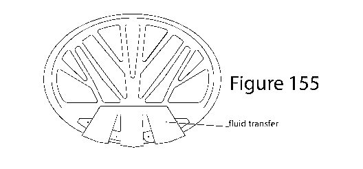
Figure 155 shows a fluid transfer system for providing the fluid biosample
from the EBC collector
to each electronic biosensor of the multi-biomarker testing unit;
Figure 156 shows the back side of the testing system support having a wick for
continuously
flowing the fluid biosample over the multi-biomarker testing unit and adhesive
for retrofitting into
a pre-existing mask;
Figure 157 shows a flow conductor with a hydrophilic pattern for transporting
EBC towards a
testing zone;
Figure 158 shows a thermal mass with a front surface forming a condensate-
forming surface;
Figure 159 shows a fluid transfer system for transporting EBC towards a
testing zone of a
biomarker testing unit;
Figure 160 shows an electronic biosensor version of the biomarker testing
unit;
Figure 161 shows a testing system support for supporting the EBC collector,
the fluid transfer
system and the biomarker testing unit and configured and dimensioned to fit
inside a pre-existing
face mask;
Figure 162 shows an assembly of the breath based diagnostic system;
Figure 163 shows the constituent parts of a mask-based diagnostic system;
Figure 164 shows the dimensions in inches and geometry of an embodiment of the
fluid
conductor;
Figure 165 shows an exhaled breath vapor containment volume defined by a face
mask with an
EBC collector and other parts of a breath based diagnostic system disposed
inside the containment
volume;
Figure 166 shows a composite thermal mass;
Figure 167 shows a water/SAP gel thermal mass;
Figure 168 shows the back side of a breath based diagnostic system with a
water/SAP thermal
mass and LFA biomarker testing unit;
Figure 169 shows an embossed metal foil thermal mass with a condensate-forming
surface and
fluid conductor channels;
Figure 170 shows an endothermic thermal mass for inserting into a holding
pocket of a mask-
based diagnostic system;
Figure 171 shows a soapstone powder/binder composite thermal mass;
Figure 172 shows a metal slug thermal mass;
18
CA 03177607 2022-09-28
WO 2021/216386 PCT/US2021/027854
Figure 173 shows a face mask constructed with an EBC collector and accumulated
fluid
biosample reservoir disposed inside of the mask, with the sample pad of an LFA
in the reservoir
and at least the visual readout portion of the LFA disposed on the outside of
the mask;
Figure 174 shows the EBC collector with the thermal disposed in an exhaled
breath vapor
containment volume on the inside of the mask;
Figure 175 showed a construction of the fluid transfer system having a fluid
dam comprising a
dissolvable adhesive;
Figure 176 illustrates an assembly of a bifurcated version of the breath based
diagnostic system;
Figure 177 illustrates an exploded view of the constituent parts of the
bifurcated version of there
breath based diagnostic system;
Figure 178 shows the bifurcated version formed with an embossed metal foil
condensate-forming
surface with contours forming fluid transfer channels;
Figure 179 is a cross section exploded view of the bifurcated version of the
breath based
diagnostic system;
Figure 180 is a cross section assembled view of the bifurcated version of the
breath based
diagnostic system;
Figure 181 shows a KN95 pre-existing mask retrofit with an LFA version of the
breath based
diagnostic system;
Figure 182 shows the retrofit testing system disposed on the inside of the
KN95 mask with an
LFA disposed on the inside of the mask;
Figure 183 shows an electronic biosensor configured as a field-effect
transistor with a graph
showing an output signal at the beginning of binding of target molecules to
capture molecules;
Figure 184 shows the electronic biosensor configured as a field-effect
transistor with more target
molecules captured and a graph showing an output signal at a time after the
beginning of binding
of target molecules to capture molecules; and
Figure 185 shows the electronic biosensor configured as a field-effect
transistor with more target
molecules captured and a graph showing an output signal at a time after the
beginning of binding
of target molecules to capture molecules.
19
CA 03177607 2022-09-28
WO 2021/216386 PCT/US2021/027854
DETAILED DESCRIPTION:
Below are provided further descriptions of various non-limiting, exemplary
embodiments. The
exemplary embodiments of the invention, such as those described immediately
below, may be
implemented, practiced or utilized in any combination (e.g., any combination
that is suitable,
practicable and/or feasible) and are not limited only to those combinations
described herein and/or
included in the appended claims.
The word "exemplary" is used herein to mean "serving as an example, instance,
or illustration."
Any embodiment described herein as "exemplary" is not necessarily to be
construed as preferred
or advantageous over other embodiments. All of the embodiments described in
this Detailed
Description are exemplary embodiments provided to enable persons skilled in
the art to make or
use the invention and not to limit the scope of the invention which is defined
by the claims.
Many configurations, embodiments, methods of manufacture, algorithms,
electronic circuits,
microprocessors, memory and computer software product combinations, networking
strategies,
database structures and uses, and other aspects are disclosed herein for a
wearable electronic
digital therapeutic device and system that has a number of medical and non-
medical uses.
Although embodiments are described herein for detection of biomarkers of SARS-
CoV-2 virus,
the systems, methods and apparatus described are not limited to any particular
virus or disease. In
most instances, where the term virus or COVID-19 is used, any other health or
fitness related
biomarker could be used instead. The description here and the drawings and
claims are therefore
not intended to be limited in any way to virus detection, the inventions
described and claimed can
be used for many diseases including lung cancer, diabetes, asthma,
tuberculosis, environmental
exposures, glucose, lactate, blood borne diseases and other ailments or
indications of the health of
the test subject. Further, the electronic biosensor, test systems, uses and
methods of manufacturing
described herein are not limited to the use of exhaled breath condensate.
Wastewater, potable
water, environmental quality samples, and any bodily fluid can be used as the
test sample. The use
of aptamers, in particular, make the inventive sensor widely useful because of
the nature of
selected aptamers being adaptable for specific engineering and selection to
have a binding affinity
CA 03177607 2022-09-28
WO 2021/216386 PCT/US2021/027854
that is tailored to a corresponding target analyte. Therefore, the
descriptions of innovations are not
intended to be limited to a particular use- case, capture molecule, biomarker
or analyte.
In immunochromatography, a capture molecule, which may be, for example, an
aptamer, naturally
occurring antibody, or engineered antibody, is disposed onto a surface of a
porous membrane, and
a sample passes along the membrane. As described herein, the term antibody,
aptamer, engineered
antibody, or capture molecule is used interchangeably. In some instances, a
specific type of
capture molecule may be described. Biomarkers in the sample is bound by the
capture molecule
which is coupled to a detector reagent. As the sample passes through the area
where the capture
molecule is disposed, a biomarker detector reagent complex is trapped, and a
color develops that
is proportional to the concentration or amount biomarker present in the
sample.
In a lateral flow assay, a liquid sample containing a target biomarker(s)
flows through a multi-
zone transfer medium through capillary action. The zones are typically made of
polymeric strips
enabling molecules attached to the strips to interact with the target
biomarker. Usually,
overlapping membranes are mounted on a backing card to improve stability and
handling. The
sample containing the target biomarker and other constituents is ultimately
received at an
adsorbent sample pad which promotes wicking of the fluid sample through the
multi-zone transfer
medium.
The fluid sample is first received at a sample pad which may have buffer salts
and surfactants
disposed on or impregnated into it to improve the flow of the fluid sample and
the interaction of
the target biomarker with the various parts of the detection system. This
ensures that the target
biomarker will bind to capture reagents as the fluid sample flows through the
membranes. The
treated sample migrates from the sample pad through a conjugate release pad.
The conjugate
release pad contains labeled antibodies or other capture molecules that are
specific to binding with
the target biomarker and are conjugated to colored or fluorescent indicator
particles. The indicator
particles are typically, colloidal gold or latex microspheres.
At the conjugate release pad, the labeled antibodies, indicator particles and
target biomarker bind
to form a target biomarker-labeled antibody complex. If a biomarker is
present, the fluid sample
21
CA 03177607 2022-09-28
WO 2021/216386 PCT/US2021/027854
now contains the indicator particles conjugated to the labeled antibody and
bound to the target
biomarker (i.e., the target biomarker-labeled antibody complex) along with
separate labeled
antibodies conjugated to the indicator particles that have not been bound to
the target biomarker.
The fluid sample migrates along the strip into a detection zone.
The detection zone is typically a nitrocellulose porous membrane and has
specific biological
components (usually antibodies or antigens) disposed on or impregnated in it
forming a test line
zone(s) and control line zone. The biological components react with the target
biomarker-labeled
antibody complex. For example, the target biomarker-labeled antibody complex
will bind to a
specifically selected primary antibody that is disposed at the test line
through competitive binding.
This results in colored or fluorescent indicator particles accumulating at the
test line zone making
a detectable test line that indicates the target biomarker is present in the
fluid sample.
The primary antibody does not bind to the separate labeled antibodies and they
continue to flow
along with the fluid sample. At a control line zone, a secondary antibody
binds with the separate
labeled antibodies conjugated to the indicator particles and thereby indicates
the proper liquid
flow through the strip.
The fluid sample flows through the multi-zone transfer medium of the testing
device through the
capillary force of the materials making up the zones. To maintain this
movement, an absorbent
pad is attached as the end zone of the multi-zone transfer medium. The role of
the absorbent pad is
to wick the excess reagents and prevent back-flow of the fluid sample.
The constituents are selected and disposed on the membranes so that if there
is no target
biomarker present in the fluid sample, there will be no target biomarker-
labeled antibody complex
present that flows through the test line zone. In this case there will be no
accumulation of the
colored or fluorescent particles and no detectable test line will form. Even
if there is no biomarker
and thus no test line, there will still be a control line formed because the
secondary antibody still
binds to the separate labeled antibodies that flow along with the fluid
sample.
22
CA 03177607 2022-09-28
WO 2021/216386 PCT/US2021/027854
The test and control lines may appear with different intensities depending on
the device structure
and the indicator particles can be assessed by eye or using an optical or
other electronic reader.
Multiple biomarkers can be tested simultaneously under the same conditions
with additional test
line zones of antibodies specific to different biomarkers disposed in the
detection zone in an array
format. Also, multiple test line zones loaded with the same antibody can be
used for quantitative
detection of the target biomarker. This is often called a 'ladder bars' assay
based on the stepwise
capture of colorimetric conjugate¨antigen complexes by the immobilized
antibody on each
successive line. The number of lines appearing on the strip is directly
proportional to the
concentration of the target biomarker.
What is needed now is a low cost, scalable, accurate and easy-to-use testing
system that can be
deployed to the masses via the mail or courier for at-home use.
Researchers have been able to detect biomarkers in the breath of patients that
have interstitial lung
disease (see, Hayton, C., Terrington, D., Wilson, A.M. et al. Breath
biomarkers in idiopathic
pulmonary fibrosis: a systematic review. Respir Res 20, 7 (2019).
https://doi.org/10.1186/
s12931-019-0971-8). An embodiment of the inventive testing system detects
COVID-19 specific
biomarkers present in the breath of infected, infectious or post-recovery
individuals.
The inventive COVID-19 testing system has the ability to coalesce breath vapor
into droplets and
then pass the droplet sample over a fluidic biosensor, such as a Lateral Flow
Assay (LFA) or
electronic Nanoscale-Biosensor (e-NSB) to enable a very low cost,
manufacturable at-scale
testing system that can be distributed to the masses for at-home triage
testing. The inventive
testing system can also be used for other biometric and environmental testing
applications other
than for virus detection.
LFAs can be used for the detection of a wide range of biomarkers present in
the breath including
cytokines, proteins, haptens (elicit the production of antibodies), nucleic
acids and amplicons
(pieces of RNA and DNA) (see, Corstj ens PL, de Dood CJ, van der Ploeg-van
Schip JJ, et al.
Lateral flow assay for simultaneous detection of cellular- and humoral immune
responses. Clin
Biochem. 2011;44(14-15):1241-1246. doi:10.1016/j.clinbiochem.2011.06.983).
23
CA 03177607 2022-09-28
WO 2021/216386 PCT/US2021/027854
A directed assembly technique for high throughput manufacturing of e-NSBs is
known where the
technique is proven to selectively assemble nanoparticles coated with specific
antibodies onto a
single microchip surface for the simultaneous detection of multiple
biomarkers. Early results
suggested sensitivity to concentrations of much less than 1 ng/mL¨a large
increase in sensitivity
relative to that of the commercially available ELISA detection kit. The
biosensor is very small,
about .25mm in diameter, and has advantages compared to traditional in vitro
techniques because
it enables disease markers detection with less false positives with a very low
detection limit. This
capability will be very useful for detecting very small changes in biomarker
concentration in
disease monitoring (see, Highly sensitive micro-scale in vivo sensor enabled
by electrophoretic
assembly of nanoparticles for multiple biomarker detection, Malima et al., Lab
chip, 2012,12,
4748-4754).
Exhaled breath collection has long been recognized as requiring the least
invasive methods, and so
is preferred for environmental and public health studies. In contrast to blood
and urine, breath
sampling does not require trained medical personnel or privacy, does not
create potentially
infectious wastes, and can be done essentially anywhere in any time frame.
Although the Exhaled
Breath Condensate (EBC) format discriminates against most non-polar VOCs, it
has the advantage
of collecting polar compounds and heavier biomarkers including semi- and non-
volatile organics,
cytokines, proteins, cellular fragments, DNA, and bacteria. Exhaled breath
also contains tiny
aerosols (including both liquid and solid particles) that are created by
surface film disruption at
the alveolar level and by upper airway turbulence. These aerosols give
mobility to materials that
are otherwise relegated to the liquid layers within the lung and, as such, are
that part of the EBC
which contributes the non-volatile biomarkers.
The usual methods for obtaining clinical specimens from the respiratory tract
are nasopharyngeal
or oropharyngeal swabs, nasopharyngeal aspirates and nasal washes, tracheal
aspirates,
bronchoalveolar lavage, or the collection of sputum. Each of these techniques
has drawbacks:
Nasopharyngeal and oropharyngeal swabs, aspirates, and washes provide mucus
from the upper
respiratory tract, which does not always contain the same viral load or the
same species of viruses
as the lower respiratory tract. The collection of aerosol particles produced
by patients during
24
CA 03177607 2022-09-28
WO 2021/216386 PCT/US2021/027854
coughing and tidal breathing potentially provides a non-invasive method for
the collection of
diagnostic specimens of respiratory viruses. Respiratory viruses have been
detected in the exhaled
breath and cough aerosols from infected patients, especially the influenza
virus. Microbial
aerosols may also be more representative of lower respiratory tract disease in
viral illnesses in
which sputum production is not common.
Because exhaled aerosol collection is non-invasive, repeated sample collection
should be more
acceptable to patients than traditional methods. If the limitations can be
overcome, exhaled
aerosol analysis could become a useful tool for the diagnosis of respiratory
infections and for
monitoring the course of illness and response to treatment (see, Fennelly KP,
Acuna-Villaorduna
C, Jones-Lopez E, Lindsley WG, Milton DK. Microbial Aerosols: New Diagnostic
Specimens for
Pulmonary Infections. Chest. 2020;157(3):540-546.
doi:10.1016/j.chest.2019.10.012).
There are more than 2,000 compounds identified in EBC (see, Montuschi P, Mores
N, Trove A,
Mondino C, Barnes PJ, The electronic nose in respiratory medicine.
Respiration.
2013;85(1):72-84) and many of them are considered to represent sensitive
biomarkers of lung
diseases (see, Sapey E, editor. Bronchial Asthma: Emerging Therapeutic
Strategies. Rijeka:
InTech). Biomarkers present in EBC depict the processes occurring in lungs
much more than
those in the entire body system.
Therefore, particular profiles of exhaled biomarkers can reveal information
exclusively applicable
to lung disease diagnoses. EBC is a biological matrix reflecting the
composition of the
bronchoalveolar extra-cellular lung fluid. The main advantage of EBC as of a
matrix is its
specificity for the respiratory tract (the liquid is not influenced by process
occurring in other parts
of the body) (see, Molecular Diagnostics of Pulmonary Diseases Based on
Analysis of Exhaled
Breath Condensate, Tereza Ka6erova, Petr NovotnY, Jan Boron and Petr Ka6er
Submitted:
October 9th 2016Reviewed: January 25th 2018Published: September 5th 2018, DOT:
10.5772/
intechopen.7440).
The surfaces in all parts of the lung down to the alveoli are coated with an
aqueous mucous layer
that can be aerosolized and carry along a variety of non-volatile
constituents. EBC and EBA are
CA 03177607 2022-09-28
WO 2021/216386 PCT/US2021/027854
different types of breath matrices used to assess human health and disease
state. EBA represents a
fraction of total EBC, and is targeted to larger molecules, such as fatty
acids and cytokines, as
well as cellular fractions, proteins, viruses, and bacteria instead of the gas-
phase. There is a wide
variety of compounds, such as volatile organic compounds (VOCs), NO, CO2, NH3,
cytokines,
and hydrogen peroxide (H202) in exhaled breath condensate (EBC), and exhaled
breath aerosol
(EBA). VOCs located in fatty tissues are released to the blood and are then
exchanged into the
breath through the alveoli and airways in the lungs. A portion of VOCs are
also retained within the
respiratory tract after exposure. Thus, breath concentrations of VOCs are
representative of blood
concentrations, but samples can be obtained non-invasively with little
discomfort to the individual
(see, Wallace MAG, Pleil JD. Evolution of clinical and environmental health
applications of
exhaled breath research: Review of methods and instrumentation for gas-phase,
condensate, and
aerosols. Anal Chim Acta. 2018;1024:18-38. doi:10.1016/j.aca.2018.01.069).
EBC and EBA are valuable non-invasive biological media used for the
quantification of
biomarkers. EBC contains exhaled water vapor, soluble gas-phase (polar)
organic compounds,
ionic species, plus other species including semi- and non-volatile organic
compounds, proteins,
cell fragments, DNA, dissolved inorganic compounds, ions, and micro-biota
(bacteria and viruses)
dissolved in the co- collected EBA (see, inters BR, Pleil JD, Angrish MM,
Stiegel MA, Risby TH,
Madden MC. Standardization of the collection of exhaled breath condensate and
exhaled breath
aerosol using a feedback regulated sampling device. J Breath Res.
2017;11(4):047107. Published
2017 Nov 1. doi:10.1088/1752-7163/aa8bbc).
An earlier reference reports detecting influenza virus RNA in the exhaled
breath of patients
infected with influenza A virus and influenza B virus. Although a sample of
EBC may have virus
RNA in less concentrations than a nasal swab, these tests did determine
detectable influenza virus
RNA in exhaled breath. Concentrations in exhaled breath samples ranged from 48
to 300
influenza virus RNA copies per filter on the positive samples, corresponding
to exhaled breath
generation rates ranging from 3.2 to 20 influenza virus RNA copies per minute
(see, Fabian P,
McDevitt JJ, DeHaan WH, et al.
26
CA 03177607 2022-09-28
WO 2021/216386 PCT/US2021/027854
Influenza virus in human exhaled breath: an observational study. PLoS One.
2008;3(7):e2691.
Published 2008 Jul 16. doi:10.1371/journal.pone.0002691). This reference shows
that nasal and
throat swabs may typically have more RNA concentrations than EBC. However, the
virus RNA is
clearly present in EBC and an EBC testing system with enough sensitivity
should be effective at
detecting the virus, bacteria, and other disease and health related
biomarkers.
Scanning Electron Microscope (SEM), polymerase chain reaction (PCR) and
colorimetry (VITEK
2) for bacteria and viruses show that bacteria and viruses in EBC can be
rapidly collected with an
observed efficiency of 100 mL EBC within 1 min (see, Xu Z, Shen F , Li X, Wu
Y, Chen Q, et al.
(2012) Molecular and Microscopic Analysis of Bacteria and Viruses in Exhaled
Breath Collected
Using a Simple Impaction and Condensing Method. PLoS ONE 7(7): e41137.
doi:10.1371/
journal.pone.0041137).
Exhaled breath contains volatile organic compounds (VOCs), a collection of
hundreds of small
molecules linked to several physiological and pathophysiological processes.
Analysis of exhaled
breath through gas-chromatography and mass-spectrometry (GC-MS) has resulted
in an accurate
diagnosis of ARDS in several studies. Most identified markers are linked to
lipid peroxidation.
Octane is one of the few markers that was validated as a marker of ARDS and is
pathophysiologically likely to be increased in ARDS (see, Bos LDJ. Diagnosis
of acute respiratory
distress syndrome by exhaled breath analysis. Ann Trans/Med. 2018;6(2):33.
doi:10.21037/
atm.2018.01.17).
The inventive testing system is designed to be self-administered, nothing more
complicated than
putting on a mask and opening a smartphone app and enable data transmission
and storage.
Alternatively, data transmission can be avoided, with no stored data, and
instead be provide with
just an indication of the results privately either with an onboard indicator
such as a LED, or
through the smartphone app. If the test results signal is transmitted, the
data is encrypted at the
source, the electronics attached to the mask, before any wireless
transmission. Privacy issues are
handled at or better than government requirements for electronic medical
records. The inventive
testing system may include wireless communications capabilities that enable
test data to be used
along with GPS location information to assist in backward and forward contact
tracing and in the
27
CA 03177607 2022-09-28
WO 2021/216386 PCT/US2021/027854
case of an epidemic or pandemic, further quicken the ability of a growing
segment of the
population to safely return to work and restart economic activities, enabling
determining through
real-time contact tracing who might have been exposed to the virus as soon as
a positive test result
is received.
Biometric data is acquired and used for the public good but the collection of
biometric
information carries with it the burden of privacy issues. There can be
considered two uses for a
patient's biometric data: Patient monitoring for prevention and treatment; and
Population studies
to improve global healthcare. The inventive testing system can be software and
hardware
configured for separately created and maintained data bases, one shared only
with trusted
receivers (e.g., healthcare providers who access the data from their patients
through a secure two-
step verification process), and demographic-only data that stores anonymized
data that will be
used for Big Data analysis to spot patterns and trends related to an outbreak.
To maximize
compliance, the test subject can be allowed to select levels of data
reporting: self-reporting;
shared only with a test subject's registered HCP; or automatic data reporting
for contact tracing
and electrical medical records. The acquired data can anonymized and encrypted
at the source
(e.g., by the electronics associated with the testing system). Using the
smartphone app the test
subject can always be in control of how their test data is reported and can
opt-out or opt-in to the
level of data sharing.
Figure 1 shows a Lateral Flow Assay (LFA) testing system showing a biomarker
sample added to
a sample pad. Figure 2 shows the LFA with a biomarker -labeled antibody
complex formed at a
conjugate release pad. Figure 3 shows the binding of biomarkers at a test line
indicating the
presence of the biomarker.
Another testing system that can be used with the inventive EBC collection
system uses an
electronic nano-scale biosensor (e-NSB). Similar to LFA, e-NSB has the
potential of a much
higher sensitivity and can be used to provide a direct-to-electrical signal to
enable, for example,
easy wireless connectivity. The inventive EBC collection system with e-NSB
testing is easily
deployable as a compliment to existing Contact Tracing APPs. The nanoscale
dimensions mean
28
CA 03177607 2022-09-28
WO 2021/216386 PCT/US2021/027854
many detectors are made at once on a single wafer or as described herein,
through a high volume
roll manufacturing process, for lower cost, high throughput manufacturing.
Figure 4 shows the mechanism of a biosensor detection system. Simplistically,
the main
components of a fluidic biosensor include a sample source (a); a biosensor
area that is
functionalized with a biomarker-specific bioreceptor (b); and a transducer for
generating a
readable signal (c). The bioreceptor is matched to a specific target biomarker
for lock and key
selectivity screening. A fluid sample with some concentration of the target
biomarker (possibly as
small as a single molecule) flows onto the biosensor field. Some of the
biosensor "locks" receive
the biomarker "keys." This causes a detectable change in the output of the
transducer that
transforms the biosensor output into a readable signal for amplification and
data processing.
For example, the desired biomarker can also be an antibody that indicates the
recovery from a
Covid-19 infection. A fluid sample can be received as a droplet of sweat or
breath or other body
fluid and if the target antibody is present in the sample it interacts with
the biomarker-specific
bioreceptor. The bioreceptor outputs a signal with defined sensitivity and the
transducer generates,
for example, a change in an electrical characteristic such as conductivity,
indicating the presence
of the antibody biomarker in the fluid sample.
In accordance with an embodiment, an apparatus for detecting a biomarker
comprises a droplet
harvesting and channeling structure for converting vapor to a fluid sample
source having a
biomarker, a biosensor area functionalized with a biomarker-specific
bioreceptor, and a transducer
for generating a readable signal depending on a change in the bioreceptor in
response to receiving
the biomarker from the sample source.
Using nano-scale sensor technology enables detection of very low
concentrations of the target
biomarker(s) such as virus RNA, proteins and/or antibodies while avoiding the
need for drawing
blood. In accordance with an embodiment of the inventive testing system, a
droplet harvesting and
channeling mechanism uses a hydrophobic field for fluid harvesting and
hydrophilic channels for
droplet movement onto the nano-sensor. This mechanism makes the inventive
system practical for
29
CA 03177607 2022-09-28
WO 2021/216386 PCT/US2021/027854
creating a very inexpensive, scalable manufacturable COVID-19 test that does
not require any
blood or the administration of the test by a skilled technician, nurse or
healthcare provider.
A mask-based testing system embodiment uses a nano-scale fluidic biosensor
technology with a
unique moisture droplet harvesting and channeling structure. This structure
unlocks the use of the
nano-scale sensor for detection possibly down to single molecules of target
biomarkers. This
enables the detection of even very low concentrations of antibodies, proteins
and other chemical
biomarkers present in any body fluid without the drawing of blood.
A non-limiting embodiment builds on the sweat chemistry sensor technology
described in PCT/
US19/45429, METHODS AND APPARATUS FOR A WEARABLE ELECTRONIC DIGITAL
THERAPEUTIC DEVICE invented by Daniels and published April 10, 2020, which is
incorporated by reference herein in its entirety. The inventive embodiment
described herein
includes a COVID-19 testing system that can be mass produced on readily
available high-volume
manufacturing equipment in the millions of units needed for mass population
testing. An
embodiment of the testing system uses a nano-scale fluidic biosensor with a
unique moisture
droplet harvesting and channeling structure.
This structure enables the use of the nano-scale sensor for detecting COVID-19
biomarkers in a
body fluid sample, such as breath condensate. This system enables the
detection of biomarker(s)
of antibodies, proteins, RNA and other chemical COVID-19 biomarkers without
the drawing of
blood, expensive equipment or technically trained personnel. The proposed
system can be
configured as at least a first pass go/no-go test that can determine who
should be more accurately
tested by the conventional testing methodologies.
There is the need for a low cost, accurate, easy-to-use testing system for
COVID-19 that ideally
can be mailed out and self-administered at home. For example, current testing
protocols require a
nasal swab for RNA testing to show active infection or a sample of blood be
taken from a
person in order to test for sufficient antibodies to the COVID-19 virus for
immunity. These tests
typically require breaking sequestration and traveling to a testing site where
a technician, nurse or
other healthcare provider administers the test. We propose a testing system
that can be used as a
CA 03177607 2022-09-28
WO 2021/216386 PCT/US2021/027854
first pass go/no go assessment to first see if a more elaborate and expensive
testing methodology
is warranted. For example, an inexpensive, easy-to-use testing system that can
be done at home
and finds a low concentration of COVID-19 antibodies present in the breath or
sweat can then be
used as the impetus for the individual to go to a testing facility for a more
accurate determination
of the person's immunity to further COVID-19 infection.
Figure 5 is a side view of a wearable electronic breath chemistry sensor.
Figure 6 is a top view of
a wearable electronic breath chemistry sensor. The biometric sensor is tuned
to detect at least one
biometric indicator associated with the presence of COVID-19 antigen, RNA
and/or antibody. A
droplet collector draws EBC droplets into a transfer aperture. The sensor is
wet by the droplet and
then the droplet is drawn through wicking into wicking/evaporation materials.
A continuous flow
of fresh droplets passes over the sensor. A hydrophobic field encourages EBC
to bead and migrate
to hydrophilic channels. Tapered hydrophilic channels use surface tension to
draw sweat into the
sweat transfer aperture. Hydrophobic and hydrophilic screen printable inks are
available from
companies such as Cytonix and Wacker.
Figure 7 is an isolated view of an Exhaled Breath Condensate (EBC) droplet
sample collector.
Figure 8 is a top view showing a step for forming the EBC droplet collector.
Figure 9 is a top view
showing another step for forming the EBC droplet sample collector. Figure 10
is a top view
showing still another step for forming the EBC droplet sample collector.
Figure 11 is a top view
showing yet another step for forming the EBC droplet sample collector. Figure
12 illustrates the
EBC sample collector showing EBS droplets. In accordance with a non-limiting
exemplary
embodiment, an at-home, triage COVID-19 testing system uses Exhaled Breath
Condensate
(EBC) for a biomarker fluid sample. Breath is an exceptional source of virus
antigens, antibodies
and RNA. EBC can be analyzed using establish methods including Lateral Flow
Assay, Nano-
scale Bioreceptor and Photonic Quantitative Assay. EBC produces much cleaner
samples to test
than nasal swabs, is non-invasive and easier than drawing blood. However,
collecting EBC
usually requires a big, expensive chiller and is always done in a clinical
setting.
There is a great push throughout the world to develop adequate testing for the
COVID-19 virus. A
conventional PCR test detects pieces of dead virus from nasal swab or sputum.
The test
31
CA 03177607 2022-09-28
WO 2021/216386 PCT/US2021/027854
determines if a person is infectious. The test is expensive, requires trained
personnel and machines
and there is a delay in obtaining the test results due to collection,
transportation and processing of
samples. PCR also requires a lot of chemical reagents and results in a lot of
false negatives.
Antibody test detects the body's immune response to the virus. It typically
requires a blood
sample. Antibody tests can be relatively fast and does necessarily require
trained personnel. False
positives are frequent because other viruses could be causing the antibodies.
EBC has been used for rapid detection of microbial DNA and RNA to demonstrate
bacterial and
viral lung infections. (see, Xu Z, Shen F, Li X, Wu Y, Chen Q, Jie X, et al.
Molecular and
microscopic analysis of bacteria and viruses in exhaled breath collected using
a simple impaction
and condensing method. PLoS One 2012;7:e41137.
https://www.ncbi.nlm.nih.gov/pubmed/
22848436).
A nasal swab sample often contains a lot of background biological materials
making it harder to
identify the RNA of the virus because of other molecules present in the
sample. Breath condensate
is naturally enriched with viruses and confounding molecules are at much lower
concentrations.
(see, https://www.zimmerpeacocktech.com/products/electrochemical-sensors/covid-
19-and-per-
on-the- breath!).
Antibodies are present in breath vapor. IgA antibodies are found in areas of
the body such the nose
and breathing passages. IgG antibodies are found in all body fluids and are
the most common
antibody (75% to 80%), that are very important in fighting bacterial and viral
infections. IgE
antibodies are found in the lungs, skin, and mucous membranes. (see, https://
www.uofmhealth.org/ health-library/hw41342).
Virus antigens are found in Airway Lining Fluid (ALF). EBC is a non-invasive
method of
sampling airway lining fluid (ALF). Constituents of ALF are representative of
the respiratory tree
lining fluids. ALF is a measure of the concentration of biomarkers directly
influenced by
respiratory cells. (see, Exhaled breath condensate: a comprehensive update,
Ahmadzai, et al.,
Clinical Chemistry and Laboratory Medicine (CCLM) 51, 7; 10.1515/cclm-2012-
0593).
32
CA 03177607 2022-09-28
WO 2021/216386 PCT/US2021/027854
Although EBC could be an exceptional source of biomarkers indicating the
stages of infection and
recovery from the COVID-19 virus, as well as other medical and fitness uses,
the conventional
equipment for obtaining an EBC fluid sample is big and expensive and is only
used in clinical
setting. The conventional equipment requires a chiller and is designed for
relatively large sample
collection. This makes conventional EBC sampling equipment unsuited for at-
home testing.
An embodiment of the inventive EBC sample collector includes a hydrophobic
field that causes
vapor to bead up into droplets. Hydrophilic channels coalesce and transfer the
droplets to form an
accessible EBC fluid sample. The hydrophobic field and hydrophilic channel can
be screen
printed or otherwise coated on a thermal mass aluminum sheet substrate, the
polished aluminum
sheet itself can be the hydrophobic field with no further treatment. This
substrate can be chilled
prior to using the testing system to improve EBC collection. The inventive EBC
sample collector
makes the low-cost Lateral Flow Assay, electronic biosensors, and other
testing systems workable
for at-home triage testing. The CDC says it is essential to quickly develop
inexpensive screening
test. The inventive EBC sample collector makes such screening testing viable
for mass
deployment to large segments of the population. There is no need to break
sequestration. No
skilled technicians, clinics or lab equipment are needed. Very high-volume
existing manufacturing
methods can be modified to product multiple-up (many at once) screen printed
EBC sample
collectors. A low-cost aluminum substrate acts as thermal mass and can be
chilled for faster
droplet harvesting. Batch fabrication can be used to manufacture multiple-up
LFA modules on a
sheet with a format that is quickly adaptable to ultra-high- volume Roll-to-
Roll manufacturing.
Figure 13 illustrates the EBC sample collector applied to an LFA testing
system. Figure 14 is an
exploded view showing the screen printed hydrophilic channels, screen printed
hydrophobic field
and thermal mass substrate of the EBC sample collector. A first
emitter/detector pair are used to
determine if the novel coronavirus N protein at the test line (T) has been
bound by the IgM-IgM
complex. A second emitter/detector pair are used to determine if free anti-
human IgM antibody
has been bound to the anti-mouse antibody at the control line (C) confirming
that the fluid sample
has traversed through the transfer medium and the test has been correctly
performed.
33
CA 03177607 2022-09-28
WO 2021/216386 PCT/US2021/027854
In accordance with an embodiment, a method of forming a biomarker testing
system comprising
forming an exhaled breath condensate fluid sample collector. Forming the
exhaled breath
condensate fluid sample collector comprise the steps of providing a substrate,
coating a
hydrophobic field on the substrate, and coating at least one hydrophilic
channel on the substrate.
The hydrophobic field is for receiving body fluid vapor and forming a fluid
droplet from the
received body fluid vapor and the hydrophilic channel is for receiving the
fluid droplet and
channeling the fluid droplet towards a testing system. At least one fluid
sample draining hole may
be formed at an end of the hydrophilic channel for draining the fluid droplet
through the at least
one fluid sample draining hole onto a sample receiving structure of the
testing system.
At least one photoemitter and one photodetector may be provided where the
photoemitter emits
radiation towards the biomarker testing zone and the photodetector receives
radiation from the
biomarker testing zone. Figure 15 is an exploded view showing the constituent
elements of a LFA.
Figure 16 illustrates an embodiment of the LFA including photonic
emitter/detector electronics. In
immunochromatography, a capture antibody is disposed onto a surface of a
porous membrane, and
a sample passes along the membrane. Biomarkers in the sample are bound by the
antibody which
is then coupled to a detector reagent. As the sample passes through the area
where the capture
reagent is disposed, the biomarker detector reagent complex is trapped, and a
color develops that
is proportional to the biomarker present in the sample. The photonics
emitter/detector pair enable
the proportional quantitative measurement of the biomarker where the biomarker
concentration if
the fluid sample is determined from an intensity or counting of received
photons at the detector.
The solid-phase lateral-flow test platform is an example of
immunochromatography that is widely
used for home pregnancy testing. Lateral flow tests have benefited from the
use of sol particles as
labels. The use of inorganic (metal) colloidal particles are typically used as
a label for
immunoassays and several techniques are used to measure the amount of bound
conjugate. These
include naked eye, colorimetry and atomic absorption spectrophotometry.
Colorimetry applies the
Beer¨Lambert law, which states that the concentration of a solute is
proportional to the
absorbance. At higher antigen concentrations, the results of
immunochromatography can be read
by the naked eye (e.g., the typical home pregnancy test). For lower
concentrations, colorimetry
has been shown to be more than 30 times more sensitive than reading by the
naked eye.
34
CA 03177607 2022-09-28
WO 2021/216386 PCT/US2021/027854
In accordance with an embodiment, immunochromatography is used to detect the
present of a
COVID-19 biomarker. Generally, immunochromatography is the separation of
components in a
mixture through a medium using capillary force and the specific and rapid
binding of an antibody
to its antigen. A dry transfer medium is coated separately with novel
coronavirus N protein ("T"
test line) and anti-mouse antibody ("C" control line). Free colloidal gold-
labeled anti-human IgM
are in a release pad section (S). The inventive vapor coalescence and droplet
harvesting structure
are used to obtain a fluid sample of breath condensate. This fluid sample is
applied to the release
pad section. The anti-human IgM antibody the binds to at least some of the IgM
antibodies (if any
are present), forming an IgM-IgM complex. The fluid sample and antibodies move
through the
transfer medium via capillary action. If coronavirus IgM antibody is present
in the fluid sample,
the novel coronavirus N protein at the test line (T) will be bound by the IgM-
IgM complex and
develop color. If there is no coronavirus IgM antibody in the sample, free
anti-human IgM does
not bind to the test line (T) and no color will develop. The free anti-human
IgM antibody will bind
to the anti-mouse antibody at the control line (C) so that the control line
develops color
confirming that the fluid sample has traversed through the transfer medium and
the test has been
correctly performed.
Figure 17 illustrates the EBC sample collector applied to a nanoscale
biosensor testing system and
showing a pull tab for holding back collected droplets on the sample pad.
Figure 18 is a
perspective view showing the EBC sample collector applied to a testing system.
Figure 19 is an
isolated view showing the pull table disposed between the sample pad and
conjugate release pad.
The EBC fluid sample will be collected over some time period, collected from
the hydrophobic
field through the hydrophilic channels, and flowing through the fluid sample
drain holes to build
up in the sample pad. Atypical LFA uses about 3 drops of buffered sample that
is added to the
sample pad. In accordance with an exemplary embodiment, buffer materials and
surfactants can be
incorporated in dry form into the sample pad so that the EBC sample, which is
mostly water, will
be suitable as a directly applied fluid sample without requiring the addition
of a fluid buffer. When
a person is at rest, there is about 17.5 ml of EBC produced per hour (see "How
much water is lost
during breathing?", Zielinski et al., Pneumonol Alergol Pol 2012;80(4):339-
342). There are 20
drops per milliliter. So, every hour at rest there is the potential to collect
about 350 drops of EBC.
CA 03177607 2022-09-28
WO 2021/216386 PCT/US2021/027854
A collection efficiency by the EBC sample collector of only about 3% should
provide an adequate
number of EBC droplets for a fluid sample in about 15 minutes from an
individual at rest.
In accordance with an embodiment, an apparatus for detecting a biomarker
comprises a droplet
harvesting structure for converting breath vapor to a fluid droplet for
forming a fluid sample and a
testing system having a biomarker testing zone for receiving the fluid sample
and detecting a
biomarker. The droplet harvesting structure may include at least one of a
hydrophobic field for
receiving the breath vapor and forming the fluid droplet from the received
breath vapor and
hydrophilic channels for receiving the fluid droplet and channeling the fluid
droplet towards the
testing system. A fluid dam member may be provided disposed between the
droplet harvesting
structure and the biomarker testing zone.
The testing system may comprise a fluidic lateral flow assay including a
sample pad for receiving
the fluid sample potentially containing a biomarker, a conjugate release pad,
a flow membrane and
an adsorbent pad for receiving and flowing the fluid sample to detect the
potential biomarker from
the sample source. A fluid dam member disposed between the sample pad and the
conjugate
release pad, the fluid dam including a pull tab structure to enable a test
subject to remove the fluid
dam member and allow the flow of the fluid sample from the sample pad to the
conjugate release
pad.
In order to flush the EBC fluid sample through the test system, the fluid dam
is provided to hold
back the EBC on the sample pad as it accumulates. The fluid dam may be, for
example, a piece of
silicone (but not limited to) coated release paper forming a pull tab that is
disposed between the
sample pad and the conjugate release pad. The pull tab is held in place on the
top adhesive and
allows for the accumulation of the EBC fluid sample. After an adequate amount
of time has
passed to saturate the sample pad with enough EBC fluid sample, the test
subject pulls the pull tab
out causing the EBC fluid sample to flow from the sample pad to the conjugate
release pad. This
enables the EBC fluid sample to flush through the various constituents of the
testing system by
capillary action. The EBC fluid sample is allowed to build up on the sample
pad so that removing
the pull tab releases sample flow all at once to ensures adequate sample flow
and promotes testing
consistency.
36
CA 03177607 2022-09-28
WO 2021/216386 PCT/US2021/027854
Figure 20 is an isolated view of a screen printed droplet sample collector
with a fluid transfer
aperture. Figure 21 is a cross-section view showing a fluid sample collected
from the EBC sample
collector flowing between a photonics emitter/detector pair. In this
embodiment, another body
fluid, such as sweat, might be used with the EBC sample collector configured
to harvest sweat
droplets from the skin instead of coalescing breath vapor into exhaled breath
condensate. It is
noted that any of the embodiments and innovations described herein may be
useful for other
medical and fitness uses, for other disease or virus testing or biometric
detection in addition to or
instead of the described use for COVID-19 testing.
The manufacturing techniques, equipment and materials for most components of
an embodiment
of the inventive COVID-19 testing system are readily available and very well
known. For
example, to create our fluid harvesting and droplet channeling structure,
screen printing is used to
pattern hydroscopic and hydrophobic inks sourced from a company such as
Cytonix and Wacker.
There is no shortage of manufacturing capacity needed to quickly screen print
for the hundreds of
millions of testing units needed. The fluidic biosensor component can be
manufactured using high
throughput equipment available from a company such as Conductive Technologies,
Inc., York, PA,
USA, and the chemistry for functionalizing the biosensor can be obtained from
a company such as
RayBiotech, Peach Tree Corners, GA, USA. Other necessary manufacturing steps,
such as wire
bonding and printed circuit board fabrication will make use of the same
ubiquitous machines that
are similarly purposed for semi-conductor and circuit board electronics.
Figure 22 shows the side views of the steps for building up an LFA testing
system, not necessarily
in the sequence order. Figure 23 shows the top view of the steps for building
up an LFA testing
system. Figure 24 shows a 4x9 ganged multiple-up sheet of LFA testing systems
formed as a
batch. Figure 25 shows a roll-to-roll manufacturing process for forming a roll
of bottom adhesive/
backing substrate/top adhesive.
Figure 26 is a perspective view illustrating the bottom adhesive/backing
substrate/top adhesive
stack. Figure 27 shows a roll-to-roll manufacturing process for forming the
constituent elements
of an LFA on a roll of bottom adhesive/backing substrate/top adhesive. Figure
28 shows the LFA
37
CA 03177607 2022-09-28
WO 2021/216386 PCT/US2021/027854
testing system formed by the roll-to-roll process cut from a continuous roll
and showing a section
of top adhesive for adhering the LFA testing system to a separately formed ENC
sample collector.
Figure 29 shows the LFA testing system formed by the roll-to-roll process cut
from a continuous
roll and showing a section of bottom adhesive for sticking onto a wearable
garment such as a face
mask. Figure 30 shows a sheet of substrate with a hydrophobic field coating on
a thermal mass
substrate with droplet collection holes. Figure 31 shows the sheet of
substrate with the
hydrophobic field coating on a thermal mass substrate with droplet collection
holes having a
coating of hydrophilic channels.
Figure 32 shows the EBC sample collector and testing system with electronics
for wireless data
acquisition and transmission along with separate trusted receiver and public
blockchain data path
and storage. Villanova University recently published an example of utilizing
blockchain to help
medical facilities track coronavirus cases globally. A private blockchain is
shared among medical
facilities around the world to publish coronavirus test results between
doctors on a trusted,
immutable ledger. IoT and AT are used to survey public spaces where high-risk
gatherings can take
place and trigger alerts over the blockchain. (see,
https://wwwl.villanova.edu/university/experts/
spotlight-detail html? spotlight=7180).
In accordance with an exemplary embodiment, the EBC collecting system with
biomarker
detection can utilize self-reporting or automatic data collection to be usable
with a new or existing
APPs for contact tracing and electrical medical records. The acquired data can
anonymized and
encrypted at the source (e.g., on the electronics associated with the testing
system). A first data
stream/data base allows a trusted receiver to access patient identifying data
while a second data
stream/data base provides anonymized data that can be provided as open source
or other data
transmission, storage and utilization mechanisms without identifying who the
source of the data is
from.
The inventive testing system has the potential to be very low cost, shippable
in a conventional
envelop for mass distribution to every household in a target region, state or
country. This enables
a much higher percentage of the population to undergo at least the baseline
testing indicating if
38
CA 03177607 2022-09-28
WO 2021/216386 PCT/US2021/027854
they should follow up with a visit to a drive through, hospital or clinic
testing facility for more
elaborate testing.
The inventive COVID-19 testing system can have the capability of testing two
or more of the
virus biomarkers at the same time. For example, RNA or protein testing can be
combined with
antibody testing. By testing for these two biomarkers the potential for false
negatives is
significantly statistically reduced and likely will be a more preferable
methodology.
The proposed COVID-19 testing system can be incorporated into personal
protection equipment,
such as masks, or provided as a patch that is stuck onto the body, or provided
as a stand-alone test
unit, similar to a home pregnancy test. This biosample can be obtained from
spit, blood, urine,
EBC, tears, sputum, feces, or any other material that may contain a target
analyte. Buffers and
diluents can be used, and if necessary, incubation and amplification
techniques, such as those used
for conventional PCR testing can be employed.
The testing system can include wireless communications capabilities, such as
RFID, near field
communication, WiFi, cellular and Bluetooth. This will enable, for example,
test data to be used
along with GPS location information to assist in contact tracing and further
quicken the ability of
a growing segment of the population to return to work and restart economic
activities, and to also
determine through real-time contact tracing who might have been exposed to the
virus.
As an enhancement to the basic system, biometric data can be acquired and used
for the public
good. The collection of biometric information carries with it the burden of
privacy issues. There
can be considered two uses for a patient's biometric data: Patient monitoring
for prevention and
treatment; and Population studies to improve global healthcare. The inventive
system uses
separately created and maintained data bases.
The biometric parameters such as those described herein with regards to the
embodiments can also
be detected, logged and/or transmitted, enabling a detailed history of the
patient's disease
progression, therapy, course of treatment, measured results of treatment,
etc., and can be made
available to improve the care given to the particular patient, and in the
aggregate, provide
39
CA 03177607 2022-09-28
WO 2021/216386 PCT/US2021/027854
significant data along with that of other patients, to assist in new drug
discovery, treatment
modifications, and a number of other advantages of the beneficial cycle
created by detection,
transmission, storage and analysis of biometric data taken directly from the
patient during the
course of drug therapy and/or other treatments.
Figure 33 shows the manufacturing processes for a heat bonded face mask.
Figure 34 shows the
fabric, filter and other layers bonded through a roll-to-roll lamination
process ore individually cut
into blanks for forming a pre-form mask stack. Figure 35 shows other materials
such as biological
reactive silver fabric and hot melt adhesive of the pre-form mask stack. The
highly contagious and
deadly effects of COVID-19 have resulting in an increased need for personal
protective masks.
Disposable masks are a good solution for healthcare providers, police and
others whose job put
them into constant contact with individuals who may or may not have the virus.
The ability to
change out a disposable mask between patients, for example, ensures that a
doctor or nurse will
have a fresh, clean, uncontaminated mask to better protect themselves and
protect their patients
from the spread of the virus. However, disposable masks are not a good
solution for the general
population. The cost and waste associated with a disposable mask makes it a
poor solution for
most people. Rather, what is needed is a mask that is low cost, easy to
manufacture and ideally
can be sterilized in a conventional home clothes washing machine and dryer.
Figure 36 is an exploded view of a mask stack. Figure 37 shows the fold lines
of the mask stack
for first and second heat press operations. Figure 38 shows the folded,
pressed and heat bonded
mask. Figure 39 shows the attachment of the EBC collector and testing system
to the folded mask.
Figure 40 shows the step of turning the folded mask inside out to dispose the
EBC collector and
testing system on the inside of the mask. Figure 41 shows a heat press
operation to bond elastic
straps onto the folded mask.
Figure 42 shows the mask with the EBC collector and testing system disposed
inside the mask
within the concentrated atmosphere of exhaled breath. In accordance with a non-
limiting
embodiment, a mask-based testing system is provided for detecting a biomarker
received from
lungs and airways of a test subject. An exhaled breath condensate (EBC)
collector is disposed on
an inside of a face mask worn by the test subject. The EBC collector converts
breath vapor
CA 03177607 2022-09-28
WO 2021/216386 PCT/US2021/027854
received from the lungs and airways of the test subject into a fluid
biosample. The EBC collector
has a thermal mass and a front face that receives the breath vapor at a
temperature greater than a
surface temperature of the front face and converts the breath vapor to a
liquid that is cooler than
the temperature of the breath vapor. The EBC collector includes a droplet
harvesting structure on
the front face including a field for receiving the breath vapor and forming
fluid droplets from the
received breath vapor, and channels for receiving the fluid droplets from the
field and channeling
the fluid droplets together to form the collected fluid biosample.
A biosensor is fixed to the face mask for receiving the fluid biosample from
the EBC collector and
testing the fluid biosample for a target biomarker. The biosensor generates a
test signal dependent
on at least the presence and absence of the target biomarker in the fluid
biosample. An electronic
circuit fixed to the face mask receives the test signal, determines from the
test signal a test result
signal depending on detecting or not detecting the target biomarker, and
transmits the test result
signal to a remote receiver.
Figure 43 shows a conventional bendable metal nose seal that is disposed
within the folds of the
mask at a location corresponding to the bridge of a test subject's nose.
Figure 44 shows a
replaceable adhesive nose strip that is disposed on the outside of the folds
of the mask at a
location corresponding to the bridge of a test subject's nose, and Figure 45
shows the components
of a magnetic removable nose seal.
Figure 46 is an exploded view of a testing system including a dissolvable flow
dam that holds
back collected EBC on the sample pad until enough has been accumulated to be
released onto the
conjugate release pad and flush the fluid sample through the components of the
testing system. A
fluid dam member may be disposed between the droplet harvesting structure and
the biomarker
testing zone, wherein the fluid dam member includes at least one of a
removable moisture
resistant sheet member and a dissolvable film for accumulating the fluid
sample from the droplet
harvesting structure and releasing the accumulated fluid sample to flow to the
biomarker testing
zone. The fluid sample testing system comprises a fluidic lateral flow assay
including a sample
pad for receiving the fluid sample potentially containing a biomarker as the
second biomarker, a
conjugate release pad, a flow membrane and an adsorbent pad for receiving and
flowing the fluid
41
CA 03177607 2022-09-28
WO 2021/216386 PCT/US2021/027854
sample to detect the potential biomarker from the sample source. The fluid dam
member may be
disposed between the sample pad and the conjugate release pad, the fluid dam
including a pull tab
structure to enable a test subject to remove the fluid dam member and allow
the flow of the fluid
sample from the sample pad to the conjugate release pad. At least one
photoemitter and one
photodetector may be provided, wherein the photoemitter emits radiation
towards the biomarker
testing zone and the photodetector receives radiation from the biomarker
testing zone.
Figure 47 is an isolated view showing the dissolvable flow dam inserted
between the sample pad
and the conjugate release pad. Figure 48 is an isolated view showing after the
dissolvable flow
dam has been dissolved away to release the accumulated fluid sample from the
sample pad to the
conjugate release pad. The inventive at-home testing system can be used for
COVID-19, other
virus, bacterial, environment, cancer, asthma, diabetes, fitness, or other
medical use-case. The
basic premise is to collect Exhaled Breath Condensate (EBC) and Exhaled Breath
Aerosols (EBA)
using a face mask.
The EBC is collected through a hydrophobic/hydrophilic droplet harvesting
structure and
channeled onto a testing system (e.g., Lateral Flow Assay or Electronic
Biosensor). To effectively
collect and accumulate EBC, a dissolvable material may be used for regulating
capillary fill time.
This allows holding back the flow of the liquid sample (EBC) from the droplet
harvesting
structure until enough sample is accumulated to flush the liquid via capillary
action through the
test system. For capturing EBA, suspending droplets and aerosol particulate on
the surface or into
a film of a dissolvable film can be used where the surface of the film is
tacky so that exhaled
particulate during breathing or coughing will stick to the adhesive surface.
If the film is also water
soluble, breath droplet will also be adsorbed into the film. This COVID-19
testing system can be
deployed for using EBC for screening (that is, a go/no-go triage test) and if
the EBC test indicates
a positive detection of a target biomarker (e.g., COVID-19 antibody or RNA),
then the mask is
shipped to a testing lab where the captured EBA is analyzed
Figure 49 is an isolated view showing a dissolvable EBC droplet and EBA
particulate collector.
Figure 50 is a cross section side view showing a section of the dissolvable
droplet and particulate
collector having particulate and droplets impinged on the surface. In an
enhanced version of the
42
CA 03177607 2022-09-28
WO 2021/216386 PCT/US2021/027854
proposed testing system an aerosol particulate collection system is provided
to capture virus
biomarkers exhaled or coughed by the test subject. The surfaces in all parts
of the lung down to
the alveoli are coated with an aqueous mucous layer that can be aerosolized
and carry along a
variety of non-volatile constituents. EBC and EBA are different types of
breath matrices used to
assess human health and disease state. EBA represents a fraction of total EBC,
and is targeted to
larger molecules, such as fatty acids and cytokines, as well as cellular
fractions, proteins, viruses,
and bacteria instead of the gas-phase (see, Wallace MAG, Pleil JD. Evolution
of clinical and
environmental health applications of exhaled breath research: Review of
methods and
instrumentation for gas-phase, condensate, and aerosols. Anal Chim Acta.
2018;1024:18-38.
doi:10.1016/j.aca.2018.01.069).
Figure 51 is a cross section side view showing the section of the dissolvable
droplet and
particulate collector having particulate embedded into the dissolvable capture
film and droplets
dissolved into and causing a detection reaction with a detection reagent of
the dissolvable capture
film. Figure 52 is a top view showing the inventive testing system including a
dissolvable EBC
droplet and EBA particulate collector having captured aerosol droplets and
aerosol particulate.
The particulate capture mechanism can be dissolvable film that has a sticky
surface and may
include a visual detection reaction to one or more target biomarkers. A
soluble biomarker that
reacts with the visual detection chemical generates a visual indication of the
biomarker presences
in the EBA. A non-soluble particulate is captured on the sticky surface and
becomes embedded
into the capture film so it can be easily shipped to a lab for analysis. A
dissolvable adhesive can be
obtained, for example, from Adhesives Research, PA. As an example, if the EBC
testing system is
used for at-home screening, a positive test result for the EBC target
biomarker can be used to
prompt the test subject to mail back the testing system so that the captured
particulate from the
EBA sample can be further analyzed with more sophisticated laboratory
equipment.
The inventive system for detecting a biological agent from the breath of a
test subject comprises
an exhaled breath condensate droplet harvester for coalescing breath vapor
into droplets to form a
fluid biological sample, a testing system for receiving the fluid biological
sample from the breath
droplet harvester and testing for a target biomarker, and a wireless
communication electronic
43
CA 03177607 2022-09-28
WO 2021/216386 PCT/US2021/027854
circuit for detecting a result of the testing for the target biomarker and
communicating the result to
a wireless receiver.
An exhaled breath aerosol capture system can be provided comprising a sheet
member having a
surface for receiving exhaled breath aerosol comprising at least one of a
particulate and a droplet.
The surface can be non-soluble, pressure sensitive adhesive or an exposed
portion of a dissolvable
film formed on, coated, adhered to or integral with the sheet member. The
dissolvable film has a
composition effective for receiving and capturing the at least one of a
particulate and a droplet by
at least one of embedding or dissolving the at least one of a particulate and
a droplet onto the
surface or into the dissolvable film.
At least one of the surfaces and the dissolvable film includes a reagent for
reacting with the at
least one particulate and droplet for detecting for the presence of a target
biomarker in the at least
one particulate and droplet.
Figure 53 is an isolated perspective view showing the dissolvable EBC droplet
and EBA
particulate collector having captured aerosol droplets and aerosol
particulate. Figure 54 is a top
view showing the inventive testing system including a dissolvable EBC droplet
and EBA
particulate collector before capturing aerosol droplets and aerosol
particulate. Figure 55 is a top
view showing the inventive testing system including a dissolvable EBC droplet
and EBA
particulate collector after capturing aerosol droplets and aerosol
particulate.
In a further enhanced version of the proposed COVID-19 test system, a nano
sensor array can be
included along with the EBC and/or EBA collection systems to also test for
VOCs, nitric oxide
and other gaseous biomarkers specific to virus and/or accompanying changes in
the body in
response to exposure to COVID-19. Figure 56 is a top view showing the
inventive testing system
including a dissolvable EBC droplet and EBA particulate collector installed
onto a face mask
substrate along with a plurality of gas sensors for detecting volatile and gas
constituents of the
exhaled breath and/or ambient atmosphere. A common feature of the inflammatory
response in
patients who have actually contracted influenza is the generation of a number
of volatile products
of the alveolar and airway epithelium. These products include a number of
volatile organic
44
CA 03177607 2022-09-28
WO 2021/216386 PCT/US2021/027854
compounds (VOCs) and nitric oxide (NO). These may be used as biomarkers to
detect the disease.
A research team has shown that a portable 3-sensor array microsystem-based
tool can detect flu
infection biomarkers (see, for example, Gouma PI, Wang L, Simon SR, Stanacevic
M. Novel
Isoprene Sensor for a Flu Virus Breath Monitor. Sensors (Basel).
2017;17(1):199. Published 2017
Jan 20. doi:10.3390/s17010199). The gas sensors can be connected with the same
electronics and
wireless communication system used by the other biometric detecting
capabilities of the inventive
testing system.
Figure 57 is a cross section side view showing a section of the dissolvable
droplet and particulate
collector having particulate and droplets impinged on the surface placed in a
beaker of dissolving
liquid. Figure 58 is a cross section side view showing a section of the
dissolvable droplet and
particulate collector having the particulate released into and the droplets
dissolved into the beaker
of dissolving liquid. In a proposed use-case, the inventive testing system can
be distributed on a
massive scale through the mail or courier systems of a country, state or
region. The inventive test
system can be incorporated into a mask as shown or provided as a standalone
system that can be
easily retrofit into an existing mask. As an alternative to the EBC Droplet
Harvester, and
alternative mechanism can be used to collect the EBC. For example, in a
hospital setting, EBC can
be collected from the face mask used to administer oxygen or other gas to a
patient. At home,
EBC can be collected by exhaling into a chiller tube (not shown) or other
breath vapor condensing
system.
The dissolvable droplet and particulate collector can be mailed to a testing
laboratory where it is
analyzed for captured biomarkers. Particulate and/or droplets can be expelled
by the test subject
through a forced cough, deep airway exhalation, sneeze, or other respiratory
maneuver. In a triage
or screening procedure, a large number of testing systems can be distributed
to a whole population
or statistically meaningful sample of the population. If the EBC testing
system indicates a
likelihood of COVID-19 current or prior infection (or other biological
condition), then the entire
testing system kit or just the dissolvable droplet and particulate collector
can be sent to laboratory
for more stringent analysis.
CA 03177607 2022-09-28
WO 2021/216386 PCT/US2021/027854
The dissolving liquid used by the laboratory (or other testing facility) for
testing for target
biomarkers may include reagents that change color, cause precipitation,
amplification or otherwise
assist in the identification of the target biomarker captured by the
dissolvable droplet and
particulate collector.
In accordance with a non-limiting exemplary embodiment, a system is provided
for detecting a
biological agent from the breath of a test subject comprises an exhaled breath
condensate droplet
harvester for coalescing breath vapor into droplets to form a fluid biological
sample, a testing
system for receiving the fluid biological sample from the breath droplet
harvester and testing for a
target biomarker, and a wireless communication electronic circuit for
detecting a result of the
testing for the target biomarker and communicating the result to a wireless
receiver. An exhaled
breath aerosol capture system can be provided comprising a sheet member having
a surface for
receiving exhaled breath aerosol comprising at least one of a particulate and
a droplet. The surface
can be non-soluble, pressure sensitive adhesive or an exposed portion of a
dissolvable film formed
on, coated, adhered to or integral with the sheet member. The dissolvable film
has a composition
effective for receiving and capturing the at least one of a particulate and a
droplet by at least one
of embedding or dissolving the at least one of a particulate and a droplet
onto the surface or into
the dissolvable film. At least one of the surfaces and the dissolvable film
includes a reagent for
reacting with the at least one particulate and droplet for detecting for the
presence of a target
biomarker in the at least one particulate and droplet.
Turning to Figure 59, this figure shows a block diagram of one possible and
non-limiting
exemplary system in which the exemplary embodiments may be practiced. In
Figure 59, a
COVID-19 testing system (C19TS) 110 is in wireless communication with a
wireless network
100. A C19TS is a wireless COVID-19 testing system that can access a wireless
network. The
C19TS 110 includes one or more processors 120, one or more memories 125, and
one or more
transceivers 130 interconnected through one or more buses 127. Each of the one
or more
transceivers 130 includes a receiver, Rx, 132 and a transmitter, Tx, 133. The
one or more buses
127 may be address, data, or control buses, and may include any
interconnection mechanism, such
as a series of lines on a motherboard or integrated circuit, or other optical
communication
equipment, and the like. The one or more transceivers 130 are connected to one
or more antennas
46
CA 03177607 2022-09-28
WO 2021/216386 PCT/US2021/027854
128. The one or more memories 125 include computer program code 123. The C19TS
110
includes a Target Biomarker Collection and Analysis (TBCA) module 140,
comprising the
inventive COVID-19 testing system described herein. An embodiment of the TBCA
also includes
wireless communication capabilities comprising one of or both parts 140-1
and/or
140-2, which may be implemented in a number of ways. The TBCA module 140 may
be
implemented in hardware as TBCA module 140-1, such as being implemented as
part of the one
or more processors 120. The TBCA module 140-1 may be implemented also as an
integrated
circuit or through other hardware such as a programmable gate array. In
another example, the
TBCA module 140 may be implemented as TBCA module 140-2, which is implemented
as
computer program code 123 and is executed by the one or more processors 120.
For instance, the
one or more memories 125 and the computer program code 123 may be configured
to, with the
one or more processors 120, cause the COVID-19 testing system 110 to perform
one or more of
the operations as described herein. The C19TS 110 communicates with Node 170
via a wireless
link 111.
The Node 170 is a base station (e.g., 5G, 4G, LTE, long term evolution or any
other cellular,
internet and/or wireless network communication system) that provides access by
wireless devices
such as the C19TS 110 to the wireless network 100. The Node 170 includes one
or more
processors 152, one or more memories 155, one or more network interfaces (N/W
I/F(s)) 161, and
one or more transceivers 160 interconnected through one or more buses 157.
Each of the one or
more transceivers 160 includes a receiver, Rx, 162 and a transmitter, Tx, 163.
The one or more
transceivers 160 are connected to one or more antennas 158. The one or more
memories 155
include computer program code 153. The Node 170 includes a Data Acquisition
and Storage
(DAS) module 150, comprising one of or both parts 150-1 and/or 150-2, which
may be
implemented in a number of ways. The DAS module 150 may be implemented in
hardware as
DAS module 150-1, such as being implemented as part of the one or more
processors 152. The
DAS module 150-1 may be implemented also as an integrated circuit or through
other hardware
such as a programmable gate array. In another example, the DAS module 150 may
be
implemented as DAS module 150-2, which is implemented as computer program code
153 and is
executed by the one or more processors 152. For instance, the one or more
memories 155 and the
computer program code 153 are configured to, with the one or more processors
152, cause the
47
CA 03177607 2022-09-28
WO 2021/216386 PCT/US2021/027854
Node 170 to perform one or more of the operations as described herein. The one
or more network
interfaces 161 communicate over a network such as via the links 176 and 131.
Two or more Nodes
170 communicate using, e.g., link 176. The link 176 may be wired or wireless
or both and may
implement, e.g., an X2 interface.
The one or more buses 157 may be address, data, or control buses, and may
include any
interconnection mechanism, such as a series of lines on a motherboard or
integrated circuit, fiber
optics or other optical communication equipment, wireless channels, and the
like. For example,
the one or more transceivers 160 may be implemented as a remote radio head
(RRH) 195, with the
other elements of the Node 170 being physically in a different location from
the RRH, and the one
or more buses 157 could be implemented in part as fiber optic cable to connect
the other elements
of the Node 170 to the RRH 195.
The wireless network 100 may include a network control element (NCE) 190 that
may include
MME (Mobility Management Entity)/SGW (Serving Gateway) functionality, and
which provides
connectivity with a further network, such as a telephone network and/or a data
communications
network (e.g., the Internet). The Node 170 is coupled via a link 131 to the
NCE 190. The link 131
may be implemented as, e.g., an Si interface. The NCE 190 includes one or more
processors 175,
one or more memories 171, and one or more network interfaces (N/W I/F(s)) 180,
interconnected
through one or more buses 185. The one or more memories 171 include computer
program code
173. The one or more memories 171 and the computer program code 173 are
configured to, with
the one or more processors 175, cause the NCE 190 to perform one or more
operations.
The wireless network 100 may implement network virtualization, which is the
process of
combining hardware and software network resources and network functionality
into a single,
software-based administrative entity, a virtual network. Network
virtualization involves platform
virtualization, often combined with resource virtualization. Network
virtualization is categorized
as either external, combining many networks, or parts of networks, into a
virtual unit, or internal,
providing network-like functionality to software containers on a single
system. Note that the
virtualized entities that result from the network virtualization are still
implemented, at some level,
48
CA 03177607 2022-09-28
WO 2021/216386 PCT/US2021/027854
using hardware such as processors 152 or 175 and memories 155 and 171, and
also such
virtualized entities create technical effects.
The computer readable memories 125, 155, and 171 may be of any type suitable
to the local
technical environment and may be implemented using any suitable data storage
technology, such
as semiconductor based memory devices, flash memory, magnetic memory devices
and systems,
optical memory devices and systems, fixed memory and removable memory. The
computer
readable memories 125, 155, and 171 may be means for performing storage
functions. The
processors 120, 152, and 175 may be of any type suitable to the local
technical environment, and
may include one or more of general purpose computers, special purpose
computers,
microprocessors, digital signal processors (DSPs) and processors based on a
multi-core processor
architecture, as non-limiting examples. The processors 120, 152, and 175 may
be means for
performing functions, such as controlling the C19TS 110, Node 170, and other
functions as
described herein.
In general, the various embodiments of the COVID-19 testing system 110 can
include, but are not
limited to, wireless communication components used for Bluetooth, cellular
telephones such as
smart phones, tablets, personal digital assistants (PDAs) having wireless
communication
capabilities, portable computers having wireless communication capabilities,
image capture
devices such as digital cameras having wireless communication capabilities,
gaming devices
having wireless communication capabilities, music storage and playback
appliances having
wireless communication capabilities, Internet appliances permitting wireless
Internet access and
browsing, tablets with wireless communication capabilities, as well as
portable units or terminals
that incorporate combinations of such functions.
Figure 60 is a logic flow diagram for Applied Probabilistic Analysis to
Determine COVID-19
Exposure. This figure further illustrates the operation of an exemplary
method, a result of
execution of computer program instructions embodied on a computer readable
memory, functions
performed by logic implemented in hardware, and/or interconnected means for
performing
functions in accordance with exemplary embodiments. For instance, the TBCA
module 140 may
include multiples ones of circuit elements for implementing the functions
shown in the blocks in
49
CA 03177607 2022-09-28
WO 2021/216386 PCT/US2021/027854
Figure 59, where each included block is an interconnected means for performing
the function in
the block. At least some of the blocks in Figure 59 are assumed to be
performed by the C19TS
110, e.g., under control of the TBCA module 140 at least in part.
For the applied probabilistic analysis to determine COVID-19 exposure,
Biomarkerl is tested
(step one), Biomarkerl is tested (step four), and BiomarkerN is tested (step
three) where N can be
any number of multiple biomarkers tested using the inventive testing system.
If no target
biomarker is detected (step three) then a Negative Test report is generated
(step four). If any target
biomarker is detected (step three) then probabilistic analysis may be
performed depending simply
on the detected presence (yes/no) or quantitative analysis (e.g.,
concentration) of the one or more
detected biomarkers (step five). If the probabilistic analysis does not exceed
a threshold (step six)
(e.g., low concentration of a particular target biomarker, or the presence of
just one weak
biomarker indicating likely infection), then a Maybe Test report is generated
(step seven). If the
probabilistic analysis does exceed a threshold (step six) (e.g., high
concentration of a particular
target biomarker, or the presence of two or more biomarkers indicating likely
infection), then a
Positive Test report is generated (step eight). The Test Report is then
transmitted (step nine) (e.g.,
in a manner described herein or other suitable transmission mechanism
including verbal, digital,
written or other communication transmission).
The logic flow of Figure 60 is implemented by a non-limiting embodiment of an
apparatus,
comprising at least one processor; and at least one memory including computer
program code, the
at least one memory and the computer program code configured to, with the at
least one processor,
cause the apparatus to perform at least the following: detecting one or more
biometric parameters
using a droplet harvesting structure for converting breath vapor to a fluid
droplet for forming a
fluid sample and a testing system having a biomarker testing zone for
receiving the fluid sample
and detecting the biometric parameter, where the biometric parameters are
biomarkers dependent
on at least one physiological change to a patient in response to a concerning
condition such as a
virus infection; receiving the one or more biometric parameters and applying
probabilistic
analysis to determine if at least one physiological change threshold has been
exceeded dependent
on the probabilistic analysis of the one or more biometric parameters; and
activating an action
depending on the determined exceeded said at least one physiological change.
CA 03177607 2022-09-28
WO 2021/216386 PCT/US2021/027854
In accordance with an embodiment, a digital testing device is provided
comprising a biomarker
testing device having one or more biometric detectors each for detecting
biomarkers as one or
more biometric parameters. The biometric parameters are dependent on at least
one physiological
change to a patient or test subject, such as the production of immune response
chemicals, the
presence in the body of an active or deactivated virus or virus component,
antibodies, antigens,
virus RNA or DNA, or other biomarker inducing change. A microprocessor
receives the one or
more biometric parameters and determines if at least one physiological change
threshold has been
exceeded dependent on the one or more biometric parameters. An activation
circuit activates an
action depending on the determined physiological change. The action includes
at least one of
transmitting an alert, modifying a therapeutic treatment, and transmitting
data dependent on at
least one physiological change, the one or more biometric parameters, and
therapeutic treatment.
The at least one physiological change can also be in response to an applied
therapeutic treatment
that causes a change in the condition of the patient enabling the monitoring
of the body's response
to the applied therapeutic. The action can include transmitting an alert,
modifying a therapeutic
treatment, and transmitting data dependent on at least one of the at least one
physiological change,
the one or more biometric parameters, and therapeutic treatment. The
microprocessor can analyze
the one or more biometric parameters using probabilistic analysis comprising
determining from a
data set of the one or more biometric parameters whether the data set is
acceptable for deciding
that the at least one physiological change threshold has been exceeded. The
probabilistic analysis
can further comprise applying a statistical weighting to each of the one or
more biometric
parameters, where the statistical weighting is dependent on a predetermined
value of a ranking of
importance in detecting each of the at least one physiological change for said
each of the one or
more biometric parameters relative to others of the one or more biometric
parameters.
Figure 61 is a logic flow diagram for Data Acquisition and Transmission for
Trusted Receiver and
Contract Tracing Uses. This figure further illustrates the operation of an
exemplary method, a
result of execution of computer program instructions embodied on a computer
readable memory,
functions performed by logic implemented in hardware, and/or interconnected
means for
performing functions in accordance with exemplary embodiments. The performance
of the Data
51
CA 03177607 2022-09-28
WO 2021/216386 PCT/US2021/027854
Acquisition and Transmission for Trusted Receiver and Contract Tracing Uses
flow can be done at
the testing system, Node, Smartphone, or combination of components located or
associated with
the test subject through the end test subject(s) or final storage location(s)
of the acquired data. The
acquired data can include patient or subject identifying information ranging
from name, GPS
location, list of known contacts, prior medical history, demographics, etc.
The Data Acquisition
and Transmission for Trusted Receiver and Contract Tracing Uses can be done at
a secure server
located anywhere on the network. For instance, the DAS module 150 may include
multiples ones
of circuit elements for implementing the functions shown in the blocks in
Figure 59, where each
included block is an interconnected means for performing the function in the
block. At least some
of the blocks in Figure 59 are assumed to be performed by a base station such
as Node 170, e.g.,
under control of the DAS module 150 at least in part.
The digital testing system architecture, manufacturing methods, and
applications, can be used for
capturing biometric data from the exhaled breath of a test subject or patient.
Biometric data can be
captured and transmitted continuously or at selected times with data access
provided directly to a
care-provider, enabling early diagnosis and ongoing monitoring, and to a
researcher to gain
valuable insights and assistance through AT analysis. This data detection is
direct from the exhaled
breath and can be provided through a wireless connection for Blockchain and AT
database
collection, access and analysis. The inventive digital testing system for
biometric capture is
adapted to mass production as a roll-to-roll manufactured testing device with
embedded sensors
and transducers.
The Test Report is received (step one) (e.g., from a Smartphone transmission
from the patient or
test subject). If the report is intended to be sent to a trusted receiver
(step two), such as a patient's
healthcare provider or insurance company, then an encrypted report can be
generated (step three)
and transmitted to the trusted receiver that includes patient identifying
information. If the report is
not for a trusted receiver (step two) but instead is to be used for contact
tracing (step four), then
only the data required for Contact Tracing is transmitted to a Contact Tracing
APP (step five). The
Contact Tracing APP may be, for example, a system provided for identifying and
notifying people
who have come in contact with the test subject or patient within a given time
prior or since testing
positive or maybe for one or more target biomarkers. If the report is not for
a trusted receiver (step
52
CA 03177607 2022-09-28
WO 2021/216386 PCT/US2021/027854
two) or for contact tracing (step four) but instead is to be used for a
population study (step six),
then only the minimum patient identifying information in compliance with
privacy regulations
and/or agreements is transmitted and/or stored along with the received test
report (step seven). If
the report is not for a trusted receiver, contact tracing or population study
(step six) then it is
determined if there is any legitimate use of the test report data and an
action is taken accordingly
or the automatically data is purged from storage.
Figure 62 is a perspective view of an embodiment of an EBC/EBA collection
system. Figure 63 is
a perspective view of the EBC/EBA collection system showing a pipette and
pipette guide. Figure
64 is an exploded view showing the constituent parts of the embodiment of the
EBC/EBA
collection system. Figure 65 is another exploded view showing the constituent
parts of the EBC/
EBA collection system. Figure 66 is a cross-sectional view of the EBC/EBA
collection system.
Figure 67 illustrates the use of the EBC/EBA collection system for obtaining
biomarker samples
from the lungs of a test subject.
In accordance with an aspect of the invention, an apparatus for detecting a
biomarker includes a
particulate capturing structure for receiving and capturing exhaled breath
aerosol (EBA)
particulate from airway linings of a test subject, the particulate capturing
structure having an
aerosol particulate testing system for receiving the captured particulate and
detecting a first
biomarker, wherein the aerosol particulate testing system includes a
dissolvable EBA sample
collector film for capturing EBA particulate. The first reagent is bound to a
first nanoparticle and
held in place at the insoluble testing area. The EBA particulate includes non-
soluble particulates
and droplet particulates, and the dissolvable EBA collector film includes a
tacky surface for
adhering to and capturing the non-soluble particulates and water-soluble bulk
for capturing
droplet particulates.
A droplet harvesting structure may be provided for converting breath vapor
from a test subject to
an exhaled breath condensate (EBC) fluid droplet for forming a fluid sample.
The test subject
exhales through a mouthpiece so that the exhaled breath impinges on the walls
of an inner
cylinder. The inner cylinder can include a thermal mass (e.g., made for
aluminum or other suitable
material, or include an inner space that can be filled with a cold thermal
mass). The walls of the
53
CA 03177607 2022-09-28
WO 2021/216386 PCT/US2021/027854
inner cylinder receive the breath vapor and forms the fluid droplet from the
received breath vapor.
The inner cylinder ends in a sharp point to help channel the fluid droplet
towards a sloped base.
The sloped bass is the end of an outer cylinder that collects that fluid
sample from the inner walls
of the outer cylinder and the outer walls of the inner cylinder. A pipette
passes through a pipette
hole in a cap and is used to draw the accumulated fluid sample from the sloped
base. Using the
pipette, the test subject expels drops of the fluid sample onto a fluid sample
testing system having
a biomarker testing zone for receiving the fluid sample and detecting a second
biomarker. The cap
may also include diverting structures to help keep the breath vapor in contact
with the walls of the
inner cylinder. All or parts of the system can be integrally formed from an
injection mold, or
separate parts assembled into the completed system. The entire system or just
the inner cylinder
can be placed in a freezer ahead of the use to facilitate droplet collection
from the chilled walls
that come in contact with the breath vapor.
Figure 68 is an isolated view showing the mouthpiece, cap, base, dissolvable
EBA sample
collector and inner cylinder of the embodiment of the EBC/EBA collection
system. Figure 69 is
an isolated view showing the dissolvable EBA sample collector and inner
cylinder having
captured EBA particles and droplets. Figure 70 shows the inner cylinder
submersed in a solvent
for dissolving the dissolvable EBA sample collector to acquire the captured
EBA particles and
droplets for biomarker testing.
Figure 71 is an isolated view of a section of an embodiment of the dissolvable
EBA sample
collector forming an aerosol particulate testing system having captured EBA
particulate, insoluble
testing areas and dissolvable capture film areas. The dissolvable EBA sample
collector film
includes a first reagent for reacting with at least one constituent of the
captured particulate in a
detection reaction for detecting the first biomarker. The detection reaction
generates at least one of
a change in an optical signal and an electrical signal dependent on the first
biomarker. The
detection reaction can in situ, so that with very closely spaced dissolvable
and insoluble test areas,
the EBA droplets dissolve into the dissolvable film where a biomarker in the
droplet is picked up,
for example, by a labeled-antibody to form a biomarker-labeled antibody
complex that is bound to
capture antibodies and retained at the non-soluble test areas for visual or
photonics detection
(similar to the action of a Lateral Flow Assay as described herein). In this
case, Figure 72 shows a
54
CA 03177607 2022-09-28
WO 2021/216386 PCT/US2021/027854
series of side views of the embodiment of the dissolvable EBS sample collector
capturing EBA
droplets and/or particulate showing the aerosol particulate testing system
with target biomarkers
captured and bound to the insoluble testing areas.
Alternatively, the captured EBA particulate and droplets can be sent in for
analysis by a lab where
a technician or automated system rinses the dissolvable film to provide a
fluid sample that
includes the captured EBA biomarkers. For example, the inner cylinder can be
rinsed with a flow
or submersed in a solvent for dissolving the dissolvable EBA sample collector
to acquire the
captured EBA particles and droplets for biomarker testing.
Figure 73 shows nanoparticles held in a trench in a substrate where the
nanoparticles include
capture antibodies or another reagent fixed to them. In this case, the fluid
sample testing system
may comprise a fluidic biosensor for receiving the fluid sample potentially
containing a biomarker
as a first or second biomarker and including a sample source having a
biomarker, a bioreceptor
area functionalized with a biomarker-specific bioreceptor, and a transducer
for generating a
readable signal depending on a change in the bioreceptor in response to
receiving the biomarker
from the sample source. The biomarker-specific biomarker includes a reagent
for creating a
detection reaction with the biomarker and where the fluidic biosensor
generates at least one of a
change in an optical signal and an electrical signal dependent on the
biomarker. The reagent is
bound to a nanoparticle and held in place at the insoluble testing area.
Figure 74 shows the EBA particles and droplets being rinsed from the
dissolvable EBA sample
collector to form a fluid sample that includes any biomarkers contained in the
particles or
droplets. This can be done by the test subject using a solution that includes
a buffer and surfactant
(and other materials, or these materials may be included in the dissolvable
film). This can also be
done in a laboratory by a technician or automated equipment.
Figure 75 illustrates the EBA/EBC testing system with a wireless communication
electronic
circuit that detects a result of the testing for at least one of the first and
second biomarker and
communicating the result to a wireless receiver. The wireless communication
electronic circuit in
communication with at least one of the aerosol particulate testing system and
the fluid sample
CA 03177607 2022-09-28
WO 2021/216386 PCT/US2021/027854
testing system for detecting one or more biometric parameters, where the
biometric parameters are
dependent on at least one physiological change to a patient in response to a
concerning condition
such as a virus infection where the one or more biometric parameters are
received and
probabilistic analysis applied by a microprocessor to determine if at least
one physiological
change threshold has been exceeded dependent on the probabilistic analysis of
the one or more
biometric parameters and where the electronic circuit transmits a signal
depending on the
determined exceeded said at least one physiological change.
In accordance with another aspect of the invention an apparatus comprises at
least one processor,
at least one memory including computer program code, the at least one memory
and the computer
program code configured to, with the at least one processor, cause the
apparatus to perform at
least the following: detecting one or more biometric parameters using a
particulate capturing
structure for receiving and capturing exhaled breath aerosol (EBA) particulate
from airway linings
of a test subject, the particulate capturing structure having an aerosol
particulate testing system for
receiving the captured particulate and detecting a first biomarker, wherein
the aerosol particulate
testing system includes a dissolvable EBA sample collector film for capturing
EBA particulate,
where the biometric parameters are biomarkers dependent on at least one
physiological change to
a patient in response to a concerning condition such as a virus infection;
receiving the one or more
biometric parameters and applying probabilistic analysis to determine if at
least one physiological
change threshold has been exceeded dependent on the probabilistic analysis of
the one or more
biometric parameters; and activating an action depending on the determined
exceeded said at least
one physiological change. The one or more biometric parameters can be further
detected using a
droplet harvesting structure for converting breath vapor to a fluid droplet
for forming a fluid
sample and a testing system having a biomarker testing zone for receiving the
fluid sample and
detecting the biometric parameter; and wherein the probabilistic analysis is
applied to the one or
more biometric parameters to determine if the at least one physiological
change threshold has
been exceeded dependent on the probabilistic analysis of the one ore more
biometric parameters
detected from both the captured particulates and the fluid sample.
Figure 76 shows an EBC/e-NSB testing system incorporated into respirator
circuit. In this use
case, the EBC is collected from the expiration of breath by a patient on a
respirator or ventilator.
56
CA 03177607 2022-09-28
WO 2021/216386 PCT/US2021/027854
Microfluidics and liquid channeling structures are used to cause a continuous
flow of EBC over an
electronic or electrochemical biosensor. The continuous flow of EBC is ensured
by a drainage or
wicking structure that allows the patient's target biomarkers to be tracked
over time. As more of
the target biomarker is present in the EBC sample, the capture molecules of
the biosensor will
continue to capture and hold onto the biomarker molecules, and the electrical
readout from the
biosensor will continue to change in proportion to the number of biomarker
molecules that are
captured. If the capture molecules of the biosensor are saturated with
captured biomarkers, a new
biosensor can be dropped in and the saturated biosensor regenerated or
disposed of.
Figure 77 shows the elements of a continuous flow embodiment where a capillary
space is formed
at a testing area of the sensor between the sensor substrate and a capillary
cap. The EBC collector
feeds the EBC through microfluidics material to the capillary space where the
capture molecules
of the biosensor will bind with target biomarker molecules. A wick or drain
structure downstream
from the testing area draws the EBC from the capillary space to create the
continuous flow of
EBC over the testing area.
Figure 78 shows the inside of a disposable mask with an EBC collector,
microfluidics and
electronic biosensor. A disposable face mask has a built-in EBC collector to
cool exhaled breath
vapor into a fluid biosample. The EBC collector has a thermal mass to cool the
breath vapor into
liquid droplets. The vapor is cooled into droplets on a hydrophobic field of
the EBC collector and
then the droplets are transferred along hydrophilic channels to a microfluidic
system. The
microfluidic system transfers the collected droplets as the fluid biosample to
an electronic
biosensor test system. The test system determines the presence of a target
biomarker in the fluid
biosample and generates a test result signal. The test result signal is
transmitted wirelessly by an
electronic circuit to a remote receiver.
In accordance with a non-limiting embodiment, a mask-based testing system is
provided for
detecting a biomarker received from lungs and airways of a test subject. An
exhaled breath
condensate (EBC) collector is disposed on an inside of a face mask worn by the
test subject. The
EBC collector converts breath vapor received from the lungs and airways of the
test subject into a
fluid biosample. The EBC collector has a thermal mass and a front face that
receives the breath
57
CA 03177607 2022-09-28
WO 2021/216386 PCT/US2021/027854
vapor at a temperature greater than a surface temperature of the front face
and converts the breath
vapor to a liquid that is cooler than the temperature of the breath vapor. The
EBC collector
includes a droplet harvesting structure on the front face including a field
for receiving the breath
vapor and forming fluid droplets from the received breath vapor, and channels
for receiving the
fluid droplets from the field and channeling the fluid droplets together to
form the collected fluid
biosample.
An electronic biosensor is fixed to the face mask for receiving the fluid
biosample from the EBC
collector and testing the fluid biosample for a target biomarker. The
electronic biosensor generates
an electrical test signal dependent on at least the presence and absence of
the target biomarker in
the fluid biosample.
An electronic circuit is fixed to the face mask for receiving the electronic
test signal, determining
from the electronic test signal a test result signal depending on detecting or
not detecting the target
biomarker, and transmitting the test result signal to a remote receiver. The
electronic circuit
includes a wireless communication circuit for wireless transmitting the test
result signal to at least
one of a smart phone, tablet, computer, relay, access point and computer
network.
In accordance with this non-limiting exemplary embodiment, a mask-based
testing system for
detecting a biomarker received from lungs and airways of a test subject
includes an exhaled breath
condensate (EBC) collector integrated into an inside of a face mask worn by
the test subject. The
EBC collector converts breath vapor received from the lungs and airways of the
test subject into a
fluid biosample. A biosensor is fixed to the inside of the face mask for
receiving a fluid biosample
from the EBC collector and testing the fluid biosample for a target analyte.
The biosensor
generates a test signal dependent on at least the presence and absence of the
target analyte in the
fluid biosample. An electronic circuit is fixed to an outside of the mask for
receiving the test
signal, determining from the test signal a test result signal depending on
detecting or not detecting
the target analyte, and transmitting the test result signal to a remote
receiver.
The EBC collector may comprise a droplet harvesting structure including a
hydrophobic field for
receiving the breath vapor and forming fluid droplets from the received breath
vapor. Hydrophilic
58
CA 03177607 2022-09-28
WO 2021/216386 PCT/US2021/027854
channels receive the fluid droplets from the hydrophobic field and channel the
fluid droplets
together to form the collected fluid biosample. The EBC collector may further
comprises a
thermal mass and a front face in thermal communication with the thermal mass.
The front face is
disposed facing towards the test subject's mouth and nose when the test
subject is wearing the
face mask and the hydrophobic field and hydrophilic channels are disposed as
parts of the front
face.
The front face may comprise an aluminum or other metal foil that can be coated
or uncoated to
perform as the hydrophobic surface. The hydrophilic channels can be screen
printed or otherwise
adhered to the meal foil. The thermal mass comprises at least one of a super
absorbent polymer,
water and an endothermic compound. In an embodiment of the EBC collector, the
water is
contained in a sealed structure and kept separate from the endothermic
compound until an
activation step where the water is released from the sealed structure to mix
with the endothermic
compound to cool down the front face to create a relatively cooler surface
that facilitates the
formation of liquid droplets from the relatively warmer exhaled breath vapor.
As an alternative to the hydrophobic/hydrophilic structures, the exhaled
breath condensate (EBC)
collector is integrated into an inside of a face mask worn by the test subject
converts breath vapor
received from the lungs and airways of the test subject into a fluid
biosample. The EBC collector
comprises a droplet harvesting structure including a field (e.g., metal foil,
plastic, metal sheet) for
receiving the breath vapor and forming fluid droplets from the received breath
vapor. Channels for
receiving the fluid droplets from the field and channeling the fluid droplets
together to form the
collected fluid biosample. The channels could be creases, troughs, raised
surfaces, or other like
structures that collect and channel the collected fluid biosample towards
microfluidic or other
structures that bring the biosample to the testing area of a biosensor.
Figure 79 shows the outside of a disposable mask showing electrical connection
from the
electronic biosensor on the inside of the mask to z-axis conductive tape on
the outside of the
mask. This mask- based testing system is for detecting a biomarker received
from lungs and
airways of a test subject. An exhaled breath condensate (EBC) collector is
integrated into an
inside of a face mask worn by the test subject. The EBC collector converts
breath vapor received
59
CA 03177607 2022-09-28
WO 2021/216386 PCT/US2021/027854
from the lungs and airways of the test subject into a fluid biosample. The EBC
collector has a
thermal mass and a droplet harvesting structure including a hydrophobic field
for receiving the
breath vapor and forming fluid droplets from the received breath vapor. The
EBC collector
includes hydrophilic channels for receiving the fluid droplets from the
hydrophobic field and
channeling the fluid droplets together to form a collected fluid biosample. A
biosensor fixed to the
inside of the face mask receives the fluid biosample and tests the fluid
biosample for a target
biomarker and generates a test signal. An electronic circuit fixed to an
outside of the mask
receives the test signal, determines from the test signal a test result signal
depending on detecting
or not detecting the target biomarker, and transmits the test result signal to
a remote receiver. With
this construction, the inexpensive disposable mask can be thrown away along
with any
contamination from the exhaled breath contained within the mask and the
components inside the
mask. The more expensive electronics and battery are disposed on the outside
of the mask during
use and can be removed when the mask is thrown away. The removable electronics
are sanitizable
so that they can used again.
In an embodiment, an electronic Nano-Scale Biosensor is provided for giving a
direct-to-electrical
test results using electrical transduction of a carbon nanotube chain
funtionalized with aptamer
capture molecules where the biosample is Exhaled Breath Condensate (EBC)
collected from a
mask-based EBC collector. The EBC collector is integrated into a disposable
mask and includes a
thermal mass to facilitate the cool of the exhaled breath into a liquid
condensate. rmal mass can be
a gel made from water mixed with super absorbent polymer (SAP). To speed the
conversion of the
relatively warmer breath vapor into liquid droplets on the relatively cooler
EBC collector surface,
the mask can be first chilled in a refrigerator or freezer, or a cooler with
dry ice or chillers can be
used. Alternatively, under most ambient conditions, because breath is warm and
moist, the EBC
collector will function to provide an adequate EBC volume for testing without
requiring any
chilling.
Figure 80 shows the constituent parts of a self-cooling EBC collector. A first
stretchable hot melt
layer, such as a TPU Bemis 3914, is provided. As described in more detail
below, a thermal mass
(e.g., water held in a super absorbent polymer) or the components of an
endothermic chemical
reaction are disposed into a pocket formed in the stretchable hot melt layer.
In Figure 80, a water
CA 03177607 2022-09-28
WO 2021/216386 PCT/US2021/027854
bag and endothermic chemical such as ammonium nitrate, calcium ammonium
nitrate or urea, are
disposed in the pocket. A second stretchable hot melt layer seals rmal mass or
the endothermic
components between layers of stretchable hot melt. A hydrophobic field
material, such as
aluminum foil, is bonded to the second stretchable hot melt layer. A
hydrophilic channel structure
is formed or disposed onto the aluminum foil hydrophobic field. Other
materials can be used to
form the EBC collector, for example, the hydrophobic field can be formed by
screen printing a
hydrophobic ink onto a foil or plastic substrate, and the hydrophilic channels
can be formed by
screen printing a hydrophilic ink on the foil or plastic substrate. The EBC
collector can be
assembled onto a disposable mask substrate (e.g., N95 mask filter sheet
material) or can be pre-
formed and then adhered onto the mask through a heat press, glue, stitching or
fastening step. An
existing off the shelf commercially available mask can be used, with the EBC
and other
components retro-fit in the inside and on the outside of the mask.
Figure 81 shows the inside of a mask splayed open with components for
collecting and testing
EBC. The EBC/e-NSB testing system can be retrofitted into an existing mask or
integrated into
the formation of a mask. Figure 81 shows a simple, low cost, disposable mask
construction. The
mask base material can be N95 mask material, filter material, cloth or paper,
or a breathable
polymer material with micropores that allow air exchange. The EBC Collector
with hydrophobic
fields and hydrophilic channels is fixed on the mask material. The fluid
sample collected by the
EBC collector is transferred by microfluidic transfer materials to the
biosensor and can be allowed
to pool on the biosensor area or flow over the biosensor area using a wicking
material located
downstream from the biosensor area. The biosensor testing area is small
typically a few
millimeters squared or less in surface area, although a larger area and
multiple testing areas or
zones can be provided. The biosensor device has electrodes with leads that
enable electrical
communication with the electronics of the EBC/ e-NSB testing system.
Preferably, the electronics
and battery are disposed on the outside of the mask when in use, and the EBC
collector,
microfluidic transfer materials, biosensor and wicking materials are disposed
on the inside of the
mask. After use, the electronics can be removed from the outside of the mask
and sanitized for a
next use. The disposable mask and components located inside the mask (and
exposed to the most
potential contamination) can be sealed in a suitable bag and thrown away
according to the
protocols for handling such materials. For home use, a bag holding an amount
of alcohol or other
61
CA 03177607 2022-09-28
WO 2021/216386 PCT/US2021/027854
virus killing material can be provided for disposal of the mask after use. To
capture aerosol
droplets and particulate, a dissolvable adhesive patch can also be provided on
the inside surface of
the mask.
Figure 82 is a block diagram of the basic components of for testing EBC and
transmitting the test
result to a smartphone and/or cloud server. The EBC collector provides a fluid
sample that is
received by the biometric or biosensor. An electrical signal conditioner, such
as a signal amplifier,
filter, etc. can be provided to condition the raw test signal from the
biosensor before a
microprocessor or analysis circuit determines the test result signal. After
processing the
conditioned signal, a test result signal is transmitted via a communications
circuit. The
communications can be wireless, such as bluetooth, cellular or wifi. A
smartphone or access point
relay can be used to receive the wireless test result signal and transmit it
to the cloud.
In accordance with an embodiment, the electronic circuit comprises an
amplification circuit for
receiving the test signal from the biosensor and amplifying the test signal to
an amplified
electrical signal. A comparator circuit compares the amplified electrical
signal with a pre-
determined value based on at least one of a computer model-derived and
empirically-derived
electrical signal calibration of the biosensor. The calibration can be
determined using at least one
of a known presence and a known concentration of the target analyte in a
calibration sample. The
comparator circuit generates the test result signal based on the amplified
electrical signal
compared with the pre-determined value.
The electronic circuit can also comprise an analyte concentration circuit for
determining a
concentration value of the target analyte depending on the amplified
electrical signal. In this case,
the amplified electrical signal changes value depending on a number of target
analyte molecules in
the fluid biosample, and the test result signal is dependent on the determined
concentration value.
In accordance with an embodiment, the electronic circuit further comprises a
wireless
communication circuit for wireless transmitting the test result signal to at
least one of a smart
phone, tablet, computer, relay, access point and computer network.
62
CA 03177607 2022-09-28
WO 2021/216386 PCT/US2021/027854
Figure 83 is a cross section side view showing disposable components on the
inside of a
disposable mask and sanitizable components on the outside of the disposable
mask. The
components disposed on the inside of the mask include an EBC collector that
has been designed
to be low cost and manufacturable at high volume. The inventive COVID-19 or
other biomarker
testing system has a unique masked-based exhaled breath condensate collector
with the ability to
coalesce breath vapor into droplets and then pass the droplet sample over an
electronic biosensor
with engineered capture molecules to enable a very low cost, manufacturable at-
scale testing
system that can be distributed to the masses. The at-home testing system uses
an electronic nano-
scale biosensor (e-NSB) with a unique moisture droplet harvesting and
channeling structure. This
structure unlocks the use of the e- NSB for detecting, for example, COVID-19
or other biomarkers
in Exhaled Breath Condensate (EBC) without the drawing of blood, discomfort,
expensive
equipment or technically trained personnel. Multiple, simultaneously tested
biomarkers can be
tested for using specifically functionalized biosensor test areas and enables
a number of direct
infectious disease control utilities including contact tracing, diagnosing,
disease progression
monitoring and predictive machine learning population data analysis.
In accordance with a non-limiting embodiment, a mask-based testing system is
provided for
detecting a biomarker received from lungs and airways of a test subject. An
exhaled breath
condensate (EBC) collector is disposed on an inside of a face mask worn by the
test subject. The
face mask is composed of a mask material, which can be cloth, woven or non-
woven material,
paper, fiber, plastic or other suitable disposable or re-usable material. The
EBC collector fixed on
the inside of the mask converts breath vapor received from the lungs and
airways of the test
subject into a fluid biosample.
A biosensor is also fixed to the inside of the face mask for receiving the
fluid biosample from the
EBC collector. As an alternative, the biosensor could be disposed on the
outside of the mask with
the appropriate fluid transfer mechanism (e.g., capillary action, pump,
tubing, etc.) used to bring
the collected fluid biosample to the biosensor. However, if the biosensor is
an electronic biosensor
capable of transmitting an electrical signal, then a potentially contaminated
biosensor and fluid
transfer mechanism can be contained within the mask making it safer to handle
and dispose of.
63
CA 03177607 2022-09-28
WO 2021/216386 PCT/US2021/027854
The biosensor tests the fluid biosample for a target biomarker and generating
a test signal
dependent on at least the presence and absence of the target biomarker in the
fluid biosample.
An electronic circuit is fixed to an outside of the mask and in electrical
communication with the
biosensor through the mask material for receiving the test signal. For
example, the electrical
connection can be done through conductive wires, printed conductors, magnets,
conductive tape,
conductive adhesive, or other mechanism that allows an electrical signal
generated on the inside
the mask to be transferred to the electronic circuit on the outside the mask.
The electronic circuit
determines from the test signal a test result signal depending on detecting or
not detecting the
target biomarker, and transmitting the test result signal to a remote
receiver.
Figure 84 is a cross section side view showing magnetic holding and electrical
connecting the
electronics to the disposable mask. The sanitizable test electronics are held
on the disposable mask
by magnets that can be disposed of with the electronics housing and magnets or
magnetic
materials on the disposable mask. The magnetic connections can also provide
electrical
communication between the biosensor conductive leads and the input to the test
electronics.
Figure 85 is a cross section view showing z-axis conductive tape holding and
electrical connecting
the electronics to the disposable masks. Z-axis conductive tape allows a low-
cost electrical
connection between the leads of the electronic biosensor and the leads for the
electronic circuit.
Figure 86 shows stretchable hot melt adhesive mounted on a form. The form is a
laser cut teflon
sheet that allows the hot melt adhesive to be mounted using a heat press.
Figure 87 shows pockets
formed in stretchable hot melt adhesive mounted on a form. The pockets are
formed for receiving
thermal mass or endothermic reaction components. Figure 88 shows an
endothermic compound
disposed in the pocket formed in stretchable hot melt adhesive. The
endothermic compound can
be, for example, urea.
Figure 89 shows a water bag added to the pocket holding the endothermic
compound. The water
bag is added to the same pocket as the endothermic compound. When the EBC
collector is
squeezed and the water bag ruptures, the urea and water react and draw heat
from the EBC
64
CA 03177607 2022-09-28
WO 2021/216386 PCT/US2021/027854
collector surface, effectively self-chilling the EBC collector to become more
efficient at
converting breath vapor to liquid.
Figure 90 shows a pre-laminated aluminum foil on adhesive sheet on top of the
stretchable hot
melt adhesive. The aluminum foil and adhesive can be pre-laminated together in
a simple roll-to-
roll heated roller press lamination step. Figure 91 shows the bottom side of
the form after press
laminating the layers forming a ganged sheet of EBCs. Thermal mass of water in
SAP can be seen
in the pockets on the left of the photo and the water bag and urea self-
cooling materials can be
seen in some of the pockets on the left in the photo.
Figure 92 shows a super absorbent polymer disposed in a pocket in stretchable
hot melt adhesive.
Figure 93 shows the super absorbent polymer after being swelled by water. When
water is added
to the SAP, a gel thermal mass is formed that maintains the soft and flexible
nature of the mask
even if chilled in a freezer. Figure 94 shows the top side of the ganged sheet
of EBCs of a heat
press operation. Figure 95 shows a completed EBC with hydrophilic channels on
a hydrophobic
field. Figure 96 shows a water bag and endothermic compound used for a self-
cooling EBC.
Figure 97 illustrates a roll to roll process for forming an Al foil and
adhesive sheet laminate;
In this first step for high volume manufacturing of the EBC collector, the
front surface of the EBC
collect is formed comprised of an aluminum foil face pre-laminated with a
stretchable hot melt
adhesive. This converting process results in a roll of aluminum
foil/adhesive/release sheet
laminate. An inline punch operation can be used to punch registration holes in
the laminate. These
registration holes are used to keep the further processing steps aligned
throughout the formation of
the EBC collectors.
Figure 98 illustrates a roll to roll process for forming EBCs. Figure 99 is a
cross section view of
an EBC. The Aluminum foil face and adhesive are pre-laminated, for example, as
described with
reference to Figure 97. rmal mass is inserted into a pocket created in another
adhesive layer at the
beginning of the process described with reference to Figure 98. This simple
construction is
adaptable to an ultra-high volume, highly automated manufacturing method as
described herein.
Figure 100 is a perspective view of the roll to roll process for forming EBC.
Figure 100 is a close-
CA 03177607 2022-09-28
WO 2021/216386 PCT/US2021/027854
up perspective view showing the conveyor belt of forms for forming pockets in
the stretchable
adhesive for forming the EBCs.
The process starts with a roll of stretchable adhesive that is drawn over a
first guide roller and a
first heated shoe. The heated adhesive becomes tacky and sticks to the forms
of the conveyor belt
under the squeezing action of two pairs of pressure rollers. The adhesive
adhered to the conveyor
belt exits the second pair of pressure rollers and the release sheet is
removed and taken up on a
release sheet roll. A vacuum station draws the heated and pliable adhesive
into the conveyor belt
forms to create pockets in the adhesive. Figure 102 shows a section of
stretchable hot melt
adhesive with pockets formed. Figure 103 shows a section of the conveyor belt
of forms. Figure
104 shows the section of stretchable hot melt adhesive and section of the
conveyor belt of forms.
A cooling station cools down the adhesive while the vacuum pressure is held so
that the pocket is
retained in the adhesive with the adhesive tacked to the forms of the conveyor
belt. At a thermal
mass/endothermic component station thermal mass materials, such as the
SAP/water gel, or a
water bag and endothermic compound, is inserted into the pockets created in
the adhesive. SAP
can also be added along with the water bag and endothermic compound to make
the cooling action
of the endothermic reaction last longer. The insertion step can be done using
automated deposition
equipment or inserted using robotic or human labor. These steps have been
approximated in a
sheet of 5x4 ganged EBC units as shown in the photos in Figures 86-96. The
adhesive with
thermal mass components in the created pockets continues towards a third pair
of pressure rollers.
The aluminum foil/adhesive on release sheet laminate roll is striped of the
release sheet which is
taken up on a second release sheet roll. The al/adhesive laminate is guided by
a guide roller over a
second heated shoe and the adhesive becomes tacky. When the al/ adhesive is
brought into contact
with the adhesive with thermal mass components in the created pockets, a
continuous ganged
sheet of multiple EBC collectors is formed with rmal mass components sealed
between the layers
of stretchable hot melt adhesive and with an aluminum foil front face. The
back face of the ENC
collectors is stretchable hot melt adhesive so that the EBC collect can be
fixed to the inside of a
face mask using a heat press operation. The ganged sheet of multiple EBC
collectors can either be
collected on a take up roll, sheeted into individual sheets of ganged EBC
collectors or go directly
to a singulation station such as a steel rule die press, slitter or laser
cutter.
66
CA 03177607 2022-09-28
WO 2021/216386 PCT/US2021/027854
Figure 105 shows a roll-to-roll process for forming aligned nanoparticles
between electrodes fixed
to a substrate for forming an electronic biosensor. A roll of pre-printed or
etched parallel
conductors is provided. Examples of a sheet of copper etched parallel
conductors is shown in
Figure 130. In the case of printed parallel conductors, the printing can be
done in line using, for
example, a rotary printing method. The substrate of the roll can include
registration and/or tractor
feed holes to facilitate the movement and alignment of the roll material as it
proceeds through the
processing. A voltage application stage applies an AC (or DC) voltage to the
parallel conductive
lines. This applied voltage is used to align conductive nanoparticles in a
later stage of the process.
Since the parallel conductive lines are continuous, the applied voltage can be
held throughout the
processing steps. At a nanotube/ solvent carrier matrix deposition/printing
stage, nanotubes (or
other nano particulate) are dispensed on to the roll of parallel conductors.
The nanotubes are
randomly dispersed in a solvent fluid carrier matrix. The solvent fluid
carrier allows the nanotubes
to align in response to the applied voltage and also the solvent aspect of the
fluid carrier softens
the substrate (or a binding film printed or disposed on the substrate between
the conductive lines).
With the voltage held to maintain the alignment of the nanotubes between the
conductive lines,
the extended polymer chains of the softened substrate partially envelopes the
nanotubes. With the
voltage held to keep the nanotubes aligned, the solvent is evaporated, and the
aligned nanotubes
are fixed in place and in orientation bound to the re-hardened substrate or
binding layer.
Figure 106 shows the steps to forming an electronic sensor with aligned
nanotubes between
conductors. Note: drawings do not show scale, just the relative orientation
and location of the
elements are shown. Also, note: this configuration of the electronic biosensor
can have other
forms and materials. For example, graphene sheets might be used, semi-
conductive carbon
nanotubes, colloidal gold nanoparticles, and other conductive materials in a
solvent carrier fluid.
The drawings show the use of single wall carbon nanotubes that are aligned
between two
conductors. The conductors can be parallel lines (e.g., as described above
with reference to Figure
105) or the circular electrodes shown, for example, in Figure 114 and 133. As
shown in Figure
106, a substrate is provided in step 1 having a conductive pattern. The
conductive pattern is
formed on the substrate defining a gap between a pair of electrodes. A mixture
of non-aligned
carbon nanotubes in a fluid carrier matrix is deposited in the gap at step 2.
The fluid carrier matrix
can be, for example, a solvent of the substrate to soften the area of the
substrate in the gap and to
67
CA 03177607 2022-09-28
WO 2021/216386 PCT/US2021/027854
allow the non-aligned nanotubes to migrate and orient in response to an
applied electrical field or
voltage. The layer thickness and aligned structure of the nanotubes can be
controlled by
controlling the concentration of the nanotubes in the fluid carrier.
At step 3, a voltage is applied to the electrodes. The applied voltage causes
the non-aligned
nanotubes dispersed in the fluid carrier matrix to migrate and change
orientation. The aligning
carbon nanotubes are directed to self-assemble into aligned carbon nanotubes
perpendicular to the
electrodes. The voltage is held to keep the aligned carbon nanotubes in
alignment in the gap
between the pair of electrodes while the fluid carrier matrix is evaporated in
step 4. Step 5 shows
the aligned carbon nanotubes between the electrodes after the fluid carrier
matrix has evaporated.
If the fluid carrier matrix has softened the area of the substrate in the gap,
the aligned carbon
nanotubes become engaged with the extended polymer chains of the softened
substrate and are
then locked in place when the fluid carrier matrix evaporates and leaves the
softened surface of
the substrate to re-harden. As an alternative or in addition, pressure and
heat (e.g., above the glass
transition temperature of the substrate or possibly lower due to solvent
softening) can be used to
embed the aligned carbon nanotubes on the surface of the substrate in the gap.
In any case, step 5
shows the aligned carbon nanotubes locked in position and orientation even
when the voltage is
removed from the electrodes, and even after a rinsing post-alignment process.
A functionalization
process is schematically shown in steps 6-8 where a capture molecule aptamer
is modified with a
PB SE linker, and then adsorbed through non-covalent bonding to the sidewall
of the aligned
carbon nanotubes. Step 9 shows the operation of the aptamer capturing a
biomarker. Step 10
shows the completed functionalized biosensor with aptamer capture molecules
adsorbed by non-
covalent bonding to the aligned carbon nanotubes.
Figure 107 shows the steps to forming an unfunctionalized electronic sensor
with aligned carbon
nanotubes held in place between electrodes on a substrate. At Step 1 the gap
between electrodes
has an exposed substrate surface. A drop of solvent carrier fluid that
contains a concentration of
non- aligned nanotubes is disposed into the gap at Step 2 and the solvent acts
on the surface of the
substrate to cause it to soften and extend polymer chains of the substrate off
from the surface into
the solvent carrier fluid at Step 3. Again, these drawings do not necessarily
show scale. When a
voltage (AC or DC depending on the characteristics of the nano materials
dispersed in the fluid
68
CA 03177607 2022-09-28
WO 2021/216386 PCT/US2021/027854
carrier) is applied, the nanotubes migrate and orient in response to the
applied voltage at Step 4.
This voltage is held to keep the nanotube in alignment in Step 5 as the
solvent carrier begins to
evaporate. Once the solvent carrier is fully evaporated, the voltage can be
taken away and the
carbon nanotubes are held in alignment embedded in the surface of the
substrate at Step 6.
Figure 108 shows the steps for functionalizing an electronic sensor with
aligned carbon nanotubes
held in place between electrodes on a substrate. A drop of a non-solvent
carrier fluid containing a
linker molecule is dispense via micro deposition, screen printing or other
deposition process in
Step 1 to the gap that now contains the aligned nanotubes fixed in place on
the surface of the
substrate in the gap between electrodes. The non-solvent carrier is formulated
so it does not
disturb the fixed nanotubes by softening the substrate surface or otherwise
disrupting the nanotube
alignment. The linker molecule is selected so that it binds to the outer wall
of the carbon
nanotubes. For example, PB SE (1-pyrenebutanoic acid succinimidyl ester) is
well known to form
a pi-pi non-covalent bond with the sidewall of single walled carbon nanotubes
and has been
successfully used as a linker molecule for forming electronic biosensors.
In accordance with the functionalization process described herein, the linker
molecule is pre-
linked at one end with a capture molecule, such as an aptamer, that is
selected to for a high
affinity and selectively with a particular target biomarker. Aptamers are
small molecules that can
be engineered to target just about any biomarkers. Aptamers are short single-
stranded nucleic acid
sequences capable of binding to target molecules in a way similar to
antibodies. The process
described herein decorates the sidewalls of carbon nanotubes with these
capture molecules
engineered to bind with very high specificity to various molecular targets
such as small molecules,
proteins, nucleic acids, and even cells and tissues. Thus, the inventive
testing system can be used
to target nucleic acid, proteins and other identifying biomarkers of a virus,
such as the SARS-
CoV-2 virus. But, since aptamers can be engineered to have an affinity and
specificity to bind with
many different target materials, the sensor described herein can be
functionalized for testing for
many diseases such as lung cancer or other molecules present in a biosample or
environmental
sample.
69
CA 03177607 2022-09-28
WO 2021/216386 PCT/US2021/027854
As described in more detail herein, for a two-sensor mask-based Covid-19
testing system,
aptamers can be chosen for detecting the N- and S- proteins of the SARS-CoV-2
virus. The
unfunctionalized biosensor can be functionalized in a few different ways. For
example, one end of
the linker molecule, PBSE, can be first added to the sidewall of the carbon
nanotubes in a first
incubation step, and then in a second incubation step the capture molecule can
be attached to the
other end of the linker. Also, spacer molecules, such as PEG can be added to
the linker molecule
prior to termination with the capture molecule so that there is more distance
from the carbon
nanotube to help avoid steric hindrances from preventing the small molecule
aptamer from
coming in contact with the relatively larger target molecule (e.g., a virus
protein or even a virus
particle or a cell).
In Step 2, the unfunctionalized biosensor with the aligned carbon nanotubes is
functionalized with
a linker/aptamer molecule structure that is pre-linked. That is, the aptamer
is linked to one end of
the PB SE linker in a prior chemical reaction so that a single incubation step
is needed to form the
pi-pi non-covalent bond between the other end of the PB SE linker with the
carbon nanotube
sidewall. This single incubation step is particularly useful in the scalable
wet incubation
manufacturing process described, for example, in Figure 111.
In step 3 the linker/capture molecule is bonded on the sidewall of the carbon
nanotubes. Other
linker molecules and aptamers and spacer molecules, or other capture molecule,
such as
antibodies, can be combined as needed depending on the target biomarker. The
sensor described
herein is not limited to a biosensor, but can be used for detecting VOCs,
gases, environmental
target molecules, such as metals in drinking water, bacteria, antibodies,
hormones, d-dimer,
proteins, glucose, lactate, etc., with a wide array of functionalized sensors
being made available
for scalable production in accordance with the structures and processes
described herein. Also,
post-functionalization steps such as rinsing and drying can be performed, and
additional layers
added such as blockers, to further enhance the functionality and stability of
the sensor.
Step 4 shows a testing step where a drop of fluid sample containing a target
biomarker is added on
top of the functionalized biosensor. As shown in Step 5, the capture molecules
bound to the
aligned carbon nanotubes by the linker will capture and hold onto the target
biomarker. When the
CA 03177607 2022-09-28
WO 2021/216386 PCT/US2021/027854
electrodes are probed, a change in the electrical characteristic caused by the
capture of the target
biomarker is determined to indicate the presence of the target biomarker in
the fluid sample.
Figure 109 shows the steps for forming an unfunctionalized sensor with aligned
carbon nanotubes
fixed on a printed binding layer. In this case the steps are essentially
similar to the steps described
for Figures 106 and 107, with the addition of a printed binding layer disposed
in Step 2. This
binding layer chemistry is chosen to be compatible with the solvent carrier
and since it is an added
layer in the gap formed by the substrate and electrodes there is more
flexibility in the choice of
materials and chemistry for the substrate, electrodes, solvent carrier, etc.
For example, the binding
layer may be more easily softened by the solvent and/or have a stronger hold
on the aligned
nanotubes when the solvent evaporates. Other chemical mechanisms, such as
catalysts, two-part
system, heat or cold activation/pressing, etc., can be employed as the
mechanism for enabling the
migration and orientation of the nano particulate and the subsequent fixation
of the aligned nano
particulate in the gap between the two electrodes. Further, in some systems
the alignment may be
done through a counter electrode formed opposite the substrate, and another
conductive layer may
be provided so that the alignment can be in a direction other than
perpendicular to the electrodes
used for probing to test for the biomarker when the biosensor is completed.
Also, insulated
sidewalls can be patterned to provide a non-contact electric field where the
aligned nano particles
are not in direct contact with the electrodes (that is, the patterned
sidewalls prevent direct physical
contact between the aligned nanotubes and the electrodes that act as the probe
conductors). The
choice of materials, alignment direction, etc., will depend on the desired
construction of a given
biosensor, however, the general steps described herein are adaptable for a
wide range of materials
and device architectures.
Figure 110 shows a continuous process for forming unfunctionalized sensors
with wet
electrodeposition/alignment of carbon nanotubes locked between parallel
conductors. In order to
enable a high-volume wet deposition process, a roll of pre-printed or etched
conductors is
provided at the input side of a roll-to-roll processing line. The conductors
can be parallel
conductors as described herein (shown for example, in Figure 133) or the
circular ganged
conductors shown for example in Figure 112. Voltage is applied at a voltage
application stage, as
described above with reference to Figure 106, 107 and 109, so that
nanoparticles in a fluid carrier/
71
CA 03177607 2022-09-28
WO 2021/216386 PCT/US2021/027854
nanotube bath are urged by an electromotive force to orient and align in a
directed assembly
process. The energized conductors and substrate enter a wet deposition tank.
As shown in the
zoomed in image towards the right side of the drawing figure, the deposition
tank has a
concentration of non-aligned nanotubes in a fluid carrier. As the substrate
and conductors travel
through the carrier fluid containing the nanotubes, the nanotubes are
attracted toward and align in
the gap between the electrodes. Note, as described above, a counter electrode
parallel to the roll of
conductors can be provided so that, for example a DC voltage can be used to
create the migration
and alignment force for the nanotubes. In any case, as shown in the zoomed in
image towards the
left side of the drawing figure as the substrate and conductor comes towards
the end of the fluid
carrier/nanotube bath the nanotubes or nanoparticles are now attracted onto
and aligned in the gap
between the electrodes. Note: in some constructions the alignment and
attraction of the nano
particulate may be perpendicular to or on top of the electrodes. The alignment
shown herein is for
illustration. After exiting the fluid carrier/nanotube bath, some of the fluid
carrier and non-aligned
nanotubes remain clinging through surface tension and other attractive force
to the substrate and
conductors. Guide rollers can be used to present a drip edge where an air
knife or other
mechanism is used to remove the clinging materials and recapture the fluid
carrier and excess
nanotubes into the fluid carrier/nanotube bath. At a post alignment fixation
step, heat and
pressure, rinsing, drying and other processing steps can be used for further
conditioning before
taking up onto a roll the un-functionalized aligned nanotubes locked on the
substrate between the
electrodes.
The variables such as the length and volume of the tank can be adjusted so
that at a given speed
the alignment and voltage, the softening (if solvent fixation processing is
used as described
herein) of the substrate or binding layer, temperature, and the alignment
process takes place in an
optimizable fashion. Stated otherwise, the process described herein is
adaptable to measurement,
modeling and then optimization by making adjustments to the chemistry,
voltage, speed, length,
etc., of the constituent elements, process steps and applied characteristics
of the materials,
equipment and process steps of the biosensor construction, the processing line
and treatment
steps, etc.
72
CA 03177607 2022-09-28
WO 2021/216386 PCT/US2021/027854
Figure 111 shows a continuous process for forming functionalized sensors with
wet binding and
incubation of linker/capture molecules on carbon nanotubes locked between
parallel conductors.
The process starts with the pre-formed roll of the un-functionalized aligned
nanotubes locked on
the substrate between the electrodes. A similar wet process is used to
incubate and bond the linker/
aptamer onto the sidewall of the aligned carbon nanotubes. As described
elsewhere herein this
process can be a multi-step incubation process, other nanoparticle materials
can be used to form
the unfunctionalized biosensor roll, and the variety of process and structure
attributes can be
changed to optimize the process and the role of functionalized electronic
biosensors that are
obtained.
The processes described herein can be used, for example, to form a sensor that
obtains a direct-to-
electrical testing result. That is, the presence of a target biomarker
captured by the aptamer or
other capture molecule changes the electrical characteristics measured at the
probe electrodes. In
the embodiment described herein for testing EBC, the electrical change is
detected only if the
biomarker is present in the EBC sample and creates an immediate opportunity to
collect biometric
information obtained from the EBC to help protect the individual and quickly
establish cloud-
based data acquisition to facilitate rapid contact tracing. Since the
biosensor is direct-to-electrical,
and an embodiment of the testing system includes a wireless bluetooth
transmitter in the detection
electronics, wireless test results can be transmitted to any database, with
suitable encryption,
privacy handing, etc. For example, in use at a place of work, worship, point
of care, sporting
event, etc., this digital data directly obtained from the testing system is an
efficient way to get the
data to employers/ administrators/healthcare/security professionals for
tracking and maintaining a
safe environment.
Figure 112 shows printed electrodes ganged together to apply an electrical
aligning force. An AC
or DC voltage can be applied to all the electrodes. For a three-or-more
electrode system,
insulation can be screen printed to enable lead lines to cross. The pattern
can be repeated as
necessary to optimize the sheet size or roll-to-roll manufacturing process.
Figure 113 shows examples of aligned nanotubes at different AC voltages and
frequencies from
Influence of AC Electric Field on Macroscopic Network of Carbon Nanotubes in
Polystyrene,
73
CA 03177607 2022-09-28
WO 2021/216386 PCT/US2021/027854
Yang, etal., Journal of Dispersion Science and Technology,28:8,1164 ¨ 1168.
Which is
incorporated by reference in its entirety. As an example of the voltage and
frequency for aligning
single wall carbon nanotubes in polystyrene, an AC electric field of 300 V and
450 Hz is proper
for alignment of the CNTs in PS matrix.
Figure 114 shows a printed electrode pattern. This printed electrode pattern
includes only a
working electrode and counter electrode. Other configurations are possible
including a third
electrode, such as a reference electrode. Figure 115 shows an optional
insulator formed on the
printed electrode pattern. The insulator may be provided to help even the
electric field effect for
more uniform alignment.
Figure 116 shows a step of printing an electrode pattern on a substrate. A
substrate is provided
with a gap between two printed electrodes. Depending on the type of sensor
being constructed,
there may be a conductive layer, semi conductive layer, patterned conductive,
insulative and semi-
conductive layers or any combination thereof printed in the gap and/or on the
electrode surfaces.
Figure 117 shows unaligned nanotube in a solvent fluid carrier. Figure 118
shows the alignment of
nanotubes in the fluid carrier by an applied AC voltage. Figure 119 shows a
step of disposing
unaligned nanotubes in a fluid carrier. Figure 120 shows a step of applying an
AC voltage to align
the nanotubes.
Figure 121 shows the aligned nanotubes locked in alignment after the
evaporation of the solvent
fluid carrier. Figure 122 shows the addition of linker/aptamer molecules to
bind to the aligned
nanotubes. Figure 123 shows the step of the aligned nanotubes locked in place
on the substrate
between electrodes. Figure 124 shows linker/aptamer molecules in a non-solvent
fluid carrier
added on top of the aligned nanotubes.
Figure 125 shows the incubation to bind the linker/aptamer on the nanotubes.
Figure 126 shows
the addition of a fluid biosample for testing. Figure 127 shows the
linker/aptamers bond to the
aligned nanotubes. Figure 128 shows the addition of a fluid biosample with
target biomarkers
captured by aptamers. Figure 129 shows different electronic and
electrochemical biosensor
74
CA 03177607 2022-09-28
WO 2021/216386 PCT/US2021/027854
strategies known in art at least some that can be utilized for forming the
sensor constructed for the
uses and with the processes described herein.
Depending on the desired cost, construction, manufacturing methods, sensor
characteristics and
target analyte(s), the biosensor may comprise at least one of a conductive and
semi-conductive
base material disposed in a gap formed on a substrate between at least two
probe electrodes.
Capture molecules are provided in electrical communication with the probe
electrodes through the
base material. The capture molecule can be fixed to the base material through
a covalent or non-
covalent bond from at least one of pi-pi stacking, amine coupling, thiol-au
bonding, click
chemistry, electrostatic interaction, biotin-avidin affinity and hybridization
of complementary
DNA. The base material can comprise at least one of graphene, carbon
nanotubes, gold, a screen
printed conductive material and a positively charged material. The capture
molecules may
comprise at least one of an aptamer and an antibody, or other suitable
molecule that has a binding
affinity for the target analyte.
The base material can comprise an electric field or magnetic field align-able
particulate locked in
alignment by a binding layer formed on a top surface of the substrate. The
binding layer can
comprises at least one of a binding layer printed on the top surface and the
top surface of the
substrate.
Figure 130 shows a section of parallel conductors with a gap between pairs of
conductors that can
be used for some of the uses and the processes described herein. Figure 131
shows a second of
parallel conductors having nanoparticles aligned in the gap between
conductors. Figure 132 shows
an electronic sensor singulated from a roll or sheet of electronic sensors
formed using the
processes described herein. Figure 133 shows an alternative screen printed
electrode structure
including a reference electrode for use in forming at least some of the
versions of electronic and
electrochemical sensors described herein.
In accordance with an embodiment, the biosensor is constructer to test for
multiple target analytes.
For example, two biosensors each functionalized for detecting a different
target analyte may be
provided or one biosensor with capture molecules that have a binding affinity
for different
CA 03177607 2022-09-28
WO 2021/216386 PCT/US2021/027854
analytes may be used. In this case, the biosensor tests the fluid biosample
for the target analyte
and test the fluid biosample for at least one other target analyte, and the
test signal is dependent on
at least the presence and absence of the target analyte and said at least one
other target analyte in
there fluid biosample.
Figure 134 shows an embodiment of a mask-based diagnostic apparatus for
detecting a biomarker
contained in exhaled breath of a test subject. In this case, an off-the-shelf
pre-existing N95 face
mask it converted into diagnostic tool for detecting biomarkers contained in
exhaled breath. A
breath based diagnostic system is retrofit on the inside of the mask and
connected with test signal
reader and wireless communication electronics on the outside of the mask.
In accordance with an exemplary embodiment, a mask-based diagnostic apparatus
is provided for
detecting a biomarker contained in exhaled breath of a test subject includes
an exhaled breath
condensate (EBC) collector for converting breath vapor received from the lungs
and airways of
the test subject into a fluid biosample. The EBC collector including a thermal
mass, a condensate-
forming surface and a fluid conductor disposed on the condensate-forming
surface. A fluid
transfer system receives the fluid biosample from the EBC collector. A
biomarker testing unit
receives the fluid biosample from the fluid transfer system and tests the
fluid biosample for a
target biomarker. A testing system support is provided for supporting the EBC
collector, the fluid
transfer system and the biomarker testing unit. The testing system support is
configured and
dimensioned to fit inside a face mask. A face mask is provided forming an
exhaled breath vapor
containment volume to hold the exhaled breath vapor in proximity to the EBC
collector to enable
the condensate-forming surface cooled by the thermal mass to coalesce the
exhaled breath vapor
into the fluid biosample.
Figure 135 shows an exhaled breath condensate (EBC) collector, thermal mass,
fluid transfer
system and biomarker testing unit installed as a retrofit into an exhaled
breath vapor containment
volume formed by a pre-existing face mask. The geometry and dimensions of the
testing assembly
fixed on the inside of the mask, and the low profile of the thermal mass and
EBC collector, enable
an efficient retrofit into a pre-existing mask or the addition of the
diagnostic system during the
manufacturing of the mask.
76
CA 03177607 2022-09-28
WO 2021/216386 PCT/US2021/027854
Figure 136(a) shows a face mask having externally mounted electronics being
worn by a test
subject at the initiation of an EBC test. The mask performs the intended
filtering and barrier
features, and the test subject only has to breath normally while wearing the
mask for the
diagnostic test to be performed.
Figure 136(b) shows the externally mounted electronics indicating the results
of the EBC test.
Exhaled breath vapor is coalesced into a fluid biosample that is collected and
transferred to a
biomarker testing unit. In this embodiment, the biomarker testing unit outputs
a signal that is read
by an electronic circuit to determine a test result and cause an LED to light
up with a color that
provides an immediate visual indication of the determined test result.
Figure 137 illustrates a configuration of a breath based diagnostic apparatus
having an electronic
biosensor. The material and shape of microfluidic fluid transfer system is
designed, for example,
with a microfluidic neck area that efficiently transports collected EBC over
the testing area of an
electronic biosensor.
Figure 138 illustrates a configuration of a breath based diagnostic apparatus
having fluid
biosample accumulation reservoir for pooling the biosample on an electronic
biosensor or for
immersing a sample pad of an LFA in the accumulated fluid biosample. The
pooling of the EBC
enables the sample pad to be soaked in the fluid biosample to ensure adequate
sample quantity for
the capillary action necessary for the LFA operation. In the case of an
electronic biosensor, the
sample accumulation reservoir provides the potential for a pooling of
collected EBC over the
testing area to allow time for the binding of the target molecules with the
capture molecules.
Figure 139 shows a testing system support supporting a EBC collector, fluid
transfer system and
biomarker testing unit. The support and the other constituent parts are
designed to fit into a wide
variety of pre-existing masks, allowing the diagnostic testing system to be
retrofit conveniently
and consistently into the large variety of disposable and re-usable masks
available throughout the
world.
77
CA 03177607 2022-09-28
WO 2021/216386 PCT/US2021/027854
Figure 140 shows a wick disposed on the back side of the testing system
support. Figure 141
illustrates the construction of the wick including a SAP layer adhered to a
microfluidic paper
layer. Figure 142 is a cross section illustrating the wick with SAP and
microfluidic paper
construction.
Figure 143 shows connecting pins for connecting the electronic biosensor on
the inside of a mask
with electronics on the outside of the mask. The fluid transfer system
includes a wick for
absorbing a flow of the fluid biosample after the biomarker testing unit tests
the flow of the fluid
biosample. The fluid biosample is caused to flow over the electronic biosensor
over time so that
the target molecules flow along with the fluid biosample to enable an
opportunity for the capture
molecules to capture the target molecules flowing along with the fluid
biosample over the
electronic biosensor. The the wick may include at least one of a super-
absorbant-polymer (SAP)
and a flow transfer layer for receiving and absorbing the flow of the fluid
biosample.
A layered structure made from filter paper, 3M double sided adhesive, SAP
powder and 3M
double sided adhesive forms a wick material having great EBC holding
capability. 3M double
sided adhesive is placed between two sheets or release paper. Holes are first
punched in the sheet
of 3M double sided adhesive and one release sheet is removed. A sheet of
filter paper is adhered to
the exposed adhesive. The other release sheet is removed and SAP powder is
sprinkled on the
newly exposed adhesive and shaken to remove any excess SAP powder. In use as a
wick, EBC is
drawn from the testing area into the filter paper by capillary action and the
holes provide access
for the EBC to flow from the filter paper to the SAP powder. The SAP powder
absorbs the EBC
and swells. A relatively large amount of EBC can be held in this layered
structure wick allowing it
to be used to provide a continuous flow of EBC over the testing area. The
layered structure wick
provides a microfluidic drain enabling continuous flow of EBC over the test
area so that the target
molecules can be captured over time and accumulated by the capture molecules.
Knowing the
flow rate set by the microfluidics materials and geometry, and the elapsed
time of the EBC flow
over the testing area enables a viral load calculation that is dependent on
the change in electrical
characteristics of the biosensor over the elapsed time.
78
CA 03177607 2022-09-28
WO 2021/216386 PCT/US2021/027854
Figure 144 shows an LFA configuration of a breath based diagnostic apparatus
having a pooling
area formed by a fluid biosample accumulation reservoir having an LFA strip
disposed with a
sample pad in a fluid biosample pooling area. The fluid transfer system can be
configured and
dimensioned to pool an accumulation of the fluid biosample over the electronic
biosensor. the
fluid biosample is pooled over a time in contact with the capture molecules of
the electronic
biosensor to provide the time and an opportunity for the capture molecules to
bind with target
molecules while the fluid biosample accumulates. In this embodiment, the
pooling of EBC allows
for binding time and access to the accumulation of target molecules in the
pooling of EBC by the
capture molecules, allowing even lower concentrations of biomarkers present in
a given volume of
EBC to be detected as the EBC volume in the pool increases. Figure 145 shows
the LFA
configuration and pooling area ready to receive an LFA constructed for a
specific target
biomarker. Figure 146 shows the LF A configuration with the testing system
retrofitted into a pre-
existing mask. Similarly, the diagnostic system can be configured so that the
electronic
biosensor(s) can be submerged over time in an accumulating pool of EBC.
In accordance with this embodiment, the fluid transfer system comprises a
biosample pooling area
for pooling the fluid biosample received from the EBC collector. The biomarker
testing unit
comprises a lateral flow assay where the fluid biosample flows through a multi-
zone transfer
medium through capillary action. The lateral flow assay including a sample pad
disposed at the
pooling area for receiving the fluid biosample, a conjugate release pad at
which is formed a
biomarker-labeled capture molecule complex, a detection zone and a flow
membrane for causing
the fluid sample flow from the sample pad through the release pad to the
detection zone to detect
the potential biomarker. In operation, the fluid biosample is generated by
condensing exhaled
breath vapor into a fluid sample containing a target biomarker. The fluid
biosample flows through
a multi-zone transfer medium through capillary action. The zones are typically
made of polymeric
strips enabling molecules attached to the strips to interact with the target
biomarker. Usually,
overlapping membranes are mounted on a backing card to improve stability and
handling. The
sample containing the target biomarker and other constituents is ultimately
received at an
adsorbent sample pad which promotes wicking of the fluid sample through the
multi-zone transfer
medium. This construction is particularly adaptable to the breath based
diagnostic system
described herein, where an LFA strip can be conveniently inserted and
configured with the rest of
79
CA 03177607 2022-09-28
WO 2021/216386 PCT/US2021/027854
the constituent parts of the diagnostic system so that the visual indication
(test and control lines)
are visible on the outside of the mask and the sample pad is soaked in
accumulated EBC on the
inside of the mask. For a point-of-care test, or at-home test, these features
are particularly useful
and provide an easy way for a test subject to be tested for a highly
infectious disease, such as
Covid-19.
In accordance with another embodiment, the fluid transfer system comprises a
biosample pooling
area for pooling the fluid biosample received from the EBC collector. The
biomarker testing unit
comprises a lateral flow assay where the fluid biosample flows through a multi-
zone transfer
medium through capillary action. The lateral flow assay including a conjugate
release pad
disposed at the pooling area for receiving the fluid biosample. The conjugate
release pad having
capture molecules for capturing target molecules of the target biomarker and
forming biomarker-
labeled capture molecule complexes. The lateral flow assay further comprising
a detection zone
and a flow membrane for causing the fluid sample to flow from the conjugate
release pad to the
detection zone to detect the potential biomarker. The fluid transfer system
further comprising a
fluid dam disposed in fluid communication between the conjugate release pad
and the detection
zone. That is, the fluid biosample can flow from the conjugate release pad to
the detection zone
(i.e., fluid communication). At the conjugate release pad, a quantity of the
fluid biosample is
pooled over a time in contact with the capture molecules to provide the time
and an opportunity
for the capture molecules to bind with target molecules until the fluid dam
releases the quantity of
the fluid biosample with the biomarker-labeled capture molecule complexes
formed over the time.
The complexes flow along with the accumulated biosample from the conjugate
release pad to the
detection zone. The fluid dam comprises one of a dissolvable material that is
removed by being
dissolved by the fluid biosample and a non-permeable material that is removed
by a pull tab. The
removal of the fluid dam releases the biomarker-labeled capture molecule
complexes formed over
the time and at least a portion of the accumulated quantity of the fluid
biosample to flow to the
detection area.
The conjugate release pad contains the labeled capture molecules with a
binding affinity with the
target biomarker and are conjugated to colored or fluorescent indicator
particles. By pooling the
fluid biosample on the conjugate release pad for the time that the fluid dam
holds back the flow, at
CA 03177607 2022-09-28
WO 2021/216386 PCT/US2021/027854
the conjugate release pad, the labeled antibodies, indicator particles and
target biomarker have the
time and opportunity to bind to form a target biomarker-labeled antibody
complex. One the fluid
dam is released, the fluid sample migrates along the strip into a detection
zone.
Figure 147 shows an hermetically sealed LFA testing configuration and face
mask. The sealed bag
prevents moisture and contaminants from altering the efficacy of the biomarker
testing unit, and
keep the EBC collector and fluid transfer parts from condensing ambient
moisture. Ideally, the
mask is stored in a refrigerator or freezer to chill the thermal mass. Figure
148 shows the LFA
testing configuration retrofitted to a pre-existing face mask and worn by a
test subject at the
initiation of a test. Figure 149 shows the LFA testing configuration after the
test subject's exhaled
breath vapor has been converted to a fluid biosample transferred through the
LFA and showing a
visual indication of the EBC test result. The chilled thermal mass facilitates
the condensation of
breath vapor into breath condensate. However, prototypes of the described
diagnostic system have
been proven effective at collecting EBC when all the parts are at room
temperature before the
mask is placed onto the test subject. In the case of a home freezer chilled
thermal mass, an LFA
version of the diagnostic system is shown to collect enough EBC for a complete
flow through the
LFA system in about three and a half minutes. For a room temperature
diagnostic system, EBC for
the LFA complete flow typically takes less than ten minutes.
Figure 150 shows an electronic biosensor testing configuration retrofitted
into a pre-existing
molded face mask. Figure 151 is a close-up showing the connection pins of the
electronic
biosensor testing configuration piercing through the wall of the pre-existing
mask. Figure 152
shows the pre-existing molded mask having the electronic biosensor testing
configuration with an
electronic circuit disposed on the outside of the mask mechanically fastened
and electrically
connected with the electronic biosensor via the connection pins. The breath
based diagnostic
system is adaptable to a wide range of pre-existing masks. In prototype
constructions, a volume of
water/SAP gel of about 5 mL is an effective thermal mass when frozen for
collecting in about five
minutes a more than adequate volume of EBC for both the LFA and electronic
biosensor versions
of the diagnostic system. At room temperature, the same thermal mass takes
about 20 minutes for
collecting adequate EBC. Additional improvements to the materials, geometry
and microfluidics
materials, etc., are expected to improve both the room temperature and chilled
collection of a
81
CA 03177607 2022-09-28
WO 2021/216386 PCT/US2021/027854
useful quantity of EBC for testing. For example, a teflon surface may provide
an enhancement as
the condensate-forming surface and may obviate the need for a fluidic
conductor, especially if
shaped to include flow channels for directing the collected EBC to a testing
area, etc.
Figure 153 shows the electronic circuit disposed on the outside of a mask
indicating an EBC test
result. In the prototype example shown, an LED indicates the presence of EBC.
As described
herein, in a functionalized device, a capture molecule with an affinity for a
target molecule is used
to change the electrical characteristics measured at two or more electrodes
depending on the
presence or absence of the target molecule in a test biosample.
Figure 154 shows a multi-biomarker testing unit supported on a testing system
support. Figure
155 shows a fluid transfer system for providing the fluid biosample from the
EBC collector to
each electronic biosensor of the multi-biomarker testing unit.
Figure 156 shows the back side of the testing system support having a wick for
continuously
flowing the fluid biosample over the multi-biomarker testing unit and adhesive
for retrofitting into
a pre- existing mask. In this embodiment, the fluid transfer system is
configured and dimensioned
to flow a predetermined volume of the fluid biosample over the electronic
biosensor during a
predetermined amount of time. For example, the selection of microfluidic
materials and the
geometry of the microfluidic path can be designed so that a predetermined
volume of EBC drawn
from a pooling area flows over the testing area (that is, the functionalized
electrodes) of the
electronic biosensor. A concentration of target molecules are determinable as
a function of the
predetermined volume of the fluid biosample flowing over the electronic
biosensor in the
predetermined amount of time and a change in the electrical signal. The
electronic biosensor
outputs an electrical signal having a change in electrical characteristics
dependent on a capture
molecule that changes electrical signal dependent on captured biomarker. The
electronic circuit
can then calculate or look up in a look-up-table stored in memory the
calculated concentration of
the target biomarker in a given volume of the collected biosample.
Figure 157 shows a flow conductor with a hydrophilic pattern for transporting
EBC towards a
testing zone. Figure 158 shows a thermal mass with a front surface forming a
condensate-forming
82
CA 03177607 2022-09-28
WO 2021/216386 PCT/US2021/027854
surface. Figure 159 shows a fluid transfer system for transporting EBC towards
a testing zone of a
biomarker testing unit. Figure 160 shows an electronic biosensor version of
the biomarker testing
unit.
Figure 161 shows a testing system support for supporting the EBC collector,
the fluid transfer
system and the biomarker testing unit and configured and dimensioned to fit
inside a pre-existing
face mask. Figure 162 shows the assembled diagnostic system.
Figure 163 shows the constituent parts of a mask-based diagnostic system. In
this embodiment the
EBC collector and parts of the fluid collection and transportation elements of
the diagnostic
system are applied to a mask substrate using low cost, high volume
manufacturing techniques
include heated roll lamination, pressing, fusing or pressure sensitive
adhesives. The disposable
mask material may be selected to perform just the function of containing the
exhaled breath vapor
during the testing procedure, or can be selected to also provide barrier and
absorption features for
example to allow use of the mask beyond just during the testing process. A
thermal mass can be
provided integrally formed in the mask, or, as shown, the thermal mass can be
taken from a
freezer and placed into a holder or pouch built into the mask.
Figure 164 shows the dimensions in inches and geometry of an embodiment of the
fluid
conductor. Figure 165 shows an exhaled breath vapor containment volume defined
by a face mask
with an EBC collector and other parts of a breath based diagnostic system
disposed inside the
containment volume.
The dimensions and geometry and other physical characteristics such as
specific heat, fluid flow
rate, etc., for the various constituent parts can be selected depending on the
particular face mask,
location, time of year, indoor or outdoor use, etc.
The thermal mass can include at least one of a metal foil, a contoured shape
having flow transfer
channels, an endothermic chemical reaction, a metal slug, and a composite
material thermally
enhanced for absorbing heat energy from the exhaled breath vapor, a water and
SAP gel, a
composite layer structured formed by a roll lamination process, and the like.
Figure 166 shows a
83
CA 03177607 2022-09-28
WO 2021/216386 PCT/US2021/027854
composite thermal mass. Figure 167 shows a water/SAP gel thermal mass. Figure
168 shows the
back side of a breath based diagnostic system with a water/SAP thermal mass
and LFA biomarker
testing unit.
Figure 169 shows an embossed metal foil thermal mass with a condensate-forming
surface and
fluid conductor channels. Figure 170 shows an endothermic thermal mass for
inserting into a
holding pouch of a mask-based diagnostic system. Figure 171 shows a soapstone
powder/binder
composite thermal mass. Figure 172 shows a metal slug thermal mass.
Figure 173 shows a face mask constructed with an EBC collector and accumulated
fluid
biosample reservoir disposed inside of the mask, with the sample pad of an LFA
in the reservoir
and at least the visual readout portion of the LFA disposed on the outside of
the mask. Figure 174
shows the EBC collector with the thermal disposed in an exhaled breath vapor
containment
volume on the inside of the mask. The fluid transfer system comprises a
biosample pooling area
for pooling the fluid biosample received from the EBC collector. In accordance
with this
embodiment, the biomarker testing unit comprises a lateral flow assay.
The fluid transfer system may also comprise a fluid dam disposed in fluid
communication
between the EBC collector and the pooling area for accumulating a quantity of
the fluid biosample
until the fluid dam releases the quantity of the fluid biosample to flood the
pooling area with the
accumulated quantity of the fluid biosample. The accumulated quantity of the
fluid biosample is
provided to the sample pad as a flood instead of being more slowly provided as
the EBC is
collected from the exhaled breath vapor to facilitate the LFA microfluidic
flow process. The fluid
dam can be a dissolvable material that is removed by being dissolved by the
fluid biosample and/
or a non-permeable material that is removed by a pull tab. The removal of the
fluid dam releases
the accumulated quantity of the fluid biosample to flood the pooling area.
Figure 175 showed a construction of the fluid transfer system having a fluid
dam comprising a
dissolvable adhesive. The fluid dam is disposed in fluid communication between
the EBC
collector and the pooling area for accumulating a quantity the fluid biosample
until the fluid dam
releases the quantity of the fluid biosample to flood the pooling area with
the accumulated
84
CA 03177607 2022-09-28
WO 2021/216386 PCT/US2021/027854
quantity of the fluid biosample and provide the accumulated quantity of the
fluid biosample to the
sample pad.The fluid dam can include a dissolvable material that is dissolved
by the fluid
biosample to release the accumulated quantity of the fluid biosample. In an
exemplary
embodiment, an LFA strip has its sample end disposed in a sample accumulation
reservoir
forming a pooling area. A fluid dam holds back the EBC from entering the
pooling area until a
suitable amount of EBC is collected. Once the fluid dam is released, the FLA
sample pad is
flooded to ensure the proper flow throughout the LFA microfluidics.
Figure 176 illustrates an assembly of a bifurcated version of the breath based
diagnostic system.
Figure 177 illustrates an exploded view of the constituent parts of the
bifurcated version of the
breath based diagnostic system. In accordance with a non-limiting embodiment,
the fluid
conductor includes a transfer volume for absorbing the fluid biosample. The
transfer volume has a
absorption saturation point, and the fluid conductor conducts the fluid
biosample at a slow rate
before the absorption saturation point is reached and at a fast rate after the
absorption saturation
point is reached. In prototypes, the fluid conductor was constructed using a
filter paper adhered to
a pressure sensitive adhesive. The pressure sensitive adhesive does not
appreciably fill in the
microcapilary channels of the filter paper so that when a liquid sample
contacts the surface or
exposed edge of the filter paper, the liquid sample will flow along being
absorbed into the filter
paper. The adhesive/filter paper layered structure is cut into a desired fluid
conductor pattern. The
adhesive provides a mechanism for adhering the patterned adhesive/filter paper
layered fluid
conductor onto, for example, the condensate-forming surface so that fluid
droplets that coalesce
onto the surface migrate by the action of gravity or shaking to the fluid
conductor. The fluid
conductor constructed as described with the adhesive/filter paper layered
structure is able to hold
a volume of liquid, like a sponge that fills up with water and once saturated
additional water
simple flows out of the sponge instead of being absorbed and held in the
sponge. The volume is
constrained by the surface that the fluid conductor is adhered to (e.g., the
condensate-forming
surface) and becomes a very fast transportation channel for any additional
fluid that comes into
contact with the saturated fluid conductor.
The condensate-forming surface is at least one of a front surface of the
thermal mass, a printed
substrate having hydrophobic and hydrophilic channels, a coating printed to
form a boundary to
CA 03177607 2022-09-28
WO 2021/216386 PCT/US2021/027854
define the fluid conductor. The fluid conductor is at least one of a coating
printed to form a
boundary and define the condensate-forming surface, the surface of the
contoured shape, defined
areas of the front surface of thermal mass having a hydrophilic texture, a
microfluidic assembly
having a transfer volume for absorbing the fluid biosample. As an example of
forming the
hydrophilic texture, a laser abatement or patterned chemical etching process
can be used to create
areas of a surface (metal or plastic) that are more hydrophobic and other
areas that are more
hydrophilic.
In an embodiment, a flow initiation fluid is provided that is freezable in the
fluid conductor to
facilitate reaching the adsorption saturation point, where the freezable
solution includes at least
one of a buffer and calibration ingredient for the fluid biosample. The
calibration ingredient
allows the electronic circuit to determine a calibration value from the
initiation fluid. Prior to use
of the mask- based diagnostic apparatus, the freezable solution is held in a
frozen state and during
use of the mass- based diagnostic apparatus the freezable solution thaws and
wets surfaces of the
EBC collector to facilitate fluid transfer of the EBC liquid biosample. By
this use of the flow
initiation fluid, the first liquid received at the test area (functionalized
electrodes) of the electronic
biosensor contains a known quantity of the calibration ingredient. The
calibration ingredient could
be, for example, an electrolyte, salt, surfactant, or other chemical that
results in an expected
change in electrical characteristics between electrodes of the electronic
biosensor.
Figure 178 shows the bifurcated version formed with an embossed metal foil
condensate-forming
surface with contours forming fluid transfer channels. Figure 179 is a cross
section exploded view
of the bifurcated version of the breath based diagnostic system. Figure 180 is
a cross section
assembled view of the bifurcated version of the breath based diagnostic
system.
The bifurcated version is particularly designed for use in a mask with a
symmetrical fold line,
such as a typical KN95 mask. Figure 181 shows a KN95 pre-existing mask
retrofit with an LFA
version of the breath based diagnostic system. Figure 182 shows the retrofit
testing system
disposed on the inside of the KN95 mask with an LFA disposed on the inside of
the mask.
86
CA 03177607 2022-09-28
WO 2021/216386 PCT/US2021/027854
The biomarker testing unit can include an electronic biosensor having capture
molecules for
capturing the target molecules contained in the fluid biosample and outputting
an electrical signal
dependent on the target molecules captured by the capture molecules.
Figure 183 shows an electronic biosensor configured as a field-effect
transistor with a graph
showing an output signal at the beginning of binding of target molecules to
capture molecules.
Figure 184 shows the electronic biosensor configured as a field-effect
transistor with more target
molecules captured and a graph showing an output signal at a time after the
beginning of binding
of target molecules to capture molecules. Figure 185 shows the electronic
biosensor configured as
a field-effect transistor with more target molecules captured and a graph
showing an output signal
at a time after the beginning of binding of target molecules to capture
molecules. The fluid
biosample is caused to flow over the electronic biosensor over time so that
the target molecules
flow along with the fluid biosample to enable an opportunity for the capture
molecules to capture
the target molecules flowing along with the fluid biosample over the
electronic biosensor.
In the case of a field effect transistor biosensor, an electrically and
chemically insulating layer,
such as silica separates the fluid biosample from elements of a semiconducting
field effect
transistor device. A polymer layer, for example, (3-
Aminopropyl)triethoxysilane (APTES), is used
to chemically link the binding surface to a capture molecule bio receptor. For
example, the capture
molecule can be an aptamer or antibody that has been engineered to have a
binding affinity for a
target molecule. Upon binding of the capture molecule with the target
molecule, changes in an
electrostatic potential at the binding surface of the electrolyte-insulator
layer occur, which in turn
results in an electrostatic gating effect of the semiconductor device, and a
measurable change in
current between the source and drain electrodes.
The electronic biosensor can comprise field-effect transistor structure that
includes an electrode
layer having at least a source and a drain electrode. A binding surface is
disposed between the
source and drain electrodes and functionalized with at least one capture
molecule to capture the
target biomarker. Capturing the target biomarker changes at least one
electrical characteristic
between the source and drain electrodes which is detected as a test result
signal. In a multiple
biomarker configuration, each capture molecule has an infinity for a
respective biomarker. For
87
CA 03177607 2022-09-28
WO 2021/216386 PCT/US2021/027854
example, in the case of a Covid-19 testing system, capture molecules can be
provided on different
electronic biosensors of the biomarker testing unit that receive the EBC
sample and test for
different target molecules. An electronic circuit receives the output signal
from the biomarker
testing unit, determining a test signal value dependent on the affinity and
calculating a result value
for each different capture molecule and its respective biomarker. This enables
the detection of
multiple biomarkers to improve the statistical accuracy of the testing system,
and/or to test for
multiple strains or types of the Covid-19 virus (or other disease). The
electronic biosensor further
comprises a substrate, and the binding surface is a top surface of a binding
bulk and a bottom
surface of the binding bulk is diffusion bonded to the substrate. The capture
molecules may
include at least one of an aptamer, an engineered antibody, an antibody,
proteins, antigens, nucleic
acid-based ligands, and small molecules engineered to mimic monoclonal
antibodies, and the like.
Various modifications and adaptations to the foregoing exemplary embodiments
of this invention
may become apparent to those skilled in the relevant arts in view of the
foregoing description,
when read in conjunction with the accompanying drawings. However, any and all
modifications
will still fall within the scope of the non-limiting and exemplary embodiments
of this invention.
The embodiments described herein are intended to exemplary and non-limiting,
the selection of
biometric, environmental, or other measured conditions is not limited to a
specific metric or
multiple metrics described herein but will depend on the particular
application and treatment, data
collection, and/or other use of the detected metrics. Also, the treatments
employed in any of the
embodiments described herein is not limited to a specific treatment or action
but will depend on
the intended use and desired outcome of the combined detected metrics and
applied treatments.
Furthermore, some of the features of the various non-limiting and exemplary
embodiments of this
invention may be used to advantage without the corresponding use of other
features. As such, the
foregoing description should be considered as merely illustrative of the
principles, teachings and
exemplary embodiments of this invention, and not in limitation thereof.
88
CA 03177607 2022-09-28
WO 2021/216386 PCT/US2021/027854
Various modifications and adaptations to the foregoing exemplary embodiments
of this invention
may become apparent to those skilled in the relevant arts in view of the
foregoing description,
when read in conjunction with the accompanying drawings. However, any and all
modifications
will still fall within the scope of the non-limiting and exemplary embodiments
of this invention.
The embodiments described herein are intended to exemplary and non-limiting,
the selection of
biometric, environmental, or other measured conditions is not limited to a
specific metric or
multiple metrics described herein but will depend on the particular
application and treatment, data
collection, and/or other use of the detected metrics. Also, the treatments
employed in any of the
embodiments described herein is not limited to a specific treatment or action
but will depend on
the intended use and desired outcome of the combined detected metrics and
applied treatments.
Furthermore, some of the features of the various non-limiting and exemplary
embodiments of this
invention may be used to advantage without the corresponding use of other
features. As such, the
foregoing description should be considered as merely illustrative of the
principles, teachings and
exemplary embodiments of this invention, and not in limitation thereof.
89