Note: Descriptions are shown in the official language in which they were submitted.
Blood Testing System and Method
TECHNICAL FIELD
This document relates to systems and method for testing characteristics of a
blood
sample, such as an automated thromboelastometry system for point-of-care whole
blood
coagulation analysis.
BACKGROUND
Hemostasis is the human body's response to blood vessel injury and bleeding.
Hemostasis involves a coordinated effort between platelets and numerous blood
clotting
proteins (or clotting factors), resulting in the formation of a blood clot and
the subsequent
stoppage of bleeding.
Various methods have been introduced to assess the potential of blood to form
an
adequate clot and to determine the blood clot's stability. Common laboratory
tests such
as thrombocyte counts or the determination of fibrin concentration provide
information
on whether the tested component is available in sufficient amount, but some of
those tests
might not answer the question of whether the tested component works properly
under
physiological conditions. Other laboratory tests work on blood plasma, which
may
impose additional preparation steps and additional time beyond what is
preferred, for
example, in the point-of-care context (e.g., in a surgical theater during a
surgical
operation).
Another group of tests to assess the potential of blood to form an adequate
clot is
known as "viscoelastic methods." In at least some viscoelastic methods, the
blood clot
firmness (or other parameters dependent thereon) is determined over a period
of time, for
example, from the formation of the first fibrin fibers until the dissolution
of the blood clot
by fibrinolysis. Blood clot firmness is a functional parameter which
contributes to
hemostasis in vivo, as a clot must resist blood pressure and shear stress at
the site of
vascular injury or incision. In many cases, clot firmness may result from
multiple
interlinked processes including coagulation activation, thrombin formation,
fibrin
formation and polymerization, platelet activation, and fibrin-platelet
interaction.
1
Date Recue/Date Received 2022-10-01
To isolate and test particular functions of thrombocytes, fibrinogen, and
other
factors in a blood sample, reagent compounds can be mixed with the blood
sample to
activate or inhibit certain components in the blood sample. In some
commercially
available point-of-care blood testing systems, liquid reagents are injected
into a
disposable plastic cup containing a blood sample, and the cup is then engaged
by the
control console of the blood testing system to evaluate characteristics of the
coagulation/clotting of the blood sample. As part of the test process, the
system requires
manual intervention by the operator for each of the assays, for example, when
pipettes are
used by an operator for the dispensing and measuring of the reagents, blood,
and mixed
samples.
SUMMARY
Some embodiments of a system for testing characteristics of a blood sample
(which, as used herein, should be understood to include blood or derivatives
of blood
such as plasma) can include a cal lt idge configured to mate with a control
console and
receive a blood sample for a point-of-care whole blood coagulation analysis.
In particular
circumstances, the cartridge is configured to interact with the control
console so as to
perform a number of automated transport and testing operations on portions of
the blood
sample so as to provide reliable and prompt results indicative of a patient's
blood
characteristics at the point-of-care (e.g., while the patient is in a surgical
room undergoing
surgery). For example, the system can serve as an automated thromboelastometry
system
for providing detailed and prompt results of blood coagulation characteristics
in response
to receiving a cal __ tlidge (and blood sample at the cal it idge) and an
indication from an
operator to begin the automated testing process.
In some embodiments, the thromboelastometry system includes a reusable
analyzer console and one or more single-use cartridge components configured to
mate
with the console. In one example, to operate the thromboelastometry system, a
user
inserts the cartridge into the analyzer console and, when prompted by the
analyzer
console, inserts a blood collection tube (containing a whole blood sample)
into a receiver
portion of the cal __ Li idge. The user is then prompted a user interface of
the analyzer
console to initiate a number of automated blood transfer and testing
operations.
Thereafter, the analyzer console automatically performs (without requiring
further user
interaction with the cal id dge or the blood sample) the testing and
displays the results on a
graphical display using qualitative graphical representations and quantitative
parameters.
2
Date Recue/Date Received 2022-10-01
In this particular example, no manual pipetting, mixing, or handling of
reagents by the
user is needed. In some embodiments, four or more assays are automatically
performed
on the blood sample using a single cal Li idge device. Such assays provide
information on
the whole kinetics of hemostasis, such as clotting time, clot formation, clot
stability, and
lysis; moreover, such information can be promptly output from a user interface
of the
system to provide reliable and prompt results indicative of a patient's blood
characteristics at the point-of-care (e.g., while the patient is in a surgical
room undergoing
surgery).
Particular embodiments described herein include a cartridge for use with a
blood
to testing console. The cartridge may include a blood sample receiver
configured to receive
a blood sample to be tested. The cartridge may also include one or more blood
processing
and testing paths. Each blood processing and testing path can receive a
portion of the
blood sample and may include a blood sample volume measurement chamber, a
mixing
chamber, and a viscoelastic blood testing chamber. The blood sample volume
measurement chamber may be in fluid communication with the blood sample
receiver,
and the blood sample volume measurement chamber may a selected internal volume
to
contain a predetermined volume of blood sample from the blood sample
container. The
mixing chamber may be in fluid communication with the blood sample volume
measurement chamber and with a reagent, and the mixing chamber may be
configured to
receive blood sample from the blood sample volume measurement chamber and mix
the
received blood with the reagent. The viscoelastic blood testing chamber may be
configured to receive mixed blood and reagent from the mixing chamber for a
viscoelastic test to be performed on the mixed blood and reagent while the
mixed blood
and reagent resides in the testing chamber.
In some embodiments described herein, a cal h idge device may include a
blood
sample receiver, and a plurality of blood sample pathways in selective fluid
communication with the blood sample receiver. Each blood sample pathway may
include: a blood measurement chamber to receive a predetermined amount of a
blood
sample via the blood sample receiver, a reagent mixing chamber for receiving
and mixing
the predetermined amount of the blood sample with one or more reagents, and a
blood
coagulation blood testing chamber for receiving from the reagent mixing
chamber the
blood sample with one or more reagents mixed therewith. Optionally, the blood
3
Date Recue/Date Received 2022-10-01
coagulation blood testing chamber may have a movable probe therein for
measuring
blood coagulation characteristics.
Various embodiments described herein include a cat ____________________ ttidge
device for a measuring
system for measuring viscoelastic characteristics of a blood sample. The cat
it idge may
include a blood sample receiver; and at least one blood sample pathway in
selective fluid
communication with the blood sample receiver. The blood sample pathway may
include:
a blood measurement chamber configured to be filled with a predetermined
amount of a
blood sample via the blood sample receiver, a reagent mixing chamber for
receiving the
predetermined amount of the blood sample from the blood measurement chamber
and for
to and mixing the predetermined amount of the blood sample with one or more
reagents, and
a blood coagulation blood testing chamber for receiving from the reagent
mixing chamber
the blood sample with one or more reagents mixed therewith, and an overflow
chamber in
fluid communication with the blood sample pathway so as to collect excess
blood from
the blood measurement chamber beyond the predetermined amount the blood
sample.
.. Optionally, the blood coagulation blood testing chamber may have a movable
probe
therein for measuring blood coagulation characteristics.
Other embodiments described herein include a measuring system for measuring
viscoelastic characteristics of a blood sample. The system may include a
control unit
housing viscoelastic measurement components. The control unit may define an
exterior
port. The system may also include at least one disposable cartridge comprising
a blood
sample input accessible along an exterior of the cat __________________ ttidge
and a plurality of blood testing
chambers positioned along an interior of the cat Li idge. Optionally, the
control unit is
configured to releasably mate with the disposable cartridge when inserted into
the exterior
port such that the blood sample input of the cartridge remains external to the
control unit
while the plurality of blood testing chambers are positioned within the
control unit.
Some embodiments described herein include a method of using a system for
measuring viscoelastic characteristics of a blood sample. The method may
include
inserting a disposable cartridge into a blood testing control console such
that a blood
sample input remains externally exposed. The method may also include attaching
a blood
sample reservoir to the blood sample input. The method may further include
providing
user input via a user interface of the blood testing control console so as to
initiate an
automated transport of blood in the blood sample reservoir to a plurality of
blood testing
4
Date Recue/Date Received 2022-10-01
chambers within the cal __ tiidge for measuring viscoelastic characteristics
of the blood in
each of the blood testing chambers.
In particular embodiments described herein, a cartridge device for a measuring
system for measuring viscoelastic characteristics of a blood sample may
include a blood
sample receiver structure defining a cavity configured to releasably mate with
a blood
sample reservoir container. The cartridge device may also include a plurality
of blood
testing chambers spaced apart from the blood sample receiver structure and
each having a
movable probe therein for measuring blood coagulation characteristics. All of
the blood
testing chambers may be in selective fluid communication the blood sample
receiver
structure.
In some embodiments described herein, a emu ____________________________ idge
device for a measuring system
for measuring viscoelastic characteristics of a blood sample may include a
plurality of
blood testing chambers for measuring blood coagulation characteristics. Each
of the
blood testing chambers may be exposed to atmosphere and may have a sample
input port
positioned along a sidewall of the blood testing chamber. Optionally, each of
the blood
testing chambers is in fluid communication with an output port of a respective
reagent
mixing chamber that is defined in cal __________________________________
tlidge device at a height below the sample input port
of the blood testing chamber.
In various embodiments described herein, a cal __ tlidge device for a
measuring
system for measuring viscoelastic characteristics of a blood sample may
include a
plurality of reagent mixing chambers for receiving and mixing a predetermined
amount of
a blood sample with one or more reagent beads. The cartridge device may also
include a
plurality of retaining elements extending into the reagent mixing chamber so
as to
maintain a predetermined vertical position of each of the reagent mixing beads
within the
mixing chamber. The retaining elements of at least one of the reagent mixing
chambers
may engage multiple reagent mixing beads to maintain the multiple reagent
mixing beads
spaced apart from one another.
In particular embodiments described herein, a cartridge device for a measuring
system for measuring viscoelastic characteristics of a blood sample may
include a
plurality of reagent mixing chambers for receiving and mixing a predetermined
amount of
a blood sample with one or more reagent beads. The cartridge device may also
include a
movable mixing element retained with the reagent mixing chamber. The movable
mixing
element may comprise a material that is inert relative to the blood sample.
The cal tlidge
5
Date Recue/Date Received 2022-10-01
device may further include a plurality of retaining elements extending into
the reagent
mixing chamber so as to maintain the reagent mixing beads in positions that
are spaced
apart from the movable mixing element.
Some embodiments described herein may include a method for measuring
coagulation characteristics of a blood sample. The method may include
detecting a blood
testing cartridge being inserted into a receiver portion of a blood testing
control unit. The
method may also include prompting a user for input via a user interface of the
blood
testing control unit to initiate automated transport of blood in the blood
sample reservoir
to one or more blood testing chambers within the cartridge for measuring
viscoelastic
characteristics of the blood in each of the blood testing chambers. The method
may
further include automatically transporting to each of the one or more blood
testing
chambers within the cal __ tiidge a predetermined amount of a blood sample
from a blood
sample receiver of the blood testing cal tlidge. Optionally, the method may
also include
moving a probe in each respective blood testing chamber of the cartridge for
measuring
blood coagulation characteristics. The method may further include displaying
via the user
interface measurement results of the blood coagulation characteristics.
Other embodiments described herein include a control console for measuring
coagulation characteristics of a blood sample. The control console may include
a control
unit housing that houses at least one interface element configured to
releasably receive a
________ disposable cal tlidge (which, optionally, may have multiple blood
testing chambers
therein, and multiple measurement components configured to measure coagulation
characteristics of the blood sample within the multiple blood testing chambers
of the
disposable cal __ tlidge). The control console may also include one or more
heating
elements positioned proximate to the interface element and configured to heat
the
cartridge to a predetermined, test-related temperature (e.g., 37 degrees C in
some
embodiments). The control console may further include one or more temperature
sensors
positioned proximate to the interface element. The control unit may be
configured to
transport blood to the multiple blood testing chambers of the disposable cal
Li idge after the
temperature sensors indicate the multiple blood testing chambers of the
disposable
cartridge have reached a predefined temperature.
6
Date Recue/Date Received 2022-10-01
Other embodiments described herein include a cartridge device for a measuring
system for measuring viscoelastic characteristics of a sample liquid,
comprising: a
measuring chamber formed therein; a mixing chamber formed therein; and a duct
connecting the measuring chamber to the mixing chamber so as to create a flow
path for
the sample liquid from the measuring chamber to the mixing chamber, the duct
comprising a stop junction configured to regulate flow of the sample liquid
from the
measuring chamber to the mixing chamber.
Other embodiments described herein include a measuring system for measuring
viscoelastic characteristics of a sample liquid, comprising: at least one
interface element;
to at least one shaft rotatably supported by the interface element to be
rotated by drive
means; at least one cartridge device according to claim 47 configured to affix
to the
interface element for holding the sample liquid, the probe element of the
testing chamber
engaging with the shaft; at least one detector engaging with the shaft for
measuring
viscoelastic characteristics of the sample liquid in contact with the probe
element; and a
controller to control the measuring system.
Other embodiments described herein include a method of transferring a sample
liquid from a measuring chamber to a mixing chamber, comprising: a. obtaining
the
device of claim 1, wherein the device comprises a sample liquid in the
measuring
chamber, and wherein the device comprises a pressure application port
configured to
apply pressure to the sample liquid in the measuring chamber; b. applying
pressure to the
sample liquid through the pressure application port, thereby initiating flow
of the sample
liquid through the stop junction; and c. flowing the sample liquid from the
measuring
chamber to the mixing chamber.
Other embodiments described herein include a cartridge device for a measuring
system for measuring viscoelastic characteristics of a sample liquid,
comprising: a
measuring chamber formed therein, wherein the measuring chamber comprises and
inlet
and an outlet, the inlet configured to flow blood into the measuring chamber,
and the
outlet configured for blood to flow out of the measuring chamber once the
measuring
chamber has filled to a predetermined volume, the outlet fluidically connected
to another
measuring chamber or to a waste chamber, and wherein the inlet and the outlet
are on
opposite sides of the chamber; a mixing chamber formed therein; and a duct
connecting
the measuring chamber to the mixing chamber so as to create a flow path for
the sample
liquid from the measuring chamber to the mixing chamber, the duct comprising a
stop
7
Date Recue/Date Received 2022-10-01
junction configured to regulate flow of the sample liquid from the measuring
chamber to
the mixing chamber.
Other embodiments described herein include a cartridge device for a measuring
system for measuring viscoelastic characteristics of a sample liquid,
comprising: a
cartridge body having at least one testing chamber formed therein, the at
least one testing
chamber configured for receiving a portion of a sample liquid, the at least
one testing
chamber comprising a probe element arranged in the at least one testing
chamber for
performing a test on the sample liquid, and the at least one testing chamber
having a
circumferential wall, the circumferential wall comprising a sample input port
defined in
the circumferential wall of the at least one testing chamber, wherein the
sample input port
and the outer diameter of the probe element are at least a minimum clearance
distance
apart, and wherein the minimum clearance distance prevents stable bridging of
the sample
liquid between the probe element and the sample input port.
Some or all of the embodiments described herein may provide one or more of the
following advantages. First, some embodiments of the thromboelastometry system
are
configured to be automated so that user interactions with the system are
minimized. As a
result, human resources¨especially in a point-of-care context like a surgical
theater¨can
be utilized with greater efficiency. The reduction of user interactions can
also reduce the
chances for manual operator errors, such as measuring inaccuracies, reagent
mixing
errors, and the like. Accordingly, more accurate thromboelastometry results
may be
attained in some circumstances.
Second, in some embodiments, the cartridge component includes multiple fluid
channels that are each individually controllable so that multiple different
assays can be
performed from a single supply of a blood sample. For example, each fluid
channel
includes a dedicated valve and a dedicated vent that are controllable by the
analyzer
console so that the blood flow and testing of each fluid channel is
individually
controllable. This feature enables the thromboelastometry system to
automatically
perform sophisticated assay processes.
Third, in some embodiments, the analyzer console can be configured to perform
a
number of quality-control operations/confirmations so as to ensure the blood
test results
are not compromised. For example, the analyzer console can be configured to
verify the
blood testing cartridge is heated to a target temperature (e.g., about 37 C)
prior to the
blood sample being distributed to testing chambers of the cartridge. Because
temperature
8
Date Recue/Date Received 2022-10-01
of the blood sample can affect the coagulation characteristics in some
circumstances, the
accuracy of the thromboelastometry results may be enhanced as a result of such
temperature-control operations/confirmations.
Fourth, in particular embodiments of the cat _______________________ hidge
device, the geometry of the
blood flow paths through the fluid channels of the cat _______ ftidge are
configured to reduce the
potential for disturbing the blood (e.g., causing bubble formation, etc.),
and/or damaging
the blood, in a manner that may negatively impact the accuracy of the blood
test results.
Fifth, in some embodiments, the blood testing cartridge (and, optionally, the
blood
collection reservoir) can be equipped with one or more computer-readable
components so
to as to promptly transfer relevant information of the analyzer console for
each blood
sample testing cycle. For example, each cartridge can be labeled with a
barcode, near-
field communication tag, and RFID tag, or the like that includes information
such as, but
not limited to, the types of assays to be performed by the cat hidge, the
type of reagents
container within the cat ______________________________________________ Ii
idge, manufacturer information, an expiration date, or the like.
In such embodiments, the analyzer console can include a barcode reader (or a
reader for a
near-field communication tag, a RFID tag, or the like) that scans the barcode
upon
insertion of the cartridge into the analyzer console. The analyzer console
automatically
performs appropriate actions in response to the data read from the barcode. In
another
example, each blood collection reservoir that is to be used with a
corresponding cartridge
can be labeled with a barcode, near-field communication tag, and RFID tag, or
the like
that includes information such as, but not limited to, patient information,
clinician
information, calibration information, or the like (e.g., which is readable by
a
corresponding reader device of the analyzer console).
Sixth, each fluid pathway of the cartridge can include a mixing chamber with
one
or more reagents and a mixing element located therein. In some embodiments,
the
reagents comprise dissolvable reagent beads. The mixing chambers of the cat
ftidge can
be configured to separate the one or more reagent beads from each other and to
inhibit the
mixing element from direct contact with the reagent beads. Further advantages
associated
with the thromboelastometry systems provided herein are also envisioned, as
will be
evident from the following disclosure.
Seventh, a cartridge device for a measuring system for measuring viscoelastic
characteristics of a sample liquid, comprises: a cat _______________ it idge
body having a measurement
chamber, a mixing chamber, and a testing chamber formed therein, the
measurement
9
Date Recue/Date Received 2022-10-01
chamber in fluid communication with the mixing chamber and the mixing chamber
in
fluid communication with the testing chamber through ductwork connecting the
chambers; and a cover being attachable to the cartridge body, the cover
comprising at
least one valve molded to the cover, the valve configured to regulate flow
through the
ductwork.
The details of one or more embodiments of the invention are set forth in the
accompanying drawings and the description below. Other features, objects, and
advantages of the invention will be apparent from the description and
drawings, and from
the claims.
DESCRIPTION OF DRAWINGS
FIGS. 1A, 1B, 2, and 3 are perspective illustrations depicting the components
and
use of an example thromboelastometry system, in accordance with some
embodiments.
FIG. 4 is a perspective view of the example cartridge component of the
thromboelastometry system of FIGS. 1A, 1B, 2, and 3.
FIG. 5 is an exploded view of the caitiidge component of FIG. 4.
FIG. 6 is a right side partial cutaway view of the cartridge component of FIG.
4.
FIG. 7 is a left side view of the cal __ tiidge component of FIG. 4.
FIG. 8A-8H are a series of schematic diagrams depicting operations of the
thromboelastometry system of FIGS. 1A, 1B, 2, and 3, in accordance with some
embodiments.
FIG. 9 is a schematic diagram of another example thromboelastometry system, in
accordance with some embodiments.
FIG. 10A is atop view of the cal __ tiidge component of FIG. 4.
FIG. 10B is a partial cross-sectional view of the cal __________________
tfidge component of FIG. 10A.
FIG. 10C is a schematic diagram depicting the partial cross-sectional view of
the
cartridge component of FIG. 10B in conjunction with associated components of
an
analyzer console of the thromboelastometry system of FIGS. 1A, 1B, 2, and 3.
FIG. ibis an exploded perspective view of a thromboelastometry analyzer
console
of the thromboelastometry system of FIGS. 1A, 1B, 2, and 3.
FIG. 12 is a block diagram that schematically depicts subsystems of the
thromboelastometry analyzer console of the thromboelastometry system of FIGS.
1A, 1B,
2, and 3.
Date Recue/Date Received 2022-10-01
FIG. 13 is a flowchart of a method of using a thromboelastometry system, in
accordance with some embodiments.
FIGS. 14A and 14B are a flowchart of a method for controlling a
thromboelastometry system, in accordance with some embodiments.
Like reference symbols in the various drawings indicate like elements.
DETAILED DESCRIPTION OF ILLUSTRATIVE EMBODIMENTS
Referring to FIGS. 1A-3, some embodiments of a blood testing system 100
include an analyzer console 140 and one or more emu ___________________ idges
120 configured to releasably
mate with analyzer console 140. In this embodiment, the blood testing system
100 is a
thromboelastometry system that is configured to determine a number of blood
coagulation characteristics of a blood sample input into the cartridge 120.
For example,
the cartridge 120 can be configured as a single-use cartridge that includes a
blood sample
receiver 122 for mating with a blood sample reservoir 10 (e.g., a vacutainer
sample tube
supplied by Becton, Dickinson & Company of Franklin Lakes, NJ, or another
blood
.. reservoir structure). In some cases, an adapter may be used to couple other
types of blood
sample reservoirs 10 with the cartridge 120 (e.g., tubing may be used through
which
blood can be injected into the cartridge 120, and the like). The
thromboelastometry
system 10 can be used as a whole blood coagulation analysis system that is
particularly
advantageous at a point-of-care site (e.g., in a surgical theater while a
patient is
undergoing or preparing for surgery, or the like). Additionally,
thromboelastometry
system 100 can be used as a whole blood coagulation analysis system in a
laboratory
setting.
The analyzer console 140 includes a user interface 142 (with touchscreen
display
in this embodiment) and a main chassis 144. The user interface display 142 can
be
configured to output one or more graphical results 143 from the blood testing
assays
performed via the cartridge 120 and console 140 (e.g., one or more plots, such
as those
sometimes refer to as a TEMogram, numeric data or measurements, or a
combination
thereof). In some embodiments, the user interface display 142 is rigidly
attached to the
analyzer console 140. In particular embodiments, the user interface display
142 is
.. pivotable and/or is otherwise positionally adjustable in relation to the
main chassis 144.
A main power switch 148 can be located at a convenient but protected location
on the
main chassis 144.
11
Date Recue/Date Received 2022-10-01
In the depicted embodiment, the touchscreen display 142 is configured to
receive
user input and to display output information to the user. For example, the
user can enter
information to the thromboelastometry system 100 by making selections of
various soft-
buttons that may be displayed on the touchscreen display 142 at times during
the
beginning, middle, and end of the testing process. In some embodiments, other
selections
such as, but not limited to, soft keyboard entries can be provided via
touchscreen display
142. In some embodiments, data entry can be performed additionally or
alternatively by
voice entry. In other embodiments, the user interface may include other
peripheral
devices can be included (e.g., a mouse, a keyboard, an additional display
device, and the
like) as part of the thromboelastometry system 100. In some embodiments, a
computer
data network (e.g., intranet, internet, LAN, etc.) may be used to allow for
remote devices
to receive and/or input information from the system 100. For example, in some
embodiments one or more remote displays can be utilized via network
connections. In
the depicted embodiment, the thromboelastometry system 100 also includes an
external
barcode reader 146. The external barcode reader 146 can facilitate convenient
one-
dimensional or two-dimensional barcode entry of data such as, but not limited
to, blood
sample data, user identification, patient identification, normal values, and
the like.
Alternatively or additionally, the thromboelastometry system 100 can be
equipped with a
reader configured to read near-field communication tags, RFID tags, or the
like.
In the depicted embodiment, the main chassis 144 houses various internal sub-
systems (as described further below), includes various electronic connection
receptacles
(not shown), and includes a cartridge port 150. The various electronic
connection
receptacles can include network and device connectors such as, but not limited
to, one or
more USB ports, Ethernet ports (e.g., RJ45), VGA connectors, Sub-D9 connectors
(RS232), and the like. Such connection receptacles can be located on the rear
of the main
chassis 144, or at other convenient locations on the main chassis 144. For
example, in
some embodiments one or more USB ports may be located on or near the front of
the
main chassis 144. A USB port, so located, may provide user convenience for
recording
data onto a memory stick, for example. In some embodiments, the
thromboelastometry
system 100 is configured to operate using wireless communication modalities
such as, but
not limited to, Wi-Fi, Bluetooth, NFC, RF, IR, and the like.
Still referring to FIGS. 1A-3, the cal __ Li idge port 150 can be located at a
readily
accessible location on the main chassis 144. In the depicted embodiment, the
cal Li idge
12
Date Recue/Date Received 2022-10-01
port 150 is located on the front of the main chassis 144 so that it is
conveniently
accessible by a user in a point-of-care site. The cat it idge port 150
defines an opening and
internal space that is shaped complementarily to the outer dimensions of the
single-use
cartridge 120. To insert the single-use cat ___________________________ it
idge 120 into the cartridge port 150, the user
can grasp the end of the cartridge 120 that includes the blood sample receiver
122 and
slidingly insert the opposite end (leading end) into the cat itidge port
150. The sliding
insertion can continue until a hard-stop is reached that defines the fully
inserted position.
In the fully inserted position, a trailing end portion (including the blood
sample receiver
122 in this embodiment) of the single-use cartridge 120 remains exterior to
the main
chassis 144. The portion of the cartridge 120 that is received into the
cartridge port 150
can include outer surface features (such as a tapered angle a rear end portion
shown in
FIG. 1B) that mate with at least one internal interface element inside the
console 140 to
ensure correct positioning of the cartridge 120. As such, at least the blood
sample
receiver 122 remains exterior to the main chassis 144 throughout the duration
of the blood
sample testing. In this configuration, the blood sample receiver 122 serves as
a blood
sample well that is accessible so that the blood sample reservoir 10 can be
inserted into
the receiver 122 while the single-use cat it idge 120 is mated with the
console 140 in the
fully inserted position. In some embodiments, the cartridge port 150 and the
main chassis
144 are configured so that the exposed portion of the cartridge 120 is
protected from
inadvertent contact. As described further below, an internal sensor (e.g., a
microswitch,
an optical sensor, etc.) can detect when the single-use cat it idge 120 has
been fully
inserted into the main chassis 144.
When the analyzer console 140 has detected that the cat _______________ itidge
120 has been fully
inserted, in some embodiments the analyzer console 140 initiates one or more
of the
following actions. An internal cartridge clamping mechanism that includes
positioning
pins can be activated to accurately position and releasably retain the single-
use cat itidge
120 in the fully inserted position. One or more cat uidge heating elements
can be
nalactivated to warm the cartridge 120. The temperature of the cartridge 120
can be
monitored. A barcode on the leading end of the cat it idge 120 can be read
and the barcode
data can be stored in memory of the analyzer console 140. One or more blood
detection
sensors can inspect the cat it idge 120 for the presence of blood (which
should not be
present at this time). The rotational thromboelastometry measuring sub-system
can be
engaged with the cat __ it idge 120 and, optionally, rotation of the rotary
thromboelastometry
13
Date Recue/Date Received 2022-10-01
measuring sub-system can begin (without the presence of blood). The cartridge
120 can
be leak tested using vacuum or air pressure delivered by the analyzer console
140. For
example, a pressure/vacuum decay test can be performed. In some embodiments,
other
actions can be additionally or alternatively activated when the analyzer
console 140 has
detected that the emu idge 120 has been fully inserted. After the
completion of such
actions, in some embodiments an indication of the results of the actions may
be displayed
on the touchscreen display 142 (e.g., pass or fail). If the analyzer console
140 determines
that the actions were completed successfully, a prompt can be provided on the
touchscreen display 142 that informs the user that the thromboelastometry
system 100 is
.. ready to receive the blood sample reservoir 10.
Briefly, in some embodiments a user can operate the depicted
thromboelastometry
system 100 embodiment as follows. First, the user can insert the single-use
cartridge 120
into the cartridge port 150 so that the cal tfidge 120 is placed into the
fully inserted
position. Completion of that step will automatically initiate a series of
operations by the
thromboelastometry system 100 as described below. Upon successful completion
of such
operations, a notification that the blood collection tube 10 can be inserted
into the sample
well 122 will be displayed on the touchscreen display 142. After the user has
mated the
blood collection tube 10 into the sample well 122, the user initiates testing
by pressing a
"start" button (or the like) on the touchscreen display 142. At least the
blood measuring,
reagent mixing, and thromboelastometry testing is performed automatically by
the system
100 thereafter (e.g., without requiring manual intervention from the user in
this
embodiment). When the testing is completed, the results are displayed on the
touchscreen
display 142 in the form of qualitative graphical representations and
quantitative
parameters (e.g., as depicted in FIG. 1A). Also, when the testing is
completed, the
cartridge 120 can be removed from the console 140 and discarded (e.g., the
cartridge 120
in such embodiments is not reusable in that the reagent beads (described
below) are no
longer present in the cal tfidge and the measurement chambers contain the
clotted blood
sample portions).
Alternately, in some embodiments the blood collection tube 10 can be inserted
_______________________ into the sample well 122 of the cal ___ hidge 120
prior to insertion of the emu idge 120 into
the cal _______________________________________________________________ Li
idge port 150. In such circumstances, the blood from the collection tube 10
may
not advance to the measurement chambers (described below) of the blood cal
tfidge 120
until after the console 140 acts upon the cal _________________________ Li
idge 120 (again, as described below). With
14
Date Recue/Date Received 2022-10-01
the blood collection tube 10 being pre-coupled with the cal ____________ it
idge 120, the combination of
the blood collection tube 10 and the cal tiidge 120 can then be inserted
into the cartridge
port 150.
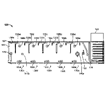
Referring now to FIGS. 4 and 5, the depicted embodiment of the single-use
cartridge 120 includes a main body 124, a right cover 126, a left cover 128,
and five pins
138a, 138b, 138c, 138d, and 138e. The right cover 126 is affixed to right side
of the main
body 124, and the left cover 128 is affixed to the left side of the main body
124. As such,
the right and left covers 126 and 128 enclose cavities and flow channels of
the main body
124 to define blood flow paths as described further below. The aforementioned
sample
well 122 is part of the main body 124. However, other constructions of the
single use
cartridge 120 are also envisioned.
In some embodiments, the main body 124, right cover 126, left cover 128, and
the
pins 138a, 138b, 138c, 138d, and 138e are made by injection molding. After
molding, the
right and left covers 126 and 128 can be affixed to the main body 124 using
various
.. techniques including, but not limited to, ultrasonic welding, laser
welding, solvent
bonding, adhesive bonding, UV curable adhesive bonding, and the like. Various
polymeric materials can be used to construct the main body 124, right cover
126, left
cover 128, and pins 138a-e. For example, such polymeric materials can include,
but are
not limited to acrylic, polycarbonate, polyvinyl chloride (PVC), polyethylene,
polypropylene, polymethyl methacry late, polystyrene, acrylonitrile butadiene
styrene
(ABS), polyethylene, polypropylene, and the like, and combinations thereof. In
some
embodiments, the materials are used to construct the main body 124, right
cover 126, left
cover 128, and pins 138a-e comprise an acrylic-based multi-polymer compound.
In some
embodiments, the main body 124, right cover 126, and left cover 128 are
essentially
.. transparent, or at least translucent. Therefore, in FIG. 4, features of the
main body 124
are visible even though the right cover 126 is attached thereto.
In some embodiments, overmolding, such as by insert molding or multi-shot
molding techniques, may be used to construct some aspects of the main body
124, right
cover 126, and/or left cover 128 (i.e., a device component). For example,
elastomeric
.. valve elements (as described further below) may be overmolded in the left
cover 128. To
generate valves by overmolding, a first mask is used to generate a device
component
without valves. The mask is an inverse of the shape of the device component,
the device
component including open spaces for later insertion of valves. A polymer is
poured into
Date Recue/Date Received 2022-10-01
the first mask to form a hard plastic device component. Then a second mask
having the
inverse of the shape of the device component with the valves is provided. The
hardened
plastic device component is placed in the mask, and an elastomeric material is
injected
into the open spaces formed in the device component by the first mask, thereby
forming
elastomeric valves in the device component. In some embodiments, the device
component is the main body 124, right cover 126, and/or left cover 128.
Exemplary
valves 160a-e, 168, and 170 in a left cover 128 formed by overmolding are
shown in FIG.
7. In some embodiments, the valves comprise an elastomeric material,
deformable upon
application of pressure. Deformation of the valves by application of external
pressure
pushes the elastomeric material into the duct, thereby fluidically sealing the
duct to
prevent flow of a sample liquid through the duct.
Further, in some embodiments secondary operations may be performed to the
cartridge 120. For example, one or more needles 123a-b (refer to FIG. 6) for
piercing a
blood collection tube may be installed within the sample well 122 using
secondary
operations.
The single-use cartridge 120 also includes the five pins 138a, 138b, 138c,
138d,
and 138e. The pins 138a-e are individual component parts (e.g., refer to FIG.
10B) that
are retained within openings of the main body 124 (e.g., within testing
chambers 136a-e
(sometimes referred to as "cups") as described further below in connection
with FIGS.
8A-10B). Tabs 129, located on the right and left covers 126 and 128,
mechanically retain
the pins 138a-e in the main body 124. However, the pins 138a-e are free to
move within
the confines of the main body 124 to a limited extent. For example, the pins
139a-e are
free to rotate uninhibitedly within the main body 124 and to translate
vertically by few
millimeters. This configuration of the pins 138a-e in relation to the other
components of
__ the cal Li idge 120 can be created as follows. Prior to affixing the
right and left covers 126
and 128 to the main body 124, the pins 138a-e can be placed within their
respective
locations in the main body 124 as shown in FIG. 5. With the pins 138a-e
positioned in
the main body 124, the right and left covers 126 and 128 can then be affixed
to the main
body 124. With the right and left covers 126 and 128 affixed to the main body
and the
pins 138a-e positioned in the main body 124, the pins are secured in place
vertically by
the tabs 129 over the top of the pin 138a-e such that they cannot fall out or
be removed
from the cup 136a-e without removal of the right and left covers 126 and 128
from the
main body 124. The tabs 129 allow free rotational movement of the pin 138a-e,
as well
16
Date Recue/Date Received 2022-10-01
as sufficient vertical motion to allow the pin 138a-e to interact with a fluid
sample to
perform a measurement of viscoelastic characteristics of a fluid sample in the
cup 136a-e,
e.g., rotational thromboelastometry. In addition, the tabs 129 provide an
opening for a
shaft 310b to couple with a pin 138b, as shown in FIG. 10C. In one example,
the right and
left covers 126 and 128 are affixed to the main body 124 and thereafter the
pins 138a-e
are pushed into the main body 122 past the tabs 129. The tabs 129 of the right
and left
covers 126 and 128 will block the pins 138a-e from falling out of the main
body 122,
even if the cal __ tlidge 120 is turned upside down. In some embodiments, the
pin and tabs
are positioned to prevent escape of semi-coagulated fluid sample in the
testing chamber
___________________________________ from escaping the testing chamber, even if
the cal tlidge 120 is turned upside down.
In some embodiments, the main body 124 includes a barcode location 125. The
barcode location 125 can be used as a location at which to adhere a barcode
label, or to
print a barcode. The barcode location 125 is on the leading end of the
cartridge 120 (in
relation to the direction of insertion of the cartridge 120 into the analyzer
console 140 as
shown in FIGS. 1-3).
In the depicted embodiment, the right cover 126 includes blood detection
locations 127a and 127b. As will be described further below, the blood
detection
locations 127a and 127b are designated locations on the cartridge 120 at which
sensors of
the analyzer console 140 interface with the cal Li idge 120. The sensors
inspect for the
presence of blood within the cartridge 120 at the blood detection locations
127a and 127b.
In some embodiments, the sensors are optical sensors (e.g., infrared sensors)
and the
blood detection locations 127a and 127b are polished areas that have enhanced
transparency and optical clarity. As such, the right cover 126 is configured
so that the
optical sensors of the analyzer console 140 can readily detect the presence or
absence of
blood at the blood detection locations 127a and 127b.
Referring now to FIGS. 4, 5, and 6, broadly speaking the single-use carnidge
120
is configured to: (i) extract blood from a blood collection tube (e.g., blood
collection tube
10 of FIGS. 1-3) and measure a precise volume of the extracted blood, (ii) mix
a precise
amount of blood with reagents, and (iii) deliver the mixture to multiple cup
and pin
locations of the cartridge 120 where thromboelastometry testing is performed.
These
steps will be described in more detail below.
In the depicted embodiment, the single-use cartridge 120 includes five
individual
blood flow channels 130a, 130b, 130c, 130d, and 130e. Alternately, in some
17
Date Recue/Date Received 2022-10-01
embodiments the cal __ Li idge includes a single individual blood flow
channel, or two
individual blood flow channels, or three individual blood flow channels, or
four
individual blood flow channels, or six individual blood flow channels, or more
than six
individual blood flow channels. Each channel 130a-e includes: (i) a measuring
chamber,
(ii) a mixing chamber containing reagent(s) and a mixing element, and (iii) a
blood
coagulation testing chamber (e.g., in this embodiment a cup having a movable
probe/pin
therein). For example, the channel 130a includes a measuring chamber 132a, a
mixing
chamber 134a, and a testing chamber 136a (refer to the example of the testing
chamber
being depicted in detail in FIGS. 10A-B). Similarly, the channel 130b includes
a
measuring chamber 132b, a mixing chamber 134b, and a testing chamber 136b; the
channel 130c includes a measuring chamber 132c, a mixing chamber 134c, and a
testing
chamber 136a; the channel 130d includes a measuring chamber 132d, a mixing
chamber
134d, and a testing chamber 136d; and the channel 130e includes a measuring
chamber
132e, a mixing chamber 134e, and a testing chamber 136e.
In some embodiments, the sample well 122 includes needles 123a and 123b that
are configured to pierce a septum of a blood collection tube when the blood
collection
tube is inserted into the sample well 122. The needle 123a is in fluid
communication with
the channels 130a-e, while the needle 123b is a vent that facilitates the
ready flow of
blood out of the blood collection tube.
In the depicted embodiment, the fluid flow paths from the needle 123a to the
channels 130a-e are as follows. The needle 123a is confluent with the
measuring
chamber 132a. The measuring chamber 132a is confluent with the measuring
chamber
132b. The measuring chamber 132b is confluent with the measuring chamber 132c.
The
measuring chamber 132c is confluent with the measuring chamber 132d. The
measuring
chamber 132d is confluent with the measuring chamber 132e. Accordingly, blood
can
flow out of the blood collection tube through the needle 123a to the measuring
chamber
132a; from the measuring chamber 132a to the measuring chamber 132b; from the
measuring chamber 132b to the measuring chamber 132c; from the measuring
chamber
132c to the measuring chamber 132d; and from the measuring chamber 132d to the
measuring chamber 132e. The measuring chambers 132a-e may also be referred to
as
metering chambers 132a-e. Each measuring chamber 132a-e has an inlet port and
an
outlet port. The inlet ports are located near the top of the measuring
chambers 132a-e.
For example, measuring chamber inlet port 132ai is located near the top of the
measuring
18
Date Recue/Date Received 2022-10-01
chamber 132a. This configuration can be advantageous if the blood contains
gaseous
bubbles, because such gas may be allowed to escape from the blood as the blood
enters
the measuring chambers 132a-e. In addition, this configuration may
advantageously
minimize fluid flow turbulence as the blood flows into the measuring chambers
132a-e,
thereby reducing the likelihood of damaging the blood cells.
The outlet ports 134ao-eo for transferring blood from the measuring chambers
132a-e to the mixing chambers 134a-e are located at the bottom of the
measuring
chambers. For example, measuring chamber outlet port 132ao is located at the
bottom of
the measuring chamber 132a. In some embodiments, the bottom of the measuring
chamber 132a is angled downward towards the outlet port 132ao. In some
embodiments,
the bottom of the measuring chamber 132a is at an angle of 2 -15 from a plane
parallel
to the bottom or top of the cal Li idge 120. In some embodiments, the
bottom of the
measuring chamber 132a is at an angle of 2 -15 from a plane orthogonal to the
direction
of force applied to move the blood sample through the outlet port 132ao. In
one
embodiment, the angles described above are approximately 2 , 30, 40, 50, 60,
70, 80, 90,
100, 11 , 12 , 13 , 14 , or 15 . In a preferred embodiment, the angles
described above are
5 , although other angles will also be effective. This configuration can help
facilitate the
complete filling of the measuring chambers 132a-e with blood. It can also
minimize
transfer of bubbles into the outlet port 132ao as more blood is transferred to
the outlet
port 132ao before the surface of the volume of blood (which may contain
bubbles)
contained in the measuring chamber 132a contacts the outlet port 132ao. As
such, a
precise volume of blood is contained within the measuring chambers 132a-e.
In some embodiments, the top of the measuring chamber 132a is angled to cause
air to escape the measuring chamber 132a from a transfer port located at the
top of the
measuring chamber opposite to the inlet port 132ai. The transfer port is used
to transfer
air and fluid out of the measuring chamber 132a and into another measuring
chamber
(e.g., 132b) or into an overflow chamber 139. In this embodiment, the top of
the
measuring chamber 132a is angled upward from a low point above an inlet port
132ai to
a higher point above the transfer port. The angle of the top of the measuring
chamber is
between 2 -15 when compared to the a plane parallel to the bottom or top of
the device,
or as compared to a plane orthogonal to the major field of gravitational force
applied to
the blood sample while in the measuring chamber 132a. In one embodiment, the
angle
described above is approximately 20, 30, 40, 50, 60, 70, 80, 90, 100, 110,
120, 130, 140, or
19
Date Recue/Date Received 2022-10-01
15 . In a preferred embodiment, the angle described above is 5 , although
other angles
will also be effective. In a device comprising the angled top of the measuring
chamber
132a, air and bubbles are transferred out of the measuring chamber 132a before
blood,
providing a measured blood sample with decreased amount of air that may impact
the
accuracy of the measurement of the blood, as well as interfere with other
downstream
applications. In some embodiments, both the top and bottom of the measuring
chamber
132a are angled as described above.
From the foregoing description of the fluid flow paths from the needle 123a to
the
measuring chambers 132a-e, and from the foregoing description of the location
of the
measuring chamber outlet ports, it should be understood that the measuring
chambers
132a-e will be filled with blood in a sequential manner. That is, first
measuring chamber
132a will be filled with blood; then blood from measuring chamber 132a will
flow to
measuring chamber 132b; then measuring chamber 132b will be filled with blood;
then
blood from measuring chamber 132b will flow to measuring chamber 132c; then
measuring chamber 132c will be filled with blood; then blood from measuring
chamber
132c will flow to measuring chamber 132d; then measuring chamber 132d will be
filled
with blood; then blood from measuring chamber 132d will flow to measuring
chamber
132e; then measuring chamber 132e will be filled with blood.
After the measuring chamber 132e is filled with blood, then blood from
measuring
chamber 132e will flow to an overflow chamber 139. The blood flowing from
measuring
chamber 132e will enter the overflow chamber 139 at an overflow chamber inlet
port
139i. As will be described further below, the overflow chamber 139 serves to
ensure that
the measuring chamber 132e becomes completely full, while preventing blood
from
exiting the cartridge 120 and flowing into a vacuum source that is used to
draw the blood
into the measuring chambers 132a-e as described above. The vacuum source is
fluidly
connected to the overflow chamber 139 at an overflow chamber outlet port 139o.
When a
negative pressure (with respect to ambient pressure) from the vacuum source is
applied at
the overflow chamber outlet port 139o, blood from a blood collection tube that
is coupled
with needle 123a will flow into the cartridge 120 to fill all the measuring
chambers 132a-
e. Some blood will also exit the measuring chamber 132e and flow towards the
overflow
chamber 139.
As described further below, various valves and vents are interspersed within
the
fluid flow paths so that the blood flow can be controlled by the analyzer
console
Date Recue/Date Received 2022-10-01
according to predefined schemes. In addition, the aforementioned blood
detection
locations 127a and 127b (refer to FIG. 5) are designated locations on the
cartridge 120 at
which sensors of the analyzer console 140 interface with the cartridge 120.
The sensors
inspect for the presence of blood within the cal ______________________ tddge
120 at the blood detection locations
127a and 127b. The blood sensor location 127a is on the fluid flow path
between the
needle 123a and the measuring chamber 132a. When the analyzer console detects
blood
at blood sensor location 127a, the analyzer console 140 determines that blood
has been
drawn into the cal __ U idge 120. The blood sensor location 127b is on the
fluid flow path
between the measuring chamber 132e and the overflow chamber 139. When the
analyzer
console detects blood at blood sensor location 127b, the analyzer console 140
determines
that blood has been drawn into and filled all the measuring chambers 132a-e.
Further,
when the analyzer console 140 detects blood at blood sensor location 127b, the
analyzer
console 140 may cease further application of negative pressure at the overflow
chamber
outlet port 139o. In other words, by detecting blood at blood sensor location
127b, the
analyzer console 140 can determine that the application of vacuum has
successfully filled
all the measuring chambers 132a-e and that the application of vacuum can be
ceased.
Optionally, the cal __ Li idge 120 may be equipped with a blood temperature
sensor at or near
the location of blood sensor location 127b so as to verify the blood sample is
at a
predetermined target temperature.
As described above, each individual channel 130a-e has a measuring chamber
132a-e respectively. In some embodiments, the fluid flow paths within the
individual
channels 130a-e are as follows. From the measuring chambers 132a-e, the blood
can flow
to the respective mixing chambers 134a-e. For example, the blood from
measuring
chamber 132a can flow to the mixing chamber 134a. Similarly, the blood from
measuring
chamber 132b can flow to the mixing chamber 134b; the blood from measuring
chamber
132c can flow to the mixing chamber 134c; the blood from measuring chamber
132d can
flow to the mixing chamber 134d; and the blood from measuring chamber 132e can
flow
to the mixing chamber 134e. From the mixing chambers 132a-e (after completion
of the
mixing), the blood can flow to the respective testing chambers 136a-e (having
a
corresponding probe/pin 138a-e therein, refer below to FIGS. 10A-b). For
example, the
blood from mixing chamber 134a can flow to the testing chamber 136a.
Similarly, the
blood from mixing chamber 134b can flow to the testing chamber 136b; the blood
from
mixing chamber 134c can flow to the testing chamber 136c; the blood from
mixing
21
Date Recue/Date Received 2022-10-01
chamber 134d can flow to the testing chamber 136d; and the blood from mixing
chamber
134e can flow to the testing chamber 136e. Various valves and vents that are
controllable
by the analyzer console 140 are interspersed within the fluid flow paths of
the individual
channels 130a-e. Using such valves and vents, the blood flow within the
individual
channels 130a-e can be controlled by the analyzer console 140 in accordance
with
predefined schemes.
Referring now to FIGS. 6 and 7, additional features of the cartridge 120 will
now
be described. In FIG. 6, a side view of particular chambers of the cal
uidge 120
(measuring chambers 132a-e, reagent mixing chambers 134a-e, and blood
coagulation
testing chambers 136a-e) is provided. In FIG. 7, a left side view of cartridge
120 and
individual channels 130a-e is provided. In this view there is visibility of
testing chamber
inlet ports 136ai, 136bi, 136ci, 136W, and 136ei for testing chambers 136a-e
respectively.
The inlet ports 136ai-ei are located near the top of the testing chambers 136a-
e, for
example, along a side wall of the chamber 136a-e and at a height above the
distal head of
the pin 138a-e that interacts with the blood sample but below the proximal end
of the pin
138a-e (refer to FIG. 10B). This configuration can be advantageous if the
blood contains
gaseous bubbles, because such gas may be allowed to escape from the blood as
the blood
enters the cups 136a-e. In viscous solutions, bubbles may be retained at the
bottom of the
cup 136a-e if the solution enters through the bottom, adversely impacting
thromboelastometric measurements by the pin 138a-e in the cup 136a-e. In
addition, this
configuration may advantageously minimize fluid flow turbulence as the blood
flows into
the testing chambers 136a-e. Fluid flow turbulence and bubble mixing is also
minimized
by having a small diameter or blood flow area of the sample inlet port 136bi
into the cup
136a-e. Bubbles present in blood from the mixing chamber 134a-e separate from
the
fluid and remain at the top surface of the blood in the cup 136a-e by using a
smaller
diameter of a sample inlet port 136bi in combination with the location of the
inlet port
136bi along the side wall of the chamber 136a-e. In some embodiments, the
diameter of
the sample inlet port 136bi is lmm. In some embodiments, the diameter of the
sample
inlet port 136bi is approximately 0.5, 0.6, 0.7, 0.8, 0.9, 1.0, 1.1, 1.2, 1.3,
1.4, or 1.5 mm.
In the depicted embodiment, the cal _________________________ uidge 120
includes two locator pin receptacles
140a and 140b. The locator pin receptacles 140a and 140b are used to mate with
locator
pins of the analyzer console 140 (as described further below). In this manner,
the
cartridge 120 can be accurately positioned in relation to the analyzer console
140.
22
Date Recue/Date Received 2022-10-01
The cal _______ Li idge 120 also includes a vacuum application port 162. When
a source of
vacuum is applied at the vacuum application port 162, and when the vents and
valves of
the cal __ Li idge 120 are in the proper configuration, blood can be drawn
into the measuring
chambers 132a-e as described above, and as described further below.
The cal hidge 120 also includes a pressure application port 164. When a
source of
pressure is applied at the pressure application port 164, and when the vents
and valves of
the cal __ Li idge 120 are in the proper configuration, blood can be forced to
flow from the
measuring chambers 132a-e into the mixing chambers 134a-e, and subsequently
from the
mixing chambers 134a-e into the testing chambers 136a-e as described above,
and as
described further below.
In the depicted embodiment, the cal __ tlidge 120 also includes vents 166a,
166b,
166c, 166d, and 166e. Other cartridge embodiments may include fewer or more
vents.
The vents 166a-e are confluent with the mixing chambers 134a-e respectively.
Accordingly, when the vents 166a-e are open to allow airflow therethrough, air
from the
mixing chambers 134a-e can be readily displaced from the mixing chambers 134a-
e as
blood flows into the mixing chambers 134a-e. Conversely, when the vents 166a-e
are
closed to prevent airflow therethrough, blood is inhibited from flowing into
the mixing
chambers 134a-e because the air within the mixing chambers 134a-e is not
allowed to be
displaced therefrom. The vents 166a-e can be individually opened and closed by
the
analyzer console 140 in accordance with predefined schemes as described
further below.
Accordingly, blood flow into the mixing chambers 134a-e can be controlled as
desired.
In the depicted embodiment, the cal __ tlidge 120 also includes valves 168,
170,
160a, 160b, 160c, 160d, and 160e. Other cal tlidge embodiments may include
fewer or
more valves. The valves 168, 170, and 160a-e are located within fluid flow
paths of the
cartridge 120. Accordingly, the valves 168, 170, and 160a-e can be actuated
(opened or
closed) by the analyzer console 140 to allow or to prevent fluid flow through
the fluid
flow paths in which the valves 168, 170, and 160a-e are respectively located.
For
example, the valve 168 is located in the fluid flow path between the needle
123a and the
measuring chamber 132a. Accordingly, when the valve 168 is open blood can flow
from
the needle 123a to the measuring chamber 132a, and when the valve 168 is
closed blood
cannot flow from the needle 123a to the measuring chamber 132a.
The valve 170 is located in the fluid flow path between the measuring chamber
132e and the overflow chamber 139. Accordingly, when the valve 170 is open
blood can
23
Date Recue/Date Received 2022-10-01
flow from the measuring chamber 132e to the overflow chamber 139, and when the
valve
170 is closed blood cannot flow from the measuring chamber 132e to the
overflow
chamber 139.
The valves 160a-e are located in the fluid flow paths between the mixing
chambers 134a-e and the testing chambers 136a-e respectively. Accordingly,
when the
valves 160a-e are open blood can flow from the mixing chambers 134a-e to the
testing
chambers 136a-e respectively, and when the valves 160a-e are closed blood
cannot flow
from the mixing chambers 134a-e to the testing chambers 136a-e.
As will be described further below, in some embodiments the valves 160a-e can
be individually actuated by pins that are translated towards and away from the
valves
160a-e. To close the valves 160a-e, the pins can engage with and distend
elastomer
members of the valves 160a-e so that the elastomer member makes contact with a
valve
seat of the valves 160a-e. When such pins are retracted away from the
elastomer
members of the valves 160a-e, the elastomer members will rebound such that the
elastomer member is no longer distended and then the valve is opened. The pins
can be
translated by solenoids in some embodiments.
Other mechanisms to regulate fluid flow in the cartridge 120 may also be
present.
For example, stop junctions may be placed between the measuring chamber 132a-e
and
the mixing chamber 134a-e to control the flow of blood from the measuring
chamber
132a-e to the mixing chamber 134a-e. In some embodiments, the stop junctions
are a
barrier that can be opened upon application of a sufficient amount of pressure
to the
barrier. In some embodiments, the stop junction comprises a narrow area for
flow of the
sample fluid such that surface tension of the sample fluid prevents flow
through the stop
junction unless sufficient pressure is applied. Once sufficient pressure is
applied, the flow
of the sample fluid through the stop junction may continue due to capillary
forces.
Referring to FIG. 6 in more detail, some embodiments of the mixing chambers
134a-e contain: (i) one or more dissolvable reagent beads 180, (ii) multiple
retaining
elements 182, and (iii) a mixing element 184. The one or more reagent beads
180 are
disposed within and retained within the confines of the multiple retaining
elements 182.
The mixing elements 184 are disposed in the bottom portions of the mixing
chambers
134a-e, and are free to move horizontally across the bottom portions of the
mixing
chambers 134a-e. The multiple retaining elements 182 separate the reagent
beads 180
from the mixing element 184, and prevent the mixing element 184 from migrating
24
Date Recue/Date Received 2022-10-01
upward away from the bottom portions of the mixing chambers 134a-e. Thus, the
multiple
retaining elements 182 prevent direct contact of the mixing element 184 with
reagent
beads 180 in the mixing chambers 134a-e. Preferably, the retaining elements
182 extend
into each mixing chamber 134a-e so as to maintain a predetermined vertical
position of
each of the reagent beads 180 within the mixing chamber (e.g., a vertical
position below
the height of the blood portion passed into the mixing chamber 134a-e),
thereby ensuring
that each of the beads 180 will be submerged when the predetermined amount of
blood is
directed into the respective mixing chamber 134a-e. In an embodiment, the
height of the
liquid that fills the mixing chamber 134a-e from the measuring chamber 132a-e
(i.e., the
fill level) is above the retaining elements 182 in the mixing chamber. In some
embodiments, the retaining elements 182 are above the height of the fill level
of the
mixing chamber. In these embodiments, the retaining elements are configured to
position
the reagent in the path of the fluid such that the reagent is dissolved by the
liquid upon
entry of the liquid into the mixing chamber. In some embodiments, the flow
path is
defined as the path the liquid travels to go from one chamber to another,
including within
the chamber itself after entering from an inlet or duct.
Also, in some embodiments, the multiple retaining elements 182 in each mixing
chamber 134a-e maintain each of the reagent beads 180 in the respective mixing
chamber
134a-e separate from one another. In such embodiments, each of the reagent
beads 180 is
not contacted by other beads 180 in the respective mixing chamber 134a-e, is
not
contacted by the mixing element 184 in the respective mixing chamber 134a-e,
and is
maintained at a vertical height within the respective mixing chamber 134a-e
below the
height of the blood portion transported into the respective mixing chamber
134a-e.
The retaining elements 182 may take the form of several unique configurations
that result in control over the location of the reagent beads 180. In some
embodiments,
the retaining elements 182 also prevent contact between different reagent
beads 180,
contact of reagent beads 180 with the mixing element 184, and/or contact of
the reagent
beads 180 with other surfaces or components in the mixing chamber 134a-e. In
some
embodiments, the retaining element 182 is configured to limit movement of the
reagent
bead 180 within the mixing chamber 134a-e and configured to allow the sample
liquid or
blood sample to dissolve the reagent bead 180. In some embodiments, the
retaining
element 182 comprises a barrier. The retaining element 182 can also comprise
an inward
protrusion or an outward protrusion in the wall of the mixing chamber 134a-e
or on the
Date Recue/Date Received 2022-10-01
surface of a right cover 126 or left cover 128, or on other surfaces of the
device. In some
embodiments, the retaining element 182 comprises a channel, a post, or a
divot. The
retaining element 182 may comprise an array of posts or an array of divots. In
some
embodiments, the array of posts comprises posts of different diameters to hold
reagent
beads of different diameters. In some embodiments, the retaining element 182
comprises
a compai __ anent or a series of compai ftnents for holding a reagent bead.
The retaining
element 182 can also be configured to both limit the movement of a reagent
bead in the
mixing chamber 134a-e, and to allow blood to flow in a way that it contacts
and dissolves
the reagent bead 180. In some embodiments, the retaining element 182 is
configured to
allow flow of a blood sample through the mixing chamber 134a-e.
The retaining element 182 can further secure the reagent bead 180 below a
predetermined blood sample fill level in the mixing chamber 134a-e. This fill
level is
determined by the volume of blood provided by the measuring chamber 132a-e,
and by
the dimensions of the mixing chamber 134a-e and volume of components or
reagents
within the mixing chamber 134a-e at the time of filling. This fill level can
be
predetermined based on the above factors. Therefore, the retaining elements
182 are
specifically designed to maintain the position of the reagent beads 180 below
this
predetermined fill level.
Additionally, the retaining elements 182 can limit the movement of a mixing
element 184 within the mixing chamber 134a-e. In some embodiments, the resting
element 182 used to restrict movement of a mixing element 184 within the
mixing
chamber 134a-e comprise an array of posts or a compartment that allows a
sample fluid
or blood sample in the mixing chamber 134a-e to contact the mixing element 184
such
that the sample fluid or blood sample is agitated to facilitate dissolving
reagents within
the mixing chamber 134a-e.
In the depicted embodiment, the one or more dissolvable reagent beads 180 are
spherical and are of two different sizes (e.g., about 2 mm diameter and about
3 mm
diameter). However, the use of other shapes and/or sizes of reagent beads 180
is also
envisioned. In some embodiments, the reagent beads 180 are lyophilized
materials, but
other forms of materials are also envisioned. The reagent beads 180 can
comprise
materials such as, but not limited to, CaCl2, ellagic acid/phospholipids,
tissue factor,
heparinase, polybrene, cytochalasin D, tranexamic acid, and the like, and
combinations
thereof. The reagent beads 180 are dissolvable in blood. For example, in this
particular
26
Date Recue/Date Received 2022-10-01
embodiment, each of the five mixing chambers 134a-e is configured to mix a
predetermined volume of blood (as defined by the respective measurement
chamber
132a-e) with a different reagent composition (from the one or more reagent
beads 180
therein) for purposes of performing five different assays. In this example,
the first mixing
chamber 134e may include multiple reagent beads 180 the provide CaCl2 and
ellagic
acid/phospholipids for mixing with the predetermined volume of blood (from the
corresponding measuring chamber 132e) so that the first sample portion can be
used in a
first type of assay. Also in this example, the second mixing chamber 134d may
include
multiple reagent beads 180 the provide CaCl2, ellagic acid/phospholipids, and
heparinase
for mixing with the predetermined volume of blood (from the corresponding
measuring
chamber 132d) so that the second sample portion can be used in a second type
of assay.
Further, in this example, the third mixing chamber 134c may include multiple
reagent
beads 180 the provide CaCl2, tissue factor, and polybrene for mixing with the
predetermined volume of blood (from the corresponding measuring chamber 132c)
so
that the third sample portion can be used in a third type of assay. Also in
this example, the
fourth mixing chamber 134b may include multiple reagent beads 180 the provide
CaCl2,
tissue factor, polybrene, and cytochalasin D for mixing with the predetermined
volume of
blood (from the corresponding measuring chamber 132b) so that the fourth
sample
portion can be used in a fourth type of assay. Lastly, in this example, the
fifth mixing
chamber 134a may include multiple reagent beads 180 the provide CaCl2, tissue
factor,
polybrene, and tranexamic acid for mixing with the predetermined volume of
blood (from
the corresponding measuring chamber 132a) so that the fifth sample portion can
be used
in a fifth type of assay.
In some embodiments, the reagent bead 180 carrying the CaCl2 reagent is
separated from the rest of the beads 180 in the respective mixing chamber 134a-
e so as to
first allow mixing and then activation/clotting of the a citrated blood
sample. Such
separation of the reagent bead 180 carrying the CaCl2 reagent may be achieved
using the
retaining elements 182 (as described above). Alternatively, such separation
can be
achieved by retaining the reagent bead 180 carrying the CaCl2 reagent in a
separate
channel or separate mixing chamber that is separated from other beads 180 in
the
respective chamber 134a-e (such that the blood portion reaches the CaCl2
reagent after
the blood portion mixes with other beads 180 within the respective mixing
chamber 134a-
e). Alternatively, such separation can be achieved by positioning a CaCl2
reagent liquid
27
Date Recue/Date Received 2022-10-01
or a dried-film CaCl2 reagent in a separate channel so that the blood portion
reaches the
CaCl2 reagent after the blood portion mixes with other beads 180 in the
respective mixing
chamber 134a-e. Alternatively, the reagent bead 180 carrying the CaCl2 reagent
can be
coated with an extra layer (and then retained by the retained by the retaining
elements 182
as described above) so that the blood portion begins to dissolve the reagent
bead 180
carrying the CaCl2 reagent after the blood portion previously mixes with other
beads 180
within the respective mixing chamber 134a-e.
Other configurations for providing a reagent to the blood sample may also be
used. In some embodiments, a reagent is coated on the wall of a mixing chamber
134a-e.
In some embodiments, a reagent is coated on the right cover 126 or the left
cover 128.
The coated reagent on the right cover 126 or the left cover 128 can be coated
in a way
that it will at least partially or entirely be contained within the mixing
chamber 134a-e. In
some embodiments, the reagent is coated so that it remains under the fill
level of the
mixing chamber 134a-e (the fill level pertaining to the height of blood in the
mixing
chamber as determined in part by the predetermined volume of blood as measured
in the
measuring chamber). In some embodiments, the coated reagent is a film layer,
i.e., a
reagent film. A reagent film is a layer of reagent coated on or near a
surface. The reagent
film may be liquid or may be dried. A liquid reagent may be retained as a film
layer by a
dissolvable layer of material placed over the liquid reagent. A liquid reagent
layer may
also be applied and then dried on the surface. A pre-dried or solid film
reagent may also
be applied to a surface to form a film layer. In some embodiments, the film
layer is in the
form of a dissolvable film strip. In some embodiments, certain reagents are
preferred to
be delivered in a reagent film as opposed to a reagent bead 180. For example,
certain
reagents that are difficult to lyophilize in a reagent bead 180 may instead be
applied on or
near a surface in the device as a film layer.
In some embodiments, the coated reagent is in the form of reagent beads 180.
Reagent beads may be secured to the wall of a chamber or to a cover using
retaining
elements 182. The retaining elements 182 may comprise a series of compai __
intents, posts,
divots, inward or outward protrusions, or an array of any of the above. Other
shapes or
configurations of reagent that can be coated or secured to the cover, a wall
of a chamber,
or within a fluidic passage between chambers, are also envisioned. In some
embodiments,
both reagent beads 180 and reagent film are coated on one or more surfaces of
the device,
e.g., in the mixing chamber 134a-e.
28
Date Recue/Date Received 2022-10-01
A reagent film may also be provided to dissolve in a blood sample in the
mixing
chamber 134a-e. The reagent film is dissolvable in blood. The reagent film is
adhered to
a surface in the mixing chamber 134a-e. In some embodiments, a reagent film is
deposited on the walls of the mixing chamber 134a-e. In some embodiments, a
reagent
film is deposited on the right cover 126 or the left cover 128 at a region
that at least
partially covers or forms a wall of the mixing chamber 134a-e. The reagent
film may be
used alone, or in addition to one or more reagent beads 180 placed in the
mixing chamber
134a-e. Thus, the use of one or more reagent films in a mixing chamber 134a-e
provides
additional mechanisms of introducing a reagent into a mixing chamber 134a-e to
dissolve
in the blood.
In some embodiments, the reagent film comprises a lyophilized material, but
other
forms of materials are also envisioned. The reagent film can comprise
materials such as,
but not limited to CaCl2, ellagic acid/phospholipids, tissue factor,
heparinase, polybrene,
cytochalasin D, tranexamic acid, and the like, and combinations thereof. In
one particular
example, each of the five mixing chambers 134a-e is configured to mix a
predetermined
volume of blood (as defined by the respective measurement chamber 132a-e) with
a
different reagent composition (from one or more reagent beads 180 and/or one
or more
reagent films therein). In this example, the first mixing chamber 134e may
include
multiple reagent beads 180 and at least one reagent film to provide CaCl2 and
ellagic
acid/phospholipids for mixing with the predetermined volume of blood (from the
corresponding measuring chamber 132e) so that the first sample portion can be
used in a
first type of assay. Also in this example, the second mixing chamber 134d may
include
multiple reagent beads 180 and at least one reagent film to provide CaCl2,
ellagic
acid/phospholipids, and heparinase for mixing with the predetermined volume of
blood
(from the corresponding measuring chamber 132d) so that the second sample
portion can
be used in a second type of assay. Further, in this example, the third mixing
chamber
134c may include multiple reagent beads 180 and at least one reagent film to
provide
CaCl2, tissue factor, and polybrene for mixing with the predetermined volume
of blood
(from the corresponding measuring chamber 132c) so that the third sample
portion can be
used in a third type of assay. Also in this example, the fourth mixing chamber
134b may
include multiple reagent beads 180 and at least one reagent film to provide
CaCl2, tissue
factor, polybrene, and cytochalasin D for mixing with the predetermined volume
of blood
(from the corresponding measuring chamber 132b) so that the fourth sample
portion can
29
Date Recue/Date Received 2022-10-01
be used in a fourth type of assay. Lastly, in this example, the fifth mixing
chamber 134a
may include multiple reagent beads 180 and at least one reagent film to
provide CaCl2,
tissue factor, polybrene, and tranexamic acid for mixing with the
predetermined volume
of blood (from the corresponding measuring chamber 132a) so that the fifth
sample
portion can be used in a fifth type of assay.
Further, a reagent film may be deposited on surfaces upstream or downstream
from the mixing chamber to mix with the blood sample before or after the
mixing
chamber. In some embodiments, a reagent film carrying the CaCl2 reagent is
placed in a
separate channel or separate mixing chamber that is separated from other
reagent beads
180 or reagent film in the respective chamber 134a-e (e.g., such that the
blood portion
reaches the CaCl2 reagent film after the blood portion mixes with other
reagent beads 180
and/or reagent films within the respective mixing chamber 134a-e).
Alternatively, a
CaCl2 reagent film may be deposited in the mixing chamber 134a-e and coated
with an
extra dissolvable film layer so that the blood portion begins to dissolve the
other reagent
film carrying the CaCl2 reagent after the blood portion previously mixes with
other
reagent beads 180 or reagent films within the respective mixing chamber 134a-
3.
In some embodiments, the reagent bead 180 or reagent film is separated from
the
rest of the reagent beads 180 or reagent film in the respective mixing chamber
134a-e so
as to allow mixing with different reagents in a preferred sequence. In one
embodiment,
such separation of the reagent bead 180 may be achieved using the retaining
elements 182
(as described above). Alternatively, such separation can be achieved by
retaining the
reagent bead 180 or reagent film in a separate channel or separate mixing
chamber that is
separated from other beads 180 or reagent films in the respective chamber 134a-
e (such
that the blood portion reaches and mixes with the loaded reagents in a
preferred
sequence). In one embodiment, such separation can be achieved by positioning a
reagent
liquid, reagent bead 180 or a dried-film reagent in a separate channel so that
the blood
portion reaches the reagent before or after the blood portion mixes with other
reagent
beads 180 or reagent films in the respective mixing chamber 134a-e. In some
embodiments, the reagent bead 180 or reagent film is placed along a duct 134ad
fluidically connecting the mixing chamber 134a-e and the testing chamber 136a-
e.
Alternatively, the reagent bead 180 or reagent film can be coated with an
extra layer (and
then retained by the retained by the retaining elements 182 as described
above) so that the
blood portion begins to dissolve the reagent in the reagent bead 180 or
reagent film
Date Recue/Date Received 2022-10-01
comprising an additional dissolvable layer after the blood portion previously
mixes with
other reagent beads 180 or reagent films within the respective mixing chamber
134a-e.In
some embodiments, the coated reagent layer is a dissolvable film layer
manufactured
from a substrate including a polymeric composition and a reagent. The
polymeric
composition forms a dissolvable barrier to maintain the coating of the reagent
on or near a
surface in the device. Upon contact with a blood sample, the polymeric
composition
dissolves to allow the blood sample to mix with the reagent.
The mixing element 184, comprises a ferromagnetic material including, but not
limited to, nickel, cobalt, chromium (IV) oxide, gadolinium, permalloy, and
alnico (an
.. aluminum-nickel-cobalt alloy) and the like, and combinations thereof. In
the depicted
embodiment, the mixing element 184 is spherical and is solid. In other
embodiments, the
mixing element 184 may have a shape such as, but not limited to, cubical,
conical,
cylindrical, fan-shaped, elongated, prismatic, and the like, as well as
irregular shapes. In
some embodiments, the mixing element 184 may include one or more surface
features
such as protrusions, indentations, or holes, and the like.
As will be described further below, the mixing elements 184 are movable within
the mixing chambers 134a-e in response to movement of magnets with which the
mixing
elements 184 magnetically couple. The magnets that the mixing elements 184
magnetically couple with are contained within the analyzer console 140. The
movement
of the mixing elements 184 encourages the reagent beads 180 to dissolve in the
blood
contained within the mixing chambers 134a-e.
Referring now to FIGS. 8A-8H schematically depict an example fluidic control
process 200 that can be used with the thromboelastometry systems provided
herein. The
process 200 begins with blood contained only within the blood collection tube
10, and
ends with blood/reagent mixtures contained in cups 136a-e that are configured
for rotary
thromboelastometry. It should be understood that, in some embodiments, the
cartridge
120 (refer to FIGS. 1-7) that is used to implement the fluidic control process
200 is heated
(e.g., to about 37 C) prior to having any blood therein.
Referring to FIG. 8A, the example fluidic control process 200 includes the
blood
.. collection tube 10, the measuring chambers 132a-e, the mixing chambers 134a-
e, and
cups 136a-e, the overflow chamber 139, the blood detection locations 127a and
127b, the
vacuum application port 162, the pressure application port 164, the vents 166a-
e, the
31
Date Recue/Date Received 2022-10-01
valves 168, 170, and 160a-e. In the depicted configuration, valve 168 is
closed, thereby
retaining the blood substantially within the blood collection tube 10.
While the example fluidic control process 200 includes five blood flow
channels
(each comprising a measuring chamber 132a-e, a mixing chamber 134a-e, and a
cup
136a-e respectively), it should be understood that having five blood flow
channels is not
required in all embodiments. For example, in some embodiments only a single
blood
flow channel is included. Alternately, two blood flow channels are included,
or three
blood flow channels are included, or four blood flow channels are included, or
six blood
flow channels are included, or more than six blood flow channels are
included.Referring
to FIG. 8B, the measuring chambers 132a-e are filled with blood, and a small
amount of
blood is contained within the overflow chamber 139. To arrive at this state,
the following
changes were made (in comparison to FIG. 8A) and/or the following conditions
existed:
(i) the valves 168 and 170 were opened, (ii) the valves 160a-e were closed,
(iii) the vents
166a-e were closed, (iv) a negative pressure was applied to the vacuum
application port
162, and (v) the pressure application port 164 was unpressurized. Accordingly,
the blood
flowed: (i) out of the blood collection tube 10, (ii) through the valve 168,
(iii) through the
blood detection location 127a, (iv) into and filling the measuring chamber
132a, (v) into
and filling the measuring chamber 132b, (vi) into and filling the measuring
chamber
132c, (vii) into and filling the measuring chamber 132d, (viii) into and
filling the
measuring chamber 132e, (ix) through blood detection location 127b, (x)
through valve
170, and (xi) into the overflow chamber 139. When blood was detected in the
blood
detection location 127b, the application of the negative pressure was
discontinued¨
thereby stopping further blood flow.
In some embodiments, the example fluidic control process 200 includes a stop
junction 132as between one, some, or each of the measuring chambers 132a-e and
the
mixing chambers 134a-e. In some embodiments, blood flows through the stop
junction
132as in the duct 132ad connecting the measuring chambers 132a-e and the
mixing
chambers 134a-e through application of a positive pressure to the measuring
chamber or a
negative pressure to the mixing chamber 134a-e. Stop junctions provide a
mechanism to
regulate flow without a connection to an external control device. The
application of
positive or negative pressure may create a pressure differential on either
side of the stop
junction, causing the stop junction to open, or drawing blood through the stop
junction by
overcoming forces due to surface tension. The desired pressure may be applied
to cause
32
Date Recue/Date Received 2022-10-01
blood to flow through the stop junction via pressure application port 164
and/or through
opening an air pressure vent 166a-e to release pressure in the corresponding
mixing
chamber 134a-e.
In some embodiments, the example fluidic control process 200 includes a stop
valve in lieu of or in addition to a stop junction between one, some, or each
of the
measuring chambers 132a-e and the mixing chambers 134a-e. In some embodiments,
the
stop valve is a snap acting valve, snapping open upon reaching a set pressure,
or a
modulating valve that opens in proportion to the pressure differential. Other
cartridge
embodiments may include pressure-controlled valves in other fluid paths.
In some embodiments, the stop valve may be opened and closed by the same
mechanism provided by the valves shown in the reaction system 168, 162, 160a-e
at FIGs
8A-8H. In some embodiments, the stop valves may be opened and closed through a
mechanism other than pressure application to the blood. In some embodiments,
the stop
valve is opened upon remote command from a control device connected to the
stop valve.
In some embodiments, the stop valve can be actuated by the analyzer console
140 to
allow or to prevent fluid flow through the fluid path from the measuring
chamber 132a-e
to the mixing chamber 134a-e.
Referring to FIG. 8C, the measuring chambers 132a-d are still filled with
blood,
but the blood from the measuring chamber 132e has transferred to the mixing
chamber
.. 134e. To arrive at this state, the following changes were made (in
comparison to FIG.
8B) and/or the following conditions existed: (i) the valves 168 and 170 were
closed, (ii)
the valves 160a-e remained closed, (iii) the vents 166a-d remained closed,
(iv) the vent
166e was opened, and (v) a source of air pressure was applied to the pressure
application
port 164. Accordingly, the blood flowed: (i) out of the measuring chamber
132e, and (ii)
.. into the mixing chamber 134e. Because the vents 166a-d and the valves 160a-
d remained
closed, the blood in the measuring chambers 132a-d did not flow into the
mixing
chambers 134a-d. With blood in the mixing chamber 134e, the mixing element in
mixing chamber 134e can move and agitate the blood to facilitate the
dissolving of the
reagent beads therein.
In some embodiments, the fluidic control process 200 shown in FIG. 8C includes
stop junctions (not shown) between the measuring chamber 132a-e and the mixing
chamber 134a-e to prevent the flow of blood from the measuring chamber to the
mixing
chamber unless a sufficient pressure differential between the measuring
chamber 132a-e
33
Date Recue/Date Received 2022-10-01
and the mixing chamber 134a-e is applied. In this embodiment, the stop
junction prevents
leakage of blood from measuring chambers 132a-d into mixing chambers 166a-d
without
opening the vents 166a-d or applying sufficient pressure to the pressure
application port
164 to cause blood to flow through the stop junction. To fill the measuring
chamber 132e
with blood from the mixing chamber 134e, the following changes were made (in
comparison to FIG. 8B) and/or the following conditions existed: (i) the valves
168 and
170 were closed, (ii) the valves 160a-e remain closed, (iii) the vents 166a-d
remain
closed, (iv) the vent 166e was opened, and (v) a source of air pressure was
applied to the
pressure application port 164 to cause blood to flow through the stop junction
from the
measuring chamber 132e into the mixing chamber 134e, while the stop junctions
between
the measuring chambers 132a-d and the mixing chambers 134a-d prevent flow of
blood
from the measuring chambers 132a-d into the mixing chambers 134a-d. With blood
in the
mixing chamber 134e, the mixing element in mixing chamber 134e can move and
agitate
the blood to facilitate the dissolving of the reagent beads therein.
Referring to FIG. 8D, the measuring chambers 132a-d are still filled with
blood,
and the blood/reagent mixture that was in the mixing chamber 134e (refer to
FIG. 8C) has
transferred to the cup 136e. To arrive at this state, the following changes
were made (in
comparison to FIG. 8C) and/or the following conditions existed: (i) the valves
168 and
170 remained closed, (ii) the valve 160e was opened, (iii) the valves 160a-d
remained
closed, (iv) the vent 166e was closed (v) the vents 166a-d remained closed,
and (vi) a
source of air pressure was applied to the pressure application port 164.
Accordingly, the
blood/reagent mixture flowed: (i) out of the mixing chamber 134e, and (ii)
into the cup
136e. Because the vents 166a-d and the valves 160a-d remained closed, the
blood did not
flow from the measuring chambers 132a-d towards the mixing chambers 134a-d.
With
the blood/reagent mixture located in the cup 136e, rotary thromboelastometry
can begin
in the cup 136e.
Referring to FIG. 8E, the measuring chambers 132a-c are still filled with
blood,
the cup 136e is still filled with blood/reagent mixture, and the blood that
was in the
measuring chamber 132d (refer to FIG. 8D) has transferred to the mixing
chamber 134d.
To arrive at this state, the following changes were made (in comparison to
FIG. 8D)
and/or the following conditions existed: (i) the valves 168 and 170 remained
closed, (ii)
the valve 160e was closed, (iii) the valves 160a-d remained closed, (iv) the
vent 166d was
opened (v) the vents 166a-c and 166e remained closed, and (vi) a source of air
pressure
34
Date Recue/Date Received 2022-10-01
was applied to the pressure application port 164. In embodiments comprising a
stop
junction between the measuring chamber 132d and the mixing chamber 134d, the,
blood
travels through the stop junction by application of pressure differential
between the
measuring chamber 132d and the mixing chamber 134d, while the stop junctions
between
the measuring chambers 132a-c and the mixing chambers 134a-c prevent flow.
Accordingly, the blood flowed: (i) out of the measuring chamber 132d, and (ii)
into the
mixing chamber 134d. Because the vents 166a-c and because the valves 160a-c
remained
closed, the blood did not flow from the measuring chambers 132a-c towards the
mixing
chambers 134a-c. With blood in the mixing chamber 134d, the mixing element in
mixing
chamber 134d can agitate the blood to facilitate the dissolving of the reagent
beads
therein.
Referring to FIG. 8F, the measuring chambers 132a-c are still filled with
blood,
the cup 136e is still filled with blood/reagent mixture, and the blood/reagent
mixture that
was in the mixing chamber 134d (refer to FIG. 8E) has transferred to the cup
136d. To
arrive at this state, the following changes were made (in comparison to FIG.
8E) and/or
the following conditions existed: (i) the valves 168 and 170 remained closed,
(ii) the
valve 160d was opened, (iii) the valves 160a-c and 160e remained closed, (iv)
the vent
166d was closed (v) the vents 166a-c and 166e remained closed, and (vi) a
source of air
pressure was applied to the pressure application port 164. Accordingly, the
blood/reagent
mixture flowed: (i) out of the mixing chamber 134d, and (ii) into the cup
136d. Because
the vents 166a-c and the valves 160a-c remained closed, the blood did not flow
from the
measuring chambers 132a-c towards the mixing chambers 134a-c. With the
blood/reagent mixture located in the cup 136d, rotary thromboelastometry can
begin in
cup 136d.
Referring to FIG. 8G, the measuring chambers 132a-b are still filled with
blood,
the cups 136d-e are still filled with blood/reagent mixture, and the blood
that was in the
measuring chamber 132c (refer to FIG. 8F) has transferred to the mixing
chamber 134c.
To arrive at this state, the following changes were made (in comparison to
FIG. 8F)
and/or the following conditions existed: (i) the valves 168 and 170 remained
closed, (ii)
the valve 160d was closed, (iii) the valves 160a-c and 160e remained closed,
(iv) the vent
166c was opened (iv) the vents 166a-b and 166d-e remained closed, and (v) a
source of
air pressure was applied to the pressure application port 164. In embodiments
comprising
a stop junction between the measuring chamber 132c and the mixing chamber
134c, the,
Date Recue/Date Received 2022-10-01
blood travels through the stop junction by application of pressure
differential between the
measuring chamber 132c and the mixing chamber 134c, while the stop junctions
between
the measuring chambers 132a-b and the mixing chambers 134a-b prevent flow.
Accordingly, the blood flowed: (i) out of the measuring chamber 132c, and (ii)
into the
mixing chamber 134c. Because the vents 166a-b and because the valves 160a-b
remained
closed, the blood did not flow from the measuring chambers 132a-b towards the
mixing
chambers 134a-b. With blood in the mixing chamber 134c, the mixing element in
mixing
chamber 134c can agitate the blood to facilitate the dissolving of the reagent
beads
therein.
Referring to FIG. 8H, the completion of the process 200 is depicted. That is,
the
cups 136a-c all contain blood/reagent mixtures and rotary thromboelastometry
can be
taking place in the cups 136a-e. This state can be attained in accordance with
the method
of actuating the valves 168, 170, and 160a-e, and the vents 166a-e, in
conjunction with
applying vacuum to the vacuum application port 162 or pressure to the pressure
application port 164 as described above.
Referring to FIG. 9, in some alternative embodiments, one or more of the
individual blood flow channels or paths can include multiple mixing chambers
that are
arranged in series. For example, the example fluidic control process 280
includes five
blood flow channels (similar to the number of channels in the embodiment of
FIGS. 8A-
H), but each of the channels include two mixing chambers that are arranged in
series
(rather than a single mixing chamber for each respective mixing chamber like
the
embodiment of FIGS. 8A-H). That is, mixing chambers 137a and 137f are arranged
in
series between the measurement chamber 132a and the cup 136a; mixing chambers
137b
and 137g are arranged in series between the measurement chamber 132b and the
cup
136b; mixing chambers 137c and 137h are arranged in series between the
measurement
chamber 132c and the cup 136c; mixing chambers 137d and 137i are arranged in
series
between the measurement chamber 132d and the cup 136d; and mixing chambers
137e
and 137j are arranged in series between the measurement chamber 132e and the
cup 136e.
In some embodiments, the reagent bead carrying the CaCl2 reagent is separated
from the other the reagent beads by locating the CaCl2 reagent in the second
of the two
mixing chambers that are arranged in series. In that manner, the serial mixing
chambers
can allow the blood sample to be mixed with reagents and subsequently, at a
controlled
point in time, activation/clotting of the blood sample can be initiated.
36
Date Recue/Date Received 2022-10-01
While the example fluidic control process 280 includes five blood flow
channels
that each include two mixing chambers that are arranged in series, it should
be understood
that such a configuration is not required in all embodiments. For example, in
some
embodiments only a single blood flow channel that includes two mixing chambers
that
are arranged in series is included in a cal Li idge. Such a single blood
flow channel with
two mixing chambers may be the only blood flow channel in the cal tlidge,
or may be
combined in a cal __ hidge with one or more other blood flow channels that
include a single
mixing chamber. It should be understood that all combinations and permutations
of
number of blood flow channels and mixing chambers are included within the
scope of this
disclosure.
Turning now to the blood coagulation testing chambers 136a-e in more detail,
the
chambers 136a-e can be configured to provide viscoelastic testing on the blood
sample
portion drawn into each chamber. Referring to FIGS. 10A and 10B, the pins 138a-
e are
located in the cartridge 120. A representative example showing the pin 138b
located in
the cup 136b illustrates that a clearance space exists between the outer
diameter of the pin
138b and the inner diameter of the cup 136b. A blood/reagent mixture will at
least
partially fill the clearance space when rotary thromboelastometry is being
performed
therein. The pin 138b has a shoulder 138bs. The clearance space between the
outer
diameter of the pin 138b and the inner diameter of the cup 136b is less in the
areas below
the shoulder 138bs than in the areas above the shoulder 138bs. The areas
between the
outer diameter of the pin 138b and the inner diameter of the cup 136b that are
below the
shoulder 138bs are the areas that are active in regard to performing rotary
thromboelastometry.
The cup 136b and pin 138b are shown in cross-section in FIG. 10B (in
accordance
with section 10B--10B of FIG. 10A). In addition, a sample inlet port 136bi
(located
behind pin 138b in the orientation of FIG. 10B) is provided so that the
blood/reagent
mixture will flow into the cup 136b via the sample inlet port 136bi. In the
depicted
embodiment, the cup inlet port 136bi is located in a sidewall of cup 136b at a
height
above the widened distal portion (refer to shoulder 138bs) of the pin 138b but
below the
proximal end of the pin 138b (refer to end near the entry to the axial bore
138bb of the
pin 138b). In this configuration, the blood/reagent mixture will flow into the
cup 136b so
as to reduce the potential for bubble formation. In addition, locating the cup
inlet port
136bi near the top of cup 136b eliminates the effects that the cup inlet port
136bi may
37
Date Recue/Date Received 2022-10-01
otherwise have on the thromboelastometry measurements performed in the cup
136b if
the cup inlet port 136bi is located in the active space between the inner
diameter of the
cup 136b and the outer diameter of the pin 138b below the shoulder 138bs.
In certain devices, bridging or other structure formation between the cup
inlet port
136bi (in the interior diameter of the cup 136b) and the outer diameter of the
pin 138b
(i.e., the probe element) may occur. This may affect the ability of blood to
flow into the
cup 136a-e, or may cause error in the thromboelastometry measurements taken in
the cup
136a-e. In some embodiments, the opening of the inlet port 136bi and the outer
diameter
of the pin 138b are at least a minimum clearance distance apart that prevents
stable
bridging of the blood sample or other coagulation structure formation between
the pin
138b and the cup inlet port 136bi. At the minimum clearance distance, bridging
between
the sample inlet port and the pin may still occur upon filling the testing
chamber,
however, the bridge will not be stable enough to persist during measurement.
Typically, a
bridge will form around a bubble, which will be instable if the diameter is
equal to or
greater than the minimum clearance distance. In some embodiments, a stable
bridge is
one that lasts longer than 1 second, 2 seconds, 3 seconds, 4 seconds, or 5
seconds. In
some embodiments, the minimum clearance distance is at least 1.5 mm. In some
embodiments, the minimum clearance distance is at least 1.5 mm, 2 mm, 2.5 mm,
or 3
mm. In the depicted embodiment in FIG. 10B, the cup inlet port 136bi is
located in a
sidewall of cup 136b at a height above the widened distal portion (refer to
shoulder
138bs) of the pin 138b but below the proximal end of the pin 138b (refer to
end near the
entry to the axial bore 138bb of the pin 138b), and the inlet port 136bi is at
least 1.5mm
from the pin 138b. In other words, the geometry of the pin can allow for this
additional
clearance since the pin has a narrower portion at the location at which the
bridging might
occur, thereby allowing for a greater clearance between the pin and the cup to
prevent
stable bridging.
In the depicted embodiment, the top of the cartridge 124 includes a vent 121.
The
vent 121 is in fluid communication with the needle 123b. Therefore, when air
for venting
a blood sample tube located in sample well 122 is needed, air is drawn through
the vent
121 and channeled into the blood sample tube via the needle 123b.
Each of the pins 138a-e includes an axial bore. For example, the pin 138b
includes an axial bore 138bb. The axial bore 138bb can be used to engage with
a shaft
(not shown in FIG. 10B) for performing rotary thromboelastometry.
38
Date Recue/Date Received 2022-10-01
Referring to FIG. 10C, an example rotary thromboelastometry assembly 300b can
engage with the pin 138b to perform rotary thromboelastometry on a blood
sample
contained in the cup 136b. In this particular embodiment, the example rotary
thromboelastometry assembly 300b includes a baseplate 302, a shaft 310b, a
bearing
312b, a mirror 314b, a counterforce spring 320b, a light source 330b, and a
detector 340b
(e.g., a charge-coupled device or the like). The baseplate 302 can be lowered,
as
represented by arrows 318b, such that a tip portion of the shaft 310b enters
the bore
138bb to become releasably coupled with the pin 138b. The bearing 312b is
engaged
with the baseplate 302 and the shaft 310b to facilitate rotational movement of
the shaft
310b in relation to the baseplate 302. The counterforce spring 320b is coupled
to the
shaft 310b and oscillation of the spring 320b can induce the shaft 310b to
oscillate back
and forth by about +1- 50 as represented by arrow 316b. The mirror 315 is
coupled to the
shaft 310b. The light source 330b is configured to project light towards the
mirror 314b,
and light can be reflected from the mirror 315 towards the detector 340b
(depending on
the rotational orientation of the shaft 310b). Accordingly, the motion of the
pin 138b is
detected by an optical detection system. It should be understood that other
configurations
of the rotary thromboelastometry assembly 300b are also envisioned within the
scope of
this disclosure.
The detected motion data is analyzed by an algorithm running on the analyzer
console 140 (refer to FIGS. 1-3) to process and determine the
thromboelastometry results.
This system facilitates various thromboelastometry parameters such as, but not
limited to,
clotting time, clot formation time, alpha angle, amplitude, maximum clot
firmness, lysis
onset time, lysis time, lysis index (%), and maximum lysis (%).
As the blood in the cup 136b begins to coagulate, the motion amplitude of the
shaft 310b starts to decrease (as detected by the deflection of the light beam
from mirror
315 towards the detector 340b). During coagulation, the blood's fibrin
backbone
(together with platelets) creates a mechanical elastic linkage between the
surfaces of the
cup 136b and the pin 138b. A proceeding coagulation process induced by adding
one or
more of the aforementioned activating factors can thus be observed and
quantified. In
this way, various deficiencies of a patient's hemostatic status can be
revealed and can be
interpreted for proper medical intervention. At the end of the test process,
the baseplate
302 can rise to uncouple the shaft 310b from the pin 138b.
39
Date Recue/Date Received 2022-10-01
Referring to FIG. 11, the main chassis 144 of the analyzer console 140 can
include
a front portion 144f and a rear portion 144b. In some embodiments, the rear
portion 144b
houses at least some of the computer and electronic components that are
necessary for the
operations of the analyzer console 140. For example, the rear portion 144b can
house
hardware devices and software such as, but not limited to, computer
processors, memory
devices, an operating system and other executable instructions, power
source(s), user
interface controls, communication devices, circuit boards, and the like.
In the depicted embodiment, the front portion 144f includes a cover 145 and a
sample handler assembly 400. The sample handler assembly 400 defines an
interior
space in which the cartridge 120 can be received. In some embodiments, the
sample
handler assembly 400 is a modular sub-assembly of the analyzer console 140,
and the
sample handler assembly 400 can be readily removed from the analyzer console
140 for
service. The sample handler assembly 400 is electrically interconnected with
the
computer and electronic components that are housed in the rear portion 144b.
As such,
the analyzer console 140 can perform rotary thromboelastometry on a blood
sample
located in cartridge 120 and display the results on the touchscreen display
142.
Referring now to FIGS. 11 and 12, the analyzer console 140 can include a
cartridge receiver and clamp 410 and a viscoelastic measurement system 480. A
mechanical frame assembly is used to support the cal _________________ ti idge
receiver and clamp 410 and
the viscoelastic measurement system 480 in orientations such that the
cartridge receiver
and clamp 410 and the viscoelastic measurement system 480 can function
symbiotically.
Portions of the cal ____________________________________________________
tiidge receiver and clamp 410 and the viscoelastic measurement
system 480 are moveable in relation to the mechanical frame assembly (which is
stationary in relation to the analyzer console 140). For example, the
viscoelastic
measurement system 480 can move upward and downward. As will be described
further
below, the viscoelastic measurement system 480 can move downward to engage
with the
cartridge 120 (e.g., refer to FIG. 11), and upward to disengage from the cal
ti idge 120. A
portion of the cal ___________________________________________________ tiidge
receiver and clamp 410 can move horizontally in relation to the
mechanical frame assembly. As will be described further below, a portion of
the cal Li idge
receiver and clamp 410 can move horizontally to clamp or unclamp the cartridge
120
within the sample handler assembly 400.
In some embodiments, the cal _________________________________________ tiidge
receiver and clamp 410 includes a movable
block sub-assembly and a stationary block sub-assembly. A space exists between
the
Date Recue/Date Received 2022-10-01
movable block sub-assembly and the stationary block sub-assembly in which the
cartridge 120 can be received. The movable block sub-assembly can be
translated
towards or away from the stationary block sub-assembly. Accordingly, the
cartridge 120
can be clamped and unclamped between the movable block sub-assembly and the
stationary block sub-assembly by virtue of the relative movement therebetween.
In some
embodiments, the viscoelastic measurement system 480 is mounted to the movable
block
sub-assembly. Therefore, as the movable block sub-assembly is translated, the
viscoelastic measurement system 480 is also translated.
In some embodiments, the moveable block sub-assembly can be translated by an
electric motor. In particular embodiments, the motor is a stepper motor. In
some
embodiments, a gear reducer is coupled to the motor. Using a belt and pulley
arrangement for compactness, the motor can be used to drive a lead screw. The
threads of
the lead screw can be engaged with complementary threads of the movable block
such
that a rotation of the lead screw results in horizontal translation of the
movable block. In
some embodiments, end-of-travel detectors (e.g., proximity sensors, optical
sensors,
micro-switches, and the like) are included to detect when the moveable block
sub-
assembly has been horizontally translated to the desired end-of-travel
positions.
In some embodiments, one or more springs can extend between the movable
moveable block sub-assembly and the stationary block sub-assembly. The springs
can
help facilitate a suitable clamping force between the movable block sub-
assembly and the
stationary block sub-assembly. In some embodiments, the springs are
adjustable.
In some embodiments, portions of the moveable block sub-assembly and the
stationary block sub-assembly that make contact with the cat itidge 120
comprise a
flexible or compressible material so that while the cartridge 120 is clamped
it is also
protected from damage.
In particular embodiments, the moveable block sub-assembly can include one or
more features on the clamping face of the moveable block sub-assembly that
serve to
position the cat ______________________________________________________ it
idge 120 in the desired location within the sample handler assembly 400.
For example, in some embodiments the moveable block sub-assembly includes two
________________________________________________________________ locator pins
that can mate with the locator pin receptacles 140a and 140b of the cat
uidge
120 (refer to FIG. 7) to accurately position the cartridge 120 in relation to
the sample
handler assembly 400.
41
Date Recue/Date Received 2022-10-01
In some embodiments, one or both of the moveable block sub-assembly and the
stationary block sub-assembly include heating devices 412 that can warm the
cartridge
120 when the cartridge 120 is clamped therebetween. For example, in some
embodiments the heaters 412 are electrical resistance heaters that are used to
heat at least
portions of the cal Li idge 120. In some embodiments, the heaters 412 are
configured to
facilitate warming of individual portions of the cartridge 120 independently
from other
portions of the cal ________________________________________________ Li idge
120. For example, one or more of the individual blood flow
channels 130a, 130b, 130c, 130d, and 130e (refer to FIGS. 4-7) can be
independently
warmed in some such embodiments. Warming may be performed to one or more sides
of
_____________________________________________________________ the cal Li
idge 120. Other types of warming modalities may be used including, but not
limited to, IR, ultrasonic, microwave, and the like.
In particular embodiments, one or more temperature sensors 414 are included
that
can detect the temperature of the cartridge 120 at one or more locations on
the cartridge
120. For example, in some embodiments the one or more temperature sensors 414
can be
thermocouples, thermistors, infra-red temperature sensors, and the like.
Accordingly, the
analyzer console 140 can control the heating of the cal ____________ Li idge
120 to a predetermined
temperature (e.g., about 37 C) using the heaters 412 and the temperature
sensors 414.
The moveable block sub-assembly can include multiple solenoids that are used
to
actuate the aforementioned vents and valves of the cartridge 120. For example
(referring
also to FIG. 7), the valves 168, 170, and 160 a-e, can be actuated by valve
actuators 430
and the vents 166a-e can be actuated by vent actuators 432. In some
embodiments, the
valve actuators 430 and the vent actuators 432 comprise solenoids. Actuation
of the
valves 168, 170, and 160 a-e by the valve actuators 430 can be accomplished by
coupling
pins to the valve actuators 430 that are extendable from the moveable block
sub-assembly
to make contact with and to distend valve elastomer members so that the
elastomer
members make contact with a valve seat within the cartridge 120. Actuation of
the vents
166a-e by the vent actuators 432 can be accomplished by coupling pins with
resilient tips
that are extendable from the moveable block sub-assembly to obstruct the vents
166 a-e.
Such pins with resilient tips can act as stoppers to substantially prevent
airflow through
.. the vents 166a-e. In some embodiments, the valve actuators 430 and the vent
actuators
432 comprise solenoids that include internal springs that cause the valve
actuators 430
and the vent actuators 432 to be normally extended (e.g., when the electrical
power is
42
Date Recue/Date Received 2022-10-01
removed from the solenoids). Accordingly, such normally closed solenoids will
close the
vents and valves of the cal nidge 120 as a default configuration.
The sample handler assembly 400 also includes pressure source 436 and vacuum
source 434 by which air pressure and vacuum can be applied to the pressure
application
port 164 and the vacuum application port 162 of cal __________ tfidge 120
respectively (refer to FIG.
7). For example, the pressure source 436 and vacuum source 434 can make
contact with
the cal _______________________________________________________________ u idge
120 and can convey pressure or vacuum to the pressure application port 164
and the vacuum application port 162 when the cartridge 120 is clamped within
the
cartridge receiver and clamp 410. The pressure source 436 and vacuum source
434 are at
least partially made of a resilient material in some embodiments. For example,
in some
embodiments the pressure source 436 and vacuum source 434 are at least
partially made
of a resilient material such as, but not limited to, silicone, butyl rubber,
nitrile rubber,
ethylene propylene rubber, fluoroelastomers, and the like. One or more
internally-housed
pressure and/or vacuum pumps (not shown) can also be included in the analyzer
console
140. Such internally-housed pressure and vacuum pumps can be used to generate
the air
pressure or vacuum that is applied to the cartridge 120 to induce the
transport of blood
within the cartridge 120 as described above in reference to FIGS. 8A-8H.
As previously described, the cartridge receiver and clamp 410 also includes
the
stationary block sub-assembly. In some embodiments, the stationary block sub-
assembly
does not move in relation to the mechanical frame assembly and in relation to
the
analyzer console 140 as a whole.
In some embodiments, the analyzer console 140inc1udes a mixing unit 440. In
particular embodiments, the mixing unit 440 includes a motor, a crank and
connecting rod
assembly, and a magnet shuttle. These components can be used to magnetically
couple
________________________ with the mixing elements of the cal tfidge 120 and
to induce movement of the mixing
elements within the mixing chambers 134a-e. The movement of the mixing
elements
encourages the reagent beads to dissolve in the blood contained within the
mixing
chambers 134a-e as described above.
The analyzer console 140 can also include one or more sensors 448. The one or
more sensors 448 can be used to detect the presence of blood in particular
locations
within the cartridge 120, such as blood detection locations 127a and 127b as
described
above (refer to FIG. 5). In some embodiments, the sensors 448 are optical
sensors, such
as IR (infrared) sensors. In some embodiments, the sensors 448 can be used to
detect
43
Date Recue/Date Received 2022-10-01
blood in other areas of the call" idge 120, such as, but not limited to, in
the cups 136a-e
(refer to FIGS. 8A-8H).
The sample handler assembly 400 of the analyzer console 140 also includes the
viscoelastic measurement system 480. The viscoelastic measurement system 480
includes the baseplate 302 (e.g., refer to FIG. 10C), one or more
thromboelastometry
assemblies (e.g., thromboelastometry assembly 300b), and a linear actuator
assembly.
The one or more thromboelastometry assemblies can each be affixed to the
baseplate 302.
In some embodiments, the linear actuator assembly can be coupled to the
baseplate 302
and to the cartridge receiver and clamp 410. Accordingly, actuation of the
linear actuator
___________________________________________________________ assembly can
translate the baseplate 302 and the cal tlidge receiver and clamp 410
towards each other or away from each other. A linear bearing assembly of the
linear
actuator can guide the baseplate 302 in a linear path, and stabilize the
baseplate 302, as
the baseplate 302 translates towards or away from the cal ____________ hidge
receiver and clamp 410.
In some embodiments, the linear actuator assembly causes the baseplate 302 to
vertically raise or lower in relation to the cartridge receiver and clamp 4
lOusing a motor
(e.g., a DC motor or a stepper motor) that rotates a lead screw that has
threads that are
engaged with a drive nut. The drive nut is coupled to the baseplate 302. In
some
embodiments, end-of-travel detectors (e.g., proximity sensors, optical
sensors, micro-
switches, and the like) are included to detect when the baseplate 302 has been
vertically
translated to the desired end-of-travel positions.
The viscoelastic measurement system 480 includes one of more rotary
thromboelastometry assemblies (e.g., rotary thromboelastometry assembly 300b
of FIG.
10C) that include a shaft configured to couple with a pin (e.g., the shaft
310b configured
to couple with the pin 138b). Because the thromboelastometry assemblies are
mounted to
the baseplate 302, the shafts are raised or lowered in conjunction with the
raising or
lowering of the baseplate 302. Accordingly, actuation of the linear actuator
assembly
causes the shafts to vertically raise or lower in relation to the cartridge
receiver and clamp
410, and in relation to a cartridge 120 when a cal _______________ Li idge 120
is clamped within the
cartridge receiver and clamp 410. Therefore, from the description herein it
can be
understood that actuation of the linear actuator assembly can engage and
disengage the
shafts from the pins of the call" idge 120 (e.g., refer to FIG. 10C that
shows baseplate 302
being lowered to engage shaft 310b with pin 138b).
44
Date Recue/Date Received 2022-10-01
In addition to the aforementioned features of the analyzer console 140, in
some
embodiments the analyzer console 140 also includes one or more of the
following
features. The analyzer console 140 can include one or more barcode scanners
450 that,
for example, can read a barcode at the barcode location 125 on the leading end
of
cartridge 120 (refer to FIG. 5). In some embodiments, the analyzer console 140
can
include one or more devices to detect the presence of the cal nidge 120 in
a desired
insertion location and/or orientation. For example, in some embodiments one or
more
micro switches can be used to detect when the cartridge 120 has been inserted
in a desired
location and orientation within the sample handler assembly 400. In some
embodiments,
the analyzer console 140 can include one or more auxiliary connections 460.
The
auxiliary connections 460 can include network and device connectors such as,
but not
limited to, one or more USB ports, Ethernet ports (e.g., RJ45), VGA
connectors, Sub-D9
connectors (RS232), and the like. Such auxiliary connections 460 can be
located on the
rear of the main chassis 144, or at other convenient locations on the main
chassis 144.
For example, in some embodiments one or more USB ports may be located on or
near the
front of the main chassis 144.
The analyzer console 140 also includes a user interface 142 (e.g., with a
touchscreen display in this embodiment). In the depicted embodiment, the user
interface
142 is configured to receive user input and to display output information to
the user. For
example, the user can enter information to the analyzer console 140 by making
selections
of various soft-buttons that may be displayed on the user interface 142 at
times during the
beginning, middle, and end of the testing process. In some embodiments, other
selections
such as, but not limited to, soft keyboard entries can be provided via user
interface 142.
In some embodiments, data entry can be performed additionally or alternatively
by voice
entry. In some embodiments, the user interface may include other peripheral
devices
(e.g., a mouse, a keyboard, an additional display device, and the like) as
part of the
analyzer console 140. In some embodiments, a computer data network (e.g.,
intranet,
internet, LAN, etc.) may be used to allow for remote devices to receive and/or
input
information from the system 100. For example, in some embodiments one or more
remote displays can be utilized via auxiliary connections 460. In the depicted
embodiment, the user interface 142 also includes an external barcode reader
146 (refer to
FIG. 1A). Alternatively or additionally, the user interface 142 of the
analyzer console 140
can be equipped with a reader configured to read near-field communication
tags, RFID
Date Recue/Date Received 2022-10-01
tags, or the like. The analyzer console 140 can also include one or more
control systems
470 that can execute instructions embodied in a computer program. The control
systems
470 can include, by way of example, both general and special purpose
microprocessors,
and any one or more processors of any kind of digital computer. In some
embodiments,
the control systems 470 includes one or more such processors, memory, storage
devices,
interfaces, and other types of electronic sub-systems and components. Such
components
may be mounted on a common motherboard or in other manners as appropriate. The
control systems 470 can process instructions for execution within the analyzer
console
140, including instructions stored in the memory or on the storage device. In
some
implementations, multiple processors and/or multiple buses may be used, as
appropriate,
along with multiple memories and types of memory. Also, multiple computing
devices
may be connected, with each device providing portions of the necessary
operations (e.g.,
as a server bank, a group of blade servers, or a multi-processor system).
The storage devices are capable of providing mass storage for the control
systems
470. In some implementations, the storage device may be or contain a computer-
readable
medium, such as a floppy disk device, a hard disk device, an optical disk
device, or a tape
device, a flash memory or other similar solid state memory device, or an array
of devices,
including devices in a storage area network or other configurations. A
computer program
product can be tangibly embodied in an information carrier. The computer
program
product may also contain instructions that, when executed, perform one or more
methods,
such as those described above in reference to FIGS. 8A-8H. The computer
program
product can also be tangibly embodied in a computer- or machine-readable
medium, such
as the memory, the storage device, or memory on the processor(s).
Referring to FIG. 13, in some implementations a user can interact with the
thromboelastometry systems provided herein according to an example process
490. In
step 492, the user can insert a cartridge into an analyzer console. In some
examples, at
least a portion of the call" __________________________________________ idge
remains exposed while other portions of the cal lt idge are
concealed within the analyzer console. For example, this step is exemplified
above in
reference to FIG. 1A. In step 494, the user can couple a blood sample
container to the
cartridge after a prompt is received from the analyzer console. Step 494 can
be
performed while the cal _______________________________________________ Li
idge remains inserted in the analyzer console as defined by step
492. At step 496, the user can press a "start" button (or equivalent) to
initiate an
automated transport of blood in the blood sample reservoir to the blood
testing chambers
46
Date Recue/Date Received 2022-10-01
of the cal _____________________________________________________________ Li
idge such that the viscoelastic characteristics of the blood can be measured.
In
some examples, the analyzer console provides an indication that the testing is
ready to be
initiated, but that indication is not required as part of process 490.
Referring to FIGS. 14A and 14B, in some implementations a thromboelastometry
system can perform thromboelastometry according to an example process 500. The
individual steps of the process 500 may not necessarily be performed in the
order listed.
Further, in some implementations some steps of the process 500 may be
performed in
parallel. The process 500 may be performed by the thromboelastometry systems
described above, such as thromboelastometry system 100.
In step 510, the presence of a cal tlidge is detected in a receptacle of an
analyzer
console of the thromboelastometry system. For example, the detection may be
performed
by a micro switch, optical sensor, barcode scanner, and the like, or a
combination thereof.
Even though the cal __ tlidge is detected in the receptacle, at least a
portion of the cartridge
may be exterior to the analyzer console.
In step 520, the analyzer console actuates a clamping mechanism to clamp the
cartridge at least partially in the analyzer console. For example, the
cartridge receiver and
clamp 410 as described above can be activated to clamp the cartridge.
In step 530, the analyzer console can optionally determine if the cal Li
idge has
characteristics that indicate the cal tlidge has been used previously. For
example, the
analyzer console may use optical sensors to inspect for the presence of blood
in the
cartridge. In some embodiments, if one or more characteristics that indicate
the cal Li idge
has been used previously are detected, the analyzer console may suspend
further steps of
process 500 and provide a pertinent message via the user interface.
In step 540, the analyzer console can perform one or more QC tests to test the
____________ integrity of the call" ____________________________ idge. For
example, in some embodiments the cal tlidge can be tested
for leaks such as by performing a pressure/vacuum decay test.
In step 550, the analyzer console scans the cal _______________________ tlidge
for a barcode. For example,
the analyzer console may scan a leading end of the cal _________________
tlidge at which a 1D or 2D barcode
may be present.
In step 560, the analyzer console determined the types of thromboelastometry
assays to be performed based on the information attained from the scan of the
barcode in
step 550.
47
Date Recue/Date Received 2022-10-01
In step 570, the shafts of the thromboelastometry sub-system of the analyzer
console are coupled with pins of the cartridge. The pins are located in cups
of the
cartridge. Accordingly, the coupling of the shafts of the thromboelastometry
sub-system
to the pins can configure the thromboelastometry system to be capable of
performing
thromboelastometry on a blood sample contained within the cups of the
cartridge. For
example, referring to FIG. 10C, the shaft 310b of the thromboelastometry
assembly 300b
can be lowered towards the cartridge so that the shafts 310b become friction-
fit and
releasably coupled with the pins 138b of the cartridge 120.
In step 580, the analyzer console can begin rotatory reciprocation of the pins
in
___________________ relation to the cups of the cat hidge. For example,
this step is exemplified above in
reference to FIG. 10C.
In step 590, the analyzer console can heat the cartridge. In some
implementations,
the analyzer console may heat the cat ttidge to a predetermined
temperature. In particular
implementations, the analyzer console may maintain the cartridge at the
predetermined
temperature. For example, in some implementations the predetermined
temperature may
be about 35 C to about 40 C, and preferably about 37 C.
In step 600, the analyzer console provides a prompt to couple a blood sample
container to the cartridge. This prompt may be provided, for example upon the
successful
completion of one or more steps, or upon the successful verification of one or
more
.. conditions, or both. For example, this prompt may be provided upon the
cartridge's
successful attainment of the predetermined temperature as per step 590, among
other
things. The prompt may be provided via the user interface of the analyzer
console. For
example, the prompt may be a visual message displayed on a touchscreen monitor
of the
analyzer console. An audible prompt may be provided in some implementations.
In step 610, the analyzer console may optionally detect the presence of blood
in
the cat __ it idge. Such detection may be performed, for example, using one or
more IR
sensors of the analyzer console. The detection of blood in the cartridge in
this step can
indicate that a blood sample container was successfully coupled to the
cartridge.
In step 620, the analyzer console can provide a prompt to "start" testing. In
some
implementations, the prompt to "start" testing may be provided on the basis of
the
successful completion of one or more steps, or upon the successful
verification of one or
more conditions, or both. The prompt may be provided via the user interface of
the
analyzer console. For example, the prompt may be a visual message displayed on
a
48
Date Recue/Date Received 2022-10-01
touchscreen monitor of the analyzer console. In some embodiments, the
touchscreen can
receive a user input to start the testing.
In step 630, the analyzer console can cause blood to flow from the sample
container into the cal Ii idge. In some implementations, a vacuum source of
the analyzer
console is used to cause blood flow into the cartridge. In some
implementations, an air
pressure source of the analyzer console is used to cause blood flow into the
cartridge.
The analyzer console may also actuate various valves or vents to control the
blood flow
within the cartridge (e.g., refer to FIGS. 8A-8H).
In step 640, the analyzer console can induce agitation to assist with the
dissolving
of reagents in the blood contained within the cartridge. This step is
exemplified above in
regard to the horizontal reciprocation of the magnet shuttle with its one or
more magnets
that are magnetically coupled with mixing elements of the cal it idge 120,
causes
movement of the mixing elements within the cal _________________________
tlidge 120 to encourage the reagent beads
to dissolve in the blood contained within the mixing chambers 134a-e.
In step 650, thromboelastometry testing is started. For example, the analyzer
console can begin to analyze the data produced the thromboelastometry
assemblies in
regard to the reciprocating rotation of the shafts that are coupled with the
pins 138a-e
located in the cups 136a-e of the cartridge (refer to FIGS. 8A-8H). In some
implementations, the analyzer console may begin to analyze the data produced
by some
of the thromboelastometry assemblies prior to beginning to analyze the data
produced by
others of the thromboelastometry assemblies. For example, as described above
in
reference to FIGS. 8A-8H, the analyzer console may begin to first analyze the
data
produced by the thromboelastometry assembly pertaining to cup 136e.
Subsequently, the
analyzer console may begin to analyze the data produced by the
thromboelastometry
assembly pertaining to cup 136d, and so on.
In step 660, the analyzer console displays the results of the
thromboelastometry.
Such results may be displayed concurrently with the performance of the testing
and at the
completion of the testing. The results can be displayed via the user interface
of the
analyzer console, such as on the touchscreen display. The results can be
displayed using
qualitative graphical representations and quantitative parameters.
In step 670, the analyzer console can unclamp the cartridge at the cessation
of the
testing. In some cases, such cessation may be initiated by a user input to the
analyzer
console to stop the testing, or by the completion of the test assays, or by
the expiration of
49
Date Recue/Date Received 2022-10-01
a time-based parameter. The unclamping may be performed, for example, by the
horizontal translation of the moveable block sub-assembly. After the
unclamping, the
cal __ Li idge can be removed from the analyzer console.
A number of embodiments of the invention have been described. Nevertheless, it
will be understood that various modifications may be made without departing
from the
spirit and scope of the invention. Accordingly, other embodiments are within
the scope of
the following claims.
Date Recue/Date Received 2022-10-01