Note: Descriptions are shown in the official language in which they were submitted.
WO 2021/242926
PCT/US2021/034352
DEVICES, SYSTEMS, AND METHODS FOR DELIVERING
FLUID TO THE INNER EAR
CROSS REFERENCE TO RELATED APPLICATIONS
100011 This application claims priority to and the benefit of
IJ.S Provisional Patent
Application Serial Nos. 63/030,519, filed May 27, 2020; 63/126,270, filed
December 16, 2020;
and 63/151,610, filed February 19, 2021, all entitled "DEVICES, SYSTEMS, AND
METHODS
FOR DELIVERING FLUID TO THE INNER EAR," the disclosures of which are
incorporated
herein by reference in their entirety.
BACKGROUND
[00021 Delivery of therapeutic agents to the inner ear presents
significant challenges.
The relevant organs are buried deep within the skull, encased in bone, and
isolated from the
blood circulatory system by a blood-cochlear barrier. Some of the organs of
the inner ear
including the organ of Corti are particularly inaccessible and fragile.
[00031 Fluids containing therapeutic agents can be delivered to
the middle ear cavity with
the hope that they diffuse across the round window membrane (RWM) into the
inner ear.
However, only a very small percentage of the administered fluid and
therapeutic agent actually
enters the fluid space of the inner ear. Distribution throughout the inner ear
generally relies on
simple diffusion, which causes the delivered fluid and therapeutic agent to be
highly diluted by
the time it reaches a target site of action in the inner ear
SUMMARY
100041 The present disclosed embodiments include devices,
systems, and methods for
delivery of fluids into the inner ear. The devices, systems, and methods
described herein include
a device that administers fluid to the perilymph fluid of the inner ear. In
some embodiments, the
described devices, systems, and methods afford potential advantages over
available devices,
systems and methods, both with respect to safety and efficacy of a fluid
including a therapeutic
agent administered via the intracochlear route.
1
CA 03178301 2022- 11- 9
WO 2021/242926
PCT/US2021/034352
100051 In some embodiments, design elements of the described
devices and systems
include maintenance of sterility of injected fluid; minimization of bubbles
introduced to the inner
ear; ability to deliver small volumes precisely at a controlled flow rate (for
example, when
coupled with the use of a standard pump); allowance for visualization of the
round window
membrane (RWM) during delivery through the external auditory canal by a
surgeon;
minimization of damage to the RWM, or to inner ear structures beyond the RWM;
and
minimization of test article leaking through the RWM.
[0006] In one aspect, the present disclosure provides a device to
deliver fluid to an ear
including: a handle portion including a proximal end and a distal end; a
needle sub-assembly
coupled to the distal end of the handle portion and including a bent needle;
and tubing coupled to
the proximal end of the handle portion. The bent needle extends through the
handle portion and
fluidly connects directly to the tubing.
[0007] In some embodiments, the device includes a telescoping
support coupled to a
proximal end of the needle sub-assembly.
[0008] In some embodiments, the distal end of the handle is
coupled to a proximal end of
the telescoping support.
100091 In some embodiments, the telescoping support includes
multiple nested
hypotubes.
[0010] In some embodiments, the bent needle includes. an angled
tip for piercing at least
one membrane; and a bent portion.
[0011] In some embodiments, the device includes a strain relief
feature coupled to the
proximal end of the handle portion.
[0012] In some embodiments, the device includes a camera (for
example, a distal tip
camera). The distal tip camera is positioned within the needle sub-assembly.
[0013] In some embodiments, the tubing couples to the bent needle
within a hollow
interior of the handle portion.
100141 In some embodiments, the device includes an inner diameter
from about 0.005
inches to about 0.01 inches.
2
CA 03178301 2022- 11- 9
WO 2021/242926
PCT/US2021/034352
100151 In some embodiments, the bent portion has a length from
about 0.5 mm to about 5
mm (for example, from about 1 mm to about 3 mm, e.g., about 1.4 mm).
[0016] In some embodiments, the angle is from about 20 degree to
about 70 degrees
(e.g., from about 20 degrees to about 60 degrees, e.g., from about 20 degrees
to about 50
degrees, e.g., from about 20 degrees to about 40 degrees, e.g., about 30
degrees, e.g., from about
30 degrees to about 70 degrees, e.g., from about 40 degrees to about 60
degrees, e.g., about 55
degrees).
[0017] In some embodiments, the angle is about 30 degrees.
[0018] In some embodiments, the angle is about 55 degrees.
[0019] In some embodiments, the bent needle includes a gauge
within a range from about
to about 35, e.g., from about 20 to about 35, e.g., from about 30 to about 35,
e.g., a 33 gauge.
[0020] In some embodiments, the bent needle is at least partially
composed of stainless
steel.
[0021] In some embodiments, the device includes an adhesive
disposed on the proximal
end and distal end of the handle portion.
[0022] In some embodiments, the device includes a stopper coupled
to the bent needle.
The stopper is shaped and sized for positioning within the inner ear and
controlling a distance
that the angled tip projects into a cochlea.
100231 In some embodiments, the stopper includes a cylinder-disk
shape.
[0024] In some embodiments, the stopper is molded in place onto
the bent needle, and
the stopper prevents the bent needle from being inserted into at least one
membrane beyond a
desired amount.
[0025] In some embodiments, the stopper is positioned at a
distance of about 0.2 mm to
about 1.2 mm (e.g., from about 0.4 mm to about 1.0 mm, e.g., from about 0.6 mm
to about 0.9
mm, e.g., about 0.85 mm) from a distal end of the angled tip.
[0026] In some embodiments, the stopper includes a diameter from
about 0.2 mm to
about 1.2 mm (e.g., from about 0.4 mm to about 1.0 mm, e.g., from about 0.6 mm
to about 0.9
mm, e.g., about 0.85 mm).
3
CA 03178301 2022- 11- 9
WO 2021/242926
PCT/US2021/034352
100271 In some embodiments, the stopper includes a height from
about 0.2 mm to about
1.0 mm (e.g., from about 0.3 mm to about 0.7 mm, e.g., from about 0.4 mm to
about 0.6 mm,
e.g., about 0.5 mm).
[0028] In some embodiments, each hyptotube of the multiple nested
hypotubes includes a
gauge from about 10 to about 30. (e.g., 14XH, 20TW, 23XTW, and/or 27TW).
[0029] In some embodiments, each hyptotube of the multiple nested
hypotubes includes
stainless steel.
[0030] In some embodiments, the handle portion further includes a
tapered portion
disposed at the distal end of the handle, the telescoping support coupling to
the tapered portion.
The handle tapers down to the first distal end (e.g., such that the second
proximal end of the
telescoping support is coupled to the first distal end).
[0031] In some embodiments, the telescoping support tapers from
an outer diameter of
about 0.2 inches or less at a proximal end to an outer diameter of about 0.01
inches or more at a
distal end.
[0032] In some embodiments, the handle portion includes machined
grooves for tactility
and control.
[0033] In some embodiments, the handle portion is shaped and
sized to facilitate
placement into the inner ear.
[0034] In some embodiments, the strain relief feature includes
layered extrusions (e.g.,
layered Pebax extrusions).
[0035] In some embodiments, the strain relief feature prevents
kinking and/or
deformation of the tubing.
[0036] In some embodiments, the tubing is coupled to the bent
needle via compression
fit.
[0037] In some embodiments, the tubing includes polyether ether
ketone (PEEK).
[0038] In some embodiments, the tubing includes an inner diameter
from about 0.003
inches to about 0.01 inches (e.g., about 0.007 inches).
4
CA 03178301 2022- 11- 9
WO 2021/242926
PCT/US2021/034352
100391 In some embodiments, the tubing includes an outer diameter
from about 1/64
inches to about 1/16 inches (e.g., about 1/32 inches).
[0040] In some embodiments, the tubing includes a length greater
than 20 inches, e.g.,
greater than 30 inches, e.g., greater than 40 inches, e.g., greater than 50
inches, e.g., greater than
60 inches, e.g., about 60 inches.
[0041] In some embodiments, the device is sterile and/or
biocompatible.
[0042] In some embodiments, the angled tip projects from the bent
portion of the bent
needle to form an outlet for dispensing fluid.
[0043] In another aspect, the present disclosure provides a
system including the device
and a sterilized syringe fluidly coupled to the tubing.
[0044] In some embodiments, the system includes a pump.
[0045] In some embodiments, the pump controls a flow rate of a
fluid through any one of
the devices (e.g., at a rate from about 10 uL/min to about 60 !IL/min, e.g.,
from about 15 uL/min
to about 55 pL/min, e.g., from about 20 uL/min to about 50 uL/min, e.g., from
about 25 pL/min
to about 45 pL/min, e.g., from about 25 uL/min to about 40 uL/min, e.g., from
about 20 pL/min
to about 35 uL/min, e.g., about 30 uL/min) (e.g., at a rate from about 10
uL/min to about 200
uL/min, e.g., from about 20 uL/min to about 180 pL/min, e.g., from about 30
!IL/min to about
180 pL/min, e.g., from about 40 uL/min to about 150 uL/min, e.g., from about
50 pL/min to
about 150 pL/min, e.g., from about 60 pL/min to about 140 pL/min, e.g., from
about 70 pL/min
to about 130 uL/min, e.g., from about 80 [IL/min to about 120 uL/min, e.g.,
from about 90
uL/min to about 110 uL/min, e.g., about 100 uL/min).
[0046] In some embodiments, the stopper is seated around a
stopper anchoring groove
disposed within the bent needle.
[0047] In some embodiments, the device includes an annular brace
disposed at the
interface between the telescoping support and the handle portion.
[0048] In some embodiments, the device includes at least one
machined barb disposed at
the proximal end of the handle portion.
CA 03178301 2022- 11- 9
WO 2021/242926
PCT/US2021/034352
100491 In some embodiments, the machined barb interfaces with the
strain relief feature
and prevents axial movement between the handle portion and the strain relief
feature.
[0050] In another aspect, the present disclosure provides a
delivery system including: a
delivery device including: a distal end; and a stopper disposed at the distal
end of the delivery
device; a distal tip camera disposed at the distal end of the delivery device,
the distal tip camera
including an image sensor; and a monitor operatively coupled to the distal tip
camera. The
monitor displays information received from the distal tip camera.
[0051] In some embodiments, the stopper is transparent.
[0052] In some embodiments, the stopper includes a transparent
portion for the distal tip
camera to see through the stopper.
[0053] In some embodiments, the distal tip camera is disposed
above the front surface of
the stopper, and the front surface of the stopper faces toward a target.
[0054] In some embodiments, the target is a part of an ear.
[0055] In some embodiments, the distal tip camera is embedded
(e.g. integrated) within
the stopper.
[0056] In some embodiments, the distal tip camera is disposed
behind the stopper.
[0057] In some embodiments, the delivery system includes a wire
that is operatively
coupled between the distal tip camera and the monitor.
[0058] In some embodiments, the distal tip camera includes
autofocus features.
100591 In some embodiments, the distal tip camera includes at
least one of a cuboid
shape, a chip shape, a cylindrical shape, and combinations thereof.
[0060] In some embodiments, the image sensor includes a field of
view from about 900 to
about 1500
.
[0061] In some embodiments, the image sensor includes a cuboid
shape with dimensions
of up to 10 mm x 10 mm with a height of up to 100 mm, and/or a cylindrical
shape including an
outer diameter of up to 10 mm with a length of up to 100 mm.
6
CA 03178301 2022- 11- 9
WO 2021/242926
PCT/US2021/034352
100621 In some embodiments, the image sensor includes an image
array capable of
capturing at least 10 x 10 pixels resolution video at a frame rate of at least
5 frames per second
(fps).
100631 In some embodiments, the image sensor includes an image
area of at most 10 mm
x 10 mm.
100641 In some embodiments, the image sensor includes an optical
format of up to 10
mm, and a pixel size of up to 10 um.
100651 In some embodiments, the delivery system includes a
processor operatively
coupled to the image sensor.
100661 In some embodiments, the delivery system includes driver
packages and/or
software packages.
100671 In some embodiments, the delivery system includes at least
one light source.
100681 In some embodiments, the delivery system includes an
optical fiber.
100691 In another aspect, the present disclosure provides a
distal tip camera system
including: an image sensor disposed at a distal end of a needle including a
stopper; a wire
operatively coupled to the image sensor; a processor operatively coupled to
the image sensor;
and a monitor operatively coupled to the processor to display information
captured by the image
sensor and processed by the processor.
100701 In another aspect, the present disclosure provides a
surgical procedure for
delivering a therapeutic fluid to a portion of the inner ear (for example,
using any one of the
devices disclosed herein) including: developing a posterior tympanomeatal
flap; creating an
opening in the stapes footplate; piercing the round window with a needle
positioned at the distal
end of a fluid delivery device; positioning the fluid delivery device at a
desired insertion depth
within the round window; and flowing the therapeutic fluid through the fluid
delivery device to
the inner ear.
100711 In some embodiments, the procedure includes: activating a
distal tip camera, an
endoscope, and/or an operating microscope prior to piercing the round window;
and monitoring a
flow rate of therapeutic fluid and/or a distribution of therapeutic fluid
across the inner ear via the
7
CA 03178301 2022- 11- 9
WO 2021/242926
PCT/US2021/034352
distal tip camera, the endoscope, and/or the operating microscope prior to
piercing the round
window.
100721 In some embodiments, the procedure includes activating a
distal tip camera prior
to piercing the round window. The distal tip camera is communicatively coupled
to at least one
monitor viewable by a surgeon during the procedure.
100731 In some embodiments, developing a posterior tympanomeatal
flap includes
cutting the posterior tympanomeatal using a micro curette and/or a drill.
100741 In some embodiments, the procedure includes prepping and
draping the ear prior
to developing the posterior tympanomeatal flap, positioning the patient prior
to prepping and
draping the ear; inducing anesthesia prior to positioning the patient; and
marking the ear prior to
inducing anesthesia.
100751 In some embodiments, the procedure includes connecting
tubing between the
fluid delivery device and an upstream pump prior to developing the posterior
tympanomeatal
flap, sterilizing the fluid delivery device prior to developing the posterior
tympanomeatal flap,
and priming the system prior to developing the posterior tympanomeatal flap.
100761 In some embodiments, the therapeutic fluid includes at
least one viral gene
therapy.
100771 In some embodiments, the procedure includes removing the
fluid delivery device
from the inner ear after flowing the therapeutic fluid through the fluid
delivery device; and
applying at least one skin treatment to the round window membrane and/or the
stapes footplate
after removing the fluid delivery device.
100781 In some embodiments, the procedure includes returning the
posterior
tympanomeatal flap back to the original position after applying at least one
skin treatment.
100791 In some embodiments, the procedure includes removing bone
from the junction of
the bony canal and the tympanic membrane, and/or pseudomembrane overhanging
bone, after
developing the posterior tympanomeatal flap.
100801 In some embodiments, the procedure includes using a
diamond drill and/or an
otologic drill to remove bone.
8
CA 03178301 2022- 11- 9
WO 2021/242926
PCT/US2021/034352
100811 In some embodiments, creating an opening in the stapes
footplate includes
creating an opening in the stapes footplate using a laser.
[0082] In some embodiments, the laser includes an otologic laser.
[0083] In some embodiments, the procedure includes applying at
least one of an
anesthetic and an adrenaline to the ear canal of the patient prior to
developing the posterior
tympanomeatal flap.
[0084] In some embodiments, prepping the ear further includes
applying at least one
antiseptic to the ear.
[0085] In some embodiments, the antiseptic includes povidone-
iodine, iodopovidone,
betadine, wokadine, and/or pyodine.
[0086] In some embodiments, the skin treatment includes sodium
hyaluronate and/or
hyaluronic acid.
[0087] In another aspect, the present disclosure provides a
method for delivering a
therapeutic fluid to a portion of the inner ear (for example, using any one of
the devices or
systems disclosed herein) including: creating an opening in the stapes
footplate; piercing the
round window with a needle positioned at the distal end of a fluid delivery
device; positioning
the fluid delivery device at a desired insertion depth within the round
window; and flowing the
therapeutic fluid through the fluid delivery device to the inner ear. The
therapeutic fluid includes
at least one viral gene therapy.
[0088] In another aspect, the present disclosure provides a
method for delivering a
therapeutic fluid to a portion of the inner ear (for example, using any one of
the devices or
systems disclosed herein including: creating an opening in the stapes
footplate; piercing the
round window with a needle positioned at the distal end of a fluid delivery
device; positioning
the fluid delivery device at a desired insertion depth within the round
window; and flowing the
therapeutic fluid through the fluid delivery device to the inner ear. The
desired insertion depth
includes a depth from about 0.7 mm to about 1.0 mm.
[0089] In some embodiments, flowing the therapeutic fluid through
the fluid delivery
device to the inner ear includes flowing the therapeutic fluid at a flow rate
from about 20 itfUmin
to about 100 iaL/min.
9
CA 03178301 2022- 11- 9
WO 2021/242926
PCT/US2021/034352
100901 In some embodiments, flowing the therapeutic fluid through
the fluid delivery
device to the inner ear includes flowing a total volume of therapeutic fluid
in a range from about
0.07 mL to about 0.11 mL.
[0091] In some embodiments, flowing the therapeutic fluid through
the fluid delivery
device to the inner ear includes flowing therapeutic fluid for a time duration
in a range from
about 1 minute to about 5 minutes.
[0092] In another aspect, the present disclosure provides a
device to deliver fluid to an
ear including: a handle portion including a proximal end and a distal end; a
telescoping support
coupled to the distal end of the handle portion; a needle sub-assembly coupled
to the distal end
of the telescoping support, the needle sub-assembly including a bent needle;
and tubing coupled
to the proximal end of the handle portion.
[0093] In another aspect, the present disclosure provides a
packaging system for holding
a delivery device that includes a distal end and a stopper disposed at the
distal end of the delivery
device. The packaging system includes: a mounting surface; and a device
nesting for holding the
delivery device. The device nesting is mounted on the mounting surface.
[0094] In another aspect, the present disclosure provides a
packaging system for holding
a delivery device, the packaging system including: a mounting surface; and a
device nesting for
holding the delivery device. The device nesting is mounted on the mounting
surface.
[0095] In some embodiments, the system includes at least one pair
of oppositely-oriented
slits disposed within the mounting surface.
[0096] In some embodiments, the pair of oppositely-oriented slits
holds tubing fluidly
coupled to a proximal end of the delivery device.
[0097] In some embodiments, the system includes a plurality of
nesting notches disposed
within the device nesting, the nesting notches holding at least one of a
proximal end, a distal end,
and a body portion of the delivery device.
[0098] In some embodiments, the system includes at least one
attachment slit disposed
within the mounting surface; and at least one locking portion extending across
the device nesting
and attaching to the attachment slit.
[0099] In some embodiments, the device nesting is fiddle-shaped.
CA 03178301 2022- 11- 9
WO 2021/242926
PCT/US2021/034352
101001 In some embodiments, the system includes at least one
twist-tie for securing the
delivery device to the device nesting.
101011 In some embodiments, the pair of oppositely-oriented slits
includes a pair of
curved ends at either end to prevent the pair of oppositely-oriented slits
from causing damage to
the mounting surface.
101021 In some embodiments, the delivery device includes: a
device body including a
distal tip and a proximal end; and tubing fluidly coupled to the proximal end.
101031 In another aspect, the present disclosure provides a
packaging system for holding
a delivery device used to deliver therapeutic fluid to the inner ear, the
packaging system
including: a mounting surface; and a device nesting for holding the delivery
device. The device
nesting is mounted to the mounting surface.
101041 In some embodiments, the system includes PEEK tubing
fluidly coupled upstream
of the delivery device; and a sleeve disposed around the PEEK tubing.
101051 In some embodiments, the system includes a sleeve disposed
concentrically
around the tubing to prevent kinking of the tubing.
101061 In some embodiments, the sleeve is composed of a polymer
material.
101071 In another aspect, the present disclosure provides a
surgical procedure for
delivering a therapeutic fluid to a portion of the inner ear of a patient
including: injecting the
therapeutic fluid via a delivery device as described herein into the inner
ear.
101081 In some embodiments, a surgical procedure includes
performing a transcanal
tympanotomy; performing a laser-assisted micro-stapedotomy; and injecting the
therapeutic fluid
via a delivery device as described herein into the inner ear.
101091 In some embodiments, a surgical procedure includes
performing a transcanal
tympanotomy; performing a laser-assisted micro-stapedotomy; injecting the
therapeutic fluid via
a delivery device as described herein into the inner ear, applying sealant
around the round
window and/or an oval window of the patient; and lowering a tympanomeatal flap
of the patient
to the anatomical position.
11
CA 03178301 2022- 11- 9
WO 2021/242926
PCT/US2021/034352
101101 In some embodiments, a surgical procedure includes
performing a transcanal
tympanotomy; preparing a round window of the patient; performing a laser-
assisted micro-
stapedotomy; preparing both a delivery device as described herein and the
therapeutic fluid for
delivery to the inner ear; injecting the therapeutic fluid via the delivery
device into the inner ear;
applying sealant around the round window and/or an oval window of the patient;
and lowering a
tympanomeatal flap of the patient to the anatomical position.
101111 In some embodiments, performing a laser-assisted micro-
stapedotomy includes
using a KTP otologic laser and/or a CO2 otologic laser.
101121 In some embodiments, the therapeutic fluid includes an AAV
vector. In some
embodiments, the AAV vector is an Anc80 AAV vector. In some embodiments, the
AAV vector
comprises a coding region encoding hOTOF.
101131 Throughout the description, where devices, systems,
procedures, and/or methods
are described as having, including, or comprising specific components, or
where methods are
described as having, including, or comprising specific steps, it is
contemplated that, additionally,
there are devices, systems, procedures, and/or methods of the present
disclosure that consist
essentially of, or consist of, the recited components, and that there are
methods according to the
present disclosure that consist essentially of, or consist of, the recited
processing steps.
101141 It should be understood that the order of steps or order
for performing certain
actions is immaterial as long as the method remains operable. Moreover, two or
more steps or
actions may be conducted simultaneously.
101151 The following description is for illustration and
exemplification of the disclosure
only, and is not intended to limit the disclosure to the specific embodiments
described.
101161 The mention herein of any publication, for example, in the
Background section, is
not an admission that the publication serves as prior art with respect to any
of the present claims.
The Background section is presented for purposes of clarity and is not meant
as a description of
prior art with respect to any claim.
12
CA 03178301 2022- 11- 9
WO 2021/242926
PCT/US2021/034352
BRIEF DESCRIPTION OF THE DRAWING
[0117] A full and enabling disclosure of the present disclosed
embodiments, including
the best mode thereof, directed to one of ordinary skill in the art, is set
forth in the specification,
which makes reference to the appended figures, in which:
[0118] Fig. 1 illustrates a perspective of a device for
delivering fluid to an inner ear,
according to aspects of the present disclosed embodiments;
[0119] Fig. 2 illustrates a sideview of a bent needle sub-
assembly, according to aspects
of the present disclosed embodiments;
[0120] Fig. 3 illustrates a perspective view of a device for
delivering fluid to an inner ear,
according to aspects of the present disclosed embodiments,
[0121] Fig. 4 illustrates a perspective view of a bent needle sub-
assembly coupled to the
distal end of a device, according to aspects of the present disclosed
embodiments;
[0122] Fig. 5 depicts a device coupled to tubing, according to
aspects of the present
disclosed embodiments,
[0123] Fig. 6 depicts a device coupled to a strain release
feature, according to aspects of
the present disclosed embodiments;
[0124] Fig. 7 illustrates a perspective view of a telescoping
hypotube needle support, a
needle, and a stopper, according to aspects of the present disclosed
embodiments;
[0125] Fig. 8 illustrates a sideview of a needle, according to
aspects of the present
disclosed embodiments;
[0126] Fig. 9 illustrates a perspective of a telescoping hypotube
needle support,
according to aspects of the present disclosed embodiments;
[0127] Fig. 10 illustrates a perspective of a strain release
feature, according to aspects of
the present disclosed embodiments;
[0128] Fig. 11 illustrates a perspective of tubing (or hooping),
according to aspects of the
present disclosed embodiments;
13
CA 03178301 2022- 11- 9
WO 2021/242926
PCT/US2021/034352
101291 Fig. 12 illustrates a device for delivering fluid to an
inner ear, according to
aspects of the present disclosed embodiments;
[0130] Fig. 13 illustrates a perspective of the bent needle sub-
assembly, according to
aspects of the present disclosed embodiments;
[0131] Fig. 13A illustrates a perspective of the bent needle sub-
assembly, according to
aspects of the present embodiments;
[0132] Fig. 13B illustrates a perspective of the bent needle sub-
assembly, according to
aspects of the present embodiments;
[0133] Fig. 13C illustrates a side view of a device for
delivering fluid to an inner ear,
according to aspects of the present disclosed embodiments,
[0134] Fig. 13D illustrates a side view of a device for
delivering fluid to an inner ear,
according to aspects of the present disclosed embodiments;
[0135] Fig. 13E illustrates a side view of a device for
delivering fluid to an inner ear,
according to aspects of the present disclosed embodiments,
[0136] Fig. 13F illustrates a side view of a device for
delivering fluid to an inner ear,
according to aspects of the present disclosed embodiments;
[0137] Fig. 13G illustrates a side view of a device for
delivering fluid to an inner ear,
according to aspects of the present disclosed embodiments;
[0138] Fig. 13H illustrates a side view of a device for
delivering fluid to an inner ear,
according to aspects of the present disclosed embodiments;
[0139] Fig. 14 depicts a packaged device in casing coupled to
tubing, according to
aspects of the present disclosed embodiments;
[0140] Fig. 14A depicts a packaged device in alternate packaging
coupled to tubing,
according to aspects of the present embodiments;
[0141] Fig. 15 illustrates a perspective view of a distal tip
camera disposed within a
system, according to aspects of the present embodiments;
14
CA 03178301 2022- 11- 9
WO 2021/242926
PCT/US2021/034352
101421 Fig. 16 illustrates a perspective view of a distal tip
camera disposed within a
system, according to aspects of the present embodiments;
[0143] Fig. 17 illustrates a perspective view of a distal tip
camera disposed within a
system, according to aspects of the present embodiments;
[0144] Fig. 18 illustrates a perspective view of a distal tip
camera disposed within a
system, according to aspects of the present embodiments;
[0145] Fig. 19 illustrates a side view of a distal tip camera,
according to aspects of the
present embodiments;
[0146] Fig. 20 illustrates a side view of a distal tip camera,
according to aspects of the
present embodiments,
[0147] Fig. 21 illustrates a side view of a distal tip camera,
according to aspects of the
present embodiments;
[0148] Fig. 22 illustrates a side view of a distal tip camera,
according to aspects of the
present embodiments,
[0149] Fig. 23 illustrates a side view of an optical fiber,
according to aspects of the
present embodiments;
[0150] Fig. 23A illustrates an embodiment of a device, according
to aspects of the
present embodiments;
[0151] Fig. 23B illustrates an embodiment of a device, according
to aspects of the
present embodiments;
[0152] Fig. 23C illustrates an embodiment of a device, according
to aspects of the
present embodiments;
[0153] Fig. 23D illustrates an embodiment of a device, according
to aspects of the
present embodiments;
[0154] Fig. 23E illustrates an embodiment of a device, according
to aspects of the
present embodiments;
CA 03178301 2022- 11- 9
WO 2021/242926
PCT/US2021/034352
101551 Fig. 23F illustrates an embodiment of a device, according
to aspects of the
present embodiments;
[0156] Fig. 23G illustrates an embodiment of a device, according
to aspects of the
present embodiments;
[0157] Fig. 2311 illustrates an embodiment of a device, according
to aspects of the
present embodiments;
[0158] Fig. 24 illustrates a side view of a delivery system,
according to aspects of the
present embodiments; and
[0159] Fig. 25 illustrates a method for delivering a therapeutic
fluid, according to aspects
of the present embodiments.
DESCRIPTION OF CERTAIN EMBODIMENTS
[0160] Reference will now be made in detail to the present
disclosed embodiments, one
or more examples of which are illustrated in the accompanying drawing. The
detailed
description uses numerical and/or letter designations to refer to features in
the drawing. Like or
similar designations in the drawing and description have been used to refer to
like or similar
parts of the present embodiments.
[0161] The devices, systems, and methods described herein afford
potential advantages
over off-the-shelf materials and other delivery systems, both with respect to
safety and efficacy
of a therapeutic agent. For example, the described devices and systems were
specifically
designed for intracochlear route of administration In some embodiments, design
elements of the
described device may include: maintenance of sterility of injected fluid;
minimization of air
bubbles introduced to the inner ear; ability to precisely deliver small
volumes at a controlled rate,
delivery through the external auditory canal by the surgeon; minimization of
damage to the
round window membrane (RWM), or to inner ear, e.g., cochlear structures beyond
the RWM;
and minimization of injected fluid leaking back out through the RWM.
[0162] The devices, systems, and methods provided herein also
describe the potential for
delivering fluids safely and efficiently into the inner ear, in order to treat
conditions and
disorders that would benefit from delivery of fluids to the inner ear,
including, but not limited to,
16
CA 03178301 2022- 11- 9
WO 2021/242926
PCT/US2021/034352
hearing and balance disorders or intracranial tumors such as vestibular
schwannoma. As another
example, by placing a vent in the stapes footplate and injecting through the
RWM, therapeutic
agents are dispersed throughout the cochlea with minimal dilution at the site
of action. The
development of the described devices allows the surgical administration
procedure to be
performed through the external auditory canal in humans. The described devices
can be
removed from the ear following infusion of an amount of fluid into the
perilymph of the cochlea.
In patients, the device may be advanced through the external auditory canal,
either under surgical
microscopic control or along with an endoscope.
Devices
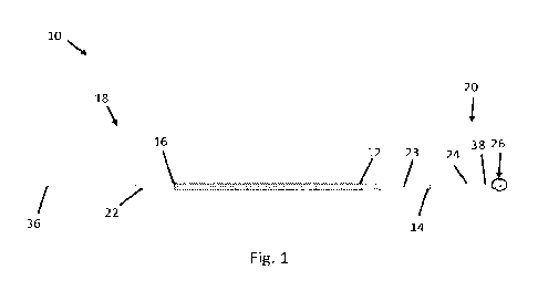
101631 Fig. 1 illustrates an exemplary device 10 for delivering
fluid to an inner ear.
Device 10 includes a knurled handle 12, and a distal handle adhesive 14 (for
example, an epoxy
such as loctite 4014) that couples to a telescoping hypotube needle support
24. The knurled
handle 12 (or handle portion) may include kurling features and/or grooves to
enhance the grip.
The knurled handle 12 (or handle portion) may be from about 5 mm to about 15
mm thick or
from about 5 mm to about 12 mm thick, or from about 6 mm to about 10 mm thick,
or from
about 6 mm to about 9 mm thick, or from about 7 mm to about 8 mm thick. The
knurled handle
12 (or handle portion) may be hollow such that fluid may pass through the
device 10 during use.
The device 10 may also include a proximal handle adhesive 16 at a proximal end
18 of the
knurled handle 12, a needle sub-assembly 26 (shown in Fig. 2) with stopper 28
(shown in Fig. 2)
at a distal end 20 of the device 10, and a strain relief feature 22. Strain
relief feature 22 may be
composed of a Santoprene material, a Pebax material, a polyurethane material,
a silicone
material, a nylon material, and/or a thermoplastic elastomer.
101641 The telescoping hypotube needle support 24 surrounds and
supports a bent needle
38 (shown in Fig. 2) disposed therewithin.
101651 Referring still to Fig. 1, the stopper 28 may be composed
of a thermoplastic
material or plastic polymer (such as a UV-cured polymer), as well as other
suitable materials,
and may be used to prevent the bent needle 38 from being inserted too far into
the ear canal (for
example, to prevent insertion of bent needle 38 into the lateral wall or other
inner ear structure).
Device 10 also may include a tapered portion 23 disposed between the knurled
handle 12 and the
17
CA 03178301 2022- 11- 9
WO 2021/242926
PCT/US2021/034352
distal handle adhesive 14 that is coupled to the telescoping hypotube needle
support 24. The
knurled handle 12 (or handle portion) may include the tapered portion 23 at
the distal end of the
handle portion 12. Device 10 may also include tubing 36 fluidly connected to
the proximal end
16 the device 10 and acts as a fluid inlet line connecting the device to
upstream components (for
example, a pump, a syringe, and/or upstream components which, in some
emboidments, may be
coupled to a control system and/or power supply (not shown)). In some
embodiments, the bent
needle 38 (shown in Fig. 2) extends from the distal end 20, through the
telescoping hypotube
needle support 24, threough the tapered portion 23, through the knurled handle
12, and through
the strain relief feature 22 and fluidly connects directly to the tubing 36.
In other embodiments,
the bent needle 38 fluidly connects with the hollow interior of the knurled
handle (for example,
via the telescoping hypotube needle support 24) which in turn fluidly connects
at a proximal end
16 with tubing 36. In embodiments where the bent needle 38 does not extend all
the way
through the interior of the device 10, the contact area (for example, between
overlapping nested
hyotubes 42), the tolerances, and/or sealants between interfacing components
must be sufficent
to prevent therapeutic fluid from leaking out of the device 10 (which operates
at a relatively low
pressure (for example, from about 1 Pascal to about 50 Pa, or from about 2 Pa
to about 20 Pa, or
from about 3 Pa to about 10 Pa)).
101661 Fig 2 illustrates a sideview of the bent needle sub-
assembly 26, according to
aspects of the present disclosed embodiments. Bent needle sub-assembly 26
includes a needle
38 that has a bent portion 32. Bent needle sub-assembly 26 may also include a
stopper 28
coupled to the bent portion 32. The bent portion 32 includes an angled tip 34
at the distal end 20
of the device 10 for piercing a membrane of the ear (for example, the RWM).
The needle 38,
bent portion 32, and angled top 34 are hollow such that fluid may flow
therethrough. The angle
46 (as shown in Fig. 4) of the bent portion 32 may vary. A stopper 28 geometry
may be
cyclidrical, disk-shaped, annulus-shaped, dome-shaped, and/or other suitable
shapes. Stopper 28
may be molded into place onto bent portion 32. For example, stopper 28 may be
positioned
concentrically around the bent portion 32 using adhesives or compression
fitting. Examples of
adhesives include an UV cure adhesive (such as Dymax 203A-CTH-F-T), elastomer
adhesives,
thermoset adhesives (such as epoxy or polyurthethane), or emulsion adhesives
(such as polyvinyl
acetate). Stopper 28 fits concentrically around the bent portion 32 such that
angled tip 34 is
18
CA 03178301 2022- 11- 9
WO 2021/242926
PCT/US2021/034352
inserted into the ear at a desired insertion depth. The bent needle 38 may be
formed from a
straight needle using incremental forming, as well as other suitable
techniques.
101671 Fig. 3 illustrates a perspective view of exemplary device
10 for delivering fluid to
an inner ear. Tubing 36 may be from about 1300 mm in length (dimension 11 in
Fig. 3) to about
1600 mm, or from about 1400 mm to about 1500 mm, or from about 1430 mm to
about 1450
mm. Strain release feature 22 may be from about 25 mm to about 30 mm in length
(dimension
15 in Fig. 3), or from about 20 mm to about 35 mm in length. Handle 12 may be
about 155.4
mm in length (dimension 13 in Fig. 3), or from about 150 mm to about 160 mm,
or from about
140 mm to about 170 mm. The telescoping hypotube needle support 24 may have
two or more
nested hypotubes, for example three nested hypotubes 42A, 42B, and 42C, or
four nested
hypotubes 42A, 42B, 42C, and 42D (shown in Fig. 9). The total length of
hypotubes 42A, 42B,
42C and tip assembly 26 (dimension 17 in Fig. 3) may be from about 25 mm to
about 45 mm, or
from about 30 mm to about 40 mm, or about 35 mm. In addition, telescoping
hypotube needle
support 24 may have a length of about 36 mm, or from about 25 mm to about 45
mm, or form
about 30 mm to about 40 mm. The three nested hypotubes 42A, 42B, and 42C each
may have a
length of 3.5 mm, 8.0 mm, and 19.8 mm, respectively, plus or minus about 20%.
The inner-most
nested hypotube (or most narrow portion) of the telescoping hypotube needle
support 24 may be
concentrically disposed around needle 38 (as shown in Fig. 7).
101681 Fig. 4 illustrates a perspective view of bent needle sub-
assembly 26 coupled to the
distal end 20 of device 10, according to aspects of the present disclosed
embodiments. As shown
in Fig. 4, bent needle sub-assembly 26 may include a needle 38 coupled to a
bent portion 32. In
other embodiments, the bent needle 38 may be a single needle (for example, a
straight needle
that is then bent such that it includes the desired angle 46). Needle 38 may
be a 33-gauge needle,
or may include a gauge from about 32 to about 34, or from about 31 to 35. At
finer gauges, care
must be taken to ensure tubing 36 is not kinked or damaged. Needle 38 may be
attached to
handle 12 for safe and accurate placement of needle 38 into the inner ear. As
shown in Fig. 4,
bent needle sub-assembly 26 may also include a stopper 28 disposed around bent
portion 32.
Fig. 4 also shows that bent portion 32 may include an angled tip 34 for
piercing a membrane of
the ear (for example, the RWM). Stopper 28 may have a height 48 of about 0.5
mm, or from
about 0.4 mm to about 0.6 mm, or from about 0.3 mm to about 0.7 mm. Bent
portion 32 may
have a length 52 of about 1.45 mm, or from about 1.35 mm to about 1.55 mm, or
from about 1.2
19
CA 03178301 2022- 11- 9
WO 2021/242926
PCT/US2021/034352
mm to about 1.7 mm. In other embodiments, the bent portion 32 may have a
length greater than
2.0 mm such that the distance between the distal end of the stopper 28 and the
distal end of the
angled tip 34 is from about 0.5 mm to about 1.7 mm, or from about 0.6 mm to
about 1.5 mm, or
from about 0.7 mm to about 1.3 mm, or from about 0.8 mm to about 1.2 mm. Fig.
4 shows that
stopper 28 may have a geometry that is cyclidrical, disk-shaped, and/or dome-
shaped. A person
of ordinary skill will appreciate that other geometries could be used.
[0169] The delivery of fluid to the cochlea to access the RWM in
non-human primates
(NHPs) differs from the approach used in human patients. For example, device
10 (as shown in
Fig. 1) may be advanced through the external auditory canal in human patients,
either under
surgical microscopic control or along with an endoscope, an approach that is
not feasible, even in
larger NHPs (such as baboons).
[0170] In NHPs, an approach to access the RWM is more similar to
that typically used
for cochlear implant procedures in patients, which results in a slightly
different angle 46 to target
the RWM. For example, angle 46 as shown in Fig. 4 may be about 55 degrees for
use in human
patients. Alternatively, angle 46 as shown in Fig. 4 may be about 30 degrees
in NHPs. In other
embodiments, angle 46 may be from about 1 degree to about 70 degrees. In other
embodiments,
angle 46 may be from about 5 degrees to about 70 degrees. In other
embodiments, angle 46 may
from about 20 degrees to about 70 degrees. In other embodiments, angle 46 may
be from about
20 degrees to about 60 degrees. In other embodiments, angle 46 may be from
about 20 degrees
to about 50 degrees. In other embodiments, angle 46 may be from about 20
degrees to about 40
degrees. In other embodiments, angle 46 may be from about 30 degrees to about
70 degrees. In
other embodiments, angle 46 may be from about 40 degrees to about 60 degrees.
In other
embodiments, angle 46 may be about 55 degrees. In some embodiments, angle 46
is adjustable
across a range of angles during use of device 10.
[0171] Fig. 5 depicts an exemplary device 10 with protective tube
casing (or sleeve) 56
around the tubing 36 (as shown in Fig. 6) to protect it from kinking or
becoming otherwise
damaged. As shown in Fig. 5, device 10 may be positioned in protective device
casing 54.
Device casing 54 may be used to facilitate storage or handling of device 10
prior to use for fluid
delivery. Tube casing 56 may include one or more cylindrical pieces 58 coupled
to tube casing
56 to increase durability and reduce kinking and deformation of tube casing 56
(and hence tubing
CA 03178301 2022- 11- 9
WO 2021/242926
PCT/US2021/034352
36). The cylindrical pieces 58 may also help to keep the tube casing 56 (and
hence tubing 36) in
a spiral configuration during transport. In some embodiments, tube casing (or
sleeve) 56 may be
composed of polyether ether ketone (PEEK). In some embodiments, tube casing 56
may be
composed of a thermoplastic material.
101721 Fig. 6 depicts an exemplary device 10 coupled to strain
release feature 22. Needle
38 (shown in Fig. 1) may be attached via or through telescoping hypotube
needle support 24
(shown in Fig. 1) through handle 12 (shown in Fig. 1) to a fixed length of
tubing 36 that may be
attached to a syringe 60 (shown in Fig. 15) used to hold device 10.
101731 Fig. 7 illustrates a perspective view of the telescoping
hypotube needle support
24, needle 38, and stopper 28 of device 10, according to aspects of the
present disclosed
embodiments. In some embodiments, needle 38 may be concentrically disposed
within the most
narrow portion of the telescoping hypotube needle support 24.
101741 Fig. 8 illustrates a sideview of needle 38, according to
aspects of the present
disclosed embodiments. Needle 38 may include a bent portion 32. The bent
portion 32 may
include an angled tip 34 for piercing a membrane of the ear (for example, the
RWM). Needle 38
may be a 33 gauge needle. Other gauges may also be used such as gauges from
about 32 to
about 34, or from about 31 to about 35. At finer gauges, care must be taken to
ensure the needle
38 is not damaged. Needle 38 may be made out of stainless steel (for example,
304 stainless
steel). Needle 38 may also be made out of any material that has similar
material properties to
stainless steel (such as strength or other mechanical properties) For example,
needle 38 may be
composed of titanium. Needle 38 may have a bent length of about 1.45 mm, or
from about 1.2
mm to about 1.7 mm (as shown in Fig. 4) and an angle of about 55 degrees, or
from about 40
degrees to about 70 degrees (as shown in Fig. 4), or from about 20 degrees to
about 70 degrees
and other sub ranges therebetween include from about 25 degrees to about 45
degrees.
101751 Fig. 9 illustrates a perspective of a telescoping hypotube
needle support 24,
according to aspects of the present disclosed embodiments. In some
embodiments, telescoping
hypotube needle support 24 may include two or more nested hypotubes, for
example four nested
hypotubes 42A, 42B, 42C, and 42D (see also Fig. 3 which shows an embodiment
with three
nested hypotubes 42A, 42B, and 42C). Needle 38 may be the most narrow portion
of the
telescoping hypotube needle support 24. In other embodiments, needle 38 is
disposed within the
21
CA 03178301 2022- 11- 9
WO 2021/242926
PCT/US2021/034352
most narrow portion 42D of the telescoping hypotube needle support 24.
Telescoping hypotube
needle support 24 may be made out of stainless steel (for example, 304
stainless steel).
Telescoping hypotube needle support 24 may also be made out of any material
that has similar
material properties to stainless steel (such as strength or other mechanical
properties). For
example, telescoping hypotube needle support 24 may be composed of titanium.
Nested
hypotubes 42A, 42B, 42C, 42D may include gauges of 14XII, 20TW, 23TW, and
27TW,
respectively. As such, nested hypotubes 42A, 42B, 42C, 42D may include outer
diameters of
0.083 inches, 0.0355 inches, 0.025 inches, and 0.014 inches, respectively, and
inner diameters of
0.039 inches, 0.0255 inches, 0.017 inches, and 0.009 inches, respectively. In
other
embodiments, nested hypotubes 42A, 42B, 42C, 42D may include outer diameters
ranging from
about 0.2 inches to about 0.01 inches and inner diameters ranging from 0.08
inches to 0.004
inches. Similarly, nested hypotubes 42A, 42B, 42C, 42D may include wall
thicknesses ranging
from about 0.022 inches to about 0.003 inches, or from about 0.05 inches to
about 0.001 inches.
101761 Referring still to Fig. 9, needle 38 may include a gauge
of from about 32 to about
34, or from about 31, to about 35, with corresponding outer diameters from
about 0.01 inches to
about 0.005 inches, thereby allowing it to fit with the inner-most nested
hypotube 42C and/or
42D depending on the number of hypotubes. Each of the nested hypotubes 42A,
42B, 42C, 42D
may also include include smoothed edges between itself and one or more of the
adjacent
hyoptubes, e.g., to reduce the likelihood of catching on certain anatomical
components within the
external auditory canal in human patients. Each of the nested hypotubes 42A,
42B, 42C, 42D
may also include include an outwardly radially extending lip at a proximal end
and an inwardly
radially extending lip at a distal end, thereby creating interference with a
neighboring nested
hypotube and preventing any of the nested hypotubes 42A, 42B, 42C, 42D from
becoming
detached from the device 10. In other embodiments, instead of being
telescopic, needle support
24 may include a single, monolithic conical member with a gradually tapering
radius (for
example, tapering from the radius of the outermost hypotube 42A to the radius
of the innermost
hypotube 42D) in place of the plurality of nested hypotubes 42A, 42B, 42C, and
42D. The
telescoping hypotube needle support 24 may include nested hypotubes 42B, 42C,
and 42D that
extend fully through each next wider hypotube (as illustrated in Fig. 9 by
hypotube 42B
extending through to a distal end of hypotube 42A).
22
CA 03178301 2022- 11- 9
WO 2021/242926
PCT/US2021/034352
101771 Fig. 10 illustrates a perspective view of a strain release
feature 22, according to
aspects of the present disclosed embodiments. Strain release feature 22 may
include layered
extrusions 58A and 58B. Layered extrusions 58A and 58B may include layered
Pebax
extrusions. Layered extrusions 58A and 58B may prevent kinking and/or
deformation of PEEK
tubing 36 (as shown in Figs. 5-6) at a proximal end 18 of the knurled handle
12 (as shown in Fig.
1).
101781 Fig. 11 illustrates a perspective view of tubing 36,
according to aspects of the
present disclosed embodiments. Tubing 36 may be made out of PEEK. Tubing 36
may also be
composed of other materials such as thermoplastics. Tubing 36 may have an
inner diameter of
about 0.007 inches, or from about 0.005 inches to 0.01 inches Tubing 36 may
have an outer
diameter of about 1/32 inches or from about 0.02 inches to about 0.05 inches.
Tubing 36 may
have a length of about 60 inches, or from about 30 inches to about 100 inches.
In embodiments
of the device 10 in which the bent needle 38 extends all the way through the
knurled handle 12
and fluidly connects directly to the tubing 36, the proximal end of the bent
needle 38 may be
coupled to the tubing 36 (for example, with the bent needle 38 being inserted
into the tubing 36)
via compression fit, adhesive, ring clamp, and other suitable connections.
101791 Fig. 12 illustrates an exemplary device 10 for delivering
fluid to an inner ear
including the telescoping hypotube needle support 24, the bent needle 38 at
the distal end 20 of
the device 10. As shown in Fig. 12, device 10 may include an alternate
embodiment of the strain
release feature 22 that may add flexibility to the interface between the
tubing 36 and the
proximal end 18 of the device. The embodiment of the strain release feature 22
of fig. 12 may
also reduce kinking or deformation of tubing 36. Strain realease feature 22
may also provide
durability where tube casing 56 (as shown in Fig. 5) interfaces with the
proximal end 18 of
handle 12.
101801 Fig. 13 illustrates a perspective of the bent needle sub-
assembly 26, according to
aspects of the present disclosed embodiments. The needle sub-assembly 26,
located at the distal
end 20 of the device 10, may include an angled tip 34. The stopper 28 may be
disposed around
the needle 38 such that a stopper proximal end 33 is adjacent the bent portion
32 of the needle
38. The stopper 28 may include a stopper tapered portion 29 with a gradually
increasing radius
23
CA 03178301 2022- 11- 9
WO 2021/242926
PCT/US2021/034352
from the stopper proximal end 33 toward the stopper distal end 35. The stopper
may also include
one or more chamfers (for example, chamfer 31 at the stopper distal end 35).
101811 Fig. 13A illustrates a perspective view of the bent needle
sub-assembly 26
including an alternate stopper 70 design, according to aspects of the present
disclosed
embodiments. The alternate stopper 70 of Fig. 13A may be more rounded as
compared to the
stopper 28 of Fig. 13, which may be more disk-shaped or donut-shaped. For
example, the
alternate stopper 70 may include a maximum outer diameter that is
approximately equal to its
maximum length, or that is from about 0.75 to about 1.5 times its maximum
length. By contrast,
the stopper 28 illustrated in Fig. 13 may include a maximum diameter that is
about twice the
maximum length, or that is from about 1.5 to about 2.5 times the maximum
length. The alternate
stopper 70 may also include a flexible portion 72 that is rounded or curved
(i.e., convex) towards
the distal end of the bent needle sub-assembly 26. The alternate stopper 70
may also include a
rigid portion 74 that is located proximate of the flexible portion 72. The
rigid portion 74 may
contain an outer diameter that is smaller than that of the flexible portion
72.
101821 Figure 13B illustrates a perspective view of the bent
needle sub-assembly 26
including an alternate stopper 71 design, according to the present disclosed
embodiments. The
alternate stopper 71 of Fig. 13B includes a tapered portion 77 that gradually
tapers from the
needle to the stopper outer circumerence 79. The alternate stopper 71 may also
include a lip
portion 75 that extends slightly toward the distal tip of the needle 34. The
alternate stopper 71
may also include a thinner aspect ratio (for example, compared to the stoppers
of Fig. 13 and
13A) such that the largest diameter of the stopper 71 (for example, as
measured at the outer
circumference 79) is about 10 times greater than the minimum thickness of the
stopper 71. In
other embodiments, the largest diameter of the stopper 71 may be from about 7
to about 12 times
greater than the minimum thickness of the stopper 71, or from about 5 to about
15 times greater
than the minimum thickness of the stopper 71.
101831 Fig. 13C illustrates an embodiment of the distal end 20 of
the device 10
(including the needle tip 34), according to aspects of present embodiments. In
the embodiment
illustrated in Fig. 13C, the device 10 includes a stopper anchoring groove
200, disposed within
the device 10 at the distal end 20. The stopper anchoring groove 200 may be
used to help anchor
24
CA 03178301 2022- 11- 9
WO 2021/242926
PCT/US2021/034352
the stopper 28, 70, 71 (shown in Figs. 2, 4, 13, 13A, 13B, and 15-18) around
the needle 38 at the
distal end 20.
101841 Fig. 13D is an enlarged view of the stopper anchoring
groove 200, according to
aspects of the present embodiments. The stopper anchoring groove 200 may
include a first
declined portion 202 (for example, adjacent a distal end 20 of the stopper
anchoring groove 200),
a second declined portion 204 (for example, adjacent a proximal end 18 of the
stop anchoring
groove 200), and a recessed portion 206 (or flat portion 206) disposed axially
between the first
and second declined portions 202, 204 (thereby forming the stopper anchoring
groove 200). The
recessed portion 206 includes a smaller diameter than the rest of the needle
38. Each of the first
and second declined portions 202, 204 may linearly transition from the outer
circumference of
the needle 38 to the recessed portion 206, and/or may be rounded (thereby
forming one or more
fillets).
101851 Fig. 13E is an enlarged view of the transition between the
device handle 12, and
the telescoping hypotubes 24, according to aspects of the present embodiments.
In the
embodiment illustrated in Fig. 13E, the device may include a lip joint 210 (or
brace, for example,
an annular brace) that reinforces the transition between the device handle 12,
and the telescoping
hypotubes 24. As such, the lip joint 210 (or brace) may be disposed at an
interface between the
handle portion 12 and the telescoping hypotubes 24 (or support). The lip joint
210 may include a
larger outer diameter than the outer diameter of the handle portion 12 such
that a portion of the
lip joint 210 is disposed around the handle portion 12. The lip joint 210 may
also include an
inner diameter that tightly fits around the outer diameter of the thickest
member of the plurality
of telescoping hypotubes 24.
101861 Fig. 13F illustrates an embodiment of the device 10,
according to aspects of the
present embodiments. In the embodiment illustrated in Fig. 13F, the device 10
includes a
machined barb 212 integrated into a proximal end 18 of the device handle 12.
The machined
barb 212 may be used in connection with the strain relief features 22 (shown
in Figs. 1, 3, 12,
and 13H), in order to prevent the strain relief features 22 from axially
detaching from the device
handle 12.
101871 Fig. 13G is an enlarged view of the machined barb 212,
according to aspects of
the present embodiments. The machined barb 212 may include a first inclined
portion 214 (for
CA 03178301 2022- 11- 9
WO 2021/242926
PCT/US2021/034352
example, on the proximal end 18 of the device handle 12), a second inclined
portion 216, and a
plateau portion disposed between the first and second inclined portions 214,
216.
101881 Fig. 13H is an enlarged view of the strain relief features
22, according to aspects
of the present embodiments. (The device depicted in Fig. 13H is illustrated in
an opposite
orientation to that of Figs. 13F and 13G.) PEEK tubing 36 may be bonded (for
example, via
epoxy 220) or otherwise coupled to the device handle 12. The strain relief
features 22 may be
disposed around the PEEK tubing 36, which itself may be disposed around needle
38, in order to
prevent kinking of (and/or other damage to) the needle 38. In addition,
machined barb 212 helps
to prevent the strain relief features 22 from dislodging or becoming decoupled
from the device
handle 212. In some embodiments, the strain relief features may be composed of
molded
Santoprene, among other suitable materials. In some embodiments, in order to
get the PEEK
tubing 36 around the needle 38, the PEEK tubing 36 may be split around the
needle 38. A heat
shrink sleeve (not shown) may be placed over the junction between the PEEK
tubing 36 and the
needle 38. The junction may then be exposed to heat. Following exposure to
heat, the junction
may be reflowed or reshaped such that the junction is left with a smooth final
outer profile.
Packaging System
101891 Fig. 14 depicts packaging 55 that includes device 10
encased in device casing 54
and coupled to tube casing 56. For example, packaging 55 may provide for a
sterile device 10
for fluid delivery to the RWM through the external auditory canal Device 10
may also be a
single-use disposable product. In this embodiment, the device is appropriately
discarded (e.g., in
a biohazard sharps container). In some embodiments, the packaging 55, tube
casing 56, device
casing 54, device 10, and components thereof are all constructed of materials
that are robust
enough to withstand gamma sterilization (for example, gamma irradiation that
uses Cobalt 60
radiation to kill microorganisms and microbes). In some embodiments, the
packaging 55, tube
casing 56, device casing 54, device 10, and components thereof are all
constructed of materials
that are sufficiently resistant to temperature to withstand steam
sterilization.
101901 Fig. 14A illustrates a top perspective view of the device
10 nested in alternate
packaging 100 (or packaging system 100), according to aspects of the present
embodiments. The
packaging (or packaging system) 100 allows for safe and sterile transport
and/or shipping of the
26
CA 03178301 2022- 11- 9
WO 2021/242926
PCT/US2021/034352
device 10. The packaging 100 may include a mounting surface 80 and device
nesting 90
disposed on the mounting surface 80. The mounting surface 80 may be composed
of cardboard,
hardened cardstock, or polymer materials. The mounting surface 80 may also be
composed of
cardboard or hardened cardstock with a polymer coating. The device nesting 90
may be
composed of similar materials to the mounting surface 80. The device nesting
90 may protrude
out of the plane of the mounting surface 80. The mounting surface 80 may
include several pairs
of oppositely-oriented slits 114. The slits may be cut into the mounting
surface 80 to help hold
the PEEK tubing 36 in place, thereby preventing the PEEK tubing 36 (and
metallic tubing
therewithin) from kinking, twisting, breaking, and/or or becoming otherwise
damaged. Each slit
of each pair of oppositely-oriented slits 114 may include a pair of curved
ends 118 at either end
to prevent the slits 114 from causing rips and/or tears in the mounting
surface 80 due to external
forces acting on the of oppositely-oriented slits 114. The oppositely-oriented
slits 114 prevent
the PEEK tubing 36 (and metallic tubing therewithin) from over-kinking and/or
under-kinking
because the slits are disposed on either side of (i.e., radially inside of and
outside of) the PEEK
tubing 36 while it is coiled, thereby ensuring that a desired radius of the
coiled tubing 36 is
maintained. In the embodiment of Fig. 14A, the mounting surface 80 includes a
total of 5 pairs
of oppositely-oriented slits 114. However, in other embodiments, the mounting
surface 80 may
include other numbers of pairs of oppositely-oriented slits 114, as necessary,
including 1, 2, 3, 4,
6, 7, 8, 9, 10 and/or more than 10.
101911 Still referring to Fig. 14A, the alternate packaging (or
packaging system) 100 may
include at least one pair of oppositely-oriented slides 116 adjacent the Luer
lock 61, in order to
support the Luer lock 61 and prevent it from getting damaged. The device
nesting 90 may be
used to support the device 10 and may include several pairs of nesting notches
84, 86, 88, 92, 94,
96 that prevent the device 10 from moving laterally or longitudinally within
the device nesting
90. For example, nesting notch pair 84 is positioned to hold the strain relief
feature 22 (shown in
Figs. 1 and 12), nesting notch pairs 86, 88, and 92 are positioned at the
proximate end of the
body of device 10, and nesting notch pairs 94 and 96 are positioned toward the
distal end of the
device 10. The device nesting 90 may include a first contoured portion 114
near the mid-section
of the body of the device 10 to accommodate a semi-flexible twist-tie 76 that
is used to secure
the device to the device nesting 90. The device nesting 90 may also include
first and second
twist-tie holes 78, 82 to allow the twist-tie 76 to extend underneath the
device nesting 90, thereby
27
CA 03178301 2022- 11- 9
WO 2021/242926
PCT/US2021/034352
extending around the bottom of a portion of the device nesting 90, in order to
hold the device
nesting 90 and the device 10 closely together. The device nesting 90 may also
include a tip hole
104 at the distal end to protect the tip and bent needle sub-assembly of the
device 10. As such,
the tip of the device 10 may rest within the tip hole 104, thereby minimizing
the risk of damage
due to the tip making contact with any structures.
101921 Referring still to Fig. 14A, the device nesting 90 may
include a second contoured
portion 102 at the distal end to accommodate the tip of the device 10 and the
tip hole 104. The
device nesting 90 gradually extends away from the device centerline (i.e., the
center of the pairs
of nesting notches 84, 86, 8, 92, 94, and 96) at each of the first and second
contoured portions
114, 102 such that the device nesting 90 forms a fiddle or violin shape,
including a neck portion
120 disposed longitudinally between the first and second contoured portions
114, 102. Walls of
the device nesting 90 that protrude out of the plane of the mounting surface
80 may do so
gradually, for example, as illustrated at taper 98 which transitions gradually
from the raised
protrusion (or wall) at the neck 120 down to the plane of the mounting surface
80. The
packaging (or packaging system) 100 in the embodiment of Fig. 14A may also
include tube
casing (or sleeve) 56 (shown in Figs. 5 and 14). The tube casing (or sleeve)
56 may be disposed
concentrically around the PEEK tubing 36, and helps protect the PEEK tubing 36
and prevents it
from kinking, bending, and/or becoming otherwise damaged The tube casing (or
sleeve) 56
may be placed around the PEEK tubing 36 (for example, prior to shipping or
transport of the
device 10 and/or system 100) and may be removed by disconnecting the Luer lock
61 from the
PEEK tubing 36 and/or prior to connecting the Luer lock 61 to the PEEK tubing
36. The tube
casing (or sleeve) 56 may be composed of any suitable material such as
polymers such as PEEK,
composite materials, metallic materials, as well as other suitable materials.
101931 Still referring to Fig. 14A, the packaging (or packaging
system) 100 may include
one or more locking features including a first locking portion 106 near the
proximal end of the
device 10 and device nesting 90, and a second locking portion 108 near the
distal end of the
device 10. The second locking portion 108 may extend across the neck 120 of
the device nesting
90. Each of the first and second locking portions 106, 108 extends from one
side of the
mounting surface 80 and across the device nesting 90 (i.e., after the device
10 been placed within
the device nesting 90) to the mounting surface 80 on the other side of the
device nesting 90. The
first and second locking portions 106, 108 may attach to first and second
attachment slits 110,
28
CA 03178301 2022- 11- 9
WO 2021/242926
PCT/US2021/034352
112, the first attachment slit 110 being disposed within the mounting surface
80 at the proximal
end of the device nesting 90, and the second attachment slit 112 being
disposed at the distal end
of the device nesting 90. The first locking portion 106 interfaces with and
attaches to the first
attachment slit 110, while the second locking portion 108 interfaces with and
attaches to the
second attachment slit 112. The device nesting 90 may be coupled to the
mounting surface 80
using any suitable mechanisms including epoxy, fusion, adhesion, glue, as well
as other suitable
means. In addition, in some embodiments, the device nesting 90 may be formed
via 3D printing
(for example, via fused deposition modeling (FDM), stereo-lithography (SLA),
as well as other
modalities). In some embodiments, the packaging 100, device nesting 90,
mounting surface 80,
and components thereof are all constructed of materials (for example,
polymers, thermoset
plastics, thermoplastics, composite materials, and other materials) that are
sufficiently resistant to
temperature to withstand steam sterilization, gamma irradiation (that uses
Cobalt 60 radiation to
kill microorganisms and microbes), and other sterilization methods.
Systems
101941 A system of the present disclosure may include at least
one distal tip camera
(DTC) for visualizing and/or monitoring the delivery of fluid to a target (for
example, outer,
middle, and/or inner ear). In some embodiments, the distal tip camera may be
operatively
coupled to the device 10, while in other embodiments, the distal tip camera
may be installed as a
part of the device 10 (that is, an all-in-one device). The distal tip camera
may include at least
one of a charge-coupled device (CCD) and a complementary metal-oxide
semiconductor
(CMOS). The distal tip camera may include at least one image sensor (for
example, a lens) to
receive, convey, and/or convert a signal (for example, an analog or a digital
signal) from the
target (for example, outer, middle, and/or inner ear). In some embodiments,
the distal tip camera
may further include at least one processor (for example, a video processor or
an in-distal tip
camera processor) for processing images and/or controlling the distal tip
camera, while in other
embodiments, the processor may be disposed separately from the distal tip
camera. The distal tip
camera and/or the image sensor may include a cuboid shape, a chip shape, a
flattened cube
shape, a cylindrical shape, and combinations thereof.
29
CA 03178301 2022- 11- 9
WO 2021/242926
PCT/US2021/034352
101951 Fig. 15 illustrates a perspective view of a distal tip
camera 1004 disposed above a
stopper 28 within a system 1000, according to aspects of the present
embodiments. The stopper
28 may be attached to an outer surface 1020 of the bent portion 32 of needle
38. The distal tip
camera 1004 may be operatively coupled with a wire 1002. In some embodiments,
the distal tip
camera 1004 may be disposed at a front surface 1006 of the stopper 28. In some
embodiments,
the distal tip camera 1004 may be embeded in the front surface 1006 of the
stopper 28. The front
surface 1006 may face toward the target (for example, outer, middle, and/or
inner ear). In some
embodiments, the distal tip camera 1004 may be disposed at a side surface 1008
of the stopper
28 (not shown).
101961 Fig. 16 illustrates a perspective view of a distal tip
camera 1004 disposed in a
cavity 1010 within the stopper 28 within a system 1000, according to aspects
of the present
embodiments. The stopper 28 may be attached to the outer surface 1020 of the
bent portion 32
of needle 38. The distal tip camera 1004 may be operatively coupled with a
wire 1002. The
cavity 1010 may be created by cutting a portion (for example, up to 30%) of
the stopper 28. In
some embodiments, the cavity 1010 may be near or beside the outer surface 1020
of the device
10.
101971 Fig. 17 illustrates a perspective view of a distal tip
camera 1004 disposed behind
the stopper 28 within a system 1000, according to aspects of the present
embodiments. The
stopper 28 may be attached to the outer surface 1020 of the bent portion 32 of
needle 38 The
distal tip camera 1004 may be operatively coupled with a wire 1002. In some
embodiments, the
distal tip camera 1004 may be disposed at a back surface 1012 of the stopper
28 (as shown in
Fig. 17), while in some embodiments, the distal tip camera 1004 may be
disposed behind the
back surface 1012 of the stopper 28 (as shown in Figs. 23D and 23F).
101981 Fig. 18 illustrates a perspective view of a distal tip
camera 1004 disposed within a
system 1000, according to aspects of the present embodiments. The stopper 28
may be attached
to the outer surface 1020 of the bent portion 32 of needle 38. The distal tip
camera 1004 may be
operatively coupled with a wire 1002. The distal tip camera 1004 may include
or be operatively
coupled to a light source 1022 (for example, an LED light source). In some
embodiments, the
distal tip camera 1004 and/or the light source 1022 may be disposed above the
stopper 28. In
some embodiments, the distal tip camera 1004 and/or the light source 1022 may
be diposed at or
CA 03178301 2022- 11- 9
WO 2021/242926
PCT/US2021/034352
embeded in the stopper 28 (not shown). In some embodiments, the distal tip
camera 1004 and/or
the light souce 1022 may be behind the stopper 28 (not shown).
101991 Referring to Figs. 15-18, the distal tip camera 1004 may
be operatively coupled to
the outside surface 1020 of the bent portion 32 of needle 38. In one or more
embodiments, the
stopper 28 may be transparent and/or include a transparent portion 1014 for
the distal tip camera
1004 to see through the stopper 28 for monitoring and/or visualizing the
target (for example,
outer, middle, and/or inner ear). The transparent portion 1014 may include at
least a transparent
material (for example, plastic, thermoplastic, polymers, or and/or other
suitable materials). In
some embodiments, the transparent portion 1014 may be a part of the cavity
1010. In one or
more embodiments, about 30% (or from about 20% to about 40%) of the stopper 28
may be
removed such that the distal tip camera 1004 may be integrated and/or included
in the needle
sub-assembly 26 (Fig. 2 and Fig. 13).
102001 Referring still to Figs. 15-18, the distal tip camera 1004
may be operatively
coupled via the wire 1002 to at least one of power supply for supplying power
and/or
communications to the distal tip camera 1004. In some embodiments, sections of
the wire 1002
may be operatively attached to the outer surface 1020 of the needle 38. In
some embodiments,
sections of the wire 1002 may be operatively embedded within a portion of the
device 10. For
example, in one or more embodiments, the wire 1002 may be disposed outside of
the bent needle
38 and telescoping hypotube needle support 24 while also extending at least
partially internal to
handle 12 of the device 10. In some embodiments, the wire 1002 may have a
diameter of up to
mm. In some embodiments, the wire 1002 may have a diameter of up to 5 mm. In
some
embodiments, the wire 1002 may have a diameter of up to 3 mm. In some
embodiments, the
wire 1002 may have a diameter of up to 1 mm. In some embodiments, the wire
1002 may have a
diameter of up to 0.8 mm. In some embodiments, the wire 1002 may have a
diameter of up to
0.6 mm. In some embodiments, the wire 1002 may have a diameter of up to 0.4 mm
In some
embodiments, the wire 1002 may have a diameter of up to 0.2 mm. In some
embodiments, the
wire 1002 may have a diameter of up to 0.1 mm. In some embodiments, the wire
1002 may have
a diameter of up to 0.05 mm.
[0201] Fig. 19 illustrates a side view of a distal tip camera
1004A, according to aspects
of the present embodiments. The distal tip camera 1004A may include an image
sensor 1102A
31
CA 03178301 2022- 11- 9
WO 2021/242926
PCT/US2021/034352
to receive, convey, and/or convert a signal (for example, an analog or digital
signal) from the
target (for example, the outer, middle, and/or inner ear) into an image. The
image sensor 1102A
may include a cuboid shape.
102021 Fig. 20 illustrates a side view of a distal tip camera
1004B, according to aspects of
the present embodiments. The distal tip camera 1004B may include a distal tip
camera module
1104A that includes an image sensor 1102A, a processor (for example, a video
processor, or a
processor embedded in the distal tip camera 1004), and/or other elements (for
example, driver
and/or software packages) for the distal tip camera 1004B to access, operate,
and/or process
images (for example, images from the image sensor 1102A). In some embodiments,
the distal
tip camera module 1104A may include a package dimension of up to 0.7 mm x 0.7
mm with a z-
height of up to 1.2 mm. In some embodiments, the distal tip camera module
1104A may include
a package dimension of up to 1.1 mm x 1.1 mm with a z-height of up to 2.4 mm.
In some
embodiments, the distal tip camera module 1104A may include a package
dimension of up to 1.5
mm x 1.5 mm with a z-height of up to 3 mm. In some embodiments, the distal tip
camera
module 1104A may include a package dimension of up to 2 mm x 2 mm with a z-
height of up to
mm.
102031 Referring to Figs. 19-20, the image sensor 1102A may be up
to 10 mm x 10 mm,
5 mm x 5 mm, 2 mm x 2 mm, 1.8 mm x 1.8 mm, 1.6 mm x 1.6 mm, 1.4 mm x 1.4 mm,
1.2 mm x
1.2 mm, 1 mm x 1 mm, U.S mm x 0.8 mm, 0.6 mm x 0.6 mm, 0.4 mm x 0.4 mm, 0.2 mm
x 0.2
mm, 0.1 mm x 0.1 mm, or 0.05 mm x 0.05 mm with a height of up to 100, 20 10,
5, 3, 2, 1, 0.8,
0.6, 0.4, 0.2, or 0.1 mm.
102041 Referring still to Figs. 19-20, the image sensor 1102A may
include an image array
capable of capturing at least 10 x 10, 50 x 50, 100 x 100, 200 x 200, 400 x
400, 500 x 500 or
1000 x 1000 pixels resolution video at a frame rate of at least 5, 10, 20, 30,
50, 100, 500, or 1000
frames per second (fps).
102051 Referring still to Figs. 19-20, the image sensor 1102A may
include an image area
of at most 10 mm x 10 mm, 5 mm x 5 mm, 2 mm x 2 mm, 1.8 mm x 1.8 mm, 1.6 mm x
1.6 mm,
1.4 mm x 1.4 mm, 1.2 mm x 1.2 mm, 1 mm x 1 mm, 0.8 mm x 0.8 mm, 0.6 mm x 0.6
mm, 0.4
mm x 0.4 mm, 0.2 mm x 0.2 mm, 0.1 mm x 0.1 mm, or 0.05 mm x 0.05 mm. The image
sensor
32
CA 03178301 2022- 11- 9
WO 2021/242926
PCT/US2021/034352
1102A may include low-light sensitivity of up to 10, 100, 500, 800, 1000,
1200, 1500, 2000,
3000, or 10,000 mV/lux-sec.
[0206] Referring still to Figs. 19-20, the image sensor 1102A may
include an optical
format of up to 10, 5, 2, 1.8, 1.6, 1.4, 1.2, 1, 0.8, 0.6, 0.4, 0.2, 0.1, or
0.05 mm, and a pixel size of
up to 10, 8, 6, 4, 3, 2.5, 2.2, 2, 1.8, 1.6, 1.4, 1.2, or 1
[0207] Fig. 21 illustrates a side view of a distal tip camera
1004C, according to aspects of
the present embodiments. The distal tip camera 1004C may include an image
sensor 1102C to
receive, convey, and/or convert a signal (for example, an analog or digital
signal) from the target
(for example, outer, middle, and/or inner ear) into an image. The image sensor
1102C of Fig. 21
may include a shape of a chip or cylinder.
[0208] Fig. 22 illustrates a side view of a distal tip camera
1004D, according to aspects
of the present embodiments. The distal tip camera 1004D may include a distal
tip camera
module 1104C that includes an image sensor 1102C, a processor (for example, a
video
processor, or an in-distal tip camera processor), and/or other elements (for
example, driver and/or
software packages) for the distal tip camera 1004D to access, operate, and/or
process images (for
example, images from the image sensor 1102C).
102091 Referring to Figs. 21-22, the image sensor 1102C may
include an outer diameter
of up to 10 mm, 5 mm, 2 mm, 1.8 mm, 1.6 mm, 1.4 mm, 1.2 mm, 1 mm, 0.8 mm, 0.6
mm, 0.4
mm, 0.2 mm, 0.1 mm, or 0.05 mm with a length of up to 100, 20, 10, 5, 3, 2, 1,
0.8, 0.6, 0.4, 0.2,
or 0.1 mm.
[0210] Referring still to Figs. 21-22, the image sensor 1102C may
include an image array
capable of capturing at least 10 x 10, 50 x 50, 100 x 100, 200 x 200, 400 x
400, 500 x 500 or
1000 x 1000 pixels resolution video at a frame rate of at least 5, 10, 20, 30,
50, 100, 500, or 1000
frames per second (fps).
[0211] Referring still to Figs. 21-22, the image sensor 1102A may
include an image area
of at most 10 mm x 10 mm, 5 mm x 5 mm, 2 mm x 2 mm, 1.8 mm x 1.8 mm, 1.6 mm x
1.6 mm,
1.4 mm x 1.4 mm, 1.2 mm x 1.2 mm, 1 mm x 1 mm, 0.8 mm x 0.8 mm, 0.6 mm x 0.6
mm, 0.5
mm x 0.5 mm, 0.4 mm x 0.4 mm, 0.2 mm x 0.2 mm, 0.1 mm x 0.1 mm, or 0.05 mm x
0.05 mm.
33
CA 03178301 2022- 11- 9
WO 2021/242926
PCT/US2021/034352
The image sensor 1102A may include a low-light sensitivity of up to 10, 100,
500, 800, 1000,
1200, 1500, 2000, 3000, or 10,000 mV/lux-sec.
102121 Referring to Figs. 19-22, the image sensors 1102A, C may
be assembled using
integrated circuits packages (for example, chip-scale packages (CSP)) that
have an area of up to
0.1 mm x 0.1 mm, 0.3 mm x 0.3 mm, 0.6 mm x 0.6 mm, 0.9 mm x 0.9 mm, 1.2 mm x
1.2 mm, or
2 mm x 2 mm.
102131 Referring still to Fig. 19-22, the distal tip cameras
1004A-D may include a shutter
(for example, a rolling shutter) (not shown). The distal tip cameras 1004A-D
may operate at a
temperature of -20 'V to 70 'C. The distal tip cameras 1004A-D may include a
field of view of
at least 90, 100, 120, 130, or 150 degrees. In some embodiments, the distal
tip cameras 1004A-D
may include at least one internal lighting source, while in some embodiments,
the distal tip
cameras 1004A-D may be coupled externally to the at least one lighting source
(for example, an
LED lighting source 1022).
102141 Fig. 23 illustrates a side view of an optical fiber 1302,
according to aspects of the
present embodiments. In the present disclosure, the optic fiber 1302 may be
used as a light
source, an image sensor 1004E, or both. The optical fiber 1302 may include at
least a single
optical fiber. In some embodiments, the optical fiber 1302 may have a diameter
up to 5 mm and
a length up to 10 m. In some embodiments, the optical fiber 1302 may have a
diameter up to 1
mm and a length up to 10 m. In some embodiments, the optical fiber 1302 may
have a diameter
up to 0.5 mm and a length up to 10 m. In some embodiments, the optical fiber
1302 may have a
diameter up to 0.4 mm and a length up to 10 m. In some embodiments, the
optical fiber 1302
may have a diameter up to 0.3 mm and a length up to 1 m. In some embodiments,
the optical
fiber 1302 may have a diameter up to 0.2 mm and a length up to 1 m. In some
embodiments, the
optical fiber 1014 may have a diameter up to 0.1 mm and a length up to 0.1 m.
102151 Referring to Fig. 19-23, the distal tip camera 1004A-E
and/or optical fiber 1302
may include or work with a processor (for example, a video processor) (not
shown) with a
dimension of up to 0.01 mm x 0.01 mm x 0.01 mm, 0.1 mm x 0.1 mm x 0.1 mm, 0.2
mm x 0.1
mm x 0.2 mm, 0.3 mm x 0.1 mm x 0.4 mm, 0.3 mm x 0.3 mm x 0.4 mm, 0.4 mm x 0.4
mm x 0.4
mm, or 1 mm x 1 mm x 1 mm. In some embodiments, the image sensors 1102A, C may
be not
larger or not longer than the distal tip camera modules 1104A, C.
34
CA 03178301 2022- 11- 9
WO 2021/242926
PCT/US2021/034352
102161 Referring still to Figs. 15-23, the distal tip camera 1004
may include at least one
of the distal tip cameras 1004A-D, optical fiber 1302, and/or a combination
thereof The image
sensor 1102 may include at least one of the image sensors 1102A, C, the
optical fiber 1302,
and/or a combination thereof.
[0217] Referring still to Figs. 15-23, in one or more
embodiments, the distal tip camera
1004 may include biocompatible, lead-free, auto-focus, disposable, reusable,
low-noise, low
power consumption, low heat, and/or low noise features for convenience. The
distal tip camera
1004 may produce color, grey images, and/or a combination thereof
[0218] Referring still to Figs. 15-23, in some embodiments, the
distal tip camera 1004
may include a working distance of up to 100 mm. In some embodiments, the
distal tip camera
1004 may include a working distance of up to 50 mm. In some embodiments, the
distal tip
camera 1004 may include a working distance of up to 20 mm. In some
embodiments, the distal
tip camera 1004 may include a working distance of up to 10 mm. In some
embodiments, the
distal tip camera 1004 may include a working distance of up to 5 mm.
[0219] Figs. 23A, 23B, and 23C illustrate a device 10 including a
distal tip camera 1004
integrated into the stopper 28. Fig. 23A illustrates the device 10 with the
tip portion circled at A,
indicating the area of detail for Figs. 23B and 23C. Fig. 23B illustrates a
perspective view of the
tip portion while Fig. 23C illustrates a side view of the tip portion. In the
embodiment of Figs.
23A, 23B, and 23C, the device 10 may include a lens or sensor 1102 and camera
unit (for
example, a distal tip camera 1004) integrated into the stopper 28. In some
embodiments, the
stopper 28 may be molded around the distal tip camera 1004. The distal tip
camera 1004 may
also be attached to the stopper 28 via adhesive, epoxy, and/or other suitable
mechanisms. Wire
1002 provides power and a communication link to the distal tip camera 1004. In
some
embodiments, the wire 102 may be sized such that it is run or routed between
two of the
telescoping hypotubes (for example, between two of members 42A, 42B, 42C, or
42D of Fig. 9).
[0220] Figs. 23D, 23E, and 23F illustrate a device 10 including a
distal tip camera 1004
axially separated from the stopper 28 (for example, behind the stopper 28). In
the embodiment
of Figs. 23D, 23E, and 23F, the device 10 may include a distal tip camera 1004
and lens 1102
that are mounted to one of the telescoping hypotubes (for example, member 42A,
42B, 42C, or
42D of Fig. 9), and may be positioned and/or angled such that they capture a
representative view
CA 03178301 2022- 11- 9
WO 2021/242926
PCT/US2021/034352
at the needle tip. The distal tip camera 1004 may be attached to the hypotube
via adhesive,
epoxy, welding and/or other suitable mechanisms. Fig. 23E includes a distal
tip camera 1004
and lens or sensor 1102 in an angled configuration 1103.
102211 Figs. 23G and 23H illustrate the device 10 including the
distal tip camera 1004,
lens 1102, and wire 1002 mounted alongside one of the telescopic hypotubes
(for example,
member 42A, 42B, 42C, or 42D of Fig. 9). The distal tip camera 1004 may be
attached to the
hypotube via adhesive, epoxy, welding and/or other suitable mechanisms. In the
embodiment of
Figs. 23G and 23H (as well as Figs. 23A-23F), the device 10 may include a
camera module
casing 1106 externally encasing the distal tip camera 1004, lens 1102, and
wire 1002.
102221 Referring to Figs. 15-23 (include Figs. 23A-23H), in some
embodiments, the
device (including the camera) includes an overall effective diameter of less
than about lOmm, or
from about lmm to about lOmm, or from about lmm to about 8mm, or from about
lmm to about
5mm, or from about lmm to about 4mm, or from about 2mm to about 4mm, or from
about lmm,
to about 3mm, or from about 1.5mm to about 3mm, or from about lmm to about
2.5mm, and/or
less than about 3mm.
102231 Fig. 24 illustrates a system 2400 including a device 10
according to aspects of the
present disclosed embodiments. The system 2400 may also include a distal tip
camera 1004,
wire 1002, light source 1022, as described herein and/or monitor 2402. The
monitor 2402 may
be operatively coupled to the distal tip camera 1004. In some embodiments, the
system 2400
may further include one or more syringes 60 for injecting fluids, one or more
pumps 2408 fluidly
connected to the syringe(s) or the tubing 36 (shown in Figs. 3-5), a power
supply 2410,
sterilization equipment, sharps containers (for example, biohazard sharps
container), drills 2404
(for example, otologic drill, and/or diamond drill), and/or lasers 2406 (for
example, a KTP or
CO2 otologic laser) (not shown). In some embodiments, the system 2400 may
include
microscopes, endoscopes, and/or fiberscopes (not shown) (for example, if the
system 2400 does
not have a distal tip camera 1004). The syringe 60 may include a "LuerLokTM
Syringe," and
may include a capacity from about 1 mL to about 100 mL, including various
capacities and
subranges therebetween including about 2 mL, about 2.5 mL, about 5 mL, about
10 mL, about
20 mL, about 30 mL, about 50 mL, and/or about 60 mL. The syringe 60 may
include
graduations of from about 0.002 mL to about 2.0 mL, or from about 0.005 mL to
about 1.0 mL,
36
CA 03178301 2022- 11- 9
WO 2021/242926
PCT/US2021/034352
or from about 0.01 mL to about 0.5 mL, or from about 0.05 mL to about 0.2 mL,
or about 0.1
mL. The syringe 60 preferably and/or optionally meets ISO 13485, as well as
other applicable
safety and quality standards.
102241 Still referring to Fig. 24, the pump 2408 is able to
produce the desired flow and
pressure conditions, in accordance with the present disclosure, and may be
integrated with the
syringe 60. The pump 2408 may include a Medfusione 3500 Syringe Pump, and/or a
Harvard
Apparatus 70-2000, and/or may also include other designs, configurations, and
arrangements
(including pumps made from other OEMs), as long as the pump 2408 is able to
produce the
desired flow and pressure conditions, in accordance with the present
disclosure. The pump 2408
may accommodate various syringe capacities including from about 1 mL to about
100 mL. The
pump 2408 may also accommodate various flow rates including from about 1 mL/hr
to about 50
mL/hr, or from about 2 mL/hr to about 40 mL/hr, or from about 3 mL/hr to about
25 mL/hr, or
from about 4 mL/hr to about 20 mL/hr, or from about 5 mL/hr to about 15 mL/hr,
or from about
8 mL/hr to about 12 mL/hr, or from about 0.5 mL/hr to about 15 mL/hr, or from
about 1 mL/hr
to about 12 mL/hr, or from about 2 mL/hr to about 10 mL/hr, or from about 2.5
mL/hr to about 8
mL/hr, or from about 3 mL/hr to about 7 mL/hr, or from about 4 mL/hr to about
6 mL/hr,. The
pump 2408 may include a relative or gauge pressure operating range from about
0 psi to about
50 psi, and may be able to accommodate back-pressures in a range from about -
300 mmHg to
about +900 mmHg, while simultaneously delivering the desired flowrate to the
device.
102251 Referring still to Fig. 24, device 10 may be coupled to
syringe 60 via a Luer lock
61 (for example, a "Luer-LokTm Syringe," which may be integrated with the
syringe 60 and/or
the pump 2408) to minimize air introduction during the delivery of fluid to
the ear and to ensure
appropriate connectivity with tubing 36 (for example, as shown in Figs. I, 3,
6, and/or 12). The
Luer lock 61 (or Luer fitting) may include a custom bushing insert used to
help de-air the system
(i.e., used to help remove air from the system). The custom bushing may be
molded silicone (or
other suitable materials) and may be inserted on the inside of the Luer lock
61 (or fitting). The
custom bushing may be generally cylindrical in shape with a center borehole
(or lumen) through
which the end of the PEEK tubing 36 may be disposed. The center borehole (or
lumen) may
include a conical entrance. The custom bushing helps to occupy "dead space"
within the Luer
lock 61 (or fitting) such that the amount of air in the interior of the Luer
lock 61 (or fitting) is
minimized. The Luer lock 61 may include Luer lock and/or Luer slip style
connectors, and may
37
CA 03178301 2022- 11- 9
WO 2021/242926
PCT/US2021/034352
include a slip tip. In addition, the Luer lock 61 may include male threads
coupled to syringe 60
that interface with female threads coupled to needle 38, as well as other
suitable leak-free
connector and/or coupling configurations. Syringe 60 may be used for injecting
fluids, e.g., a
therapeutic fluid that contains a therapeutic agent that can be a small
molecule or a biologic, e.g.,
an antibody or viral gene therapy. Syringe 60 may be packaged separately from
other device
components to accommodate device preparation. Syringe 60 may be sterilized.
Device
preparation may occur outside of the operating room (for example, in a
pharmacy). Device
preparation may occur inside of the operating room. Device preparation may be
performed using
a sterilization field and/or sterilization equipment. By including a distal
tip camera 1004 on the
device 10, the surgeon can see around corners, thereby avoiding the need to
remove obstructions
such as overhanging bone. In addition, using a delivery device 10 (or
microcatheter) with an
embedded distal tip camera 1004 allows the surgeon to operate with a single
tool for piercing the
round window, delivering a therapeutic fluid, and visualizing the outer,
middle, and/or inner ear.
Methods
102261 Fig. 25 illustrates a method 2500 that may be used to
install the device 10 (or
microcatheter) and deliver a fluid, e.g., a therapeutic fluid to the inner
ear, according to aspects
of the present embodiments. In some embodiments, the present disclosure
describes a delivery
approach that utilizes a minimally invasive, well-accepted surgical technique
for accessing the
middle ear and/or inner ear through the external auditory canal. The procedure
includes opening
one of the physical barriers between the middle and inner ear at the oval
window, and
subsequently using the delivery device 10 (or microcatheter) to deliver the
therapeutic fluid (for
example, including one or more biologics such as a viral gene therapy to treat
a hearing disorder)
at a controlled flow rate and in a fixed volume, via the round window
membrane. Fig. 25
generally describes a surgical procedure as it applies to humans. However,
similar
methodologies and procedures also apply to mice, rodents, and non-human
primates, as
described in the following paragraphs.
102271 The delivery device 10 may be placed in a sterile field of
an operating room and
the end of the tubing 36 may be removed from the sterile field and connected
to the syringe 60
that has been loaded with the therapeutic fluid and mounted in the pump. After
appropriate
38
CA 03178301 2022- 11- 9
WO 2021/242926
PCT/US2021/034352
priming of the system 2400 in order to remove any air, the needle 38 may then
be passed through
the middle ear under visualization (surgical microscope, endoscope, and/or
distal tip camera
1004). The needle 38 (or microneedle) may be used to puncture the RWM. The
needle 38 may
be inserted until the stopper 28 contacts the RWM. The device 10 may then be
held in that
position while the therapeutic fluid is delivered at a controlled flow rate to
the inner ear. Once
the delivery has been completed, the device 10 may be removed. The device 10
may be
configured as a single-use disposable product. In other embodiments, the
device 10 may be
configured as a multi-use, sterilizable product, for example, with a
replaceable and/or sterilizable
needle sub-assembly 26. Single use devices 10 may be appropriately discarded
(for example, in
a biohazard sharps container) after administration is complete.
102281 Referring to Fig. 25, the surgical procedure or method
2500 of delivering a
therapeutic fluid to the inner ear of a patient may include, at step 2502,
marking the ear to be
treated with an indelible marker. At step 2504, the method 2500 may include
inducing general
anesthesia in the patient. At step 2506, the method 2500 may include
positioning the patient in a
supine position (that it, on his or her back) with the patient's head turned
to the side such that the
marked ear is facing upward. At step 2508, the method 2500 may include
prepping the ear with
an antiseptic (such as povidone-iodine, iodopovidone, betadine, wokadine,
pyodine, and/or other
suitable antiseptics) and draping the ear and surrounding area (for example,
covering the
immediate area around the ear and/or otherwise creating a sterile barrier
surrounding the ear
while allowing access to the ear to minimize the risk of infection and/or
contamination). At step
2510, the method 2500 may include applying lidocaine, epinephrine, and/or
other anesthetics and
adrenaline in a four-quadrant block to the ear canal. An operating microscope,
an endoscope,
and/or a distal tip camera 1004 may be used to apply the lidocaine and
epinephrine precisely.
Without limitation, the application of lidocaine and epinephrine may include a
composition with
about 1% lidocaine and epinephrine at a dilution of one part per eight
thousand (1:8,000). In
some embodiments, endoscopic surgeons may use higher or lower concentrations
of lidocaine or
epinephrine depending on the volume of the composition and the desired total
dose of lidocaine
or epinephrine per injection. In some embodiments the volume of the
composition is less than 1
mL per injection.
102291 Still referring to Fig. 25, steps 2512, 2514, and 2516
describe steps for prepping
the system 2400. The system prepping steps (2512, 2514, and 2516) may occur at
the same time
39
CA 03178301 2022- 11- 9
WO 2021/242926
PCT/US2021/034352
as, before, and/or after the patient prepping steps (that is steps 2502, 2504,
2506, 2508, and
2510). At step 2512, the method 2500 may include sterilizing the device 10,
for example via a
sterilizing field (or, for example, via gamma irradiation or steam
sterilization prior to packaging
the device). At step 2514, the method 2500 may include connecting tubing 36 to
the proximal
end 16 of the device 10. At step 2516, the method 2500 may include priming and
purging the
system 2400 to ensure that air bubbles have been removed from all lines and to
ensure that fluid
suction is established within the pump. The system 2400 may be primed and
purged using
therapeutic fluid as the purging fluid. In one embodiment, a first amount of
therapeutic fluid (for
example, from about 8 [tLto about 24 RL, or from about 12 [11_, to about 20
1,õ or about 16 [11_,) is
pushed through the device 10 until drops emerge from distal end 20 of the
device A second
amount of therapeutic fluid (for example, about 3 [t.L to about 7 iiAL or
about 5 pL) are then
pushed through the device 10 to ensure the device is sufficiently purged. At
step 2518, once the
system and the patient have both been prepped, the method 2500 may include
developing a
posterior tympanomeatal flap such that the device 10 may reach the oval and
round windows in
the middle ear. At step 2520, the method 2500 may include removing a finite
amount of bone at
the junction of the bony canal and the tympanic membrane using a micro curette
or drill. At step
2522, the method 2500 may include forming a hole or in the stapes footplate
(or fenestrating the
stapes footplate), which is located on the opposite side of the cochlea as the
round window,
thereby allowing for proper venting during solution delivery to the inner ear.
The hole in the
stapes footplate may be formed using an otologic laser (for example, a KTP or
CO2 otologic
laser). At step 2524, the method 2500 may include removing overhanging bone
(for example, a
pseudomembrane or ledge of overhanging bone) as needed to expose the round
window.
Overhanging bone may be removed using a 1 mm diamond drill. At step 2526, the
method 2500
may include activating a distal tip camera 1004 (which may be embedded in the
device 10) to aid
in the insertion and movement of the device 10 into and along the external
auditory canal.
102301 Referring still to Fig. 25, at step 2528, the method 2500
may include inserting the
device 10 into the external auditory canal. At step 2528, the method 2500 may
include the distal
end 20 of the device 10 piercing the round window (step 2530) and passing
through the round
window to a depth of no more than about 1 mm (for example, to an insertion
depth from about
0.7 mm to about 1 mm, or from about 0.8 mm to about 0.95 mm, or from about
0.85 mm to
about 1.0 mm, or from about 0.85 mm to about 0.95 mm). The stopper 28 may be
concentrically
CA 03178301 2022- 11- 9
WO 2021/242926
PCT/US2021/034352
positioned about the bent portion 32 of the needle 38 (or microneedle) at an
appropriate location
to ensure the correct insertion depth of the needle 38 into the round window.
In some
embodiments, the distal tip camera 1004 (or endoscope and/or operating
microscope) may be
used (for example, in connection with the monitor 2402 or display screen to
which it is
communicatively coupled) by the surgeon to ensure the device is inserted to
the correct insertion
depth (step 2532). As such, the stopper 28 may be used primarily to ensure an
insertion depth of
1.0 mm is not exceeded while the distal tip camera 1004 may be used to
precisely position the
device 10 prior to (and during) insertion of the needle 38 (or microneedle)
into the round
window. In other embodiments, the insertion depth may be greater than 1.0 mm
and may
include, for example, a depth of about 1,1 mm, about 1.2 mm, about 1.3 mm,
about 1.4 mm,
about 1.5 mm, about 1.6 mm, about 1.7 mm, as well as other subranges
therebetween. At steps
2528, 2530, and/or 2532, the method 2500 may include adjusting the angle or
orientation of the
device 10 on the fly, as needed during the procedure. As such, even though the
angle of the tip
34 of the device 10 is fixed relative to the handle portion 12, the
orientation of the tip 34 relative
to the RWM may be adjusted on the fly based on the angle or range of angles at
which the
surgeon orients the device 10. At step 2534, the method 2500 may include
flowing the
therapeutic fluid through the device 10 at a selected flow rate for a selected
duration of time.
102311
Still referring to Fig 25, in some embodiments, the flow rate (or infusion
rate)
may include a rate of about 30 pL/min, or from about 25 pL/min to about 35
pL/min, or from
about 20 [iL/min to about 40 [iL/min, or from about 20 [iL/min to about 70
[1L/min, or from
about 20 [1L/min to about 90 [1L/min, or from about 20 [1L/min to about 100
[IL/min,. In some
embodiments, the selected duration of time (that is, the time during which the
therapeutic fluid is
flowing) may be about 3 minutes, or from about 2.5 minutes to about 3.5
minutes, or from about
2 minutes to about 4 minutes, or from about 1.5 minutes to about 4.5 minutes,
or from about 1
minute to about 5 minutes. In some embodiments, the total volume of
therapeutic fluid that
flows to the inner ear may be about 0.09 mL, or from about 0.08 mL to about
0.10 mL, or from
about 0.07 mL to about 0.11 mL. In some embodiments, the treatment duration
may be less than
a minute (for example, from about 25 second to about 59 seconds, or from about
30 seconds to
about 55 seconds, or from about 3 1 seconds to about 45 seconds). In some
embodiments, the
total volume of therapeutic fluid equates to from about 40% to about 50% of
the volume of the
inner ear. At step 2536, the method 2500 may include monitoring the
distribution of the
41
CA 03178301 2022- 11- 9
WO 2021/242926
PCT/US2021/034352
therapeutic fluid (or therapeutic agent) within the inner ear including the
base, middle, and apex
of the cochlea (for example, via the distal tip camera 1004, the endoscope
and/or the operating
microscope) to determine if the insertion depth of the device 10 within the
round window should
be adjusted (for example, deeper or shallower). For example, in one or more
embodiments, the
cumulative volume could be monitored while in other embodiments, fluorescent
agents may be
added to the therapeutic fluid which may then be excited and/or activated via
optical fiber or
distal tip camera 1004 such that the distribution of the therapeutic fluid to
the inner ear may be
visualized. At step 2538, the method 2500 may include removing the device 10.
At step 2540,
the method 2500 may include applying a skin treatment (for example, Healon
(sodium
hyaluronate) or hyaluronic acid) to both (or either of) the round window
membrane and the
stapes footplate to create functional seals of both areas (or either area)
while healing occurs over
a subsequent period, e.g., from about 24 hours to about 48 hours. At step
2542, the method 2500
may include returning the posterior tympanomeatal flap back to its original
(biological) position.
In some embodiments according to the present disclosure, method 2500 may
include performing
one or more steps in a different order than what is illustrated in Fig. 25 as
well as additional steps
not illustrated in Fig. 25. In some embodiments, one or more steps may be
omitted and/or
performed concurrently with at least one other step.
CERTAIN DEFINITIONS
102321 In order for the present disclosure to be more readily
understood, certain terms are
first defined below. Additional definitions for the following terms and other
terms are set forth
throughout the specification.
102331 A device, composition, or method described herein as -
comprising" one or more
named elements or steps is open-ended, meaning that the named elements or
steps are essential,
but other elements or steps may be added within the scope of the composition
or method. To
avoid prolixity, it is also understood that any device, composition, or method
described as
"comprising" (or which "comprises") one or more named elements or steps also
describes the
42
CA 03178301 2022- 11- 9
WO 2021/242926
PCT/US2021/034352
corresponding, more limited composition or method "consisting essentially of'
(or which
"consists essentially of') the same named elements or steps, meaning that the
composition or
method includes the named essential elements or steps and may also include
additional elements
or steps that do not materially affect the basic and novel characteristic(s)
of the composition or
method. It is also understood that any device, composition, or method
described herein as
"comprising" or "consisting essentially of' one or more named elements or
steps also describes
the corresponding, more limited, and closed-ended composition or method
"consisting of" (or
-consists of") the named elements or steps to the exclusion of any other
unnamed element or
step. In any composition or method disclosed herein, known or disclosed
equivalents of any
named essential element or step may be substituted for that element or step.
102341 As used herein, -a" or -an" with reference to a claim
feature means -one or
more," or "at least one."
102351 As used herein, "biocompatible" refers to materials that
do not cause significant
harm to living tissue when placed in contact with such tissue, e.g., in vivo.
In some
embodiments, materials are "biocompatible- if they are amenable to
administration to the inner
ear. In some embodiments, materials are "biocompatible- if they are not toxic
to cells. In
certain embodiments, materials are "biocompatible" if their addition to cells
in vitro results in
less than or equal to 20% cell death, and/or their administration in vivo does
not induce
significant inflammation or other such adverse effects. In some embodiments,
the materials used
for the described devices and systems are biocompatible and tested to meet
Class II
biocompatibility requirement (e.g., devices with short term dwell times (less
than 24 hours) and
an indirect blood path).
102361 As used herein, "disease", "disorder-, and/or "condition"
refers to any disease,
disorder, and/or condition that may be treated by accessing the inner ear. In
some embodiments,
the disease, disorder, and/or condition is a hearing disorder (such as hearing
loss). In some
embodiments, the disease, disorder, and/or condition is a balance disorder. In
some
embodiments, the disease, disorder, and/or condition is a tumor such as an
inner ear tumor. In
some embodiments, the disease, disorder, and/or condition is a tumor such as a
vestibular
schwannoma. Other diseases, disorders, and/or conditions include, but are not
limited to,
acoustic neuromas, age-related dizziness and imbalance, autoimmune inner ear
disease, benign
43
CA 03178301 2022- 11- 9
WO 2021/242926
PCT/US2021/034352
paroxysmal positional vertigo, bilateral vestibular hypofunction, CANVAS
syndrome, and
chloesteatoma.
102371 As used herein, "therapeutic fluid", refers to a fluid
composition that includes a
therapeutic agent or a delivery modality for providing a therapeutic agent to
the inner ear, e.g., a
nucleic acid vector that encodes a therapeutic agent. A therapeutic agent can
be any modality
such as a small molecule or biologic that has the function of treating
diseases or disorders, e.g.,
hearing diseases, disorders, and/or conditions, tumors, etc. In some
embodiments, the
therapeutic agent is a viral gene therapy. In some embodiments, the
therapeutic agent is a
therapeutic antibody. In some embodiments, the therapeutic agent is a
therapeutic antisense
oligonucleotide. In some embodiments, the therapeutic agent is a therapeutic
nucleic acid (such
as a RNA or DNA). In some embodiments, the therapeutic agent is a therapeutic
miRNA. In
some embodiments, the therapeutic agent is a therapeutic shRNA. In some
embodiments, the
therapeutic agent is a therapeutic CRISPR/Cas system that includes a Cas
protein and guide
molecule, e.g., a guide RNA. In some embodiments the therapeutic agent is
delivered to the
inner ear within the therapeutic fluid. In some embodiments the therapeutic
agent is encoded by
a delivery modality, e.g., a nucleic acid vector that is delivered to the
inner ear within the
therapeutic fluid. In some embodiments, the therapeutic agent is formulated
together with one or
more pharmaceutically acceptable carriers In some embodiments, an active agent
is present in
unit dose amount appropriate for administration in a therapeutic regimen that
shows a
statistically significant probability of achieving a predetermined therapeutic
effect when
administered to a relevant population. In some embodiments, a therapeutic
fluid may be
specially adapted for administration via injection, that is, e.g., an aqueous
or non-aqueous
solution or suspension.
102381 As used herein, the term "pharmaceutically acceptable"
which, for example, may
be used in reference to a carrier used to formulate a therapeutic fluid as
disclosed herein, means
that a carrier is compatible with other ingredients of a fluid composition and
not deleterious to a
recipient thereof.
102391 As used herein, the term "treat" (also "treatment" or
"treating") refers to any
administration of a therapeutic agent (also "therapy") that partially or
completely alleviates,
ameliorates, eliminates, reverses, relieves, inhibits, delays onset of,
reduces severity of, and/or
44
CA 03178301 2022- 11- 9
WO 2021/242926
PCT/US2021/034352
reduces incidence of one or more symptoms, features, and/or causes of a
particular disease,
disorder, and/or condition. In some embodiments, such treatment may be of a
patient who does
not exhibit signs of the relevant disease, disorder and/or condition and/or of
a patient who
exhibits only early signs of the disease, disorder, and/or condition
Alternatively, or additionally,
such treatment may be of a patient who exhibits one or more established signs
of the relevant
disease, disorder and/or condition. In some embodiments, treatment may be of a
patient who has
been diagnosed as suffering from the relevant disease, disorder, and/or
condition. In some
embodiments, treatment may be of a patient known to have one or more
susceptibility factors
that are statistically correlated with increased risk of development of a
given disease, disorder,
and/or condition. In some embodiments the patient may be a human.
[0240] As used herein, the term -substantially" refers to the
qualitative condition of
exhibiting total or near-total extent or degree of a characteristic or
property of interest.
EQUIVALENTS
[0241] It is to be understood that while the disclosure has been
described in conjunction
with the detailed description thereof, the foregoing description is intended
to illustrate and not
limit the scope of the claims. Other aspects, advantages, and modifications
are within the scope
of the claims.
[0242] This written description uses examples to disclose the
invention, including the
best mode, and also to enable any person skilled in the art to practice the
present embodiments,
including making and using any devices or systems and performing any
incorporated methods.
The patentable scope of the present embodiments is defined by the claims, and
may include other
examples that occur to those skilled in the art. Such other examples are
intended to be within the
scope of the claims if they include structural elements that do not differ
from the literal language
of the claims, or if they include equivalent structural elements with
insubstantial differences from
the literal languages of the claims.
CA 03178301 2022- 11- 9