Note: Descriptions are shown in the official language in which they were submitted.
One-step fast gradient method for nanoantibody generation
Specification
Field of invention
The invention relates to a new one-step fast gradient method for the rapid
separation
of nanoantibodies against any antigen.
State of the art
Some naturally occurring antibodies lack light chains, known as single-domain
antibodies (HCAb). They are derived from IgG and are found throughout the
family
Camelidae. The Camelidae or camelid family is composed of camels, dromedaries,
llamas, vicuñas, guanacos and alpacas. The antigen-binding fragment of an HCAb
contains a single variable HHV domain consisting of 3 hypervariable regions
(RDA).
When isolated, the HHV domain is also known as a nanoantibody. Target-specific
nanoantibodies derived from camelid HCAbs are obtained after immunization with
the antigen, plus the adjuvant. Our platform has developed an improved
procedure
for obtaining nanoantibodies by using alpacas as a donor species.
To isolate the genetic sequences of target-specific nanoantibodies produced
after
immunization, we must first isolate peripheral B cells to obtain the total
RNA,
followed by cDNA preparation to finally amplify the nanoantibody region. The
cDNA
fragment encoding the nanoantibody is as short as 360 nt, and up to ¨3 x 10⁶
individual clones can be obtained from 120 ml of blood from an immunized
alpaca. A
bacteria or yeast visualization system is used to clone the various complete
individual
nanoantibodies, generating what is known as a nanoantibody or HHV library.
Microorganism presentation technology allows nanoantibodies to be expressed on
the surfaces of microorganisms and therefore researchers can separate and
enrich
bacteria or yeasts expressing the nanoantibody of interest on their surface by
means
CA 03178663 2022- 11- 11
of affinity purification. Final isolated nanoantibodies are expressed
recombinantly in
bacteria and their binding capabilities can be characterized by ELISA and
quantitative
biochemical parameters such as ITC. In addition to targeted immunization, it
is also
possible to use very large libraries of nanoantibodies (1x10⁹) to find binding
nanoantibodies using a stochastic approach. The nanoantibodies are then
produced
in a renewable and economical manner.
In fact, the inventors demonstrated that specific nanoantibodies against a
particular
antigen from a bacterial presentation library can be selected with the
protocol of the
invention in a quick and economical manner by the use of common reagents and a
conventional centrifuge. This procedure may also be applicable to yeast
presentation
libraries.
Today, more than at any time in modern history, diagnostic and neutralization
measures are urgently needed to control a global pandemic. Recombinant
antibodies, including alpaca nanoantibodies, are excellent candidates for this
challenge. However, the process of producing a classic nanoantibody takes
months,
for example, complete immunization takes an average of 8 weeks, followed by
building the nanoantibody library and selecting affinity of clones, which
takes about
6 months even through the use of advanced and combined sequencing and mass
spectrometry techniques (Pardon, Els, et al. "A general protocol for the
generation
of Nanobodies for structural biology." Nature protocols 9.3 (2014): 674-693)
or
classical phage presentation techniques (Verheesen, P., & Laeremans, T.
(2012).
Selection by phage display of single domain antibodies specific to antigens in
their
native conformation. In Single Domain Antibodies (pp. 81-104). Humana Press,
Totowa, NJ.).
Here, we optimize a simple, fast, and inexpensive density gradient method for
selecting nanoantibodies that bind to the antigen of interest.
Nanoanticuerpos
In nature, there are some exceptions to functional antibodies that lack light
chains,
CA 03178663 2022- 11- 11
known as single-domain antibodies (HCAb). They are derived from true IgG2 and
IgG3
type IgG and are found throughout the Camilidae family, and also in nurse
sharks,
orectolobids and perhaps spotted ratfish. The Camilidae or camelid family is
composed of camels, dromedaries, llamas, vicuñas, guanacos and alpacas. The
antigen-binding fragment of an HCAb contains a single variable HHV domain
consisting of 3 hypervariable regions. When isolated, the HHV domain is also
known
as a nanoantibody. Target-specific nanoantibodies derived from camelid HCAb
are
usually obtained rapidly after immunization with the most adjuvant target
protein.
Analysis of the structure of the nanoantibody reveals how hypervariable
regions
project in loops outside the structure of the nucleus.
Among the advantages of nanoantibodies we find their small size, they can be
humanized, their structure and behavior are stable in aqueous solutions, their
specific binding and high affinity to a single target protein and their
natural
production by camelids. Therefore, nanoantibodies are the best tools available
today
for affinity-based diagnostics and therapies.
Here, the advantages and uses of nanoantibodies are summarized in detail.
Advantages of nanoantibodies
Purification
Purification of nanoantibodies is simple compared to any other source of
antibodies.
They are often expressed attached to an affinity label, such as 6x histidine
labels, to
allow affinity purification. Enrichment often sets in the bacterial periplasm
where the
oxidizing environment allows for the formation of suitable disulfide bonds.
Several
milligrams of a liter of culture can be isolated and recombinant isolated
nanoantibodies can be further isolated using standard biochemical techniques.
Stability
Nanoantibodies are small, compact polypeptides and are often expressed in the
periplasm of bacteria. They are very stable at high temperatures, starting at
6 C
compared to human VH, and are also resistant to denaturing chemical agents. In
CA 03178663 2022- 11- 11
addition, molecular engineering of the nanoantibody structure has shown that
stability increases when a cysteine is introduced at positions 54 and 78 to
form an
additional disulfide bond. Interestingly, the resulting superstable
nanoantibody is
also more resistant to proteases such as pepsin or chymotrypsin.
Immunological invisibility
Nanoantibodies can be used as therapeutic bullets against tumors, pathogens
and
chronic diseases, however, as foreign proteins, they could trigger an immune
response on their own. Fortunately, the small size, rapid blood clearance and
high
homology with the human variable region of the VH heavy chain make them
immunogenically small. Only some amino acids differ between nanoantibodies and
human OAB, replacing these camelid amino acids with human amino acids has been
used to humanize camelid nanoantibodies and make them even safer for
therapies.
Accessibility
Nanoantibodies are strict monomers, their affinity for substrates depends on
the
projection of the three hypervariable loops. Consequently, nanoantibodies tend
to
interact with cavities of the spatial structure of polypeptides, but not
efficiently with
peptides. For example, several identified nanoantibodies directly block active
enzyme
sites. The FC5 nanoantibody can even cross the blood-brain barrier via
transcytosis
and form partially bispecific antibodies used for therapeutic approaches.
52'53' Finally,
molecular accessibility impacts access to macromolecular complexes.
Use of nanoantibodies
Cell biology and molecular biology tools
After genome sequencing, new fields began to gain territory: the proteome and
the
interactome. The interacting partners of a protein define several of its
regulatory
functions and mechanisms. Today, modern tools based on proteomic approaches
can
accurately identify interactors even when they are in low concentrations.
However,
the limiting step is how to selectively enrich a particular protein and its
interactants.
CA 03178663 2022- 11- 11
Nanoantibodies have been used for the purification by affinity of proteins in
a specific
way and even for the detection of post-translational modifications. However,
the
number of nanoantibodies available is very limited and many more are needed.
Nanoantibodies can be used for various experimental environments such as
ELISA,
immunoprecipitation, CHIP-seq, immunostaining, to induce degradation of
specific
proteins by recruiting degradation machinery and blocking and activating
cellular
pathways. A nanoantibody obtained from alpacas specifically recognizes the
green
fluorescence protein, known as the GFP trap system, became the first-line tool
for
interaction and proteomics studies based on its extraordinary affinity and
ability to
bind to GFP fusion proteins. Today, it can even be used for GFP extraction
from
endogenously labeled proteins in combination with CRISPR/Cas9. GFP
nanoantibody
can also be coupled to fluorescent dyes to detect GFP by microscopy approaches
when GFP levels are very low.
Structural studies
When a nanoantibody binds to a three-dimensional structure, some properties of
the
target protein change. One of the common features is that nanoantibodies limit
the
conformational changes of the target protein by promoting a unique
conformation
step. Surprisingly, these phenomena positively impact the possibilities of
crystallization, therefore, nanoantibodies have been used as helpers to
facilitate
protein crystallization.
Diagnostics
Nanoantibodies are a superior tool for diagnosis. Their unlimited in vitro
production
capacity makes nanoantibodies more reliable than conventional antibodies and
independent of batch preparation or animal serum limitations. Nanoantibodies
can
be produced as a protein fused with informant peptides or proteins for
staining or
direct visualization, including affinity tags (Flag, HA, V5, and cMyc),
fluorescent
proteins (GFP, RFP, etc.), and enzymes for colorimetric measurements such as
horseradish peroxidase (HRP). For example, ELISA assays can be improved by the
use
CA 03178663 2022- 11- 11
of nanoantibodies for specific immobilization or detection by the use of a
specific
nanoantibody coupled to horseradish peroxidase (HRP).
Nanoantibodies meet most of the requirements of an ideal probe for successful
molecular imaging. Several imaging techniques such as SPECT, PET, optical
imaging
and ultrasound have been successfully employed for molecular imaging by the
use of
in vivo nanoantibodies. In SPECT, specific targeted nanoantibodies are coupled
to an
irradiation source y administered systemically. The nanoantibodies are then
detected
in a whole-body scanner. The PET strategy is similar to SPECT, but instead
uses
positron-emitting radionuclide-labeled nanoantibodies. These imaging
technologies
are used for various tumor diagnostic techniques in animal models: for
example, an
anti-EGFR nanoantibody can be used to detect human squamous cell carcinoma and
human prostate carcinoma. In another extraordinary case, they have used a
combination of nanoantibodies targeting carbonic anhydrase IX (CAIX) and human
epidermal growth factor receptor 2 (HER2) proteins conjugated with different
fluorophores. Here, the authors conclude that simultaneous detection of the
expression status of multiple clinically relevant tumor markers could lead to
better
detection of primary tumors and their metastases.
Today, the best chances of cancer survival are early detection and timely
surgery.
Thus, in vivo detection based on nanoantibodies is one of the most promising
future
technologies to fight cancer.
Therapies
Several nanoantibodies have been developed in the context of different
experimental
therapeutic applications against different viruses: HIV, hepatitis B virus,
influenza
virus, respiratory syncytial virus, rabies virus, foot-and-mouth disease
virus,
poliovirus, rotavirus and PERV. Surprisingly, nanoantibodies can neutralize
HIV
infection; cell-to-cell spread has been inhibited by the use of HIV isolated
from
patients. Due to the low immunoreactivity of nanoantibodies, they can be
injected
into patients with very few or no side effects. To make them more efficient
and
specific, nanoantibodies can be linked to produce bivalent, multivalent,
and/or
CA 03178663 2022- 11- 11
multispecific nanoantibodies, or combined with other nanoantibodies or
circulating
proteins such as albumin to increase their turnover and therapeutic
effectiveness.
The rabies virus causes a lethal brain infection in people. Shortly after
exposure,
rabies prophylaxis with immunoglobulins and plasma-derived vaccines is
administered. Often, this occurs directly after the attack of an animal that
might be
infected. Anti-rabies nanoantibodies can significantly prolong survival or
even
completely cure the disease in animal models. The respiratory syncytial virus,
RSV, is
one of the leading causes of hospitalization in children, each year more than
1.9
million children under 1 year of age are infected and there are more than 0.3
million
children under 5 years of age hospitalized. Current RSV therapy is not
available.
However, trivalent nanoantibody-based therapy is in phase ll clinical trials.
The
absolute novelty of the RSV therapy developed by Ablynx, ADC-0171, is the
direct
neutralization of the virus in the lung of infected experimental animals. The
nanoantibody is administered by nebulization and reduces the virus titer by
10,000
times. Nanoantibodies are also used for cancer immunotherapies.
According to the above, a method such as the invention that allows for rapid
selection
of HHV with affinity to an antigen of interest has a great application in the
industry
for generation of these nanoantibodies for all the uses already described.
CA 03178663 2022- 11- 11
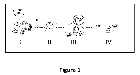
Description of the figures.
Figure 1. Schematic representation of the protocol for HHV isolation using
density
gradient separation: the bacterial presentation library (I) expressing HHV on
the
surface of bacteria is briefly incubated (II) with conventional sepharosase
beads
coated with the antigen of interest. Immediately after depositing the mixture
in a
conical Ficoll density gradient (III) tube and centrifuging at 200 g for 1
min, the beads
pass through the sequential selection of the density gradient to the bottom of
the
tube with the bacteria expressing specific HHV, while the unbound bacteria
remain
on the gradient surface. Next, the beads are resuspended and (IV) the
incubating
bacterial clones are isolated in agar plate.
Figure 2 Immunodetection of Spike-SARS-CoV2 in a nitrocellulose membrane with
a
pore size of 0.2 ilm with the HHV of the invention as primary antibody,
followed by
mouse anti-Myc and goat anti-mouse IgG coupled to HRP. It includes photograph
of
each reaction for different nanoantibody selected with the method of the
invention,
as well as qualitative assessment (+, ++, +++)
Figure 3 Dot Blot against recombinant and synthetic polyubiquitin chains, by
the use
of Nb No. 34, mouse anti-myc and HRP-coupled anti-mouse. Nb No. 34 was
expressed
in the plasmid pNae2 (fused with intimin) with labels 6xHis and Myc.
Figure 4 Western Blot against recombinant and synthetic polyubiquitin chains,
by the
use of Nb No.34, mouse anti-myc and mouse anti-HRP. Nb No. 34 was expressed in
BL21 cells by the use of pNae2 (without intimin) with the Nb coding sequence
between 6xHis and Myc.
CA 03178663 2022- 11- 11
Detailed description of the invention
The invention allows for obtaining nanoantibodies quickly and easily, against
any type
of antigen, such as viruses, bacteria or microorganisms usually proteins or
molecules,
among others. But due to its speed, the method of this invention is very
efficient for
the generation of nanoantibodies against pathogens of emerging diseases, such
as
COVI D.
The invention relates to a method of separation of nanoantibodies (HHV)
against a
specific antigen from a nanoantibody expression library, wherein the method
comprises the following steps:
(a) binding the antigen of interest to polymeric beads that allow protein
binding
(b) incubating the microorganisms of the expression library with the beads of
step (a), where the expression library corresponds to microorganisms
transformed
with the cDNA of fragments corresponding to the HHV domains of HHV-producing
animal previously immunized with that antigen of interest, or synthetic
libraries;
(c) placing the beads of step (b) in a tube with an inert medium with a
density
greater than or equal to 1 g/mL and centrifuging for 45 s to 2 minutes at a
rate
between 150 and 250 g;
d) discarding the upper fraction and supernatant with the free
microorganisms, and obtaining the beads with the linked microorganisms, which
correspond to those that express HHV that recognizes the antigen.
Where the beads used are sepharose, cellulose, latex, agarose or nickel and
can be
modified to bind proteins, and have a density greater than the medium used in
(c).
These beads bind to the antigen of interest in a stable manner in a time of
between
2 to 12 hours and then the sites that have not reacted with the antigen are
blocked,
by any means available in the technique.
The bond between the beads and the antigen can be non-covalent covalent.
CA 03178663 2022- 11- 11
Where this inert medium of stage (c) is preferably chosen between Ficol,
percol or
sucrose.
To ensure that the microorganisms in the expression library are expressing HHV
they
should be treated with a protein expression inducer for 2 to 4 hours prior to
step (b).
In one embodiment, the protein expression inducer is IPTG at a concentration
between 20 to 100 M. Later in step (b) the microorganisms of the expression
library
are incubated with the beads attached to the antigen for between 20 to 60
minutes
at room temperature.
Finally, after separation in the density gradient, in step (d) the beads are
washed with
PBS and seeded on agar plates with culture medium in order to obtain isolated
colonies. Where each colony corresponds to a microorganism that expresses an
HHV
capable of binding to the antigen of interest.
First, you must have a nanoantibody or HHV library.
If you want to generate this library yourself, you require a purified antigen,
which can
be obtained with a system of expression of baculovirus, bacteria, yeast or
directly
from its natural source. Next, an animal producing nanoantibodies, such as an
alpaca
or llama, is immunized between one- to four times with 50 to 500 ilg of the
antigen.
The immune response of alpaca serum against the antigen after immunization can
be
observed rapidly and qualitatively by means of Dot Blot analysis, by
immobilization
of the epitope to a nitrocellulose membrane and by the use of alpaca serum as
a
source of primary antibodies; or analytically and comparatively by means of
ELISA by
the use of the complete antigen that is immobilized in the ELISA plate and the
alpaca
serum as a source of primary antibodies. Thus, a bacterial presentation
library of
individual nanoantibody clones is quickly built.
Once the library is obtained, the method of the invention is applied for the
selection
of nanoantibodies based on a density gradient, for example the simple use of
Ficoll,
percol or sucrose. Ficoll is an inexpensive reagent available worldwide
commonly
used for the fragmentation of blood samples. The inventors developed the
method
CA 03178663 2022- 11- 11
of the invention inspired by the main observation that red blood cells
accumulate at
the bottom of the Ficoll density gradient, while PBMCs remain in the upper
fraction.
The inventors created a method using beads made of polymers such as
conventional
sepharose or agarose which, in a density gradient, such as one obtained by the
Ficoll
reagent have shown that the density of the chosen beads was suitable for
precipitating at the bottom of a 15 ml tube. At the same time, the inventors
showed
that, when performing the same test with free bacteria, the bacteria remained
in the
upper fraction. Revealing the possibility of separating free bacteria from
those that
express HHV and bind beads with the antigen, in a density gradient.
To obtain specific clones of HHV or nanoantibodies, a bacterial presentation
system
is usually used, where each bacterium expresses a single type of nanoantibody
in its
outer bacterial membrane after IPTG induction. Therefore, the inventors
obtained
sefarose beads coated with the antigen of interest to bind to individual
bacteria
expressing specific nanoantibodies on their surface against said antigen.
Thus,
bacteria expressing an HHV or nanoantibody that binds to the antigen sink to
the
bottom of the density gradient, while unbound bacteria would remain in the
upper
fraction (Figure 1).
Examples
Example 1. The simple density gradient method of the invention and its use to
obtain nanoantibodies against the Spike protein of SARS-CoV-2
First, the inventors obtained the lyophilized SARS-CoV-2 Spike protein
generated in a
baculovirus expression system. Prior to immunization, Spike protein integrity
was
tested by SDS-Page and Coomassie staining (Figure la). An alpaca named Buddha
(Figure lb) was immunized twice with 100 ilg of the entire Spike protein. The
immune
response of alpaca serum prior to immunization revealed a fortunate basal
cross-
reaction against the Spike protein. Then, after the second immunization, a
significant
increase in IgG antibodies was observed in alpaca serum qualitatively rapidly
by Dot
CA 03178663 2022- 11- 11
Blot analysis, by immobilization of the epitope in a nitrocellulose membrane
and by
the use of alpaca serum as a primary antibody source (Figure 1c). In addition,
the
inventors verified the increase of IgG antibodies (analytically and
comparatively) by
means of ELISA by the use of the complete Spike protein immobilized in the
ELISA
plate and by the use of alpaca serum as a source of primary antibodies (Figure
1d).
Therefore, the inventors quickly built a bacteria presentation library
consisting of 2.3
x 106 clones of individual nanoantibodies with 0.7% vector religation.
Building immunization and HHV libraries
The alpaca immunization process followed the protocol "Animal use in research"
generated by the Bioethics Committee of the Universidad Austral de Chile. One
day
before immunization, 5 ml of blood was collected for preimmune serum testing.
For
immunization (day 1), 100 ilg of full-length SARS-CoV2 spike protein
(SINOBiological)
was used. The cold lyophilized protein was dissolved in 2 ml of adjuvant
(veterinary
vaccine adjuvant, GERBU FAMA) diluted 1:1 in sterile water and injected
subcutaneously into a male alpaca (Vicugna pacos). A total volume of 4 ml was
injected into four different sites of the alpaca. A 5 ml blood sample was
collected
seven days after the first immunization. On day 14, the alpaca was immunized
again
with 100 ilg of Spike and on day 15 a 120 ml sample of blood was collected
from the
jugular vein in tubes containing 3.8% sodium citrate as an anticoagulant. The
noncoagulated blood sample was mixed with the same volume of calcium-free HBSS
medium (Gibco), divided into 10 ml aliquots, and 5 ml of Ficoll-Paque Premium
(GE
Healthcare) was added on top of each aliquot in 15 ml of sterile Falcon tubes.
After
centrifugation (1,200 x rpm, 80 min, room temperature), the PBMC fraction was
recovered from the interface, washed twice in HBSS by centrifugation (3,500 x
rpm,
min), resuspended in 4 ml sterile PBS 1X (Gibco). RNA extraction and cDNA
production were carried out using the commercial RNeasy Mini (Qiagen) kit and
the
QuantiTect Reverse Transcription Kit (Qiagen), respectively. Approximately 2
ill of
each synthesized cDNA was used as a template in a total PCR reaction volume of
50
ill with the oligonucleotides CALL001 (5 -GTC CTG GCT CTC TTC TAC AAG G-3) and
CA 03178663 2022- 11- 11
CALL002 (5 -GGTACGTGCTGTTGAACTGTTCC- 3) (Conrath etal., 2001). The amplified
fragments of ¨0.6 kb, corresponding to the VHH-CH2 domains, and ¨0.9 kb,
corresponding
to the conventional VH-CH1-CH2 domains, were separated into 1.2% low melting
point agarose gel (w/v) and a band of ¨0.6 kb was purified (QIAEX ll Gel
Extraction
Kit, Qiagen). This fragment was used as a template in a second PCR reaction
with
oligonucleotides VHH-Sfi2 (5 -GTC CTC GCA ACT GCG GCC CAG CCGGCC ATG GCT CAG
GTG CAG CTG GTG GA-3') and VHH-Not2 (5- GGA CTA GTG CGG CCG CTG AGG AGA
CGG TGA CCT GGG T-3) to finally obtain the amplified fragments of ¨0.4 kb,
corresponding to VHH domains. The amplified HHV fragments were digested with
Sfi
I and Notl (Thermo Scientific) restriction enzymes and bound at the same sites
of the
purified pNeae2 vector (Salema et al., 2016). The ligations were subjected to
electroporation in cells of E. coli DH10B-T1 R achieving a library size of 2.3
x 106
individual clones, as determined by seeding on LB-Cloramphenicol agar plates
with
glucose at 2% w/v incubated at 30 C. Less than 0.7% of vectors re-linked from
a
control ligation carried out in parallel without the DNA insert was estimated.
The
transformed bacteria were scraped off the plates and stored at -80 degrees in
LB
broth with 30% glycerol.
Once the library was obtained, the inventors applied the method of the
invention for
the selection of nanoantibodies based on a simple density gradient by the use
of
Ficoll. (Figure le).
Coupling epitopes to beads
1 ml of NHS-activated sepharose 4 Fast Flow beads (General Electric) with 2 ml
of cold
HCI 1 mM were washed immediately before use, then washed 5 times with PBS
(Saline phosphate buffer) 1Xsterile cold. 200 ilg of purified protein in PBS
1X were
added to the beads and incubated in rotation until the next day. The groups
that did
not react in the medium were blocked by the addition of ethanolamine at a
final
concentration of 0.5 M. The beads were washed 5 times with PBS 1X and stored
at
4 C.
CA 03178663 2022- 11- 11
Density gradient separation
1 ml of glycerol stock solution from the library was inoculated into a flask
containing
20 ml of LB medium with 25 lg/m1 chloramphenicol and 2% glucose. The flask was
incubated (pre-inoculum) until the next day at 37 C with 200 rpm stirring.
The same
procedure was repeated with control bacteria transformed with a kanamycin-
resistant plasmid (control). The preinoculum was sedimented and resuspended in
LB
medium with 25 lg/mL chloramphenicol and then diluted to 0.02 optical density
at
600 nm in 100 ml of fresh LB medium with 25 lg/mL chloramphenicol without
glucose, incubated at 37 C with 200 rpm stirring to reach an optical density
of 600
nm of 0.45 to 0.6. IPTG was added at a final concentration of 50 M to induce
protein
expression for 3 hours at 30 C and 200 rpm. The OD 600 nm absorbance of the
library
and control bacteria cultures was measured. 50 mL of both cultures were washed
three times with 10 mL of PBS 1X filtered. Centrifugation was always at 3000 x
g for
min. Both cultures were resuspended in a final volume of 10 ml of PBS 1X. 2 ml
of
library culture and 2 ml of control culture were mixed (if the final 600 nm
optical
density was the same, if not, the volume of the control bacteria was adjusted
according to the OD to ensure an equal number of bacteria) and incubated with
300
111_ of beads coupled to the epitope in a 15 ml conical tube on a rotating
platform for
30 min at temperature environment. The mixture was added to Ficoll 6 ml
(Ficoll-
PaqueTM PLUS GE Healthcare) in a 15 ml conical tube and centrifuged at 200xg
for 1
min. The unbound fraction was discarded (upper fractions).
The visible bead sediment contains bacteria that express an HHV that binds to
antigens, in this case Sars-CoV2 Spike. This sediment was resuspended in 4 ml
of PBS
lx and rotated for 5 min at room temperature. This step was repeated six
times, in
order to eliminate any bacteria not attached to the beads.
In order to amplify the bacteria attached to the beads, 1 mL of LB medium was
added
and incubated for 5 min at room temperature, then 50 111_ were seeded in LB
agar
plates with 25 lg/mL chloramphenicol and 2% glucose a, incubated at 37 C
until the
CA 03178663 2022- 11- 11
next day (> 20 hours recommended).
Where each colony obtained corresponds to a bacterium containing a
nanoantibody
or HHV that binds specifically to the antigen of interest.
Binding test of selected HHV to SARS-CoV-2 spike
To verify that the nanoantibodies of the invention bind to the Spike protein,
an
immunoblot was made for the 22 nanoantibodies, which faced Spike-GFP expressed
in natural viral conditions in human cells (Figure 2).
It was observed that all nanoantibodies selected by the method of the
invention were
attached to the Spike antigen of SARS-CoV-2. The results demonstrate that the
method of the invention allows to quickly and efficiently select
nanoantibodies that
bind specifically to an antigen of interest, such as the SARS-CoV2 Spike.
Ubiquitin
Ubiquitin is the founding member of the UBL family. It participates in many
biological
processes, but its most studied effect is its covalent conjugation with other
proteins,
which targets proteins modified for proteasome-mediated degradation. Ubiquitin
is
one of the most conserved proteins in eukaryotes, however, it is found neither
in
bacteria nor in archaea. In humans, 14 copies of ubiquitin are expressed from
different loci. Ubiquitin translates as an inactive precursor that is
activated by means
of a proteolytic cleavage immediately behind its amino acid glycine 76 by the
same
enzymes also responsible for the deubiquitination of ubiquitin substrates.
Mature
and active ubiquitin is a small globular protein of 76 amino acids (-8 KDa)
[107].
Covalent modification of proteins by ubiquitin (ubiquitination) is defined as
the
formation of an isopeptide bond between the E-amino group of a lysine in the
target
protein and the C-terminal glycine (G76) of ubiquitin. Ubiquitination occurs
in
different ways that regulate biological processes. It changes the specific
properties of
the target proteins that depend on i) the target itself and ii) the way
ubiquitin binds.
Ubiquitination of proteins with a single ubiquitin molecule (mono-
ubiquitination) has
CA 03178663 2022- 11- 11
been described as regulating biological processes such as endocytosis,
trafficking, and
gene expression regulation.
To exemplify the method of the invention VHH against ubiquitin were developed.
Example 2. The simple density gradient method of the invention and its use to
obtain nanoantibodies against ubiquitin
First, the inventors obtained the various chains of polyubiquitins (K6, K11,
K27, K29,
K33, K48 and K63). An alpaca named Nick was immunized four times with 100 ilg
of
the K48 polyubiquitin chain. Then, after the third immunization, a significant
increase
in IgG antibodies was observed in alpaca serum qualitatively rapidly by Dot
Blot
analysis, by immobilization of the epitope in a nitrocellulose membrane and by
the
use of alpaca serum as a source of primary antibodies. Therefore, the
inventors
quickly built a bacterial presentation library consisting of 1 x 10⁶ clones of
individual
nanoantibodies with 1.2% vector religation.
Building immunization and HHV libraries
The alpaca immunization process followed the protocol "Animal use in research"
generated by the Bioethics Committee of the Universidad Austral de Chile. One
day
before immunization, 5 ml of blood was collected for preimmune serum testing.
For
immunization (day 1), 100 ilg of polyubiquitin k48 chain was used. The cold
lyophilized protein was dissolved in 2 ml of adjuvant (veterinary vaccine
adjuvant,
GERBU FAMA) diluted 1:1 in sterile water and injected subcutaneously into a
male
alpaca (Vicugna pacos). A total volume of 4 ml was injected into four
different sites
of the alpaca. A 5 ml blood sample was collected seven days after the first
immunization. On day 14, the alpaca was immunized again with 100 ilg of the
polyubiquitin k48 chain and so on for two more immunizations, until a day
after the
fourth immunization a 120 ml sample of blood was collected from the jugular
vein in
tubes containing 3.8% sodium citrate as an anticoagulant. And a library was
generated in the same way as indicated for example 1.
CA 03178663 2022- 11- 11
Once the library was obtained, the inventors applied the method of the
invention for
the selection of nanoantibodies based on a simple density gradient.
Coupling epitopes to beads
1 ml of NHS-activated sepharose 4 Fast Flow beads (General Electric) with 2 ml
of cold
HCI 1 mM were washed immediately before use, then washed 5 times with cold
sterile PBS. 200 ilg of polyubiquitin k48 chains in PBS1X were added to the
beads and
incubated in rotation until the next day. The groups that did not react in the
medium
were blocked by the addition of ethanolamine at a final concentration of 0.5
M. The
beads were washed 5 times with PBS 1X and stored at 4 C.
Density gradient separation
1 ml of glycerol stock solution from the library was inoculated into a flask
containing
20 ml of LB medium with 25 lg/m1 chloramphenicol and 2% glucose. The flask was
incubated (pre-inoculum) until the next day at 37 C with 200 rpm stirring.
The same
procedure was repeated with control bacteria transformed with a kanamycin-
resistant plasmid (control). The preinoculum was sedimented and resuspended in
LB
medium with 25 ilg mL-1 chloramphenicol and then diluted to 0.02 OD 600 nm in
100 ml of fresh LB medium with 25 ilg mL-1 chloramphenicol without glucose,
incubated at 37 C with 200 rpm stirring to a OD 600 nm of 0.45 to 0.6. IPTG
was
added at a final concentration of 50 11M to induce protein expression for 3
hours at
30 C and 200 rpm. The OD 600 nm absorbance of the library and control
bacteria
cultures was measured. 50 mL of both cultures were washed three times with 10
mL
of filtered PBS 1C. Centrifugation was always at 3000 x g for 5 min. Both
cultures were
resuspended in a final volume of 10 ml of PBS1X. 2 ml of library culture and 2
ml of
control culture were mixed (if the final 600 nm optical density was the same,
if not,
the volume of the control bacteria was adjusted according to the optical
density to
ensure an equal number of bacteria) and incubated with 300 L of NHS beads
coupled
to the epitope protein in a 15 ml conical tube on a rotating platform for 30
min at
room temperature. The mixture was added to Ficoll 6 ml (Ficoll-PaqueTM PLUS GE
Healthcare) in a 15 ml conical tube and centrifuged at 200xg for 1 min.
Unbound
CA 03178663 2022- 11- 11
fraction was discarded (upper fractions)
The visible bead sediment contains bacteria that express an HHV that binds to
antigens, in this case ubiquitin. This sediment was resuspended in 4 ml of
PBS1X and
rotated for 5 min at room temperature. This step was repeated six times, in
order to
eliminate any bacteria not attached to the beads.
In order to amplify the bacteria attached to the beads, 1 mL of LB medium was
added
and incubated for 5 min at room temperature, then 50 ilL were seeded in LB
agar
plates with 25 ilg/mL chloramphenicol and 2% glucose incubated at 37 C until
the
next day (> 20 hours recommended).
Where each colony obtained corresponds to a bacterium containing a
nanoantibody
or HHV that binds specifically to the antigen of interest
Confirmation of antigen binding.
The bacterial presentation system expresses nanoantibodies on the surface of
bacteria fused with an intein protein and a myc tag. Buffer conditions were
optimized
to extract nanoantibody-intein fusion from the bacterial membrane and
bacterial
extract was used directly to confirm binding to polyubiquitin chains by dot
blot. After
selection of nanoantibodies using our simple density gradient protocol, 100
colonies
were used to inoculate liquid LB medium and additionally induced for the
expression
of intein nanoantibodies. The cells were lysed under optimized conditions and
the
extract was used as a source of nanoantibodies as primary antibodies for the
selection of antibodies that bind effectively by dot blot of the various
chains of
polyubiquitins immobilized in a nitrocellulose membrane.
They were revealed by sequential incubation with mouse anti-myc antibody and
an
antibody conjugated with anti-mouse HRP.
In this way, it was possible to identify Nb. No. 34 capable of recognizing all
the
ubiquitin chains tested by Dot Blot (Figure 3) and also by Western Blot
(Figure 4).
This was expressed in the plasmid pNae2 (fused with intimin) with 6xHis and
Myc tags
and then purified by nickel affinity chromatography. To perform the western
blot, the
different chains of polyubiquitins were separated electrophoretically in a 12%
CA 03178663 2022- 11- 11
polyacrylamide gel and then transferred by electrotransfer to a nitrocellulose
membrane which was incubated with the extract of Nb. No. 34 and successively
with
anti-myc antibody and anti-mouse antibody coupled to HRP.
Thus the invention proved useful for quickly and efficiently selecting
nanoantibodies
that bind specifically to an antigen of interest.
CA 03178663 2022- 11- 11