Note: Descriptions are shown in the official language in which they were submitted.
WO 2021/236616
PCT/US2021/032932
SYSTEMS AND METHODS FOR REMOTE DENTAL MONITORING
CROSS-REFERENCE
[0001] This application claims priority to U.S. Provisional Patent
Application No.
63/027,883 filed on May 20, 2020 and U.S. Provisional Patent Application No.
63/144,088
filed on February 1,2021, each of which is incorporated herein by reference in
its entirety for
all purposes.
BACKGROUND
[0002] Dental professionals and orthodontists may treat and monitor
a patient's dental
condition based on in-person visits. Treatment and monitoring of a patient's
dental condition
may require a patient to schedule multiple in-person visits to a dentist or
orthodontist. The
quality of treatment and the accuracy of monitoring may vary depending on how
often and
how consistently a patient sees a dentist or orthodontist. In some cases,
suboptimal treatment
outcomes may result if a patient is unable or unwilling to schedule regular
visits to a dentist
or orthodontist.
SUMMARY
[0003] Recognized herein is a need for remote dental monitoring
solutions to allow dental
patients to receive high quality dental care, without requiring a dental
professional to be
physically present with the patient. Some dental professionals and
orthodontists may use
conventional teledentistry solutions to accommodate patients' needs and
schedules.
However, such conventional teledentistry solutions may not provide adequate
levels of
supervision. Further, such conventional teledentistry solutions may be limited
by an
inaccurate or insufficient monitoring of patients' dental condition based on
one or more
photos taken by the patients, if the photos do not adequately capture various
intraoral features
of interest.
[0004] The present disclosure provides systems and methods for
intraoral imaging to
enhance remote dental monitoring capabilities. The systems and methods
disclosed herein
may provide a convenient solution and user experience for dental patients to
capture one or
more intraoral images or videos using a mobile device such as a smartphone.
The systems
and methods disclosed herein may allow patients to capture improved self-scans
of full dental
arches using a mobile application installed on their mobile device, and may
provide
automated, personalized guidance to the patients to allow the patients to
capture high quality
- 1 -
CA 03179459 2022- 11- 18
WO 2021/236616
PCT/US2021/032932
self-scans that are useful for dentists to monitor and track the patients'
progress during a
dental treatment. The systems and methods disclosed herein may enhance a
patient's ability
to assess or evaluate their dental condition based on one or more full arch
self-scans, and may
provide dentists and orthodontists with a detailed analysis of the patient's
dental condition
based on one or more full arch scans captured remotely by the patient.
100051 The systems and methods of the present disclosure may also
be used to provide
dental patients with an intuitive, user-friendly interface for remote dental
scanning without
requiring assistance from a dentist or a dental assistant. The intuitive, user-
friendly interface
may be provided as part of a software application that provides step-by-step
guidance for
dental patients to acquire images and videos of one or more intraoral regions
that are of
sufficient quality and detail to enable dentists to accurately assess a need
for dental treatment,
a need to update or modify a dental treatment, or a patient's compliance with
the dental
treatment The software applications disclosed herein may also permit dentists
to request and
view high quality images or videos of a patient's teeth so that the dentist
can continue to
monitor the patient's teeth or treatment progress without being physically
present and without
having to provide personalized or customized instructions for how to acquire
the intraoral
images or videos. The software applications disclosed herein may also provide
a convenient
way for dentists to monitor dental patients and dental treatment progress when
the dental
patients are unable to physically travel to the dentist for treatment or
treatment monitoring
(e.g., due to shelter-in-place orders). The systems and methods of the present
disclosure may
provide dentists and dental patients with the ability to continue treatment or
treatment
monitoring while practicing social distancing, thereby reducing a possibility
of the
transmission of infectious diseases.
100061 In an aspect, the present disclosure provides a computer-
implemented method for
remote dental monitoring. The method comprises (a) providing a patient portal
for one or
more patients to remotely communicate with a caregiver, wherein the patient
portal
comprises a graphical user interface that is configured to aid the one or more
patients in
capturing one or more dental scans, wherein the one or more dental scans
comprise (i) one or
more intraoral videos and (ii) a plurality of images derived from the one or
more intraoral
videos; and (b) providing the one or more dental scans to the caregiver for an
assessment of a
dental condition based on the one or more dental scans. In some embodiments,
the
assessment of the dental condition comprises one or more annotations to the
one or more
dental scans. In some embodiments, the assessment of the dental condition
comprises at least
one of audio commentary and visual commentary to the one or more dental scans.
In some
- 2 -
CA 03179459 2022- 11- 18
WO 2021/236616
PCT/US2021/032932
embodiments, the assessment of the dental condition comprises an audio or
video recording
of the caregiver providing commentary or annotations to the one or more dental
scans.
100071 In some embodiments, the caregiver may comprise a dentist,
an orthodontist, an
oral surgeon, a dental staff practitioner, or an individual having one or more
dental
specialties. In some embodiments, the plurality of images may be derived from
one or more
frames of the one or more intraoral videos. The plurality of images may
comprise a plurality
of intraoral images of one or more intraoral regions of the patient. The one
or more dental
scans may further comprise one or more selfie videos or selfie images of the
one or more
patients.
100081 In some embodiments, the graphical user interface may be
configured to provide
visual, textual and/or audio guidance to aid the one or more patients in
capturing the one or
more dental scans. The graphical user interface may be configured to provide
the one or
more patients with a patient-specific treatment timeline comprising one or
more customized
treatment milestones and dates. The graphical user interface may be configured
to prompt
the one or more patients to capture the one or more dental scans on or before
the one or more
treatment milestones and dates. The patient-specific timeline may be
configured to display a
plurality of dental scans captured by a patient for each treatment milestone
and date. The
plurality of dental scans may be arranged in chronological order.
100091 In some embodiments, the one or more dental scans may be
captured using one or
more cameras of a mobile device. The mobile device may be coupled to an
intraoral adapter
comprising a viewing channel for capturing the one or more dental scans. The
viewing
channel may comprise a hexagonal or a polygonal rounded cross-sectional shape.
The one or
more dental scans may comprise a dental imaging region in which one or more
dental
features or intraoral regions are visible or displayed after the one or more
dental scans are
captured. The dental imaging region may be in a shape that is configured to
enable the one or
more patients to capture one or more intraoral videos or intraoral images of a
molar region
while minimizing a movement of an image capture device that is used to capture
the one or
more dental scans, so as to provide stabilization to enable consistent image
alignment for
processing. In some cases, the shape may be a hexagon or a rounded polygon.
100101 In some embodiments, the visual guidance may comprise one or
more visual
markings or guides for alignment with one or more teeth of the one or more
patients. The
one or more dental scans may be automatically captured when the one or more
teeth of the
one or more patients are aligned with the one or more visual markings or
guides. The visual
guidance may comprise one or more visual markings or guides for the one or
more patients to
- 3 -
CA 03179459 2022- 11- 18
WO 2021/236616
PCT/US2021/032932
adjust a position or an orientation of a camera of a mobile device to align
one or more teeth
with the one or more visual markings or guides. The textual guidance may
comprise textual
instructions for the one or more patients to attach a mobile device to an
intraoral adapter and
to move the mobile device or the intraoral adapter to capture the one or more
dental scans.
The textual instructions may be configured to instruct the one or more
patients to adjust a
position or an orientation of the mobile device or the intraoral adapter to
capture the one or
more dental scans.
100111 In some embodiments, the visual, textual and/or audio
guidance may comprise
one or more video tutorials containing examples to guide the one or more
patients for
capturing the one or more dental scans. The visual, textual and/or audio
guidance may
comprise one or more software tools to assist the one or more patients in
performing the one
or more dental scans. The one or more software tools may comprise a scan
module that is
configured to detect an orientation of a mobile device used to capture the one
or more dental
scans. The scan module may be configured to (1) pause the capture of the one
or more dental
scans, and/or (2) generate a visual or audio prompt to the one or more
patients, upon
detecting that the mobile device is not positioned in a predefined orientation
for capturing the
one or more dental scans. In some cases, the predefined orientation may be a
landscape
orientation of the mobile device. In some embodiments, the visual, textual
and/or audio
guidance may comprise a set of instructions to the one or more patients for
retaking the one
or more dental scans.
100121 In some embodiments, the treatment milestones and dates may
be influenced in
part by one or more factors including an aligner replacement schedule. The
aligner
replacement schedule may be associated with a plurality of trays containing a
set of aligners
that are designed to be worn by the one or more patients in a sequence. The
plurality of trays
may be provided to the one or more patients in a pre-locked state. Unlocking
of one or more
of the trays may be contingent upon the one or more patients successfully
completing one or
more of the customized treatment milestones. In some cases, a code may be
generated upon
successful completion of a particular treatment milestone by the one or more
patients,
wherein the code is useable to unlock a tray containing a new aligner to be
worn by the one
or more patients. In some cases, the code may comprise an alphanumeric code, a
barcode, or
a QR code.
100131 In some embodiments, the patient portal may comprise a chat
user interface that is
configured to facilitate remote communication between the patient and the
dentist. The chat
user interface may be configured to display the one or more dental scans
captured by the one
- 4 -
CA 03179459 2022- 11- 18
WO 2021/236616
PCT/US2021/032932
or more patients to enable viewing and access by the caregiver and the one or
more patients
while chatting.
[0014] In another aspect, a computer-implemented method for remote
dental monitoring
is provided. The method comprises providing a caregiver portal for one or more
caregivers to
remotely monitor a dental condition of one or more patients, wherein the
caregiver portal
comprises a graphical user interface that is configured to display one or more
dental scans
captured by the one or more patients, wherein the one or more dental scans
comprise (i) one
or more intraoral videos and (ii) a plurality of images derived from the one
or more intraoral
videos.
[0015] In some embodiments, the caregiver portal may comprise a
task board interface
configured to provide caregivers with information on one or more tasks
associated with
incoming patients, current patients, and preparation for dental treatment of
the incoming
patients or the current patients The caregiver portal may comprise a chat user
interface that
is configured to facilitate remote communication between the caregiver and the
patient. The
chat user interface may be configured to display the one or more dental scans
captured by the
one or more patients to enable viewing and access by the caregiver and the one
or more
patients while chatting. The chat interface may be configured to display the
one or more
dental scans according to a patient-specific treatment timeline comprising one
or more
treatment milestone and dates.
[0016] Another aspect of the present disclosure provides a non-
transitory computer
readable medium comprising machine executable code that, upon execution by one
or more
computer processors, implements any of the methods above or elsewhere herein.
[0017] Another aspect of the present disclosure provides a system
comprising one or
more computer processors and computer memory coupled thereto. The computer
memory
comprises machine executable code that, upon execution by the one or more
computer
processors, implements any of the methods above or elsewhere herein.
[0018] Additional aspects and advantages of the present disclosure
will become readily
apparent to those skilled in this art from the following detailed description,
wherein only
illustrative embodiments of the present disclosure are shown and described. As
will be
realized, the present disclosure is capable of other and different
embodiments, and its several
details are capable of modifications in various obvious respects, all without
departing from
the disclosure. Accordingly, the drawings and description are to be regarded
as illustrative in
nature, and not as restrictive.
- 5 -
CA 03179459 2022- 11- 18
WO 2021/236616
PCT/US2021/032932
INCORPORATION BY REFERENCE
100191 All publications, patents, and patent applications mentioned
in this specification
are herein incorporated by reference to the same extent as if each individual
publication,
patent, or patent application was specifically and individually indicated to
be incorporated by
reference. To the extent publications and patents or patent applications
incorporated by
reference contradict the disclosure contained in the specification, the
specification is intended
to supersede and/or take precedence over any such contradictory material.
BRIEF DESCRIPTION OF THE DRAWINGS
100201 The novel features of the invention are set forth with
particularity in the appended
claims. A better understanding of the features and advantages of the present
invention will be
obtained by reference to the following detailed description that sets forth
illustrative
embodiments, in which the principles of the invention are utilized, and the
accompanying
drawings (also "Figure" and "FIG." herein), of which.
100211 FIG. 1 schematically illustrates a method for capturing
intraoral images or videos
using a software application, in accordance with some embodiments.
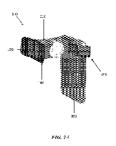
100221 FIGs. 2A, 2B, 2C, 2D, and 2E schematically illustrate an
exemplary intraoral
adapter to which a mobile device can be coupled for intraoral scanning, in
accordance with
some embodiments.
100231 FIG. 3 schematically illustrates a plurality of intraoral
images captured using the
software application, in accordance with some embodiments.
100241 FIG. 4 schematically illustrates a computer system that is
programmed or
otherwise configured to implement methods provided herein
100251 FIGs. 5A, 5B, 5C, and 5D schematically illustrate various
user interfaces for
patients to sign up for remote dental monitoring, in accordance with some
embodiments.
100261 FIGs. 6A, 6B, 6C, and 6D schematically illustrate various
user interfaces for
patients to view their account information and dentist information, in
accordance with some
embodiments
100271 FIGs. 7A, 7B, 7C, and 7D schematically illustrate various
user interfaces for
patients to view tutorials and refer friends, in accordance with some
embodiments.
100281 FIGs. 8A, 8B, 8C, and 8D schematically illustrate a chat
user interface for a
patient-side remote dental monitoring software application, in accordance with
some
embodiments.
- 6 -
CA 03179459 2022- 11- 18
WO 2021/236616
PCT/US2021/032932
100291 FIGs. 9A and 9B schematically illustrate a user interface
for patients to capture
and share dental scans and/or selfie images, in accordance with some
embodiments.
100301 FIG. 9C schematically illustrates a treatment timeline with
milestone dates, in
accordance with some embodiments.
100311 FIG. 9D schematically illustrates a chat user interface that
is configured to enable
patients to capture dental scans, in accordance with some embodiments.
100321 FIGs. 10A, 10B, 10C, and 10D schematically illustrate visual
and/or textual
guidance to aid patients in capturing dental scans, in accordance with some
embodiments.
100331 FIGs. 11A, 11B, 12A, 12B, 13A, 13B, 14A, 14B, 15A, 15B, and
16A
schematically illustrate a plurality of user interfaces for capturing dental
scans using a
patient-side remote dental monitoring software application, in accordance with
some
embodiments
100341 FIGs. 16B, 17A, 17B, 17C, 17D, 18A, and 18B schematically
illustrate a plurality
of user interfaces for capturing selfie scans using a patient-side remote
dental monitoring
software application, in accordance with some embodiments.
100351 FIG. 18C schematically illustrates a plurality of dental
scans and/or selfie scans
that may be sent to a dentist using a patient-side remote dental monitoring
software
application, in accordance with some embodiments.
100361 FIG. 18D schematically illustrates an intraoral video that
may be sent to a dentist
using a patient-side remote dental monitoring software application, in
accordance with some
embodiments.
100371 FIGs. 19A and 19B schematically illustrate a user interface
for a dentist to sign up
for a practitioner-side remote dental monitoring software application, in
accordance with
some embodiments.
100381 FIGs. 20A and 20B schematically illustrate a task dashboard
for a practitioner-
side remote dental monitoring software application, in accordance with some
embodiments
100391 FIGs. 21A, 21B, and 22A schematically illustrate a chat user
interface for a
practitioner-side remote dental monitoring software application, in accordance
with some
embodiments
100401 FIG. 22B schematically illustrates a dentist profile page,
in accordance with some
embodiments.
100411 FIGs. 23A, 23B, 23C, 23D, 24A, and 24B schematically
illustrate a user interface
for a practitioner to review a patient's dental scans, in accordance with some
embodiments.
- 7 -
CA 03179459 2022- 11- 18
WO 2021/236616
PCT/US2021/032932
100421 FIGs. 25A, 25B, 25C, and 25D schematically illustrate
various examples of
annotations that may be provided by a practitioner during a scan review.
100431 FIGs. 26A, 26B, 27, and 28 schematically illustrate a text
box containing a
transcription of audio commentary provided by a practitioner during a scan
review.
DETAILED DESCRIPTION
100441 While various embodiments of the invention have been shown
and described
herein, it will be obvious to those skilled in the art that such embodiments
are provided by
way of example only. Numerous variations, changes, and substitutions may occur
to those
skilled in the art without departing from the invention. It should be
understood that various
alternatives to the embodiments of the invention described herein may be
employed.
100451 The term "real-time," as used herein, generally refers to a
simultaneous or
substantially simultaneous occurrence of a first event or action with respect
to an occurrence
of a second event or action. A real-time action or event may be performed
within a response
time of less than one or more of the following: ten seconds, five seconds, one
second, a tenth
of a second, a hundredth of a second, a millisecond, or less relative to at
least another event
or action. A real-time action may be performed by one or more computer
processors.
100461 Whenever the term "at least,- "greater than," or "greater
than or equal to"
precedes the first numerical value in a series of two or more numerical
values, the term "at
least," "greater than" or "greater than or equal to" applies to each of the
numerical values in
that series of numerical values. For example, greater than or equal to 1, 2,
or 3 is equivalent
to greater than or equal to 1, greater than or equal to 2, or greater than or
equal to 3.
100471 Whenever the term "no more than," "less than," or "less than
or equal to"
precedes the first numerical value in a series of two or more numerical
values, the term "no
more than," "less than," or "less than or equal to" applies to each of the
numerical values in
that series of numerical values. For example, less than or equal to 3, 2, or 1
is equivalent to
less than or equal to 3, less than or equal to 2, or less than or equal to 1.
100481 The terms "a," "an," and "the," as used herein, generally
refer to singular and
plural references unless the context clearly dictates otherwise
100491 Overview
100501 In an aspect, the present disclosure provides systems and
method for remote
dental monitoring. The present disclosure provides systems and methods for
intraoral
imaging to enhance remote dental monitoring capabilities. The systems and
methods
disclosed herein may provide a convenient solution and user experience for
dental patients to
- 8 -
CA 03179459 2022- 11- 18
WO 2021/236616
PCT/US2021/032932
capture one or more intraoral images or videos using a mobile device such as a
smartphone.
The systems and methods disclosed herein may allow patients to capture
improved self-scans
of a full dental arch using a mobile application installed on their mobile
device, and may
provide automated, personalized guidance to the patients to allow the patients
to capture high
quality self-scans that are useful for dentists to monitor and track the
patient's progress
during a dental treatment. The systems and methods disclosed herein may
enhance a
patient's ability to assess or evaluate their dental condition based on one or
more full arch
self-scans, and may provide dentists and orthodontists with a detailed
analysis of the patient's
dental condition based on one or more full arch scans captured remotely by the
patient.
100511 The systems and methods of the present disclosure may also
be used to provide
dental patients with an intuitive, user-friendly interface for remote dental
scanning without
requiring assistance from a dentist or a dental assistant. The intuitive, user-
friendly interface
may be provided as part of a software application that provides step-by-step
guidance for
dental patients to acquire images and videos of one or more intraoral regions
that are of
sufficient quality and detail to enable dentists to accurately assess a need
for dental treatment,
a change in a dental treatment, or a patient's compliance with the dental
treatment. The
software applications disclosed herein may permit dentists to request and view
high quality
images or videos of a patient's teeth so that the dentist can continue to
monitor the patient's
teeth or treatment progress without being physically present and without
having to provide
personalized or customized instructions for how to acquire the intraoral
images or videos.
The software applications of the present disclosure may further provide a
convenient way for
dentists to monitor dental patients and dental treatment progress when the
dental patients are
unable to physically travel to the dentist for treatment or treatment
monitoring (e.g., due to
shelter-in-place orders). The systems and methods of the present disclosure
may provide
dentists and dental patients with the ability to continue treatment or
treatment monitoring
while practicing social distancing, thereby reducing a possibility of the
transmission of
infectious diseases.
100521 In an aspect, the present disclosure provides systems and
methods for remote
monitoring. The systems and methods of the present disclosure may be
implemented using a
software application that is configured to enable a dental patient to capture
images and/or
videos of intraoral regions. The software application may be used by a user or
a subject (e.g.,
a dental patient) in conjunction with a mobile device to remotely monitor a
dental anatomy or
a dental condition of the subject. A dental anatomy may comprise one or more
dental
structures of the patient, including one or more tooth structures or dental
arches of the
- 9 -
CA 03179459 2022- 11- 18
WO 2021/236616
PCT/US2021/032932
subject. The dental condition may comprise a development, a growth, a
movement, an
appearance, a condition, a physical arrangement, a position, and/or an
orientation of the
subject's teeth. In some cases, the dental condition may comprise a functional
aspect of the
user's teeth, such as how two or more teeth contact each other, how the teeth
move relative to
each other, or how the teeth move over a period of time.
100531 The software application may be used to enable remote dental
monitoring. As
used herein, remote monitoring may refer to monitoring a dental anatomy or a
dental
condition of a patient that is performed at one or more locations remote from
the patient. For
example, a dentist or a medical specialist may monitor the dental anatomy or
dental condition
in a first location that is different than a second location where the patient
is located. The
first location and the second location may be separated by a distance spanning
at least about 1
meter, 1 kilometer, 10 kilometers, 100 kilometers, 1000 kilometers, or more
100541 The remote monitoring may be performed by assessing a dental
anatomy or a
dental condition of the subject using one or more intraoral images or videos
captured by the
subject when the patient is located remotely from the dentist or a dental
office. In some
cases, the remote monitoring may be performed in real-time such that a dentist
is able to
assess the dental anatomy or the dental condition when a subject uses a mobile
device to
acquire one or more intraoral images or videos of one or more intraoral
regions of interest in
the patient's mouth. The remote monitoring may be performed using equipment,
hardware,
and/or software that is not physically located at a dental office.
100551 The software application may be configured to run on a
mobile device. The
mobile device may comprise a smartphone, a tablet, a laptop, or any suitable
computing
device that may be used by a patient to capture one or more dental scans. The
software
application may be installed on a mobile device of a patient undergoing a
dental treatment or
who will be undergoing a dental treatment. The software application may be a
patient-side
software application.
100561 In some cases, the patient-side software application may be
used in a compatible
manner with a practitioner-side software application that is accessible by a
caregiver. The
patient-side software application and the practitioner-side software
application may enable
real-time communication and sharing of images, videos, or data between one or
more patients
and one or more caregivers. The one or more caregivers may comprise, for
example, a
dentist, an orthodontist, an oral surgeon, individuals having one or more
dental specialties, or
a dental staff practitioner. The practitioner-side software application may be
implemented
using a computer, a mobile device, or a server (e.g., a cloud server). The
practitioner-side
- 10 -
CA 03179459 2022- 11- 18
WO 2021/236616
PCT/US2021/032932
software application may be accessed through a computer, a mobile device, or a
web
interface.
100571 Patient Portal
100581 The software application may be configured to implement a
computer-
implemented method for remote dental monitoring. The method may comprise (a)
providing
a patient portal for one or more patients to remotely communicate with a
caregiver. The
patient portal may comprise a graphical user interface that is configured to
aid the one or
more patients in capturing one or more dental scans. In some cases, the one or
more dental
scans may comprise one or more intraoral images or videos. In other cases, the
one or more
dental scans may comprise one or more intraoral videos and/or one or more
images derived
from the one or more intraoral videos. The method may further comprise (b)
providing the
one or more dental scans to a caregiver for an assessment of a dental
condition based on the
one or more dental scans
[0059] The patient portal may be an interactive visual interface
that can be displayed on a
mobile device of a patient. The patient portal may be navigated, manipulated,
and/or updated
using one or more user gestures or inputs (e.g., taps, swipes, etc.) that are
provided to the
mobile device. The patient may use the patient portal to remotely communicate
with a
caregiver who wishes to monitor a dental condition of the patient.
100601 The patient portal may comprise a chat user interface that
is configured to
facilitate remote communication between the patient and the dentist. The chat
user interface
may provide a chat window for the patient to send text, images, videos, and/or
data to the
dentist. The chat user interface can provide a virtual chat room for the
patient to
communicate with the dentist, dental staff or assistants, receptionist, etc.
Likewise, the
dentist or any of the dental staff/assistants may use the chat user interface
to send text,
images, videos, and/or data to the patient.
100611 The chat user interface may be configured to display one or
more dental scans
captured by a patient. The patient may use the chat user interface to send one
or more dental
scans captured by the patient to the dentist. In such instances, the chat user
interface may
enable the dentist to view and/or access the one or more dental scans while
communicating
with the patient (e.g., through an exchange of text messages, images, videos,
data, etc.) The
chat user interface may also permit the patient and the dentist to discuss the
patient's dental
treatment, dental condition, and/or any specific visual features in the one or
more dental
scans, without having to navigate to a separate user interface to view (i) the
patient's dental
scans or (ii) any images, videos, or data associated with the patient's dental
treatment or
- 11 -
CA 03179459 2022- 11- 18
WO 2021/236616
PCT/US2021/032932
dental condition. In addition to improving ease of use, the functionality and
layout of the
chat user interface may enhance efficient communication between the patient
and dentist so
that the dentist can remotely provide a quick and convenient assessment of the
patient's
dental condition or dental treatment.
[0062] The patient portal may be configured to enable the patient
to capture one or more
dental scans. The dental scans may be of a dental feature of the patient. The
dental feature
may comprise, for example, the patient's teeth, gums, dental arches, and/or
any treatment
devices (e.g., braces or aligners) in contact and/or proximal to the user's
teeth, gums, or
dental arches.
[0063] The one or more dental scans may comprise one or more
intraoral images of a
dental feature of the patient. The one or more dental scans may comprise one
or more
intraoral videos of a dental feature of the patient. In some cases, the
intraoral images may be
derived from the one or more intraoral videos The intraoral images may
comprise one or
more still images associated with one or more frames of the intraoral videos.
[0064] The dental scans may comprise a plurality of intraoral
images or a plurality of
intraoral videos of one or more intraoral regions within the patient's mouth.
The one or more
intraoral regions may comprise one or more dental features as described above.
100651 In some cases, the one or more dental scans may further
comprise one or more
selfie videos or selfie images of the one or more patients. The selfie videos
or selfie images
may be used to visualize an appearance or a change of an appearance of the
user's smile, jaw
structure, cheeks, mouth, teeth, dental arches, etc. before, during, and/or
after a dental
treatment.
[0066] Capturing Scans
[0067] In some cases, the patient portal may comprise a graphical
user interface that is
configured to aid the one or more patients in capturing one or more dental
scans. In some
cases, the graphical user interface may comprise an image capture user
interface which may
be launched through the chat user interface described herein. The chat user
interface may
include a camera icon that may be pressed or tapped by a patient to launch the
image capture
user interface to capture one or more dental scans.
[0068] The image capture user interface may be displayed on a
screen of the patient's
mobile device. The patient may attach a mobile device to a compatible
intraoral adapter to
capture the dental scans. The intraoral adapter may provide an imaging channel
for the
mobile device to capture the dental scans. The screen of the mobile device may
be oriented
away from the patient when coupled to the intraoral adapter so that a rear
camera of the
- 12 -
CA 03179459 2022- 11- 18
WO 2021/236616
PCT/US2021/032932
mobile device may be used to capture the dental scans. In such cases, the
patient may face a
mirror and use a reflected mirror image of the patient to view and manipulate
the image
capture user interface displayed on the screen of the patient's mobile device.
100691 The image capture user interface may show an imaging window
that is configured
to display a live, real-time visual representation of an imaging region that
is visible to the one
or more cameras of the mobile device. The patient may tap an image capture
button to
capture one or more images, videos, or dental scans corresponding to the live,
real-time
visual representation of the imaging region displayed in the imaging window.
The image
capture user interface may further comprise an image gallery button that may
be pressed by
the patient to access images, videos, or dental scans already captured by the
patient using the
mobile device. The image capture user interface may further comprise a camera
swap button
that may be pressed by the patient to switch between one or more cameras
(e.g., a front
camera or a rear camera) of the patient's mobile device The images, videos,
and/or dental
scans captured using the one or more cameras of the mobile device may be
shared with or
sent to a dentist through the chat user interface described herein.
100701 The one or more dental scans may be captured using one or
more cameras of a
mobile device. In some cases, the mobile device may be coupled to a compatible
intraoral
adapter comprising a viewing channel for capturing the one or more dental
scans. The
intraoral adapter may be configured to provide an imaging region for the one
or more
cameras of the mobile device. One or more of the patient's dental features or
intraoral
regions may be viewable within the imaging region, which may be defined by a
size and/or a
shape of the intraoral adapter. In some cases, the viewing channel of the
intraoral adapter
may comprise a hexagonal or a polygonal rounded cross-sectional shape. In some
cases, the
shape and/or size of the viewing channel may provide or define one or more
main imaging
regions for imaging a first intraoral region and one or more peripheral
imaging regions for
imaging a second intraoral region (e.g., the patient's molar regions) Using a
compatible
intraoral adapter may provide multiple benefits, such as consistently defining
an appropriate
or suitable dental imaging region and standardizing a distance between the
cameras of the
patient's mobile device and the one or more dental features the patient wishes
to image.
100711 In some cases, the dental imaging region may comprise a
shape that is configured
to enable the patient to capture one or more intraoral videos or intraoral
images of a molar
region while minimizing a movement of the mobile device and the intraoral
adapter that is
used to capture the one or more dental scans, so as to provide stabilization
to enable
consistent image alignment for (e.g., for subsequent image processing). The
shape of the
- 13 -
CA 03179459 2022- 11- 18
WO 2021/236616
PCT/US2021/032932
dental imaging region may be a circle, an ellipse, an oval, or a polygon with
three or more
side. In some cases, the dental imaging region may be in the shape of a
hexagon or a rounded
polygon. In such cases, the dental scans captured using the mobile device and
the intraoral
adapter may be framed within a hexagonal shape as shown in FIG. 3.
100721 In some cases, the intraoral adapter may further comprise
one or more sensors
configured to automatically detect when a patient' s smartphone camera and the
intraoral
adapter are optically aligned to capture one or more dental scans. In some
cases, the intraoral
adapter may be configured to permit dynamic adjustment of lighting,
illumination, brightness,
contrast, and/or any other imaging conditions so that the patient may take
suitable dental
scans using the intraoral adapter and the patient's mobile device. In some
cases, the intraoral
adapter may be configured to permit the patient to take a plurality of images
with different
lighting conditions or illumination conditions to reveal certain
characteristics that are only
apparent or visible under such conditions The plurality of images may comprise
one or more
bright-field images and/or one or more dark-field images. In some cases, the
intraoral
adapter may be configured to adjust an amount of light that is transmitted
through the
intraoral adapter (e.g., from a camera flash of the patient's mobile device).
100731 Visual / Textual / Audio Guidance
100741 In some cases, the graphical user interface and/or the image
capture user interface
of the patient-side software application may be configured to provide the
patient with visual,
textual, and/or audio guidance to aid the patient in capturing the one or more
dental scans.
The visual and/or textual guidance may be visible on the display of the
patient's mobile
device and may be viewed by the patient using a reflected mirror image that is
visible to the
patient when the patient is positioned in front of a mirror or another
reflective surface. The
patient-side software application may be configured to provide audio guidance,
for example a
voice over to assist the patient with the proper capture of the dental scans.
The audio
guidance may be transmitted via a speaker on the patient's mobile device. The
visual,
textual, and/or audio guidance may include one or more video tutorials
containing examples
to guide the one or more patients.
100751 In some cases, the textual guidance may comprise textual
instructions for the
patient to attach a mobile device to a compatible intraoral adapter, to place
the intraoral scope
over the patient's mouth and face a mirror, to bite down and/or smile, to turn
the patient's
head relative to the intraoral scope, to open the patient's mouth for imaging
of top or bottom
dental arches, and/or to move the mobile device or the intraoral adapter
relative to one or
more dental features or intraoral regions of the patient to capture the one or
more dental
- 14 -
CA 03179459 2022- 11- 18
WO 2021/236616
PCT/US2021/032932
scans. In some cases, the textual instructions may be configured to instruct
the patient to
adjust a position or an orientation of the mobile device or the intraoral
adapter relative to the
one or more dental features or intraoral regions to capture the dental scans.
In some cases,
the textual instructions may instruct the patient to adjust a position or an
orientation of the
patient's head, face, or mouth relative to the mobile device or the intraoral
adapter to capture
the one or more dental scans.
100761 In some cases, the visual, textual, and/or audio guidance
may include one or more
software tools to assist the one or more patients in performing a scan. The
software tools
may include a scan module that is configured to detect an orientation of a
mobile device used
to capture the one or more dental scans. The scan module can be configured to
(1) pause the
capture of the one or more dental scans, and/or (2) generate a textual or
audio prompt to the
one or more patients, if the mobile device is not positioned in a predefined
orientation (e.g.
landscape orientation) for capturing the one or more dental scans
100771 In some cases, the visual guidance may provide a
visualization of how to attach
the mobile device to the intraoral adapter. In some cases, the visual guidance
may comprise
one or more visual markings or guides for the one or more patients to adjust a
position or an
orientation of a camera of a mobile device to align one or more teeth with the
one or more
visual markings or guides. The one or more visual markings or guides may be
used at least in
part to perform an alignment between the camera of the mobile device and one
or more dental
features or intraoral regions of the patient. The visual markings or guides
may comprise one
or more dots, lines, shapes (e.g., regular shapes, irregular shapes, and/or
amorphous shapes),
or other two-dimensional or three-dimensional features that the patient may
use to align a
camera of the mobile device with one or more dental features or intraoral
regions of the
patient. In some cases, the visual marking or guides may be sized and/or
shaped to
approximate a profile, a shape, or an arrangement of the patient's teeth or
dental arches. The
patient-side software application may be configured to automatically capture
the one or more
dental scans when one or more dental features or intraoral regions of the
patient are aligned
with the one or more visual markings or guides, or a subset thereof
100781 After one or more intraoral images or videos are captured,
the patient-side
software application may provide visual and/or textual instructions for the
patient to capture
one or more selfie images or videos. The selfie images or videos may be
captured using the
mobile device of the patient. The mobile device may not or need not be coupled
to the
intraoral adapter to capture the selfie images. The visual and/or textual
instructions may
instruct the patient to relax the patient's lips, to smile, and to take a
picture of the patient's
- 15 -
CA 03179459 2022- 11- 18
WO 2021/236616
PCT/US2021/032932
face. The visual and/or textual instructions may instruct the patient to turn
the patient's face
(e.g., up, down, left, and/or right) or to face forward towards a camera of
the mobile device.
100791 After the intraoral images/videos and the selfie
images/videos are captured, the
patient-side software application may be configured to present the patient
with an option to
retake the images and/or videos if needed or desired. As an example, the
visual, textual,
and/or audio guidance disclosed elsewhere herein may include instructions to
the patient for
retaking the one or more dental scans. In some cases, the patient-side
software application
may be configured to present the patient with an option to send the images
and/or videos to a
dentist via the chat user interface.
100801 Patient timeline / milestones
100811 In some cases, the graphical user interface of the patient-
side software application
may be configured to provide the patient with a patient-specific treatment
timeline associated
with a dental treatment of the patient In some cases, the dental treatment may
comprise
changing a layout, an arrangement, a position, or an orientation of the
patient's teeth using
aligners or braces. The patient-specific treatment timeline may comprise one
or more
treatment milestones and dates. The treatment milestones and dates may
comprise dates on
which the dentist wishes to check up on the patient or to assess a progress of
the dental
treatment. The patient-specific treatment timeline may comprise at least 1, 2,
3, 4, 5, 6, 7, 8,
9, 10, or more treatment milestones and dates.
100821 In some cases, the graphical user interface of the patient-
side software application
may be configured to prompt the patient to capture the one or more dental
scans on or before
the one or more treatment milestones and dates. In some cases, the patient-
specific timeline
may be configured to display a plurality of dental scans captured by the
patient for each
treatment milestone and date.
100831 The patient-specific treatment timeline may be updated based
on the dental scans
captured by the patient using a mobile device and/or the intraoral adapter.
The treatment
milestones and dates may likewise be updated based on the dental scans
captured by the
patient. In some cases, the patient-specific treatment timeline and/or the
treatment milestones
and dates may be updated based on one or more trends associated with the
dental scans
captured by the patient over a period of time.
100841 In some cases, the patient-specific treatment timeline may
be a predicted patient-
specific treatment timeline that is generated based on one or more predictions
of how long a
dental treatment may take, or an estimated progress of the dental treatment
over a period of
time based on patient-specific parameters. The patient-specific parameters may
be associated
- 16 -
CA 03179459 2022- 11- 18
WO 2021/236616
PCT/US2021/032932
with a dental condition of the patient or one or more physical characteristics
(e.g., layout,
arrangement, relative position, and/or relative orientation) of the patient's
dental features.
The predicted patient-specific treatment timeline may be updated based on one
or more
dental scans captured by the patient using the systems and methods disclosed
herein. The
predicted patient-specific treatment timeline may be updated based on one or
more dental
scans captured by the patient for one or more treatment milestones and dates
associated with
the predicted patient-specific treatment timeline.
100851 In any of the embodiments described herein, the treatment
timeline associated
with a patient's dental treatment may be automatically and dynamically
changed, modified,
or updated in real-time based on the dental scans captured by the patient.
Likewise, the steps
or the milestones or dates associated with the patient's dental treatment may
be automatically
and dynamically changed, modified, or updated in real-time based on the dental
scans
captured by the patient In some cases, new steps and/or milestones may be
added to the
dental treatment plan to influence or change the course of treatment. The new
steps and/or
milestones may be inserted chronologically between two or more existing steps
or
milestones. In some cases, one or more existing milestones associated with the
patient's
dental treatment can be changed, deleted, or moved anywhere along the
patient's treatment
timeline. In some cases, one or more portions of the treatment timeline may be
expedited or
compressed if the dental scans suggest that the patient is showing faster than
expected
progress and/or a favorable response during the course of treatment.
100861 In any of the embodiments described herein, the treatment
timeline may
graphically display changes in the treatment timeline or treatment milestones
to the patient
once the patient's dental scans are assessed or analyzed. The treatment
timeline may be
configured to keep the user informed as to the current stage of the dental
treatment, what
steps or milestones the patient has completed, and what steps or milestones
the patient can
expect next or in the near future. The patient-side software application may
be configured to
permit the patient to click on any point or milestone along the treatment
timeline, to scroll
along the treatment timeline and view previously captured dental scans, to
annotate one or
more portions of the treatment timeline with questions or comments for the
dentist or
caregiver to address. The patient-side software application may be further
configured to send
alerts or notifications to the patient in real-time about changes in the
patient's treatment
timeline or changes to one or more milestones associated with the patient's
treatment
timeline.
- 17 -
CA 03179459 2022- 11- 18
WO 2021/236616
PCT/US2021/032932
100871 In some cases, the dental scans may be visually analyzed by
the dentist or a dental
assistant to determine a dental condition of the patient. In some cases, the
dental scans may
be visually analyzed by the dentist or a dental assistant to assess the
patient's progress in
relation to a dental treatment plan or a predicted patient-specific treatment
timeline associated
with the dental treatment plan. The dentist may use the visual analysis of the
dental scans to
update or change the dental treatment plan, the patient-specific treatment
timeline, or one or
more treatment milestones and dates associated with the patient-specific
treatment timeline.
In other cases, the dental scans may be analyzed and/or processed using an
image processing
algorithm.
100881 In some cases, the treatment milestones and dates may be influenced in
part by one or
more factors, such as an aligner replacement schedule. The aligner replacement
schedule
may be associated with a plurality of trays containing a set of aligners that
are designed to be
worn by the patient in sequence In some cases, the plurality of trays may be
provided to the
patient in a pre-locked state. When the patient successfully completes a
particular treatment
milestone at a certain point, the patient portal described herein can generate
a code to the
patient. The code may be specific or unique to a tray containing a prescribed
aligner for
achieving the next treatment milestone. The patient can then use the code to
unlock the tray
to access the new aligner.
100891 The code for unlocking aligner trays may comprise an alphanumeric code,
a barcode,
or a QR code. Other examples of codes that may include Aztec, ColorCode, Color
Construct
Code, CrontoSign, CyberCode, d-touch, DataGlyphs, Data Matrix, Datastrip Code,
Digimarc
Barcode, DotCode, DWCode, EZcode, High Capacity Color Barcode, Han Xin
Barcode,
HueCode, InterCode, MaxiCode, Mobile Multi-Colored Composite (MMCC), NexCode,
PDF417, Qode, ShotCode, Snapcode, SPARQCode, VOICEYE, a modification thereof,
or a
combination thereof The code can be of any image format (e.g. EPS or SVG
vector graphs,
PNG, GIF, or JPEG raster graphics format)
100901 Controlling access to the aligner trays can help to increase patient
compliance and
improve quality of care. The trays can be numbered or marked sequentially to
allow the user
to progress to the next tray upon completion of the previous step/milestone.
In some cases,
the trays need not be numbered or marked sequentially, and the patient portal
may reveal the
next tray only upon successful completion of a required action by the patient
(for example,
completion of a milestone scan), or based on a message from the patient to the
caregiver, or
based on an authorization from the caregiver permitting the patient to access
the next tray.
- 18 -
CA 03179459 2022- 11- 18
WO 2021/236616
PCT/US2021/032932
The examples above can help to streamline caregiver supervision of the
treatment progress
and effectiveness, and also increase patient compliance with the treatment
process.
100911 Improved User Interaction
100921 In some cases, the patient-side software application may be
configured to
incentivize and/or improve patient interaction and engagement. For example,
the patient-side
software application may be configured to implement one or more aspects of
gamification by
providing incentives, rewards, or points to a patient when the patient
successfully completes
and uploads one or more dental scans. In some cases, the incentives may be
provided by
dental insurance companies (e.g., reductions in insurance payout costs for
patients who
maintain good dental health or comply with their dental treatment plan). The
patient-side
software application may also be configured to provide positive reinforcement
by visually
showing the patient's progress with respect to a dental treatment plan (e.g.
80% towards end
goal or completion of treatment) The patient-side software application may be
configured to
encourage users to collect regular scans, may give personalized messages to
the patient to
encourage regular scanning, and may provide reminders and alerts to the
patient's
smartphone for upcoming treatment milestones and dates. In some cases, the
patient-side
software application may be configured to graphically display a scoreboard
corresponding to
the patient's completion of one or more steps in a dental treatment plan or a
completion of
one or more milestone scans.
100931 Image Processing
100941 In some cases, the dental scans captured using the patient's
mobile device may be
provided to an image processing algorithm. The image processing algorithm may
be
implemented on the mobile device on which the software application is
installed. The image
processing algorithm may be implemented on a remote server or a cloud server.
The image
processing algorithm may be implemented on a computing device that is
accessible by the
caregiver. The image processing algorithm may be configured to process one or
more
intraoral images and/or one or more intraoral videos.
100951 The image processing algorithm may be configured to (i)
process the dental scans
captured using the camera of the mobile device, and (ii) determine a dental
condition of the
subject based at least in part on the processed dental scans. The dental
condition may
comprise (i) a movement of one or more teeth of the subject, (ii) an
accumulation of plaque
on the one or more teeth of the subject, (iii) a change in a color or a
structure of the one or
more teeth of the subject, (iv) a change in a color or a structure of a tissue
adjacent to the one
or more teeth of the subject, and/or (v) a presence or lack of presence of one
or more cavities.
- 19 -
CA 03179459 2022- 11- 18
WO 2021/236616
PCT/US2021/032932
In some embodiments, the image processing algorithm may be configured to
process the
dental scans to (i) predict a movement of one or more teeth of the subject,
(ii) identify enamel
wear patterns, (iii) create or modify a dental treatment plan, or (iv)
generate or update a
medical record associated with the dental condition of the subject. Processing
the dental
scans may comprise comparing a first set of pixel values within a dental scan
to a second set
of pixel values within the dental scan. The pixel values may comprise a value
corresponding
to a color or a brightness of one or more pixels. In some cases, processing
the dental scans
may comprise comparing one or more pixel values within a dental scan to a set
of reference
pixel values within a reference image. The set of reference pixel values may
be accessed
through a database that is located remote from a mobile device of the patient.
In some cases,
the set of reference pixel values may indicate a certain dental condition
(e.g., a presence of
plaque or a presence of cavities). In some cases, processing the plurality of
intraoral images
may comprise comparing a first dental scan to a second dental scan_ Comparing
a first dental
scan to a second dental scan may comprise tracking a movement of one or more
features that
are visible within the first dental scan and the second dental scan. Comparing
a first dental
scan to a second dental scan may comprise tracking a change in a shape of a
subject's dental
arches between the first dental scan and the second dental scan. In some
cases, comparing a
first dental scan to a second dental scan may comprise tracking a change in
one or more pixel
values between the first dental scan and the second dental scan. In some
cases, the first
dental scan and the second dental scan may be obtained within a single
scanning session. In
some cases, the first dental scan may be obtained during a first scanning
session (e.g., at a
first treatment milestone and date) and the second dental scan may be obtained
during a
second scanning session that is initiated after the first scanning session
(e.g., at a second
treatment milestone and date).
100961 The processed dental scans may be used to automatically
update a patient's dental
treatment plan, predicated treatment timeline, and/or the treatment milestones
and dates
associated with the patient's predicted treatment timeline. In some
embodiments, the
processed dental scans may be usable to generate or update a dental treatment
plan. In some
embodiments, the processed dental scans may be usable to track one or more
changes in a
dental structure or a dental condition of the patient over time. In some
embodiments, the
processed dental scans may be usable to assess the subject's actual progress
in relation to a
dental treatment plan based at least in part on a comparison of one or more
changes in the
dental structure or the dental condition of the subject over two or more
dental scans. In some
embodiments, the processed dental scans may be usable to assess the subject's
actual
- 20 -
CA 03179459 2022- 11- 18
WO 2021/236616
PCT/US2021/032932
progress in relation to a dental treatment plan based at least in part on a
comparison of a
planned or estimated change in the dental structure or the dental condition of
the subject and
an actual change in the dental structure or the dental condition of the
subject.
100971 In some cases, processing the dental scans may comprise
classifying the dental
scans based on the dental features present within the dental scans. In some
cases, processing
the dental scans may comprise classifying the dental scans based on a type of
dental
treatment that is being remotely monitored. The dental scans may be used to
build a database
of temporal and treatment-based images.
100981 In some cases, processing the dental scans may comprise
comparing the dental
scans to prior scans or reference scans to determine deviations between a
patient's actual and
predicted treatment progress, or to determine a degree of similarity or
correlation between
actual and predicted treatment progress. In some cases, processing the dental
scans may
comprise identifying areas or sections of dental structures that deviate from
predicted
outcomes. The systems and methods disclosed herein may be configured to
optimize, adjust,
or fine tune dental treatment plans, treatment timelines, and/or treatment
milestones and dates
based on information or data derived from processed dental scans.
100991 Machine Learning / Neural Networks
1001001 In some cases, the dental scans may be provided to a machine learning
algorithm
or one or more neural networks. The machine learning algorithm may be applied
to a
plurality of features extracted from or identified within the dental scans. In
some
embodiments, the machine learning algorithm may be, for example, an
unsupervised learning
algorithm, a supervised learning algorithm, or a combination thereof The
unsupervised
learning algorithm may comprise or may be configured to implement, for
example,
clustering, hierarchical clustering, k-means, mixture models, DB SCAN, OPTICS
algorithm,
anomaly detection, local outlier factor, neural networks, autoencoders, deep
belief nets,
Hebbi an learning, generative adversarial networks, self-organizing map,
expectation¨
maximization algorithm (EM), method of moments, blind signal separation
techniques,
principal component analysis, independent component analysis, non-negative
matrix
factorization, singular value decomposition, or any combination thereof In
some
embodiments, the supervised learning algorithm may comprise or may be
configured to
implement, for example, support vector machines, linear regression, logistic
regression, linear
discriminant analysis, decision trees, k-nearest neighbor algorithm, neural
networks,
similarity learning, or any combination thereof.
1001011 In some embodiments, the machine learning algorithm may comprise a
deep
- 21 -
CA 03179459 2022- 11- 18
WO 2021/236616
PCT/US2021/032932
neural network (DNN). The deep neural network may comprise a convolutional
neural
network (CNN). The CNN may be, for example, U-Net, ImageNet, LeNet-5, AlexNet,
ZFNet, GoogleNet, VGGNet, ResNet18 or ResNet, etc. Other neural networks may
be, for
example, deep feed forward neural network, recurrent neural network, LSTM
(Long Short
Term Memory), GRU (Gated Recurrent Unit), Auto Encoder, variational
autoencoder,
adversarial autoencoder, denoising auto encoder, sparse auto encoder,
boltzmann machine,
RBM (Restricted BM), deep belief network, generative adversarial network
(GAN), deep
residual network, capsule network, or attention/transformer networks, etc. In
some
embodiments, the neural network may comprise neural network layers. The neural
network
may have at least about 2 to 1000 or more neural network layers. In some
cases, the machine
learning algorithm may be, for example, a random forest, a boosted decision
tree, a
classification tree, a regression tree, a bagging tree, a neural network, or a
rotation forest.
[00102] The machine learning algorithm and/or the one or more neural networks
may be
configured to collect large amounts of patient dental data to train machine
learning models
for a more accurate prediction of a patient's treatment progress or for a more
accurate
prediction of one or more likely treatment outcomes for a patient's dental
treatment plan.
The machine learning models may be used to predict a course of treatment based
on a
patient's profile, dental history, treatment outcomes for similar patients,
and factors such as a
patient's age, gender, ethnicity, genetic profile, dietary profile, and/or
existing health
conditions. In some cases, the machine learning models may be used to perform
feature
extraction, feature identification, and/or feature classification for one or
more dental features
present or visible within a patient's dental scans.
[00103] The machine learning algorithm and the one or more neural networks may
be
configured to compile the dental scans into a dental data set. The dental data
set may be
generated based on a plurality of dental scans captured by a plurality of
different dental
patients. The dental scans may comprise intraoral images and/or intraoral
videos captured
from a plurality of different angles or perspectives. The machine learning
algorithm and the
one or more neural networks may be configured to update the dental data set
with additional
dental scans received by one or more dental patients. The machine learning
algorithm and
the one or more neural networks may use the dental data set to generate one or
more dental
models.
1001041 The one or more dental models may be configured to receive a set of
input dental
scans from a dental patient and to update, modify, or change the dental
patient's treatment
plan and/or treatment timeline based on the input dental scans and the dental
data set. The
- 22 -
CA 03179459 2022- 11- 18
WO 2021/236616
PCT/US2021/032932
dental data set may further comprise dental scans from patients who have
similar
characteristics (e.g., age, race, ethnicity, gender, treatment type, etc.) to
the patient providing
the input dental scans. In some cases, the dental data set may comprise dental
scans from
patients who have characteristics (e.g., age, race, ethnicity, gender,
treatment type, etc.) that
are different than those of the patient providing the input dental scans. The
dental models
may be configured to update, modify, or change a dental patient's treatment
plan and/or
treatment timeline based on the patient's input dental scans and the dental
data set compiled
using dental scans captured by other patients.
1001051 In some cases, the dental models may be configured to take into
account a
patient's latest dental scans or a patient's historical dental scans when
updating, modifying,
or changing a dental patient's treatment plan and/or treatment timeline. In
some cases, the
dental models may be configured to take into account one or more trends
associated with the
patient's dental scans (e g , a movement of the patient's teeth and/or a
change in a position or
an orientation of the patient's teeth over time) when updating, modifying, or
changing a
dental patient's treatment plan and/or treatment timeline. In some cases, the
dental models
may be configured to compare a first dental patient's dental scans to a second
dental patient's
dental scans before updating, modifying, or changing the first dental
patient's treatment plan
and/or treatment timeline. The second dental patient's dental scans may be
part of the dental
data set compiled using dental scans captured by a plurality of dental
patients who are not the
first dental patient.
1001061 In some cases, the dental models may be configured to estimate a
treatment
progress for a first dental patient based on a treatment progress for a second
dental patient
who is undergoing or has undergone a same or similar dental treatment. In some
cases, the
dental models may be configured to update, modify, and/or change the first
dental patient's
dental treatment or treatment timeline based on a discrepancy between the
estimated
treatment progress and the actual treatment progress for the first dental
patient. In some
cases, the dental models may be used to determine whether a patient is on
track with an
estimated treatment progress or if the patient's dental treatment plan
requires adjustments.
1001071 In some cases, the dental models may be used to simulate a dental
condition or a
treatment progress of a dental patient at a certain timepoint in the future
based on the one or
more dental scans. The timepoint may be at least about 1 day, 2 days, 3 days,
4 days, 5 days,
6 days, 1 week, 2 weeks, 3 weeks, 4 weeks, 1 month, 2 months, 3 months, 4
months, 5
months, 6 months, 7 months, 8 months, 9 months, 10 months, 11 months, 12
months, 1 year,
or more from a milestone date on which the one or more dental scans are
captured by the
- 23 -
CA 03179459 2022- 11- 18
WO 2021/236616
PCT/US2021/032932
dental patient.
[00108] 3D Models
1001091 In some embodiments, the system, methods, software applications and/or
image
processing algorithms disclosed herein may be configured to generate one or
more updated
three-dimensional (3D) oral models of a dental feature or dental structure of
a patient. In one
aspect, the present disclosure provides a method for forming the one or more
updated 3D oral
models. The method may comprise receiving an initial 3D oral model and a
plurality of
dental scans or milestone scans. The method may further comprise segmenting
the initial 3D
oral model based on a position and/or an orientation of one or more teeth of
the patient. The
method may further comprise reconstructing a rough 3D oral model based on the
plurality of
dental scans or milestone scans to generate a rough 3D point cloud. The method
may further
comprise sampling a second point cloud from the initial 3D oral model and
determining a
point-correspondence between at least a portion of the rough 3D point cloud
and the second
point cloud associated with the initial 3D oral model. The method may further
comprise
performing an approximate piecewise rigid registration of the initial 3D oral
model to the
rough 3D oral model. The method may further comprise determining one or more
rigid
transformation parameters based on at least a portion of the rough 3D point
cloud, the second
point cloud associated with the initial 3D oral model, and the point-
correspondence between
the point clouds. The method may further comprise applying a mesh deformation
to the
initial 3D oral model based on the rigid transformation parameters to generate
the updated
three-dimensional (3D) oral model. The updated 3D oral model may be a high-
quality model
that shows the patient's teeth in their updated positions and orientations.
[00110] In some embodiments, the initial 3D oral model may be generated by
and/or
received from a clinical dental scanner. The initial 3D oral model may
comprise one or more
triangle meshes representing a surface of a dental feature or an intraoral
region of the patient.
In some embodiments, the segmentation may comprise one or more visual
annotations for
one or more portions of the initial 3D oral model. In some embodiments, the
plurality of
dental scans or milestone scans may be captured using a mobile phone of a
patient. The
mobile phone may be coupled to a compatible intraoral adapter as described
elsewhere
herein. In some embodiments, the point-correspondence may comprise an
association
between reference points in the segmented initial 3D oral model and reference
points in the
rough 3D oral model. The reference points may correspond to points of the
point clouds
associated with the initial 3D oral model and the rough 3D oral model. The
reference points
may correspond to a gum portion and/or a tooth portion of the initial 3D oral
model and the
- 24 -
CA 03179459 2022- 11- 18
WO 2021/236616
PCT/US2021/032932
rough 3D oral model. In some embodiments, the reference points may be
associated with a
fixed region (e.g. a gum portion of the patient) that serves as an anchor
point for determining
the rigid transformation parameters. Such anchor points may place the rough 3D
reconstruction and the sampled point cloud from the initial 3D oral model in a
same frame of
reference. In some embodiments, the reference points may correspond to one or
more teeth
of the patient. In such cases, the reference points may be used to determine a
transformation
(i.e., a translation and/or a rotation) of the position or the orientation of
the patient's teeth
between the initial 3D oral model and the rough 3D oral model generated based
on the one or
more dental scans captured by the patient on a particular treatment milestone
and date. In
some embodiments, the rigid transformation may comprise a change in a position
of a tooth
(i.e., a translation in one or more directions) and/or a change in an
orientation of a tooth (i e ,
a rotation about one or more axes). The rigid transformation may be
represented as six
floating-point numbers corresponding to three or more degrees of freedom in
translation and
three or more degrees of freedom in rotation relative to three or more spatial
axes in three
dimensional space. In some embodiments, at least one of the initial 3D oral
model and the
updated 3D oral model may be saved as an STL file, a WRL file, a 3MI file, an
OBJ, a FBX
file, a 3DS file, an IGES file, or a STEP file.
1001111 In some embodiments, an image processing algorithm may be used to (i)
generate
a three-dimensional (3D) model of a dental structure of the subject based at
least in part on
the plurality of intraoral images, and (ii) determine a dental condition of
the subject based at
least in part on the three-dimensional model.
1001121 In some embodiments, the image processing algorithm may be configured
to
generate the 3D model based at least in part on an initial clinical scan of
one or more intraoral
regions of interest in the subject's mouth. In some embodiments, the initial
clinical scan may
comprise an initial three-dimensional (3D) model of the subject's dental
structure that is
generated before the plurality of intraoral images are captured using the
camera of the mobile
device.
1001131 In some embodiments, the three-dimensional (3D) model of the subject's
dental
structure may be generated based at least in part on motion data associated
with a movement
of the intraoral adapter relative to one or more intraoral regions of
interest.
1001141 In some cases, the image processing algorithm may be configured to (i)
generate a
three-dimensional (3D) model of a dental structure of the subject based at
least in part on the
plurality of intraoral images. The three-dimensional model may be generated by
overlaying
and/or combining two or more intraoral images. In some cases, the three-
dimensional model
- 25 -
CA 03179459 2022- 11- 18
WO 2021/236616
PCT/US2021/032932
may be provided in an STL (Standard Triangle Language or Standard Tessellation
Language)
file format.
1001151 In some cases, the mobile device may comprise a stereoscopic camera.
In such
cases, the image processing algorithm may be configured to generate the three-
dimensional
oral model using one or more images or videos obtained using the stereoscopic
camera. In
some cases, the mobile device may comprise a depth sensor. In such cases, the
image
processing algorithm may be configured to generate the three-dimensional model
using depth
information (e.g., a depth map) obtained using the depth sensor.
100H61 In some cases, the image processing algorithm may be configured to
generate the
three-dimensional model using one or more aspects of passive triangulation.
Passive
triangulation may involve using stereo-vision methods to generate a three-
dimensional model
based on a plurality of images or videos obtained using a stereoscopic camera
comprising two
or more lenses In other cases, the image processing algorithm may be
configured to generate
the three-dimensional model using one or more aspects of active triangulation.
Active
triangulation may involve using a light source (e.g., a laser source) to
project a plurality of
optical features (e.g., a laser stripe, one or more laser dots, a laser grid,
or a laser pattern) onto
one or more intraoral regions of a subject's mouth. Active triangulation may
involve
computing and/or generating a three-dimensional representation of the one or
more intraoral
regions of the subject's mouth based on a relative position or a relative
orientation of each of
the projected optical features in relation to one another. Active
triangulation may involve
computing and/or generating a three-dimensional representation of the one or
more intraoral
regions of the subject's mouth based on a relative position or a relative
orientation of the
projected optical features in relation to the light source or a camera of the
mobile device.
1001171 In some cases, the image processing algorithm may be configured to
generate the
three-dimensional (3D) model based at least in part on an initial clinical
scan of one or more
intraoral regions of interest in the subject's mouth. The initial clinical
scan may comprise an
initial three-dimensional (3D) model of the subject's dental structure that is
generated before
the plurality of intraoral images are captured using the camera of the mobile
device.
1001181 In some cases, the three-dimensional (3D) model of the
subject's dental structure
may be generated based at least in part on motion data associated with a
movement of the
intraoral adapter relative to one or more intraoral regions of interest. The
motion data may be
obtained using a motion sensor (e.g., an inertial measurement unit, an
accelerometer, or a
gyroscope).
- 26 -
CA 03179459 2022- 11- 18
WO 2021/236616
PCT/US2021/032932
1001191 The image processing algorithm may be configured to determine a dental
condition of the subject based at least in part on the three-dimensional
model. As described
elsewhere herein, the dental condition may comprise (i) a movement of one or
more teeth of
the subject, (ii) an accumulation of plaque on the one or more teeth of the
subject, (iii) a
change in a color or a structure of the one or more teeth of the subject, (iv)
a change in a color
or a structure of a tissue adjacent to the one or more teeth of the subject,
and/or (v) a presence
or lack of presence of one or more cavities. In some cases, the three-
dimensional model may
be used to (i) predict a movement of one or more teeth of the subject, (ii)
identify enamel
wear patterns, (iii) create or modify a dental treatment plan, or (iv)
generate or update an
electronic medical record associated with a dental condition of the subject.
1001201 Providing Scans to Dentist
1001211 The computer-implement methods of the present disclosure may further
comprise
providing the one or more dental scans to the caregiver for an assessment of a
dental
condition based on the one or more dental scans. The one or more dental scans
may be
provided to and/or transmitted to the dentist through the chat user interface.
The dentist may
use the dental scans to update, modify, and/or change the patient-specific
treatment timeline
and/or the one or more treatment milestones and dates associated with the
patient-specific
treatment timeline. The dentist may use the dental scans to update, modify,
and/or change the
patient's dental treatment plan.
1001221 Caregiver Portal
1001231 The patient-side software application may be used in a compatible
manner with a
practitioner-side software application. For example, the practitioner-side
software
application may be configured to receive images, videos, or data sent by a
patient using the
patient-side software application. The practitioner-side software application
may be
configured to display the images, videos, or data sent by a patient using the
patient-side
software application to a dentist or a dental assistant for visual assessment
or evaluation.
1001241 The practitioner-side software application may be configured to
implement a
computer-implemented method for remote dental monitoring. The method may
comprise
providing a caregiver portal for one or more caregivers to remotely monitor a
dental
condition of one or more patients.
1001251 The caregiver portal may comprise a graphical user interface that is
configured to
display one or more dental scans captured by the one or more patients. As
described above,
the one or more dental scans may comprise at least one of (i) one or more
intraoral videos and
(ii) a plurality of images derived from the one or more intraoral videos. In
some cases, the
- 27 -
CA 03179459 2022- 11- 18
WO 2021/236616
PCT/US2021/032932
caregiver portal may be configured to provide an interactive sidebar
comprising a list of
patients undergoing a dental treatment plan under the remote supervision of
the caregiver.
The interactive sidebar may be configured to display a chat user interface for
each respective
patient of the caregiver when the caregiver selects the patient's chat log or
profile for
viewing.
1001261 The chat user interface may be configured to facilitate remote
communication
between the caregiver and the patient. In some embodiments, the chat user
interface may be
configured to display the one or more dental scans captured by the one or more
patients to
enable viewing and access by the caregiver and the one or more patients while
chatting. In
some embodiments, the chat interface may be configured to display the one or
more dental
scans according to a patient-specific treatment timeline comprising one or
more treatment
milestones and dates
1001271 When the caregiver selects a particular patient's chat log
or profile for viewing,
the chat user interface may be configured to display a patient-specific
treatment timeline for
the patient, as well as the treatment milestones and dates associated with the
patient-specific
treatment timeline and the one or more dental scans captured by the patient
for each treatment
milestone and date.
1001281 In some embodiments, the caregiver portal may comprise a task board
interface
configured to provide caregivers with information on one or more tasks
associated with
incoming patients, current patients, and preparation for dental treatment of
the incoming
patients or the current patients.
1001291 FIG. 1 illustrates an exemplary method for intraoral imaging. First, a
subject may
reach a milestone and date associated with a dental treatment (10). Next, the
intraoral
imaging system may provide the subject with a notification to take an
intraoral scan (11).
Next, the subject may connect a mobile device to an intraoral adapter (12).
Next, the subject
may use the mobile device to initiate an intraoral scan (13). The intraoral
scan may be
initiated and performed using the software applications provided and described
herein. The
software application may provide one or more instructions for the subject to
stand in front of
a mirror and to adjust a position and/or an orientation of the mobile device
to take one or
more intraoral scans (14). Next, the software application may provide guided
instructions for
the subject to take one or more intraoral scans (15). Next, the subject may
take a plurality of
intraoral scans (16). The plurality of intraoral scans (e.g., images or
videos) may comprise a
left to right or a right to left movement of the intraoral adapter while the
subject has a closed
bite. The plurality of intraoral scans may comprise a left to right or a right
to left movement
- 28 -
CA 03179459 2022- 11- 18
WO 2021/236616
PCT/US2021/032932
of the intraoral adapter while the subject has an open bite. The plurality of
intraoral scans
may comprise one or more scans of an upper dental arch and/or a lower dental
arch of the
subject. Next, the software application installed on the mobile device may
assess whether or
not the intraoral scans are acceptable (17). The acceptability of the
intraoral scans may be
assessed based on lens cleanliness, image clarity, sufficient focus, centering
of the intraoral
images, and/or whether the subject has achieved a full occlusion capture
including internal
edges of a left dental arch, a right dental arch, a top dental arch, and/or a
bottom dental arch.
If an intraoral scan is not acceptable, the subject may be prompted to perform
another
intraoral scan. If the intraoral scan is acceptable, the mobile device may
upload the intraoral
scan to a patient's electronic dental record (18). Next, the intraoral scans
may be processed
to determine a dental condition of the subject (19). The dental condition may
comprise (i) a
movement of one or more teeth of the subject, (ii) an accumulation of plaque
on the one or
more teeth of the subject, (iii) a change in a color or a structure of the one
or more teeth of the
subject, (iv) a change in a color or a structure of a tissue adjacent to the
one or more teeth of
the subject, and/or (v) a presence or lack of presence of one or more
cavities. In some cases,
the image processing unit may use the plurality of intraoral images to (i)
predict a movement
of one or more teeth of the subject, (ii) identify enamel wear patterns, (iii)
create or modify a
dental treatment plan, and/or (iv) generate or update an electronic medical
record associated
with a dental condition of the subject.
1001301 The software applications disclosed herein may be used with any type
of device or
intraoral adapter that is configured to permit capture of a patient's teeth or
dental structure.
The intraoral adapter may be configured to permit the patient to capture one
or more intraoral
images or videos using a mobile device or a smartphone. The intraoral adapter
may be
configured to position the mobile device or smartphone such that the patient
is able to capture
the images or videos from one or more predetermined positions or viewing
angles.
1001311 FIG. 2A illustrates an example of an intraoral adapter 100 that may be
used with
the systems and methods disclosed herein. The intraoral adapter 100 may
comprise an
elongated housing 110 with a mounting mechanism 200 and a mobile device 300
coupled to
the intraoral adapter 100 via the mounting mechanism 200. The mounting
mechanism 200
may be configured to couple the mobile device 300 to the intraoral adapter 100
such that a
longitudinal axis of a viewing channel 150 of the intraoral adapter 100 is
substantially aligned
with an optical axis of one or more cameras of the mobile device 300. The
mounting
mechanism 200 may be configured to mechanically engage with the mobile device
300 or a
casing of the mobile device 300. In some embodiments, the mounting mechanism
may
- 29 -
CA 03179459 2022- 11- 18
WO 2021/236616
PCT/US2021/032932
comprise an elastic band, a clamp, a hook, a magnet, a bracket, or a holder.
1001321 The optical axis of the one or more cameras of the mobile device 300
may be
aligned with one or more intraoral regions of the subject's mouth when a
flange 140 of the
elongated housing 110 is positioned between a tooth portion and a gum portion
of the
subject's mouth. The mobile device may comprise an imaging device (e.g., a
camera) that
can be used to capture the one or more intraoral images or videos.
1001331 The viewing channel 150 of the elongated housing 110 may be configured
to
define a field of view of an intraoral region of a subject's mouth. The field
of view may be
sized and/or shaped to permit one or more cameras of the mobile device 300 to
capture one or
more images or videos of one or more intraoral regions in a subject's mouth.
In some cases,
the one or more images or videos may comprise one or more intraoral images or
videos
showing a portion of a subject's mouth. In some cases, the one or more images
or videos
may comprise one or more intraoral images showing a full dental arch of the
subject
1001341 The flange 140 may be sized and shaped to couple the intraoral adapter
to the
subject's mouth when the flange 140 is positioned between a gum portion and a
tooth portion
of the subject's mouth. The intraoral adapter 100 may be suspended from the
subject's
mouth when the flange 140 is positioned between the gum portion and the tooth
portion of
the subject's mouth. The gum portion and the tooth portion may be in contact
with a first
side of the flange 140 and a second side of the flange 140 to support a weight
of the intraoral
adapter 100 when the intraoral adapter 100 is suspended from the subject's
mouth. The
flange 140 may be sized and shaped to permit the subject to move the intraoral
adapter and/or
to adjust a position or an orientation of the intraoral adapter relative to
one or more intraoral
regions in the subject's mouth. Adjusting the position or the orientation of
the intraoral
adapter relative to one or more intraoral regions in the subject's mouth may
also adjust a
position or an orientation of the camera of the mobile device relative to the
one or more
intraoral regions in the subject's mouth. Adjusting a position or an
orientation of the camera
of the mobile device relative to the one or more intraoral regions in the
subject's mouth may
further adjust a relative position and/or a relative orientation of an optical
axis of the camera
relative to the one or more intraoral regions in the subject's mouth. The
flange 140 may
remain between the gum portion and the tooth portion of the subject's mouth
while the
subject moves the intraoral adapter around in the subject's mouth. The flange
140 may be
sized and shaped to permit the subject to capture one or more intraoral images
or videos of a
full dental arch of the subject. In any of the embodiments described herein,
the flange 140 of
- 30 -
CA 03179459 2022- 11- 18
WO 2021/236616
PCT/US2021/032932
the intraoral adapter may be positioned outside the field of view defined by
the viewing
channel of the intraoral adapter.
1001351 FIGs. 2B, 2C, 2D, and 2E illustrate another embodiment of an intraoral
adapter
100 that may be used to capture intraoral images or videos. In some cases, the
intraoral
adapter 100 may comprise an attachment mechanism 160 for coupling a mobile
device 300 to
the intraoral adapter 100. The attachment mechanism 160 may comprise, for
example, a
strap for securing the mobile device 300 to the intraoral adapter 100. The
strap may comprise
a flexible and/or compliant material, such as silicone. In some cases, the
strap may comprise
any biocompatible material, or any material that is dishwasher safe. The strap
may be
adjustable to enable a user to couple various mobile devices having different
sizes, shapes,
and/or form factors The adjustability of the strap may provide several
advantages, including
improved compatibility with different mobile devices having distinct camera
configurations,
or imaging sensors disposed on different portions or locations on the mobile
device
1001361 The software application may permit the patient to take one or more
intraoral
images or videos. The intraoral images or videos may be capture while the
patient is moving
the intraoral adapter, or after the patient moves the intraoral adapter to a
predetermined
location.
1001371 FIG. 3 illustrates a plurality of intraoral images that may be
captured using the
software application. The plurality of intraoral images may comprise a front
view, a side
view, a bottom view, and/or a top view of one or more portions of the
subject's upper dental
arch or lower dental arch. The one or more intraoral images may be captured
while the
subject is biting down (i.e., when the subject's upper dental arch and lower
dental arch are in
contact with or adjacent to each other). The plurality of intraoral images may
be captured
while the subject is not biting down completely (i.e., when at least a portion
of the subject's
upper dental arch and lower dental arch are not in contact with each other, or
when the
subject's upper dental arch and lower dental arch are separated by a
separation distance). The
plurality of intraoral images may comprise images of different intraoral
regions within the
subject's mouth.
1001381 Patient GUI
1001391 Provided herein are various exemplary embodiments of a mobile
generated user
interface (GUI) for a patient-side remote dental monitoring application. FIG.
5A shows an
exemplary mobile generated user interface (GUI) for a home page that allows a
patient to log
in to or sign up for the dental monitoring application. To sign up for the
dental monitoring
application, the user may enter their name, email address, and select a
password, per FIG.
- 31 -
CA 03179459 2022- 11- 18
WO 2021/236616
PCT/US2021/032932
5B. In some embodiments, per FIG. 5C the new patient is then prompted to enter
their
birthday (e.g. month, day, year) and address (e.g. street address, city,
state, zip code). In some
embodiments, per FIG. 5D the new patient is further prompted to accept the
terms and
conditions of the mobile service.
1001401 Per FIG. 6A, after the patient is registered and logged in, the new
patient, in some
embodiments, is shown a description of their caregiver. During registration,
the patient's
account may be associated with the patient's caregiver. The patient may press
a button to get
started with using the software application, in which case the patient may be
presented with a
main menu that provides an overview of the dental monitoring application. FIG.
6B shows
an exemplary patient GUI for a menu page that allows the patient to access a
chat feature,
account information, a tutorial, and a feature to refer a friend.
1001411 As shown in FIG. 6C and FIG. 60, the software application may permit
the
patient to access his or her account by pressing an account button The patient
may access his
or her account to edit email and/or password information. The patient may
access his or her
account to edit any other personal information provided when creating and/or
registering the
account.
1001421 As shown in FIG. 7A and FIG. 7B, the software application may permit
the
patient to access one or more tutorials by pressing a tutorials button. The
one or more
tutorials may provide the patient with an introduction to one or more
intraoral adapters that
are compatible with the software application, and how to use the one or more
compatible
intraoral adapters to take intraoral scans remotely. In some cases, the one or
more tutorials
may provide the patient with an instructional video on how to perform the
remote intraoral
scans, and a graphical illustration of the best angles for each photo needed
to perform the
remote scan. The one or more tutorials may also provide the patient with an
instructional
video or live guidance on how to optimally position or move the compatible
intraoral adapter
or the mobile device coupled to the compatible intraoral adapter relative to
patient's teeth to
capture the intraoral images or videos.
1001431 As shown in FIG. 7C and FIG. 70, the software application may permit
the
patient to refer a friend by pressing a refer a friend button. The patient may
then share a link
to another third party who may be interested in or may be required to undergo
remote dental
monitoring. The link may be copied for sharing via email or another messaging
application.
The link may be used by the third party to download the remote monitoring
application and to
begin remote dental monitoring.
- 32 -
CA 03179459 2022- 11- 18
WO 2021/236616
PCT/US2021/032932
[00144] The patient may press the chat button or icon to initiate a private
chat with the
patient's dentist or a dental assistant of the patient's dentist. As shown in
FIG. 8A, 8B, 8C,
and 8D, in some cases, the software application may provide a chat environment
for the
patient to chat with the dentist. The user interface shown in FIG. 8A, 8B, 8C,
and 8D is an
exemplary patient GUI that enables the patent to chat with their care provider
and/or an
account assistant. In some cases, the chat environment may provide a camera
icon for the
patient to take one or more dental scans. The one or more dental scans may
comprise a video
or a plurality of images of various intraoral regions of the patient. The one
or more dental
scans may be uploaded to the chat environment for the dentist to view,
analyze, and/or
annotate.
[00145] In some cases, pressing the chat icon may initiate a messaging
interface between
the patient and a dentist. Alternatively, pressing the chat icon may initiate
a messaging
interface between the patient and an automated, dynamic chat assistant The
dynamic chat
assistant may be configured to ask the patient one or more questions about how
a treatment is
going to (i) assess a patient's need to undergo a dental treatment plan, (ii)
assess the patient's
compliance with a dental treatment plan, or (iii) assess a need for the
patient to modify a
current dental treatment plan or undergo a new dental treatment plan. The one
or more
questions presented to the patient may be customized or tailored based on a
treatment history
of the patient, previous scans taken by the patient, or a dentist's notes or
commentary in
response to the previous scans. In some cases, the dynamic chat assistant may
be configured
to walk the patient through one or more steps to obtain new dental scans. In
other cases, the
dynamic chat assistant may be configured to review previous dental scans with
the patient.
[00146] In some embodiments, the dental monitoring application allows the
patient to send
an image to their dentist or caregiver. FIG. 8D shows an exemplary patient
graphical user
interface (GUI) for sending a photo via the chat feature. When the patient
presses the camera
icon, the user interface may be configured to open the image and video capture
GUI
illustrated in FIG. 9A to begin capturing, or recapturing one or more images
or videos of the
patient. In some cases, the one or more images or videos may be of the patient
while the
patient is wearing an aligner. In some cases, the one or more images or videos
may be of the
patient when the patient is not wearing an aligner. In some cases, the one or
more images or
videos may be one or more intraoral scans. The image and video capture GUI may
permit the
patient to switch between one or more cameras (e.g., one or more front-facing
cameras and/or
one or more rear-facing cameras) of the mobile device for image or video
capture. In some
cases, the image and video capture GUI may permit the patient to switch
between different
- 33 -
CA 03179459 2022- 11- 18
WO 2021/236616
PCT/US2021/032932
scan modes. The different scan modes may allow the patient to take different
types of images
or videos (e.g., a picture or a video of the patient's face when the patient
is smiling, or one or
more intraoral images or videos of a dental structure of the patient). In some
cases, the image
and video capture GUI may permit the patient to save captured images or videos
to a gallery
of the patient's mobile device. In some cases, the image and video capture GUI
may permit
the patient to view prior images or videos taken by the patient. After the
patient captures one
or more images or videos, the patient may review the captured images or videos
before
sharing them with the dentist. After the patient captures one or more images
or videos, the
images or videos may be sent via the chat interface to the dentist or
caregivers, as illustrated
in FIG. 9B.
1001471 In some embodiments, the chat interface may comprise an interactive
button that
may be pressed by the patient to access a treatment timeline associated with
the patient's
dental treatment, as shown in FIG. 9C The treatment timeline may comprise a
visual
representation or roadmap of the patient's treatment. The visual
representation may provide a
schedule for the treatment. The schedule may comprise one or more milestones
corresponding to different steps of the treatment plan to be completed by the
patient by
certain milestone and dates. The schedule may comprise one or more milestones
or
milestone dates by which or after which a patient is encouraged or instructed
to capture one
or more images, videos, or dental scans. The treatment timeline may provide a
list of
milestone and dates and a chronological ordering of the various scans captured
by the patient
during the treatment process. The treatment timeline may provide a plurality
of buttons
which may be pressed by the patient to access previous scans. The treatment
timeline may
show the patient how many days are left until a next scan is required or
suggested. The
patient may be able to use the timeline to determine how many milestones have
been
completed, and how many scans remain before completion of the treatment.
[00148] In some embodiments, when a milestone and date arrives, the chat
interface and/or
the dentist may prompt the patient to perform a milestone scan, as illustrated
in FIG. 9D.
When prompted to take one or more scans, the patient may press a first button
to take the
scans, or may press a second button to take the scans at a later time. If the
patient presses the
first button, the remote monitoring application may be configured to enter a
scan mode. Per
FIGs. 10A-10D, the milestone scan mode GUI may prompt or instruct the patient
to tap
anywhere on the screen to initiate scanning. The milestone scan mode GUI may
prompt or
instruct the patient to attach a compatible intraoral adapter to the patient's
mobile device such
that a camera of the mobile device is optically aligned with a viewing channel
of the intraoral
- 34 -
CA 03179459 2022- 11- 18
WO 2021/236616
PCT/US2021/032932
adapter. The milestone scan mode GUI may prompt or instruct the patient to
insert a portion
of the intraoral adapter (e.g., a flange of the intraoral adapter) inside the
patient's mouth to
permit imaging of one or more intraoral regions in the patient's mouth. The
milestone scan
mode GUI may prompt or instruct the patient to face a mirror so that the
patient may view
and follow the instructions provided by the milestone scan mode GUI while
moving the
intraoral adapter to capture one or more images or videos of the patient's
teeth. In cases
where the patient uses a rear-facing camera of the mobile device to capture
the one or more
images or videos, the patient may use a reflection of himself or herself in
the mirror to view
the instructions provided on a screen of the mobile device, since the screen
may be oriented
towards the mirror.
1001491 In some embodiments, per FIGS. 11A-11B, the milestone scan mode GUI
may be
configured to instruct the patient to bite down and show a full smile to
capture a first
milestone scan The first milestone scan may comprise one or more intraoral
images taken
from different angles, as shown in FIG. 3. Alternatively, the first milestone
scan may
comprise one or more videos of an intraoral region of the patient's mouth. The
one or more
videos may be spliced into a plurality of still images later for review by the
dentist. The
milestone scan mode GUI may provide a plurality of boxes near a bottom portion
of the GUI
to show how many required scans have been taken or how many required scans are
remaining. The plurality of boxes may provide a visual representation of the
scans taken for
each required angle. In some embodiments, per FIGS. 12A-12B, the exemplary
milestone
scan mode GUI may then instruct the patient to turn to the right to capture a
second milestone
scan of a right side of the patient's smile. In some embodiments, per FIGS.
13A-13B, the
exemplary milestone scan mode GUI may then instruct the patient to turn to the
left to
capture a third milestone scan of a left side of the patient's smile. In some
embodiments, per
FIGS. 14A-14B, the exemplary milestone scan mode GUI may then instruct the
patient to
point the camera to their bottom teeth to capture a fourth milestone scan of a
bottom dental
arch of the patient. In some embodiments, per FIGS. 15A-15B, the exemplary
milestone scan
mode GUI may then instruct the patient to point the camera to their top teeth
to capture a
fourth milestone scan of a top dental arch of the patient. Thereafter, in some
embodiments,
per FIG. 16A, the exemplary milestone scan mode GUI may display an indication
of a
completed milestone scan. The exemplary milestone scan mode GUI may be further
configured to display a series of intraoral images taken by the patient in
accordance with the
instructions provided by the exemplary milestone scan mode GUI.
- 35 -
CA 03179459 2022- 11- 18
WO 2021/236616
PCT/US2021/032932
1001501 In some alternative embodiments, the exemplary milestone scan mode GUI
may
be configured to instruct the patient to capture one or more intraoral videos.
In such cases,
the exemplary milestone scan mode GUI may provide the patient with
instructions on how to
move the mobile device or the intraoral adapter coupled to the mobile device
to capture a
plurality of different views of the patient's teeth. The plurality of
different views may
comprise a front view of the patient's full smile, a left view of the
patient's smile, a right
view of the patient's smile, a view of the patient's bottom dental arch,
and/or a view of the
patient's top dental arch.
1001511 In some cases, the one or more intraoral videos captured by the
patient may be
provided to the dentist or caregiver. The dentist may view the one or more
intraoral videos
and may obtain one or more still images from the intraoral videos. The one or
more still
images may correspond to different views of various intraoral regions of the
patient's mouth,
such as, for example, a front view of the patient's full smile, a left view of
the patient's smile,
a right view of the patient's smile, a view of the patient's bottom dental
arch, and/or a view of
the patient's top dental arch. Capturing one or more videos may provide a
benefit over
capturing intraoral images because the videos may permit the dentist to derive
optimal still
images of the patient's teeth without relying solely on the patient's ability
to capture
acceptable images for analysis.
1001521 As shown in FIG. 16B, in some cases the milestone scan mode GUI may be
further configured to provide the patient with on-screen instructions for the
patient to capture
a plurality of selfies. The plurality of selfies may comprise one or more
selfie images or
selfie videos of the patient. The plurality of selfies may be captured before
or after the
patient captures the one or more intraoral images or videos using the
milestone scan mode
GUI. The plurality of selfies may be required to complete the milestone scans.
In some
embodiments, the GUI may be configured to instruct the patient to relax his or
her lips during
the capture of the selfie milestone scans As shown in FIG. 17A, in some
embodiments, a
first selfie milestone scan may be captured of a front view of the patient. As
shown in FIG.
17B, in some embodiments, the GUI may instruct the patient to turn his or her
head in a left
and/or right direction and to keep his or her lips relaxed in preparation for
a second selfie
milestone scan. The second selfie milestone scan may then be captured for a
left side and/or
a right side of the patient's face as shown in FIG. 17C. As shown in FIG. 17D,
once the
second selfie milestone scan is completed, the GUI may instruct the patient to
face forward
and smile in preparation for a third selfie milestone scan. As shown in FIGs.
17A-17D, the
selfie scan GUI may provide a plurality of boxes near a bottom portion of the
selfie scan GUI
- 36 -
CA 03179459 2022- 11- 18
WO 2021/236616
PCT/US2021/032932
to show how many required selfie scans have been taken and/or how many
required scans are
remaining. The plurality of boxes may provide a visual representation of the
selfie scans
taken for each required angle.
1001531 Once the patient completes all selfie scans as shown in FIG. 18A, the
selfie scan
GUI may provide a scan complete notification as illustrated in FIG. 18B. As
shown in FIG.
18C and FIG. 18D, the scan GUI may be configured to display the plurality of
intraoral
images or videos taken by the patient in accordance with the instructions
provided by the
scan GUI. In some cases, the scan GUI may provide a first option for the
patient to retake
one or more of the plurality of intraoral images or videos. In other cases,
the scan GUI may
provide a second option for the patient to send the scans to the dentist or
caregiver via the
chat application described elsewhere herein.
1001541 Caregiver GUI
1001551 In another aspect, the systems and methods of the present disclosure
provide a
practitioner-side software application comprising a caregiver user interface
that allows a
caregiver to interact with the patients remotely and to view the milestone and
selfie scans
captured by patients using the patient graphical user interface described
herein. In some
embodiments, the dentist application may be configured to permit the dentist
to visually
compare milestone and selfie scans for each of one or more patients. In some
embodiments,
one or more of the milestone or selfie scans can be sorted or filtered based
on a patient's
treatment or scan timeline.
1001561 FIGs. 19A-19B show an exemplary onboarding page for a dentist or
caregiver to
create an account for dental remote monitoring applications. FIG. 20A shows an
exemplary
dashboard for a dentist to view information about incoming new patients,
treatment
preparation for patients that have accepted treatment proposals, and current
patients with
ongoing treatments. FIG. 20B shows the exemplary dashboard of FIG. 20A
populated with
tasks for the dentist to complete. In some embodiments, the tasks may be
grouped into tasks
for incoming patients, tasks for treatment preparation, tasks for current
patients, and tasks
completed today. The populated dashboard may provide the dentist with
scheduling alerts to
inform the dentist of new patients who have requested a consultation for
dental treatment.
The populated dashboard may also provide the dentist with a notice to send a
proposal for
dental treatment to new incoming patients. The populated dashboard may also
provide the
dentist with a notice that a simulation of a proposed treatment is ready for
review, and that a
treatment plan should be sent to the patient for review. The populated
dashboard may also
provide the dentist with a notice that a patient has accepted a treatment, and
that the dentist
- 37 -
CA 03179459 2022- 11- 18
WO 2021/236616
PCT/US2021/032932
should start treatment prescription. The populated dashboard may also provide
the dentist
with information about current patients. For example, the populated dashboard
may provide
the dentist with a notice that a current patient has started a treatment, or
that a current patient
has completed and sent milestone scans for the dentist to review. The
populated dashboard
may also provide the dentist with a notice that a current patient is on track
with a treatment
plan, and a notice that one or more milestone scans are ready to be checked.
The populated
dashboard may also provide the dentist with information about completed tasks
(e.g.,
treatment prescription started for a new patient, or milestone scans checked
for a current
patient).
1001571 FIG. 21A shows an exemplary dentist app GUI of a first patient tab
that allows a
caregiver to view previous patient chats and to converse with a patient via
the chat interface.
The GUI may provide a list of conversations with different patients for the
dentist to access.
When the dentist selects a patient from the patient tab, the GUI may be
configured to display
a chat interface comprising one or more messages between the dentist and the
selected
patient. The chat interface may provide a progress bar that shows how many
milestones the
patient has completed for the patient's current dental treatment plan.
1001581 FIG. 21B shows an interactive chat interface between the dentist and a
selected
patient. The chat interface may be configured to display one or more intraoral
scans taken by
the patient for each milestone of the treatment plan. The chat interface may
be configured to
permit the dentist to view the individual milestone scans sent by the patient.
The chat
interface may be configured to provide the dentist with a first button to
indicate that the
milestone scans look good and that the patient is on track with the dental
treatment plan. The
chat interface may be configured to provide the dentist with a second button
to schedule an
appointment with the patient.
1001591 FIG. 22A shows a drop down menu that may be provided for the dentist
to access
a dentist profile page. The drop down menu may also provide the dentist with
an option to
update his or her availability and to access account settings. FIG. 22B shows
an exemplary
profile page for the dentist. The profile page may display the name of the
dentist, the
credentials of the dentist, contact information, address information, and
hours of operation.
The profile page may permit the dentist to update his or her availability. The
profile page
may also display when the dentist joined the remote monitoring platform, how
many
consultations the dentist performed, how many patients the dentist currently
has, and how
many treatments the dentist completed.
1001601 Scan Review
- 38 -
CA 03179459 2022- 11- 18
WO 2021/236616
PCT/US2021/032932
1001611 In some embodiments, the practitioner-side software application may
comprise a
scan review user interface that allows a practitioner or caregiver to remotely
review one or
more dental scans obtained by a patient. The one or more dental scans may
comprise a video
and/or a plurality of images of one or more intraoral regions or features of a
patient. In some
cases, the video and/or the plurality of images may correspond to different
views of a
patient's dental features or structures. The scan review user interface may
permit remote
consultation by allowing practitioners to review scans and provide live
commentary or
annotations while viewing one or more intraoral images or videos taken by the
patient. In
any of the embodiments described herein, the scan review performed by a
practitioner may
comprise a recording (e.g., an audio and/or video recording) of the
practitioner as the
practitioner views and/or provides commentary or feedback on the one or more
intraoral
images or videos. The recording may be captured using the scan review user
interface and
may be transmitted or provided to the dental patient who captured the one or
more intraoral
images or videos using the patient-side user interfaces disclosed herein.
1001621 As shown in FIG. 23A, the practitioner-side UT may be configured to
display one
or more dental scans taken by patient. The practitioner-side UI may provide a
practitioner
with an option to conduct and/or send a review of the one or more dental
scans. The review
of the one or more dental scans may comprise one or more annotations, visual
commentary
(e.g., movement of a pointer or a cursor to indicate one or more features or
regions of
interest), and/or audio commentary on the one or more dental scans. The one or
more
annotations and/or the visual or audio commentary may be provided by the
practitioner as the
practitioner reviews the dental scans received from the patient. In some
cases, the one or
more annotations may be manually created or provided by the practitioner as
the practitioner
reviews the dental scans. In other cases, the practitioner may select one or
more annotations
from a library of annotations and manually place or position the annotations
onto a desired
feature or region that appears in the dental scans.
1001631 In some cases, the one or more annotations may comprise, for example,
a
bounding box that is generated around one or more regions or features of the
dental scans. In
some cases, the one or more annotations may comprise a zero-dimensional
feature that is
generated within the dental scans. In some instances, the zero-dimensional
feature may
comprise a dot. In some cases, the one or more annotations may comprise a one-
dimensional
feature that is generated within the dental scans. In some instances, the one-
dimensional
feature may comprise a line, a line segment, or a broken line comprising two
or more line
segments. In some cases, the one-dimensional feature may comprise a linear
portion. In
- 39 -
CA 03179459 2022- 11- 18
WO 2021/236616
PCT/US2021/032932
some cases, the one-dimensional feature may comprise a curved portion. In some
cases, the
one or more annotations may comprise a two-dimensional feature that is
generated within the
dental scans. In some cases, the two-dimensional feature may comprise a
circle, an ellipse, or
a polygon with three or more sides. Alternatively, the two-dimensional feature
may comprise
any amorphous, irregular, indefinite, random, or arbitrary shape. Such
amorphous, irregular,
indefinite, random, or arbitrary shape may be drawn or generated by the
practitioner or
caregiver using one or more input devices (e.g., a computer mouse, a laptop
trackpad, or a
mobile device touch screen). In some cases, two or more sides of the polygon
may comprise
a same length. In other cases, two or more sides of the polygon may comprise
different
lengths. In some cases, the two-dimensional feature may comprise a shape with
two or more
sides having different lengths or different curvatures. In some cases, the two-
dimensional
feature may comprise a shape with one or more linear portions and/or one or
more curved
portions In some cases, the two-dimensional feature may comprise an amorphous
shape that
does not correspond to a circle, an ellipse, or a polygon. In some cases, the
two-dimensional
feature may comprise an arbitrary shape that is drawn or generated by an
annotator (e.g., the
practitioner reviewing the dental scans). In some cases, the one or more
annotations may
comprise a textual, numerical, or visual annotation to the dental scans.
1001641 FIGs. 25A, 25B, 25C, and 25D illustrate various examples of
annotations that
may be provided by a practitioner reviewing a dental scan. In some cases, the
annotations
may comprise, for example, a predetermined shape 2501 (e.g., a circle or a
square) that may
be placed or overlaid on one or more dental features or dental regions
appearing in a dental
scan. The predetermined shape 2501 may be positioned or repositioned using a
click to place
or drag and drop operation. In other cases, the annotations may comprise, for
example, any
manually drawn shape 2502 generated by the practitioner using an input device
such as a
computer mouse, a mobile device touchscreen, or a laptop touchpad. The
manually drawn
shape 2502 may comprise any amorphous, irregular, indefinite, random, or
arbitrary shape.
In some alternative embodiments, the annotations may comprise an arrow 2503 or
a text-
based annotation 2504 that is placed on or near one or more dental features or
regions
appearing in a dental scan.
1001651 In some embodiments, the one or more annotations may be provided to
identify,
highlight, point out, or mark a particular feature or region that appears in
the dental scans. In
some cases, the scan review interface may be configured to permit the
practitioner to
manually point out and/or track one or more features or regions of interest as
the scan review
progresses (e.g., by using a virtual cursor or pointer). In other cases, the
remote monitoring
- 40 -
CA 03179459 2022- 11- 18
WO 2021/236616
PCT/US2021/032932
platform may be configured to automatically update a position and/or an
orientation of the
one or more annotations to continuously track the features or regions of
interest appearing
within the dental scans as the scan review progresses. Such automatic tracking
of features or
regions may be implemented using any of the artificial intelligence or machine
learning
algorithm disclosed herein.
1001661 In some embodiments, the practitioner may provide audio commentary as
the
practitioner reviews the one or more dental scans. The audio commentary may be
provided
alone or in combination with other forms of commentary (e.g., visual
commentary or
annotations). In some cases, the audio commentary may be transcribed into a
textual format.
The audio commentary may be transcribed into a textual format using, for
example, a speech-
to-text engine, a phonetics-based speech analytics engine, or natural language
processing
(NLP). In some instances, NLP may be implemented using one or more artificial
intelligence
or machine learning algorithms configured to analyze speech and text The
machine learning
algorithms may comprise any of the machine learning algorithms described
elsewhere herein.
The transcribed text and the video recording of the scan review performed by
the practitioner
may be provided to the patient through a chat interface. As described
elsewhere herein, the
chat interface may enable remote communications between the practitioner and
the patient.
In some cases, the transcribed text may appear in a text box or as a text
message in the chat
interface, as shown in FIG. 26B.
1001671 As shown in FIG. 23B, FIG. 23C, and FIG. 23D, once a practitioner
selects a
particular dental scan to review, the practitioner-side UI may provide the
practitioner with an
enlarged view of the dental scan. The UI may also permit the practitioner to
view (e.g.,
watch or play) the dental scan and begin recording commentary or providing
annotations
concurrently as the dental scan is being viewed. In some cases, the UI may
also permit the
practitioner to initiate a live video stream of the practitioner as the
practitioner is providing or
recording the commentary or annotations.
1001681 FIG. 24A and FIG. 24B illustrate an example of a dental scan review
being
recorded. In some cases, the UI may be configured such that the practitioner
reviewing the
dental scan can control a pointer to indicate one or more features or regions
of interest in the
dental scans as the practitioner records the review of the dental scan. In
some cases, the UI
may permit the practitioner to provide one or more annotations (or other forms
of visual or
audio commentary) to the dental scans as the practitioner records the review
of the dental
scan. Once a practitioner concludes the review of the dental scan, the
practitioner can stop
the recording, and the practitioner-side UI may be configured to send the
recorded review to
- 41 -
CA 03179459 2022- 11- 18
WO 2021/236616
PCT/US2021/032932
the patient (e.g., via the chat interface between the practitioner and the
patient). As shown in
FIG. 26A, FIG. 26B, FIG. 27, and FIG. 28, the patient-side UI may be
configured to display
the recorded dental scan review 2601 for the patient to view. The recorded
dental scan
review 2601 may be displayed within a chat interface. In some cases, the
patient-side UI may
be further configured to display a transcription 2602 of any audio commentary
provided by
the practitioner during and/or after the practitioner's review of the
patient's dental scans.
1001691 Methods of capturing a milestone scan
1001701 In an aspect, the present disclosure provides methods of capturing a
milestone
scan of a patient using a mobile device and the intraoral adapter provided
herein. In some
embodiments, the method comprises one or more of: capturing a first milestone
scan;
capturing a second milestone scan; capturing a third milestone scan; capturing
a fourth
milestone scan; capturing a fifth milestone scan; capturing a first milestone
selfie scan;
capturing a second milestone self-le scan; and capturing a third milestone
selfie scan
1001711 In some embodiments, the method comprises one or more of: prompting
the
patient to record the milestone scan; prompting the patient to orient the
mobile device
horizontally; prompting the patient to attach the intraoral adaptor to the
mobile device;
prompting the patient to place the intraoral adaptor over their mouth;
prompting the patient to
face a mirror; prompting the patient to bite down and smile; capturing a first
milestone scan;
prompting the patient to turn to their right; capturing a second milestone
scan; prompting the
patient to turn to the left; capturing a third milestone scan; prompting the
patient to open their
mouth and point the intraoral adaptor at their bottom teeth; capturing a
fourth milestone scan;
prompting the patient to open their mouth and point the intraoral adaptor at
their top teeth:
capturing a fifth milestone scan; prompting the patient to exit the scan mode;
prompting the
patient to capture a milestone selfie scan of the front of their face;
capturing a first milestone
selfie scan; prompting the patient to capture a milestone selfie scan of the
right side of their
face; capturing a second milestone selfie scan; prompting the patient to
capture a milestone
selfie scan of the left side of their face; capturing a third milestone selfie
scan; prompting the
patient to confirm their milestone scans; and notifying the patient that the
milestone scans
have been sent.
1001721 In some embodiments, one or more of the first milestone scan, the
second
milestone scan, the third milestone scan, the fourth milestone scan, the fifth
milestone scan,
the first selfie scan, the second selfie scan, and the third selfie scan may
comprise a single
image. In some embodiments, one or more of the first milestone scan, the
second milestone
scan, the third milestone scan, the fourth milestone scan, the fifth milestone
scan, the first
- 42 -
CA 03179459 2022- 11- 18
WO 2021/236616
PCT/US2021/032932
selfie scan, the second selfie scan, and the third selfie scan may comprise a
plurality of
images. In some embodiments, one or more of the first milestone scan, the
second milestone
scan, the third milestone scan, the fourth milestone scan, the fifth milestone
scan, the first
selfie scan, the second selfie scan, and the third selfie scan may comprise a
video or a
plurality of videos. In some embodiments, one or more of the first milestone
scan, the second
milestone scan, the third milestone scan, the fourth milestone scan, the fifth
milestone scan,
the first selfie scan, the second selfie scan, and the third selfie scan may
further comprise
audio data.
1001731 Computer Systems
1001741 In an aspect, the present disclosure provides computer systems that
are
programmed or otherwise configured to implement methods of the disclosure.
FIG. 4 shows
a computer system 401 that is programmed or otherwise configured to implement
a method
for remote dental monitoring The computer system 401 may be configured to, for
example,
process a plurality of intraoral images captured using the camera of the
mobile device, and
determine a dental condition of the subject based at least in part on the
plurality of intraoral
images. The computer system 401 may be configured to (i) predict a movement of
one or
more teeth of the subject, (ii) identify enamel wear patterns, (iii) create or
modify a dental
treatment plan, or (iv) generate or update an electronic medical record
associated with the
dental condition of the subject. The computer system 401 may be configured to
generate a
three-dimensional (3D) model of a dental structure of the subject based at
least in part on the
plurality of intraoral images, and determine a dental condition of the subject
based at least in
part on the three-dimensional model. The computer system 401 can be an
electronic device
of a user or a computer system that is remotely located with respect to the
electronic device.
The electronic device can be a mobile electronic device.
1001751 The computer system 401 may include a central processing unit (CPU,
also
"processor" and "computer processor" herein) 405, which can be a single core
or multi core
processor, or a plurality of processors for parallel processing. The computer
system 401 also
includes memory or memory location 410 (e.g., random-access memory, read-only
memory,
flash memory), electronic storage unit 415 (e.g., hard disk), communication
interface 420
(e.g., network adapter) for communicating with one or more other systems, and
peripheral
devices 425, such as cache, other memory, data storage and/or electronic
display adapters.
The memory 410, storage unit 415, interface 420 and peripheral devices 425 are
in
communication with the CPU 405 through a communication bus (solid lines), such
as a moth-
erboard. The storage unit 415 can be a data storage unit (or data repository)
for storing data.
- 43 -
CA 03179459 2022- 11- 18
WO 2021/236616
PCT/US2021/032932
The computer system 401 can be operatively coupled to a computer network
("network") 430
with the aid of the communication interface 420. The network 430 can be the
Internet, an
internet and/or extranet, or an intranet and/or extranet that is in
communication with the
Internet. The network 430 in some cases is a telecommunication and/or data
network. The
network 430 can include one or more computer servers, which can enable
distributed
computing, such as cloud computing. The network 430, in some cases with the
aid of the
computer system 401, can implement a peer-to-peer network, which may enable
devices
coupled to the computer system 401 to behave as a client or a server.
1001761 The CPU 405 can execute a sequence of machine-readable instructions,
which can
be embodied in a program or software. The instructions may be stored in a
memory location,
such as the memory 410. The instructions can be directed to the CPU 405, which
can
subsequently program or otherwise configure the CPU 405 to implement methods
of the
present disclosure Examples of operations performed by the CPU 405 can include
fetch,
decode, execute, and writeback.
1001771 The CPU 405 can be part of a circuit, such as an integrated circuit.
One or more
other components of the system 401 can be included in the circuit. In some
cases, the circuit
is an application specific integrated circuit (ASIC).
1001781 The storage unit 415 can store files, such as drivers,
libraries and saved programs.
The storage unit 415 can store user data, e.g., user preferences and user
programs. The
computer system 401 in some cases can include one or more additional data
storage units that
are located external to the computer system 401 (e.g., on a remote server that
is in
communication with the computer system 401 through an intranet or the
Internet).
[00179] The computer system 401 can communicate with one or more remote
computer
systems through the network 430. For instance, the computer system 401 can
communicate
with a remote computer system of a user (e.g., a subject, a dental patient, or
a dentist).
Examples of remote computer systems include personal computers (e.g., portable
PC), slate
or tablet PC's (e.g., Apple iPad, Samsung Galaxy Tab), telephones, Smart
phones (e.g.,
Apple iPhone, Android-enabled device, Blackberry ), or personal digital
assistants. The
user can access the computer system 401 via the network 430.
1001801 Methods as described herein can be implemented by way of machine
(e.g.,
computer processor) executable code stored on an electronic storage location
of the computer
system 401, such as, for example, on the memory 410 or electronic storage unit
415. The
machine executable or machine readable code can be provided in the form of
software.
During use, the code can be executed by the processor 405. In some cases, the
code can be
- 44 -
CA 03179459 2022- 11- 18
WO 2021/236616
PCT/US2021/032932
retrieved from the storage unit 415 and stored on the memory 410 for ready
access by the
processor 405. In some situations, the electronic storage unit 415 can be
precluded, and
machine-executable instructions are stored on memory 410.
1001811 The code can be pre-compiled and configured for use with a machine
having a
processor adapted to execute the code, or can be compiled during runtime. The
code can be
supplied in a programming language that can be selected to enable the code to
execute in a
pre-compiled or as-compiled fashion.
1001821 Aspects of the systems and methods provided herein, such as the
computer system
401, can be embodied in programming. Various aspects of the technology may be
thought of
as "products" or "articles of manufacture" typically in the form of machine
(or processor)
executable code and/or associated data that is carried on or embodied in a
type of machine
readable medium. Machine-executable code can be stored on an electronic
storage unit, such
as memory (e g , read-only memory, random-access memory, flash memory) or a
hard disk
"Storage" type media can include any or all of the tangible memory of the
computers,
processors or the like, or associated modules thereof, such as various
semiconductor
memories, tape drives, disk drives and the like, which may provide non-
transitory storage at
any time for the software programming. All or portions of the software may at
times be
communicated through the Internet or various other telecommunication networks.
Such
communications, for example, may enable loading of the software from one
computer or
processor into another, for example, from a management server or host computer
into the
computer platform of an application server. Thus, another type of media that
may bear the
software elements includes optical, electrical and electromagnetic waves, such
as used across
physical interfaces between local devices, through wired and optical landline
networks and
over various air-links. The physical elements that carry such waves, such as
wired or wireless
links, optical links or the like, also may be considered as media bearing the
software. As used
herein, unless restricted to non-transitory, tangible "storage" media, terms
such as computer
or machine "readable medium" refer to any medium that participates in
providing instructions
to a processor for execution.
1001831 Hence, a machine readable medium, such as computer-executable code,
may take
many forms, including but not limited to, a tangible storage medium, a carrier
wave medium
or physical transmission medium. Non-volatile storage media including, for
example, optical
or magnetic disks, or any storage devices in any computer(s) or the like, may
be used to
implement the databases, etc. shown in the drawings. Volatile storage media
include dynamic
memory, such as main memory of such a computer platform. Tangible transmission
media
- 45 -
CA 03179459 2022- 11- 18
WO 2021/236616
PCT/US2021/032932
include coaxial cables; copper wire and fiber optics, including the wires that
comprise a bus
within a computer system. Carrier-wave transmission media may take the form of
electric or
electromagnetic signals, or acoustic or light waves such as those generated
during radio
frequency (RF) and infrared (IR) data communications. Common forms of computer-
readable
media therefore include for example: a floppy disk, a flexible disk, hard
disk, magnetic tape,
any other magnetic medium, a CD-ROM, DVD or DVD-ROM, any other optical medium,
punch cards paper tape, any other physical storage medium with patterns of
holes, a RAM, a
ROM, a PROM and EPROM, a FLASH-EPROM, any other memory chip or cartridge, a
carrier wave transporting data or instructions, cables or links transporting
such a carrier wave,
or any other medium from which a computer may read programming code and/or
data. Many
of these forms of computer readable media may be involved in carrying one or
more
sequences of one or more instructions to a processor for execution.
1001841 The computer system 401 can include or be in communication with an
electronic
display 435 that comprises a user interface (UI) 440 for providing, for
example, a portal for a
practitioner, a subject, or a dental patient to view one or more intraoral
images or videos
captured using a mobile device of the subject or the dental patient. In some
cases, the user
interface may provide a portal for a practitioner, a subject, or a dental
patient to view one or
more three-dimensional models of the subject's or dental patient's dental
structure generated
based on the one or more intraoral images captured using the mobile device. In
some cases,
the user interface may provide a portal for a practitioner, a subject, or a
dental patient to view
one or more treatment plans generated based on the one or more intraoral
images and/or the
one or more three-dimensional models of the subject's dental structure. The
portal may be
provided through an application programming interface (API). A user or entity
can also
interact with various elements in the portal via the UI. Examples of UI's
include, without
limitation, a graphical user interface (GUI) and web-based user interface.
1001851 Methods and systems of the present disclosure can be implemented by
way of one
or more algorithms. An algorithm can be implemented by way of software upon
execution by
the central processing unit 405. The algorithm can, for example, implement a
method for
remote dental monitoring. The method may comprise processing a plurality of
intraoral
images or videos captured using the camera of the mobile device, and
determining a dental
condition of the subject based at least in part on the plurality of intraoral
images. In some
cases, the method may comprise (i) predicting a movement of one or more teeth
of the
subject, (ii) identifying enamel wear patterns, (iii) creating or modifying a
dental treatment
plan, or (iv) generating or updating an electronic medical record associated
with the dental
- 46 -
CA 03179459 2022- 11- 18
WO 2021/236616
PCT/US2021/032932
condition of the subject, based at least in part on the plurality of intraoral
images. In some
cases, the method may comprise using the intraoral images or videos to
generate a three-
dimensional (3D) model of a dental structure of the subject, and determining a
dental
condition of the subject based at least in part on the three-dimensional
model.
1001861 While preferred embodiments of the present invention have been shown
and
described herein, it will be obvious to those skilled in the art that such
embodiments are
provided by way of example only. It is not intended that the invention be
limited by the
specific examples provided within the specification. While the invention has
been described
with reference to the aforementioned specification, the descriptions and
illustrations of the
embodiments herein are not meant to be construed in a limiting sense. Numerous
variations,
changes, and substitutions will now occur to those skilled in the art without
departing from
the invention. Furthermore, it shall be understood that all aspects of the
invention are not
limited to the specific depictions, configurations or relative proportions set
forth herein which
depend upon a variety of conditions and variables. It should be understood
that various
alternatives to the embodiments of the invention described herein may be
employed in
practicing the invention. It is therefore contemplated that the invention
shall also cover any
such alternatives, modifications, variations or equivalents. It is intended
that the following
claims define the scope of the invention and that methods and structures
within the scope of
these claims and their equivalents be covered thereby.
- 47 -
CA 03179459 2022- 11- 18