Note: Descriptions are shown in the official language in which they were submitted.
WO 2021/243279
PCT/US2021/034977
SYSTEM, APPARATUS, AND METHOD FOR REMOVING PATHOGENS FROM A
DENTAL OPER ATORY
CROSS-REFERENCE TO RELATED APPLICATIONS
[00011 The present application claims priority to U.S. Provisional Application
No.
63/031,351, filed May 28, 2020, the teaching of which is incorporated by
reference herein in
its entirety for all purposes.
BACKGROUND
FIELD OF THE DISCLOSURE
[0002] The present disclosure relates to a system and method for reducing the
incidence of
pathogen transmission between patient and doctor during dental surgery.
DESCRIPTION OF THE RELATED ART
[0003] Despite compliance of the surgeon and surgical staff with rigorous
hygiene principles
and infection control protocols, airflow in an operating room can affect
infection rates by
allowing certain bacteria to get to a wound or other access site in a patient.
The bacteria can
be blown into the wound from health care providers' (surgeon, nurses,
anesthesiologist,
technicians, etc.) skin, hair, clothing, or hands. In addition, bacteria or
other pathogens can
contaminate an open wound, for example, when entrained in air entering the
operating room
while a door to the operating room is open. Bacteria or other pathogens from a
prior surgical
procedure or cleaning exercise by cleaning staff may become airborne. As a
result, and in an
attempt to mitigate and/or prevent such contamination, laminar flow of HEPA-
filtered air has
also been employed in the operating room to reduce scatter of bacteria into a
surgical wound.
[0004] Conventional laminar flow systems operate by drawing ambient air, under
negative
pressure, into a laminar flow unit. This air first passes through a pre-filter
which traps the
larger size dust and dirt particles. A blower in the unit then directs this
pre-filtered air, now
under positive pressure, through a conventionally-known 99.97% efficient HEPA
filter to
generate sterile, unidirectional ultraclean air. The HEPA filter can remove
particles down to a
size of 0.300 microns. Bacteria range in size from 0.3 pm to 5 pm. Viruses
range in size from
0.250 p.m to 0.500 p.m.. Fortunately, viruses circulating in air are part of
droplet nuclei
measuring 1 p.m to 5 p.m in size.
[00051 Understanding the deficiencies of operating theaters in preventing the
potential spread
of some pathogens, including viruses, it can be appreciated that the ability
to perform surgical
1
CA 03180037 2022- 11-23
WO 2021/243279
PCT/US2021/034977
procedures is greatly hampered by the emergence of a novel coronavirus. Such
viruses can
have a particular impact on dental surgeries that are most often performed in
a small room
(i.e., ¨80 square feet) that may be an enclosed, a semi-enclosed, or an. open
bay with little
preparation for airborne pathogens. Often, these rooms have half height walls
for privacy and
are placed within a commercial office setting or converted home setting that
includes particle
board drop ceilings, standard office finish materials, and commercial office-
grade above-
ceiling mounted heating and cooling, among others. The typical dental
operatory equipment
includes a dental patient chair, stool(s), a dental unit for powered
instruments and fluid
management devices, and an overhead light source. Cutting or drilling the
dentition of a
patient is performed by a dentist using a hand-held high speed pneumatic air
turbine or
electric drill having variable rotational speeds of up to and over 200,000
rpm. In order to
manage heat generation and debris, as hard tissues or hard prosthetic
materials of diseased or
failed dentition are surgically cut and shaped at high drilling speeds, an
irrigation fluid such
as air or water may be sprayed directed from a jet orifice at the head of the
air turbine or drill,
and/or a separate three-way air/water hand-held syringe device, and directed
by a dental
assistant who also suctions the oral cavity free of irrigation fluid and
excess saliva with a high
volume suction device held by the opposite hand.
100061 During an operation, however, and as a result of high speed drilling
and use of
irrigation fluids for debris control and thermal management, certain matter
may become
aerosolized, referred to herein by the general term of 'bioaerosor. Considered
in view of
airborne pathogens, including novel coronaviruses, the relatively unregulated
setting of a
dental operatory confronts a unique challenge. Novel coronaviruses, for
instance, are highly
contagious airborne pathogens, with an estimated hospitalization rate of 20%
of those
infected, and deadly to vulnerable populations. A.s a result, in times of
pandemic-levels of
airborne pathogens, dental operations may be all but ceased, especially
aerosolizing
procedures, as efforts to prevent transmission of disease are the focus.
100071 To this end, dentistry is considered by Centers for Disease Control and
Prevention and
Occupational Safety and Health Administration to be a very high risk
occupation as it is
believed most dental procedures aerosolize saliva which could contain SARS-CoV-
2, a novel
coronavirus. This determination is based on the assumption that drilling or
polishing teeth
under inrigation fluids can spread pathogens into the ambient air. In reality,
however, this
phenomenon has not been scientifically evaluated. Regardless, dentistry, now
faced with a
pandemic of epic proportions, must adapt to new technologies never developed.
Even
assuming the advent of an effective vaccine for COVID-19, recent world
experiences of
2
CA 03180037 2022- 11-23
WO 2021/243279
PCT/US2021/034977
SARS-1, MERS, and SARS-2 will compel healthcare facilities to upgrade
engineering/environmental controls and PPE to mitigate the onslaught of
airborne pathogen
threats.
100081 With this in mind, the present disclosure addresses the need to
evaluate airborne
pathogens in dental operating rooms and the need to develop new technologies
to mitigate the
spread of disease.
[00091 The foregoing "Background" description is for the purpose of generally
presenting the
context of the disclosure. Work of the inventors, to the extent it is
described in this
background section, as well as aspects of the description which may not
otherwise qualify as
prior art at the time of filing, are neither expressly or impliedly admitted
as prior art against
the present invention.
SUMMARY
100101 The present disclosure relates to removing pathogens from an ambient
environment of
a dental operatory.
[001.11 According to an embodiment, the present disclosure further relates to
a system for
removing pathogens from a dental operatory, comprising a manifold in fluid
connection with
a fluid source, the manifold including a plurality of apertures for directing
a fluid stream, a
fluid pump for pressurizing a fluid within the fluid source, and processing
circuitry
configured to instruct the fluid pump to pressurize the fluid, and instruct
the fluid pump to
pump the pressurized fluid to the manifold such that the directed fluid stream
generates a
fluid shield relative to an object.
[001.21 According to an embodiment, the present disclosure further relates to
an apparatus for
removing pathogens from a dental operatory, comprising a manifold in fluid
connection with
a fluid source, the manifold including a plurality of apertures for directing
a fluid stream, a
fluid pump for pressurizing a fluid within the fluid source, and processing
circuitry
configured to instruct the fluid pump to pressurize the fluid, and instruct
the fluid pump to
pump the pressurized fluid to the manifold such that the directed fluid stream
generates a
fluid shield relative to an object.
[001.31 According to an embodiment, the present disclosure further relates to
a system for
removing pathogens, comprising a manifold in fluid connection with a fluid
source, the
manifold including a plurality of apertures for directing a fluid stream, a
fluid pump for
pressurizing a fluid within the fluid source, and processing circuitry
configured to instruct the
fluid pump to pressurize the fluid, and instruct the fluid pump to pump the
pressurized fluid
3
CA 03180037 2022- 11-23
WO 2021/243279
PCT/US2021/034977
to the manifold such that the directed fluid stream generates a fluid shield
relative to an
object.
100141 The foregoing paragraphs have been provided by way of general
introduction, and are
not intended to limit the scope of the following claims. The described
embodiments, together
with further advantages, will be best understood by reference to the following
detailed
description taken in conjunction with the accompanying drawings.
BRIEF DESCRIPTION OF THE DRAWINGS
[001.51 A more complete appreciation of the disclosure and many of the
attendant advantages
thereof will be readily obtained as the same becomes better understood by
reference to the
following detailed description when considered in connection with the
accompanying
drawings, wherein:
100161 FIG. 1 is a schematic of a dental operatory, according to an exemplary
embodiment of
the present disclosure;
[00171 FIG. 2 is a schematic of an air control device implemented within a
dental operatoty,
according to an exemplary embodiment of the present disclosure;
[001.81 FIG. 2 is an illustration of an air control device implemented within
a dental
operatory, according to an exemplary embodiment of the present disclosure;
100191 FIG. 3A is an illustration of an air control device implemented within
a dental
operatory, according to an exemplary embodiment of the present disclosure;
[00201 FIG. 313 is an illustration of an air control device implemented within
a dental
operatory, according to an exemplary embodiment of the present disclosure;
[00211 FIG. 3C is an illustration of an air control device implemented within
a dental
operatoiy, according to an exemplary embodiment of the present disclosure;
[00221 FIG. 4 is a schematic of an air control device implemented within
personal space of a
standing person, according to an exemplary embodiment of the present
disclosure;
100231 FIG. 5 is a schematic of an air control device implemented within
personal space of a
seated person, according to an. exemplary embodiment of the present
disclosure;
[00241 FIG. 6 is a schematic of an air control device implemented within a
space of one or
more persons, according to an exemplary embodiment of the present disclosure;
100251 FIG. 7 is a hardware schematic of an air control device, according to
an exemplary
embodiment of the present disclosure;
100261 FIG. 8 is a hardware schematic of processing circuitry of a biochamber
device,
according to an exemplary embodiment of the present disclosure;
4
CA 03180037 2022- 11-23
WO 2021/243279
PCT/US2021/034977
[00271 FIG. 9 is an illustration of a simulation model for evaluating an air
control device
implemented within a dental operators', according to an exemplary embodiment
of the present
disclosure;
100281 FIG. 10 is an illustration of an air control device implemented within
a dental
operatory, according to an exemplary embodiment of the present disclosure;
[00291 FIG. 11A is a graphic of a mesh modeling a head of a patient, according
to an
exemplary embodiment of the present disclosure;
[00301 FIG. 11B is a graphic of a refined mesh modeling a head of a patient,
according to an
exemplary embodiment of the present disclosure;
[00311 FIG. 11C is a graphic illustrating velocity contours of a mesh modeling
a head of a
patient, according to an exemplary embodiment of the present disclosure;
100321 FIG. 11D is a graphic illustrating velocity contours of a mesh modeling
a head of a
patient, according to an exemplary embodiment of the present disclosure;
100331 FIG. 12A is a graphic illustrating streamlines at a low shield air
velocity, according to
an exemplary embodiment of the present disclosure;
[00341 FIG. 12B is a contour plot at a low shield air velocity, according to
an exemplary
embodiment of the present disclosure;
[00351 FIG. 12C is a graphic illustrating streamlines at a high shield air
velocity, according to
an exemplary embodiment of the present disclosure;
[00361 FIG. 12D is a contour plot at a high shield air velocity, according to
an exemplary
embodiment of the present disclosure;
[0037[ FIG. 12E is a graphic illustrating particle tracks of triethylene
glycol particles,
according to an exemplary embodiment of the present disclosure;
[00381 FIG. 12F is a graphic illustrating particle tracks of small water
droplets, according to
an exemplary embodiment of the present disclosure;
(00391 FIG. 12G is a graphic illustrating particle tracks of large water
droplets, according to
an exemplary embodiment or the present disclosure;
[00401 FIG. I2H is a graphic illustrating particle tracks of water droplets
with a breath stream
from outside the shield, according to an exemplary embodiment of the present
disclosure;
[00411 FIG. 121 is a graphic illustrating a top view of velocity contours
without a breath
stream from outside the shield, according to an exemplary embodiment of the
present
disclosure;
100421 FIG. 12J is a graphic illustrating a top view of velocity contours with
a breath stream
from outside the shield, according to an exemplary embodiment of the present
disclosure;
CA 03180037 2022- 11-23
WO 2021/243279
PCT/US2021/034977
[0043] FIG. 13A is a graphic illustrating air streamlines and breath droplet
tracks for a weak
cough during a given time segment, according to an exemplary embodiment of the
present
disclosure;
100441 FIG. 13B is a graphic illustrating velocity contours for a weak cough
during a given
time segment, according to an exemplary embodiment of the present disclosure;
[0045] FIG. 13C is a graphic illustrating air streamlines and breath droplet
tracks for a weak
cough during a given time segment, according to an exemplary embodiment of the
present
disclosure;
[0046] FIG. 13D is a graphic illustrating velocity contours for a weak cough
during a given
time segment, according to an exemplary embodiment of the present disclosure;
[0047] FIG. 13E is a graphic illustrating air streamlines and breath droplet
tracks for a weak
cough during a given time segment, according to an exemplary embodiment of the
present
disclosure;
[0048] FIG. 13F is a graphic illustrating velocity contours for a weak cough
during a given
time segment, according to an exemplary embodiment of the present disclosure;
[0049] FIG. 13G is a graphic illustrating air streamlines and breath droplet
tracks for a weak
cough during a given time segment, according to an exemplary embodiment of the
present
disclosure;
(0050] FIG. 13H is a graphic illustrating velocity contours for a weak cough
during a given
time segment, according to an exemplary embodiment of the present disclosure;
[0051] FIG. 131 is a graphic illustrating air streamlines and breath droplet
tracks for a weak
cough during a given time segment, according to an exemplary embodiment of the
present
disclosure;
[00521 FIG. 13J is a graphic illustrating velocity contours for a weak cough
during a given
time segment, according to an exemplary embodiment of the present disclosure;
(00531 FIG. 14A is a graphic illustrating air streamlines and breath droplet
tracks for a strong
cough during a given time segment, according to an exemplary embodiment of the
present
disclosure;
[0054] FIG. 14B is a graphic illustrating velocity contours for a strong cough
during a given
time segment, according to an exemplary embodiment of the present disclosure;
[0055] FIG. 14C is a graphic illustrating air streamlines and breath droplet
tracks for a strong
cough during a given time segment, according to an exemplary embodiment of the
present
disclosure;
6
CA 03180037 2022- 11-23
WO 2021/243279
PCT/US2021/034977
[00561 FIG. 14D is a graphic illustrating velocity contours for a strong cough
during a given
time segment, according to an exemplary embodiment of the present disclosure;
100571 FIG. 14E is a graphic illustrating air streamlines and breath droplet
tracks for a strong
cough during a given time segment, according to an exemplary embodiment of the
present
disclosure;
[00581 FIG. 14F is a graphic illustrating velocity contours for a strong cough
during a given
time segment, according to an exemplary embodiment of the present disclosure;
[00591 FIG. 14G is a graphic illustrating air streamlines and breath droplet
tracks for a strong
cough during a given time segment, according to an exemplary embodiment of the
present
disclosure;
[00601 FIG. 14H is a graphic illustrating velocity contours for a strong cough
during a given
time segment, according to an exemplary embodiment of the present disclosure;
[00611 FIG. 141 is a graphic illustrating air streamlines and breath droplet
tracks for a strong
cough during a given time segment, according to an exemplary embodiment of the
present
disclosure; and
[00621 FIG. 14i is a graphic illustrating velocity contours .for a strong
cough during a given
time segment, according to an exemplary embodiment of the present disclosure.
DETAILED DESCRIPTION
[00631 The terms "a" or "an", as used herein, are defined as one or more than
one. The term
"plurality", as used herein, is defined as two or more than two. The term
"another", as used
herein, is defined as at least a second or more. The terms "including" and/or
"having", as
used herein, are defined as comprising (i.e., open language). Reference
throughout this
document to "one embodiment", "certain embodiments", "an embodiment", "an
implementation", "an example" or similar terms means that a particular
feature, structure, or
characteristic described in connection with the embodiment is included in at
least one
embodiment of the present disclosure. Thus, the appearances of such phrases or
in various
places throughout this specification are not necessarily all referring to the
same embodiment.
Furthermore, the particular features, structures, or characteristics may be
combined in any
suitable manner in one or more embodiments without limitation.
100641 Bioaerosols can be generated in any setting involving a human. For
instance, the
setting may be a seminar, a dinner, an emergency room setting, an interaction
with a
passerby, and the like.
CA 03180037 2022- 11-23
WO 2021/243279
PCT/US2021/034977
[00651 Generation of bioaerosols in the dental setting, in particular, occur
when high speed
drills, ultrasonic devices, water/air sprays and high flow suctions are used
in the performance
of cutting procedures on the hard tissues and soft tissues of the oral cavity.
These procedures
require water and air irrigation to cool the surfaces of the instruments, with
high-speed
suction to evacuate the irrigation fluids. The irrigation fluids are mixed
with saliva which can
be tainted with bacterial and viral pathogens. The result is bioaerosols which
leave the oral
cavity at varying velocities and distances to contaminate the breathing zones
of the nearby
dental team and office environment.
[0066] The generation of bioaerosols in dentistry and the risk of transmission
of contagious
respiratory diseases has been a known risk for decades but mitigation has been
focused on
identifying those patients with disease and avoiding aerosolizing procedures
on those
patients. When dental aerosolizing procedures are required on a patient with a
known
respiratory disease (e.g., tuberculosis), the procedures are carried out in
dedicated negative
pressure rooms with dental team members fitted with at least N-95 masks and
sealed eye
protection.
[0067J Masks, in particular, are used in many clinical settings to avoid
transmission of
airborne particles between patient and clinician. However, masks restrict
access to the patient
during ear, nose, and throat (ENT) and dental procedures and may not be
feasible in such
scenarios. Further, dedicated negative pressure rooms, and other similarly
equipped rooms for
the performance of dentistry, are rarely available, even in tertiary medical
care centers.
[0068] Moreover, as it particularly relates to novel coronavirus, even as the
population of
possibly contaminated people decreases, special facility designs, equipment,
procedures and
personal protective equipment continue to be required of dental operators in
order to perform
aerosolizing dental procedures.
[0069] Thus, until technology addresses the problem of bioaerosols as a risk
to dental team
members, patients, and the dental office environment at large, dentistry will
remain a highest
risk occupation.
[0070] Accordingly, the present disclosure describes a device for local
control of an air
volume for the prevention of disease transmission. The device, which may be
referred to
herein as an air control device, or ACD, may be configured to release a flow
of air over a
breathing zone of a dental patient in order to envelope and entrain
bioaerosols generated
by/via the dental patient during dental procedures, thereby protecting dental
team members
and the dental office environment. In an embodiment, such a fluidic shield of
air flow may be
supplemented by polymers configured to bind to pathogens that may be present
in the air.
8
CA 03180037 2022- 11-23
WO 2021/243279
PCT/US2021/034977
[00711 In an embodiment, the air control device would be placed between a
clinician and a
patient and would generate a high-velocity air flow to create a fluid barrier
or shield between
the subjects. The fluidic shield would primarily consist of filtered air but
may also carry other
sanitizing substances, such as triethylene glycol (TEG), to adsorb and entrain
moisture
particles in the air flow.
[00721 In an embodiment, the fluid of the fluidic barrier or the fluidic
shield may be a gas or
a liquid or other flowable material.
[00731 In an embodiment, the air control device may include a manifold set
over the upper
torso of a patient in a semi-reclined position on a dental chair, the manifold
being configured
to release a flow of air over a breathing zone of the patient. Further, the
manifold may be
configured to release harmless polymers within the air flow. In this way,
contaminants in the
breathing zone of the patient can be more readily entrained within the air
flow and,
subsequently, captured by a suction device positioned at a head of the
patient.
100741 In an embodiment, air, which may be filtered, can be delivered to the
breathing zone
of the patient via the manifold. The air may include a sanitizing substance,
such as a polymer,
and may be flowed in order to maintain a polymer envelope and prevent the
polymer
envelope from intruding into the breathing zone. By exploiting the Coanda
effect, it can be
appreciated that the introduction of hands and instruments into the mouth of
the patient in the
performance of dentistry will not interrupt the polymer flow.
100751 In an embodiment, the polymers within the polymer envelope would
include one or
more of TEG, polyethylene glycol (PEG), and the like. TEG, in particular, is a
compound
used to disinfect and kill bacteria and viruses in hospitals and is generally
recognized as safe
by the EPA. Further, TEG is hygroscopic, allowing it to effectively bind
airborne water
droplets tainted with viral pathogens.
[00761 In an embodiment, the manifold can be shaped relative to a generalized
contour of an
upper torso of a patient.
100771 According to an embodiment, a fluid shield (e.g. air shield, humidified
air shield,
liquid shield) generated by the air control device of the present disclosure
has been shown to
be capable of deflecting breath of a patient when the shield velocity exceeds
the breath
velocity at the point of impact, redirecting/deflecting moisture droplets
carried in the breath
from. either side of the shield making it effective for patient and clinician,
deflecting breath of
a patient when a protruding object like a human hand or a clinical tool
penetrates the air
shield, and suspending and propelling TEG particles to intersect and interact
with the breath
of the users.
9
CA 03180037 2022- 11-23
WO 2021/243279
PCT/US2021/034977
[00781 The air control device of the present disclosure will be critical
equipment in the
performance of dentistry and in any other professional setting requiring close
face to face
contact, especially those generating bioaerosols.
100791 With reference now to the Figures, it can be appreciated that the air
control device
may be implemented within a dental operatory. A dental operatory may be
evaluated using
rnultivariate and configurable parameters of environmental parameters and
engineering
parameters as well as varying preparations of personal protective equipment
and
disinfectants.
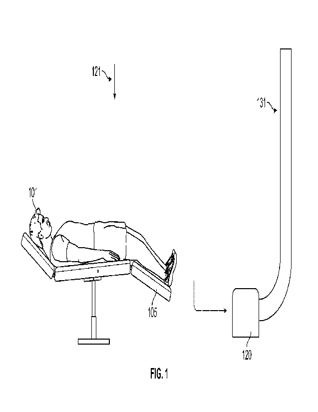
[00801 In an embodiment, and as in FIG. 1, a dental operatory may include a
patient 101 on a
table 105, room-defined variables such as an air scrubber 120 in fluid
communication with
room air inflow 121 and room air outflow 131. Further to the above, it can be
appreciated that
a number of variables within the dental operatory may be modifiable in order
to evaluate a
number of working states of the dental operatory. For instance, the variables
may include
size, shape, and volume of the procedure room(s) and adjacent spaces,
construction materials,
ventilation, air flow, air change per hour, temperature, humidity, location of
vents and
diffusers, ceiling lights, ultraviolet (UV) lights, ionized air devices,
professional equipment,
water and air producing devices, drills (e.g., electric or pneumatic), high
speed suction, low
speed suction, open suction, closed suction, air source evacuators, negative
pressure, positive
pressure, mixed pressure, laminar pressure, wall textures, ceiling textures,
flooring, window
treatments, facility air curtains, PPE air curtains, door types, construction
seams, and sources
of penetrations, such as electrical outlets, vents, and projections, as well
as disinfectant
materials/application, among others. In an embodiment, the dental operatory
may include, as
variables, a number of dental professionals in the room, a type of dental
procedure being
performed and tools being used, patient health factors, as well as
characteristics of potential
types and load of pathogens that may be shed by one or more persons within the
room. For
instance, the types of pathogens may be airborne pathogens, such as novel
coronaviruses,
may be modeled using tagged particles such as Iluores. cein-tagged attenuated
Influenza A
virus. The tagged particles may be emitted as microdroplets of water from.
device(s) meant to
simulate patients, doctors, and assistants, among others, breathing talking,
coughing, and the
like, with varying degrees of viral load, during the performance of common
dental
procedures.
100811 In an embodiment, the dental operator), may be a simulated dental
operatory, with
standardized but configurable parameters, that can be used to evaluate,
develop, and optimize
CA 03180037 2022- 11-23
WO 2021/243279
PCT/US2021/034977
environmental controls, equipment performance, and PPE which has been
marketed,
designed and/or considered to protect healthcare professionals.
100821 As in FIG. 2 through. FIG. 313, it can be appreciated that, accounting
for the dental
operatory factors and variables outlined above, the air control device of the
present disclosure
can deployed.
[00831 For instance, in an embodiment, the air control device can. include a
manifold that is
manufactured in modules of a specified length (e.g., straight or curvilinear)
with devices
connected and sealed together. The ACD may include a console that may be a
control device
connected to a power supply mounted to a wall and/or a ceiling and connected
to a fluid
supply. In an embodiment, the console may be further interfaced and/or
coordinated with the
airflow dynamics, temperature, and humidity of the facility HVAC system. The
system may
include sensors configured to measure parameters of ambient air including
airflow pressure,
air humidity, air temperature, and air particulates.
100841 The ACD may include networks of micro-tubing regulated by the manifold.
The
manifold may be arranged above the head of the patient, below the head of the
patient, or
around each side of the head of the patient, as is appropriate. The manifold
may have a shape
substantially that of a circle or some portion. thereof, the circle or a
portion of the circle (e.g.
an arch) having a diameter or defining a diameter suitable to the head of the
patient. In an
example, the diameter may be approximately 20 cm. The manifold may be arranged
based on
a position of the head of the patient and the arrangement may be adjustable
between different
patients. In an example, the manifold may be arranged at 30 cm above the head
of the patient
or 30 cm below the head of the patient. In an embodiment, during use, a fluid
stream of
filtered air may be injected downward and outward to contain gaseous
bioaerosols within a
funnel-shaped air containment envelope around the head of the patient. The
funnel-shaped air
containment envelope may be defined by the downward and outward injection of
fluid at an
angle relative to an axis of the manifold, or at about 15 to 30 degrees
relative thereto. Infrared
light or other wavelength light projected from the ACD may image the water
vapor or
chemicals of envelope fluid for proper source positioning. In an embodiment,
during use, a
fluid stream of filtered air may be injected upward and outward to contain
gaseous
bioaerosols within a funnel-shaped air containment envelope around the head of
the patient.
The funnel-shaped air containment envelope may be defined by the upward and
outward
injection of fluid at an angle relative to an axis of the manifold, or at
about 15 to 30 degrees
relative thereto. Infrared light or other wavelength light projected from the
ACD may image
the water vapor or chemicals of envelope fluid for proper source positioning.
Concurrently,
II
CA 03180037 2022- 11-23
WO 2021/243279
PCT/US2021/034977
the air containment envelope allows for the unimpeded insertion of dental
workers arms,
hands, and instruments in order to perform dental procedures. The Coanda
effect maintains a
seal around the penetration of the air containment envelope by arms and
curvilinear objects.
100851 In an embodiment, a suction device may be arranged opposite the
manifold of the
ACD in order to scavenge the bioaerosols safely from the field. In an example,
through
hydrogen bonds, pathogens may interact with dense air/fluid in the air
containment envelope
and be directed downward to lower pressure zones where a low speed suction
device at the
base of the funnel-shaped containment envelope will remove the bioaerosols
safely from the
field. Pathogens in the bioaerosols can be further degraded by aerosolized
surfactants,
chemicals, and electrically charged water vapor in a vacuum or collection
chamber.
100861 In an embodiment, a similar but more forceful ACD, with networks of
micro-tubing
regulated by computer-directed manifolds, may be implemented in order to frame
laboratory
work spaces and contain fumes, debris, and bioaerosols generated when patient
dental
devices are modified or cleaned with rotary devices.
[0087] In an embodiment, the ACD may include visual alarms and audible alarms
with
central controls to detect failures within a network of air containment
envelopes.
[0088] In particular, and with. reference now to FIG. 2, an ACD may be
deployed in a dental
operatory to isolate a patient's head from the medical staff, according to an
exemplary
embodiment of the present disclosure. For instance, a patient 201 may be on a
table 205
within, a dental operatory 200. The dental operator), 200 may include, among
other items, a
room air inflow 221, an air scrubber 220, and a room air outflow 231. An air
control device
210 (ACD) may be arranged above a region of interest of the patient 201 or, in
particular, a
head of the patient 201. The ACD 210 may include a power supply and a lumen
211
connected to a fluid supply 214 and configured in fluid communication with a
manifold 212,
the manifold 212 having a plurality of apertures for distributing a fluid from
the fluid supply
214 to the patient. The ACD 210 may further include one or more patient
sensors, in certain
implementations. The plurality of apertures may be distributed at equal
distances around a
diameter of the manifold 212. The distributed fluid may form a fluid envelope
213 around the
head of the patient 201. Air contained within the fluid envelope 213 may be
evacuated from
the dental operatory via vacuum 216. In an embodiment, the fluid supply 214
may be an air
supply supplied at a predefined pressure and/or a predefined humidity. The
predefined
pressure may be 250 mBar, in an example, in order to generate a high pressure
area within
the air containment envelope. Further, a filter may be supplied within the ACD
210 on one
side of the head of the patient 201 or may be supplied within the vacuum 216.
In an example,
12
CA 03180037 2022- 11-23
WO 2021/243279
PCT/US2021/034977
the filter is a HEPA filter and is installed within the ACD 210 device to
provide filtered fluid
to the fluid envelope 213.
100891 In an embodiment, the plurality of apertures of the manifold 212 may be
individually
maneuverable according to a position of the head of the patient 201 and a
desired volume of
the fluid envelope 213. Each of the plurality of apertures may be configured
to adapt to a
position of the head of the patient. In this way, the plurality of apertures
may generate the
fluid envelope 213 in a number of volumes relative to the manifold 212.
Moreover, as in the
example of FIG. 2, the plurality of apertures may be configured to generate
the fluid envelope
213 such that the fluid envelope 213 has an outer angle relative to a normal
axis passing
through the manifold 212. This can be accomplished by arranging the plurality
of apertures at
an angle relative to the normal axis of the manifold 212. The outer angle may
be 15 , in an
example, or any angle appropriate for enveloping a region of interest of the
patient 201 and
providing air containment therein, such as 30 .
100901 In an embodiment, the manifold 212 may have a geometric form according
to a
demand of the implementation. For instance, as in the dental operatory 200 of
FIG. 2, it is
important to generate the air/fluid envelope 213 around the head of the
patient 201 such that a
circular curtain is formed. In this way, the manifold 212 may have a circular
shape. However,
it can be appreciated that any shape or combination of shapes may be
implemented within the
manifold 212, including linear structures, curvilinear structures, and a
variety of closed
structures such as a square, rectangle, and triangle.
[00911 In an embodiment, the fluid supply 214 includes air, water, and
surfactant,
combinations of which can be controlled and provided in order to generate a
specific air
containment envelope about a region of interest of the patient 201.
[00921 In an embodiment, the one or more position sensors or the ACD 210 may
be a
proximity sensor able to detect a region of interest of the patient 201. For
instance, the
proximity sensor may be a Bluetooth sensor or may emit an electromagnetic
signal, such as
an infrared siwial, in order to determine a heat level or a target and to,
accordingly, identify
the region of interest of the patient 201.
[00931 In an embodiment, upon identification of a position of a target via the
one or more
position sensors, the plurality of apertures of the manifold 212 may be
rearranged to focus on
the target relative to the normal axis of the manifold 212 using electric
motors or the like
[00941 A schematic of the ACD, including processing circuitry configured to
control the
ACD and components thereof, is provided with reference to FIG. 7.
13
CA 03180037 2022- 11-23
WO 2021/243279
PCT/US2021/034977
[00951 As a variation of FIG. 2, FIG. 3A and FIG. 3B provide illustrations of
an ACD
deployed in a dental operatory to isolate a patient's head .from the medical
staff, according to
an. exemplary embodiment of the present disclosure.
100961 In an embodiment, the present disclosure describes a device that may be
worn by the
user or suspended in proximity of the head. Generally, the device consists of
a nozzle
directing fluid flow in front of the face of a user. The fluid creates a
barrier ("fluidic shield")
between the user and their surroundings and is intended to direct the breath
and any
respiratory droplets away from others. The fluid is primarily filtered air
supplied by a
stationary pressure source. Other fluids with hygroscopic or disinfecting
properties may be
injected into the air stream to capture or neutralize droplets or particles
entrained by the
fluidic shield.
100971 More specifically, and as it relates to FIG. 3A and FIG. 3B, a patient
301 may be on a
table 305 within a dental operatory. The dental operatory may include, among
other items, a
room air inflow, an air scrubber, and a room air outflow. An air control
device 310 (ACD)
may he arranged relative to a region of interest of the patient 301 or, in
particular, a head of
the patient 301. The ACD 310 may include a power supply and a lumen 311
connected to a
fluid supply 314 and configured in fluid communication with a manifold 312,
the manifold
312 having a plurality of apertures 317 for distributing a fluid from the
fluid supply 314 to
the patient, as shown in FIG. 38. The ACD 310 may further include one or more
patient
sensors, in certain implementations. The plurality of apertures 317 may be
distributed at
equal distances around a diameter of the manifold 312. The distributed fluid
may form a fluid
envelope 313 around the head of the patient 301. Air contained within the
fluid envelope 313
may be evacuated from the dental operatory via vacuum or may dissipate and be
diluted into
ambient air. In an embodiment, the fluid supply 314 may be an air supply
supplied at a
predefined pressure and/or a predefined humidity. The predefined pressure may
be 250 mBar,
in an example, in order to generate a high pressure area within the air
containment envelope.
Further, a filter may be supplied within the ACD 310 on one side of the head
of the patient
301 or may be supplied within the vacuum. In an example, the filter is a HEPA
filter and is
installed within the ACD 310 device to provide filtered fluid to the fluid
envelope 313.
[00981 In an embodiment, the plurality of apertures 317 of the manifold 312
may be
individually maneuverable according to a position of the head of the patient
301 and a desired
volume of the fluid envelope 313. Each of the plurality of apertures may be
configured to
adapt to a position of the head of the patient. In this way, the plurality of
apertures 317 may
generate the fluid envelope 313 in a number of volumes relative to the
manifold 312.
14
CA 03180037 2022- 11-23
WO 2021/243279
PCT/US2021/034977
Moreover, the plurality of apertures 317 may be configured to generate the
fluid envelope
313 such that the fluid envelope 313 has an outer angle relative to a normal
axis passing
through the manifold 312. This can. be accomplished by arranging the plurality
of apertures
317 at an angle relative to the normal axis of the manifold 312. The outer
angle may be 15 ,
in an example, or any angle appropriate for enveloping a region of interest of
the patient 301
and providing air containment therein, such as 30 .
[00991 In an embodiment, the manifold 312 may have a geometric form according
to a
demand of the implementation. For instance, as in the dental operatory of FIG.
3A, it is
important to generate the air/fluid envelope 313 around the head of the
patient 301 such that a
circular curtain is formed. In this way, the manifold 312 may have a circular
shape. However,
it can be appreciated that any shape or combination of shapes may be
implemented within the
manifold 312, including linear structures, curvilinear structures, and a
variety of closed
structures such as a square, rectangle, and triangle.
101001 In an embodiment, the fluid supply 314 includes air, water, and
surfactant,
combinations of which can be controlled and provided in order to generate a
specific air
containment envelope about a region of interest of the patient 301.
101011 In an embodiment, the one or more position sensors of the ACD 310 may
be a
proximity sensor able to detect a region of interest of the patient 301. For
instance, the
proximity sensor may be a Bluetooth sensor or may emit an electromagnetic
signal, such as
an infrared signal, in order to determine a heat level of a target and to,
accordingly, identify
the region of interest of the patient 301.
[01021 In an embodiment, upon identification of a position of a target via the
one or more
position sensors, the plurality of apertures 317 of the manifold 312 may be
rearranged to
focus on the target relative to the normal axis of the manifold 312 using
electric motors or the
like
(01031 A schematic of the ACD, including processing circuitry configured to
control the
ACD and components thereof, is provided with reference to FIG. 7.
101041 FIG. 3C provides an. additional description of the present disclosure,
wherein an A.CD
may be deployed in a dental operatory to isolate a patient's head from the
medical staff,
according to an exemplary embodiment. For instance, a patient 301 may be on a
table 305
within a dental operatory. The dental operatory may include, among other
items, a room air
inflow, an air scrubber, and a room air outflow. An air control device 310
(AC)) may be
arranged relative to a region of interest of the patient 301 or, in
particular, a head of the
patient 301. The ACD 310 may include a power supply and a lumen 311 connected
to a fluid
CA 03180037 2022- 11-23
WO 2021/243279
PCT/US2021/034977
supply 314 and configured in fluid communication with a manifold 312, the
manifold 312
having a plurality of apertures for distributing a fluid from the fluid supply
314 to the patient.
The ACD 310 may further include one or more patient sensors, in certain
implementations.
The plurality of apertures may be distributed at equal distances around a
diameter of the
manifold 312. The distributed fluid may form a fluid envelope 313 around the
head of the
patient 301. Air contained within the fluid envelope 313 may be evacuated from
the dental
operator)" via vacuum 316. In an embodiment, the fluid supply 314 may be an
air supply
supplied at a predefined pressure and/or a predefined humidity. The predefined
pressure may
be 250 mBar, in an example, in order to generate a high pressure area within
the air
containment envelope. Further, a filter may be supplied within the .ACD 310 on
one side of
the head of the patient 301 or may be supplied within the vacuum 316. In an
example, the
filter is a HEPA filter and is installed within the ACD 310 device to provide
filtered fluid to
the fluid envelope 313.
101051 ACDs, such as that described above, include networks of pressurized
fluid within
micro-tubes enclosed in channels with computer-controlled manifold systems for
generation
of air containment envelopes within in a room. The fluid may be air, liquid,
or other flowable
material. The air containment envelope may be generated by pressurized air,
water vapor, and
aerosolized surfactant. To direct airflow to form the air containment
envelope, airflow is
projected in a continuous slit stream of fluid, which flows more like a "wave"
of cohesive
low density liquid, within a media of lower density liquid. To further enhance
the ``wave"
projection, successive loads of fluid can be air "pistoned" forward in a
manner similar to
decorative water fountains. Noise generated by jet streams of fluid can be
canceled by a
manner similar to noise canceling headphones (e.g., equal but opposite sound
waves).
[01061 In this way, and in view of the above, ACDs provide an invisible
barrier ideal for the
containment of bioaerosols.
101071 It can be appreciated that any technology developed to mitigate viral
airborne threats
in dentistry will be embraced by dental professionals, mandated by regulators,
and may
incubate an. entirely new industry which can expand to mitigate viral airborne
threats in fields
other than dentistry such as medicine, meat processing plants, office spaces,
sport arena
seating, movie theaters, restaurants, public transportation, airlines, cruise
ships and many
other venues. Such venues include military government educational
institutions, essential
businesses, hotels, retail stores, theme parks, casinos, manufacturing
centers, apartment
condominium common areas, churches, theaters, food outlets, and the like.
16
CA 03180037 2022- 11-23
WO 2021/243279
PCT/US2021/034977
[01081 To this end, ACDs may be deployed in a variety of settings. For
instance, in a nursing
home, an ACD may be mounted on the ceiling over the resident in bed and/or
over the
resident while sitting or in the dining room, and may provide an invisible
protective envelope
from bioaerosol transmission for the resident, staff, and visitors.
[01091 To make this possible, the ACD can be spatially arranged around room
occupant(s) in
a specific mariner based on a particular venue and may deploy corrective
measures to create a
protective microclimate around room occupant(s), accordingly. By optimizing
features of
controlling airflow, water vapor, and surfactant, ACDs are scalable to any
size and venue, as
will be exemplified with reference to FIG. 4 through FIG. 6.
[011.01 With reference to FIG. 4, an ACD 410 may be a wearable device
appropriate for daily
wear and may include a manifold 412, a fluid supply 414, and mobile power
supply
connected to processing circuitry configured to control the ACD 410. The ACD
410 may be
arranged around a neck of a subject 401 in order to generate a fluid envelope
413 that
encompasses a microclimate of the subject 401. In an embodiment, the ACD 410
may further
include a low pressure exhaust system configured to expel disinfected exhaust
from the
microclimate of the subject 401. It can be appreciated that the ACD 410 may
include other
features described with. reference to FIG. 2 through FIG. 38.
[01111 With reference to FIG. 5, an ACD 510 may be configured to generate an
air
containment envelope from above a subject 501 and may be appropriate for an
airplane seat,
a theater seat, and/or concert or sports stadium seating. The ACD 510 may
include a manifold
512, a lumen 511 connected to a fluid supply, and power supply connected to
processing
circuitry configured to control the ACD 510. The ACD 510 may be arranged above
a subject
501 in order to generate a fluid envelope 513 that encompasses a microclimate
of the subject
501. In an embodiment, the ACD 510 may further include a low pressure exhaust
system
configured to expel disinfected exhaust from the microclimate of the subject
501. It can be
appreciated that the ACD 510 may include other features described with
reference to FIG. 2
through FIG. 3B.
[01121 With reference to FIG. 6, an ACD 610 may be configured to generate an
air
containment envelope from above one or more subjects 601 and may be
appropriate for a
restaurant, office, and/or cruise ship setting. The ACD 610 may include a
manifold 612, a
fluid supply, and power supply connected to processing circuitry configured to
control the
ACD 610. The ACD 610 may be arranged above the one or more subjects 601 in
order to
generate a fluid envelope 613 that encompasses a microclimate of the one or
more subjects
601. In an embodiment, the ACD 610 may further include a low pressure exhaust
system
17
CA 03180037 2022- 11-23
WO 2021/243279
PCT/US2021/034977
configured to expel disinfected exhaust from the microclimate of the one or
more subjects
601. It can be appreciated that the ACD 610 may include other Features
described with
reference to FIG. 2 through FIG. 3B.
[0113] According to an embodiment, an ACED may continuously monitor and
display all
components of device performance through cell phone technology. Wireless
communication
technology, such as Bluetootht..), may alert individuals of air containment
envelope protection
either as an individual or when entering a venue offering broader air
containment envelope
protection, such as retail stores, schools, banks, theaters, healthcare
facilities, and the like.
Depending on community viral threat level (e.g., mild or high), venue viral
threat level (e.g.,
A, B, C or rating) and an individual's tolerance to the viral threat, layers
of air containment
envelopes can be added or subtracted.
101141 In an embodiment, parameters of ambient air can be evaluated through
sensors. For
instance, air pressure, air direction, humidity, temperature, and air
particulates can be
measured.
[01151 In an embodiment, an ACD can be configured to deploy corrective
measures to create
a protective microclimate around room occupant(s).
[011.6] In view of the above, it can be appreciated that air, for instance, is
a low density fluid.
An ACD directs airflow, enriched into a higher density fluid with moisture and
disinfectant,
to generate an air containment envelope and to protect an occupant(s). ACDs
exploit the
knowledge that higher air pressure directs airflow, higher humidity scavenges
microdroplets
by hydrogen bond affinity and directs particles below breathing zone by
gravity, and
detergents in enhanced airflow kill airborne pathogens.
[011.7] With reference now to FIG. 7, a non-limiting example of a control
device for an .ACD
will now be described. An ACD 700 may include a power supply 771 coupled to a
central
processing unit (CPU) 776 and a fan/blower 772. The CPU 776 may be configured
to control
a fluid source 775, a thermal source 774, and a disinfectant source 770. The
fluid source 775
may be a gas or a liquid and may be pressurized. The fluid source 775 may also
be an
existing laminar air flow system. already built into a room structure, or a
source of
compressed or pressurized air. In one embodiment, there is a pre-filter that
excludes particles
of 5 microns or more, for example. The filtered air may optionally be passed
through another
filter that excludes bacteria and other microbes such as fungi and viruses. A.
filter with a
porosity of 0.22-0.30 gm or less would be suitable for the second stage
filter. Alternatively,
one or more filters with 0.22-0.30 gm can be used. The thermal source 774 may
be a heat
generator for warming a fluid of the fluid source 775. The fluid of the fluid
source 775 can be
18
CA 03180037 2022- 11-23
WO 2021/243279
PCT/US2021/034977
heated by any means known to those skilled in the art of heating air, such as
a resistive
element or heater near the air. The disinfectant source 770 may include
surfactants and sterile
solutions of antimicrobial or antibiotic agents that can. be mixed with the
air before or after
filtration so that a germicidal effect is afforded to the fluid envelope.
Suitable anti-microbial
agents include antibiotics, triclosan, ethanol, or chlorhexidene gluconate in
concentrations of
0.1-1.0 percent in a sterile saline or suitable physiological buffer such as
phosphate buffered
saline. In addition, nebulized mists of anti-microbial solutions to further
retard bacterial
survival can also be utilized. Fluid passed through filters 773 can be
delivered to a manifold
777 having a plurality of apertures and which is controlled by the CPU 776 to
generate an air
containment envelope. The manifold 777 may include one or more position
sensors, in an
example, to detect a target and in order to adjust an orientation of the
plurality of apertures of
the manifold 777. Pathogenic material entrapped within the generated air
containment
envelope may be collected via exhaust 778. The air containment envelope may be
provided at
any appropriate speed. For instance, the air containment envelope can be
provided at a speed
as described with reference to the below Non-Limiting Experimental Results.
[01.1.8J Next, a hardware description of the biochamber device of FIG. 1,
according to
exemplary- embodiments, is described with reference to FIG. 8. In FIG. 8, the
biochamber
device includes a CPU 840 which performs the processes described above/below.
The
process data and instructions may be stored in memory 841. These processes and
instructions
may also be stored on a storage medium disk 842 such as a hard drive (I-113D)
or portable
storage medium or may be stored remotely. Further, the claimed advancements
are not
limited by the form of the computer-readable media on which the instructions
of the inventive
process are stored. For example, the instructions may be stored on CDs, DVDs,
in FLASH
memory, RAM, ROM, PROM, EPROM, EEPROM, hard disk or any other information
processing device with which the biochamber device communicates, such as a
server or
computer.
101191 Further, the claimed advancements may be provided as a utility
application,
background daemon, or component of an operating system, or combination
thereof, executing
in conjunction with CPU 840 and an operating system such as Microsoft Windows,
UNIX,
Solaris, LINUX, Apple MAC-OS and other systems known to those skilled in the
art.
101201 The hardware elements in order to achieve the biochamber device may be
realized by
various circuitry elements, known to those skilled in the art. For example,
CPU 840 may be a
Xenon or Core processor from Intel of America or an Opteron processor from AMD
of
America, or may be other processor types that would be recognized by one of
ordincuy skill
19
CA 03180037 2022- 11-23
WO 2021/243279
PCT/US2021/034977
in the art. Alternatively, the CPU 840 may be implemented on an FPGA, ASIC,
PLD or using
discrete logic circuits, as one of ordinary skill in the art would recognize.
Further, CPU 840
may be implemented as multiple processors cooperatively working in parallel to
perform the
instructions of the inventive processes described above.
101211 The biochamber device in FIG. 8 also includes a network controller 843,
such as an
Intel Ethernet PRO network interface card from Intel Corporation of America,
for interfacing
with network 855. As can be appreciated, the network 855 can be a public
network, such as
the Internet, or a private network such as an LAN or WAN network, or any
combination
thereof and can also include PSTN or ISDN sub-net-works. The network 855 can
also be
wired, such as an Ethernet network, or can be wireless such as a cellular
network including
EDGE, 3G and 4G wireless cellular systems. The wireless network can also be
WiFi,
Bluetooth, or any other wireless form of communication that is known.
101221 The biochamber device further includes a display controller 844, such
as a NVIDIA
GeForce GTX or Quadro graphics adaptor from NVIDIA Corporation of America for
interfacing with display 845, such as a Hewlett Packard HPL2445w LCD monitor.
A general
purpose I/O interface 846 interfaces with a keyboard and/or mouse 847 as well
as a touch
screen panel 848 on or separate from display 845. General purpose I/O
interface also
connects to a variety of peripherals 849 including printers and scanners, such
as an OfficeJet
or DeskJet from Hewlett Packard.
101231 A sound controller 850 is also provided in the biochamber device, such
as Sound
Blaster X-Fi Titanium from Creative, to interface with speakers/microphone 851
thereby
providing sounds and/or music.
101241 The general purpose storage controller 852 connects the storage medium
disk 842
with communication bus 853, which may be an ISA, EISA, VESA, PC1, or similar,
for
interconnecting all of the components of the biochamber device. A description
of the general
features and functionality of the display 845, keyboard and/or mouse 847, as
well as the
display controller 844, storage controller 852, network controller 843, sound
controller 850,
and general purpose I/0 interface 846 is omitted herein for brevity as these
features are
known.
Non-limiting Experimental Results
101251 To demonstrate feasibility of the above-described ACD, a series of
theoretical
evaluations were performed based on a developed model of the ACD.
CA 03180037 2022- 11-23
WO 2021/243279
PCT/US2021/034977
[01261 In order to create a model of the fluidic shield, it was assumed that
the air flow is
oriented upward from the chest of the user. An upward flow also allows for
sufficient space
to add a suction device to collect the users' breath for filtering, if
desired. The flow which
forms the shield external to the collar is modeled.
101271 As will be realized below, the person on the concave side of ACD is
sometimes called
the patient and the person on the convex side of the ACD is called the
clinician. The choice
of terminology is arbitrary and the conclusions are equally valid with the
reverse
assumptions.
[0128] The model used in computational fluid dynamics simulations consists of
a human
head and of a hand holding a tool and reaching toward the face originating
from the convex
side of the shield, as shown in FIG. 9. It also includes a curved nozzle, as
in FIG. 10 and
similarly presented in FIG. 3B, that provides the air flow to develop the
shield. The model
was chosen to mimic typical patient-clinician interaction in a dentistry
setting. The model is
bounded by a rectangular box.
[0129] The efficacy of the fluidic shield is dependent on a number of design
and operating
parameters. Such design and operating parameters can be selected in order to
satisfy certain
objectives. For instance, such parameters may include slit exit air speed,
slit width, size of
chemical aerosol, droplets in breath, and whether the patient is coughing. The
slit exit air
speed relates to a relationship defining the ability of the fluidic shield to
divert breath
streams, wherein the ability of the fluidic shield to divert breath streams is
proportional to the
slit exit air speed. The slit width relates to a relationship defining a
robustness of the fluidic
shield, wherein the robustness of the fluidic shield is dependent on the
shield mass flow,
which is proportional to the slit width (i.e., thickness). The size of
chemical aerosol may be
based on a specific hygroscopic or disinfectant fluid injected into the shield
air stream to
serve as a secondary barrier against transmission between patient and
clinician. The
hygroscopic or disinfectant fluid may be, for instance TEG (0.5% to 20%). The
droplets in
breath relate to water droplets contained within either or the patient's or
the clinician's
breath. To investigate how droplets are transported by each breath, two sizes
of respiratory
droplets are simulated for the patient's breath and one droplet size is
simulated for the
clinician's breath. As it relates to a coughing patient, a cough ejects air at
higher velocity than
normal breathing and is likely to challenge the integrity of the fluidic
shield.
101301 The considerations above are investigated within the limits shown in
Table 1. The
relevant parameters are combined into eight simulation cases as follows
(letter labels refer to
the rows in Table 1 while the numeric labels designate the corresponding
column): (1) Al B1
21
CA 03180037 2022- 11-23
WO 2021/243279
PCT/US2021/034977
- Slow air speed and narrow slit; (2) A2B1 - High air speed and narrow slit;
(3) A2B2 - High
air speed and wide s1it2; (4) A2B1C2D1 - High air speed, narrow slit, large
TEG particles
injected from device, and small moisture droplets contained in the patient's
breath; (5)
A2B1C2D2 - High air speed, narrow slit, large TEG particles injected from
device, and large
moisture droplets contained in the patient's breath; (6) A2B1C2D1E - High air
speed, narrow
slit, large TEG particles injected from device, small moisture droplets
contained in the
patient's breath, and medium moisture droplets contained in the clinician's
breath; (7)
A2B1D1FI - High air speed, narrow slit, weak cough by the patient transporting
small
moisture droplets; and (8) A2B1D1F2 - High air speed, narrow slit, strong
cough by the
patient tran,portitut small moisture droplets.
'77.77777,x.
.x.7777:77=7,77ir 7.757777777.7777777T 7.7777777
-77.777777.7777]7.7.777.7777]7.7.777.7.7.7.7.77 in
F -77.717.77
EEn e
(ion :!M!mam2n!!mn!ape!!!*jittfittnowlcapittw, !Ivcax(rautti_Nialltioni!i
A Slit Air Speed at Collar 3 m/s
15 in/s
(6.7 mph) (33.6 mph)
Slit Width 3 mm 6
mm
8
(0.12 in) (0.24 in)
Injection of TEG Particles from 1.26 um 3.72 gm
Device Slit (5 x 10⁻⁵ in) (1.5 x 10-
4 in)
Injection of Water Droplets from 0.3 gm
10 gm
Patient's Mouth (1.2 x 10⁻⁵ in) (3.9 x
104 in)
Injection of Water Droplets from
1.0 gm (3.9 x 10.5 in)
Clinician's Side
Coughing Patient (Includes Small 2.2 m/s
10 m/s
Water Droplets) (4.9 mph) (22.4
mph)
Table 1
[0131j In view of the above, computational fluid dynamic studies were
performed under the
following assumptions: (1) That the air flows vertically upward from the
nozzle from a semi-
circular slit on a curved collar. This upward flow modeling avoids impingement
of the flow
on the patient's chest and leaves open the possibility of adding a suction
device in later
design stages. This assumption was made based on practical considerations and
is not
expected to influence the strength (i.e., shielding capability) of the shield:
and (2) That the
respiratory droplets and TEG particles do not significantly alter the airflow.
Therefore, one-
way coupling is used to describe the drag force between the airflow and the
droplets and
particles. This assumption is valid because the mass fraction of droplets and
particles in the
airflow is small.
101321 As described above, the model of FIG. 9 consists of a human head and of
a hand
holding a tool reaching toward the face originating from the convex side of
the shield. It also
22
CA 03180037 2022- 11-23
WO 2021/243279
PCT/US2021/034977
includes a nozzle that provides the air flow to develop the shield. The model
was chosen to
mimic typical patient-clinician interaction in a dentist-ry setting.
101.331 To perform subsequent analyses, a mesh must be generated to bridge
between the
geometry model and the computational model. The mesh is a grid upon which the
fluid
dynamics equations are solved to obtain velocity and pressure results. Meshing
was
performed using the ANSYS Workbench Meshing module. The mesh was built with
sufficient density in key locations to capture the relevant physics of the
process. Inflation
layers were added to key surfaces to resolve the flow around these obstacles.
Further
refinement was added between the hand and face to better resolve the complex
flows
expected in this region. A second, refined mesh was develop to determine if
the first mesh
produced sufficiently accurate results. The refined mesh has approximately
twice as many
nodes as the original mesh. Meshing parameters and selected results for both
meshes are
provided in Table 2. A. side-by-side view of the meshes is shown in FIG. 11A
and FIG. 11B.
Velocity contour plots for both meshes are shown in FIG. 11C and FIG. 11D. The
plots show
that the original mesh resolves the flow pattern reasonably well, although
there are some
localized differences in the peak magnitude of the velocity. Based on the
results, it was
concluded that the original mesh is sufficiently refined for this feasibility
assessment.
Radius of Sphere of Influence meters 0.02
0.073
Inflation Layer on Lips no yes
Face Sizing on Hand meters 0.003
0.002
Total Node Count 2,029.814
3,953,070
Average Velocity on Vertical miles per
4.12
2.55
Plane hour
Average Velocity on Breath miles per
2.91. 3.11
Stream Lines hour
Average Velocity on Stream Lines miles per
20.53
18.48
----------------- from Left Inlet hour
Average Velocity on Stream Lines miles per
23.68
21.98
from R.ight Inlet hour
Table 2
[01341 Table 3 summarizes the boundary conditions for the simulation. Note
that some
boundary conditions change for different cases.
23
CA 03180037 2022- 11-23
WO 2021/243279
PCT/US2021/034977
gna00E0d0OiliMPrtrREHRtitnMEMV0WilinE REM
Boundary Type Solid Wall
Head, Hand,
Mass and Momentum No-Slip
Tool
Coefficient of
Particle Interaction 0
Restitution
Boundary Type Opening
Opening Pressure and
Mass and Momentum 0 Ic.Pa
(relative)
Surrounding Direction
Box
Flow Direction Normal to Boundary
Turbulence Medium Intensity
Boundary Type Inlet
Cartesian Velocity u = 1.4 m/s,
v = 0 m/s,
Mass and Momentum
Components
w 0 m/s
Upper Lip Turbulence Medium Intensity
Off or On
Material
Water
Particle Injection
Size 0.3 gm or
10 urn
Velocity
Zero slip
Boundary Type Inlet
Cartesian Velocity u ¨ 0 m/s, v
3m/s or
Mass and Momentum
Components 15 m/s, w
=0 m/s
Surrounding
Box Turbulence Medium. Intensity
Off or On
Material
l'EG
Particle Injection
Size
3.72 f.t Ill
Velocity
Zero slip
Boundary Type Inlet
Cartesian Velocity u -1.1 m/s,
v = -0.5
Mass and Momentum
________________________________________________ Components _____ m/s, w = -
0.7 m/s
Clinician's
Breath (one Turbulence Medium Intensity
case only)
Off or On
Material
Water
Particle Injection
Size I
um
Velocity
Zero slip
Table 3
24
CA 03180037 2022- 11-23
WO 2021/243279
PCT/US2021/034977
[01351 The simulation involves three materials: air, water droplets, and TEG
particles. Air
was modeled with constant properties corresponding to a temperature of 25 C
and a pressure
of 1 atm. Water and TEG were modeled using a Lagranian approach (i.e., as
distinct
particles) with one-way coupling to the air flow. For this modeling approach,
the density and
spherical diameter of the particles are the only parameters of consequence.
The droplet and
particle diameters for water and TEG are listed in Table 3. Water was modeled
with a density
of 997 kg/m3. TEG particles were modeled as a mixture of 20 weight percent TEG
(density
1125 kg/m3) in water with a mixture density of 1023 kg/m3.
[01361 The analysis was performed using ANSYS CFX Version 2020 RI. An overview
of
the user-selected options in the CFX Preprocessor is given in Table 4.
77777.7.7.7.77.77.7.7,7,7,77:777777
.7.7:77:777:777,x7.x.,x.x.x77,7777777.7.7777x,'' -7,737777,- 7771
Pgiii-ametm : =Seli;;diain : : ............
..:........
The hand and tool make the model non-
Dimensionality 3D
symmetric.
The SST model provides good near wall and far
Turbulence Model SST
field performance.
In the temperature and pressure range of the
Equation of State ideal Gas
simulation, an is considered an ideal
___________________________________________
The hand. tool and head were modeled as smooth
Wall Roughness Smooth
surfaces.
This scheme provides a blend of first order and
Space
Hi 2.h Resolution hi2her order schemes to achieve
accuracy and
Di scret]zati on
boundedness.
Convergence criteria for RMS residuals of all
RM.S Residuals < 10-4
equations were less than 10-4
Solver Precision Double Reduces truncation error
Transient Simulation
Second Order
Time Discretization Program recommended setting for accuracy
Backward Euler
Time step is adjusted by the solver to achieve a
Time Step Adaptive
Courant-Friedrich-Levy (CFL) number of <5
Table 4
0137] In the analysis of the cases including particle tracks, the Stokes
number is used as an
indicator of the particles' ability to follow th.e bulk flow. A Stokes number
above one
indicates that the suspended particle is likely to make contact with an
obstacle in the flow.
The particle would then adhere to the obstacle and, in the case of TEG
particles, would be
unable to adsorb and capture moisture contained in the breath downstream of
the obstacle. A
Stokes number less than one indicates that the suspended particle is likely to
follow the flow
CA 03180037 2022- 11-23
WO 2021/243279
PCT/US2021/034977
around the obstacle and remain suspended. The Stokes number is calculated
according to the
following formula:
prcil,u0
Stokes ¨ ____________________________________________
18 0/0
where pp is the particle density, di, is the particle diameter, u0 is the
fluid velocity, 1.1.0 is the
fluid's dynamic viscosity, and lo is a characteristic length of the obstacle.
A characteristic
length of 1 cm was used during post processing to represent the diameter of a
likely obstacle
such as a tool.
[01.381 The simulations were performed as outlined above and subsequent
analysis and
conclusions are summarized below.
101391 As described above, the person on the concave side or the shield is
referred to as the
patient, while the person on the convex side of the shield is referred to as
the clinician. This
choice of terminology is arbitrary and does not impact the results or
conclusions. FIG. .12A
and FIG. 12B show the results of the simulation at low shield air speed
(A1B1). FIG. 12A
shows streamlines from the air nozzle colored by velocity, and streamlines
from the patient's
mouth in grey. FIG. 128 shows a contour plot of velocity overlaid with
streamlines starting at
the patient's mouth. In both images, blue indicates low velocities and red
indicates high
velocities. Both plots show that the air velocity is insufficient to create a
shield and the breath
of a user is not diverted effectively. FIG. 12C and FIG. 12D show the results
of the
simulation at high shield air speed (A281). As before, blue indicates low
velocities and red
indicates high velocities. In this case, the air velocity is high enough to
separate a user's
breath within the shield. Similar results are obtained from the simulation
using a wide slit
(A2B2). FIG. 12E through FIG. 12G show the results of a simulation including
TEG particles
in the airstream and water droplets in a user's breath (A2B1C2 and
A2B1131/23). The lines
shown in the Figures are tracks that trace the path of representative
particles. As opposed to
streamlines, which trace hypothetical massless particles, particle tracks
follow finite-sized
particles with mass. The lines arc colored by the Stokes number which is a
measure of the
suspended particle's ability to follow the bulk flow. A Stokes number above
one indicates
that the suspended particle is likely to make contact with an obstacle in the
flow. The particle
would then adhere to the obstacle and, in the case of TEG particles, would be
unable to
adsorb and capture moisture contained in the breath downstream of the
obstacle. A Stokes
number less than one indicates that the suspended particle will follow the
flow around the
obstacle and remain suspended. A mathematical definition of the Stokes number
is provided
26
CA 03180037 2022- 11-23
WO 2021/243279
PCT/US2021/034977
above. The Stokes numbers for all cases are significantly less than one
indicating that the
droplets and TEG particles are well entrained in the flow. Note that the color
patterns on the
particle tracks for the respiratory droplets in FIG. 12F and FIG. 12G are
similar in appearance
but different in magnitude. The difference in magnitude is due to the
different size (0.3 pm
vs. 10 um) of the droplets in FIG. 12F and FIG. 12G, respectively. In general,
the smaller
droplets resulted in smaller Stokes numbers compared to the larger droplets,
indicating the
smaller droplets will entrain in the airstream more easily than the larger
droplets, although
both the small and large droplets were sufficiently entrained in the
airstream.
[01401 FIG. 121-I through FIG. 12J show the results from the simulation
including a breath
stream outside the fluidic shield (A2B1C2D1E1.) in addition to the normal
breathing of the
user on the concave side of the shield. The particle tracks for the
respiratory droplets show
that the breath streams from both parties are effectively separated by the
fluidic shield (FIG.
12H). The droplets released from. the user's mouth on the concave side of the
shield (patient)
are smaller than those released by the person on the convex side of the shield
(clinician), and
therefore, the Stokes number for the patient's breath droplets are lower.
However, the Stokes
number for both streams indicate good entrainment (i.e., Stokes number << I).
Different
droplet sizes were used for patient and clinician to confirm that droplets of
either size can be
deflected by the shield flow. The size of both droplets is within the range
identified through
experiments. In FIG. 121 and FIG. 12J, the black lines show the approximate
location of the
clinician's hand penetrating the shield and slightly reducing the shield's
velocity. The red
arrow indicates the approximate direction of the clinician's breath, which
causes a small
deformation of the shield but does not lead to breakdown of the shield
integrity.
[01411 FIG. 13A through FIG. 13J show the transient evolution of a weak cough
(2.2 m/s, 4.9
mph). The cough pulse starts at 0 seconds from a steady-state flow field of
the shield in
which the patient does not exhale, and ends after 0.1 seconds. FIG. 13A, FIG.
13C, FIG. 13E,
FIG. 136, and FIG. 131 illustrate grey-colored streamlines starting from the
inlet nozzle of
the collar, and respiratory droplet tracks colored by velocity starting from
the patient's
mouth.. Note that the droplet size used is for normal breathing and that the
size of droplets
expelled through a cough may vary. FIG. 13B, FIG. 13D, FIG. 13F, FIG. 1311,
and FIG. 133
illustrate velocity contours for the same period. The cough slightly deforms
the fluidic shield
but does not break through the shield. FIG. 14A through FIG. 14J show similar
images for a
strong cough (10 m/s, 22.4 mph). In this case, the cough and shield velocities
are similar in
magnitude and the cough has sufficient momentum to break through the shield.
After the
cough has passed, the fluidic shield is reestablished by the airflow from the
collar.
27
CA 03180037 2022- 11-23
WO 2021/243279
PCT/US2021/034977
[01421 The computation fluid dynamics simulations and parametric study support
the
following conclusions. First, the fluidic shield concept is feasible. In the
investigated
configuration, a shield with an initial air velocity of 15 m/s (33.6 mph) at
the source is
capable of deflecting the user's breath for a range of scenarios and operating
conditions,
including a weak cough carrying respiratory droplets. At the point of
interaction the velocity
of the shield air and breath are approximately 4 m/s (9 mph) and 1.1 m/s (2.5
mph),
respectively, for A2B1. Next, the shield is capable of redirecting/deflecting
moisture droplets
carried in the breath from either side of the shield making it effective for
the patient and
clinician. Interference from the hand and tool leads to a minor disruption of
the shield but
does not cause a breakdown. However, other hand positions, or multiple hands
in the
airstream, were not investigated and could lead to a larger disruption in the
shield that may
lead to a breakdown in confinement. These types of situations should be
studied in a future
analysis in conjunction with a human factors study. The fluidic shield is able
to transport
TEG particles to provide additional protection against transmission between
the patient and
clinician. This may be beneficial in cases where the shield is disrupted due
to obstacles and
there is a higher degree of mixing between breath and shield air flows. The
shield is
challenged when the cough velocity approaches the velocity of the shield air.
[0143] Obviously, numerous modifications and variations are possible in light
of the above
teachings. It is therefore to be understood that within the scope of the
appended claims, the
invention may be practiced otherwise than as specifically described herein.
[0144] Embodiments of the present disclosure may also be as set forth in the
following
parentheti cal s
[0145] (1) A system for removing pathogens from a dental operatory, comprising
a manifold
in fluid connection with a fluid source, the manifold including a plurality of
apertures for
directing a fluid stream, a fluid pump for pressurizing a fluid within the
fluid source, and
processing circuitry configured to instruct the fluid pump to pressurize the
fluid, and instruct
the fluid pump to pump the pressurized fluid to the manifold such that the
directed fluid
stream generates a fluid shield relative to an object.
[0146] (2) The system of (1), wherein the pressurized fluid is water or air.
[0147] (3) The system of either (1) or (2), further comprising a reservoir
containing at least a
disinfectant, the processing circuitry being further configured to instruct
mixing of the
disinfectant with the pressurized fluid pumped to the manifold.
101481 (4) The system of any one of (1) to (3), wherein the disinfectant is
mixed with the
pressurized fluid at a concentration of between 0.5% and 20%.
28
CA 03180037 2022- 11-23
WO 2021/243279
PCT/US2021/034977
[01491 (5) The system of any one of (1) to (4), wherein the disinfectant
includes triethylene
glycol particles.
101501 (6) The system of any one of (1) to (5), wherein the triethylene glycol
particles are
between 1.26 pm and 3.72 pm.
101511 (7) The system of any one of (1) to (6), wherein the manifold has a
substantially-arced
shape, the fluid stream directed therefrom forming a semi-cylindrical fluid
column.
[01521 (8) The system of any one of (1) to (7), further comprising a vacuum,
the processing
circuity being further configured to instruct the vacuum to evacuate the
directed fluid stream
from the dental operatory.
[01531 (9) The system of any one of (1) to (8), wherein the plurality of
apertures have a
diameter of between 3 mm and 6 mm.
101541 (10) An apparatus for removing pathogens from a dental operatory,
comprising a
manifold in fluid connection with a fluid source, the manifold including a
plurality of
apertures for directing a fluid stream, a fluid pump for pressurizing a fluid
within the fluid
source, and processing circuitry configured to instruct the fluid pump to
pressurize the fluid,
and instruct the fluid pump to pump the pressurized fluid to the manifold such
that the
directed fluid stream generates a fluid shield relative to an object.
[01551 (11) The apparatus of (10), wherein the pressurized fluid is water or
air.
101561 (12) The apparatus of either (10) or (11), further comprising a
reservoir containing at
least a disinfectant, the processing circuity being further configured to
instruct mixing of the
disinfectant with the pressurized fluid pumped to the manifold.
[01571 (13) The apparatus of any one of (10) to (12), wherein the disinfectant
includes
triethylene glycol particles.
[01581 (14) The apparatus of any one of (10) to (13), wherein the triethylene
glycol particles
are between 1.26 gm and 3.72 gm.
101591 (15) The apparatus of any one of (10) to (14), wherein the manifold has
a
substantially-arced shape, the fluid stream directed therefrom forming a semi-
cylindrical fluid
column.
[01601 (16) The apparatus of any one of (10) to (15), wherein the plurality of
apertures have
a diameter of between 3 mm and 6 mm.
101.611 (17) A system for removing pathogens, comprising a manifold in fluid
connection
with a fluid source, the manifold including a plurality of apertures for
directing a fluid
stream, a fluid pump for pressurizing a fluid within the fluid source, and
processing circuitry
configured to instruct the fluid pump to pressurize the fluid, and instruct
the fluid pump to
29
CA 03180037 2022- 11-23
WO 2021/243279
PCT/US2021/034977
pump the pressurized fluid to the manifold such that the directed fluid stream
generates a
fluid shield relative to an object.
101621 (18) The system of (17), wherein the pressurized fluid is water or air.
101631 (19) The system of either (17) or (18), further comprising a reservoir
containing at
least a disinfectant, the processing circuitry being further configured to
instruct mixing of the
disinfectant with the pressurized fluid pumped to the manifold.
101641 (20) The system of any one of (17) to (19), further comprising a
vacuum, the
processing circuitry being further configured to instruct the vacuum to
evacuate the directed
fluid stream.
[01651 (21) A method for removing pathogens from a dental operatory,
comprising
instructing, by processing circuitry, a fluid pump to pressurize a fluid
within a fluid source
that is in fluid connection with a manifold, the manifold including a
plurality of apertures for
directing a fluid stream, and instructing, by the processing circuitry, the
fluid pump to pump
the pressurized fluid to the manifold such that the directed fluid stream
generates a fluid
shield relative to an object.
101661 (22) The method of (21), further comprising providing, by the
processing circuitry, a
disinfectant with the pressurized fluid pumped to the manifold, the
disinfectant including
triethylene glycol particles of between 1.26 lam and 3.72 gm.
101671 (23) The method of either (21) or (22), wherein an exit velocity of the
fluid stream
being directed from the plurality of apertures of the manifold is between 3
m/s and 15 m/s.
[01681 (24) The method of any one of (21) to (23), further comprising
removing, by the
processing circuitry, the directed fluid stream from the dental operatory via
activation of a
vacuum source.
[01691 Thus, the foregoing discussion discloses and describes merely exemplary
embodiments of the present invention. As will be understood by those skilled
in the art, the
present invention may be embodied in other specific forms without departing
from the spirit
or essential characteristics thereof. Accordingly, the disclosure of the
present invention is
intended to be illustrative, but not limiting of the scope of the invention,
as well as other
claims. The disclosure, including any readily discernible variants of the
teachings herein,
defines, in part, the scope of the foregoing claim terminology such that no
inventive subject
matter is dedicated to the public.
CA 03180037 2022- 11-23