Note: Descriptions are shown in the official language in which they were submitted.
WO 2021/242848
PCT/US2021/034230
ANTI-GD2 SADA CONJUGATES AND USES THEREOF
CROSS-REFERENCE TO RELATED APPLICATIONS
100011 This application claims the benefit of and priority to U.S. Provisional
Patent
Application No. 63/030,591, filed May 27, 2020, the entire contents of which
are
incorporated herein by reference.
TECHNICAL FIELD
100021 The present technology relates to methods employing conjugates that
include a self-
assembly and disassembly (SADA) polypeptide and a GD2-specific antigen binding
domain.
In particular, the present disclosure provides methods for preventing or
mitigating off-target
tissue toxicity, such as brain, kidney, and/or myeloid damage, in a subject
undergoing
targeted alpha radioimmunotherapy. Also disclosed herein are pretargeted
radioimmunotherapy (PRIT) methods that improve the durability of the anti-
tumor response
of anti-GD2-SADA protein conjugates in vivo.
STATEMENT OF GOVERNMENT SUPPORT
100031 This invention was made with government support under CA008748, awarded
by
the National Cancer Institute/National Institutes of Health. The government
has certain rights
in the invention
BACKGROUND
100041 The following description of the background of the present
technology is provided
simply as an aid in understanding the present technology and is not admitted
to describe or
constitute prior art to the present technology.
100051 Metastatic disease remains a major barrier to cancer cures.
While localized
disease can be controlled by surgery or radiation therapy, widespread, distant
and occult
metastases require systemic therapies. Yet, many of these treatments have
unintended dose-
limiting toxicities to vital organs due to poor therapeutic indices (Ti, the
ratio of cumulative
tumor uptake to cumulative normal tissue uptake) (Lin, A. et al., Sci Transl
Med 11 (2019)).
Currently, over 90% of clinical trials fail to receive FDA approval (Dowden,
H. & Munro, J.
Nature Reviews Drug Discovery 18, 495-496 (2019)), with a significant number
due to dose-
limiting renal, hepatic or myelotoxicities. For instance, if a therapeutic is
too small (<70kDa)
1
CA 03180445 2022- 11- 25
WO 2021/242848
PCT/US2021/034230
and filtered through the renal glomeruli, either larger doses or extended
dosing regimens are
necessary to overcome the short serum half-life, which is associated with the
accompanying
shortcomings of excessive cost, logistics, and increased risk of organ
toxicity. See, e.g.,
Pinzani, V. et al., Cancer Chemoth Phctrm 35, 1-9 (1994). Even with tumor-
specific targets,
conventional 1-step delivery systems, such as antibody drug conjugates (ADC)
or
radiolabeled immunoglobulin G (IgG) proteins typically have TI below 10:1, and
are dosed
limited by toxicities to kidneys, liver or bone marrow. Accordingly, the off-
target effects of
systemic cytotoxic therapy present major hurdles to cancer cures, particularly
in children for
whom the genomic, physical and intellectual consequences can be severe and
long-lasting.
[0006] Thus, there is an on-going need for agents that have
effective kinetic and/or
pharmacological properties with reduced or without associated toxicities.
SUMMARY OF THE PRESENT TECHNOLOGY
[0007] In one aspect, the present disclosure provides a method for
reducing or mitigating
alpha-radioimmunotherapy-associated toxicity in a subject in need thereof
comprising
administering to the subject an effective amount of an anti-GD2 SADA conjugate
of the
present technology comprising a self-assembly disassembly (SADA) polypeptide
of p53 or
p63, a GD2-specific antigen binding domain, and a DOTA-specific antigen
binding domain,
wherein the anti-GD2 SADA conjugate is configured to localize to a tumor
expressing GD2;
and administering to the subject an effective amount of a DOTA hapten
comprising an alpha
particle-emitting isotope, wherein the DOTA hapten is configured to bind to
the anti-GD2
SADA conjugate. In certain embodiments, the subject has received or is
receiving one or
more cycles of alpha-radioimmunotherapy. Examples of alpha particle-emitting
isotopes
include, but are not limited to, 213Bi, 211Ar, 225Ac, 152Dy, 212Bi, 223Ra,
219Rn, 215p0, 211Bi, 221Fr,
'At, or "Fm. The alpha-radioimmunotherapy-associated toxicity may be toxicity
to one or
more organs selected from the group consisting of brain, kidney, bladder,
liver, bone marrow
and spleen. In some embodiments, the subject is human.
100081 In another aspect, the present disclosure provides a method
for increasing the
efficacy of beta-radioimmunotherapy in a subject in need thereof comprising
(a)
administering to the subject an effective amount of an anti-GD2 SADA conjugate
of the
present technology comprising a self-assembly disassembly (SADA) polypeptide
of p53 or
2
CA 03180445 2022- 11- 25
WO 2021/242848
PCT/US2021/034230
p63, a GD2-specific antigen binding domain, and a DOTA-specific antigen
binding domain,
wherein the anti-GD2 SADA conjugate is configured to localize to a tumor
expressing GD2;
(b) administering to the subject a first dose of a DOTA hapten about 48 hours
after
administration of the anti-GD2 SADA conjugate, wherein the DOTA hapten (i)
comprises a
beta particle-emitting isotope, and (ii) is configured to bind to the anti-GD2
SADA conjugate;
(c) administering to the subject a second dose of the DOTA hapten about 24
hours after
administration of the first dose of the DOTA hapten, and (d) administering to
the subject a
third dose of the DOTA hapten about 24 hours after administration of the
second dose of the
DOTA hapten. In some embodiments, the radiolabeled-DOTA hapten are
administered
without further administration of the anti-GD2 SADA conjugate of the present
technology.
In other embodiments, the method further comprises repeating steps (a)-(d) for
at least 1, 2, 3,
4, 5, 6, 7, 8, 9, 10 or more additional cycles. In some embodiments, the
subject is human.
100091
In yet another aspect, the present disclosure provides a method for
increasing the
efficacy of beta-radioimmunotherapy in a subject in need thereof comprising
(a)
administering to the subject a first effective amount of an anti-GD2 SADA
conjugate of the
present technology comprising a self-assembly disassembly (SADA) polypeptide
of p53 or
p63, a GD2-specific antigen binding domain, and a DOTA-specific antigen
binding domain,
wherein the anti-GD2 SADA conjugate is configured to localize to a tumor
expressing GD2;
(b) administering to the subject a first dose of a DOTA hapten about 48 hours
after
administration of the first effective amount of the anti-GD2 SADA conjugate,
wherein the
DOTA hapten (i) comprises a beta particle-emitting isotope, and (ii) is
configured to bind to
the anti-GD2 SADA conjugate; (c) administering to the subject a second
effective amount of
the anti-GD2 SADA conjugate about 7 days after administration of the first
effective amount
of the anti-GD2 SADA conjugate; (d) administering to the subject a second dose
of the
DOTA hapten about 48 hours after administration of the second effective amount
of the anti-
GD2 SADA conjugate; (e) administering to the subject a third effective amount
of the anti-
GD2 SADA conjugate about 7 days after administration of the second effective
amount of the
anti-GD2 SADA conjugate; and (f) administering to the subject a third dose of
the DOTA
hapten about 48 hours after administration of the third effective amount of
the anti-GD2
SADA conjugate. In some embodiments, the subject is human.
3
CA 03180445 2022- 11- 25
WO 2021/242848
PCT/US2021/034230
100101 Additionally or alternatively, in some embodiments of the
methods disclosed
herein, the first, second, and third doses of the DOTA hapten are identical.
In other
embodiments of the methods disclosed herein, any two of the first, second, and
third doses of
the DOTA hapten may be identical. In certain embodiments of the methods
disclosed herein,
the first, second, and third doses of the DOTA hapten are different. In any of
the preceding
embodiments of the methods disclosed herein, the beta particle-emitting
isotope is 86Y, "Y,
89 sr, 165
186Re, '88Re, 177Lu, or 67Cu.
[0011] In one aspect, the present disclosure provides a method for
treating a GD2-
associated cancer in a subject in need thereof comprising (a) administering to
the subject an
effective amount of an anti-GD2 SADA conjugate of the present technology
comprising a
self-assembly disassembly (SADA) polypeptide of p53 or p63, a GD2-specific
antigen
binding domain, and a DOTA-specific antigen binding domain, wherein the anti-
GD2 SADA
conjugate is configured to localize to a tumor expressing GD2; (b)
administering to the
subject a first dose of a DOTA hapten about 48 hours after administration of
the anti-GD2
SADA conjugate, wherein the DOTA hapten (i) comprises a beta particle-emitting
isotope or
an alpha particle-emitting isotope, and (ii) is configured to bind to the anti-
GD2 SADA
conjugate; (c) administering to the subject a second dose of the DOTA hapten
about 24 hours
after administration of the first dose of the DOTA hapten, and (d)
administering to the subject
a third dose of the DOTA hapten about 24 hours after administration of the
second dose of
the DOTA hapten. In some embodiments, the radiolabeled-DOTA hapten are
administered
without further administration of the anti-GD2 SADA conjugate of the present
technology.
In other embodiments, the method further comprises repeating steps (a)-(d) for
at least 1, 2, 3,
4, 5, 6, 7, 8, 9, 10 or more additional cycles. In some embodiments, the
subject is human.
100121 In another aspect, the present disclosure provides a method
for treating a GD2-
associated cancer in a subject in need thereof comprising (a) administering to
the subject a
first effective amount of an anti-GD2 SADA conjugate of the present technology
comprising
a self-assembly disassembly (SADA) polypeptide of p53 or p63, a GD2-specific
antigen
binding domain, and a DOTA-specific antigen binding domain, wherein the anti-
GD2 SADA
conjugate is configured to localize to a tumor expressing GD2; (b)
administering to the
subject a first dose of a DOTA hapten about 48 hours after administration of
the first
effective amount of the anti-GD2 SADA conjugate, wherein the DOTA hapten (i)
comprises
4
CA 03180445 2022- 11- 25
WO 2021/242848
PCT/US2021/034230
a beta particle-emitting isotope or an alpha particle-emitting isotope, and
(ii) is configured to
bind to the anti-GD2 SADA conjugate; (c) administering to the subject a second
effective
amount of the anti-GD2 SADA conjugate about 7 days after administration of the
first
effective amount of the anti-GD2 SADA conjugate; (d) administering to the
subject a second
dose of the DOTA hapten about 48 hours after administration of the second
effective amount
of the anti-GD2 SADA conjugate; (e) administering to the subject a third
effective amount of
the anti-GD2 SADA conjugate about 7 days after administration of the second
effective
amount of the anti-GD2 SADA conjugate; and (0 administering to the subject a
third dose of
the DOTA hapten about 48 hours after administration of the third effective
amount of the
anti-GD2 SADA conjugate. In some embodiments, the subject is human.
100131 Additionally or alternatively, in some embodiments of the
methods disclosed
herein, the first, second, and third doses of the DOTA hapten are identical.
In other
embodiments of the methods disclosed herein, any two of the first, second, and
third doses of
the DOTA hapten may be identical. In certain embodiments of the methods
disclosed herein,
the first, second, and third doses of the DOTA hapten are different Examples
of the beta
particle-emitting isotope include 86Y, 90y, 89sr, 165Dy, 186¨I( e,
'88Re, 177Lu, or 67CU. Examples
of the alpha particle-emitting isotope include 213Bi, 211At, 225Ao, 152Dy,
212Bi, 223Ra, 219Rn,
215po, 211Bi, 221Fr, 217
At or 2-"Fm.
100141 In any and all embodiments of the methods disclosed herein,
the subject suffers
from or is diagnosed as having a GD2-associated cancer, such as neuroblastoma,
melanoma,
soft tissue sarcoma, brain tumor, osteosarcoma, small-cell lung cancer,
retinoblastoma,
liposarcoma, fibrosarcoma, malignant fibrous histiocytoma, leimyosarcoma,
breast cancer, or
spindle cell sarcoma.
100151 In any of the above embodiments of the methods disclosed
herein, the DOTA
hapten is selected from the group consisting of DOTA, Proteus-DOTA, DOTA-Bn,
DOTA-
desferrioxamine, DOTA-Phe-Lys(HSG)-D-Tyr-Lys(HSG)-NH2, Ac-Lys(HSG)D-Tyr-
Lys(HSG)-Lys(Tscg-Cys)-NH2, DOTA-D-Asp-D-Lys(HSG)-D-Asp-D-Lys(HSG)-NH2;
DOTA-D-Glu-D-Lys(HSG)-D-Glu-D-Lys(HSG)-NH2, DOTA-D-Tyr-D-Lys(HSG)-D-Glu-
D-Lys(HSG)-NH2, DOTA-D-Ala-D-Lys(HSG)-D-Glu-D-Lys(HSG)-NH2, DOTA-D-Phe-D-
Lys(HSG)-D-Tyr-D-Lys(HSG)-NH2, Ac-D-Phe-D-Lys(DOTA)-D-Tyr-D-Lys(DOTA)-NH2,
Ac-D-Phe-D-Lys(DTPA)-D-Tyr-D-Lys(DTPA)-NH2, Ac-D-Phe-D-Lys(Bz-DTPA)-D-Tyr-
CA 03180445 2022- 11- 25
WO 2021/242848
PCT/US2021/034230
D-Lys(Bz-DTPA)-NH2, Ac-D-Lys(HSG)-D-Tyr-D-Lys(HSG)-D-Lys(Tscg-Cys)-
NI-12, DOTA-D-Phe-D-Lys(HSG)-D-Tyr-D-Lys(HSG)-D-Lys(Tscg-Cys)-NI-12, (Tscg-
Cys)-
D-Phe-D-Lys(HSG)-D-Tyr-D-Lys(HSG)-D-Lys(DOTA)-NH2, Tscg-D-Cys-D-Glu-D-
Lys(HSG)-D-Glu-D-Lys(HSG)-NH2, (Tscg-Cys)-D-Glu-D-Lys(HSG)-D-Glu-D-Lys(HSG)-
Nfla, Ac-D-Cys-D-Lys(DOTA)-D-Tyr-D-Ala-D-Lys(DOTA)-D-Cys-NH2, Ac-D-Cys-D-
Lys(DTPA)-D-Tyr-D-Lys(DTPA)-NH2, Ac-D-Lys(DTPA)-D-Tyr-D-Lys(DTPA)-D-
Lys(Tscg-Cys)-NH2, and Ac-D-Lys(DOTA)-D-Tyr-D-Lys(DOTA)-D-Lys(Tscg-Cys)-NH2.
[0016] Additionally or alternatively, in some embodiments of the
methods disclosed
herein, the administration of the anti-GD2 SADA conjugate results in decreased
renal
apoptosis in the subject compared to a GD2-associated cancer patient that has
been treated
with an anti-DOTA anti-GD2 IgG-scFv-BsAb. In certain embodiments of the
methods
described herein, administration of the anti-GD2 SADA conjugate results in
reduced
immunogenicity in the subject compared to a GD2-associated cancer patient that
has been
treated with an anti-DOTA x anti-GD2 IgG-scFv-BsAb. Additionally or
alternatively, in
some embodiments of the methods disclosed herein,administration of the anti-
GD2 SADA
conjugate results in decreased severity of ovarian atrophy in the subject
compared to a GD2-
associated cancer patient that has been treated with an anti-DOTA X anti-GD2
IgG-scFv-
BsAb. In some embodiments of the methods disclosed herein, administration of
the anti-
GD2 SADA conjugate results in prolonged remission in the subject compared to a
GD2-
associated cancer patient that has been treated with an anti-DOTA X anti-GD2
IgG-scFv-
BsAb. In any of the preceding embodiments of the methods described herein, the
anti-DOTA
x anti-GD2 IgG-scFv-BsAb comprises (a) a GD2-specific antigen binding domain
comprising a heavy chain variable domain (VH) sequence and a light chain
variable domain
(VI) sequence of SEQ ID NO: 1 and SEQ ID NO: 5, respectively, and (b) a DOTA-
specific
antigen binding domain comprising a heavy chain variable domain (VII) sequence
of SEQ ID
NO: 9 or SEQ ID NO: 17, and a light chain variable domain (VI) sequence of SEQ
ID NO:
13 or SEQ ID NO: 18.
[0017] In any and all embodiments of the methods disclosed herein,
administration of the
anti-GD2 SADA conjugate results in decreased renal apoptosis, decreased
severity of ovarian
atrophy, and/or prolonged remission in the subject compared to a control GD2-
associated
cancer patient that does not receive the anti-GD2 SADA conjugate.
6
CA 03180445 2022- 11- 25
WO 2021/242848
PCT/US2021/034230
100181 In any and all embodiments of the methods disclosed herein,
the GD2-specific
antigen binding domain of the anti-GD2 SADA conjugates comprise a heavy chain
variable
domain (VH) sequence and a light chain variable domain (VL) sequence of SEQ ID
NO: 1 and
SEQ ID NO: 5, respectively. Additionally or alternatively, in some
embodiments, the
DOTA-specific antigen binding domain of the anti-GD2 SADA conjugates comprise
a heavy
chain variable domain (VH) sequence of SEQ ID NO: 9 or SEQ ID NO: 17, and a
light chain
variable domain (VL) sequence of SEQ ID NO. 13 or SEQ ID NO: 18.
100191 In any of the preceding embodiments of the methods disclosed
herein, the VH
domain sequence and the VL domain sequence in the GD2-specific antigen binding
may be
linked via an intra-peptide linker. Additionally or alternatively, in some
embodiments, the
sequence of the intra-peptide linker between the VH domain sequence and the VL
domain
sequence in the GD2-specific antigen binding domain is any one of SEQ ID NOs:
19-21.
100201 In any and all embodiments of the methods disclosed herein,
the VH domain
sequence and the VL domain sequence in the DOTA-specific antigen binding may
be linked
via an intra-peptide linker. Additionally or alternatively, in some
embodiments, the sequence
of the intra-peptide linker between the VH domain sequence and the VL domain
sequence in
the DOTA-specific antigen binding domain is any one of SEQ ID NOs- 19-21_
100211 In any and all embodiments of the methods of the present
technology, the GD2-
specific antigen binding domain and the DOTA-specific antigen binding domain
may be
linked via an intra-peptide linker. Additionally or alternatively, in some
embodiments, the
sequence of the intra-peptide linker between the GD2-specific antigen binding
domain and
the DOTA-specific antigen binding domain is any one of SEQ ID NOs: 19-21.
100221 In certain embodiments, the anti-GD2 SADA conjugate of the
present technology
comprises a first polypeptide chain, wherein the first polypeptide chain
comprises in the N-
terminal to C-terminal direction: (i) the VL sequence of SEQ ID NO: 5; (ii) a
flexible peptide
linker comprising the amino acid sequence of any one of SEQ ID NOs: 19-21;
(iii) the VH
sequence of SEQ ID NO: 1; (iv) a flexible peptide linker comprising the amino
acid sequence
of any one of SEQ ID NOs: 19-21; (v) the VH sequence of SEQ ID NO: 9 or SEQ ID
NO: 17;
(vi) a flexible peptide linker comprising the amino acid sequence of any one
of SEQ ID NOs:
19-21; (vii) the VL sequence of SEQ ID NO: 13 or SEQ ID NO: 18; (viii) a
flexible peptide
7
CA 03180445 2022- 11- 25
WO 2021/242848
PCT/US2021/034230
linker sequence comprising the amino acid sequence TPLGDTTHT (SEQ ID NO: 40);
and
(ix) a self-assembly disassembly (SADA) polypeptide sequence of SEQ ID NO: 36
or SEQ
ID NO: 37.
100231
In some embodiments, the anti-GD2 SADA conjugate of the present technology
comprises a first polypeptide chain, wherein the first polypeptide chain
comprises in the N-
terminal to C-terminal direction: (i) the VL sequence of SEQ ID NO: 5; (ii) a
flexible peptide
linker comprising the amino acid sequence of any one of SEQ ID NOs: 19-21;
(iii) the VH
sequence of SEQ ID NO: 1; (iv) a flexible peptide linker comprising the amino
acid sequence
of any one of SEQ ID NOs: 19-21; (v) the VL sequence of SEQ ID NO: 13 or SEQ
ID NO:
18; (vi) a flexible peptide linker comprising the amino acid sequence of any
one of SEQ ID
NOs: 19-21; (vii) the NTH sequence of SEQ ID NO: 9 or SEQ ID NO: 17; (viii) a
flexible
peptide linker sequence comprising the amino acid sequence TPLGDTTHT (SEQ ID
NO:
40); and (ix) a self-assembly disassembly (SADA) polypeptide sequence of SEQ
ID NO: 36
or SEQ ID NO: 37.
100241
In other embodiments, the anti-GD2 SADA conjugate of the present
technology
comprises a first polypeptide chain, wherein the first polypeptide chain
comprises in the N-
terminal to C-terminal direction: (i) the Vii sequence of SEQ ID NO: 1; (ii) a
flexible peptide
linker comprising the amino acid sequence of any one of SEQ ID NOs: 19-21;
(iii) the VL
sequence of SEQ ID NO: 5; (iv) a flexible peptide linker comprising the amino
acid sequence
of any one of SEQ ID NOs: 19-21; (v) the Vii sequence of SEQ ID NO: 9 or SEQ
ID NO: 17;
(vi) a flexible peptide linker comprising the amino acid sequence of any one
of SEQ ID NOs:
19-21; (vii) the VL sequence of SEQ ID NO: 13 or SEQ ID NO: 18; (viii) a
flexible peptide
linker sequence comprising the amino acid sequence TPLGDTTHT (SEQ ID NO: 40);
and
(ix) a self-assembly disassembly (SADA) polypeptide sequence of SEQ ID NO: 36
or SEQ
ID NO: 37.
100251
In some embodiments, the anti-GD2 SADA conjugate of the present technology
comprises a first polypeptide chain, wherein the first polypeptide chain
comprises in the N-
terminal to C-terminal direction: (i) the VII sequence of SEQ ID NO: 1; (ii) a
flexible peptide
linker comprising the amino acid sequence of any one of SEQ ID NOs: 19-21;
(iii) the VL
sequence of SEQ ID NO: 5; (iv) a flexible peptide linker comprising the amino
acid sequence
of any one of SEQ ID NOs: 19-21; (v) the VL sequence of SEQ ID NO: 13 or SEQ
ID NO:
8
CA 03180445 2022- 11- 25
WO 2021/242848
PCT/US2021/034230
18; (vi) a flexible peptide linker comprising the amino acid sequence of any
one of SEQ ID
NOs: 19-21; (vii) the VH sequence of SEQ ID NO: 9 or SEQ ID NO: 17; (viii) a
flexible
peptide linker sequence comprising the amino acid sequence TPLGDTTHT (SEQ ID
NO:
40); and (ix) a self-assembly disassembly (SADA) polypeptide sequence of SEQ
ID NO: 36
or SEQ ID NO: 37.
100261 In any and all of the preceding embodiments of the methods
disclosed herein, the
amino acid sequence of the GD2 SADA conjugate is selected from among SEQ ID
NOs: 22-
35 or 38-39.
100271 Also disclosed herein are kits comprising at least one anti-
GD2 SADA conjugate
of the present technology, a DOTA hapten, and instructions for using the same
in alpha- or
beta-radioimmunotherapy (e.g., PRIT).
BRIEF DESCRIPTION OF THE DRAWINGS
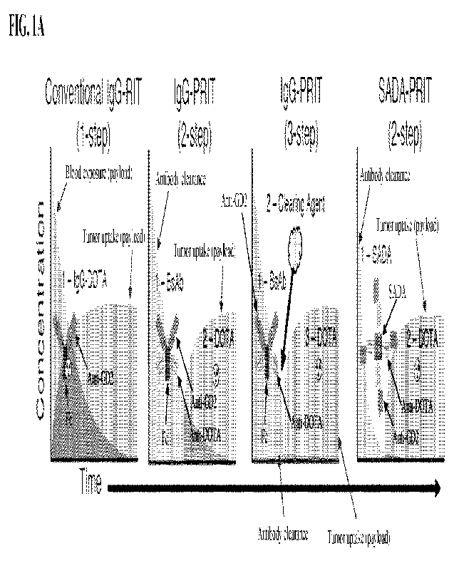
100281 FIGs. 1A-1E show an overview of multi-step payload delivery and anti-
GD2/anti-
DOTA SADA conjugate (a.k.a. SADA-BsAb) activity in vitro. FIG. 1A shows a
schematic
of 4 different payload delivery strategies. Tumor-specific domains and DOTA-
specific
domains are indicated. The concentration of payload in the blood over time,
the
concentration of payload in the tumor and the concentration of non-payload
antibody in the
blood are also indicated. The area of each curve (AUC) represents the relative
exposure of
each. FIG. 1B shows a schematic of a representative anti-GD2/anti-DOTA SADA
conjugate. Each monomer is made of 3 domains: an anti-tumor domain, an anti-
DOTA
domain and a SADA domain, from N-terminus to C-terminus, respectively. SADA
domains
self-assemble into tetramers (220 Ma) but also disassemble into monomers (55
Ma). FIG.
IC shows a representative SEC-HPLC chromatogram of anti-GD2/anti-DOTA P53noHIS-
SADA conjugate (a.k.a. P53-SADA-BsAb noHIS; (SEQ ID NO: 22)) with high and low
molecular weight impurities indicated. FIG. 1D shows normalized GD2 binding
kinetics of
anti-GD2/anti-DOTA P53-SADA conjugate (a.k.a. P53-SADA-13sAb; (SEQ ID NO: 27))
and
anti-GD2/anti-DOTA P63-SADA conjugate (a k.a. P63-SADA-BsAb; (SEQ ID NO: 28))
compared to anti-GD2/anti-DOTA IgG-scFv-BsAb (a.k.a. IgG-scFv-BsAb), as
measured by
surface plasmon resonance (SPR). For each curve maximum binding was normalized
to 100.
FIG. 1E shows representative cell binding analysis of anti-GD2/anti-DOTA P53-
SADA
9
CA 03180445 2022- 11- 25
WO 2021/242848
PCT/US2021/034230
conjugate (a.k.a. P53-SADA-BsAb LS; (SEQ ID NO: 23)) and anti-GD2/anti-DOTA
P63-
SADA conjugate (a.k.a. P63-SADA-BsAb LS; (SEQ ID NO: 24)) by flow cytometry.
Each
curve represents the fluorescence histogram of one BsAb or a control BsAb
(irrelevant tumor
specificity).
100291 FIGs. 2A-2D show in vivo pharmacokinetics and biodistribution of SADA-
BsAbs of
the present technology. FIG. 2A shows serum clearance kinetics of the tested
SADA-BsAbs.
Tumor-free mice (n=3) were injected with 131I- radiolabeled P53- SADA-BsAb LS
(SEQ ID
NO: 23) or P63-SADA-BsAb LS (SEQ lD NO: 24) and serially bled over 48 hours.
The
graph represents the amount of remaining BsAb per unit of blood normalized to
peak
concentration (0.5 hour). FIG. 2B shows the relationship between administered
dose and
tissue uptake using 2-step SADA-PRIT. Mice (n=5) were administered P53-SADA-
BsAb
(SEQ ID NO: 27) (1.25 nmol) and one of 3 doses of DOTA [177Lu]: 3.7, 18.5 or
37 MBq (20,
100 or 200 pmol, respectively). The level of DOTA payload in the tumor,
kidney, and blood
are indicated. The therapeutic index between tumor and blood at each dose is
also shown.
Tissue uptake was normalized to pmol of DOTA[77Lu] per gram of tissue. FIGs.
2C-2D
show an exemplary schematic and PET/CT images using the SADA-BsAbs of the
present
technology, respectively. As depicted in the schematic, mice (n=1-2) were
injected with P53-
SADA-BsAb (SEQ ID NO: 27) or IgG-scFv-BsAb (with and without clearing agents)
followed by DOTA[86Y] (downward arrows correspond to each injection). Mice
were
imaged for 30 minutes (upward arrow) 18 hours after the administration of
DOTA. The
representative images were normalized using the same scale. Arrows point to
the
subcutaneous tumor (left panel) or the bladder (middle panel).
100301 FIGs. 3A-3B show the immunogenicity of the SADA-BsAb of the present
technology. Mice (n=5) were immunized with P53-SADA-BsAb (SEQ ID NO: 27) or
IgG-
scFv-BsAb and bled 4 weeks later. Mice received a follow up dose of BsAb and
were bled
again 4 weeks later. Anti-BsAb titers were measured by ELISA and normalized to
a
monoclonal anti-BsAb standard. Statistical significances were calculated using
a Mann
Whitney test. **P = 0.0079 for IgG-scFv-BsAb compared to P53-SADA-BsAb.
100311 FIG. 4A shows a schematic of a neuroblastoma xenograft treatment model
(left) and
mean tumor responses (right). One dose of BsAb (SEQ ID NO: 27 or (SEQ ID NO:
28) (1.25
nmol, triangle) was followed by one dose of DOTA[I77Lu] (18.5 MBq, 100 pmol,
star) 48
CA 03180445 2022- 11- 25
WO 2021/242848
PCT/US2021/034230
hours later, once per week for 3 weeks. Each solid line represents one
treatment group
(n=10). The dotted black line represents no measurable tumor, and the boxed
hexagon
represents the tumor implantation. Tumor averages were calculated until at
least one mouse
had to be euthanized. Data are shown as means standard deviation. FIG. 4B
shows
individual tumor responses for each experimental group. Each solid line
represents tumors
from a single mouse, and the dashed line represents the group average. FIG. 4C
shows
progression-free survival analysis for each experimental group. Tumors were
considered
"progressing" when their volume reached 500 mm3. Mice were censored if they
were
sacrificed for histological analysis but were otherwise healthy at the time.
FIG. 4D shows
graphical representation of organ pathologies observed in treated mice. Each
bar represents
one treatment group and each graph represents analysis of either ovaries
(left) or bladders
(right). Y-axis values represent the percentage of analyzed mice displaying
the toxicity.
Grade 4, Grade 3, and Grade 2 toxicities vs. normal phenotype are indicated.
n=9 for IgG-
scFv-BsAb, P53-SADA-BsAb (SEQ ID NO: 27) and control mice (age-matched non-
tumor
littermates), and n=6 for P63-SADA-BsAb (SEQ ID NO: 28). Statistical
significances were
calculated by two-way analysis of variance (ANOVA) with Tukey correction or
Log-rank
(Mantel-Cox) test. ****P <0.0001 between DOTA[1:771-u] alone and P53-SADA-
BsAb, P63-
SADA-BsAb or IgG-scFv-BsAb.
100321 FIG. 5A shows a schematic of a DOTA[177Lu] neuroblastoma xenograft
treatment
model (left) and mean tumor responses (right). One dose of BsAb (SEQ ID NO:
27) (1.25
nmol, triangle) was followed by one dose of DOTA[177Lu] (55.5 MBq, 300 pmol,
star) 48
hours later, once per week for 3 weeks. Each solid line represents one
treatment group (n=5).
The dotted black line represents no measurable tumor, and the boxed hexagon
represents the
tumor implantation. Tumor averages were calculated until at least one mouse
had to be
euthanized. Data are shown as means standard deviation. FIG. 5B shows
progression-free
survival analysis for each experimental group. Tumors were considered
"progressing" when
their volume reached 500 mm3. Mice were censored if they were sacrificed for
histological
analysis but were otherwise healthy at the time.
100331 FIG. 5C shows a schematic of a Proteus[225Ac] neuroblastoma xenograft
treatment
model (left) and mean tumor responses (right). The structure of the Proteus
DOTA-hapten is
described in W02019/010299. Proteus-DOTA was synthesized by mixing two
bifunctional
11
CA 03180445 2022- 11- 25
WO 2021/242848
PCT/US2021/034230
DOTA chelators: commercial 2,2',2"-(10-(17-amino-2-oxo-6,9,12,15-tetraoxa-3-
azaheptadecy1)-1,4,7,10-tetraazacyclododecane-1,4,7-triy1)tri acetic acid
(amine-PEG4-
DOTA) and the non-radioactive lutetium-complex of 2-(4-isothiocyanatobenzy1)-
1,4,7,10-
tetraazacyclododecane-tetraacetic acid (p-SCN-Bn-DOTA.Lu3 complex) prepared
from
commercial p-SCN-Bn-DOTA and LuC13.6 H20. One dose of BsAb (SEQ ID NO: 27)
(1.25
nmol, triangle) was followed by one dose of Proteus[225Ac] (37 lcBci, 2.4
nmol, star) 48 hours
later. Each solid line represents one treatment group (n=5). The dotted black
line represents
no measurable tumor, and the boxed hexagon represents the tumor implantation.
Tumor
averages were calculated until at least one mouse had to be euthanized. Data
are shown as
means standard deviation. FIG. 5D shows progression-free survival analysis
for each
experimental group. Tumors were considered -progressing" when their volume
reached 500
m11113. Mice were censored if they were sacrificed for histological analysis
but were otherwise
healthy at the time. Statistical significances were calculated by two-way
analysis of variance
(ANOVA) with Tukey correction or Log-rank (Mantel-Cox) test. **P = 0.034,
****P <
0.0001 between DOTA[177Lu] alone or Proteus[225Ac] alone and P53-SADA-BsAb
(SEQ ID
NO: 27) or IgG-scFv-BsAb.
100341 FIG. 6A shows a schematic of Proteus[225Ac] small-cell lung cancer
patient-derived
xenograft (PDX) treatment model (left) and mean tumor responses (right). One
dose of BsAb
(SEQ ID NO: 27) (1.25 nmol, triangle) was followed by one dose of
Proteus[225Ac] (37 kBci,
621 pmol, star) 48 hours later. Each line represents one treatment group
(n=5). The dotted
black line represents no measurable tumor, and the boxed hexagon represents
the tumor
implantation. Tumor averages were calculated until at least one mouse had to
be euthanized.
Data are shown as means standard deviation. FIG. 6B shows individual tumor
responses
for each experimental group. Each solid line represents tumors from a single
mouse, and the
dashed line represents the group average. FIG. 6C progression-free survival
analysis for
each experimental group. Tumors were considered "progressing" when their
volume reached
500 mm3. No mice died unexpectedly in this study. Statistical significances
were calculated
by two-way analysis of variance (ANOVA) with Sidak correction or Log-rank
(Mantel-Cox)
test. ****P <0.0001 between Proteus[225Ac] alone and P53-SADA-BsAb.
100351 FIG. 7A shows a schematic of a neuroblastoma xenograft treatment model
(left) and
mean tumor responses (right). One dose of BsAb (SEQ ID NO: 28) (1.25 nmol,
triangle) was
12
CA 03180445 2022- 11- 25
WO 2021/242848
PCT/US2021/034230
followed by 3 subsequent doses of DOTA[177Lu] (18.5 MBq, 100 pmol, vertical
bar) at 48
hrs, 72 his and 96 hours after administration of the BsAb, once per week for 3
weeks. Each
solid line represents one treatment group (n=5-10). The dotted black line
represents no
measurable tumor, and the boxed hexagon represents the tumor implantation.
Tumor
averages were calculated until at least one mouse had to be euthanized. Data
are shown as
means standard deviation. FIG. 7B shows individual tumor responses for each
experimental group. Each solid line represent tumors from a single mouse, and
the dashed
line represents the group average. FIG. 7C shows progression-free survival
analysis for each
experimental group. Tumors were considered "progressing" when their volume
reached 500
mm3. Mice were censored if they were sacrificed for histological analysis but
were otherwise
healthy at the time. FIG. 7D shows a graphical representation of organ
pathologies observed
in treated mice. Each bar represents one treatment group and each graph
represents analysis
of either ovaries (left) or bladders (right). Y-axis values represent the
percentage of analyzed
mice displaying the toxicity. Grade 3 toxicity and no pathologies (normal) are
indicated.
n=6 for 3x-3x, n=2 for lx-3x and 2x-6x, and n=9 for the controls (age-matched
non-tumor
littermates). Statistical significances were calculated by two-way analysis of
variance
(ANOVA) with Tukey correction or Log-rank (Mantel-Cox) test. ***P <0.0005,
****P <
0.0001 between DOTA[177Lu] alone and 3x-3x, lx-3x or 2x-6x.
100361 FIG. 8A shows an exemplary hematology analysis of DOTA[177Lu] treated
mice in
each of the following experimental groups: P53-SADA-BsAb (SEQ ID NO: 27), P63-
SADA-
BsAb (SEQ ID NO: 28), IgG-scFv-BsAb, control group (DOTA[177Lu] Alone), and
age-
matched non-tumor littermates. White blood cell (WBC, left), Red blood cell
(RBC, center),
and platelet (PLT, right) counts from mouse blood are shown. All mice were
bled 14 days
after the first dose of DOTA[177Lu]. Each symbol refers to a single mouse
(n=10). The black
dotted line refers to mean values from age-matched mice irradiated with 300
cGy of total
body irradiation (TBI) on day 0, and the grey bar represents the one standard
deviation above
and below this mean. FIG. 8B shows FLT3L levels in the plasma of treated mice.
All mice
were bled 21 days after the first dose of DOTA[177Lu]. Each symbol refers to a
single mouse
(n=10). FIG. 8C shows weight change in treated mice. Weights were monitored at
least
once per week and normalized to each individual mouse's pre-treatment weight.
Each solid
line represents one treatment group (n=10). The dotted black line represents
10% increases
13
CA 03180445 2022- 11- 25
WO 2021/242848
PCT/US2021/034230
or decreases in weight. Average weights were calculated until at least one
mouse had to be
euthanized. Data are shown as means standard deviation.
100371 FIG. 9A shows an exemplary hematology analysis of DOTA[177Lu] treated
mice in
each of the following experimental groups: the P63-SADA-BsAb (SEQ ID NO: 28)
3x-3x
regimen, P63-SADA-BsAb (SEQ ID NO: 28) lx-3x regimen, P63-SADA-BsAb (SEQ ID
NO: 28) 2x-6x regimen, the control group (DOTA[177Lu] Alone), and age-matched
non-
tumor littermates. White blood cell (WBC, left), Red blood cell (RBC, center),
and platelet
(PLT, right) counts from mouse blood are shown. All mice were bled 14 days
after the first
dose of DOTA[177Lu]. Each symbol refers to a single mouse (n=5-10). The black
dotted line
refers to mean values from age-matched mice irradiated with 300 cGy of total
body
irradiation (TBI) on day 0. The grey bar represents the mean one standard
deviation. FIG.
9B shows FLT3L levels in the plasma of treated mice. All mice were bled 21
days after the
first dose of DOTA[177Lu]. Each symbol refers to a single mouse (n=10). FIG.
9C shows
weight change in treated mice. Weights were monitored at least once per week
and
normalized to each individual mouse's pre-treatment weight. Each solid line
represents one
treatment group (n=10). The dotted black line represents 10% increases or
decreases in
weight. Average weights were calculated until at least one mouse had to be
euthanized. Data
are shown as means standard deviation.
100381 FIG. 10 shows representative H&E staining of ovaries from treated nude
mice.
Normal ovary (left, littermate control), grade 3 atrophied ovary (center, P53-
SADA-BsAb
(SEQ ID NO: 27)) and grade 4 atrophied ovary (right, IgG-scEv-BsAb). Mice were
sacrificed between day 110 and day 230 after treatment start.
100391 FIG. 11A shows individual DOTA[77Lu] tumor responses in a neuroblastoma
PDX
treatment model treated with P53-SADA-BsAb (SEQ ID NO: 27). Each solid line
represent
tumors from a single mouse, and the dashed line represents the group average.
FIG. 11B
shows a graphical representation of bladder pathologies observed in treated
mice. Each bar
represents one treatment group (n=5). Y-axis values represent the percentage
of analyzed
mice displaying the toxicity. Grade 4 toxicity, grade 3 toxicity, grade 2
toxicity and no
pathologies (normal) are indicated. FIG. 11C shows individual Proteus[225Ac]
tumor
responses in a neuroblastoma model treated with P53-SADA-BsAb (SEQ ID NO: 27),
where
each line represent tumors from a single mouse, and the dashed line represents
the group
14
CA 03180445 2022- 11- 25
WO 2021/242848
PCT/US2021/034230
average. FIG. 11D shows a graphical representation of kidney pathologies
observed in
treated mice. All pathologies were measured as the number of observations per
10-
consecutive fields, beginning with the field containing the most pathologies.
Each group (x-
axis) represents one treatment group or age-matched littermate control, and
each individual
scatter plot represents a different stain for kidney damage. Tubular
proteinosis, epithelial cell
apoptosis, Cleaved Caspase 3 (CC-3) positive cells, and Terminal
deoxynucleotidyl
transferase dUTP nick end labeling (TUNEL) positive cells are depicted.
100401 FIG. 12A shows an exemplary hematology analysis of DOTA[177Lu] treated
mice in
a neuroblastoma model treated with P53-SADA-BsAb (SEQ ID NO: 27). White blood
cell
(WBC, left), Red blood cell (RBC, center), and platelet (PLT, right) counts
from mouse
blood are shown. All mice were bled 14 days after the first dose of
DOTA[177Lu]. Each
symbol refers to a single mouse (n=5). FIG. 12B shows representative H&E
staining of
bladders from treated mice. Normal bladder (left, littermate control), grade 2
bladder (center
left, IgG-scFv-BsAb), grade 3 multifocal bladder (center right, P53-SADA-BsAb)
and grade
4 diffuse bladder (right, P53-SADA-FisAb) are shown. Mice were sacrificed at
day 120 after
treatment initiation.
100411 FIG. 13A shows an exemplary hematology analysis of Proteus[225Ac]
treated mice in
a neuroblastoma model treated with P53-SADA-BsAb (SEQ ID NO: 27). White blood
cell
(WBC, left), Red blood cell (RBC, center), and platelet (PLT, right) counts
from mouse
blood are shown. All mice were bled 14 days after the first dose of
Proteus[225Ac]. Each
symbol refers to a single mouse (n=5). FIG. 13B show representative images of
kidneys
from IgG-scFv-BsAb treated mice. Cleaved Caspase 3 (CC-3) positive kidney
(left), terminal
deoxynucleotidyl transferase dUTP nick end labeling (TUNEL) positive kidney
(center left),
H&E stained kidney with epithelial necrosis, and tubular proteinosis and grade
4 multifocal
bladder (center right) and grade 4 diffuse bladder (right). Mice were
sacrificed between day
100 and 120 after treatment initiation.
100421 FIG. 14 shows structural properties of candidate SADA domains. The
sequence
refers to the specific amino acids used, counting from the N-terminal amino
acid. PDB ID
refers to a referenced crystal structure. The molecular size of monomer
displays the
theoretical molecular weight for each SADA domain. The surface areas and the
number of
hydrogen bonds were calculated using Discovery Studio.
CA 03180445 2022- 11- 25
WO 2021/242848
PCT/US2021/034230
100431 FIG. 15 shows the biochemical properties of candidate SADA-BsAbs (SEQ
ID NO:
27, SEQ ID NO: 28) of the present technology. Total monomer size was
calculated assuming
25 kDa for each scFv. Yield was calculated from at least 2 transfections using
expi293 cells.
Purity was determined by SEC-HPLC. High and low molecular weight impurities
were
defined as peaks before or after the main peak, respectively. Stability was
determined by
incubation at 37 C with weekly quantitation by SEC-HPLC.
100441 FIG. 16 shows the summary of GD2 binding kinetics of the SADA-BsAbs
disclosed
herein as determined by SPR. Values were calculated using a two-state reaction
model. Chi2
values show the error between the raw and fitted data (RU). Fold-change was
calculated by
dividing the KD of the IgG-scFv-BsAb by the KD of either P53-SADA-BsAb (SEQ ID
NO:
27) or P63-SADA-BsAb (SEQ ID NO: 28).
100451 FIG. 17 shows a summary of the pharmacokinetic properties of P53-SADA-
BsAb
(SEQ ID NO: 27). NSG mice (n=10) were serially bleed from 0.5 to 168 hours
after
intravenous BsAb administration. Pharmacokinetic analysis was carried out by
non-
compartmental analysis of the sen_ina concentration-time data using WinNonlin
software
program (Pharsight Corp.).
100461 FIG. 18 shows SADA PRIT dosimetry estimates calculated from mouse
biodistribution studies, and their corresponding tumor-to-non-tumor ratios.
Tumor bearing
mice (n=3-5 per time point) were dosed with each BsAb (SEQ ID NO: 27 and SEQ
ID NO:
28) (1.25 nmol) and DOTA[177Lu] (18.5 MBq), 48 hours apart. Mice were
sacrificed either
2, 24, 48, or 120 hours after payload delivery. IgG-scFv-BsAb treated mice
received 25 [ig
of clearing agent 4 hours prior to the administration of DOTA[177Lu].
100471 FIG. 19 shows a summary of tissue biodistribution of DOTA[86Y] after
PET/CT scan.
Tumor bearing mice were dosed with each BsAb (SEQ ID NO: 27) (1.25 nmol) and
DOTA[86Y] (3.7 MBq), 48 hours apart, and sacrificed immediately after imaging.
Values are
normalized to percentage of injected dose per gram of tissue (%ID/g). 2-step
IgG-scFv-BsAb
treated mice did not receive clearing agent (CA). 3-step IgG-scFv-BsAb treated
mice
received 25 mg of CA.
100481 FIG. 20 shows a summary of serum chemistry, complete blood counts, and
histopathology in nude mice treated with the indicated BsAb (SEQ ID NO: 27 and
SEQ ID
NO: 28) /DOTA[1771_,u] payload regimen. Interpretation was performed by board-
certified
16
CA 03180445 2022- 11- 25
WO 2021/242848
PCT/US2021/034230
veterinary pathologists. Normal was defined as being not significantly
different from
untreated age-matched littermate control mice, or within known normal ranges
for this strain
of mice at the same age. Histopathologic abnormalities were determined by
microscopic
analysis of H&E slides. Mice were submitted for assessment 111, 155 and 230
days after
treatment was initiated.
100491 FIG. 21 shows a summary of serum chemistry, complete blood counts, and
histopathology in DKO mice treated with the indicated BsAb (SEQ ID NO:
27)/DOTA[177Lu] payload regimen. Interpretation was performed by board-
certified
veterinary pathologists. Normal was defined as being not significantly
different from
untreated age-matched littermate control mice, or within known normal ranges
for this strain
of mice at the same age. Histopathologic abnormalities were determined by
microscopic
analysis of H&E slides. Mice were submitted for assessment 120 days after
treatment was
initiated.
100501 FIG. 22A shows a summary of serum chemistry, complete blood counts, and
histopathology in DKO mice treated with the indicated BsAb (SEQ ID NO. 27)/
Proteus[225Ac] payload regimen. Interpretation was performed by board-
certified veterinary
pathologists. Normal was defined as being not significantly different from
untreated age-
matched littermate control mice, or within known normal ranges for this strain
of mice at the
same age. Histopathologic abnormalities were determined by microscopic
analysis of H&E
slides. Mice were submitted for assessment 80-120 days after treatment was
initiated. CC-3:
Cleaved caspase-3 immunohistochemistry.
100511 FIG. 22B shows shows a summary of serum chemistry, complete blood
counts, and
histopathology in DKO mice treated with the indicated BsAb (SEQ ID NO: 27)/
Proteus1225Ac] payload regimen. Interpretation was performed by board-
certified veterinary
pathologists. Normal was defined as being not significantly different from
untreated age-
matched littermate control mice, or within known normal ranges for this strain
of mice at this
age. Histopathologic abnormalities were determined by microscopic analysis of
H&E slides.
Mice were submitted for assessment 163, 210 and 309 days after treatment
began. MF:
Multifocal. Grade 1: Minimal; 2: Mild; 3: Moderate
100521 FIG. 23A shows mean tumor responses in DOTA [177Lu] small-cell lung
cancer
patient-derived xenograft (PDX) treatment model. Each dose of BsAb (SEQ lID
NO: 27)
17
CA 03180445 2022- 11- 25
WO 2021/242848
PCT/US2021/034230
(1.25 nmol, triangle) was followed by a dose of DOTA[177Lu] (37 kBq, 700 pmol,
star) 48
hours later. Each line represents one treatment group (n= 4-5). The dotted
black line
represents no measurable tumor, and the asterisk represents the tumor
implantation. Tumor
averages were calculated until at least one mouse had to be euthanized. Data
are shown as
means standard deviation. FIG. 23B shows mean tumor responses in DOTA
[225Ac] small-
cell lung cancer patient-derived xenograft (PDX) treatment model. Each dose of
BsAb (SEQ
ID NO: 27) (1.25 nmol, triangle) was followed by a dose of DOTA[225Ac] (37
kBq, 700
pmol, star) 48 hours later. Each line represents one treatment group. The
dotted black line
represents no measurable tumor. Tumor averages were calculated until at least
one mouse
had to be euthanized. Data are shown as means standard deviation. FIG. 23C
shows
progression-free survival analysis for each experimental group. Tumors were
considered
"progressing" when their volume reached 500 mm3.
100531 FIGs. 24A-24B show in vivo bi odi stributi on of SADA-13sAbs of the
present
technology. Tumor bearing nude mice (IVIR32Luc Sc, right flank) were treated
with
1.25nmo1 of each BsAb and 18.5 NIBq (100pmol) of DOTAmLu 48 hours later. BC151
(IgG-scFv-BsAb) were treated with a clearing agent 4 hours prior to DOTALu177.
Mice were
sacrificed 24hrs after administration of Lu177 and organs were collected and
read on a gamma
counter (Perkin Elmer). Counts were decay corrected and normalized to the
injected dose
(18.5MBq and the weight of the organs). TC101 = SEQ ID NO. 22, TC134 = SEQ ID
NO:
27, TC135 = SEQ ID NO: 28, and TC135-H = SEQ ID NO: 38. Kidney uptake was not
impacted by the presence or absence of a 6xHIS tag.
100541 FIGs. 25A-25B show in vivo biodistribution of SADA-BsAbs of the present
technology and their corresponding tumor-to-non-tumor ratios based on the
results described
in FIGs. 24A-24B (the values are normalized to the tumor uptake (tumor to
blood, tumor to
liver, tumor to kidney). TC101 = SEQ ID NO: 22, TC 134 = SEQ ID NO: 27, TC135
= SEQ
ID NO: 28, and TC135-H = SEQ ID NO: 38. FIG. 25B represents tabulated data
from FIGs.
24A-24B. Kidney uptake is not impacted by the presence or absence of a 6xHIS
tag.
100551 FIG. 26 shows the concentration of P53-SADA-BsAb (SEQ ID NO: 27) in
blood at
24 hours and at 48 hours (n=5). NSG mice (n=10) were serially bleed from 0.5
to 168 hours
after intravenous BsAb administration. Pharmacokinetic analysis was carried
out by non-
18
CA 03180445 2022- 11- 25
WO 2021/242848
PCT/US2021/034230
compartmental analysis of the serum concentration-time data using WinNonlin
software
program (Pharsight Corp.).
DETAILED DESCRIPTION
100561 It is to be appreciated that certain aspects, modes,
embodiments, variations and
features of the present methods are described below in various levels of
detail in order to
provide a substantial understanding of the present technology.
100571 In practicing the present methods, many conventional
techniques in molecular
biology, protein biochemistry, cell biology, immunology, microbiology and
recombinant
DNA are used. See, e.g., Sambrook and Russell eds. (2001)Molecular Cloning: A
Laboratory Manual, 3rd edition; the series Ausubel et al. eds. (2007) Current
Protocols in
Molecular Biology; the series Methods in Enzymology (Academic Press, Inc.,
N.Y.);
MacPherson et al. (1991) PCR 1: A Practical Approach (IRL Press at Oxford
University
Press); MacPherson et al. (1995) 1'C1?2: A Practical Approach; Harlow and Lane
eds. (1999)
Antibodies, A Laboratory Manual; Freshney (2005) Culture of Animal Cells: A
Manual of
Basic Technique, 5th edition; Gait ed. (1984) Oligonucleotide Synthesis;U U.S.
Patent No.
4,683,195; Hames and Higgins eds. (1984) Nucleic Acid Hybridization; Anderson
(1999)
Nucleic Acid Hybridization; Hames and Higgins eds. (1984) Transcription and
Translation;
Immobilized Cells and Enzymes (IRL Press (1986)); Perbal (1984)A Practical
Guide to
Molecular Cloning; Miller and Cabs eds. (1987) Gene Transfer Vectors for
Mammalian
Cells (Cold Spring Harbor Laboratory); Makrides ed. (2003) Gene Transfer and
Expression
in Mammalian Cells; Mayer and Walker eds. (1987) Immunochemical Methods in
Cell and
Molecular Biology (Academic Press, London); and Herzenberg et al. eds (1996)
Weir's
Handbook of Experimental Immunology. Methods to detect and measure levels of
polypeptide gene expression products (i.e., gene translation level) are well-
known in the art
and include the use of polypeptide detection methods such as antibody
detection and
quantification techniques. (See also, Strachan & Read, Human Molecular
Genetics, Second
Edition. (John Wiley and Sons, Inc., NY, 1999)).
100581 Multi-step targeting strategies are utilized to overcome TI
limitations by
delivering tumor targeting agents (e.g-. anti-tumor IgG) separately from the
cytotoxic
payloads (e.g. chelated radioisotopes). As an example, conventional 2-step
pretargeted
radioimmunotherapy (PRIT) administers engineered bispecific antibodies (BsAb)
or
19
CA 03180445 2022- 11- 25
WO 2021/242848
PCT/US2021/034230
chemically modified monoclonal antibodies first (step 1, ty2 ¨ days), followed
hours or days
later with the delivery of small radioactive payloads (step 2, ti/2 ¨ minutes)
that seek out the
tumor-bound antibodies (FIG. IA). While this strategy does reduce toxicities
in some
tissues, the residual circulating antibody in the blood is enough to prevent
any substantial
improvement in therapeutic index or efficacy. One solutions is 3-step PRIT,
where after the
administration of tumor targeting IgG (step 1), a clearing agent step (step 2)
is introduced to
remove circulating antibody from the blood before the delivery of the
cytotoxic payload (step
3). While inclusion of a clearing agent may improve Tls, the optimal clearing
agent dose will
vary depending on tumor size and antigen density, substantially complicating
clinical
translation. While a high dose of clearing agent should maximize removal of
IgG, it could
also interfere with payload uptake at the tumor. In contrast, an insufficient
dose of clearing
agent would leave considerable IgG in the blood, capturing the injected
payload, circulating it
and ultimately harming the bone marrow and other normal tissues. Thus, the
ideal targeting
strategy requires a tumor-targeting platform that can consistently clear
itself from the blood
before payload delivery without the need for optimization of additional or
exogenous
reagents.
100591 The present disclosure provides a novel platform for the
multi-step delivery of
cytotoxic payloads to tumors using specially designed Self-Assembling and
DisAssembling
(SADA) domains (FIG. 1B). When fused to BsAb, the resulting SADA-BsAb self-
assemble
into stable tetrameric complexes (220 kDa) that bind tumors with high avidity
but could also
disassemble into small dimers (110 kDa) or monomers (55 kDa) after a period of
circulation
in the blood (hours). Importantly, while the tetrameric complexes exceed the
molecular
weight (MW) cut off for renal filtration, the small monomers fall below the
threshold and are
able to rapidly and completely clear from the blood.
100601 While many protein therapies benefit from long terminal half-
lives, the delivery of
highly cytotoxic payloads using such proteins inevitably harms sensitive
tissues such as bone
marrow. To date, all 8 FDA approved antibody-drug conjugates, and both FDA
approved
radiolabeled protein therapies have demonstrated some myelotoxicity during
clinical
development, using substantially lower doses of payload than were achieved
with the 2-step
SADA-PRIT methods disclosed herein. The present disclosure demonstrates that
the SADA-
BsAbs of the present technology in combination with radioactive payloads
carrying alpha
CA 03180445 2022- 11- 25
WO 2021/242848
PCT/US2021/034230
(225AC 1.48 MBq/kg) or beta (177Lu 6,660 MBq/kg) radioisotopes ablate
established solid
tumors in multiple mouse models without the need for any clearing agent.
Instead, the
SADA-platform utilized the narrow window in blood retention between long-lived
large size
proteins and small peptides, temporarily maintaining a plasma half-life for
just enough time
to effectively reach the tumor, followed by rapid and complete clearance from
the blood.
Additionally, this fast clearance rendered SADA-BsAb substantially less
immunogenic
compared to more conventional IgG-based platforms, a crucial advantage in
therapeutic
strategies that necessitate multiple treatment cycles.
[0061] The methods disclosed herein eliminated all clinical or
histologic toxicities to the
kidneys, liver, bone marrow, spleen, or brain while delivering enormous doses
of cytotoxic
payloads. These results are critical and clinically relevant given the
sensitivity of these
organs to radiation-related toxicities in conventional KIT. See e.g, Bodei et
al., European
Journal of Nuclear Medicine and Molecular Imaging 42, 5-19 (2015); Forster et
at., Journal
of Nuclear Medicine 47, 140-149 (2006); Gupta et at., Cancer Biotherapy and
Radiopharmaceuticals 27, 593-599 (2012); Heskamp, et al., Journal of Nuclear
Medicine 58,
926-933 (2017); Muselaers et al, Journal of Nuclear Medicine 57, 34-34 (2016);
Poty et al .,
Clinical Cancer Research 25, 868-880 (2019); Vallabhajosula et at., Journal of
Nuclear
Medicine 46, 850-858 (2005).
In particular, the myelotoxicity-free dose levels achieved in the present
disclosure (up to
6,600 MBq/kg) are exponentially higher than those currently used in the clinic
(typically <
150 MBq/kg), demonstrating the safety margin that SADA-BsAb provided to
radiosensitive
tissues.
Definitions
100621 Unless defined otherwise, all technical and scientific terms
used herein generally
have the same meaning as commonly understood by one of ordinary skill in the
art to which
this technology belongs. As used in this specification and the appended
claims, the singular
forms "a", "an" and "the" include plural referents unless the content clearly
dictates
otherwise. For example, reference to "a cell" includes a combination of two or
more cells,
and the like. Generally, the nomenclature used herein and the laboratory
procedures in cell
culture, molecular genetics, organic chemistry, analytical chemistry and
nucleic acid
21
CA 03180445 2022- 11- 25
WO 2021/242848
PCT/US2021/034230
chemistry and hybridization described below are those well-known and commonly
employed
in the art.
100631 As used herein, the term "about" in reference to a number is
generally taken to
include numbers that fall within a range of 1%, 5%, or 10% in either direction
(greater than
or less than) of the number unless otherwise stated or otherwise evident from
the context
(except where such number would be less than 0% or exceed 100% of a possible
value).
100641 As used herein, the "administration" of an agent or drug to
a subject includes any
route of introducing or delivering to a subject a compound to perform its
intended function.
Administration can be carried out by any suitable route, including but not
limited to, orally,
intranasally, parenterally (intravenously, intramuscularly, intraperitoneally,
or
subcutaneously), rectally, intrathecally, intratumorally or topically.
Administration includes
self-administration and the administration by another.
100651 As used herein, the term "antibody" collectively refers to
immunoglobulins or
immunoglobulin-like molecules including by way of example and without
limitation, IgA,
IgD, IgE, IgG and IgM, combinations thereof, and similar molecules produced
during an
immune response in any vertebrate, for example, in mammals such as humans,
goats, rabbits
and mice, as well as non-mammalian species, such as shark immunoglobulins. As
used
herein, "antibodies" (includes intact immunoglobulins) and "antigen binding
fragments"
specifically bind to a molecule of interest (or a group of highly similar
molecules of interest)
to the substantial exclusion of binding to other molecules (for example,
antibodies and
antibody fragments that have a binding constant for the molecule of interest
that is at least 103
M-1 greater, at least 104M1 greater or at least 105 M-1 greater than a binding
constant for
other molecules in a biological sample). The term "antibody" also includes
genetically
engineered forms such as chimeric antibodies (for example, humanized murine
antibodies),
heteroconjugate antibodies (such as, bispecific antibodies). See also, Pierce
Catalog and
Handbook, 1994-1995 (Pierce Chemical Co., Rockford, Ill.); Kuby, J.,
Immunology, 3rd Ed.,
W.H. Freeman & Co., New York, 1997.
100661 More particularly, antibody refers to a polypeptide ligand
comprising at least a
light chain immunoglobulin variable region or heavy chain immunoglobulin
variable region
which specifically recognizes and binds an epitope of an antigen. Antibodies
are composed
22
CA 03180445 2022- 11- 25
WO 2021/242848
PCT/US2021/034230
of a heavy and a light chain, each of which has a variable region, termed the
variable heavy
(VH) region and the variable light (VL) region. Together, the Vu region and
the VL region are
responsible for binding the antigen recognized by the antibody. Typically, an
immunoglobulin has heavy (H) chains and light (L) chains interconnected by
disulfide bonds.
There are two types of light chain, lambda (X) and kappa (x). There are five
main heavy
chain classes (or isotypes) which determine the functional activity of an
antibody molecule:
IgM, IgD, IgG, IgA and IgE. Each heavy and light chain contains a constant
region and a
variable region, (the regions are also known as "domains") In combination, the
heavy and
the light chain variable regions specifically bind the antigen. Light and
heavy chain variable
regions contain a "framework" region interrupted by three hypervariable
regions, also called
"complementarity-determining regions" or "CDRs". The extent of the framework
region and
CDRs have been defined (see, Kabat et cu., Sequences of Proteins of
Immunological Interest,
U.S. Department of Health and Human Services, 1991, which is hereby
incorporated by
reference). The Kabat database is now maintained online. The sequences of the
framework
regions of different light or heavy chains are relatively conserved within a
species. The
framework region of an antibody, that is the combined framework regions of the
constituent
light and heavy chains, largely adopt a 13-sheet conformation and the CDRs
form loops which
connect, and in some cases form part of, the 13-sheet structure. Thus,
framework regions act
to form a scaffold that provides for positioning the CDRs in correct
orientation by inter-
chain, non-covalent interactions.
100671 The CDRs are primarily responsible for binding to an epitope
of an antigen. The
CDRs of each chain are typically referred to as CDR1, CDR2, and CDR3, numbered
sequentially starting from the N-terminus, and are also typically identified
by the chain in
which the particular CDR is located. Thus, a VII CDR3 is located in the
variable domain of
the heavy chain of the antibody in which it is found, whereas a VI_ CDR1 is
the CDR1 from
the variable domain of the light chain of the antibody in which it is found.
An antibody that
binds a target antigen (e.g., GD2) will have a specific Vit region and the
V1_, region sequence,
and thus specific CDR sequences. Antibodies with different specificities (i.e.
different
combining sites for different antigens) have different CDRs. Although it is
the CDRs that
vary from antibody to antibody, only a limited number of amino acid positions
within the
CDRs are directly involved in antigen binding. These positions within the CDRs
are called
23
CA 03180445 2022- 11- 25
WO 2021/242848
PCT/US2021/034230
specificity determining residues (SDRs). "Immunoglobulin-related compositions"
as used
herein, refers to antibodies (including monoclonal antibodies, polyclonal
antibodies,
humanized antibodies, chimeric antibodies, recombinant antibodies, multi-
specific
antibodies, bispecific antibodies, etc.,) as well as antibody fragments. An
antibody or antigen
binding fragment thereof specifically binds to an antigen.
100681 As used herein, the term "antibody-related polypeptide"
means antigen-binding
antibody fragments, including single-chain antibodies, that can comprise the
variable
region(s) alone, or in combination, with all or part of the following
polypeptide elements.
hinge region, CHi, CH2, and CH3 domains of an antibody molecule. Also included
in the
technology are any combinations of variable region(s) and hinge region, CH2,
CH2, and CH3
domains. Antibody-related molecules useful in the present methods, e.g., but
are not limited
to, Fab, Fab' and F(ab')2, Fd, single-chain Fvs (scFv), single-chain
antibodies, disulfide-
linked Fvs (sdFv) and fragments comprising either a VL or VII domain. Examples
include: (i)
a Fab fragment, a monovalent fragment consisting of the VL, VH, CL and CHi
domains; (ii) a
F(ab')2 fragment, a bivalent fragment comprising two Fab fragments linked by a
disulfide
bridge at the hinge region; (iii) a Fd fragment consisting of the V14 and CHi
domains; (iv) a
Fv fragment consisting of the VL and VH domains of a single arm of an
antibody, (v) a dAb
fragment (Ward et al., Nature 341: 544-546, 1989), which consists of a VH
domain; and (vi)
an isolated complementarity determining region (CDR). As such "antibody
fragments" or
"antigen binding fragments" can comprise a portion of a full length antibody,
generally the
antigen binding or variable region thereof. Examples of antibody fragments or
antigen
binding fragments include Fab, Fab', F(ab')2, and Fv fragments; diabodies;
linear antibodies;
single-chain antibody molecules; and multi-specific antibodies formed from
antibody
fragments.
100691 "Bispecific antibody" or "BsAb", as used herein, refers to
an immunoglobulin-
related composition that can bind simultaneously to two targets that have a
distinct structure,
e.g., two different target antigens, two different epitopes on the same target
antigen, or a
hapten and a target antigen or epitope on a target antigen. A variety of
different bispecific
antibody structures are known in the art. In some embodiments, each antigen
binding moiety
in a bispecific antibody includes VII and/or VL regions; in some such
embodiments, the Vii
and/or VL regions are those found in a particular monoclonal antibody. In some
24
CA 03180445 2022- 11- 25
WO 2021/242848
PCT/US2021/034230
embodiments, the bispecific antibody contains two antigen binding moieties,
each including
VH and/or VL regions from different monoclonal antibodies. In some
embodiments,
the bispecific antibody contains two antigen binding moieties, wherein one of
the two antigen
binding moieties includes an immunoglobulin molecule having VH and/or VL
regions that
contain CDRs from a first monoclonal antibody, and the other antigen binding
moiety
includes an antibody fragment (e.g., Fab, F(ab'), F(ab')2, Fd, Fv, dAB, scFv,
etc.) having VH
and/or VL regions that contain CDRs from a second monoclonal antibody.
100701 As used herein, the term "conjugated" refers to the
association of two molecules
by any method known to those in the art. Suitable types of associations
include chemical
bonds and physical bonds. Chemical bonds include, for example, covalent bonds
and
coordinate bonds. Physical bonds include, for instance, hydrogen bonds,
dipolar interactions,
van der Waal forces, electrostatic interactions, hydrophobic interactions and
aromatic
stacking.
100711 As used herein, the term "diabodies" refers to small
antibody fragments with two
antigen-binding sites, which fragments comprise a heavy-chain variable domain
(VH)
connected to a light-chain variable domain (VL) in the same polypeptide chain
(VH VL). By
using a linker that is too short to allow pairing between the two domains on
the same chain,
the domains are forced to pair with the complementary domains of another chain
and create
two antigen binding sites. Diabodies are described more fully in, e.g., EP
404,097;
WO 93/11161; and Hollinger et al., Proc. Natl. Acad. Sci. USA, 90: 6444-6448
(1993).
100721 As used herein, the terms "single-chain antibodies" or
"single-chain Fv (scFv)"
refer to an antibody fusion molecule of the two domains of the Fv fragment, VI
and VH.
Single-chain antibody molecules may comprise a polymer with a number of
individual
molecules, for example, dimer, trimer or other polymers. Furthermore, although
the two
domains of the Fv fragment, VL and VH, are coded for by separate genes, they
can be joined,
using recombinant methods, by a synthetic linker that enables them to be made
as a single
protein chain in which the VI_ and VH regions pair to form monovalent
molecules (known as
single-chain Fv (scFv)). Bird etal. (1988) Science 242:423-426 and Huston et
al. (1988)
Proc. Natl. Acad Sci . USA 85:5879-5883. Such single-chain antibodies can be
prepared by
recombinant techniques or enzymatic or chemical cleavage of intact antibodies.
CA 03180445 2022- 11- 25
WO 2021/242848
PCT/US2021/034230
100731 Any of the above-noted antibody fragments are obtained using
conventional
techniques known to those of skill in the art, and the fragments are screened
for binding
specificity and neutralization activity in the same manner as are intact
antibodies.
100741 As used herein, an "antigen" refers to a molecule to which
an antibody (or antigen
binding fragment thereof) can selectively bind. The target antigen may be a
protein,
carbohydrate, nucleic acid, lipid, hapten, or other naturally occurring or
synthetic compound.
In some embodiments, the target antigen may GD2. An antigen may also be
administered to
an animal to generate an immune response in the animal.
100751 The term "antigen binding fragment" refers to a fragment of
the whole
immunoglobulin structure which possesses a part of a polypeptide responsible
for binding to
antigen. Examples of the antigen binding fragment useful in the present
technology include
scFv, (scFv)2, scFvFc, Fab, Fab' and F(ab)2, but are not limited thereto.
100761 By "binding affinity" is meant the strength of the total
noncovalent interactions
between a single binding site of a molecule (e.g., an antibody) and its
binding partner (e.g., an
antigen or antigenic peptide). The affinity of a molecule X for its partner Y
can generally be
represented by the dissociation constant (Ku). Affinity can be measured by
standard methods
known in the art, including those described herein. A low-affinity complex
contains an
antibody that generally tends to dissociate readily from the antigen, whereas
a high-affinity
complex contains an antibody that generally tends to remain bound to the
antigen for a longer
duration.
100771 "Binding domain", as used herein, refers to a moiety or
entity that specifically
binds to a target moiety or entity. Typically, the interaction between a
binding domain and its
target is non-covalent. In some embodiments, a binding domain may be or
comprise a
moiety or entity of any chemical class including, for example, a carbohydrate,
a lipid, a
nucleic acid, a metal, a polypeptide, a small molecule. In some embodiments, a
binding
domain may be or comprise a polypeptide (or complex thereof), a target-binding
portion of
an immunoglobulin-related composition, a cytokine, a ligand (e.g., a receptor
ligand), a
receptor, a toxin, etc. In certain embodiments, a binding domain may be or
comprise an
aptamer. In other embodiments, a binding domain may be or comprise a peptide
nucleic acid
(PNA).
26
CA 03180445 2022- 11- 25
WO 2021/242848
PCT/US2021/034230
100781 As used herein, the term "biological sample" means sample
material derived from
living cells. Biological samples may include tissues, cells, protein or
membrane extracts of
cells, and biological fluids (e.g., ascites fluid or cerebrospinal fluid
(CSF)) isolated from a
subject, as well as tissues, cells and fluids present within a subject.
Biological samples of the
present technology include, but are not limited to, samples taken from breast
tissue, renal
tissue, the uterine cervix, the endometrium, the head or neck, the
gallbladder, parotid tissue,
the prostate, the brain, the pituitary gland, kidney tissue, muscle, the
esophagus, the stomach,
the small intestine, the colon, the liver, the spleen, the pancreas, thyroid
tissue, heart tissue,
lung tissue, the bladder, adipose tissue, lymph node tissue, the uterus,
ovarian tissue, adrenal
tissue, testis tissue, the tonsils, thymus, blood, hair, buccal, skin, serum,
plasma, CSF, semen,
prostate fluid, seminal fluid, urine, feces, sweat, saliva, sputum, mucus,
bone marrow, lymph,
and tears. Biological samples can also be obtained from biopsies of internal
organs or from
cancers. Biological samples can be obtained from subjects for diagnosis or
research or can be
obtained from non-diseased individuals, as controls or for basic research.
Samples may be
obtained by standard methods including, e.g., venous puncture and surgical
biopsy. In certain
embodiments, the biological sample is a tissue sample obtained by needle
biopsy.
100791 As used herein, the term "chimeric antibody" means an
antibody in which the Fc
constant region of a monoclonal antibody from one species (e.g., a mouse Fc
constant region)
is replaced, using recombinant DNA techniques, with an Fc constant region from
an antibody
of another species (e.g., a human Fc constant region). See generally, Robinson
et at.,
PCT/US86/02269; Akira et aL, European Patent Application 184,187; Taniguchi,
European
Patent Application 171,496; Morrison et al., European Patent Application
173,494;
Neuberger et ed., WO 86/01533; Cabilly et aL U.S. Patent No. 4,816,567;
Cabilly et al.,
European Patent Application 0125,023; Better et at., Science 240: 1041-1043,
1988; Liu et
al., Proc. Natl. Acad. Sci. USA 84: 3439-3443, 1987; Liu et a/.õ1. Immunol
139: 3521-3526,
1987; Sun et al., Proc. Natl. Acad. Sci. USA 84: 214-218, 1987; Nishimura et
aL , Cancer Res
47: 999-1005, 1987; Wood et al., Nature 314: 446-449, 1885; and Shaw et al, 1
Natl.
Cancer Inst. 80: 1553-1559, 1988.
100801 As used herein, a "clearing agent" is an agent that binds to
excess bifunctional
antibody that is present in the blood compartment of a subject to facilitate
rapid clearance via
kidneys. The use of the clearing agent prior to hapten administration
facilitates better tumor-
27
CA 03180445 2022- 11- 25
WO 2021/242848
PCT/US2021/034230
to-background ratios in PRIT systems. Examples of clearing agents include 500
k1D-dextran-
DOTA-Bn(Y) (Orcutt et al., Mol Cancer Ther.11(6): 1365-1372 (2012)), 500 kD
aminodextran-DOTA conjugate, antibodies against the pretargeting antibody,
etc.
100811 As used herein, the term "consensus FR" means a framework
(FR) antibody
region in a consensus immunoglobulin sequence. The FR regions of an antibody
do not
contact the antigen.
100821 As used herein, a "control" is an alternative sample used in
an experiment for
comparison purpose. A control can be "positive" or "negative." For example,
where the
purpose of the experiment is to determine a correlation of the efficacy of a
therapeutic agent
for the treatment for a particular type of disease, a positive control (a
compound or
composition known to exhibit the desired therapeutic effect) and a negative
control (a subject
or a sample that does not receive the therapy or receives a placebo) are
typically employed.
100831 "Dosage form" and "unit dosage form", as used herein, the
term "dosage form"
refers to physically discrete unit of a therapeutic agent for a subject (e.g.,
a human patient) to
be treated. Each unit contains a predetermined quantity of active material
calculated or
demonstrated to produce a desired therapeutic effect when administered to a
relevant
population according to an appropriate dosing regimen. For example, in some
embodiments,
such quantity is a unit dosage amount (or a whole fraction thereof)
appropriate for
administration in accordance with a dosing regimen that has been determined to
correlate
with a desired or beneficial outcome when administered to a relevant
population (i.e., with a
therapeutic dosing regimen). It will be understood, however, that the total
dosage
administered to any particular patient will be selected by a medical
professional (e.g., a
medical doctor) within the scope of sound medical judgment.
100841 "Dosing regimen" (or "therapeutic regimen"), as used herein
is a set of unit doses
(typically more than one) that are administered individually to a subject,
typically separated
by periods of time. In some embodiments, a given therapeutic agent has a
recommended
dosing regimen, which may involve one or more doses. In some embodiments, a
dosing
regimen comprises a plurality of doses each of which are separated from one
another by a
time period of the same length; in certain embodiments, a dosing regimen
comprises a
plurality of doses and at least two different time periods separating
individual doses. In some
28
CA 03180445 2022- 11- 25
WO 2021/242848
PCT/US2021/034230
embodiments, the therapeutic agent is administered continuously (e.g., by
infusion) over a
predetermined period. In other embodiments, a therapeutic agent is
administered once a day
(QD) or twice a day (BID). In some embodiments, a dosing regimen comprises a
plurality of
doses each of which are separated from one another by a time period of the
same length; in
other embodiments, a dosing regimen comprises a plurality of doses and at
least two different
time periods separating individual doses. In some embodiments, all doses
within a dosing
regimen are of the same unit dose amount. In certain embodiments, different
doses within a
dosing regimen are of different amounts. In some embodiments, a dosing regimen
comprises
a first dose in a first dose amount, followed by one or more additional doses
in a second dose
amount different from the first dose amount. In other embodiments, a dosing
regimen
comprises a first dose in a first dose amount, followed by one or more
additional doses in a
second dose amount same as the first dose amount. In some embodiments, a
dosing regimen
is correlated with a desired or beneficial outcome when administered across a
relevant
population (i.e., is a therapeutic dosing regimen).
100851 As used herein, the term "effective amount" refers to a
quantity sufficient to
achieve a desired therapeutic and/or prophylactic effect, e.g., an amount
which results in the
prevention of, or a decrease in a disease or condition described herein or one
or more signs or
symptoms associated with a disease or condition described herein. In the
context of
therapeutic or prophylactic applications, the amount of a composition
administered to the
subject will vary depending on the composition, the degree, type, and severity
of the disease
and on the characteristics of the individual, such as general health, age,
sex, body weight and
tolerance to drugs. The skilled artisan will be able to determine appropriate
dosages
depending on these and other factors. The compositions can also be
administered in
combination with one or more additional therapeutic compounds. In the methods
described
herein, the therapeutic compositions may be administered to a subject having
one or more
signs or symptoms of a disease or condition described herein. As used herein,
a
"therapeutically effective amount" of a composition refers to composition
levels in which the
physiological effects of a disease or condition are ameliorated or eliminated.
A
therapeutically effective amount can be given in one or more administrations.
100861 As used herein, the term "effector cell" means an immune
cell which is involved
in the effector phase of an immune response, as opposed to the cognitive and
activation
29
CA 03180445 2022- 11- 25
WO 2021/242848
PCT/US2021/034230
phases of an immune response. Exemplary immune cells include a cell of a
myeloid or
lymphoid origin, e.g., lymphocytes (e.g., B cells and T cells including
cytolytic T cells
(CTLs)), killer cells, natural killer cells, macrophages, monocytes,
eosinophils, neutrophils,
polymorphonuclear cells, granulocytes, mast cells, and basophils. Effector
cells express
specific Fc receptors and carry out specific immune functions. An effector
cell can induce
antibody-dependent cell-mediated cytotoxicity (ADCC), e.g., a neutrophil
capable of
inducing ADCC. For example, monocytes, macrophages, neutrophils, eosinophils,
and
lymphocytes which express FcaR are involved in specific killing of target
cells and
presenting antigens to other components of the immune system, or binding to
cells that
present antigens.
100871 As used herein, the term "epitope- means a protein
determinant capable of
specific binding to an antibody. Epitopes usually consist of chemically active
surface
groupings of molecules such as amino acids or sugar side chains and usually
have specific
three dimensional structural characteristics, as well as specific charge
characteristics.
Conformational and non-conformational epitopes are distinguished in that the
binding to the
former but not the latter is lost in the presence of denaturing solvents.
100881 As used herein, "expression" includes one or more of the
following: transcription
of the gene into precursor mRNA; splicing and other processing of the
precursor mRNA to
produce mature mRNA, mRNA stability; translation of the mature mRNA into
protein
(including codon usage and tRNA availability); and glycosylation and/or other
modifications
of the translation product, if required for proper expression and function.
100891 As used herein, the term "gene" means a segment of DNA that
contains all the
information for the regulated biosynthesis of an RNA product, including
promoters, exons,
introns, and other untranslated regions that control expression.
100901 "Homology" or "identity" or "similarity" refers to sequence
similarity between
two peptides or between two nucleic acid molecules. Homology can be determined
by
comparing a position in each sequence which may be aligned for purposes of
comparison.
When a position in the compared sequence is occupied by the same base or amino
acid, then
the molecules are homologous at that position. A degree of homology between
sequences is
a function of the number of matching or homologous positions shared by the
sequences. A
CA 03180445 2022- 11- 25
WO 2021/242848
PCT/US2021/034230
polynucleotide or polynucleotide region (or a polypeptide or polypeptide
region) has a certain
percentage (for example, at least 60%, 65%, 70%, 75%, 80%, 85%, 9-0,0/0,
95%, 98% or 99%)
of "sequence identity" to another sequence means that, when aligned, that
percentage of
bases (or amino acids) are the same in comparing the two sequences. This
alignment and the
percent homology or sequence identity can be determined using software
programs known in
the art. In some embodiments, default parameters are used for alignment. One
alignment
program is BLAST, using default parameters. In particular, programs are BLASTN
and
BLASTP, using the following default parameters: Genetic code=standard;
filter=none;
strand=both; cutoff=60; expect=10; Matrix=BLOSUM62; Descriptions=50 sequences;
sort
by =HIGH SCORE; Databases=non-redundant, GenBank+EMBL+DDBJ+PDB+GenBank
CDS translations+SwissProtein+SPupdate+PIR. Details of these programs can be
found at
the National Center for Biotechnology Information. Biologically equivalent
polynucleotides
are those having the specified percent homology and encoding a polypeptide
having the same
or similar biological activity. Two sequences are deemed "unrelated" or "non-
homologous"
if they share less than 40% identity, or less than 25% identity, with each
other.
100911 As used herein, "humanized" forms of non-human (e.g.,
murine) antibodies are
chimeric antibodies which contain minimal sequence derived from non-human
immunoglobulin. For the most part, humanized antibodies are human
immunoglobulins in
which hypervariable region residues of the recipient are replaced by
hypervariable region
residues from a non-human species (donor antibody) such as mouse, rat, rabbit
or nonhuman
primate having the desired specificity, affinity, and capacity. In some
embodiments, Fy
framework region (FR) residues of the human immunoglobulin are replaced by
corresponding non-human residues. Furthermore, humanized antibodies may
comprise
residues which are not found in the recipient antibody or in the donor
antibody. These
modifications are made to further refine antibody performance such as binding
affinity.
Generally, the humanized antibody will comprise substantially all of at least
one, and
typically two, variable domains (e.g., Fab, Fab', F(ab1)2, or Fv), in which
all or substantially
all of the hypervariable loops correspond to those of a non-human
immunoglobulin and all or
substantially all of the FR regions are those of a human immunoglobulin
consensus FR
sequence although the FR regions may include one or more amino acid
substitutions that
improve binding affinity. The number of these amino acid substitutions in the
FR are
31
CA 03180445 2022- 11- 25
WO 2021/242848
PCT/US2021/034230
typically no more than 6 in the H chain, and in the L chain, no more than 3.
The humanized
antibody optionally may also comprise at least a portion of an immunoglobulin
constant
region (Fc), typically that of a human immunoglobulin. For further details,
see Jones et at.,
Nature 321:522-525 (1986); Reichmann et al õNature 332:323-329 (1988); and
Presta, Curr.
Op. Struct. Biol. 2:593-596 (1992). See e.g., Ahmed & Cheung, FEBS Letters
588(2):288-
297 (2014).
100921 As used herein, the term "hypervariable region" refers to
the amino acid residues
of an antibody which are responsible for antigen-binding. The hypervariable
region generally
comprises amino acid residues from a -complementarity determining region" or -
CDR" (e.g.,
around about residues 24-34 (L1), 50-56 (L2) and 89-97 (L3) in the VL, and
around about 31-
35B (H1), 50-65 (H2) and 95-102 (H3) in the VH (Kabat et at., Sequences of
Proteins of
Immunological Interest, 5th Ed. Public Health Service, National Institutes of
Health,
Bethesda, MD. (1991)) and/or those residues from a "hypervariable loop" (e.g.,
residues 26-
32 (L1), 50-52 (L2) and 91-96 (L3) in the VL, and 26-32 (H1), 52A-55 (H2) and
96-101 (H3)
in the VH (Chothia and Lesk J. Mol. Biol. 196:901-917 (1987)).
100931 As used herein, the terms "identical" or percent "identity",
when used in the
context of two or more nucleic acids or polypeptide sequences, refer to two or
more
sequences or subsequences that are the same or have a specified percentage of
amino acid
residues or nucleotides that are the same (i.e., about 60%, 65%, 70%, 75%,
80%, 85%, 90%,
91%, 92%, 93%, 94%, 95%, 96%, 97%, 98%, 99%, or higher identity over a
specified region
(e.g-., nucleotide sequence encoding a SADA-BsAb described herein or amino
acid sequence
of a SADA-BsAb described herein)), when compared and aligned for maximum
correspondence over a comparison window or designated region as measured using
a BLAST
or BLAST 2.0 sequence comparison algorithms with default parameters described
below, or
by manual alignment and visual inspection (e.g., NCBI web site). Such
sequences are then
said to be "substantially identical." This term also refers to, or can be
applied to, the
complement of a test sequence. The term also includes sequences that have
deletions and/or
additions, as well as those that have substitutions. In some embodiments,
identity exists over
a region that is at least about 25 amino acids or nucleotides in length, or 50-
100 amino acids
or nucleotides in length.
32
CA 03180445 2022- 11- 25
WO 2021/242848
PCT/US2021/034230
[0094] As used herein, the term "intact antibody" or "intact
immunoglobulin" means an
antibody that has at least two heavy (H) chain polypepti des and two light (L)
chain
polypeptides interconnected by disulfide bonds. Each heavy chain is comprised
of a heavy
chain variable region (abbreviated herein as HCVR or VH) and a heavy chain
constant region.
The heavy chain constant region is comprised of three domains, CHi, CH2 and
CH3. Each
light chain is comprised of a light chain variable region (abbreviated herein
as LCVR or VL)
and a light chain constant region. The light chain constant region is
comprised of one
domain, CL. The VH and VL regions can be further subdivided into regions of
hypervariability, termed complementarity determining regions (CDR),
interspersed with
regions that are more conserved, termed framework regions (FR). Each VH and V4
is
composed of three CDRs and four FRs, arranged from amino-terminus to carboxyl-
terminus
in the following order: FRi, CDRi, FR2, CDR2, FR3, CDR3, FR4. The variable
regions of the
heavy and light chains contain a binding domain that interacts with an
antigen. The constant
regions of the antibodies can mediate the binding of the immunoglobulin to
host tissues or
factors, including various cells of the immune system (e.g., effector cells)
and the first
component (Clq) of the classical complement system.
[0095] As used herein, the terms "individual", "patient", or
"subject" can be an individual
organism, a vertebrate, a mammal, or a human. In some embodiments, the
individual, patient
or subject is a human.
[0096] "Kip", as used herein, refers to the dissociation constant
of a binding domain (e.g.,
a SADA domain, an antibody or binding component thereof) from a complex with
its partner
(e.g., a corresponding SADA domain or an epitope to which the antibody or
binding
component thereof binds).
[0097] As used herein, "kat' refers to the off rate constant for
dissociation of a binding
agent (e.g., a SADA domain, an antibody or binding component thereof) from a
complex
with its partner (e.g., a corresponding SADA domain or an epitope to which the
antibody or
binding component thereof binds).
100981 As used herein, "kon" refers to the on rate constant for
association of a binding
agent (e.g., a SADA domain, an antibody or binding component thereof) with its
partner
33
CA 03180445 2022- 11- 25
WO 2021/242848
PCT/US2021/034230
(e.g., a corresponding SADA domain or an epitope to which the antibody or
binding
component thereof binds).
100991 The term "monoclonal antibody" as used herein refers to an
antibody obtained
from a population of substantially homogeneous antibodies, i.e., the
individual antibodies
comprising the population are identical except for possible naturally
occurring mutations that
may be present in minor amounts. For example, a monoclonal antibody can be an
antibody
that is derived from a single clone, including any eukaryotic, prokaryotic, or
phase clone, and
not the method by which it is produced. A monoclonal antibody composition
displays a
single binding specificity and affinity for a particular epitope. Monoclonal
antibodies are
highly specific, being directed against a single antigenic site. Furthermore,
in contrast to
conventional (polyclonal) antibody preparations which typically include
different antibodies
directed against different determinants (epitopes), each monoclonal antibody
is directed
against a single determinant on the antigen. The modifier "monoclonal"
indicates the
character of the antibody as being obtained from a substantially homogeneous
population of
antibodies, and is not to be construed as requiring production of the antibody
by any
particular method. Monoclonal antibodies can be prepared using a wide variety
of techniques
known in the art including, e.g., but not limited to, hybridoma, recombinant,
and phage
display technologies. For example, the monoclonal antibodies to be used in
accordance with
the present methods may be made by the hybridoma method first described by
Kohler et at.,
Nature 256:495 (1975), or may be made by recombinant DNA methods (See, e.g.,U
U.S.
Patent No. 4,816,567). The "monoclonal antibodies- may also be isolated from
phage
antibody libraries using the techniques described in Clackson et al., Nature
352:624-628
(1991) and Marks etal., J. Mol. Biol. 222:581-597 (1991), for example.
1001001 As used herein, "linker" typically refers to a portion of a molecule
or entity that
connects two or more different regions of interest (e.g., particular
structural and/or functional
domains or moieties of interest). The linker may lack a defined or rigid
structure and/or may
not materially alter the relevant function of the domain(s) or moiety(ies)
within the two or
more different regions of interest. In some embodiments, the linker is or
comprises a
polypeptide and may be 2, 3, 4, 5, 6, 7, 8, 9, 10, 11, 12, 13, 14, 15, 16, 17,
18, 19, 20, 21, 22,
23, 24, 25, 26, 27, 28, 29, 30, 35, 40, 45, 50, 55, 60, 65, 70, 75, 80, 85,
90, 95, 100 or more
amino acids long. In certain embodiments, a polypeptide linker may have an
amino acid
34
CA 03180445 2022- 11- 25
WO 2021/242848
PCT/US2021/034230
sequence that is or comprises GGGGS GGGGS GGGGS (i.e., [G4S13) (SEQ ID NO:
19),
GGGGS GGGGS GGGGS GGGGS (i.e., [G4S14) (SEQ ID NO: 20), or GGGGS GGGGS
GGGGS GGGGS GGGGS GGGGS (i.e [G4S]6) (SEQ ID NO: 21).
[00101] As used herein, a "multimer" refers to a complex of monomeric units
and may
include trimers, and multimers of four monomers (tetramers), or of more than
four monomers
(pentamers, hexamers, septamers, octamers, nonamers, decamers, etc.). A domain
that
promotes association of monomeric units to form multimeric complexes is
referred to as a
ccmultimerization domain."
[00102] "Payload", as used herein, refers to a moiety or entity that
is delivered to a site of
interest (e.g., to a cell, tissue, tumor, or organism) by association with
another entity. In
some embodiments, a payload is or comprises a detection agent or a therapeutic
agent. Those
of ordinary skill in the art will appreciate that a payload entity may be of
any chemical class.
For example, in some embodiments, a payload entity may be or comprise a
carbohydrate, an
isotope, a lipid, a nucleic acid, a metal, a nanoparticle (e.g., a ceramic or
polymer
nanoparticle), polypeptide, a small molecule, etc. To give but a few examples,
in some
embodiments, a therapeutic agent payload may be or comprise a toxin (e.g., a
toxic peptide,
small molecule, or isotope [e.g., radioisotope]); in some embodiments, a
detection agent
payload may be or comprise a fluorescent entity or agent, a radioactive entity
or agent, an
agent or entity detectable by binding (e.g., a tag, a hapten, a ligand, etc.),
a catalytic agent,
etc.
[00103] As used herein, the term "pharmaceutically-acceptable carrier" is
intended to
include any and all solvents, dispersion media, coatings, antibacterial and
antifungal
compounds, isotonic and absorption delaying compounds, and the like,
compatible with
pharmaceutical administration. Pharmaceutically-acceptable carriers and their
formulations
are known to one skilled in the art and are described, for example, in
Remington's
Pharmaceutical Sciences (20th edition, ed. A. Gennaro, 2000, Lippincott,
Williams & Wilkins,
Philadelphia, Pa.).
[00104] As used herein, the term "polynucleotide" or "nucleic acid" means any
RNA or
DNA, which may be unmodified or modified RNA or DNA. Polynucleotides include,
without limitation, single- and double-stranded DNA, DNA that is a mixture of
single- and
CA 03180445 2022- 11- 25
WO 2021/242848
PCT/US2021/034230
double-stranded regions, single- and double-stranded RNA, RNA that is mixture
of single-
and double-stranded regions, and hybrid molecules comprising DNA and RNA that
may be
single-stranded or, more typically, double-stranded or a mixture of single-
and double-
stranded regions. In addition, polynucleotide refers to triple-stranded
regions comprising
RNA or DNA or both RNA and DNA. The term polynucleotide also includes DNAs or
RNAs containing one or more modified bases and DNAs or RNAs with backbones
modified
for stability or for other reasons.
[00105] As used herein, the terms "polypeptide," "peptide" and "protein" are
used
interchangeably herein to mean a polymer comprising two or more amino acids
joined to
each other by peptide bonds or modified peptide bonds, i.e., peptide
isosteres. Polypeptide
refers to both short chains, commonly referred to as peptides, glycopeptides
or oligomers, and
to longer chains, generally referred to as proteins. Polypeptides may contain
amino acids
other than the 20 gene-encoded amino acids. Polypeptides include amino acid
sequences
modified either by natural processes, such as post-translational processing,
or by chemical
modification techniques that are well known in the art. Such modifications are
well
described in basic texts and in more detailed monographs, as well as in a
voluminous research
literature.
[00106] The term "radioactive isotope" as used herein has its art- understood
meaning
referring to an isotope that undergoes radioactive decay. In some embodiments,
a radioactive
isotope may be or comprise one or more of actinium-225, astatine-21 1, bismuth-
212, carbon-
14, chromium-51 , chlorine-36, cobalt-57, cobalt-58, copper-67, Europium-152,
gallium-67,
hydrogen-3, iodine-123, iodine-124, iodine-125, iodine-131, indium-111, iron-
59, lead-212,
lutetium-177, phosphorus-32, radium-223, radium-224, rhenium-186, rhenium-188,
selenium-75, sulphur-35, technicium-99m, thorium- 227, yttrium-90, and
zirconium-89.
[00107] As used herein, the term "recombinant" when used with reference, e.g.,
to a cell,
or nucleic acid, protein, or vector, indicates that the cell, nucleic acid,
protein or vector, has
been modified by the introduction of a heterologous nucleic acid or protein or
the alteration
of a native nucleic acid or protein, or that the material is derived from a
cell so modified.
Thus, for example, recombinant cells express genes that are not found within
the native (non-
recombinant) form of the cell or express native genes that are otherwise
abnormally
expressed, under expressed or not expressed at all.
36
CA 03180445 2022- 11- 25
WO 2021/242848
PCT/US2021/034230
[00108] As used herein, the term "separate" therapeutic use refers to an
administration of
at least two active ingredients at the same time or at substantially the same
time by different
routes.
[00109] As used herein, the term "sequential" therapeutic use refers to
administration of at
least two active ingredients at different times, the administration route
being identical or
different. More particularly, sequential use refers to the whole
administration of one of the
active ingredients before administration of the other or others commences. It
is thus possible
to administer one of the active ingredients over several minutes, hours, or
days before
administering the other active ingredient or ingredients. There is no
simultaneous treatment
in this case.
[00110] As used herein, "specifically binds" refers to a molecule
(e.g., an antibody or
antigen binding fragment thereof) which recognizes and binds another molecule
(e.g., an
antigen), but that does not substantially recognize and bind other molecules.
The terms
"specific binding," "specifically binds to," or is "specific for" a particular
molecule (e.g., a
polypeptide, or an epitope on a polypepti de), as used herein, can be
exhibited, for example,
by a molecule having a KD for the molecule to which it binds to of about 104M,
105M,
106M, 10-7M, 10M, 109M, 10 M, 10M, or 10-12M. The term "specifically binds"
may also refer to binding where a molecule (e.g., an antibody or antigen
binding fragment
thereof) binds to a particular antigen (e.g., GD2), or an epitope on a
particular antigen,
without substantially binding to any other antigen, or antigen epitope.
[00111] As used herein, the term "simultaneous" therapeutic use refers to the
administration of at least two active ingredients by the same route and at the
same time or at
substantially the same time.
[00112] "Surface plasmon resonance", as used herein, refers to an optical
phenomenon that
allows for the analysis of specific binding interactions in real-time, for
example through
detection of alterations in protein concentrations within a biosensor matrix,
such as by using a
BIAcore system (Pharmacia Biosensor AB, Uppsala, Sweden and Piscataway, N.J.).
For
further descriptions, see Jonsson, U., et al. (1993) Ann. Biol. Cl/n. 51: 19-
26; Jonsson, U., et
al , (1991) Biotechniques 11:620-627; Johnsson, B., et al , (1995)1 Mol.
Recognit. 8: 125-
131 ; and Johnnson, B., et al , (1991) Anal Biochein. 198:268-277.
37
CA 03180445 2022- 11- 25
WO 2021/242848
PCT/US2021/034230
[00113] As used herein, the term "therapeutic agent" is intended to mean a
compound that,
when present in an effective amount, produces a desired therapeutic effect on
a subject in
need thereof.
[00114] "Treating" or "treatment" as used herein covers the treatment of a
disease or
disorder described herein, in a subject, such as a human, and includes: (i)
inhibiting a disease
or disorder, i.e., arresting its development; (ii) relieving a disease or
disorder, i.e., causing
regression of the disorder; (iii) slowing progression of the disorder; and/or
(iv) inhibiting,
relieving, or slowing progression of one or more symptoms of the disease or
disorder. In
some embodiments, treatment means that the symptoms associated with the
disease are, e.g.,
alleviated, reduced, cured, or placed in a state of remission.
[00115] It is also to be appreciated that the various modes of treatment of
disorders as
described herein are intended to mean -substantial," which includes total but
also less than
total treatment, and wherein some biologically or medically relevant result is
achieved. The
treatment may be a continuous prolonged treatment for a chronic disease or a
single, or few
time administrations for the treatment of an acute condition.
[00116] Amino acid sequence modification(s) of the anti-GD2 SADA conjugates
described herein are contemplated. For example, it may be desirable to improve
the binding
affinity and/or other biological properties of the anti-GD2 SADA conjugate.
Amino acid
sequence variants of an anti-GD2 SADA conjugate are prepared by introducing
appropriate
nucleotide changes into the anti-GD2 SADA conjugate nucleic acid, or by
peptide synthesis.
Such modifications include, for example, deletions from, and/or insertions
into and/or
substitutions of, residues within the amino acid sequences of the anti-GD2
SADA conjugate.
Any combination of deletion, insertion, and substitution is made to obtain the
anti-GD2
SADA conjugate of interest, as long as the obtained anti-GD2 SADA conjugate
possesses the
desired properties. The modification also includes the change of the pattern
of glycosylation
of the protein. The sites of greatest interest for substitutional mutagenesis
include the
hypervari able regions, but FR alterations are also contemplated.
"Conservative substitutions"
are shown in the Table below.
Table 1. Amino Acid Substitutions
38
CA 03180445 2022- 11- 25
WO 2021/242848
PCT/US2021/034230
Original Conservative
Exemplary Substitutions
Residue Substitutions
Ala (A) val; leu; ile val
Arg (R) lys; gln; asn lys
Asn (N) gln, his; asp, lys; arg gin
Asp (D) glu, asn glu
Cys (C) ser, ala ser
Gln (Q) asn; glu asn
Glu (E) asp; gin asp
Gly (G) ala ala
His (H) asn; gin; lys; arg arg
leu; val; met; ala; phe;
Ile (I) leu
norleucine
norleucine; ile; val, met;
Leu (L) ile
ala; phe
Lys (K) arg, gin; asn arg
Met (M) leu; phe; ile leu
Phe (F) leu; val; ile; ala; tyr tyr
Pro (P) ala ala
Ser (S) thr thr
Thr (T) ser ser
Trp (W) tyr; phe tyr
Tyr (Y) trp; phe; thr, ser phe
39
CA 03180445 2022- 11- 25
WO 2021/242848
PCT/US2021/034230
Table 1. Amino Acid Substitutions
Original Conservative
Exemplary Substitutions
Residue Substitutions
ile; leu; met; phe; ala;
Val (V) leu
norleucine
Anti-GD2 SADA Conjugate Compositions of the Present Technology
[00117] The anti-GD2 SADA conjugates (e.g., anti-DOTA bispecific antigen
binding
fragments) of the present technology comprise a self-assembly disassembly
(SADA)
polypeptide of P53 or P63, fused to a GD2-specific antigen binding domain, and
a DOTA-
specific antigen binding domain. In some embodiments, such conjugates are
characterized in
that they multimerize to form a complex of a desired size under relevant
conditions (e.g., in a
solution in which the conjugate is present above a threshold concentration or
pH and/or when
present at a target site characterized by a relevant level or density of
receptors for the
payload), and disassemble to a smaller form under other conditions (e.g.,
absent the relevant
environmental multimerization trigger).
[00118] A SADA domain is composed of multimerization domains which are each
composed of helical bundles that associate in a parallel or anti-parallel
orientation. Examples
of SADA domain containing human polyp eptides include p53, p63, p73,
heterogeneous
nuclear Ribonucleoprotein (hnRNPC) C, or N-terminal domain of Synaptosomal-
associated
protein 23 (SNAP-23), Cyclin-D-related protein (CBFA2T1), or variants or
fragments
thereof. See FIG. 14. Exemplary amino acid sequences of human p53
tetramerization
domain and p63 tetramerization domain are provided below:
[00119] Human p53 tetramerization domain amino acid sequence (321-359) (SEQ ID
NO:
36)
KPLDGEYFTLQIRGRERFEMFRELNEALELKDAQAGKEP
[00120] Human p63 tetramerization domain amino acid sequence (396-450) (SEQ ID
NO:
37)
RSPDDELLYLPVRGRETYEMLLK1KESLELMQYLPQHTIETYRQQQQQQHQHLLQK
CA 03180445 2022- 11- 25
WO 2021/242848
PCT/US2021/034230
[00121] Each of the GD2-specific antigen binding domain and the DOTA-specific
antigen
binding domain of the anti-GD2 SADA conjugates disclosed herein may comprise a
heavy
chain variable domain (VII) sequence and a light chain variable domain (VL)
sequence.
Exemplary VH and VL amino acid sequences of the GD2-specific antigen binding
domain of
the anti-GD2 SADA conjugates are provided below:
hu3F8 VII
QVQLVE S GP GVVQP GR SLRI S CAV S GF SVTNYGVHWVRQPPGKCLEWLGVIWAGGI
TNYNSAFMSRLTISKDNSKNTVYLQMNSLRAEDTANIYYCASRGGHYGYALDYWG
QGTLVTVSS (SEQ ID NO: 1)
hu3F8 VL
EIVMTQTPATL SVSAGERVTITCKASQSVSNDVTWYQQKPGQAPRLLIYSASNRYSG
VPARFSGSGYGTEFTFTISSVQSEDFAVYFCQQDYSSFGCGTKLEIKR (SEQ ID NO: 5)
[00122] The VH CDR1, VH CDR2 and VH CDR3 sequences of SEQ ID NO: 1 are NYGVH
(SEQ ID NO: 2), VIWAGGITNYNSAFMS (SEQ ID NO: 3), and RGGHYGYALDY (SEQ
ID NO: 4), respectively, and are underlined in order of appearance. The VL,
CDR1, VL CDR2
and VL CDR3 sequences of SEQ ID NO: 5 are KASQSVSNDVT (SEQ ID NO: 6),
SASNRYS (SEQ ID NO: 7), and QQDYSS (SEQ ID NO: 8), respectively, and are
underlined in order of appearance.
[00123] Exemplary VH and VL amino acid sequences of the DOTA-specific antigen
binding domain of the anti-GD2 SADA conjugates are provided below:
huC825 VH
HVQLVESGGGLVQPGGSLRL SCAASGF SLTDYGVHWVRQAPGKGLEWLGVIW SGG
GTAYNTALISRF TISRDNSKNTLYLQMNSLRAEDTAVYYCARRGSYPYNYFDAWGC
GTLVTVSS (SEQ ID NO: 9)
huC825 Vi
QAVVT QEP SL TV SP GGTVTL TC G S STGAVTASNYANWVQQKPGQCPRGLIGGHNNR
PPGVPARF SGSLLGGKAALTLLGAQPEDEAEYYCALWYSDHWVIGGGTKLTVLG
(SEQ ID NO: 13)
41
CA 03180445 2022- 11- 25
WO 2021/242848
PCT/US2021/034230
C825 Vu
HVKL QE S GP GLVQP S Q SL SL TC TV S GF SLTDYGVHVVVRQ SP GKGLEWL GVIW SGGG
TAYNTALISRLNIYRDNSKNQVFLEMNSLQAEDTAMYYCARRGSYPYNYFDAWGC
GTTVTVSS (SEQ ID NO: 17)
C825 VI
QAVVIQESALTTPPGETVTLTCGSSTGAVTASNYANWVQEKPDHCFTGLIGGHNNRP
PGVPARF SGSLIGDKAALTIAGTQTEDEAIYFCALWYSDHWVIGGGTRLTVLG (SEQ
ID NO: 18)
[00124] The VH CDR1, VH CDR2 and VH CDR3 sequences of SEQ ID NOs: 9 and 17 are
DYGVH (SEQ ID NO: 10), VIWSGGGTAYNTALIS (SEQ ID NO: 11), RGSYPYNYFDA
(SEQ ID NO: 12), respectively, and are underlined in order of appearance. The
VLCDRI, Vt,
CDR2 and VLCDR3 sequences of SEQ ID NOs: 13 and 18 are GSSTGAVTASNYAN (SEQ
ID NO: 14), GHNNRPP (SEQ ID NO: 15), and ALWYSDHWV (SEQ ID NO: 16),
respectively, and are underlined in order of appearance.
1001251 In some embodiments, the GD2-specific antigen binding domain of the
anti-GD2
SADA conjugates comprise a heavy chain variable domain (VH) sequence and a
light chain
variable domain (VL) sequence of SEQ ID NO. 1 and SEQ ID NO: 5, respectively.
Additionally or alternatively, in some embodiments, the DOTA-specific antigen
binding
domain of the anti-GD2 SADA conjugates comprise a heavy chain variable domain
(VH)
sequence of SEQ ID NO: 9 or SEQ ID NO: 17, and a light chain variable domain
(VI)
sequence of SEQ ID NO: 13 or SEQ ID NO: 18. In any and all embodiments of the
anti-GD2
SADA conjugates of the present technology, the SADA polypeptide is or
comprises a
tetramerization domain of p53, or p63. In some embodiments, the SADA
polypeptide is or
comprises a sequence that is at least 75%, 80%, 85%, 90%, 91%, 92%, 93%, 94%,
95%,
96%, 97%, 98%, 99% or 100% identical to a sequence as set forth in any one of
SEQ ID
NOs: 36, and 37. In some embodiments, the SADA polypeptide is or comprises a
sequence
that is at least 75%, 80%, 85%, 90%, 91%, 92%, 93%, 94%, 95%, 96%, 97%, 98%,
or 99%
identical to a sequence as set forth in any one of SEQ ID NOs: 36, and 37, and
wherein the
underlined amino acid residues in these sequences above are conserved.
42
CA 03180445 2022- 11- 25
WO 2021/242848
PCT/US2021/034230
[00126] Additionally or alternatively, in certain embodiments of the anti-GD2
SADA
conjugates of the present technology, the SADA polypeptide is covalently
linked to the GD2-
specific antigen binding domain, or the DOTA-specific antigen binding domain
via a linker.
Any suitable linker known in the art can be used. In some embodiments, the
SADA
polypeptide is linked to the GD2-specific antigen binding domain, or the DOTA-
specific
antigen binding domain via a polypeptide linker. In certain embodiments, the
polypeptide
linker is a Gly-Ser linker. In further embodiments, a polypeptide linker is or
comprises a
sequence of (GGGGS)n, where n represents the number of repeating GGGGS units
and is 1,
2, 3, 4, 5, 6, 7, 8, 9, 10, 11, 12, 13, 14, 15, 16, 17, 18, 19, 20, 25, 30 or
more. In other
embodiments of the anti-GD2 SADA conjugates, the SADA polypeptide is directly
fused to
the GD2-specific antigen binding domain, or the DOTA-specific antigen binding
domain.
[00127] In any of the preceding embodiments of the anti-GD2 SADA conjugates
disclosed
herein, the Vx domain sequence and the V1_, domain sequence in the GD2-
specific antigen
binding may be linked via an intra-peptide linker. In certain embodiments, the
intra-peptide
linker is a Gly-Ser linker or comprises a sequence of (GGGGS)n, where n
represents the
number of repeating GGGGS units and is 1, 2, 3, 4, 5, 6, 7, 8, 9, 10, 11, 12,
13, 14, 15, 16,
17, 18, 19, 20, 25, 30 or more. Additionally or alternatively, in some
embodiments, the
sequence of the intra-peptide linker between the VH domain sequence and the VL
domain
sequence in the GD2-specific antigen binding domain is any one of SEQ ID NOs:
19-21.
[00128] In any and all embodiments of the anti-GD2 SADA conjugates disclosed
herein,
the Nin domain sequence and the VL domain sequence in the DOTA-specific
antigen binding
may be linked via an intra-peptide linker. In certain embodiments, the intra-
peptide linker is
a Gly-Ser linker or comprises a sequence of (GGGGS)n, where n represents the
number of
repeating GGGGS units and is 1,2, 3,4, 5, 6, 7, 8, 9, 10, 11, 12, 13, 14, 15,
16, 17, 18, 19,
20, 25, 30 or more. Additionally or alternatively, in some embodiments, the
sequence of the
intra-peptide linker between the VH domain sequence and the Vr, domain
sequence in the
DOTA-specific antigen binding domain is any one of SEQ ID NOs: 19-21.
[00129] In any and all embodiments of the anti-GD2 SADA conjugates of the
present
technology, the GD2-specific antigen binding domain and the DOTA-specific
antigen
binding domain may be linked via an intra-peptide linker. In certain
embodiments, the intra-
peptide linker is a Gly-Ser linker or comprises a sequence of (GGGGS)n, where
n represents
43
CA 03180445 2022- 11- 25
WO 2021/242848
PCT/US2021/034230
the number of repeating GGGGS units and is 1, 2, 3, 4, 5, 6, 7, 8, 9, 10, 11,
12, 13, 14, 15, 16,
17, 18, 19, 20, 25, 30 or more. Additionally or alternatively, in some
embodiments, the
sequence of the intra-peptide linker between the GD2-specific antigen binding
domain and
the DOTA-specific antigen binding domain is any one of SEQ ID NOs: 19-21.
[00130] In certain embodiments, the anti-GD2 SADA conjugate of the present
technology
comprises a first polypeptide chain, wherein the first polypeptide chain
comprises in the N-
terminal to C-terminal direction: (i) the Vi. sequence of SEQ ID NO: 5; (ii) a
flexible peptide
linker comprising the amino acid sequence of any one of SEQ ID NOs: 19-21,
(iii) the VH
sequence of SEQ ID NO: 1; (iv) a flexible peptide linker comprising the amino
acid sequence
of any one of SEQ ID NOs: 19-21; (v) the VH sequence of SEQ ID NO: 9 or SEQ ID
NO: 17;
(vi) a flexible peptide linker comprising the amino acid sequence of any one
of SEQ ID NOs:
19-21; (vii) the VL sequence of SEQ ID NO: 13 or SEQ ID NO: 18; (viii) a
flexible peptide
linker sequence comprising the amino acid sequence TPLGDTTHT (SEQ ID NO: 40);
and
(ix) a self-assembly disassembly (SADA) polypeptide sequence of SEQ ID NO: 36
or SEQ
ID NO: 37.
[00131] In some embodiments, the anti-GD2 SADA conjugate of the present
technology
comprises a first polypeptide chain, wherein the first polypeptide chain
comprises in the N-
terminal to C-terminal direction: (i) the Vt., sequence of SEQ ID NO: 5; (ii)
a flexible peptide
linker comprising the amino acid sequence of any one of SEQ ID NOs: 19-21,
(iii) the VH
sequence of SEQ ID NO: 1; (iv) a flexible peptide linker comprising the amino
acid sequence
of any one of SEQ ID NOs: 19-21; (v) the Vr, sequence of SEQ ID NO: 13 or SEQ
ID NO:
18; (vi) a flexible peptide linker comprising the amino acid sequence of any
one of SEQ ID
NOs: 19-21; (vii) the VH sequence of SEQ ID NO: 9 or SEQ ID NO: 17; (viii) a
flexible
peptide linker sequence comprising the amino acid sequence TPLGDTTHT (SEQ ID
NO:
40); and (ix) a self-assembly disassembly (SADA) polypeptide sequence of SEQ
ID NO: 36
or SEQ ID NO: 37.
[00132] In other embodiments, the anti-GD2 SADA conjugate of the present
technology
comprises a first polypeptide chain, wherein the first polypeptide chain
comprises in the N-
terminal to C-terminal direction: (i) the VII sequence of SEQ ID NO: 1; (ii) a
flexible peptide
linker comprising the amino acid sequence of any one of SEQ ID NOs: 19-21;
(iii) the VL
sequence of SEQ ID NO: 5; (iv) a flexible peptide linker comprising the amino
acid sequence
44
CA 03180445 2022- 11- 25
WO 2021/242848
PCT/US2021/034230
of any one of SEQ ID NOs: 19-21; (v) the VH sequence of SEQ ID NO: 9 or SEQ ID
NO: 17;
(vi) a flexible peptide linker comprising the amino acid sequence of any one
of SEQ ID NOs:
19-21; (vii) the VL sequence of SEQ ID NO: 13 or SEQ ID NO: 18; (viii) a
flexible peptide
linker sequence comprising the amino acid sequence TPLGDTTHT (SEQ ID NO: 40);
and
(ix) a self-assembly disassembly (SADA) polypeptide sequence of SEQ ID NO: 36
or SEQ
ID NO: 37.
[00133] In some embodiments, the anti-GD2 SADA conjugate of the present
technology
comprises a first polypeptide chain, wherein the first polypeptide chain
comprises in the N-
terminal to C-terminal direction: (i) the Nix sequence of SEQ ID NO: 1; (ii) a
flexible peptide
linker comprising the amino acid sequence of any one of SEQ ID NOs: 19-21;
(iii) the VL,
sequence of SEQ ID NO: 5; (iv) a flexible peptide linker comprising the amino
acid sequence
of any one of SEQ ID NOs: 19-21; (v) the Aft, sequence of SEQ ID NO: 13 or SEQ
ID NO:
18; (vi) a flexible peptide linker comprising the amino acid sequence of any
one of SEQ ID
NOs: 19-21; (vii) the VH sequence of SEQ ID NO: 9 or SEQ ID NO: 17; (viii) a
flexible
peptide linker sequence comprising the amino acid sequence TPLGDTTHT (SEQ ID
NO:
40); and (ix) a self-assembly disassembly (SADA) polypeptide sequence of SEQ
ID NO: 36
or SEQ ID NO: 37.
[00134] Exemplary anti-GD2 SADA conjugates (e.g., anti-DOTA bispecific antigen
binding fragments) of the present technology are provided below:
[00135] Anti-GD2 x anti-DOTA P53 SADA (noHIS) polypeptide (hu3F8-scFv, GS
linker,
huC825-scFv, (IgG3 spacer), huP53-tet) (SEQ ID NO: 22)
EIVIVITQTPATL SVSAGERVTITCKASQ SVSNDVTWYQQKPGQAPRLLIYSASNRYSG
VPARF S GS GYGTEF TF TIS SVQ SEDFAVYF C QQDY S SF GC GTKLEIKRGGGG S GGGGS
GGGGS QVQL VE S GP GVVQP GRSLRI S C AV S GF S VTNYGVHWVRQPPGK CLEWL GVI
WAGGITN YN SAFM SRL TISKDN SKNTVYLQMNSLRAEDTAMYYCASRGGHYGYAL
DYWGQGTLVTVSSGGGGSGGGGSGGGGSGGGGSHVQL VESGGGLVOPGGSLRLSCA
ASGFSLTDYGVHWFRQAPGKGLEWLGVIWSGGGTAYNTALISRFTISRDNSKNTLYLQMNS
LRAEDTAVYYCARRGSYPYNYFDAWGCGTLVTVSSGGGGSGGGGSGGGGSQAVVTQEPS
LTVSPGGTVTLTCGSSTGAVTASNYA1VWVQQKPGQCPRGLIGGHNNRPPGVPARFSGSLL
CA 03180445 2022- 11- 25
WO 2021/242848
PCT/US2021/034230
GGKAALTLLGAOPEDEAEYYCALWYSDHWVIGGGTKLTVLG(TPLGDTTHT)SGKPLDG
EYFTLQIRGRERFEMFRELNEALELKDAQAGKEPGGSGGA
Anti-GD2 anti-DOTA P53 SADA (LS) polypeptide (hu3F8-scFv, GS linker, hitC825-
scFv,
(IgG3 spacer), huP53-tet) (SEQ ID NO: 23)
EIVIVITQTPATLSVSAGERVTITCKASQSVSNDVTWYQQKPGQAPRLLIYSASNRYSG
VPARF S GS GYGTEF TF TI S SVQ SEDFAVYFCQQDYS SF GC GTKLEIKRGGGG S GGGGS
GGGGSGGGGSGGGGSGGGGSQVQLVESGPGVVQPGRSLRISCAVSGFSVTNYGVH
WVRQPPGKCLEWLGVIWAGGITNYNSAFM SRL TI SKDN SKNTVYL QMN SLRAED T A
MYYCASRGGHYGYALDYWGQGTLVTVS SGGGGSGGGGSGGGGSGGGGSHVOL VE
SGGGLVQPGGSLRLSCAASGESLTDYGVHWVRQAPGKGLEWLGVIWSGGGTAYNTALISR
E1LS'RDN,S'KN1LYLQMNSLRALD1AVYYCARI?GSY PY NY 1,DAWGC(IlLVTVSSG(IGG SGG
GGSGGGG SQAVVTQEPSLTVSPGGTVTLTC GSSTGAVTA ANWVQQKPGQCPRGLIG
GHNNRPPGVPARFSG SLLGGKAALTLLGAQPEDEAEYYC ALWYSDHWVIGGGTKLTVLG(
TPLGDTTHT)SGKPLDGEYFTLQIRGRERFEMFRELNEALELKDAQAGKEPGGSG
GAPHHHIHHH
Anti-GD2 > anti-DOTA P63 SADA (LS) polypeptide (hu3F8-scFv, GS linker, IniC825-
scFv,
(IgG3 spacer), huP63-tet) (SEQ ID NO: 24)
EIVNITQTPATLSVSAGERVTITCKASQSVSNDVTWYQQKPGQAPRLLIYSASNRYSG
VPARF S GS GYGTEF TF TI S SVQ SEDFAVYFCQQDYS SF GC GTKLEIKRGGGG S GGGGS
GGGGSGGGGSGGGGSGGGGSQVQLVESGPGVVQPGRSLRISCAVSGFSVTNYGVH
WVRQPPGKCLEWLGVIWAGGITNYNSAFM SRL TI SKDN SKNTVYL QMN SLRAED T A
MYYCASRGGHYGYALDYWGQGTLVTVS SGGGGSGGGGSGGGGSGGGGSHVQL VE
SGGGLVOPGGSLRLSCAAS'GlilS11DYGVHWVIZOAPGKGLEWLGVIWSUGGlAYN1ALLS7?
FTISRDNSKNTLYLQMNSLRAEDTAVYYCARRGSYPYNYFDAWGCGTLVTVSSGGGGSGG
GGSGGGGSQAVVTQEPSLTVSPGGTVTLTCGSSTGAVTASNYANWVQQKPGQCPRGLIG
GHNNRPPGVPARFSGSLLGGKAALTLLGAQPEDEAEYYCALWYSDHWVIGGGTKLTVLG(
TPLGDTTHT)SGRSPDDELLYLPVRGRETYEMLLKIKESLELMQYLPQHTIETYR
QQQQQQHQHLLQKQGGSGGAPHHHH1111
46
CA 03180445 2022- 11- 25
WO 2021/242848
PCT/US2021/034230
Anti-GD2 x anti-DOTA P53 SADA (SS) polypeptide (hu3F8-scFv, GS linker, hitC825-
scFv,
(IgG3 spacer), huP53-tet) (SEQ ID NO: 25)
EIVNITQTPATLSVSAGERVTITCKASQSVSNDVTWYQQKPGQAPRLLIYSASNRYSG
VPARF SGSGYGTEFTFTIS SVQSEDFAVYFCQQDYSSFGCGTKLEIKRGGGGSGGGGS
GGGGSQVQLVESGPGVVQPGRSLRISCAVSGFSVTNYGVHWVRQPPGKCLEWLGVI
WAGGITNYNSAFMSRLTISKDNSKNTVYL QMNSLRAEDTANIYYCASRGGHYGYAL
DYVVGQGTLVTVS S GGGGSGGGGSGGGGSGGGGSHVOLVESGGGLVOP GGSLRLSCA
ASUEST ID YGVH W IROAPGKGLEWLG VI WSUGGlAYNIALISREllSRDN,SKA IL YL OMN S
LRAED TA VYYCARRGSYP YNYFDAWGCGTLVTVSSGGGGSGGGGSGGGGSOAVVTOEP S
LTVSPGGTVTLTCGSSTGAVTASNYANWVQQKPGQCPRGLIGGHNNRPPGVPARFSGSLL
GGKAALTLL GAOPEDEAEYYCALWYSDHWVIGGG TKLTVLG(TPLGDT THT) SGKPLDG
EYFTLQIRGRERVEMFRELNEALELKDAQAGKEPGGSGGAPHHHHHH
Anti-GD2 > anti-DOTA P63 SADA (SS) polypeptide (hu3F8-scFv, GS linker, iniC825-
scFv,
(IgG3 spacer), huP63-tet) (SEQ ID NO. 26)
EIVNITQTPATLSVSAGERVTITCKASQSVSNDVTWYQQKPGQAPRLLIYSASNRYSG
VPARF SGSGYGTEFTFTIS SVQSEDFAVYFCQQDYSSFGCGTKLEIKRGGGGSGGGGS
GGGGSQVQLVESGPGVVQPGRSLRISCAVSGFSVTNYGVHWVRQPPGKCLEWLGVI
WAGGITNYNSAFMSRLTISKDNSKNTVYL QMNSLRAEDTAMYYCASRGGHYGYAL
DYVVGQGTLVTVSSGGGGSGGGGSGGGGSGGGGSHVQL VESGGGLVOPGGSLRLSCA
A SGFSL TDYGVHWFRQAPGKGLEWLGVIWSGGG TAYNTALISRFTISRDNSKNTLYLQMNS
LRAED TA VYYCARRGSYPYNYFDAWGCGTLVTVSSGGGGSGGGGSGGGGSQAVVTQEPS
LTVSPGGTVTLTCGSS TGAVTASNYANWVQQKPGQCPRGLIGGHNNRPPGVPARFSGSLL
GGKAALTLLGAOP EDEAEYYCALWYSDHWVIGGG TKL TVLG(T PLGDT T HT) SGRSPDDE
LLYLPVRGRETYEMLLKIKESLELMQYLPQHTIETYRQQQQQQHQHLLQKQG
GSGGAPHEIHHHEI
Anti-GD2 anti-DOTA P53 SADA (LL) polypeptide (hu3F8-scFv, GS linker, huC825-
scFv,
(IgG3 spacer), huP53-tet) (SEQ ID NO: 27)
EIVNITQTPATLSVSAGERVTITCKASQSVSNDVTWYQQKPGQAPRLLIYSASNRYSG
VPARF SGSGYGTEFTFTIS SVQSEDFAVYFCQQDYSSFGCGTKLEIKRGGGGSGGGGS
47
CA 03180445 2022- 11- 25
WO 2021/242848
PCT/US2021/034230
GGGGSGGGGSGGGGSGGGGSQVQLVESGPGVVQPGRSLRISCAVSGESVTNYGVH
WVRQPPGK CLEWL GVIW A GGITNYN S A FM SRL TI SK DN SKNTVYL Q MN SLR A ED T A
MYYC A SRGGHYGYALD YW GQ GTL VT V S SGGGGSGGGGSGGGGSGGGGSHVQL VE
SGGGLVQPGGSLRLSCAASGESLTDYGVHWVRQAPGKGLEWLGVIWSGGGTAYNTALISR
FTISRDNSKNTLYLQMNSLRAEDTAVYYCARRGSYPYNYFDAWGCGTLVTVSSGGGGSGG
GGSGGGGSGGGGSGGGGSGGGGSQAVVTOEPSLTVSPGGTVTLTCGSSTGAVTASNYAN
WVOOKPGOCPRGLIGGHNNRPPGYPARFSGSLLGGKAALTLLGAOPEDEAEYYCALWYS
DHWVIGGG1K111/LG(TPLGDTTHT)SGKPLDGEYFTLQIRGRERFEMFRELNEALE
LKDAQAGKEPGGSGGAPHFIFIE-IFILI
Anti-GD2 anti-DOTA P63 SADA (LL) polypeptide (hu3F8-scFv, GS linker, huC825-
scFv,
(IgG3 spacer), huP63-tet) (SEQ ID NO: 28)
EIVMTQTP A TL SV S A GERVTITCK A SQ SVSNDVTWYQQKPGQAPRLLIYS A SNRYSG
VPARFSGSGYGTEFTFTISSVQSEDFAVYFCQQDYSSFGCGTKLEIKRGGGGSGGGGS
GGGGSGGGGSGGGGSGGGGSQVQLVESGPGVVQPGRSLRISCAVSGFSVTNYGVH
WVRQPPGKCLEWLGVIWAGGITNYNSAFMSRLTISKDNSKNTVYLQMNSLRAEDTA
MYYC A SRGGHYGYALD YW GQ GTL VT V S SGGGGSGGGGSGGGGSGGGGSHVQL VE
SGGGLVQPGGSLRLSCAASGESLTDYGVHWVRQAPGKGLEWLGVIWSGGGTAYNTALISR
FTISRDNSKNTLYLQMNSLRAEDTAVYYCARRGSYPYNYFDAWGCGTLVTVSSGGGGSGG
GGSGGGGSGGGGSGGGGSGGGGSQAVVTOEP SLTVSPGGTVTLTCGSSTGAVTASNYAN
WVOQKPGQCPRGLIGGHNNRPPGYPARFSGSLLGGKAALTLLGAQPEDEAEYYCALWYS
DHWVIGGGTKL TVLG(TPLGDTTHT)SGRSPDDELLYLPVRGRETYEMLLKIKESLE
LMQYLPQHTIETYRQQQQQQHQHLLQKQGGSGGAPI-11-111111-11-1
Anti-GD2 >< murine anti-DOTA P53 SADA (noHIS) polypeptide (hu3F8-scFv, GS
linker,
C825-scFv, (IgG3 spacer), huP53-tet) (SEQ ID NO: 29)
EIVNITQTPATLSVSAGERVTITCKASQSVSNDVTWYQQKPGQAPRLLIYSASNRYSG
VPARFSGSGYGTEFTFTISSVQSEDFAVYFCQQDYSSFGCGTKLEIKRGGGGSGGGGS
GGGGSQVQLVESGPGVVQPGRSLRISCAVSGESVTNYGVHWVRQPPGKCLEWLGVI
WAGGITNYN S AFM SRL TI SKDN SKNTVYL QMN SLRAED TAMYYC A SRGGHYGYAL
DYWGQGTLVTVS SGGGGSGGGGSGGGGSGGGGSHVKLOESGPGL VQPSOSLSLTC TV
48
CA 03180445 2022- 11- 25
WO 2021/242848
PCT/US2021/034230
SGFSLTDYGVHWFRQSPGKGLEWLGVIWSGGGTAYNTALISRLNIYRDNSKNQVFLEIVINS
LQA ED TAIVYYCA RRG SYPYlVYFDAWGCGTTVTVSSGGGGSGGGGSGGGGSQAVVIQESA
LTTPPGETVTLTCGSSTGAVTASNYANWVQEKPDHCFTGLIGGHNNRPPGVPARFSGSLIG
DKAALTIAGTQTEDEAIYFCALWYSDHWFIGGG TRLTVLG(TPLGDTTHT) SGKPLDGEY
FTLQIRGRERFEMFRELNEALELKDAQAGKEPGGSGGA
Anti-GD2 murine anti-DOTA P53 SADA (LS) polypeptide (hu3F8-scFv, GS linker,
C825 -
sc , (IgG3 spacer), huP53-tet) (SEQ ID NO: 30)
EIVIVITQTPATLSVSAGERVTITCKASQSVSNDVTWYQQKPGQAPRLLIYSASNRYSG
VPARF SGSGYGTEFTFTIS S VQ SEDFAVYFCQQDY S SF GCGTKLEIKRGGGGSGGGGS
GGGGSGGGGSGGGGSGGGGSQVQLVESGPGV VQPGRSLRISCAVSGFS VTNYGVH
WVRQPPGKCLEWLGVIWAGGITNYNSAFMSRLTISKDNSKNTVYLQMNSLRAEDTA
MYYCASRGGIIYGYALDYWGQGTLVTVSSGGGGSG6GGSGGGGSGGGG SHVKLOE
SGPGLVQPSOSLSLTC TVSGFSLTDYGVHWVRQSPGKGLEWLGVIWSGGGTAYNTA LISRL
NIYRDNSKNQVFLEIVINSLOAEDTAMYYCARRGSYPYIVYFDAWGCGTIVTVSSGGGGSGG
GGSGGGGSQAVV IQESALTTP PGETVTLTCGSSTGAVTASNY ANWFOEKPDHCF TGLIGG
HNNRPPGVPARFSGSLIGDKAALTIAGTQTEDEAIYFCALWYSDHWVIGGGTRLTVLG(TP
LGDTTHT)SGKPLDGEYFTLQIRGRERFEMFRELNEALELKDAQAGKEPGGSGG
APHIIIIHHH
Anti-GD2 x murine anti-DOTA P63 SADA (LS) polypeptide (hu3F8-scFv, GS linker,
C825 -
scFv , (IgG3 spacer), huP63-tet) (SEQ ID NO: 31)
EIV1VITQTPATLSVSAGERVTITCKASQSVSNDVTWYQQKPGQAPRLLIYSASNRYSG
VPARF SGSGYGTEFTFTIS S VQ SEDFAVYFCQQDY S SF GCGTKLEIKRGGGGSGGGGS
GGGGSGGGGSGGGGSGGGGSQVQLVESGPGVVQPGRSLRISCAVSGFSVTNYGVH
WVRQPPGKCLEWLGVIWAGGITNYNSAFMSRLTISKDNSKNTVYLQMNSLRAEDTA
MYYCASRGGHYGYALDYWGQGTLVTVS SGGGGSGGGGSGGGGSGGGGSHVKLOE
SGP GLVOP SOSLSLTC TVSGFSLTDYGVHWVRO SPGKGLEWLGVIWSGGG TAYNTALISRL
NIYRDNSKNOVFLEIVINSLQAEDTAMYYCARRGSYPYNYFDAWGCGTTVTVSSGGGGSGG
GGSGGGGSOAVVIOESALTTP PGETVTLTCGSSTGAVTAS1VY ANWFOEKPDHCF TGLIGG
HNNRPPGVPARFSGSLIGDKAALTIAGTOTEDEAIYFC ALWYSDHWVIGGGTRLTVLG(TP
49
CA 03180445 2022- 11- 25
WO 2021/242848
PCT/US2021/034230
LGDTTHT)SGRSPDDELLYLPVRGRETYEMLLKIKESLELMQYLPQHTIETYRQQ
QQQQHQHLLQKQGGSGGAPHI-TITHFIH
Anti-GD2 x murine anti-DOTA P53 SADA (SS) polypeptide (hu3F8-scFv, GS linker,
C 825 -
scEv , (IgG3 spacer), huP53-tet) (SEQ ID NO: 32)
EIVNITQTPATL SVSAGERVTITCKASQ SVSNDVTWYQQKPGQAPRLLIYSASNRYSG
VPARF S GS GYGTEF TF TI S SVQ SEDFAVYFCQQDYS SF GC GTKLEIKRGGGG S GGGGS
GGGGS QVQL VE S GP GVVQP GRSLRI S C AV S GF S VTNYGVHWVRQPPGK CLEWL GVI
WAGGITNYNSAFM SRL TI SKDN SKNTVYL QMN SLRAED TANIYYC A SRGGHYGYAL
DY WGQGTL V TV S S GGGGSGGGGSGGGGSGGGGSHVKLQESGPGL VOPSQSLSLTC TV
SG TISL1DYG VH WVI?Q,SP G KG LEW LG VI WS(; (; (; TAY NiALLSRLNIY 1?DN,S'KNQ
VITEMN,S'
LQAEDTAAJYYC A RRG SYPY1VYFDAWGC GTTVTVS SGGGGSGGGGSGGGGSQAVVIQESA
LTTPPGETVTLTCGSSTG AVTA S NY ANWVQEKPDHCITTG I ,IGGHNNRPPGVP A RE SG SLIG
DKAALTIAGTQTEDEAIYFC ALWYSDHWFIGGG TRLTVLG(TPLGDTTHT) SGKPLDGEY
FTLQIRGRERFEMFRELNEALELKDAQAGKEPGGSGGAPHEHHHEI
Anti-GD2 > murine anti-DOTA P63 SADA (SS) polypeptide (hu3F8-scFv, GS linker,
C 825 -
scF v , (IgG3 spacer), huP63-tet) (SEQ ID NO: 33)
EIVMTQTPATL SVSAGERVTITCKASQ SVSNDVTWYQQKPGQAPRLLIYSASNRYSG
VPARF S GS GYGTEF TF TI S SVQ SEDFAVYFCQQDYS SF GC GTKLEIKRGGGG S GGGGS
GGGGS QVQL VE S GP GVVQP GRSLRI S C AV S GF S VTNYGVHWVRQPPGK CLEWL GVI
WAGGITNYNSAFM SRL TI SKDN SKNTVYL QMN SLRAED TAMYYC A SRGGHYGYAL
DYWGQGTLVTVS S GGGGSGGGGSGGGGSGGGGSHVKLQESGPGL VOP SQSLSLTC TV
SGESL ID Y G VH WVRO S P GKGLEW LC; VI WS'GGG"I'AY NiALISRLNIY 1?DN ,S'KNO VP
LEMNS
LQAEDTAMYYCARRGSYPYNYEDAWGCGTITTVSSGGGGSGGGGSGGGGSQAVVIQESA
LTTPPGETVTLTCGSSTGAVTASIVYANWVQEKPDHCFTGLIGGHNNRPPGVPARFSGSLIG
DKAALTIAGTQTEDEATYFCALWYSDHWFIGGGTRLTVLG(TPLGDITHT)SGRSPDDEL
LYLPVRGRETYEMLLKIKESLELMQYLPQHTIETYRQQQQQQHQHLLQKQGGS
GGAPHHITHHH
CA 03180445 2022- 11- 25
WO 2021/242848
PCT/US2021/034230
Anti-GD2 x murine anti-DOTA P53 SADA (LL) polypeptide (hu3F8-scFv, GS linker,
C825-
scFv, , (IgG3 spacer), huP53-tet) (SEQ ID NO: 34)
EIVNITQTPATLSVSAGERVTITCKASQSVSNDVTWYQQKPGQAPRLLIYSASNRYSG
VPARFSGSGYGTEFTFTISSVQSEDFAVYFCQQDYSSFGCGTKLEIKRGGGGSGGGGS
GGGGSGGGGSGGGGSGGGGSQVQLVESGPGVVQPGRSLRISCAVSGFSVTNYGVH
WVRQPPGKCLEWLGVIWAGGITNYNSAFMSRLTISKDNSKNTVYLQMNSLRAEDTA
MYYC A SRGGHYGYALD YW GQ GTL VT V S SGGGGSGGGGSGGGGSGGGGSHVKLOE
,SUPGL VOP SO StSL1 Ci'VSGI'ISL1D YG W 1/1?0 SPGKGLE WLG VIWSGGGIA YNIALISRL
NIYRDNSKNOVFLEIVINSLOAEDTAMYYCARRGSYPYNYFDAWGCGTTVIVSSGGGGSGG
GGSGGGGSGGGGSGGGGSGGGGSQAVVIQESALTTPPGETVTLTCGSSTGAVTASIVYAN
W VO EKPDHCF1GLIGGHNNRPPG VPAI?f,IS'G SLIGDKAALTIAG1Q TLDEAIYICALWESD
HYVVIGGGTRITVIG(TPLGDTTHT)SGKPLDGEN7FTLQIRGRERFEMFRELNEALEL
KDAQAGKEPGGSGGAPTIIIIIIIIITI
Anti-GD2 > murine anti-DOTA P63 SADA (LL) polypeptide (hu3F8-scFv, GS linker,
C825-
scFv, , (IgG3 spacer), huP63-tet) (SEQ ID NO: 35)
EIVNITQTPATLSVSAGERVTITCKASQSVSNDVTWYQQKPGQAPRLLIYSASNRYSG
VPARFSGSGYGTEFTFTISSVQSEDFAVYFCQQDYSSFGCGTKLEIKRGGGGSGGGGS
GGGGSGGGGSGGGGSGGGGSQVQLVESGPGVVQPGRSLRISCAVSGFSVTNYGVH
WVRQPPGKCLEWLGVIWAGGITNYNSAFMSRLTISKDNSKNTVYLQMNSLRAEDTA
MYYC A SRGGHYGYALD )(WGQ GTL VT V S SGGGGSGGGGSGGGGSGGGGSHVKLOE
SGPGLVQPSOSLSLTC TVSGFSLTDYGVHWVRQSPGKGLEWLGVIWSGGGTAYNTALISRL
NIYRDNSKNQVFLEIVINSLOAEDTA11/1YYCARRGSYPYNYFDAWGCGTTFTVSSGGGGSGG
GGSGGGGSGGGGSGGGGSGGGG,S024VVIOESAL17PPGEl'VlL1CGSS1 GA V TASN YAN
WVOEKPDHCFTGLIGGHNIVRPPGVPARFSGSLIGDKAALTL4GTQTEDEATYFCALWYSD
HWVIGGGTRLTVLG(TPLGDTTHT)SGRSPDDELLYLPVRGRETYEMLLKIKESLEL
MQYLPQHTIETYRQQQQQQHQHLLQKQGGSGGAPI-111111-11-11-1
Anti-GD2 anti-DOTA P63 SADA (LL) (noHIS) polypeptide (hu3F8-scFv, GS linker,
huC825-scFv, (IgG3 spacer), huP63-tet) (SEQ ID NO: 38)
EIVNITQTPATLSVSAGERVTITCKASQSVSNDVTWYQQKPGQAPRLLIYSASNRYSG
VPARFSGSGYGTEFTFTISSVQSEDFAVYFCQQDYSSFGCGTKLEIKRGGGGSGGGGS
51
CA 03180445 2022- 11- 25
WO 2021/242848
PCT/US2021/034230
GGGGS GGGGS GGGGS GGGGS QVQLVE S GP GVVQP GR SLRI S C AV S GF SVTNYGVH
WVRQPPGK CLEWL GVIW A GGITNYN S AFM SRL TI SKDNSKNTVYL QMN SLR AED T A
MYYCASRGGHYGYALDYWGQGTLVTVS SGGGGSGGGGSGGGGSGGGGSHVQL VE
SGGGLVQPGGSLRLSCAASGFSLTDYGVHWVRQAPGKGLEWLGVIWSGGGLAYNTALISR
FTISRDNSKNTLYLQMNSLRAEDTAVYYCARRGSYPYNYFDAWGCGTLVTVSSGGGGSGG
GGSGGGGSGGGGSGGGGSGGGGSQAVVTOEPSLTVSPGGTVTLTCGSSTGAVTASNYAN
WVOOKPG0CPRGLIGGHNNRPPGVPARFSGSLLGGKAALTLLGAOPEDEAEYYCALWYS
DHWVIGGG1K11'VLG(TPLGDTTHT)SGRSPDDELLYLPVRGRETYEMLLKIKESLE
LMQYLPQHTIETYRQQQQQQHQHLLQKQGGSGGA
Anti-GD2 x anti-DOTA P53 SADA (LL) (noHIS) polypeptide (hu3F8-scFv, GS linker,
huC825-scry, (IgG3 spacer), huP53-tet) (SEQ ID NO: 39)
EIVMTQTPATL S V SAGERVTITCKA SQ S V SND V TW YQQKPGQAPRLLIYSASNRY SG
VPARF S GS GYGTEF TF TIS SVQ SEDFAVYF C QQDY S SF GC GTKLEIKRGGGG S GGGGS
GGGGS GGGGS GGGGS GGGGS QVQLVE S GP GVVQP GR SLRI S C AV S GF SVTNYGVH
WVRQPPGK CLEWL GVIW A G GITNYN S AFM SRL TI SKDNSKNTVYL QMN SLR AED T A
MYYCASRGGHYGYALDYVVGQGTLVTVS SGGGGSGGGGSGGGGSGGGGSHVQL VE
SGGGLVQPGGSLRLSC AASGFSLTDYGVHWVRQAPGKGLEWLGVIWSGGGTAYNTALISR
FTISRDNSKNTLYLQMNSLRAEDTAVYYCARRGSYPYNYFDAWGCGTLVTVSSGGGGSGG
GGSGGGGSGGGGSGGGGSGGGGSQAVVTOEPSLTVSPGGTVTLTCGSSTGAVTASNYAN
WVOQKPGQCPRGLIGGHNNRPPGVPARFSG.S'LLGGKAALTLLGAQPEDEAEYYCALWYS
DHWVIGGGTKLTVLG(TPLGDTTHT)SGKPLDGEYFTLQIRGRERFEMFRELNEALE
LKDAQAGKEPGGSGGA
[00136] Conjugate Production. The anti-GD2 SADA conjugates described herein
may be
produced from nucleic acid molecules using molecular biological methods known
in the art.
Nucleic acid molecules are inserted into a vector that is able to express the
fusion proteins
when introduced into an appropriate host cell. Appropriate host cells include,
but are not
limited to, bacterial, yeast, insect, and mammalian cells. Any of the methods
known to one
skilled in the art for the insertion of DNA fragments into a vector may be
used to construct
expression vectors encoding the anti-GD2 SADA conjugates of the present
technology under
control of transcriptional/translational control signals. These methods may
include in vitro
52
CA 03180445 2022- 11- 25
WO 2021/242848
PCT/US2021/034230
recombinant DNA and synthetic techniques and in vivo recombination (See
Sambrook et at.
Molecular Cloning, A Laboratory Manual, Cold Spring Harbor Laboratory; Current
Protocols
in Molecular Biology, Eds. Ausubel, et al, Greene Publ. Assoc., Wiley-
Interscience, NY).
[00137] Expression of nucleic acid molecules encoding the anti-GD2 SADA
conjugates of
the present technology may be regulated by a second nucleic acid sequence so
that the
molecule is expressed in a host transformed with the recombinant DNA molecule.
For
example, expression of the nucleic acid molecules encoding the anti-GD2 SADA
conjugates
of the present technology may be controlled by a promoter and/or enhancer
element that are
known in the art.
[00138] Nucleic acid constructs include sequences that encode anti-GD2 SADA
conjugates that include a SADA domain, a GD2-specific antigen binding domain,
and a
DOTA-specific antigen binding domain. Typically, such antigen binding domains
will be
generated from VH and/or VL regions. After identification and selection of
antigen binding
domains exhibiting desired binding and/or functional properties, variable
regions of each
antigen binding domain are isolated, amplified, cloned and sequenced.
Modifications may be
made to the Vx and VL nucleotide sequences, including additions of nucleotide
sequences
encoding amino acids and/or carrying restriction sites, deletions of
nucleotide sequences
encoding amino acids, or substitutions of nucleotide sequences encoding amino
acids. The
antigen binding domains may be generated from human, humanized or chimeric
antibodies.
[00139] Nucleic acid constructs encoding the anti-GD2 SADA conjugates of the
present
technology are inserted into an expression vector or viral vector by methods
known in the art,
and nucleic acid molecules are operatively linked to an expression control
sequence.
[00140] Where appropriate, nucleic acid sequences that encode the anti-GD2
SADA
conjugates as described herein may be modified to include codons that are
optimized for
expression in a particular cell type or organism (e.g., see U.S. Patent No.
5,670,356 and U.S.
Patent No. 5,874,304). Codon optimized sequences are synthetic sequences, and
preferably
encode the identical polypeptide (or a biologically active fragment of a full
length
polypeptide which has substantially the same activity as the full length
polypeptide) encoded
by the non-codon optimized parent polynucleotide. In some embodiments, the
coding region
of the genetic material encoding antibody components, in whole or in part, may
include an
53
CA 03180445 2022- 11- 25
WO 2021/242848
PCT/US2021/034230
altered sequence to optimize codon usage for a particular cell type (e.g., a
eukaryotic or
prokaryotic cell). For example, the coding sequence for a humanized heavy (or
light) chain
variable region as described herein may be optimized for expression in a
bacterial cells.
Alternatively, the coding sequence may be optimized for expression in a
mammalian cell
(e.g., a CHO). Such a sequence may be described as a codon-optimized sequence.
[00141] An expression vector containing a nucleic acid molecule is transformed
into a
suitable host cell to allow for production of the protein encoded by the
nucleic acid
constructs. Exemplary host cells include prokaryotes (e.g., E. coli) and
eukaryotes (e.g., a
COS or CHO cell). Host cells transformed with an expression vector are grown
under
conditions permitting production of anti-GD2 SADA conjugate of the present
technology
followed by recovery of the anti-GD2 SADA conjugate.
[00142] Anti-GD2 SADA conjugates of the present disclosure may be purified by
any
technique. For example, anti-GD2 SADA conjugates may be recovered from cells
either as
soluble polypeptides or as inclusion bodies, from which they may be extracted
quantitatively
by 8M guanidinium hydrochloride and dialysis. In order to further purify anti-
GD2 SADA
conjugates of the present technology, conventional ion exchange
chromatography,
hydrophobic interaction chromatography, reverse phase chromatography or gel
filtration may
be used. Anti-GD2 SADA conjugates of the present technology may also be
recovered from
conditioned media following secretion from eukaryotic or prokaryotic cells.
[00143] In some embodiments, as will be understood in the art, an anti-GD2
SADA
conjugate may be utilized without further modification. In some embodiments,
an anti-GD2
SADA conjugate may be incorporated into a composition or formulation.
[00144] A variety of technologies for conjugating agents, or components
thereof, with
other moieties or entities are well known in the art and may be utilized in
accordance with the
practice of the present disclosure. To give but one example, radioactively-
labeled anti-GD2
SADA conjugates may be produced according to well-known technologies in the
art. For
instance, in some embodiments, anti-GD2 SADA conjugates can be iodinated by
contact with
sodium and/or potassium iodide and a chemical oxidizing agent such as sodium
hypochlorite,
or an enzymatic oxidizing agent, such as lactoperoxidase. In some embodiments,
anti-GD2
SADA conjugates may be labeled with technetium-99m by ligand exchange process,
for
54
CA 03180445 2022- 11- 25
WO 2021/242848
PCT/US2021/034230
example, by reducing pertechnate with stannous solution, chelating the reduced
technetium
onto a Sephadex column and applying the anti-GD2 SADA conjugate to the column.
In some
embodiments, anti-GD2 SADA conjugates of the present technology are labeled
using direct
labeling techniques, e.g., by incubating pertechnate, a reducing agent such as
SNC12, a buffer
solution such as sodium-potassium phthalate solution, and the anti-GD2 SADA
conjugate.
Intermediary functional groups which are often used to bind radioisotopes
which exist as
metallic ions to anti-GD2 SADA conjugates are diethylenetriaminepentaacetic
acid (DTPA),
or ethylene diaminetetracetic acid (EDTA), or 1,4,7,10-tetraazacyclododecane-
1,4,7,10-
tetraacetic acid (DOTA), or p-aminobenzyl-DOTA (Bn-DOTA). Radioactive isotopes
may
be detected by, for example, dosimetry.
Therapeutic Use of the Anti-GD2 SADA Conjugates of the Present Technology
1001451 In one aspect, the anti-GD2 SADA conjugate compositions of the present
technology (e.g., any of the anti-GD2 anti DOTA antigen binding fragments
thereof
described herein) are useful for the treatment of GD2-associated cancers. Such
treatment can
be used in patients identified as having pathologically high levels of the GD2
(e.g., those
diagnosed by conventional detection methods known in the art) or in patients
diagnosed with
a disease known to be associated with such pathological levels. Examples of
GD2-associated
cancers that can be treated by the anti-GD2 SADA conjugate compositions of the
present
technology include, but are not limited to: neuroblastoma, melanoma, soft
tissue sarcoma,
brain tumor, osteosarcoma, small-cell lung cancer, breast cancer, or
retinoblastoma. In some
embodiments, the soft tissue sarcoma is liposarcoma, fibrosarcoma, malignant
fibrous
histiocytoma, leimyosarcoma, or spindle cell sarcoma.
[00146] The compositions of the present technology may be employed in
conjunction with
other therapeutic agents useful in the treatment of GD2-associated cancers.
For example, the
anti-GD2 SADA conjugates of the present technology may be separately,
sequentially or
simultaneously administered with at least one additional therapeutic agent-
selected from the
group consisting of alkylating agents, platinum agents, taxanes, vinca agents,
anti-estrogen
drugs, aromatase inhibitors, ovarian suppression agents, VEGF/VEGFR
inhibitors,
EGF/EGFR inhibitors, PARP inhibitors, cytostatic alkaloids, cytotoxic
antibiotics,
antimetabolites, endocrine/hormonal agents, bisphosphonate therapy agents and
targeted
biological therapy agents (e.g., therapeutic peptides described in US 6306832,
WO
CA 03180445 2022- 11- 25
WO 2021/242848
PCT/US2021/034230
2012007137, WO 2005000889, WO 2010096603 etc.) nanoparticles, liposomes, other
DOTA-haptens (Proteus-like, etc). In some embodiments, the at least one
additional
therapeutic agent is a chemotherapeutic agent. Specific chemotherapeutic
agents include, but
are not limited to, cyclophosphamide, fluorouracil (or 5-fluorouracil or 5-
FU), methotrexate,
edatrexate (10-ethyl-10-deaza-aminopterin), thiotepa, carboplatin, cisplatin,
taxanes,
paclitaxel, protein-bound paclitaxel, docetaxel, vinorelbine, tamoxifen,
raloxifene,
toremifene, fulvestrant, gemcitabine, irinotecan, ixabepilone, temozolmide,
topotecan,
yincristine, vinblastine, eribulin, mutamycin, capecitabine, anastrozole,
exemestane,
letrozole, leuprolide, abarelix, buserlin, goserelin, megestrol acetate,
risedronate,
pamidronate, ibandronate, alendronate, denosumab, zoledronate, trastuzumab,
tykerb,
anthracyclines (e.g., daunorubicin and doxorubicin), bevacizumab, oxaliplatin,
melphalan,
etoposide, mechlorethamine, bleomycin, microtubule poisons, annonaceous
acetogenins, or
combinations thereof.
[00147] The anti-GD2 SADA conjugate compositions of the present technology may
optionally be administered as a single dose to a subject in need thereof
Alternatively, the
dosing regimen may comprise multiple administrations performed at various
times after the
appearance of tumors. Administration can be carried out by any suitable route,
including
orally, intranasally, parenterally (intravenously, intramuscularly,
intraperitoneally, or
subcutaneously), rectally, intracrani ally, intratumorally, intrathecally, or
topically.
Administration includes self-administration and the administration by another.
It is also to be
appreciated that the various modes of treatment of medical conditions as
described are
intended to mean -substantial", which includes total but also less than total
treatment, and
wherein some biologically or medically relevant result is achieved. In some
embodiments,
the anti-GD2 SADA conjugate compositions of the present technology comprise
pharmaceutical formulations which may be administered to subjects in need
thereof in one or
more doses. Dosage regimens can be adjusted to provide the desired response
(e.g., a
therapeutic response).
[00148] Typically, an effective amount of the anti-GD2 SADA conjugate
compositions of
the present technology, sufficient for achieving a therapeutic effect, ranges
from about
0.000001 mg per kilogram body weight per day to about 10,000 mg per kilogram
body
weight per day. Typically, the dosage ranges are from about 0.0001 mg per
kilogram body
56
CA 03180445 2022- 11- 25
WO 2021/242848
PCT/US2021/034230
weight per day to about 100 mg per kilogram body weight per day. For
administration of
anti-GD2 SADA conjugate, the dosage ranges from about 0.0001 to 100 mg/kg, and
more
usually 0.01 to 5 mg/kg every week, every two weeks or every three weeks, of
the subject
body weight. For example, dosages can be 1 mg/kg body weight or 10 mg/kg body
weight
every week, every two weeks or every three weeks or within the range of 1-10
mg/kg every
week, every two weeks or every three weeks. In one embodiment, a single dosage
of anti-
GD2 SADA conjugate ranges from 0.1-10,000 micrograms per kg body weight. In
one
embodiment, anti-GD2 SADA conjugate concentrations in a carrier range from 0.2
to 2000
micrograms per delivered milliliter. Anti-GD2 SADA conjugates may be
administered on
multiple occasions. Intervals between single dosages can be hourly, daily,
weekly, monthly
or yearly. Intervals can also be irregular as indicated by measuring blood
levels of the anti-
GD2 SADA conjugate in the subject. In some methods, dosage is adjusted to
achieve a
serum anti-GD2 SADA conjugate concentration in the subject of from about 75
jig/mL to
about 125 jig/mL, 100 jig/mL to about 150 jtg/mL, from about 125 jig/mL to
about 175
jig/mL, or from about 150 jig/mL to about 200 Fig/mL Alternatively, anti-GD2
SADA
conjugate can be administered as a sustained release formulation, in which
case less frequent
administration is required. Dosage and frequency vary depending on the half-
life of the anti-
GD2 SADA conjugate in the subject. The dosage and frequency of administration
can vary
depending on whether the treatment is prophylactic or therapeutic. In
prophylactic
applications, a relatively low dosage is administered at relatively infrequent
intervals over a
long period of time. In therapeutic applications, a relatively high dosage at
relatively short
intervals is sometimes required until progression of the disease is reduced or
terminated, or
until the subject shows partial or complete amelioration of symptoms of
disease. Thereafter,
the patient can be administered a prophylactic regime.
1001491 PRIT. In one aspect, the present disclosure provides a method for
detecting
tumors in a subject in need thereof comprising (a) administering to the
subject an effective
amount of an anti-GD2 SADA conjugate of the present technology that is capable
of binding
to a DOTA hapten, and a GD2 antigen, wherein the anti-GD2 SADA conjugate is
configured
to localize to a tumor expressing the GD2 antigen recognized by the anti-GD2
SADA
conjugate; (b) administering to the subject an effective amount of a
radiolabeled DOTA
hapten, wherein the radiolabeled DOTA hapten is configured to bind to the anti-
GD2 SADA
57
CA 03180445 2022- 11- 25
WO 2021/242848
PCT/US2021/034230
conjugate; and (c) detecting the presence of tumors in the subject by
detecting radioactive
levels emitted by the anti-GD2 SADA conjugate that are higher than a reference
value In
some embodiments, the subject is human. Additionally or alternatively, in some
embodiments, the radiolabel is an alpha particle-emitting isotope, a beta
particle-emitting
isotope, an Auger-emitter, or any combination thereof. Examples of beta
particle-emitting
86-y, 90-y, , , , , 89sr, 165Dy, 186Re 188Re
177
isotopes include Lu, and 67Cu. Examples of
alpha
particle-emitting isotopes include 213Bi, lliAt 225Ac, 152Dy, 212Bi, 223Ra,
219Rn, 215po, 211Bi,
221Fr, 217At, and 255FM. Examples of Auger-emitters include 67Ga, 51^r,
58CO, 99mTC,
103mRh, 195mp.t, 119sb, 161Ho, 189mos, 1921r, 201T1, and 203Pb. Additionally
or alternatively, in
some embodiments of the methods disclosed herein, the radioactive levels
emitted by the
anti-GD2 SADA conjugate are detected using positron emission tomography or
single photon
emission computed tomography.
[00150] In one aspect, the present disclosure provides a method for
selecting a subject for
pretargeted radioimmunotherapy comprising (a) administering to the subject an
effective
amount of an anti-GD2 SADA conjugate of the present technology that is capable
of binding
to a DOTA hapten, and a GD2 antigen, wherein the anti-GD2 SADA conjugate is
configured
to localize to a tumor expressing the GD2 antigen recognized by the anti-GD2
SADA
conjugate; (b) administering to the subject an effective amount of a
radiolabeled DOTA
hapten, wherein the radiolabeled DOTA hapten is configured to bind to the anti-
GD2 SADA
conjugate; (c) detecting radioactive levels emitted by the anti-GD2 SADA
conjugate; and (d)
selecting the subject for pretargeted radioimmunotherapy when the radioactive
levels emitted
by the anti-GD2 SADA conjugate are higher than a reference value. In some
embodiments,
the subject is human.
[00151] In any of the preceding embodiments of the methods disclosed herein,
the DOTA
haptens is selected from the group consisting of (i) DOTA-Phe-Lys(HSG)-D-Tyr-
Lys(HSG)-
NH2; (ii) Ac-Lys(HSG)D-Tyr-Lys(HSG)-Lys(Tscg-Cys)-NH2; (iii) DOTA-D-Asp-D-
Lys(HSG)-D-Asp-D-Lys(HSG)-NH2; (iv) DOTA-D-Glu-D-Lys(HSG)-D-Glu-D-Lys(HSG)-
NE12; (v) DOTA-D-Tyr-D-Lys(HSG)-D-Glu-D-Lys(HSG)-NH2; (vi) DOTA-D-Ala-D-
Lys(HSG)-D-Glu-D-Lys(HSG)-N112; (vii) DOTA-D-Phe-D-Lys(HSG)-D-Tyr-D-Lys(HSG)-
NH2; (viii) Ac-D-Phe-D-Lys(DOTA)-D-Tyr-D-Lys(DOTA)-NH2; (ix) Ac-D-Phe-D-
Lys(DTPA)-D-Tyr-D-Lys(DTPA)-NH2; (x) Ac-D-Phe-D-Lys(Bz-DTPA)-D-Tyr-D-Lys(Bz-
58
CA 03180445 2022- 11- 25
WO 2021/242848
PCT/US2021/034230
DTPA)-NH2; (xi) Ac-D-Lys(HSG)-D-Tyr-D-Lys(HSG)-D-Lys(Tscg-Cys)-NH2; (xii) DOTA-
D-Phe-D-Lys(HSG)-D-Tyr-D-Lys(HSG)-D-Lys(Tscg-Cys)-NH2; (xiii) (Tscg-Cys)-D-Phe-
D-
Lys(HSG)-D-Tyr-D-Lys(HSG)-D-Lys(DOTA)-NH2; (xiv) Tscg-D-Cys-D-Glu-D-Lys(HSG)-
D-Glu-D-Lys(HSG)-NH2; (xv) (Tscg-Cys)-D-G1u-D-Lys(HSG)-D-Glu-D-Lys(HSG)-NH2;
(xvi) Ac-D-Cys-D-Lys(DOTA)-D-Tyr-D-Ala-D-Lys(DOTA)-D-Cys-NH2; (xvii) Ac-D-Cys-
D-Lys(DTPA)-D-Tyr-D-Lys(DTPA)-NH2; (xviii) Ac-D-Lys(DTPA)-D-Tyr-D-Lys(DTPA)-
D-Lys(Tscg-Cys)-NH2; (xix) Ac-D-Lys(DOTA)-D-Tyr-D-Lys(DOTA)-D-Lys(Tscg-Cys)-
NH2, (xx) DOTA and (xxi) Proteus-DOTA. The radiolabel may be an alpha particle-
emitting
isotope, a beta particle-emitting isotope, or an Auger-emitter. Examples of
radiolabels
include 213Bi, 211At, 225Ac,152Dy, 212Bi, 221Ra, 219Rn, 215p0, 211Bi, 221Fr,
217At, 255Fm, My, 90y,
89sr, 165Dy, 186Re, 188Re, 177Ln, 67cn,
67Gra, 51cr, 58co, 99mTc, 103mRn, 195mpt, 119sn,
16114o, 189m0s, 1921r, 201Ti, 203pb, 68Ga, 227Th, or 64co.
[00152]
Additionally or alternatively, in some embodiments of the methods disclosed
herein, the subject is diagnosed with, or is suspected of having a GD2-
associated cancer such
as neuroblastoma, melanoma, brain tumor, osteosarcoma, small-cell lung cancer,
retinoblastoma, liposarcoma, fibrosarcoma, malignant fibrous histiocytoma,
leimyosarcoma,
breast cancer, or spindle cell sarcoma.
[00153] Additionally or alternatively, in some embodiments of the methods
disclosed
herein, the anti-GD2 SADA conjugate and/or the radiolabeled DOTA hapten is
administered
intravenously, intramuscularly, intraarterially, intrathecally,
intracapsularly, intraorbitally,
intradermally, intraperitoneally, transtracheally, subcutaneously,
intracerebroventricularly,
orally, intratumorally, or intranasally. In certain embodiments, the anti-GD2
SADA
conjugate and/or the radiolabeled DOTA hapten is administered into the
cerebral spinal fluid
or blood of the subject.
[00154] In some embodiments of the methods disclosed herein, the radioactive
levels
emitted by the anti-GD2 SADA conjugate are detected between 2 to 120 hours
after the
radiolabeled DOTA hapten is administered. In certain embodiments of the
methods disclosed
herein, the radioactive levels emitted by the anti-GD2 SADA conjugate are
expressed as the
percentage injected dose per gram tissue (%ID/g). The reference value may be
calculated by
measuring the radioactive levels present in non-tumor (normal) tissues, and
computing the
average radioactive levels present in non-tumor (normal) tissues standard
deviation. In
59
CA 03180445 2022- 11- 25
WO 2021/242848
PCT/US2021/034230
some embodiments, the reference value is the standard uptake value (SUV). See
Thie JA,
Nuel Med. 45(9):1431-4 (2004). In some embodiments, the ratio of radioactive
levels
between a tumor and normal tissue is about 2:1, 3:1, 4:1, 5:1, 6:1, 7:1, 8:1,
9:1, 10:1, 15:1,
20:1, 25:1, 30:1, 35:1, 40:1, 45:1, 50:1, 55:1, 60:1, 65:1, 70:1, 75:1, 80:1,
85:1, 90:1, 95:1 or
100:1.
[00155] In one aspect, the present disclosure provides a method for reducing
or mitigating
alpha-radioimmunotherapy-associated toxicity in a subject in need thereof
comprising
administering to the subject an effective amount of an anti-GD2 SADA conjugate
of the
present technology comprising a self-assembly disassembly (SADA) polypeptide
of p53 or
p63, a GD2-specific antigen binding domain, and a DOTA-specific antigen
binding domain,
wherein the anti-GD2 SADA conjugate is configured to localize to a tumor
expressing GD2;
and administering to the subject an effective amount of a DOTA hapten
comprising an alpha
particle-emitting isotope, wherein the DOTA hapten is configured to bind to
the anti-GD2
SADA conjugate. In certain embodiments, the subject has received or is
receiving one or
more cycles of alpha-radioimmunotherapy. Examples of alpha particle-emitting
isotopes
include, but are not limited to, 213Bi, 211At, 225Ac, 152Dy, 212Bi, 223Ra,
219Rn, 215po, 211BI, 221Fr,
217At, or 'Fm. The alpha-radioimmunotherapy-associated toxicity may be
toxicity to one or
more organs selected from the group consisting of brain, kidney, bladder,
liver, bone marrow
and spleen. In some embodiments, the subject is human.
[00156] In another aspect, the present disclosure provides a method for
increasing the
efficacy of beta-radioimmunotherapy in a subject in need thereof comprising
(a)
administering to the subject an effective amount of an anti-GD2 SADA conjugate
of the
present technology comprising a self-assembly disassembly (SADA) polypeptide
of p53 or
p63, a GD2-specific antigen binding domain, and a DOTA-specific antigen
binding domain,
wherein the anti-GD2 SADA conjugate is configured to localize to a tumor
expressing GD2;
(b) administering to the subject a first dose of a DOTA hapten about 36-96
hours (e.g., about
48 hours) after administration of the anti-GD2 SADA conjugate, wherein the
DOTA hapten
(i) comprises a beta particle-emitting isotope, and (ii) is configured to bind
to the anti-GD2
SADA conjugate; (c) administering to the subject a second dose of the DOTA
hapten about
24 hours after administration of the first dose of the DOTA hapten; and (d)
administering to
the subject a third dose of the DOTA hapten about 24 hours after
administration of the second
CA 03180445 2022- 11- 25
WO 2021/242848
PCT/US2021/034230
dose of the DOTA hapten. In some embodiments, the radiolabeled-DOTA hapten are
administered without further administration of the anti -GD2 SADA conjugate of
the present
technology. In other embodiments, the method further comprises repeating steps
(a)-(d) for
at least 1, 2, 3, 4, 5, 6, 7, 8, 9, 10 or more additional cycles. In some
embodiments, the
subject is human. Additionally or alternatively, in some embodiments of the
methods
disclosed herein, the effective amount of the anti-GD2 SADA conjugate may be
about 0.5
mg/kg to about 400 mg/kg. Additionally or alternatively, in some embodiments
of the
methods of the present technology, the effective amount of the anti-GD2 SADA
conjugate is
about 0.5 mg/kg, about 0.55 mg/kg, about 0.6 mg/kg, about 0.65 mg/kg, about
0.7 mg/kg,
about 0.75 mg/kg, about 0.8 mg/kg, about 0.85 mg/kg, about 0.9 mg/kg, about
0.95 mg/kg,
about 1 mg/kg, about 2 mg/kg, about 3 mg/kg, about 4 mg/kg, about 5 mg/kg,
about 6 mg/kg,
about 7 mg/kg, about 8 mg/kg, about 9 mg/kg, about 10 mg/kg, about 15 mg/kg,
about 20
mg/kg, about 25 mg/kg, about 30 mg/kg, about 35 mg/kg, about 40 mg/kg, about
45 mg/kg,
about 50 mg/kg, about 55 mg/kg, about 60 mg/kg, about 65 mg/kg, about 70
mg/kg, about 75
mg/kg, about 80 mg/kg, about 85 mg/kg, about 90 mg/kg, about 95 mg/kg, about
100 mg/kg,
about 105 mg/kg, about 110 mg/kg, about 115 mg/kg, about 120 mg/kg, about 125
mg/kg,
about 130 mg/kg, about 135 mg/kg, about 140 mg/kg, about 145 mg/kg, about 150
mg/kg,
about 175 mg/kg, about 200 mg/kg, about 225 mg/kg, about 250 mg/kg, about 275
mg/kg,
about 300 mg/kg, about 325 mg/kg, about 350 mg/kg, about 375 mg/kg, or about
400 mg/kg.
Values and ranges intermediate to the recited values are also contemplated. In
any of the
preceding embodiments of the methods disclosed herein, the first, second,
and/or third doses
of the DOTA hapten may be 50 pmo1-500pmo1 per gram of tumor. Additionally or
alternatively, in some embodiments of the methods of the present technology,
the first,
second, and/or third doses of the DOTA hapten is about 50 pmol/g of tumor,
about 55 pmol/g
of tumor, about 60 pmol/g of tumor, about 65 pmol/g of tumor, about 70 pmol/g
of tumor,
about 75 pmol/g of tumor, about 80 pmol/g of tumor, about 85 pmol/g of tumor,
about 90
pmol/g of tumor, about 95 pmol/g of tumor, about 100 pmol/g of tumor, about
125 pmol/g of
tumor, about 150 pmol/g of tumor, about 175 pmol/g of tumor, about 200 pmol/g
of tumor,
about 225 pmol/g of tumor, about 250 pmol/g of tumor, about 275 pmol/g of
tumor, about
300 pmol/g of tumor, about 325 pmol/g of tumor, about 350 pmol/g of tumor,
about 375
pmol/g of tumor, about 400 pmol/g of tumor, about 425 pmol/g of tumor, about
450 pmol/g
61
CA 03180445 2022- 11- 25
WO 2021/242848
PCT/US2021/034230
of tumor, about 475 pmol/g of tumor, or about 500 pmol/g of tumor. Values and
ranges
intermediate to the recited values are also contemplated. In certain
embodiments, the first,
second, and/or third doses of the DOTA hapten is about 50 pmol to 10 nmol
(e.g., 50 pmol,
60 pmol, 70 pmol, 80 pmol, 90 pmol, 100 pmol, 200 pmol, 300 pmol, 400 pmol,
500 pmol,
600 pmol, 700 pmol, 800 pmol, 900 pmol, 1 nmol, 2 nmol, 3 nmol, 4 nmol, 5
nmol, 6 nmol, 7
nmol, 8 nmol, 9 nmol, 10 nmol). Values and ranges intermediate to the recited
values are
also contemplated. Additionally or alternatively, in some embodiments of the
methods
disclosed herein, the first, second, and third doses of the DOTA hapten are
identical. In other
embodiments of the methods disclosed herein, any two of the first, second, and
third doses of
the DOTA hapten may be identical. In certain embodiments of the methods
disclosed herein,
the first, second, and third doses of the DOTA hapten are different. In any of
the preceding
embodiments of the methods disclosed herein, the beta particle-emitting
isotope is 86Y, 90Y,
89Sr, 165Dy, 186Re, 188Re, 177Lu, or 67cu.
[00157] In yet another aspect, the present disclosure provides a method for
increasing the
efficacy of beta-radioimmunotherapy in a subject in need thereof comprising
(a)
administering to the subject a first effective amount of an anti-GD2 SADA
conjugate of the
present technology comprising a self-assembly disassembly (SADA) polypeptide
of p53 or
p63, a GD2-specific antigen binding domain, and a DOTA-specific antigen
binding domain,
wherein the anti-GD2 SADA conjugate is configured to localize to a tumor
expressing GD2;
(b) administering to the subject a first dose of a DOTA hapten about 36-96
hours (e.g., about
48 hours) after administration of the first effective amount of the anti-GD2
SADA conjugate,
wherein the DOTA hapten (i) comprises a beta particle-emitting isotope, and
(ii) is
configured to bind to the anti-GD2 SADA conjugate; (c) administering to the
subject a
second effective amount of the anti-GD2 SADA conjugate about 7 days after
administration
of the first effective amount of the anti-GD2 SADA conjugate; (d)
administering to the
subject a second dose of the DOTA hapten about 36-96 hours (e.g., about 48
hours) after
administration of the second effective amount of the anti-GD2 SADA conjugate;
(e)
administering to the subject a third effective amount of the anti-GD2 SADA
conjugate about
7 days after administration of the second effective amount of the anti-GD2
SADA conjugate;
and (f) administering to the subject a third dose of the DOTA hapten about 36-
96 hours (e.g.,
about 48 hours) after administration of the third effective amount of the anti-
GD2 SADA
62
CA 03180445 2022- 11- 25
WO 2021/242848
PCT/US2021/034230
conjugate. In some embodiments, the subject is human. In any and all
embodiments of the
methods disclosed herein, the first, second, and/or third effective amounts of
the anti-GD2
SADA conjugate may be about 0.5 mg/kg to about 400 mg/kg. Additionally or
alternatively,
in some embodiments, the first, second, and/or third effective amounts of the
anti-GD2
SADA conjugate is about 0.5 mg/kg, about 0.55 mg/kg, about 0.6 mg/kg, about
0.65 mg/kg,
about 0.7 mg/kg, about 0.75 mg/kg, about 0.8 mg/kg, about 0.85 mg/kg, about
0.9 mg/kg,
about 0.95 mg/kg, about 1 mg/kg, about 2 mg/kg, about 3 mg/kg, about 4 mg/kg,
about 5
mg/kg, about 6 mg/kg, about 7 mg/kg, about 8 mg/kg, about 9 mg/kg, about 10
mg/kg, about
15 mg/kg, about 20 mg/kg, about 25 mg/kg, about 30 mg/kg, about 35 mg/kg,
about 40
mg/kg, about 45 mg/kg, about 50 mg/kg, about 55 mg/kg, about 60 mg/kg, about
65 mg/kg,
about 70 mg/kg, about 75 mg/kg, about 80 mg/kg, about 85 mg/kg, about 90
mg/kg, about 95
mg/kg, about 100 mg/kg, about 105 mg/kg, about 110 mg/kg, about 115 mg/kg,
about 120
mg/kg, about 125 mg/kg, about 130 mg/kg, about 135 mg/kg, about 140 mg/kg,
about 145
mg/kg, about 150 mg/kg, about 175 mg/kg, about 200 mg/kg, about 225 mg/kg,
about 250
mg/kg, about 275 mg/kg, about 300 mg/kg, about 325 mg/kg, about 350 mg/kg,
about 375
mg/kg, or about 400 mg/kg. Values and ranges intermediate to the recited
values are also
contemplated. Additionally or alternatively, in some embodiments of the
methods disclosed
herein, the first, second, and third effective amounts of the anti-GD2 SADA
conjugate are
identical. In other embodiments of the methods disclosed herein, any two of
the first, second,
and third effective amounts of the anti-GD2 SADA conjugate may be identical.
In certain
embodiments of the methods disclosed herein, the first, second, and third
effective amounts
of the anti-GD2 SADA conjugate are different. In any of the preceding
embodiments of the
methods disclosed herein, the first, second, and/or third doses of the DOTA
hapten may be 50
pmo1-500pmo1 per gram of tumor. Additionally or alternatively, in some
embodiments of the
methods of the present technology, the first, second, and/or third doses of
the DOTA hapten
is about 50 pmol/g of tumor, about 55 pmol/g of tumor, about 60 pmol/g of
tumor, about 65
pmol/g of tumor, about 70 pmol/g of tumor, about 75 pmol/g of tumor, about 80
pmol/g of
tumor, about 85 pmol/g of tumor, about 90 pmol/g of tumor, about 95 pmol/g of
tumor, about
100 pmol/g of tumor, about 125 pmol/g of tumor, about 150 pmol/g of tumor,
about 175
pmol/g of tumor, about 200 pmol/g of tumor, about 225 pmol/g of tumor, about
250 pmol/g
of tumor, about 275 pmol/g of tumor, about 300 pmol/g of tumor, about 325
pmol/g of tumor,
63
CA 03180445 2022- 11- 25
WO 2021/242848
PCT/US2021/034230
about 350 pmol/g of tumor, about 375 pmol/g of tumor, about 400 pmol/g of
tumor, about
425 pmol/g of tumor, about 450 pmol/g of tumor, about 475 pmol/g of tumor, or
about 500
pmol/g of tumor. Values and ranges intermediate to the recited values are also
contemplated.
In certain embodiments, the first, second, and/or third doses of the DOTA
hapten is about 50
pmol to 10 nmol (e.g., 50 pmol, 60 pmol, 70 pmol, 80 pmol, 90 pmol, 100 pmol,
200 pmol,
300 pmol, 400 pmol, 500 pmol, 600 pmol, 700 pmol, 800 pmol, 900 pmol, 1 nmol,
2 nmol, 3
nmol, 4 nmol, 5 nmol, 6 nmol, 7 nmol, 8 nmol, 9 nmol, 10 nmol). Values and
ranges
intermediate to the recited values are also contemplated. Additionally or
alternatively, in
some embodiments of the methods disclosed herein, the first, second, and third
doses of the
DOTA hapten are identical. In other embodiments of the methods disclosed
herein, any two
of the first, second, and third doses of the DOTA hapten may be identical. In
certain
embodiments of the methods disclosed herein, the first, second, and third
doses of the DOTA
hapten are different. In any of the preceding embodiments of the methods
disclosed herein,
the beta particle-emitting isotope is 86y, 90y, 89sr, 165
186Re, 188Re, 177Lu, or 67Cu.
[00158] The anti-GD2 SADA conjugate is administered under conditions and for a
period
of time (e.g., according to a dosing regimen) sufficient for it to saturate
tumor cells. In some
embodiments, unbound anti-GD2 SADA conjugate is cleared from the blood stream
after
administration of the anti-GD2 SADA conjugate. In some embodiments, the
radiolabeled-
DOTA hapten is administered after a time period that may be sufficient to
permit clearance of
unbound anti-GD2 SADA conjugate.
[00159] The radiolabeled-DOTA hapten may be administered at any time between
1.5 to 4
days following administration of the anti-GD2 SADA conjugate. For example, in
some
embodiments, the radiolabeled-DOTA hapten is administered 36 hours, 48 hours,
96 hours,
or any range therein, following administration of the anti-GD2 SADA conjugate.
[00160] The therapeutic effectiveness of such an anti-GD2 SADA conjugate
described
herein may be determined by computing the area under the curve (AUC) tumor:
AUC normal
tissue ratio. In some embodiments, the anti-GD2 SADA conjugate has a AUC
tumor: AUC
normal tissue ratio of about 2:1, 3:1, 4:1, 5:1, 6:1, 7:1, 8:1, 9:1, 10:1,
15:1, 20:1, 25:1, 30:1,
35:1, 40:1, 45:1, 50:1, 55:1, 60:1, 65:1, 70:1, 75:1, 80:1, 85:1, 90:1, 95:1
or 100:1.
[00161] Additionally or alternatively, in some embodiments of the preceding
methods
disclosed herein, the anti-GD2 SADA conjugate and/or the radiolabeled-DOTA
hapten is
64
CA 03180445 2022- 11- 25
WO 2021/242848
PCT/US2021/034230
administered intravenously, intramuscularly, intraarterially, intrathecally,
intracapsularly,
intraorbitally, intraderm ally, intraperitoneally, transtracheally,
subcutaneously,
intracerebroventricularly, intratumorally, orally or intranasally.
[00162] In one aspect, the present disclosure provides a method for treating a
GD2-
associated cancer in a subject in need thereof comprising (a) administering to
the subject an
effective amount of an anti-GD2 SADA conjugate of the present technology
comprising a
self-assembly disassembly (SADA) polypeptide of p53 or p63, a GD2-specific
antigen
binding domain, and a DOTA-specific antigen binding domain, wherein the anti-
GD2 SADA
conjugate is configured to localize to a tumor expressing GD2; (b)
administering to the
subject a first dose of a DOTA hapten about 36-96 hours (e.g., about 48 hours)
after
administration of the anti-GD2 SADA conjugate, wherein the DOTA hapten (i)
comprises a
beta particle-emitting isotope or an alpha particle-emitting isotope, and (ii)
is configured to
bind to the anti-GD2 SADA conjugate; (c) administering to the subject a second
dose of the
DOTA hapten about 24 hours after administration of the first dose of the DOTA
hapten; and
(d) administering to the subject a third dose of the DOTA hapten about 24
hours after
administration of the second dose of the DOTA hapten. In some embodiments, the
radiolabeled-DOTA hapten are administered without further administration of
the anti-GD2
SADA conjugate of the present technology. In other embodiments, the method
further
comprises repeating steps (a)-(d) for at least 1, 2, 3, 4, 5, 6, 7, 8, 9, 10
or more additional
cycles. In some embodiments, the subject is human. Additionally or
alternatively, in some
embodiments of the methods disclosed herein, the effective amount of the anti-
GD2 SADA
conjugate may be about 0.5 mg/kg to about 400 mg/kg. Additionally or
alternatively, in
some embodiments, the effective amount of the anti-GD2 SADA conjugate is about
0.5
mg/kg, about 0.55 mg/kg, about 0.6 mg/kg, about 0.65 mg/kg, about 0.7 mg/kg,
about 0.75
mg/kg, about 0.8 mg/kg, about 0.85 mg/kg, about 0.9 mg/kg, about 0.95 mg/kg,
about 1
mg/kg, about 2 mg/kg, about 3 mg/kg, about 4 mg/kg, about 5 mg/kg, about 6
mg/kg, about 7
mg/kg, about 8 mg/kg, about 9 mg/kg, about 10 mg/kg, about 15 mg/kg, about 20
mg/kg,
about 25 mg/kg, about 30 mg/kg, about 35 mg/kg, about 40 mg/kg, about 45
mg/kg, about 50
mg/kg, about 55 mg/kg, about 60 mg/kg, about 65 mg/kg, about 70 mg/kg, about
75 mg/kg,
about 80 mg/kg, about 85 mg/kg, about 90 mg/kg, about 95 mg/kg, about 100
mg/kg, about
105 mg/kg, about 110 mg/kg, about 1115 mg/kg, about 120 mg/kg, about 125
mg/kg, about
CA 03180445 2022- 11- 25
WO 2021/242848
PCT/US2021/034230
130 mg/kg, about 135 mg/kg, about 140 mg/kg, about 145 mg/kg, about 150 mg/kg,
about
175 mg/kg, about 200 mg/kg, about 225 mg/kg, about 250 mg/kg, about 275 mg/kg,
about
300 mg/kg, about 325 mg/kg, about 350 mg/kg, about 375 mg/kg, or about 400
mg/kg.
Values and ranges intermediate to the recited values are also contemplated. In
any of the
preceding embodiments of the methods disclosed herein, the first, second,
and/or third doses
of the DOTA hapten may be 50 pmo1-500pmo1 per gram of tumor. Additionally or
alternatively, in some embodiments of the methods of the present technology,
the first,
second, and/or third doses of the DOTA hapten is about 50 pmol/g of tumor,
about 55 pmol/g
of tumor, about 60 pmol/g of tumor, about 65 pmol/g of tumor, about 70 pmol/g
of tumor,
about 75 pmol/g of tumor, about 80 pmol/g of tumor, about 85 pmol/g of tumor,
about 90
pmol/g of tumor, about 95 pmol/g of tumor, about 100 pmol/g of tumor, about
125 pmol/g of
tumor, about 150 pmol/g of tumor, about 175 pmol/g of tumor, about 200 pmol/g
of tumor,
about 225 pmol/g of tumor, about 250 pmol/g of tumor, about 275 pmol/g of
tumor, about
300 pmol/g of tumor, about 325 pmol/g of tumor, about 350 pmol/g of tumor,
about 375
pmol/g of tumor, about 400 pmol/g of tumor, about 425 pmol/g of tumor, about
450 pmol/g
of tumor, about 475 pmol/g of tumor, or about 500 pmol/g of tumor. Values and
ranges
intermediate to the recited values are also contemplated. In certain
embodiments, the first,
second, and/or third doses of the DOTA hapten is about 50 pmol to 10 nmol
(e.g., 50 pmol,
60 pmol, 70 pmol, 80 pmol, 90 pmol, 100 pmol, 200 pmol, 300 pmol, 400 pmol,
500 pmol,
600 pmol, 700 pmol, 800 pmol, 900 pmol, 1 nmol, 2 nmol, 3 nmol, 4 nmol, 5
nmol, 6 nmol, 7
nmol, 8 nmol, 9 nmol, 10 nmol). Values and ranges intermediate to the recited
values are
also contemplated. Additionally or alternatively, in some embodiments of the
methods
disclosed herein, the first, second, and third doses of the DOTA hapten are
identical. In other
embodiments of the methods disclosed herein, any two of the first, second, and
third doses of
the DOTA hapten may be identical. In certain embodiments of the methods
disclosed herein,
the first, second, and third doses of the DOTA hapten are different. In any of
the preceding
embodiments of the methods disclosed herein, the beta particle-emitting
isotope is 86Y, 90Y,
89Sr, 165Dy, 186Re, 188Re, 177= n,
1. or 67Cu. Examples of the alpha particle-
emitting isotope
include 213131, 211At, 225Ac, 152Dy, 212Bi, 223Ra, 219Rn, 215p0, 211Bi, 221Fr,
217
At or 255FM.
1001631 In another aspect, the present disclosure provides a method for
treating a GD2-
associated cancer in a subject in need thereof comprising (a) administering to
the subject a
66
CA 03180445 2022- 11- 25
WO 2021/242848
PCT/US2021/034230
first effective amount of an anti-GD2 SADA conjugate of the present technology
comprising
a self-assembly disassembly (SADA) polypeptide of p53 or p63, a GD2-specific
antigen
binding domain, and a DOTA-specific antigen binding domain, wherein the anti-
GD2 SADA
conjugate is configured to localize to a tumor expressing GD2; (b)
administering to the
subject a first dose of a DOTA hapten about 36-96 hours (e.g., about 48 hours)
after
administration of the first effective amount of the anti-GD2 SADA conjugate,
wherein the
DOTA hapten (i) comprises a beta particle-emitting isotope or an alpha
particle-emitting
isotope, and (ii) is configured to bind to the anti-GD2 SADA conjugate; (c)
administering to
the subject a second effective amount of the anti-GD2 SADA conjugate about 7
days after
administration of the first effective amount of the anti-GD2 SADA conjugate;
(d)
administering to the subject a second dose of the DOTA hapten about 36-96
hours (e.g.,
about 48 hours) after administration of the second effective amount of the
anti-GD2 SADA
conjugate; (e) administering to the subject a third effective amount of the
anti-GD2 SADA
conjugate about 7 days after administration of the second effective amount of
the anti-GD2
SADA conjugate; and (f) administering to the subject a third dose of the DOTA
hapten about
36-96 hours (e.g., about 48 hours) after administration of the third effective
amount of the
anti-GD2 SADA conjugate. In some embodiments, the subject is human. In any and
all
embodiments of the methods disclosed herein, the first, second, and/or third
effective
amounts of the anti-GD2 SADA conjugate may be about 0.5 mg/kg to about 400
mg/kg.
Additionally or alternatively, in some embodiments, the first, second, and/or
third effective
amounts of the anti-GD2 SADA conjugate is about 0.5 mg/kg, about 0.55 mg/kg,
about 0.6
mg/kg, about 0.65 mg/kg, about 0.7 mg/kg, about 0.75 mg/kg, about 0.8 mg/kg,
about 0.85
mg/kg, about 0.9 mg/kg, about 0.95 mg/kg, about 1 mg/kg, about 2 mg/kg, about
3 mg/kg,
about 4 mg/kg, about 5 mg/kg, about 6 mg/kg, about 7 mg/kg, about 8 mg/kg,
about 9 mg/kg,
about 10 mg/kg, about 15 mg/kg, about 20 mg/kg, about 25 mg/kg, about 30
mg/kg, about 35
mg/kg, about 40 mg/kg, about 45 mg/kg, about 50 mg/kg, about 55 mg/kg, about
60 mg/kg,
about 65 mg/kg, about 70 mg/kg, about 75 mg/kg, about 80 mg/kg, about 85
mg/kg, about 90
mg/kg, about 95 mg/kg, about 100 mg/kg, about 105 mg/kg, about 110 mg/kg,
about 115
mg/kg, about 120 mg/kg, about 125 mg/kg, about 130 mg/kg, about 135 mg/kg,
about 140
mg/kg, about 145 mg/kg, about 150 mg/kg, about 175 mg/kg, about 200 mg/kg,
about 225
mg/kg, about 250 mg/kg, about 275 mg/kg, about 300 mg/kg, about 325 mg/kg,
about 350
67
CA 03180445 2022- 11- 25
WO 2021/242848
PCT/US2021/034230
mg/kg, about 375 mg/kg, or about 400 mg/kg. Values and ranges intermediate to
the recited
values are also contemplated. Additionally or alternatively, in some
embodiments of the
methods disclosed herein, the first, second, and third effective amounts of
the anti-GD2
SADA conjugate are identical. In other embodiments of the methods disclosed
herein, any
two of the first, second, and third effective amounts of the anti-GD2 SADA
conjugate may be
identical. In certain embodiments of the methods disclosed herein, the first,
second, and third
effective amounts of the anti-GD2 SADA conjugate are different. In any of the
preceding
embodiments of the methods disclosed herein, the first, second, and/or third
doses of the
DOTA hapten may be 50 pmol-500 pmol per gram of tumor. Additionally or
alternatively, in
some embodiments of the methods of the present technology, the first, second,
and/or third
doses of the DOTA hapten is about 50 pmol/g of tumor, about 55 pmol/g of
tumor, about 60
pmol/g of tumor, about 65 pmol/g of tumor, about 70 pmol/g of tumor, about 75
pmol/g of
tumor, about 80 pmol/g of tumor, about 85 pmol/g of tumor, about 90 pmol/g of
tumor, about
95 pmol/g of tumor, about 100 pmol/g of tumor, about 125 pmol/g of tumor,
about 150
pmol/g of tumor, about 175 pmol/g of tumor, about 200 pmol/g of tumor, about
225 pmol/g
of tumor, about 250 pmol/g of tumor, about 275 pmol/g of tumor, about 300
pmol/g of tumor,
about 325 pmol/g of tumor, about 350 pmol/g of tumor, about 375 pmol/g of
tumor, about
400 pmol/g of tumor, about 425 pmol/g of tumor, about 450 pmol/g of tumor,
about 475
pmol/g of tumor, or about 500 pmol/g of tumor. Values and ranges intermediate
to the
recited values are also contemplated. In certain embodiments, the first,
second, and/or third
doses of the DOTA hapten is about 50 pmol to 10 nmol (e.g., 50 pmol, 60 pmol,
70 pmol, 80
pmol, 90 pmol, 100 pmol, 200 pmol, 300 pmol, 400 pmol, 500 pmol, 600 pmol, 700
pmol,
800 pmol, 900 pmol, 1 nmol, 2 nmol, 3 nmol, 4 nmol, 5 nmol, 6 nmol, 7 nmol, 8
nmol, 9
nmol, 10 nmol). Values and ranges intermediate to the recited values are also
contemplated.
Additionally or alternatively, in some embodiments of the methods disclosed
herein, the first,
second, and third doses of the DOTA hapten are identical. In other embodiments
of the
methods disclosed herein, any two of the first, second, and third doses of the
DOTA hapten
may be identical. In certain embodiments of the methods disclosed herein, the
first, second,
and third doses of the DOTA hapten are different. Examples of the beta
particle-emitting
isotope include 86Y, 90Y, 89Sr, 165Dy, e,
188Re, 177Lu, or 67Cu. Examples of the alpha
68
CA 03180445 2022- 11- 25
WO 2021/242848
PCT/US2021/034230
particle-emitting isotope include 213Bi, 211At, 225Ac, 152Dy, 212Bi, 223Ra,
219Rn, 215p0, 211Bi,
221Fr, 217At, or 255Frn.
[00164] The anti-GD2 SADA conjugate is administered under conditions and for a
period
of time (e.g., according to a dosing regimen) sufficient for it to saturate
tumor cells. In some
embodiments, unbound anti-GD2 SADA conjugate is cleared from the blood stream
after
administration of the anti-GD2 SADA conjugate. In some embodiments, the
radiolabeled-
DOTA hapten is administered after a time period that may be sufficient to
permit clearance of
unbound anti-GD2 SADA conjugate.
[00165] The radiolabeled-DOTA hapten may be administered at any time between
1.5 to 4
days following administration of the anti-GD2 SADA conjugate. For example, in
some
embodiments, the radiolabeled-DOTA hapten is administered 36 hours, 48 hours,
96 hours,
or any range therein, following administration of the anti-GD2 SADA conjugate.
[00166] In any and all embodiments of the methods disclosed herein, the
subject suffers
from or is diagnosed as having a GD2-associated cancer, such as neuroblastoma,
melanoma,
soft tissue sarcoma, brain tumor, osteosarcoma, small-cell lung cancer,
retinobla stoma,
liposarcoma, fibrosarcoma, malignant fibrous histiocytoma, leimyosarcoma,
breast cancer, or
spindle cell sarcoma.
[00167] In any of the above embodiments of the methods disclosed herein, the
DOTA
hapten is selected from the group consisting of DOTA, Proteus-DOTA, DOTA-Bn,
DOTA-
desferrioxamine, DOTA-Phe-Lys(HSG)-D-Tyr-Lys(HSG)-NH2, Ac-Lys(HSG)D-Tyr-
Lys(HSG)-Lys(Tscg-Cys)-NH2, DOTA-D-Asp-D-Lys(HSG)-D-Asp-D-Lys(HSG)-NH2;
DOTA-D-Glu-D-Lys(HSG)-D-Glu-D-Lys(HSG)-NH2, DOTA-D-Tyr-D-Lys(HSG)-D-Glu-
D-Lys(HSG)-NH2, DOTA-D-Ala-D-Lys(HSG)-D-Glu-D-Lys(HSG)-NH2, DOTA-D-Phe-D-
Lys(HSG)-D-Tyr-D-Lys(HSG)-NH2, Ac-D-Phe-D-Lys(DOTA)-D-Tyr-D-Lys(DOTA)-NH2,
Ac-D-Phe-D-Lys(DTPA)-D-Tyr-D-Lys(DTPA)-NH2, Ac-D-Phe-D-Lys(Bz-DTPA)-D-Tyr-
D-Lys(Bz-DTPA)-NH2, Ac-D-Lys(HSG)-D-Tyr-D-Lys(HSG)-D-Lys(Tscg-Cys)-
N112, DOTA-D-Phe-D-Lys(HSG)-D-Tyr-D-Lys(HSG)-D-Lys(Tscg-Cys)-NH2, (Tscg-Cys)-
D-Phe-D-Lys(HSG)-D-Tyr-D-Lys(HSG)-D-Lys(DOTA)-NH2, Tscg-D-Cys-D-Glu-D-
Lys(HSG)-D-Glu-D-Lys(HSG)-NH2, (Tscg-Cys)-D-Glu-D-Lys(HSG)-D-Glu-D-Lys(HSG)-
N112, Ac-D-Cys-D-Lys(DOTA)-D-Tyr-D-A1a-D-Lys(DOTA)-D-Cys-NH2, Ac-D-Cys-D-
69
CA 03180445 2022- 11- 25
WO 2021/242848
PCT/US2021/034230
Lys(DTPA)-D-Tyr-D-Lys(DTPA)-NH2, Ac-D-Lys(DTPA)-D-Tyr-D-Lys(DTPA)-D-
Lys(Tscg-Cys)-NI-12, and Ac-D-Lys(DOTA)-D-Tyr-D-Lys(DOTA)-D-Lys(Tscg-Cys)-NI-
12.
[00168] Additionally or alternatively, in some embodiments of the methods
disclosed
herein, the administration of the anti-GD2 SADA conjugate results in decreased
renal
apoptosis in the subject compared to a GD2-associated cancer patient that has
been treated
with an anti-DOTA anti-GD2 IgG-scFv-BsAb. In certain embodiments of the
methods
described herein, administration of the anti-GD2 SADA conjugate results in
reduced
immunogenicity in the subject compared to a GD2-associated cancer patient that
has been
treated with an anti-DOTA anti-GD2 IgG-scFv-BsAb. Additionally or
alternatively, in
some embodiments of the methods disclosed herein,administration of the anti-
GD2 SADA
conjugate results in decreased severity of ovarian atrophy in the subject
compared to a GD2-
associated cancer patient that has been treated with an anti-DOTA x anti-GD2
IgG-scFv-
BsAb. In some embodiments of the methods disclosed herein, administration of
the anti-
GD2 SADA conjugate results in prolonged remission in the subject compared to a
GD2-
associated cancer patient that has been treated with an anti-DOTA x anti-GD2
IgG-scFv-
BsAb. In any of the preceding embodiments of the methods described herein, the
anti-DOTA
x anti-GD2 IgG-scFv-BsAb comprises (a) a GD2-specific antigen binding domain
comprising a heavy chain variable domain (VH) sequence and a light chain
variable domain
(VI) sequence of SEQ ID NO: 1 and SEQ ID NO: 5, respectively, and (b) a DOTA-
specific
antigen binding domain comprising a heavy chain variable domain (VH) sequence
of SEQ ID
NO: 9 or SEQ ID NO: 17, and a light chain variable domain (VL) sequence of SEQ
ID NO:
13 or SEQ ID NO: 18.
[00169] In any and all embodiments of the methods disclosed herein,
administration of the
anti-GD2 SADA conjugate results in decreased renal apoptosis, decreased
severity of ovarian
atrophy, and/or prolonged remission in the subject compared to a control GD2-
associated
cancer patient that does not receive the anti-GD2 SADA conjugate.
[00170] Toxicity. Optimally, an effective amount (e.g., dose) of an anti-GD2
SADA
conjugate described herein will provide therapeutic benefit without causing
substantial
toxicity to the subject. Toxicity of the anti-GD2 SADA conjugate described
herein can be
determined by standard pharmaceutical procedures in cell cultures or
experimental animals,
e.g., by determining the LD50 (the dose lethal to 50% of the population) or
the LD100 (the
CA 03180445 2022- 11- 25
WO 2021/242848
PCT/US2021/034230
dose lethal to 100% of the population). The dose ratio between toxic and
therapeutic effect is
the therapeutic index. The data obtained from these cell culture assays and
animal studies
can be used in formulating a dosage range that is not toxic for use in human.
The dosage of
the anti-GD2 SADA conjugate described herein lies within a range of
circulating
concentrations that include the effective dose with little or no toxicity. The
dosage can vary
within this range depending upon the dosage form employed and the route of
administration
utilized. The exact formulation, route of administration and dosage can be
chosen by the
individual physician in view of the subject's condition. See, e.g., Fingl et
al, In: The
Pharmacological Basis of Therapeutics, Ch. 1 (1975).
[00171] Formulations of Pharmaceutical Compositions. According to the methods
of the
present technology, the anti-GD2 SADA conjugate can be incorporated into
pharmaceutical
compositions suitable for administration. The pharmaceutical compositions
generally
comprise recombinant or substantially purified anti-GD2 SADA conjugate and a
pharmaceutically-acceptable carrier in a form suitable for administration to a
subject.
Pharmaceutically-acceptable carriers are determined in part by the particular
composition
being administered, as well as by the particular method used to administer the
composition.
Accordingly, there is a wide variety of suitable formulations of
pharmaceutical compositions
for administering the anti-GD2 SADA conjugate compositions (See, e.g.,
Remington' s
Pharmaceutical Sciences, Mack Publishing Co., Easton, PA 18th ed., 1990). The
pharmaceutical compositions are generally formulated as sterile, substantially
isotonic and in
full compliance with all Good Manufacturing Practice (GMT) regulations of the
U.S. Food
and Drug Administration.
[00172] The terms "pharmaceutically-acceptable,- "physiologically-
tolerable," and
grammatical variations thereof, as they refer to compositions, carriers,
diluents and reagents,
are used interchangeably and represent that the materials are capable of
administration to or
upon a subject without the production of undesirable physiological effects to
a degree that
would prohibit administration of the composition. For example,
"pharmaceutically-
acceptable excipient" means an excipient that is useful in preparing a
pharmaceutical
composition that is generally safe, non-toxic, and desirable, and includes
excipients that are
acceptable for veterinary use as well as for human pharmaceutical use. Such
excipients can
be solid, liquid, semisolid, or, in the case of an aerosol composition,
gaseous.
71
CA 03180445 2022- 11- 25
WO 2021/242848
PCT/US2021/034230
"Pharmaceutically-acceptable salts and esters" means salts and esters that are
pharmaceutically-acceptable and have the desired pharmacological properties.
Such salts
include salts that can be formed where acidic protons present in the
composition are capable
of reacting with inorganic or organic bases. Suitable inorganic salts include
those formed
with the alkali metals, e.g., sodium and potassium, magnesium, calcium, and
aluminum.
Suitable organic salts include those formed with organic bases such as the
amine bases, e.g.,
ethanolamine, diethanolamine, triethanolamine, tromethamine, N-
methylglucamine, and the
like. Such salts also include acid addition salts formed with inorganic acids
(e.g.,
hydrochloric and hydrobromic acids) and organic acids (e.g., acetic acid,
citric acid, maleic
acid, and the alkane- and arene-sulfonic acids such as methanesulfonic acid
and
benzenesulfonic acid). Pharmaceutically-acceptable esters include esters
formed from
carboxy, sulfonyloxy, and phosphonoxy groups present in the anti-GD2 SADA
conjugate,
e.g., C1-6 alkyl esters. When there are two acidic groups present, a
pharmaceutically-
acceptable salt or ester can be a mono-acid-mono-salt or ester or a di-salt or
ester; and
similarly where there are more than two acidic groups present, some or all of
such groups can
be salified or esterified. An anti-GD2 SADA conjugate named in this technology
can be
present in unsalified or unesterified form, or in salified and/or esterified
form, and the naming
of such anti-GD2 SADA conjugate is intended to include both the original
(unsalified and
unesterified) compound and its pharmaceutically-acceptable salts and esters.
Also, certain
embodiments of the present technology can be present in more than one
stereoisomeric form,
and the naming of such anti-GD2 SADA conjugate is intended to include all
single
stereoisomers and all mixtures (whether racemic or otherwise) of such
stereoisomers. A
person of ordinary skill in the art, would have no difficulty determining the
appropriate
timing, sequence and dosages of administration for particular drugs and
compositions of the
present technology.
[00173] Examples of such carriers or diluents include, but are not
limited to, water, saline,
Ringer's solutions, dextrose solution, and 5% human serum albumin. Liposomes
and non-
aqueous vehicles such as fixed oils may also be used. The use of such media
and compounds
for pharmaceutically active substances is well known in the art. Except
insofar as any
conventional media or compound is incompatible with the anti-GD2 SADA
conjugate, use
72
CA 03180445 2022- 11- 25
WO 2021/242848
PCT/US2021/034230
thereof in the compositions is contemplated. Supplementary active compounds
can also be
incorporated into the compositions.
[00174] A pharmaceutical composition of the present technology is formulated
to be
compatible with its intended route of administration. The anti-GD2 SADA
conjugate
compositions of the present technology can be administered by parenteral,
topical,
intravenous, oral, subcutaneous, intraarterial, intradermal, transdermal,
rectal, intracranial,
intrathecal, intraperitoneal, intranasal; or intramuscular routes, or as
inhalants. The anti-GD2
SADA conjugates can optionally be administered in combination with other
agents that are at
least partly effective in treating various GD2-associated cancers.
[00175] Solutions or suspensions used for parenteral, intradermal,
or subcutaneous
application can include the following components: a sterile diluent such as
water for
injection, saline solution, fixed oils, polyethylene glycols, glycerine,
propylene glycol or
other synthetic solvents; antibacterial compounds such as benzyl alcohol or
methyl parabens;
antioxidants such as ascorbic acid or sodium bisulfite; chelating compounds
such as
ethylenediaminetetraacetic acid (EDTA); buffers such as acetates, citrates or
phosphates, and
compounds for the adjustment of tonicity such as sodium chloride or dextrose.
The pH can
be adjusted with acids or bases, such as hydrochloric acid or sodium hydroxide
The
parenteral preparation can be enclosed in ampoules, disposable syringes or
multiple dose
vials made of glass or plastic.
[00176] Pharmaceutical compositions suitable for injectable use include
sterile aqueous
solutions (where water soluble) or dispersions and sterile powders for the
extemporaneous
preparation of sterile injectable solutions or dispersion. For intravenous
administration,
suitable carriers include physiological saline, bacteriostatic water,
Cremophor ELT" (BASF,
Parsippany, N.J.) or phosphate buffered saline (PBS). In all cases, the
composition must be
sterile and should be fluid to the extent that easy syringeability exists. It
must be stable under
the conditions of manufacture and storage and must be preserved against the
contaminating
action of microorganisms such as bacteria and fungi. The carrier can be a
solvent or
dispersion medium containing, e.g., water, ethanol, polyol (e.g., glycerol,
propylene glycol,
and liquid polyethylene glycol, and the like), and suitable mixtures thereof.
The proper
fluidity can be maintained, e.g., by the use of a coating such as lecithin, by
the maintenance
of the required particle size in the case of dispersion and by the use of
surfactants. Prevention
73
CA 03180445 2022- 11- 25
WO 2021/242848
PCT/US2021/034230
of the action of microorganisms can be achieved by various antibacterial and
antifungal
compounds, e.g., parabens, chlorobutanol, phenol, ascorbic acid, thimerosal,
and the like.
In many cases, it will be desirable to include isotonic compounds, e.g.,
sugars, polyalcohols
such as manitol, sorbitol, sodium chloride in the composition. Prolonged
absorption of the
injectable compositions can be brought about by including in the composition a
compound
which delays absorption, e.g., aluminum monostearate and gelatin.
[00177] Sterile injectable solutions can be prepared by incorporating an anti-
GD2 SADA
conjugate of the present technology in the required amount in an appropriate
solvent with one
or a combination of ingredients enumerated above, as required, followed by
filtered
sterilization. Generally, dispersions are prepared by incorporating the anti-
GD2 SADA
conjugate into a sterile vehicle that contains a basic dispersion medium and
the required other
ingredients from those enumerated above. In the case of sterile powders for
the preparation
of sterile injectable solutions, methods of preparation are vacuum drying and
freeze-drying
that yields a powder of the active ingredient plus any additional desired
ingredient from a
previously sterile-filtered solution thereof. The anti-GD2 SADA conjugates of
the present
technology can be administered in the form of a depot injection or implant
preparation which
can be formulated in such a manner as to permit a sustained or pulsatile
release of the active
ingredient.
[00178] Oral compositions generally include an inert diluent or an
edible carrier. They can
be enclosed in gelatin capsules or compressed into tablets. For the purpose of
oral
therapeutic administration, the anti-GD2 SADA conjugate can be incorporated
with
excipients and used in the form of tablets, troches, or capsules. Oral
compositions can also
be prepared using a fluid carrier for use as a mouthwash, wherein the compound
in the fluid
carrier is applied orally and swished and expectorated or swallowed.
Pharmaceutically
compatible binding compounds, and/or adjuvant materials can be included as
part of the
composition. The tablets, pills, capsules, troches and the like can contain
any of the
following ingredients, or compounds of a similar nature: a binder such as
microcrystalline
cellulose, gum tragacanth or gelatin; an excipient such as starch or lactose,
a disintegrating
compound such as alginic acid, Primogel, or corn starch; a lubricant such as
magnesium
stearate or Sterotes; a glidant such as colloidal silicon dioxide; a
sweetening compound such
74
CA 03180445 2022- 11- 25
WO 2021/242848
PCT/US2021/034230
as sucrose or saccharin; or a flavoring compound such as peppermint, methyl
salicylate, or
orange flavoring.
[00179] For administration by inhalation, the anti-GD2 SADA conjugate is
delivered in
the form of an aerosol spray from pressured container or dispenser which
contains a suitable
propellant, e.g., a gas such as carbon dioxide, or a nebulizer.
[00180] Systemic administration can also be by transmucosal or transdermal
means. For
transmucosal or transdermal administration, penetrants appropriate to the
barrier to be
permeated are used in the formulation. Such penetrants are generally known in
the art, and
include, e.g., for transmucosal administration, detergents, bile salts, and
fusidic acid
derivatives. Transmucosal administration can be accomplished through the use
of nasal
sprays or suppositories. For transdermal administration, the anti-GD2 SADA
conjugate is
formulated into ointments, salves, gels, or creams as generally known in the
art.
[00181] The anti-GD2 SADA conjugate can also be prepared as pharmaceutical
compositions in the form of suppositories (e.g., with conventional suppository
bases such as
cocoa butter and other glycerides) or retention enemas for rectal delivery.
[00182] In one embodiment, the anti-GD2 SADA conjugate is prepared with
carriers that
will protect the anti-GD2 SADA conjugate against rapid elimination from the
body, such as a
controlled release formulation, including implants and microencapsulated
delivery systems.
Biodegradable, biocompatible polymers can be used, such as ethylene vinyl
acetate,
polyanhydri des, polyglycolic acid, collagen, polyorthoesters, and polylactic
acid. Methods
for preparation of such formulations will be apparent to those skilled in the
art. The materials
can also be obtained commercially from Alza Corporation and Nova
Pharmaceuticals, Inc.
Liposomal suspensions (including liposomes targeted to infected cells with
monoclonal
antibodies to viral antigens) can also be used as pharmaceutically-acceptable
carriers. These
can be prepared according to methods known to those skilled in the art, e.g.,
as described in
U.S. Pat. No. 4,522,811.
C. Kits
[00183] The present technology provides kits for the detection and/or PRIT-
related
treatment of GD2-associated cancers, comprising at least one immunoglobulin-
related
composition of the present technology (e.g., any anti-GD2 SADA conjugate
described
CA 03180445 2022- 11- 25
WO 2021/242848
PCT/US2021/034230
herein), or a functional variant (e.g., substitutional variant) thereof.
Optionally, the above
described components of the kits of the present technology are packed in
suitable containers
and labeled for diagnosis and/or radioimmunotherapy-based treatment of GD2-
associated
cancers.
[00184] In one aspect, the kits comprise at least one anti-GD2 SADA conjugate
(e.g., anti-
DOTA bispecific antigen binding fragments) of the present technology, a DOTA
hapten, and
instructions for using the same in alpha- or beta-radioimmunotherapy (e.g.,
PRIT). Examples
of suitable DOTA haptens include, but are not limited to, DOTA, Proteus-DOTA,
DOTA-Bn,
DOTA-desferrioxamine, DOTA-Phe-Lys(HSG)-D-Tyr-Lys(HSG)-NH2, Ac-Lys(HSG)D-
Tyr-Lys(HSG)-Lys(Tscg-Cys)-NH2, DOTA-D-Asp-D-Lys(HSG)-D-Asp-D-Lys(HSG)-NH2;
DOTA-D-Glu-D-Lys(HSG)-D-Glu-D-Lys(HSG)-NH2, DOTA-D-Tyr-D-Lys(HSG)-D-Glu-
D-Lys(HSG)-NH2, DOTA-D-Ala-D-Lys(HSG)-D-Glu-D-Lys(HSG)-NH2, DOTA-D-Phe-D-
Lys(HSG)-D-Tyr-D-Lys(HSG)-NH2, Ac-D-Phe-D-Lys(DOTA)-D-Tyr-D-Lys(DOTA)-NH2,
Ac-D-Phe-D-Lys(DTPA)-D-Tyr-D-Lys(DTPA)-NH2, Ac-D-Phe-D-Lys(Bz-DTPA)-D-Tyr-
D-Lys(Bz-DTPA)-NH2, Ac-D-Lys(HSG)-D-Tyr-D-Lys(HSG)-D-Lys(Tscg-Cys)-
NH2, DOTA-D-Phe-D-Lys(HSG)-D-Tyr-D-Lys(HSG)-D-Lys(Tscg-Cys)-NH2, (Tscg-Cys)-
D-Phe-D-Lys(HSG)-D-Tyr-D-Lys(HSG)-D-Lys(DOTA)-NH2, Tscg-D-Cys-D-Glu-D-
Lys(HSG)-D-Glu-D-Lys(HSG)-NH2, (Tscg-Cys)-D-Glu-D-Lys(HSG)-D-Glu-D-Lys(HSG)-
NH2, Ac-D-Cys-D-Lys(DOTA)-D-Tyr-D-Ala-D-Lys(DOTA)-D-Cys-NH2, Ac-D-Cys-D-
Lys(DTPA)-D-Tyr-D-Lys(DTPA)-NH2, Ac-D-Lys(DTPA)-D-Tyr-D-Lys(DTPA)-D-
Lys(Tscg-Cys)-NH2, and Ac-D-Lys(DOTA)-D-Tyr-D-Lys(DOTA)-D-Lys(Tscg-Cys)-NH2
[00185] The kits may further comprise one or more radionuclides. Additionally
or
alternatively, in some embodiments of the kits of the present technology, the
one or more
radionuclides are selected from among 213Bi, 211At, 225Ac, 152Dy, 212Bi,
223Ra, 219Rn, 215p0,
211Bi, 221-r,
217At, and 255Fm. Additionally or alternatively, in certain embodiments, the
one
or more radionuclides are selected from the group consisting of 86Y, 90y,
89sr, 165Dy, 186Re,
188Re, 177Ln, 67cn, 111n, 67Ga, 51^,r,
58CO, 99mTC, 103mRh, 195mpt, 119sb, 161-1-10,
1891110S, 1921r,
201T1, 203pb, 68Ga, 227Th, and 64Cu.
[00186] The above-mentioned components may be stored in unit or multi-dose
containers,
for example, sealed ampoules, vials, bottles, syringes, and test tubes, as an
aqueous,
preferably sterile, solution or as a lyophilized, preferably sterile,
formulation for
76
CA 03180445 2022- 11- 25
WO 2021/242848
PCT/US2021/034230
reconstitution. The kit may further comprise a second container which holds a
diluent
suitable for diluting the pharmaceutical composition towards a higher volume
Suitable
diluents include, but are not limited to, the pharmaceutically acceptable
excipient of the
pharmaceutical composition and a saline solution. Furthermore, the kit may
comprise
instructions for diluting the pharmaceutical composition and/or instructions
for administering
the pharmaceutical composition, whether diluted or not. The containers may be
formed from
a variety of materials such as glass or plastic and may have a sterile access
port (for example,
the container may be an intravenous solution bag or a vial having a stopper
which may be
pierced by a hypodermic injection needle). The kit may further comprise more
containers
comprising a pharmaceutically acceptable buffer, such as phosphate-buffered
saline, Ringer's
solution and dextrose solution. It may further include other materials
desirable from a
commercial and user standpoint, including other buffers, diluents, filters,
needles, syringes,
culture medium for one or more of the suitable hosts. The kits may optionally
include
instructions customarily included in commercial packages of therapeutic or
diagnostic
products, that contain information about, for example, the indications, usage,
dosage,
manufacture, administration, contraindications and/or warnings concerning the
use of such
therapeutic or diagnostic products.
[00187] The kits are useful for detecting the presence of an immunoreactive
GD2 protein
in a biological sample, e.g., any body fluid including, but not limited to,
e.g., serum, plasma,
lymph, cystic fluid, urine, stool, cerebrospinal fluid, ascitic fluid or blood
and including
biopsy samples of body tissue. For example, the kit can comprise: one or more
bispecific
anti-GD2 SADA conjugates of the present technology capable of binding a GD2
protein in a
biological sample; means for determining the amount of the GD2 protein in the
sample; and
means for comparing the amount of the immunoreactive GD2 protein in the sample
with a
standard. One or more of the anti-GD2 SADA conjugates may be labeled. The kit
components, (e.g., reagents) can be packaged in a suitable container. The kit
can further
comprise instructions for using the kit to detect the immunoreactive GD2
protein.
[00188] The kit can also comprise, e.g., a buffering agent, a
preservative or a protein-
stabilizing agent. The kit can also contain a control sample or a series of
control samples,
which can be assayed and compared to the test sample. Each component of the
kit can be
enclosed within an individual container and all of the various containers can
be within a
77
CA 03180445 2022- 11- 25
WO 2021/242848
PCT/US2021/034230
single package, along with instructions for interpreting the results of the
assays performed
using the kit. The kits of the present technology may contain a written
product on or in the
kit container. The written product describes how to use the reagents contained
in the kit, e.g.,
for detection of a GD2 protein in vitro or in vivo, or for PRIT-based
treatment methods of
GD2-associated cancers in a subject in need thereof In certain embodiments,
the use of the
reagents can be according to the methods of the present technology.
EXAMPLES
[00189] The present technology is further illustrated by the following
Examples, which
should not be construed as limiting in any way.
Example 1: Materials and Methods
[00190] Study Design. To identify the effects of SADA domains on BsAbs used
for multi-
step drug-payload delivery, multiple SADA-BsAbs were expressed and
characterized in vitro
and in vivo, using both cell lines and patient-derived xenograft (PDX) models.
[00191] For in vivo experiments, sample sizes were determined based on the
observed
variation in tumor progression and response in previous studies (Cheal, S. M.
etal., Eitr J
Nicol Med Mol Imaging 43, 925-937 (2016); Cheal, S. M. et al., Mot Cancer Ther
13, 1803-
1812 (2014); Cheal, S. M. etal., J Micl Med 58, 1735-1742 (2017); Cheal, S. M.
etal.,
Theranostics 8, 5106-5125 (2018); Cheal, S. et al., Journal of Nuclear
Medicine 59 (2018)).
Mice were followed until tumors became too large (>1,500 mm3), and no data
were excluded.
All mice from the same treatment groups were co-housed in the same cage.
Experiments
using female mice were completely randomized after tumor implantation, but
before their
initial treatment. Experiments using male mice had cages randomized after
tumor
implantation and before the start of treatment. Blinding of treatment or
experimental
measurements was not carried out.
[00192] Animal Studies. Weights and tumor volumes were measured once per week,
and
overall mouse health was evaluated at least three times per week. Tumor
volumes were
calculated by caliper using the following formula: [(L) x (W) x (W) x 0.5],
where L is the
longest diameter of the tumor, and W is the diameter perpendicular to L. Mice
were
sacrificed once tumor volumes reached 1.5-2.0 cm3. Throughout these
experiments, treated
mice did not display weight loss, hair loss or weakness outside of normal
limits. Radiation
78
CA 03180445 2022- 11- 25
WO 2021/242848
PCT/US2021/034230
studies were performed on female BALB/c nude mice (Envigo, Hsd:athymic Nude-
Fox/11u
069(nu)/070(nu/+)) and both male and female BALB/c DKO mice (Taconic, C.Cg-
Rag2"niFwa Il2rg "RisugaicTac, 11503). Pharmacokinetic studies were carried
out using female
NOD. Cg-Prkdc'd2rel'f7i1/SzJ mice (NSG, Jackson Laboratory, 005557).
Immunogenicity
studies were performed on female C57BL/6J mice (Jackson Laboratory, 000664).
Nude mice
and C57BL6/J mice were purchased while DKO and NSG mice were bred in the MSKCC
animal facility.
[00193] Nude mice were implanted subcutaneously with EMR32 neuroblastoma cells
when
they were 8-10 weeks old. After 16 days (tumors approximately 100-200 mm3),
mice were
treated intravenously with BsAb (1.25 nmol) and DOTA[177Lu] (18.5 MBq) once
per week
each for up to 3-weeks (3x-3x). Alternative regimens treated mice once per
week with BsAb
and 3-times per week with DOTA[177Lu], for either 1 week (lx-3x) or 2 weeks
(2x-6x).
External beam treated control mice were irradiated with 300 cGy of radiation.
[00194] DKO mice were implanted subcutaneously with digested neuroblastoma or
small-
cell lung cancer PDX tumors (each tumor was passaged into 10 new mice).
Treatment began
18-20 days after implantation Mice were treated intravenously with BsAb (1.25
nmol) and
either Proteus[225Ac] (37 kFiq) or DOTA ['Lit] (55.5 MIlq) For studies using
Proteus[225Ac], mice were dosed once with BsAb and once with payload For
studies using
DOTA[177Lu], mice were dosed once per week with BsAb and payload, for 3 weeks.
Differences in specific activity between the two Proteus[225Ac] preparations
resulted in
different molar doses despite equivalent activities (2.4 nmol vs 700 pmol). 3-
step IgG-PRIT
followed the same schedule as 2-step SADA-PRIT, with the additional clearing
agent step
occurring 4 hours prior to the administration of DOTA or Proteus payloads. 25
ttg of DOTA-
dendrimer clearing agent (Cheal et al., Bioconjug Chem. 31(3):501-506 (2020))
was used for
all experiments where clearing agent was used. All cell line implantations
used Matrigel
(Corning, 354234) at a ratio of 3:1 by volume (Matrigel to cells). Plasma was
collected retro-
orbitally and stored at -80 C until assayed. CBC measurements during treatment
was done
on freshly collected whole blood (EDTA-neutralized) using an HT5 Hematology
Analyzer
(Heska). Data was plotted using GraphPad Prism 8.
79
CA 03180445 2022- 11- 25
WO 2021/242848
PCT/US2021/034230
1001951 Pharmacokinenc Analysis. NS G mice were injected with 100 lig of P53-
SADA-
BsAb and bled serially over 7 days (30 minutes ¨ 168 hours). Blood was
processed as
plasma and frozen until all samples were acquired. Plasma concentrations of
BsAb were
determined by ELISA. Briefly, for each plate, half of the wells were coated
with ganglioside
GD2 overnight at 4 C (EMD Millipore, 345743, 1 vig/m1 in 90% ethanol,
20111/well), and half
were left blank. Plates were washed with PBS and blocked with PBS supplemented
with
0.5% bovine serum albumin (Sigma, A7906) for one hour at room temperature.
Plasma
samples were added at 1:100 and 1:200 dilutions (>48 hours) or 1:2000 and
1:4000 dilutions
(0.5-24 hours) in duplicate across both coated and uncoated wells and
incubated at 37 C for
2.5 hours. P53-SADA-BsAb was used as a standard curve (100 ng/ml to 0.41
ng/ml, 3-fold
dilutions). Samples were detected using a mouse anti-HIS specific secondary
antibody
(Biorad, clone AD1.1.10, MCA1396) for one hour at room temperature. Samples
were then
incubated with a rat anti-mouse detection antibody conjugated to horse-radish
peroxidase
(Jackson ImmunoResearch, 415-035-166) for one hour at 4 C. The color reaction
was
developed with o-phenylenediamine (Sigma, P8287-100TAB, 150 ul/well) and
stopped with
5N sulfuric acid (30 ul/well). Plates were read at 490 nm using a Biotek H1
plate reader
(Synergy) with the Gen5 software (version v2.09). Protein concentrations were
calculated
using a standard curve fitted to a linear regression. Pharmacokinetic analysis
was carried out
by non-compartmental analysis of the serum concentration-time data using
WinNonlin
software program (Pharsight Corp.).
1001961 Serum clearance measurements were also determined using 1-31I-labeled
SADA-
RsAb SADA-RsAb were labeled with n'T (IRA Molecular or MSK CC) using precoated
IODOGEN tubes (Pierce) as previously described for the radioiodination of the
IgG-scFv-
BsAb (Cheal, S. M. et al., Mal Cancer Ther 13, 1803-1812 (2014)). Purity of
the 1-311-
SADA-BsAb was validated by SEC-HPLC. Each mouse (nude, tumor free) was
injected
with 740 kBq of '31I-SADA-BsAb and bled serially (0.5 to 48 hours). Blood
samples were
radio-assayed on a gamma counter (PerkinElmer, Wallac Wizard 3 automatic gamma
counter) and plotted using GraphPad Prism 8.
1001971 Immunogenicily Analysis. C57BL/6J mice were injected with P53-SADA-
BsAb
or IgG-scFv-BsAb (0.5 nmol) on days 0 and 28, intravenously and
intraperitoneally,
respectively. Mice were bled retro-orbitally on days 27 and 55. Blood was
processed as
CA 03180445 2022- 11- 25
WO 2021/242848
PCT/US2021/034230
plasma and frozen at -80 C until all samples were acquired. Plasma
concentrations of each
BsAb were determined by ELISA. Briefly, for each plate, half of the wells were
coated with
P53-SADA-BsAb or IgG-scFv-BsAb (10[1g/m1 in PBS, 5041/well) overnight at 4 C,
and the
other half were left blank). After this, plates were washed with PBS and
blocked with PBS
supplemented with 0.5% bovine serum albumin (Sigma, A7906) for one hour at
room
temperature. Plasma samples were added at 1:100 and 1:200 dilutions in
duplicate across
both coated and uncoated wells and incubated at 37 C for 2.5 hours. A standard
curve was
generated using either mouse-anti-HIS antibody (SADA-BsAb) or an anti-human
IgG-hinge
(IgG-scFv-BsAb, Southern Biotech, Clone 4E3, 9052-01) monoclonal antibodies.
Next,
samples were detected with a goat anti-mouse antibody detection antibody
conjugated to
horse-radish peroxidase (Jackson ImmunoResearch, 115-005-003). The color
reaction was
developed with o-phenylenediamine (Sigma, P8287-100TAB, 150 ul/well) and
stopped with
N sulfuric acid (30 ul/well). Plates were read at 490 nm using a Biotek H1
plate reader
(Synergy) with the Gen5 software (version v2.09). Protein concentrations were
estimated
using a standard curve fitted to a linear regression. Data was plotted using
GraphPad Prism
8.
[00198] Anatomic and Clinical Pathology for Toxicology Assessment. Mice were
sacrificed by carbon dioxide asphyxiation, and immediately dissected and fixed
in 10%
neutral buffered formalin. Age-matched littermates were used as reference in
all studies.
Tissues were processed in ethanol and xylene and embedded in paraffin in a
Leica A5P6025
tissue processor. Paraffin blocks were sectioned at 5 microns, stained with
hematoxylin and
eosin (H&E), and histopathologic examination was performed by two board-
certified
veterinary pathologists. (SM, AOM). The following tissues were processed and
evaluated:
heart, lungs, thymus, kidneys, liver, gallbladder, stomach, duodenum, jejunum,
ileum, cecum,
colon, mesenteric lymph node, salivary glands, submandibular lymph node,
uterus, cervix,
vagina, urinary bladder, spleen, pancreas, adrenals, ovaries, oviducts,
trachea, esophagus,
thyroid, parathyroid, skin (trunk, perigenital, head), mammary glands, bones
(femur, tibia,
sternum, vertebrae, skull), bone marrow (femur, tibia, sternum, vertebrae),
stifle joint,
skeletal muscles (hind limb, spine), nerves (hind limb, spine), spinal cord,
oral cavity, teeth,
nasal cavity, eyes, harderian gland, pituitary, brain, ears. For serum
chemistry, blood was
collected into tubes containing a serum separator and centrifuged. Serum
samples were
81
CA 03180445 2022- 11- 25
WO 2021/242848
PCT/US2021/034230
analyzed on an AU 680 chemistry analyzer (Beckman Coulter Inc, Pasadena, CA)
and the
concentration of the following analytes was determined: alkaline phosphatase,
alanine
aminotransferase, aspartate aminotransferase, creatine kinase, gamma-glutamyl
transpeptidase, albumin, total protein, globulin, total bilirubin, blood urea
nitrogen,
creatinine, cholesterol, triglycerides, glucose, calcium, phosphorus,
chloride, potassium, and
sodium. Na/K ratio, albumin/globulin ratio were calculated. For hematology,
blood was
collected into tubes containing EDTA and automated Complete blood counts (CBC)
were
performed on a Procyte Dx (Idexx laboratories Inc., Westbrook, ME) with manual
differential performed by blood smear examination for validation. For further
kidney
analysis, sections of kidney were also stained with the TdT-mediated dUTP-
biotin nick end
labeling (TUNEL) method as previously described (Gavrieli, Y. et al., J Cell
Biol 119, 493-
501 (1992)) and IHC against cleaved caspase 3 (Cell Signaling Technology Inc.,
Cat#9661)
were performed on a Leica Bond RX automated stainer using Bond reagents (Leica
Biosystems, Buffalo Grove, IL). Following heat-induced epitope retrieval in a
citrate buffer,
the primary antibody was applied at a 1:250 concentration and was followed by
a polymer
detection system (DS9800, Novocastra Bond Polymer Refine Detection, Leica
Biosystems).
The chromogen was 3,3 diaminobenzidine tetrachloride (DAB), and sections were
counterstained with hematoxylin. The total number of TUNEL positive and CC-3
immunoreactive cells were counted in ten, 400x fields on an Olympus BX45
microscope with
a UPlanFL 40x/0.75 objective (Olympus Corp., Tokyo, Japan).
[00199] PET/CT Imaging Analysis. Female nude mice were implanted with
subcutaneous
IMR32 neuroblastoma xenografts on day 0. On day 16, mice were administered
BsAb (1.25
nmol) intravenously. On day 18, mice were administered DOTA[86Y] (3.7 MBq, 30
pmol).
On day 19 mice were imaged using PET/CT (Siemens, Inveon PET/CT scanner) for a
minimum of lx106 coincidence events while under the influence of 1.5-2%
isofluorane
(Baxter Healthcare). Typically PET data were collected for 30 minutes followed
by CT.
Whole-body CT scans were acquired with a voltage of 80 kV and 500 [EA. A total
of 120
rotational steps for a total of 220 were acquired with a total scan time of
120 s and 145 ms
per frame exposure. List-mode emission data were sorted into two-dimensional
histograms
by Fourier rebinning, and the images were reconstructed using a 2DOSEM
algorithm (16
subsets, four iterations) into a 128 x 128 x 159 (0.78 x 0.78 x 0.80 mm)
matrix. The image
82
CA 03180445 2022- 11- 25
WO 2021/242848
PCT/US2021/034230
data were normalized to correct for nonuniformity of response of the PET, dead-
time count
losses, positron branching ratio, and physical decay to the time of injection
but no
attenuation, scatter, or partial-volume averaging correction was applied. 3
mice were imaged
together and separated during analysis.
[00200] Tissue Biodistribution Analysis. Female nude mice were implanted with
subcutaneous IMR32 neuroblastoma xenografts on day 0. On day 16, mice were
administered BsAb (1.25 nmol) intravenously. On day 18, mice were administered
DOTA[177Lu] (1.85 to 18.5 MBq, 10 to 100 pmol). Mice were sacrificed and
dissected 2
hours, 24 hours, 48 hours, 72 hours or 120 hours after administration of
DOTA[177Lu]. The
following tissues were collected and residual radiation was counted on a gamma
scintillation
counter (PerkinElmer, Wallac Wizard 3 automatic gamma counter): Blood, brain,
spine,
tumor, heart, lungs, liver, spleen, stomach, small intestine, large intestine,
kidneys, muscle,
long bone, and tail.
[00201] Tissue Dosimetry Analysis. Dosimetry estimates were modeled using
tissue
biodistribution results from each BsAb. For each tissue, the non-decay-
corrected time-
activity concentration data were fit using Excel to a 1-component, 2-
component, or more
complex exponential function as appropriate, and analytically integrated to
yield the
accumulated activity concentration per administered activity (MBq-h/g per
MBq). The 177Lu
equilibrium dose constant for non-penetrating radiations (8.49 g-cGy/MBq-h)
was used to
estimate the tumor-to-tumor and select organ-to-organ self-absorbed doses,
assuming
complete local absorption of the 1771-u beta rays only and ignoring the gamma
ray and non-
self-dose contributions.
[00202] Protein Sequences. Anti-GD2 antibodies used VH and VL domains from
hu3F8
(Cheung et al., Oncoimmunology 1, 477-486 (2012)). Anti-DOTA antibodies used
VH and
VL domains from huC825. SADA domains were derived from select portions of
1P53, TP63,
TP73, or HNRPC or SNAP23 genes. The IgG-scFv-BsAb proteins used a human IgG1
framework that contained both N297A and K322A mutations to eliminate Fe
receptor and
complement binding activities, respectively. All scFv domains included six
GAS' domains
between the VII and VL domains, the SADA-BsAb included four additional GIS'
domains
between both scFv, and the IgG-scFv-BsAb included three additional GaSu
domains between
the CL and scFv domains.
83
CA 03180445 2022- 11- 25
WO 2021/242848
PCT/US2021/034230
[00203] Protein Production. All SADA-BsAb proteins were expressed using the
Expi293
Expression System (Invitrogen, A14524), according to manufacturer's
instructions. Briefly,
expression plasmids for each bispecific antibody (BsAb) were amplified and
purified using
the PureLinkTM HiPure Plasmid Filter Maxiprep Kit (Invitrogen, K210016), then
diluted and
incubated with Expifectamine (Invitrogen) for 20 minutes before being added to
cell
suspensions. IgG-scFv-BsAb proteins were expressed using previously developed
stable
expression cell lines (CHO-S ) (Cheal, S. M. et al., Mol Cancer Ther 13, 1803-
1812 (2014)).
In both cases cells were incubated in shaker culture until cell viability
dropped <70% (4-14
days). IgG-based proteins were purified with a protein A column using a P920
AKTA FPLC
(GE) and eluted with a 1:1 (v/v) mix of citric acid buffer [43 mM citric acid
(Sigma A104) 3
mM sodium citrate (Sigma, S1804)] and sodium citrate solution [25 mM sodium
citrate
(Sigma), 150 mM sodium chloride (Fisher, S271)]. SADA-BsAb proteins were
purified
using prepacked Ni2+ NTA columns (GE, 11003399) and eluted using 250 mM
imidazole
(Sigma, 792527). All proteins were buffer exchanged overnight into a pH 8.2
sodium citrate
solution [25 mM sodium citrate (Sigma), 150 mM sodium chloride (Fisher)] and
subsequently analyzed by SEC-HPLC (Shimadzu) to determine purity.
[00204] Radionietal Labeling. For DOTA[86Y], S-2-(4-Aminobenzy1)-1,4,7,10-
tetraazacyclododecane tetraacetic acid (DOTA, Macrocyclics, B-200, 181065-46-
3) was
mixed with 86Y nitrate (Radiological Chemistry and Imaging Laboratory at
Washington
University in St. Louis) 60 minutes at 80 C. Labeled DOTA was separated from
free
radiometal by passing through a sepak column (Waters). For DOTA[177Lu], DOTA
was
incubated with 177LuC13 (Perkin Elmer) at a ratio of 37 MBq to 1.1 mmol of
DOTA in
ammonium acetate (pH 5.6) for 60 minutes at 85 C. For Proteus[225Ac], 225Ac
nitrate (Oak
Ridge National Laboratory) was mixed with Proteus for 30 min at 60 C. After
incubation the
sample was purified using a Sephadex C-25 column (GE) pre-equilibrated with 6
mL of
normal sterile isotonic saline solution (NSS).
[00205] Cells and Cell Lines. IMR32 cell lines were obtained from ATCC
(Manassas,
VA). IMR32 cells were transfected with luciferase before use in all assays.
M14 cell lines
were obtained from University of California, Los Angeles, and transfected with
luciferase
before use in all assays. IMR32 and M14 melanoma cells were validated by STR.
All cell
lines were maintained in RPMI medium (Corning, 15-040-CM) supplemented with
10% heat
84
CA 03180445 2022- 11- 25
WO 2021/242848
PCT/US2021/034230
inactivated fetal calf serum (VWR, 96068-085), 2 mM L-glutamine (Sigma,
G5792), and 1%
penicillin/streptomycin (Corning, 30-002-CI). Neuroblastoma patient-derived
xenograft
(PDX) tumors were established from surgical samples of patients (consented in
protocol
NCT00588068).
[00206] Cell Binding Measurements. Cell binding of BsAbs were measured by flow
cytometry. M14 melanoma cells were incubated with each BsAb and detected using
both a
biotinylated-DOTA[175Lu] and a PE-conjugated streptavidin protein (Sigma,
S3402-1ML).
Biotinylated-DOTA[17'Lu] was generated at the Organic Synthesis Core at MSKCC.
All
incubations were for 30 minutes at 4 'C. Experiments were repeated multiple
times, with
graphs presenting a single representative experiment. Samples were acquired
using a BD
FACSCalibur and analyzed by FlowJo 10.5.3 and GraphPad Prism 8.
[00207] Affinity Measurements. Binding kinetics were evaluated using SPR (GE,
Biacore
T200) as described previously (Santich, B. H. etal., Sci Trans' Med 12,
(2020)). Briefly, SA
chips were coated with biotin-GD2 (Elicityl Oligotech) A five-step titration
series of each
BsAb was flowed over them, followed by two blank cycles and two regeneration
cycles.
Binding affinities were calculated using a two-state reaction model with the
GE Biacore
Evaluation software. Data were plotted using GraphPad Prism S.
[00208] Statistical Analyses. All statistical analyses were performed using
Prism software
version 8.4 (GraphPad). Statistical significances were determined by Man
Whitney tests
(ADA titers), two-way analysis of variance (ANOVA) with subsequent Tukey or
Sidak
correction (tumor responses), or a Log-rank (Mantel-Cox) test (survival
analyses). For all
statistical tests, a P value of < 0.05 was used to denote statistical
significance. All error bars
denote the standard deviation, unless otherwise noted in the figure legends.
Example 2: TP53 and TP63 can Stably Tetramerize anti-GD2 x anti-DO TA BsAb
[00209] Anti-GD2/anti-DOTA SADA-BsAb conjugates were designed by fusing a
small
tetramerizing SADA domain to a humanized tandem single-chain fragment (scFv)
BsAb,
where one scFy bound to tumor antigen ganglioside GD2 and the other bound to
DOTA, a
small molecule payload that chelates lutetium. The resulting SADA-BsAb would
have a self-
assembled size of ¨200 kDa and a disassembled size of ¨50 kDa (FIG. 1B).
Candidate
SADA domains were selected based on several criteria: human derived, non-
membrane
CA 03180445 2022- 11- 25
WO 2021/242848
PCT/US2021/034230
protein, naturally tetramerizing, and below 15 kDa in molecular size. Six
candidate
sequences were identified including TP53, TP63 and TP73 (FIG. 14). Among them,
four
expressed sufficiently well as a SADA-BsAb (>1mg/L) and demonstrated high
purity at the
expected tetrameric sizes (FIG. 1C, FIG. 15). Of these four sequences, those
derived from
TP53 and TP63 were chosen based on their superior stability at 37 C, high
expression yield,
and high purity.
[00210] The binding affinity of P53-SADA-BsAb and P63-SADA-BsAb were evaluated
by surface plasmon resonance (SPR) and flow cytometric analysis (FIGs. 1D and
1E, FIG.
16). SPR revealed enhanced GD2 binding avidity (KD) and slower dissociation
(kat-) for both
P53- and P63-SADA-BsAb compared to the corresponding anti-GD2 x anti-DOTA IgG-
[1_]-
scFv formatted BsAb (Cheal, S. M. etal., Mol Cancer Ther 13, 1803-1812 (2014))
used in 3-
step IgG-PRIT (1.2 nM vs 4.6 nM KD, respectively). In addition, flow cytometry
confirmed
P53- and P63-SADA-BsAb could bind DOTA payloads (biotinylated-DOTA[175Lup to
tumor cells (GD2+ neuroblastoma), with comparable binding intensity to the IgG-
scFv-BsAb.
Example 3: SADA-BsAbs Rapidly Clear from the Body without Compromising Tumor
Uptake
[00211] Previous studies have shown that monomeric or dimeric anti-GD2 tandem-
scFv
BsAb exhibit very short terminal half-lives (t112 = 0.5 hour) in mice (Ahmed
et al.,
Oncoimmunology 4, e989776 (2015)), while anti-GD2 IgG or IgG-scFv-BsAb are
much
longer (ti/2 = 72 hours) (Santich, B. H. et al., Sci Transl Illed 12 (2020)).
In contrast, both
P53- and P63-SADA domains substantially altered the pharmacokinetics of the
BsAb
monomers (tin = 9 hours) while also permitting their complete removal from the
blood within
48 hours (FIG. 2A, FIG. 17). As shown in FIG. 26, P53-SADA-BsAb levels were
still
detectable in blood 24 hours post administration, and were substantially
cleared from blood
after 48 hours.
[00212] To measure the utility of SADA on payload delivery, a multi-step
targeting
strategy using DOTA[177Lu] as the cytotoxic payload was employed. The protocol
began
with a dose escalation study in athymic nude mice bearing subcutaneous GD2+
neuroblastoma xenografts (IMR32). Mice were dosed with P53-SADA-BsAb (1.25
nmol),
followed 48 hours later with 3.7, 18.5 or 37 MBq of DOTA[177Lu] (20, 100, or
200 pmol,
respectively). Tumor uptake of the payload revealed a strong linear
correlation with
administered dose (slope of 0.45 pmol/g/MBq, R2 = 0.94), while activity in the
blood
86
CA 03180445 2022- 11- 25
WO 2021/242848
PCT/US2021/034230
remained low at all dose levels (slope <0.001, R2 = 0.70), resulting in higher
tumor to blood
ratios with higher doses of payload (FIG. 2B, Pearson coefficient of 0.9939).
Kidney uptake
also increased with administered dose but at a much shallower slope (slope =
0.04, R2 =
0.77). These results contrasted to previous studies of 3-step IgG-PRIT (Cheal,
S. M. et at.,
Eur J Nucl Med Mol Imaging 43, 925-937 (2016)), where tumor uptake plateaued
at about 11
pmol/g, suggesting that SADA-BsAb could more effectively deliver DOTA payloads
to the
tumor, with minimal exposure to the kidneys or blood. FIGs. 24A-24B and FIGs.
25A-25B
show that kidney uptake was not impacted by the presence or absence of a 6xHIS
tag in the
SADA-BsAbs.
[00213] Payload dosimetry estimates for 2-step SADA-PRIT were generated from
serial
biodistribution studies using the same model (FIG. 18). Here, both P53- and
P63-SADA-
BsAb were administered without clearing agent, while IgG-scFv-BsAb followed
the 3-step
regimen (with clearing agent). While P53-SADA-BsAb and the IgG-scFv-BsAb
delivered
comparable total doses of radiation to the tumor and kidneys, both P53- and
P63-SADA-
BsAb delivered substantially lower doses to the blood (1.2-2.9 cGy/MBq vs 8.1,
TI >100:1)
and bone marrow (1.1-1.8 cGy/MBq vs 4.7, TI >120:1) compared to the IgG-scFv-
BsAb.
From these estimates, P53-SADA-BsAb was expected to safely deliver an absorbed
dose of
5,000 cGy to the tumor from a 15 MBq (405 CO dose of DOTA1177Lul payload,
with
kidneys and blood receiving only 191 cGy and 44 cGy, respectively.
[00214] Since DOTA payloads can used for both therapeutic and diagnostic
(theranostic)
applications, the quantitative payload delivery of P53-SADA-BsAb using
positron emission
tomography (PET) was evaluated by swapping177Lu with 'Y (FIGs. 2C-2D).
Xenografted
nude mice were dosed with P53-SADA-BsAb at t=0, followed by DOTA[86Y] at
t=48h, and
were imaged by PET/CT at t=66h (FIG. 19). For comparison, two additional
groups were
included: mice dosed with (i) IgG-scFv-BsAb and DOTA[86Y] without clearing
agent (2-
step) or (ii) IgG-scFv-BsAb and DOTA[86Y] with clearing agent (3-step). IgG-
scFv-BsAb
administered without clearing agent resulted in significant retention of
DOTA[86Y] payload
in the blood due to the high amounts of residual circulating BsAb. By
contrast, inclusion of
clearing agent after administering the IgG-scFv-BsAb improved tumor contrast
but also
increased gut uptake, possibly due to the hepatobiliary clearance. Treatment
with P53-
87
CA 03180445 2022- 11- 25
WO 2021/242848
PCT/US2021/034230
SADA-BsAb (without clearing agent), however, gave the best contrast,
displaying strong
tumor uptake and almost no detectable signal in any other organ.
[00215] These results confirmed that the SADA-BsAbs of the present technology
can
effectively target payloads to the tumor. Accordingly, the anti-GD2 SADA
conjugates of the
present technology are useful in both therapeutic and diagnostic (theranostic)
applications.
Example 4: P53-SADA-BsAb is Significantly Less Immunogenic than IgG-scFv-BsAb
1002161 To test whether SADA-BsAbs exhibited less immunogenicity,
immunocompetent
mice were immunized (day 0) and challenged (day 28) with P53-SADA-BsAb or IgG-
scFv-
BsAb, and anti-drug antibody (ADA) titers were measured in the plasma (FIGs.
3A-3B).
Despite their high sequence homology, mice immunized with P53-SADA-BsAb showed
significantly lower ADA titers than mice immunized with IgG-scFv-BsAb after
both primary
and secondary immunizations (P = 0.008). These results demonstrate that SADA-
BsAbs are
less immunogenic compared to IgG-scFv-BsAbs, with respect to the emergence of
ADA that
is typically seen in IgG-based therapies. Such a benefit is critical to
clinical translation,
where multiple doses of antibody might be needed.
Example 5: SADA-BsAbs Safely Deliver Beta-emitter Payloads to Ablate
Established
Neuroblastoma Tumors
1002171 The anti-tumor function of 2-step SADA-PRIT was evaluated using the
same
xenograft model as before (FIG. 4A). Mice were treated using a 3x-3x schedule,
where each
week, for three weeks, one dose of BsAb (1.25 nmol) was followed by one dose
of
DOTA[177Lu] 48 hours later (18.5 MBq, 100 pmol). Within two weeks all treated
tumors had
decreased in size, and within five weeks they had completely responded (FIGs.
4A-4C),
extending survival significantly (median survival >115 d vs 20 d for control
groups, P <
0.0001). After extended follow-up, 100% of mice treated with IgG-scFv-BsAb
(10/10)
remained in complete remission, compared to 70% (7/10) for P53-SADA-BsAb and
50%
(5/10) for P63-SADA-BsAb. The complete remission rates observed with the SADA-
BsAb
3x-3x schedule were significantly improved compared to the 0% complete
remission rate
observed in a previous experiment in which mice with significant tumor burden
(>500 mm3
tumor volumes) received a single 250 ug (1.25 nmol) dose of P53-SADA-BsAb
(lacking HIS
tag) followed by 2mCi of 177Lu-Bn-DOTA 24 hours later.
88
CA 03180445 2022- 11- 25
WO 2021/242848
PCT/US2021/034230
[00218] Two additional treatment schedules were explored for 2-step SADA-PRIT,
where
each dose of P63-SADA-BsAb was followed by three doses of DOTA[177Lu] instead
of one
(FIGs. 7A-7D), either for one week (lx-3x, 55.5 1VB3q of DOTA[I77Lu] per
mouse) or two
weeks (2x-6x, 111 MBq of DOTA[177Lu] per mouse). The first dose of DOTA[177Lu]
was
administered 48hrs after being administered the SADA-BsAb, the second dose of
DOTA[177Lu] was administered 24 hours after the first dose of DOTA[177Lu], and
the third
dose of DOTA[I77Lu] was administered 24 hours after the second dose of
DOTA[177Lu].
While all treated tumors completely responded, the 2x-6x schedule displayed
the best
durability (median survival >250 d for 2x-6x vs 119 d for lx-3x), which
suggested higher
doses of payload could improve response durability. Complete remission rates
were as
follows: lx-3x: 60% remission vs. 2x-6x: 20% remission.
[00219] Treatment-related toxicities stemming from SADA-BsAbs were determined
by in-
life observation (body weight), clinical pathology (complete blood counts,
serum chemistry,
plasma FLT3L cytokine) and anatomic pathology (gross necropsy and
histopathology) after
both short-term (0-30 days) and long-term (3-8 months) follow-up (FIGs. 8A-8C,
9A-9C and
10, FIG. 20). Overall, toxicities were mild or absent after treatment.
Notably, mice showed
no reduction in body weight throughout treatment, and CBCs were normal both
during and
after treatment. In addition, serum levels of FLT3L, a cytokine previously
shown to correlate
with radiation damage in the bone marrow of human patients, did not change
with treatment.
Lastly, serum chemistry did not reveal any dysfunction in the kidney or liver
and histological
analyses of the kidney, liver, spleen, bone marrow, brain and spine revealed
no treatment-
related pathologies. This was highly relevant given the sensitivity of these
organs to
radiation-related toxicities in conventional RIT (Repetto-Llamazares, A. H. et
at., PLoS One
9, e103070 (2014); Cheung, N. K. et al. J Nati Cancer Inst 77, 739-745 (1986);
Subbiah, K.
et at., .1 Nucl Med 44, 437-445 (2003)), as well as the presence of
ganglioside GD2 in the
mouse brain (Furukawa, K. et al., .I Neurochem 105, 1057-1066 (2008)).
Interestingly,
despite no observations in the kidneys, some mild hyperplasia and hypertrophy
were
observed in the adrenal glands of mice treated with IgG-scFv-BsAb (3/3 at day
230). These
adrenal gland pathologies were not observed in any other mice_ However, two
clinically
significant treatment-related toxicities were observed: moderate to marked
atrophy of the
89
CA 03180445 2022- 11- 25
WO 2021/242848
PCT/US2021/034230
ovaries and mild to moderate chronic cystitis of the bladder (FIG. 4D, FIGs.
7A-7D and
FIG. 10).
[00220] Ovarian atrophy was observed in ten mice: seven treated with IgG-scFv-
BsAb 3x-
3x (2/3 at 110 days, 2/3 at 155 days, 3/3 at 230 days), one mouse treated with
P53-SADA-
BsAb 3x-3x (1 of 9 checked) and two mice treated with P63-SADA-BsAb using the
2x-6x
regimen (2 of 2 checked). Notably, this was both more frequent (7 mice vs 1-2)
and of higher
severity in the IgG-scFv-BsAb treated mice compared with either group of SADA-
BsAb
treated mice, especially among mice analyzed after 230 days (3/3 grade 4),
suggesting that
the ovaries atrophied over time, not immediately after radiation treatment.
This toxicity was
also more common among mice treated with the 2x-6x regimen compared with lx-3x
or even
3x-3x treated mice, indicating that ovarian toxicity was likely a consequence
of non-specific
exposure to radiation, either resulting from high doses of administered 177Lu,
or as a
bystander effect from long-lived circulating payload in the blood pool of
treated mice (i.e.,
DOTA bound to insufficiently cleared IgG-scFv-BsAb).
[00221] Chronic cystitis of the bladder, characterized by mild to
moderate urothelial
hyperplasi a and variably associated with inflammatory infiltrates and
fibrosis was observed
in four mice, one among each of the treatment groups. IgG-scFv-BsAb 3x-3x
(1/9), P53-
SADA-BsAb 3x-3x (1/9), P63-SADA-BsAb lx-3x (1/2) and P63-SADA-BsAb 2x-6x
(1/2).
The lack of specificity to one treatment suggested that this toxicity stemmed
from the payload
itself, which is known to clear into the urine. Additionally, the toxicity's
presence among
only a minority of treated mice was related to the amount of time 'Lu remained
in the
bladder, which may make it improvable, if not completely avoidable. These
toxicity data
confirmed the safety and efficacy of SADA-BsAb for the treatment of solid
tumors. Notably
this regimen did not require any clearing agent and appeared to elicit both
fewer and less
intense toxicities to non-tumor tissues compared to 3-step IgG-PRIT.
Example 6: P53-SADA-BsAb Ablates Established Neuroblastoma PDX Tumors
[00222] Based on the improved tumor responses observed in mice treated with
additional
doses of DOTA[177Lu], the efficacy of P53-SADA-BsAb using a 3-fold higher dose
of
DOTA[177Lu] payload (55.5 MBq/dose, 300 pmol) was evaluated. In this model,
Rag2"/-
IL2rge-/- double knockout (DKO) mice bearing subcutaneous GD2 patient derived
xenograft
(PDX) tumors were treated with either P53-SADA-BsAb or IgG-scFv-BsAb, using
the same
CA 03180445 2022- 11- 25
WO 2021/242848
PCT/US2021/034230
3x-3x schedule as before (FIG. 5A). All treatment groups displayed complete
responses
without relapse (5/5 mice cured, in both groups), while control groups
displayed uncontrolled
tumor growth and were sacrificed within 30 days (FIG. 5B, FIG. 11A).
[00223] Treatment toxicities were evaluated as before, assessing measurements
of short
and long-term treatment related toxicities (FIG. 11B, FIGs. 12A-12B, and FIG.
21).
Consistent with the previous model, neither P53-SADA-BsAb nor IgG-scFv-BsAb
elicited
any toxicity to the kidney, liver, bone marrow, spleen, brain or spine.
However, nearly all
mice displayed a severe cystitis of the bladder. Since these mice received 3-
fold more
payload than the previous model (FIGs. 4A-40), the increased frequency and
severity of
urothelial degeneration, hyperplasia and fibrosis of the bladder suggested
that this amount of
payload (6,600 MBq/kg 'Lu) was approaching the maximum tolerable dose to the
bladder.
[00224] It is important to note, however, that serum chemistry for all treated
mice was
normal at the time of sacrifice, and mice did not show overt urinary
dysfunction during or
after treatment. This indicated that this toxicity likely developed over many
weeks, which
was consistent with the phenotype of radiation induced hemorrhagic cystitis in
the patients
(Manikandan et al.,. Indian J tIrol 26, 1 59-1 66 (2010)). These results
demonstrate that the
SADA-BsAbs are useful in methods for delivering exceptionally large doses of
beta-emitting
radioisotope payloads to the tumor without renal, hepatic or myelotoxicities.
Example 7: P53-SADA-BsAb can Safely Deliver Alpha-particles to Ablate
Established
Neuroblastoma Tumors
[00225] A long-standing goal for radioimmunotherapy has been the safe delivery
of alpha-
particles to tumors, due to the higher energy release per degradation and
increased rate of
double strand DNA breaks. The Proteus DOTA hapten was used to deliver the
alpha emitter
225Ac with 2-step SADA-PRIT (Cheal, S. et al., Journal of Nuclear Medicine 59
(2018). Due
to the increased radio biological effect of alpha-particle payloads, DKO mice
bearing
neuroblastoma PDX tumors were treated with only a single dose of SADA-BsAb
(1.25
nmol), followed by a single dose of Proteus[225Ac] 48 hours later (37 kBq, 2.4
nmol).
Tumors in all treatment groups responded, including one which was over 500
mm3, while
control groups showed uncontrolled tumor growth (FIGs. 5C-5D, FIG. 11C).
[00226] Previous attempts to deliver alpha-particles payloads to tumors have
been met
with numerous toxicities, especially to the kidneys and bone marrow (Jaggi, J.
S. et al., J Am
91
CA 03180445 2022- 11- 25
WO 2021/242848
PCT/US2021/034230
Soc Nephrol 16, 2677-2689, (2005)). However, treatment with P53-SADA-BsAb and
Proteus[225Ac] payloads did not show any observable toxicities (FIG. 11D,
FIGs. 13A-13B).
CBC analysis 14 days after treatment demonstrated no myelosuppression and
serum
chemistry values remained normal at 120 days after treatment. In addition,
histologic
examinations of liver, brain, bone marrow and spleen tissues showed no
evidence of radiation
damage (FIG. 22A).
[00227] Interestingly, bladder toxicity was entirely absent,
indicating that the bladder
cystitis observed in earlier experiments came from the specific payload used
(177Lu), not the
targeting strategy or BsAb. Since SADA-BsAb cleared primarily by renal
filtration, kidneys
from treated mice were thoroughly analyzed for histological changes using H&E,
TUNEL
staining, and cleaved caspase-3 (CC-3) immunohistochemistry. Consistent with
the previous
findings described herein, P53-SADA-BsAb did not elicit any observable damage
to the
kidneys, although some mice treated with IgG-scFv-BsAb did show mild elevation
in
TUNEL staining and CC-3 immunoreactivity in renal tubules, presumably due to
the
circulation of insufficiently cleared IgG-scFv-BsAb.
[00228] Toxicity of P53-SADA-BsAb and Proteus[225Ac] payload therapy was
further
assessed at 163, 210 and 309 days post treatment As shown in FIG. 22B, no
evidence of
myelosuppression and radiation damage to liver, brain, bone marrow and spleen
tissues was
observed at up to 309 days post treatment. Moreover, animals treated with P53-
SADA-BsAb
showed mostly minimal to mild histopathologic abnormalities in kidneys
relative to those
treated with IgG-scFv-BsAbs. See FIG. 22B.
[00229] These results demonstrate that the SADA-BsAbs disclosed herein can
safely
deliver highly cytotoxic alpha-particle emitting payloads.
Example 8: P53-SADA-BsAb can Ablate Established Small-cell Lung Cancer PDX
Tumors
[00230] Ganglioside GD2 is expressed in a broad spectrum of human tumors
besides
neuroblastoma. Among them, small-cell lung cancer (SCLC) is perhaps the most
difficult to
treat (5-year survival of < 5%). Since SCLC has previously been shown to be
sensitive to
radiation (Carmichael, J. et al., Eur J Cancer Clin Oncol 25, 527-534 (1989)),
its response to
2-step SADA-PRIT was evaluated using DOTA [177Lu] (FIG. 23) and Proteus[225Ac]
payloads (FIGs. 6A-6C).
92
CA 03180445 2022- 11- 25
WO 2021/242848
PCT/US2021/034230
[00231] DKO mice were implanted with SCLC PDX tumors (LX22) and treated with a
single cycle of SADA-BsAb (1 25 nmol) and Proteus[177Lu] (37.5 kBq, 700 pmol).
FIG.
23A shows that SADA-BsAb (SEQ ID NO: 27) in combination with DOTA [177Lu]
payload
induced a robust anti-tumor response in the SCLC patient-derived xenograft
(PDX) treatment
model that was comparable to IgG-scFv-BsAb.
[00232] DKO mice were implanted with SCLC PDX tumors (LX22) and treated with a
single cycle of SADA-BsAb (1.25 nmol) and Proteus[225Ac] (37.5 kBq, 700 pmol).
Despite
their massive size at the time of treatment, all treated tumors responded
(FIGs. 6A-6C).
Additionally, all but one tumor, the largest among them, shrank completely and
durably,
while tumors in control groups rapidly grew out to the maximum allowed sizes.
These results
demonstrate that even large masses could be effectively treated with alpha-
particles, despite
having a short path length compared to beta-particles. FIGs. 23B-23C
demonstrate that
SADA-BsAb in combination with DOTA [225Ac] payload also induced a dose-
dependent
anti-tumor response in the SCLC patient-derived xenograft (PDX) treatment
model. A 50
jig/dose anti-GD2 SADA conjugate treated group showed low durability responses
whereas
mice dosed with 250 jig anti-GD2 SADA conjugate showed near complete responses
(10/10).
[00233] These results demonstrate that the SADA-RsAbs of the present
technology are
useful in methods for treating tumors with cytotoxic payloads, especially
alpha- or beta-
emitting radioisotopes.
EQUIVALENTS
[00234] The present technology is not to be limited in terms of the particular
embodiments
described in this application, which are intended as single illustrations of
individual aspects
of the present technology. Many modifications and variations of this present
technology can
be made without departing from its spirit and scope, as will be apparent to
those skilled in the
art. Functionally equivalent methods and apparatuses within the scope of the
present
technology, in addition to those enumerated herein, will be apparent to those
skilled in the art
from the foregoing descriptions. Such modifications and variations are
intended to fall within
the scope of the present technology. It is to be understood that this present
technology is not
limited to particular methods, reagents, compounds compositions or biological
systems,
93
CA 03180445 2022- 11- 25
WO 2021/242848
PCT/US2021/034230
which can, of course, vary. It is also to be understood that the terminology
used herein is for
the purpose of describing particular embodiments only, and is not intended to
be limiting.
[00235] In addition, where features or aspects of the disclosure are described
in terms of
Markush groups, those skilled in the art will recognize that the disclosure is
also thereby
described in terms of any individual member or subgroup of members of the
Markush group.
[00236] As will be understood by one skilled in the art, for any and all
purposes,
particularly in terms of providing a written description, all ranges disclosed
herein also
encompass any and all possible subranges and combinations of subranges
thereof. Any listed
range can be easily recognized as sufficiently describing and enabling the
same range being
broken down into at least equal halves, thirds, quarters, fifths, tenths, etc.
As a non-limiting
example, each range discussed herein can be readily broken down into a lower
third, middle
third and upper third, etc. As will also be understood by one skilled in the
art all language
such as "up to," "at least," "greater than," "less than," and the like,
include the number
recited and refer to ranges which can be subsequently broken down into
subranges as
discussed above. Finally, as will be understood by one skilled in the art, a
range includes
each individual member. Thus, for example, a group having 1-3 cells refers to
groups having
1, 2, or 3 cells. Similarly, a group having 1-5 cells refers to groups having
1, 2, 3, 4, or 5
cells, and so forth.
[00237] All patents, patent applications, provisional applications,
and publications referred
to or cited herein are incorporated by reference in their entirety, including
all figures and
tables, to the extent they are not inconsistent with the explicit teachings of
this specification.
94
CA 03180445 2022- 11- 25