Note: Descriptions are shown in the official language in which they were submitted.
WO 2021/245391
PCT/GB2021/051333
AN INFECTIOUS DISEASE SCREENING DEVICE
Cross Reference to Related Applications
The present application claims the benefit of priority to and incorporates by
reference
herein the entirety of each of: European patent application no. 20177685.3,
filed on 1
June 2020; US provisional patent application no. 63/064386, filed on 11 August
2020;
European patent application no. 20200852.0, filed on 8 October 2020; European
patent
application no. 20214228.7, filed on 15 December 2020; and International
patent
application no. PCT/GB2021/050822, filed on 1 April 2021.
Field
The present invention relates to an infectious disease screening device for
screening
for an infectious disease including, but not limited to, COVID-19 disease. The
present
invention more particularly relates to a device for screening for viral
infections using a
Polymerase Chain Reaction (PCR) process including, but not limited to, the
screening
for SARS-CoV-2 viral infections.
Background
Technological advancements in the medical field have improved the efficiency
of
diagnostic methods and devices. Testing times have reduced drastically, while
ensuring reliable results. There are various testing methods to test for
infections of all
types. To test for viral infections, PCR (Polymerase Chain Reaction) is proven
to be
the most reliable method. As with other methods, PCR has evolved to be more
time-
efficient and cost-effective, while maintaining high standards of reliability.
PCR is a technique that uses the two matching strands in DNA to amplify a
targeted
DNA sequence from just a few samples to billions of copies, which are then
analyzed
using Gel Electrophoresis, which separates DNA samples according to their
size.
1
CA 03180787 2022- 11- 29
WO 2021/245391
PCT/GB2021/051333
Conventional Polymerase Chain Reaction (PCR):
A complete conventional PCR test comprises 3 or 4 steps as described below:
1. Cell Lysis and nucleic acid (DNA/RNA) extraction:
Once a patient sample is collected, either from the nose (nasopharyngeal swab)
or the
throat (oropharyngeal swab), the sample is mixed with the elution buffer. The
eluted
solution is then filtered to remove any large particles (hair, skin fragments,
etc.). The
filtered solution is poured into a lysing chamber.
Cell lysis is then performed to break or rupture the lipid bilayer of the
cells in the
sample to provide a gateway through which cell's components, including
DNA/RNA,
are extracted.
Cell lysis is performed either chemically or electromechanically, or a
combination of
both. The process extracts the components and the solution is filtered to
separate the
nucleic acids (DNA/RNA) from other cell components. The DNA/RNA is then ready
for
the next step.
2. Reverse Transcription (RT):
This step is only required if the nucleic acid is RNA and not DNA.
The process involves introducing an enzyme, known as reverse transcriptase, to
the
PCR solution containing the RNA to create a complementary DNA (cDNA) sequence
from the RNA at a temperature between 40-50 C. The reverse transcription step
would
precede any PCR related action since PCR requires DNA or cDNA.
3. Polymerase Chain Reaction (PCR)
The principle of PCR is same regardless of the type of DNA sample. PCR
requires five
core ingredients to be processed: the DNA sample, primers, DNA nucleotide
bases, a
polymerase enzyme, and a buffer solution to ensure appropriate conditions for
the
reaction.
2
CA 03180787 2022- 11- 29
WO 2021/245391
PCT/GB2021/051333
The PCR involves a process of heating and cooling known as thermal cycling.
The
thermal cycling has three steps: Denaturation, Annealing, and Extension.
Denaturation starts with heating the reaction solution to 95 C - 100 C. The
high
temperature is required for separation of the double-stranded DNA or cDNA into
single
strands.
Annealing is the binding of primers to the denatured strands of sample DNA or
cDNA.
This process requires a temperature of 55 C - 62 C. Once the temperature is
reached, it initiates the annealing stage in which the primers attach to the
single
strands.
Once the primers are attached, the temperature is raised to around 72 C for
the
polymerase to attach and extend the primers along the length of the single
strand to
make a new double-stranded DNA.
To achieve optimal results, the thermal cycle is repeated -20-40 times,
depending on
the number of base pairs required for the test, and ensuring that the desired
temperature is achieved at each stage.
4. Gel Electrophoresis
After PCR has been completed, a method known as electrophoresis can be used to
check the quantity and size of the DNA fragments produced. DNA is negatively
charged and, to separate it by size, the PCR-processed sample is placed in an
agarose gel with a current running through the gel that pulls the negatively
charged
DNA to the opposite end. Larger pieces of DNA encounter more resistance in the
solution and therefore do not move as far as smaller segments over the same
period of
time.
The distance the DNA fragments travel, when compared to a known sample, gives
the
result of the test. During solution preparation, before the gel
electrophoresis step, a
3
CA 03180787 2022- 11- 29
WO 2021/245391
PCT/GB2021/051333
fluorescent dye is added in order to see the bands of DNA and based on their
location
the length of the DNA is known.
Rapid PCR:
Rapid PCR is performed using shorter thermal cycle times (20-60 seconds per
cycle)
than conventional PCR to reduce overall test times. Rapid PCR also uses real-
time
PCR, an automated rapid thermocycling process that incorporates amplification
and
detection in a single process inside a closed reaction vessel. This process
significantly
reduces the risk of contamination. Rapid PCR uses Fluorescence spectroscopy
for
detection simultaneously with the PCR's thermal cycles.
Rapid RT-PCR incorporates another process in the overall test when testing for
viruses
(RNA). The additional process is the Reverse Transcription used to create cDNA
from
the RNA prior to the PCR process as described above.
Fluorescence Spectroscopy:
Fluorescence spectroscopy is used as an alternative to Gel Electrophoresis to
reduce
overall duration of the test. Fluorescence spectroscopy uses light to excite
the
electrons in molecules of certain compounds and causes them to emit light.
That light
is detected by a detector for fluorescence measurement which can be used for
identification of molecule(s) or changes in the molecule.
A global virus outbreak of the SARS-CoV-2 virus (COVID-19 disease), classed as
a
pandemic has sky-rocketed the demand for virus test kits. The demand also
requires
tests to be performed more quickly than conventional tests that typically take
4 ¨ 8
hours to complete, or even rapid tests that take more than 2 hours to give
results.
Conventional virus testing methods are usually performed for large quantities
of
samples and processed simultaneously. However, the long duration for each
step,
majorly PCR, increases wait-time for results. The rapid-PCR technique provides
some
4
CA 03180787 2022- 11- 29
WO 2021/245391
PCT/GB2021/051333
lead time over the conventional PCR by reducing the thermal cycle time,
shortening the
overall test time to around 1-2 hours. However, even this test time is too
long for
useful mass rapid screening for infectious diseases, such as COVID-19.
There is a need for improved systems and devices for infectious disease
screening
which alleviate at least some of the problems outlined herein.
Summary
An infectious disease screening device of some arrangements comprises: a
substrate
which is at least partly composed of silicon; a sonication chamber formed on
the
substrate, the sonication chamber having a sample inlet, a sample outlet and
an
ultrasonic transducer, wherein the ultrasonic transducer is configured to
generate
ultrasonic waves in a frequency range of approximately 2800kHz to
approximately
3200kHz to lyse cells in a sample fluid within the sonication chamber; a
controller
comprising: an AC driver which is configured to generate an AC drive signal at
a
predetermined frequency within the frequency range of approximately 2800kHz to
approximately 3200kHz and is configured to output the AC drive signal to drive
the
ultrasonic transducer; an active power monitor which is configured to monitor
active
power used by the ultrasonic transducer when the ultrasonic transducer is
driven by
the AC drive signal, wherein the active power monitor is configured to provide
a
monitoring signal which is indicative of the active power used by the
ultrasonic
transducer; a processor which is configured to control the AC driver and to
receive the
monitoring signal from the active power monitor; and
a memory storing instructions which, when executed by the processor, cause the
processor to:
A. control the AC driver to output the AC drive signal to the ultrasonic
transducer at a
predetermined sweep frequency;
B. calculate the active power being used by the ultrasonic transducer based on
the
monitoring signal;
C. control the AC driver to modulate the AC drive signal to maximize the
active power
5
CA 03180787 2022- 11- 29
WO 2021/245391
PCT/GB2021/051333
being used by the ultrasonic transducer;
D. store a record in the memory of the maximum active power used by the
ultrasonic
transducer and the sweep frequency of the AC drive signal;
E. repeat steps A-D for a predetermined number of iterations with the sweep
frequency
incrementing with each iteration such that, after the predetermined number of
iterations
has occurred, the sweep frequency has been incremented from a start sweep
frequency to an end sweep frequency;
F. identify from the records stored in the memory an optimum frequency for the
AC
drive signal which is the sweep frequency of the AC drive signal at which the
maximum
active power is used by the ultrasonic transducer; and
G. control the AC driver to output the AC drive signal to the ultrasonic
transducer at the
optimum frequency, wherein the device further comprises: a reagent chamber
formed
on the substrate, the reagent chamber having an inlet and an outlet, the inlet
being
coupled with the sample outlet of the sonication chamber to permit at least
part of a
sample fluid to flow from the sonication chamber to the reagent chamber so
that the
sample fluid mixes with a liquid PCR reagent in the reagent chamber, wherein
the
device further comprises: a PCR heating apparatus comprising: a channel formed
on
the substrate, the channel defining a fluid flow path between a channel inlet
and a
channel outlet; and a first heating element which is carried by the substrate,
wherein
the first heating element is configured to be controlled by the controller to
heat a
sample fluid flowing along the channel, and wherein the channel inlet is
coupled with
the outlet of the reagent chamber to receive at least part of a sample fluid
from the
reagent chamber, wherein the device further comprises: an infectious disease
detection apparatus which is coupled to the channel outlet, wherein the
detection
apparatus is configured to detect a presence of an infectious disease in a
sample fluid
flowing out of the channel outlet, wherein the detection apparatus is
configured to
provide an output which is indicative of whether or not the infectious disease
detection
apparatus detects the presence of an infectious disease in the sample fluid.
In some arrangements, the active power monitor comprises: a current sensor
which is
6
CA 03180787 2022- 11- 29
WO 2021/245391
PCT/GB2021/051333
configured to sense a drive current of the AC drive signal driving the
ultrasonic
transducer, wherein the active power monitor is configured to provide a
monitoring
signal which is indicative of the sensed drive current.
In some arrangements, the memory stores instructions which, when executed by
the
processor, cause the processor to: repeat steps A-D with the sweep frequency
being
incremented from a start sweep frequency of 2800kHz to an end sweep frequency
of
3200kHz.
In some arrangements, the memory stores instructions which, when executed by
the
processor, cause the processor to: in step G, control the AC driver to output
the AC
drive signal to the ultrasonic transducer at a frequency which is shifted by
between 1-
10% of the optimum frequency.
In some arrangements, the AC driver is configured to modulate the AC drive
signal by
pulse width modulation to maximize the active power being used by the
ultrasonic
transducer.
In some arrangements, the memory stores instructions which, when executed by
the
processor, cause the processor to: control the AC driver to alternately output
the AC
drive signal to the ultrasonic transducer at the optimum frequency for a first
predetermined length of time and to not output the AC drive signal to the
ultrasonic
transducer for a second predetermined length of time.
In some arrangements, the memory stores instructions which, when executed by
the
processor, cause the processor to: alternately output the AC drive signal and
to not
output the AC drive signal according to an operating mode selected from:
7
CA 03180787 2022- 11- 29
WO 2021/245391
PCT/GB2021/051333
First Second
predetermined predetermined
Operating length of time length of time
mode (seconds) (seconds)
1 4 2
2 3 2
3 2 2
4 1 2
1 1
6 2 1
7 3 1
8 4 1
9 4 3
3 3
11 2 3
12 1 3
In some arrangements, the device further comprises: a filter which is provided
between
the sonication chamber and the reagent chamber to filter sample fluid flowing
from the
5 sonication chamber to the reagent chamber.
In some arrangements, the filter has pores of 0.1 pm to 0.5 pm in diameter.
In some arrangements, the device further comprises: at least one further
chamber
10 which is formed on the substrate, the at least one further chamber being
coupled for
fluid communication with the sonication chamber.
In some arrangements, the device further comprises: a plurality of valves
which are
controlled by the controller to selectively open and close to permit or
restrict the flow of
8
CA 03180787 2022- 11- 29
WO 2021/245391
PCT/GB2021/051333
liquids between each further chamber and the sonication chamber.
In some arrangements, a further chamber stores a lysing agent having a formula
selected from one of: a first lysis formula consisting of 10mM Tris, 0.25%
Igepal CA-
630 and 150mM NaCI; a second lysis formula consisting of 10mM Tris-HCI, 10mM
NaCI, 10mM EDTA and 0.5% Triton-X100; or a third lysis formula consisting of
0.1M
LiCI, 0.1M Tris-HCI, 1% SDS or 10mm EDTA.
In some arrangements, the sonication chamber has a volume of 100 pl to 1000
pl.
In some arrangements, the sonication chamber contains a plurality of beads,
each
bead having a diameter of approximately 100 pm.
In some arrangements, the channel comprises a first channel portion having a
first
cross-sectional area and a second channel portion having a second cross-
sectional
area, wherein the second cross-sectional area is greater than the first cross-
sectional
area.
In some arrangements, the first channel portion has a depth of approximately
60 pm
and a width of approximately 200 pm, and the second channel portion has a
depth of
approximately 60 pm and a width of approximately 400 pm.
In some arrangements, the channel comprises a plurality of first channel
portions and a
plurality of second channel portions.
In some arrangements, the channel comprises a third channel portion having a
third
cross-sectional area which the same as the first cross-sectional area.
In some arrangements, the first heating element heats a first portion of the
channel and
the device further comprises: a second heating element which is carried by the
9
CA 03180787 2022- 11- 29
WO 2021/245391
PCT/GB2021/051333
substrate, the second heating element being configured to be controlled by the
controller to heat a sample fluid flowing along a second portion of the
channel.
In some arrangements, the device further comprises: a third heating element
which is
carried by the substrate, the third heating element being controlled by the
controller to
heat a sample fluid flowing along a third portion of the channel.
In some arrangements, the device is a COVID-19 disease screening device and
the
infectious disease detection apparatus is a SARS-CoV-2 virus detection
apparatus
which is configured to detect a presence of the SARS-CoV-2 virus that causes
COVID-
19 disease in the sample fluid and to provide an output which is indicative of
whether
or not the SARS-CoV-2 virus detection apparatus detects the presence of the
COVID-
19 disease in the sample fluid.
Brief Description of the Drawings
So that the present invention may be more readily understood, embodiments of
the
present invention will now be described, by way of example, with reference to
the
accompanying drawings, in which:
Figure 1 is a perspective schematic view of a system of some arrangements
with an assay device of some arrangements,
Figure 2 is a schematic drawing of an assay device of some arrangements,
Figure 3 is a schematic drawing of part of a system of some arrangements with
an assay device of some arrangements,
Figure 4 is a perspective schematic view of part of an assay device of some
arrangements,
Figure 5 is a side view of the part of the assay device shown in Figure 4,
Figure 6 is an end view of the part of the assay device shown in Figure 4,
Figure 7 is a schematic drawing of part of an assay device of some
arrangements,
CA 03180787 2022- 11- 29
WO 2021/245391
PCT/GB2021/051333
Figure 8 is a cross-sectional view of the part of the assay device shown in
Figure 7,
Figure 9 is a cross-sectional view of the part of the assay device shown in
Figure 7,
Figure 10 is a schematic diagram of the components of a filtration arrangement
of some arrangements,
Figure 11 is a schematic drawing of part of an assay device of some
arrangements,
Figure 12 is schematic diagram showing a piezoelectric transducer modelled as
an RLC circuit,
Figure 13 is graph of frequency versus log impedance of an RLC circuit,
Figure 14 is graph of frequency versus log impedance showing inductive and
capacitive regions of operation of a piezoelectric transducer,
Figure 15 is flow diagram showing the operation of a controller of some
arrangements,
Figure 16 is a perspective view of part of an assay device of some
arrangements,
Figure 17 is a perspective view of part of an assay device of some
arrangements,
Figure 18 is a perspective view of part of an assay device of some
arrangements,
Figure 19 is a side view of the part of the assay device shown in Figure 18,
Figure 20 is an end view of the part of the assay device shown in Figure 18,
Figure 21 is a cross-sectional view of part of a system of some arrangements
and part of an assay device of some arrangements,
Figure 22 is a perspective view of part of a system of some arrangements and
part of an assay device of some arrangements,
Figure 23 is a side view of part of an assay device of some arrangements,
Figure 24 is a perspective view of part of a system of some arrangements,
Figure 25 is a schematic diagram of a chamber array of an assay device of
11
CA 03180787 2022- 11- 29
WO 2021/245391
PCT/GB2021/051333
some arrangements,
Figure 26 is a schematic diagram of a chamber array of an assay device of
some arrangements,
Figure 27 is a schematic diagram of a chamber array of an assay device of
some arrangements,
Figure 28 is a schematic diagram of a chamber array of an assay device of
some arrangements,
Figure 29 is a schematic diagram of a chamber array of an assay device of
some arrangements,
Figure 30 is a schematic diagram of a chamber array of an assay device of
some arrangements,
Figure 31 is a schematic top view of a PCR heating arrangement of an assay
device of some arrangements,
Figure 32 is a schematic side view of the PCR heating arrangement shown in
Figure 31,
Figure 33 is a schematic top view of heating elements of the PCR heating
arrangement shown in Figure 31,
Figure 34 is a schematic top view of a first heating element of the PCR
heating
arrangement shown in Figure 31,
Figure 35 is a schematic top view of a second heating element of the PCR
heating arrangement shown in Figure 31,
Figure 36 is a schematic view of part of a channel of the PCR heating
arrangement shown in Figure 31,
Figure 37 is a schematic view of part of a channel of the PCR heating
arrangement shown in Figure 31,
Figure 38 is a schematic view of part of a channel of the PCR heating
arrangement shown in Figure 31, and
Figure 39 is a schematic view of part of a channel of the PCR heating
arrangement shown in Figure 31.
12
CA 03180787 2022- 11- 29
WO 2021/245391
PCT/GB2021/051333
Detailed Description
Aspects of the present disclosure are best understood from the following
detailed
description when read with the accompanying figures. It is noted that, in
accordance
with the standard practice in the industry, various features are not drawn to
scale. In
fact, the dimensions of the various features may be arbitrarily increased or
reduced for
clarity of discussion.
The following disclosure provides many different embodiments, or examples, for
implementing different features of the provided subject matter. Specific
examples of
components, concentrations, applications and arrangements are described below
to
simplify the present disclosure. These are, of course, merely examples and are
not
intended to be limiting. For example, the attachment of a first feature and a
second
feature in the description that follows may include embodiments in which the
first
feature and the second feature are attached in direct contact, and may also
include
embodiments in which additional features may be positioned between the first
feature
and the second feature, such that the first feature and the second feature may
not be in
direct contact. In addition, the present disclosure may repeat reference
numerals
and/or letters in the various examples. This repetition is for the purpose of
simplicity
and clarity and does not in itself dictate a relationship between the various
embodiments and/or configurations discussed.
The following disclosure describes representative arrangements or examples.
Each
example may be considered to be an embodiment and any reference to an
"arrangement" or an "example" may be changed to "embodiment" in the present
disclosure.
This disclosure establishes improved aspects of a rapid result diagnostic
assay system
designed for point of care (POC) and/or home use for infectious disease
screening,
specifically SARS-CoV-2 known to cause COVID-19 disease.
13
CA 03180787 2022- 11- 29
WO 2021/245391
PCT/GB2021/051333
The assay devices and systems of some arrangements are for screening any other
infectious disease caused by pathogens, such as bacteria or viruses. In some
arrangements, the assay devices and systems are for screening for an
infectious agent
or disease selected from a group including, but not limited to, influenza,
coronavirus,
measles, HIV, hepatitis, meningitis, tuberculosis, Epstein-Barr virus
(glandular fever),
yellow fever, malaria, norovirus, zika virus infection or anthrax.
In some arrangements, the assay devices and systems are for screening a target
sample in the form of a saliva sample, a sputum sample or a blood sample. In
other
arrangements, the assay devices and systems are for screening a target sample
which
is collected from a user by a nasopharyngeal swab or an oropharyngeal swab.
The assay system of some arrangements comprises 13 main components: an assay
device or pod containing various liquid chambers, a plunger column, a flow
directing
cog, a sonication chamber, a filtration array, a PCR fin, PCR reagents, a PCR
method,
a thermal cycler, an infectious disease detection apparatus, a lid, a method
for
reporting results, and a housing that contains all necessary parts to
manipulate the
pod.
Referring to Figure 1 of the accompanying drawings, a system 1 for infectious
disease
screening is configured for use with a removable assay device 2 which, in this
arrangement, is in the form of a single-use pod. In some arrangements, the
system 1
is provided separately from the assay device 2. In other arrangements, the
system 1 is
provided in combination with the assay device 2. In further arrangements, the
assay
device 2 is provided without the system 1 but for use with the system 1.
The system 1 comprises a housing 3 which houses the various components of the
system 1. In this arrangement, the housing 3 comprises an opening 4 which is
closed
by a door 5. The door 5 is configured to move between an open position, as
shown in
Figure 1 and a closed position in which the door 5 closes the opening 4 in the
housing
14
CA 03180787 2022- 11- 29
WO 2021/245391
PCT/GB2021/051333
3. In this arrangement, the door 5 is provided with a handle 6 to facilitate
opening and
closing by a user. In this embodiment, the door 5 is provided to enable a user
to open
the system 1 to insert the assay device 2 into the system 1, as indicated
generally by
arrow 7 in Figure 1. Other arrangements incorporate a different access means
to
permit a user to insert the assay device 2 into the system 1.
In this arrangement, the system 1 is a portable system. The housing 3 is
compact to
enable the system 1 to be carried easily and for the system 1 to be positioned
unobtrusively at a convenient location, such as adjacent an entrance door of a
building.
The portable configuration of the system 1 of some arrangements enables the
system
1 to be carried easily to a location where there is a need for infectious
disease
screening. In some arrangements, the system 1 is configured to be powered by a
battery or another low power source of electricity so that the system 1 can be
used at a
remote location, without the need for mains electricity. In other
arrangements, the
system 1 comprises a power source input to be connected to mains electricity
to power
the system 1 and/or to charge a battery within the system 1.
The system 1 comprises a support platform 8 which is provided at the base of
the
housing 3. The support platform 8 comprises a surface for carrying the assay
device 2.
The support platform 8 comprises a plurality of guide members 9 which are
located
around the support platform 8 to guide the assay device 2 into a predetermined
position when the assay device 2 is inserted into the system 1. In this
arrangement,
the support platform 8 is provided with a central aperture 10 which is
positioned
beneath the assay device 2 when the assay device 2 is carried by the support
platform
8.
Referring now to Figure 2 of the accompanying drawings, the assay device 2
comprises a base 11 which, in this arrangement, comprises an enlarged lower
end in
order to provide stability to the assay device 2 when the assay device 2 is
resting on
the base 11. The assay device 2 further comprises an assay device housing 12
which
CA 03180787 2022- 11- 29
WO 2021/245391
PCT/GB2021/051333
houses the internal components of the assay device 2, which are described in
more
detail below. The assay device housing 12 comprises an upper end 13 which is
remote from the base 11 and which is configured to be opened to provide access
to
within the assay device 2. A cover 14 is movably mounted to the assay device
housing
12 to at least partly cover the upper end 13. The cover 14 comprises a central
aperture 15. The cover 14 will be described in more detail below.
The assay device 2 comprises a PCR apparatus 16 which protrudes from one side
of
the assay device 2. The PCR apparatus 16 will be described in more detail
below.
Referring now to Figure 3 of the accompanying drawings, when the assay device
2 is
inserted into the system 1, the assay device 2 is guided into the
predetermined position
on the support platform 8 such that the PCR apparatus 16 is at least partly
received
within a heating recess of a heating apparatus 17, which is described in
detail below.
The assay device 2 sits beneath a drive arrangement 18 which forms part of the
system 1. In this arrangement, the drive arrangement 18 comprises a drive
element in
the form of a plunger 19 which is configured to be moved by the drive
arrangement 18
outwardly from the drive arrangement 18 so that a tip 20 of the plunger 19
moves
through the aperture 15 in the cover 14 of the assay device 2 along the
direction
generally indicated by arrow 21 to engage a piston 22 within the assay device
2. The
system 1 is configured to extend and retract the plunger 19 in order to move
the piston
22 during the operation of the system 1.
The system 1 comprises a controller 23 which incorporates a computing device,
such
as a microprocessor, and a memory. The controller 23 is configured to control
the
operation of the system 1 as described below.
Referring now to figures 4-6 of the accompanying drawings, the assay device 2
comprises a body portion 24 which is elongate and which defines at least one
internal
16
CA 03180787 2022- 11- 29
WO 2021/245391
PCT/GB2021/051333
chamber. In this arrangement, the body portion 24 has sides which are defined
by
eight generally planar surfaces which are arranged such that the body portion
24 has
an octagonal cross-section. It is, however, to be appreciated that other
arrangements
incorporate a body portion having a different shape and different cross-
section.
In this arrangement, the body portion 24 defines a plurality of internal
chambers. In
this arrangement, the body portion 24 defines six internal chambers; a sample
chamber 25, a wash chamber 26, a lysing agent chamber 27, a liquid reagent
chamber
28, a dry reagent chamber 29 and a waste chamber 30. The body portion 24 is
also
provided with a central aperture 31.
The number of chambers within the assay device can vary in different
arrangements
from 1 to as many as 10. In an arrangement for an SARS-CoV-2 assay, the assay
device 2 comprises six chambers.
One end of the body portion 24 is provided with a protrusion 32, as shown in
Figure 5.
The protrusion 32 is provided with a plurality of apertures 33, as shown in
Figure 6.
Each aperture 33 provides a fluid communication path with a respective one of
the
chambers 25-30.
Referring now to Figure 7 of the accompanying drawings, the assay device 2
comprises a transfer apparatus 34 which is movably mounted to the body portion
24.
The transfer apparatus 34 comprises a plunger column 35 which defines an
elongate
transfer chamber 36. In this arrangement, the plunger column 35 is an elongate
and
generally cylindrical column which is configured to be at least partly
received within the
central aperture 31 of the assay device body 24.
The plunger column 35 is the central part of the assay device 2. It is also
how the
liquid contained in the assay device 2 is moved and manipulated to and from
the
various chambers as it goes through all the stages of preparation for PCR. The
17
CA 03180787 2022- 11- 29
WO 2021/245391
PCT/GB2021/051333
transfer chamber 36 contains a piston 22 in the form of a rubber plunger tip
that
connects to a plunger 19 contained within the housing 3 of the system 1.
Liquid is
drawn into the transfer chamber 36 via negative pressure before being forced
out of
the transfer chamber 36 towards its destination chamber via positive pressure.
The transfer apparatus 34 comprises an enlarged end 37. In this arrangement,
the
enlarged end 37 is generally cylindrical and is provided with a drive
formation in the
form of teeth 38 which are provided at spaced apart positions around the
enlarged end
37. The teeth 38 are configured to engage a corresponding drive formation on
the
system 1 such that rotation of the corresponding drive formation of the system
1
rotates the transfer apparatus 34. The movement of the transfer apparatus is
controlled by a motor contained within the housing of the system 1. The motor
is a
brushless DC motor, a stepper motor or any sort of electronically driven motor
Referring now to figures 8 and 9 of the accompanying drawings, the transfer
apparatus
34 comprises a moveable flow path 39 which is defined by internal passages
within the
enlarged end 37. The moveable flow path 39 is configured to move with the
transfer
apparatus 34 relative to the assay device body 24. The transfer apparatus 34
is
provided with flow apertures 40, 41 which are fluidly coupled to the moveable
flow path
39. The flow apertures 40, 41 are positioned such that the flow apertures 40,
41 are
selectively aligned with the apertures 33 on the assay device body 24 in order
to
selectively fluidly couple each respective chamber 25-30 to the moveable flow
path 39
depending on the position of the transfer apparatus 34 relative to the assay
device
body 24.
One of the flow apertures 40 is fluidly coupled with the transfer chamber 36
to permit
fluid to flow into or out from the transfer chamber 36 when the piston 22 is
moved along
at least part of the length of the transfer chamber 36 due to the positive or
negative
pressure produced within the transfer chamber 36 as a result of the movement
of the
piston 22.
18
CA 03180787 2022- 11- 29
WO 2021/245391
PCT/GB2021/051333
The transfer apparatus 34 comprises a filtration arrangement 42 which is
provided
within the enlarged end 37 such that fluid flowing along the moveable flow
path 39
passes through the filtration arrangement 42.
In this arrangement, the filtration
arrangement 42 comprises an array of filters, gaskets and microbeads designed
to
separate larger pollutants from the cells contained in the sample and trap the
cells
within a "lysing area".
Referring to Figure 10 of the accompanying drawings, the filtration
arrangement 42
comprises at least one filter element. In this arrangement, the filtration
arrangement 42
comprises a first filter element 43 which is provided with pores of between 2
pm and 30
pm in diameter designed to filter out pollutants such as hair or dust.
In this
arrangement, the filtration arrangement 42 comprises a second filter element
44 which
is superimposed on the first filter element 43. The second filter element 44
is provided
with pores of between 0.1 pm and 5 pm in diameter where the pore size is
selected to
be slightly smaller than the average size of the target cells so they are
unable to pass
through the second filter element 44.
In this arrangement, the filtration arrangement 42 comprises gaskets 45-47
which
provide seals around the filter elements 43, 44. In this arrangement, a larger
gasket
(approximately 200 pm thick) is provided between the first and second filter
elements
43, 44 to create space between the first and second filter for the lysing
area.
In this arrangement, the filtration arrangement 42 comprises a plurality of
beads B
which are retained between the first filter element 43 and the second filter
44. In some
arrangements, the beads B are microbeads having a diameter of approximately
100
microns. In some arrangements, approximately half of the beads B are buoyant
so
they collect near the top of the filter arrangement 42 during sonication and
the other
half are designed to not be buoyant and collect near the bottom of the filter
arrangement 42. Between the two types of beads, a majority of the lysing area
will be
19
CA 03180787 2022- 11- 29
WO 2021/245391
PCT/GB2021/051333
filled with microbeads that help disrupt cell membranes during sonication.
Referring now to Figure 11 of the accompanying drawings, the transfer
apparatus 34
comprises a sonication chamber 48 which is positioned adjacent to the
filtration
arrangement 42 and which is fluidly coupled to the moveable flow path 39. In
some
arrangements, the sonication chamber 48 has a volume of between 100 pl to 1000
pl.
In some arrangements, the inlet to the sonication chamber 48 is positioned at
a level
below the outlet of the sonication chamber 48, when the assay device 2 is
standing
upright, to allow liquid to flow from low to high and to let any air bubbles
escape in the
process.
The filtration arrangement 42 is provided within the sonication chamber and an
ultrasonic transducer 49 is provided at the one end of the sonication chamber
48. In
some arrangements, the filtration arrangement 42 separates the inlet area of
the
sonication chamber 48 from the outlet area of the sonication chamber 48,
substantially
between on half or one quarter of the distance between the inlet and the
outlet of the
sonication chamber 48.
The ultrasonic transducer 49 is coupled electrically to the controller 23 of
the system 1
when the assay device 2 is inserted into the system 1. The ultrasonic
transducer 49 is
configured to be controlled by the controller 23. The controller 23 comprises
a
processor configured to control at least one process of the system and a
memory, the
memory storing executable instructions which, when executed by the processor,
cause
the processor to provide an output to perform the at least one process. The
memory of
the controller 23 stores executable instructions which, when executed by the
processor, cause the processor to control the ultrasonic transducer 49 to
oscillate at a
selected frequency in order to lyse cell within the sonication chamber 48 to
release
nucleic acid (DNA/RNA) from the cells.
In some arrangements, the ultrasonic transducer 49 is at least partly of a
compound
CA 03180787 2022- 11- 29
WO 2021/245391
PCT/GB2021/051333
comprising lead, zirconium and titanium. The compound of the ultrasonic
transducer
49 is selected to provide the ultrasonic transducer 49 with the properties for
it to
oscillate at a frequency of approximately 2.8MHz to approximately 3.2MHz. This
frequency range is the preferred frequency range for the ultrasonic transducer
49 to
produce ultrasonic waves which lyse or rupture cells.
In some arrangements, the ultrasonic transducer 49 comprises a first electrode
on an
upper side and a second electrode on a lower side which is on the opposing
side of the
ultrasonic transducer 49. In some arrangements, the first electrode and the
second
electrode comprise silver, for instance in the form of silver stamp paint. In
some
arrangements, the capacitance between the first electrode and the second
electrode is
800pF to 1300pF.
In some arrangements, the first electrode on the upper side of the ultrasonic
transducer
49 is at least partly covered with a glass coating. The glass coating
minimizes or
prevents possible contamination of liquid within the sonication chamber 48 by
the
material of the first electrode. The glass coating also minimizes or prevents
erosion of
the silver of the first electrode, for instance due to cavitation bubble
collapse caused by
ultrasonic waves travelling through liquid within the sonication chamber 48
when the
system is in use.
The first and second electrodes of the ultrasonic transducer 49 are connected
electrically to respective first and second electrical terminals of the
controller 23.
In some arrangements, the controller 23 comprises an AC driver. The AC driver
generates an AC drive signal at a predetermined frequency and outputs the AC
drive
signal to drive the ultrasonic transducer 49. The AC driver comprises a
circuit
incorporating electronic components which are connected to generate an AC
drive
signal from power received from a power source. In some arrangements, the AC
driver
comprises a H-bridge circuit. In some arrangements, the H-bridge circuit
comprises
21
CA 03180787 2022- 11- 29
WO 2021/245391
PCT/GB2021/051333
four MOSFETs which are connected to convert a direct current into an
alternating
current at high frequency (e.g. a frequency in the range 2.8MHz to 3.2MHz).
In some arrangements, the controller 23 comprises an active power monitor. The
active power monitor comprises an electronic circuit which monitors the active
power
used by the ultrasonic transducer 49 when the ultrasonic transducer 49 is
driven by the
AC drive signal. The active power monitor provides a monitoring signal which
is
indicative of the active power used by the ultrasonic transducer 49. In some
arrangements, the active power monitor comprises a current sensor which senses
a
drive current of the AC drive signal driving the ultrasonic transducer 49 and
provides a
monitoring signal which is indicative of the sensed drive current.
The processor of the controller 23 controls the AC driver and receives the
monitoring
signal from the active power monitor.
In some arrangements, the controller 23 comprises a frequency controller which
is
implemented in the executable code stored in the memory which, when executed
by
the processor, cause the processor to perform at least one function of the
frequency
controller.
The memory of the controller 23 stores executable instructions which, when
executed
by the processor, cause the processor to control the ultrasonic transducer 49
to
oscillate at a plurality of frequencies within a predetermined sweep frequency
range
and to select a drive frequency for the ultrasonic transducer 49 which is
between a first
predetermined frequency and a second predetermined frequency for lysing cells
within
the son ication chamber 48.
In some arrangements, the frequency will be determined by the type of cells
that are
being lysed as some cells may require different frequencies due to their
physical
characteristics (size, shape, presence of cell wall, etc.).
22
CA 03180787 2022- 11- 29
WO 2021/245391
PCT/GB2021/051333
There is an optimum frequency or frequency range for lysing cells within the
sonication
chamber. The optimum frequency or frequency range will depend on at least the
following four parameters:
1. Transducer Manufacturing Processes
In some arrangements, the ultrasonic transducer 49 comprises a piezoelectric
ceramic.
The piezoelectric ceramic is manufactured by mixing compounds to make a
ceramic
dough and this mixing process may not be consistent throughout production.
This
inconsistency can give rise to a range of different resonant frequencies of
the cured
piezoelectric ceramic.
If the resonant frequency of the piezoelectric ceramic does not correspond to
the
required frequency of operation, the process of lysing cells is not optimal.
Even a slight
offset in the resonant frequency of the piezoelectric ceramic is enough to
impact the
lysing process, meaning that the system will not function optimally.
2. Load on transducer
During operation, any changes in the load on the ultrasonic transducer 49 will
inhibit
the overall displacement of the oscillation of the ultrasonic transducer 49.
To achieve
optimal displacement of the oscillation of the ultrasonic transducer 49, the
drive
frequency must be adjusted to enable the controller 23 to provide adequate
power for
maximum displacement.
The types of loads that can affect the efficiency of the ultrasonic transducer
49 can
include the amount of liquid on the transducer (i.e. the amount of liquid
within the
son ication chamber 48).
3. Temperature
Ultrasonic oscillations of the ultrasonic transducer 49 are partially damped
by its
23
CA 03180787 2022- 11- 29
WO 2021/245391
PCT/GB2021/051333
assembly in the assay device 2. This dampening of the oscillations can cause a
rise in
local temperatures on and around the ultrasonic transducer 49.
An increase in temperature affects the oscillation of the ultrasonic
transducer 49 due to
changes in the molecular behavior of the ultrasonic transducer 49. An increase
in the
temperature means more energy to the molecules of the ceramic, which
temporarily
affects its crystalline structure. Although the effect is reversed as the
temperature
reduces, a modulation in supplied frequency is required to maintain optimal
oscillation.
An increase in temperature also reduces the viscosity of the solution within
the
sonication chamber 48, which may require an alteration to the drive frequency
to
optimize lysis of cells within the sonication chamber 48.
4. Distance to Power Source
The oscillation frequency of the ultrasonic transducer 49 can change depending
on the
wire-lengths between the ultrasonic transducer 49 and the oscillator-driver.
The
frequency of the electronic circuit is inversely proportional to the distance
between the
ultrasonic transducer 49 and the controller 23.
Although the distance parameter is primarily fixed in this arrangement, it can
vary
during the manufacturing process of the system 1. Therefore, it is desirable
to modify
the drive frequency of the ultrasonic transducer 49 to compensate for the
variations
and optimize the efficiency of the system.
An ultrasonic transducer 49 can be modelled as an RLC circuit in an electronic
circuit,
as shown in Figure 12. The four parameters described above may be modelled as
alterations to the overall inductance, capacitance, and/or resistance of the
RLC circuit,
changing the resonance frequency range supplied to the transducer. As the
frequency
of the circuit increases to around the resonance point of the transducer, the
log
Impedance of the overall circuit dips to a minimum and then rises to a maximum
before
24
CA 03180787 2022- 11- 29
WO 2021/245391
PCT/GB2021/051333
settling to a median range.
Figure 13 shows a generic graph explaining the change in overall impedance
with
increase in frequency in the RLC circuit. Figure 14 shows how a piezoelectric
transducer acts as a capacitor in a first capacitive region at frequencies
below a first
predetermined frequency fs and in a second capacitive region at frequencies
above a
second predetermined frequency fp. The piezoelectric transducer acts as an
inductor
in an inductive region at frequencies between the first and second
predetermined
frequencies fs, fp. In order to maintain optimal oscillation of the transducer
and hence
maximum efficiency, the current flowing through the transducer must be
maintained at
a frequency within the inductive region.
The memory of the controller 23 stores executable instructions which, when
executed
by the processor, cause the processor to maintain the frequency of oscillation
of the
ultrasonic transducer 49 within the inductive region, in order to maximize the
efficiency
of the lysis of cells within the sonication chamber 48.
The memory of the controller 23 stores executable instructions which, when
executed
by the processor, cause the processor to perform a sweep operation in which
the
controller 23 drives the transducer at frequencies which track progressively
across a
predetermined sweep frequency range. In other words, the driver apparatus 2
drives
the transducer at a plurality of different frequencies across the
predetermined sweep
frequency range. For instance at frequencies which increment by a
predetermined
frequency from one end of the sweep frequency range to the other end of the
sweep
frequency range.
In some arrangements, as the controller 23 performs the sweep, the controller
23
monitors an Analog-to-Digital Conversion (ADC) value of an Analog-to-Digital
converter
which is provided within the controller 23 and coupled to the ultrasonic
transducer 49.
In some arrangements the ADC value is a parameter of the ADC which is
proportional
CA 03180787 2022- 11- 29
WO 2021/245391
PCT/GB2021/051333
to the voltage across the ultrasonic transducer 49. In other arrangements, the
ADC
value is a parameter of the ADC which is proportional to the current flowing
through the
ultrasonic transducer 49.
During the sweep operation, the controller 23 locates the inductive region of
the
frequency for the transducer. Once the controller 23 has identified the
inductive region,
the controller 23 records the ADC value and locks the drive frequency of the
transducer
at a frequency within the inductive region (i.e. between the first and second
predetermined frequencies fs, fp) in order to optimize the operation of the
ultrasonic
transducer 49. When the drive frequency is locked within the inductive region,
the
electro-mechanical coupling factor of the transducer is maximized, thereby
maximizing
the operation of the ultrasonic transducer 49.
In some arrangements, the controller 23 determines the active power being used
by the
ultrasonic transducer 49 by monitoring the current flowing through the
transducer 49.
The active power is the real or true power which is dissipated by the
ultrasonic
transducer 49.
Ultrasonic (piezoelectric) transducer mechanical deformation is linked to the
AC Voltage
amplitude that is applied to it, and in order to guarantee optimal functioning
and delivery
of the system, the maximum deformation must be supplied to the ultrasonic
transducer
all the time. By Pulse Width Modulation (PWM) of the AC voltage applied to the
ultrasonic transducer, the mechanical amplitude of the vibration remains the
same. In
some arrangements, the system actively adjusts the duty cycle of the AC
voltage
waveform to maximize deformation of the ultrasonic transducer in order to
guarantee
optimal functioning and delivery of the system.
One approach involves modifying the AC voltage applied to the ultrasonic
transducer
via the use of a Digital to Analog Converter (DAC). The energy transmitted to
the
ultrasonic transducer would be reduced but so would the mechanical deformation
which
26
CA 03180787 2022- 11- 29
WO 2021/245391
PCT/GB2021/051333
as a result does not produce maximum deformation. The RMS voltage applied to
the
ultrasonic transducer would be the same with effective Duty Cycle modulation
as with
Voltage modulation, but the active power transferred to the ultrasonic
transducer would
degrade. Indeed, given the formula below:
Active Power displayed to the ultrasonic transducer being:
Pa = ¨2 Irms * Vrms * cosq),
2z-
Where
co is the shift in phase between current and voltage
Irms is the root mean square Current
Vrms is the root mean square Voltage.
When considering the first harmonic, Irms is a function of the real voltage
amplitude
applied to the ultrasonic transducer, as the pulse width modulation alters the
duration of
voltage supplied to the ultrasonic transducer, controlling !rms.
In this arrangement, the memory of the controller 23 stores instructions
which, when
executed by the processor of the controller 23, cause the processor to:
A. control the AC driver of the controller 23 to output the AC drive signal to
the
ultrasonic transducer 49 at a predetermined sweep frequency;
B. calculate the active power being used by the ultrasonic transducer 49 based
on the monitoring signal;
C. control the AC driver to modulate the AC drive signal to maximize the
active
power being used by the ultrasonic transducer 49;
D. store a record in the memory of the maximum active power used by the
ultrasonic transducer 49 and the sweep frequency of the AC drive signal;
E. repeat steps A-D for a predetermined number of iterations with the sweep
frequency incrementing with each iteration such that, after the predetermined
number
of iterations has occurred, the sweep frequency has been incremented from a
start
27
CA 03180787 2022- 11- 29
WO 2021/245391
PCT/GB2021/051333
sweep frequency to an end sweep frequency;
F. identify from the records stored in the memory an optimum frequency for the
AC drive signal which is the sweep frequency of the AC drive signal at which
the
maximum active power is used by the ultrasonic transducer 49; and
G. control the AC driver to output the AC drive signal to the ultrasonic
transducer
49 at the optimum frequency.
In some arrangements, the start sweep frequency is 2800kHz and the end sweep
frequency is 3200kHz. In other arrangements, the start sweep frequency and the
end
sweep frequency are lower and upper frequencies of a frequency range within
the
range of 2800kHz to 3200kHz.
In some arrangements, the processor controls the AC driver to output the AC
drive
signal to the ultrasonic transducer 49 at a frequency which is shifted by
between 1-10%
of the optimum frequency. In these arrangements, the frequency shift is used
to
prolong the life of the ultrasonic transducer 49 by minimizing potential
damage caused
to the ultrasonic transducer 49 when the ultrasonic transducer 49 is driven
continuously
at the optimum drive frequency which produces maximum displacement.
In some arrangements, the AC driver modulates the AC drive signal by pulse
width
modulation to maximize the active power being used by the ultrasonic
transducer 49.
In some arrangements, the processor 40 controls the AC driver to alternately
output
the AC drive signal to the ultrasonic transducer 49 at the optimum frequency
for a first
predetermined length of time and to not output the AC drive signal to the
ultrasonic
transducer 49 for a second predetermined length of time. This alternate
activation and
deactivation of the ultrasonic transducer 49 has been found to optimize the
process of
lysing cells in a sample within the sonication chamber 48.
28
CA 03180787 2022- 11- 29
WO 2021/245391
PCT/GB2021/051333
In some arrangements, in order to ensure optimal operation of the ultrasonic
transducer
49, the controller 23 operates in a recursive mode. When the controller 23
operates in
the recursive mode, the controller 23 runs the sweep of frequencies in steps A-
D
periodically during the operation of the system.
In some arrangements, the AC driver of the controller 23 is configured to
alternately
output the AC drive signal and to not output the AC drive signal according to
an
operating mode. The timings of twelve operating modes of some arrangements are
shown in Table 1 below.
First Second
predetermined predetermined
Operating length of time length of time
mode (seconds) (seconds)
1 4 2
2 3 2
3 2 2
4 1 2
5 1 1
6 2 1
7 3 1
8 4 1
9 4 3
3 3
11 2 3
12 1 3
Table 1
In some arrangements, the memory of the controller 23 stores executable
instructions
which, when executed by the processor, cause the processor to perform the
sweep
29
CA 03180787 2022- 11- 29
WO 2021/245391
PCT/GB2021/051333
operation to locate the inductive region each time the oscillation is started
or re-started.
In these arrangements, the memory of the controller 23 stores executable
instructions
which, when executed by the processor, cause the processor to lock the drive
frequency at a new frequency within the inductive region each time the
oscillation is
started and thereby compensate for any changes in the parameters that affect
the
efficiency of operation of the ultrasonic transducer 49.
In some arrangements, in order to ensure optimal operation of the ultrasonic
transducer 49, the controller 23 operates in a recursive mode. When the
controller 23
operates in the recursive mode, the controller 23 runs the sweep of
frequencies
periodically during the operation of the system and monitors the ADC value to
determine if the ADC value is above a predetermined threshold which is
indicative of
optimal oscillation of the operation of the ultrasonic transducer 49.
In some arrangements, the controller 23 runs the sweep operation while the
system is
in the process of lysing cells in case the controller 23 is able to identify a
possible
better frequency for the ultrasonic transducer 49 which maximizes displacement
of the
ultrasonic transducer 49. If the controller 23 identifies a better frequency,
the controller
23 locks the drive frequency at the newly identified better frequency in order
to
maintain optimal operation of the ultrasonic transducer 49.
Figure 15 shows a flow diagram of the operation of the controller 23 of some
arrangements.
Referring now to figures 16 and 17 of the accompanying drawings, the lid 14 of
the
assay device 2 comprises a generally planar cover 50 which is configured to
close an
open end of at least the sample chamber 25 of the assay device body 24. The
lid 14
comprises side walls 51 which extend around the periphery of the cover 50. In
this
arrangement, an air inlet aperture 52 is provided in one of the side walls 51.
30
CA 03180787 2022- 11- 29
WO 2021/245391
PCT/GB2021/051333
In this arrangement, the lid 14 comprises a pivotal mounting arrangement 53
for
pivotally mounting the lid 14 to the assay device body 24. In other
arrangements, the
lid 14 is configured with a different movable mounting arrangement to moveably
mount
the lid 14 to the assay device body 24.
The lid 14 comprises a gas permeable membrane 54 which is superimposed beneath
the lid member 50 around the ends of the side walls 51. The gas permeable
membrane 54 provides a substantially gas tight seal around the side walls 51
and
around the central aperture 15 to prevent cross contamination or accidental
spillage.
In some arrangements, the gas permeable membrane 54 is a Gore-Tex TM material.
In use, the air inlet aperture 52 allows air to flow into the lid 14 and for
the air to flow
through the gas permeable membrane 54 and into at least the sample chamber 25
within the assay device body 24.
In other arrangements, the gas permeable membrane 54 may be replaced with
another
one-way gas flow member, such as a valve.
Referring now to figures 18-20 of the accompanying drawings, the PCR apparatus
16
of the assay device 2 comprises a fin 55 which is coupled to the assay device
body 24
such that the fin 55 protrudes outwardly from the assay device body 24. The
fin 55
comprises an enlarged mounting member 56 which is configured to be connected
to
the assay device body 24. The mounting member 56 is provided with a first
aperture
57 and a second aperture 58 which extend through to the fin 55 such that the
apertures
57, 58 are in fluid communication with a PCR chamber 59 which is defined
within the
fin 55. In this arrangement, the fin 55 further comprises a plurality of
internal chambers
60 in a central portion 61 which partly surrounds the PCR chamber 59.
The fin 55 is generally rectangular with angled ends 62, 63 which converge to
a point
64. In use, after the sample passes through both the reagent chambers of the
assay
31
CA 03180787 2022- 11- 29
WO 2021/245391
PCT/GB2021/051333
device 2, it is pushed into the PCR fin 55 which contains the PCR chamber 59.
In some arrangements, the reagents selected for the PCR process are chosen in
order
to facilitate an extreme rRT-PCR process as well as allow for temperature
monitoring
via fluorescence. In some arrangements, the reagent formula consists of or
comprises:
5 pM of each forward and reverse primer (6 total primers, 2 sets for detecting
SARS-
CoV-2 and 1 set to serve as a control for a successful PCR reaction), IX
LCGreen+
dye, 0.2 pM of each deoxynucleoside triphosphate (dNTP): dATP, dTTP, dGTP,
dCTP,
50mM Tris, 1.65 pM KlenTaq, 25 ng/pL BSA, 1.25 U/pL Malone Murine leukemia
virus
reverse transcriptase (MMLV), 7.4mM MgCl2, and sulforhodamine B.
Referring now to figures 21 and 22 of the accompanying drawings, the fin 55 of
the
PCR apparatus 16 is configured to be at least partly received within the
heating
apparatus 17.
In this arrangement, the heating apparatus 17 comprises two generally circular
planar
discs 65, 66 which are spaced apart from one another and rotatably mounted to
a pivot
member 67. A heating recess 68 is defined by a part of the space between the
discs
65, 66.
In this arrangement, disc 65 is a movable support element which carries a
first heating
element 69a and a second heating element 69b, as shown in Figure 23. The first
and
second heating elements 69a, 69b are spaced apart from one another on either
side of
the disc 65.
The heating apparatus 17 further comprises a motor which is configured to move
the
disc 65 to rotate about the pivot member 67 so that the disc 65 moves between
a first
position in which the first heating element 69a is positioned closer to the
heating recess
68 than the second heating element 69b and a second position in which the
second
heating element 69b is positioned closer to the heating recess 68 than the
first heating
32
CA 03180787 2022- 11- 29
WO 2021/245391
PCT/GB2021/051333
element 69a. The motor is coupled electrically to the controller 23 so that
the controller
23 can control the motor to move the disc 65 cyclically between the first
position and
the second position.
In some arrangements, the heating apparatus 17 comprises a temperature sensor
which is configured to sense the temperature of a liquid within the PCR
apparatus
positioned within the heating recess 68 and the system is configured to
control the
movement of the first and second heating elements in response to the sensed
temperature.
Referring now to Figure 24 of the accompanying drawings, the system 1
comprises an
infectious disease detection arrangement in the form of a fluorescence
detection
arrangement 70 which comprises a generally planar support member 71 which is
provided with an aperture 72 through which the pivot member 67 extends. The
fluorescence detection arrangement comprises a first triangular portion 73 and
a
second triangular portion 74 and an indented portion 75. The planar body 71
and the
triangular portions 73, 74 are positioned in the space between the discs 65,
66 of the
heating apparatus.
The indented portion 75 is shaped to receive the pointed end of the fin 55 of
the PCR
apparatus 16.
The detection apparatus 70 is provided with a plurality of light emitters 76
along one
edge of the recessed portion 75 and a plurality of photo receptors 77 along
another
edge of the recessed portion 75. In this arrangement, there are four light
emitters in
the form of four LEDs which are each configured to transmit light at a
different
wavelength and there are four photo detectors 77 which are each configured to
detect
light at a different wavelength. However, in other arrangements, there are a
different
number of light emitters and photo detectors.
33
CA 03180787 2022- 11- 29
WO 2021/245391
PCT/GB2021/051333
The detection apparatus 70 is, in some arrangements, configured to detect the
fluorescence emitted from the LCGreen+ and sulforhodamine B dyes to monitor
PCR,
melting curves and temperature changes.
In some arrangements, the detection apparatus is a SARS-CoV-2 virus detection
apparatus detects a presence of the SARS-CoV-2 virus that causes COVID-19
disease.
Result Reporting
In some arrangements, the system 1 comprises a display, such as an LCD
monitor, on
the exterior of the housing 3. After the information from the system has been
processed by the controller 23, the result of the test will be displayed on
the display.
The four possible results of the assay are as follows: Positive, Negative,
Inconclusive,
or Invalid. In the case of a COVID-19 test, the criteria for the four results
are shown in
Table 2 below.
COVID COVID RNAse P Result Report
Genel Gene2 'control'
+/- 2019-nCOV Positive
detected
One of two is + +/- Inconclusive
Inconclusive
2019-nCOV Negative
not detected
Invalid result Invalid
Table 2
SARS-CoV-2 Example
The operation of a system of some arrangements will now be described for a
SARS-
CoV-2 assay.
34
CA 03180787 2022- 11- 29
WO 2021/245391
PCT/GB2021/051333
In the assay device 2, the first chamber is the sample chamber into which a
user adds
a target sample to be screened. In some arrangements, the target sample is a
saliva
sample or a sputum sample. In other arrangements, the target sample is
collected
from a user by a nasopharyngeal swab or an oropharyngeal swab. In further
arrangements, the target sample is a blood sample.
In some arrangements, the target sample is between 1 ml to 5 ml in volume. The
sample, after being collected from the patient, is placed into an elution
buffer prior to
being added to the sample chamber. In some arrangements, the elution buffer
comprises: 1M Imidazole solution, 1M Tris, 0.5M EDTA, Milli-Q or Deionized
water.
The next chamber is the wash chamber. In some arrangements, the wash chamber
contains an excess amount (3 ml to 5 ml) of an elution buffer as mentioned
above.
The wash buffer is used to wash the sample to remove any potential
contaminants.
The next chamber is the lysing agent chamber. In some arrangements, the lysing
agent chamber contains a mixture of chemicals to assist in the cell lysing
step of the
assay. In some arrangements, the lysing agent comprises a formulation,
including, but
not limited to the following three formulations:
Lysis Formula #1:
= 10mM Tris
= 0.25% Igepal
= CA-630
= 150mM NaCI
Lysis Formula #2:
= 10mM Tris-HCI
= 10mM NaCI
= 10mM EDTA
CA 03180787 2022- 11- 29
WO 2021/245391
PCT/GB2021/051333
= 0.5% Triton-X100
Lysis Formula #3:
= 0.1M LiCI
= 0.1M Tris-HCI
= 1% SDS
= 10mm EDTA
The next chamber is the liquid reagent mixing chamber. Once the sample has
been
sonicated and cell lysis has occurred, the freed nucleic acid is then pushed
to the liquid
reagent mixing chamber via pressure from the plunger column. The liquid
reagent
chamber contains the liquid-stable components of the rRT-PCR reagent mixture.
Example components held in this chamber are, in some arrangements: Tris, IX
LCGreen Dye, free nucleotides, MgCl2 or sulforhodamine B.
The next chamber is the lyophilized reagent mixing chamber. This chamber
contains a
freeze-dried or lyophilized form of reagents that are not able to be stored
for long
periods in a liquid or hydrated state such as proteins. Example components
that would
be lyophilized for long-term storage in the assay device are, in some
arrangements:
primers, polymerases, reverse transcriptase or bovine serum albumin (BSA).
The next chamber is the PCR chamber, this chamber is located external to the
main
section of the pod in the PCR fin. This chamber is where the final mixed PCR
solution
(containing the freed nucleic acid from the initial sample and all of the PCR
reagents) is
sent prior to the rRT-PCR process.
The final chamber is the waste chamber. This chamber holds all the discarded
components throughout the cycles of the assay device. For example, when the
wash
solution is pushed through the sonication chamber, the solution is sent
directly to the
waste chamber upon exiting the sonication chamber. The volume of this chamber
36
CA 03180787 2022- 11- 29
WO 2021/245391
PCT/GB2021/051333
should be at minimum the total volume of all the liquid in the pod, plus the
volume of
the sample added.
PCR Methods
The method of some arrangements performs rRT-PCR for rapid detection and
confirmation of the presence of SARS-CoV-2 in a sample. In order to control
the
heating and cooling process necessary for a RT-PCR reaction to occur, the
system of
some arrangements uses the heating apparatus 17 as a thermal cycler with dual
heating elements that provide the necessary temperature cycles.
The discs 65, 66 of the heating apparatus 17 rotate rapidly during the extreme
rRT-
PCR cycling to apply different heat levels to heat the PCR chamber to the
desired
ternperatures. Heating elements 69a, 69b are located on opposite sides of the
disc and
each occupy an area of a quarter of the surface area of the disc. Each heating
element 69a, 69b is programmed to reach a certain temperature.
The first heating element 69a heats initially to 45 C, pauses for the reverse
transcriptase step, then heats to its PCR temperature of 55 C. The second
heating
element 69b heats to 95 C and is only used during the PCR step. The other two
sections of the disc 65 serve as insulating areas between the heating elements
69a,
69b.
In some arrangement, the heat cycling occurs as follows: a ramp up to 45 C of
the first
heating element 69a while the PCR chamber is exposed to an insulating section
of the
disc. Once the first heating element reaches 45 C, the disc 65 rotates to
expose the
PCR chamber to the second heating element 69b for 2 seconds to allow the
reverse
transcriptase process to occur. Immediately following that, the first heating
element
heats to 55 C and the PCR process begins.
In some arrangements, the disc 65 begins to rapidly alternate between exposing
the
37
CA 03180787 2022- 11- 29
WO 2021/245391
PCT/GB2021/051333
PCR chamber to the first and second heating elements for approximately 30-35
cycles
of heating and cooling. After each rotation of the disc 65, the temperature of
the liquid
in the PCR chamber is monitored using passive fluorescence detection of the
sulforhodamine B dye.
When the second heating element 69b is adjacent to the PCR chamber and the
temperature of the liquid within the PCR chamber reaches 95 C, the disc 65 is
triggered to rotate and move first heating element 69a adjacent to the PCR
chamber.
When the temperature then drops to 55 C, the disc 65 rotates back to the
second
heating element 69b. This completes one cycle.
Following the last PCR cycle, the first heating element 69a is rotated
adjacent to the
PCR chamber and begins heating at a rate of 8 C/s to a temperature between 90
C
and 100 C to allow for the melting analysis to be performed to confirm the
presence of
specific PCR products.
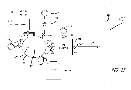
Infectious disease screening device
An infectious disease screening device 100 of some arrangements comprises
eight
main components: a chamber array containing various liquid chambers and
passages,
a sonication chamber, valves, pressure inlets (e.g. for attaching a Luer lock
syringe),
particulate filters, a PCR printed circuit board with heating elements and
microfluidic
chambers, PCR reagents and a final detection chamber.
Whilst the arrangements described above comprise an assay device 2 having a
transfer arrangement in the form of a piston, an infectious disease screening
device
100 of other arrangements comprises chambers formed on a substrate 101, as
shown
in Figure 25. In some arrangements, the substrate 101 is entirely or at least
partly
composed of silicon. The components of the infectious disease screening device
100
are formed in or on a film deposited on the silicon substrate and/or by
etching in the
silicon substrate. In some arrangements, the infectious disease screening
device 100
38
CA 03180787 2022- 11- 29
WO 2021/245391
PCT/GB2021/051333
is at least partly formed from a silicon wafer which is processed using
techniques that
are more traditionally used for manufacturing semiconductor microchips.
The use of the substrate enables the infectious disease screening device to be
manufactured at low cost and in a high volume because existing semiconductor
processing techniques allow for such low cost and high volume production.
The infectious disease screening device 100 comprises a sonication chamber 102
formed on the substrate 101. The sonication chamber 102 has a sample inlet
103, a
sample outlet 104 and an ultrasonic transducer 105.
The sample inlet 103 is coupled in fluid communication with the sonication
chamber
102 by a flow path 106. A valve 107 is provided along the flow path 106 to
selectively
allow or prevent a sample liquid from flowing from the sample inlet 103 into
the
sonication chamber 102.
The sample outlet 104 is provided with a filter 108 which filters sample fluid
flowing out
from the sample outlet 104. A valve 109 is provided along a fluid flow path
110 which
is coupled in fluid communication with the sample outlet 104.
The ultrasonic transducer 105 is configured to generate ultrasonic waves to
lyse cells
in a sample fluid within the sonication chamber 102. The ultrasonic transducer
105 is
configured to be controlled to oscillate to generate ultrasonic waves by a
controller,
such as the controller 23 described above. The infectious disease screening
device
100 of some arrangements comprises the controller 23 described above. The
controller 23 is coupled electrically to the ultrasonic transducer 105 to
control the
ultrasonic transducer 105 to generate ultrasonic waves.
The infectious disease screening device 100 comprises a reagent chamber 111
which
is formed on the substrate 101 for receiving a liquid PCR reagent. The reagent
39
CA 03180787 2022- 11- 29
WO 2021/245391
PCT/GB2021/051333
chamber 111 has an inlet 112 and an outlet 113. The inlet 112 is coupled with
the
sample outlet 104 of the sonication chamber 102. The reagent chamber 111 is
provided with a pressure drive port 114 which is in fluid communication with
the PCR
chamber 111, with a valve 115 being provided between the pressure drive port
114
and the reagent chamber 111.
The pressure drive port 114, and the other pressure drive ports described
herein, are
configured to be connected to a pressure drive arrangement. The pressure drive
arrangement may be any kind of pressure drive arrangement. In some
arrangements,
the pressure drive arrangement is a Luer lock syringe. In other arrangements,
the
pressure drive arrangement is a pressure drive arrangement within the system 1
which
is controlled by the controller 23. As will be described in more detail below,
the
pressure drive arrangement applies a positive or negative pressure to a
pressure drive
port which acts on fluid within one or more chambers of the infectious disease
screening device 100 to cause the fluid to flow between the chambers.
The outlet 113 of the reagent chamber 111 is coupled via a valve 116 to an
outlet flow
path 117. The fluid outlet 117 is coupled fluidly with a PCR chamber, as
described
below.
The infectious disease screening device 100 further comprises at least one
further
chamber which is formed on the substrate 101. One such further chamber is a
wash
chamber 118 which is coupled fluidly via a valve 119 to the sonication chamber
102. A
pressure drive port 120 is provided on the wash chamber 118 such that a
pressure
drive arrangement can exert a pressure to drive a wash liquid from within the
wash
chamber 118 into the sonication chamber 102.
In some arrangements, the infectious disease screening device 100 further
comprises
a lysing agent chamber 121 which is coupled fluidly with the sonication
chamber 102
via a valve 122. The lysing agent chamber 121 is provided with a pressure
drive port
CA 03180787 2022- 11- 29
WO 2021/245391
PCT/GB2021/051333
123 which is configured to apply a pressure to drive a lysing agent liquid
from the
lysing agent chamber 121 into the sonication chamber 102.
The infectious disease screening device 100 of some arrangements further
comprises
a waste chamber 124 which is coupled fluidly with the sonication chamber 102
via a
valve 125 and a filter 126. In other arrangements, the number of chambers
within the
infectious disease screening device 100 may be different from the arrangement
described above. In some arrangements, the number of chambers can vary from
one
chamber to as many as ten chambers. For the SARS-CoV-2 infectious disease
screening device, the infectious disease screening device comprises six
chambers.
In use, a user injects a sample that is to be analyzed into the sample inlet
103. The
sample is preferably placed into an elution buffer prior to being added to the
infectious
disease screening device 100.
In some arrangements, the elution buffer consists of: 1M Imidazole solution,
1M Tris,
0.5M EDTA, Milli-Q, sterile saline or Deionized water.
When the target sample is loaded into the device via the sample inlet 103, the
sample
is deposited directly into the sonication chamber 102, as shown in Figure 26.
In some
arrangements, the sonication chamber 102 has a volume of 100 p1 to 1000 pl. In
some
arrangements, the sonication chamber 102 comprises ports to all of the other
chambers in the infectious disease screening device 100. The flow of liquid
through
these ports is directed by a system of valves that can be opened or closed by
the user
and/or by the controller 23.
The ports that lead from the sonication chamber 102 to the waste chamber 124
and the
PCR reagent chamber 111 include filters 126, 108. In some arrangements, the
filters
126, 108 have pores of 0.1pm to 0.5pm in diameter. The filters trap the target
cells
and/or viral particles and retain them in the sonication chamber 102 as
various washes
41
CA 03180787 2022- 11- 29
WO 2021/245391
PCT/GB2021/051333
and other solutions pass through the sonication chamber 102 and into the waste
chamber 124. The filter 108 on the PCR reagent port 104 serves to contain the
cells
and/or viral particles within the sonication chamber 102 until lysis occurs.
After lysis,
the pores in the filter 108 are designed to be large enough to still trap the
broken cells
and/or viral particles, but allow their genetic material to pass through.
In some arrangements, the base of the sonication chamber 102 is a
piezoelectric disc
which functions as the ultrasonic transducer 105 to send acoustic waves
through the
liquid medium of the filled sonic chamber to disrupt the target cells and
release their
genetic material. In some arrangements, the height of the sonication chamber
102 is
approximately 200 pm.
In some arrangements, the sonication chamber 102 contains beads 127 or
microbeads
with a diameter of approximately 100pm (only some of which are shown in Figure
25).
In some arrangements, approximately half of the beads 127 are buoyant so they
exist
near the top of the sonication chamber 102 during sonication and the other
half are
designed to not be buoyant and exist near the bottom of the sonication chamber
102.
Between the two types of beads 127, a majority of the "lysing area" within the
sonication chamber 102 will be encompassed with beads 127 that can help
disrupt cell
membranes during sonication.
The next chamber is the wash chamber 118. In use, the wash chamber 118
contains
an excess amount (3 ml to 5 ml) of an elution buffer as mentioned above. The
wash
buffer is used to wash the sample once it is delivered to the sonication
chamber 102
and remove any potential contaminants.
The next chamber is the lysing agent chamber 121. The lysing agent chamber 121
contains a mixture of chemicals to assist in the cell lysing step of the
assay. A lysing
agent is pushed from the lysing agent chamber 121 into the sonication chamber
102
where it mixes with the sample, as shown in Figure 27. In some arrangements,
lysing
42
CA 03180787 2022- 11- 29
WO 2021/245391
PCT/GB2021/051333
agent consists of formulations, including, but not limited to:
= Lysis Formula #1:
O 10mM Tris
O 0.25% Igepal CA-630
0 150mM NaCI
= Lysis Formula #2:
O 10mM Tris-HCI
lo 0 10mM NaCl
O 10mM EDTA
O 0.5% Triton-X100
= Lysis Formula #3:
o 0.1M LiCI
O 0.1M Tris-HCI
O 1 % SDS
O 1 Omm EDTA
The next chamber is the PCR reagent chamber 111. Once the sample has been
sonicated and cell lysis has occurred, as shown in Figure 28, the freed
nucleic acid is
then pushed through the filter 108 to the PCR reagent chamber 111, as shown in
Figure 29. The PCR reagent chamber 111 contains the components needed for the
rRT-PCR process. The sample enters the PCR reagent chamber 111 and is then
toggled in and out of the PCR reagent chamber 111 by pressure exerted by a
pressure
drive arrangement (not shown) coupled to the pressure drive port 114. This
toggling
back and forth ensures the sample is sufficiently mixed with the PCR reagents,
as
shown in Figure 30. Once the sample and PCR reagents are sufficiently mixed,
the
mixture is then pushed from the PCR reagent chamber 111 out through the flow
path
117 to a PCR chamber. The PCR chamber is, in some arrangements, a channel in a
43
CA 03180787 2022- 11- 29
WO 2021/245391
PCT/GB2021/051333
PCR heating arrangement where the RT-PCR process will occur, as described
below.
The final chamber is the waste chamber 124. The waste chamber 124 holds all
the
discarded components throughout the cycles of the chamber array. For example,
when the wash solution is pushed through the sonication chamber 102, the
solution is
sent directly to the waste chamber 124 upon exiting the sonication chamber
102. The
minimum volume of the waste chamber 124 is the total volume of all the liquid
in the
infectious disease screening device 100, plus the volume of the sample added.
PCR Reagents
In some arrangements, the PCR reagents in the PCR reagent chamber 111 are
selected to facilitate the extreme rRT-PCR process as well as to allow for
temperature
monitoring via fluorescence.
In some arrangements, the PCR reagent formula is as follows: 5 pM of each
forward
and reverse primer (6 total primers, 2 sets for detecting SARS-CoV-2 and 1 set
to
serve as a control for a successful PCR reaction), IX LCGreen+ dye, 0.2 pM of
each
deoxynucleoside triphosphate (dNTP): dATP, dTTP, dGTP, dCTP, 50mM Tris, 1.65
pM
KlenTaq, 25 ng/pL BSA, 1.25 U/pL Malone Murine leukemia virus reverse
transcriptase
(MMLV), and 7.4mM MgCl2.
PCR Methods
Some examples of rRT-PCR processes are the processes described in
international
patent application no. PCT/US2016/060650 for rapid detection and confirmation
of the
presence of SARS-CoV-2 in a sample, incorporated by reference herein.
In order to control the heating and cooling process necessary for a RT-PCR
reaction to
occur, the sample is output from the PCR reagent chamber 111 to a heating
arrangement 128, as shown in Figure 31. The heating arrangement 128 comprises
a
channel 129 which is formed on a substrate 130. The channel 129 defines a
fluid flow
44
CA 03180787 2022- 11- 29
WO 2021/245391
PCT/GB2021/051333
path between a channel inlet 131 and a channel outlet 132. The channel
functions as
a PCR chamber for performing a PCR process on a sample fluid flowing along the
channel 129.
In some arrangements, the substrate 130 comprising the chamber array described
above is formed integrally with the substrate 101 of the heating arrangement
128. In
some embodiments, the substrates 130, 101 are portions of the same silicon
wafer.
In some arrangements, the heating arrangement 128 is a microfluidic chip
containing
microchannels of varying sizes and heating elements to heat and cool the
sample as it
flows through the channels.
In some arrangements, the channel 129 is formed in a polyimide layer 133 which
is
deposited on the substrate 130, shown in Figure 32. The polyimide layer 133 is
deposited on a first side 134 of the substrate 130.
A second side 135 of the substrate 130 carries a first heating element 136. In
this
arrangement, the heating arrangement 128 comprises a second heating element
137
and a third heating element 138. The heating elements 136-138 are positioned
adjacent one another on the second side 135 of the substrate 130. In some
arrangements, the heating elements 136-138 are of copper which is deposited on
the
substrate 130 using printed circuit board manufacturing techniques.
In some
arrangements, the copper has a conductivity of approximately 1.7E-8 0-m. The
heating elements 136-138 each have a predetermined electrical resistance which
causes the temperature of the heating elements 136-138 to increase when a
current
flows through the heating elements 136-138. In some arrangements, each heating
element 136-138 has a resistance of approximately 2.5 0.
In some arrangements, an electrical connection is established (directly or
indirectly)
between the heating elements 136-138 and the controller 23. The controller 23
CA 03180787 2022- 11- 29
WO 2021/245391
PCT/GB2021/051333
controls the supply of electricity to each of the heating elements 136-138
which in turn
controls the temperature of each of the heating elements 136-138.
Referring now to Figures 33-35, each of the heating elements 136-138 is formed
along
the length of the substrate 130. Each heating element 136-138 comprises
multiple
interconnected S-shaped turns. The first heating element 136 has an overall
first width
139, the second heating element 137 has an overall second width 140 and the
third
heating element 138 has an overall third width 141. In this arrangement, the
first and
third overall widths 139, 141 are approximately 6 mm and the second overall
width 140
is approximately 11.5 mm. The spacings 142, 143 between the heating elements
136-
138 are approximately 0.5 mm.
Each of the heating elements 136-138 is an elongate electrical conductor
which, in this
arrangement, has a conductor width 144 of approximately 100 pm and a conductor
depth 145 of approximately 18 pm. Each of the heating elements 136-138 has a
turn
width 146 of approximately 400 pm.
As will be described in more detail below, the first heating element 136 is
configured to
heat to a temperature of approximately 95 C, the second heating element 137 is
configured to heat to a temperature of approximately 77 C and the third
heating
element 138 is configured to heat to a temperature of approximately 55 C.
Returning now to Figure 31, the channel 129 is formed from multiple
interconnected S-
shaped turns which provide a fluid flow path back and forth across the surface
of the
substrate 130. In this arrangement, there are thirty turns of the channel 129.
The
microfluidic channel is photopatterned into the polyimide film 133 with great
accuracy.
Figure 36 shows one of the S-shaped turns of the channel 129.
Referring now to Figures 37-39, the channel 129 comprises a first channel
portion 147
having a first cross-sectional area and a second channel portion 148 having a
second
46
CA 03180787 2022- 11- 29
WO 2021/245391
PCT/GB2021/051333
cross-sectional area, wherein the second cross-sectional area is greater than
the first
cross-sectional area. In this arrangement, the first channel portion 147 has a
depth of
approximately 60 pm and a width of approximately 200 pm and the second channel
portion 148 has a depth of approximately 60 pm and a width of approximately
400 pm.
In this arrangement, the channel 129 comprises a third channel portion 149
having a
third cross-sectional area which is the same as the first cross-sectional
area.
In this arrangement, the channel 129 comprises a fourth channel portion 150
having a
third cross-sectional area which is less than the first and second cross-
sectional areas.
In this arrangement, the fourth channel portion 150 has a depth of
approximately 60
pm and a width of approximately 100 pm.
In this arrangement, the first and third channel portions 147, 149 each have a
length Ll
of approximately 12.5 mm and the second channel portion 148 has a length L2 of
approximately 12.5 mm.
The sample is pushed from the PCR reagent chamber 111, through the channel
inlet
131 and along the channel 129 of the heating arrangement 128 of the PCR
arrangement. In some arrangements, the inlet velocity of the sample is
approximately
5 mm/s.
In this arrangement, each turn of the channel 129 comprises four distinct 12.5
mm long
sections of different widths in order to control the rate of flow through each
section.
The first section of each wind is 200 pm wide. Each wind of the microfluidic
channel
traverses the substrate 130 from one side to the other and each wind passes
over the
three heating elements 136-138.
The first portion 147 of the wind passes over the first heating element 136
set to 95 C.
The second portion 148 of the wind passes over the second heating element 137
set to
47
CA 03180787 2022- 11- 29
WO 2021/245391
PCT/GB2021/051333
77 C. The third and "final" portion 149 of the wind is identical but opposite
to the first
portion 147; it is 200pm wide but it passes over the third heating element 138
which is
set to 55 C.
The fourth portion 150 of the wind is a small 100 pm section that connects the
third
portion 149 of the wind back to the first portion 147 of the next wind. The
fourth portion
150 is small so that the liquid flowing through it moves quickly back to the
first portion
147 of the next wind and does not spend a significant length of time over the
second
heating element 137.
As the sample passes through the 30 heating and cooling loops, the rRT-PCR
reaction
occurs and by the time the sample exits at the channel outlet 132, it has
completed the
30 cycles of heating and cooling required for completion of the rRT-PCR
process. The
sample exits the heating arrangement 128 and flows into a detection chamber.
In
some arrangements, the detection chamber is a chamber of the detection
arrangement
70 of the system described herein.
The detection arrangement 70 detects fluorescence emitted from the sample and
reports the result of the assay as described herein. In some arrangements, the
detection arrangement is a SARS-CoV-2 virus detection apparatus which is
coupled to
the channel outlet 132. The detection apparatus detects a presence of the SARS-
CoV-
2 virus that causes COVID-19 disease in a sample fluid flowing out of the
channel
outlet. The detection apparatus provides an output which is indicative of
whether or
not the SARS-CoV-2 virus detection apparatus detects the presence of the COVID-
19
disease in the sample fluid. In other arrangements, the detection apparatus
detects
the presence of a different infectious disease from COVID-19 disease.
In some arrangements, the formation of the sonication chamber (102), the
reagent
chamber (111), any further chambers, and the heating arrangement (128) on the
same
substrate (101) in combination with the controller 23 provides a compact and
relatively
48
CA 03180787 2022- 11- 29
WO 2021/245391
PCT/GB2021/051333
low cost device (compared with larger laboratory PCR systems). Consequently,
the
device (100) of some arrangements can be mass produced easily using
conventional
semiconductor manufacturing techniques.
The device or system of some arrangements seeks to provide test results within
10
minutes and, in some arrangements, as little as 5 minutes or less. This is
significantly
faster than conventional PCR tests and it opens up the possibility for rapid
testing at
homes, shops, entertainment venues, as well as airports, bus and train
terminals and
other transport facilities.
The device or system of some arrangements is highly portable and can be
carried
easily to a location where testing is required. The efficient operation of the
device or
system enables the device or system of some arrangements to be powered by a
battery, enabling the system to provide tests at virtually any location.
The devices and systems of the arrangements that can screen a saliva or sputum
sample make the screening process easier and quicker, especially for children
or
sensitive individuals, as compared with systems that require a nasopharyngeal
or
oropharyngeal swab sample.
The foregoing outlines features of several embodiments so that those of
ordinary skill
in the art may better understand various aspects of the present disclosure.
Those of
ordinary skill in the art should appreciate that they may readily use the
present
disclosure as a basis for designing or modifying other processes and
structures for
carrying out the same purposes and/or achieving the same advantages of various
embodiments introduced herein. Those of ordinary skill in the art should also
realize
that such equivalent constructions do not depart from the spirit and scope of
the
present disclosure, and that they may make various changes, substitutions, and
alterations herein without departing from the spirit and scope of the present
disclosure.
49
CA 03180787 2022- 11- 29
WO 2021/245391
PCT/GB2021/051333
Although the subject matter has been described in language specific to
structural
features or methodological acts, it is to be understood that the subject
matter of the
appended claims is not necessarily limited to the specific features or acts
described
above. Rather, the specific features and acts described above are disclosed as
example forms of implementing at least some of the claims.
Various operations of embodiments are provided herein. The order in which some
or
all of the operations are described should not be construed to imply that
these
operations are necessarily order dependent. Alternative ordering will be
appreciated
having the benefit of this description. Further, it will be understood that
not all
operations are necessarily present in each embodiment provided herein. Also,
it will
be understood that not all operations are necessary in some embodiments.
Moreover, "exemplary" is used herein to mean serving as an example, instance,
illustration, etc., and not necessarily as advantageous. As used in this
application, "or"
is intended to mean an inclusive "or" rather than an exclusive "or". In
addition, "a" and
"an" as used in this application and the appended claims are generally be
construed to
mean "one or more" unless specified otherwise or clear from context to be
directed to a
singular form. Also, at least one of A and B and/or the like generally means A
or B or
both A and B. Furthermore, to the extent that "includes", "having", "has",
"with", or
variants thereof are used, such terms are intended to be inclusive in a manner
similar
to the term "comprising". Also, unless specified otherwise, "first," "second,"
or the like
are not intended to imply a temporal aspect, a spatial aspect, an ordering,
etc. Rather,
such terms are merely used as identifiers, names, etc. for features, elements,
items,
etc. For example, a first element and a second element generally correspond to
element A and element B or two different or two identical elements or the same
element.
Also, although the disclosure has been shown and described with respect to one
or
more implementations, equivalent alterations and modifications will occur to
others of
CA 03180787 2022- 11- 29
WO 2021/245391
PCT/GB2021/051333
ordinary skill in the art based upon a reading and understanding of this
specification
and the annexed drawings. The disclosure comprises all such modifications and
alterations and is limited only by the scope of the following claims. In
particular regard
to the various functions performed by the above described features (e.g.,
elements,
resources, etc.), the terms used to describe such features are intended to
correspond,
unless otherwise indicated, to any features which performs the specified
function of the
described features (e.g., that is functionally equivalent), even though not
structurally
equivalent to the disclosed structure. In addition, while a particular feature
of the
disclosure may have been disclosed with respect to only one of several
implementations, such feature may be combined with one or more other features
of the
other implementations as may be desired and advantageous for any given or
particular
application.
Embodiments of the subject matter and the functional operations described
herein can
be implemented in digital electronic circuitry, or in computer software,
firmware, or
hardware, including the structures disclosed in this specification and their
structural
equivalents, or in combinations of one or more of them.
Features of some embodiments are implemented using one or more modules of
computer program instructions encoded on a computer-readable medium for
execution
by, or to control the operation of, a data processing apparatus or a
controller. The
computer-readable medium can be a manufactured product, such as hard drive in
a
computer system or an embedded system. The computer-readable medium can be
acquired separately and later encoded with the one or more modules of computer
program instructions, such as by delivery of the one or more modules of
computer
program instructions over a wired or wireless network. The computer-readable
medium can be a machine-readable storage device, a machine-readable storage
substrate, a memory device, or a combination of one or more of them.
The terms "computing device" and data processing apparatus" encompass all
51
CA 03180787 2022- 11- 29
WO 2021/245391
PCT/GB2021/051333
apparatus, devices, and machines for processing data, including by way of
example a
programmable processor, a computer, or multiple processors or computers. The
apparatus can include, in addition to hardware, code that creates an execution
environment for the computer program in question, e.g., code that constitutes
processor firmware, a protocol stack, a database management system, an
operating
system, a runtime environment, or a combination of one or more of them. In
addition,
the apparatus can employ various different computing model infrastructures,
such as
web services, distributed computing and grid computing infrastructures.
The processes and logic flows described in this specification can be performed
by one
or more programmable processors executing one or more computer programs to
perform functions by operating on input data and generating output.
As used herein, in some embodiments the term module comprises a memory and/or
a
processor configured to control at least one process of a system or a circuit
structure.
The memory storing executable instructions which, when executed by the
processor,
cause the processor to provide an output to perform the at least one process.
Embodiments of the memory include non-transitory computer readable media.
Processors suitable for the execution of a computer program include, by way of
example, both general and special purpose microprocessors, and any one or more
processors of any kind of digital computer. Generally, a processor will
receive
instructions and data from a read-only memory or a random access memory or
both.
The essential elements of a computer are a processor for performing
instructions and
one or more memory devices for storing instructions and data. Generally, a
computer
will also include, or be operatively coupled to receive data from or transfer
data to, or
both, one or more mass storage devices for storing data, e.g., magnetic,
magneto-optical disks, or optical disks. However, a computer need not have
such
devices. Devices suitable for storing computer program instructions and data
include
all forms of non-volatile memory, media and memory devices, including by way
of
52
CA 03180787 2022- 11- 29
WO 2021/245391
PCT/GB2021/051333
example semiconductor memory devices, e.g., EPROM (Erasable Programmable
Read-Only Memory), EEPROM (Electrically Erasable Programmable Read-Only
Memory), and flash memory devices; magnetic disks, e.g., internal hard disks
or
removable disks; magneto-optical disks; and CD-ROM and DVD-ROM disks.
To provide for interaction with a user, some embodiments are implemented on a
computer having a display device, e.g., a CRT (cathode ray tube) or LCD
(liquid crystal
display) monitor, for displaying information to the user and a keyboard and a
pointing
device, e.g., a mouse or a trackball, by which the user can provide input to
the
computer. Other kinds of devices can be used to provide for interaction with a
user as
well; for example, feedback provided to the user can be any form of sensory
feedback,
e.g., visual feedback, auditory feedback, or tactile feedback; and input from
the user
can be received in any form, including acoustic, speech, or tactile input.
In the present specification "comprise" means "includes or consists of" and
"comprising" means "including or consisting of".
The features disclosed in the foregoing description, or the following claims,
or the
accompanying drawings, expressed in their specific forms or in terms of a
means for
performing the disclosed function, or a method or process for attaining the
disclosed
result, as appropriate, may, separately, or in any combination of such
features, be
utilized for realizing the invention in diverse forms thereof.
53
CA 03180787 2022- 11- 29