Note: Descriptions are shown in the official language in which they were submitted.
CA 03181963 2022-11-01
WO 2021/224913 PCT/IL2021/050506
PRECURSOR TRI-SPECIFIC ANTIBODY CONSTRUCTS AND METHODS OF USE
THEREOF
FIELD OF THE INVENTION
[0001] The present disclosure relates generally to the field of antibody
construct. In one
embodiment, disclosed herein are precursor tri-specific antibody constructs
and methods of
using the same (e.g. treatment of cancer).
SEQUENCE LISTING STATEMENT
[0002] The instant application contains a Sequence Listing which has been
submitted
electronically in ASCII format and is hereby incorporated by reference in its
entirety. Said ASCII
copy, created on April 29, 2021, is named P-594180-PC SL.txt and is 1,730,884
bytes in size.
BACKGROUND OF THE INVENTION
[0003] The functionality of monoclonal antibodies (non conjugated or naked
antibody)
currently approved by drug regulatory agencies worldwide for clinical use in
oncology setting
are known to use one or a combination of the following mechanisms: 1) blocking
cell growth
signaling, 2) blocking blood supply to cancer cells, 3) directly mediating
cell apoptosis, 4)
eliciting immunological effector functions such as antibody dependent cellular
cytoxicity
(ADCC), antibody dependent cellular phagocytosis (ADCP) and complement
dependent
cytotoxicity (CDC), and 5) promoting adaptive immunity towards tumors.
[0004] Monoclonal antibody therapies have demonstrated survival benefits in
the clinic.
However, the overall response rates in cancer patients are low, and the
survival benefits are
marginal (several months) compared to chemotherapy. Although the underlying
reasons for the
lack of robust clinical anti-cancer activities are not fully understood,
research has suggested
that cancer cells often quickly develop compensating signaling pathways to
escape cell death.
Also, cancer stem cells (CSC), which are considered as potent cancer
initiating cells, are less
active at cell proliferation therefore they tend to sustain the lack of growth
signal better.
[0005] In an attempt to improve anti-tumor activity of monoclonal
antibodies, multi-specific
antibodies are being developed. In contrast to classical monoclonal
antibodies, which are the
standard first-line therapy in several tumor entities, these multi-specific
antibodies may bring
together a tumor cell and the means to destroy the tumor cell, thereby
increasing the efficiency
of treatment. These multi-specific antibodies provide for new treatment
options for cancer
patients.
[0006] Another anti-cancer therapeutic approach is to utilize T cells. T
cells provide defense
1
SUBSTITUTE SHEET (RULE 26)
CA 03181963 2022-11-01
WO 2021/224913 PCT/IL2021/050506
against cancer throughout life by patrolling the body in search for newly
arisen cancer cells
and eliminating them effectively and promptly. Therapeutic approaches
utilizing T cells have
proved successful in cancer treatment of at least metastatic melanoma,
metastatic kidney
cancer, asymptomatic metastatic hormone refractory prostate cancer, and
advanced
melanoma.
[0007] While the use of targeting activated T cells provides one pathway
for destruction of
tumor cells, another pathway of cellular cytotoxicity is through the
recruitment and targeting
of nature killer (NK) cells. NK cells are white blood cells, part of the
innate immune system
that plays a major role in the host-rejection of tumors. NK cells are
cytotoxic, wherein small
granules in their cytoplasm contain special proteins such as perforin and
proteases known as
granzymes. Upon release in close proximity to a tumor cell slated for killing,
perforin forms
pores in the cell membrane of the targeted cell through which the granzymes
and associated
molecules can enter, inducing apoptosis. Therapeutic antibodies, such as
RITUXAN and
HERCEPTIN, can drive killing of bound tumors through NK-cell-mediated antibody-
dependent cell-mediated cytotoxicity (ADCC).
[0008] Tumor escape from NK cell immune surveillance predominantly occurs via
two
mechanisms: reduction of activating signals or increases in inhibitory signals
delivered to NK
cells. Thus, another potential target of cancer immunotherapy is the
removal/blocking of
molecules that suppress NK activation, and removal/blocking of molecules that
result in NK
cell hypo-responsiveness. Restoring NK cell antitumor activity is critical for
establishing host
immunity against cancer, which is a primary objective of cancer immunotherapy.
[0009] Another consideration in tumor cytotoxicity is the tumor
microenvironment (TME).
The TME includes novel targets that can help direct and improve the actions of
antibody
therapies by potentiating host antitumor immune responses. For example, T
cells play an
unexpectedly critical role in anti-tumor antigen antibody therapy, although
their importance is
often not observed due to studies being performed in immunodeficient mice. IL-
2 treatment
was shown to amplify monoclonal antibody therapy not simply via the previously
assumed
NK-mediated ADCC, but also by boosting the CD8+ T cell adaptive response,
since IL-2 exerts
significant pleiotropic effects on regulatory, helper, and cytolytic memory T-
cells (Liao et al.,
Immunity. 2013, 38:13) (Zhu EF et.al Cancer Cell. 2015 27:489).
[0010] A pitfall of antibody therapeutics used in cancer treatment is the
"off-target" binding
of the antibody to non-cancer tumor-associated-antigen-expressing cells,
especially if such
binding leads to cytotoxicity. Thus, "off-target" binding by multi-specific
and bispecific
antibodies presents a potential challenge to controlling their "off-target"
activity against normal
2
CA 03181963 2022-11-01
WO 2021/224913 PCT/IL2021/050506
tissues that also express the antigen, even at extremely low levels. These
"off-target" effects
are a serious limitation to multi-specific and bispecific antibody
therapeutics. Another
drawback of many bispecific or multi-specific antibodies is their short half-
life.
[0011] There remains a need to provide multi-specific trivalent antibodies
with qualities that
specifically target cytotoxicity to tumor cells while reducing the toxic side
effects and
preserving the antibodies effectiveness.
SUMMARY OF THE INVENTION
[0012] Reducing the non-specific toxic side effects of multi-specific
antibodies and
concurrently enhancing the effectiveness of these antibodies require an
antibody having a
precursor form that (1) engages a target associated with a tumor cell, a tumor-
associated cell,
or a tumor cell environment, and (2) activates cytotoxic cells, for example T
cells or NK cells,
once localized within or adjacent to the tumor microenvironment (TME).
Further, it is essential
that such multi-specific antibodies do not reduce significantly the
immunogenicity to a tumor
or tumor-associated target. In one embodiment, the precursor tri-specific
antibody constructs
described herein addresses these needs by attaching a regulatable half-life
enhancing
component and a blocking component that inhibits the antibody from engaging a
toxicity-
providing cell prior to binding to a tumor or tumor-associated target or at
the TME. Further,
the tri-specific antibodies presented herein may concurrently engage two
different types of
cytotoxic cells (e.g. T cells and NK cells).
[0013] In one embodiment, after the precursor tri-specific antibody
constructs described
herein are administered to a cancer patient, the precursor antibody constructs
eventually reach
the tumor-associated targets or the TME. At the vicinity of the tumor-
associated targets or the
TME, the precursor antibody constructs would bind to a tumor-associated
antigen, and the
blocking component(s) that inhibits the antibody from engaging T cells and/or
NK cells would
be removed by proteases present at the TME. Subsequently, the antibody
constructs (now in
"active" form) would bind to T cells and/or NK cells, thereby recruiting T
cells and/or NK cells
from the circulation to the tumor-associated targets or the TME.
[0014] In one embodiment, disclosed herein is a precursor tri-specific
antibody construct,
comprising: a) a first binding domain that binds to a tumor associated antigen
(TAA); b) a
second binding domain comprising a cytokine receptor engager or a second
binding domain that
binds to a first natural killer (NK) cell surface antigen; c) a third binding
domain that binds to a
T cell surface antigen or a second NK cell surface antigen; and d) a
regulatory domain
comprising either (i) a first and a second sub-regulatory domain, the first
sub-regulatory domain
3
CA 03181963 2022-11-01
WO 2021/224913 PCT/IL2021/050506
comprising a first protease cleavage domain and a half-life prolonging (HLP)
domain, and the
second sub-regulatory domain comprising a second protease cleavage domain and
a CAP
component that reduces the ability of the third binding domain to bind to its
target antigen; or (ii)
a single regulatory domain comprising a protease cleavage domain, a half-life
prolonging (HLP)
domain, and a CAP component that reduces the ability of the third binding
domain to bind to its
target antigen.
[0015] In one embodiment, when the second binding domain binds to a NK cell
surface
antigen, the second binding domain further comprises a third regulatory domain
comprising a third
protease cleavage domain and a CAP component that reduces the ability of the
second binding
domain to bind to the NK cell surface antigen.
[0016] In one embodiment, the first binding domain, the second binding
domain each
comprises a single chain variable fragment (scFv). In one embodiment, the
third binding domain
comprises a Fab antigen binding fragment.
[0017] In one embodiment, the T cell surface antigen is CD3. In another
embodiment, one or
both of the NK cell surface antigens bound by the antibody construct disclosed
herein can be an
activating NK cell receptor or an inhibitory NK cell receptor. It is known in
the art that activation
of the NK cells is mediated by a network of activating and inhibitory
receptors; it is the integration
of the activating and inhibitory signals that determines if the NK cells
become cytotoxic (see e.g.
Chester et al., Frontiers in Immunology, 2015, 6:Article 601). Activating
receptors for NK cells
include, but are not limited to, CD16, TRAIL, NKG2D, 2B4, DNAM-1, NKp30,
NKp44, NKp46
and NKp80. Inhibitory receptors for NK cells include, but are not limited to,
MR (killer cell
immunoglobulin-like receptor) and CD94/NKG2A heterodimer. Moreover, there are
co-
stimulatory proteins with key roles in regulating the activation of NK cells,
for example, CD137,
0X40 and CD27. In another embodiment, one or both of the NK cell surface
antigens bound by
the antibody construct disclosed herein can be, but is not limited to, CD16
(FcyRIII), CD16a
(FcyRIIIa), CD56, sMICA/B, ILT, SLAMF7, NKp44, NKp30, DNAM-1, NKG2A, NKG2D,
NKG2C/CD94, NKp46, KIR2/DL3, KIR2DL1, NKRP1, NKG2E/CD94, NKG2F/CD94, CD69,
LLT1, ILT2, AICL, CD26, NKp80, MR family receptors, or CD122/IL-2Rbeta.
[0018] In one embodiment, the second binding domain and the third binding
domain bind to
different NK cell surface antigens. In another embodiment, the second binding
domain and the
third binding domain bind to the same NK cell surface antigen. In one
embodiment, the second
binding domain binds to NKG2A and the third binding domain binds to NKG2D. In
another
embodiment, the second binding domain binds to NKG2D and the third binding
domain binds to
NKG2A.
4
CA 03181963 2022-11-01
WO 2021/224913 PCT/IL2021/050506
[0019] In one embodiment of the precursor tri-specific antibody construct,
the first binding
domain binds to a TAA, the second binding domain binds to a NK cell surface
antigen, and the
third binding domain binds to a T cell surface antigen.
[0020] In one embodiment of the precursor tri-specific antibody construct,
the first binding
domain binds to a TAA, the second binding domain binds to a NK cell surface
antigen, and the
third binding domain binds to a T cell surface antigen, wherein the second
binding domain further
comprises a third regulatory domain comprising a third protease cleavage
domain and a CAP
component that reduces the ability of the second binding domain to bind to the
NK cell surface
antigen.
[0021] In one embodiment of the precursor tri-specific antibody construct,
the first binding
domain binds to a TAA, the second binding domain binds to a first NK cell
surface antigen, and
the third binding domain binds to a second NK cell surface antigen.
[0022] In one embodiment of the precursor tri-specific antibody construct,
the first binding
domain binds to a TAA, the second binding domain binds to a first NK cell
surface antigen, and
the third binding domain binds to a second NK cell surface antigen, wherein
the second binding
domain further comprises a third regulatory domain comprising a third protease
cleavage domain
and a CAP component that reduces the ability of the second binding domain to
bind to the NK
cell surface antigen.
[0023] In one embodiment of the precursor tri-specific antibody construct,
the first binding
domain binds to a TAA, the cytokine receptor engager of the second binding
domain comprises a
cytokine that binds to a cytokine receptor, and the third binding domain binds
to a NK cell surface
antigen. In one embodiment, the cytokine is a pro-inflammatory cytokine. In
another embodiment,
the cytokine is an anti-inflammatory cytokine. In one embodiment, the cytokine
can be IL-15, IL-
2, IL-12, TNF-alpha, IL-6, TGF-beta, IL-10, IL-8, IL-17, IL-21, INF, and VEGF.
In one
embodiment, the cytokine is IL-15 that binds to an IL-15 receptor.
[0024] In one embodiment, the HLP molecule comprises a human serum albumin
(HSA)
polypeptide.
[0025] In one embodiment, the CAP component that reduces binding to the T
cell surface
antigen comprises an amino acid sequence of an extracellular epitope of human
CD3E. In one
embodiment, the CAP component comprises the amino acid sequence of SEQ ID
NO:5, or a
homolog thereof.
[0026] In one embodiment, the CAP component that reduces binding to the NK
cell surface
antigen comprises an amino acid sequence of an extracellular epitope of the NK
surface antigen.
[0027] In one embodiment, the protease cleavage domains of the antibody
construct disclosed
CA 03181963 2022-11-01
WO 2021/224913 PCT/IL2021/050506
herein are cleaved by the same protease. In another embodiment, the protease
cleavage domains
of the antibody construct disclosed herein are cleaved by different proteases.
In one embodiment,
one or more of the protease cleavage domains comprise an amino acid sequence
cleavable by a
serine protease, a cysteine protease, an aspartate protease, or a matrix
metalloprotease (MMP). In
another embodiment, one or more of the protease cleavage domains comprise
amino acid
sequence that is a combination substrate cleaved by one or more of MMP2/9,
uPA, matriptase,
and legumain.
[0028] In one embodiment, the MMP can be, but is not limited to, matrix
metalloprotease 1
(MMP-1), matrix metalloprotease 2 (MMP-2), matrix metalloprotease 9 (MMP-9),
or matrix
metalloprotease 14 (MMP-14). In one embodiment, the serine protease is an
urokinase-type
plasminogen activator (uPA) protease or a membrane-type serine protease (MT-
SP1). In one
embodiment, the combination substrate has the amino acid sequence of SEQ ID
NO:35. In another
embodiment, one or more of the protease cleavage domains comprise an amino
acid sequence
having the sequence of one of SEQ ID NOs:9-14 and SEQ ID NO:35.
[0029] In one embodiment, the tumor associated antigen (TAA) can be, but is
not limited to,
a tumor cell surface antigen, a tumor micro-environment antigen, a stromal
antigen in the tumor
micro-environment (TME), an angiogenic antigen in the TME, or an antigen on a
blood vessel in
a TME.
[0030] In one embodiment, the TAA can be, but is not limited to, EGFR,
FcyRI, FcyRIIa
FcyRIIb FcyRIIIa FcyRIIIb, CD28, CD137, CTLA-4, FAS, fibroblast growth factor
receptor 1
(FGFR1), FGFR2, FGFR3, FGFR4, glucocorticoid-induced TNFR-related (GITR)
protein,
lymphotoxin-beta receptor (LTPR), toll-like receptors (TLR), tumor necrosis
factor-related
apoptosis-inducing ligand-receptor 1 (TRAIL receptor 1), TRAIL receptor 2,
prostate-specific
membrane antigen (PSMA) protein, prostate stem cell antigen (PSCA) protein,
tumor-associated
protein carbonic anhydrase IX (CAIX), epidermal growth factor receptor 1
(EGFR1), EGFRvIII,
human epidermal growth factor receptor 2 (Her2/neu; Erb2), ErbB3 (HER3),
Folate receptor,
ephrin receptors, PDGFRa, ErbB-2, CD20, CD22, CD30, CD33, CD40, CD37, CD38,
CD70,
CD74, CD56, CD40), CD80, CD86, CD2, p53, cMet (tyrosine-protein kinase Met)
(hepatocyte
growth factor receptor) (HGFR), MAGE-A 1, MAGE-A2, MAGE-A3, MAGE-A4, MAGE-A6,
MAGE-A10, MAGE-Al2, BAGE, DAM-6, DAM -10, GAGE-1, GAGE-2, GAGE-8, GAGE-3,
GAGE -4, GAGE-5, GAGE-6, GAGE-7B, NA88-A, NY-ESO-1, BRCA1, BRCA2, MART-1,
MC1R, Gp100, PSA, PSM, Tyrosinase, Wilms' tumor antigen (WT1), TRP-1, TRP-2,
ART-4,
CAMEL, Cyp-B, hTERT, hTRT, iCE, MUC1, MUC2, P-cadherin, Myostatin (GDF8),
Cripto
(TDGF1), MUC5AC, PRAME, P15, RU1, RU2, SART-1, SART-3, WT1, AFP, f3-catenin/m,
6
CA 03181963 2022-11-01
WO 2021/224913 PCT/IL2021/050506
Caspase-8/m, CDK-4/m, ELF2M, GnT-V, G250, HSP70-2M, HST-2, KIAA0205, MUM-1,
MUM-2, MUM-3, Myosin/m, RAGE, SART-2, TRP-2/INT2, 707-AP, Annexin II, CDC27/m,
TPI/mbcr-abl, ETV6/AML, LDLR/FUT, Pml/RARa, TEL/AML1, CD28, CD137, CanAg,
Mesothelin, DR5, PD-1, PD1L, IGF-1R, CXCR4, Neuropilin 1, Glypicans, EphA2,
CD138, B7-
H3, B7-H4, gpA33, GPC3, SSTR2, ROR1, 5T4, or VEGF-R2.
[0031] In one embodiment, the TAA is EGFR, ROR1, PSMA, or 5T4. In one
embodiment,
when the antigen is EGFR, the first binding domain comprises the amino acid
sequence of SEQ
ID NO:34 or SEQ ID NO:37; when the antigen is ROR1, the first binding domain
comprises the
amino acid sequence of SEQ ID NO: i56 or SEQ ID NO: i66; when the antigen is
PSMA, the first
binding domain comprises the amino acid sequence of SEQ ID NO: i68 or SEQ ID
NO: i70; and
when the antigen is 5T4, the first binding domain comprises the amino acid
sequence of SEQ ID
NO:172 or SEQ ID NO:174.
[0032] In one embodiment, the tumor micro-environment antigen can be, but
is not limited to,
KIR, NKG2A, ILT, LILR, or TIGIT.
[0033] In one embodiment, the stromal antigen in the tumor micro-
environment can be, but is
not limited to, fibroblast activation protein (FAP), alpha smooth muscle actin
(aSMA), PDGFRa,
Integrin a 1 1(31(ITGA11)VEGF, Tenascin-C, periostin, fibroblast specific
protein 1 (S 10A4,
FSP1), desmin, vimentin, paladin, urokinase-type plasminogen activator
receptor associated
protein (UPARAP), galectin-3, podoplanin, platelet, CCL2, or CXCL12.
[0034] In one embodiment, the angiogenic antigen in the tumor micro-
environment can be, but
is not limited to, bFGF, INF, or VEGF.
[0035] In one embodiment, the antigen on a blood vessel in the tumor micro-
environment
comprises an endothelial cell surface antigen such as CD31, CD105, CD146, and
CD144 etc.
[0036] In one embodiment, the third binding domain comprises a Fab region
having a heavy
chain (VH-CH) region and a light chain (VL-CL) region, and the first binding
domain is located
C-terminally to the VL-CL or VH-CH region of the third binding domain. In one
embodiment,
when the first binding domain is located C-terminally to the VL-CL region, the
second binding
domain is located C-terminally to the VH-CH region. Alternatively, when the
first binding domain
is located C-terminally to the VH-CH region, the second binding domain is
located C-terminally
to the VL-CL region.
[0037] In another embodiment, the third binding domain comprises a heavy
chain variable
region (VH) and a light chain variable region (VL), wherein the first
regulatory domain, which
comprises a HLP domain located N-terminally to the protease cleavage domain,
is located N-
terminally to the VH or VL region of the third binding domain. In one
embodiment, when the first
7
CA 03181963 2022-11-01
WO 2021/224913 PCT/IL2021/050506
regulatory domain is located N-terminally to the VL region, the second
regulatory domain, which
comprises a CAP component located N-terminally to the protease cleavage
domain, is located N-
terminally to the VH region. Alternatively, when the first regulatory domain
is located N-
terminally to the VH region, the second regulatory domain, which comprises a
CAP component
located N-terminally to the protease cleavage domain, is located N-terminally
to the VL region.
[0038] In another embodiment, the third binding domain comprises a heavy
chain variable
region (VH) and a light chain variable region (VL), wherein the single
regulatory domain
comprising a protease cleavage domain, a half-life prolonging (HLP) domain,
and a CAP
component is located N-terminally to the VH region or to the VL region of the
third binding
domain.
[0039] In one embodiment, the precursor tri-specific antibody construct
comprises two
polypeptides, polypeptide A and polypeptide B, each of which comprising one or
more heavy
chain variable region (VH) and one or more light chain variable region (VL),
for example,
(a) polypeptide A comprises components having an order N-terminal to C-
terminal: HLP
domain, protease cleavage domain, VH of the third binding domain, first
binding domain
comprising first VH-first VL; and polypeptide B comprises components having an
order
N-terminal to C-terminal: CAP component, protease cleavage domain, VL of the
third
binding domain, second binding domain comprising second VH-second VL; or
(b) polypeptide A comprises components having an order N-terminal to C-
terminal: HLP
domain, protease cleavage domain, VH of the third binding domain, first
binding domain
comprising first VL-first VH; and polypeptide B comprises components having an
order
N-terminal to C-terminal: CAP component, protease cleavage domain, VL of the
third
binding domain, second binding domain comprising second VH-second VL; or
(c) polypeptide A comprises components having an order N-terminal to C-
terminal: HLP
domain, protease cleavage domain, VH of the third binding domain, first
binding domain
comprising first VH-first VL; and polypeptide B comprises components having an
order
N-terminal to C-terminal: CAP component, protease cleavage domain, VL of the
third
binding domain, second binding domain comprising second VL-second VH; or
(d) polypeptide A comprises components having an order N-terminal to C-
terminal: HLP
domain, protease cleavage domain, VH of the third binding domain, first
binding domain
comprising first VL-first VH; and polypeptide B comprises components having an
order
N-terminal to C-terminal: CAP component, protease cleavage domain, VL of the
third
binding domain, second binding domain comprising second VL-second VH; or
(e) polypeptide A comprises components having an order N-terminal to C-
terminal: CAP
8
CA 03181963 2022-11-01
WO 2021/224913 PCT/IL2021/050506
component, protease cleavage domain, VH of the third binding domain, first
binding
domain comprising first VH-first VL; and polypeptide B comprises components
having an
order N-terminal to C-terminal: HLP domain, protease cleavage domain, VL of
the third
binding domain, second binding domain comprising second VH-second VL; or
(f) polypeptide A comprises components having an order N-terminal to C-
terminal: CAP
component, protease cleavage domain, VH of the third binding domain, first
binding
domain comprising first VL-first VH; and polypeptide B comprises components
having an
order N-terminal to C-terminal: HLP domain, protease cleavage domain, VL of
the third
binding domain, second binding domain comprising second VH-second VL; or
(g) polypeptide A comprises components having an order N-terminal to C-
terminal: CAP
component, protease cleavage domain, VH of the third binding domain, first
binding
domain comprising first VH-first VL; and polypeptide B comprises components
having an
order N-terminal to C-terminal: HLP domain, protease cleavage domain, VL of
the third
binding domain, second binding domain comprising second VL-second VH; or
(h) polypeptide A comprises components having an order N-terminal to C-
terminal: CAP
component, protease cleavage domain, VH of the third binding domain, first
binding
domain comprising first VL-first VH; and polypeptide B comprises components
having an
order N-terminal to C-terminal: HLP domain, protease cleavage domain, VL of
the third
binding domain, second binding domain comprising second VL-second VH.
[0040] In one embodiment, the precursor tri-specific antibody construct
comprises two
polypeptides, polypeptide A and polypeptide B, each of which comprising one or
more heavy
chain variable region (VH) and one or more light chain variable region (VL),
for example,
(a) polypeptide A comprises components having an order N-terminal to C-
terminal: VH of the
third binding domain, first binding domain comprising first VH-first VL; and
polypeptide
B comprises components having an order N-terminal to C-terminal: VL of the
third binding
domain, second binding domain comprising second VH-second VL, wherein a
regulatory
domain is located N-terminal to either polypeptide A or B, the regulatory
domain comprises
components having an order N-terminal to C-terminal: a CAP component, a half-
life
prolonging (HLP) domain, and a protease cleavage domain; or
(b) polypeptide A comprises components having an order N-terminal to C-
terminal: VH of the
third binding domain, first binding domain comprising first VL-first VH; and
polypeptide
B comprises components having an order N-terminal to C-terminal: VL of the
third binding
domain, second binding domain comprising second VH-second VL, wherein a
regulatory
domain is located N-terminal to either polypeptide A or B, the regulatory
domain comprises
9
CA 03181963 2022-11-01
WO 2021/224913 PCT/IL2021/050506
components having an order N-terminal to C-terminal: a CAP component, a half-
life
prolonging (HLP) domain, and a protease cleavage domain; or
(c) polypeptide A comprises components having an order N-terminal to C-
terminal: VH of the
third binding domain, first binding domain comprising first VH-first VL; and
polypeptide
B comprises components having an order N-terminal to C-terminal: VL of the
third binding
domain, second binding domain comprising second VL-second VH, wherein a
regulatory
domain is located N-terminal to either polypeptide A or B, the regulatory
domain comprises
components having an order N-terminal to C-terminal: a CAP component, a half-
life
prolonging (HLP) domain, and a protease cleavage domain; or
(d) polypeptide A comprises components having an order N-terminal to C-
terminal: VH of the
third binding domain, first binding domain comprising first VL-first VH; and
polypeptide
B comprises components having an order N-terminal to C-terminal: VL of the
third binding
domain, second binding domain comprising second VL-second VH, wherein a
regulatory
domain is located N-terminal to either polypeptide A or B, the regulatory
domain comprises
components having an order N-terminal to C-terminal: a CAP component, a half-
life
prolonging (HLP) domain, and a protease cleavage domain.
[0041] In another embodiment, the precursor tri-specific antibody construct
comprises two
polypeptides, polypeptide A and polypeptide B, each of which comprising one or
more heavy
chain variable region (VH) and one or more light chain variable region (VL),
for example,
(a) polypeptide A comprises components having an order N-terminal to C-
terminal: HLP
domain, protease cleavage domain, VH of the third binding domain, first
binding domain
comprising first VH-first VL; and polypeptide B comprises components having an
order
N-terminal to C-terminal: CAP component, protease cleavage domain, VL of the
third
binding domain, second binding domain comprising second VH-second VL, protease
cleavage domain, and a second CAP component; or
(b) polypeptide A comprises components having an order N-terminal to C-
terminal: HLP
domain, protease cleavage domain, VH of the third binding domain, first
binding domain
comprising first VL-first VH; and polypeptide B comprises components having an
order
N-terminal to C-terminal: CAP component, protease cleavage domain, VL of the
third
binding domain, second binding domain comprising second VH-second VL, protease
cleavage domain, and a second CAP component; or
(c) polypeptide A comprises components having an order N-terminal to C-
terminal: HLP
domain, protease cleavage domain, VH of the third binding domain, first
binding domain
comprising first VH-first VL; and polypeptide B comprises components having an
order
CA 03181963 2022-11-01
WO 2021/224913 PCT/IL2021/050506
N-terminal to C-terminal: CAP component, protease cleavage domain, VL of the
third
binding domain, second binding domain comprising second VL-second VH, protease
cleavage domain, and a second CAP component; or
(d) polypeptide A comprises components having an order N-terminal to C-
terminal: HLP
domain, protease cleavage domain, VH of the third binding domain, first
binding domain
comprising first VL-first VH; and polypeptide B comprises components having an
order
N-terminal to C-terminal: CAP component, protease cleavage domain, VL of the
third
binding domain, second binding domain comprising second VL-second VH, protease
cleavage domain, and a second CAP component; or
(e) polypeptide A comprises components having an order N-terminal to C-
terminal: CAP
component, protease cleavage domain, VH of the third binding domain, first
binding
domain comprising first VH-first VL; and polypeptide B comprises components
having an
order N-terminal to C-terminal: HLP domain, protease cleavage domain, VL of
the third
binding domain, second binding domain comprising second VH-second VL, protease
cleavage domain, and a second CAP component; or
(f) polypeptide A comprises components having an order N-terminal to C-
terminal: CAP
component, protease cleavage domain, VH of the third binding domain, first
binding
domain comprising first VL-first VH; and polypeptide B comprises components
having an
order N-terminal to C-terminal: HLP domain, protease cleavage domain, VL of
the third
binding domain, second binding domain comprising second VH-second VL, protease
cleavage domain, and a second CAP component; or
(g) polypeptide A comprises components having an order N-terminal to C-
terminal: CAP
component, protease cleavage domain, VH of the third binding domain, first
binding
domain comprising first VH-first VL; and polypeptide B comprises components
having an
order N-terminal to C-terminal: HLP domain, protease cleavage domain, VL of
the third
binding domain, second binding domain comprising second VL-second VH, protease
cleavage domain, and a second CAP component; or
(h) polypeptide A comprises components having an order N-terminal to C-
terminal: CAP
component, protease cleavage domain, VH of the third binding domain, first
binding
domain comprising first VL-first VH; and polypeptide B comprises components
having an
order N-terminal to C-terminal: HLP domain, protease cleavage domain, VL of
the third
binding domain, second binding domain comprising second VL-second VH, protease
cleavage domain, and a second CAP component.
[0042] In another embodiment, the precursor tri-specific antibody construct
comprises two
11
CA 03181963 2022-11-01
WO 2021/224913 PCT/IL2021/050506
polypeptides, polypeptide A and polypeptide B, each of which comprising one or
more heavy
chain variable region (VH) and one or more light chain variable region (VL),
for example,
(a) polypeptide A comprises components having an order N-terminal to C-
terminal: VH of the
third binding domain, first binding domain comprising first VH-first VL; and
polypeptide
B comprises components having an order N-terminal to C-terminal: VL of the
third binding
domain, second binding domain comprising second VH-second VL, protease
cleavage
domain, and a CAP component, wherein a regulatory domain is located N-terminal
to either
polypeptide A or B, the regulatory domain comprises components having an order
N-
terminal to C-terminal: a second CAP component, a half-life prolonging (HLP)
domain,
and a protease cleavage domain; or
(b) polypeptide A comprises components having an order N-terminal to C-
terminal: VH of the
third binding domain, first binding domain comprising first VL-first VH; and
polypeptide
B comprises components having an order N-terminal to C-terminal: VL of the
third binding
domain, second binding domain comprising second VH-second VL, protease
cleavage
domain, and a CAP component, wherein a regulatory domain is located N-terminal
to either
polypeptide A or B, the regulatory domain comprises components having an order
N-
terminal to C-terminal: a second CAP component, a half-life prolonging (HLP)
domain,
and a protease cleavage domain; or
(c) polypeptide A comprises components having an order N-terminal to C-
terminal: VH of the
third binding domain, first binding domain comprising first VH-first VL; and
polypeptide
B comprises components having an order N-terminal to C-terminal: VL of the
third binding
domain, second binding domain comprising second VL-second VH, protease
cleavage
domain, and a CAP component, wherein a regulatory domain is located N-terminal
to either
polypeptide A or B, the regulatory domain comprises components having an order
N-
terminal to C-terminal: a second CAP component, a half-life prolonging (HLP)
domain,
and a protease cleavage domain; or
(d) polypeptide A comprises components having an order N-terminal to C-
terminal: VH of the
third binding domain, first binding domain comprising first VL-first VH; and
polypeptide
B comprises components having an order N-terminal to C-terminal: VL of the
third binding
domain, second binding domain comprising second VL-second VH, protease
cleavage
domain, and a CAP component, wherein a regulatory domain is located N-terminal
to either
polypeptide A or B, the regulatory domain comprises components having an order
N-
terminal to C-terminal: a second CAP component, a half-life prolonging (HLP)
domain,
and a protease cleavage domain.
12
CA 03181963 2022-11-01
WO 2021/224913 PCT/IL2021/050506
[0043] In another embodiment, the precursor tri-specific antibody construct
comprises two
polypeptides, polypeptide A and polypeptide B, comprising heavy chain variable
region (VH) or
light chain variable region (VL), wherein
(a) polypeptide A comprises components having an order N-terminal to C-
terminal: HLP
domain, protease cleavage domain, VH of the third binding domain, first
binding domain
comprising first VH-first VL; and polypeptide B comprises components having an
order
N-terminal to C-terminal: CAP component, protease cleavage domain, VL of the
third
binding domain, and a second binding domain comprising a cytokine receptor
engager; or
(b) polypeptide A comprises components having an order N-terminal to C-
terminal: HLP
domain, protease cleavage domain, VH of the third binding domain, first
binding domain
comprising first VL-first VH; and polypeptide B comprises components having an
order
N-terminal to C-terminal: CAP component, protease cleavage domain, VL of the
third
binding domain, and a second binding domain comprising a cytokine receptor
engager; or
(c) polypeptide A comprises components having an order N-terminal to C-
terminal: CAP
component, protease cleavage domain, VH of the third binding domain, first
binding
domain comprising first VH-first VL; and polypeptide B comprises components
having an
order N-terminal to C-terminal: HLP domain, protease cleavage domain, VL of
the third
binding domain, and a second binding domain comprising a cytokine receptor
engager; or
(d) polypeptide A comprises components having an order N-terminal to C-
terminal: CAP
component, protease cleavage domain, VH of the third binding domain, first
binding
domain comprising first VL-first VH; and polypeptide B comprises components
having an
order N-terminal to C-terminal: HLP domain, protease cleavage domain, VL of
the third
binding domain, and a second binding domain comprising a cytokine receptor
engager.
[0044] In another embodiment, the precursor tri-specific antibody construct
comprises two
polypeptides, polypeptide A and polypeptide B, comprising heavy chain variable
region (VH) or
light chain variable region (VL), wherein
(a) polypeptide A comprises components having an order N-terminal to C-
terminal: VH of the
third binding domain, first binding domain comprising first VH-first VL; and
polypeptide
B comprises components having an order N-terminal to C-terminal: VL of the
third binding
domain, and a second binding domain comprising a cytokine receptor engager,
wherein a
regulatory domain is located N-terminal to either polypeptide A or B, the
regulatory domain
comprises components having an order N-terminal to C-terminal: a CAP
component, a half-
life prolonging (HLP) domain, and a protease cleavage domain; or
13
CA 03181963 2022-11-01
WO 2021/224913 PCT/IL2021/050506
(b) polypeptide A comprises components having an order N-terminal to C-
terminal: VH of the
third binding domain, first binding domain comprising first VL-first VH; and
polypeptide
B comprises components having an order N-terminal to C-terminal: VL of the
third binding
domain, and a second binding domain comprising a cytokine receptor engager,
wherein a
regulatory domain is located N-terminal to either polypeptide A or B, the
regulatory domain
comprises components having an order N-terminal to C-terminal: a CAP
component, a half-
life prolonging (HLP) domain, and a protease cleavage domain; or
(c) polypeptide A comprises components having an order N-terminal to C-
terminal: VH of the
third binding domain, first binding domain comprising first VH-first VL; and
polypeptide
B comprises components having an order N-terminal to C-terminal: VL of the
third binding
domain, and a second binding domain comprising a cytokine receptor engager,
wherein a
regulatory domain is located N-terminal to either polypeptide A or B, the
regulatory domain
comprises components having an order N-terminal to C-terminal: a CAP
component, a half-
life prolonging (HLP) domain, and a protease cleavage domain; or
(d) polypeptide A comprises components having an order N-terminal to C-
terminal: VH of the
third binding domain, first binding domain comprising first VL-first VH; and
polypeptide
B comprises components having an order N-terminal to C-terminal: VL of the
third binding
domain, and a second binding domain comprising a cytokine receptor engager,
wherein a
regulatory domain is located N-terminal to either polypeptide A or B, the
regulatory domain
comprises components having an order N-terminal to C-terminal: a CAP
component, a half-
life prolonging (HLP) domain, and a protease cleavage domain.
[0045] In some embodiments of the above-described precursor tri-specific
antibody
constructs, a second binding domain comprises two scFv, each binding to the
same or different
target antigen. The VH and VL domains of the two scFv can be arranged, from
the N-terminal to
C-terminal, as VH-VL-VH-VL, VH-VL-VL-VH, VL-VH-VH-VL, or VL-VH-VL-VH
[0046] In one embodiment, the third binding domain comprises a light chain
variable region
(VL) and a heavy chain variable region (VH), wherein the VL comprises a light
chain CDR1
having the sequence of one of SEQ ID NOs:107-109, a light chain CDR2 (SEQ ID
NO:110), and
a light chain CDR3 having the sequence of one of SEQ ID NOs:111-112, and the
VH comprises
a heavy chain CDR1 (SEQ ID NO:104), a heavy chain CDR2 (SEQ ID NO:105), and a
heavy
chain CDR3 (SEQ ID NO:106). In one embodiment, the VL comprises an amino acid
sequence
having the sequence of one of SEQ ID NOs:75-103 and 116, or an amino acid
sequence having
at least 80% homology thereof. In one embodiment, the VH comprises the amino
acid sequence
14
CA 03181963 2022-11-01
WO 2021/224913 PCT/IL2021/050506
having the sequence of one of SEQ ID NOs:46-72 and 114, or an amino acid
sequence having at
least 80% homology thereto.
[0047] In one embodiment, disclosed herein is a pharmaceutical composition
comprising a
pharmaceutically acceptable carrier and the precursor tri-specific antibody
construct disclosed
herein.
[0048] In one embodiment, disclosed herein is a nucleic acid construct
comprising one or more
nucleic acid sequences, wherein the nucleic acid construct encodes a precursor
tri-specific
antibody disclosed herein. In one embodiment, there is provided an expression
vector comprising
such nucleic acid construct. In another embodiment, there is provided an
isolated host cell
comprising such expression vector.
[0049] In one embodiment, there is provided a method of treating,
preventing, inhibiting the
growth of, delaying disease progression, reducing tumor load, or reducing the
incidence of a
cancer or a tumor, or any combination thereof, in a subject in need of such
treatment, comprising
a step of administering to the subject a pharmaceutical composition comprising
a precursor tri-
specific antibody construct disclosed herein. In one embodiment, this method
reduces the minimal
residual disease, increases remission, increases remission duration, reduces
tumor relapse rate,
prevents metastasis of the tumor or the cancer, or reduces the rate of
metastasis of the tumor or
the cancer, or any combination thereof, compared with a subject not
administered with the
pharmaceutical composition. In one embodiment, the cancer or tumor can be a
solid tumor or non-
solid tumor, or the cancer or tumor can be a metastasis of a cancer or tumor.
[0050] In one embodiment, examples of non-solid tumor include, but are not
limited to, a
hematopoietic malignancy, a blood cell cancer, a leukemia, a myelodysplastic
syndrome, a
lymphoma, a multiple myeloma (a plasma cell myeloma), an acute lymphoblastic
leukemia, an
acute myelogenous leukemia, a chronic myelogenous leukemia, a Hodgkin
lymphoma, a non-
Hodgkin lymphoma, and plasma cell leukemia; or wherein the solid tumor is
selected from the
group consisting of a sarcoma, a carcinoma, a fibrosarcoma, a myxosarcoma, a
liposarcoma, a
chondrosarcoma, an osteogenic sarcoma, a chordoma, an angiosarcoma, an
endotheliosarcoma, a
lymphangiosarcoma, a lymphangioendotheliosarcoma, a synovioma, a mesothelioma,
an Ewing's
tumor, a leiomyosarcoma, a rhabdomyosarcoma, a colon carcinoma, a pancreatic
cancer or tumor,
a breast cancer or tumor, an ovarian cancer or tumor, a prostate cancer or
tumor, a squamous cell
carcinoma, a basal cell carcinoma, an adenocarcinoma, a sweat gland carcinoma,
a sebaceous
gland carcinoma, a papillary carcinoma, a papillary adenocarcinomas, a
cystadenocarcinoma, a
medullary carcinoma, a bronchogenic carcinoma, a renal cell carcinoma, a
hepatoma, a bile duct
carcinoma, a choriocarcinoma, a seminoma, an embryonal carcinoma, a Wilm's
tumor, a cervical
CA 03181963 2022-11-01
WO 2021/224913 PCT/IL2021/050506
cancer or tumor, a uterine cancer or tumor, a testicular cancer or tumor, a
lung carcinoma, a small
cell lung carcinoma, a bladder carcinoma, an epithelial carcinoma, a glioma,
an astrocytoma, a
medulloblastoma, a craniopharyngioma, an ependymoma, a pinealoma, a
hemangioblastoma, an
acoustic neuroma, an oligodenroglioma, a schwannoma, a meningioma, a melanoma,
a
neuroblastoma, or a retinoblastoma.
[0051] In another embodiment, there is provided a method of treating,
preventing, inhibiting
the growth of, delaying disease progression, reducing tumor load, or reducing
the incidence of a
cancer or a tumor, or any combination thereof, in a subject in need of such
treatment, comprising
a step of administering to the subject a pharmaceutical composition comprising
nucleic acid
construct (which may one or more nucleic acid sequences) that encodes a
precursor tri-specific
antibody construct disclosed herein. In one embodiment, this method reduces
the minimal residual
disease, increases remission, increases remission duration, reduces tumor
relapse rate, prevents
metastasis of the tumor or the cancer, or reduces the rate of metastasis of
the tumor or the cancer,
or any combination thereof, compared with a subject not administered with the
pharmaceutical
composition.
[0052] In another embodiment, there is provided a method of producing a
precursor tri-specific
antibody construct disclosed herein, the method comprising the steps of: (i)
culturing a host cell
comprising nucleic acid sequences that encodes precursor tri-specific antibody
construct
polypeptides A and B, (ii) expressing the polypeptides A and B, (iii)
isolating the expressed
polypeptides A and B, and (iv) dimerizing the polypeptides A and B. In one
embodiment,
expressing these two polypeptides comprises expression from a single type of
host cells, or
expression from two types of host cells each expressing a different
polypeptide, polypeptide A
and polypeptide B, respectively.
BRIEF DESCRIPTION OF THE DRAWINGS
[0053] The subject matter regarded as the precursor tri-specific (tri-body)
antibody
constructs that bind to at least an NK cell surface antigen disclosed herein
is particularly
pointed out and distinctly claimed in the concluding portion of the
specification. The precursor
tri-specific (tri-body) antibody constructs, however, both as to organization
and method of use,
together with objects, features, and advantages thereof, may best be
understood by reference
to the following detailed description when read with the accompanying drawings
in which:
[0054] Figure 1 shows flow diagrams of protease specific activation within
tumor tissue or
within a tumor environment of precursor tri-specific antibody constructs,
wherein T cell and/or
NK cell engagement and activation is limited to tumor sites.
16
CA 03181963 2022-11-01
WO 2021/224913 PCT/IL2021/050506
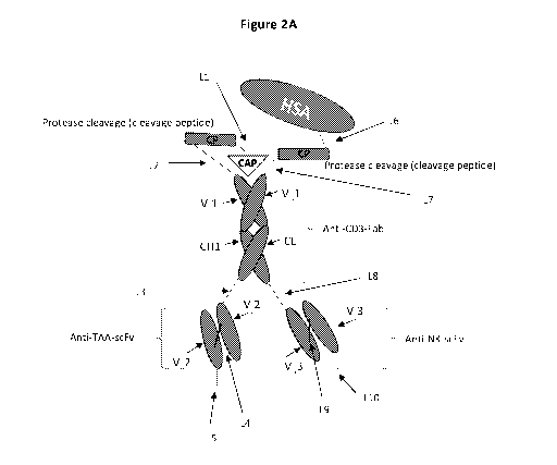
[0055] Figure 2A presents one embodiment of a precursor tri-specific
antibody (tri-body)
construct having antibody binding domains and regulatory domains as described
herein, the
Fab portion recognizes a CD3 surface antigen, one scFv recognizes a tumor
associated antigen
(TAA), and one scFv recognizes a NK cell surface antigen (e.g. NKG2A or
NKG2D). Figure
2B presents another embodiment of the tri-body construct of Figure 2A. Figure
2C presents
one embodiment of a precursor tri-specific antibody construct as described
herein but lacking
the regulatory domain comprising the half-life extending component (HSA).
Figure 2D
presents one embodiment of a precursor tri-specific antibody construct as
described herein but
lacking the regulatory domain comprising the CD3 CAP domain. Figure 2E
presents one
embodiment of an active tri-specific (tribody) antibody construct, lacking the
regulatory
domains with the HSA and CAP domains. Figure 2F presents another embodiment of
an active
tri-specific (tribody) antibody construct, lacking the regulatory domains with
the HSA and CAP
domains. Figure 2G presents one embodiment of a precursor tri-specific
antibody construct as
described herein, wherein the regulatory domain N-terminal to the Fab
comprises a single
regulatory domain comprising a CAP domain, a HSA sequence, and a protease
cleavable linker
on the same polypeptide.
[0056] Figure 3A presents one embodiment of a precursor tri-specific
antibody (tri-body)
construct having antibody binding domains and regulatory domains as described
herein, the
Fab portion recognizes a CD3 surface antigen, one scFv recognizes a tumor
associated antigen
(TAA), and one scFv recognizes a NK cell surface antigen (e.g. NKG2A or
NKG2D). The anti-
NK scFv further comprises a regulatory domain comprising a CAP domain. Figure
3B presents
one embodiment of a precursor tri-specific antibody construct as described
herein, wherein the
regulatory domain N-terminal to the Fab comprises a single regulatory domain
comprising a CAP
domain, a HSA sequence, and a protease cleavable linker on the same
polypeptide. Figure 3C
presents another embodiment of a precursor tri-specific antibody construct as
described in
Figure 3B. Figure 3D presents one embodiment of a precursor tri-specific
antibody construct
as described herein but lacking the regulatory domain comprising the half-life
extending
component (HSA). Figure 3E presents one embodiment of a precursor tri-specific
antibody
construct as described herein but lacking the regulatory domain comprising the
CD3 CAP
domain. Figure 3F presents one embodiment of an active tri-specific (tribody)
antibody
construct, lacking the regulatory domains with the HSA and CAP domains.
[0057] Figure 4A presents one embodiment of a precursor tri-specific
antibody (tri-body)
construct having antibody binding domains as described herein, the Fab portion
recognizes a
NK cell surface antigen (e.g. NKG2A or NKG2D), one scFv recognizes a tumor
associated
17
CA 03181963 2022-11-01
WO 2021/224913 PCT/IL2021/050506
antigen (TAA). The regulatory domain N-terminal to the Fab comprises a single
regulatory
domain comprising a CAP domain, a HSA sequence, and a protease cleavable
linker on the same
polypeptide. The tri-body construct also has a cytokine receptor engager
comprising a cytokine
that binds to a cytokine receptor, for example, an IL-15 that binds to IL-15
receptor. Figure 4B
presents another embodiment of the tri-body construct of Figure 4A, wherein
the regulatory
domain N-terminal to the Fab is the one shown in Figure 2A. Figure 4C presents
one
embodiment of an active tri-specific (tribody) antibody construct derived from
the precursor
construct of Figure 4A or 4B, wherein the Fab portion recognizes a NK cell
surface antigen
(e.g. NKG2A or NKG2D), one scFv recognizes a tumor associated antigen (TAA),
and there
is a cytokine receptor engager such as IL-15.
[0058] Figure 5A presents one embodiment of a precursor tri-specific
antibody (tri-body)
construct having three antibody binding domains and regulatory domains as
described herein,
the Fab portion recognizes a NK cell surface antigen (e.g. NKG2D), one scFv
recognizes a
tumor associated antigen (TAA), and one scFv recognizes another NK cell
surface antigen (e.g.
NKG2A). Figure 5B presents another embodiment of a precursor tri-specific
antibody
construct of Figure 5A, wherein the Fab binds to NKG2A and one scFv binds to
NKG2D. In
another embodiment, the anti-NK Fab binds to NKG2A and the anti-NK-scFv binds
to
NKG2D. Figure 5C presents one embodiment of a precursor tri-specific antibody
(tri-body)
construct having the antibody binding domains and regulatory domains as
described herein, the
Fab portion recognizes a NK cell surface antigen (e.g. NKG2D), one scFv
recognizes a tumor
associated antigen (TAA), and one scFv recognizes another NK cell surface
antigen (e.g.
NKG2A). The regulatory domain N-terminal to the Fab comprises a single
polypeptide chain.
Figure 5D presents another embodiment of a precursor tri-specific antibody
construct of Figure
5C, wherein the Fab binds to NKG2A and one scFv binds to NKG2D. Figure 5E
presents one
embodiment of an active tri-specific (tribody) antibody construct derived from
the precursor
constructs of Figures 5A-5D, wherein the anti-NK Fab binds to NKG2D and the
anti-NK-scFv
binds to NKG2A. Figure 5F presents another embodiment of an active tri-
specific (tribody)
antibody construct derived from the precursor constructs of Figures 5A-5D,
wherein the anti-
NK Fab binds to NKG2A and the anti-NK-scFv binds to NKG2D.
[0059] Figures 6A and 6B present embodiments of an amino acid sequence of a
Heavy
Chain (HC) polypeptide of an activated tri-specific (tri-body) antibody
construct (Construct 1;
VLVH) and an optimized nucleotide sequences encoding the Heavy Chain (HC)
activated
construct, Amino acid sequences are presented N-terminal to C-terminal, and
nucleic acid
sequences are presented 5' to 3'. Figure 6A presents one embodiment of an
amino acid
18
CA 03181963 2022-11-01
WO 2021/224913 PCT/IL2021/050506
sequence of a Heavy Chain (HC) polypeptide of an activated construct, having
the N-terminal
to C-terminal order and components as follows: h1F3.5-G1Fd anti-EGFR VL-linker-
VH (SEQ
ID NO: 138). The amino acid sequences of the component parts of the HC
polypeptide shown
in Figure 6A include: Linker (SEQ ID NO: 158), anti-CD3epsilon variable heavy
chain and
constant heavy chain region 1 (SEQ ID NO: 113), followed by two marked
cysteine residues
(marked bold and underline), which may participate in disulfide double bond,
followed by an
anti-EGFR scFv VL (SEQ ID NO: 34)-linker (SEQ ID NO: 39)-VH (SEQ ID NO: 37)
chain.
Figure 6B presents one embodiment of an optimized nucleic acid sequence (DNA)
encoding
a Heavy Chain (HC) polypeptide of an activated tri-specific (tri-body)
construct, having the 5'
to 3' order and components as follows (SEQ ID NO: 150). The nucleic acid
sequences encoding
the component parts of the HC polypeptide shown in Figure 6A include: Linker
(SEQ ID NO:
154), anti-CD3epsilon variable heavy chain and constant heavy chain region 1
(SEQ ID NO:
155), followed by two marked cysteine residues (marked bold and underline),
which may
participate in disulfide double bond, followed by an anti-EGFR scFv VL (SEQ ID
NO: 36)-
linker (SEQ ID NO: 40)-VH (SEQ ID NO: 38) chain.
[0060] Figures 7A and 7B present embodiments of an amino acid sequence of a
Light Chain
(LC) polypeptide of an activated tri-specific (tri-body) antibody construct
(Construct 1; VLVH)
and an optimized nucleotide sequences encoding the Light Chain (LC) activated
construct,
Amino acid sequences are presented N-terminal to C-terminal, and nucleic acid
sequences are
presented 5' to 3'. Figure 7A presents one embodiment of an amino acid
sequence of a Light
Chain (LC) polypeptide of an activated construct, having the N-terminal to C-
terminal order
and components as follows: h1F3.1-XLC anti-EGFR VL-linker-VH (SEQ ID NO: 139).
The
amino acid sequences of the component parts of the LC polypeptide shown in
Figure 7A
include: Linker (SEQ ID NO: 158), anti-CD3epsilon variable light chain and
lambda light
chain (SEQ ID NO: 74), followed by marked cysteine residues (marked bold and
underline),
which may participate in a disulfide double bond, followed by an anti-EGFR
scFv VL (SEQ
ID NO: 34)-linker (SEQ ID NO: 39)-VH (SEQ ID NO: 37) chain. Figure 7B presents
one
embodiment of an optimized nucleic acid sequence (DNA) encoding a Light Chain
(LC)
polypeptide of an activated tri-specific (tri-body) construct, having the 5'
to 3' order and
components as follows (SEQ ID NO: 151). The nucleic acid sequences encoding
the
component parts of the LC polypeptide shown in Figure 7A include: Linker (SEQ
ID NO: 154),
anti-CD3epsilon variable light chain and lambda light chain region (SEQ ID NO:
159),
followed by marked cysteine residues (marked bold and underline), which may
participate in
a disulfide double bond, followed by an anti-EGFR scFv VL (SEQ ID NO: 36)-
linker (SEQ ID
19
CA 03181963 2022-11-01
WO 2021/224913 PCT/IL2021/050506
NO: 40)-VH (SEQ ID NO: 38) chain.
[0061] Figures 8A and 8B present embodiments of an amino acid sequence of a
Heavy
Chain (HC) polypeptide of an activated tri-specific (tri-body) antibody
construct (Construct 2;
VHVL) and an optimized nucleotide sequences encoding the Heavy Chain (HC)
activated
construct. Amino acid sequences are presented N-terminal to C-terminal, and
nucleic acid
sequences are presented 5' to 3'. Figure 8A presents one embodiment of an
amino acid
sequence of a Heavy Chain (HC) polypeptide of an activated construct, having
the N-terminal
to C-terminal order and components as follows: h1F3.5-G1Fd-(VH-linker-VL) (SEQ
ID NO:
140). The amino acid sequences of the component parts of the HC polypeptide
shown in Figure
8A include: Linker (SEQ ID NO: 158), anti-CD3epsilon variable heavy chain and
constant
heavy chain region 1 (SEQ ID NO: 113), followed by two marked cysteine
residues (marked
bold and underline), which may participate in disulfide double bond, followed
by an anti-EGFR
scFv VH (SEQ ID NO: 37)-linker (SEQ ID NO: 39)-VL (SEQ ID NO: 34) chain.
Figure 8B
presents one embodiment of an optimized nucleic acid sequence (DNA) encoding a
Heavy
Chain (HC) polypeptide of an activated tri-specific (tri-body) construct,
having the 5' to 3'
order and components as follows (SEQ ID NO: 152). The nucleic acid sequences
encoding the
component parts of the HC polypeptide shown in Figure 8A include: Linker (SEQ
ID NO:
154), anti-CD3epsilon variable heavy chain and constant heavy chain region 1
(SEQ ID NO:
155), followed by two marked cysteine residues (marked bold and underline),
which may
participate in disulfide double bonds, followed by an anti-EGFR scFv VH (SEQ
ID NO: 38)-
linker (SEQ ID NO: 40)-VH (SEQ ID NO: 36) chain.
[0062] Figures 9A and 9B present embodiments of an amino acid sequence of a
Light
Chain (LC) polypeptide of an activated tri-specific (tri-body) antibody
construct (Construct 2;
VHVL) and an optimized nucleotide sequences encoding the Light Chain (LC)
activated
construct. Amino acid sequences are presented N-terminal to C-terminal, and
nucleic acid
sequences are presented 5' to 3'. Figure 9A presents one embodiment of an
amino acid
sequence of a Light Chain (LC) polypeptide of an activated construct, having
the N-terminal
to C-terminal order and components as follows: h1F3.1-XLC- Anti-EGFR (VH-
linker-VL)
(SEQ ID NO: 141). The amino acid sequences of the component parts of the LC
polypeptide
shown in Figure 9A include: Linker (SEQ ID NO: 158), anti-CD3epsilon variable
light chain
and lambda light chain (SEQ ID NO: 74), followed by marked cysteine residues
(marked bold
and underline), which may participate in a disulfide double bond, followed by
an anti-EGFR
scFv VH (SEQ ID NO: 37)-linker (SEQ ID NO: 39)-VL (SEQ ID NO: 34) chain.
Figure 9B
presents one embodiment of an optimized nucleic acid sequence (DNA) encoding a
Light
CA 03181963 2022-11-01
WO 2021/224913 PCT/IL2021/050506
Chain (LC) polypeptide of an activated tri-specific (tri-body) construct,
having the 5' to 3' order
and components as follows (SEQ ID NO: 153). The nucleic acid sequences
encoding the
component parts of the LC polypeptide shown in Figure 9A include: Linker (SEQ
ID NO: 154),
anti-CD3epsilon variable light chain and lambda light chain region (SEQ ID NO:
159),
followed by marked cysteine residues (marked bold and underline), which may
participate in
a disulfide double bond, followed by an anti-EGFR scFv VH (SEQ ID NO: 38)-
linker (SEQ
ID NO: 40)-VL (SEQ ID NO: 36) chain.
[0063] Figures 10A and 10B present embodiments of an amino acid sequence of a
Heavy
Chain (HC) polypeptide of precursor tri-specific (tri-body) antibody construct
(Construct 3;
VLVH) and an optimized nucleotide sequences encoding the Heavy Chain (HC)
precursor
construct, Amino acid sequences are presented N-terminal to C-terminal, and
nucleic acid
sequences are presented 5' to 3'. Figure 10A presents one embodiment of an
amino acid
sequence of a Heavy Chain (HC) polypeptide of a precursor construct, having
the N-terminal
to C-terminal order and components as follows: hHSA-G-PLGLAG (MMP2/9)-
(cloning)-
h1F3.5-G1Fd anti-EGFR VL-linker-VH (SEQ ID NO: 130). The amino acid sequences
of the
component parts of the HC polypeptide shown in Figure 10A include: human serum
albumin
(HSA) (SEQ ID NO: 7), MMP2/9 protease cleavable Linker (SEQ ID NO: 160 (linker
with
cleavable sequence and SEQ ID NO: 9 (cleavable sequence)), anti-CD3epsilon
variable heavy
chain and constant heavy chain region 1 (SEQ ID NO: 113), followed by two
marked cysteine
residues (marked bold and underline), which may participate in disulfide
double bonds,
followed by an anti-EGFR scFv VL (SEQ ID NO: 34)-linker (SEQ ID NO: 39)-VH
(SEQ ID
NO: 37) chain. Figure 10B presents one embodiment of an optimized nucleic acid
sequence
(DNA) encoding a Heavy Chain (HC) polypeptide of the precursor tri-specific
(tri-body)
antibody construct, having the 5' to 3' order and components as follows (SEQ
ID NO: 142).
The nucleic acid sequences encoding the component parts of the HC polypeptide
shown in
Figure 10A include: human serum albumin (HSA) (SEQ ID NO: 8), MMP2/9 protease
cleavable Linker (SEQ ID NO: 161 (linker with cleavable sequence and SEQ ID
NO: 33
(cleavable sequence)), anti-CD3epsilon variable heavy chain and constant heavy
chain region
1 (SEQ ID NO: 155), followed by two marked cysteine residues (marked bold and
underline),
which may participate in disulfide double bond, followed by an anti-EGFR scFv
VL (SEQ ID
NO: 36)-linker (SEQ ID NO: 40)-VH (SEQ ID NO: 38) chain.
[0064] Figures 11A and 11B present embodiments of an amino acid sequence of a
Light
Chain (LC) polypeptide of a precursor tri-specific (tri-body) antibody
construct (Construct 3;
VLVH) and an optimized nucleotide sequences encoding the Light Chain (LC)
precursor
21
CA 03181963 2022-11-01
WO 2021/224913 PCT/IL2021/050506
construct, Amino acid sequences are presented N-terminal to C-terminal, and
nucleic acid
sequences are presented 5' to 3'. Figure 11A presents one embodiment of an
amino acid
sequence of a Light Chain (LC) polypeptide of a precursor construct, having
the N-terminal to
C-terminal order and components as follows: Cap-h1F3.1-XLC anti-EGFR VL-linker-
VH
MM2/9 cleavage; SEQ ID NO: 131). The amino acid sequences of the component
parts of the
LC polypeptide shown in Figure 11A include: CAP (SEQ ID NO: 5), MMP2/9
protease
cleavable Linker (SEQ ID NO: 160 and SEQ ID NO: 9 (cleavable sequence)), anti-
CD3epsilon
variable light chain and lambda light chain (SEQ ID NO: 74), followed by
marked cysteine
residues (marked bold and underline), which may participate in a disulfide
double bond,
followed by an anti-EGFR scFv VL (SEQ ID NO: 34)-linker (SEQ ID NO: 39)-VH
(SEQ ID
NO: 37) chain. Figure 11B presents one embodiment of an optimized nucleic acid
sequence
(DNA) encoding the Light Chain (LC) polypeptide of the precursor tri-specific
(tri-body)
antibody construct, having the 5' to 3' order and components as follows (SEQ
ID NO: 143).
The nucleic acid sequences encoding the component parts of the LC polypeptide
shown in
Figure 11A include: CAP (SEQ ID NO: 164), MMP2/9 protease cleavable Linker
(SEQ ID
NO: 161 and SEQ ID NO: 33 (cleavable sequence)), anti-CD3epsilon variable
light chain and
lambda light chain region (SEQ ID NO: 159), followed by marked cysteine
residues (marked
bold and underline), which may participate in a disulfide double bond,
followed by an anti-
EGFR scFv VL (SEQ ID NO: 36)-linker (SEQ ID NO: 40)-VH (SEQ ID NO: 38) chain.
[0065] Figures 12A and 12B present embodiments of an amino acid sequence of a
Heavy
Chain (HC) polypeptide of precursor tri-specific (tri-body) antibody construct
(Construct 4;
VHVL) and an optimized nucleotide sequences encoding the Heavy Chain (HC)
precursor
construct. Amino acid sequences are presented N-terminal to C-terminal, and
nucleic acid
sequences are presented 5' to 3'. Figure 12A presents one embodiment of an
amino acid
sequence of a Heavy Chain (HC) polypeptide of a precursor construct, having
the N-terminal
to C-terminal order and components as follows: hHSA-G-PLGLAG (MMP2/9)-
(cloning)-
h1F3.5-G1Fd Anti-EGFR (VH-linker-VL) (SEQ ID NO: 132). The amino acid
sequences of
the component parts of the HC polypeptide shown in Figure 12A include: human
serum
albumin (HSA) (SEQ ID NO: 7), MMP2/9 protease cleavable Linker (SEQ ID NO: 160
and
SEQ ID NO: 9 (cleavable portion)), anti-CD3epsilon variable heavy chain and
constant heavy
chain region 1 (SEQ ID NO: 113), followed by two marked cysteine residues
(marked bold
and underline), which may participate in disulfide double bonds, followed by
an anti-EGFR
scFv VH (SEQ ID NO: 37)-linker (SEQ ID NO: 40)-VL (SEQ ID NO: 34) chain.
Figure 12B
presents one embodiment of an optimized nucleic acid sequence (DNA) encoding
the Heavy
22
CA 03181963 2022-11-01
WO 2021/224913 PCT/IL2021/050506
Chain (HC) polypeptide of the precursor tri-specific (tri-body) antibody
construct, having the
5' to 3' order and components as follows (SEQ ID NO: 144). The nucleic acid
sequences
encoding the component parts of the HC polypeptide shown in Figure 12A
include: human
serum albumin (HSA) (SEQ ID NO: 8), MMP2/9 protease cleavable Linker (SEQ ID
NO: 161
and SEQ ID NO: 33 (cleavable linker)), anti-CD3epsilon variable heavy chain
and constant
heavy chain region 1 (SEQ ID NO: 155), followed by two marked cysteine
residues (marked
bold and underline), which may participate in disulfide double bond, followed
by an anti-EGFR
scFv VH (SEQ ID NO: 38)-linker (SEQ ID NO: 40)-VL (SEQ ID NO: 36) chain.
[0066] Figures 13A and 13B present embodiments of an amino acid sequence of a
Light
Chain (LC) polypeptide of a precursor tri-specific (tri-body) antibody
construct (Construct 4;
VLVH) and an optimized nucleotide sequences encoding the Light Chain (LC)
precursor
construct. Amino acid sequences are presented N-terminal to C-terminal, and
nucleic acid
sequences are presented 5' to 3'. Figure 13A presents one embodiment of an
amino acid
sequence of a Light Chain (LC) polypeptide of a precursor construct, having
the N-terminal to
C-terminal order and components as follows: Cap-MMP2/9 cleavage -h1F3.1-kLC-
Anti-
EGFR(VH-linker-VL) (SEQ ID NO: 133; plasmid 7). The amino acid sequences of
the
component parts of the LC polypeptide shown in Figure 13A include: CAP (SEQ ID
NO: 5),
MMP2/9 protease cleavable Linker (SEQ ID NO: 160 and SEQ ID NO: 9 (cleavable
linker),
anti-CD3epsilon variable light chain and lambda light chain (SEQ ID NO: 74),
followed by
marked cysteine residues (marked bold and underline), which may participate in
a disulfide
double bond, followed by an anti-EGFR scFv VH (SEQ ID NO: 37)-linker (SEQ ID
NO: 40)-
VL (SEQ ID NO: 34) chain. Figure 13B presents one embodiment of an optimized
nucleic
acid sequence (DNA) encoding the Light Chain (LC) polypeptide of the precursor
tri-specific
(tri-body) antibody construct, having the 5' to 3' order and components as
follows (SEQ ID
NO: 145). The nucleic acid sequences encoding the component parts of the LC
polypeptide
shown in Figure 13A include: CAP (SEQ ID NO: 164), MMP2/9 protease cleavable
Linker
(SEQ ID NO: 161 and SEQ ID NO: 33 (cleavable sequence)), anti-CD3epsilon
variable light
chain and lambda light chain region (SEQ ID NO: 159), followed by marked
cysteine residues
(marked bold and underline), which may participate in a disulfide double bond,
followed by an
anti-EGFR scFv VH (SEQ ID NO: 38)-linker (SEQ ID NO: 40)-VL (SEQ ID NO: 36)
chain.
[0067] Figures 14A and 14B present embodiments of an amino acid sequence of a
Heavy
Chain (HC) polypeptide of non-cleavable (non-activatable) precursor tri-
specific (tri-body)
antibody construct (Construct 5; VLVH) and an optimized nucleotide sequences
encoding the
Heavy Chain (HC) non-cleavable precursor construct. Amino acid sequences are
presented N-
23
CA 03181963 2022-11-01
WO 2021/224913 PCT/IL2021/050506
terminal to C-terminal, and nucleic acid sequences are presented 5' to 3'.
Figure 14A presents
one embodiment of an amino acid sequence of a Heavy Chain (HC) polypeptide of
a non-
cleavable precursor construct, having the N-terminal to C-terminal order and
components as
follows: hHSA-G-PLGLAG NC-h1F3.5-G1Fd anti-EGFR VL-linker-VH (SEQ ID NO: 134).
The amino acid sequences of the component parts of the HC polypeptide shown in
Figure 14A
include: human serum albumin (HSA) (SEQ ID NO: 7), non-cleavable Linker (SEQ
ID NO:
162), anti-CD3epsilon variable heavy chain and constant heavy chain region 1
(SEQ ID NO:
113), followed by two marked cysteine residues (marked bold and underline),
which may
participate in disulfide double bonds, followed by an anti-EGFR scFv VL (SEQ
ID NO: 34)-
linker (SEQ ID NO: 40)-VH (SEQ ID NO: 37) chain. Figure 14B presents one
embodiment
of an optimized nucleic acid sequence (DNA) encoding a Heavy Chain (HC)
polypeptide of
the non-cleavable precursor tri-specific (tri-body) antibody construct, having
the 5' to 3' order
and components as follows (SEQ ID NO: 146). The nucleic acid sequences
encoding the
component parts of the HC polypeptide shown in Figure 14A include: human serum
albumin
(HSA) (SEQ ID NO: 8), non-cleavable Linker (SEQ ID NO: 163), anti-CD3epsilon
variable
heavy chain and constant heavy chain region 1 (SEQ ID NO: 155), followed by
two marked
cysteine residues (marked bold and underline), which may participate in
disulfide double bond,
followed by an anti-EGFR scFv VL (SEQ ID NO: 36)-linker (SEQ ID NO: 40)-VH
(SEQ ID
NO: 38) chain.
[0068] Figures 15A and 15B present embodiments of an amino acid sequence of a
Light
Chain (LC) polypeptide of a non-cleavable precursor tri-specific (tri-body)
antibody construct
(Construct 5; VLVH) and an optimized nucleotide sequences encoding the Light
Chain (LC)
non-cleavable precursor construct. Amino acid sequences are presented N-
terminal to C-
terminal, and nucleic acid sequences are presented 5' to 3'. Figure 15A
presents one
embodiment of an amino acid sequence of a Light Chain (LC) polypeptide of a
non-cleavable
precursor construct, having the N-terminal to C-terminal order and components
as follows:
Cap-(h1F3.1-XLC anti-EGFR VL-linker-VH Non-Cleavable (SEQ ID NO: 135). The
amino
acid sequences of the component parts of the LC polypeptide shown in Figure
15A include:
CAP (SEQ ID NO: 5), non-cleavable Linker (SEQ ID NO: 162), anti-CD3epsilon
variable light
chain and lambda light chain (SEQ ID NO: 74), followed by marked cysteine
residues (marked
bold and underline), which may participate in a disulfide double bond,
followed by an anti-
EGFR scFv VL (SEQ ID NO: 34)-linker (SEQ ID NO: 40)-VH (SEQ ID NO: 37) chain.
Figure
15B presents one embodiment of an optimized nucleic acid sequence (DNA)
encoding the
Light Chain (LC) polypeptide of the non-cleavable precursor tri-specific (tri-
body) antibody
24
CA 03181963 2022-11-01
WO 2021/224913 PCT/IL2021/050506
construct, having the 5' to 3' order and components as follows (SEQ ID NO:
147). The nucleic
acid sequences encoding the component parts of the LC polypeptide shown in
Figure 13A
include: CAP (SEQ ID NO: 164), non-cleavable Linker (SEQ ID NO: 163),anti-
CD3epsilon
variable light chain and lambda light chain region (SEQ ID NO: 159), followed
by marked
cysteine residues (marked bold and underline), which may participate in a
disulfide double
bond, followed by an anti-EGFR scFv VL (SEQ ID NO: 36)-linker (SEQ ID NO: 40)-
VH (SEQ
ID NO: 38) chain.
[0069] Figures 16A and 16B present embodiments of an amino acid sequence of a
Heavy
Chain (HC) polypeptide of a non-cleavable precursor tri-specific (tri-body)
antibody construct
(Construct 6; VHVL) and an optimized nucleotide sequences encoding the Heavy
Chain (HC)
non-cleavable precursor construct. Amino acid sequences are presented N-
terminal to C-
terminal, and nucleic acid sequences are presented 5' to 3'. Figure 16A
presents one
embodiment of an amino acid sequence of a Heavy Chain (HC) polypeptide of a
non-cleavable
precursor construct, having the N-terminal to C-terminal order and components
as follows:
hHSA-G-PLGLAG (NC)- (cloning)-h1F3.5-G1Fd-(VH-linker-VL) (SEQ ID NO: 136). The
amino acid sequences of the component parts of the HC polypeptide shown in
Figure 16A
include: human serum albumin (HSA) (SEQ ID NO: 7), non-cleavable Linker (SEQ
ID NO:
162), anti-CD3epsilon variable heavy chain and constant heavy chain region 1
(SEQ ID NO:
113), followed by two marked cysteine residues (marked bold and underline),
which may
participate in disulfide double bonds, followed by an anti-EGFR scFv VH (SEQ
ID NO: 37)-
linker (SEQ ID NO: 40)-VL (SEQ ID NO: 34) chain. Figure 16B presents one
embodiment of
an optimized nucleic acid sequence (DNA) encoding the Heavy Chain (HC)
polypeptide of the
non-cleavable precursor tri-specific (tri-body) antibody construct, having the
5' to 3' order and
components as follows (SEQ ID NO: 148). The nucleic acid sequences encoding
the
component parts of the HC polypeptide shown in Figure 16A include: human serum
albumin
(HSA) (SEQ ID NO: 8), non-cleavable Linker (SEQ ID NO: 163), anti-CD3epsilon
variable
heavy chain and constant heavy chain region 1 (SEQ ID NO: 155), followed by
two marked
cysteine residues (marked bold and underline), which may participate in
disulfide double bond,
followed by an anti-EGFR scFv VH (SEQ ID NO: 38)-linker (SEQ ID NO: 40)-VL
(SEQ ID
NO: 36) chain.
[0070] Figures 17A and 17B present embodiments of an amino acid sequence of a
Light
Chain (LC) polypeptide of a non-cleavable precursor tri-specific (tri-body)
antibody construct
(Construct 6; VLVH) and an optimized nucleotide sequences encoding the Light
Chain (LC)
non-cleavable precursor construct. Amino acid sequences are presented N-
terminal to C-
CA 03181963 2022-11-01
WO 2021/224913 PCT/IL2021/050506
terminal, and nucleic acid sequences are presented 5' to 3'. Figure 17A
presents one
embodiment of an amino acid sequence of a Light Chain (LC) polypeptide of a
precursor
construct, having the N-terminal to C-terminal order and components as
follows: Cap (NC)-
h1F3.1-XLC- Anti-EGFR (VH-linker-V) (SEQ ID NO: 137). The amino acid sequences
of the
component parts of the LC polypeptide shown in Figure 17A include: CAP (SEQ ID
NO: 5),
non-cleavable Linker (SEQ ID NO: 162), anti-CD3epsilon variable light chain
and lambda
light chain (SEQ ID NO: 74), followed by marked cysteine residues (marked bold
and
underline), which may participate in a disulfide double bond, followed by an
anti-EGFR scFv
VH (SEQ ID NO: 37)-linker (SEQ ID NO: 40)-VL (SEQ ID NO: 34) chain. Figure 17B
presents one embodiment of an optimized nucleic acid sequence (DNA) encoding
the Light
Chain (LC) polypeptide of the non-cleavable precursor tri-specific (tri-body)
antibody
construct, having the 5' to 3' order and components as follows (SEQ ID NO:
149). The nucleic
acid sequences encoding the component parts of the LC polypeptide shown in
Figure 17A
include: CAP (SEQ ID NO: 164), non-cleavable Linker (SEQ ID NO: 163), anti-
CD3epsilon
variable light chain and lambda light chain region (SEQ ID NO: 159), followed
by marked
cysteine residues (marked bold and underline), which may participate in a
disulfide double
bond, followed by an anti-EGFR scFv VH (SEQ ID NO: 38)-linker (SEQ ID NO: 40)-
VL (SEQ
ID NO: 36) chain.
[0071] Figures 18A-18B present embodiments of amino acid and nucleic acid
sequences of
scFv anti-ROR1 VL-VH. Figure 18A presents one embodiment of an amino acid
sequence of
an scFv anti-ROR1 having the N-terminal to C-terminal order: VL-VH, and
components as
follows: anti-ROR1 VL, linker, and anti-ROR1 VH (SEQ ID NO: 156). Figure 18B
presents
one embodiment of an optimized nucleic acid sequence encoding an scFv anti-
ROR1 having
the N-terminal to C-terminal order: VL-VH (SEQ ID NO: 157).
[0072] Figures 19A-19B present embodiments of amino acid and nucleic acid
sequences of
scFv anti-ROR1 VH-VL. Figure 19A presents one embodiment of an amino acid
sequence of
an scFv anti-ROR1 having the N-terminal to C-terminal order: VH-VL, and
components as
follows: anti-ROR1 VH, linker, and anti-ROR1 VL (SEQ ID NO: 166). Figure 19B
presents
one embodiment of an optimized nucleic acid sequence encoding an scFv anti-
ROR1 having
the N-terminal to C-terminal order: VH-VL (SEQ ID NO: 167).
[0073] Figures 20A-20B present embodiments of amino acid and nucleic acid
sequences of
scFv anti-PSMA VL-VH. Figure 20A presents one embodiment of an amino acid
sequence of
an scFv anti-PSMA having the N-terminal to C-terminal order: VL-VH, and
components as
follows: anti-PSMA VL, linker, and anti-PSMA VH (SEQ ID NO: 168). Figure 20B
presents
26
CA 03181963 2022-11-01
WO 2021/224913 PCT/IL2021/050506
one embodiment of an optimized nucleic acid sequence encoding an scFv anti-PS
MA having
the N-terminal to C-terminal order: VL-VH (SEQ ID NO: 169).
[0074] Figures 21A-21B present embodiments of amino acid and nucleic acid
sequences of
scFv anti-PSMA VH-VL. Figure 21A presents one embodiment of an amino acid
sequence of
an scFv anti-PSMA having the N-terminal to C-terminal order: VH-VL, and
components as
follows: anti-PSMA VH, linker, and anti-PSMA VL (SEQ ID NO: 170). Figure 21B
presents
one embodiment of an optimized nucleic acid sequence encoding an scFv anti-PS
MA having
the N-terminal to C-terminal order: VH-VL (SEQ ID NO: 171).
[0075] Figures 22A-22B present embodiments of amino acid and nucleic acid
sequences of
scFv anti-5T4 VL-VH. Figure 22A presents one embodiment of an amino acid
sequence of an
scFv anti-5T4 having the N-terminal to C-terminal order: VL-VH, and components
as follows:
anti-5T4 VL, linker, and anti-5T4 VH (SEQ ID NO: 172). Figure 22B presents one
embodiment of an optimized nucleic acid sequence encoding an scFv anti-5T4
having the N-
terminal to C-terminal order: VL-VH (SEQ ID NO: 173).
[0076] Figures 23A-23B present embodiments of amino acid and nucleic acid
sequences of
scFv anti-5T4 VH-VL. Figure 23A presents one embodiment of an amino acid
sequence of an
scFv anti-5T4 having the N-terminal to C-terminal order: VH-VL, and components
as follows:
anti-5T4 VH, linker, and anti-5T4 VL (SEQ ID NO: 174). Figure 23B presents one
embodiment of an optimized nucleic acid sequence encoding an scFv anti-5T4
having the N-
terminal to C-terminal order: VH-VL (SEQ ID NO: 175).
[0077] Figures 24A-24B present embodiments of an amino acid sequence of a
Heavy Chain
(HC) polypeptide of precursor tri-specific (tri-body) antibody construct.
Amino acid sequences
are presented N-terminal to C-terminal. Figures 24A and 24B presents
embodiments of an amino
acid sequence of a Heavy Chain (HC) polypeptide of a precursor construct,
having the N-terminal
to C-terminal order and components as follows: CAP (Bold), linker
(italicized), human serum
albumin (underlined); protease cleavage sequence (bold and italicized);
VH1/CH1 of anti-CD3e
Fab (Bold and underlined), VL (italicized and underlined) VH (italicized,
bold, and underlined)
(EGFR scFv). The two marked cysteine residues (double underline), may
participate in disulfide
double bonds. The protease cleavage sequence in Figure 24A is a multiple,
protease cleavage
sequence. The protease cleavage sequence in Figure 24B is a MMP2/9 protease
cleavage
sequence. The amino acid sequence of Figure 24A is set forth in SEQ ID NO: 28.
The amino acid
sequence of Figure 24B is set forth in SEQ ID NO: 31. Embodiments of sequences
of the
component parts are described throughout this application.
27
CA 03181963 2022-11-01
WO 2021/224913 PCT/IL2021/050506
[0078] Figure 25 presents an embodiment of an amino acid sequence of a Light
Chain (LC)
polypeptide of a precursor tri-specific (tri-body) antibody construct. The
amino acid sequence is
presented N-terminal to C-terminal. Figure 25 presents one embodiment of an
amino acid
sequence of a Light Chain (LC) polypeptide of a precursor construct, having
the N-terminal to C-
terminal order and components as follows: Linker (italicized), VL1-CL of anti-
CD3e Fab (double
underline and bold), VL (italicized and underlined), and VH (italicized bold
and underlined)
wherein the VH is an EGFR scFv). The two marked cysteine residues (double
underline), may
participate in disulfide double bonds. The amine acid sequence of the LC shown
in Figure 25 is
set forth in SEQ ID NO: 32. Embodiments of amino acid sequences of the
component parts of the
LC polypeptide are described throughout the application.
[0079] Figures 26A-26B present embodiments of an amino acid sequence of a
Heavy Chain
(HC) polypeptide of precursor tri-specific (tri-body) antibody construct
described herein. Amino
acid sequences are presented N-terminal to C-terminal. Figures 26A and 26B
presents
embodiments of an amino acid sequence of a Heavy Chain (HC) polypeptide of a
precursor
construct, having the N-terminal to C-terminal order and components as
follows: CAP (Bold),
linker (italicized), human serum albumin (underlined); protease cleavage
sequence (bold and
italicized); VH1/CH1 of anti-CD3e Fab (Bold and underlined), VL (italicized
and underlined) VH
(italicized, bold, and underlined) (5T4 scFv). The two marked cysteine
residues (double
underline), may participate in disulfide double bonds. The protease cleavage
sequence in Figure
26A is a multiple, protease cleavage sequence. The protease cleavage sequence
in Figure 26B is
a MMP2/9 protease cleavage sequence. The amino acid sequence of Figure 26A is
set forth in
SEQ ID NO: 118. The amino acid sequence of Figure 26B is set forth in SEQ ID
NO: 176.
Embodiments of sequences of the component parts are described throughout this
application.
[0080] Figure 27 presents an embodiment of an amino acid sequence of a Light
Chain (LC)
polypeptide of a precursor tri-specific (tri-body) antibody construct as
described herein. The
amino acid sequence is presented N-terminal to C-terminal. Figure 27 presents
one embodiment
of an amino acid sequence of a Light Chain (LC) polypeptide of a precursor
construct, having the
N-terminal to C-terminal order and components as follows: Linker (italicized),
VL1-CL of anti-
CD3e Fab (double underline and bold), VL (italicized and underlined), and VH
(italicized bold
and underlined) wherein the VH is an 5T4 scFv) The two marked cysteine
residues (double
underline), may participate in disulfide double bonds. The amine acid sequence
of the LC shown
in Figure 27 is set forth in SEQ ID NO: 177. Embodiments of amino acid
sequences of the
component parts of the LC polypeptide are described throughout the
application.
28
CA 03181963 2022-11-01
WO 2021/224913 PCT/IL2021/050506
[0081] Figure 28 presents one embodiment of a precursor tri-specific
antibody (tri-body)
construct having antibody binding domains and regulatory domains as described
herein, the
Fab portion recognizes a CD3 surface antigen, one scFv recognizes a tumor
associated antigen
(TAA), and one scFv recognizes a NK cell surface antigen (e.g., NKG2A or
NKG2D). The
regulatory domain N-terminal to the Fab comprises a CAP domain, a HSA
sequence, and a
protease cleavable linker on the same polypeptide.
[0082] Figure 29 presents another embodiment of the tri-body construct of
Figure 28,
wherein the regulatory domain does not contain a protease cleavable linker.
[0083] Figure 30 presents one embodiment of a precursor tri-specific
antibody (tri-body)
construct having antibody binding domains and regulatory domains as described
herein, the
Fab portion recognizes a CD3 surface antigen, one scFv recognizes a tumor
associated antigen
(TAA), and one scFv recognizes a NK cell surface antigen (e.g. NKG2A or
NKG2D). The anti-
NK scFv further comprises a regulatory domain comprising a CAP domain and a
protease
cleavable linker.
[0084] Figure 31 presents one embodiment of a precursor tri-specific
antibody (tri-body)
construct having antibody binding domains and regulatory domains as described
herein, the
Fab portion recognizes a CD3 surface antigen, one scFv recognizes a tumor
associated antigen
(TAA), and one scFv recognizes a NK cell surface antigen (e.g. NKG2A or
NKG2D). The anti-
NK scFv and anti-CD3 Fab each further comprise a regulatory domain comprising
a CAP
domain without a protease cleavable linker.
[0085] Figure 32 presents one embodiment of a precursor tri-specific
antibody (tri-body)
construct having antibody binding domains and regulatory domains as described
herein, the
Fab portion recognizes a CD3 surface antigen, one scFv recognizes a tumor
associated antigen
(TAA), and one scFv region comprising two scFv that recognize one or two NK
cell surface
antigen (e.g. NKG2A and/or NKG2D). The two scFv can bind the same or different
NK cell
surface antigens.
[0086] Figures 33A-33B present some other embodiments of the tri-body
construct of
Figure 32, further having a regulatory domain N-terminal to the Fab comprising
a CAP domain,
a HSA sequence, and a protease cleavable linker on the same polypeptide.
Figure 33A shows the
anti-TAA scFv is located C-terminal to the CL of the anti-CD3 Fab, whereas the
2 anti-NK scFv s
are located C-terminal to the CH of the anti-CD3 Fab. Figure 33B shows the
anti-TAA scFv is
located C-terminal to the CH of the anti-CD3 Fab, whereas the 2 anti-NK scFvs
are located C-
terminal to the CL of the anti-CD3 Fab. In either embodiment, the two scFv can
bind the same
or different NK cell surface antigens.
29
CA 03181963 2022-11-01
WO 2021/224913 PCT/IL2021/050506
[0087] Figure 34A presents another embodiment of the tri-body construct of
Figure 33A,
wherein the regulatory domain does not contain a protease cleavable linker.
Figure 34B presents
another embodiment of the tri-body construct of Figure 33B, wherein the
regulatory domain
does not contain a protease cleavable linker.
[0088] Figure 35 presents one embodiment of a precursor tri-specific
antibody (tri-body)
construct having antibody binding domains and regulatory domains as described
herein, the
Fab portion recognizes a CD3 surface antigen, one scFv recognizes a tumor
associated antigen
(TAA), and one binding domain comprising a cytokine receptor engager (e.g. IL-
15).
[0089] Figure 36 presents another embodiment of the tri-body construct of
Figure 35,
further having a regulatory domain N-terminal to the Fab comprising a CAP
domain, a HSA
sequence, and a protease cleavable linker on the same polypeptide.
[0090] Figure 37 presents another embodiment of the tri-body construct of
Figure 36,
wherein the regulatory domain does not contain a protease cleavable linker.
[0091] Figure 38A shows IM-1062 Tribody protein characterization by SDS-PAGE.
Figure
38B shows IM-1062 Tribody protein characterization by SEC-HPLC.
[0092] Figure 39A shows IM-1184 ProTribody protein characterization by SDS-
PAGE.
Figure 39B shows IM-1184 ProTribody protein characterization by SEC-HPLC.
[0093] Figures 40A-40C show MS analysis of IM-1062 Tribody. Figure 40A shows
MS
analysis of light chain. Figure 40B shows MS analysis of heavy chain. Figure
40C shows MS
analysis of intact Tribody.
[0094] Figures 41A-41B show MS analysis of IM-1184 ProTribody. Figure 41A
shows MS
analysis of light chain. Figure 41B shows MS analysis of heavy chain.
[0095] Figures 42A-42B show results of protease cleavage assay of various
ProTribody
constructs under non-reducing (Figure 42A) or reducing condition (Figure 42B).
Lane M: Maker;
Lane 1: CD3/5T4/NKG2A(VL-VH) (IM1062); Lane 2: Cap137 HSA MC3 CD3/5T4/NKG2A
(IM1188); Lane 3: IM1188+ Matriptase/ST14; Lane 4: IM1188+ MMMP-9; Lane 5:
IM1188+UPA; Lane 6: Cap137 HSA NC CD3/5T4/NKG2A (IM1193); Lane 7: IM1193 +
Matriptase/ST14; Lane 8: IM1193 + MMMP-9; Lane 9: IM1193+UPA; Lane 10:
Cap137 HSA NC CD3/5T4/NKG2A (IM1193); Lane 11: IM1184 + MMMP-9; Lane 12:
IM1184+UPA; Lane 13: Cap137 HSA MC2 CD3/5T4/NKG2A (IM1184); Lane 14:
CD3/5T4/NKG2A(VL-VH) (TM i062).
[0096] Figures 43A-43D show ELISA binding of various Tribody and Tribody
mutants on
recombinant protein CD3 (Figure 43A), 5T4 (Figure 43B), or NKG2A (Figures 43C-
43D).
CA 03181963 2022-11-01
WO 2021/224913 PCT/IL2021/050506
Figure 43D shows binding to human NKG2A by bi-NKG2A scFv Tribodies variants
that harbors
two anti NKG2A scFv in tandem (IM-1272, IM-1273).
[0097] Figures 44A-44C show ELISA binding of various cleaved and non-cleaved
ProTribody on recombinant CD3 protein. Figure 44A: IM-1184 ProTribody; Figure
44B: IM-
1188 ProTribody; Figure 44C: IM-1193 ProTribody.
[0098] Figures 45A-45F show FACS binding of Tribody and Tribody mutants on
different
cell lines. Figure 45A: Jurkat cells; Figure 45B: NCI-H226 cells; Figure 45C:
NK92 cells;
Figure 45D: CHO cells over-expressing 5T4; Figure 45E: CHO cells over-
expressing
NKG2A/CD94. Figure 45F: CHO cells over expressing human NKG2A tested with the
bi-
NKG2A scFv Tribody variants that harbor two anti NKG2A scFv in tandem (IM-
1272, IM-1273).
[0099] Figures 46A-46C show FACS binding of various cleaved and non-cleaved
ProTribody
on Jurkat cell line. Figure 46A: IM-1184 ProTribody; Figure 46B: IM-1188
ProTribody; Figure
46C: IM-1193 ProTribody.
[0100] Figures 47A-47F show in vitro killing function of various Tribody
and Tribody
mutants. Figure 47A: T cell-mediated cytotoxicity on NCI-H226 cells; Figure
47B: NK92-
mediated cytotoxicity against LCL721.221 cells; Figure 47C: PBMC-mediated
cytotoxicity on
A549 cells; Figures 47D-F: PBMC-mediated cytotoxicity on MDA-MB231 cells with
Tribody
concentrations at 1pM or lOpM (Figure 47D), 100nM (Figure 47E) or 100pM
(Figure 47F).
[0101] Figure 48 shows IM-1062 Tribody induces IFNy secretion in various
cell lines.
[0102] Figure 49 shows the ability of various Tribody and Tribody mutants
to block NKG2A-
HLA interaction.
[0103] Figures 50A-50B show the in vivo efficacy of IM-1062 Tribody in MDA-MB-
231
xenograft mouse model. Figure 50A shows average tumor volume over time. Figure
50B shows
tumor volume of individual mice treated with vehicle control (left panel) or
IM-1062 Tribody
(right panel).
[0104] Figures 51A-51B show the in vivo efficacy of a IM-1062 Tribody and a
Tribody
mutant in the A549 tumor model. Figure 51A shows average tumor volume over
time. Figure
51B shows tumor volume of individual mice treated with vehicle control (left
panel), IM-1062
Tribody (middle panel), or IM-1093 Tribody (right panel).
[0105] Figures 52A-52B show the percentage of immune cells in a tumor
microenvironment.
Figure 52A shows %CD45+ cells. Figure 52B shows % activated CD3 cells.
[0106] Figures 53A-53C show the in vivo efficacy of IM-1062 Tribody on
HCT116 cells.
Figure 53A shows average tumor volume over time. Figure 53B shows tumor volume
of
31
CA 03181963 2022-11-01
WO 2021/224913 PCT/IL2021/050506
individual mice treated with control (left panel) or IM-1062 Tribody (right
panel). Figure 53C
shows the in vivo efficacy of IM-1062 Tribody, IM-1184 and IM-1193 ProTribody
on MDA-MB-
231 cells.
[0107] Figure 54 lists the SEQ ID NOs for the various components of the
Tribody or
ProTribody construct, such as VH, VL, CDRs, regulatory domains, protease
cleavage sequences,
and linkers.
[0108] Figure 55 lists the names and SEQ ID NOs of various embodiments of the
Tribody or
ProTribody constructs disclosed herein, each construct comprising polypeptide
A (HC) and
polypeptide B (LC). AA: amino acid sequence; NA: nucleotide sequence.
[0109] It will be appreciated that for simplicity and clarity of
illustration, elements shown
in the figures have not necessarily been drawn to scale. For example, the
dimensions of some
of the elements may be exaggerated relative to other elements for clarity.
Further, where
considered appropriate, reference numerals may be repeated among the figures
to indicate
corresponding or analogous elements.
DETAILED DESCRIPTION OF THE INVENTION
[0110] In the following detailed description, numerous specific details are
set forth in order
to provide a thorough understanding of precursor tri-specific antibody
constructs. However, it
will be understood by those skilled in the art that the precursor constructs
presented herein, the
production of, and the use thereof may be practiced without these specific
details. In other
instances, well-known methods, procedures, and components have not been
described in detail
so as not to obscure the disclosure.
[0111] Described herein are precursor tri-specific antibody constructs
comprising separate
cleavable masking and half-life prolonging domains, wherein these cleavable
regulatory
domains provide reduced binding to T-cells or NK cells by the precursor tri-
specific constructs
when outside the tumor micro-environment (TME) and provide extended half-life.
Half-life
extension may be limited to the time a precursor tri-specific construct is
outside the cancer
microenvironment or it may extend to the time a precursor tri-specific
construct resides within
a cancer microenvironment. An advantage of the precursor tri-specific antibody
constructs
described herein, have a protease cleavable masking domain and a protease
cleavable half-life
prolonging (HLP) domain may be the improved protease-activated controlled
release of the
masking CAP and the HLP domain.
[0112] Reduction in T-cell or NK cell binding may lead to a reduction in T-
cell or NK cell
activation. In some embodiments, the precursor tri-specific antibody
constructs described
32
CA 03181963 2022-11-01
WO 2021/224913 PCT/IL2021/050506
herein are regulatable precursor constructs. The regulatable precursor tri-
specific antibody
constructs describe herein may have an extended half-life, or reduced T-
cell/NK cell binding,
or reduced T-cell/NK cell activation, or any combination thereof.
[0113] As used herein, the terms "half-life prolonging", "HLP", "serum half-
life prolonging",
"extended half-life", "extended serum half-life", "increased half-life",
"increased serum half-life",
and other similar terms may be used interchangeably having all the same
qualities and meanings.
In some embodiments, a half-life prolonging domain comprise a human serum
albumin (HSA).
As used herein, the term "human serum albumin" may in some embodiments be used
interchangeably with "HSA" or "ALB" having all the same meanings and
qualities.
[0114] In some embodiments, the precursor tri-specific antibody constructs
described
herein provide for a regulatable T-cell/NK cell activation, wherein the
precursor construct
provides that T-cell/NK cell activation is restricted to a tumor
microenvironment. In some
embodiments, the precursor tri-specific antibody construct described herein
have an increased
half-life and provide that T-cell/NK cell activation is restricted to a tumor
microenvironment,
compared with non-precursor always active multi-valent antibodies. In some
embodiments, the
precursor tri-specific antibody constructs described herein have reduced T-
cell/NK cell
activation in non-tumor microenvironments, compared with non-precursor always
active multi-
valent antibodies.
[0115] In some embodiments, the precursor tri-specific antibody constructs
described
herein have an extended half-life in non-tumor microenvironments, compared
with non-
precursor tri-specific antibodies. In some embodiments, the precursor tri-
specific antibody
constructs described herein have reduced T-cell/NK cell binding and/or
activation in non-tumor
microenvironments and an extended half-life in a non-tumor microenvironment,
compared
with non-precursor always active multi-valent antibodies.
[0116] In some embodiments, the precursor tri-specific antibody constructs
described
herein may bind to T-cells and to Natural Killer (NK) cells. In some
embodiments, the
precursor tri-specific antibody constructs described herein may bind to T-
cells and to Natural
Killer (NK) cells while also binding to a tumor associated antigen (TAA). In
some
embodiments, the precursor tri-specific antibody constructs described herein
may bind to one
NK cell surface antigen with one binding domain and bind to another NK cell
surface antigen
with another binding domain while also binding to a tumor associated antigen
(TAA) with yet
another binding domain. In some embodiments, the precursor tri-specific
antibody constructs
described herein may bind to one NK cell surface antigen with one binding
domain and bind
to a TAA with another binding domain while also carrying a cytokine receptor
engager
33
CA 03181963 2022-11-01
WO 2021/224913 PCT/IL2021/050506
comprising an agent that binds to a cytokine receptor (e.g. the agent can be a
cytokine such as
IL-15). Examples of TAA and other antigens that can be recognized by the tri-
specific antibody
constructs are disclosed herein.
[0117] In some embodiments, described herein are pharmaceutical compositions
comprising a precursor tri-specific antibody construct that provides a
regulatable T-cell and/or
NK cell activation in non-tumor microenvironments. In some embodiments,
described herein
are pharmaceutical compositions comprising a precursor tri-specific antibody
construct having
an increased half-life and providing that T-cell/NK cell activation is
restricted to a tumor
microenvironment. In some embodiments, described herein are pharmaceutical
compositions
comprising a precursor tri-specific antibody construct comprising an extended
half-life in non-
tumor microenvironments. In some embodiments, described herein are
pharmaceutical
compositions comprising a precursor tri-specific antibody construct comprising
an extended
half-life in non-tumor microenvironments, wherein the half-life is reduced in
a tumor
microenvironment compared with the half-life in a non-tumor microenvironment.
In some
embodiments, the pharmaceutical compositions comprising a precursor antibody
construct that
recognizes a T-cell, an NK cell, and a TAA. In some embodiments, the
pharmaceutical
compositions comprising a precursor antibody construct that recognizes two NK
cell surface
antigens and a TAA. In some embodiments, the pharmaceutical compositions
comprising a
precursor antibody construct having binding domains for a NK cell surface
antigen, a TAA,
and carrying a cytokine receptor engager comprising an agent that binds to a
cytokine receptor
(e.g. the engager can be a cytokine such as IL-15).
[0118] In some embodiment, described herein are methods of use of a
precursor tri-specific
antibody construct, as disclosed herein, for use treating, preventing,
inhibiting the growth of,
delaying disease progression, reducing the tumor load, or reducing the
incidence of a cancer or
tumor in a subject, or any combination thereof. In some embodiments, the
method of treating
disclosed herein reduces the minimal residual disease, increases remission,
increases remission
duration, reduces tumor relapse rate, prevents metastasis of the tumor or the
cancer, or reduces
the rate of metastasis of the tumor or the cancer, or any combination thereof,
in the treated
subject compared with a subject not administered with the pharmaceutical
composition.
Precursor Tr-specific Antibody Constructs
[0119] In some embodiments, a precursor tri-specific antibody construct
comprises 1) a first
binding domain binding to a TAA; 2) a second binding domain binding to an
extracellular
epitope of a Natural Killer (NK) cell antigen; 3) a third binding domain
binding to a T cell
34
CA 03181963 2022-11-01
WO 2021/224913 PCT/IL2021/050506
surface antigen, e.g. an extracellular epitope of human CD3E; and 4) a
regulatory domain. The
regulatory domain may comprise: (1) a first and a second sub-regulatory
domain, the first sub-
regulatory domain comprising a first protease cleavage domain and a half-life
prolonging (HLP)
domain, and the second sub-regulatory domain comprising a second protease
cleavage domain
and a CAP component that reduces the ability of the third binding domain to
bind to its target
antigen; or (2) a single regulatory domain comprising a protease cleavage
domain, a half-life
prolonging (HLP) domain, and a CAP component that reduces the ability of the
third binding
domain to bind to its target antigen. In one embodiment, when the second
binding domain binds
to a NK cell surface antigen, the second binding domain further comprises a
third regulatory
domain comprising a third protease cleavage domain and a CAP component that
reduces the
ability of the second binding domain to bind to the NK cell surface antigen.
[0120] In some embodiments, a precursor tri-specific antibody construct
comprises 1) a first
binding domain binding to a tumor associated antigen (TAA); 2) a second
binding domain
binding to an extracellular epitope of a first Natural Killer (NK) cell
antigen; 3) a third binding
domain binding to an extracellular epitope of a second NK cell surface
antigen; and 4) a
regulatory domain. The regulatory domain may comprise: (1) a first and a
second sub-regulatory
domain, the first sub-regulatory domain comprising a first protease cleavage
domain and a half-
life prolonging (HLP) domain, and the second sub-regulatory domain comprising
a second
protease cleavage domain and a CAP component that reduces the ability of the
third binding
domain to bind to its target antigen; or (2) a single regulatory domain
comprising a protease
cleavage domain, a half-life prolonging (HLP) domain, and a CAP component that
reduces the
ability of the third binding domain to bind to its target antigen. In one
embodiment, when the
second binding domain binds to a NK cell surface antigen, the second binding
domain further
comprises a third regulatory domain comprising a third protease cleavage
domain and a CAP
component that reduces the ability of the second binding domain to bind to the
NK cell surface
antigen. The first and second NK cell surface antigens can be the same or
different antigens. .
[0121] In some embodiments, a precursor tri-specific antibody construct
comprises 1) a first
binding domain binding to a tumor associated antigen (TAA); 2) a second
binding domain
having a cytokine receptor engager comprising an agent that binds to a
cytokine receptor (e.g. the
agent can be a cytokine such as IL-15); 3) a third binding domain binding to
an extracellular
epitope of a NK cell surface antigen; and 4) a regulatory domain. The
regulatory domain may
comprise: (1) a first and a second sub-regulatory domain, the first sub-
regulatory domain
comprising a first protease cleavage domain and a half-life prolonging (HLP)
domain, and the
second sub-regulatory domain comprising a second protease cleavage domain and
a CAP
CA 03181963 2022-11-01
WO 2021/224913 PCT/IL2021/050506
component that reduces the ability of the third binding domain to bind to its
target antigen; or (2)
a single regulatory domain comprising a protease cleavage domain, a half-life
prolonging (HLP)
domain, and a CAP component that reduces the ability of the third binding
domain to bind to its
target antigen.
Cytokine Receptor Engager
[0122] In some embodiments, a precursor tri-specific antibody construct
comprises a binding
domain having a cytokine receptor engager comprising an agent that binds to a
cytokine receptor.
In one embodiment, the cytokine receptor engager is a cytokine that binds to a
cytokine receptor.
As it is generally known in the art, cytokines is a general name; other names
include lymphokine
(cytokines made by lymphocytes), monokine (cytokines made by monocytes),
chemokine
(cytokines with chemotactic activities), and interleukin (cytokines made by
one leukocyte and
acting on other leukocytes). Cytokines may act on the cells that secrete them
(autocrine action),
on nearby cells (paracrine action), or in some instances on distant cells
(endocrine action).
Numerous cytokines are known in the art. There are pro-inflammatory cytokines
and anti-
inflammatory cytokines. Examples of pro-inflammatory cytokines include, but
are not limited to,
IL-113, IL-6, IL-12, and TNF-a. Examples of anti-inflammatory cytokines
include, but are not
limited to, IL-1 receptor antagonist, IL-4, IL-10, IL-11, and IL-13. Depending
on the
circumstances, certain cytokines can be categorized as either anti-
inflammatory or pro-
inflammatory cytokines, for example, Leukemia inhibitory factor, interferon-
alpha, IL-6, and
transforming growth factor (TGF)-(3. In one embodiment, the cytokine receptor
engager is a pro-
inflammatory cytokine. In another embodiment, the cytokine receptor engager is
an anti-
inflammatory cytokine.
[0123] In some embodiments, the cytokine receptor engager is a lymphokine. In
some
embodiments, the cytokine receptor engager is a monokine. In some embodiments,
the cytokine
receptor engager is a chemokine. In some embodiments, the cytokine receptor
engager is a
interleukin. In some embodiments, the cytokine receptor engager is a pro-
inflammatory cytokine.
In some embodiments, the cytokine receptor engager is an anti-inflammatory
cytokine.
[0124] In some embodiments, a precursor tri-specific antibody construct
comprises a
binding domain having a cytokine receptor engager comprising an IL-15
molecule. In one
embodiment, the cytokine receptor engager is a cytokine that binds to its
cytokine receptor.
Examples of cytokines have been disclosed herein. For example, the engager can
be IL-15. In
some embodiments, the IL-15 engager binds to IL-15 receptor. The cytokine can
be from human
or other non-human animals. The cytokine can be purified from native source,
or constructed by
36
CA 03181963 2022-11-01
WO 2021/224913 PCT/IL2021/050506
recombinant techniques. In some embodiments, binding of the IL-15 engager
domain to a target
induces the proliferation of natural killer cells. It is known in the art that
IL-15 plays important
role on NK cell development and homeostasis without stimulating regulatory T
cells. Results from
clinical trials show that 1L-15 administration was associated with a 38-fold
increase in the number
and activation status of circulating natural :killer (NK) cells and activation
of na acropl -ages which
together are ADCC effectors. Furthermore, 1L--15 greatly enhanced the
therapeutic efficacy of
both rituximab and alemtuzurnab in tumor models. In some embodiments, binding
of the IL-15
engager domain to a target induces increase of the cytotoxic function of
natural killer cells. In
some embodiments, binding of the IL-15 engager domain to a target induces both
the
proliferation of natural killer cells and increases their cytotoxic function.
In some
embodiments, binding of the IL-15 engager domain to a target enhances the anti-
tumor
response by an activated precursor tri-specific antibody construct comprising
an IL-15 engager.
In some embodiments, binding of the IL-15 engager domain to a target enhances
the efficacy
in inducing tumor regression by an activated precursor tri-specific antibody
construct
comprising an IL-15 engager.
[0125] Regarding cytokine requirements for natural killer (NK) cell
development and function,
it is known that NK cell development from hematological stem cells (HSCs) is
regulated by
multiple cytokines in fetal liver, bone marrow, and thymus. The sequential
expression of receptors
for different cytokines implies functional maturation of NK cells. The
proliferation and
differentiation of HSCs requires FL (fms-like tyrosine kinase 3 ligand), KL
(kit ligand), IL-3, and
IL-7, which interact with their respective receptors. The acquisition of CD122
expression is
indicative of the commitment of NK cells. IL-15 is indispensable for NK cell
differentiation from
common lymphoid progenitors to mature NK cells. Mature NK cells are shaped by
cytokine
signals from the diverse tissue environments in which they reside. In the
peripheral blood or
spleen, the abundance of stimulatory cytokines, such as IL-2, IL-12, IL-15, IL-
18, and IL-21, may
maintain NK cells in a cytotoxic state to combat infections. Tolerant NK cells
that reside in the
liver, and regulatory NK cells that reside in the uterus, are primarily
regulated by TGF-f3 and IL-
or by TGF-f3 and IL-15, respectively.
Embodiments of Precursor Tr -specific Antibody Constructs
[0126] For the precursor constructs described throughout, the skilled
artisan will appreciate
that the modular structure of said constructs allows for different binding
partners based on the
amino acid sequences comprised in the first, second and third binding domains.
In some
embodiments, the first binding domain and the second binding domain each
comprises a single
37
CA 03181963 2022-11-01
WO 2021/224913 PCT/IL2021/050506
chain variable fragment (scFv). In another embodiment, the third binding
domain comprises a Fab
antigen binding fragment.
[0127] In some embodiments, a binding domain comprises two scFv in tandem. For
example,
in some embodiments, a second binding domain comprises two scFv targeting NK
cells. In some
embodiments, the two scFv target the same antigen on an NK cell. In some
embodiments, the
two scFv target different antigens on an NK cell.
[0128] A skilled artisan would recognize that a precursor antibody
construct is a precursor
form or "Pro" form of the active antibody protein. In some embodiments, the
term "Pro" is
used interchangeably with the term "Precursor" having all the same meanings
and qualities.
[0129] As used herein, the term "precursor tri-specific antibody" or
"ProTribody antibody"
refers to a tri-specific antibody comprising one or more regulatory domains
that regulate
antibody binding to one or more target antigens. (See for examples, Figures 32
and 33A-33B)
[0130] A skilled artisan would appreciate that as used throughout, in some
embodiments the
terms "precursor tri-specific antibody construct", "precursor antibody",
"precursor construct",
"precursor antibody construct", "precursor tri-specific antibody", "tri-
specific antibody",
"antibody", "Tribody", "tri-specific antibody construct", and "tri-specific
construct" may be
used interchangeably having all the same qualities and meanings. Additionally,
in some
embodiments the term "tri-specific" may be replaced with the term "tri-body"
or "Tribody"
with the recognition that the antibody constructs disclosed herein have three
binding regions,
wherein each region may bind a different antigen (tri-body or tri-specific) or
two of the three
binding regions may bind the same antigen (tri-body or tri-specific wherein
two of the specific
binding antigens are the same). Thus, terms as listed above, for example
"precursor tri-specific
antibody construct" in some embodiments may be used interchangeably with the
term
"precursor tri-body construct", having all the same meanings and qualities.
[0131] A skilled artisan would appreciate that in some embodiments, the term
"tumor
associated antigen" (TAA) may encompass a molecule or a portion thereof, which
is displayed
on the surface of a cell or a molecule which is present within the milieu of a
tumor, that is
within the tumor micro-environment. In some embodiments, a TAA encompasses a
cell surface
tumor associated antigen (TAA). In some embodiments, the cell is a tumor cell.
In some
embodiments, the cell is a non-tumor cell present in the milieu of a tumor,
for example but not
limited to a cell present within vasculature tissue associated with a tumor or
cancer. In some
embodiments, a TAA is an angiogenic antigen in a tumor micro-environment. In
some
embodiments, a TAA is an antigen on a blood vessel in a tumor micro-
environment. In some
embodiments, the cell is a stromal cells present in the milieu of a tumor. In
some embodiments,
38
CA 03181963 2022-11-01
WO 2021/224913 PCT/IL2021/050506
a TAA is a stromal cell antigen within a tumor micro-environment. In some
embodiments, a
TAA encompasses an extracellular epitope of a tumor-cell-surface antigen. In
some
embodiments, a TAA encompasses an extracellular matrix antigen.
[0132] In some embodiments, an angiogenic antigen comprises a bFGF. In some
embodiments, an angiogenic antigen comprises a INF. In some embodiments, an
angiogenic
antigen comprises a VEGF. In some embodiments, an angiogenic antigen comprises
a bFGF,
a INF, or a VEGF.
[0133] In some embodiments, a TAA comprises an antigen present in a TME. In
some
embodiments, a TAA comprises a cytokine antigen in a TME. In some embodiments,
a TAA
comprises a molecule secreted by a tumor cell into the TME. In some
embodiments, a TAA
comprises an effector molecule secreted by a tumor cell into the TME. In some
embodiments,
a TAA comprises an effector molecule secreted by a tumor cell into the TME in
order to
downregulate or inhibit the activity of cytotoxic natural killed (NK) cells.
In some
embodiments, a TAA comprises an effector molecule secreted by a tumor cell
into the TME in
order to downregulate or inhibit the activity of NK cells. In some
embodiments, a TAA
comprises soluble activating receptor ligand secreted by a tumor cell into the
TME in order to
block the recognition of the tumor cell by an NK cell. In some embodiments, a
TAA comprises
a suppressive immune cell in the TME that would otherwise inhibit NK cell
activation. In some
embodiments, a TAA comprises a suppressive molecule in the TME that would
otherwise
inhibit NK cell activation. In some embodiments, the effector molecule
comprises a cytokine
antigen. In some embodiments, the effector molecule comprises a cytokine
antigen in the TME.
[0134] In some embodiments, a cytokine antigen in the TME comprises a TNF-
alpha, an
IL-6, a TGF-beta, an IL-10, an IL-8, an IL-17, an IL-21, an INF, or a VEGF. In
some
embodiments, a TAA is selected from a TNF-alpha, an IL-6, a TGF-beta, an IL-
10, an IL-8, an
IL-17, an IL-21, an INF, or an VEGF. In some embodiments, a cytokine antigen
for use as a
TAA comprises a cytokine antigen known in the art.
[0135] A skilled artisan would appreciate that the terms "tumor micro-
environment"
(TME), "cancer microenvironment" and "tumor milieu" may be used
interchangeably having
the same qualities and meanings and encompassing the microenvironment to tumor
development. While the normal cellular microenvironment can inhibit malignant
cell growth,
the modifications that occur in the tumor microenvironment may synergistically
support cell
proliferation.
[0136] In some embodiments, the second binding domain and the third binding
domain of
a precursor construct disclosed herein bind to the same NK cell surface
antigen. In some
39
CA 03181963 2022-11-01
WO 2021/224913 PCT/IL2021/050506
embodiments, the second binding domain and the third binding domain of a
precursor construct
disclosed herein bind to different NK cell surface antigens. In some
embodiments, the second
binding domain and the third binding domain of a precursor construct disclosed
herein bind to
the same or different NK cell surface antigens on the same cell. In some
embodiments, the
second binding domain and the third binding domain of a precursor construct
disclosed herein
bind to the same or different NK cell surface antigens on different NK cells.
[0137] In some embodiments, the second binding domain binds to an NK cell and
the first
binding domain binds to a TAA. The TAA may be for example, but not limited to
an
extracellular epitope of a tumor-cell surface antigen, a TME antigen, a stomal
antigen in a
TME, an angiogenic antigen in a TME, an antigen on a blood vessel in a TME, or
a cytokine
in a TME.
[0138] In some embodiments, binding to an NK cell comprises binding to an
extracellular
epitope of the NK cell antigen.
[0139] In some embodiments, the second binding domain binds to a negative
effector
molecule of an NK cell and the first binding domain binds to a TAA. The TAA
may be for
example, but not limited to an extracellular epitope of a tumor-cell surface
antigen, a TME
antigen, a stomal antigen in a TME, an angiogenic antigen in a TME, an antigen
on a blood
vessel in a TME, or a cytokine in a TME.
[0140] A skilled artisan would appreciate that the terms "antigen" or
"immunogen"
encompass a peptide, protein, or a polypeptide, or any fragment thereof, which
is
immunogenic. In some embodiments, an antigen is capable of eliciting an immune
response in
a mammal, and therefore contains at least one and may contain multiple
epitopes. An "antigen"
molecule or a portion of a molecule is capable of being bound by a selective
binding agent,
such as an antigen-binding portion of a Fab fragment or an antigen-binding
portion of a single
chain variable fragment (scFv). Additionally, an "antigen" is capable of being
used in an animal
to produce antibodies capable of binding to an epitope of that antigen. In
some embodiments,
a CAP component comprises the portion of an antigen to which a binding domain
binds.
[0141] The term "epitope" includes any determinant, in certain embodiments,
a polypeptide
determinant, capable of specific binding to an anti-TAA binding domain or an
anti-NK cell
binding domain or an anti-T-cell receptor binding domain. An epitope is a
region of an antigen
that is bound by an antibody or an antigen-binding fragment thereof. In some
embodiments, a
CAP component comprises the epitope to which a binding domain binds.
[0142] In certain embodiments, epitope determinants include chemically
active surface
groupings of molecules such as amino acids, sugar side chains, phosphoryl or
sulfonyl, and
CA 03181963 2022-11-01
WO 2021/224913 PCT/IL2021/050506
may in certain embodiments have specific three-dimensional structural
characteristics, and/or
specific charge characteristics. In certain embodiments, a precursor tri-
specific antibody
construct is said to specifically bind an antigen when it preferentially
recognizes its target
antigen in a complex mixture of proteins and/or macromolecules. A precursor
tri-specific
antibody construct is said to specifically bind an antigen when the
equilibrium dissociation
constant is < 10-5, 10-6 or 10-7 M. In some embodiments, the equilibrium
dissociation constant
may be < 10-8 M or 10-9 M. In some further embodiments, the equilibrium
dissociation constant
may be < 10-10 M or 10-11 M. Antigens disclosed herein included but are not
limited to TAA,
CAP components, NK cells, and immuno-effector molecules such as a human CD3
epsilon
polypeptide.
[0143] In some embodiments, the tumor associated antigen (TAA) is a tumor
antigen. In
some embodiments, tumor antigens comprise those antigens are presented on
tumor cells. In
some embodiments, the tumor antigen is present on a cell of solid tumor. In
some embodiments,
the tumor antigen is a cancer antigen, present on a cell of a non-solid tumor.
[0144] In some embodiments, when the TAA is a tumor cell antigen, the tumor
cell
comprises a cell from a solid tumor. Solid tumors may be benign (not cancer),
or malignant
(cancer). Different types of solid tumors are named for the type of cells that
form them.
Examples of solid tumors are sarcomas, carcinomas, and lymphomas. In some
embodiments,
solid tumors are neoplasms (new growth of cells) or lesions (damage of
anatomic structures or
disturbance of physiological functions) formed by an abnormal growth of body
tissue cells
other than blood, bone marrow or lymphatic cells. In some embodiments, a solid
tumor consists
of an abnormal mass of cells which may stem from different tissue types such
as liver, colon,
breast, or lung, and which initially grows in the organ of its cellular
origin. However, such
cancers may spread to other organs through metastatic tumor growth in advanced
stages of the
disease.
[0145] In some embodiments, the solid tumor comprises a sarcoma or a
carcinoma, a
fibrosarcoma, a myxosarcoma, a liposarcoma, a chondrosarcoma, an osteogenic
sarcoma, a
chordoma, an angiosarcoma, an endotheliosarcoma, a lymphangiosarcoma, a
lymphangioendotheliosarcoma, a synovioma, a mesothelioma, an Ewing's tumor, a
leiomyosarcoma, a rhabdomyosarcoma, a colon carcinoma, a pancreatic cancer or
tumor, a
breast cancer or tumor, an ovarian cancer or tumor, a prostate cancer or
tumor, a squamous cell
carcinoma, a basal cell carcinoma, an adenocarcinoma, a sweat gland carcinoma,
a sebaceous
gland carcinoma, a papillary carcinoma, a papillary adenocarcinomas, a
cystadenocarcinoma,
a medullary carcinoma, a bronchogenic carcinoma, a renal cell carcinoma, a
hepatoma, a bile
41
CA 03181963 2022-11-01
WO 2021/224913 PCT/IL2021/050506
duct carcinoma, a choriocarcinoma, a seminoma, an embryonal carcinoma, a
Wilm's tumor, a
cervical cancer or tumor, a uterine cancer or tumor, a testicular cancer or
tumor, a lung
carcinoma, a small cell lung carcinoma, a bladder carcinoma, an epithelial
carcinoma, a glioma,
an astrocytoma, a medulloblastoma, a craniopharyngioma, an ependymoma, a
pinealoma, a
hemangioblastoma, an acoustic neuroma, an oligodenroglioma, a schwannoma, a
meningioma,
a melanoma, a neuroblastoma, or a retinoblastoma. In some embodiments, the
solid tumor
comprises an Adrenocortical Tumor (Adenoma and Carcinoma), a Carcinoma, a
Colorectal
Carcinoma, a Desmoid Tumor, a Desmoplastic Small Round Cell Tumor, an
Endocrine Tumor,
an Ewing Sarcoma, a Germ Cell Tumor, a Hepatoblastoma a Hepatocellular
Carcinoma, a
Melanoma, a Neuroblastoma, an Osteosarcoma, a Retinoblastoma, a
Rhabdomyosarcoma, a
Soft Tissue Sarcoma Other Than Rhabdomyosarcoma, and a Wilms Tumor. In some
embodiments, the solid tumor is a breast tumor. In another embodiment, the
solid tumor is a
prostate cancer. In another embodiment, the solid tumor is a colon cancer. In
some
embodiments, the tumor is a brain tumor. In another embodiment, the tumor is a
pancreatic
tumor. In another embodiment, the tumor is a colorectal tumor.
[0146] In some embodiments, the tumor cell comprises a cell from a non-solid
tumor, that
is a non-solid cancer. In some embodiments, a cancer may be a diffuse cancer,
wherein the
cancer is widely spread; not localized or confined. In some embodiments, a
diffuse cancer may
comprise a non-solid tumor. Examples of diffuse cancers include leukemias.
Leukemias
comprise a cancer that starts in blood-forming tissue, such as the bone
marrow, and causes
large numbers of abnormal blood cells to be produced and enter the
bloodstream.
[0147] In some embodiments, a diffuse cancer comprises a B-cell malignancy. In
some
embodiments, the diffuse cancer comprises leukemia. In some embodiments, the
cancer is
lymphoma. In some embodiments, the lymphoma is large B-cell lymphoma.
[0148] In some embodiments, the diffuse cancer or tumor comprises a
hematological tumor.
In some embodiments, hematological tumors are cancer types affecting blood,
bone marrow,
and lymph nodes. Hematological tumors may derive from either of the two major
blood cell
lineages: myeloid and lymphoid cell lines. The myeloid cell line normally
produces
granulocytes, erythrocytes, thrombocytes, macrophages, and mast cells, whereas
the lymphoid
cell line produces B, T, NK and plasma cells. Lymphomas (e.g. Hodgkin's
Lymphoma),
lymphocytic leukemias, and myeloma are derived from the lymphoid line, while
acute and
chronic myelogenous leukemia (AML, CML), myelodysplastic syndromes and
myeloproliferative diseases are myeloid in origin.
[0149] In some embodiments, a non-solid (diffuse) cancer or tumor comprises a
42
CA 03181963 2022-11-01
WO 2021/224913 PCT/IL2021/050506
hematopoietic malignancy, a blood cell cancer, a leukemia, a myelodysplastic
syndrome, a
lymphoma, a multiple myeloma (a plasma cell myeloma), an acute lymphoblastic
leukemia, an
acute myelogenous leukemia, a chronic myelogenous leukemia, a Hodgkin
lymphoma, a non-
Hodgkin lymphoma, or plasma cell leukemia.
[0150] In some embodiments, the tumor or cancer comprises a metastasis of a
tumor or
cancer.
[0151] In some embodiments a cell surface TAA is located in or on the plasma
membrane
of the cell, such that at least part of this molecule remains accessible from
outside the cell in
tertiary form. In some embodiments, a cell surface TAA that is located in the
plasma membrane
is a transmembrane protein comprising, in its tertiary conformation, regions
of hydrophilicity
and hydrophobicity.
[0152] These antigens can be presented on the cell surface with an
extracellular part which
is often combined with a transmembrane and cytoplasmic part of the molecule.
These antigens
can sometimes be presented only by tumor cells and never by the normal ones.
Tumor antigens
can be exclusively expressed on tumor cells or might represent a tumor
specific mutation
compared to normal cells. In this case, they are called tumor-specific
antigens. More common
are antigens that are presented by tumor cells and normal cells. In some
embodiments, TAA
include antigens exclusively expressed on a tumor cell. In some embodiments,
TAA include
antigens expressed on both tumor and normal cells.
[0153] In some embodiments, TAA can be overexpressed on tumor cells compared
to
normal cells or are accessible for antibody binding in tumor cells due to the
less compact
structure of the tumor tissue compared to normal tissue.
[0154] In some embodiments, a precursor tri-specific antibody constructs
described herein
comprises (a) an scFv fragment comprising a first binding domain, binding to a
TAA (TAA
binding domain); (b) an scFv fragment comprising a second binding domain,
binding to a NK
cell antigen (NK cell binding domain); (c) an Fab fragment comprising a third
binding domain,
binding to an extracellular epitope of a T cell surface antigen such as human
CD3E (CD3
binding domain); and (d) a regulatory domain. The regulatory domain may
comprise (1) a first
and a second sub-regulatory domain, the first sub-regulatory domain comprising
a first protease
cleavage domain and a half-life prolonging (HLP) domain, and the second sub-
regulatory domain
comprising a second protease cleavage domain and a CAP component that reduces
the ability of
the third binding domain to bind to its target antigen; or (2) a single
regulatory domain comprising
a protease cleavage domain, a half-life prolonging (HLP) domain, and a CAP
component that
reduces the ability of the third binding domain to bind to its target antigen.
43
CA 03181963 2022-11-01
WO 2021/224913 PCT/IL2021/050506
[0155] In some embodiments, a precursor tri-specific antibody constructs
described herein
comprises (a) an scFv fragment comprising a first binding domain, binding to a
TAA (TAA
binding domain); (b) an scFv fragment comprising a second binding domain,
binding to a NK
cell surface antigen, or the second binding domain comprises a cytokine
receptor engager having
an agent that binds to a cytokine receptor (e.g. the agent can be a cytokine
such as IL-15); (c) an
Fab fragment comprising a third binding domain, binding to an extracellular
epitope of another
NK cell antigen; and (d) a regulatory domain. In one embodiment, a The
regulatory domain
may comprise (1) a first and a second sub-regulatory domain, the first sub-
regulatory domain
comprising a first protease cleavage domain and a half-life prolonging (HLP)
domain, and the
second sub-regulatory domain comprising a second protease cleavage domain and
a CAP
component that reduces the ability of the third binding domain to bind to its
target antigen; or (2)
a single regulatory domain comprising a protease cleavage domain, a half-life
prolonging (HLP)
domain, and a CAP component that reduces the ability of the third binding
domain to bind to its
target antigen.
[0156] In some embodiments, when the second binding domain bind to an NK
antigen, the
precursor constructs further comprise additional regulatory domains linked to
the second
binding domain. For example, when the binding site binds to an NK antigen, in
some
embodiments, a third regulatory domain comprising a protease cleaving domain
and a CAP
component that reduces the ability of the second binding domain to bind the
extracellular
epitope of the NK is linked to the precursor construct C-terminal to the
second binding domain
and having the order N-terminal to C-terminal: Linker (L)-protease cleavage
peptide-L-CAP.
In some embodiments, the amino acid sequences of the CAP components that
reduce the ability
of the second and third binding domains to bind to their target NK cell
antigens are the same. In
some embodiments, the amino acid sequences of the CAP components that reduce
the ability of
the second and third binding domains to bind to their target NK cell antigens
are different.
[0157] A skilled artisan would appreciate that in some embodiments, a
precursor antibody
construct encompasses a precursor or derivative form of a pharmaceutically
active antibody.
In some embodiments, a medicinal preparation comprises a precursor antibody
construct. In
some embodiments, a formulation comprises a precursor antibody construct. In
some
embodiments, a precursor antibody construct has reduced adverse effects
compared to the
activated antibody. In some embodiments, a precursor antibody construct has
reduced adverse
effects compared to the activated antibody, wherein the precursor antibody may
be
enzymatically activated or converted into the active form of the antibody.
[0158] In certain embodiments, a precursor antibody construct has a
prolonged half-life
44
CA 03181963 2022-11-01
WO 2021/224913 PCT/IL2021/050506
compared to the activated antibody. In certain embodiments, a precursor
antibody construct
has a prolonged half-life compared to the activated antibody, wherein the
precursor antibody
may be enzymatically activated or converted into the active form of the
antibody and the active
form has a decreased half-life compared with the precursor antibody construct.
[0159] In some embodiments, a precursor antibody construct has reduced
ability to bind a
T-cell. In certain embodiments, a precursor antibody construct has a reduced
ability to activate
T-cells compared to the activated antibody. In some embodiments, a precursor
antibody
construct has reduced ability to bind a NK cell. In certain embodiments, a
precursor antibody
construct has a reduced ability to activate NK cells compared to the activated
antibody, wherein
the precursor antibody may be enzymatically activated or converted into the
active form of the
antibody.
[0160] In certain embodiments, a precursor antibody construct has both a
prolonged half-
life and a reduced ability to activate T-cells compared to the activated
antibody. In certain
embodiments, a precursor antibody construct has both a prolonged half-life and
a reduced
ability to bind NK cells compared to the activated antibody. In certain
embodiments, a
precursor antibody construct has both a prolonged half-life and a reduced
ability to activate T-
cells compared to the activated antibody, wherein the precursor antibody may
be enzymatically
activated or converted into the active form of the antibody. In certain
embodiments, a precursor
antibody construct has both a prolonged half-life and a reduced ability to
bind NK cells
compared to the activated antibody, wherein the precursor antibody may be
enzymatically
activated or converted into the active form of the antibody.
[0161] In some embodiments, a precursor antibody has reduced ability to
bind a T-cell,
wherein the regulatory domain comprising the CAP component is cleaved but the
regulatory
domain comprising the HLP has not been cleaved, wherein the "partially"
activated antibody
may bind to a T-cell and retain an extended half-life. In some embodiments,
binding of a
partially activated precursor antibody to a T-cell is reduced compared to a
fully activated
antibody that has both regulatory arms proteolytically cleaved. In some
embodiments, a
precursor antibody has reduced ability to activate a NK cell, wherein the
regulatory domain
comprising the CAP component is cleaved but the regulatory domain comprising
the HLP has
not been cleaved, wherein the "partially" activated antibody may activate a NK
cell and retain
an extended half-life. In some embodiments, activation of a NK cell is reduced
following
binding of a partially activated precursor construct, compared to a fully
activated antibody that
has both regulatory arms proteolytically cleaved.
[0162] In some embodiments, a precursor antibody construct is synthesized
in vitro. In some
CA 03181963 2022-11-01
WO 2021/224913 PCT/IL2021/050506
embodiments, a precursor antibody construct is not converted to an active form
of the antibody,
when the precursor is present in vivo (e.g., in circulation) in a non-tumor
microenvironment.
[0163] In some embodiments, a precursor antibody construct comprises
multiple regulatory
domains, in addition to antigen binding domains. In some embodiments, a
precursor antibody
construct comprises two regulatory domains, in addition to antigen binding
domains. In some
embodiments, a precursor antibody construct comprises enzymatically cleavable
regulatory
domains, in addition to antigen binding domains. In some embodiments, a
precursor antibody
construct comprises multiple regulatory domains in addition to antigen binding
domains,
wherein a portion of said regulatory domains is enzymatically cleavable. In
some
embodiments, a precursor antibody construct comprises two regulatory domains
in addition to
antigen binding domains, wherein a portion of said regulatory domains is
enzymatically
cleavable. In some embodiments, a precursor antibody construct comprises two
regulatory
domains in addition to three antigen binding domains, wherein said regulatory
domains are
enzymatically cleavable.
[0164] In some embodiments, a precursor tri-specific antibody described
herein comprises
enhanced selectivity at targeting tumor cells over normal cells prior to
cytotoxic activation of
T-cells or NK cells.
[0165] Immunological binding generally refers to the non-covalent
interactions of the type
which occur between an immunoglobulin molecule and an antigen for which the
immunoglobulin is specific, for example by way of illustration and not
limitation, as a result
of electrostatic, ionic, hydrophilic and/or hydrophobic attractions or
repulsion, steric forces,
hydrogen bonding, van der Waals forces, and other interactions. The strength,
or affinity of
immunological binding interactions can be expressed in terms of the
dissociation constant (Ka)
of the interaction, wherein a smaller Ka represents a greater affinity.
Immunological binding
properties of selected polypeptides can be quantified using methods well known
in the art. One
such method entails measuring the rates of antigen-binding site/antigen
complex formation and
dissociation, wherein those rates depend on the concentrations of the complex
partners, the
affinity of the interaction, and on geometric parameters that equally
influence the rate in both
directions. Thus, both the "on rate constant" (lc.) can be determined by
calculation of the
concentrations and the actual rates of association and the "off rate constant"
(kat-) and can be
determined by the actual rates of dissociation. The ratio of koff/kon is thus
equal to the
dissociation constant KD. See, generally, Davies et al. (1990) Annual Rev.
Biochem. 59:439-
473.
[0166] A skilled artisan would appreciate that a "binding domain" or
related expressions
46
CA 03181963 2022-11-01
WO 2021/224913 PCT/IL2021/050506
such as a domain that "binds" or has "reactivity with/to" a specific target
encompasses the
ability of the domain to discriminate between the respective antigens and to
specifically
associate with a target antigen. A "binding domain" or "binding region"
according to the present
disclosure may be, for example, any protein, polypeptide, oligopeptide, or
peptide that
possesses the ability to specifically recognize and bind to a biological
molecule (e.g., a cell
surface receptor or tumor protein, or a component thereof, e.g., an
extracellular component
thereof). A binding domain includes any naturally occurring, synthetic, semi-
synthetic, or
recombinantly produced binding partner for a biological molecule of interest.
For example, and
as further described herein, a binding domain may be antibody light chain and
heavy chain
variable regions, or the light and heavy chain variable regions can be joined
together in a single
chain and in either orientation (e.g., VL-VH or VH-VL). A variety of assays
are known for
identifying binding domains of the present disclosure that specifically bind
with a particular
target, including Western blot, ELISA, flow cytometry, or surface plasmon
resonance analysis
(e.g., using BIACORE.TM. analysis).
[0167] In some embodiments, binding domain or a portion thereof
"specifically binds" to a
target molecule if it binds to or associates with a target molecule with an
affinity or Ka (i.e., an
equilibrium association constant of a particular binding interaction with
units of 1/M) of, for
example, greater than or equal to about 105M-1. In certain embodiments, a
binding domain or
a portion thereof binds to a target with a Ka greater than or equal to about
106M-1, 107 M-1, 108
M-1, 109 M-1, 1010 M-1, 1011 M-1, 1012 M-1, or 1013 M-1, "High affinity"
binding domains may
encompass those binding domains with a Ka of at least 107 M-1, at least 108 M-
1, at least 109 M-
1, at least 1010 M-1, at least 1011 M-1, at least 1012M-1, at least 1013 M-1,
or greater. Alternatively,
affinity may be defined as an equilibrium dissociation constant (Kd) of a
particular binding
interaction with units of M (e.g., 10-5 M to 10-13 M, or less). Affinities of
binding domain
polypeptides and portions thereof, as described herein can be readily
determined using
conventional techniques (see, e.g., Scatchard et al. (1949) Ann. N.Y. Acad.
Sci. 51:660; and
U.S. Pat. Nos. 5,283,173; 5,468,614, or the equivalent, which are incorporated
herein in full).
[0168] Illustrative binding domains are described herein. In certain
embodiments, the target
molecule may be a cell surface expressed protein, such as a receptor or a
tumor antigen. In
some embodiments, the target molecule is a tumor associated antigen (TAA). In
some
embodiments, the target molecule is a surface antigen of a Natural Killer (NK)
cell. Illustrative
binding domains include immunoglobulin antigen-binding domains such as scFv,
scTCR,
extracellular domains of receptors, ligands for cell surface
molecules/receptors, or receptor
binding domains thereof, and tumor binding proteins. In certain embodiments,
the antigen
47
CA 03181963 2022-11-01
WO 2021/224913 PCT/IL2021/050506
binding domains can be an scFv, a VH, a VL, a domain antibody variant (dAb), a
camelid
antibody (VHH), a fibronectin 3 domain variant, an ankyrin repeat variant and
other antigen-
specific binding domain derived from other protein scaffolds (Owen, B. (2017)
Nat Biotechnol
Jul 12:35(7):602-603).
[0169] Thus, in certain embodiments, a binding domain comprises an antibody-
derived
binding domain but can be a non-antibody derived binding domain. An antibody-
derived
binding domain can be a fragment of an antibody or a genetically engineered
product of one or
more fragments of the antibody, which fragment is involved in binding with the
antigen.
Examples include, without limitation, a complementarity determining region
(CDR), a variable
region (Fv), a heavy chain variable region (VH), a light chain variable region
(VL), a heavy
chain, a light chain, a single chain variable region (scFv), a Fab, a single
domain camel antibody
(camelid VHH), and single domain antibodies (dAb).
[0170] Figure 1 shows flow diagrams of protease specific activation within
tumor tissue or
within a tumor environment of precursor tri-specific antibody constructs,
wherein T cell and/or
NK cell engagement and activation is limited to tumor sites. In one
embodiment, the precursor
tri-specific antibody construct comprises binding sites to T cells and NK
cells. These binding
sites are blocked or masked in the precursor antibody (the Pro-form). Entry
into a cancer
microenvironment, which is known to be rich in cancer-cell secreted proteases,
results in
protease cleavage and removable of the masking moieties. In some embodiments,
the protease
cleavable masking moieties may be cleavable by the same or difference
proteases. The resultant
activated antibody (activated tri-specific antibody construct) may now bind
and activate T cell
and NK cell. Were the precursor construct to bind to a tumor associated
antigen (TAA) not in
a tumor microenvironment, protease cleavage would not occur, and neither would
activation
of the tri-specific antibody. The activated tri-specific antibody construct
antibody has a reduced
half-life of hours compared with days to weeks for the precursor tri-specific
antibody construct
(data not shown). Furthermore, in some embodiments, a cleaved and activated
tri-specific
antibody comprises a smaller size than the precursor construct, which may
improve tumor
penetration of the activated tri-specific antibody.
[0171] Figures 2-5 and 28-37 present schematic embodiments of precursor tri-
specific (tri-
body) antibody constructs having modular components, for example but not
limited to: (1)
modular regulatory domains including in certain embodiments, modular
functional
components and modular protease cleavage peptides, and (2) modular binding
domains
including in certain embodiments modular anti-tumor associated antigen binding
domains,
modular anti-T cell extracellular antigen binding domains, and modular anti-
Natural Killer
48
CA 03181963 2022-11-01
WO 2021/224913 PCT/IL2021/050506
(NK) cell extracellular antigen binding domains.
[0172] Figure 2A presents one embodiment of a precursor tri-specific
antibody (tri-body)
construct having three antibody binding domains, the Fab portion recognizes a
CD3 surface
antigen, one scFv recognizes a TAA, and the other scFv recognizes a NK cell
surface antigen.
In the embodiment shown here, the precursor tri-body construct is formed by
two polypeptides.
The anti-TAA and anti-NK binding domains are C-terminal to the anti-CD3 Fab
binding
domain. Furthermore, each polypeptide includes a regulatory domain N-terminal
to the anti-
CD3 Fab binding domain, wherein a first sub-regulatory domain comprises a
protease cleavage
domain and a serum half-life prolonging domain (in this embodiment, a human
serum albumin
HSA), and a second sub-regulatory domain comprising a protease cleavage domain
and a CAP
component that reduces the ability of the Fab binding domain to bind the
surface antigen of
CD3. In embodiments not shown, the two sub-regulatory domains N-terminal to
the Fab region
could be linked to a polypeptide chain different from the one shown here, for
example the HSA
regulatory domain could be linked N-terminal to the Light chain variable
region (VL1) of the
Fab fragment and the CAP regulatory domain could be linked N-terminal to the
Heavy chain
variable region (VH1) of the Fab fragment. As shown here, the anti-TAA and
anti-NK cell
binding domains are single chain variable fragments (scFv). In one embodiment,
the N-
terminal to C-terminal order of one of the scFv is a Light chain variable
region (VL2) followed
by a linker (L4) followed by a Heavy chain variable region (VH2) followed by a
linker (L5),
while the N-terminal to C-terminal order of the other scFv is a Heavy chain
variable region
(VH3) followed by a linker (L9) followed by a Light chain variable region
(VL3) followed by
a linker (L10). The order of the light chain variable region (VL) and heavy
chain variable region
(VH) regions for the two scFv domains, from N-terminal to C-terminal, may be
different from
one another (as shown here), or be the same as one another (not shown). The
linkers between
components and between domains are identified by an "L" followed by a numeral,
e.g., Li,
L2, L3, L4, L5, L6, L7, L8, L9, L10. Linkers may or may not be present. VL1 is
a variable
light-chain region of the Fab binding site, VH1 is a variable heavy-chain
region of the Fab
binding site. The oval shape indicated as HSA is the human serum albumin
component. The
shape indicated as CP is the protease cleavable peptide. The triangle shape is
the CAP
component.
[0173] Figure 2B presents another embodiment of the precursor tri-specific
antibody (tri-
body) construct of Figure 2A, showing a different order of VH, VL in the scFv
domains. As
shown here, the anti-TAA and anti-NK cell binding domains are single chain
variable
fragments (scFv). In one embodiment, the N-terminal to C-terminal order of one
of the scFv is
49
CA 03181963 2022-11-01
WO 2021/224913 PCT/IL2021/050506
a Variable Heavy chain region (VH2) followed by a linker (L4) followed by a
Variable Light
chain region (VL2) followed by a linker (L5), while the N-terminal to C-
terminal order of the
other scFv is a Variable Light chain region (VL3) followed by a linker (L9)
followed by a
Variable Heavy chain region (VH3) followed by a linker (L10). Other components
in the figure
are the same as in Figure 2A.
[0174] Figure 2C presents one embodiment of a precursor tri-specific
antibody construct
as described above, having the regulatory domain comprising a CAP component
that reduces
the ability of the Fab binding domain to bind its target, but lacking the
regulatory domain
comprising the serum half-life extending component (HSA).
[0175] Figure 2D presents one embodiment of a precursor tri-specific
antibody construct
as described above, having the regulatory domain comprising the serum half-
life extending
component (HSA), but lacking the regulatory domain comprising the CAP domain
that reduces
the ability of the Fab binding domain to bind its target.
[0176] Figure 2E presents one embodiment of an active tri-specific
(tribody) antibody
construct derived from the precursor tri-specific antibody construct as
described above,
wherein the active construct lacks the two regulatory domains having the serum
half-life
extending component and the CAP domain.
[0177] Figure 2F presents one embodiment of a precursor tri-specific
antibody construct as
described above, wherein the regulatory domain N-terminal to the Fab comprises
a single
regulatory domain comprising a CAP domain, a HSA sequence, and a protease
cleavable linker
on the same polypeptide.
[0178] Figure 3A presents one embodiment of a precursor tri-specific
antibody (tri-body)
having regulated anti-T cell and anti-NK cell binding. The precursor construct
comprises three
antibody binding domains, the Fab portion recognizes a CD3 surface antigen,
one scFv
recognizes a TAA, and the other scFv recognizes a NK cell surface antigen. In
the embodiment
shown here, the precursor tri-body construct is formed by two polypeptides.
The anti-TAA and
anti-NK binding domains are C-terminal to the anti-CD3 Fab binding domain as
described
above. In one embodiment, the anti-NK binding domain may further include a
regulatory
module (third regulatory domain) comprising linkers (L10, Li 1), a protease
cleavable peptide
(CP) domain, and a CAP component that reduces the ability of the anti-NK
binding domain to
bind the surface antigen of an NK cell. There are regulatory domains N-
terminal to the anti-
CD3 Fab binding domain as described above. In the embodiment shown here, the
anti-TAA
binding domain and the anti-NK antigen binding domain are each single chain
variable
fragments (scFv). In the embodiment shown here for the anti-TAA-scFv, the N-
terminal to C-
CA 03181963 2022-11-01
WO 2021/224913 PCT/IL2021/050506
terminal order is a Variable Light chain region (VL2) followed by a linker
(L4) followed by a
Variable Heavy chain region (VH2) followed by a linker (L5) while the N-
terminal to C-
terminal order of the anti-NK-scFv is a Variable Heavy chain region (VH3)
followed by a
linker (L9) followed by a Variable Light chain region (VL3) followed by a
linker (L10)
followed by protease cleavable domain, linker (L11) which is followed by a CAP
component
that reduces the ability of the anti-NK binding domain to bind a surface
antigen on an NK cell.
The order of the light chain variable (VL) and heavy chain variable (VH)
regions for the two
scFv domains, from N-terminal to C-terminal, may be different from one another
(as shown
here), or be the same as one another (not shown). In embodiments not shown,
the two sub-
regulatory domains N-terminal to the Fab region could be linked to a
polypeptide chain
different from the one shown here, for example the HSA regulatory domain could
be linked N-
terminal to the Light chain variable region (VL1) of the Fab fragment and the
CAP regulatory
domain could be linked N-terminal to the Heavy chain variable region (VH1) of
the Fab
fragment. Other components are the same as described above.
[0179] Figure 3B presents another embodiment of a precursor tri-specific
antibody
construct having regulated anti-T cell and anti-NK cell binding, wherein the
regulatory domain
N-terminal to the Fab comprises a single regulatory domain comprising a CAP
domain, a HSA
sequence, and a protease cleavable linker on the same polypeptide. The
precursor tri-specific
antibody (tri-body) construct has three antibody binding domains, the Fab
portion recognizes
a CD3 surface antigen, one scFv recognizes a TAA, and the other scFv
recognizes a NK cell
surface antigen as described above. The regulatory domain N-terminal to the
anti-CD3 Fab
binding domain comprises a single regulatory domain comprising a protease
cleavage domain, a
serum half-life prolonging (HLP) domain, and a CAP component that reduces the
ability of the
Fab to bind to its target antigen. In the embodiment shown here for the anti-
TAA-scFv, the N-
terminal to C-terminal order is a Variable Heavy chain region (VH2) followed
by a linker (L4)
followed by a Variable Light chain region (VL2) followed by a linker (L5)
while the N-terminal
to C-terminal order of the anti-NK-scFv is a Variable Light chain region (VL3)
followed by a
linker (L9) followed by a Variable Heavy chain region (VH3) followed by a
linker (L10)
followed by protease cleavable domain, linker (L11) which is followed by a CAP
component
that reduces the ability of the anti-NK binding domain to bind a surface
antigen on an NK cell.
In embodiments not shown, the regulatory domains N-terminal to the Fab region
could be
linked to a polypeptide chain different from the one shown here, for example
the HSA/CAP
regulatory domain could be linked N-terminal to the Variable Light chain (VL1)
of the Fab
fragment.
51
CA 03181963 2022-11-01
WO 2021/224913 PCT/IL2021/050506
[0180] Figure 3C presents one embodiment of a precursor tri-specific
antibody construct
having the regulatory domain comprising a CAP component that reduces the
ability of the Fab
binding domain to bind its target, but lacking the regulatory domain
comprising the serum half-
life extending component (HSA). The Fab portion, the anti-TAA scFv and the
anti-NK scFv
are the same as shown in Figure 3B.
[0181] Figure 3D presents one embodiment of a precursor tri-specific
antibody construct
having the serum half-life extending component (HSA), but lacking the
regulatory domain
comprising a CAP component that reduces the ability of the Fab binding domain
to bind its
target. The Fab portion, the anti-TAA scFv and the anti-NK scFv are the same
as shown in
Figure 3B.
[0182] Figure 3E presents one embodiment of an active tri-specific
(tribody) antibody
construct, lacking the two regulatory domains that include the serum half-life
extending
component and the CAP domain. The Fab portion, the anti-TAA scFv and the anti-
NK scFv
are the same as shown in Figure 3B.
[0183] Figure 4A presents one embodiment of a precursor tri-specific
antibody (tri-body)
construct having a cytokine receptor engager. The precursor construct also
comprises regulatory
domains all contained in one polypeptide chain. The precursor construct
comprises a Fab portion
recognizing a NK cell surface antigen (e.g. NKG2A or NKG2D etc), an anti-TAA
scFv, and a
cytokine receptor engager (e.g. IL-15). In the embodiment shown here, the
precursor tri-body
construct is formed by two polypeptides. The anti-TAA scFv and the cytokine
receptor engager
are located C-terminal to the Fab region. The regulatory domain N-terminal to
the Fab comprises
a single regulatory domain comprising a CAP domain, a HSA sequence, and a
protease cleavable
linker on the same polypeptide. In the embodiment shown here for the anti-TAA-
scFv, the N-
terminal to C-terminal order is a Variable Heavy chain region (VH2) followed
by a linker (L4)
followed by a Variable Light chain region (VL2) followed by a linker (L5). In
embodiments
not shown, the N-terminal to C-terminal order of the anti-TAA scFv may be
reversed, i.e. VL2-
L4-VH2-L5. In embodiments not shown, the regulatory domains N-terminal to the
Fab region
could be linked to a polypeptide chain different from the one shown here, for
example the
HSA/CAP regulatory domain could be linked N-terminal to the Variable Light
chain (VL1) of
the Fab fragment. Other components in the figure are the same as those shown
above.
[0184] Figure 4B presents another embodiment of a precursor tri-specific
antibody
construct having a cytokine receptor engager. The precursor construct
comprises a Fab portion
recognizing a NK cell surface antigen (e.g. NKG2A or NKG2D etc), an anti-TAA
scFv, and a
cytokine receptor engager (e.g. IL-15). In the embodiment shown here, the
precursor tri-body
52
CA 03181963 2022-11-01
WO 2021/224913 PCT/IL2021/050506
construct is formed by two polypeptides. The anti-TAA scFv and the cytokine
receptor engager
are located C-terminal to the Fab region. Each polypeptide includes a
regulatory domain N-
terminal to the Fab binding domain, wherein a first sub-regulatory domain
comprises a protease
cleavage domain and a half-life prolonging domain (in this embodiment, a human
serum
albumin HSA), and a second sub-regulatory domain comprising a protease
cleavage domain
and a CAP component that reduces the ability of the Fab binding domain to bind
its target. In
embodiments not shown, the two sub-regulatory domains N-terminal to the Fab
region could
be linked to a polypeptide chain different from the one shown here, for
example the HSA
regulatory domain could be linked N-terminal to the Variable Light chain (VL1)
of the Fab
fragment and the CAP regulatory domain could be linked N-terminal to the Heavy
chain
variable region (VH1) of the Fab fragment. In the embodiment shown here for
the anti-TAA-
scFv, the N-terminal to C-terminal order is a Light chain variable region
(VL2) followed by a
linker (L4) followed by a Heavy chain variable region (VH2) followed by a
linker (L5). In
embodiments not shown, the N-terminal to C-terminal order of the anti-TAA scFv
may be
reversed, i.e. VH2-L4-VL2-L5. Other components in the figure are the same as
those shown
above.
[0185] Figure 4C presents one embodiment of an active tri-specific
(tribody) antibody
construct derived from the precursor construct of Figure 4A.
[0186] Figure 5A presents one embodiment of a precursor tri-specific
antibody (tri-body)
construct having dual specificities to NK cells, both with regulated anti-NK
cell binding. The
precursor construct comprises three antibody binding domains, the Fab portion
recognizes a
Natural Killer (NK) cell surface antigen (e.g. NKG2D), a scFv recognizes a
TAA, and another
scFv recognizes another NK cell surface antigen (e.g. NKG2A). In the
embodiment shown
here, the precursor tri-body construct is formed by two polypeptides, the two
scFv binding
domains are C-terminal to the anti-NK Fab binding domain. In one embodiment,
the anti-NK
scFv binding domain may further include a regulatory module (third regulatory
domain)
comprising linkers (L10, L11), a protease cleavable peptide (CP) domain, and a
CAP
component that reduces the ability of the anti-NK binding domain to bind the
surface antigen
of a NK cell. There are regulatory domains N-terminal to the anti-NK Fab
binding domain,
wherein a first sub-regulatory domain comprises a protease cleavage domain and
a half-life
prolonging domain (in this embodiment, a human serum albumin HSA), and a
second sub-
regulatory domain comprising a protease cleavage domain and a CAP component
that reduces
the ability of the Fab binding domain to bind its target. In embodiments not
shown, the two
sub-regulatory domains N-terminal to the Fab region could be linked to a
polypeptide chain
53
CA 03181963 2022-11-01
WO 2021/224913 PCT/IL2021/050506
different from the one shown here, for example the HSA regulatory domain could
be linked N-
terminal to the Light chain variable region (VL1) of the Fab fragment and the
CAP regulatory
domain could be linked N-terminal to the Heavy chain variable region (VH1) of
the Fab
fragment. In the embodiment shown here for the anti-TAA-scFv, the N-terminal
to C-terminal
order is a Light chain variable region (VL2) followed by a linker (L4)
followed by a Heavy
chain variable region (VH2) followed by a linker (L5), whereas the N-terminal
to C-terminal
order of the anti-NK-scFv is a Heavy chain variable region (VH3) followed by a
linker (L9)
followed by a Light chain variable region (VL3) followed by a linker (L10)
followed by
protease cleavable domain, linker (L11) which is followed by a CAP component
that reduces
the ability of the anti-NK binding domain to bind a surface antigen on an NK
cell. The order
of the light chain variable (VL) and heavy chain variable (VH) regions for the
two scFv
domains, from N-terminal to C-terminal, may be different from one another (as
shown here),
or be the same as one another (not shown).
[0187] Figure 5B presents another embodiment of a precursor tri-specific
antibody
construct of Figure 5A, wherein the Fab portion recognizes a Natural Killer
(NK) cell surface
antigen (e.g. NKG2A), a scFv recognizes a TAA, and another scFv recognizes
another NK cell
surface antigen (e.g. NKG2D). Other components in the figure are the same as
those in Figure
5A.
[0188] Figure 5C presents one embodiment of an active tri-specific
(tribody) antibody
construct having dual specificities to NK cells, both with regulated anti-NK
cell binding,
wherein the regulatory domain N-terminal to the Fab comprises a single
regulatory domain
comprising a CAP domain, a HSA sequence, and a protease cleavable linker on
the same
polypeptide. The precursor construct comprises three antibody binding domains,
the Fab
portion recognizes a Natural Killer (NK) cell surface antigen (e.g. NKG2D), a
scFv recognizes
a TAA, and another scFv recognizes another NK cell surface antigen (e.g.
NKG2A). In the
embodiment shown here, the precursor tri-body construct is formed by two
polypeptides, The
two scFv binding domains are C-terminal to the anti-NK Fab binding domain. In
one
embodiment, the anti-NK scFv binding domain may further include a regulatory
module (third
regulatory domain) comprising linkers (L10, L11), a protease cleavable peptide
(CP) domain,
and a CAP component that reduces the ability of the anti-NK binding domain to
bind the
surface antigen of a NK cell. The regulatory domain N-terminal to the Fab
comprises a single
regulatory domain comprising a CAP domain, a HSA sequence, and a protease
cleavable linker
on the same polypeptide. In embodiments not shown, the regulatory domains N-
terminal to the
Fab region could be linked to a polypeptide chain different from the one shown
here, for
54
CA 03181963 2022-11-01
WO 2021/224913 PCT/IL2021/050506
example the HSA/CAP regulatory domain could be linked N-terminal to the
Variable Light
chain (VL1) of the Fab fragment. In the embodiment shown here for the anti-TAA-
scFv, the
N-terminal to C-terminal order is a Variable Heavy chain region (VH2) followed
by a linker
(L4) followed by a Variable Light chain region (VL2) followed by a linker
(L5), whereas the
N-terminal to C-terminal order of the anti-NK-scFv is a Variable Light chain
region (VL3)
followed by a linker (L9) followed by a Variable Heavy chain region (VH3)
followed by a
linker (L10) followed by protease cleavable domain, linker (L11) which is
followed by a CAP
component that reduces the ability of the anti-NK binding domain to bind a
surface antigen on
an NK cell. The order of the light chain variable (VL) and heavy chain
variable (VH) regions
for the two scFv domains, from N-terminal to C-terminal, may be different from
one another
(as shown here), or be the same as one another (not shown).
[0189] Figure 5D presents another embodiment of a precursor tri-specific
antibody
construct of Figure 5C, wherein the Fab portion recognizes a Natural Killer
(NK) cell surface
antigen (e.g. NKG2A), a scFv recognizes a TAA, and another scFv recognizes
another NK cell
surface antigen (e.g. NKG2D). Other components in the figure are the same as
those in Figure
5C.
[0190] Figure 5E presents one embodiment of an active tri-specific
(tribody) antibody
construct derived from the precursor of Figure 5C, wherein the regulatory
domains N-terminal
to the Fab domain and the regulatory domain linked to the anti-NK scFv have
all been removed.
[0191] Figure 5F presents one embodiment of an active tri-specific
(tribody) antibody
construct derived from the precursor of Figure 5D, wherein the regulatory
domains N-terminal
to the Fab domain and the regulatory domain linked to the anti-NK scFv have
all been removed.
[0192] Figure 28 presents one embodiment of a precursor tri-specific
antibody (Tribody)
construct having antibody binding domains and regulatory domains as described
herein, the
Fab portion recognizes a CD3 surface antigen, one scFv recognizes a tumor
associated antigen
(TAA), and one scFv recognizes a NK cell surface antigen (e.g. NKG2A or
NKG2D). The
regulatory domain N-terminal to the Fab comprises a CAP domain, a HSA
sequence, and a
protease cleavable linker on the same polypeptide. The principles and
description discussed in the
embodiments above also apply to the embodiments disclosed herein and below.
For example, as
discussed above, the regulatory domain in this embodiment can be N-terminal to
the VH or VL
region of the Fab; and the VH and VL of the scFv can be in the order of, from
N-terminal to C-
terminal, VH-VL or VL-VH as discussed above.
[0193] Figure 29 presents another embodiment of the Tribody construct of
Figure 28, in that
the regulatory domain comprises a CAP domain that inhibits binding to CD3 and
a HSA sequence
CA 03181963 2022-11-01
WO 2021/224913 PCT/IL2021/050506
without a protease cleavable linker. As discussed above, there are a number of
permutations on
arranging the various components of the Tribody construct; for example, the
regulatory domain
in this embodiment can be N-terminal to the VH or VL region of the Fab; and/or
the VH and VL
of the scFv can be in the order of, from N-terminal to C-terminal, VH-VL or VL-
VH as discussed
above.
[0194] Figure 30 presents one embodiment of a precursor Tribody construct
having
antibody binding domains and regulatory domains as described herein, the Fab
portion
recognizes a CD3 surface antigen, one scFv recognizes a tumor associated
antigen (TAA), and
one scFv recognizes a NK cell surface antigen (e.g. NKG2A or NKG2D). The anti-
NK scFv
further comprises a regulatory domain comprising a protease cleavable linker
and a CAP domain
that inhibits binding to the NK cell surface antigen. As discussed above, the
VH and VL in each
of the scFv can be in the order of, from N-terminal to C-terminal, VH-VL or VL-
VH.
[0195] Figure 31 presents one embodiment of a precursor Tribody construct
having
antibody binding domains and regulatory domains as described herein, the Fab
portion
recognizes a CD3 surface antigen, one scFv recognizes a tumor associated
antigen (TAA), and
one scFv recognizes a NK cell surface antigen (e.g. NKG2A or NKG2D). The Fab
portion
further comprises a regulatory domain comprising a CAP domain that inhibits
binding to CD3,
and the anti-NK scFv further comprises a CAP domain that inhibits binding to
the NK cell
surface antigen. In one embodiment, one or both of the regulatory domains do
not comprise a
protease cleavable linker. In another embodiment, one or both of the
regulatory domains may
further comprise a protease cleavable linker. Moreover, other permutations
discussed above can
also be applied here; for example, the regulatory domain N-terminal to the Fab
can be linked
to the VH or VL of the Fab; and/or the VH and VL in each of the scFv can be in
the order of,
from N-terminal to C-terminal, VH-VL or VL-VH.
[0196] Figure 32 presents one embodiment of a precursor Tribody construct
having
antibody binding domains and regulatory domains as described herein, the Fab
portion
recognizes a CD3 surface antigen, one scFv recognizes a tumor associated
antigen (TAA), and
one scFv region comprising two scFvs that recognize one or two NK cell surface
antigen (e.g.
NKG2A and/or NKG2D). As discussed above, the VH and VL of the scFv can be in
the order of,
from N-terminal to C-terminal, VH-VL or VL-VH. Thus, the VH and VL of the 2
anti-NK scFv s
can be in the order of, from N-terminal to C-terminal, VH-VL-VH-VL, VH-VL-VL-
VH, VL-VH-
VH-VL, or VL-VH-VL-VH. These VH/VL arrangements for the 2 scFv can also be
applied to
the embodiments shown in Figures 33-34.
56
CA 03181963 2022-11-01
WO 2021/224913 PCT/IL2021/050506
[0197] Figures 33A-33B present some other embodiments of the Tribody
construct of
Figure 32, further having a regulatory domain N-terminal to the Fab comprising
a CAP domain,
a HSA sequence, and a protease cleavable linker on the same polypeptide.
Figure 33A shows the
anti-TAA scFv is located C-terminal to the CL of the anti-CD3 Fab, whereas the
2 anti-NK scFv s
are located C-terminal to the CH of the anti-CD3 Fab. Figure 33B shows the
anti-TAA scFv is
located C-terminal to the CH of the anti-CD3 Fab, whereas the 2 anti-NK scFvs
are located C-
terminal to the CL of the anti-CD3 Fab. The regulatory domain N-terminal to
the Fab can be
linked to the VH or VL of the Fab.
[0198] Figure 34A presents another embodiment of the Tribody construct of
Figure 33A,
wherein the regulatory domain does not contain a protease cleavable linker.
Figure 34B presents
another embodiment of the Tribody construct of Figure 33B, wherein the
regulatory domain
does not contain a protease cleavable linker. The regulatory domain N-terminal
to the Fab can
be linked to the VH or VL of the Fab.
[0199] Figure 35 presents one embodiment of a precursor Tribody construct
having
antibody binding domains and regulatory domains as described herein, the Fab
portion
recognizes a CD3 surface antigen, one scFv recognizes a tumor associated
antigen (TAA), and
one binding domain comprising a cytokine receptor engager (e.g. IL-15). As
discussed above,
the VH and VL of the scFv can be arranged in the order of VH-VL or VL-VH.
[0200] Figure 36 presents another embodiment of the Tribody construct of
Figure 35,
further having a regulatory domain N-terminal to the Fab comprising a CAP
domain, a HSA
sequence, and a protease cleavable linker on the same polypeptide. As
discussed above, the
regulatory domain in this embodiment can be N-terminal to the VH or VL region
of the Fab.
[0201] Figure 37 presents another embodiment of the Tribody construct of
Figure 36. The
components shown in the figure are the same as those in Figure 36, except the
regulatory
domain N-terminal to the Fab does not comprise a protease cleavable linker.
Examples of Full Length Sequences for Precursor Tr -specific Antibody
Constructs
[0202] As discussed herein, the present disclosure provides precursor
Tribody construct
polypeptides comprising various components. In one embodiment, the precursor
Tribody
construct comprises two polypeptides: polypeptide A and polypeptide B, each
comprising
various binding domains and regulatory components disclosed herein. These
binding domains
and regulatory components would have various alternative placement, order and
arrangement
as disclosed herein. For example, the regulatory domain can be linked to
polypeptide A or
polypeptide B, and the VH and VL of each binding domain can be arranged in the
order of VH-
57
CA 03181963 2022-11-01
WO 2021/224913 PCT/IL2021/050506
VL or VL-VH. Thus, there would be various permutations of arranging the
binding domains
and regulatory components on polypeptide A and polypeptide B. In view of the
disclosure
provided herein, one of ordinary skill in the art would readily combine these
various
components into the precursor tri-specific antibody construct polypeptides
described herein.
The present disclosure encompasses all the possible permutations of these
binding domains
and regulatory components on polypeptide A and polypeptide B.
[0203] In some other embodiments, the present disclosure encompasses tri-
specific
antibodies (Tribodies) derived from the ProTribody constructs disclosed
herein, each Tribody
comprises two polypeptides: polypeptide A and polypeptide B, each comprising
various
binding domains but without regulatory components disclosed herein. The
binding domains
would have various alternative placement, order and arrangement as disclosed
herein.
[0204] In one embodiment of the present Tribody construct, the Fab portion
recognizes the
CD3 surface antigen, one scFv recognizes the tumor associated antigen EGFR,
and one scFv
recognizes the NK cell surface antigen NKG2A (see Figure 2F). Examples of
amino acid
sequences for polypeptide A and polypeptide B of this Tribody construct
include, but are not
limited to, SEQ ID NOs:180 and 32, or SEQ ID NOs:180 and 141 (IM1064 or
IM1065).
[0205] In one embodiment of the present Tribody construct, the Fab portion
recognizes the
CD3 surface antigen, one scFv recognizes the tumor associated antigen EGFR,
and one scFv
recognizes the NK cell surface antigen NKG2D (see Figure 2F). Examples of
amino acid
sequences for polypeptide A and polypeptide B of this Tribody construct
include, but are not
limited to, SEQ ID NOs:178 and 32, or SEQ ID NOs:178 and 141 (IM1066 or
IM1067).
[0206] In one embodiment of the present Tribody construct, the Fab portion
recognizes the
CD3 surface antigen, one scFv recognizes the tumor associated antigen 5T4, and
one scFv
recognizes the NK cell surface antigen NKG2A (see Figure 2F). Examples of
amino acid
sequences for polypeptide A and polypeptide B of this Tribody construct
include, but are not
limited to, SEQ ID NOs:180 and 177, SEQ ID NOs:181 and 177, SEQ ID NOs:184 and
177,
SEQ ID NOs:185 and 177, SEQ ID NOs:180 and 354, SEQ ID NOs:180 and 355, SEQ ID
NOs:180 and 356, SEQ ID NOs:180 and 357, SEQ ID NOs:186 and 358, SEQ ID
NOs:188
and 359, SEQ ID NOs:180 and 363, SEQ ID NOs:180 and 368, SEQ ID NOs:180 and
369,
SEQ ID NOs:180 and 370, SEQ ID NOs:180 and 371, SEQ ID NOs:180 and 372, SEQ ID
NOs:180 and 373, SEQ ID NOs:180 and 374, SEQ ID NOs:180 and 375, SEQ ID
NOs:180
and 376, SEQ ID NOs:180 and 377, SEQ ID NOs:180 and 378, SEQ ID NOs:180 and
379,
SEQ ID NOs:180 and 380, SEQ ID NOs:180 and 381, SEQ ID NOs:180 and 382, SEQ ID
NOs:180 and 383, SEQ ID NOs:180 and 384, SEQ ID NOs:180 and 385, SEQ ID
NOs:180
58
CA 03181963 2022-11-01
WO 2021/224913 PCT/IL2021/050506
and 386, SEQ ID NOs:180 and 387, SEQ ID NOs:180 and 388, SEQ ID NOs:180 and
389,
SEQ ID NOs:180 and 390, SEQ ID NOs:180 and 391, SEQ ID NOs:180 and 392, SEQ ID
NOs:180 and 393, SEQ ID NOs:180 and 394, SEQ ID NOs:180 and 395, SEQ ID
NOs:180
and 396, SEQ ID NOs:180 and 397, or SEQ ID NOs:219 and 358 (any one of IM1062,
IM1063,
IM1093, IM1094, IM1127. IM1128, IM1129, IM1130, IM1153, IM1155, IM1198,
IM1199,
IM1200, IM1201, IM1202, IM1203, IM1204, IM1205, IM1206, IM1207, IM1208,
IM1209,
IM1210, IM1211, IM1212, IM1213, IM1214, IM1215, IM1216, IM1217, IM1218,
IM1219,
IM1220, IM1221, IM1222, IM1223, IM1224, IM1225, IM1226, IM1227, IM1230,
IM1236).
[0207] In one embodiment of the present Tribody construct, the Fab portion
recognizes the
CD3 surface antigen, one scFv recognizes the tumor associated antigen 5T4, and
one scFv
recognizes the NK cell surface antigen NKG2D (see Figure 2F). Examples of
amino acid
sequences for polypeptide A and polypeptide B of this Tribody construct
include, but are not
limited to, SEQ ID NOs:178 and 177, SEQ ID NOs: 179 and 177, SEQ ID NOs:182
and 177,
SEQ ID NOs:183 and 177, SEQ ID NOs:178 and 344, SEQ ID NOs:178 and 345, SEQ ID
NOs:178 and 346, SEQ ID NOs:178 and 347, SEQ ID NOs:178 and 348, SEQ ID
NOs:178
and 349, SEQ ID NOs:178 and 350, SEQ ID NOs:178 and 351, SEQ ID NOs:178 and
352,
SEQ ID NOs:178 and 353, SEQ ID NOs:178 and 354, SEQ ID NOs:178 and 355, SEQ ID
NOs:178 and 356, SEQ ID NOs:178 and 357, SEQ ID NOs:187 and 358, SEQ ID
NOs:189
and 359, SEQ ID NOs:178 and 360, SEQ ID NOs:178 and 361, SEQ ID NOs:178 and
362,
SEQ ID NOs:178 and 363, SEQ ID NOs:178 and 364, SEQ ID NOs:178 and 365, SEQ ID
NOs:178 and 366, SEQ ID NOs:178 and 367, SEQ ID NOs:218 and 358, SEQ ID
NOs:223
and 392, SEQ ID NOs:224 and 392, SEQ ID NOs:225 and 392, SEQ ID NOs:226 and
392,
SEQ ID NOs:227 and 392, SEQ ID NOs:228 and 392, SEQ ID NOs:229 and 392, or SEQ
ID
NOs:230 and 392 (any one of IM1060, IM1061, IM1091, IM1092, IM1100, IM1101,
IM1102,
IM1103, IM1104, IM1105, IM1106, IM1107, IM1108, IM1109, IM1132, IM1133,
IM1134,
IM1135, IM1154, IM1156, IM1175, IM1176, IM1177, IM1178, IM1179, IM1180,
IM1181,
IM1182, IM1229, IM1244, IM1245, IM1246, IM1247, IM1248, IM1249, IM1250,
IM1251).
[0208] In one embodiment of the present Tribody construct, the Fab portion
recognizes the
CD3 surface antigen, one scFv recognizes the tumor associated antigen 5T4, and
one scFv
recognizes the NK cell surface antigen CD16 (see Figure 2F). Examples of amino
acid
sequences for polypeptide A and polypeptide B of this Tribody construct
include, but are not
limited to, SEQ ID NOs:252 and 177, or SEQ ID NOs:254 and 358 (IM1280 or
IM1282).
[0209] In one embodiment of the present precursor Tribody construct, the Fab
portion
recognizes the CD3 surface antigen, one scFv recognizes the tumor associated
antigen 5T4,
59
CA 03181963 2022-11-01
WO 2021/224913 PCT/IL2021/050506
and one scFv recognizes the NK cell surface antigen NKG2A. The anti-CD3 Fab
further
comprise a regulatory domain comprising a CAP domain, a HSA sequence, and a
protease
cleavable linker (see Figure 28). Examples of amino acid sequences for
polypeptide A and
polypeptide B of this Tribody construct include, but are not limited to, SEQ
ID NOs:206 and
177, SEQ ID NOs:207 and 177, SEQ ID NOs:208 and 177, SEQ ID NOs:209 and 177,
SEQ
ID NOs:210 and 177, SEQ ID NOs:211 and 177, SEQ ID NOs:212 and 177, SEQ ID
NOs:213
and 177, SEQ ID NOs:206 and 363, SEQ ID NOs:210 and 363, SEQ ID NOs:206 and
398,
SEQ ID NOs:210 and 398, SEQ ID NOs:256 and 392, SEQ ID NOs:257 and 392, SEQ ID
NOs:258 and 392, SEQ ID NOs:259 and 392, or SEQ ID NOs:260 and 392 (any one of
IM1184,
IM1185, IM1186, IM1187, IM1188, IM1189, IM1190, IM1191, IM1232, IM1233,
IM1237,
IM1238, IM1240, IM1241, IM1284, IM1285, IM1286, IM1287, IM1288).
[0210] In some embodiments, a precursor tri-specific antibody construct
comprises
polypeptide A and polypeptide B, wherein polypeptide A and polypeptide B
comprise amino
acid sequences have the sequences of SEQ ID NOs: 206 and 398, respectively.
[0211] In one embodiment of the present precursor Tribody construct, the Fab
portion
recognizes the CD3 surface antigen, one scFv recognizes the tumor associated
antigen 5T4,
and one scFv recognizes the NK cell surface antigen CD16. The anti-CD3 Fab
further
comprises a regulatory domain comprising a CAP domain, a HSA sequence, and a
protease
cleavable linker (see Figure 28). Examples of amino acid sequences for
polypeptide A and
polypeptide B of this Tribody construct include, but are not limited to, SEQ
ID NOs:253 and
177 (IM1281).
[0212] In one embodiment of the present precursor Tribody construct, the Fab
portion
recognizes the CD3 surface antigen, one scFv recognizes the tumor associated
antigen 5T4,
and one scFv recognizes the NK cell surface antigen NKG2A. The anti-CD3 Fab
further
comprise a regulatory domain comprising a CAP domain, a HSA sequence, but
without a protease
cleavable linker (see Figure 29). Examples of amino acid sequences for
polypeptide A and
polypeptide B of this Tribody construct include, but are not limited to, SEQ
ID NOs:214 and
177, SEQ ID NOs:215 and 177, SEQ ID NOs:216 and 177, SEQ ID NOs:217 and 177,
SEQ
ID NOs:222 and 177, SEQ ID NOs:214 and 363, or SEQ ID NOs:214 and 398 (any one
of
IM1193, IM1194, IM1195, IM1196, IM1234, IM1239, IM1242).
[0213] In one embodiment of the present precursor Tribody construct, the Fab
portion
recognizes the CD3 surface antigen, one scFv recognizes the tumor associated
antigen 5T4,
and one scFv recognizes the NK cell surface antigen CD16. The anti-CD3 Fab
further
comprises a regulatory domain comprising a CAP domain, a HSA sequence, but
without a
CA 03181963 2022-11-01
WO 2021/224913 PCT/IL2021/050506
protease cleavable linker (see Figure 29). Examples of amino acid sequences
for polypeptide A
and polypeptide B of this Tribody construct include, but are not limited to,
SEQ ID NOs:255
and 177 (IM1283).
[0214] In one embodiment of the present precursor Tribody construct, the Fab
portion
recognizes the CD3 surface antigen, one scFv recognizes the tumor associated
antigen 5T4,
and one scFv recognizes the NK cell surface antigen NKG2A. The anti-NK scFv
further
comprises a regulatory domain comprising a CAP domain and a protease cleavable
linker (see
Figure 30). Examples of amino acid sequences for polypeptide A and polypeptide
B of this
Tribody construct include, but are not limited to, SEQ ID NOs:198 and 177, SEQ
ID NOs:199
and 177, SEQ ID NOs:200 and 177, SEQ ID NOs:201 and 177, SEQ ID NOs:202 and
177,
SEQ ID NOs:203 and 177, SEQ ID NOs:204 and 177, SEQ ID NOs:205 and 177, SEQ ID
NOs: 231 and 392, SEQ ID NOs: 232 and 392, SEQ ID NOs: 233 and 392, SEQ ID
NOs: 234
and 392, or SEQ ID NOs: 235 and 392 (any one of IM1166, IM1167, IM1168,
IM1169,
IM1170, IM1171, IM1172, IM1173, IM1253, IM1254, IM1255, IM1256, IM1257).
[0215] In one embodiment of the present precursor Tribody construct, the Fab
portion
recognizes the CD3 surface antigen, one scFv recognizes the tumor associated
antigen 5T4,
and one scFv recognizes the NK cell surface antigen NKG2D. The anti-NK scFv
further
comprises a regulatory domain comprising a CAP domain and a protease cleavable
linker (see
Figure 30). Examples of amino acid sequences for polypeptide A and polypeptide
B of this
Tribody construct include, but are not limited to, SEQ ID NOs:190 and 177, SEQ
ID NOs:191
and 177, SEQ ID NOs:192 and 177, SEQ ID NOs:193 and 177, SEQ ID NOs:194 and
177,
SEQ ID NOs:195 and 177, SEQ ID NOs:196 and 177, SEQ ID NOs:197 and 177 (any
one of
IM1158, IM1159, IM1160, IM1161, IM1162, IM1163, IM1164, IM1165).
[0216] In one embodiment of the present precursor Tribody construct, the Fab
portion
recognizes the CD3 surface antigen, one scFv recognizes the tumor associated
antigen 5T4,
and one scFv recognizes the NK cell surface antigen NKG2A. The anti-NK scFv
further
comprises a regulatory domain comprising a CAP domain and a protease cleavable
linker, and
the anti-CD3 Fab further comprises a regulatory domain comprising a CAP
domain, a HSA
sequence, and a protease cleavable linker (see Figure 3C). As discussed
herein, the VH and VL
domain of the scFv can be arranged, from the N-terminal to the C-terminal, in
the order of VH-
VL or VL-VH. Examples of amino acid sequences for polypeptide A and
polypeptide B of this
Tribody construct include, but are not limited to, SEQ ID NOs:236 and 392, SEQ
ID NOs:237
and 392, SEQ ID NOs:238 and 392, SEQ ID NOs:239 and 392, or SEQ ID NOs:240 and
392
(any one of IM1258, IM1259, IM1260, IM1261, IM1262).
61
CA 03181963 2022-11-01
WO 2021/224913 PCT/IL2021/050506
[0217] In one embodiment of the present precursor Tribody construct, the Fab
portion
recognizes the CD3 surface antigen, one scFv recognizes the tumor associated
antigen 5T4,
and one scFv recognizes the NK cell surface antigen NKG2A. The anti-NK scFv
further
comprises a regulatory domain comprising a CAP domain without a protease
cleavable linker,
and the anti-CD3 Fab further comprises a regulatory domain comprising a CAP
domain, a HSA
sequence, without a protease cleavable linker (see Figure 31). Examples of
amino acid
sequences for polypeptide A and polypeptide B of this Tribody construct
include, but are not
limited to, SEQ ID NOs:241 and 392, SEQ ID NOs:242 and 392, SEQ ID NOs:243 and
392,
SEQ ID NOs:244 and 392, or SEQ ID NOs:245 and 392 (any one of IM1263, IM1264,
IM1265,
IM1266, IM1267).
[0218] In one embodiment of the present Tribody construct, the Fab portion
recognizes the
CD3 surface antigen, one scFv recognizes the tumor associated antigen 5T4, and
one binding
region comprising two scFv that recognize the NK cell surface antigen NKG2A
(see Figure 32).
Examples of amino acid sequences for polypeptide A and polypeptide B of this
Tribody construct
include, but are not limited to, SEQ ID NOs:246 and 177, or SEQ ID NOs:247 and
358 (IM1272
or IM1273). In some embodiments, a precursor tri-specific antibody construct
upon cleavage
of cleavable peptides yields a tri-specific antibody derived from the
precursor tri-specific
antibody construct comprising a polypeptide A and a polypeptide B, wherein
said polypeptide
A and polypeptide B comprise amino acid sequences having the sequences of SEQ
ID NOs:
246 and 177 (IM1272). A tri-specific antibody derived from a precursor tri-
specific antibody
construct disclosed herein comprises polypeptide A and polypeptide B, said
polypeptide A and
polypeptide B comprising amino acid sequences having the sequences of SEQ ID
NOs: 246
and 177, respectively (IM1272).
[0219] In one embodiment of the present precursor Tribody construct, the Fab
portion
recognizes the CD3 surface antigen, one scFv recognizes the tumor associated
antigen 5T4,
and one binding region comprising two scFv that recognize the NK cell surface
antigen
NKG2A, and further having a regulatory domain N-terminal to the Fab comprising
a CAP
domain, a HSA sequence, and a protease cleavable linker on the same
polypeptide. In one
embodiment, the anti-5T4 scFv is located C-terminal to the CL of the anti-CD3
Fab, whereas the
2 anti-NKG2A scFvs are located C-terminal to the CH of the anti-CD3 Fab (see
Figure 33A).
Examples of amino acid sequences for polypeptide A and polypeptide B of this
Tribody
construct include, but are not limited to, SEQ ID NOs:248 and 177, or SEQ ID
NOs:248 and
392 (IM1274 or IM1276).
[0220] In one embodiment of the present precursor Tribody construct, the Fab
portion
62
CA 03181963 2022-11-01
WO 2021/224913 PCT/IL2021/050506
recognizes the CD3 surface antigen, one scFv recognizes the tumor associated
antigen 5T4,
and one binding region comprising two scFv that recognize the NK cell surface
antigen
NKG2A, and further having a regulatory domain N-terminal to the Fab comprising
a CAP
domain, a HSA sequence, and a protease cleavable linker on the same
polypeptide. In one
embodiment, the anti-5T4 scFv is located C-terminal to the CH of the anti-CD3
Fab, whereas the
2 anti-NKG2A scFvs are located C-terminal to the CL of the anti-CD3 Fab (see
Figure 33B).
Examples of amino acid sequences for polypeptide A and polypeptide B of this
Tribody
construct include, but are not limited to, SEQ ID NOs:250 and 399 (IM1278).
[0221] In one embodiment of the present precursor Tribody construct, the Fab
portion
recognizes the CD3 surface antigen, one scFv recognizes the tumor associated
antigen 5T4,
and one binding region comprising two scFv that recognize the NK cell surface
antigen
NKG2A, and further having a regulatory domain N-terminal to the Fab comprising
a CAP
domain, a HSA sequence, without a protease cleavable linker. In one
embodiment, the anti-5T4
scFv is located C-terminal to the CL of the anti-CD3 Fab, whereas the 2 anti-
NKG2A scFvs are
located C-terminal to the CH of the anti-CD3 Fab (see Figure 34A). Examples of
amino acid
sequences for polypeptide A and polypeptide B of this Tribody construct
include, but are not
limited to, SEQ ID NOs:249 and 177, or SEQ ID NOs:249 and 392 (IM1275 or
IM1277).
[0222] In one embodiment of the present precursor Tribody construct, the Fab
portion
recognizes the CD3 surface antigen, one scFv recognizes the tumor associated
antigen 5T4,
and one binding region comprising two scFv that recognize the NK cell surface
antigen
NKG2A, and further having a regulatory domain N-terminal to the Fab comprising
a CAP
domain, a HSA sequence, without a protease cleavable linker. In one
embodiment, the anti-5T4
scFv is located C-terminal to the CH of the anti-CD3 Fab, whereas the 2 anti-
NKG2A scFvs are
located C-terminal to the CL of the anti-CD3 Fab (see Figure 34B). Examples of
amino acid
sequences for polypeptide A and polypeptide B of this Tribody construct
include, but are not
limited to, SEQ ID NOs:251 and 399 (IM1279).
[0223] In some embodiments, the first and second binding domains each comprise
a single
chain variable fragment (scFv). A skilled artisan would appreciate that a scFv
is not actually a
fragment of an antibody, but instead is a fusion polypeptide comprising the
heavy chain
variable (VH) and light chain variable (VL) regions of an immunoglobulin,
connected by a
short linker peptide of ten to about 25 amino acids.
[0224] In some embodiments, the third binding domain comprises a Fab fragment,
wherein
the first binding domain is attached to the C-terminal end of the light chain
constant region
(CL) of the Fab and the second domain is attached at the C-terminal end of the
heavy chain
63
CA 03181963 2022-11-01
WO 2021/224913 PCT/IL2021/050506
constant region (CH) of the Fab. Alternatively, the third binding domain
comprises a Fab
fragment, wherein the first binding domain is attached to the C-terminal end
of the heavy chain
constant region (CH) of the Fab and the second domain is attached at the C-
terminal end of the
light chain constant region (CL) of the Fab.
[0225] In some embodiments, the third binding domain comprises a Fab fragment,
wherein
a first regulatory domain, for example a CAP masking domain, is attached to
the N-terminal
end of the VL chain of the Fab and a second regulatory domain, for example an
HSA HLP
domain, is attached at the N-terminal end of the VH chain of the Fab. In some
embodiments,
the third binding domain comprises a Fab fragment, wherein a first regulatory
domain, for
example a CAP masking domain, is attached to the N-terminal end of the VH
chain and a
second regulatory domain, for example an HSA HLP domain, is attached at the N-
terminal end
of the VL chain.
[0226] In some embodiments, between the scFv of the first binding domain and
the CL of
the third binding domain there may be a linker sequence. In some embodiments,
between the
scFv of the first binding domain and the CH of the third binding domain there
may be a linker
sequence. In some embodiments, between the scFv of the second binding domain
and the CL
of the third binding domain there may be a linker sequence. In some
embodiments, between
the scFv of the second binding domain and the CH of the third binding domain
there may be a
linker sequence. In some embodiments, between the scFv of the first and second
binding
domains, and the CL and CH of the third binding domains, respectively, there
may be linker
sequences.
[0227] In some embodiments, between the first sub-regulatory domain and the VH
chain of
the third binding domain, there may be a linker sequence which is cleavable.
In some
embodiments, between the first sub-regulatory domain and the VL chain of the
third binding
domain, there may be a linker sequence which is cleavable. In some
embodiments, between the
second sub-regulatory domain and the VH chain of the third binding domain,
there may be a
linker sequence which is cleavable. In some embodiments, between the second
sub-regulatory
domain and the VL chain of the third binding domain, there may be a linker
sequence which is
cleavable. In some embodiments, between the first and second sub-regulatory
domains, and the
VH and VL chains of the third binding domains, respectively, there may be
linker sequences
which are cleavable.
[0228] These general formats are the basic structure that can be built upon
to construct the
precursor tri-specific (tribody) antibody constructs described herein (Figures
2-5).
[0229] In some embodiments, the regulatory domain comprises two sub-domains: a
first
64
CA 03181963 2022-11-01
WO 2021/224913 PCT/IL2021/050506
sub-regulatory domain comprising a protease cleavable linker component and a
serum half-life
prolonging (HLP) domain (e.g. human serum albumin polypeptide sequence
component), and
the second sub-regulatory domain comprising protease cleavable linker and a
CAP amino acid
(masking) component (see e.g. Figure 2A). In some embodiments, in addition to
the first and
second sub-regulatory domains, there is a third regulatory domain comprising a
third protease
cleavage domain and a CAP component that reduces the ability of the second
binding domain to
bind to its target antigen (see e.g. Figure 3A). In some embodiments, there is
only a single
regulatory domain comprising a protease cleavage domain, a half-life
prolonging (HLP) domain,
and a CAP component that reduces the ability of a binding domain to bind to
its target antigen
(see Figure 2E). The skilled artisan would appreciate that the presence of
linkers, for example
anyone of the linkers displayed in the constructs shown in Figures 2-5 provide
flexibility to a
polypeptide while not necessarily providing essential regulatory feature to
the regulatory
domain, such as is provided by a CAP (a masking activity) or by an HSA
component (increased
half-life).
[0230] In some embodiments, the third binding domain having a binding
specificity to an
immune effector molecule, for example but not limited to a CD3 epsilon chain
(CD3c)
extracellular epitope, binds specifically to the CAP amino acid component. In
some
embodiments, the CAP component effectively blocks binding of the precursor tri-
specific
antibody construct to the immune effector target molecule, for example an
antigen on T-cell or
NK cell. In some embodiments, activation of cytotoxicity to a target is
specifically masked by
the CAP component. In some embodiments, wherein the regulatory domain
comprises a
cleavable CAP component, activation of cytotoxicity is limited to a tumor
milieu (see Figure
1).
[0231] In some embodiments, the CAP component comprises an amino acid sequence
present within the human CD3 epsilon polypeptide chain. In some embodiments,
the CAP
component comprises an amino acid sequence present as part of the
extracellular portion of the
human CD3 epsilon chain. In some embodiments, the CAP component comprises an
amino
acid sequence selected from the amino acid sequence of the N-terminal end of
human CD3
epsilon precursor polypeptide. In some embodiments, the CAP component
comprises an amino
acid sequence selected from the amino acid sequence of the N-terminal end of
human CD3
epsilon mature polypeptide. In some embodiments, the CAP component comprises
an amino
acid sequence present as part of the extracellular portion of a NK cell
surface antigen.
[0232] In one embodiment, the amino acid sequence of the precursor human CD3
epsilon is
set for in SEQ ID NO: 1. Human CD3 epsilon is expressed in a precursor form,
wherein amino
CA 03181963 2022-11-01
WO 2021/224913 PCT/IL2021/050506
acids 1-21 form the signal peptide. The amino acid sequence of the mature
human CD3 epsilon
is set forth in amino acids 22-207 of SEQ ID NO: 1, as set forth herein in SEQ
ID NO: 2. In
some embodiments, the extracellular epitope of human CD3 epsilon is located
within the
precursor sequence, as set forth in SEQ ID NO: 3. In some embodiments, the
extracellular
epitope of a mature human CD3 epsilon is located within amino acids 1-27 of
the precursor
sequence, and is set forth in SEQ ID NO: 4. In some embodiments, the
extracellular epitope of
human CD3 epsilon is located within amino acids QDGNEEMGGITQTPYKVSISGTTVILT
(SEQ ID NO: 5; AA1-27).
[0233] In some embodiments, the amino acid sequence of a CAP component is set
forth in
SEQ ID NO: 5, or a homolog thereof. In some embodiments, the amino acid
sequence of a
CAP component is a selected contiguous sequence within SEQ ID NO: 4, or a
homolog thereof.
[0234] In some embodiments, homologues of SEQ ID NO: 5 or of a CAP sequence
selected
from SEQ ID NO: 4, comprise polypeptides which are at least 50%, at least 55%,
at least 60%,
at least 65%, at least 70%, at least 75%, at least 80%, at least 85%, at least
87%, at least 89%,
at least 91%, at least 93%, at least 95%, at least 96%, at least 97%, at least
98%, or at least 99%
homologous to the amino acid sequence.
[0235] In some embodiments, homologues comprise polypeptides which are at
least 50%,
at least 55%, at least 60%, at least 65%, at least 70%, at least 75%, at least
80%, at least 85%,
at least 87%, at least 89%, at least 91%, at least 93%, at least 95%, at least
96%, at least 97%,
at least 98%, or at least 99% homologous to a human CD3 epsilon polypeptide or
a portion
thereof, as determined using BlastP software of the National Center of
Biotechnology
Information (NCBI) using default parameters.
[0236] A skilled artisan would appreciate that the term "homology", and
grammatical forms
thereof, encompasses the degree of similarity between two or more structures.
The term
"homologous sequences" refers to regions in macromolecules that have a similar
order of
monomers. When used in relation to nucleic acid sequences, the term "homology"
refers to the
degree of similarity between two or more nucleic acid sequences (e.g., genes)
or fragments
thereof. Typically, the degree of similarity between two or more nucleic acid
sequences refers
to the degree of similarity of the composition, order, or arrangement of two
or more nucleotide
bases (or other genotypic feature) of the two or more nucleic acid sequences.
The term
"homologous nucleic acids" generally refers to nucleic acids comprising
nucleotide sequences
having a degree of similarity in nucleotide base composition, arrangement, or
order. The two
or more nucleic acids may be of the same or different species or group. The
term "percent
homology" when used in relation to nucleic acid sequences, refers generally to
a percent degree
66
CA 03181963 2022-11-01
WO 2021/224913 PCT/IL2021/050506
of similarity between the nucleotide sequences of two or more nucleic acids.
[0237] When used in relation to polypeptide (or protein) sequences, the term
"homology"
refers to the degree of similarity between two or more polypeptide (or
protein) sequences (e.g.,
genes) or fragments thereof. Typically, the degree of similarity between two
or more
polypeptide (or protein) sequences refers to the degree of similarity of the
composition, order,
or arrangement of two or more amino acid of the two or more polypeptides (or
proteins). The
two or more polypeptides (or proteins) may be of the same or different species
or group. The
term "percent homology" when used in relation to polypeptide (or protein)
sequences, refers
generally to a percent degree of similarity between the amino acid sequences
of two or more
polypeptide (or protein) sequences. The term "homologous polypeptides" or
"homologous
proteins" generally refers to polypeptides or proteins, respectively, that
have amino acid
sequences and functions that are similar. Such homologous polypeptides or
proteins may be
related by having amino acid sequences and functions that are similar but are
derived or
evolved from different or the same species using the techniques described
herein.
[0238] In some embodiments, homologues comprise polypeptides which are at
least 50%,
at least 55%, at least 60%, at least 65%, at least 70%, at least 75%, at least
80%, at least 85%,
at least 87%, at least 89%, at least 91%, at least 93%, at least 95%, at least
96%, at least 97%,
at least 98%, or at least 99% homologous to a polypeptide or a portion thereof
disclosed herein,
as determined using BlastP software of the National Center of Biotechnology
Information
(NCBI) using default parameters. In some embodiments, homologues comprise a
nucleotide
sequences which is at least 50%, at least 55%, at least 60%, at least 65%, at
least 70%, at least
75%, at least 80%, at least 85%, at least 87%, at least 89%, at least 91%, at
least 93%, at least
95%, at least 96%, at least 97%, at least 98%, or at least 99% homologous to a
nucleotide
sequence or a portion thereof disclosed herein, as determined using BlastN
software of the
National Center of Biotechnology Information (NCBI) using default parameters.
[0239] In some embodiments, homology also encompasses deletion, insertion, or
substitution variants, including an amino acid substitution, thereof and
biologically active
polypeptide fragments thereof. In one embodiment, the variant comprises
conservative
substitutions, or deletions, insertions, or substitutions that do not
significantly alter the three-
dimensional structure of the polypeptide component of interest described
herein. In some
embodiments, the deletion, insertion, or substitution does not alter the
function of interest of
the polypeptide component of interest disclosed herein.
[0240] In some embodiments, homology also encompasses deletion, insertion, or
substitution variants, including an amino acid substitution thereof and
biologically active
67
CA 03181963 2022-11-01
WO 2021/224913 PCT/IL2021/050506
polypeptide fragments thereof. In one embodiment, the variant comprises
conservative
substitutions, or deletions, insertions, or substitutions that do not
significantly alter the three-
dimensional structure of the CAP component, e.g., the portion of a human CD3
epsilon
polypeptide present in the CAP component, particularly in the areas of the
epitope recognized
and bound by the third binding domain. In some embodiments, the deletion,
insertion, or
substitution does not alter the function of interest of the CAP component,
which in some
embodiment, is binding to the third binding domain, or reducing T-cell
binding, or reducing T-
cell activation, or any combination thereof.
[0241] In some embodiments, the CAP component comprises an amino acid sequence
comprising a portion of a polypeptide binding domain binding to an NK antigen,
particularly
in the areas of the epitope recognized and bound by the NK binding domain.
Thus, in some
embodiments, a CAP component reduces the ability of the second and/or third
binding domain
to bind the extracellular epitope of an NK antigen.
[0242] In some embodiments, a CAP component is 6-110 amino acids long. In some
embodiments, a CAP component is between about 6-10 amino acids long. In some
embodiments, a CAP component is between about 10-20 amino acids long. In some
embodiments, a CAP component is between about 20-30 amino acids long. In some
embodiments, a CAP component is between about 20-40 amino acids long. In some
embodiments, a CAP component is between about 30-40 amino acids long. In some
embodiments, a CAP component is between about 40-60 amino acids long. In some
embodiments, a CAP component is between about 60-80 amino acids long. In some
embodiments, a CAP component is between about 80-100 amino acids long. In some
embodiments, a CAP component is between about 80-110 amino acids long.
[0243] In some embodiments, a CAP component is 6 amino acids long. In some
embodiments, a CAP component is 7 amino acids long. In some embodiments, a CAP
component is 8 amino acids long. In some embodiments, a CAP component is 9
amino acids
long. In some embodiments, a CAP component is 10 amino acids long. In some
embodiments,
a CAP component is 11 amino acids long. In some embodiments, a CAP component
is 12
amino acids long. In some embodiments, a CAP component is 13 amino acids long.
In some
embodiments, a CAP component is 14 amino acids long.
[0244] In some embodiments, a CAP component is 15 amino acids long. In some
embodiments, a CAP component is 16 amino acids long. In some embodiments, a
CAP
component is 17 amino acids long. In some embodiments, a CAP component is 18
amino acids
long. In some embodiments, a CAP component is 19 amino acids long. In some
embodiments,
68
CA 03181963 2022-11-01
WO 2021/224913 PCT/IL2021/050506
a CAP component is 20 amino acids long. In some embodiments, a CAP component
is 21
amino acids long. In some embodiments, a CAP component is 22 amino acids long.
In some
embodiments, a CAP component is 23 amino acids long. In some embodiments, a
CAP
component is 24 amino acids long. In some embodiments, a CAP component is 25
amino acids
long. In some embodiments, a CAP component is 26 amino acids long. In some
embodiments,
a CAP component is 27 amino acids long. In some embodiments, a CAP component
is 28
amino acids long. In some embodiments, a CAP component is 29 amino acids long.
In some
embodiments, a CAP component is 30 amino acids long. In some embodiments, a
CAP
component is 31, 32, 33, 34, 35, 36, 37, 38, 39, or 40 amino acids long.
[0245] In some embodiments, the CAP component specifically binds to the third
binding
region, thereby reducing T-cell binding of the precursor construct. In some
embodiments, the
CAP component specifically binds to the third binding region, thereby
inhibiting NK cell
binding of the precursor construct. In some embodiments, the CAP component
specifically
binds to the third binding region, thereby reducing T-cell activation by the
precursor construct.
In some embodiments, the CAP component specifically binds to the third binding
region,
thereby inhibiting NK cell activation by the precursor construct.
[0246] In some embodiments, the CAP component specifically binds to the second
binding
region, thereby reducing NK cell binding of the precursor construct. In some
embodiments, the
CAP component specifically binds to the third binding region, thereby
inhibiting NK cell
binding of the precursor construct. In some embodiments, the CAP component
specifically
binds to the second binding region, thereby reducing NK cell activation of the
precursor
construct. In some embodiments, the CAP component specifically binds to the
third binding
region, thereby inhibiting NK cell activation by the precursor construct.
[0247] Examples of CAP components that inhibit binding to CD3 include, but are
not
limited to, polypeptides having the sequences of SEQ ID NO: 695, 696, or 697,
or homologous
sequences thereof as explained and determined above. Examples of CAP
components that
inhibit binding to NKG2A include, but are not limited to, polypeptides having
the sequences
of SEQ ID NO: 698, 699, 700, 701, 702, 703, or 704, or homologous sequences
thereof as
explained and determined above. Examples of CAP components that inhibit
binding to NKG2D
include, but are not limited to, polypeptides having the sequences of SEQ ID
NO:705, 706,
707, or 708, or homologous sequences thereof as explained and determined
above.
[0248] In some embodiments, a regulatory domain comprises a cleavable half-
life
prolonging domain. In some embodiments, a cleavable half-life prolonging
domain comprises
an HS A polypeptide.
69
CA 03181963 2022-11-01
WO 2021/224913 PCT/IL2021/050506
[0249] In some embodiments, there is a linker between the components of the
regulatory
domains. In some embodiments, there is a linker between a regulatory domain
and the N-
terminus of the VH chain of the Fab fragment. In some embodiments, there is a
linker between
a regulatory domain and the N-terminus of the VL chain of the Fab fragment. In
some
embodiments, there is a linker between a regulatory domain and the N-terminus
of the VH
chain of the Fab fragment and a linker between a regulatory domain and the N-
terminus of the
VL chain of the Fab fragment. In some embodiments, a linker between components
of the
regulatory domain and the N-terminus of an Fab fragment polypeptide is a
cleavable linker. In
some embodiments, any of the linkers between components of the regulatory
domain and the
Fab polypeptide is a cleavable linker. In some embodiments, a linker between
components of
the regulatory domain and the Fab polypeptide is not cleavable.
[0250] In some embodiments, a regulatory domain comprises a cleavable serum
half-life
prolonging domain comprising a protease cleavable domain and a human serum
albumin
polypeptide (HSA). In some embodiments, the order of components in the
regulatory domain
is (N-terminal to C-terminal) HSA-L-protease cleavable domain, wherein L is a
possible linker
amino acid sequence (see e.g. Figure 2A). In some embodiments, wherein the
protease
cleavable domain is C-terminal to an HSA polypeptide sequence, the precursor
construct has a
regulatable enhanced half-life wherein the precursor construct has an enhanced
half-life in
circulation in vivo and in the absence of a tumor microenvironment.
[0251] In some embodiments, a regulatory domain comprises a cleavable half-
life
prolonging domain comprising a protease cleavable domain and a CAP masking
domain. In
some embodiments, the order of components in the regulatory domain is (N-
terminal to C-
terminal) CAP-L-protease cleavable domain, wherein L is a possible linker
amino acid
sequence (see Figure 2E). Prior to entry of the precursor antibody into a
tumor
microenvironment, the tri-specific precursor construct is effectively blocked
from binding with
an immune effector target molecule
[0252] In some embodiments, there are two sub-regulatory domains: one
comprising a
cleavable serum half-life prolonging domain and one comprising a cleavable CAP
masking
domain. In some embodiments, there are three regulatory domains: one
comprising a cleavable
half-life prolonging domain and two comprising a cleavable CAP masking domain.
A precursor
tri-specific construct with HSA regulatory domain and at least one CAP
regulatory domain,
has a regulatable enhances half-life wherein the precursor tri-specific
antibody construct has
an enhanced half-life and is effectively blocked from binding with at least
one immune effector
target molecule. Half-life may be enhanced in circulation in vivo and in the
absence of a tumor
CA 03181963 2022-11-01
WO 2021/224913 PCT/IL2021/050506
milieu. In some embodiments, activation of cytotoxicity by a precursor tri-
specific antibody
construct is limited to the tumor milieu. In some embodiments, the precursor
construct
maintains an enhanced half-life in circulation in vivo and is effectively
blocked from binding
with an immune effector target molecule in circulation in vivo within a non-
tumor milieu
(Figure 1). In some embodiments, activation of cytotoxicity to target is
specifically masked by
a CAP component of the precursor construct in circulation and in the absence
of a tumor milieu,
and the precursor construct comprises an enhance half-life in circulation in
vivo and in the
absence or presence of a tumor milieu. In some embodiments, activation of
cytotoxicity is
limited to the tumor milieu.
[0253] In some embodiments, activation of cytotoxicity to target is
specifically masked by
the CAP component in circulation and in a non-tumor milieu. In some
embodiments, activation
of cytotoxicity is limited to the tumor milieu. In some embodiments,
activation of a T-cell is
specifically masked by a CAP component. In some embodiments, activation of an
NK cell is
specifically masked by a CAP component. In some embodiments, activation of
both a T-cell
and an NK cell is specifically masked by a CAP component.
[0254] In some embodiments, the amino acid sequence of the HSA component is
set forth
in SEQ ID NO: 6. In some embodiments, the amino acid sequence of the HSA
component is
set forth in SEQ ID NO: 7.
[0255] In some embodiments, the amino acid sequence of the HSA components is
any HSA
polypeptide sequence known in the art or a portion thereof, or a homolog
thereof. In some
embodiments, the HSA component of a precursor tri-specific antibody construct
comprises, for
example but not limited to, any human albumin protein sequence disclosed in a
known database
such as the protein data base of National Center of Biotechnology Information
(NCBI) or
Swiss-Prot, wherein the sequence might be identified specifically as human or
may be
identified as a synthetic construct.
[0256] In some embodiments, the HSA component is encoded by the nucleotide
sequence
set forth in SEQ ID NO: 8.
[0257] In some embodiments, the nucleic acid sequence of the HSA components is
any
HSA nucleotide sequence known in the art or a portion thereof, or a homolog
thereof. In some
embodiments, the HSA component of a precursor tri-specific antibody construct
comprises a
nucleic acid sequence that encodes, for example but not limited to, any human
albumin protein
sequence disclosed in a known database such as the protein data base the is
part of National
Center of Biotechnology Information (NCBI) or Swiss-Prot, wherein the sequence
might be
identified specifically as human or may be identified as a synthetic
construct.
71
CA 03181963 2022-11-01
WO 2021/224913 PCT/IL2021/050506
[0258] In some embodiments, homologues of an HSA component comprise
polypeptides
which are at least 50%, at least 55%, at least 60%, at least 65%, at least
70%, at least 75%, at
least 80%, at least 85%, at least 87%, at least 89%, at least 91%, at least
93%, at least 95%, at
least 96%, at least 97%, at least 98%, or at least 99% homologous to the amino
acid sequence.
In some embodiments, homologues comprise polypeptides which are at least 50%,
at least
55%, at least 60%, at least 65%, at least 70%, at least 75%, at least 80%, at
least 85%, at least
87%, at least 89%, at least 91%, at least 93%, at least 95%, at least 96%, at
least 97%, at least
98%, or at least 99% homologous to an HSA polypeptide or a portion thereof, as
determined
using BlastP software of the National Center of Biotechnology Information
(NCBI) using
default parameters. In some embodiments, homologues encoding an HSA component
comprise
nucleotides which are at least 50%, at least 55%, at least 60%, at least 65%,
at least 70%, at
least 75%, at least 80%, at least 85%, at least 87%, at least 89%, at least
91%, at least 93%, at
least 95%, at least 96%, at least 97%, at least 98%, or at least 99%
homologous to the nucleic
acid sequence. In some embodiments, homologues encode polypeptides which are
at least 50%,
at least 55%, at least 60%, at least 65%, at least 70%, at least 75%, at least
80%, at least 85%,
at least 87%, at least 89%, at least 91%, at least 93%, at least 95%, at least
96%, at least 97%,
at least 98%, or at least 99% homologous to an HSA polypeptide or a portion
thereof, as
determined using BlastP software of the National Center of Biotechnology
Information (NCBI)
using default parameters.
[0259] In some embodiments, homology also encompasses deletion, insertion, or
substitution variants, including an amino acid substitution, thereof and
biologically active
polypeptide fragments thereof. In one embodiment, the variant comprises
conservative
substitutions, or deletions, insertions, or substitutions that do not
significantly alter the three-
dimensional structure of the HSA component. In some embodiments, the deletion,
insertion, or
substitution does not alter the function of interest of the HSA component,
which in some
embodiment, is providing half-life prolonging domain.
[0260] Linear representation of embodiments of regulatory domains of a
precursor tri-
specific antibody construct disclosed herein include but are not limited to (N-
terminal to C-
terminal)
(1) CAP-L-protease cleavable domain-L, wherein the L may or may not be
present;
(2) HSA-L-protease cleavable domain-L, wherein the L may or may not be
present;
(3) CAP-L-non-cleavable domain-L, wherein the L may or may not be present;
(4) HSA-L-non-cleavable domain-L, wherein the L may or may not be present;
(5) Protease cleavable domain-L-CAP;
72
CA 03181963 2022-11-01
WO 2021/224913 PCT/IL2021/050506
(6) Protease cleavable domain-L-HSA;
(7) Non-cleavable domain-L-CAP; and
(8) Non-cleavable domain-L-HS A.
[0261] In
some embodiments, a precursor tri-specific antibody construct disclosed herein
comprises a precursor construct having an increased therapeutic window,
wherein its restricted
presence provides the ability to target a wide array of new targets or provide
improved activities
or a combination thereof, for example but not limited to, the ability to
activate T-cells and/or
NK cells in the cancer microenvironment and targeting cancer-specific TAAs
depending on a
cancer type and the specific TAAs that are uniquely expressed by this cancer
type in
conjunction with the proteases produced by this cancer type. In some
embodiments, the
precursor construct has the ability to activate T-cells only in the TME and
targets a cancer
specific TAA and a NK cell. In some embodiments, the precursor construct has
the ability to
activate T-cells only in the TME and targets a TAA present in the TME and a NK
cell.
[0262] As
used herein, the "C-terminal" of a polypeptide and the like, e.g., carboxyl-
terminus, carboxy-terminus, C-terminal tail, C-terminal end, or COOH-terminus)
is the end of
an amino acid chain (protein or polypeptide), terminated by a free carboxyl
group (-COOH).
When the protein is translated from messenger RNA, it is created from N-
terminus to C-
terminus. The convention for writing peptide sequences is to put the C-
terminal end on the
right and write the sequence from N- to C-terminus. In some embodiments, the C-
terminal end
of a polypeptide encompasses to the last amino acid residue of the polypeptide
which donates
its amine group to form a peptide bond with the carboxyl group of its adjacent
amino acid
residue.
[0263] As
used herein, the "N-terminal" of a polypeptide and the like, e.g., amino-
terminus, NH2-terminus, N-terminal end or amine-terminus)
is the start of
a protein or polypeptide referring to the free amine group (-NH2) located at
the end of a
polypeptide. Normally the amine group is bonded to another carboxylic group in
a protein to
make it a chain, but since the end of a protein has only 1 out of 2 areas
chained, the free amine
group is referred to the N-terminus. As stated above, by convention, peptide
sequences are
written N-terminus to C-terminus, left to right in LTR languages. This
correlates
the translation direction to the text direction (because when a protein is
translated
from messenger RNA, it is created from N-terminus to C-terminus - amino acids
are added to
the carbonyl end). In some embodiments, the N-terminal end of a polypeptide
encompasses the
first amino acid of the polypeptide which donates its carboxyl group to form a
peptide bond
with the amine group of its adjacent amino acid residue.
73
CA 03181963 2022-11-01
WO 2021/224913 PCT/IL2021/050506
[0264] A skilled artisan would appreciate that a linker component may
encompass an amino
acid peptide linking through one or more chemical bonds or indirect linking
through one or
more linkers. Any suitable chemical bonds can be used to make a direct link,
including without
limitation, covalent bonds such as peptide bond and disulfide bond, non-
covalent bonds such
as hydrogen bond, hydrophobic bond, ionic bond, and Van der Waals bond.
[0265] A "covalent bond" refers herein to a stable association between two
atoms which
share one or more electrons. Examples of the covalent bonds include, without
limitation, a
peptide bond and a disulfide bond. "Peptide bond" as used herein refers to the
covalent bond
formed between the carboxyl group of an amino acid and the amine group of the
adjacent amino
acid. "Disulfide bond" as used herein refers to a covalent bond formed between
two sulfur
atoms. A disulfide bond can be formed from oxidation of two thiol groups. In
certain
embodiments, the covalently link is direct link through a covalent bond. In
certain
embodiments, the covalently link is direct link through a peptide bond or a
disulfide bond.
[0266] A "non-covalent bond" refers herein to an attractive interaction
between two
molecules or two chemical groups that does not involve sharing of electrons.
Examples of non-
covalent bonds include, without limitation, a hydrogen bond, a hydrophobic
bond, an ionic
bond, and a Van der Waals bond. A "hydrogen bond" refers herein to attractive
force between
a hydrogen atom of a first molecule/group and an electronegative atom of a
second
molecule/group. A "hydrophobic bond" refers herein to a force that causes
hydrophobic or non-
polar molecules/groups to aggregate or associate together in an aqueous
environment. An
"ionic bond" refers herein to an attraction between a positive ion and a
negative ion. A "Van
der Waals bond" refers herein to a non-specific attraction force between two
adjacent
molecules/groups which have momentary random fluctuations in the distribution
of electrons.
In certain embodiments, the covalently link is direct link through a non-
covalent bond. In
certain embodiments, the covalently link is direct link through a hydrogen
bond, a hydrophobic
bond, an ionic bond, or a Van der Waals bond.
[0267] A skilled artisan would appreciate that a protease cleavable domain
described herein
encompasses linker comprising a protease cleavage site. Thus, the terms
"protease cleavable
domain" and protease cleavable linker" may be used interchangeably herein
having all the same
meanings and qualities.
[0268] A skilled artisan would appreciate that the terms "tumor
microenvironment", "cancer
microenvironment", "TME", and "tumor milieu" may be used interchangeably
having the same
qualities and meanings and encompassing the microenvironment to tumor
development. While
the normal cellular microenvironment can inhibit malignant cell growth, the
modifications that
74
CA 03181963 2022-11-01
WO 2021/224913 PCT/IL2021/050506
occur in the tumor microenvironment may synergistically support cell
proliferation.
[0269] Tumors shape their microenvironment and support the development of both
tumor
cells and non-malignant cells. The tumor microenvironment affects angiogenesis
by interfering
with the signaling pathways required for cell recruitment and vascular
construction.
Endothelial progenitor cells (EPCs) that are recruited under hypoxic
conditions for
angiogenesis have been associated as well with metastasis. In some
embodiments, TAA
comprise cell surface antigens associated with angiogenesis. In some
embodiments, a TAA is
overexpressed by a cancer cell. In some embodiments, a TAA is expressed on an
embryonic
cell. In some embodiments, a TAA is expressed on an embryonic cell and on a
cancer cell but
has no or only minimal expression on normal adult cells. In some embodiments,
a TAA is
expressed on a solid tumor cell. In some embodiments, a TAA is expression on a
non-solid
cancerous cell. In some embodiments, a TAA is expressed on an angiogenic
tissue cell.
[0270] In addition. proteins secreted by the tumor modify the microenvironment
by
contributing growth factors and proteases that degrade the extracellular
matrix and affect cell
motility and adhesion. Stromal cells secrete ECM proteins, cytokines, growth
factors,
proteases, protease inhibitors, and endoglycosidases such as heparanase.
Matrix
metalloproteinases (MMP) are important secreted proteins closely associated
with cancer
development. MMP are expressed at higher levels by tumor-associated epithelial
cells than by
normal epithelial cells. In some embodiments, the microenvironment of a tumor
comprises
increased protease activity compared with a non-tumor environment.
[0271] In some embodiments, a protease cleavable domain comprises a protease
cleavable
amino acid sequence (cleavable peptide/cleavable linker; CP) comprises a
peptide cleavable
by a serine protease, a cysteine protease, an aspartate protease, or a matrix
metalloprotease
(MMP) cleavable sequence. In some embodiments, a protease cleavable domain
comprises a
protease cleavable amino acid sequence (cleavable peptide/cleavable linker;
CP) comprises a
peptide, which is a substrate for cleavage by multiple difference proteases.
In some
embodiments, a protease cleavable domain comprises a protease cleavable amino
acid
sequence (cleavable peptide/cleavable linker; CP) comprises a peptide, which
is a substrate for
cleavage by a MMP2/MMP9 protease, or a urokinase-type plasminogen activator
(uPA)
protease, or a matriptase, or a legumain protease. In some embodiments, the
serine protease,
cysteine protease, aspartate protease, uPA protease, matriptase, legumain
protease, or matrix
metalloprotease (MMP) is expressed at higher levels in a tumor
microenvironment. In some
embodiments, the matrix metalloprotease is expressed at higher levels in a
tumor
microenvironment.
CA 03181963 2022-11-01
WO 2021/224913 PCT/IL2021/050506
[0272] In some embodiments, the protease cleavable sequence is an MMP
cleavable
sequence. In some embodiments, the matrix metalloprotease cleavable sequence
may be a
matrix metalloprotease 1 (MMP-1), a matrix metalloprotease 2 (MMP-2), a matrix
metalloprotease 9 (MMP-9), or a matrix metalloprotease 14 (MMP-14) cleavable
sequence.
[0273] In some embodiments, the protease cleavable sequence is a uPA
(urokinase-type
plasminogen activator) cleavable sequence. In some embodiments, the protease
cleavable
sequence is a MT-SP1 (matripase) cleavable sequence.
[0274] In some embodiments, the protease cleavable sequence is an MMP, uPA,
matriptase,
and legumain cleavable sequence.
[0275] In some embodiments, the protease cleavable domain comprises an amino
acid
sequence 1 to 10 amino acids long. In some embodiments, the protease cleavable
domain is 1
to 20 amino acids long.
[0276] In some embodiments, a protease cleavable domain comprises a protease
substrate
cleavage sequence, for example but not limited to, an MMP substrate cleavage
sequence. A
well-known peptide sequence of PLGLAG (SEQ ID NO: 9) in a substrate can be
cleaved by
most MMPs. Substrate sequences that can be cleaved by MMPs have been
extensively studied.
A protease substrate cleavage sequence refers to a peptide sequence that can
be cleaved by
protease treatment. An MMP substrate sequence refers to a peptide sequence
that can be
cleaved by incubation with an MMP. SEQ ID NO: 9 is a commonly used MMP
substrate
cleavage sequence (see e.g., Jiang, PNAS (2004) 101:17867-72; Olson, PNAS
(2010)
107:4311-6). In another embodiment, the protease cleavage site is recognized
by MMP-2,
MMP-9, or a combination thereof. In yet another embodiment, the protease site
comprises the
sequence set forth as GPLGMLSQ (SEQ ID NO: 10), GPLGLWAQ (SEQ ID NO: 11),
GPLGLAG (SEQ ID NO: 12), KKNPAELIGPVD (SEQ ID NO: 13), KKQPAANLVAPED
(SEQ ID NO: 14), GPLGIAGQ (SEQ ID NO: 15), or PVGLIG (SEQ ID NO: 16). In some
embodiments, the protease cleavage site comprises any protease cleavage site
(protease
cleavable peptide; CP) known in the art to be susceptible to proteases present
in a tumor
environment, for example by not limited to the protease cleavage sites
disclosed in Eckhard,
U, et al., (2016) Matrix Biol. Jan;49:37-60.
[0277] In some embodiments, a protease cleavable sequence comprising a uPA
cleavable
sequence comprises the sequence set forth as NSGRAV (SEQ ID NO: 17), SGRSA
(SEQ ID
NO: 18), LGGSGRSANAILE (SEQ ID NO: 19), SGRS (SEQ ID NO: 20), GGSGRSANK
(SEQ ID NO: 21), LGGSGRSANAILEC (SEQ ID NO: 22), GGGRR (SEQ ID NO: 23),
TGRGPS (SEQ ID NO: 24), LSGRSDNH (SEQ ID NO: 25), or PLTGRSGG (SEQ ID NO:
76
CA 03181963 2022-11-01
WO 2021/224913 PCT/IL2021/050506
26).
[0278] In some embodiments, a protease cleavable sequence comprising a
matripase
cleavable sequence comprises the sequence set forth as QRRVVGG (SEQ ID NO:
27), QAR ,
AANL (SEQ ID NO: 29), PTNL (SEQ ID NO: 30), PTN , or SAN. In some embodiments,
a
protease cleavable sequence comprises the sequences of SEQ ID NO:709, 710,
711, 712, 713,
714, or 715. As discussed herein, any combination of protease cleavable
sequences can be
found in a Tribody or ProTribody construct; for example, the protease
cleavable sequences
could be cleaved by the same or different proteases, or the protease cleavable
sequences could
all have the same or different sequences.
[0279] In some embodiments, a cleavable peptide is encoded by the nucleic acid
sequence
set forth in SEQ ID NO: 33: CCACTGGGCCTGGCCGGC.
[0280] In some embodiments, the amino acid sequence of a protease cleavable
sequence
that serves as a substrate for MMP2/9, uPA, matriptase, and legumain cleavable
sequence is
set forth as PLGLAGSGRSDNH (SEQ ID NO: 35). In some embodiments, all of the
protease
cleavable sequences comprised in a precursor construct comprise SEQ ID NO: 35.
In some
embodiments, at least one of the protease cleavable sequences comprised in a
precursor
construct comprise SEQ ID NO: 35. In some embodiments, at least 2 of the
protease cleavable
sequences comprised in a precursor construct comprise SEQ ID NO: 35. In some
embodiments,
at least 3 of the protease cleavable sequences comprised in a precursor
construct comprise SEQ
ID NO: 35.
[0281] In some embodiments, the sequence of the protease cleavable peptide
component of
regulatory domain one is the same as the protease cleavable peptide component
of regulatory
domain two. In some embodiments, the sequence of the protease cleavable
peptide component
of regulatory domain one is not the same as the protease cleavable peptide
component of
regulatory domain two. In some embodiments, the protease cleaving the
cleavable peptide
component of regulatory domain one is the same protease as is cleaving the
protease cleavable
peptide component of regulatory domain two. In some embodiments, the protease
cleaving the
cleavable peptide component of regulatory domain one is not the same protease
as is cleaving
the protease cleavable peptide component of regulatory domain two.
[0282] In some embodiments, the protease cleaving the first and second
regulatory domains
is an MMP protease. In some embodiments, the protease cleaving the first and
second
regulatory domains is a uPA protease. In some embodiments, the protease
cleaving the first
and second regulatory domains is a matripase protease. In some embodiments,
one of the first
or second regulatory domains is cleaved by an MMP protease, while the other
regulatory
77
CA 03181963 2022-11-01
WO 2021/224913 PCT/IL2021/050506
domain is cleaved by a non-MMP protease. In some embodiments, one of the first
or second
regulatory domains is cleaved by an MMP protease, while the other regulatory
domain is
cleaved by a uPA protease. In some embodiments, one of the first or second
regulatory domains
is cleaved by an MMP protease, while the other regulatory domain is cleaved by
a matripase
protease. In some embodiments, one of the first or second regulatory domains
is cleaved by
one MMP protease, while the other regulatory domain is cleaved by another MMP
protease.
[0283] A stable linker or a protease non-cleavable linker refers to a
linker peptide sequence
that does not belong to the known protease substrate sequences and thus does
not lead to
significant cleavage product formation upon incubation with a protease.
[0284] In some embodiments, the cleavage substrate (or cleavage sequence)
of the linker
may include an amino acid sequence that can serve as a substrate for a
protease, usually an
extracellular protease. In other embodiments, the cleavage sequence comprises
a cysteine-
cysteine pair capable of forming a disulfide bond, which can be cleaved by
action of a reducing
agent. In other embodiments the cleavage sequence comprises a substrate
capable of being
cleaved upon photolysis.
[0285] The cleavage substrate is positioned within the protease cleavable
domain such that
when the cleavage substrate is cleaved by a cleaving agent (e.g., a cleavage
substrate of a linker
is cleaved by the protease and/or the cysteine-cysteine disulfide bond is
disrupted via reduction
by exposure to a reducing agent) or by light-induced photolysis, in the
presence of a target,
resulting in cleavage products having various functional properties as
described herein. In some
embodiments, cleavage products have decreased half-life. In some embodiments,
cleavage
product has the ability to activate T-cell (Figure 1).
[0286] The cleavage substrate of a cleavage domain may be selected based on a
protease
that is co-localized in the diseased tissue, or on the surface of the cell
that expresses the target
antigen of interest of a binding domain of a fusion moiety. A variety of
different conditions are
known in which a target of interest is co-localized with a protease, where the
substrate of the
protease is known in the art. In the example of cancer, the target tissue can
be a cancerous
tissue, particularly cancerous tissue of a solid tumor. There are reports in
the literature of
increased levels of proteases having known substrates in a number of cancers,
e.g., solid
tumors. See, e.g., [La Rocca et al, (2004) British J. of Cancer 90(7): 1414-
1421. Radisky ES,
Front Biosci (Landmark Ed). 2015 Jun 1;20:1144-63; Miao C, et al., Oncotarget.
2017 May
9;8(19):32309-32321[. Non-limiting examples of disease include: all types of
cancers (breast,
lung, colorectal, prostate, head and neck, pancreatic, etc), rheumatoid
arthritis, Crohn's disease,
melanomas, SLE, cardiovascular damage, ischemia, etc. Furthermore, anti-
angiogenic targets,
78
CA 03181963 2022-11-01
WO 2021/224913 PCT/IL2021/050506
such as VEGF, are known.
[0287] In some embodiments, where the TAA of the first or second binding
domain is
selected such that it is capable of binding a tumor antigen, a suitable
cleavage substrate
sequence for the linker will be one which comprises a peptide substrate that
is cleavable by a
protease that is present at the cancerous treatment site, that is the tumor
microenvironment that
is particularly present at elevated levels at the cancer treatment site as
compared to non-
cancerous tissues.
[0288] In some embodiments, the first or second, or both the first and
second binding
domain of a precursor construct disclosed herein can bind a TAA, e.g., EGFR
and the cleavage
substrate sequence can be a matrix metalloprotease (MMP) substrate, and thus
is cleavable by
an MMP. In other embodiments, a TAA comprises ROR1 and the cleavage substrate
sequence
can be a matripase (MT-SP1, TADG-15, epithin, ST14) substrate, and thus is
cleavable by a
matriptase. In other embodiments, the first or second, or both the first and
second binding
domain of a precursor construct can bind a target of interest and the cleavage
substrate present
in the cleavable domain can be, for example, legumain, plasmin, TMPRSS-3/4,
MMP-9, MT 1-
MMP, cathepsin, caspase, human neutrophil elastase, beta-secretase, uPA, or
PSA. In other
embodiments, the cleavage domain is cleaved by other disease-specific
proteases, in diseases
other than cancer such as multiple sclerosis or rheumatoid arthritis.
[0289] In some embodiments, a precursor tri-specific antibody construct may
bind to a TAA
by way of the first binding domain, wherein the cleavable domain of the
regulatory arm remains
uncleaved and therefore the third binding domain of a precursor construct or a
partially cleaved
precursor construct may be specifically unavailable to a T cell or NK cell
target antigen due to
the presence of the CAP component. In some embodiments, a precursor tri-
specific antibody
construct may bind to a TAA by way of the first binding domain, wherein the
cleavable domain
of the regulatory arm remains uncleaved, wherein the precursor construct or
partially cleaved
precursor construct has enhanced half-life due to a half-life prolonging
domain (e.g., an HSA
polypeptide sequence) and the third binding domain is available or partially
available to a T
cell or NK cell target antigen. In some embodiments, a precursor tri-specific
antibody construct
may bind to a TAA by way of the first binding domain, wherein the cleavable
domain of both
regulatory arms remain uncleaved, wherein the precursor construct has enhanced
half-life due
to a half-life prolonging domain (e.g., an HSA polypeptide sequence) and the
third binding
domain remains specifically unavailable to a T cell or NK cell target antigen
due to the presence
of the CAP component.
[0290] In some embodiments, there are linkers (L) between any of the component
parts of
79
CA 03181963 2022-11-01
WO 2021/224913 PCT/IL2021/050506
the precursor tri-specific antibody construct. In some embodiments, the
linkers of the precursor
construct (e.g., the linkers between the VL and VH of the Fab and the
regulatory domains)
comprise cleavable domain linkers. In some embodiments, the ability to be
cleaved is
independently selected for each linker. In some embodiments, the ability to be
cleaved by a
protease is independently selected for each linker. In some embodiments, a
linker is cleavable
by a protease. In some embodiments, a linker is not cleavable by a protease.
In some
embodiments, the linker between the CH or CL of the Fab and the scFv of the
first binding
domain comprises a non-cleavable linker. In some embodiments, the linker
between the CH or
CL of the Fab and the scFv of the second binding domain comprises a non-
cleavable linker.
[0291] A skilled artisan would appreciate that in some embodiments, a
linker comprises a
spacer between two active components or between two regions of an active
component.
[0292] A skilled artisan would appreciate that the cleavable domain
comprises a linear
amino acid sequence comprising an enzyme cleavage site and may, in certain
embodiments, be
termed a "cleavable linker" or a "linker" or a "cleavable peptide" or a "CP",
wherein linkers
disclosed herein may be cleavable or non-cleavable.
[0293] In some embodiments, a linker is present C-terminal to the Heavy chain
constant
region (CH) of the Fab fragment. In some embodiments, a linker is present C-
terminal to the
Light chain constant region (CL) of the Fab fragment. In some embodiments, the
linker C-
terminal to the CH is cleavable. In some embodiments, the linker C-terminal to
the CH is non-
cleavable. In some embodiments, the linker C-terminal to the CL is cleavable.
In some
embodiments, the linker C-terminal to the CL is non-cleavable.
[0294] In some embodiments, a linker is a single amino acid. In some
embodiments, a
cleavable linker comprises the amino acid sequence set forth in any of SEQ ID
NOs: 9-17, or
29-30 or amino acid sequences QAR, PTN, or SAN. In some embodiments, a
cleavable linker
is encoded by the nucleic acid sequence set forth in SEQ ID NO: 33. In some
embodiments, a
cleavable linker is encoded by the nucleic acid sequence set forth in SEQ ID
NO: 35.
[0295] In some embodiments, a non-cleavable linker comprises the amino acid
sequence set
forth in SEQ ID NOs: 162. In some embodiments, a non-cleavable linker is
encoded by the
nucleic acid sequence set forth in SEQ ID NO: 163.
[0296] For specific cleavage by an enzyme protease, contact between the enzyme
and the
cleavage substrate is made. When the precursor construct comprising a first
and a second
binding domain binding to a TAA or an extracellular NK antigen, or any
combination thereof
as described herein in detail, a third binding domain binding to an
extracellular epitope of a T
cell or NK cell surface antigen, and two regulatory domains comprising
cleavable linkers is in
CA 03181963 2022-11-01
WO 2021/224913 PCT/IL2021/050506
the presence of sufficient enzyme activity, the cleavable domains can be
cleaved. Sufficient
enzyme activity can refer to the ability of the enzyme to make contact with a
protease cleavable
domain having the cleavage site and effect cleavage. In some embodiments, an
enzyme may
be in the vicinity of the precursor construct but unable to cleave because of
other cellular factors
or protein modification of the enzyme. In some embodiments, an anti-TAA
comprises an anti-
NK antigen.
[0297] In some embodiments, cleavable domain substrates can include but are
not limited
to substrates cleavable by one or more of the following enzymes or proteases:
ADAM10;
Caspase 8, Cathepsin S, MMP 8, ADAM12, Caspase 9, FAP, MMP 9, ADAM17, Caspase
10,
Granzyme B, MMP 13, ADAMTS, Caspase 11, Guanidinobenzotase (GB), MMP 14,
ADAMTS5. Caspase 12, Hepsin, MT-SP1, BACE, Caspase 13, Human Neutrophil
Elastase
Neprilysin (HNE), Caspases, Caspase 14, Legumain, N53/4A, Caspase 1,
Cathepsins,
Matriptase 2, Plasmin, Caspase 2, Cathepsin A, Meprin, PSA, Caspase 3,
Cathepsin B, MMP
1, PSMA, Caspase 4, Cathepsin D, MMP 2, TACE, Caspase 5, Cathepsin E, MMP 3,
TMPRSS
3/4, Caspase 6, Cathepsin K, MMP 7, uPA, Caspase 7, Matripase (MT-SP1, TADG-
15, epithin,
ST14) and MT1-MMP.
[0298] In another embodiment, the cleavage substrate can involve a
disulfide bond of a
cysteine pair, which is thus cleavable by a reducing agent such as, for
example, but not limited
to a cellular reducing agent such as glutathione (GSH), thioredoxins, NADPH,
flavins,
ascorbate, and the like, which can be present in large amounts in tissue of or
surrounding a
solid tumor.
[0299] Other appropriate protease cleavage sites for use in the cleavable
linkers herein are
known in the art or may be identified using methods such as those described by
Turk et al.,
2001 Nature Biotechnology 19, 661-667.
[0300] In some embodiments, both the first binding domain, the second binding
domain,
and the third binding domain of the precursor tri-specific antibody constructs
can bind to their
respective human and non-chimpanzee primate target molecules. The first
binding domain,
thus, binds to a human cell surface tumor associated antigen (TAA) and to the
corresponding
homolog of the cell surface TAA in a non-chimpanzee primate. The
identification and
determination of homologs of human cell surface TAA in non-chimpanzee primates
is well
known to the person skilled in the art and can be carried out e.g. by sequence
alignments. The
third binding domain can bind to a T cell or NK cell surface antigen, e.g. an
human CD3E
extracellular epitope, and can bind to the corresponding homolog of the CD3E
in a non-
chimpanzee primate. In some embodiments, the first or second or third binding
domains, or
81
CA 03181963 2022-11-01
WO 2021/224913 PCT/IL2021/050506
any combination thereof, also bind to their respective chimpanzee target
molecules.
[0301] A skilled artisan would appreciate that in some embodiments, a cell
surface tumor
associated antigen (TAA) encompasses a molecule which is displayed on the
surface of a cell.
In some embodiments, the cell is a tumor cell. In some embodiments, the cell
is a non-tumor
cell present in the milieu of a tumor, for example but not limited to a cell
present within
vasculature tissue associated with a tumor or cancer. In some embodiments, the
cell is a non-
tumor cell present in the milieu of a tumor, for example but not limited to an
NK cell.
[0302] A skilled artisan would appreciate that the terms "antigen" or
"immunogen"
encompass a peptide, protein, polypeptide which is immunogenic. In some
embodiments, an
antigen is capable of eliciting an immune response in a mammal, and therefore
contains at least
one and may contain multiple epitopes. An "antigen" molecule or a portion of a
molecule is
capable of being bound by a selective binding agent, such as an antigen-
binding portion of a
Fab fragment or an antigen-binding portion of an scFv fragment. Additionally,
an "antigen" is
capable of being used in an animal to produce antibodies capable of binding to
an epitope of
that antigen. In some embodiments, a CAP component comprises the portion of an
antigen to
which the second binding domain binds.
[0303] The term "epitope" includes any determinant, in certain embodiments,
a polypeptide
determinant, capable of specific binding to a TAA or an immunoglobulin or T-
cell receptor, or
an NK surface antigen. An epitope is a region of an antigen that is bound by
an antibody or an
antigen-binding fragment thereof. In some embodiments, a CAP component
comprises the
epitope to which the third binding domain binds.
[0304] In certain embodiments, epitope determinants include chemically
active surface
groupings of molecules such as amino acids, sugar side chains, phosphoryl or
sulfonyl, and
may in certain embodiments have specific three-dimensional structural
characteristics, and/or
specific charge characteristics. In certain embodiments, a precursor tri-
specific antibody
construct is said to specifically bind an antigen when it preferentially
recognizes its target
antigen in a complex mixture of proteins and/or macromolecules. A precursor
tri-specific
antibody construct is said to specifically bind an antigen when the
equilibrium dissociation
constant is < 10-5, 10-6 or 10-7 M. In some embodiments, the equilibrium
dissociation constant
may be < 10-8 M or 10-9 M. In some further embodiments, the equilibrium
dissociation
constant may be < 10-10 M or 10-11 M. Antigens disclosed herein included but
are not limited
to TAA, CAP components, NK antigens, and immuno-effector molecules such as a
human
CD3 epsilon polypeptide.
[0305] In some embodiments, the tumor associated antigen (TAA) is a tumor
antigen. In
82
CA 03181963 2022-11-01
WO 2021/224913 PCT/IL2021/050506
some embodiments, tumor antigens comprise those antigens are presented on
tumor cells. In
some embodiments, the tumor antigen is present on a cell of solid tumor. In
some embodiments,
the tumor antigen is a cancer antigen, present on a cell of a non-solid tumor.
[0306] Precursor Tr-specific antibodies can be designed to bind at least
one tumor
associated antigen (TAA), which in some embodiments comprises a tumor cell
surface antigen,
a T-cell antigen, and either an NK cell antigen or a second antigen (e.g. a
cytokine), with the
goal of the antibody being to bind and kill tumor cells more selectively over
normal cells, and
ultimately to increase the efficacy and safety over a monospecific reagent,
wherein a tri-
specific antibody comprises a binding domain binding at least one cell surface
tumor associated
antigen (TAA), a binding domain binding a cell surface NK cell antigen or
second TAA, and
a binding domain binding an extracellular epitope of a T-cell. However, such
tri-specific
antibodies fail to regulate the order of binding and therefore, may bind a T-
cell prior to or in
the absence of binding a TAA, wherein cytotoxicity provided by the activated T-
cell may
actually cause harmful side effects by non-specifically causing non-tumor cell
death. In some
embodiments, a TAA comprises a human antigen.
[0307] In some embodiments, a second or a third binding site binds to an
antigen comprising
an NK extracellular surface antigen. In some embodiments, an NK surface
antigen comprises
an extracellular portion of CD56 or an extracellular portion of CD16. In some
embodiments,
an NK surface antigen comprises an extracellular portion of a CD16 (FcyRIII),
a CD16A
(FcyRIIIa), a CD56, a sMICA/B, an ILT, a SLAMF7, a NKp44, a NKp30, a DNAM-1, a
NKG2D, a NKG2C/CD94, or a NKp46 antigen. In some embodiments, the NK cell
antigen
comprises a NK cell activating receptor, a NK cell inhibitory receptor, or a
NK cell co-
stimulatory receptor. Examples of NK cell activating receptors, NK cell
inhibitory receptors,
and NK cell co-stimulatory receptors have been discussed above.
[0308] In some embodiments, a first binding domain binds to a polypeptide
target which in
some embodiments is associated with a particular cancer or cancers or disease
condition, for
example but not limited to a TAA comprising FcyRI, FcyRIIa FcyRIIb FcyRIIIa
FcyRIIIb,
CD28, CD137, CTLA-4, FAS, fibroblast growth factor receptor 1 (FGFR1), FGFR2,
FGFR3,
FGFR4, glucocorticoid-induced TNFR-related (GITR) protein, lymphotoxin-beta
receptor
(LTPR), toll-like receptors (TLR), tumor necrosis factor-related apoptosis-
inducing ligand-
receptor 1 (TRAIL receptor 1; multiple malignancies including ovarian and
colorectal
carcinomas) and TRAIL receptor 2, prostate-specific membrane antigen (PSMA;
prostate
carcinoma) protein, prostate stem cell antigen (PSCA) protein (prostate
adenocarcinoma),
CA125 (multiple cancers including ovarian carcinoma), tumor-associated protein
carbonic
83
CA 03181963 2022-11-01
WO 2021/224913 PCT/IL2021/050506
anhydrase IX (CAIX; multiple cancers including renal cell carcinoma),
epidermal growth
factor receptor 1 (EGFR1; epithelial malignancies), EGFR (non-small cell lung
cancer,
epithelial ovarian cancer, colorectal cancer, head & neck cancer, breast
cancer, lung cancer,
esophageal cancer), EGFRvIII, human epidermal growth factor receptor 2
(Her2/neu; Erb2;
epithelial malignancies), ErbB3 also known as HER3 (epithelial malignancies),
Folate
receptor, ephrin receptors, PDGFRa (epithelial malignancies), ErbB-2, CD20 (B
cells,
autoimmune, allergic or malignant), CD22 (B cells, autoimmune or malignant),
CD30 (B cell
malignancies), CD33 (myeloid malignancies), CD40, CD37, CD38, CD70 (B cells,
autoimmune, allergic or malignant), CD74 (B cells, autoimmune, allergic or
malignant), CD56
(T cell or NK cell lymphomas), CD40 (B cells, autoimmune, allergic or
malignant); CD80 (B
cells, autoimmune, allergic or malignant), CD86 (B cells, autoimmune, allergic
or malignant),
CD2 (T cell or NK cell lymphomas), p53, cMet also known as tyrosine-protein
kinase Met or
hepatocyte growth factor receptor (HGFR; Gastrointestinal tract and hepatic
malignancies),
MAGE-A 1 , MAGE-A2, MAGE-A3, MAGE-A4, MAGE-A6, MAGE-A10, MAGE-Al2,
BAGE, DAM-6, -10, GAGE-1, -2, -8, GAGE-3, -4, -5, -6, -7B, NA88-A, NY-ESO-1,
BRCA1,
BRCA2, MART-1, MC1R, Gp100, PSA, PSM, Tyrosinase, Wilms' tumor antigen (WT1),
TRP-1, TRP-2, ART-4, CAMEL, Cyp-B, hTERT, hTRT, iCE, MUC1 (epithelial
malignancies), MUC2, P-cadherin (Epithelial malignancies, including breast
adenocarcinoma),
Myostatin (GDF8) (many tumors including sarcoma and ovarian and pancreatic
adenocarcinoma), Cripto (TDGF1) (Epithelial malignancies including colon,
breast, lung,
ovarian, and pancreatic cancers), ACVRL1/ALK1 (multiple malignancies including
leukemias
and lymphomas), MUC5AC (Epithelial malignancies, including breast
adenocarcinoma),
PRAME, P15, RU1, RU2, SART-1, SART-3, WT1, AFP, f3-catenin/m, Caspase-8/m, CDK-
4/m, ELF2M, GnT-V, G250, HSP70-2M, HST-2, KIAA0205, MUM-1, MUM-2, MUM-3,
Myosin/m, RAGE, SART-2, TRP-2/INT2, 707-AP, Annexin II, CDC27/m, TPI/mbcr-abl,
ETV6/AML, LDLR/FUT, Pml/RARa, TEL/AML1, CD28, CD137 (B cells or T cells,
autoimmune, allergic or malignant), CanAg (tumors such as carcinomas of the
colon and
pancreas), Mesothelin (many tumors including mesothelioma and ovarian and
pancreatic
adenocarcinoma), DRS (multiple malignancies including ovarian and colorectal
carcinoma),
PD-1 (B cells, autoimmune, allergic or malignant), PD1L (Multiple malignancies
including
epithelial adenocarcinoma), IGF-1R (Most malignancies including epithelial
adenocarcinoma),
CXCR4 (B cells or T cells, autoimmune, allergic or malignant), Neuropilin 1
(Epithelial
malignancies, including lung cancer), Glypicans (multiple cancers including
liver, brain and
breast cancers), EphA2 (multiple cancers including neuroblastoma, melanoma,
breast cancer,
84
CA 03181963 2022-11-01
WO 2021/224913 PCT/IL2021/050506
and small cell lung carcinoma), CD138 (Myeloma), B7-H3 (CSC, stroma, NSCLC,
Bladder
tumors, mesothelioma, melanoma), gpA33 (colorectal cancers), GPC3 (liver,
lung, esophageal,
gastric, head and neck cancers), SSTR2 (Neuroendocrine tumors, GIST), ROR1
(Hematological, pancreatic, ovarian, renal cell carcinoma, NSCLC, and triple
negative breast
cancer), 5T4 (mesothelioma, gastic, ovarin, renal cancer, cancer stem cells in
NSCLC, head
and neck cancer), or a VEGF-R2 (vasculature associated with the majority of
malignancies
including epithelial adenocarcinomas). Examples of the unwanted target cells
or cancer cells
associated with the TAA presented are included in italics in parenthesis.
[0309] In some embodiments, a TAA is selected from the group consisting of
EGFR, ROR1,
PSMA, and 5T4. In some embodiments, a first binding domain comprises an scFv
that binds
to a human EGFR (anti-hEGFR), or a human ROR1 (anti-ROR1), or a human PSMA
(anti-
PSMA), or a human 5T4 (anti-5T4).
[0310] In some embodiments, the TAA is EGFR. In some embodiments, a first
binding
domain comprises a scFv that binds to human EGFR (anti-hEGFR). In some
embodiments, the
amino acid sequence of an anti-hEGFR-scFv light chain variable region (VL) is
set forth in
SEQ ID NO: 34. In some embodiments, an anti-hEGFR scFv VL sequence comprises a
homolog of SEQ ID NO: 34.
[0311] In some embodiments, the anti-hEGFR-scFv light chain variable region
(VL) is
encoded by the nucleic acid sequence set forth in SEQ ID NO: 36. In some
embodiments, the
anti-hEGFR-scFv light chain variable region (VL) is encoded by a homolog of
the nucleic acid
sequence set forth in SEQ ID NO: 36.
[0312] In some embodiments, the amino acid sequence of an anti-hEGFR-scFv
heavy chain
variable region (VH) set forth in SEQ ID NO: 37. In some embodiments, an anti-
hEGFR scFv
VH sequence comprises a homolog of SEQ ID NO: 37.
[0313] In some embodiments, the anti-hEGFR-scFv heavy chain variable region
(VH) is
encoded by the nucleic acid sequence set forth in SEQ ID NO: 38. In some
embodiments, the
anti-hEGFR-scFv heavy chain variable region (VH1) is encoded by a homolog of
the nucleic
acid sequence set forth in SEQ ID NO: 38.
[0314] In some embodiments, an anti-EGFR scFv comprises a linker between a VL
and a
VH region. In some embodiments, the linker between a VL and a VH region
comprises any
linker disclosed herein. In some embodiments, the amino acid sequence of a
linker between a
VL and VH region of an anti-EGFR scFv is set forth by SEQ ID NO: 39. In some
embodiments,
the linker between a VL and a VH region of an anti-EGFR scFv comprises a
homolog of SEQ
ID NO: 39. In some embodiments, a linker between a VL and VH region of an anti-
EGFR scFv
CA 03181963 2022-11-01
WO 2021/224913 PCT/IL2021/050506
is encoded by the nucleic acid sequence set forth in SEQ ID NO:40. In some
embodiments, a
linker between a VL and VH region of an anti-EGFR scFv is encoded by a homolog
by the
nucleic acid sequence set forth in SEQ ID NO: 40.
[0315] In some embodiments, components of an anti-EGFR scFv comprises a VL-
linker-
VH order (N-terminal to C-terminal) (Figures 8A and 9A). In some embodiments,
components
of an anti-EGFR scFv comprises a VH-linker-VL order (N-terminal to C-terminal)
(Figures
8B and 9B).
[0316] In some embodiments, an anti-EGFR scFv sequence including a linker
sequence
comprises the sequence SEQ ID NO: 41. In some embodiments, an anti-EGFR scFv
including
a linker sequence comprises a homolog of SEQ ID NO: 41.
[0317] In some embodiments, an anti-EGFR scFv sequence including a linker
sequence
comprises the sequence SEQ ID NO: 42. In some embodiments, an anti-EGFR scFv
including
a linker sequence comprises a homolog of SEQ ID NO: 42.
[0318] In some embodiments, the amino acid sequence of an anti-hROR1-scFv, or
an anti-
PSMA-scFv, or an anti-5T4-scFv light chain variable region are set forth in in
Table 1 below:
Table 1: Amino acid Sequences of and Optimized Nucleotide Acid sequences
encoding
anti-hROR1-scFv, or an anti-PSMA-scFv, or an anti-5T4-scFv
Antigen Binding (anti-antigen) SEQ ID NO:
ROR1 (VL-VH) 156
ROR1 (VL-VH) 157
ROR1 (VH-VL) 166
ROR1 (VH-VL) 167
PSMA (VL-VH) 168
PSMA (VL-VH) 169
PSMA (VH-VL) 170
PSMA (VH-VL) 171
5T4 (VL-VH) 172
5T4 (VL-VH) 173
5T4 (VH-VL) 174
5T4 (VH-VL) 175
[0319] In some embodiments, homologues comprise polypeptides which are at
least 50%,
at least 55%, at least 60%, at least 65%, at least 70%, at least 75%, at least
80%, at least 85%,
86
CA 03181963 2022-11-01
WO 2021/224913 PCT/IL2021/050506
at least 87%, at least 89%, at least 91%, at least 93%, at least 95%, at least
96%, at least 97%,
at least 98%, or at least 99% homologous to the amino acid sequence of an anti-
EGFR scFv.
In some embodiments, homologues comprise polypeptides which are at least 50%,
at least
55%, at least 60%, at least 65%, at least 70%, at least 75%, at least 80%, at
least 85%, at least
87%, at least 89%, at least 91%, at least 93%, at least 95%, at least 96%, at
least 97%, at least
98%, or at least 99% identical to an amino acid sequence of an anti-EGFR scFv,
or any anti-
ROR1 scFv, or an anti-PSMA scFv, or an anti-5T4 scFv.
[0320] In some embodiments, a nucleotide sequence encoding an anti-EGFR scFv
including
a linker sequence comprises the sequence SEQ ID NO: 43. In some embodiments,
an anti-
EGFR scFv including a linker sequence comprises a homolog of SEQ ID NO: 43.
[0321] In some embodiments, a nucleotide sequence encoding an anti-EGFR scFv
including
a linker sequence comprises the sequence SEQ ID NO: 44. In some embodiments,
an anti-
EGFR scFv including a linker sequence comprises a homolog of SEQ ID NO: 44.
[0322] In some embodiments, homologues comprise nucleotides which are at least
50%, at
least 55%, at least 60%, at least 65%, at least 70%, at least 75%, at least
80%, at least 85%, at
least 87%, at least 89%, at least 91%, at least 93%, at least 95%, at least
96%, at least 97%, at
least 98%, or at least 99% homologous to the nucleic acid sequence of an anti-
EGFR scFv, . In
some embodiments, homologues comprise nucleotides which are at least 50%, at
least 55%, at
least 60%, at least 65%, at least 70%, at least 75%, at least 80%, at least
85%, at least 87%, at
least 89%, at least 91%, at least 93%, at least 95%, at least 96%, at least
97%, at least 98%, or
at least 99% identical to the nucleic acid sequence of an anti-EGFR scFv or
any anti-ROR1
scFv, or an anti-PSMA scFv, or an anti-5T4 scFv.
[0323] In some embodiments, disclosed herein are homologues of an anti-hEGFR
scFv VL
(SEQ ID NO: 34 or SEQ ID NO: 35) or anti-hEGFR scFv VH (SEQ ID NO: 37) or an
anti-
hEGFR scFv (SEQ ID NO: 41) or an anti-hEGFR scFv (SEQ ID NO: 42),
respectively, as
determined using BlastP software of the National Center of Biotechnology
Information (NCBI)
using default parameters. In some embodiments, disclosed herein are homologues
of nucleotide
sequences encoding an anti-hEGFR scFv VL (SEQ ID NO: 36) or anti-hEGFR scFv VH
(SEQ
ID NO: 38) or an anti-hEGFR scFv (SEQ ID NO: 43) or an anti-hEGFR scFv (SEQ ID
NO:
44), respectively, as determined using BlastP software of the National Center
of Biotechnology
Information (NCBI) using default parameters.
[0324] In some embodiments, disclosed herein are homologues of an anti-hROR1
scFv VL-
VH (SEQ ID NO: 156) or an anti-hROR1 scFv VH-VL (SEQ ID NO: 169), as
determined
using BlastP software of the National Center of Biotechnology Information
(NCBI) using
87
CA 03181963 2022-11-01
WO 2021/224913 PCT/IL2021/050506
default parameters. In some embodiments, disclosed herein are homologues of
nucleotide
sequences encoding an anti-hROR1 scFv VL-VH (SEQ ID NO: 157) or an anti-hROR1
scFv
VH-VL (SEQ ID NO:167), as determined using BlastP software of the National
Center of
Biotechnology Information (NCBI) using default parameters.
[0325] In some embodiments, disclosed herein are homologues of an anti-hPSMA
scFv VL-
VH (SEQ ID NO: 168) or an anti-hPSMA scFv VH-VL (SEQ ID NO: 170), as
determined
using BlastP software of the National Center of Biotechnology Information
(NCBI) using
default parameters. In some embodiments, disclosed herein are homologues of
nucleotide
sequences encoding an anti-hPSMA scFv VL-VH (SEQ ID NO: 169) or an anti-hPSMA
scFv
VH-VL (SEQ ID NO: 171), as determined using BlastP software of the National
Center of
Biotechnology Information (NCBI) using default parameters.
[0326] In some embodiments, disclosed herein are homologues of an anti-h5T4
scFv VL-
VH (SEQ ID NO: 172) or an anti-h5T4 scFv VH-VL (SEQ ID NO: 174), as determined
using
BlastP software of the National Center of Biotechnology Information (NCBI)
using default
parameters. In some embodiments, disclosed herein are homologues of nucleotide
sequences
encoding an anti-h5T4 scFv VL-VH (SEQ ID NO: 173) or an anti-h5T4 scFv VH-VL
(SEQ
ID NO: 174), as determined using BlastP software of the National Center of
Biotechnology
Information (NCBI) using default parameters.
[0327] In some embodiments, homology also encompasses deletion, insertion, or
substitution variants, including an amino acid substitution, thereof and
biologically active
polypeptide fragments thereof. In one embodiment, the variant comprises
conservative
substitutions, or deletions, insertions, or substitutions that do not
significantly alter the three-
dimensional structure of the polypeptide of interest, e.g., VL or VH region of
the first binding
domain, particularly in the areas of the CDR epitope binding regions. In some
embodiments,
the deletion, insertion, or substitution does not alter the function of
interest of the anti-hEGFR
VL or anti-hEGFR VH or an anti-EGFR scFv, present in the first, or the second
binding
domain, or in both the first and the second binding domains of a precursor
construct, which in
some embodiment, is binding to an EGFR on a target tumor cell. In some
embodiments, the
deletion, insertion, or substitution does not alter the function of interest
of an anti-ROR1 scFv,
or an anti-PSMA, or an anti-5T4 scFv present in the first, or the second
binding domain, or in
both the first and the second binding domains of a precursor construct, which
in some
embodiment, is binding to an ROR1, PSMA, or 5T4, respectively on a target
tumor cell.
[0328] In some embodiments, a first or second binding domain, or both a first
and second
binding domain binding to a cell surface tumor associated antigen includes the
sequence set
88
CA 03181963 2022-11-01
WO 2021/224913 PCT/IL2021/050506
forth in SEQ ID NO: 34, or a homolog thereof. In some embodiments, a first or
second binding
domain, or both a first and second binding domain binding to a cell surface
tumor associated
antigen includes the sequence set forth in SEQ ID NO: 37, or a homolog
thereof.
[0329] In some embodiments, a first or second binding domain, or a first and
second binding
domain binding to a cell surface tumor associated antigen includes the
sequence set forth in
any of SEQ ID NO: 34, 37, 156, 166, 168, 170, 172, or 174 or a homolog
thereof.
[0330] In some embodiments, a first or second binding domain, or a first and
second binding
domain binding to a cell surface tumor associated antigen includes the
sequence set forth in
SEQ ID NO: 41 or a homolog thereof. In some embodiments, a first or second
binding domain,
or a first and second binding domain binding to a cell surface tumor
associated antigen includes
the sequence set forth in SEQ ID NO: 42 or a homolog thereof.
[0331] In some embodiments, a first or second binding domain, or both a first
and second
binding domain binding to a cell surface tumor associated antigen and encoded
by a nucleotide
sequence including the sequences set forth in any of SEQ ID NO: 36, 38, 157,
167, 169, 171,
173, or 175, or a homolog thereof.
[0332] In some embodiments, a first or second binding domain, or a first and
second binding
domain binding to a cell surface tumor associated antigen is encoded by a
nucleotide sequence
that includes the sequence set forth in SEQ ID NO: 36 or a homolog thereof,
and the sequence
set forth in SEQ ID NO: 38 or a homolog thereof.
[0333] In some embodiments, a first or second binding domain, or a first and
second binding
domain binding to a cell surface tumor associated antigen in encoded by a
nucleotide sequence
that includes the sequence set forth in SEQ ID NO: 43 or a homolog thereof. In
some
embodiments, a first or second binding domain, or a first and second binding
domain binding
to a cell surface tumor associated antigen includes the sequence set forth in
SEQ ID NO: 44 or
a homolog thereof.
[0334] In some embodiments, the nucleotide sequences encoding a precursor
tri-specific
antibody construct polypeptide is optimized for mammalian transcription and
translation. In
some embodiments, the nucleotide sequences encoding a first binding domain or
a second
binding domain or both a first and a second binding domain of a precursor tri-
specific antibody
construct polypeptides is optimized for mammalian transcription and
translation. In some
embodiments, the nucleotide sequence of a VL or VH, or of both a VL and VH
regions of a
first binding domain or a second binding domain, or a first and a second
binding domain are
optimized for mammalian transcription and translation.
[0335] In another embodiment, the TAA provided herein is an angiogenic antigen
which is
89
CA 03181963 2022-11-01
WO 2021/224913 PCT/IL2021/050506
expressed on both activated pericytes and pericytes in tumor angiogenic
vasculature, which is
associated with neovascularization in vivo. Angiogenic antigens are known in
the art see for
example W02010/102140, which is incorporated by reference herein. For example,
an
angiogenic antigen may be selected from; Angiopoietin-1 (Ang 1), Angiopoietin
3,
Angiopoietin 4, Angiopoietin 6; Del-1; Fibroblast growth factors: acidic
(aFGF) and basic
(bFGF); Follistatin; Granulocyte colony-stimulating factor (G-CSF); Hepatocyte
growth factor
(HGF) /scatter factor (SF); Interleukin-8 (IL-8); Leptin; Midkine; Placental
growth factor;
Platelet-derived endothelial cell growth factor (PD-ECGF); Platelet-derived
growth factor-BB
(PDGF-BB); Pleiotrophin (PTN); Progranulin; Proliferin; survivin; Transforming
growth
factor-alpha (TGF-alpha); Transforming growth factor-beta (TGF-beta); Tumor
necrosis
factor-alpha (TNF-alpha); Vascular endothelial growth factor (VEGF)/vascular
permeability
factor (VPF).
[0336] As described above and throughout, in some embodiments the first
binding domain
or the second binding domain, or the first and a second binding domain (TAA
binding domain)
comprises a single chain variable fragment (scFv). In some embodiments the
first binding
domain or the second binding domain, but not the first and the second binding
domain (TAA
binding domain) comprises a single chain variable fragment (scFv) that binds
to an
extracellular epitope of a Natural Killer (NK) cell antigen.
[0337] In some embodiments, the third binding domain comprises a Fab fragment.
The
specific structural order of components of a precursor tri-specific antibody
construct, for
example comprised in a polypeptide A and a polypeptide B, is described
throughout in more
detail.
[0338] In some embodiments, a precursor tri-specific antibody construct
comprises at its
core, an Fab fragment, which in some embodiments, comprises the third binding
domain. As
would be understood by the skilled person, a Fab fragment is the antigen-
binding fragment of
an antibody. The Fab is composed of one constant and one variable region of an
immunoglobulin heavy and an immunoglobulin light chain. The heavy chain
constant (CH)
and variable (VH) regions heterodimerize with the light chain variable (VL)
and constant (CL)
regions and are usually covalently linked by a disulfide bond between the
heavy and light chain
constant regions (see e.g., the amino acid sequences presented in Figures 8A,
9A, 10A, 11A,
24A, 24B, 25, 26A, 26B, and 27 wherein the cysteine residues that may form a
disulfide bond
(Cys-S-S-Cys bond) between polypeptides A and B of a precursor construct are
indicated). The
codons encoding these Cys residues are indicated in the nucleic acid sequences
presented in
Figures 8B, 9B, 10B, and 11B. Thus, a skilled artisan would appreciate that
the term "Fab"
CA 03181963 2022-11-01
WO 2021/224913 PCT/IL2021/050506
with regard to an antibody generally encompasses that portion of the antibody
consisting of a
single light chain (both variable and constant regions) bound to the variable
region and first
constant region of a single heavy chain by a disulfide bond.
[0339] As would be recognized by the skilled person, a disulfide bond between
the heavy
and light chain is preferable, but not essential for function (Orcutt, et al.
(2010), PEDS, 23:221-
228). Thus, in certain embodiments the Fab fragment disclosed herein may not
comprise a
disulfide bond. In this regard, the heavy and light chains may be engineered
in such a way so
as to stably interact without the need for disulfide bond. For example, in
certain embodiments,
the heavy or light chain can be engineered to remove a cysteine residue and
wherein the heavy
and light chains still stably interact and function as a Fab. In some
embodiments, mutations are
made to facilitate stable interaction between the heavy and light chains. For
example, a "knobs
into holes" engineering strategy can be used to facilitate dimerization
between the heavy and
light chains of a Fab (see e.g., 1996 Protein Engineering, 9:617-621). Using
this strategy,
"knobs" are created by replacing small amino acid side chains at the interface
between
interacting domains with larger ones. Corresponding "holes" are made at the
interface between
interacting molecules by replacing large side chains with smaller ones. Thus,
also contemplated
for use herein are variant Fab fragments designed for a particular purpose,
for example, amino
acid changes in the constant domains of CH1 and or CL, and removal of a
disulfide bond or
addition of tags for purification.
[0340] In some embodiments, the configuration of the variable and constant
regions within
the Fab fragment may be different from what is found in a native Fab. In other
words, in one
embodiment, the orientation of the variable and constant regions may be VH-CL
in one chain
and in another VL-CH (Shaefer et al. (2011), PNAS, 108:111870-92). Such
modified Fab
fragments still function to bind their particular target antigen and are
contemplated for use in
the precursor construct disclosed herein. Thus, in this regard the variable
regions and constant
regions that make up the Fab are considered modular.
[0341] In certain embodiments, the Fab fragments of this disclosure are
derived from
monoclonal antibodies and may be derived from antibodies of any type,
including IgA, IgM,
IgD, IgG, IgE and subtypes thereof, such as IgG 1 , IgG2, IgG3, and IgG4. The
light chain
domains may be derived from the kappa or lambda chain. The Fab fragments for
use herein
may be made recombinantly.
[0342] As is well known in the art, an antibody is an immunoglobulin molecule
capable of
specific binding to a target, such as a carbohydrate, polynucleotide, lipid,
polypeptide, etc.,
through at least one epitope recognition site, located in the variable region
of the
91
CA 03181963 2022-11-01
WO 2021/224913 PCT/IL2021/050506
immunoglobulin molecule. A skilled artisan would appreciate that the term
"antibody"
encompasses not only intact polyclonal or monoclonal antibodies, but also
humanized
antibodies, chimeric antibodies, antibody fragments including antibody
fragments lacking an
Fc region, and any other modified configuration of the immunoglobulin molecule
that
comprises an antigen-binding site or fragment (epitope recognition site) of
the required
specificity, including scFv fragments and Fab fragments. In some embodiments,
the precursor
antibody constructs described herein lack an Fc region.
[0343] The Fab fragment as disclosed herein, comprises an antigen-binding
portion (third
binding domain) comprised of an immunoglobulin heavy chain variable region and
an
immunoglobulin light chain variable region (VH and VL, respectively).
Similarly, the scFv
fragment described above (first or second binding domain), comprises an
antigen-binding
portion comprised of an immunoglobulin heavy chain variable region and an
immunoglobulin
light chain variable region (VH and VL, respectively). More specifically, the
term "antigen-
binding portion" as used herein refers to a polypeptide fragment that contains
at least one CDR
of an immunoglobulin heavy and/or light chain that binds to the target antigen
of interest, such
as the TAA of the first or second binding region, or a CD3 molecule of the
third binding region.
In this regard, an antigen-binding portion of the herein described precursor
constructs may
comprise 1, 2, 3, 4, 5, or all 6 CDRs of a VH and VL sequence of a parent
antibody that binds
to a target antigen of interest. In certain embodiments, the antigen-binding
portion of the scFv
fragment (first or second binding domain, or both first and second binding
domains) of a
precursor tri-specific antibody construct binds to a TAA, for example but not
limited to a
human EGFR. In certain embodiments, the antigen-binding portion of the Fab
fragment of a
precursor tri-specific antibody construct binds to CD3.
[0344] In certain embodiments, a specific VH and/or VL of the precursor tri-
specific
antibody construct described herein may be used to screen a library of the
complementary
variable region to identify VH/VL with desirable properties, such as increased
affinity for a
target antigen of interest. Such methods are described, for example, in
Portolano et al., J.
Immunol. (1993) 150:880-887; Clarkson et al., Nature (1991) 352:624-628.
[0345] Other methods may also be used to mix and match CDRs to identify Fab
having
desired binding activity (such as binding to CD3, or other target antigen of
interest as described
herein for other binding domains present in the precursor tri-specific
antibody construct). For
example: Klimka et al., British Journal of Cancer (2000) 83: 252-260, describe
a screening
process using a mouse VL and a human VH library with CDR3 and FR4 retained
from the
mouse VH. After obtaining antibodies, the VH was screened against a human VL
library to
92
CA 03181963 2022-11-01
WO 2021/224913 PCT/IL2021/050506
obtain antibodies that bound antigen. Beiboer et al., J. Mol. Biol. (2000)
296:833-849 describe
a screening process using an entire mouse heavy chain and a human light chain
library. After
obtaining antibodies, one VL was combined with a human VH library with the
CDR3 of the
mouse retained. Antibodies capable of binding antigen were obtained. Rader et
al., PNAS
(1998) 95:8910-8915 describe a process similar to Beiboer et al above.
[0346] These just-described techniques are, in and of themselves, known as
such in the art.
The skilled person will, however, be able to use such techniques to obtain
antigen-binding
fragments of antibodies according to several embodiments of the disclosure
described herein,
using routine methodology in the art.
[0347] Also disclosed herein is a method for obtaining an antibody antigen
binding domain
specific for a target antigen (e.g., CD3 or any target antigen described
elsewhere herein for
targets of binding domains described herein), the method comprising providing
by way of
addition, deletion, substitution or insertion of one or more amino acids in
the amino acid
sequence of a VH domain set out herein a VH domain which is an amino acid
sequence variant
of the VH domain, optionally combining the VH domain thus provided with one or
more VL
domains, and testing the VH domain or VH/VL combination or combinations to
identify a
specific binding member or an antibody antigen binding domain specific for a
target antigen of
interest (e.g., CD3) and optionally with one or more desired properties. The
VL domains may
have an amino acid sequence which is substantially as set out herein. An
analogous method
may be employed in which one or more sequence variants of a VL domain
disclosed herein are
combined with one or more VH domains.
[0348] A skilled artisan would appreciate that an epitope that
"specifically binds" or
"preferentially binds" (used interchangeably herein) to an antibody or a
polypeptide is a term
well understood in the art, and methods to determine such specific or
preferential binding are
also well known in the art. A molecule is said to exhibit "specific binding"
or "preferential
binding" if it reacts or associates more frequently, more rapidly, with
greater duration and/or
with greater affinity with a particular cell or substance than it does with
alternative cells or
substances. An antibody, or Fab or scFv thereof, "specifically binds" or
"preferentially binds"
to a target if it binds with greater affinity, avidity, more readily, and/or
with greater duration
than it binds to other substances. For example, an antibody that specifically
or preferentially
binds to a CD3 epitope is an antibody that binds one CD3 epitope with greater
affinity, avidity,
more readily, and/or with greater duration than it binds to other CD3 epitopes
or non-CD3
epitopes. It is also understood by reading this definition that, for example,
an antibody (or
moiety or epitope) that specifically or preferentially binds to a first target
may or may not
93
CA 03181963 2022-11-01
WO 2021/224913 PCT/IL2021/050506
specifically or preferentially bind to a second target. As such, "specific
binding" or
"preferential binding" does not necessarily require (although it can include)
exclusive binding.
Generally, but not necessarily, reference to binding means preferential
binding.
[0349] In certain embodiments, antigen-binding portions of the Fab fragment
(third binding
domain) as described herein include a heavy chain and a light chain CDR set,
respectively
interposed between a heavy chain and a light chain framework region (FR) set
which provide
support to the CDRs and define the spatial relationship of the CDRs relative
to each other. As
used herein, the term "CDR set" refers to the three hypervariable regions of a
heavy or light
chain V region. Proceeding from the N-terminus of a heavy or light chain,
these regions are
denoted as "CDR1," "CDR2," and "CDR3" respectively. An antigen-binding site,
therefore,
includes six CDRs, comprising the CDR set from each of a heavy and a light
chain V region.
A polypeptide comprising a single CDR, (e.g., a CDR1, CDR2 or CDR3) is
referred to herein
as a "molecular recognition unit." Crystallographic analysis of a number of
antigen-antibody
complexes has demonstrated that the amino acid residues of CDRs form extensive
contact with
bound antigen, wherein the most extensive antigen contact is with the heavy
chain CDR3. Thus,
the molecular recognition units are primarily responsible for the specificity
of an antigen-
binding site.
[0350] As used herein, the term "FR set" refers to the four flanking amino
acid sequences
which frame the CDRs of a CDR set of a heavy or light chain V region. Some FR
residues may
contact bound antigen; however, FRs are primarily responsible for folding the
V region into
the antigen-binding site, particularly the FR residues directly adjacent to
the CDRs. Within
FRs, certain amino residues and certain structural features are very highly
conserved. In this
regard, all V region sequences contain an internal disulfide loop of around 90
amino acid
residues. When the V regions fold into a binding-site, the CDRs are displayed
as projecting
loop motifs which form an antigen-binding surface. It is generally recognized
that there are
conserved structural regions of FRs which influence the folded shape of the
CDR loops into
certain "canonical" structures¨regardless of the precise CDR amino acid
sequence. Further,
certain FR residues are known to participate in non-covalent interdomain
contacts which
stabilize the interaction of the antibody heavy and light chains.
[0351] The structures and locations of immunoglobulin variable regions may be
determined
by reference to Kabat, E. A. et al., Sequences of Proteins of Immunological
Interest. 4th Edition.
US Department of Health and Human Services. 1987, and updates thereof, now
available on
the Internet (94mmune.bme.nwu.edu).
[0352] A skilled artisan would recognize that the term "monoclonal antibody"
encompasses
94
CA 03181963 2022-11-01
WO 2021/224913 PCT/IL2021/050506
a homogeneous antibody population wherein the monoclonal antibody is comprised
of amino
acids (naturally occurring and non-naturally occurring) that are involved in
the selective
binding of an epitope. Monoclonal antibodies are highly specific, being
directed against a
single epitope. The term "monoclonal antibody" encompasses not only intact
monoclonal
antibodies and full-length monoclonal antibodies, but also fragments thereof
(such as Fab,
Fab', F(ab' )2, Fv), single chain (scFv), variants thereof, fusion proteins
comprising an antigen-
binding portion, humanized monoclonal antibodies, chimeric monoclonal
antibodies, and any
other modified configuration of the immunoglobulin molecule that comprises an
antigen-
binding fragment (epitope recognition site) of the required specificity and
the ability to bind to
an epitope. It is not intended to be limited as regards the source of the
antibody or the manner
in which it is made (e.g., by hybridoma, phage selection, recombinant
expression, transgenic
animals, etc.). The term includes whole immunoglobulins as well as the
fragments etc. as
described herein.
[0353] The proteolytic enzyme papain preferentially cleaves IgG molecules
to yield several
fragments, two of which (the F(ab) fragments) each comprise a covalent
heterodimer that
includes an intact antigen-binding site. The enzyme pepsin is able to cleave
IgG molecules to
provide several fragments, including the F(ab')2 fragment which comprises both
antigen-
binding sites. An Fv fragment for use according to certain embodiments as
disclosed herein,
can be produced by preferential proteolytic cleavage of an IgM, and on rare
occasions of an
IgG or IgA immunoglobulin molecule. Fv fragments are, however, more commonly
derived
using recombinant techniques known in the art. The Fv fragment includes a non-
covalent
VH::VL heterodimer including an antigen-binding site which retains much of the
antigen
recognition and binding capabilities of the native antibody molecule. Inbar et
al. (1972) Proc.
Nat. Acad. Sci. USA 69:2659-2662; Hochman et al. (1976) Biochem 15:2706-2710;
and
Ehrlich et al. (1980) Biochem 19:4091-4096.
[0354] In some embodiments of the present disclosure, the Fab fragment
comprising a third
binding domain binds to CD3. In some embodiments of the present disclosure,
the Fab
fragment comprising a third binding domain binds to CD3epsilon.
[0355] "T-cell receptor" (TCR) is a molecule found on the surface of T-
cells that, along
with CD3, is generally responsible for recognizing antigens bound to major
histocompatibility
complex (MHC) molecules. It consists of a disulfide-linked heterodimer of the
highly variable
(alpha) and (beta) chains in most T-cells. In other T-cells, an alternative
receptor made up of
variable Y and (delta) chains is expressed. Each chain of the TCR is a member
of the
immunoglobulin superfamily and possesses one N-terminal immunoglobulin
variable region,
CA 03181963 2022-11-01
WO 2021/224913 PCT/IL2021/050506
one immunoglobulin constant region, a transmembrane region, and a short
cytoplasmic tail at
the C-terminal end (see, Abbas and Lichtman, Cellular and Molecular Immunology
(5th Ed.),
Editor: Saunders, Philadelphia, 2003; Janeway et ai, Immunobiology: The Immune
System in
Health and Disease, 4th Ed., Current Biology Publications, p 148, 149, and
172, 1999). TCR as
used in the present disclosure may be from various animal species, including
human, mouse,
rat, or other mammals.
[0356] "Anti-TCR Fab" or "Anti-TCR precursor bispecific antibody
construct", refers to a
Fab or a precursor tri-specific antibody construct comprising an Fab that
specifically binds to
a TCR molecule or one of its individual chains (e.g., TCR (alpha), TCR (beta),
TCRY or TCR
(delta) chain). In certain embodiments, an anti-TCR Fab binds to a TCR
(alpha), a TCR (beta),
or both. A skilled person would appreciate that the term "Anti-TCR Fab", may
in some
embodiments encompass the third binding domain of a precursor tri-specific
antibody construct
described herein. In some embodiments, the term "Anti-TCR Fab" may encompass
the
precursor construct, wherein reference is being made to the binding attributes
of the third
binding domain.
[0357] "CD3" is known in the art as a multi-protein complex of six chains
(see, Smith-
Garvin et al., Annu Rev Immunol. 2009;27:591-619 )). In mammals, the complex
comprises a
CD3(gamma) chain, a CD3(delta) chain, two CD3(epsilon; 6) chains, and a
homodimer of
CD3(zeta) chains. The CD3(gamma), CD3(delta), and CD3(epsilon) chains are
highly related
cell surface proteins of the immunoglobulin superfamily containing a single
immunoglobulin
domain. The transmembrane regions of the CD3(gamma), CD3(delta), and
CD3(epsilon)
chains are negatively charged, which is a characteristic that allows these
chains to associate
with the positively charged T-cell receptor chains. The intracellular tails of
the CD3(gamma),
CD3(delta), and CD3(epsilon) chains each contain a single conserved motif
known as an
immunoreceptor tyrosine-based activation motif or ITAM, whereas each CD3(zeta)
chain has
three. Without wishing to be bound by theory, it is believed the ITAMs are
important for the
signaling capacity of a TCR complex. CD3 as used in the present disclosure may
be from
various animal species, including human, mouse, rat, or other mammals.
[0358] "Anti-CD3 Fab" as used herein, refers to a Fab comprising a third
binding domain
that specifically binds to individual CD3 chains (e.g., CD3(gamma) chain,
CD3(delta) chain,
or CD3(epsilon; 6) chain) or a complex formed from two or more individual CD3
chains (e.g.,
a complex of more than one CD3(epsilon) chains, a complex of a CD3(gamma) and
CD3(epsilon) chain, a complex of a CD3(delta) and CD3(epsilon) chain). In
certain
embodiments, an anti-CD3 Fab specifically binds to a CD3(gamma), a CD3(delta),
or a
96
CA 03181963 2022-11-01
WO 2021/224913 PCT/IL2021/050506
CD3(epsilon), or any combination thereof, and in certain embodiments, a
CD3(epsilon). In
some embodiments, an anti-CD3 Fab binds to the N-terminus of CD3 epsilon. In
some
embodiments, an anti-CD3 Fab binds to an extracellular epitope of CD3 epsilon.
[0359] In some embodiments, the anti-CD3 Fab binds to an epitope comprised
within amino
acids 1-27 of CD3 epsilon. In some embodiments, the anti-CD3 Fab binds to
amino acids 1-27
of CD3 epsilon. In some embodiments, the anti-CD3 Fab binds to amino acids 1-
27 of a human
CD3 epsilon. Amino acids 1-27 of CD3 epsilon are set forth in SEQ ID NO: 5.
[0360] A skilled person would appreciate that the term "Anti-CD3 Fab", may in
some
embodiments encompass the third binding domain of a precursor tri-specific
antibody construct
described herein. In some embodiments, the term "Anti-CD3 Fab" may encompass
the
precursor construct, wherein reference is being made to the binding attributes
of the third
binding domain.
[0361] In some embodiments, a third binding domain of a precursor construct
comprises a
Fab. In some embodiments, when referring to a third binding domain of a
precursor construct
the term "Fab" will be used, wherein the term encompasses a third bind domain
of a precursor
construct. In some embodiments, the term "Fab" may be used interchangeably
with the phrase
"third binding domain" having all the same qualities and meanings.
[0362] In some embodiments, a precursor tri-specific antibody construct
comprises a third
binding domain that binds to an extracellular epitope of CD3 epsilon. In some
embodiments, a
precursor tri-specific antibody construct comprises a third binding domain
that binds to the N-
terminus of CD3 epsilon. In some embodiments, a precursor tri-specific
antibody construct
comprises a third binding domain that binds to an epitope with amino acids 1-
27 of CD3
epsilon. In some embodiments, the anti-CD3 Fab binds to amino acids 1-27 of
CD3 epsilon. In
some embodiments, the anti-CD3 Fab binds to amino acids 1-27 of a human CD3
epsilon.
Amino acids 1-27 of CD3 epsilon are set forth in SEQ ID NO: 5.
[0363] "TCR complex," as used herein, refers to a complex formed by the
association of
CD3 with TCR. For example, a TCR complex can be composed of a CD3(gamma)
chain, a
CD3(delta) chain, two CD3(epsilon) chains, a homodimer of CD3(zeta) chains, a
TCR(alpha)
chain, and a TCR(beta) chain. Alternatively, a TCR complex can be composed of
a
CD3(gamma) chain, a CD3(delta) chain, two CD3(epsilon) chains, a homodimer of
CD3(zeta)
chains, a TCRY chain, and a TCR(delta) chain.
[0364] "A component of a TCR complex," as used herein, refers to a TCR chain
(i.e.,
TCR(alpha), TCR(beta), TCRY or TCR(delta)), a CD3 chain (i.e., CD3(gamma),
CD3(delta),
CD3(epsilon) or CD3(zeta)), or a complex formed by two or more TCR chains or
CD3 chains
97
CA 03181963 2022-11-01
WO 2021/224913 PCT/IL2021/050506
(e.g., a complex of TCR(alpha) and TCR(beta), a complex of TCRY and
TCR(delta), a
complex of CD3(epsilon) and CD3(delta), a complex of CD3(gamma) and
CD3(epsilon), or a
sub-TCR complex of TCR(alpha), TCR(beta), CD3(gamma), CD3(delta), and two
CD3(epsilon) chains).
[0365] By way of background, the TCR complex is generally responsible for
initiating a T-
cell response to antigen bound to MHC molecules. It is believed that binding
of a peptide:MHC
ligand to the TCR and a co-receptor (i.e., CD4 or CD8) brings together the TCR
complex, the
co-receptor, and CD45 tyrosine phosphatase. This allows CD45 to remove
inhibitory phosphate
groups and thereby activate Lck and Fyn protein kinases. Activation of these
protein kinases
leads to phosphorylation of the ITAM on the CD3(zeta) chains, which in turn
renders these
chains capable of binding the cytosolic tyrosine kinase ZAP-70. The subsequent
activation of
bound ZAP-70 by phosphorylation triggers three signaling pathways, two of
which are initiated
by the phosphorylation and activation of PLC-(gamma), which then cleaves
phosphatidylinositol phosphates (PIPs) into diacylglycerol (DAG) and inositol
trisphosphate
(IP3). Activation of protein kinase C by DAG leads to activation of the
transcription factor
NFKB. The sudden increase in intracellular free Ca2+ as a result of IP3 action
activates a
cytoplasmic phosphatase, calcineurin, which enables the transcription factor
NFAT (nuclear
factor of activated T-cells) to translocate form the cytoplasm to the nucleus.
Full transcriptional
activity of NFAT also requires a member of the AP-1 family of transcription
factors; dimers of
members of the Fos and Jun families of transcription regulators.
[0366] A third signaling pathway initiated by activated ZAP-70 is the
activation of Ras and
subsequent activation of a MAP kinase cascade. This culminates in the
activation of Fos and
hence of the AP-1 transcription factors. Together, NFKB, NFAT, and AP-1 act on
the T-cell
chromosomes, initiating new gene transcription that results in the
differentiation, proliferation
and effector actions of T-cells. See, Pitcher et al., 2003., TRENDS in
Immunol. 24, 554-560;
Smith-Garvin et al., Annu Rev Immunol. 2009;27:591-619.
[0367] In certain embodiments, the Fab specifically binds to an individual
human CD3
chain (e.g., human CD3(gamma) chain, human CD3(delta) chain, or human
CD3(epsilon)
chain) or a combination of two or more of the individual human CD3 chains
(e.g., a complex
of human CD3(gamma) and human CD3(epsilon) or a complex of human CD3(delta)
and
human CD3(epsilon)). In certain embodiments, the Fab specifically binds to a
human
CD3(epsilon) chain. In certain embodiments, the Fab specifically binds to an
extracellular
epitope of a human CD3(epsilon) chain. In certain embodiments, the Fab
specifically binds to
an epitope within SEQ ID NO: 3.
98
CA 03181963 2022-11-01
WO 2021/224913 PCT/IL2021/050506
[0368] In certain embodiments, the third binding domain specifically binds
to an individual
human CD3 chain (e.g., human CD3(gamma) chain, human CD3(delta) chain, or
human
CD3(epsilon) chain) or a combination of two or more of the individual human
CD3 chains
(e.g., a complex of human CD3(gamma) and human CD3(epsilon) or a complex of
human
CD3(delta) and human CD3(epsilon)). In certain embodiments, the third binding
domain
specifically binds to a human CD3(epsilon) chain. In certain embodiments, the
third binding
domain specifically binds to an extracellular epitope of a human CD3(epsilon)
chain. In certain
embodiments, the third binding domain specifically binds to an epitope within
SEQ ID NO: 3.
[0369] In certain other embodiments, a Fab of the present disclosure
comprising a third
binding domain specifically binds to TCR(alpha), TCR(beta), or a heterodimer
formed from
TCR(alpha) and TCR(beta). In certain embodiments, a Fab specifically binds to
one or more
of human TCR(alpha), human TCR(beta), or a heterodimer formed from human
TCR(alpha)
and human TCR(beta).
[0370] In certain embodiments, a Fab of the present disclosure comprising a
third binding
domain binds to a complex formed from one or more CD3 chains with one or more
TCR chains,
such as a complex formed from a CD3(gamma) chain, a CD3(delta) chain, a
CD3(epsilon)
chain, a TCR(alpha) chain, or a TCR(beta) chain, or any combination thereof.
In other
embodiments, a Fab of the present disclosure binds to a complex formed from
one
CD3(gamma) chain, one CD3(delta) chain, two CD3(epsilon) chains, one
TCR(alpha) chain,
and one TCR(beta) chain. In further embodiments, a Fab of the present
disclosure binds to a
complex formed from one or more human CD3 chains with one or more human TCR
chains,
such as a complex formed from a human CD3(gamma) chain, a human CD3(delta)
chain, a
human CD3(epsilon), a human TCR(alpha) chain, or a human TCR(beta) chain, or
any
combination thereof. In certain embodiments, a Fab of the present disclosure
binds to a
complex formed from one human CD3(gamma) chain, one human CD3(delta) chain,
two
human CD3(epsilon) chains, one human TCR(alpha) chain, and one human TCR(beta)
chain.
[0371] Fabs of this disclosure can be generated as described herein or by a
variety of
methods known in the art (see, e.g., U.S. Pat. Nos. 6,291,161; 6,291,158).
Sources of Fabs
include monoclonal antibody nucleic acid sequences from various species (which
can be
formatted as antibodies, Fvs, scFvs or Fabs, such as in a phage library),
including human,
camelid (from camels, dromedaries, or llamas; Hamers-Casterman et al. (1993)
Nature,
363:446 and Nguyen et al. (1998) J. Mol. Biol., 275:413), shark (Roux et al.
(1998) Proc. Nat'l.
Acad. Sci. (USA) 95:11804), fish (Nguyen et al. (2002) Immunogenetics, 54:39),
rodent, avian,
or ovine.
99
CA 03181963 2022-11-01
WO 2021/224913 PCT/IL2021/050506
[0372] An anti-human CD3 antibody with cross reactivity to monkey CD3 is
particularly
desirable, such as the SP34 mouse monoclonal antibody, which binds
specifically to human
CD3 in denatured form (Western blot or dot blot) and in native form (on T-
cells) (Pressano, S.
The EMBO J. 4:337-344, 1985; Alarcon, B. EMBO J. 10:903-912, 1991). SP34 mouse
monoclonal antibody also binds to CD3c singly transfected COS cells as well as
CD3E/y or
CD3.6/6 double transfectants (Salmeron A. et al., J. Immunol. 147:3047-52,
1991). SP34
antibody also cross reacts non-human primates (Yoshino N. et al., Exp. Anim
49:97-110, 2000;
Conrad M L. et al., Cytometry 71A:925-33, 2007). In addition, SP34 activates T-
cell when
cross-linked (Yang et al., J. Immunol. 137:1097-1100, 1986). Cross-reactivity
to monkey CD3
is important as this allows toxicity studies to be carried out in non-human
primates using the
clinical candidate directly, rather than in chimpanzee or using a surrogate
molecule. Thus,
toxicity studies using such cross-reactive anti-CD3 Fab in a precursor
bispecific antibody
construct of the present disclosure provide more relevant safety assessments.
[0373] Other illustrative anti-CD3 antibodies include the Cris-7 monoclonal
antibody
(Reinherz, E. L. et al. (eds.), Leukocyte typing II., Springer Verlag, New
York, (1986)), BC3
monoclonal antibody (Anasetti et al. (1990) J. Exp. Med. 172:1691), OKT3
(Ortho multicenter
Transplant Study Group (1985) N. Engl. J. Med. 313:337) and derivatives
thereof such as
OKT3 ala-ala (Herold et al. (2003) J. Clin. Invest. 11:409), visilizumab
(Carpenter et al. (2002)
Blood 99:2712), and 145-2C11 monoclonal antibody (Hirsch et al. (1988) J.
Immunol. 140:
3766). Further CD3 binding molecules contemplated for use herein include UCHT-
1
(Beverley, P C and Callard, R. E. (1981) Eur. J. Immunol. 11: 329-334) and CD3
binding
molecules described in W02004/106380; W02010/037838; W02008/119567;
W02007/042261; W02010/0150918, which are incorporated herein in their
entirety.
[0374] In some embodiments, the amino acid sequence of a third binding region
comprising
an anti-CD3 epsilon binding activity comprises any anti-CD3epsilon sequence
known in the
art. In some embodiments, the amino acid sequence of a third binding region
comprising
binding activity to an anti-CD3 epsilon or a derivative thereof or an antibody
fragment thereof,
comprises any anti-CD3epsilon sequence known in the art. Examples of known
anti-CD3
epsilon amino acid sequences may be found for example but no limit to United
States Patents
Nos: 9,822,180; 9,493,563; 9,587,021; 9,562,073; United States Published
Application Nos:
2013/0129729; 2017/0247476; 2016/0194399; 2010/0150918; 2018/0112011; and
W02017/162587, which all included herein in their entirety.
[0375] An exemplary anti-TCR antibody is H57 monoclonal antibody (Lavasani et
al.
(2007) Scandinavian Journal of Immunology 65:39-47).
100
CA 03181963 2022-11-01
WO 2021/224913 PCT/IL2021/050506
[0376] Antigen binding fragment sequences (e.g., heavy and light chain
variable region
sequences) for Fab fragments may be available in public databases or using
traditional
strategies for hybridoma development using a CD3 chain, TCR component, or
other Fab
binding target as an immunogen in convenient systems (e.g., mice, HuMAb
Mouse®, TC
Mouse.TM., KM-Mouse®, llamas, chicken, rats, hamsters, rabbits, etc.) can
be used to
develop Fabs for use herein. As would be understood by the skilled person, Fab
fragments may
be generated using various technologies known in the art, including antibody
display
technologies such as phage, yeast, ribosome and mRNA display technologies; B
cell culture
technology such as SLAM technology; or using high throughput gene sequencing
technologies
on B cells or plasma B cells isolated from an immunized animal subject or
immunized human
subject.
[0377] In some embodiments, a third binding domain (an Fab) disclosed herein,
comprises
humanized FR amino acid sequence and native sequence of a mouse monoclonal
antibody for
the CDR amino acid sequences. Examples of anti-CD3 epsilon amino acid
sequences wherein
the FR sequences have been humanized while the CDR amino acid sequences remain
those of
the 5P34 mouse monoclonal antibody, are disclosed in International Application
Publication
No. WO 2007/042261, which is incorporated here in its entirety.
[0378] Illustrative third binding domains (for example but not limited to
anti-CD3 epsilon
Fabs) sequences comprised within a precursor bispecific antibody construct of
the present
disclosure include the VH, CH1, VL, and CL amino acid sequences, and the
polynucleotides
encoding them, as set forth in Tables 1 and 2, respectively. Amino acid
sequences comprising
a third binding domain include those set forth as: SEQ ID NOs: 46-72 and 114
(VH) and 75-
103 and 116 (VL) including CDRs thereof, such as those set forth in SEQ ID
NOs: 104-112.
In some embodiments, third binding domains (e.g., Fabs) sequences comprised
within a
precursor tri-specific antibody construct of the present disclosure include
the VH, CH1, VL,
and CL amino acid sequences, as set forth in Table 2, or a homolog thereof. In
some
embodiments, homologs of SEQ ID NOs: 46-72 and 114, and 75-103 and 116,
maintain their
CDR regions, for example as set for the in SEQ ID NOs: 104-112.
Table 2: Amino Acid Sequences of Anti-CD3 VH, VL, HC, LC, and CDR, and
Combinations Thereof.
SEQ ID NOs Description
45 Mouse monoclonal 5P34(mu) variable heavy chain
46-72 Variable Heavy chains
101
CA 03181963 2022-11-01
WO 2021/224913 PCT/IL2021/050506
73 Variable Light chain from mouse monoclonal antibody
SP34VL(mu)
74, 461, 466, 468 Variable Light chain and Constant Light Chain
457, 465, 467 Variable Heavy chain and Constant Heavy Chain
75-103 Variable Light chains
104, 458 CDR1 of Heavy Chain (CDR-H1)
105, 459 CDR2 of Heavy Chain (CDR-H2)
106, 460 CDR3 of Heavy Chain (CDR-H3)
107, 108, 109, 462 CDR1 of Light Chain (CDR-L1)
110, 463 CDR2 of Light Chain (CDR-L2)
111, 112, 464 CDR3 of Light Chain (CDR-L3)
113 anti-CD3 Variable Heavy chain and heavy constant region 1
114 anti-CD3 epsilon VH
115 anti-CD3 epsilon Constant Heavy Chain.
116 anti-CD3 epsilon VL
117 anti-CD3 epsilon Constant Light Chain.
[0379] In some embodiments, a third binding domain binds a CD3 epsilon
polypeptide. In
some embodiments, a third binding domain binds an extracellular domain of a
human CD3
epsilon polypeptide. In some embodiments, a third binding domain comprises an
Fab fragment
comprising a variable heavy chain region (VH) comprising a CDR-H1, a CDR-H2,
and a CDR-
H3, and a variable light chain region (VL) comprising a CDR-L1, a CDR-L2, and
a CDR-L3,
wherein the third binding domain binds an extracellular domain of a human CD3
epsilon
polypeptide. In some embodiments, a third binding domain binds to an epitope
within SEQ ID
NO: 3. In some embodiments, a third binding domain binds SEQ ID NO: 5.
[0380] In some embodiments, the amino acid sequence of an anti-human CD3
epsilon CDR-
H1 is set forth in SEQ ID NO: 104. In some embodiments, the amino acid
sequence of an anti-
human CD3 epsilon CDR-H2 is set forth in SEQ ID NO: 105. In some embodiments,
the amino
acid sequence of an anti-human CD3 epsilon CDR-H3 is set forth in SEQ ID NO:
106. In some
embodiments, the amino acid sequence of an anti-human CD3 epsilon CDR-L1 is
set forth in
any one of SEQ ID NOs: 107-109. In some embodiments, the amino acid sequences
of an anti-
human CD3 epsilon CDR-L2 is set forth in SEQ ID NO: 110. In some embodiments,
the amino
acid sequences of an anti-human CD3 epsilon CDR-L3 is set forth in SEQ ID NO:
111-112.
[0381] In some embodiments, a third binding domain comprises an Fab fragment
102
CA 03181963 2022-11-01
WO 2021/224913 PCT/IL2021/050506
comprising a variable heavy chain region (VH) and a variable light chain
region (VL) that
binds an extracellular domain of a human CD3 epsilon polypeptide. In some
embodiments, the
amino acid sequence of VH and VL for an anti-human CD3 epsilon are selected
from the amino
acid sequences set forth in any of SEQ ID NO: 46-72 and 114 (VH), and 75-103
and 116 (VL).
In some embodiments, the amino acid sequence of VH and VL for an anti-human
CD3 epsilon
comprises a homolog of sequences selected from the amino acid sequences set
forth in any of
SEQ ID NO: 46-72 and 114 (VH), and 75-103 and 116 (VL).
[0382] In some embodiments, the amino acid sequence of a VH for a human CD3
epsilon
third binding domain (VH1) are selected from the amino acid sequences set
forth in any of
SEQ ID NOs: 46-72 and 114, or a homolog thereof. In some embodiments, the
amino acid
sequence of a VL for a human CD3 epsilon third binding domain (VL1) are
selected from the
amino acid sequences set forth in any of SEQ ID NOs: 75-103 and 116, or a
homolog thereof.
[0383] In some embodiments, homologues comprise polypeptides which are at
least 50%,
at least 55%, at least 60%, at least 65%, at least 70%, at least 75%, at least
80%, at least 85%,
at least 87%, at least 89%, at least 91%, at least 93%, at least 95%, at least
96%, at least 97%,
at least 98%, or at least 99% homologous to the amino acid sequence of
variable light or
variable heavy chains of anti-CD3epsilon. In some embodiments, homologues
comprise
polypeptides which are at least 50%, at least 55%, at least 60%, at least 65%,
at least 70%, at
least 75%, at least 80%, at least 85%, at least 87%, at least 89%, at least
91%, at least 93%, at
least 95%, at least 96%, at least 97%, at least 98%, or at least 99% identical
to the amino acid
sequence of variable light or variable heavy chains of anti-CD3epsilon. In
some embodiments,
homologues comprise polypeptides which are at least 50%, at least 55%, at
least 60%, at least
65%, at least 70%, at least 75%, at least 80%, at least 85%, at least 87%, at
least 89%, at least
91%, at least 93%, at least 95%, at least 96%, at least 97%, at least 98%, or
at least 99%
homologous to an anti-human CD3 epsilon VH or an anti-human CD3 epsilon VL,
respectively, as determined using BlastP software of the National Center of
Biotechnology
Information (NCBI) using default parameters.
[0384] In some embodiments, homology also encompasses deletion, insertion, or
substitution variants, including an amino acid substitution, thereof and
biologically active
polypeptide fragments thereof. In one embodiment, the variant comprises
conservative
substitutions, or deletions, insertions, or substitutions that do not
significantly alter the three-
dimensional structure of the polypeptide of interest, e.g., VL or VH region,
particularly in the
areas of the CDR epitope binding regions. In some embodiments, the deletion,
insertion, or
substitution does not alter the function of interest of the anti-human CD3
epsilon Fab, which
103
CA 03181963 2022-11-01
WO 2021/224913 PCT/IL2021/050506
in some embodiments, is binding to a CD3 epsilon sequence on a target T-cell.
[0385] In some embodiments, a third binding domain binding to a CD3 epsilon
extracellular
epitope VH1 includes the sequences set forth in SEQ ID NOs: 46-72 and 114, or
a homolog
thereof. In some embodiments, a second binding domain binding to a CD3 epsilon
extracellular
epitope VL1 includes the sequences set forth in SEQ ID NO: 75-103 and 116, or
a homolog
thereof. In some embodiments, a third binding domain binding to a CD3 epsilon
extracellular
epitope comprises a sequence selected from the sequences set forth in SEQ ID
NOs: 46-72, 74,
and 114 or a homolog thereof, and a sequence selected from the sequences set
forth in SEQ ID
NOs: 75-103, 113, 115, and 116, or a homolog thereof. In some embodiments, a
third binding
domain binding to a CD3 epsilon extracellular epitope comprises the sequence
set forth in SEQ
ID NO: 113 or a homolog thereof, and the sequence set forth in SEQ ID NO: 74
or a homolog
thereof.
[0386] In some embodiments, the third binding domain VL region comprises amino
acid
sequences as set forth for CDR-L1 (selected from SEQ ID NOs:107-109, 462), CDR-
L2 (SEQ
ID NOs:110, 463), and CDR-L3 (selected from SEQ ID NOs:111, 112, or 464), and
the third
binding domain VH region comprises CDR-H1 (SEQ ID NOs:104, 458), CDR-H2 (SEQ
ID
NOs:105, 459), and CDR-H3 (SEQ ID NOs:106, 460).
[0387] In some embodiments of a precursor tri-specific antibody construct,
the VL region
of the third binding domain comprises the amino acid sequence set forth in any
of SEQ ID NO:
75-103 and 116, or an amino acid sequence having at least 80% homology
thereto. In some
embodiments, a VL region of the third binding comprising an amino acid
sequence having at
least 80% homology thereto, comprises framework sequences having at least 80%
homology,
wherein the CDR regions are "as is" in the selected amino acid sequence (e.g.
SEQ ID
NOs:107-112).
[0388] In some embodiments of a precursor tri-specific antibody construct,
the VH region
of the third binding comprises the amino acid sequence set forth in any of SEQ
ID NO: 46-72
and 114, or an amino acid sequence having at least 80% homology thereto. In
some
embodiments, a VH region of the third binding comprising an amino acid
sequence having at
least 80% homology thereto, comprises framework sequences having at least 80%
homology,
wherein the CDR regions are "as is" in the selected amino acid sequence (e.g.
SEQ ID
NOs:104-106).
[0389] In some embodiments, a first binding domain comprises a humanized
binding
domain. In some embodiments, a second binding domain comprises a humanized
binding
domain. In some embodiments, a third binding domain comprises a humanized
binding
104
CA 03181963 2022-11-01
WO 2021/224913 PCT/IL2021/050506
domain. In some embodiments, a first, or a second or a third binding domain,
or any
combination thereof, comprises a humanized binding domain.
[0390] As would be understood by the skilled person and as described herein,
in some
embodiments, a complete antibody comprises two heavy chains and two light
chains Each
heavy chain consists of a variable region and a first, second, and third
constant region, while
each light chain consists of a variable region and a constant region.
Mammalian heavy chains
are classified as a, 6. E, y, and 11, and mammalian light chains are
classified as 2\., or K.
Immunoglobins comprising the a, 6. E, y, and 11, heavy chains are classified
as immunoglobin
(Ig)A, IgD, IgE, IgG, and IgM. The complete antibody forms a "Y" shape. The
stem of the Y
consists of the second and third constant regions (and for IgE and IgM, the
fourth constant
region) of two heavy chains bound together and disulfide bonds (inter-chain)
are formed in the
hinge. Heavy chains y, a, and 6 have a constant region composed of three
tandem (in a line) Ig
domains, and a hinge region for added flexibility; heavy chains 11 and have
a constant region
composed of four immunoglobulin domains. The second and third constant regions
are referred
to as "CH2 domain" and "CH3 domain", respectively. Each arm of the Y includes
the variable
region and first constant region of a single heavy chain bound to the variable
and constant
regions of a single light chain. The variable regions of the light and heavy
chains are
responsible for antigen binding.
[0391] "Complementarity determining region" or "CDR" with regard to an
antibody refers
to a highly variable loop in the variable region of the heavy chain or the
light chain of an
antibody. CDRs can interact with the antigen conformation and largely
determine binding to
the antigen (although some framework regions are known to be involved in
binding). The heavy
chain variable region and the light chain variable region each contain 3 CDRs.
The CDRs can
be defined or identified by conventional methods, such as by sequence
according to Kabat et
al (Wu, T T and Kabat, E. A., J Exp Med. 132(2):211-50, (1970); Borden, P. and
Kabat E. A.,
PNAS, 84: 2440-2443 (1987); Kabat, E. A. et al, Sequences of proteins of
immunological
interest, Published by DIANE Publishing, 1992), or by structure according to
Chothia et al
(Choithia, C. and Lesk, A. M., J. Mol. Biol., 196(4): 901-917 (1987),
Choithia, C. et al, Nature,
342: 877-883 (1989)).
[0392] "Heavy chain variable region" or "VH" with regard to an antibody refers
to the
fragment of the heavy chain that contains three CDRs interposed between
flanking stretches
known as framework regions, which are more highly conserved than the CDRs and
form a
scaffold to support the CDRs.
[0393] "Light chain variable region" or "VL" with regard to an antibody
refers to the
105
CA 03181963 2022-11-01
WO 2021/224913 PCT/IL2021/050506
fragment of the light chain that contains three CDRs interposed between
framework regions.
[0394] "Fv" with regard to an antibody refers to the smallest fragment of
the antibody to
bear the complete antigen binding site. An Fv fragment consists of the
variable region of a
single light chain bound to the variable region of a single heavy chain.
[0395] "Single-chain Fv antibody" or "scFv" with regard to an antibody
refers to an
engineered antibody consisting of a light chain variable region and a heavy
chain variable
region connected to one another directly or via a peptide linker sequence.
[0396] "Single domain camel antibody" or "camelid VHH" as used herein refers
to the
smallest known antigen-binding unit of a heavy chain antibody (Koch-Nolte, et
al, FASEB J.,
21: 3490-3498 (2007)). A "heavy chain antibody" or a "camelid antibody" refers
to an antibody
that contains two VH domains and no light chains (Riechmann L. et al, J.
Immunol. Methods
231:25-38 (1999); W094/04678; W094/25591; U.S. Pat. No. 6,005,079).
[0397] "Single domain antibody" or "dAb" refers to an antibody fragment
that consists of
the variable region of an antibody heavy chain (VH domain) or the variable
region of an
antibody light chain (VL domain) (Holt, L., et al, Trends in Biotechnology,
21(11): 484-490).
[0398] The term "disulfide bond" as used herein refers to the binding of a
heavy chain
fragment and a light chain fragment through one or more disulfide bonds. The
one or more
disulfide bonds can be formed between the two fragments by linking the thiol
groups in the
two fragments. In certain embodiments, the one or more disulfide bonds can be
formed between
one or more cysteine residues in the heavy chain fragment and the light chain
fragment,
respectively.
[0399] A "variable region linking sequence" is an amino acid sequence that
connects a
heavy chain variable region to a light chain variable region and provides a
linker function
compatible with interaction of the two sub-binding domains so that the
resulting polypeptide
retains a specific binding affinity to the same target molecule as an antibody
that comprises the
same light and heavy chain variable regions. In certain embodiments, a hinge
useful for linking
a binding domain to an immunoglobulin CH2 or CH3 region polypeptide may be
used as a
variable region linking sequence.
[0400] In some embodiments, a third binding domain comprises a heavy chain
variable
(VH) region and a light chain variable (VL) region, wherein said first or
second regulatory
domain is located N-terminally to said VL region or VH region of said third
binding domain.
In some embodiments, a third binding domain comprises a heavy chain variable
(VH) region
and a light chain variable (VL) region, wherein said first regulatory domain
is located N-
terminally to said VL region of said third binding domain, and a second
regulatory domain is
106
CA 03181963 2022-11-01
WO 2021/224913 PCT/IL2021/050506
located N-terminally to said VH region of said third binding domain. In some
embodiments, a
third binding domain comprises a heavy chain variable (VH) region and a light
chain variable
(VL) region, wherein said first regulatory domain is located N-terminally to
said VH region of
the third binding domain, and the second regulatory domain is located N-
terminally to said VL
region of the third binding domain.
[0401] In some embodiments, a third binding domain comprises a heavy chain
constant
(CH1) region and a light chain constant (CL) region, wherein said first or
second binding
domain is located C-terminally to said CH1 region or CL region of said third
binding domain.
In some embodiments, a third binding domain comprises a heavy chain constant
(CH1) region
and a light chain constant (CL) region, wherein said first binding domain is
located C-
terminally to said CL region of said third binding domain, and a second
binding domain is
located C-terminally to said CH1 region of said third binding domain. In some
embodiments,
a third binding domain comprises a heavy chain constant (CH1) region and a
light chain
constant (CL) region, wherein said first binding domain is located C-
terminally to said CH1
region of the third binding domain, and the second binding domain is located C-
terminally to
said CL region of the third binding domain. A skilled artisan would appreciate
that the first and
second regulatory domains are located N-terminally to the VH and VL, wherein
when a first
regulatory domain is located N-terminally to VH than the second regulatory
domain is located
N-terminally to the VL, and vice-versa when the second regulatory domain is
located N-
terminal to the VH of the third binding domain, the first regulatory domain is
located N-
terminally to the VL of the third binding domain. Similarly, the skilled
artisan would appreciate
that the first and second binding domains are located C-terminally to the CH1
and CL domains
of the third binding domain, wherein when the first binding domain is located
C-terminally to
the CH1 the second binding domain is location C-terminally to the CL, and vice-
versa, when
the second binding domain is located C-terminally to the CH1, the first
binding domain is
located C-terminally to the CL of the third binding domain.
[0402] An alternative source of binding domains may include sequences that
encode
random peptide libraries or sequences that encode an engineered diversity of
amino acids in
loop regions of alternative non-antibody scaffolds, such as fibrinogen domains
(see, e.g.,
Weisel et al. (1985) Science 230:1388), Kunitz domains (see, e.g., U.S. Pat.
No. 6,423,498),
lipocalin domains (see, e.g., WO 2006/095164), V-like domains (see, e.g., US
Patent
Application Publication No. 2007/0065431), C-type lectin domains (Zelensky and
Gready
(2005) FEBS J. 272:6179), or Fcab.TM. (see, e.g., PCT Patent Application
Publication Nos.
WO 2007/098934; WO 2006/072620), or the like.
107
CA 03181963 2022-11-01
WO 2021/224913 PCT/IL2021/050506
[0403] As depicted in Figures 4-5, Fab third binding domains may comprise anti-
NK cell
surface antigen binding domains. For example, but not limited to anti-NKG2A or
anti-NKG2D
Fab third binding domains.
[0404] As depicted in the Figures 2-5, scFv are particularly illustrative
binding domains. A
first or second binding domain, or both a first and second binding domain,
which in some
embodiments comprises an scFv fragment, may bind to any of a variety of target
molecules,
including but not limited to a FcyRI, FcyRIIa FcyRIIb FcyRIIIa FcyRIIIb, CD28,
CD137,
CTLA-4, FAS, fibroblast growth factor receptor 1 (FGFR1), FGFR2, FGFR3, FGFR4,
glucocorticoid-induced TNFR-related (GITR) protein, lymphotoxin-beta receptor
(LTPR),
toll-like receptors (TLR), tumor necrosis factor-related apoptosis-inducing
ligand-receptor 1
(TRAIL receptor 1) and TRAIL receptor 2, prostate-specific membrane antigen
(PSMA)
protein, prostate stem cell antigen (PSCA) protein, tumor-associated protein
carbonic
anhydrase IX (CAIX), epidermal growth factor receptor 1 (EGFR1), EGFRvIII,
human
epidermal growth factor receptor 2 (Her2/neu; Erb2), ErbB3 also known as HER3,
Folate
receptor, ephrin receptors, PDGFRa, ErbB-2, CD20, CD22, CD30, CD33, CD40,
CD37,
CD38, CD70, CD74, CD56, CD40), CD80, CD86, CD2, p53, cMet also known as
tyrosine-
protein kinase Met or hepatocyte growth factor receptor (HGFR), MAGE-A 1 ,
MAGE-A2,
MAGE-A3, MAGE-A4, MAGE-A6, MAGE-A10, MAGE-Al2, BAGE, DAM-6, -10, GAGE-
1, -2, -8, GAGE-3, -4, -5, -6, -7B, NA88-A, NY-ESO-1, BRCA1, BRCA2, MART-1,
MC1R,
Gp100, PSA, PSM, Tyrosinase, Wilms' tumor antigen (WT1), TRP-1, TRP-2, ART-4,
CAMEL, Cyp-B, hTERT, hTRT, iCE, MUC1, MUC2, P-cadherin, Myostatin (GDF8),
Cripto
(TDGF1), MUC5AC, PRAME, P15, RU1, RU2, SART-1, SART-3, WT1, AFP, f3-catenin/m,
Caspase-8/m, CDK-4/m, ELF2M, GnT-V, G250, HSP70-2M, HST-2, KIAA0205, MUM-1,
MUM-2, MUM-3, Myosin/m, RAGE, SART-2, TRP-2/INT2, 707-AP, Annexin II, CDC27/m,
TPI/mbcr-abl, ETV6/AML, LDLR/FUT, Pml/RARa, TEL/AML1, CD28, CD137, CanAg,
Mesothelin, DRS, PD-1, PD1L, IGF-1R, CXCR4, Neuropilin 1, Glypicans, EphA2,
CD138,
B7-H3, B7-H4, gpA33, GPC3, SSTR2, ROR1, 5T4, an NK surface antigen, or a VEGF-
R2. In
some embodiments, a TAA comprises a PSMA, CD30, B7-H3, B7-H4, gpA33, HER2, P-
cadherin, gp100, DRS, GPC3, SSTR2, Mesothelin, ROR1, 5T4, Folate receptor, an
NK surface
antigen, or an EGFR. In some embodiments, a TAA is selected from the group
consisting of a
PSMA, an ROR1, a 5T4, and an EGFR. These and other tumor proteins or tumor
associated
proteins are known to the skilled artisan.
[0405] In certain embodiments, the first or second binding domain, or both
the first and
second binding domain specifically binds to an antigen target that is
associated with a disease
108
CA 03181963 2022-11-01
WO 2021/224913 PCT/IL2021/050506
condition. The disease condition may include a physiological condition, a
pathological
condition and a cosmetic condition. Examples of illustrative conditions
include, without
limitation, cancer, inflammatory disorders, allograft transplantation, type I
diabetes, type II
diabetes, and multiple sclerosis.
[0406] In some embodiments, the specific structural components of a
precursor tri-specific
antibody construct comprise a first and second binding domain, for example but
not limited to
an scFv fragment, a third binding domain, for example but not limited to an
Fab fragment,
linker regions, and a first and second regulatory domain, where said
regulatory domains may
each comprise a protease cleavable domain and an HSA polypeptide sequence or a
protease
cleavable domain and a CAP component, and linkers, or any combination thereof,
as have been
described herein detail.
[0407] In some embodiments, a precursor tri-specific antibody construct
comprises two
polypeptides. In some embodiments, these polypeptides may be identified based
on the Heavy
chain (HC) or Light chain (LC) components based of the third binding domain.
In some
embodiments, these polypeptides may be identified as polypeptide A and
polypeptide B. In
some embodiments, polypeptide A comprises a HC polypeptide and polypeptide B
comprises
a LC polypeptide. In other embodiments, polypeptide A comprises a LC
polypeptide and
polypeptide B comprises a HC polypeptide.
[0408] In some embodiments, a precursor tri-specific antibody construct
described herein,
comprises a third binding domain comprising a heavy chain variable (VH) region
and a light
chain variable (VL) region; wherein a first binding domain is located C-
terminally to said CL
or said CH1 region of the third binding domain; wherein when said first
binding domain is
located C-terminally to said CL region, said second binding domain is located
C-terminally to
said CH1 region, and when said first binding domain is located C-terminally to
said CH1
region, said second binding domain is located C-terminally to said CL region.
In some
embodiments, a precursor tri-specific antibody construct described herein,
comprises a third
binding domain comprising a heavy chain variable (VH) region and a light chain
variable (VL)
region;
wherein said first and second binding domains are located as described above,
and
wherein said first regulatory domain, comprising said HLP domain located N-
terminally to said
protease cleavage domain, is located N-terminally to said VH region or to said
VL region of
said third binding domain;
wherein (a) when said first regulatory domain is located N-terminally to said
VL region, said
second regulatory domain, comprising said CAP component located N-terminally
to said
109
CA 03181963 2022-11-01
WO 2021/224913 PCT/IL2021/050506
protease cleavage domain, is located N-terminally to said VH region, and (b)
when said first
regulatory domain is located N-terminally to said VH region, said second
regulatory domain
comprising said CAP component located N-terminally to said protease cleavage
domain, is
located N-terminally to said VL region.
[0409] A skilled artisan would appreciate that the designations
"Polypeptide A" and
"Polypeptide B" are merely names indicating two heterologous polypeptide
chains, and as such
the names themselves may be interchanged or change, e.g., Polypeptide 1 and
Polypeptide 2.
Further, encompassed by this terminology are two structurally different
polypeptide chains that
together form a precursor tri-specific antibody construct, as described
herein. Further, the
skilled artisan would appreciate the modular nature of the precursor
constructs described
herein, wherein modules may be substituted one for another, wherein they
provide similar or
different activities. For example, but not limited to, an scFv may have to
order N-terminus to
C-terminus VL-VH or VH-VL; or a regulatory domain may comprise a CAP component
or a
HLP domain.
[0410] One skilled in the art would appreciate that a masking regulatory
domain linked may
be linked to the C-terminal region of Polypeptide A if the binding region were
to an NK antigen,
and could in another embodiment, be linked instead to polypeptide B if said
binding region
were to an NK antigen, or in yet another embodiment, a regulatory domain with
a CAP could
be linked at both the C-terminal of a polypeptide A and a polypeptide B if the
first and second
binding regions were both to NK antigens.
[0411] In some embodiments, the first or second regulatory domains are
components of
either the HC or LC and may be positioned N-terminal to the third binding
domain, or C-
terminal to a first or second binding domain, and the first or second binding
domains comprises
scFv, wherein the components of the scFv (VL, L, and VH) are independently
ordered VL-L-
VH or VH-L-VL, are positioned C-terminal to the third binding domain, and are
components
of either the HC or LC, respectively.
[0412] A skilled artisan would appreciate that different regulatory domains
may be included
with a precursor construct depending on the desired functionality of the
precursor construct.
For example, a tri-specific precursor construct may comprise just a single
regulatory domain,
either an HLP domain or a CAP domain (See Figures 2-5, respectively).
[0413] In some embodiments, the precursor tri-specific antibody construct
comprises two
polypeptides, polypeptide A and polypeptide B, each of which comprising one or
more heavy
chain variable region (VH) and one or more light chain variable region (VL),
with linkers where
appropriate, and each polypeptide is linked to a regulatory domain such as a
HLP domain or a
110
CA 03181963 2022-11-01
WO 2021/224913 PCT/IL2021/050506
CAP component. Examples of these polypeptide A and polypeptide B include:
(a) polypeptide A comprises components having an order N-terminal to C-
terminal: HLP
domain, protease cleavage domain, VH of the third binding domain, first
binding domain
comprising first VH-first VL; and polypeptide B comprises components having an
order
N-terminal to C-terminal: CAP component, protease cleavage domain, VL of the
third
binding domain, second binding domain comprising second VH-second VL; or
(b) polypeptide A comprises components having an order N-terminal to C-
terminal: HLP
domain, protease cleavage domain, VH of the third binding domain, first
binding domain
comprising first VL-first VH; and polypeptide B comprises components having an
order
N-terminal to C-terminal: CAP component, protease cleavage domain, VL of the
third
binding domain, second binding domain comprising second VH-second VL; or
(c) polypeptide A comprises components having an order N-terminal to C-
terminal: HLP
domain, protease cleavage domain, VH of the third binding domain, first
binding domain
comprising first VH-first VL; and polypeptide B comprises components having an
order
N-terminal to C-terminal: CAP component, protease cleavage domain, VL of the
third
binding domain, second binding domain comprising second VL-second VH; or
(d) polypeptide A comprises components having an order N-terminal to C-
terminal: HLP
domain, protease cleavage domain, VH of the third binding domain, first
binding domain
comprising first VL-first VH; and polypeptide B comprises components having an
order
N-terminal to C-terminal: CAP component, protease cleavage domain, VL of the
third
binding domain, second binding domain comprising second VL-second VH; or
(e) polypeptide A comprises components having an order N-terminal to C-
terminal: CAP
component, protease cleavage domain, VH of the third binding domain, first
binding
domain comprising first VH-first VL; and polypeptide B comprises components
having an
order N-terminal to C-terminal: HLP domain, protease cleavage domain, VL of
the third
binding domain, second binding domain comprising second VH-second VL; or
(f) polypeptide A comprises components having an order N-terminal to C-
terminal: CAP
component, protease cleavage domain, VH of the third binding domain, first
binding
domain comprising first VL-first VH; and polypeptide B comprises components
having an
order N-terminal to C-terminal: HLP domain, protease cleavage domain, VL of
the third
binding domain, second binding domain comprising second VH-second VL; or
(g) polypeptide A comprises components having an order N-terminal to C-
terminal: CAP
component, protease cleavage domain, VH of the third binding domain, first
binding
domain comprising first VH-first VL; and polypeptide B comprises components
having an
111
CA 03181963 2022-11-01
WO 2021/224913 PCT/IL2021/050506
order N-terminal to C-terminal: HLP domain, protease cleavage domain, VL of
the third
binding domain, second binding domain comprising second VL-second VH; or
(h) polypeptide A comprises components having an order N-terminal to C-
terminal: CAP
component, protease cleavage domain, VH of the third binding domain, first
binding
domain comprising first VL-first VH; and polypeptide B comprises components
having an
order N-terminal to C-terminal: HLP domain, protease cleavage domain, VL of
the third
binding domain, second binding domain comprising second VL-second VH.
[0414] In one embodiment, the third binding domain is a Fab, and the first
and second binding
domains are scFv. In one embodiment, the Fab binds to a T cell surface
antigen, one scFv binds
to a TAA and the other scFv binds to a NK cell surface antigen. In another
embodiment, the Fab
binds to a NK cell surface antigen, one scFv binds to a TAA and the other scFv
binds to another
NK cell surface antigen.
[0415] In other embodiments, the precursor tri-specific antibody construct
comprises two
polypeptides, polypeptide A and polypeptide B, and instead of linking a
regulatory domain to each
polypeptide, only one polypeptide is linked to a regulatory domain, for
example,
(a) polypeptide A comprises components having an order N-terminal to C-
terminal: VH of the
third binding domain, first binding domain comprising first VH-first VL; and
polypeptide
B comprises components having an order N-terminal to C-terminal: VL of the
third binding
domain, second binding domain comprising second VH-second VL, wherein a
regulatory
domain is located N-terminal to either polypeptide A or B, the regulatory
domain comprises
components having an order N-terminal to C-terminal: a CAP component, a half-
life
prolonging (HLP) domain, and a protease cleavage domain; or
(b) polypeptide A comprises components having an order N-terminal to C-
terminal: VH of the
third binding domain, first binding domain comprising first VL-first VH; and
polypeptide
B comprises components having an order N-terminal to C-terminal: VL of the
third binding
domain, second binding domain comprising second VH-second VL, wherein a
regulatory
domain is located N-terminal to either polypeptide A or B, the regulatory
domain comprises
components having an order N-terminal to C-terminal: a CAP component, a half-
life
prolonging (HLP) domain, and a protease cleavage domain; or
(c) polypeptide A comprises components having an order N-terminal to C-
terminal: VH of the
third binding domain, first binding domain comprising first VH-first VL; and
polypeptide
B comprises components having an order N-terminal to C-terminal: VL of the
third binding
domain, second binding domain comprising second VL-second VH, wherein a
regulatory
domain is located N-terminal to either polypeptide A or B, the regulatory
domain comprises
112
CA 03181963 2022-11-01
WO 2021/224913 PCT/IL2021/050506
components having an order N-terminal to C-terminal: a CAP component, a half-
life
prolonging (HLP) domain, and a protease cleavage domain; or
(d) polypeptide A comprises components having an order N-terminal to C-
terminal: VH of the
third binding domain, first binding domain comprising first VL-first VH; and
polypeptide
B comprises components having an order N-terminal to C-terminal: VL of the
third binding
domain, second binding domain comprising second VL-second VH, wherein a
regulatory
domain is located N-terminal to either polypeptide A or B, the regulatory
domain comprises
components having an order N-terminal to C-terminal: a CAP component, a half-
life
prolonging (HLP) domain, and a protease cleavage domain.
[0416] In some embodiments as described above, the third binding domain may
bind to a T
cell surface antigen, the first binding domain may bind to a TAA and the
second binding domain
may bind to a NK cell surface antigen. In another embodiment, the third
binding domain may bind
to a NK cell surface antigen, the first binding domain may bind to a TAA and
the second binding
domain may bind to another NK cell surface antigen. In some embodiments, when
the second
binding domain bind to a NK cell surface antigen, the second binding domain
may further
comprise a regulatory CAP component, for example:
(a) polypeptide A comprises components having an order N-terminal to C-
terminal: HLP
domain, protease cleavage domain, VH of the third binding domain, first
binding domain
comprising first VH-first VL; and polypeptide B comprises components having an
order
N-terminal to C-terminal: CAP component, protease cleavage domain, VL of the
third
binding domain, second binding domain comprising second VH-second VL, protease
cleavage domain, and a second CAP component; or
(b) polypeptide A comprises components having an order N-terminal to C-
terminal: HLP
domain, protease cleavage domain, VH of the third binding domain, first
binding domain
comprising first VL-first VH; and polypeptide B comprises components having an
order
N-terminal to C-terminal: CAP component, protease cleavage domain, VL of the
third
binding domain, second binding domain comprising second VH-second VL, protease
cleavage domain, and a second CAP component; or
(c) polypeptide A comprises components having an order N-terminal to C-
terminal: HLP
domain, protease cleavage domain, VH of the third binding domain, first
binding domain
comprising first VH-first VL; and polypeptide B comprises components having an
order
N-terminal to C-terminal: CAP component, protease cleavage domain, VL of the
third
binding domain, second binding domain comprising second VL-second VH, protease
cleavage domain, and a second CAP component; or
113
CA 03181963 2022-11-01
WO 2021/224913 PCT/IL2021/050506
(d) polypeptide A comprises components having an order N-terminal to C-
terminal: HLP
domain, protease cleavage domain, VH of the third binding domain, first
binding domain
comprising first VL-first VH; and polypeptide B comprises components having an
order
N-terminal to C-terminal: CAP component, protease cleavage domain, VL of the
third
binding domain, second binding domain comprising second VL-second VH, protease
cleavage domain, and a second CAP component; or
(e) polypeptide A comprises components having an order N-terminal to C-
terminal: CAP
component, protease cleavage domain, VH of the third binding domain, first
binding
domain comprising first VH-first VL; and polypeptide B comprises components
having an
order N-terminal to C-terminal: HLP domain, protease cleavage domain, VL of
the third
binding domain, second binding domain comprising second VH-second VL, protease
cleavage domain, and a second CAP component; or
(f) polypeptide A comprises components having an order N-terminal to C-
terminal: CAP
component, protease cleavage domain, VH of the third binding domain, first
binding
domain comprising first VL-first VH; and polypeptide B comprises components
having an
order N-terminal to C-terminal: HLP domain, protease cleavage domain, VL of
the third
binding domain, second binding domain comprising second VH-second VL, protease
cleavage domain, and a second CAP component; or
(g) polypeptide A comprises components having an order N-terminal to C-
terminal: CAP
component, protease cleavage domain, VH of the third binding domain, first
binding
domain comprising first VH-first VL; and polypeptide B comprises components
having an
order N-terminal to C-terminal: HLP domain, protease cleavage domain, VL of
the third
binding domain, second binding domain comprising second VL-second VH, protease
cleavage domain, and a second CAP component; or
(h) polypeptide A comprises components having an order N-terminal to C-
terminal: CAP
component, protease cleavage domain, VH of the third binding domain, first
binding
domain comprising first VL-first VH; and polypeptide B comprises components
having an
order N-terminal to C-terminal: HLP domain, protease cleavage domain, VL of
the third
binding domain, second binding domain comprising second VL-second VH, protease
cleavage domain, and a second CAP component.
[0417] In another embodiment, the second binding domain may comprise a
regulatory CAP
component as described above, wherein only one polypeptide (A or B) is linked
to a regulatory
domain comprising another CAP component, for example:
(a) polypeptide A comprises components having an order N-terminal to C-
terminal: VH of the
114
CA 03181963 2022-11-01
WO 2021/224913 PCT/IL2021/050506
third binding domain, first binding domain comprising first VH-first VL; and
polypeptide
B comprises components having an order N-terminal to C-terminal: VL of the
third binding
domain, second binding domain comprising second VH-second VL, protease
cleavage
domain, and a CAP component, wherein a regulatory domain is located N-terminal
to either
polypeptide A or B, the regulatory domain comprises components having an order
N-
terminal to C-terminal: a second CAP component, a half-life prolonging (HLP)
domain,
and a protease cleavage domain; or
(b) polypeptide A comprises components having an order N-terminal to C-
terminal: VH of the
third binding domain, first binding domain comprising first VL-first VH; and
polypeptide
B comprises components having an order N-terminal to C-terminal: VL of the
third binding
domain, second binding domain comprising second VH-second VL, protease
cleavage
domain, and a CAP component, wherein a regulatory domain is located N-terminal
to either
polypeptide A or B, the regulatory domain comprises components having an order
N-
terminal to C-terminal: a second CAP component, a half-life prolonging (HLP)
domain,
and a protease cleavage domain; or
(c) polypeptide A comprises components having an order N-terminal to C-
terminal: VH of the
third binding domain, first binding domain comprising first VH-first VL; and
polypeptide
B comprises components having an order N-terminal to C-terminal: VL of the
third binding
domain, second binding domain comprising second VL-second VH, protease
cleavage
domain, and a CAP component, wherein a regulatory domain is located N-terminal
to either
polypeptide A or B, the regulatory domain comprises components having an order
N-
terminal to C-terminal: a second CAP component, a half-life prolonging (HLP)
domain,
and a protease cleavage domain; or
(d) polypeptide A comprises components having an order N-terminal to C-
terminal: VH of the
third binding domain, first binding domain comprising first VL-first VH; and
polypeptide
B comprises components having an order N-terminal to C-terminal: VL of the
third binding
domain, second binding domain comprising second VL-second VH, protease
cleavage
domain, and a CAP component, wherein a regulatory domain is located N-terminal
to either
polypeptide A or B, the regulatory domain comprises components having an order
N-
terminal to C-terminal: a second CAP component, a half-life prolonging (HLP)
domain,
and a protease cleavage domain.
[0418] In certain embodiments, the first and third binding domains are
those as described
above, whereas the second binding domain comprises a cytokine receptor
engager, for example:
(a) polypeptide A comprises components having an order N-terminal to C-
terminal: HLP
115
CA 03181963 2022-11-01
WO 2021/224913 PCT/IL2021/050506
domain, protease cleavage domain, VH of the third binding domain, first
binding domain
comprising first VH-first VL; and polypeptide B comprises components having an
order
N-terminal to C-terminal: CAP component, protease cleavage domain, VL of the
third
binding domain, and a second binding domain comprising a cytokine receptor
engager; or
(b) polypeptide A comprises components having an order N-terminal to C-
terminal: HLP
domain, protease cleavage domain, VH of the third binding domain, first
binding domain
comprising first VL-first VH; and polypeptide B comprises components having an
order
N-terminal to C-terminal: CAP component, protease cleavage domain, VL of the
third
binding domain, and a second binding domain comprising a cytokine receptor
engager; or
(c) polypeptide A comprises components having an order N-terminal to C-
terminal: CAP
component, protease cleavage domain, VH of the third binding domain, first
binding
domain comprising first VH-first VL; and polypeptide B comprises components
having an
order N-terminal to C-terminal: HLP domain, protease cleavage domain, VL of
the third
binding domain, and a second binding domain comprising a cytokine receptor
engager; or
(d) polypeptide A comprises components having an order N-terminal to C-
terminal: CAP
component, protease cleavage domain, VH of the third binding domain, first
binding
domain comprising first VL-first VH; and polypeptide B comprises components
having an
order N-terminal to C-terminal: HLP domain, protease cleavage domain, VL of
the third
binding domain, and a second binding domain comprising a cytokine receptor
engager.
[0419] In some embodiments, the second binding domain comprises a cytokine
receptor
engager, wherein only one polypeptide (A or B) is linked to a regulatory
domain, for example:
(a) polypeptide A comprises components having an order N-terminal to C-
terminal: VH of
the third binding domain, first binding domain comprising first VH-first VL;
and
polypeptide B comprises components having an order N-terminal to C-terminal:
VL of the
third binding domain, and a second binding domain comprising a cytokine
receptor
engager, wherein a regulatory domain is located N-terminal to either
polypeptide A or B,
the regulatory domain comprises components having an order N-terminal to C-
terminal: a
CAP component, a half-life prolonging (HLP) domain, and a protease cleavage
domain;
or
(b) polypeptide A comprises components having an order N-terminal to C-
terminal: VH of
the third binding domain, first binding domain comprising first VL-first VH;
and
polypeptide B comprises components having an order N-terminal to C-terminal:
VL of the
third binding domain, and a second binding domain comprising a cytokine
receptor
engager, wherein a regulatory domain is located N-terminal to either
polypeptide A or B,
116
CA 03181963 2022-11-01
WO 2021/224913 PCT/IL2021/050506
the regulatory domain comprises components having an order N-terminal to C-
terminal: a
CAP component, a half-life prolonging (HLP) domain, and a protease cleavage
domain;
or
(c) polypeptide A comprises components having an order N-terminal to C-
terminal: VH of
the third binding domain, first binding domain comprising first VH-first VL;
and
polypeptide B comprises components having an order N-terminal to C-terminal:
VL of the
third binding domain, and a second binding domain comprising a cytokine
receptor
engager, wherein a regulatory domain is located N-terminal to either
polypeptide A or B,
the regulatory domain comprises components having an order N-terminal to C-
terminal: a
CAP component, a half-life prolonging (HLP) domain, and a protease cleavage
domain;
or
(d) polypeptide A comprises components having an order N-terminal to C-
terminal: VH of
the third binding domain, first binding domain comprising first VL-first VH;
and
polypeptide B comprises components having an order N-terminal to C-terminal:
VL of the
third binding domain, and a second binding domain comprising a cytokine
receptor
engager, wherein a regulatory domain is located N-terminal to either
polypeptide A or B,
the regulatory domain comprises components having an order N-terminal to C-
terminal: a
CAP component, a half-life prolonging (HLP) domain, and a protease cleavage
domain.
[0420] In some embodiments, the second binding domain comprises an IL-15
polypeptide,
wherein only one polypeptide (A or B) is linked to a regulatory domain, for
example:
(a) polypeptide A comprises components having an order N-terminal to C-
terminal: VH of
the third binding domain, first binding domain comprising first VH-first VL;
and polypeptide
B comprises components having an order N-terminal to C-terminal: VL of the
third binding
domain, and a second binding domain comprising a IL-15 polypeptide, wherein a
regulatory
domain is located N-terminal to either polypeptide A or B, the regulatory
domain comprises
components having an order N-terminal to C-terminal: a CAP component, a half-
life
prolonging (HLP) domain, and a protease cleavage domain; or
(b) polypeptide A comprises components having an order N-terminal to C-
terminal: VH of
the third binding domain, first binding domain comprising first VL-first VH;
and polypeptide
B comprises components having an order N-terminal to C-terminal: VL of the
third binding
domain, and a second binding domain comprising an IL-15 polypeptide, wherein a
regulatory
domain is located N-terminal to either polypeptide A or B, the regulatory
domain comprises
components having an order N-terminal to C-terminal: a CAP component, a half-
life
prolonging (HLP) domain, and a protease cleavage domain; or
117
CA 03181963 2022-11-01
WO 2021/224913 PCT/IL2021/050506
(c) polypeptide A comprises components having an order N-terminal to C-
terminal: VH of
the third binding domain, first binding domain comprising first VH-first VL;
and polypeptide
B comprises components having an order N-terminal to C-terminal: VL of the
third binding
domain, and a second binding domain comprising an IL-15 polypeptide, wherein a
regulatory
domain is located N-terminal to either polypeptide A or B, the regulatory
domain comprises
components having an order N-terminal to C-terminal: a CAP component, a half-
life
prolonging (HLP) domain, and a protease cleavage domain; or
(d) polypeptide A comprises components having an order N-terminal to C-
terminal: VH of
the third binding domain, first binding domain comprising first VL-first VH;
and polypeptide
B comprises components having an order N-terminal to C-terminal: VL of the
third binding
domain, and a second binding domain comprising an IL-15 polypeptide, wherein a
regulatory
domain is located N-terminal to either polypeptide A or B, the regulatory
domain comprises
components having an order N-terminal to C-terminal: a CAP component, a half-
life
prolonging (HLP) domain, and a protease cleavage domain.
[0421] In certain embodiments, a first or second binding domain, or both a
first and second
binding domain, or a first or second regulatory domain, or both a first and
second regulatory
domain, or a combination thereof are linked directly to the respective termini
of the VH-CH1
or VL-CL of the third binding domain, e.g., an Fab (i.e., with no additional
amino acids added
between). In other embodiments, the linking with the third binding domain,
e.g., an Fab,
comprises use of a linker as described above (with additional amino acids as
described below).
In some embodiments, it may be necessary to delete several amino acids (e.g.,
from 1-3 amino
acids or from 1-10 amino acids; e.g., 1, 2, 3, 4, 5, 6, 7, 8, 9, or 10 amino
acids) from the C-
terminus of a given first or second binding domain or both, and/or a first or
second regulatory
domain or both, depending on the third binding domain target and the
surrounding space of the
first and second binding domain targets on the cell surface (i.e., for
example, accessibility of
the CD3 epsilon target on the cell surface of a T-cell).
[0422] In other embodiments, it may be necessary to delete several amino
acids (e.g., from
1-3 amino acids or from 1-10 amino acids; e.g., 1, 2, 3, 4, 5, 6, 7, 8, 9, or
10 amino acids) from
the N-terminus of the heavy and/or light chain of the third binding domain. In
yet further
embodiments, it may be necessary to delete several amino acids (e.g., from 1-3
amino acids or
from 1-10 amino acids; e.g., 1, 2, 3, 4, 5, 6, 7, 8, 9, or 10 amino acids)
from the N-terminus of
the first or second binding domains or both, and/or the C-terminus of the
first or second
regulatory domains or both, and at the same time, to delete several amino
acids (e.g., from 1-3
amino acids or from 1-10 amino acids; e.g., 1, 2, 3, 4, 5, 6, 7, 8, 9, or 10
amino acids) from the
118
CA 03181963 2022-11-01
WO 2021/224913 PCT/IL2021/050506
N-terminus and/or C-terminus of a third binding domain chain (VH-CH1 or VL-
CL). The
length and the sequence of the junction between a first or second binding
domain, and/or a first
or second regulatory domain, and the third binding domain VH-CH1 and VL-CL
chains can be
the same or different.
[0423] The junction between the first or second binding domain or both, and
/or the first or
second regulatory domain or both, and the third binding domain VH-CH1 and VL-
CL chains
may make use of a combination of deletions and linkers as needed. As would be
understood by
the skilled artisan, the junction between the third binding domain VH-CH1 and
VL-CL chains
and the first or second binding domain or both and/or the first or second
regulatory domain or
both, can be adjusted accordingly and tested for desired functionality (e.g.,
binding affinity, T-
cell activity) using methods known in the art and described herein.
[0424] As described herein, junctions between domain or between components
within
domains comprises linkers. In some embodiments, a linker is present between
domains. In
some embodiments, there is not a linker between domains. In some embodiments,
a linker is
present between components that comprise a domain. In some embodiments, there
is not a
linker between components that comprise a domain.
[0425] Figures 2-5 show graphically where linkers may exist in an embodiment
of
precursor tri-specific antibody constructs disclosed herein.
[0426] In some embodiments, the linker between a first or second binding
domain or both
and a third binding domain VH-CH1 or VL-CL is 1-10 amino acids long. In other
embodiments, the linker between a first or second binding domain or both, and
a third binding
domain VH-CH1 or VL-CL is 1-20 or 20 amino acids long. In this regard, the
linker may be 1,
2, 3, 4, 5, 6, 7, 8, 9, 10, 11, 12, 13, 14, 15, 16, 17, 18, 19 or 20 amino
acids long. In further
embodiments, the linker may be 21, 22, 23, 24, 25, 26, 27, 28, 29 or 30 amino
acids long.
[0427] In some embodiments, the linker between a first or second regulatory
domain or both
and a third binding domain VH-CH1 or VL-CL is 1-10 amino acids long. In other
embodiments, the linker between a first or second regulatory domain or both,
and a third
binding domain VH-CH1 or VL-CL is 1-20 or 20 amino acids long. In this regard,
the linker
may be 1, 2, 3, 4, 5, 6, 7, 8, 9, 10, 11, 12, 13, 14, 15, 16, 17, 18, 19 or 20
amino acids long. In
further embodiments, the linker may be 21, 22, 23, 24, 25, 26, 27, 28, 29 or
30 amino acids
long.
[0428] In some embodiments, the linker between components within a first or
second
binding domain or both is 1-10 amino acids long. In other embodiments, the
linker between
components within a first or second binding domain or both is 1-20 or 20 amino
acids long. In
119
CA 03181963 2022-11-01
WO 2021/224913 PCT/IL2021/050506
this regard, the linker between components may be 1, 2, 3, 4, 5, 6, 7, 8, 9,
10, 11, 12, 13, 14,
15, 16, 17, 18, 19 or 20 amino acids long. In further embodiments, the linker
between
components may be 21, 22, 23, 24, 25, 26, 27, 28, 29 or 30 amino acids long.
[0429] In some embodiments, the linker between components within a first or
second
regulatory domain or both is 1-10 amino acids long, wherein it should be
understood that a
linker between different components need not be the same length. In other
embodiments, the
linker between components within a first or second regulatory domain or both
is 1-20 or 20
amino acids long, wherein it should be understood that a linker between
different components
need not be the same length. In this regard, the linker between each set of
components may be
1, 2, 3, 4, 5, 6, 7, 8, 9, 10, 11, 12, 13, 14, 15, 16, 17, 18, 19 or 20 amino
acids long. In further
embodiments, the linker between each set of components may be 21, 22, 23, 24,
25, 26, 27, 28,
29 or 30 amino acids long.
[0430] In some embodiments, there are linkers between components within an HC
or LC
Fab polypeptide components VH and CH1, and polypeptide components VL and CL,
respectively. In some embodiments, a linker is 1-10 amino acids long, wherein
it should be
understood that a linker between different components need not be the same
length. In other
embodiments, the linker between components within a third binding domain is 1-
20 or 20
amino acids long, wherein it should be understood that a linker between
different components
need not be the same length. In this regard, the linker between each set of
components may be
1, 2, 3, 4, 5, 6, 7, 8, 9, 10, 11, 12, 13, 14, 15, 16, 17, 18, 19 or 20 amino
acids long. In further
embodiments, the linker between each set of components may be 21, 22, 23, 24,
25, 26, 27, 28,
29 or 30 amino acids long.
[0431] In certain embodiments, linkers suitable for use in the precursor
constructs described
herein are flexible linkers. Suitable linkers can be readily selected and can
be of any of a
suitable of different lengths, such as from 1 amino acid (e.g., Gly) to 20
amino acids, from 2
amino acids to 15 amino acids, from 3 amino acids to 12 amino acids, including
4 amino acids
to 10 amino acids, 5 amino acids to 9 amino acids, 6 amino acids to 8 amino
acids, or 7 amino
acids to 8 amino acids, and may be 1, 2, 3, 4, 5, 6, or 7 amino acids.
[0432] In some embodiments, flexible linkers include glycine polymers (G)n,
glycine-serine
polymers (including, for example, (GS)n, (GSGGS)n (SEQ ID NO: 119) and (GGGS)n
(SEQ
ID NO: 120), where n is an integer of at least one), glycine-alanine polymers,
alanine-serine
polymers, and other flexible linkers known in the art. Glycine and glycine-
serine polymers are
relatively unstructured, and therefore may be able to serve as a neutral
tether between
components. Glycine accesses significantly more phi-psi space than even
alanine and is much
120
CA 03181963 2022-11-01
WO 2021/224913 PCT/IL2021/050506
less restricted than residues with longer side chains (see Scheraga, Rev.
Computational Chem.
11173-142 (1992)). In some embodiments, flexible linkers include, but are not
limited to Gly-
Gly-Ser-Gly (GGSG; SEQ ID NO: 121), Gly-Gly-Ser-Gly-Gly (GGSGG; SEQ ID NO:
122),
Gly-Ser-Gly-Ser-Gly (GSGSG; SEQ ID NO: 123), Gly-Ser-Gly-Gly-Gly (GSGGG; SEQ
ID
NO: 124), Gly-Gly-Gly-Ser-Gly (GGGSG; SEQ ID NO: 125), Gly-Ser-Ser-Ser-Gly
(GSSSG;
SEQ ID NO: 126), Gly-Gly-Ser-Gly-Gly-Ser (GGSGGS; SEQ ID NO: 165) and the
like. The
ordinarily skilled artisan will recognize that design of a precursor tri-
specific antibody
construct can include linkers that are all or partially flexible, such that
the linker can include a
flexible linker as well as one or more portions that confer less flexible
structure to provide for
a desired precursor tri-specific antibody construct structure.
[0433] In
some embodiments, a flexible linker used in a precursor construct comprises
any
flexible linker known in the art. In some embodiments, a flexible linker
comprises a flexible
unstructured linker. Linkers known in the art have been describe at least in
Chengcheng Liu,
Ju Xin Chin, Dong-Yup Lee; SynLinker: an integrated system for designing
linkers and
synthetic fusion proteins, Bioinformatics, Volume 31, Issue 22, 15 November
2015, Pages
3700-3702; Fusion protein linkers: property, design and functionality Chen X
et al, Adv Drug
Deliv Rev. 2013 Oct;65(10):1357-69; The Linker Data base provided by The
Centre for
Integrative Bioinformatics vrije Universiteit
Amsterdam
(http://www.ibi.vu.nl/programs/linkerdbwww); and the CSD Linker Database
provided by The
Cambridge Crystallographic Data
Centre
(http s ://www . ccdc . c am. ac . uk/solutions/p artnersoftw are/c
sdlinkerdatab ase/) .
[0434] In
certain embodiments, the linker between the third binding domain and the first
or
second binding domain or both, or the first or second regulatory domain or, or
both binding
and regulatory domains is a stable linker (not cleavable by protease,
especially MMPs). In
certain embodiments, the linker is a peptide linker.
[0435] In some embodiments, the linker between the third binding domain VH-CH1
or VL-
CL chains and a first or second regulatory domain or both, comprises a
protease substrate
cleavage sequence, for example, an MMP substrate cleavage sequence. In some
embodiments,
the linker between the third binding domain VH-CH1 or VL-CL chains and a first
or second
regulatory domain or both, comprises a protease substrate cleavage sequence,
for example, an
MMP2/9, uPA, matriptase, and legumain substrate cleavage sequence. A peptide
sequence of
SEQ ID NO: 9 in a substrate can be cleaved by most MMPs. A peptide sequence of
SEQ ID
NO: 35 in a substrate can be cleaved by MMP2/9, uPA, matriptase, and legumain.
[0436] A
protease substrate cleavage sequence refers to a peptide sequence that can be
121
CA 03181963 2022-11-01
WO 2021/224913 PCT/IL2021/050506
cleaved by protease treatment. An MMP substrate sequence refers to a peptide
sequence that
can be cleaved by incubation with a MMP. SEQ ID NO: 9 is a commonly used MMP
substrate
cleavage sequence (see e.g., Jiang, PNAS (2004) 101:17867-72; Olson, PNAS
(2010)
107:4311-6). In another embodiment, the protease cleavage site is recognized
by MMP-2,
MMP-9 or a combination thereof. In yet another embodiment, the protease site
comprises the
sequence selected from the group consisting of (SEQ ID NO: 9, SEQ ID NO: 10,
SEQ ID NO:
11, and SEQ ID NO: 35). In a further embodiment, the protease site comprises
the sequence
set forth in SEQ ID NO: 35.
[0437] In some embodiments, all of the protease sites comprise the same
proteolytic
sequence. In other embodiments, the protease sites of a precursor construct
differ. Differences
of the proteolytic sequences within a precursor construct may provide for
additional regulation
of function of the precursor or partially activated construct.
[0438] A stable linker or a protease non-cleavable linker refers to a
linker peptide sequence
that does not belong to the known protease substrate sequences and thus does
not lead to
significant cleavage product formation upon incubation with a protease.
[0439] In some embodiments, the cleavage substrate (or cleavage sequence or
protease
cleavage domain) of the linker may include an amino acid sequence that can
serve as a substrate
for a protease, usually an extracellular protease. In other embodiments, the
cleavage sequence
comprises a cysteine-cysteine pair capable of forming a disulfide bond, which
can be cleaved
by action of a reducing agent. In other embodiments the cleavage sequence
comprises a
substrate capable of being cleaved upon photolysis.
[0440] The cleavage substrate is positioned in the linker such that when
the cleavage
substrate is cleaved by a cleaving agent (e.g., a cleavage substrate of a
linker is cleaved by the
protease and/or the cysteine-cysteine disulfide bond is disrupted via
reduction by exposure to
a reducing agent) or by light-induced photolysis, in the presence of a target,
resulting in
cleavage products having various functional properties as described herein.
[0441] The cleavage substrate of a linker may be selected based on a
protease that is co-
localized in the diseased tissue, or on the surface of the cell that expresses
the target antigen of
interest of a binding domain of a fusion moiety. A variety of different
conditions are known in
which a target of interest is co-localized with a protease, where the
substrate of the protease is
known in the art. In the example of cancer, the target tissue can be a
cancerous tissue,
particularly cancerous tissue of a solid tumor. There are reports in the
literature of increased
levels of proteases having known substrates in a number of cancers, e.g.,
solid tumors. (See,
e.g., La Rocca et al, (2004) British J. of Cancer 90(7): 1414-1421). Non-
limiting examples of
122
CA 03181963 2022-11-01
WO 2021/224913 PCT/IL2021/050506
disease include: all types of cancers (breast, lung, colorectal, prostate,
head and neck,
pancreatic, etc), rheumatoid arthritis, Crohn's disease, melanomas, SLE,
cardiovascular
damage, ischemia, etc. Furthermore, anti-angiogenic targets, such as VEGF, are
known. As
such, where the binding domains of a fusion moiety of the precursor tri-
specific antibody
construct of the present disclosure is selected such that it is capable of
binding a TAA/NK, a
suitable cleavage substrate sequence for a protease cleavable linker will be
one which
comprises a peptide substrate that is cleavable by a protease that is present
at the cancerous
treatment site, particularly that is present at elevated levels at the cancer
treatment site as
compared to non-cancerous tissues.
[0442] In some embodiments, the first or second binding domain or both of a
precursor tri-
specific antibody construct can bind, e.g., Her2, and the cleavage substrate
sequence can be a
matrix metalloprotease (MMP) substrate, and thus is cleavable by an MMP. In
other
embodiments, the first or second binding domain or both of a fusion moiety in
the precursor
tri-specific antibody construct can bind a target of interest or two targets
of interest, and the
cleavage substrate present in the linker can be, for example, legumain,
plasmin, TMPRSS-3/4,
MMP-9, MT1-MMP, cathepsin, caspase, human neutrophil elastase, beta-secretase,
uPA, or
PSA. In other embodiments, the first or second binding domain or both of a
fusion moiety in
the precursor tri-specific antibody construct can bind a target of interest or
two targets of
interest, and the cleavage substrate present in the linker can be, for
example, a combination of
MMP2/9, legumain, uPA, and matriptase. In some embodiments, the first or
second binding
domain or both of a fusion moiety in the precursor tri-specific antibody
construct can bind a
target of interest or two targets of interest, and the cleavage substrate
present in the linker
comprises a combination of MMP2/9, legumain, uPA, and matriptase as set forth
in SEQ ID
NO: 35. In other embodiments, the cleave substrate is cleaved by other disease-
specific
proteases, in diseases other than cancer such as multiple sclerosis or
rheumatoid arthritis.
[0443] The unmodified or uncleaved linker can allow for tethering the binding
domains
(first, second, or third, or a combination thereof) and regulatory domains
(first or second, or
both).
[0444] The linkers of the precursor tri-specific antibody construct (e.g.,
the linker between
the CH1 or CL of the third binding domain and a first or second binding domain
or both, and
the linker between the VH or VL of the third binding domain and a first or
second regulatory
domain or both, can comprise the same cleavage substrate or may comprise
different cleavage
substrates, e.g., the first linker may comprise a first cleavage substrate and
the second linker
may comprise a second cleavage substrate, etc. The cleavage substrates can be
different
123
CA 03181963 2022-11-01
WO 2021/224913 PCT/IL2021/050506
substrates for the same enzyme (for example exhibiting different binding
affinities to the
enzyme), or different substrates for different enzymes, or one of the cleavage
substrates can be
an enzyme substrate and another of the cleavage substrate can be a photolysis
substrate, or
another cleavage substrate can be a substrate for reduction, or any
combination thereof.
[0445] In some embodiments, some of the linkers may be non-cleavable while the
others of
the linkers are cleavable linker. For example, but not limited to the linkers
between the Fab
CH1 and CL and the first and second binding domain are non-cleavable, while
the linkers
between the Fab VH and VL and the first and second regulatory domains are
cleavable. A
skilled artisan would appreciate that there are a limited number of
combinations of the linkers,
and in some embodiments, each linker may be cleavable or non-cleavable. Thus,
in some
embodiments, a linker between the third binding domain and a first or second
regulatory
domain or both is cleavable while the linker between the third binding domain
and the first or
second binding domain or both is not cleavable.
[0446] For specific cleavage by an enzyme, contact between the enzyme and the
cleavage
substrate is made. When the precursor tri-specific antibody construct is
present within a
microenvironment comprising sufficient enzyme activity, the cleavage substrate
can be
cleaved. Sufficient enzyme activity can refer to the ability of the enzyme to
make contact with
the linker having the cleavage substrate and effect cleavage. It can readily
be envisioned that
an enzyme may be in the vicinity of the precursor tri-specific antibody
construct but unable to
cleave because of other cellular factors or protein modification of the
enzyme.
[0447] In some embodiments, substrates can include but are not limited to
substrates
cleavable by one or more of the following enzymes or proteases: ADAM10;
Caspase 8,
Cathepsin S, MMP 8, ADAM12, Caspase 9, FAP, MMP 9, ADAM17, Caspase 10,
Granzyme
B, MMP-13, ADAMTS, Caspase 11, Guanidinobenzotase (GB), MMP 14, ADAMTS5.
Caspase 12, Hepsin, MT-SP1, BACE, Caspase 13, Human Neutrophil Elastase
Neprilysin
(HNE), Caspases, Caspase 14, Legumain, N53/4A, Caspase 1, Cathepsins,
Matriptase 2,
Plasmin, Caspase 2, Cathepsin A, Meprin, PSA, Caspase 3, Cathepsin B, MMP 1,
PSMA,
Caspase 4, Cathepsin D, MMP 2, TACE, Caspase 5, Cathepsin E, MMP 3, TMPRSS
3/4,
Caspase 6, Cathepsin K, MMP 7, uPA, Caspase 7, Matripase (MT-SP1, TADG-15,
epithin,
ST14), and MT1-MMP.
[0448] In other embodiments, the cleavage substrate can involve a disulfide
bond of a
cysteine pair, which is thus cleavable by a reducing agent such as, for
example, but not limited
to a cellular reducing agent such as glutathione (GSH), thioredoxins, NADPH,
flavins,
ascorbate, and the like, which can be present in large amounts in tissue of or
surrounding a
124
CA 03181963 2022-11-01
WO 2021/224913 PCT/IL2021/050506
solid tumor.
[0449] Other appropriate protease cleavage sites for use in the cleavable
linkers herein are
known in the art or may be identified using methods such as those described by
Turk et al.,
2001 Nature Biotechnology 19, 661-667.
[0450] In certain embodiments, the linker can be a peptide linker, a thiol
residue-containing
peptide linker, such as a cysteine residue, a polymer linker or a chemical
linker. In certain
embodiments, the precursor tri-specific antibody construct comprises a linker
where one end
of the linker is covalently linked to the N-terminal of a first or second
binding domain or both
fusion moiety, and the other end of the linker is covalently linked to the C-
terminal of the CH1
or CL of the third binding domain.
[0451] In some embodiment, there is just one or a few amino acids between
domains or
components within domains. In certain embodiments, there may be one or a few
amino acid
residues between two domains of a precursor tri-specific antibody construct,
such as between
a binding domain and a linker polypeptide, such as amino acid residues
resulting from construct
design of the precursor construct (e.g., amino acid residues resulting from
the use of a
restriction enzyme site during the construction of a nucleic acid molecule
encoding the
polypeptide chains (polypeptide A and polypeptide B). As described herein,
such amino acid
residues may be referred to "junction amino acids" or "junction amino acid
residues", or
"peptide linkers".
[0452] In certain illustrative embodiments, a peptide linker is between 1
to 5 amino acids,
between 5 to 10 amino acids, between 5 to 25 amino acids, between 5 to 50
amino acids,
between 10 to 25 amino acids, between 10 to 50 amino acids, between 10 to 100
amino acids,
or any intervening range of amino acids. In other illustrative embodiments, a
peptide linker
comprises about 1, 5, 10, 15, 20, 25, 30, 35, 40, 45, 50 or more amino acids
in length.
[0453] Such junctional amino acids link any of the domains or components
within domain
of the precursor tri-specific antibody construct. In certain embodiments, the
junctional amino
acid(s) is a hinge, or a part of a hinge as defined herein. In certain
embodiments, a variable
region linking sequence useful for connecting a heavy chain variable region to
a light chain
variable region may be used as a peptide linker.
[0454] In one illustrative embodiment, peptide linker sequences contain,
for example, Gly,
Asn and Ser residues. Other near neutral amino acids, such as Thr and Ala, may
also be
included in the linker sequence.
[0455] Other amino acid sequences which may be usefully employed as linkers
include
those disclosed in Maratea et al., Gene 40:39 46 (1985); Murphy et al., Proc.
Natl. Acad. Sci.
125
CA 03181963 2022-11-01
WO 2021/224913 PCT/IL2021/050506
USA 83:8258 8262 (1986); U.S. Pat. No. 4,935,233 and U.S. Pat. No. 4,751,180,
incorporated
herein in their entirety.
[0456] Other illustrative linkers may include, for example, Glu-Gly-Lys-Ser-
Ser-Gly-Ser-
Gly-Ser-Glu-Ser-Lys-Val-Asp (EGKSSGSGSESKVD; SEQ ID NO: 127) (Chaudhary et
al.,
1990, Proc. Natl. Acad. Sci. U.S.A. 87:1066-1070) and Lys-Glu-Ser-Gly-Ser-Val-
Ser-Ser-Glu-
Gln-Leu-Ala-Gln-Phe-Arg-Ser-Leu-Asp (KESGSVSSEQLAQFRSLD; SEQ ID NO: 128)
(Bird et al., 1988, Science 242:423-426).
[0457] In some embodiments, linker sequences are not required when the HC and
LC
polypeptides (polypeptides A and B) have non-essential N-terminal amino acid
regions that
can be used to separate the functional domains and prevent steric
interference. Coding
sequences or domains of the precursor tri-specific antibody construct of the
present disclosure
can be fused directly without any junctional amino acids or by using a
flexible polylinker
composed, for example, of the pentamer Gly-Gly-Gly-Gly-Ser (GGGGS; SEQ ID NO:
129)
repeated 1 to 3 times. Such a linker has been used in constructing single
chain antibodies (scFv)
by being inserted between VH and VL (Bird et al., 1988, Science 242:423-426;
Huston et al.,
1988, Proc. Natl. Acad. Sci. U.S.A. 85:5979-5883).
[0458] Examples of linkers include, but are not limited to, those having
the sequences of
(GS)n, SEQ ID NO:716; (GS)n(GGS)n, SEQ ID NO:119; (GS)n(GGGS)n, SEQ ID NO:717;
(GS)n(GGGGS)n, SEQ ID NO:718; (GGS)n, SEQ ID NO:719; (GGS)n(GGGS)n, SEQ ID
NO:720; (GGS)n(GGGGS)n, SEQ ID NO:721; (GGGS)n, SEQ ID NO:120;
(GGGS)n(GGGGS)n, SEQ ID NO:722; (GGGGS)n, SEQ ID NO:129; or (SGGGS)n, SEQ ID
NO:723, wherein the "n" in these sequences equal to 1, 2, 3, 4, 5, 6,7, 8, 9
or 10.
[0459] A peptide linker, in certain embodiments, is designed to enable the
correct
interaction between two beta-sheets forming the variable region of the single
chain antibody.
Any suitable linkers can be used to make an indirect link, such as without
limitation, peptide
linker, polymer linker, and chemical linker. In certain embodiments, the
covalent link is an
indirect link through a peptide linker.
[0460] A distinguishing characteristic of the precursor tri-specific
antibody construct, as
described herein, is that the precursor construct does not depend on steric
hindrance to reduce
or inhibit the binding affinity of the third binding domain. In the case of
the precursor constructs
described herein, reduction or inhibition of binding affinity is due to
specific binding between
a CAP component and a third binding domain of the precursor construct. On the
contrary, the
proteins described in US 2012/0321626 depend solely on three-dimensional
structure for
reduced specificity, wherein the polypeptides may or may not form such a three-
dimensional
126
CA 03181963 2022-11-01
WO 2021/224913 PCT/IL2021/050506
structure, and thus they lack any specificity for the reduction or inhibition
of binding to the
antibody target of the second binding region. Further distinguishing
characteristics in
comparison with other multi- or tri-specific antibodies that include a mask,
is that the reduction
or inhibition of binding to a target of the third binding domain by precursor
tri-specific antibody
construct, may also be temporally controlled, wherein reduction or inhibition
of binding to a
target of the third binding domain may be maintained when the precursor
construct is in
circulation in vivo or within a non-tumor microenvironment. This may reduce
negative side
effects caused by the use of multi- or bi-specific antibodies lacking this
temporal regulation.
[0461] In some embodiments, precursor tri-specific antibody constructs of
the present
disclosure comprise a Polypeptide A (HC polypeptide) comprising the amino acid
sequences
set forth in SEQ ID NO:130, or a homologue thereof; and a Polypeptide B (LC
polypeptide)
comprising the amino acid sequence set forth in SEQ ID NO: 131. In some
embodiments,
precursor tri-specific antibody constructs of the present disclosure comprise
a Polypeptide A
(HC polypeptide) comprising the amino acid sequences set forth in SEQ ID
NO:132, or a
homologue thereof; and a Polypeptide B (LC polypeptide) comprising the amino
acid sequence
set forth in SEQ ID NO: 133.
[0462] In some embodiments, precursor tri-specific antibody constructs of
the present
disclosure comprise a Polypeptide A (HC polypeptide) encoded by the nucleotide
sequence set
forth in SEQ ID NO: 142, or a homologue thereof; and comprise a Polypeptide B
(LC
polypeptide) encoded by the nucleotide sequence set forth in SEQ ID NO: 143.
In some
embodiments, precursor tri-specific antibody constructs of the present
disclosure comprise a
Polypeptide A (HC polypeptide) encoded by the nucleotide sequence set forth in
SEQ ID NO:
144, or a homologue thereof; and comprise a Polypeptide B (LC polypeptide)
encoded by the
nucleotide sequence set forth in SEQ ID NO: 145.
Functionality of Precursor Tr-specific Antibody Constructs
[0463] In some embodiments, a precursor tri-specific antibody construct
disclosed herein
possesses many unique features and these features can be utilized to develop
human
therapeutics with desirable attributes in drug safety, efficacy and
manufacturability. In some
embodiments, the precursor tri-specific antibody constructs of this disclosure
comprising a first
and a second binding domain binding to cell surface tumor associated antigens
(TAA) and or
to an extracellular epitope of a Natural Killer (NK) cell, a third binding
domain binding to an
extracellular epitope of human CD3 epsilon, and two regulatory domains,
possesses many
unique features and these features can be utilized to develop human
therapeutics with desirable
127
CA 03181963 2022-11-01
WO 2021/224913 PCT/IL2021/050506
attributes in drug safety, efficacy and manufacturability. These features have
been described in
detail above and will not necessarily be repeated herein. A skilled artisan
would appreciate that
the uses as described herein below, include use of the many embodiments of
precursor tri-
specific antibody constructs as described above.
[0464] As described herein, the property of a precursor construct
comprising a regulatable
extended half-life, a regulatory reduction of T-cell binding (reduction of T-
cell activation), or
a combination thereof, may be used advantageously in the precursor tri-
specific antibody
construct of the present disclosure to mask T-cell binding until the precursor
tri-specific
antibody construct are in an appropriate microenvironment (e.g., in the
vicinity of a tumor). In
some embodiments, a pharmaceutical composition comprises a precursor tri-
specific antibody
construct, as described herein, and a pharmaceutically acceptable carrier.
[0465] A skilled artisan would recognize that in some embodiments, the term
"precursor tri-
specific antibody construct" may be used interchangeably with the term "drug"
having all the
same meanings and qualities. In some embodiments, a drug comprising a
precursor tri-specific
antibody construct comprises a pharmaceutical composition.
[0466] In some embodiments, a precursor tri-specific antibody disclosed
herein comprises
a first binding domain binding to a TAA, a second binding domain binding to a
NK cell surface
antigen, and a third binding domain binding to a T cell surface antigen or a
NK cell surface
antigen. A precursor tri-specific antibody comprising these three binding
domains and
regulatory domains comprising for example, a cleavable half-life prolonging
domain
comprising a protease cleavable domain and a human serum albumin (HSA)
polypeptide (a
first sub-regulatory domain), and a cleavable masking domain comprising a
protease cleavable
domain and a CAP region (a second sub-regulatory domain), provides unique
properties as
described throughout.
[0467] The precursor tri-specific antibody construct of the present
disclosure functions to
enhance drug stability, specificity, selectivity, potency, and safety and the
convenience of drug
administration. In certain embodiments, the third binding domain, when
expressed without the
regulatory domain comprising said CAP region linked to its N-terminus (VH or
VL chain), is
able to bind to its target antigen in soluble recombinant form (usually the
extracellular domain
of a receptor protein, e.g., a T-cell receptor component such as CD3) as well
as on the cell
surface. In certain embodiments, the third binding domain, when expressed with
a regulatory
domain fused to its N-terminus (VL or VH chain) and comprising a CAP region,
and a first
and a second binding domain fused to the C-terminus (CL or CH1 chains), has no
binding or
has reduced binding to its specific antigen presented on a T or NK cell
surface at
128
CA 03181963 2022-11-01
WO 2021/224913 PCT/IL2021/050506
pharmacological concentrations of the drug (concentration of the polypeptide
in treated
patients) compared with a third binding domain present in a construct lacking
a regulatory
domain comprising a CAP region. Lack of binding or greatly reduced binding to
a surface
antigen in the absence of the target antigen binding by a third binding domain
may be explained
by the dramatically reduced affinity resulting from the specific blocking of
the antigen binding
site by a CAP component.
[0468] Lack of binding or greatly reduced binding to cell surface antigen,
for example an
antigen on a T-cell or NK cell, in the absence of the TAA target antigen
binding by a first
binding domain, may be viewed as a desirable property for use of a precursor
tri-specific
antibody construct as a human therapeutic. It is important to note that lack
of binding or
significantly reduced binding of the precursor tri-specific antibody construct
alone (in the
absence of tumor target cells) to, e.g., T or NK cells can, 1) dramatically
improve the
undesirable systematic T or NK cell activation, therefore to dramatically
improve the drug
safety profile; 2) dramatically improve the feasibility of subcutaneous route
of drug
administration; and 3) dramatically increase the drug tolerability of high
drug concentration in
blood circulation. Further, the regulatable temporal regulation provided by a
second regulatory
domain comprising a half-life prolonging component (e.g., an HSA polypeptide)
of a precursor
construct may ensure the extended presence of the precursor construct in
circulation until such
time as the drug is present in the environment of TAA target cells (e.g.,
tumor target cell
microenvironment).
[0469] It is important to note that T-cell binding by antibodies such as OKT3
or UCHT-1
via conformational epitopes may transduce partial signaling, leading either to
unwanted T-cell
activation (causing cytokine storm) or T-cell anergy (resulting in T-cells
unable to kill tumor
cells). Mu-1F3, hu-1F3 and its variants binding to a linear epitope of CD3 is
conceivably less
likely to induce T-cell signaling in the absence of cross linking of the CD3.
This property may
be advantageous for reducing systemic side effects that occur when using OKT3
and UCHT-1
like antibodies.
[0470] It is also important to note that once a regulatory domain comprising a
CAP region
or a portion thereof comprising the CAP region in a precursor tri-specific
antibody construct is
cleaved by protease, its function such that the specific binding inhibition at
the third antigen-
binding site (e.g., CD3 epsilon binding site) is removed so that it can then
bind to its target with
high affinity, particularly target antigens expressed on the cell surface.
Therefore, following
cleavage at the cleavage substrate sequence in a protease cleavable linker
(thereby releasing a
regulatory domain comprising a CAP region and releasing a regulatory domain
comprising a
129
CA 03181963 2022-11-01
WO 2021/224913 PCT/IL2021/050506
HLP) the precursor tri-specific antibody construct is converted into a more
potent cross linker
between tumor and T-cells (Figure 1). In some embodiments, the regulatory
domain
comprising the HLP is not cleaved, yet the precursor construct is converted
into a more potent
cross linker between tumor and T-cells.
[0471] Similarly, once a regulatory domain comprising a CAP region able to
associated with
a binding region in a precursor tri-specific antibody construct is cleaved by
protease, its
function such that the specific binding inhibition at the antigen-binding site
(e.g., NK antigen
binding site) is removed so that it can then bind to its target with high
affinity. In some
embodiments, the precursor tri-specific antibody construct is converted into a
more potent cross
linker between NK cells, tumor cells and T-cells (Figure 1). In some
embodiments, when the
precursor tri-specific antibody construct comprises an NK engager only (no
other TAA binding
site) the construct is converted into a more potent cross linker between NK
cells and T-cells.
In some embodiments, the regulatory domain comprising the HLP is not cleaved,
yet the
precursor construct is converted into a more potent cross linker between
tumor, NK cells, and
T-cells, or NK cells and T-cells.
[0472] Furthermore, it is important to note that once a TAA first binding
domain binds to
its target antigen, the precursor tri-specific antibody construct molecules
become highly
concentrated on a tumor cell surface to create high avidity based binding
toward the third
binding domain target on T cells or NK cells. Therefore, only in the presence
of the TAA first
binding domain is the third antigen-binding domain able to bind its target,
for precursor tri-
specific antibody construct to function as a cross-linker between tumor, NK
cells, and T-cells.
[0473] The properties of the precursor tri-specific antibody construct of
the present
disclosure allow for relatively high dose of the precursor tri-specific
antibody construct in
circulation for an enhanced period of time, without unwanted side-effects,
e.g., the precursor
tri-specific antibody construct does not bind to the third binding domain
target antigen (e.g.,
CD3) when in circulation. This also allows for reduced dosing frequency and
promotes tissue
penetration by diffusion driven by concentration gradient.
[0474] The properties of the precursor tri-specific antibody construct of
the present
disclosure also allow the potential for the subcutaneous administration, which
can enhance
access to the target. Further, although in certain embodiments the precursor
tri-specific
antibody construct are permissive for cross-linking without protease
treatment, in certain
embodiments, the binding activity and the tumor killing potency increase
dramatically after
protease treatment.
[0475] In one embodiment, the third binding domain antigen binding domain
formed by VH
130
CA 03181963 2022-11-01
WO 2021/224913 PCT/IL2021/050506
and VL is stabilized by the CH1 and CL heterodimerizing domain, and is further
stabilized by
the disulfide bond, or other stabilizing interaction (e.g., knobs/hole
interaction), between CH1
and CL.
[0476] In some embodiments, the third binding domain in the precursor tri-
specific antibody
construct is specifically blocked by the CAP regulatory domain at its N-
terminus, such that
binding to the third binding domain target antigen (especially when cell
surface target antigens
are concerned) is specifically reduced or inhibited in a statistically
significant manner (i.e.,
relative to an appropriate control as will be known to those skilled in the
art; e.g., as compared
to the same third binding domain in a format without a regulatory domain
comprising a CAP
component, at its N-termini (either VH and VL)). In a further embodiment, the
third binding
domain in the precursor tri-specific antibody construct is specifically
blocked so that binding
to the desirable antigen (especially when cell surface target antigens are
concerned) is reduced
by at least 2 fold, 3 fold, 4 fold, 5 fold, 6 fold, 7 fold, 8 fold, 9 fold, 10
fold, 11 fold, 12 fold,
13 fold, 14 fold, 15 fold, 20 fold, 30 fold, or 100 fold, or 1000 fold, or
10,000 fold as compared
to the same third binding domain in a format without a regulatory domain
comprising a CAP
component at its N-terminus (either VH and VL).
[0477] In certain embodiments, the affinity of the third binding domain to T
cells or NK
cells in the precursor construct is below 500 nM. In further embodiments, the
affinity of the
third binding domain of the precursor construct demonstrates no significant
detectible binding
as measured using FACS or other binding measurement method (e.g., cell binding
ELISA) at
concentration ranges of the therapeutics used in humans. In one embodiment,
less than 1% of
a population of target T or NK cells will be bound by the third binding domain
of a precursor
construct at a therapeutic concentration (this is in the absence of a tumor
cell
microenvironment). In one embodiment, less than 5% population of the target
cells will be
bound by the precursor tri-specific antibody construct at a therapeutic
concentration. In yet
another embodiment, less than 10% population of the target cells will be bound
by the precursor
tri-specific antibody construct at a therapeutic concentration.
[0478] The elevated level of proteases, especially MMPs, present in tumor
tissues (tumor
microenvironment) will generate cleavage products at the MMP substrate
cleavage site of a
protease cleavable linker. Because the cleavage of the protease substrate
sequence of a linker
results in the release of a CAP component that may be bound at the third
binding domain
antigen-binding region, the binding to the third binding domain cell surface
target will be fully
restored or at least partially restored. The restored binding can be
demonstrated using
techniques of FACS, cell-based ELISA) or other cell binding techniques known
to the skilled
131
CA 03181963 2022-11-01
WO 2021/224913 PCT/IL2021/050506
person.
[0479] A skilled artisan would appreciate that the term "dramatically
reduced affinity" may
encompass at least 30% reduction in the binding of the third binding domain
antigen-binding
domain, as compared to the binding in the absence of a CAP component of a
regulatory domain
present at the N-terminus of the third binding domain. The percentage of
reduction can be, for
example without limitation, 30%, about 40%, about 50%, about 60%, about 70%,
about 80%,
about 90%, or about 99% or greater. Methods for detecting binding are known to
the skilled
person and can be performed using FACS, cell binding ELISA or cell binding
using radio-
isotype labeled antibodies.
[0480] An embodiment of a third binding domain for use in the precursor tri-
specific
antibody construct of the present disclosure as described herein binds to a T
cell or NK cell
surface antigen. In this regard, the precursor tri-specific antibody construct
functions such that,
when the anti-TAA first binding domain binds to a tumor associated antigen,
the precursor
construct is present within a tumor microenvironment comprising proteases,
wherein the
cleavable regulatory domains are cleaved releasing a CAP component such that
the third
binding domain is now able to bind to T or NK cells, thereby redirecting the T
or NK cells and
activating them to kill the tumor cell or tumor associate cell. (Figure 1) In
another embodiment,
an activated tri-specific antibody construct (also referred to herein as
activated tri-specific
antibody construct where the third binding domain fragment binds to T or NK
cells) can exhibit
an avidity effect when clustered on tumor cell surface via TAA (e.g., tumor
antigen) binding
by the first binding domain. As such, the apparent binding to immune cells by
the third binding
domain can increase due to avidity. As such, an activated tri-specific
antibody construct
becomes capable of bridging immune and tumor cells thereby mediating anti-
tumor activity.
Additionally, an activated Tr-specific antibody construct may have improved
tumor
penetration compared with the precursor construct, due to decreased size.
[0481] In certain embodiments, a precursor tri-specific antibody construct
is separately
bound to a TAA, and not bound to a T cell or NK cell, and thus the T-cells or
NK cells will not
be activated. In some embodiments, this lack of binding to immune cells may be
a result of the
TAA(s) present on a non-tumor cell. In some embodiments, this lack of binding
to immune
cells may be the results of the TAA(s) present on a cell in a non-tumor
microenvironment. As
such, the precursor construct bound to a TAA(s) in a non-tumor environment and
in certain
embodiments, can therefore not be activated (i.e., there will be no cleavage
of the regulatable
domain comprising the CAP or a portion thereof). This avoids significant side
effects and tissue
damage that may occur were the precursor construct to activate T or NK cells
in a non-tumor
132
CA 03181963 2022-11-01
WO 2021/224913 PCT/IL2021/050506
cell environment. However, within a tumor microenvironment, when the T or NK
cells and
tumor surface antigen are simultaneously bound to the activated tri-specific
antibody construct
and when multiple copies of the bound complexes are anchored and clustered on
tumor cell
surface, the T or NK cells are activated in the vicinity of cancer cells
bearing the tumor surface
antigen, and therefore significantly enhance the tumor killing efficiency of T
or NK cells
locally and avoid the side effects due to cytokine storm.
[0482] In certain embodiments, the combination of a third binding domain
targeting T or
NK cells and a first binding domain targeting a cell surface tumor associated
antigen comprised
within a precursor construct, which is temporally regulated by a half-life
enhancing regulatory
domain and activity regulated by a CAP regulatory domain, the combination of
which provide
for enhanced tumor killing effects by T or NK cells once the precursor
construct has been
located to a tumor microenvironment. In certain embodiments, the combination
of the third
binding domain antigen target and the first binding domain antigen target can
be FcyR and
TAA, respectively, which combination can induce FcyR-expressing immune cells
to kill tumor
cells once the precursor construct has been located to a tumor
microenvironment. In certain
embodiments, the combination of the third binding domain antigen target and
the first and
second binding domain antigen targets can be CD3c, a TAA and NKG2D,
respectively, which
combination can activate T cells and induce natural killer (NK) cell to kill
tumor cells, once
the precursor construct has been located to a tumor microenvironment. In
another embodiment,
the third binding domain may bind to a NK cell, the first binding domain may
bind to a TAA,
and the second binding domain binds to another NK cell surface antigen. In
another
embodiment, the third binding domain may bind to a NK cell, the first binding
domain may
bind to a TAA, and the second binding domain comprises a cytokine receptor
engager as
described herein.
[0483] Thus, in some embodiments, the precursor tri-specific antibody
construct of the
present disclosure comprises a third binding domain that binds to T cells or
NK cells, e.g. the
TCR or a component thereof, such as a CD3 polypeptide. As noted above, the
precursor tri-
specific antibody construct of the present disclosure does not bind to the
third binding domain
target antigen except following a linker cleavage event, wherein a CAP
component is release
or in the absence of a CAP component comprised within the precursor construct.
[0484] Thus, in certain embodiments, a precursor tri-specific antibody
construct of the
present disclosure does not activate T-cells or NK cells in the absence of
target antigen
engagement at the third binding domain. A precursor tri-specific antibody
construct "does not
or minimally or nominally activates T or NK cells" if the precursor tri-
specific antibody
133
CA 03181963 2022-11-01
WO 2021/224913 PCT/IL2021/050506
construct does not cause a statistically significant increase in the
percentage of activated T or
NK cells as compared to activation of such immune cells in the presence of
cells expressing
TAA recognized by the first binding domain (e.g., an appropriate tumor
cell/cell line; tumor
micro-environment), as measured in at least one in vitro or in vivo assay.
Such assays are known
in the art and include, without limitation, proliferation assays, CTL chromium
release assays
(see e.g., Lavie et al., (2000) International Immunology 12(4):479-486),
ELISPOT assays,
intracellular cytokine staining assays, and others as described, for example,
in Current
Protocols in Immunology, John Wiley & Sons, New York, N.Y. (2009). In certain
embodiments, T-cell activation is measured using an in vitro primed T-cell
activation assay.
[0485] In a related aspect, therefore, the present disclosure provides a
method for detecting
T or NK cell activation induced by an activated precursor tri-specific
antibody construct that
comprises a first binding domain that specifically binds to a TAA, a third
binding domain that
specifically binds to T or NK cells, and two sub-regulatory domains, wherein
the precursor
construct is activated in the presence of a tumor microenvironment. In some
embodiments,
activation of a precursor construct comprises cleavage of a both regulatory
domains. In some
embodiments, activation of a precursor construct comprises cleavage of one sub-
regulatory
domain. In some embodiments, activation of a precursor construct comprises
cleavage of a
complete regulatory domain comprising a CAP component. In some embodiments,
activation
of a precursor construct comprises cleavage of a portion of a regulatory
domain, wherein the
portion of the regulatory domain comprises a CAP component. In some
embodiments,
activation of a precursor construct comprises cleavage of a complete
regulatory domain
comprising a HLP component. In some embodiments, activation of a precursor
construct
comprises cleavage of a portion of a regulatory domain, wherein the portion of
the regulatory
domain comprises a HLP component. In some embodiments, activation of a
precursor
construct comprises cleavage of both sub-regulatory domains, one comprising a
CAP
component and the other comprising an HSA component.
[0486] In some embodiments, a method for detecting T cell activation comprises
(a)
providing antigen or mitogen-primed T cells, (b) treating the primed T cells
of step (a) with the
precursor tri-specific antibody construct that comprises a third binding
domain that specifically
binds to a TCR complex or a component thereof (following exposure to a tumor
microenvironment and cleavage of a regulatory CAP domain or a portion
thereof), and (c)
detecting activation of the primed T cells that have been treated in step (b).
[0487] The term "mitogen" as used herein refers to a chemical substance
that induces
mitosis in lymphocytes of different specificities or clonal origins. Exemplary
mitogens that
134
CA 03181963 2022-11-01
WO 2021/224913 PCT/IL2021/050506
may be used to prime T-cells include phytohaemagglutinin (PHA), concanavalin A
(ConA),
lipopolysaccharide (LPS), pokeweed mitogen (PWM), and phorbol myristate
acetate (PMA).
Antigen-loaded beads or PBMC can also be used to prime T-cells.
[0488] In certain embodiments of methods for detecting T-cell activation
provided herein,
the precursor tri-specific antibody construct comprising a third binding
domain that specifically
binds to a TCR complex or a component thereof comprises a first and a second
binding domain
that bind to tumor associated antigens and a NK surface antigen, and two sub-
regulatory
domains, wherein one provides enhanced half-life prolonging properties, and
the second
provide reduction in T-cell binding properties, reduction in T-cell activation
properties, or any
combination thereof. In certain embodiments, methods for detecting T-cell
activation provided
herein, are performed in tumor and non-tumor microenvironments.
[0489] T-cell activation may be detected by measuring the expression of
activation markers
known in the art, such as CD25, CD40 ligand, and CD69. Activated T-cells may
also be
detected by cell proliferation assays, such as CFSE labeling and thymidine
uptake assays
(Adams (1969) Exp. Cell Res. 56:55). T-cell effector function (e.g., cell
killing) can be
measured, for example, by chromium release assays or FACS based assays using
fluorescent
dyes (e.g. TP3). In a related aspect, T-cell activation and cytolytic activity
can be measured by
lytic synapse formation between T-cell and tumor cell. Effector molecules such
as Granzymes
and porforin can be detected in the cytolytic synapse.
[0490] In another related aspect, T-cell activation may be measured by
cytokine release. A
method for detecting cytokine release induced by a precursor tri-specific
antibody construct
that comprises a third binding domain that specifically binds to a TCR complex
or a component
thereof, may comprise: (a) providing primed T-cells, (b) treating the primed T-
cells of step (a)
with the precursor tri-specific antibody construct that comprises a third
binding domain that
specifically binds to a TCR complex or a component thereof, (c) incubating the
precursor
construct in a tumor microenvironment e.g., with tumor cells associated with
the antigen target
of the anti-TAA first binding domain, and (d) detecting release of a cytokine
from the primed
T-cells that have been treated in step (b). In some embodiments, experiments
are carried out in
the presence or absence of appropriate cancer cells or cell lines expressing
target tumor antigens
bound by binding domains present in the first binding domain of the precursor
tri-specific
antibody construct (step c).
[0491] In certain embodiments of methods for detecting cytokine release
provided herein,
the precursor tri-specific antibody construct that comprises a third binding
domain that
specifically binds to a TCR complex or a component thereof is performed in the
presence or
135
CA 03181963 2022-11-01
WO 2021/224913 PCT/IL2021/050506
absence of appropriate cancer cells or cell lines expressing target tumor
antigens bound by
binding domains present in the first binding domain of the precursor tri-
specific antibody
construct
[0492] In some embodiment, the precursor tri-specific antibody construct
disclosed herein
that contains at least one binding domain to NK cells can be used to activate
NK cells, and NK
cell activation can be measured by experiments similar to the ones described
above. In some
embodiments, the precursor tri-specific antibody construct disclosed herein
that contains an
IL-15 polypeptide can be used to activate NK cells, and NK cell activation and
proliferation
can be measured by experiments similar to the ones described above. In
general, NK cell
activation can be measured by cytolytic activity or degranulation assay. Such
assays are known
in the art and include, without limitation, chromium release assays, ELISPOT
assays,
intracellular cytokine staining assays, and others as described, for example,
in Current
Protocols in Immunology, John Wiley & Sons, New York, N.Y. (2009).
[0493] In some embodiments, the precursor tri-specific antibody construct
disclosed herein
that contains an IL-15 polypeptide can be used to increase proliferation of NK
cells, and NK
cell proliferation can be measured by experimental methods known in the art,
for example but
not limited to methods as provided in J. Immunol. 2015, 195:4810-4821. In some
embodiments, the precursor tri-specific antibody construct disclosed herein
that contains an
IL-15 polypeptide can be used to increase cytotoxicity of NK cells, and NK
cell cytotoxicity
can be measured by experimental methods known in the art, for example but not
limited to
methods as provided in J. Immunol. 2015, 195:4810-4821.
[0494] In certain embodiments, the precursor tri-specific antibody
construct of the present
disclosure does not induce a cytokine storm or does not induce a cytokine
release sufficient to
induce toxic side-effects. A precursor tri-specific antibody construct "does
not induce a
cytokine storm" (also referred to as "inducing an undetectable, nominal, or
minimal cytokine
release" or "does not induce or induces a minimally detectable cytokine
release") if, in the
absence of TAA target cells or appropriate linker cleavage agents (such as
proteases), it does
not cause a statistically significant increase in the amount of at least one
cytokine including
IFNy.; In certain embodiments at least two cytokines including IFNy and TNFa
or IL-6 and
TNFa.; in one embodiment three cytokines including IL-6, IFNy and TNFa; in
another
embodiment four cytokines including IL-2, IL-6, IFNy, and TNFa; and in yet a
further
embodiment at least five cytokines including IL-2, IL-6, IL-10õ IFNy, and
TNFa; released
from treated cells in the absence of TAA target cells (e.g., an appropriate
cancer cell line) or
appropriate linker cleavage agents, as compared to from treated cells in the
presence of
136
CA 03181963 2022-11-01
WO 2021/224913 PCT/IL2021/050506
appropriate TAA target cells or linker cleavage agents, in at least one in
vitro or in vivo assay
known in the art or provided herein. Clinically, cytokine-release syndrome is
characterized by
fever, chills, rash, nausea, and sometimes dyspnea and tachycardia, which is
in parallel with
maximal release of certain cytokines, such as IFNy, as well as IL-2, IL-6, and
TNFa. Cytokines
that may be tested for release in an in vitro assay or in vivo include G-CSF,
GM-CSF, IL-2, IL-
4, IL-5, IL-6, IL-10, IL-13, IL-17, IP-10, KC, MCP1, IFNy, and TNFa; and in
another
embodiment include IL-2, IL-6, IL-10, IFNy, and TNFa.
[0495] In further embodiments, a precursor tri-specific antibody construct
of the present
disclosure causes an increase in calcium flux in cells, such as immune cells.
A precursor tri-
specific antibody construct causes an "increase in calcium" if, when used to
activate immune
cells in the presence of an appropriate TAA target cell (e.g., cancer cell) or
linker cleavage
agents, it causes a statistically significant, rapid increase in calcium flux
of the treated cells
(within 300 seconds, or within 200 seconds, or within 100 seconds of
treatment) as compared
to cells treated in the absence of an appropriate TAA target cell or linker
cleavage agents, as
measured in an in vitro assay known in the art or provided herein.
[0496] In further embodiments, a precursor tri-specific antibody construct
of the present
disclosure induces phosphorylation of a molecule in the TCR signal
transduction pathway. The
"TCR signal transduction pathway" refers to the signal transduction pathway
initiated via the
binding of a peptide:MHC ligand to the TCR and its co-receptor (CD4 or CD8). A
"molecule
in the TCR signal transduction pathway" refers to a molecule that is directly
involved in the
TCR signal transduction pathway, such as a molecule whose phosphorylation
state (e.g.,
whether the molecule is phosphorylated or not), whose binding affinity to
another molecule, or
whose enzymatic activity, has been changed in response to the signal from the
binding of a
peptide:MHC ligand to the TCR and its co-receptor. Exemplary molecules in the
TCR signal
transduction pathway include the TCR complex or its components (e.g., CD3
chains), ZAP-70,
Fyn, Lck, phospholipase c-y, protein kinase C, transcription factor NFKB,
phosphatase
calcineurin, transcription factor NFAT, guanine nucleotide exchange factor
(GEF), Ras, MAP
kinase kinase kinase (MAPKKK), MAP kinase kinase (MAPKK), MAP kinase (ERK1/2),
and
Fos.
[0497] A precursor tri-specific antibody construct of this disclosure "induces
phosphorylation of a molecule in the TCR signal transduction pathway" if it
causes a
statistically significant increase in phosphorylation of a molecule in the TCR
signal
transduction pathway (e.g., CD3 chains, ZAP-70, and ERK1/2) only in the
presence of cells
expressing TAA antigen (e.g., cancer cells expressing tumor antigens bound by
a first binding
137
CA 03181963 2022-11-01
WO 2021/224913 PCT/IL2021/050506
domains, or when the TAA is present on a non-tumor cell, tumor cells
expressing proteases
able to cleave the protease cleavable domain of a regulatory domain are
present) or linker
cleavage agents, in an in vitro or in vivo assay or receptor signaling assays
known in the art.
Results from most receptor signaling assays known in the art are determined
using
immunohistochemical methods, such as western blots or fluorescence microscopy.
[0498] Similarly, an activated precursor tri-specific antibody construct of
the present
disclosure may induce killing of TAA target cell, such as tumor cells or
vascular cells which
support the growth and maintenance of tumor cells, by T-cells and/or NK cells
following
exposure to a tumor cell microenvironment or exposure to a protease or
proteases able to cleave
the protease cleavable component of a regulatory domain(s). Such cell killing
can be measured
using a variety of assays known in the art, including chromium release assays.
[0499] The specificity and function of a precursor tri-specific antibody
construct of the
present disclosure may be tested by contacting the precursor tri-specific
antibody construct
with appropriate test sample and, in certain embodiments, treating the
precursor tri-specific
antibody construct with an appropriate protease which is thought to be
specific for the cleavage
recognition site in the linker and assaying for cleavage products. Proteases
may be isolated, for
example from cancer cells or they may be prepared recombinantly, for example
following the
procedures in Darket et al. (J. Biol. Chem. 254:2307-2312 (1988)). The
cleavage products may
be identified for example based on size, antigenicity or activity. The
toxicity of the precursor
tri-specific antibody construct may be investigated by subjecting the
precursor tri-specific
antibody construct and cleavage products thereof to in vitro cytotoxicity,
proliferation, binding,
or other appropriate assays known to the skilled person. Toxicity of the
cleavage products may
be determined using a ribosomal inactivation assay (Westby et al.,
Bioconjugate Chem. 3:377-
382 (1992)). The effect of the cleavage products on protein synthesis may be
measured in
standardized assays of in vitro translation utilizing partially defined cell
free systems composed
for example of a reticulocyte lysate preparation as a source of ribosomes and
various essential
cofactors, such as mRNA template and amino acids. Use of radiolabeled amino
acids in the
mixture allows quantitation of incorporation of free amino acid precursors
into trichloroacetic
acid precipitable proteins. Rabbit reticulocyte lysates may be conveniently
used (O'Hare, FEBS
Lett. 273:200-204 (1990)).
[0500] The ability of an activated precursor tri-specific antibody
construct as disclosed
herein, to destroy cancer cells and/or activate T-cells and/or NK cells may be
readily tested in
vitro using cancer cell lines, T or NK cells or isolated PBMC. The effects of
the precursor tri-
specific antibody construct of the present disclosure may be determined, for
example, by
138
CA 03181963 2022-11-01
WO 2021/224913 PCT/IL2021/050506
demonstrating by selective lysis of cancer cells. In addition, the protease
specificity can be
tested by comparing the inhibition of cellular proliferation using a precursor
bispecific antibody
construct of the present disclosure alone or in the presence of protease-
specific inhibitors. Such
protease inhibitors may include MMP-2/MMP-9 inhibitors GM1489, GM6001 and GI-I
to GI-
IV.
[0501] Toxicity may also be measured based on cell viability, for example
the viability of
normal and cancerous cell cultures exposed to the precursor tri-specific
antibody construct may
be compared. Cell viability may be assessed by known techniques, such as
trypan blue
exclusion assays. Toxicity may also be measured based on cell lysis, for
example the lysis of
normal and cancerous cell cultures exposed to the precursor bispecific
antibody construct may
be compared. Cell lysis may be assessed by known techniques, such as Chromium
(Cr) release
assays or dead cell indicator dyes (propidium Iodide, TO-PRO-3 Iodide).
Precursor Bispecific Antibody Construct Components
[0502] The present disclosure provides precursor tri-specific antibody
construct
polypeptides comprising various components as described herein. As described
in detail above,
in some embodiments a precursor tri-specific antibody construct comprises two
polypeptides:
polypeptide A and polypeptide B, each comprising various binding domains and
regulatory
components disclosed herein. The present disclosure has disclosed various
examples of VH
and VL for anti-T cell, anti-NK cell, or anti-TAA Fab or scFv; sequences for
linkers; and
sequences for various regulatory domains. In view of the disclosure provided
herein, one of
ordinary skill in the art would readily combine these various components into
the precursor tri-
specific antibody construct polypeptides described herein.
[0503] A skilled artisan would appreciate that terms "polypeptide"
"protein" and "peptide"
and "glycoprotein" are used interchangeably and encompass a polymer of amino
acids not
limited to any particular length. The term does not exclude modifications such
as myristylation,
sulfation, glycosylation, phosphorylation and addition or deletion of signal
sequences. The
terms "polypeptide" or "protein" may encompass one or more chains of amino
acids, wherein
each chain comprises amino acids covalently linked by peptide bonds, and
wherein said
polypeptide or protein can comprise a plurality of chains non-covalently
and/or covalently
linked together by peptide bonds, having the sequence of native proteins, that
is, proteins
produced by naturally-occurring and specifically non-recombinant cells, or
genetically-
engineered or recombinant cells, and comprise molecules having the amino acid
sequence of
the native protein, or molecules having deletions from, additions to, and/or
substitutions of one
or more amino acids of the native sequence. The terms "polypeptide" and
"protein" may
139
CA 03181963 2022-11-01
WO 2021/224913 PCT/IL2021/050506
encompass a polypeptide A or a polypeptide B of a precursor tri-specific
antibody construct
and heterodimers thereof of the present disclosure, or sequences that have
deletions from,
additions to, and/or substitutions of one or more amino acid of a precursor
bispecific antibody
construct as disclosed herein. Thus, a "polypeptide" or a "protein" can
comprise one (termed
"a monomer") or a plurality (termed "a multimer") of amino acid chains.
[0504] The term "isolated protein" referred to herein encompasses a subject
protein (1) is
free of at least some other proteins with which it would typically be found in
nature, (2) is
essentially free of other proteins from the same source, e.g., from the same
species, (3) is
expressed by a cell from a different species, (4) has been separated from at
least about 50
percent of polynucleotides, lipids, carbohydrates, or other materials with
which it is associated
in nature, (5) is not associated (by covalent or noncovalent interaction) with
portions of a
protein with which the "isolated protein" is associated in nature, (6) is
operably associated (by
covalent or noncovalent interaction) with a polypeptide with which it is not
associated in
nature, or (7) does not occur in nature. Such an isolated protein can be
encoded by genomic
DNA, cDNA, mRNA or other RNA, of may be of synthetic origin, or any
combination thereof.
In certain embodiments, the isolated protein is substantially free from
proteins or polypeptides
or other contaminants that are found in its natural environment that would
interfere with its use
(therapeutic, diagnostic, prophylactic, research or otherwise).
[0505] The term "polypeptide fragment" encompasses a polypeptide, which can be
monomeric or multimeric, that has an amino-terminal deletion, a carboxyl-
terminal deletion,
and/or an internal deletion or substitution of a naturally-occurring or
recombinantly-produced
polypeptide. In certain embodiments, a polypeptide fragment can comprise an
amino acid chain
at least 5 to about 500 amino acids long. It will be appreciated that in
certain embodiments,
fragments are at least 5, 6, 7, 8, 9, 10, 11, 12, 13, 14, 15, 16, 17, 18, 19,
20, 21, 22, 23, 24, 25,
26, 27, 28, 29, 30, 31, 32, 33, 34, 35, 36, 37, 38, 39, 40, 41, 42, 43, 44,
45, 46, 47, 48, 49, 50,
55, 60, 65, 70, 75, 80, 85, 90, 95, 100, 110, 150, 200, 250, 300, 350, 400, or
450 amino acids
long. Particularly useful polypeptide fragments include functional domains,
including antigen-
binding domains or fragments of antibodies. In the case of an anti-CD3, or
other antibody,
useful fragments include, but are not limited to: a CDR region, especially a
CDR3 region of
the heavy or light chain; a variable region of a heavy or light chain; a
portion of an antibody
chain or just its variable region including two CDRs; and the like.
[0506] Polypeptides may comprise a signal (or leader) sequence at the N-
terminal end of
the protein, which co-translationally or post-translationally directs transfer
of the protein. The
polypeptide may also be fused in-frame or conjugated to a linker or other
sequence for ease of
140
CA 03181963 2022-11-01
WO 2021/224913 PCT/IL2021/050506
synthesis, purification or identification of the polypeptide (e.g., poly-His),
or to enhance
binding of the polypeptide to a solid support.
[0507] Amino acid sequence modification(s) of the precursor tri-specific
antibody
constructs described herein are contemplated. For example, it may be desirable
to improve the
binding affinity and/or other biological properties of the precursor tri-
specific antibody
construct. For example, amino acid sequence variants of a linker sequence, or
a binding
domain, or a regulatory component(s) thereof may be prepared by introducing
appropriate
nucleotide changes into a polynucleotide that encodes the precursor tri-
specific antibody
construct polypeptides, or a domain thereof, or by peptide synthesis. Such
modifications
include, for example, deletions from, and/or insertions into and/or
substitutions of, residues
within the amino acid sequences of the precursor tri-specific antibody
construct polypeptides.
Any combination of deletion, insertion, and substitution may be made to arrive
at the final
precursor tri-specific antibody construct polypeptides, provided that the
final construct
possesses the desired characteristics, such as specific binding to a target
antigen of interest by
a first or second binding domain or both, or a third binding domain, or
enhanced half-life by
an HSA polypeptide comprised in a regulatory domain, or specific binding to a
third binding
domain by a regulatory domain comprising a CAP component, or protease cleavage
by a
protease cleavage domain(s) (linker). The amino acid changes also may alter
post-translational
processes of the precursor tri-specific antibody construct polypeptides, such
as changing the
number or position of glycosylation sites. Any of the variations and
modifications described
above for polypeptides as disclosed herein, may be included in precursor tri-
specific antibody
constructs presented herein.
[0508] The present disclosure provides variants of the precursor tri-
specific antibody
construct polypeptides disclosed herein. In certain embodiments, such variant
precursor tri-
specific antibody construct polypeptides comprise variant binding domains or
fragments
thereof, or antigen-binding fragments, or TAA binding fragments, or NK binding
fragments,
or CDRs of binding domains, bind to a target of interest at least about 50%,
at least about 70%,
and in certain embodiments, at least about 90% as well as a given reference or
wild-type
sequence, including any such sequences specifically set forth herein. In
further embodiments,
such variants bind to a target antigen with greater affinity the reference or
wild-type sequence
set forth herein, for example, that bind quantitatively at least about 105%,
106%, 107%, 108%,
109%, or 110% as well as a reference sequence specifically set forth herein.
In certain
embodiments, such variant precursor tri-specific antibody construct
polypeptides comprise
variant regulatory domains or fragments thereof, or HSA components, or CAP
components or
141
CA 03181963 2022-11-01
WO 2021/224913 PCT/IL2021/050506
fragments thereof, wherein said variant has at least about 50%, at least about
70%, and in
certain embodiments, at least about 90% of the activity of a reference or wild-
type regulatory
domain or component, including any such sequences specifically set forth
herein.
[0509] In certain embodiments, the present disclosure provides variants of
the precursor tri-
specific antibody constructs or polypeptides thereof, disclosed herein where
such variants
comprise third binding domains that have been modified with regard to the
disulfide bond
between the VH and VL chains. As would be recognized by the skilled person, in
certain
embodiments the third binding domain, which in some embodiments comprises a
Fab
fragment, used in the precursor tri-specific antibody construct described
herein may not
comprise a disulfide bond. In this regard, the heavy and light chains may be
engineered in such
a way so as to stably interact without the need for disulfide bond. For
example, in certain
embodiments, the heavy or light chain can be engineered to remove a cysteine
residue and
wherein the heavy and light chains still stably interact and function as a
binding domain e.g. a
Fab fragment. In some embodiments, mutations are made to facilitate stable
interaction
between the heavy and light chains. For example, a "knobs into holes"
engineering strategy can
be used to facilitate dimerization between the heavy and light chains of a Fab
second binding
domain (see e.g., 1996 Protein Engineering, 9:617-621). Thus, also
contemplated for use herein
are variant amino acid sequences of the third binding domain (e.g., Fab
fragments) designed
for a particular purpose, for example, removal of a disulfide bond addition of
tax for
purification, etc.
[0510] In particular embodiments, a subject precursor tri-specific antibody
construct
polypeptide may have: an amino acid sequence that is at least 80% identical,
at least 95%
identical, at least 90%, at least 95% or at least 98% or 99% identical, to the
VH and VL portions
of the precursor tri-specific antibody construct polypeptides described
herein.
[0511] Determination of the three-dimensional structures of representative
polypeptides
may be made through routine methodologies such that substitution, addition,
deletion or
insertion of one or more amino acids with selected natural or non-natural
amino acids can be
virtually modeled for purposes of determining whether a so derived structural
variant retains
the space-filling properties of presently disclosed species. See, for
instance, Donate et al., 1994
Prot. Sci. 3:2378; Bradley et al., Science 309: 1868-1871 (2005); Schueler-
Furman et al.,
Science 310:638 (2005); Dietz et al., Proc. Nat. Acad. Sci. USA 103:1244
(2006); Dodson et
al., Nature 450:176 (2007); Qian et al., Nature 450:259 (2007); Raman et al.
Science 327:1014-
1018 (2010). Some additional non-limiting examples of computer algorithms that
may be used
for these and related embodiments, include VMD which is a molecular
visualization program
142
CA 03181963 2022-11-01
WO 2021/224913 PCT/IL2021/050506
for displaying, animating, and analyzing large biomolecular systems using 3-D
graphics and
built-in scripting (see the website for the Theoretical and Computational
Biophysics Group,
University of Illinois at Urbana-Champagne, at ks.uiuc.edu/Research/vmd/).
Many other
computer programs are known in the art and available to the skilled person and
which allow
for determining atomic dimensions from space-filling models (van der Waals
radii) of energy-
minimized conformations; GRID, which seeks to determine regions of high
affinity for
different chemical groups, thereby enhancing binding, Monte Carlo searches,
which calculate
mathematical alignment, and CHARMM (Brooks et al. (1983) J. Comput. Chem.
4:187-217)
and AMBER (Weiner et al (1981) J. Comput. Chem. 106: 765), which assess force
field
calculations, and analysis (see also, Eisenfield et al. (1991) Am. J. Physiol.
261:C376-386;
Lybrand (1991) J. Pharm. Belg. 46:49-54; Froimowitz (1990) Biotechniques 8:640-
644;
Burbam et al. (1990) Proteins 7:99-111; Pedersen (1985) Environ. Health
Perspect. 61:185-
190; and Kini et al. (1991) J. Biomol. Struct. Dyn. 9:475-488). A variety of
appropriate
computational computer programs are also commercially available, such as from
Schrodinger
(Munich, Germany).
Polynucleotides Encoding Precursor Bispecific Antibody Construct Components,
Vectors,
Host Cells, and Methods of Producing Precursor Bispecific Antibody Constructs
[0512] The present disclosure further provides in certain embodiments an
isolated nucleic
acid encoding the polypeptide precursor tri-specific antibody construct as
described herein.
Illustrative polynucleotides and fragments thereof, are provided in Table 3
below. Nucleic
acids include DNA and RNA. These and related embodiments may include
polynucleotides
encoding the precursor tri-specific antibody construct as described herein.
The term "isolated
polynucleotide" as used herein shall mean a polynucleotide of genomic, cDNA,
or synthetic
origin or some combination thereof, which by virtue of its origin the isolated
polynucleotide
(1) is not associated with all or a portion of a polynucleotide in which the
isolated
polynucleotide is found in nature, (2) is linked to a polynucleotide to which
it is not linked in
nature, or (3) does not occur in nature as part of a larger sequence.
[0513] A skilled artisan would appreciate that the terms "polynucleotide"
and "nucleic acid
sequence" may in some embodiments be used interchangeably having all the same
meanings
and qualities.
In some embodiments, isolated nucleic acid sequences encode a polypeptide A
and a
polypeptide B of a precursor tri-specific antibody construct as disclosed
herein throughout in
detail. In some embodiments, an isolated nucleic acid sequences encodes
polypeptide A of a
precursor tri-specific antibody construct, as described above in detail. In
some embodiments,
143
CA 03181963 2022-11-01
WO 2021/224913 PCT/IL2021/050506
an isolated nucleic acid sequences encodes polypeptide B of a precursor tri-
specific antibody
construct, as described above in detail. In some embodiments, polypeptides A
and B form a
heterodimer comprising a precursor construct as described in detail above. .
Table 3: Nucleotide Sequences of Encoding Anti-CD3 VH, VL, HC, LC, Anti-EGFR,
Regulatory components, and Combinations thereof (See also nucleotide sequences
in
Example 1 below.
SEQ ID No Description
155 Anti-CD3 epsilon VH-CH1
159 Anti-CD3ep silon VL-CL
36 Anti-EGFR VL
38 Anti-EGFR VH
43 Anti-EGFR VL-L-VH
44 Anti-EGFR VH-L-VL
8 HS A
164 CAP
33 MMP Cleavable Sequence
[0514] The term "operably linked" encompasses components to which the term is
applied
are in a relationship that allows them to carry out their inherent functions
under suitable
conditions. For example, a transcription control sequence "operably linked" to
a protein coding
sequence is ligated thereto so that expression of the protein coding sequence
is achieved under
conditions compatible with the transcriptional activity of the control
sequences.
[0515] The term "control sequence" as used herein encompasses
polynucleotide sequences
that can affect expression, processing or intracellular localization of coding
sequences to which
they are ligated or operably linked. The nature of such control sequences may
depend upon the
host organism. In particular embodiments, transcription control sequences for
prokaryotes may
include a promoter, ribosomal binding site, and transcription termination
sequence. In other
particular embodiments, transcription control sequences for eukaryotes may
include promoters
comprising one or a plurality of recognition sites for transcription factors,
transcription
enhancer sequences, transcription termination sequences and polyadenylation
sequences. In
certain embodiments, "control sequences" can include leader sequences and/or
fusion partner
sequences.
[0516] The term "polynucleotide" as used herein encompasses single-stranded
or double-
stranded nucleic acid polymers. In certain embodiments, the nucleotides
comprising the
polynucleotide can be ribonucleotides or deoxyribonucleotides or a modified
form of either
type of nucleotide. Said modifications include base modifications such as
bromouridine, ribose
modifications such as arabinoside and 2',3'-dideoxyribose and internucleotide
linkage
144
CA 03181963 2022-11-01
WO 2021/224913 PCT/IL2021/050506
modifications such as pho sphorothio ate, pho sphorodithio ate, pho sphoro
seleno ate,
phosphorodiselenoate, phosphoroanilothioate, phoshoraniladate and
phosphoroamidate. The
term "polynucleotide" specifically includes single and double stranded forms
of DNA.
[0517] The term "naturally occurring nucleotides" includes
deoxyribonucleotides and
ribonucleotides. The term "modified nucleotides" includes nucleotides with
modified or
substituted sugar groups and the like. The term "oligonucleotide linkages"
includes
oligonucleotide linkages such as phosphorothioate, phosphorodithioate,
phosphoroselenoate,
phosphorodiselenoate, phosphoroanilothioate, phoshoraniladate,
phosphoroamidate, and the
like. See, e.g., LaPlanche et al., 1986, Nucl. Acids Res., 14:9081; Stec et
al., 1984, J. Am.
Chem. Soc., 106:6077; Stein et al., 1988, Nucl. Acids Res., 16:3209; Zon et
al., 1991, Anti-
Cancer Drug Design, 6:539; Zon et al., 1991, OLIGONUCLEOTIDES AND ANALOGUES:
A PRACTICAL APPROACH, pp. 87-108 (F. Eckstein, Ed.), Oxford University Press,
Oxford
England; Stec et al., U.S. Pat. No. 5,151,510; Uhlmann and Peyman, 1990,
Chemical Reviews,
90:543, the disclosures of which are hereby incorporated by reference for any
purpose. An
oligonucleotide can include a detectable label to enable detection of the
oligonucleotide or
hybridization thereof.
[0518] In other related embodiments, polynucleotide variants may have
substantial identity
to a polynucleotide sequence encoding a precursor tri-specific antibody
construct, or domain
thereof as described herein. For example, a polynucleotide may be a
polynucleotide comprising
at least 70% sequence identity, preferably at least 75%, 80%, 85%, 90%, 95%,
96%, 97%,
98%, or 99% or higher, sequence identity compared to a reference
polynucleotide sequence
such as a sequence encoding a precursor bispecific antibody construct or
domain thereof
described herein, using the methods described herein, (e.g., BLAST analysis
using standard
parameters, as described below). One skilled in this art will recognize that
these values can be
appropriately adjusted to determine corresponding identity of proteins encoded
by two
nucleotide sequences by taking into account codon degeneracy, amino acid
similarity, reading
frame positioning and the like.
[0519] Typically, polynucleotide variants will contain one or more
substitutions, additions,
deletions and/or insertions, preferably such that the binding affinity of a
binding domain, or
binding affinity of a first or second or a third binding domain, or function
of the precursor tri-
specific antibody construct polypeptide encoded by the variant polynucleotide
is not
substantially diminished relative to the unmodified reference protein encoded
by a
polynucleotide sequence specifically set forth herein.
[0520] In certain other related embodiments, polynucleotide fragments may
comprise or
145
CA 03181963 2022-11-01
WO 2021/224913 PCT/IL2021/050506
consist essentially of various lengths of contiguous stretches of sequence
identical to or
complementary to a sequence encoding a precursor tri-specific antibody
construct polypeptide
or domain thereof as described herein. For example, polynucleotides are
provided that
comprise or consist essentially of at least about 5, 6, 7, 8, 9, 10, 11, 12,
13, 14, 15, 16, 17, 18,
19, 20, 21, 22, 23, 24, 25, 26, 27, 28, 29, 30, 31, 32, 33, 34, 35, 36, 37,
38, 39, 40, 45, 50, 55,
60, 65, 70, 75, 80, 85, 90, 95, 100, 110, 120, 130, 140, 150, 200, 300, 400,
500 or 1000 or more
contiguous nucleotides of a sequences the encodes a precursor tri-specific
antibody construct
polypeptide or domain thereof, such as a first binding domain or a second
binding domain or a
third binding domain or a first regulatory domain or a second regulatory
domain, or
components thereof, disclosed herein as well as all intermediate lengths there
between. It will
be readily understood that "intermediate lengths", in this context, means any
length between
the quoted values, such as 50, 51, 52, 53, etc.; 100, 101, 102, 103, etc.;
150, 151, 152, 153, etc.;
including all integers through 200-500; 500-1,000, and the like. A
polynucleotide sequence as
described here may be extended at one or both ends by additional nucleotides
not found in the
native sequence. This additional sequence may consist of 1, 2, 3, 4, 5, 6, 7,
8, 9, 10, 11, 12, 13,
14, 15, 16, 17, 18, 19, or 20 nucleotides at either end of a polynucleotide
encoding a precursor
tri-specific antibody construct polypeptide or domain or component part
thereof described
herein or at both ends of a polynucleotide encoding a precursor tri-specific
antibody construct
polypeptide or domain or component part thereof described herein.
[0521] In another embodiment, polynucleotides are provided that are capable
of hybridizing
under moderate to high stringency conditions to a polynucleotide sequence
encoding precursor
tri-specific antibody construct polypeptide or domain or component part
thereof, such as a first
binding domain or a second binding domain or a third bindig domain or a first
regulatory
domain or a second regulatory domain, or component parts thereof, as provided
herein, or a
fragment thereof, or a complementary sequence thereof. Hybridization
techniques are well
known in the art of molecular biology. For purposes of illustration, suitable
moderately
stringent conditions for testing the hybridization of a polynucleotide as
provided herein with
other polynucleotides include prewashing in a solution of 5XSSC, 0.5% SDS, 1.0
mM EDTA
(pH 8.0); hybridizing at 50 C.-60 C., 5XSSC, overnight; followed by washing
twice at 65 . C.
for 20 minutes with each of 2X, 0.5X and 0.2XSSC containing 0.1% SDS. One
skilled in the
art will understand that the stringency of hybridization can be readily
manipulated, such as by
altering the salt content of the hybridization solution and/or the temperature
at which the
hybridization is performed. For example, in another embodiment, suitable
highly stringent
hybridization conditions include those described above, with the exception
that the temperature
146
CA 03181963 2022-11-01
WO 2021/224913 PCT/IL2021/050506
of hybridization is increased, e.g., to 60 C-65 C or 65 C-70 C.
[0522] In certain embodiments, the polynucleotides described above, e.g.,
polynucleotide
variants, fragments and hybridizing sequences, encode a precursor tri-specific
antibody
construct polypeptide or domain thereof or component part thereof, such as a
first or a second
binding domain or both, e.g., a scFv that binds to a human EGFR, or a third
binding domain,
e.g., a Fab fragment that binds CD3 epsilon, or a regulatory domain comprising
an HSA
polypeptide that extends half-life, or a regulatory domain comprising a CAP
component that
specifically binds to a third binding domain. In other embodiments, such
polynucleotides
encode precursor tri-specific antibody construct polypeptides or domains or
components
thereof that bind to T or NK cells and/or a tumor associated antigen at least
about 50%, at least
about 70%, and in certain embodiments, at least about 90% as well as a
precursor tri-specific
antibody construct polypeptide sequence specifically set forth herein. In
other embodiments,
such polynucleotides encode precursor tri-specific antibody construct
polypeptides or domains
or components thereof that extend the half-life of the precursor construct at
least about 50%, at
least about 70%, and in certain embodiments, at least about 90% as well as a
precursor tri-
specific antibody construct polypeptide sequence specifically set forth
herein. In other
embodiments, such polynucleotides encode precursor tri-specific antibody
construct
polypeptides or domains or components thereof that specifically bind to the
third binding site
of the precursor construct at least about 50%, at least about 70%, and in
certain embodiments,
at least about 90% as well as a precursor tri-specific antibody construct
polypeptide sequence
specifically set forth herein. In further embodiments, such polynucleotides
encode a precursor
tri-specific antibody construct polypeptide or domain thereof, that, e.g.,
bind to CD3 and/or a
tumor associated antigen with greater affinity than the precursor tri-specific
antibody construct
polypeptide, or domain thereof, set forth herein, for example, that bind
quantitatively at least
about 105%, 106%, 107%, 108%, 109%, or 110% as well as a precursor tri-
specific antibody
construct polypeptide or domain thereof sequence specifically set forth
herein.
[0523] The polynucleotides described herein, or fragments thereof,
regardless of the length
of the coding sequence itself, may be combined with other DNA sequences, such
as promoters,
polyadenylation signals, additional restriction enzyme sites, multiple cloning
sites, other
coding segments, and the like, such that their overall length may vary
considerably. It is
therefore contemplated that a nucleic acid fragment of almost any length may
be employed,
with the total length preferably being limited by the ease of preparation and
use in the intended
recombinant DNA protocol. For example, illustrative polynucleotide segments
with total
lengths of about 10,000, about 5000, about 3000, about 2,000, about 1,000,
about 500, about
147
CA 03181963 2022-11-01
WO 2021/224913 PCT/IL2021/050506
200, about 100, about 50 base pairs in length, and the like, (including all
intermediate lengths)
are contemplated to be useful.
[0524] When comparing polynucleotide sequences, two sequences are said to be
"identical"
if the sequence of nucleotides in the two sequences is the same when aligned
for maximum
correspondence, as described below. Comparisons between two sequences are
typically
performed by comparing the sequences over a comparison window to identify and
compare
local regions of sequence similarity. A "comparison window" as used herein,
refers to a
segment of at least about 20 contiguous positions, usually 30 to about 75, 40
to about 50, in
which a sequence may be compared to a reference sequence of the same number of
contiguous
positions after the two sequences are optimally aligned.
[0525] Optimal alignment of sequences for comparison may be conducted using
the
Megalign program in the Lasergene suite of bioinformatics software (DNASTAR,
Inc.,
Madison, Wis.), using default parameters. This program embodies several
alignment schemes
described in the following references: Dayhoff, M. 0. (1978) A model of
evolutionary change
in proteins¨Matrices for detecting distant relationships. In Dayhoff, M. 0.
(ed.) Atlas of
Protein Sequence and Structure, National Biomedical Research Foundation,
Washington D.C.
Vol. 5, Suppl. 3, pp. 345-358; Hein J., Unified Approach to Alignment and
Phylogenes, pp.
626-645 (1990); Methods in Enzymology vol. 183, Academic Press, Inc., San
Diego, Calif.;
Higgins, D. G. and Sharp, P. M., CABIOS 5:151-153 (1989); Myers, E. W. and
Muller W.,
CABIOS 4:11-17 (1988); Robinson, E. D., Comb. Theor 11:105 (1971); Santou, N.
Nes, M.,
Mol. Biol. Evol. 4:406-425 (1987); Sneath, P. H. A. and Sokal, R. R.,
Numerical Taxonomy--
the Principles and Practice of Numerical Taxonomy, Freeman Press, San
Francisco, Calif.
(1973); Wilbur, W. J. and Lipman, D. J., Proc. Natl. Acad., Sci. USA 80:726-
730 (1983).
[0526] Alternatively, optimal alignment of sequences for comparison may be
conducted by
the local identity algorithm of Smith and Waterman, Add. APL. Math 2:482
(1981), by the
identity alignment algorithm of Needleman and Wunsch, J. Mol. Biol. 48:443
(1970), by the
search for similarity methods of Pearson and Lipman, Proc. Natl. Acad. Sci.
USA 85: 2444
(1988), by computerized implementations of these algorithms (GAP, BESTFIT,
BLAST,
FASTA, and TFASTA in the Wisconsin Genetics Software Package, Genetics
Computer
Group (GCG), 575 Science Dr., Madison, Wis.), or by inspection.
[0527] One example of algorithms that are suitable for determining percent
sequence
identity and sequence similarity are the BLAST and BLAST 2.0 algorithms, which
are
described in Altschul et al., Nucl. Acids Res. 25:3389-3402 (1977), and
Altschul et al., J. Mol.
Biol. 215:403-410 (1990), respectively. BLAST and BLAST 2.0 can be used, for
example with
148
CA 03181963 2022-11-01
WO 2021/224913 PCT/IL2021/050506
the parameters described herein, to determine percent sequence identity among
two or more
the polynucleotides. Software for performing BLAST analyses is publicly
available through
the National Center for Biotechnology Information. In one illustrative
example, cumulative
scores can be calculated using, for nucleotide sequences, the parameters M
(reward score for a
pair of matching residues; always >0) and N (penalty score for mismatching
residues; always
<0). Extension of the word hits in each direction are halted when: the
cumulative alignment
score falls off by the quantity X from its maximum achieved value; the
cumulative score goes
to zero or below, due to the accumulation of one or more negative-scoring
residue alignments;
or the end of either sequence is reached. The BLAST algorithm parameters W, T
and X
determine the sensitivity and speed of the alignment. The BLASTN program (for
nucleotide
sequences) uses as defaults a wordlength (W) of 11, and expectation (E) of 10,
and the
BLOSUM62 scoring matrix (see Henikoff and Henikoff, Proc. Natl. Acad. Sci. USA
89:10915
(1989)) alignments, (B) of 50, expectation (E) of 10, M=5, N=-4 and a
comparison of both
strands.
[0528] In certain embodiments, the "percentage of sequence identity" is
determined by
comparing two optimally aligned sequences over a window of comparison of at
least 20
positions, wherein the portion of the polynucleotide sequence in the
comparison window may
comprise additions or deletions (i.e., gaps) of 20 percent or less, usually 5
to 15 percent, or 10
to 12 percent, as compared to the reference sequences (which does not comprise
additions or
deletions) for optimal alignment of the two sequences. The percentage is
calculated by
determining the number of positions at which the identical nucleic acid bases
occurs in both
sequences to yield the number of matched positions, dividing the number of
matched positions
by the total number of positions in the reference sequence (i.e., the window
size) and
multiplying the results by 100 to yield the percentage of sequence identity.
[0529] It will be appreciated by those of ordinary skill in the art that,
as a result of the
degeneracy of the genetic code, there are many nucleotide sequences that
encode a precursor
bispecific antibody construct as described herein. Some of these
polynucleotides bear minimal
sequence identity to the nucleotide sequence of the native or original
polynucleotide sequence
that encode precursor tri-specific antibody construct polypeptides or domains
or components
thereof, for example forming a precursor bispecific antibody construct that
binds to CD3 and
or a tumor associated antigen. Nonetheless, polynucleotides that vary due to
differences in
codon usage are expressly contemplated by the present disclosure. In certain
embodiments,
sequences that have been codon-optimized for mammalian expression are
specifically
contemplated.
149
CA 03181963 2022-11-01
WO 2021/224913 PCT/IL2021/050506
[0530] Therefore, in another embodiment as disclosed herein, a mutagenesis
approach, such
as site-specific mutagenesis, may be employed for the preparation of variants
and/or derivatives
of the precursor tri-specific antibody construct polypeptides described
herein. By this approach,
specific modifications in a polypeptide sequence can be made through
mutagenesis of the
underlying polynucleotides that encode them. These techniques provide a
straightforward
approach to prepare and test sequence variants, for example, incorporating one
or more of the
foregoing considerations, by introducing one or more nucleotide sequence
changes into the
polynucleotide.
[0531] Site-specific mutagenesis allows the production of mutants through
the use of
specific oligonucleotide sequences which encode the DNA sequence of the
desired mutation,
as well as a sufficient number of adjacent nucleotides, to provide a primer
sequence of
sufficient size and sequence complexity to form a stable duplex on both sides
of the deletion
junction being traversed. Mutations may be employed in a selected
polynucleotide sequence to
improve, alter, decrease, modify, or otherwise change the properties of the
polynucleotide
itself, and/or alter the properties, activity, composition, stability, or
primary sequence of the
encoded polypeptide.
[0532] In certain embodiments, mutagenesis of the polynucleotide sequences
that encode a
precursor tri-specific antibody construct polypeptide or domain thereof or
component part
thereof, as disclosed herein, is contemplated in order to alter one or more
properties of the
encoded polypeptide/domain/component, such as the binding affinity of a first
binding domain
or a second binding domain or a third binding domain, or the function of a
first or second
regulatory domain or component thereof. The techniques of site-specific
mutagenesis are well-
known in the art and are widely used to create variants of both polypeptides
and
polynucleotides. For example, site-specific mutagenesis is often used to alter
a specific portion
of a DNA molecule. In such embodiments, a primer comprising typically about 14
to about 25
nucleotides or so in length is employed, with about 5 to about 10 residues on
both sides of the
junction of the sequence being altered.
[0533] As will be appreciated by those of skill in the art, site-specific
mutagenesis
techniques have often employed a phage vector that exists in both a single
stranded and double
stranded form. Typical vectors useful in site-directed mutagenesis include
vectors such as the
M13 phage. These phages are readily commercially-available and their use is
generally well-
known to those skilled in the art. Double-stranded plasmids are also routinely
employed in site
directed mutagenesis that eliminates the step of transferring the gene of
interest from a plasmid
to a phage.
150
CA 03181963 2022-11-01
WO 2021/224913 PCT/IL2021/050506
[0534] In general, site-directed mutagenesis in accordance herewith is
performed by first
obtaining a single-stranded vector or melting apart of two strands of a double-
stranded vector
that includes within its sequence a DNA sequence that encodes the desired
peptide. An
oligonucleotide primer bearing the desired mutated sequence is prepared,
generally
synthetically. This primer is then annealed with the single-stranded vector
and subjected to
DNA polymerizing enzymes such as E. coli polymerase I Klenow fragment, in
order to
complete the synthesis of the mutation-bearing strand. Thus, a heteroduplex is
formed wherein
one strand encodes the original non-mutated sequence and the second strand
bears the desired
mutation. This heteroduplex vector is then used to transform appropriate
cells, such as E. coli
cells, and clones are selected which include recombinant vectors bearing the
mutated sequence
arrangement.
[0535] The preparation of sequence variants of the selected peptide-encoding
DNA
segments using site-directed mutagenesis provides a means of producing
potentially useful
species and is not meant to be limiting as there are other ways in which
sequence variants of
peptides and the DNA sequences encoding them may be obtained. For example,
recombinant
vectors encoding the desired peptide sequence may be treated with mutagenic
agents, such as
hydroxylamine, to obtain sequence variants. Specific details regarding these
methods and
protocols are found in the teachings of Maloy et al., 1994; Segal, 1976;
Prokop and Bajpai,
1991; Kuby, 1994; and Maniatis et al., 1982, each incorporated herein by
reference, for that
purpose.
[0536] As used herein, the term "oligonucleotide directed mutagenesis
procedure"
encompasses template-dependent processes and vector-mediated propagation which
result in
an increase in the concentration of a specific nucleic acid molecule relative
to its initial
concentration, or in an increase in the concentration of a detectable signal,
such as
amplification. As used herein, the term "oligonucleotide directed mutagenesis
procedure"
encompasses a process that involves the template-dependent extension of a
primer molecule.
The term "template dependent process" encompasses nucleic acid synthesis of an
RNA or a
DNA molecule wherein the sequence of the newly synthesized strand of nucleic
acid is dictated
by the well-known rules of complementary base pairing (see, for example,
Watson, 1987).
Typically, vector mediated methodologies involve the introduction of the
nucleic acid fragment
into a DNA or RNA vector, the clonal amplification of the vector, and the
recovery of the
amplified nucleic acid fragment. Examples of such methodologies are provided
by U.S. Pat.
No. 4,237,224, specifically incorporated herein by reference in its entirety.
[0537] In another approach for the production of polypeptide variants,
recursive sequence
151
CA 03181963 2022-11-01
WO 2021/224913 PCT/IL2021/050506
recombination, as described in U.S. Pat. No. 5,837,458, may be employed. In
this approach,
iterative cycles of recombination and screening or selection are performed to
"evolve"
individual polynucleotide variants having, for example, increased binding
affinity. Certain
embodiments also provide constructs in the form of plasmids, vectors,
transcription or
expression cassettes which comprise at least one polynucleotide as described
herein.
[0538] In certain embodiments, the isolated polynucleotide is inserted into
a vector. The
term "vector" as used herein encompasses a vehicle into which a polynucleotide
encoding a
protein may be covalently inserted so as to bring about the expression of that
protein and/or the
cloning of the polynucleotide. The isolated polynucleotide may be inserted
into a vector using
any suitable methods known in the art, for example, without limitation, the
vector may be
digested using appropriate restriction enzymes and then may be ligated with
the isolated
polynucleotide having matching restriction ends.
[0539] Examples of suitable vectors include, without limitation, plasmids,
phagemids,
cosmids, artificial chromosomes such as yeast artificial chromosome (YAC),
bacterial artificial
chromosome (BAC), or P1-derived artificial chromosome (PAC), bacteriophages
such as
lambda phage or M13 phage, and animal viruses. Examples of categories of
animal viruses
useful as vectors include, without limitation, retrovirus (including
lentivirus), adenovirus,
adeno-associated virus, herpesvirus (e.g., herpes simplex virus), poxvirus,
baculovirus,
papillomavirus, and papovavirus (e.g., 5V40).
[0540] For expression of the polypeptide, the vector may be introduced into
a host cell to
allow expression of the polypeptide within the host cell. The expression
vectors may contain a
variety of elements for controlling expression, including without limitation,
promoter
sequences, transcription initiation sequences, enhancer sequences, selectable
markers, and
signal sequences. These elements may be selected as appropriate by a person of
ordinary skill
in the art. For example, the promoter sequences may be selected to promote the
transcription
of the polynucleotide in the vector. Suitable promoter sequences include,
without limitation,
T7 promoter, T3 promoter, 5P6 promoter, beta-actin promoter, EF 1 a promoter,
CMV
promoter, and 5V40 promoter. Enhancer sequences may be selected to enhance the
transcription of the polynucleotide. Selectable markers may be selected to
allow selection of
the host cells inserted with the vector from those not, for example, the
selectable markers may
be genes that confer antibiotic resistance. Signal sequences may be selected
to allow the
expressed polypeptide to be transported outside of the host cell.
[0541] A vector may also include materials to aid in its entry into the
cell, including but not
limited to a viral particle, a liposome, or a protein coating.
152
CA 03181963 2022-11-01
WO 2021/224913 PCT/IL2021/050506
[0542] In some embodiments, an expression vector comprises an isolated
nucleic acid
sequence encoding a polypeptide of a precursor construct, or encoding a domain
within a
polypeptide of the precursor construct, or encoding a component part of a
domain within a
polypeptide of the precursor construct. Binding domains and the components
thereof have been
described in detail above.
[0543] In some embodiments, an expression vector comprises an isolated
nucleic acid
sequence encoding a polypeptide A. In some embodiments, an expression vector
comprises an
isolated nucleic acid sequence encoding a polypeptide B. In some embodiments,
an expression
vector comprises an isolated nucleic acid sequence encoding a part of a
polypeptide A. In some
embodiments, an expression vector comprises an isolated nucleic acid sequence
encoding a
part of a polypeptide B. In some embodiments, an expression vector comprises
an isolated
nucleic acid sequence encoding a first binding domain. In some embodiments, an
expression
vector comprises an isolated nucleic acid sequence encoding an scFv of a first
binding domain.
In some embodiments, an expression vector comprises an isolated nucleic acid
sequence
encoding an scFv of a second binding domain. In some embodiments, an
expression vector
comprises an isolated nucleic acid sequence encoding part of an scFv of a
first binding domain.
In some embodiments, an expression vector comprises an isolated nucleic acid
sequence
encoding part of an scFv of a second binding domain. In some embodiments, an
expression
vector comprises an isolated nucleic acid sequence encoding an EGFR binding
domain. In
some embodiments, an expression vector comprises an isolated nucleic acid
sequence encoding
an EGFR scFv binding domain. In some embodiments, an expression vector
comprises an
isolated nucleic acid sequence encoding a Natural Killer cell scFv binding
domain. In some
embodiments, an expression vector comprises an isolated nucleic acid sequence
encoding a
VH region of a CD3 epsilon binding domain. In some embodiments, an expression
vector
comprises an isolated nucleic acid sequence encoding a VL region of a CD3
epsilon binding
domain. In some embodiments, an expression vector comprises an isolated
nucleic acid
sequence encoding a CAP regulatory domain. In some embodiments, an expression
vector
comprises an isolated nucleic acid sequence encoding an HSA regulatory domain.
In some
embodiments, an expression vector comprises an isolated nucleic acid sequence
encoding a
component part of a regulatory domain. In some embodiments, an expression
vector comprises
an isolated nucleic acid sequence encoding a CAP component of a regulatory
domain. In some
embodiments, an expression vector comprises an isolated nucleic acid sequence
encoding an
HSA component of a regulatory domain. In some embodiments, an expression
vector
comprises an isolated nucleic acid sequence encoding a CAP component of a
regulatory
153
CA 03181963 2022-11-01
WO 2021/224913 PCT/IL2021/050506
domain, and a linker(s) including a protease cleavable linker. In some
embodiments, an
expression vector comprises an isolated nucleic acid sequence encoding a HSA
component of
a regulatory domain, and a linker(s) including a protease cleavable linker.
[0544] For cloning of the polynucleotide, the vector may be introduced into
a host cell (an
isolated host cell) to allow replication of the vector itself and thereby
amplify the copies of the
polynucleotide contained therein. The cloning vectors may contain sequence
components
generally include, without limitation, an origin of replication, promoter
sequences,
transcription initiation sequences, enhancer sequences, and selectable
markers. These elements
may be selected as appropriate by a person of ordinary skill in the art. For
example, the origin
of replication may be selected to promote autonomous replication of the vector
in the host cell.
[0545] In certain embodiments, the present disclosure provides isolated
host cells
containing the vector provided herein. The host cells containing the vector
may be useful in
expression or cloning of the polynucleotide(s) contained in the vector.
[0546] Suitable host cells can include, without limitation, prokaryotic
cells, fungal cells,
yeast cells, or higher eukaryotic cells such as mammalian cells.
[0547] Suitable prokaryotic cells for this purpose include, without
limitation, eubacteria,
such as Gram-negative or Gram-positive organisms, for example,
Enterobactehaceae such as
Escherichia, e.g., E. coli, Enterobacter, Erwinia, Klebsiella, Proteus,
Salmonella, e.g.,
Salmonella typhimurium, Serratia, e.g., Serratia marcescans, and Shigella, as
well as Bacilli
such as B. subtilis and B. licheniforrnis, Pseudomonas such as P. aeruginosa,
and Streptomyces.
[0548] The expression of antibodies and antigen-binding fragments in
prokaryotic cells
such as E. coli is well established in the art. For a review, see for example
Pluckthun, A.
Bio/Technology 9: 545-551 (1991). Expression in eukaryotic cells in culture is
also available
to those skilled in the art as an option for production of antibodies or
antigen-binding fragments
thereof, see recent reviews, for example Ref, M. E. (1993) Curr. Opinion
Biotech. 4: 573-576;
Trill J. J. et al. (1995) Curr. Opinion Biotech 6: 553-560.
[0549] Suitable fungal cells for this purpose include, without limitation,
filamentous fungi
and yeast. Illustrative examples of fungal cells include, Saccharomyces
cerevisiae, common
baker's yeast, Schizosaccharomyces pombe, Kluyveromyces hosts such as, eg., K.
lactis, K.
fragilis (ATCC 12,424), K. bulgaricus (ATCC 16,045), K. wickeramii (ATCC
24,178), K.
waltii (ATCC 56,500), K. drosophilarum (ATCC 36,906), K. thermotolerans, and
K.
marxianus; yarrowia (EP 402,226); Pichia pastoris (EP 183,070); Candida;
Trichoderma reesia
(EP 244,234); Neurospora crassa; Schwanniomyces such as Schwanniomyces
occidentalis; and
filamentous fungi such as, e.g., Neurospora, Penicillium, Tolypocladium, and
Aspergillus hosts
154
CA 03181963 2022-11-01
WO 2021/224913 PCT/IL2021/050506
such as A. nidulans and A. niger.
[0550] Higher eukaryotic cells, in particular, those derived from
multicellular organisms
can be used for expression of glycosylated polypeptide provided herein.
Suitable higher
eukaryotic cells include, without limitation, invertebrate cells and insect
cells, and vertebrate
cells. Examples of invertebrate cells include plant and insect cells. Numerous
baculoviral
strains and variants and corresponding permissive insect host cells from hosts
such as
Spodoptera frugiperda (caterpillar), Aedes aegypti (mosquito), Aedes
albopictus (mosquito),
Drosophila melanogaster (fruiffly), and Bombyx mori have been identified. A
variety of viral
strains for transfection are publicly available, e.g., the K-1 variant of
Autographa californica
NPV and the Bm-5 strain of Bombyx mori NPV, and such viruses may be used as
the virus
herein as described herein, particularly for transfection of Spodoptera
frugiperda cells. Plant
cell cultures of cotton, corn, potato, soybean, petunia, tomato, and tobacco
can also be utilized
as hosts. Examples of vertebrate cells include mammalian host cell lines such
as monkey
kidney CV1 line transformed by 5V40 (COS-7, ATCC CRL 1651); human embryonic
kidney
line (293 or 293 cells subcloned for growth in suspension culture, Graham et
al., J. Gen Virol.
36:59 (1977)); baby hamster kidney cells (BHK, ATCC CCL 10); Chinese hamster
ovary
cells/-DHFR (CHO, Urlaub et al., Proc. Natl. Acad. Sci. USA 77:4216 (1980));
mouse Sertoli
cells (TM4, Mather, Biol. Reprod. 23:243-251 (1980)); monkey kidney cells (CV1
ATCC CCL
70); African green monkey kidney cells (VERO-76, ATCC CRK-1587); human
cervical
carcinoma cells (HELA, ATCC CCL 2); canine kidney cells (MDCK, ATCC CCL 34);
buffalo
rat liver cells (BRL 3A, ATCC CRL 1442); human lung cells (W138, ATCC CCL 75);
human
liver cells (Hep G2, HB 8065); mouse mammary tumor (MMT 060562, ATCC CCL51);
TRI
cells (Mather et al., Annals N.Y. Acad. Sci. 383:44-68 (1982)); MRC 5 cells;
F54 cells; and a
human hepatoma line (Hep G2).
[0551] The vector can be introduced to the host cell using any suitable
methods known in
the art, including, without limitation, DEAE-dextran mediated delivery,
calcium phosphate
precipitate method, cationic lipids mediated delivery, liposome mediated
transfection,
electroporation, microprojectile bombardment, receptor-mediated gene delivery,
delivery
mediated by polylysine, histone, chitosan, and peptides. Standard methods for
transfection and
transformation of cells for expression of a vector of interest are well known
in the art.
[0552] In certain embodiments, the host cells comprise a first vector
encoding a first
polypeptide and a second vector encoding a second polypeptide. In certain
embodiments, the
first vector and the second vector may be the same or not the same. In certain
embodiments,
the first polypeptide and the second polypeptide may be the same or not the
same.
155
CA 03181963 2022-11-01
WO 2021/224913 PCT/IL2021/050506
[0553] In certain embodiments, the host cells comprise a first vector
encoding a polypeptide
A and a second vector encoding a polypeptide B. In certain embodiments, the
first vector and
the second vector may be the same or not the same. In certain embodiments, the
polypeptide A
and the polypeptide B may be encoded on the same vector.
[0554] In some embodiments, an isolated cell comprises an isolated nucleic
acid sequence,
as disclosed herein. In some embodiments, an isolated cell comprises two
isolated nucleic acid
sequences as disclosed herein, wherein one nucleic acid encodes polypeptide A
and the other
nucleic acid encodes polypeptide B. In some embodiments, an isolated cell
comprises two
expression vectors as disclosed herein, wherein one vector comprises a nucleic
acid encoding
polypeptide A and the other vector comprises a nucleic acid encoding
polypeptide B.
[0555] In certain embodiments, the first vector and the second vector may or
may not be
introduced simultaneously. In certain embodiments, the first vector and the
second vector may
be introduced together into the host cell. In certain embodiments, the first
vector may be
introduced first into the host cell, and then the second vector may be
introduced. In certain
embodiments, the first vector may be introduced into the host cell which is
then established
into a stable cell line expressing the first polypeptide, and then the second
vector may be
introduced into the stable cell line.
[0556] In certain embodiments, the host cells comprise a vector encoding
for a first
polypeptide and a second polypeptide.
[0557] In certain embodiments, the present disclosure provides methods of
expressing the
polypeptide provided herein, comprising culturing the host cell containing the
vector under
conditions in which the inserted polynucleotide in the vector is expressed.
[0558] Suitable conditions for expression of the polynucleotide may
include, without
limitation, suitable medium, suitable density of host cells in the culture
medium, presence of
necessary nutrients, presence of supplemental factors, suitable temperatures
and humidity, and
absence of microorganism contaminants. A person with ordinary skill in the art
can select the
suitable conditions as appropriate for the purpose of the expression.
[0559] In some embodiments, a method of producing a precursor tri-specific
antibody
construct comprising (a) a first binding domain binding to a cell surface
tumor associated
antigen (TAA binding domain); (b) a second binding domain binding to a NK
surface antigen;
(c) a third binding domain binding to an extracellular epitope of a T cell or
NK cell surface
antigen; (d) a CAP regulatory domain; and (e) an HSA regulatory domain,
comprises steps of
culturing a cell or cells comprising a nucleic acid sequence encoding
polypeptide A and
polypeptide B of the precursor tri-specific antibody construct, wherein said
precursor tri-
156
CA 03181963 2022-11-01
WO 2021/224913 PCT/IL2021/050506
specific antibody construct polypeptides are expressed and isolated, and
wherein said isolated
polypeptides A and B form a heterodimer. As disclosed herein in detail, the
isolated nucleic
acid sequences encoding polypeptides A and B may be comprised within vectors,
wherein the
same vector or different vectors are used. In some embodiments, each
polypeptide may be
expressed from a different host cell, wherein dimerization occurs following
isolation or
purification of the component polypeptides A and B. In some embodiments,
polypeptides A
and B may be expressed from a same host cell, wherein dimerization occurs in
culture or
following isolation or purification of the component polypeptides A and B.
[0560] In certain embodiments, the polypeptide expressed in the host cell can
form a dimer
and thus produce a precursor tri-specific antibody construct dimer, for
example a heterodimer
comprising a polypeptide A and a polypeptide B. In certain embodiments, where
the host cells
express a first polynucleotide and a second polynucleotide, the first
polynucleotide (A) and the
second polynucleotide (B) can form a polypeptide complex which is a
heterodimer.
[0561] In certain embodiments, the polypeptide complex may be formed inside
the host cell.
For example, the heterodimer may be formed inside the host cell with the aid
of relevant
enzymes and/or cofactors. In certain embodiments, the polypeptide complex may
be secreted
out of the cell. In certain embodiments, the first polypeptide (A) and the
second polypeptide
(B) may be secreted out of the host cell and form a heterodimer outside of the
host cell.
[0562] In certain embodiments, the first polypeptide and the second
polypeptide may be
separately expressed and allowed to dimerize under suitable conditions. For
example, the first
polypeptide (A) and the second polypeptide (B) may be combined in a suitable
buffer and allow
the first protein monomer (A) and the second protein monomer (B) to dimerize
through
appropriate interactions such as hydrophobic interactions. For another
example, the first
polypeptide (A) and the second polypeptide (B) may be combined in a suitable
buffer
containing an enzyme and/or a cofactor which can promote the dimerization of
the first
polypeptide (A) and the second polypeptide (B). For another example, the first
polypeptide (A)
and the second polypeptide (A) may be combined in a suitable vehicle and allow
them to react
with each other in the presence of a suitable reagent and/or catalyst.
[0563] In certain embodiments, the first polypeptide (A) and the second
polypeptide (B)
may be generated by DNA synthesis and PCR. In certain embodiments, the
generated
sequences may be subcloned into an expression vector. In certain embodiments,
the generated
sequences may be subcloned into two expression vectors. In certain
embodiments, said
expression vector is a plasmid. In certain embodiments, said plasmid is pTT5-
based plasmid.
[0564] In certain embodiments, transient expression is performed by co-
transfecting the
157
CA 03181963 2022-11-01
WO 2021/224913 PCT/IL2021/050506
expression vector encoding the first polypeptide (A) and the second
polypeptide (B) or by
transfecting an expression vector encoding both into a suitable cell. A
skilled artisan would
appreciate that there are a number of transfection methods and protocols that
can be used for
this purpose. In certain embodiments, transfection or co-transfection is
executed using the PEI
method. In certain embodiments, 1L of CHO cells at approximately 2.3x10⁶/m1 in
a 3L shake
flask is used as the host. Transfection is initiated by adding a mixture of
2mg of total DNA and
4mg PEI in 100m1 OptiMEM medium (Invitrogen) to the cells and gentle mixing.
Cells are
then cultured in an incubator shaker at 120 rpm, 37 C, and 8% CO2, for 8-10
days. Feeding
with peptone and glucose is carried out 24h later and every 2-3 days
thereafter depending on
the cell density and viability. The cell culture is terminated on day 8-10
when cell viability
reduces to <70%. The conditioned medium is then harvested for protein
purification.
[0565] The expressed polypeptides (A) and (B) and/or the polypeptide complex
can be
collected using any suitable methods. The polypeptides (A) and (B) and/or the
polypeptide
complex can be expressed intracellularly, in the periplasmic space or be
secreted outside of the
cell into the medium. If the polypeptides (A) and (B) and/or the polypeptide
complex is
expressed intracellularly, the host cells containing the polypeptides (A) and
(B) and/or the
polypeptide complex may be lysed and polypeptide and/or the polypeptide
complex may be
isolated from the lysate by removing the unwanted debris by centrifugation or
ultrafiltration. If
the polypeptides (A) and (B) and/or the polypeptide complex is secreted into
periplasmic space
of E. coli, the cell paste may be thawed in the presence of agents such as
sodium acetate (pH
3.5), EDTA, and phenylmethylsulfonylfluoride (PMSF) over about 30 min, and
cell debris can
be removed by centrifugation (Carter et al., BioTechnology 10:163-167 (1992)).
If the
polypeptides (A) and (B) and/or the polypeptide complex is secreted into the
medium, the
supernatant of the cell culture may be collected and concentrated using a
commercially
available protein concentration filter, for example, an Amincon or Millipore
Pellicon
ultrafiltration unit. A protease inhibitor and/or an antibiotic may be
included in the collection
and concentration steps to inhibit protein degradation and/or growth of
contaminated
microorganisms.
[0566] The expressed polypeptides (A) and (B) and/or the polypeptide complex
can be
further purified by a suitable method, such as without limitation, affinity
chromatography,
hydroxylapatite chromatography, size exclusion chromatography, gel
electrophoresis, dialysis,
ion exchange fractionation on an ion-exchange column, ethanol precipitation,
reverse phase
HPLC, chromatography on silica, chromatography on heparin sepharose,
chromatography on
an anion or cation exchange resin (such as a polyaspartic acid column),
chromatofocusing,
158
CA 03181963 2022-11-01
WO 2021/224913 PCT/IL2021/050506
SDS-PAGE, and ammonium sulfate precipitation (see, for review, Bonner, P. L.,
Protein
purification, published by Taylor & Francis, 2007; Janson, J. C., et al,
Protein purification:
principles, high resolution methods and applications, published by Wiley-VCH,
1998).
[0567] In certain embodiments, the polypeptides (A) and (B) and/or polypeptide
dimer
complexes can be purified by affinity chromatography. In certain embodiments,
protein A
chromatography or protein A/G (fusion protein of protein A and protein G)
chromatography
can be useful for purification of polypeptides and/or polypeptide complexes
comprising a
component derived from antibody CH2 domain and/or CH3 domain (Lindmark et al.,
J.
Immunol. Meth. 62:1-13 (1983)); Zettlit, K. A., Antibody Engineering, Part V,
531-535, 2010).
In certain embodiments, a precursor tri-specific antibody construct disclosed
herein does not
bind to protein A. In certain embodiments, protein G chromatography can be
useful for
purification of polypeptides and/or polypeptide complexes comprising IgGy3
heavy chain
(Guss et al., EMBO J. 5:1567 1575 (1986)). In certain embodiments, protein L
chromatography
can be useful for purification of polypeptides and/or polypeptide complexes
comprising K light
chain (Sudhir, P., Antigen engineering protocols, Chapter 26, published by
Humana Press,
1995; Nilson, B. H. K. et al, J. Biol. Chem., 267, 2234-2239 (1992)). The
matrix to which the
affinity ligand is attached is most often agarose, but other matrices are
available. Mechanically
stable matrices such as controlled pore glass or poly(styrenedivinyl)benzene
allow for faster
flow rates and shorter processing times than can be achieved with agarose.
Where the antibody
comprises a CH3 domain, the Bakerbond ABX resin (J. T. Baker, Phillipsburg,
N.J.) is useful
for purification.
[0568] Following any preliminary purification step(s), the mixture
comprising the precursor
tri-specific antibody construct and contaminants may be subjected to low pH
hydrophobic
interaction chromatography using an elution buffer at a pH between about 2.5-
4.5, preferably
performed at low salt concentrations (e.g., from about 0-0.25M salt).
[0569] In certain embodiments, the polypeptides (A) and (B) and/or polypeptide
dimer
complexes can be purified by affinity chromatography and size exclusion
chromatography
(SEC). A skilled artisan would appreciate that there are a number of methods
and protocols
suitable for this purpose. In certain embodiments, protein purification by
affinity
chromatography and SEC is performed using an AKTA pure instrument (GE
Lifesciences). In
certain embodiments, affinity capture of the precursor bispecific antibody is
achieved by
passing the harvested supernatants over a column of CaptureSelectTM CH1-XL
Affinity Matrix
(Thermo Scientific). After washing column with PBS, the protein is eluted with
0.1M Glycine,
pH 2.5, and immediately neutralized with 1/6 volume of 1M Tris-HC1, pH 8Ø
The affinity
159
CA 03181963 2022-11-01
WO 2021/224913 PCT/IL2021/050506
purified protein is then concentrated to 5-10mg/m1 using Amicon 30kD
concentrator (Merck
Millipore) and subjected to SEC purification on a 5uperdex200 column (GE
Lifesciences)
equilibrated with PBS. Protein fractions are then collected and analyzed using
SDS-PAGE and
HPLC-SEC.
Methods of Use of Precursor Tr-specific Antibody Constructs
[0570] In some embodiments, described herein are compositions comprising
the precursor
tri-specific antibody construct as described herein and administration of such
composition in a
variety of therapeutic settings. In one embodiment, the precursor tri-specific
antibody construct
comprises three binding domains. In one embodiment, the third binding domain
is a Fab, and the
first and second binding domains are scFv. In one embodiment, the third
binding domain binds to
a T cell surface antigen, the first binding domain binds to a TAA and the
second binding domain
binds to a NK cell surface antigen. In another embodiment, the third binding
domain binds to a
NK cell surface antigen, the first binding domain binds to a TAA and the
second binding domain
binds to another NK cell surface antigen. In another embodiment, the third
binding domain binds
to a NK cell surface antigen, the first binding domain binds to a TAA and the
second binding
domain comprises a cytokine receptor engager (e.g. IL-15).
[0571] Administration of the precursor tri-specific antibody constructs
described herein, in
pure form or in an appropriate pharmaceutical composition, can be carried out
via any of the
accepted modes of administration of agents for serving similar utilities. The
pharmaceutical
compositions can be prepared by combining a precursor tri-specific antibody
construct or a
precursor tri-specific antibody construct-containing composition with an
appropriate
physiologically acceptable carrier, diluent or excipient, and may be
formulated into
preparations in solid, semi-solid, liquid or gaseous forms, such as tablets,
capsules, powders,
granules, ointments, solutions, suppositories, injections, inhalants, gels,
microspheres, and
aerosols. In addition, other pharmaceutically active ingredients (including
other anti-cancer
agents as described elsewhere herein) and/or suitable excipients such as
salts, buffers and
stabilizers may, but need not, be present within the composition.
Administration may be
achieved by a variety of different routes, including oral, parenteral, nasal,
intravenous,
intradermal, subcutaneous or topical. In some embodiments, modes of
administration depend
upon the nature of the condition to be treated or prevented. An amount that,
following
administration, reduces, inhibits, prevents or delays the progression and/or
metastasis of a
cancer is considered effective. A skilled artisan would appreciate that the
term "physiologically
acceptable carrier, diluent or excipient", may in some embodiments be used
interchangeably
with the term "pharmaceutically acceptable carrier" having all the same means
and qualities.
160
CA 03181963 2022-11-01
WO 2021/224913 PCT/IL2021/050506
[0572] In some embodiments, a pharmaceutical composition described herein
comprises a
nucleotide sequence encoding a precursor tri-specific antibody construct. In
some
embodiments, a nucleotide sequence encoding a precursor construct disclosed
herein,
comprises a single linear nucleotide sequence. In some embodiments, a
nucleotide sequence
encoding a precursor construct disclosed herein, comprises two nucleotide
sequences. In some
embodiments, a nucleotide sequence encoding a precursor construct disclosed
herein,
comprises two nucleotide sequences present on the same vector. In some
embodiments, a
nucleotide sequence encoding a precursor construct disclosed herein, comprises
two nucleotide
sequences present on different vectors.
[0573] In some embodiments, the nucleotide sequence encodes polypeptide A and
polypeptide B. In some embodiments, the same nucleotide sequence encodes
polypeptide A
and polypeptide B. In some embodiments, different nucleotide sequences encode
polypeptide
A and polypeptide B. In some embodiments, one nucleotide sequence encodes
polypeptide A
and another nucleotide sequence encodes polypeptide B. In some embodiments,
one nucleotide
sequence encodes polypeptide A and another nucleotide sequence encodes
polypeptide B
having a protease cleavage sequence between them, thus allowing polypeptide A
and
polypeptide B to hetero-dimerize, as described in Duperret EK et al., Cancer
Res, Oct. 4 ( doi:
10.1158/0008-5472.CAN-18-1429)In some embodiments, a method of treating,
preventing,
inhibiting the growth of, delaying disease progression, reducing the tumor
load, or reducing
the incidence of a cancer or a tumor in a subject, or any combination thereof,
comprises a step
of administering to a subject in need thereof a pharmaceutical composition
comprising one of
the precursor tri-specific antibody constructs disclosed herein, wherein the
method treats,
prevents, inhibits the growth of, delays the disease progression, reduces the
tumor load, or
reduces the incidence of the cancer or a tumor in said subject, or reduces the
minimal residual
disease, increases remission, increases remission duration, reduces tumor
relapse rate, prevents
metastasis of said tumor or said cancer, or reduces the rate of metastasis of
said tumor or said
cancer, or any combination thereof, compared with a subject not administered
said
pharmaceutical composition.
[0574] In some embodiments, a method of treating, preventing, inhibiting
the growth of,
delaying disease progression, reducing the tumor load, or reducing the
incidence of a cancer or
a tumor in a subject, or any combination thereof, comprises a step of
administering to a subject
in need thereof a pharmaceutical composition comprising a nucleotide sequence
encoding one
of the precursor bispecific antibody constructs disclosed herein, wherein the
method treats,
prevents, inhibits the growth of, delays the disease progression, reduces the
tumor load, or
161
CA 03181963 2022-11-01
WO 2021/224913 PCT/IL2021/050506
reduces the incidence of the cancer or a tumor in said subject, or reduces the
minimal residual
disease, increases remission, increases remission duration, reduces tumor
relapse rate, prevents
metastasis of said tumor or said cancer, or reduces the rate of metastasis of
said tumor or said
cancer, or any combination thereof, compared with a subject not administered
said
pharmaceutical composition.
[0575] A skilled artisan would appreciate that the term "treating" and
grammatical forms
thereof, may in some embodiments encompass both therapeutic treatment and
prophylactic or
preventative measures with respect to a tumor or cancer as described herein,
wherein the object
is to prevent or lessen the targeted tumor or cancer as described herein.
Thus, in some
embodiments of methods disclosed herein, treating may include directly
affecting or curing,
suppressing, inhibiting, preventing, reducing the severity of, delaying the
onset of, reducing
symptoms associated with the disease, disorder or condition, or a combination
thereof; for
example, when said disease or disorder comprises a cancer or tumor. Thus, in
some
embodiments, "treating" encompasses preventing, delaying progression,
inhibiting the growth
of, delaying disease progression, reducing tumor load, reducing the incidence
of, expediting
remission, inducing remission, augmenting remission, speeding recovery,
increasing efficacy
of or decreasing resistance to alternative therapeutics, or a combination
thereof. In some
embodiments, "preventing" encompasses delaying the onset of symptoms,
preventing relapse
to a disease, decreasing the number or frequency of relapse episodes,
increasing latency
between symptomatic episodes, or a combination thereof. In some embodiments,
"suppressing" or "inhibiting", encompass reducing the severity of symptoms,
reducing the
severity of an acute episode, reducing the number of symptoms, reducing the
incidence of
disease-related symptoms, reducing the latency of symptoms, ameliorating
symptoms,
reducing secondary symptoms, reducing secondary infections, prolonging patient
survival, or
a combination thereof.
[0576] In some embodiments, the size of a cancer or tumor is reduced. In some
embodiments, the growth rate of a cancer or tumor is reduced. In some
embodiments, the size
or the growth rate or a combination thereof, of a cancer or tumor is reduced.
In some
embodiments, the survival of the subject in need is increased. In some
embodiments, the size
or the growth rate or a combination thereof, of a cancer or tumor is reduced,
or wherein the
survival of the subject in need is increased or a combination thereof.
[0577] In some embodiments, the subject in need is a human subject. In some
embodiments,
the subject in need is a human child. In some embodiments, the subject in need
is an adult
human. In some embodiments, the subject in need is a human infant.
162
CA 03181963 2022-11-01
WO 2021/224913 PCT/IL2021/050506
[0578] In certain embodiments, the amount administered is sufficient to
result in tumor
regression, as indicated by a statistically significant decrease in the amount
of viable tumor, for
example, at least a 50% decrease in tumor mass, or by altered (e.g., decreased
with statistical
significance) scan dimensions. In other embodiments, the amount administered
is sufficient to
result in clinically relevant reduction in disease symptoms as would be known
to the skilled
clinician.
[0579] The precise dosage and duration of treatment is a function of the
disease being
treated and may be determined empirically using known testing protocols or by
testing the
compositions in model systems known in the art and extrapolating therefrom.
Controlled
clinical trials may also be performed. Dosages may also vary with the severity
of the condition
to be alleviated. A pharmaceutical composition is generally formulated and
administered to
exert a therapeutically useful effect while minimizing undesirable side
effects. The
composition may be administered one time, or may be divided into a number of
smaller doses
to be administered at intervals of time. For any particular subject, specific
dosage regimens
may be adjusted over time according to the individual need.
[0580] The precursor tri-specific antibody construct-containing
compositions may be
administered alone or in combination with other known cancer treatments, such
as radiation
therapy, chemotherapy, transplantation, immunotherapy, hormone therapy,
photodynamic
therapy, etc. In some embodiments, compositions comprising nucleotide
sequences encoding
a precursor bispecific antibody construct, may be administered alone or in
combination with
other known cancer treatments, such as radiation therapy, chemotherapy,
transplantation,
immunotherapy, hormone therapy, photodynamic therapy, etc. The compositions
may also be
administered in combination with antibiotics.
[0581] Typical routes of administering these and related pharmaceutical
compositions thus
include, without limitation, oral, topical, transdermal, inhalation,
parenteral, sublingual, buccal,
rectal, vaginal, and intranasal. The term parenteral as used herein includes
subcutaneous
injections, intravenous, intramuscular, intrasternal injection or infusion
techniques.
Pharmaceutical compositions according to certain embodiments as described
herein, are
formulated so as to allow the active ingredients contained therein to be
bioavailable upon
administration of the composition to a patient. Compositions that will be
administered to a
subject or patient may take the form of one or more dosage units, where for
example, a tablet
may be a single dosage unit, and a container of a herein described precursor
tri-specific
antibody construct in aerosol form may hold a plurality of dosage units.
Actual methods of
preparing such dosage forms are known, or will be apparent, to those skilled
in this art; for
163
CA 03181963 2022-11-01
WO 2021/224913 PCT/IL2021/050506
example, see Remington: The Science and Practice of Pharmacy, 20th Edition
(Philadelphia
College of Pharmacy and Science, 2000). The composition to be administered
will, in any
event, contain a therapeutically effective amount of a precursor bispecific
antibody construct
of the present disclosure, for treatment of a disease or condition of interest
in accordance with
teachings herein.
[0582] A pharmaceutical composition may be in the form of a solid or liquid.
In one
embodiment, the pharmaceutically acceptable carrier(s) are particulate, so
that the
compositions are, for example, in tablet or powder form. The pharmaceutically
acceptable
carrier(s) may be liquid, with the compositions being, for example, an oral
oil, injectable liquid
or an aerosol, which is useful in, for example, inhalatory administration.
When intended for
oral administration, the pharmaceutical composition is preferably in either
solid or liquid form,
where semi-solid, semi-liquid, suspension and gel forms are included within
the forms
considered herein as either solid or liquid.
[0583] As a solid composition for oral administration, the pharmaceutical
composition may
be formulated into a powder, granule, compressed tablet, pill, capsule,
chewing gum, wafer or
the like. Such a solid composition will typically contain one or more inert
diluents or edible
pharmaceutically acceptable carriers. In addition, one or more of the
following may be present:
binders such as carboxymethylcellulose, ethyl cellulose, microcrystalline
cellulose, gum
tragacanth or gelatin; excipients such as starch, lactose or dextrins,
disintegrating agents such
as alginic acid, sodium alginate, Primogel, corn starch and the like;
lubricants such as
magnesium stearate or Sterotex; glidants such as colloidal silicon dioxide;
sweetening agents
such as sucrose or saccharin; a flavoring agent such as peppermint, methyl
salicylate or orange
flavoring; and a coloring agent. When the pharmaceutical composition is in the
form of a
capsule, for example, a gelatin capsule, it may contain, in addition to
materials of the above
type, a liquid pharmaceutically acceptable carrier such as polyethylene glycol
or oil.
[0584] The pharmaceutical composition may be in the form of a liquid, for
example, an
elixir, syrup, solution, emulsion or suspension. The liquid may be for oral
administration or for
delivery by injection, as two examples. When intended for oral administration,
preferred
composition contain, in addition to the present compounds, one or more of a
sweetening agent,
preservatives, dye/colorant and flavor enhancer. In a composition intended to
be administered
by injection, one or more of a surfactant, preservative, wetting agent,
dispersing agent,
suspending agent, buffer, stabilizer and isotonic agent may be included.
[0585] The liquid pharmaceutical compositions, whether they be solutions,
suspensions or
other like form, may include one or more of the following adjuvants: sterile
diluents such as
164
CA 03181963 2022-11-01
WO 2021/224913 PCT/IL2021/050506
water for injection, saline solution, preferably physiological saline,
Ringer's solution, isotonic
sodium chloride, fixed oils such as synthetic mono or diglycerides which may
serve as the
solvent or suspending medium, polyethylene glycols, glycerin, propylene glycol
or other
solvents; antibacterial agents such as benzyl alcohol or methyl paraben;
antioxidants such as
ascorbic acid or sodium bisulfite; chelating agents such as
ethylenediaminetetraacetic acid;
buffers such as acetates, citrates or phosphates and agents for the adjustment
of tonicity such
as sodium chloride or dextrose. The parenteral preparation can be enclosed in
ampoules,
disposable syringes or multiple dose vials made of glass or plastic.
Physiological saline is a
preferred adjuvant. An injectable pharmaceutical composition is preferably
sterile.
[0586] A liquid pharmaceutical composition intended for either parenteral
or oral
administration should contain an amount of a precursor tri-specific antibody
construct as herein
disclosed such that a suitable dosage will be obtained. Typically, this amount
is at least 0.01%
of the precursor bispecific antibody construct in the composition. When
intended for oral
administration, this amount may be varied to be between 0.1 and about 70% of
the weight of
the composition. Certain oral pharmaceutical compositions contain between
about 4% and
about 75% of the precursor bispecific antibody construct. In certain
embodiments,
pharmaceutical compositions and preparations according to the embodiments
described herein,
are prepared so that a parenteral dosage unit contains between 0.01 to 10% by
weight of the
precursor tri-specific antibody construct prior to dilution.
[0587] The pharmaceutical composition may be intended for topical
administration, in
which case the pharmaceutically acceptable carrier may suitably comprise a
solution, emulsion,
ointment or gel base. The base, for example, may comprise one or more of the
following:
petrolatum, lanolin, polyethylene glycols, bee wax, mineral oil, diluents such
as water and
alcohol, and emulsifiers and stabilizers. Thickening agents may be present in
a pharmaceutical
composition for topical administration. If intended for transdermal
administration, the
composition may include a transdermal patch or iontophoresis device. The
pharmaceutical
composition may be intended for rectal administration, in the form, for
example, of a
suppository, which will melt in the rectum and release the drug. The
composition for rectal
administration may contain an oleaginous base as a suitable nonirritating
excipient. Such bases
include, without limitation, lanolin, cocoa butter and polyethylene glycol.
[0588] The pharmaceutical composition may include various materials, which
modify the
physical form of a solid or liquid dosage unit. For example, the composition
may include
materials that form a coating shell around the active ingredients. The
materials that form the
coating shell are typically inert, and may be selected from, for example,
sugar, shellac, and
165
CA 03181963 2022-11-01
WO 2021/224913 PCT/IL2021/050506
other enteric coating agents. Alternatively, the active ingredients may be
encased in a gelatin
capsule. The pharmaceutical composition in solid or liquid form may include an
agent that
binds to the antibody as disclosed herein, and thereby assists in the delivery
of the compound.
Suitable agents that may act in this capacity include other monoclonal or
polyclonal antibodies,
one or more proteins or a liposome. The pharmaceutical composition may consist
essentially
of dosage units that can be administered as an aerosol. The term aerosol is
used to denote a
variety of systems ranging from those of colloidal nature to systems
consisting of pressurized
packages. Delivery may be by a liquefied or compressed gas or by a suitable
pump system that
dispenses the active ingredients. Aerosols may be delivered in single phase,
bi-phasic, or tri-
phasic systems in order to deliver the active ingredient(s). Delivery of the
aerosol includes the
necessary container, activators, valves, subcontainers, and the like, which
together may form a
kit. One of ordinary skill in the art, without undue experimentation may
determine preferred
aerosols.
[0589] The pharmaceutical compositions may be prepared by methodology well
known in
the pharmaceutical art. For example, a pharmaceutical composition intended to
be administered
by injection can be prepared by combining a composition that comprises a
precursor tri-specific
antibody construct as described herein and optionally, one or more of salts,
buffers and/or
stabilizers, with sterile, distilled water so as to form a solution. A
surfactant may be added to
facilitate the formation of a homogeneous solution or suspension. Surfactants
are compounds
that non-covalently interact with the precursor bispecific antibody construct
composition so as
to facilitate dissolution or homogeneous suspension of the precursor
bispecific antibody
construct in the aqueous delivery system.
[0590] The compositions may be administered in a therapeutically effective
amount, which
will vary depending upon a variety of factors including the activity of the
specific compound
(e.g., precursor bispecific antibody construct) employed; the metabolic
stability and length of
action of the compound; the age, body weight, general health, sex, and diet of
the patient; the
mode and time of administration; the rate of excretion; the drug combination;
the severity of
the particular disorder or condition; and the subject undergoing therapy.
Generally, a
therapeutically effective daily dose is (for a 70 kg mammal) from about 0.001
mg/kg (i.e., 0.07
mg) to about 100 mg/kg (i.e., 7.0 g); preferably a therapeutically effective
dose is (for a 70 kg
mammal) from about 0.01 mg/kg (i.e., 0.7 mg) to about 50 mg/kg (i.e., 3.5 g);
more preferably
a therapeutically effective dose is (for a 70 kg mammal) from about 1 mg/kg
(i.e., 70 mg) to
about 25 mg/kg (i.e., 1.75 g).
[0591] Compositions comprising the precursor tri-specific antibody
construct of the present
166
CA 03181963 2022-11-01
WO 2021/224913 PCT/IL2021/050506
disclosure or comprising a nucleotide sequence encoding the precursor tri-
specific antibody
construct may also be administered simultaneously with, prior to, or after
administration of one
or more other therapeutic agents. Such combination therapy may include
administration of a
single pharmaceutical dosage formulation which contains a compound as
disclosed herein, and
one or more additional active agents, as well as administration of
compositions comprising
precursor tri-specific antibody construct as disclosed herein, and each active
agent in its own
separate pharmaceutical dosage formulation. For example, a precursor tri-
specific antibody
construct or comprising a nucleotide sequence encoding the precursor tri-
specific antibody
construct, as described herein, and the other active agent can be administered
to the patient
together in a single oral dosage composition such as a tablet or capsule, or
each agent
administered in separate oral dosage formulations. Similarly, a precursor tri-
specific antibody
construct or comprising a nucleotide sequence encoding the precursor tri-
specific antibody
construct, as described herein, and the other active agent can be administered
to the patient
together in a single parenteral dosage composition such as in a saline
solution or other
physiologically acceptable solution, or each agent administered in separate
parenteral dosage
formulations. Where separate dosage formulations are used, the compositions
comprising
precursor tri-specific antibody construct or comprising a nucleotide sequence
encoding the
precursor tri-specific antibody construct, and one or more additional active
agents can be
administered at essentially the same time, i.e., concurrently, or at
separately staggered times,
i.e., sequentially and in any order; combination therapy is understood to
include all these
regimens.
[0592] Thus, in certain embodiments, also contemplated is the
administration of precursor
tri-specific antibody construct compositions of this disclosure or comprising
a nucleotide
sequence encoding the precursor tri-specific antibody construct, in
combination with one or
more other therapeutic agents. Such therapeutic agents may be accepted in the
art as a standard
treatment for a particular disease state as described herein, such as cancer,
inflammatory
disorders, allograft transplantation, type I diabetes, and multiple sclerosis.
Exemplary
therapeutic agents contemplated include cytokines, growth factors, steroids,
NSAIDs,
DMARDs, anti-inflammatories, chemotherapeutics, radiotherapeutics, or other
active and
ancillary agents.
[0593] In certain embodiments, the precursor tri-specific antibody
construct or comprising
a nucleotide sequence encoding the precursor tri-specific antibody construct,
disclosed herein
may be administered in conjunction with any number of chemotherapeutic agents.
Examples
of chemotherapeutic agents include alkylating agents such as thiotepa and
cyclophosphamide
167
CA 03181963 2022-11-01
WO 2021/224913 PCT/IL2021/050506
(CYTOXAN.TM.); alkyl sulfonates such as busulfan, improsulfan and piposulfan;
aziridines
such as benzodopa, carboquone, meturedopa, and uredopa; ethylenimines and
methylamelamines including altretamine, triethylenemelamine,
trietylenephosphoramide,
triethylenethiophosphaoramide and trimethylolomelamine; nitrogen mustards such
as
chlorambucil, chlornaphazine, cholophosphamide,
estramustine, ifosfamide,
mechlorethamine, mechlorethamine oxide hydrochloride, melphalan, novembichin,
phenesterine, prednimustine, trofosfamide, uracil mustard; nitrosureas such as
carmustine,
chlorozotocin, fotemustine, lomustine, nimustine, ranimustine; antibiotics
such as
aclacinomy sins, actinomycin, authramycin, azaserine, bleomycins,
cactinomycin,
calicheamicin, carabicin, caminomycin, carzinophilin, chromomycins,
dactinomycin,
daunorubicin, detorubicin, 6-diazo-5-oxo-L-norleucine, doxorubicin,
epirubicin, esorubicin,
idarubicin, marcellomycin, mitomycins, mycophenolic acid, nogalamycin,
olivomycins,
peplomycin, potfiromycin, puromycin, quelamycin, rodorubicin, streptonigrin,
streptozocin,
tubercidin, ubenimex, zinostatin, zorubicin; anti-metabolites such as
methotrexate and 5-
fluorouracil (5-FU); folic acid analogues such as denopterin, methotrexate,
pteropterin,
trimetrexate; purine analogs such as fludarabine, 6-mercaptopurine,
thiamiprine, thioguanine;
pyrimidine analogs such as ancitabine, azacitidine, 6-azauridine, carmofur,
cytarabine,
dideoxyuridine, doxifluridine, enocitabine, floxuridine, 5-FU; androgens such
as calusterone,
dromostanolone propionate, epitiostanol, mepitiostane, testolactone; anti-
adrenals such as
aminoglutethimide, mitotane, trilostane; folic acid replenisher such as
frolinic acid; aceglatone;
aldophosphamide glycoside; aminolevulinic acid; amsacrine; bestrabucil;
bisantrene;
edatraxate; defofamine; demecolcine; diaziquone; elformithine; elliptinium
acetate; etoglucid;
gallium nitrate; hydroxyurea; lentinan; lonidamine; mitoguazone; mitoxantrone;
mopidamol;
nitracrine; pentostatin; phenamet; pirarubicin; podophyllinic acid; 2-
ethylhydrazide;
procarbazine; PS K® ; razoxane; sizofuran; spirogermanium; tenuazonic
acid; triaziquone;
2,2',2"-trichlorotriethylamine; urethan; vinde sine ; dacarbazine;
mannomustine; mitobronitol;
mitolactol; pipobroman; gacyto sine; arabino side ("Ara-C"); cyclopho sph
amide ; thiotep a;
taxoids, e.g. paclitaxel (TAXOL®, Bristol-Myers Squibb Oncology,
Princeton, N.J.) and
doxetaxel (TAXOTERE®, Rhne-Poulenc Rorer, Antony, France); chlorambucil;
gemcitabine; 6-thioguanine; mercaptopurine; methotrexate; platinum analogs
such as cisplatin
and
carboplatin; vinblastine; platinum; etopo side (VP-16); ifosfamide; mitomycin
C;
mitoxantrone; vincristine; vinorelbine; navelbine; novantrone; teniposide;
daunomycin;
aminopterin; xeloda; ibandronate; CPT-11; topoisomerase inhibitor RFS 2000;
difluoromethylomithine (DMF0); retinoic acid derivatives such as Targretin.TM.
168
CA 03181963 2022-11-01
WO 2021/224913 PCT/IL2021/050506
(bexarotene), Panretin.TM. (alitretinoin); ONTAK.TM. (denileukin diftitox);
esperamicins;
capecitabine; and pharmaceutically acceptable salts, acids or derivatives of
any of the above.
Also included in this definition are anti-hormonal agents that act to regulate
or inhibit hormone
action on tumors such as anti-estrogens including for example tamoxifen,
raloxifene, aromatase
inhibiting 4(5)-imidazoles, 4-hydroxytamoxifen, trioxifene, keoxifene,
LY117018,
onapristone, and toremifene (Fareston); and anti-androgens such as flutamide,
nilutamide,
bicalutamide, leuprolide, and goserelin; and pharmaceutically acceptable
salts, acids or
derivatives of any of the above.
[0594] A variety of other therapeutic agents may be used in conjunction
with the precursor
tri-specific antibody construct described herein. In one embodiment, the
precursor tri-specific
antibody construct or comprising a nucleotide sequence encoding the precursor
tri-specific
antibody construct, is administered with an anti-inflammatory agent. Anti-
inflammatory agents
or drugs include, but are not limited to, steroids and glucocorticoids
(including betamethasone,
budesonide, dexamethasone, hydrocortisone acetate, hydrocortisone,
hydrocortisone,
methylprednisolone, prednisolone, prednisone, triamcinolone), nonsteroidal
anti-inflammatory
drugs (NSAIDS) including aspirin, ibuprofen, naproxen, methotrexate,
sulfasalazine,
leflunomide, anti-TNF medications, cyclophosphamide and mycophenolate.
[0595] The compositions comprising herein described precursor tri-specific
antibody
construct or comprising a nucleotide sequence encoding the precursor tri-
specific antibody
construct may be administered to an individual afflicted with a disease as
described herein,
including, but not limited to cancer and autoimmune and inflammatory diseases.
For in vivo
use for the treatment of human disease, the precursor tri-specific antibody
construct or
comprising a nucleotide sequence encoding the precursor tri-specific antibody
construct
described herein are generally incorporated into a pharmaceutical composition
prior to
administration. A pharmaceutical composition comprises one or more of the
precursor tri-
specific antibody construct or comprising a nucleotide sequence encoding the
precursor tri-
specific antibody construct described herein in combination with a
pharmaceutically acceptable
carrier or excipient as described elsewhere herein. To prepare a
pharmaceutical composition,
an effective amount of one or more of the precursor tri-specific antibody
constructs or
comprising a nucleotide sequence encoding the precursor tri-specific antibody
construct is
mixed with any pharmaceutically acceptable carrier(s) or excipient known to
those skilled in
the art to be suitable for the particular mode of administration.
[0596] A pharmaceutically acceptable carrier may be liquid, semi-liquid or
solid. Solutions
or suspensions used for parenteral, intradermal, subcutaneous or topical
application may
169
CA 03181963 2022-11-01
WO 2021/224913 PCT/IL2021/050506
include, for example, a sterile diluent (such as water), saline solution,
fixed oil, polyethylene
glycol, glycerin, propylene glycol or other synthetic solvent; antimicrobial
agents (such as
benzyl alcohol and methyl parabens, phenols or cresols, mercurials,
chlorobutanol, methyl and
propyl p-hydroxybenzoic acid esters, thimerosal, benzalkonium chloride and
benzethonium
chloride); antioxidants (such as ascorbic acid and sodium bisulfite;
methionine, sodium
thiosulfate, platinum, catalase, citric acid, cysteine, thioglycerol,
thioglycolic acid, thiosorbitol,
butylated hydroxyanisol, butylated hydroxytoluene, and/or propyl gallate) and
chelating agents
(such as ethylenediaminetetraacetic acid (EDTA)); buffers (such as acetates,
citrates and
phosphates). If administered intravenously, suitable pharmaceutically
acceptable carriers
include physiological saline or phosphate buffered saline (PBS), and solutions
containing
thickening and solubilizing agents, such as glucose, polyethylene glycol,
polypropylene glycol
and mixtures thereof.
[0597] The compositions comprising precursor tri-specific antibody
construct as described
herein may be prepared with pharmaceutically acceptable carriers that protect
the precursor tri-
specific antibody construct against rapid elimination from the body, such as
time release
formulations or coatings. Such pharmaceutically acceptable carriers include
controlled release
formulations, such as, but not limited to, implants and microencapsulated
delivery systems,
and biodegradable, biocompatible polymers, such as ethylene vinyl acetate,
polyanhydrides,
polyglycolic acid, polyorthoesters, polylactic acid and others known to those
of ordinary skill
in the art.
[0598] The present precursor tri-specific antibody construct are useful for
the treatment of
a variety of cancers or tumors. In some embodiments, the cancer or tumor
comprises a solid
tumor. In some embodiments, the cancer or tumor comprises a non-solid tumor.
In some
embodiments, the cancer or tumor comprises a metastasis of a cancer or tumor.
[0599] For example, some embodiments of a method for the treatment of a
cancer are
directed to cancers including, but not limited to, melanoma, non-Hodgkin's
lymphoma,
Hodgkin's disease, leukemia, plasmocytoma, sarcoma, glioma, thymoma, breast
cancer,
prostate cancer, cob-rectal cancer, kidney cancer, renal cell carcinoma,
uterine cancer,
pancreatic cancer, esophageal cancer, brain cancer, lung cancer, ovarian
cancer, cervical
cancer, testicular cancer, gastric cancer, esophageal cancer, multiple
myeloma, hepatoma,
acute lymphoblastic leukemia (ALL), acute myelogenous leukemia (AML), chronic
myelogenous leukemia (CML), and chronic lymphocytic leukemia (CLL), or other
cancers, by
administering to a cancer patient a therapeutically effective amount of a
herein disclosed
precursor bispecific antibody construct or a nucleotide sequence encoding the
precursor tri-
170
CA 03181963 2022-11-01
WO 2021/224913 PCT/IL2021/050506
specific antibody construct.
[0600] Solid tumors may be benign (not cancer), or malignant (cancer).
Different types of
solid tumors are named for the type of cells that form them. Examples of solid
tumors for which
treatment may be provided include sarcomas, carcinomas, and lymphomas. In some
embodiments, solid tumors for which treatment may be provided include
neoplasms (new
growth of cells) or lesions (damage of anatomic structures or disturbance of
physiological
functions) formed by an abnormal growth of body tissue cells other than blood,
bone marrow
or lymphatic cells. In some embodiments, a solid tumor for which treatment may
be provided
consists of an abnormal mass of cells which may stem from different tissue
types such as liver,
colon, breast, or lung, and which initially grows in the organ of its cellular
origin. However,
such cancers may spread to other organs through metastatic tumor growth in
advanced stages
of the disease.
[0601] In some embodiments of a method for treatment of a cancer or tumor, the
solid tumor
or cancer comprises a sarcoma or a carcinoma, adrenocortical tumor (adenoma
and carcinoma),
a fibrosarcoma, a myxo-sarcoma, a liposarcoma, a chondrosarcoma, an osteogenic
sarcoma, a
chordoma, an angiosarcoma, an endothelio sarcoma, a lymphangiosarcoma, a
lymphangioendothelio sarcoma, a synovioma, a mesothelioma, an Ewing's tumor, a
leiomyosarcoma, a rhabdomyosarcoma, a colon carcinoma, a pancreatic cancer or
tumor, a
breast cancer or tumor, an ovarian cancer or tumor, a prostate cancer or
tumor, a squamous cell
carcinoma, a squamous cell carcinoma of the lung, a basal cell carcinoma, an
adenocarcinoma,
a sweat gland carcinoma, a sebaceous gland carcinoma, a papillary carcinoma, a
papillary
adenocarcinomas, a cystadenocarcinoma, a medullary carcinoma, a bronchogenic
carcinoma,
a renal cell carcinoma, a hepatoma, a bile duct carcinoma, a choriocarcinoma,
a seminoma, an
embryonal carcinoma, a colorectal carcinoma, a desmoid tumor, a desmoplastic
small round
cell tumor, an endocrine tumor, a germ cell tumor, a hepatoblastoma, a
hepatocellular
carcinoma, a melanoma, a neuroblastoma, an osteosarcoma, a retinoblastoma, a
rhabdomyosarcoma, a soft tissue sarcoma other than rhabdomyosarcoma, a Wilms,
Tumor, a
cervical cancer or tumor, a uterine cancer or tumor, a testicular cancer or
tumor, a lung
carcinoma, a small cell lung carcinoma, an anal cancer, a glioblastoma, an
epithelial tumor of
the head and neck, a bladder carcinoma, an epithelial carcinoma, a glioma, an
astrocytoma, a
medulloblastoma, a craniopharyngioma, an ependymoma, a pinealoma, a
hemangioblastoma,
an acoustic neuroma, an oligodenroglioma, a schwannoma, a meningioma, a
melanoma, a
neuroblastoma, or a retinoblastoma.
[0602] In some embodiments of a method for treatment of a cancer or tumor, the
tumor or
171
CA 03181963 2022-11-01
WO 2021/224913 PCT/IL2021/050506
cancer comprises a non-solid tumor, that is a non-solid cancer. In some
embodiments, methods
for treatment of a cancer or tumor may be for a diffuse cancer, wherein the
cancer is widely
spread; not localized or confined. In some embodiments, a diffuse cancer may
comprise a non-
solid tumor. Examples of diffuse cancers include leukemias. Leukemias comprise
a cancer that
starts in blood-forming tissue, such as the bone marrow, and causes large
numbers of abnormal
blood cells to be produced and enter the bloodstream.
[0603] In some embodiments of a method for treatment of a cancer or tumor, the
diffuse
cancer comprises a B -cell malignancy. In some embodiments, the diffuse cancer
comprises
leukemia. In some embodiments, the cancer is lymphoma. In some embodiments,
the
lymphoma is large B-cell lymphoma.
[0604] In some embodiments of a method for treatment of a cancer or tumor, the
diffuse
cancer or tumor comprises a hematological tumor. In some embodiments,
hematological
tumors are cancer types affecting blood, bone marrow, and lymph nodes.
Hematological tumors
may derive from either of the two major blood cell lineages: myeloid and
lymphoid cell lines.
The myeloid cell line normally produces granulocytes, erythrocytes,
thrombocytes,
macrophages, and masT-cells, whereas the lymphoid cell line produces B, T, NK
and plasma
cells. Lymphomas (e.g. Hodgkin's Lymphoma), lymphocytic leukemias, and myeloma
are
derived from the lymphoid line, while acute and chronic myelogenous leukemia
(AML, CML),
myelodysplastic syndromes and myeloproliferative diseases are myeloid in
origin.
[0605] In some embodiments of a method for treatment of a cancer or tumor, the
non-solid
(diffuse) cancer or tumor comprises a hematopoietic malignancy, a blood cell
cancer, a
leukemia, a myelodysplastic syndrome, a lymphoma, a multiple myeloma (a plasma
cell
myeloma), an acute lymphoblastic leukemia, an acute myelogenous leukemia, a
chronic
myelogenous leukemia, a Hodgkin lymphoma, a non-Hodgkin lymphoma, or plasma
cell
leukemia.
[0606] An amount that, following administration, inhibits, prevents reduces
the incidence
of, reduces the tumor load, or delays the growth, progression and/or
metastasis of a cancer in a
statistically significant manner (i.e., relative to an appropriate control as
will be known to those
skilled in the art) is considered effective.
[0607] Another embodiment provides a method for preventing metastasis of a
cancer
including, but not limited to a solid or non-solid tumor or cancer as
disclosed above, by
administering to a cancer patient a therapeutically effective amount of a
herein disclosed
precursor tri-specific antibody construct or a nucleotide sequence encoding
the precursor
bispecific antibody construct (e.g., an amount that, following administration,
inhibits, prevents
172
CA 03181963 2022-11-01
WO 2021/224913 PCT/IL2021/050506
or delays metastasis of a cancer in a statistically significant manner, i.e.,
relative to an
appropriate control as will be known to those skilled in the art).
[0608] Another embodiment provides a method for preventing a cancer including,
but not
limited to a solid or non-solid tumor or cancer as disclosed above, by
administering to a cancer
patient a therapeutically effective amount of a herein disclosed precursor tri-
specific antibody
construct or a nucleotide sequence encoding the precursor bispecific antibody
construct.
[0609] Another embodiment provides a method for treating, inhibiting the
progression of a
tumor or cancer including but not limited to a solid or non-solid tumor or
cancer as disclosed
above, by administering to a patient afflicted by one or more of these
diseases a therapeutically
effective amount of a herein disclosed precursor bispecific antibody construct
or a nucleotide
sequence encoding the precursor tri-specific antibody construct.
[0610] In one embodiment, the present disclosure provides a method for
directing T cell
and/or NK cell activation, comprising administering to a patient in need
thereof an effective
amount of a precursor tri-specific antibody construct that comprises a CD3
binding domain or
NK cell binding domain, as described herein, that is able to specifically
binds NK cells, TCRa,
TCRP, CD3y, CD36, CD3E, or a combination thereof, and a TAA first binding
domain that
specifically binds a TAA target, for instance, a tumor-specific antigen (e.g.,
EGFR) or other
antigens of choice at a site or cell where T-cell and/or NK cell activation is
desired.
[0611] In one embodiment, the present disclosure provides a precursor tri-
specific antibody
construct, comprising: (i) a first binding domain that binds to a tumor
associated antigen (TAA);
(ii) a second binding domain that binds to a first natural killer (NK) cell
surface antigen or a second
binding domain comprising a cytokine receptor engager; (iii) a third binding
domain that binds to
a T cell surface antigen or a second NK cell surface antigen; and (iv) a
regulatory domain, said
regulatory domain comprising either (a) a first and a second sub-regulatory
domain, said first sub-
regulatory domain comprising a first protease cleavage domain and a half-life
prolonging (HLP)
domain, and said second sub-regulatory domain comprising a second protease
cleavage domain
and a CAP component that reduces the ability of the third binding domain to
bind to its target
antigen; or (b) a single regulatory domain comprising a protease cleavage
domain, a half-life
prolonging (HLP) domain, and a CAP component that reduces the ability of the
third binding
domain to bind to its target antigen.
[0612] In one embodiment, the second binding domain further comprises a
third regulatory
domain comprising a third protease cleavage domain and a CAP component that
reduces the
ability of the second binding domain to bind to said first NK cell surface
antigen.
[0613] In one embodiment, the first binding domain and the second binding
domain each
173
CA 03181963 2022-11-01
WO 2021/224913 PCT/IL2021/050506
comprises a single chain variable fragment (scFv), and the third binding
domain comprises a Fab
antigen binding fragment. In another embodiment, the first binding domain
comprises a single
chain variable fragment (scFv), the second binding domain comprises two scFv,
and the third
binding domain comprises a Fab antigen binding fragment.
[0614] In one embodiment, the first binding domain binds to a TAA, the second
binding
domain binds to a NK cell surface antigen, and the third binding domain binds
to T cell surface
antigen CD3. In one embodiment, the first binding domain binds to 5T4, and the
second binding
domain binds to NKG2A. In another embodiment, the first binding domain binds
to 5T4, and the
second binding domain binds to NKG2D. In another embodiment, the first binding
domain binds
to 5T4, and the second binding domain binds to CD16.
[0615] In one embodiment, the first binding domain that binds to 5T4
comprises three
complementarity determining regions (CDRs) on a heavy chain (HCDR1, HCDR2, and
HCDR3)
and three CDRs on a light chain (LCDR1, LCDR2, and LCDR3), wherein
(i) the HCDR1, HCDR2, and HCDR3 comprises the amino acid sequences of SEQ ID
NOs:476-478, and the LCDR1, LCDR2, and LCDR3 comprises the amino acid
sequences of SEQ ID NOs:480-482; or
(ii) the HCDR1, HCDR2, and HCDR3 comprises the amino acid sequences of SEQ ID
NOs:484-486, and the LCDR1, LCDR2, and LCDR3 comprises the amino acid
sequences of SEQ ID NOs:488-490; or
(iii) the HCDR1, HCDR2, and HCDR3 comprises the amino acid sequences of SEQ ID
NOs:492-494, and the LCDR1, LCDR2, and LCDR3 comprises the amino acid
sequences of SEQ ID NOs:496-498; or
(iv) the HCDR1, HCDR2, and HCDR3 comprises the amino acid sequences of SEQ ID
NOs:500-502, and the LCDR1, LCDR2, and LCDR3 comprises the amino acid
sequences of SEQ ID NOs:504-506; or
(v) the HCDR1, HCDR2, and HCDR3 comprises the amino acid sequences of SEQ ID
NOs:508-510, and the LCDR1, LCDR2, and LCDR3 comprises the amino acid
sequences of SEQ ID NOs:512-514; or
(vi) the HCDR1, HCDR2, and HCDR3 comprises the amino acid sequences of SEQ ID
NOs:516-518, and the LCDR1, LCDR2, and LCDR3 comprises the amino acid
sequences of SEQ ID NOs:520-522; or
(vii) the HCDR1, HCDR2, and HCDR3 comprises the amino acid sequences of SEQ ID
NOs:524-526, and the LCDR1, LCDR2, and LCDR3 comprises the amino acid
sequences of SEQ ID NOs:528-530; or
174
CA 03181963 2022-11-01
WO 2021/224913 PCT/IL2021/050506
(viii) the HCDR1, HCDR2, and HCDR3 comprises the amino acid sequences of SEQ
ID
NOs:532-534, and the LCDR1, LCDR2, and LCDR3 comprises the amino acid
sequences of SEQ ID NOs:536-538; or
(ix) the HCDR1, HCDR2, and HCDR3 comprises the amino acid sequences of SEQ ID
NOs:540-542, and the LCDR1, LCDR2, and LCDR3 comprises the amino acid
sequences of SEQ ID NOs:544-546.
[0616] In one embodiment, the first binding domain that binds to 5T4
comprises a heavy chain
variable region and a light chain variable region, said heavy chain variable
region and light chain
variable region comprise the amino acid sequences of SEQ ID NOs:475 and 479;
SEQ ID
NOs:483 and 487; SEQ ID NOs:491 and 495; SEQ ID NOs:499 and 503; SEQ ID
NOs:507 and
511; SEQ ID NOs:515 and 519; SEQ ID NOs:523 and 527; or SEQ ID NOs:531 and
535; SEQ
ID NOs:539 and 543; SEQ ID NOs:547-548; SEQ ID NOs:549-550; SEQ ID NOs:551-
552; SEQ
ID NOs:553-554; SEQ ID NOs:555-556; SEQ ID NOs:557-558; SEQ ID NOs:559-560;
SEQ ID
NOs:561-562; SEQ ID NOs:563-564; SEQ ID NOs:565-566; SEQ ID NOs:567-568; SEQ
ID
NOs:569-570; SEQ ID NOs:571-572; SEQ ID NOs:573-574; SEQ ID NOs:575-576; SEQ
ID
NOs:577-578; SEQ ID NOs:579-580; SEQ ID NOs:581-582; SEQ ID NOs:583-584; SEQ
ID
NOs:585-586; SEQ ID NOs:587-588; SEQ ID NOs:589-590; SEQ ID NOs:591-592; SEQ
ID
NOs:593-594; SEQ ID NOs:595-596; SEQ ID NOs:597-598; SEQ ID NOs:599-600; SEQ
ID
NOs:601-602; SEQ ID NOs:603-604; SEQ ID NOs:605-606; SEQ ID NOs:607-608; SEQ
ID
NOs:609-610; SEQ ID NOs:611-612; SEQ ID NOs:613-614; SEQ ID NOs:615-616; SEQ
ID
NOs:617-618; SEQ ID NOs:619-620; SEQ ID NOs:621-622; SEQ ID NOs:623-624; SEQ
ID
NOs:625-626; SEQ ID NOs:627-628; SEQ ID NOs:629-630; SEQ ID NOs:631-632; or
SEQ ID
NOs:633-634.
[0617] In one embodiment, for any precursor tri-specific antibody
constructs disclosed above,
the second binding domain that binds to NKG2D comprises three complementarity
determining
regions (CDRs) on a heavy chain (HCDR1, HCDR2, and HCDR3) and three CDRs on a
light
chain (LCDR1, LCDR2, and LCDR3), wherein
(i) the HCDR1, HCDR2, and HCDR3 comprises the amino acid sequences of SEQ ID
NOs:646-648, and the LCDR1, LCDR2, and LCDR3 comprises the amino acid
sequences of SEQ ID NOs:650-652; or
(ii) the HCDR1, HCDR2, and HCDR3 comprises the amino acid sequences of SEQ ID
NOs:656-658, and the LCDR1, LCDR2, and LCDR3 comprises the amino acid
sequences of SEQ ID NOs:660-662; or
(iii) the HCDR1, HCDR2, and HCDR3 comprises the amino acid sequences of SEQ ID
175
CA 03181963 2022-11-01
WO 2021/224913 PCT/IL2021/050506
NOs:664-666, and the LCDR1, LCDR2, and LCDR3 comprises the amino acid
sequences of SEQ ID NOs:668-670; or
(iv) the HCDR1, HCDR2, and HCDR3 comprises the amino acid sequences of SEQ ID
NOs:672-674, and the LCDR1, LCDR2, and LCDR3 comprises the amino acid
sequences of SEQ ID NOs:676-678; or
(v) the HCDR1, HCDR2, and HCDR3 comprises the amino acid sequences of SEQ ID
NOs:680-682, and the LCDR1, LCDR2, and LCDR3 comprises the amino acid
sequences of SEQ ID NOs:684-686.
[0618] In one embodiment, the binding domain that binds to NKG2D comprises a
heavy chain
variable region and a light chain variable region, said heavy chain variable
region and light chain
variable region comprise the amino acid sequences of SEQ ID NOs:645 and 649;
SEQ ID
NOs:655 and 659; SEQ ID NOs:663 and 667; SEQ ID NOs:671 and 675; SEQ ID
NOs:653 and
654; or SEQ ID NOs:679 and 683.
[0619] In one embodiment, for any precursor tri-specific antibody
constructs disclosed above,
the second binding domain that binds to NKG2A comprises three complementarity
determining
regions (CDRs) on a heavy chain (HCDR1, HCDR2, and HCDR3) and three CDRs on a
light
chain (LCDR1, LCDR2, and LCDR3), wherein the HCDR1, HCDR2, and HCDR3 comprises
the
amino acid sequences of SEQ ID NOs:636-638, and the LCDR1, LCDR2, and LCDR3
comprises
the amino acid sequences of SEQ ID NOs:640-642. In one embodiment, the second
binding
domain that binds to NKG2A comprises a heavy chain variable region and a light
chain variable
region, said heavy chain variable region and light chain variable region
comprise the amino acid
sequences of SEQ ID NOs:635 and 639; or SEQ ID NOs:643 and 644.
[0620] In one embodiment, the tri-specific antibody derived from the
ProTribody construct
disclosed herein comprises polypeptide A and polypeptide B, said polypeptide A
and polypeptide
B comprise amino acid sequences having the sequences of SEQ ID NOs: 180 and
177 (Tribody
IM1062).
[0621] In one embodiment, the precursor tri-specific antibody constructs
disclosed herein
comprise a binding region having 2 scFv connected in tandem. Such constructs
comprise
polypeptide A and polypeptide B, said polypeptide A and polypeptide B comprise
amino acid
sequences having the sequences of SEQ ID NOs: 248 and 177; SEQ ID NOs: 249 and
177; SEQ
ID NOs: 248 and 392; SEQ ID NOs: 249 and 392; SEQ ID NOs: 250 and 399; or SEQ
ID NOs:
251 and 399. In another embodiment, the tri-specific antibodies derived from
the ProTribody
constructs disclosed herein comprise a binding region having 2 scFv connected
in tandem. Such
constructs comprise polypeptide A and polypeptide B, having the sequences of
SEQ ID NOs: 246
176
CA 03181963 2022-11-01
WO 2021/224913 PCT/IL2021/050506
and 177, or SEQ ID NOs: 247 and 358.
[0622] In one embodiment, the precursor tri-specific antibody constructs
disclosed herein
comprise a first binding domain binding to a TAA, a second binding domain
comprises a cytokine
receptor engager, and a third binding domain binding to T cell surface antigen
CD3. Examples of
TAA include, but are not limited to, 5T4, ROR1, EGFR, FcyRI, FcyRIIa FcyRIIb
FcyRIIIa
FcyRIIIb, CD28, CD137, CTLA-4, FAS, fibroblast growth factor receptor 1
(FGFR1), FGFR2,
FGFR3, FGFR4, glucocorticoid-induced TNFR-related (GITR) protein, lymphotoxin-
beta
receptor (LTPR), toll-like receptors (TLR), tumor necrosis factor-related
apoptosis-inducing
ligand-receptor 1 (TRAIL receptor 1), TRAIL receptor 2, prostate-specific
membrane antigen
(PSMA) protein, prostate stem cell antigen (PSCA) protein, tumor-associated
protein carbonic
anhydrase IX (CAIX), epidermal growth factor receptor 1 (EGFR1), EGFRvIII,
human epidermal
growth factor receptor 2 (Her2/neu; Erb2), ErbB3 (HER3), Folate receptor,
ephrin receptors,
PDGFRa, ErbB-2, CD20, CD22, CD30, CD33, CD40, CD37, CD38, CD70, CD74, CD56,
CD40), CD80, CD86, CD2, p53, cMet (tyrosine-protein kinase Met) (hepatocyte
growth factor
receptor) (HGFR), MAGE-Al, MAGE-A2, MAGE-A3, MAGE-A4, MAGE-A6, MAGE-A10,
MAGE-Al2, BAGE, DAM-6, DAM -10, GAGE-1, GAGE-2, GAGE-8, GAGE-3, GAGE -4,
GAGE-5, GAGE-6, GAGE-7B, NA88-A, NY-ES 0-1, BRCA1, BRCA2, MART-1, MC1R,
Gp100, PSA, PSM, Tyrosinase, Wilms' tumor antigen (WT1), TRP-1, TRP-2, ART-4,
CAMEL,
Cyp-B, hTERT, hTRT, iCE, MUC1, MUC2, P-cadherin, Myostatin (GDF8), Cripto
(TDGF1),
MUC5AC, PRAME, P15, RU1, RU2, SART-1, SART-3, WT1, AFP, f3-catenin/m, Caspase-
8/m,
CDK-4/m, ELF2M, GnT-V, G250, HSP70-2M, HST-2, KIAA0205, MUM-1, MUM-2, MUM-
3, Myosin/m, RAGE, SART-2, TRP-2/INT2, 707-AP, Annexin II, CDC27/m, TPI/mbcr-
abl,
ETV6/AML, LDLR/FUT, Pml/RARa, TEL/AML1, CD28, CD137, CanAg, Mesothelin, DRS,
PD-1, PD1L, IGF-1R, CXCR4, Neuropilin 1, Glypicans, EphA2, CD138, B7-H3, B7-
H4, gpA33,
GPC3, SSTR2, or VEGF-R2
[0623] In one embodiment, examples of NK cell surface antigens recognized
by the precursor
tri-specific antibody constructs disclosed herein include, but are not limited
to, NKG2A, NKG2D,
CD16, NKp46, CD16a (FcyRIIIa), CD56, sMICA/B, ILT, SLAMF7, NKp44, NKp30, DNAM-
1, NKG2C/CD94, KIR2/DL3, KIR2DL1, NKRP1, NKG2E/CD94, NKG2F/CD94, CD69,
LLT1, ILT2, AICL, CD26, NKp80, MR family receptors, or CD122/IL-2Rbeta.
[0624] In one embodiment, the precursor tri-specific antibody constructs
disclosed herein
comprise a cytokine receptor engager such as IL-15, IL-2, IL-12, TNF-alpha, IL-
6, TGF-beta, IL-
10, IL-8, IL-17, IL-21, INF, or VEGF.
[0625] In one embodiment, for any precursor tri-specific antibody
constructs disclosed above,
177
CA 03181963 2022-11-01
WO 2021/224913 PCT/IL2021/050506
the third binding domain comprises a Fab region comprising a heavy chain
polypeptide and a light
chain polypeptide, said heavy chain polypeptide comprises a heavy chain
variable region and a
heavy chain constant region (VH-CH), said light chain polypeptide comprises a
light chain
variable region and a light chain constant region (VL-CL), wherein when said
first binding domain
is located C-terminally to said VL-CL region, said second binding domain is
located C-terminally
to said VH-CH region, or when said first binding domain is located C-
terminally to said VH-CH
region, said second binding domain is located C-terminally to said VL-CL
region. In some
embodiments for the precursor tri-specific antibody constructs disclosed
above, a single regulatory
domain comprising a protease cleavage domain, a half-life prolonging (HLP)
domain, and a CAP
component is located N-terminally to said VH region or to said VL region of
said third binding
domain.
[0626] In one embodiment, the present disclosure provides a pharmaceutical
composition
comprising a precursor tri-specific antibody construct disclosed herein, and a
pharmaceutically
acceptable carrier.
[0627] In one embodiment, the present disclosure provides a nucleic acid
construct comprising
one or more nucleic acid sequences, said sequences encoding a precursor tri-
specific antibody
construct disclosed herein. In one embodiment, the present disclosure provides
an expression
vector comprising such nucleic acid construct.
[0628] In one embodiment, the present disclosure provides a method of
treating, preventing,
or delaying disease progression, reducing tumor load, or reducing the
incidence of a cancer or a
tumor, or any combination thereof, in a subject in need of such treatment,
comprising a step of
administering to the subject a pharmaceutical composition comprising the
precursor tri-specific
antibody constructs disclosed herein. In one embodiment, the cancer or tumor
comprises a solid
tumor or non-solid tumor, or the cancer or tumor comprises a metastasis of a
cancer or tumor.
[0629] While certain features of the precursor tribody constructs have been
illustrated and
described herein, many modifications, substitutions, changes, and equivalents
will now occur
to those of ordinary skill in the art. It is, therefore, to be understood that
the appended claims
are intended to cover all such modifications and changes as fall within the
true spirit of these
precursor constructs.
EXAMPLE 1
Expression and Purification of Tribody and ProTribody Antibody Constructs
[0630] Objective: To express and purify a cleaved precursor trispecific
antibody, a non-cleaved
precursor trispecific antibody, and a trispecific antibody construct.
178
CA 03181963 2022-11-01
WO 2021/224913 PCT/IL2021/050506
[0631] Methods: Gene Synthesis And Plasrnid Construction. The coding
sequences for the
heavy chain (HC) and light chain (LC) of the precursor bispecific antibody
were generated by
DNA synthesis and PCR, subsequently subcloned into pCDNA3.4-based plasmid
(Invitrogen) for
protein expression in mammalian cell system. Finally, the gene sequences in
the expression
vectors were confirmed by DNA sequencing.
[0632] Expression of Trispecific Antibody Construct. Transient expression
of the Tribody/Pro-
Tribody antibodies was performed by co-transfection of paired HC and LC
constructs (at 1:1
HC/LC ratio for the Tribody format or 2.5:1 HC/LC ratio for the ProTribody
format) into CHO
cells using PEI method. Briefly, 1L of CHO cells at approximately 5.5x10⁶/m1
in a 3L shake flask
was used as the host, Transfection was initiated by adding a mixture of lmg of
total DNA and
4mg PEI in 100m1 OptiMEM medium (Invitrogen) to the cells and gentle mixing.
Cells were then
cultured in an incubator shaker at 120 rpm, 37 C, and 8% CO2, for 8-10 days.
Feeding with
peptone and glucose was carried out 24h later and every 2-3 days thereafter
depending on the cell
density and viability. The cell culture was terminated on day 8-10 when cell
viability reduced to
<80%. The conditioned medium was harvested for protein purification.
[0633] Purification of Trispecific Antibody Construct. Protein purification
by affinity
chromatography and SEC was performed using an AKTA pure instrument (GE
Lifesciences).
Affinity capture of the tribody was achieved by passing the harvested
supernatants over a column
of CaptureSelectTm CH1-XL Affinity Matrix (Thermo Scientific). After washing
column with
Buffer A (25 mM Tris, 150 mM NaCl, 5 mM EDTA, pH 7.5), the protein was eluted
with Buffer
B (50 mM Sodium citrate, 150 mM NaCl, pH 3.0), and immediately neutralized
with 1/6 volume
of Buffer D (1 M Aarginine, 400 mM Succinic acid, pH 9.0). The affinity
purified protein was
then concentrated to 5-10mg/m1 using Amicon 30kD concentrator (Merck
Millipore) and
subjected to SEC purification on a 5uperdex200 column (GE Lifesciences)
equilibrated with SEC
Buffer: 200mM Arginine, 137mM Succinic acid, 0.05%Tween-80,150mM NaCl, pH5Ø
The
target tribody fractions were collected, then added 5% trehalose (146m1v1).
The target tribody will
be analysized using SDS-PAGE and HPLC-SEC.
[0634] SDS-PAGE Analysis of Trispecific Antibody Construct. SDS -PAGE
analysis of tribody
was carried out under reducing and non-reducing conditions in pre-cast
polyacrylamide gels.
Briefly, 2 ug tribody samples were mixed by NuPAGETM LDS sample buffer
(thermofisher-
NP0008) with 70mM DTT add or not. After incubating at 25 C or 90 C for
10min, the samples
and Unstained Protein Standards (BIO RAD-161-0363 were loaded onto the gels.
Electrophoresis
was carried out at a constant voltage of 120 V with lx Tris¨glycine¨SDS
running buffer.
Following electrophoresis, gels were stained for overnight using Coomassie
blue and de-stained
179
CA 03181963 2022-11-01
WO 2021/224913 PCT/IL2021/050506
with destaining solution (10% acetic acid, 40% methanol and 50% water).
Destained gels were
scanned with a Gel imaging system (Tanon-2500R).
[0635] SEC-HPLC Analysis of Precursor Trispecific Antibody Construct.
Analytical SEC-
HPLC was performed using Shimadzu LC-10 HPLC instrument (Shimadzu Corp.). 20p1
sample
on lmg/m1 will be loaded to a Superdex 200 Increase 5/150GL column (GE
Lifesciences). The
mobile phase was 2*PBS with a flow rate of 0.3m1/min, 15min.
[0636] LC-MS Analysis of Tribody Construct. The Tribody was separated with
ACQUITY
UPLC BEH200 A, SEC column (Waters 1.7 pm, 4.6x 300 mm) at room temperature and
detected
by ESI-MS(Thermo, MS-B20-03). The mobile phase was 0.1% formic acid:
acetonitrile (75:25,
v/v) with a flow rate of 0.2 mL/min. Mass spectrometry was performed in the
positive ion. Other
parameters for mass spectrometry were: resolution of 17500, Scan range of 1000-
5000 m/z, In-
source CID of 60 eV, sheath gas flow rate of 30 L/min, capillary temperature
of 350 C, Spray
voltage of 2.5 Ky.
[0637] Results: The expressed HC and LC Tribody constructs associate to
form a single
molecule, as indicated by the single ¨100 kDA band observed in the SDS-PAGE,
and by a single
major peak at retention time of ¨5.6 min in SEC-HPLC (Figures 38A and 38B,
respectively). The
expressed HC and LC of the ProTribody cleaved/non-cleaved trispecific antibody
construct
produced a ¨180 kDA band in SDS-PAGE, and a major peak at retention time of
¨4.9 min in
SEC-HPLC (Figures 39A and 39B, respectively). These results are in agreement
with the
expected retention time of the expected Mw based on mass calibration curve.
[0638] MS analysis of the Tribody constructs confirmed a 49.5kDa pick for
the LC, a 52kD
pick for the HC and 102kDa for the intact protein (Figures 40A-C,
respectively). MS analysis of
the ProTribody constructs confirmed a 49.5kDa pick for the LC and a 123.5kD
pick for the HC
(Figures 41A-B, respectively).
[0639] Conclusion: a Trispecific Tribody and ProTribody format constructs
can be
successfully expressed and purified.
EXAMPLE 2
Protease Cleavage Assay of ProTribody Products
[0640] Objective: To validate in vitro the cleavage of the purified
ProTribody formats by
specific proteases.
[0641] Methods: Conversion of the ProTribody variants to the active
activated formats was
performed by recombinant Human proteases (R&D Systems). Briefly, The
ProTribody with
180
CA 03181963 2022-11-01
WO 2021/224913 PCT/IL2021/050506
multiple cleavage sites MC2 and MC3 (MMP-9 and uPA/ MMP-9, Matriptase and uPA
respectively) as well as the non-cleavable (NC) format were digested by
different protease
separately on R.T. overnight. The MMP-9 cleavage assay were carried out in
50mM Tris,10mM
CaC12,150mM NaCl, 0.05%(w/v) Brij-35,pH7.5, and the mass ratio of MMP-9 to
tribody is
100:1. For the uPA cleavage assay, the assay buffer is 50mM Tris,
0.01%(v/v)Tween20,pH8.5,
and the mass ratio of uPA to tribody is 100:1. The assay buffer for Matriptase
cleavage assay is
50mM Tris, 50mM NaCl, 0.01%(v/v)Tween20, pH9.0 and the mass ratio of
Matriptase to tribody
is 1000:1. The cleavage products were detected by SDS-PAGE.
[0642] Results: The cleavable ProTribody variants (IM-1188, IM-1184) as
well as the non-
cleavable Protribody format (IM-1193) were treated with the respective
proteases and lug was
loaded on 4-20% SDS-PAGE. In non-reduced conditions (Figure 42A), IM-1188
Matriptase
treated, MMP-9 treated and uPA treated (Figure 42A, lanes 3,4,5, respectively)
revealed the
cleaved formats at the expected bands size of ¨130kDa +50kDa, while in the
untreated (1ane2
Figure 42A) an expected ¨180kDa is observed. Similarly, IM-1184 MMP-9 treated
and uPA
treated (Figure 42A, lanes 11,12, respectively) indicate for the cleaved
formats at the expected
bands size of ¨130kDa +50kDa while in the untreated (1ane13 Figure 42A) an
expected band of
¨180kDa is observed. IM-1193 untreated, Matriptase treated, MMP-9 treated and
uPA treated
(Figure 42A, lanes 6,7,8,9, respectively) revealed an expected band of
¨180kDa, indicating for
uncleavable product.
[0643] In reduced conditions (Figure 42B) IM-1188 Matriptase treated, MMP-9
treated and
uPA treated (Figure 54 lanes 3,4,5, respectively) revealed the cleaved formats
at the expected
bands size of ¨70kDa+50+60kDa, while in the untreated (1ane2 Figure 42B)
expected bands of
¨50+130kDa are observed. Similarly, IM-1184 MMP-9 treated and uPA treated
(Figure 42B,
lanes 11,12, respectively) indicate for the cleaved formats at the expected
bands size of
¨70kDa+50+60kDa, while in the untreated (lane 13 Figure 42B) expected bands of
¨50kDa+130kDa are observed. IM-1193 untreated, Matriptase treated, MMP-9
treated and uPA
treated (Figure 42B, lanes 6,7,8,9, respectively) revealed the expected bands
of ¨50kDa+130kDa.
[0644] Conclusion: Cleavable formats can be successfully cleaved in vitro
by the respective
proteases, and a non-cleavable format is not cleaved by the same proteases.
EXAMPLE 3
Binding of Tribody and ProTribody Antibody Constructs to
Recombinant Proteins by ELISA
181
CA 03181963 2022-11-01
WO 2021/224913 PCT/IL2021/050506
[0645] Objective: To study the binding efficacy of a variety of
Tribody/ProTribody antibody
constructs that binds by the TAA ScFv domain (5T4), by the Tcell engager
domain (anti CD3E
Fab), and by the NK engager domain (anti NKG2A/ anti NKG2D) to 5T4, CD3E and
NKG2A
recombinant proteins, respectively by ELISA. The various formats may be
comprised of a CAP
masking sequences, a cleavable linker, a non-cleavable linker, as well as a
point-mutated engager
sequences that are lack of binding activity to the specific engager and serve
as negative controls
for the Tribody/Protribody formats.
[0646] Methods: ELISA binding of Tribody/ProTribody antibody constructs to
antigens:
Dilute target protein (hCD3epsilon-His (cat # 10977-H08S, supplier Sino
Biological); h5T4-His
(cat# 19845-H08H, supplier Sino Biological); NKG2A-CD94-ECD-hFc,
Lot:20200724002,
provided by CP; or NKG2D-ECD-hFc, Lot: 20200413002, provided by CP) into PBS
with final
concentration of 0.01 i.t.g/mL for hCD3epsilon-His, 0.3 i.t.g/mL for h5T4-His,
1 i.t.g/mL for
NKG2A-CD94-ECD-hFc and 0.7 i.t.g/mL for hNKG2D-ECD-hFc respectively and coat
100
ilL/well on ELISA plate (cat: 9018, supplier Corning) respectively. Incubate
0/N, 4 C. The plates
were blocked with 250 0_, 1% BSA in PBST for 1 hr at 37 C.Wash 4 times with
PBST. All the
washes are done using Biotek (Elx 405). All the Tribody set antibodies were
diluted to 400 nM
and make 4-fold serially dilutions (12 points, including 0 point). Add 100
tt/well diluted antibody
constructs solution to plate, incubate for 1 hr at 37 C.Wash 4 times with
PBST. Add 100 .t.L/well
anti-human kappa light chain-HRP (1:10000), incubate for 0.5 hr at 37 C. Wash
4 times with
PBST. 100 tt/well of TMB substrate was added and incubated at room temperature
for 5 min.
100 tt/well of 1N HC1 to terminate reaction. Plates were read using ELISA
plate reader at 450nm
wavelength (instrunet SpectraMax M5e). Data Analysis was performed using
Graphpad prism 5
software by using nonlinear regression (curve fit): log (agonist) vs.
response, agonist is antibody
concentration (nM) and response is OD value.
[0647] Results: The expressed trispecific constructs were analyzed for
their binding to CD3E,
to 5T4 and to NKG2A/NKG2D. The binding EC50 to human CD3E was of 1.69 nM for
the IM-
1062 (circles) and 1.84nM for IM-1093 NKG2A mutant variant (squares), while no
binding
observed for IM-1153 CD3 mutant variant (triangles), as expected (Figure 43A).
The EC50 to
human 5T4 was of 45 nM for the IM-1062 (circles), 12nM for IM-1093 NKG2A
mutant variant
(squares), and 31nM for IM-1153 CD3 mutant variant (triangles) (Figure 43B).
The EC50 to
human NKG2A was of 4.4 nM for the IM-1062 (circles) and 2.1nM for IM-1153 CD3
mutant
variant (squares), while no binding observed for IM-1093 NKG2A mutant
variant(triangles), as
expected (Figure 43C). The EC50 to human NKG2A tested with the bi-NKG2A scFv
Tribodies
variants that harbors two anti NKG2A scFv in tandem, was of 0.2 nM for the IM-
1272 (circles)
182
CA 03181963 2022-11-01
WO 2021/224913 PCT/IL2021/050506
and 0.1nM for IM-1273 CD3 mutant variant (squares), while 5.7nM was observed
for IM-1062
(triangles), suggesting ¨60-fold improved binding affinities for the double-
ScFv NKG2A arm.
(Figure 43D).
[0648] Conclusion: As shown in Figures 43A-D, the binding of the Tribody
format was
confirmed in each of the Tribody arms. The Tribody format that harbors point
mutations in the
NKG2A arm (IM-1093) lack binding to the NKG2A antigen as expected, while
maintaining the
binding of the other structural arms to the respective proteins (5T4 and
CD3E). Similar data is
observed on the NKG2D mutant variant (IM-1091, data not shown). The Tribody
format that
harbors point mutations in the CD3E CDR domain arm (IM-1153) lack binding to
the CD3E
antigen protein as expected, while maintaining the binding of the other arms
to the respective
proteins (5T4 and NKG2A). Similar data is observed on additional version of
CD3E mutant variant
where other mutations were introduced into the CDRs, which lacks biding (IM-
1155, data not
shown). In addition, the Tribody format that harbors two anti NKG2A scFv in
tandem exhibited
¨60-fold higher binding affinities to NKG2A than the single scFv.
[0649] Results: EC50 of 1.7 nM for the MMP-9 cleaved IM-1184 construct
(squares) and
2.2nM for the uPA cleaved IM-1184 construct (triangles up) binding to
recombinant hCD3E
protein were observed, while no binding of the un-cleaved IM-1184 construct
(circles) as expected
(Figure 44A). EC50 of 0.9 nM for the MMP-9 cleaved IM-1188 construct
(squares), 2.1nM for
the uPA cleaved IM-1188 construct (triangles up) and 1.9nM for the Matriptase
cleaved IM-1188
construct (triangles down) binding to recombinant hCD3E protein were observed,
while no
binding of the un-cleaved IM-1188 construct (circles) as expected (Figure
44B). Almost no
binding was detected for the non-cleavable ProTribody format (IM-1193) as
expected (Figure
44C).
[0650] As shown in Figures 44A-C, binding to human CD3E is influenced by
the presence or
absence of a CAP and serum half-life prolonging (HLP) moiety elements. Absence
of a CAP in
the Tribody construct (IM-1062) or in the cleaved ProTribody results in
increased binding affinity
compared to the either non-cleaved ProTribody constructs or the non-cleavable
construct (IM-
1193). The results demonstrate regulatory binding of ProTribody constructs (IM-
1184, IM-1188
and IM-1193) to human CD3E, wherein the incubation of cleavable ProTribody
constructs (IM-
1184 and IM-1188) with each of the proteases leads to increased affinity of
the CAP-removed
structure with the CD3E antigen . A similar increase in affinity was not
observed in the construct
having a non-cleavable linker (ProTribody-NC, IM-1193) incubated with the same
proteases.
[0651] The Tribody construct and the cleaved ProTribody constructs have a
relatively similar
binding affinity to CD3E by ELISA, while un-cleaved ProTribody constructs as
well as non-
183
CA 03181963 2022-11-01
WO 2021/224913 PCT/IL2021/050506
cleavable Protribody format in the presence of proteases were shown
dramatically reduced
binding to CD3e.
[0652] Conclusion: ProTriBody variants having the CAP-HLP masking fused
lack binding to
CD3e, while constructs lacking a CAP element showed increased binding. Binding
affinity to
CD3E of ProTribody constructs could be regulated by incubation with proteases,
wherein
incubation of a cleavable ProTribody construct in the presence of protease
result with a significant
increase in binding affinity of the construct. Protease-cleaved ProTribody has
restored CD3E
binding.
EXAMPLE 4
Binding of Trispecific Antibody Constructs to Cells
[0653] Objective: To study the binding efficacy of a variety of
Tribody/ProTribody antibody
constructs that binds by the TAA ScFv domain (5T4), by the Tcell engager
domain (anti CD3E
Fab), and by the NK engager domain (anti NKG2A/anti NKG2D) to cells expressing
membrane
bound endogenous 5T4, CD3E and NKG2A/NKG2D proteins, respectively, as well as
ectopic
expression in CHO cells over expressing 5T4, CD3E and NKG2A/NKG2D proteins,
respectively,
by FACS. Specifically, to study the binding efficacy of Tribody (IM-1062, IM-
1093 and IM-
1153), to Jurkat T-cell line (CD3E), NCI-H226 (5T4), NK92 cell line (NKG2A)
and to CHO cells
over expressing either 5T4 or NKG2A proteins. In addition to study the binding
efficacy of the
ProTribody (cleavable and non-cleavable formats (IM-1184/IM-1188 and IM-1193,
respectively)
to Jurkat Tcell line (CD3E). The various formats may be comprised of a CAP
masking sequences,
a cleavable linker, a non-cleavable linker, as well as a point-mutated engager
sequences that are
lack of binding activity to the specific engager and serve as negative
controls for the
Tribody/Protribody formats.
[0654] Methods: FACS binding of Tribody/ProTribody antibody constructs to
cells
[0655] Suspension cultured cells was harvested directly, and adherent cell
were digested using
TrypLE Express Enzyme (cat: 12604-013, supplier Life technologies). Centrifuge
at 1000rpm for
5min and discard the supernatant. Cells are suspended at a concentration of
2x10⁶/mL in FACS
buffer (2% FBS in PBS) and add 100 lL/well of cell suspension to the plate
(cat #3799, supplier
Corning). Centrifuging the plates at 2000 rpm for 5 min and discard the
supernatant. Re-suspend
the cells in 100 lL/well of Tribody set antibodies (400 nM start, 4-fold
dilution, 8 point including
0 point) and incubate the plate for 60 min at 4 C. Centrifuge the plate at
2000 rpm, 4 C for 5 min
and discard supernatant. Then wash the cells 3 times with 170 i.t.L FACS
buffer. Re-suspend the
184
CA 03181963 2022-11-01
WO 2021/224913 PCT/IL2021/050506
cells at 100 .t.L/well with secondary antibody (goat anti-human Ig Fab-FITC,
Cat # 2085-02,
Southern biotech) with 1:400 dilution and incubate the plate for 30 min at 4 C
in dark. Centrifuge
the plate at 2000 rpm, 4 C for 5 min and discard supernatant. Then wash the
cells 3 times with
FACS buffer and analyze the sample with FACS verse. The fluorescence intensity
of the staining
was measured using flow cytometer (BD, FACSVerse). The geometric mean
fluorescence
intensity (GMFI; median fluorescence intensity (MFI)) of set antibodies
staining was calculated
(BD FACSuite software). Dose-response curves were generated and EC50s for
thetrispecific
variants binding were calculated using GraphPad Prism software.
[0656] Results: The expressed trispecific constructs were analyzed for
their binding to cells
expressing endogenous CD3E, 5T4 and NKG2A/NKG2D. The binding EC50 to Jurkat
Tcells
expressing endogenous hCD3E was 1.1 nM for the IM-1062 (circles) and 1.3nM for
IM-1093
NKG2A mutant variant (squares), while no binding observed for IM-1153 CD3
mutant variant
(rhombus), as expected (Figure 45A). The EC50 to NCI-H226 cell line expressing
endogenous
5T4 was of 1.78 nM for the IM-1062 (circles), and 1.64nM for IM-1063 variant
(squares) (Figure
45B). The EC50 to NK92 cell line expressing endogenous NKG2A was 6.5nM for the
IM-1062
(circle) and 3.7nM for IM-1153 CD3 mutant variant (rhombus), while no binding
observed for
IM-1093 NKG2A mutant variant (squares), as expected (Figure 45C). The EC50 to
CHO cell
line over expressing ectopic 5T4 was of 0.9nM for the IM-1062 (circles), and
1.3nM for IM-1093
variant (squares) and 1.33nM for the IM-1153 CD3 mutant variant (rhombus)
(Figure 45D). The
EC50 to CHO cell line over expressing ectopic NKG2A was 29nM for the IM-1062
(circle) and
3nM for IM-1153 CD3 mutant variant (rhombus), while no binding observed for IM-
1093
NKG2A mutant variant (squares), as expected (Figure 45E). The EC50 to CHO
cells over
expressing human NKG2A tested with the bi-NKG2A scFv Tribody variants that
harbor two anti
NKG2A scFv in tandem, was of 0.4 nM for the IM-1272 (circles) and 0.2nM for IM-
1273 CD3
mutant variant (squares), while 16nM was observed for IM-1062 (rhombus),
suggesting ¨80-fold
improved binding affinities for the double-ScFv NKG2A arm. (Figure 45F).
[0657] Conclusion: As shown in Figures 45A-F, the binding of the Tribody
format to cell
membrane bound form was confirmed in each of the Tribody arms. The Tribody
format that
harbors point mutations in the NKG2A arm (IM-1093) lack binding to the NKG2A-
expressing
cells as expected, while maintaining the binding of the other arms to the
respective expressing
receptors (5T4 and CD3E). Similar data is observed on the NKG2D mutant variant
(IM-1091, data
not shown). The Tribody format that harbors point mutations in the CD3E arm
(IM-1153) lack
binding to the CD3E expressing cells as expected, while maintaining the
binding of the other arms
to the respective expressing receptors (5T4 and NKG2A). Similar data is
observed on additional
185
CA 03181963 2022-11-01
WO 2021/224913 PCT/IL2021/050506
version of CD3E mutant variant (IM-1155, data not shown). In addition, the
Tribody format that
harbors two scFv anti NKG2A in tandem exhibited higher binding affinities to
cells expressing
NKG2A than the single scFv.
[0658] Results: Binding of the uPA cleaved IM-1184 construct (triangles up)
to Jurkat cells
was observed, with lower efficacy than the IM-1062 Tribody, while no binding
of the un-cleaved
IM-1184 construct (circles) as expected (Figure 46A). Similarly, binding of
the uPA cleaved IM-
1188 construct (triangles up) and the Matriptase cleaved IM-1188 construct
(triangles down) to
Jurkat cells was observed, while lower binding of the un-cleaved IM-1188
construct (circles) as
expected (Figure 46B). Almost no binding is detected for the non-cleavable
ProTribody format
(IM-1193) as expected (Figure 46C).
[0659] Conclusion: The ProTribody formats were analyzed for their ability
to bind to tumor
cells expressing CD3 epsilon (Jurkat T cells). The cleaved cleavable
ProTribody formats had
significantly increased affinity to Jurkat cells as compared to the non-
cleaved ProTriboby formats.
Constructs lacking a CAP-HLP element showed increased binding. Binding
affinity to CD3E of
ProTribody constructs could be regulated by incubation with proteases, wherein
incubation of a
cleavable ProTribody construct in the presence of protease result with
significant increased
binding affinity of the construct. Protease-cleaved ProTribody has restored
CD3E binding.
EXAMPLE 5
In Vitro Functional Evaluation of Tribody and ProTribody Antibody Constructs
[0660] Objective: To evaluate in vitro, dose dependent T-cell/PBMCs
mediated cytotoxicity
of Tribody/ProTribody variants of colon, breast, lung cancer cells (HCT116,
MDA-MB-231,
NCI-H226 and A549, respectively).
[0661] Methods: Lactate Dehydrogenase (LDH) Cytotoxicity Assay: Tribody and
ProTribody
variants were analyzed for their potential to induce T cells/PBMCs cell-
mediated cytotoxicity in
5T4 expressing cancer cells. Briefly, Isolate T cell using EasySep Human T
Cell Isolation Kit
(STEMCELL, Cat: 17951). Adjust concentration of target cell to 2x10⁵/mL in
assay buffer (blank
RPMI 1640, Gibco, Cat-10491 plus 5%FBS), and add 50 0_, to wells of a round-
bottom 96-well
plate (Cat-3799, Corning). Adjust concentration of effect cell (Isolated T
cells or PBMCs from
ALLCELLS) to 2E6/mL in assay buffer, and add 50 i.t.L to wells with ET ratio
of 10:1. Then add
100 .tt/well of 2-fold diluted antibodies, mix sufficiently. Incubate at 37 C,
5% CO2 for 24 hr.
Centrifuge the plate at 300 g for 5 min and collect supernatant. LDH release
would be tested by
CytoTox 96 Non-radioactive cytotoxicity assay kit (Promega, G1780). Add 20 0_,
lysis solution
186
CA 03181963 2022-11-01
WO 2021/224913 PCT/IL2021/050506
(10*) to max well, mix sufficiently and incubate at 37 C for 45 min. Then
transfer 50 0_, aliquots
from all test and control wells to a fresh 96-well flat clear bottom plate
(Cat-3599, Corning). Add
50 i.t.L CytoTox reagent to each well. Protect plates from light and incubate
for 30 min at room
temperature. Finally add 50 [IL Stop Solution to each well of the 96-well
plate. Record the
absorbance at 490 nm or 492 nm within 1 hr after adding the Stop Solution. The
result of
Calculation is %Cytotoxicity = (Experimental ¨ E only ¨ T only)/ (T Max ¨ T
only) x100). The
EC50 values were measured using GraphPad Prism software.
[0662] DELFIA Assay. LCL721.221 cells were induced by loading 1.0 mM peptide
(GL
Biochem, cat-217445) and incubate at 26 C 0/N. Label LCL721.221 cells with
fluorescence
enhancing ligand (DELFIA BATDA Reagent, Perkin Elmer, cat: AD0116), incubate
at 37 C for
20 min. Re-suspend the LCL721.221 pellet in RPMI 1640 plus 5% FBS and 1mM
peptide at a
concentration of lx105 cells/mL after 3 times of washing by PBS. NK92 cells
was re-suspended
at a concentration of 2x10⁶ cells/mL in assay buffer (RPMI 1640 containing 5%
FBS plus 10
ng/mL IL-2). Serially dilute antibody in assay buffer and add 50 [IL (4*)
diluted antibodies to
assay plate (Cat-3599, Corning). Add 50 [IL NK92 cell suspension to the plate
and incubate with
antibodies for 0.5 hr. And then add 100 [IL labeled LCL721.221 cell suspension
to the plate, mix
sufficiently and incubate for 2-4 hrs at 37 C. Add 10 [IL of Lysis Buffer to
the maximum release
well. Centrifuge the plate for 5 min at 500 g. Transfer 25 [IL supernatant to
a flat-bottom detection
plate. Add 200 [IL Europium Solution and shake the plate at 250 rpm for 15 min
at room
temperature. Measure fluorescence in a time-resolved fluorometer within 5 hrs.
[0663] PBMCs Mediated Cytotoxicity Assay Using Annexin V Apoptosis Marker.
PBMCs
were thawed and allowed to rest in growth medium for 18h in 37C prior to assay
beginning. On
the day of the assay A549 cells (Target) were trypsinized and stained with
CFSE. CFSE-stained
cells were seeded 50,000 cells per well in 96-well plate in triplicates. Then,
500,000 PBMCs were
added to each well with target cells (E:T ratio of 10:1). Subsequently, test
items 1062, 1093 and
1153 at 6 concentrations (10nM, 1nM, 0.1nM, 0.01M, 0.001M and 0.0001nM) wad
added. PBS
was used as vehicle control. The co-culture was incubated for 24h at 37C 5%
CO2. PBMCs from
healthy donor was used. After the incubation period, conditioned media were
collected and frozen.
Annexin V staining was performed, and cells were analyzed by flow cytometry
using CytoFLEX
instrument.
[0664] PBMCs Mediated Cytotoxicity Assay Using Incucyte Live Imaging on GFP
Labeled
Cells. Abs diluents were prepared using 96 V plate. Seed MDA-MB-231-GFP to
greiner-655090
plate,100u1/well. Cells were counted before seeding. Add Ab to target cells,
20u1/well.Final Ab
buffer concentration in total media is 2% only for the top dose. Seed effeft
cells,100u1/well(NK
187
CA 03181963 2022-11-01
WO 2021/224913 PCT/IL2021/050506
from isolation of fresh PBMC, NK:target cells=3:1),1 donor;NK media must be
change to EMEM.
Plate was analyzed in IncucyteS3 incubator every 2hrs.
[0665] IFN7 secretion. Supernatants analysis from the PBMC LDH cytotoxicity
assay were
analyzed for IFNg levels by ELISA. IFN-gamma was measured according to the
protocol of
Human INF gamma DuoSet ELISA kit (R&D, DY285). In brief, coat the Capture
Antibody with
the working concentration in PBS onto a 96-well microplate (Cat-9018,
Corning), incubate
overnight at RT. After the three times of wash, block plates with 300 [IL of
Reagent Diluent
(1%BSA in PBS) for at least 1 hr at RT. After the three times of wash, add 100
[IL sample with
proper dilution or standards in Reagent Diluent, and incubate for 2 hrs at RT.
After the three times
of wash, add 100 [IL diluted Detection Antibody, and incubate for 2 hrs at RT.
After the three
times of wash, add 100 [IL of the working dilution of Streptavidin-HRP B, and
incubate for 20
min at RT. After the three times of wash, add 100 [IL of Substrate Solution,
and incubate for 20
min at RT. Add 50 [IL of Stop Solution. Read the 0D450 using a microplate
reader (Molecular
Device, cat: Spectra Max M5e).
[0666] Receptor-Ligand Blocking (RBA) Assay. Competitive FACS based RBA:
1.3x10⁵/well
of CHOK1-NKG2A-CD94 cells were put into 96-well round-bottom polystyrene
plates. After
centrifuging the plates at 2000 rpm for 5 min, discard the supernatant. Re-
suspend the cells with
50 0_, diluted antibodies solution and incubate the plates at 4 C for 0.5 hr,
then add 50 .tt/well
HLA-E PE (1:2000) into plate, mix and incubate the plates at 4 C for 2.5 hrs.
Centrifuge the plate
at 2000 rpm, 4 C for 5 min and discard supernatant. Then wash the cells 3
times with FACS buffer
and analyze the sample with FACS verse.
[0667] Results: Using lactate dehydrogenase assay, ¨60% T cells mediated
cytotoxicity was
achieved with EC50 of 0.054nM for IM-1062 Tribody (triangles up) and 0.144nM
for IM-1093
Tribody NKG2A mutated format (triangles down), while no cytotoxicity for the
CD3-mutated
Tribody variants IM-1153 and IM-1154 (circles and squares, respectively) in
NCI-H226 lung
cancer cells (Figure 47A).
[0668] Using DELIFIA assay, ¨40% NK mediated cytotoxicity was achieved at
>100nM
concentration in the presence of IM-1062 Tribody (circles), while no activity
of the NKG2A
mutated form, IM-1093 (squares) in breast LCL721.221 cancer cells (Figure
47B).
[0669] In PBMCs mediated cytotoxicity assay performed on CFSE labeled A549
cells using
Annexin V, ¨85% cell cytotoxicity was observed with EC50 of 0.0036nM for IM-
1062 Tribody
(circles) and 0.0662nM for IM-1093 Tribody NKG2A mutated variant (squares),
while no
cytotoxicity for the CD3mutated Tribody variant IM-1153 (triangles) in A549
lung cancer cells
188
CA 03181963 2022-11-01
WO 2021/224913 PCT/IL2021/050506
(Figure 47C).
[0670] In PBMCs mediated cytotoxicity assay performed on GFP-MDA-MB231 cells,
using
Incucyte live cell imaging, maximum of ¨70% killing was observed at 1pM
following 36hrs in
the presence of for IM-1062 Tribody (circles) and ¨30% killing for IM-1093
Tribody NKG2A
mutated variant (squares), while no cytotoxicity for the CD3mutated Tribody
variant, IM-1153
(triangles) (Figure 47D, left). At higher Ab concentration of 100nM, maximum
of >85% killing
was observed following 36hrs in the presence of for IM-1062 Tribody (circles)
and IM-1093
Tribody NKG2A mutated variant (squares), while at 100nM, ¨30% cytotoxicity
with maximum
of ¨70% cytotoxicity over time for the CD3mutated Tribody variant IM-1153
(triangles) (Figure
47D, middle). Similar performance was observed at 0.1nM for IM-1062 and IM093
suggesting
saturating conditions (data not shown). These findings were also observed when
using HCT116-
GFP expressing cells as the target cells (data not shown).
[0671] In PBMCs mediated cytotoxicity assay performed on GFP-MDA-MB231 cells,
the
ProTribody formats IM-1184, IM-1188 and IM-1193 exhibited ¨15%, ¨25% and ¨10%
cytotoxicity, respectively, following 40 hrs, as compared to the Tribody IM-
1062 that exhibited
¨90% cytotoxicity (Figure 47D, right). No activity was observed at lower Abs
concentrations of
lOpM, 1pM and 0.1pM (data not shown).
[0672] Supernatants from the PBMCs mediated cytotoxicity assay were
analyzed for IFNg
levels upon Tribody IM-1062 treatment. Tribody IM-1062 induce IFNg secretion
in a dose
dependent manner (10nM,1nM, 0.1nM, 0.01M, OnM; left to right, respectively) in
NCI-H226,
HCT116, and MDA-MB-231 target cell lines co-cultured with PBMCs effector cells
(Figure 48).
Similar pattern was also observed in the supernatants derived from the PBMCs
mediated
cytotoxicity assay using Annexin V apoptosis marker using A549 target cells
(data not shown).
[0673] Various Tribody variants IM-1062, IM-1153 and IM-1155 (CD3 mutant
variants) as
well as IM-1093 (NKG2A mutant variant) were analyzed for their ability to
block NKG2A-HLA
interaction. The inhibition rate of IM-1062 (squares), IM-1153(triangles up),
IM-1155 (rhombus)
and IM-1093 (triangles down) measured as IC50 of 29nM, 18nM and 60nM,
respectively, while
no inhibition activity was observed in the IM-1093 NKG2A mutated variant
(Figure 49).
[0674] Conclusion: In T cells and PBMCs mediated cell cytotoxicity assays,
various cancer
cell lines were undergoing cell killing in the presence of Tribody format IM-
1062 and IM1093,
with higher efficacy for IM-1062. IM-1062 also induced NK cell mediated
cytotoxicity as shown
in Figure 47B. The ProTribody formats had significantly reduced activity. A
pronounced
difference in cell killing can be observed between Tribody and ProTribody due
to the un-cleaved
CD3 CAP masking. In addition, IM-1062 was also shown to induce dose dependent
IFNg
189
CA 03181963 2022-11-01
WO 2021/224913 PCT/IL2021/050506
secretion as shown in Figure 48, as well as blocking activity for the NKG2A-
HLA-E interaction,
as shown in Figure 49.
EXAMPLE 6
In vivo Efficacy in Xenograft NSG Mouse Model
[0675] Objective: To examine the inhibition of tumor growth induced by the
Tribody in
humanized mouse model.
[0676] Methods: In-vivo xenograft assay: NOD/SCID/IL2Rynull (NSG) Mice
(Charles River
Laboratory) were used in accordance with a protocol reviewed and approved by
the Institutional
Animal Care and User Ethical Committee. Mice were housed in sterile conditions
using high-
efficiency particulate arrestance filtered micro-isolators and fed with
irradiated food and acidified
water. Xenograft tumors were generated by SC injection of 3x10e6 cancer target
cells (in 200 pi
of PBS) into 6-8 weeks old mice. When tumors of ¨50 mm3 were formed, PBMC
cells derived
from two healthy donors peripheral blood were IV injected into tail vain at
E:T ratio of 3:1. When
tumors of ¨80-120 mm3 were formed, different Tribodies were daily IP-injected
(Intraperitoneal
injection). After tumor inoculation, the animals were checked daily for
morbidity and mortality.
At the time of routine monitoring, the animals were checked for any effects of
tumor growth and
treatments on normal behavior such as mobility, food and water consumption,
body weight
gain/loss (body weights were measured twice weekly), eye/hair, matting and any
other abnormal
effect. The major endpoint was the tumor take rate and the tumor growth curve.
Tumor sizes were
measured twice weekly in two dimensions using a caliper, and the volume
expressed in mm3
using the formula: V = 0.5 (a) x (b) 2 where a and b are the long and short
diameters of the tumor,
respectively. Body weights were measured twice weekly.
[0677] Specifically, mice were inoculated either with MDA-MB-231 cells,
A549 cells or
HCT116 cells. The study included three treatment arms, IM-1062, IM-1093 and
control (PBS
only). Each arm was comprised of 6 animals, where 3 mice were PBMCs injected
from one
healthy donor and 3 mice were PBMCs injected with second healthy donor at E:T
ratios of 3:1,
and dosed daily with 100ug/Kg/day.
[0678] Results: Figure 50A presents the tumor volume (mm3) for the mice
treated with Tribody
(IM-1062, light lower line) and the mice treated with PBS control (dark upper
line). Similar
observations were when treating with IM-1093, suggesting saturating conditions
in this model
(data not shown). Figure 50B present tumor volume of individual mice treated
with Tribody IM-
1062 (left) and mice treated with PBS (right). Administration of Tribody
reduced dramatically
190
CA 03181963 2022-11-01
WO 2021/224913 PCT/IL2021/050506
tumor size compared to the control samples and showed complete response.
[0679] In additional study administrated with A549 cells (Figure 51A),
efficacy of ¨45%
tumor growth inhibition (TGI) was observed in the IM-1062 Tribody treated mice
(dark grey line)
versus the PBS treated mice (black line), while no significant activity for
the NKG2A mutated
Tribody IM-1093 (light grey line). Figure 51B demonstrate tumor volume of
individual mice of
PBS treated mice (left), Tribody IM1062 treated mice (middle) and IM-1093
treated mice (left).
Similar observation of 47% and 55% TGI for IM-1062 in HCT116 engrafted mice
and NCI-H226
engrafted NCG mice, respectively (data not shown). Tumors were further
dissected, and cells were
dissociated for immune phenotyping. Figure 52 presents % immune cells
population in the tumor
microenvironment (TME), with significantly higher %CD45 cells in the IM-1062
and IM-1093
Tribody treated groups versus PBS treated control group. Similarly, %
activated T-cells is
significantly higher in the IM-1062 and IM-1093 Tribody treated groups versus
PBS treated
control group (Figure 52).
[0680] Conclusion: Efficacy of Tribody IM-1062 was consistently
demonstrated in multiple
in-vivo xenograft models with responses ranges from ¨50% TGI up to complete
response, with
some studies indicating for lower efficacy for the NKG2A mutated IM-1093
Tribody. In addition,
tumors were evaluated for % immune cell populations by FACS and the analysis
indicated for
massive recruitment and infiltration of immune cells into the TME in the
Tribody treated groups.
EXAMPLE 7
In vivo Efficacy of Tribody/ProTribody in Humanized Engrafted CD34+ NSG Mouse
Expressing Human IL15
[0681] Objective: To examine the inhibition of tumor growth induced by the
Tribody in
HCT116 or MDA-MB-231engrafted hu-CD34 NS G-TM ¨IL15 mice expressing human IL15
for
maintaining NK cells and other immune cell's proliferation and population.
[0682] Methods: female hu-CD34 NSGTm ¨ SGM3 mice (Jax stock # 030890) mice
from 4
different donors (150,165,173,174) were enrolled on this study.
[0683] HCT-116 cells were cultured as per ATCC protocol. HCT- 116 cells
were suspended
in PBS mixed 1:1 with GFR Matrigel to a final concentration of 50x106cells/ml.
100 ul per mouse
were injected SC in the right flank of each mouse for a total of 5x10⁶ cells
per mouse. Mice were
randomized into treatment groups based on the tumor volume (50-150 mm3). 10
mice (10 animals/
4 donors) in each group were inoculated with HCT116 cells. The study included
IM-1062, IM-
1093 and control (PBS only) and IP dosed daily with either 20ug/Kg/day or
100ug/Kg/day.
Animals were monitored daily and twice a week measured for tumor volume and
body weight.
191
CA 03181963 2022-11-01
WO 2021/224913 PCT/IL2021/050506
[0684] In another study, MDA-MB231 cells were used. 6 mice (6 animals/ 2
donors) in each
group were inoculated with MDA-MB-231 cells. The study included Tribody IM-
1062,
ProTribody IM-1184, ProTribody IM-1193 and control (PBS only) and IP dosed
daily with either
20ug/Kg/day or 100ug/Kg/day for the Tribody, and either with 50 ug/Kg/day or
200 ug/Kg/day
for the ProTribodies. Animals were monitored daily and twice a weekly measured
for tumor
volume and body weight.
[0685] Results: Figure 53A presents the tumor volume (mm3) for the mice
treated with Tribody
IM-1062 at 20ug/Kg/day (squares), 100ug/Kg/day (triangles) and the mice
treated with PBS
control (circles). Administration of Tribody reduced dramatically tumor size
compared with the
control samples and showed >80% TGI. Similar observations were when treating
with IM-1093,
suggesting saturating conditions in this model (data not shown). Figure 53B
present tumor
volume of individual mice treated with Tribody PBS (left) and mice treated
with IM-1062 (right).
[0686] Figure 53C presents the tumor volume (mm3) for the mice treated with
Tribody IM-1062
at 100ug/Kg/day (line marked with triangles), or with ProTribody IM-1184 at
200ug/Kg/day (line
marked with circle), or with non-cleavable ProTribody IM-1193 at 200ug/Kg/day
(line marked
with square) or with PBS vehicle control (line marked with ellipse).
Administration of Tribody
reduced dramatically tumor size compared with the vehicle control samples.
Similar observations
were when treating with ProTribody IM-1184 showing dramatic tumor volume
reduction as
compared to the control group (vehicle). IM-1193 non-cleavable control
ProTribody showed very
little tumor growth inhibition as expected, indicating the masking of the CD3
CAP that blocks the
binding to CD3 and hence lack of protease linker in this molecule prevents
prominent activity in
the TME.
[0687] In additional study administrated with HCT116 cells to NSG CD34+
engrafted mice
boosted with IL15 plasmid, where mice dosed with 20ug/Kg/day, significant
efficacy of ¨85%
tumor growth inhibition (TGI) was observed in the IM-1062 Tribody treated mice
versus the PBS
treated mice, while no significant activity for the NKG2A mutated Tribody IM-
1093 (data not
shown). Tumors were further dissected, and cells were dissociated for immune
phenotyping with
significantly higher #CD45 and CD3 cells in the IM-1062 and IM-1093 Tribody
treated groups
versus s PBS treated control group (data not shown).
[0688] Conclusions: Efficacy of Tribody IM-1062 was consistently
demonstrated in CD34+
engrafted mouse models with responses ranges from ¨80%% TGI up to complete
response .
Efficacy of the ProTribody IM-1184 was demonstrated as well with complete
response indicating
that the active form was generated in the TME upon protease cleavage, while
the non-cleavable
ProTribody control format of IM-1193 showed low activity due to lack of cap
removal at the
192
CA 03181963 2022-11-01
WO 2021/224913
PCT/IL2021/050506
TME.
193