Note: Descriptions are shown in the official language in which they were submitted.
CA 03182142 2022-11-02
WO 2022/015303 PCT/US2020/042129
Device, Kit and Methods for Creating Platelet Rich Plasma
[0001] Field
[0002] The invention relates to a method of needless fluid transfer from
specimen collection tubes
into syringes. The invention and the method have applications in biological,
pharmaceutical,
and medical fields where extraction of fluids and separation of fluid
fractions take place. A
preparation of platelet rich plasma (PRP) and separation of platelet poor
plasma (PPP) from
whole blood are just some of many uses for the device that employ the method
of transfer
described herein.
[0003] Background
[0004] Platelet Rich Plasma (PRP) is increasingly being used in various
medical procedures as
a catalyst for regeneration processes. PRP consists of blood plasma with
concentrated
platelets, which contain various growth factors and other cytokines that are
known to stimulate
regenerative processes of body tissues like bone, ligaments, skin, hair and
much more. It is
obtained from the patient's own blood after red blood cells (RBC) have been
removed and the
platelets are concentrated in a small volume of plasma to 4-8 times (or more)
its normal count
in blood.
[0005] Platelet Poor Plasma (PPP) is used in many laboratory tests (including
detecting
antibodies in patient blood) and is obtained by removing from whole blood all
cellular elements
(red blood cells, platelets, white blood cells etc.).
[0006] The central part in the process of PRP preparation is prompt separation
of blood fractions.
Undisturbed blood left alone will separate on its own, due to gravity forces
into density layers,
but usually a centrifuge is used to accelerate the process.
[0007] GENERIC PROCESS
[0008] Traditionally PRP is obtained in several steps using a two-spin method.
In the first step
the patient's whole blood is drawn to a fluid collection tube. See, FIGs. 1A
and 1B. Next, the
tube undergoes a first spin cycle (hereinafter "first spin") in the centrifuge
and the whole blood
is separated into three broad fractions: red blood cells (RBC), buffy coat
(leukocytes and
platelets) and plasma. See, FIG. 1C.
[0009] In the next step, the buffy coat and plasma, collectively Platelet
Enriched Plasma (PEP),
which contains slightly concentrated platelets (up to two times normal blood
count) are
transferred to a second tube (FIG.1D) for a second spin to further concentrate
the platelets.
After the second spin cycle (hereinafter "second spin") the PEP will separate
into Platelet
1
CA 03182142 2022-11-02
WO 2022/015303 PCT/US2020/042129
Pallet (PP) and plasma with very few (substantially no) platelets called
Platelet Poor Plasma
(PPP). See, FIG. 1E.
[00010] In the final step, about two-thirds to three-quarters (% - %) of
PPP is removed. It
contains essentially no cellular elements and can be used in various
laboratory tests. The
remaining plasma is mixed with Platelet Pallet. The resulting mixture is
called PRP with
platelet concentration of 4-8 (or more) times normal blood count. See, FIG.
1F.
[00011] PRIOR ART SHORTCOMINGS
[00012] After the first spin in the above-described process, a syringe is
used to aspirate
the plasma and buffy coat through a needle, in order to transfer both into a
second tube for a
second spin. However, in order to reach the buffy coat located just above the
RBC, a small
diameter syringe and/or a long needle, are required. Most importantly, it is
very difficult to
aspirate all buffy coat (layered on top of RBC), without also aspirating a
significant quantity of
the undesired RBC.
[00013] Most commonly, commercial PRP tubes containing separating gel, are
used to
collect blood. See, FIGs. 2A and 2B. While this PRP preparation process is
somewhat easier,
it also is more costly. The separating gel acts as a semi-permeable membrane.
During
centrifugation, RBC is forced to pass through the gel and collects beneath it,
which leaves
plasma and buffy coat physically separated above it. After centrifugation, the
gel functions as
a barrier, allowing the tube to be tilted or turned up-side down (to
facilitate aspiration of the
buffy coat and plasma), without causing the RBC to mix with the buffy coat and
plasma. The
mixture of buffy coat and plasma is called Platelets Enriched Plasma (PEP). In
the next step,
a syringe is used to aspirate the PEP through a needle, in order to transfer
it into a second
tube to undergo a second spin. See, FIGs. 2C and 2D.
[00014] Regardless of whether tubes with separating gel are used, after the
first spin
plasma and buffy coat (PEP) need to be transferred to another tube for a
second spin, to
further concentrate the platelets. Because of the relative complexity of those
additional steps
involved, the medical practitioners often choose to settle for PEP in their
procedures, or in
some cases proceed with the suboptimal PRP obtained by removing the excess
plasma from
the single spin.
[00015] PRESENT INVENTION BENEFITS
[00016] The present invention addresses the shortcomings of the existing
methods for
transferring fluid density layers from specimen tubes into syringes. With
respect to PRP
preparation, it is a system which makes it possible to transfer a chosen layer
of blood fraction
2
CA 03182142 2022-11-02
WO 2022/015303 PCT/US2020/042129
after centrifugation, from a fluid collection tube to a syringe or a syringe-
like receptacle, without
the need for needles and without relying solely on negative pressure
aspiration.
[00017] The present invention also eliminates the need for separating gel,
because it
allows a precise transfer of plasma and buffy coat to a syringe or syringe-
like device, with
minimal RBC contamination. This is possible because the transfer of the
lightest density fluid,
which has a tendency to stay on top of heavier density fluids, always takes
place first, and the
quantity being transferred can be easily controlled.
[00018] Eliminating the separating gel also eliminates the possibility of
contaminating the
plasma with gel particles; it also significantly reduces the cost to the
operator as well as to the
patient. Elimination of needles diminishes the risk of sample contamination
and the risk of
accidental needle poke that could lead to the transmission of infectious
diseases (bacteria,
viruses) to the operator.
[00019] In addition to eliminating the needles, the present invention also
eliminates the
need for a second fluid collection tube and for the transfer syringe (used to
transfer the product
of the first spin (PEP) from the first tube into the second tube to perform
the second spin).
[00020] The core parts of the invention are a tube seal that eliminates the
need for needles
and renders the separating gel unnecessary, and a barrel that replaces both
the transfer
syringe and the second-spin tube.
[00021] Summary of Invention
[00022] Example 1: A device for extracting plasma from a fluid collection
tube containing a
sample of whole blood which has been centrifuged to form a red blood cell
layer, a buffy coat
layer and a plasma layer, the device comprising:
[00023] a tubular barrel having sidewall surrounding a lumen which extends
between
proximal and distal ends thereof, the tubular barrel forming a tip at a distal
end;
[00024] a barrel seal movingly seated within the lumen of the tubular
barrel, the barrel seal
closing and sealing the proximal end of the tubular barrel;
[00025] a tube seal having a proximal end, a distal end, and a lumen
extending
therebetween, the proximal end having a frustoconical or chamfered face, the
tube seal having
an outer diameter sized to sealingly engage with an inner surface of the fluid
collection tube,
and an inner diameter sized to sealingly engage with an outer surface of the
tip of the tubular
barrel, the tube seal mounted on the tubular barrel such that the tip of the
tubular barrel
extends into the tube seal lumen;
3
CA 03182142 2022-11-02
WO 2022/015303 PCT/US2020/042129
[00026] wherein as the tubular barrel is advanced into the fluid collection
tube, the tube
seal engages with inner walls of the fluid collection tube and the outer
surface of the tip of the
tubular barrel, and the barrel seal is pushed proximally by plasma flowing
from the fluid
collection tube into the lumen of the tubular barrel.
[00027] Example 2: The tube seal of Example 1, wherein the tube seal is an
elastomeric
member having at least one sealing ring provided on the exterior surface
thereof.
[00028] Example 3: The device of Examples 1-2, further comprising an
elongate rod having
an outer diameter which is smaller than a diameter of the lumen of the tubular
barrel, the rod
being removably inserted into the lumen of the tubular barrel.
[00029] Example 4: The device of Example 3, further comprising:
[00030] a tubular casing having a closed proximal end and an open distal
end and a lumen
extending between the closed proximal end and the open distal end;
[00031] a diameter of the rod being less than a diameter of the lumen of
the tubular casing;
[00032] at least a portion of the rod coaxially received within the tubular
casing, with a gap
G defined between an inner surface of the tubular casing and an exterior
surface of the rod;
[00033] a portion of the tubular barrel sidewall being coaxially received
in the gap G.
[00034] Example 5: The device of Example 1, further comprising a barrel cap
configured
to sealingly engage with the tip of the tubular barrel and engaging an
exterior surface of the
tip and/or having a plug which fits into the lumen of the tip.
[00035] Example 6: A kit for extracting plasma from a fluid collection tube
containing a
centrifuged sample of whole blood which has been centrifuged to form a red
blood cell layer,
a buffy coat layer and a plasma layer, the kit comprising a tube seal having a
lumen
therethrough, the tube seal sized to movably engage with an inner surface of
the fluid
collection tube, the lumen of tube seal being sized to sealingly engage with a
tip of a syringe-
like device.
[00036] Example 7: The kit of Example 6, further comprising:
[00037] a tubular barrel having sidewall surrounding a lumen which extends
between
proximal and distal ends thereof, the tubular barrel forming a tip at a distal
end; and
[00038] a barrel seal movingly seated within the lumen of the tubular
barrel, the barrel seal
closing and sealing the proximal end of the tubular barrel;
[00039] wherein the tip of the tubular barrel sized to snugly fit into and
sealingly engage
with the lumen of the tube seal;
4
CA 03182142 2022-11-02
WO 2022/015303 PCT/US2020/042129
[00040] wherein as the tubular barrel is advanced into the fluid collection
tube, the tube
seal engages with the inner surface of the fluid collection tube and engages
with an outer
surface of the tip of the tubular barrel, and the barrel seal is pushed
proximally by plasma
flowing from the fluid collection tube into the lumen of the tubular barrel.
[00041] Example 8: The kit of Example 7, comprising an elongate rod having
an outer
diameter which is smaller than a diameter of the lumen of the tubular barrel,
the elongate rod
being removably inserted into the lumen of the tubular barrel.
[00042] Example 9: The kit of Example 8, further comprising a tubular
casing having a
closed proximal end and an open distal end and a lumen extending between the
closed
proximal end and the open distal end.
[00043] Example 10: The kit of Example 6-9, wherein the tube seal is
removably mounted
to the tip of the tubular barrel.
[00044] Example 11: The kit of Example 10, further comprising a barrel cap
configured to
sealingly engage with the tip of the tubular barrel and engaging an exterior
surface of the tip
and/or having a plug which fits into the lumen of the tip.
[00045] Example 12: A tube seal, comprising: an elastomeric member having a
longitudinal
axis, a proximal end, a distal end, and a lumen extending therebetween, the
proximal end
having a frustoconical or chamfered face, the elastomeric member having an
outer diameter
sized to sealingly engage with an inner surface of a fluid collection tube,
the lumen of tube
seal being sized to sealingly engage with a tip of a syringe-like device.
[00046] Example 13: The tube seal of Example 12, further comprising at
least one sealing
ring provided on the exterior surface of the elastomeric member.
[00047] Example 14: A method for creating for extracting plasma from a
fluid collection
tube containing a sample of whole blood which has been centrifuged to form a
red blood cell
layer, a buffy coat layer and a plasma layer, comprising the steps of:
[00048] providing a tubular barrel having sidewall surrounding a lumen
which extends
between proximal and distal ends thereof, the tubular barrel forming a tip at
a distal end, a
barrel seal movingly seated within the lumen of the tubular barrel, the barrel
seal closing and
sealing the proximal end of the tubular barrel, and a tube seal having a
proximal end, a distal
end, and a lumen extending therebetween, the proximal end having a
frustoconical or
chamfered face, the tube seal having an outer diameter sized to sealingly
engage with an
inner surface of the fluid collection tube, and an inner diameter sized to
sealingly engage with
CA 03182142 2022-11-02
WO 2022/015303 PCT/US2020/042129
an outer surface of the tip of the tubular barrel, the tip of the tubular
barrel extending into the
tube seal lumen;
[00049] inserting the distal end of the tubular barrel into the fluid
collection tube such that
the tube seal engages with an inner surface of the fluid collection tube;
[00050] as the tubular barrel is advanced into the fluid collection tube
pushing the tube seal
distally, plasma will flow through the tube seal lumen into the lumen of the
tubular barrel and
the barrel seal is pushed proximally by the plasma flowing into the tubular
barrel, wherein the
tubular barrel is advanced until red blood cells just start to enter into the
tubular barrel, at
which point the plasma and the buffy coat have been transferred to the tubular
barrel;
[00051] the tubular barrel is withdrawn from the fluid collection tube,
leaving the tube seal
engaged within the fluid collection tube along with the remaining red blood
cells; and
[00052] the tubular barrel containing the plasma and buffy coat is
centrifuged to separate
the plasma into platelet poor plasma (PPP) and platelet pallet.
[00053] Example 15: The method of Example 14, comprising the steps of:
[00054] providing a first syringe having a first plunger movably mounted
therein;
[00055] fluidically coupling a tip of the first syringe to the tip of the
first tubular barrel; and
[00056] transferring between 2/3 and 3/4 of the platelet poor plasma from
the tubular barrel
to the attached syringe by advancing a distal end of a rod distally within the
lumen of the
tubular barrel toward the tip of the tubular barrel pushing the barrel seal
distally with the rod
and/or retracting the first syringe plunger.
[00057] Example 16: The method of Example 15, comprising the steps of:
[00058] disconnecting the syringe containing the platelet poor plasma from
the tip of the
tubular barrel, and discarding the syringe containing the platelet poor
plasma;
[00059] providing a second syringe having a second plunger movably mounted
therein;
[00060] connecting the second syringe to the tip of the tubular barrel; and
[00061] transferring the platelet poor plasma and buffy coat back-and-forth
between the
tubular barrel and the second syringe thereby mixing the platelet poor plasma
and the buffy
coat to create platelet rich plasma (PRP).
[00062] Example 17: A method for creating PRP, comprising the steps:
[00063] providing a device according to Example 3;
[00064] inserting the distal end of the tubular barrel with the tube seal
mounted thereon
into the fluid collection tube;
6
CA 03182142 2022-11-02
WO 2022/015303 PCT/US2020/042129
[00065] advancing the tubular barrel and tube seal distally into the fluid
collection tube,
wherein plasma will flow proximally through the lumen of the tube seal into
the tubular barrel
pushing the barrel seal proximally, wherein the tubular barrel should be
advanced until red
blood cells just start to enter into the tube seal; and
[00066] withdrawing the tubular barrel from the fluid collection tube
leaving the tube seal in
the fluid collection tube with the remaining red blood cells.
[00067] Example 18: The method of Example 17, further comprising the steps:
[00068] centrifuging the tubular barrel to separate the plasma and buffy
coat into platelet
poor plasma and platelet pallet;
[00069] providing a first syringe having a first plunger movably mounted
therein;
[00070] fluidically coupling the first syringe to the tip of the tubular
barrel;
[00071] inserting the rod into the proximal end of the tubular barrel, and
advancing the rod
distally within the lumen of the tubular barrel toward the tip pushing the
barrel seal distally and
transferring any residual air and 2/3 ¨ 3/4 of the platelet poor plasma to the
first syringe, or
instead of advancing the rod, retracting the plunger of the first syringe to
transfer of air and
platelet poor plasma;
[00072] disconnecting the first syringe with air and the platelet poor
plasma from the tip of
the tubular barrel;
[00073] providing a second syringe having a second plunger movably mounted
therein;
and
[00074] fluidically coupling the second syringe with the tip of the tubular
barrel, and
transferring the platelet pallet and remaining plasma back-and-forth between
the tubular barrel
and the second syringe.
[00075] Example 19: The method of Example 17, wherein after step of
inserting the distal
end of the tubular barrel with the tube seal mounted thereon into the fluid
collection tube,
gently removing the tubular barrel with a twisting motion leaving the tube
seal engaged with
the lumen of the fluid collection tube, placing the proximal end of the
tubular barrel in abutment
with the tube seal and advancing the tubular barrel to push or advance the
tube seal until it
contacts the plasma, withdrawing the proximal end of the tubular barrel from
the fluid collection
tube, and placing the distal end of the tubular barrel in sealing engagement
with the tube seal.
[00076] Example 20: A method for creating PRP, comprising the steps of:
[00077] providing a first syringe having a first plunger movably mounted
therein, the first
syringe containing a specimen of whole blood;
7
CA 03182142 2022-11-02
WO 2022/015303 PCT/US2020/042129
[00078] providing a first tubular barrel having sidewall surrounding a
lumen which extends
between proximal and distal ends thereof, the first tubular barrel forming a
first tip at a distal
end, a first barrel seal movingly seated within the lumen of the first tubular
barrel, the first
barrel seal closing and sealing the proximal end of the first tubular barrel;
[00079] fluidically coupling the first syringe to the tip of the first
tubular barrel;
[00080] transferring the specimen of whole blood from the first syringe
into the first tubular
barrel by advancing the first plunger within the first syringe, wherein the
first barrel seal is
pushed toward the proximal end of the first tubular barrel by the blood
entering the first tubular
barrel;
[00081] disconnecting and discarding the first syringe;
[00082] centrifuging the first tubular barrel with the whole blood,
separating the whole blood
into a layer of red blood cells, buffy coat, and plasma;
[00083] providing a second syringe having a second plunger movably mounted
therein;
[00084] fluidically coupling the second syringe to the tip of the first
tubular barrel; and
[00085] transferring the plasma and buffy coat from the first tubular
barrel into the second
syringe by retracting the second plunger within the second syringe or by
advancing the first
barrel seal within the first tubular barrel using a rod.
[00086] Example 21: The method of Example 20, comprising:
[00087] disconnecting and discarding the first tubular barrel;
[00088] providing a second tubular barrel having sidewall surrounding a
lumen which
extends between proximal and distal ends thereof, the second tubular barrel
forming a second
tip at a distal end, a second barrel seal movingly seated within the lumen of
the tubular barrel,
the second barrel seal closing and sealing the proximal end of the second
tubular barrel;
[00089] fluidically coupling the second syringe to the tip of the second
tubular barrel;
[00090] transferring the plasma and buffy coat from the second syringe into
the second
tubular barrel; and
[00091] centrifuging the second tubular barrel to separate the plasma and
buffy coat into
its constituent platelet poor plasma and platelet pallet.
[00092] Example 22: The method of Example 21, comprising:
[00093] providing a third syringe having a third plunger movably mounted
therein;
[00094] fluidically coupling the third syringe to the tip of the second
tubular barrel; and
8
CA 03182142 2022-11-02
WO 2022/015303 PCT/US2020/042129
[00095] transferring any residual air and 2/3 ¨ 3/4 of the platelet poor
plasma into the third
syringe by either advancing the distal end of the rod within the lumen of the
second tubular
barrel toward the tip or retracting the third plunger of the third syringe.
[00096] Example 23: The method of Example 22, comprising:
[00097] disconnecting and discarding the third syringe with the platelet
poor plasma;
[00098] providing a fourth syringe having a fourth plunger movably mounted
therein;
[00099] fluidically couple the fourth syringe with the tip of the second
tubular barrel; and
[000100] transferring the platelet pallet and remaining platelet poor
plasma back-and-forth
between the second tubular barrel and the fourth syringe to dislodge the
platelet pallet from
the fourth syringe and mix it with remaining plasma thereby creating plasma
rich platelets.
[000101] Example 24: A method for creating PRP, comprising the steps of:
[000102] providing a fluid collection tube containing a sample of whole
blood which has been
centrifuged to separate the whole blood into layers of red blood cells, buffy
coat, and plasma;
[000103] providing a first tubular barrel having sidewall surrounding a
lumen which extends
between proximal and distal ends thereof, the first tubular barrel forming a
tip at a distal end,
a first barrel seal movingly seated within the lumen of the first tubular
barrel, the first barrel
seal closing and sealing the proximal end of the tubular barrel;
[000104] inserting the tip of the first tubular barrel with a first tube
seal mounted thereon into
the fluid collection tube;
[000105] advancing the first tubular barrel within the fluid collection
tube, wherein as the first
tubular barrel is advanced distally into the fluid collection tube, plasma
enters into the first
tubular barrel and pushes the first barrel seal proximally, wherein the first
tubular barrel is
advanced until % of the plasma has been transferred into the first tubular
barrel, leaving the
red blood cells, buffy coat, and 1/4 of the plasma;
[000106] disconnecting and discarding the first tubular barrel with the
plasma;
[000107] providing a second tubular barrel having sidewall surrounding a
lumen which
extends between proximal and distal ends thereof, the second tubular barrel
forming a tip at
a distal end, a second barrel seal movingly seated within the lumen of the
second tubular
barrel, the second barrel seal closing and sealing the proximal end of the
second tubular
barrel;
[000108] inserting the tip of the second tubular barrel into the fluid
collection tube;
[000109] advancing the second tubular barrel such that the tip of the
second tubular barrel
sealingly engages with the first tube seal and continuing to advance the
second tubular barrel
9
CA 03182142 2022-11-02
WO 2022/015303 PCT/US2020/042129
distally into the fluid collection tube until all of the plasma and the buffy
coat are transferred
into the second tubular barrel, leaving the red blood cells;
[000110] removing the second tubular barrel from the fluid collection tube;
[000111] providing a second syringe having a second plunger movably mounted
therein;
[000112] fluidically coupling the second syringe with the tip of the second
tubular barrel; and
[000113] transferring the plasma and buffy coat back-and-forth between the
second tubular
barrel and the second syringe to mix the buffy coat with remaining plasma
thereby creating
plasma rich platelets.
[000114] Example 25: A method for creating PRP, comprising the steps of:
[000115] providing a fluid collection tube containing a sample of whole
blood which has been
centrifuged to separate the whole blood into layers of red blood cells, buffy
coat, and plasma;
[000116] providing a first syringe having a plunger movably mounted
therein;
[000117] providing a first tube seal on a tip of the first syringe;
[000118] inserting the tip of the first syringe with the first tube seal
mounted thereon into the
fluid collection tube;
[000119] advancing the first syringe within the fluid collection tube,
wherein as the first
syringe is advanced distally into the fluid collection tube, plasma enters
into the first syringe
and pushes the first plunger proximally, wherein the first syringe is advanced
until % of the
plasma has been transferred into the first syringe;
[000120] disconnecting and discarding the first syringe while leaving the
first tube seal
mounted within the fluid collection tube;
[000121] providing a second syringe having a second plunger movably mounted
therein;
[000122] inserting a tip of the second syringe into the fluid collection
tube;
[000123] advancing the second syringe until it sealingly engages with the
first tube seal and
continuing to advance the second syringe distally into the fluid collection
tube until all of the
plasma and the buffy coat are transferred into the second syringe; and
[000124] disconnecting the second syringe from the fluid collection tube,
and discarding the
fluid collection tube.
[000125] Example 26: A method for transferring a first layer of fluid from
a fluid specimen
tube containing at least two layers of fluid where each fluid had a different
specific gravity,
using the device of Example 1, comprising the steps of:
[000126] providing a tubular barrel having sidewall surrounding a lumen
which extends
between proximal and distal ends thereof, the tubular barrel forming a tip at
a distal end, a
CA 03182142 2022-11-02
WO 2022/015303 PCT/US2020/042129
barrel seal movingly seated within the lumen of the tubular barrel, the barrel
seal closing and
sealing the proximal end of the tubular barrel, and a tube seal having a
proximal end, a distal
end, and a lumen extending therebetween, the proximal end having a
frustoconical or
chamfered face, the tube seal having an outer diameter sized to sealingly
engage with an
inner surface of the fluid collection tube, and an inner diameter sized to
sealingly engage with
an outer surface of the tip of the tubular barrel, the tip of the tubular
barrel extending into the
tube seal lumen;
[000127] inserting the distal end of the tubular barrel into the fluid
collection tube such that
the tube seal engages with an inner surface of the fluid collection tube;
[000128] as the tubular barrel is advanced into the fluid collection tube
pushing the tube seal
distally, fluid 1 will flow through the tube seal lumen into the lumen of the
tubular barrel and
the barrel seal is pushed proximally by the plasma flowing into the tubular
barrel, wherein the
tubular barrel is advanced until fluid 2 just starts to enter into the tubular
barrel, at which point
the fluid 1 has been transferred to the tubular barrel;
[000129] the tubular barrel is withdrawn from the fluid collection tube,
leaving the tube seal
engaged within the fluid collection tube along with fluid 2.
[000130] Brief Description of Drawings
[000131] FIG. 1A shows a fluid collection tube with anticoagulant;
[000132] FIG. 1B shows the fluid collection tube of FIG. 1A with a sample
of whole blood;
[000133] FIG. 10 shows the fluid collection tube of FIG. 1B after it has
been centrifuged
[000134] FIG. 1D shows a fluid collection tube of FIG. 10 containing plasma
and buffy coat
after the red blood cells have been removed;
[000135] FIG. 1E shows the fluid collection tube of FIG. 1D after it has
been centrifuged
[000136] FIG. 1F shows the fluid collection tube of FIG. lE after 3/4 of
the plasma has been
removed leaving 1/4 of the plasma and the platelet pallet (collectively PRP)
[000137] FIG. 2A depicts a fluid collection tube containing separating gel
and anti-coagulant
[000138] FIG. 2B depicts the fluid collection tube of FIG. 2A with a sample
of whole blood
[000139] FIG. 20 depicts the fluid collection tube of FIG. 2B after it has
been centrifuged
[000140] FIG. 2D shows a syringe attached to the fluid collection tube of
FIG. 20
[000141] FIGs. 3A-30 are drawings illustrating examples of the tube seal in
a fluid collection
tube;
11
CA 03182142 2022-11-02
WO 2022/015303 PCT/US2020/042129
[000142] FIGs. 4A-4D show a needleless transfer method for transferring
fluid from a fluid
collection tube into a syringe using the tube seal
[000143] FIGs. 5A-5D show a method for inserting a tube seal into a fluid
collection tube
using a dispenser;
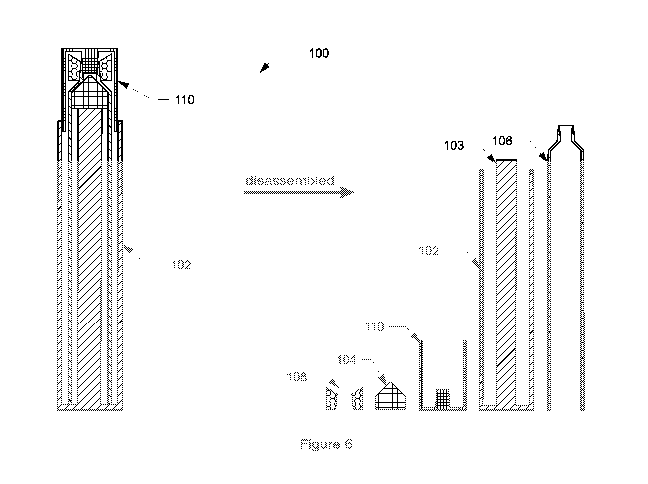
[000144] FIG. 6 shows a fully assembled view and an exploded view of device
100;
[000145] FIGs. 7A - 7B are enlarged views of the tube seal;
[000146] FIGs. 8A-8D are drawings illustrating the principle of fluid
transfer from tube to
syringe due to positive pressure build up in the tube;
[000147] FIGs. 9A-9D are drawings illustrating the principle of fluid
transfer from tube to
syringe due to pressure drop in the syringe;
[000148] FIGs. 10A-10F depict a method for transferring at least one layer
fluid from a
collection tube 602 containing two or more layers of fluid
[000149] FIGs. 11A-10J are drawings illustrating an example method with
tube and regular
syringes;
[000150] FIG. 12 is an enlarged view of the barrel of device 100;
[000151] FIG. 13 is an enlarged view of the barrel seal of device 100;
[000152] FIGs. 14A - 14B are enlarged views of case of device 100;
[000153] FIGs. 15A - 15B are enlarged views of the cap 100;
[000154] FIG. 16 is an exploded view of an example PRP device 100;
[000155] FIGs. 17A - 17B are views of the device 100 fully assembled;
[000156] FIGs. 18A-18M are drawings illustrating an example method (with a
tube and a
barrel) of using device 100;
[000157] FIGs. 19A-19I are drawings illustrating an example method (with a
tube and
barrels) of using device 100; and
[000158] FIGs. 20A-20M are drawings illustrating an example method (syringe
and barrels)
of using device 100.
[000159] Detailed Description
[000160] Described herein is a tube seal and a barrel, which may be used to
facilitate the
removal of fluid layers having different density. The examples disclosed
herein are described
with reference to centrifuging whole blood in order to separate it into its
constituent
components, each of which has a different density. However, one of ordinary
skill in the art
will appreciate that the invention is not limited to the constituent layers of
whole blood. For
12
CA 03182142 2022-11-02
WO 2022/015303 PCT/US2020/042129
instance, the invention can be used in situations/applications when a
particular fraction of fluid
has to be removed and transferred from specimen tube to another syringe. For
example, the
invention may be used in the process of obtaining adipose derived tissue
stromal vascular
fraction (AD-tSVF) from body's fat aspirate, after emulsification and
separation into density
layers by centrifugation.
[000161] THE TUBE SEAL
[000162] The tube seal may be sized to fit commercially available fluid
collection tubes. FIG.
3A shows the tube seal 108 of the present invention inserted into a
conventional fluid
collection tube 602. FIG. 3B shows the tube seal 108 inserted into a
conventional fluid
collection tube 602 with an anticoagulant, FIG. 30 shows the tube seal 108
inserted into a
conventional fluid collection tube 602 with both an anticoagulant and a
separating gel.
[000163] FIG. 4A shows how the tube seal 108 is mounted on a distal end of
a conventional
syringe 180.
[000164] FIG. 4B shows how the syringe 180 with the tube seal 108 is
mounted on a distal
end thereof is inserted into the mouth of a conventional fluid collection tube
602.
[000165] FIGs. 40, 4D show the syringe 180 with the tube seal 108 is
mounted on a distal
end thereof is moved distally within the conventional fluid collection tube
602 until a portion of
the fluid within the fluid collection tube 602 is transferred into the syringe
180.
[000166] In some examples it may be desirable to use a tube seal dispenser
to insert the
tube seal into the fluid collection tube 602 instead of manually placing the
tube seal in the tube
with one's hand, or mounting the tube seal on the distal end of the syringe
180. A method of
using a dispenser to insert the tube seal into the fluid collection tube 602
using a dispenser is
shown in FIGs. 5A-5D.
[000167] FIG. 5A shows how the tube seal 108 is mounted on a distal end of
a dispenser
rod.
[000168] FIG. 5B shows how the dispenser rod with the tube seal 108 mounted
on a distal
end thereof is inserted into the mouth of a conventional fluid collection tube
602.
[000169] FIGs. 50, 5D show the syringe 180 is placed in abutment or
engagement with the
tube seal inside of the fluid collection tube 602, and how the syringe is
moved distally within
the conventional fluid collection tube 602 until a portion of the fluid within
the fluid collection
tube 602 is transferred into the syringe 180.
13
CA 03182142 2022-11-02
WO 2022/015303 PCT/US2020/042129
[000170] The tube seal 108 (FIGs. 7A, 7B) has a proximal end face 108P and
a distal end
face 108D. In some examples, the end face 108P, 108D may have a shape 108-2,
108-3
configured to compliment or mattingly engage the tapered end face of a
conventional syringe.
The tube seal 108 may be formed of resilient, elastomeric material such as
rubber.
[000171] An outer surface of the tube seal 108 may have a shape which
mirrors the shape
of the inner surface of the fluid collection tube thereby ensuring sealing
engagement
therebetween. In some examples, one or more raised sealing rings 108S spanning
the outer
circumference (surface) of the tube seal may be provided. In the example shown
in FIGs. 3A-
30, the fluid collection tube 602 and the tube seal 108 each have a circular
cross-section;
however, these components may have any complimentary shaped cross-section.
[000172] As best seen in FIGs. 7A, 7B, the tube seal 108 has a lumen 108L.
In some
examples, the lumen 108L has a dual taper with a first taper 108-1 extending
from the proximal
end face 108P towards the distal end face 108D and a second taper 108-2
extending from
the distal end face 108D towards the proximal end face 108P. See, FIG. 7B.
Additionally, the
inner wall of the tube seal 108 which bounds or surrounds the lumen, may be
tapered. In the
example depicted in FIG. 7B, the inner wall is tapered such that the lumen
108L is wider at
proximal end 108P than at distal end 108D. The inner wall may have a dual
taper as desired.
The tube seal 108 may be formed of an elastomeric material.
[000173] In some examples, the proximal end of the tube seal 108 is of a
conical or funnel
shape to direct any residual blood through the lumen 108L to the other side of
the tube seal
108.
[000174] The tube seal 108 is sized to sealingly engage the inner surface
of a fluid collection
tube. An outer surface of the tube seal 108 may have a shape which mirrors the
shape of the
inner surface of the fluid collection tube thereby ensuring sealing engagement
therebetween.
[000175] In some examples, the proximal end face 108P of the tube seal 108
may have a
shape which compliments or mattingly engages the tapered end face of the
barrel 106.
[000176] In some examples, the proximal end of the tube seal 108 is of a
conical or funnel
shape to direct any residual blood through the lumen 108L to the other side of
the tube seal
108.
[000177] The tube seal 108 may be provided by itself or as part of a kit or
assembly. The kit
may include a fluid collection tube, cap for fluid collection tube, and tube
seal. In some
examples, the fluid collection tube will be prefilled with an anticoagulant.
In some examples,
the fluid collection tube will be prefilled with an anticoagulant and a
separating gel. In some
examples, the tube seal is preloaded into the fluid collection tube. In some
examples, the kit
14
CA 03182142 2022-11-02
WO 2022/015303 PCT/US2020/042129
may include a dispenser for introducing the tube seal into the tube. The tube
seal may also
be pre-mounted on the tip of a conventional syringe or any syringe-like
device.
[000178] Throughout this disclosure, the term syringe should be understood
to encompass
any syringe or syringe-like device.
[000179] As will be explained below, the tube seal 108 may be used as a
connector and
adapter for transferring fluids between a fluid collection tube and a syringe,
and provides a
fluidic connection between the tube and the syringe. In some examples the tube
seal facilitates
fluid transfer from the tube to the syringe due to a pressure rise in the tube
(caused by
advancing the syringe distally and exerting a pressure against the tube seal)
FIGs. 8A-8D.
FIG. 8A shows a fluid collection tube 602 containing a volume V1 of fluid (at
ambient pressure
A), a tube seal 108 and a syringe 180. FIG. 8B shows the fluid collection tube
602 and the
syringe 180 of FIG. 8A after the syringe 180 has been advanced distally into
the fluid collection
tube 602 thereby increasing the pressure of the volume of fluid V1 (in the
moment before fluid
is transferred into the syringe due to the pressure gradient therebetween).
FIG. 80 shows the
fluid collection tube 602 and syringe 180 of FIG. 8B after a volume V2 has
been transferred
from the fluid collection tube 602 to the syringe 180 due to the pressure
gradient therebetween
(in the moment before the pressure in the fluid collection tube 602 goes back
to ambient. FIG.
8D shows the fluid collection tube 602 and syringe 180 of FIG. 80 after a
volume V2 has been
transferred to the fluid collection tube 602 to the syringe 180 due to the
pressure gradient
therebetween, after the pressure in the fluid collection tube 602 goes back to
ambient.
[000180] In some examples the tube seal facilitates fluid transfer from the
tube to the syringe
due to a pressure drop in the connected syringe (caused by retracting the
plunger proximally
and creating suction in the syringe) FIG. 9A shows a fluid collection tube 602
containing a
volume V1 of fluid (at ambient pressure A), a tube seal 108 and a syringe 180.
FIG. 9B shows
the fluid collection tube 602 and the syringe 180 of FIG. 9A after the plunger
of syringe 180
has been retracted proximally thereby decreasing the pressure within the
syringe 180 below
ambient (in the moment before fluid is transferred into the syringe due to the
pressure gradient
therebetween). FIG. 90 shows the fluid collection tube 602 and syringe 180 of
FIG. 9B after
a volume V2 has been transferred from the fluid collection tube 602 to the
syringe 180 due to
the pressure gradient therebetween (in the moment before the pressure in the
syringe 180
goes back to ambient. FIG. 9D shows the fluid collection tube 602 and syringe
180 of FIG. 90
after a volume V2 has been transferred to the fluid collection tube 602 to the
syringe 180 due
to the pressure gradient therebetween, after the pressure in the syringe 180
goes back to
ambient.
CA 03182142 2022-11-02
WO 2022/015303 PCT/US2020/042129
[000181] GENERIC PROCESS OF FLUID TRANSFER
[000182] Turning now to FIGs. 10A-10F, a method for transferring at least
one layer fluid
from a collection tube 602 containing two or more layers of fluid, where each
layer of fluid has
a different specific gravity will be explained. The generic process used to
transfer fluid from a
fluid collection tube 602 to a syringe 180 utilizing the tube seal 108 will be
explained.
[000183] In FIG. 10A, a fluid collection tube 602 containing a fluid
specimen is centrifuged
to separate the fluid specimen into its constituent components by density:
layer 1, layer 2, and
layer 3. One of ordinary skill in the art will appreciate that the method may
be used with any
number of different density fluid layers.
[000184] In FIG. 10B, cap 602C is removed from the fluid collection tube
602, and tube seal
108 is inserted into the mouth of the fluid collection tube 602. One of
ordinary skill in the art
will appreciate that the tube seal 108 may be inserted into the fluid
collection tube 602 prior
to the centrifuging step illustrated in FIG. 10A. Or the fluid collection tube
602 may be
equipped or supplied with the tube seal already inside the tube prior to
insertion of the fluid.
[000185] In FIGs. 10C, 10D, a syringe 180 (without a needle) is inserted
into the mouth of
the fluid collection tube 602 such that the distal tip of the syringe 180 is
placed in sealing
engagement with the tube seal 108.
[000186] In FIG. 10E, as the syringe 180 is advanced distally into the
fluid collection tube
602, fluid 1 is displaced from the fluid collection tube 602 into the syringe
180. The tube seal
108 seals the fluid collection tube 602 with the syringe, enabling the
transfer of fluid. It should
be appreciated that the plunger of the syringe 180 may be retracted instead of
or in addition
to advancing the syringe 180 distally into the fluid collection tube 602.
[000187] If FIG. 10F, the syringe 180 containing fluid 1 may be removed
from the tube seal
108 upon the transfer or displacement of the desired quantity of fluid 1. One
of ordinary skill
will appreciate that the syringe 180 may be removed from the tube seal 108 and
at any time,
and a new syringe or syringe-like device 180 may be introduced to transfer
desired quantities
of the remaining fluids 1, 2, 3.
[000188] EXAMPLE PRP EXTRACTION USING THE TUBE SEAL WITH ORDINARY
SYRINGES (ONE-SPIN)
[000189] Turning now to FIGs. 11A-11J, a method for transferring fluid from
a fluid collection
tube 602 to a syringe 180 utilizing the tube seal 108 will be explained. The
example process
pertains to the creation of platelet rich plasma but one of ordinary skill in
the art will appreciate
that the tube seal may be used generally to facilitate fluid transfer.
16
CA 03182142 2022-11-02
WO 2022/015303 PCT/US2020/042129
[000190] In FIG. 11A, a fluid collection tube 602 containing a specimen of
whole blood is
centrifuged to separate the whole blood into its constituent components by
density: red blood
cells (RBC), buffy coat, and plasma.
[000191] In FIG. 11B, cap 6020 is removed from the fluid collection tube
602, and tube seal
108 is inserted into the mouth of the fluid collection tube 602. One of
ordinary skill in the art
will appreciate that the tube seal 108 may be inserted into the fluid
collection tube prior to the
centrifuging step illustrated in FIG. 11A. Or the tube may be equipped or
supplied with the
tube seal already inside the tube prior to fluid collection.
[000192] In FIGs. 110, 11D, a syringe 180 (without a needle) is inserted
into the mouth of
the fluid collection tube 602 such that the distal tip of the syringe 180 is
placed in sealing
engagement with the tube seal 108.
[000193] In FIG. 11E, as the syringe 180 is advanced distally into the
fluid collection tube
602, plasma is displaced from the fluid collection tube 602 into the syringe
180 until between
% and % (by volume) of the plasma is transferred into the syringe. The tube
seal 108 seals
the fluid collection tube 602 with the syringe, enabling the transfer of
fluid. It should be
appreciated that the plunger of the syringe 180 may be retracted instead of or
in addition to
advancing the syringe 180 distally into the fluid collection tube 602.
[000194] In FIG. 11F, the syringe 180 with the plasma is discarded, and a
fresh (empty)
syringe 180 is placed into sealing engagement with the tube seal 108.
[000195] In FIGs. 11G, 11H, the syringe 180 and the tube seal 108 are
advanced distally
such that the remaining plasma and the buffy coat (collectively PRP) are
transferred into the
syringe 180. Again, the tube seal 108 seals the fluid collection tube 602 and
the syringe 180,
enabling the transfer of fluid. Also, the plunger of the syringe 180 may be
retracted instead of
or in addition to advancing the syringe 180 distally into the fluid collection
tube 602.
[000196] In FIGs. 111 and 11J, the syringe 180 with the PRP is withdrawn
from the fluid
collection tube 602, and a needle is attached to the syringe.
[000197] The aforementioned process using the tube seal 108 is an
improvement over the
conventional process for creating PRP, because it eliminates the needles,
eliminates
separating gel, and does not solely rely on aspiration.
[000198] Also disclosed is a system and kit for obtaining PRP using the
tube seal 108, as
well as associated methods for separating platelet rich plasma ("PRP") from
whole blood. The
system, kit, and associated methods of the present invention addresses several
shortcomings
of conventional PRP kits in that it reduces the number of components needed,
eliminates the
17
CA 03182142 2022-11-02
WO 2022/015303 PCT/US2020/042129
need for a separating gel, in some examples enables separation of PRP from the
tube after a
single centrifuge spin cycle, eliminates the need for needles thereby reducing
the risk of
accidental needle stick, is simpler to use, and reduces the risk of sample
contamination.
[000199] THE BARREL
[000200] As will be explained below, the barrel 106 is a fluid transfer
receptacle equipped
with a piston-like barrel seal 108. The barrel is sized to fit within the
lumen of a standard fluid
collection tube. The barrel features a tip, whose outer surface is capable of
sealingly engaging
with the tube seal, and an inner surface capable of sealingly engaging with a
male Luer
connector of a syringe. The below mentioned process using the tube seal 108
with the barrel
106, is an improvement over the conventional process for creating PRP, because
while the
tube seal eliminates the needles and separating gel and does not solely rely
on aspiration,
the barrel replaces both the transfer syringe and the second-spin tube.
[000201] The barrel seal 108 may be formed of an elastomeric material which
may be the
same material used to form the tube seal.
[000202] FIG. 12: The barrel 106 is an elongate hollow tube having
sidewalls which surround
a central lumen 106L. A proximal end 106P of the barrel is open and
communicates with the
lumen 106L. A distal portion of the barrel gradually tapers narrower to a kind
of Luer tip 106T.
In some examples the distal end 106D is conical shaped. The tip 106T tappers
narrower. A
width of the sidewall of the barrel 106 is less than gap G, and a diameter of
lumen 106L is
greater than the diameter of the rod 103. The distal end of the rod 103 fits
into the proximal
end of the barrel 106 and the rod 103 can be loosely inserted into the lumen
106L.
[000203] The barrel 106 may have the general appearance of a conventional
syringe but in
some examples differs from a conventional syringe in several key aspects. One
notable
difference is that barrel 106 is not meant to be equipped with a needle. The
outer side of the
tip 106T forms an oversized male to sealingly engage with a tube seal 108, and
cannot
accommodate a needle. The inner side of the tip 106T forms a female Luer
connection
configured to sealingly engage with a male Luer connection of a regular
syringe. Another
notable difference is that the proximal end 106P of the barrel 106 lacks the
flanges or gripping
portions provided on conventional syringes which are used to assist advancing
the plunger.
The barrel 106 is never used to inject anything. Lacking a flange and without
the plunger rod,
the barrel 106 is configured to be securely received within a conventional
centrifuge device.
18
CA 03182142 2022-11-02
WO 2022/015303 PCT/US2020/042129
[000204] The barrel seal 104 (FIGs. 13, 16) is movably provided within the
lumen 106L and
will only move when pushed proximally by fluid entering the barrel 106 through
the Luer tip
106T or when advanced distally by the rod 103.
[000205] As best seen in FIGs. 17A, 17B the rod 103 fits within lumen 106L
while the barrel
106 fits in the gap G between the rod 103 and the barrel 106.
[000206] As best seen in FIGs. 15A, 15B, the barrel cap 110 is an elongate
hollow tube
having a central lumen 110L. In some examples, a proximal end 110P is open and
communicates with the lumen 110L, and distal end 110D is closed. In FIG. 15A,
the barrel
cap 110 includes a male plug 110X attached to an inner surface thereof which
is configured
to sealingly engage the female aspects of the lumen of the tip 106T. In FIG.
15B, the barrel
cap 110 includes a female plug 110X which is configured to sealingly engage
the exterior wall
of the tip 106T.
[000207] FIGs. 17A, 17B show the device 100 fully assembled with the barrel
seal 104 within
the barrel 106, the barrel 106 coaxially mounted over the rod 103 and received
within the
casing 102, the tube seal 108 removably mounted on the tip 106T, and the
barrel cap 110
mounted over the tube seal 108 and a distal portion of the barrel 106. In FIG.
17Athe distal
most part of the casing 102 overlaps the barrel cap 110, whereas in FIG 17B
the distal most
portion of the cap overlaps the distal most part of the casing 102.
[000208] FIG. 6 shows a fully assembled view and an exploded view of device
100.
[000209] It should be noted that the device 100 does not utilize needles to
transfer the
plasma and buffy coat out of the fluid collection tube 602 and eliminates the
need for using a
separating gel.
[000210] FIG. 16 is an exploded view of an example device 100 which
includes a casing
102 (FIGs. 14A, 14B) with its rod 103, a barrel seal 104 (FIG. 13), a barrel
106 (FIG. 12), a
tube seal 108 (FIGs. 7A, 7B) and a barrel cap 110 (FIGs. 15A, 15B).
[000211] FIGs. 14A, 14B: The casing 102 is an elongate hollow tube with a
central lumen
102L. In some examples, proximal end 102P of the casing 102 is closed, and
distal end 102D
is open and communicates with the central lumen 102L. A rod 103 is partially
housed within
the central lumen 102L. In FIG. 14A, a distal end 103D extends beyond the
distal end 102D
of the casing 102. In FIG. 14B, the distal end 102D of the casing extends
beyond the distal
end 103D of the rod 103. In some examples, the rod 103 is attached to the
casing. For
example, a proximal end 103P of the rod 103 may be attached to the proximal
end 102P of
the casing 102. The diameter of the lumen 102L is greater than the diameter of
the rod 103
19
CA 03182142 2022-11-02
WO 2022/015303 PCT/US2020/042129
such that a gap G is formed between an external surface of the rod 103 and an
interior wall
of the casing 102.
[000212] The rod 103 may be hollow or solid. The rod 103 serves to advance
the barrel seal
104 (FIGs. 13, 16) from a proximal end 106P of the barrel 106 towards the
distal end 106D of
the barrel. The barrel seal 104 may be formed of an elastomeric material and
fluidically seals
the inner surface or lumen 106L of the barrel 106. In some examples, the
barrel seal 104
abuts but is not attached to the rod 103. In this example, once the rod 103
has advanced the
seal 104 distally, retracting the rod 103 proximally will not retract the seal
104. However, in
other examples, the seal 104 may be attached to the rod 103.
[000213] The rod 103 may have any shape and need not have a circular cross-
section. The
rod 103 must merely have sufficient structural integrity to advance the barrel
seal 104 within
the lumen 106L.
[000214] EXAMPLE PRP EXTRACTION USING THE TUBE SEAL WITH THE BARREL
(TWO-SPIN)
[000215] FIGs. 18A-18M illustrate steps in a method using device 100. Some
of the steps
are optional, and the order in which the steps are described are not limiting.
[000216] In FIG. 18A, a fluid collection tube 602 containing whole blood
which has been
centrifuged to separate the whole blood into its constituent parts; namely,
red blood cells,
buffy coat and plasma.
[000217] In FIG. 18B, the tube cap 602C is removed from the fluid
collection tube 602, and
the casing 102 with the rod 103 are removed from the fully-assembled device
100.
[000218] In FIG. 18C, the barrel cap 110 is removed, exposing the tube seal
108 and the
distal end of the barrel 106. The distal end 106D of the barrel 106 with the
tube seal 108 are
inserted into the fluid collection tube 602.
[000219] In FIG. 18D (optional), the distal end of the barrel 106 is gently
removed with a
twisting motion leaving the tube seal 108 engaged with the lumen of the fluid
collection tube
602.
[000220] In FIG. 18E (optional), the proximal end of barrel 106 is placed
in abutment with
the tube seal 108 and is used to push or advance the tube seal 108 within the
fluid collection
tube until the tube seal just contacts the plasma.
[000221] In FIG. 18F (optional), the proximal end 106P of the barrel 106 is
withdrawn, the
barrel 106 is flipped, and the distal end 106D is placed in sealing engagement
with the tube
seal 108.
CA 03182142 2022-11-02
WO 2022/015303 PCT/US2020/042129
[000222] In FIG. 18G, as the barrel 106 and tube seal 108 are advanced
distally into the
fluid collection tube 602, plasma will flow proximally (in the opposite
direction) through lumen
108L into the hollow interior 106H of the barrel 106. The barrel seal 104 is
pushed proximally
by the fluid flowing into the barrel 106. The barrel 106 should be advanced
until red blood
cells just start to enter into the barrel. At that point, plasma and the whole
buffy coat have
been transferred to the barrel 106. The conical shape of the distal end of the
tube seal 108
will preferentially move the outer part of the buffy coat to the center of the
tube seal 108 before
red blood cells start to enter the tip 106T.
[000223] In FIG. 18H, the barrel 106 is withdrawn from the fluid collection
tube 602 using a
twisting and pulling motion to disengage the tube seal 108 from the barrel
106. In other words,
as the barrel 106 is withdrawn, the tube seal 108 remains in the fluid
collection tube 602 with
the remaining red blood cells. The barrel cap 110 is engaged with the distal
end 106D of the
barrel 106.
[000224] In FIG. 181, the barrel 106 containing the plasma and buffy coat
(collectively PEP)
is capped with device barrel cap 110 and centrifuged (second spin cycle) to
separate the PEP
into its constituent parts; namely, platelet poor plasma (PPP) on the top and
platelet pallet
(compacted platelets) proximate the barrel seal 104. The centrifuged sample by
volume
comprises approximately 9/10 platelet poor plasma and 1/10 platelet pallet.
One of ordinary
skill in the art will appreciate that when inserting the tubular barrel into
the centrifuge, the tip
of the barrel should be pointing to the center axis of rotation.
[000225] In FIG. 18J, a conventional syringe 180 is attached to the barrel
106. More
particularly, the male aspect of the syringe 180 interfaces with the female
aspect of the barrel
tip 106T. In some examples, the rod 103 is inserted into the proximal end of
the barrel 106.
[000226] In FIG. 18K, the distal end of the rod 103 is advanced distally
within the barrel
lumen 106L toward the tip 106T pushing the barrel seal 104 distally and
expelling any residual
air out of the barrel first (ideally), and then transferring 2/3 ¨ 3/4 of the
platelets poor plasma
(PPP) to the attached syringe 180. One of ordinary skill in the art will
appreciate that instead
of (or in addition to) advancing the rod 103, the plunger of the attached
conventional syringe
180 may be retracted to affect the transfer of air and PPP. The conventional
syringe 180 with
air and the platelet poor plasma is disconnected from the barrel tip 106T.
[000227] In FIG. 18L, an empty conventional syringe 180 is attached to the
Luer tip 106T.
The platelet pallet and remaining plasma are transferred back-and-forth
between the barrel
106 and the syringe 180 by alternatingly pushing the rod 103 and the syringe
plunger of the
conventional syringe 180. This back-and-forth transfer dislodges the platelet
pallet and mixes
21
CA 03182142 2022-11-02
WO 2022/015303 PCT/US2020/042129
it with remaining plasma thereby creating platelet rich plasma (PRP). See,
FIG. 18M. Now the
whole PRP is transferred to the conventional syringe 180 and is ready for use.
[000228] EXAMPLE PRP EXTRACTION USING THE BARREL WITH ORDINARY
SYRINGES (ONE-SPIN)
[000229] FIGs. 20A-20M illustrate an example process for creating PRP. In
this example,
the barrel 106 is the key feature, because it integrates the functions of both
the fluid collection
tube and the tube seal.
[000230] In FIG. 20A the fully assembled device 100 is disassembled by
removing the barrel
cap 110, the tube seal 108, case 102 and rod 103 from the barrel 106, leaving
the barrel seal
104 within the lumen of the tube 106.
[000231] In FIG. 20B a conventional syringe 180 containing whole blood is
attached to the
barrel 106, and the blood is transferred from the syringe 180 into the barrel
106 by advancing
the plunger within the syringe. The barrel seal 104 is pushed toward the
proximal end 106P
of the barrel 106 by the blood entering the tube 106.
[000232] In FIG. 20C the now empty conventional syringe 108 is disengaged
from the barrel
106 (and discarded), and the barrel cap 110 is placed in sealing engagement
with the tip 106T
of the barrel 106.
[000233] In FIG. 20D, the barrel 106 with the blood and the barrel cap 110
is centrifuged,
separating the blood into a layer of red blood cells (RBC), buffy coat, and
plasma.
[000234] In FIG. 20E, the barrel cap 110 is removed from the barrel 106,
and a fresh (empty)
conventional syringe 180 having a plunger movably mounted within is placed in
engagement
with the tip 106T of the barrel 106.
[000235] In FIG. 20F the plasma and buffy coat (collectively "PEP") are
transferred from the
barrel 106 into the syringe 180. This may be accomplished either by (a)
retracting the plunger
within the syringe 180; or (b) by advancing the rod 103 and the barrel seal
104 within the
barrel 106. Or both, as is the case with the tube. The red blood cells are not
transferred from
the barrel 106 into the syringe 180.
[000236] In FIG. 20G, the syringe 180 with the PEP are connected to a fresh
barrel 106.
[000237] In FIG. 20H, the PEP is transferred from the syringe 180 into the
barrel 106, and
barrel cap 110 is placed in sealing engagement with the barrel tip 106T.
[000238] In FIG. 201, the capped barrel 106 containing the PEP is
centrifuged (second spin
cycle) to separate the PEP into its constituent parts; namely, platelet poor
plasma (PPP) on
the top and platelet pallet (compacted platelets) proximate the barrel seal
104. The
22
CA 03182142 2022-11-02
WO 2022/015303 PCT/US2020/042129
centrifuged sample by volume comprises approximately 9/10 platelet poor plasma
and 1/10
platelet pallet.
[000239] In FIG. 20J, a conventional syringe 180 is attached to the barrel
106. More
particularly, the male aspect of the syringe interfaces with the female aspect
of the barrel tip
106T. The rod 103 is inserted into the proximal end of the barrel 106.
[000240] In FIG. 20K, the distal end of the rod 103 is advanced distally
within the barrel
lumen 106L toward the tip 106T pushing the barrel seal 104 distally and
expelling any residual
air and 2/3 ¨ 3/4 of the platelets poor plasma (PPP) to the attached syringe
180. One of
ordinary skill in the art will appreciate that instead of (or in addition to)
advancing the rod 103,
the plunger of the attached conventional syringe 180 may be retracted to
affect the transfer
of air and PPP. The conventional syringe 180 with air and the platelet poor
plasma is
disconnected from the barrel tip 106T. One of ordinary skill in the art will
appreciate that the
residual air could be expelled from the barrel prior to connecting the syringe
180.
[000241] In FIG. 20L, an empty conventional syringe 180 is attached to the
Luer tip 106T.
The platelet pallet and remaining plasma is transferred back-and-forth between
the barrel 106
and the syringe 180 by alternatingly pushing the rod 103 and the syringe
plunger of the
conventional syringe 180. This back-and-forth transfer is also applicable to
the methods with
the tube, that also use the barrel. This back-and-forth transfer dislodges
platelet pallet and
mixes it with remaining plasma thereby creating plasma rich platelets (PRP).
See, FIG. 20M.
Now the whole PRP is transferred to the conventional syringe 180 and is ready
for use.
[000242] EXAMPLE PRP EXTRACTION USING THE TUBE SEAL WITH THE BARREL
(SINGLE-SPIN)
[000243] FIGs. 19A-19I illustrate another example process for creating PRP.
[000244] In FIG. 19A, a fluid collection tube containing a sample of whole
blood is
centrifuged to separate the blood into constituent layers of red blood cells
(RBC), buffy coat,
and plasma.
[000245] In FIG. 19B, the casing 102 and rod 103 are removed from the
barrel 106, and the
cap 602C is removed from the fluid collection tube 602.
[000246] In FIG. 19C, the barrel cap 110 is removed from the barrel 106,
and the distal end
of the barrel 106 with the barrel seal 104 inside the lumen of the barrel and
the tube seal 108
mounted on the tip 106T are inserted into the open mouth of a fluid collection
tube 602.
23
CA 03182142 2022-11-02
WO 2022/015303 PCT/US2020/042129
[000247] In FIG 19D (optional), the barrel is removed from the fluid
collection tube with a
gentle twisting motion to separate the tube seal 108 from the tip 106T of the
barrel 106, leaving
the tube seal 108 engaged with the inner surface of the fluid collection tube
602.
[000248] In FIG. 19E (optional), the barrel 106 is flipped and the proximal
end 106P is
inserted into the fluid collection tube 602 and placed in abutment with the
tube seal 108. The
barrel 106 is used to push the tube seal distally in the fluid collection tube
until it comes in
contact with the plasma. The proximal end of the barrel 106 is withdrawn, and
the distal end
of the barrel 106 is re-inserted into the sealing engagement with the fluid
collection tube 602.
[000249] In FIG. 19F the barrel 106 is used to advance the tube seal 108
within the fluid
collection tube 602. As the barrel 106 is advanced distally into the fluid
collection tube 602,
plasma enters into the barrel and pushes the barrel seal 104 proximally. The
barrel 106 is
advanced until between % and % of the plasma has been transferred into the
barrel 106,
leaving the red blood cells, buffy coat, and 1/4 of the plasma. The barrel
with the plasma is
disengaged from the fluid collection tube 602 and discarded.
[000250] In FIG. 19G, an empty barrel 106 is attached to the fluid
collection tube 602
containing red blood cells, buffy coat, and remaining plasma. The barrel 106
is advanced into
the fluid collection tube 602 until all of the plasma and the buffy coat are
transferred into the
barrel 106, leaving the red blood cells.
[000251] In FIG. 19H a fresh conventional syringe 180 is placed in sealing
engagement with
the tip 106T of the barrel 106 containing the plasma and the buffy coat
(collectively PRP).
[000252] In step 191, the plasma and buffy coat (PRP) are transferred from
the barrel into
the syringe by either (a) retracting the plunger within the syringe 180; or
(b) by advancing the
rod 103 and the barrel seal 104 within the barrel 106. The barrel 106 is
disconnected from the
syringe and discarded, and a needle is attached to the syringe.
[000253] While the exemplary embodiments have been described in some
detail, by way of
example and for clarity of understanding, those of skill in the art will
recognize that a variety
of modification, adaptations, and changes may be employed. The scope of the
present
invention may be limited solely by the appending claims.
24