Note: Descriptions are shown in the official language in which they were submitted.
WO 2021/263254
PCT/US2021/070512
FIXATION OF IMPLANTABLE DEVICE FOR URINARY CONTINENCE
CLAIM OF PRIORITY
100011 This application claims the benefit of priority to
U.S. Patent
Application No. 16/949,991, filed November 23, 2020, which claims the benefit
of priority under 35 U.S.C. 119(e) of U.S. Provisional Patent Application
Serial Number 63/042,947, entitled "METHOD AND APPARATUS FOR
FIXATION OF IMPLANTABLE DEVICE FOR URINARY CONTINENCE",
filed on June 23, 2020, each of which are herein incorporated by reference in
their entirety.
TECHNICAL FIELD
100021 This document relates generally to implantable medical devices and
more particularly to a method and apparatus for limiting migration of a device
for treating urinary incontinence after its implantation in a patient.
BACKGROUND
100031 An example of an implantable device for treating urinary
incontinence includes an adjustable membrane element, such as a balloon,
connected to a rear port with a conduit. The implantable device can be
implanted in a patient with the adjustable membrane element placed adjacent to
the patient's urethra and the rear port placed underneath the patient's skin
by
minimally invasive surgery. The adjustable membrane element can be adjusted
during and after the surgery by injecting fluid into the rear port or
extracting
fluid from the rear port percutaneously using a needle. In an exemplary
treatment, two of such implantable devices are placed in the patient such that
the
two adjustable membrane elements provide pressure and support at the patient's
bladder neck to protect against accidental leaking of urine in cases such as
stress
urinary incontinence (e.g., leaking during sneeze, cough, or physical
activity) or
neurogenic bladder (e.g., leaking caused by spinal injury). The efficacy of
this
treatment depends on accurate placement of the adjustable membrane element at
a target position in the patient, adjustment of the adjustable membrane
element
1
CA 03183882 2022- 12- 21
WO 2021/263254
PCT/US2021/070512
after the placement, and maintaining the position of the adjustable membrane
element over time.
SUM:MARY
100041 An implantable device includes a conduit, an adjustable membrane
element coupled to the conduit near the front end of the conduit for
controllable
coaptation of a body lumen, such as coaptation of a urethra as treatment for
urinary incontinence, and a fixation mechanism at or near the front end of the
conduit. In various embodiments, the fixation mechanism can anchor the
implantable device to the tissue using a movement of a push wire. Optionally,
the fixation mechanism can also allow the implantable device to be released
from the tissue using another movement of the push wire, to allow for re-
positioning or removal of the implantable device.
100051 In one exemplary embodiment, an implantable device is
configured
to be positioned in tissue of a living body using a push wire for coaptation
of a
body lumen of the living body. The implantable device includes an adjustable
membrane element, an elongate conduit, and a fixation mechanism. The
adjustable membrane element includes a continuous wall having an inner surface
defining a chamber. The elongate conduit includes a conduit peripheral
surface,
a conduit rear end, a conduit front end, and a push wire lumen. The conduit
peripheral surface is connected to and sealed to the adjustable membrane
element at or near the conduit front end. The push wire lumen extends
longitudinally in the conduit and has an inlet to receive a portion of the
push
wire and a diameter suitable for accommodating the received portion of the
push
wire. The fixation mechanism is coupled to the conduit front end and
configured
to anchor the implantable device to the tissue using a movement of the push
wire.
100061 In another exemplary embodiment, a method for
coapting a body
lumen in tissue of a living body is provided. The method includes providing an
implantable device and operating a fixation mechanism of the implantable
device using a push wire. The implantable device includes an adjustable
membrane element, an elongate conduit, and the fixation mechanism. The
adjustable membrane element includes a continuous wall having an inner surface
defining a chamber. The elongate conduit includes a conduit peripheral
surface,
2
CA 03183882 2022- 12- 21
WO 2021/263254
PCT/US2021/070512
a conduit rear end, a conduit front end, and a push wire lumen. The conduit
peripheral surface is connected to and sealed to the adjustable membrane
element at or near the conduit front end. The push wire lumen extends
longitudinally in the conduit and has an inlet to receive a portion of the
push
wire and a diameter suitable for accommodating the received portion of the
push
wire. The fixation mechanism is configured to anchor the implantable device to
the tissue.
100071 This summary is an overview of some of the teachings
of the present
application and not intended to be an exclusive or exhaustive treatment of the
present subject matter. Further details about the present subject matter are
found
in the detailed description and appended claims. The scope of the present
invention is defined by the appended claims and their legal equivalents.
BRIEF DESCRIPTION OF THE DRAWINGS
100081 FIG. 1 is a perspective view of an implantable device and a syringe
source for providing a flowable material to an adjustable membrane element of
the implantable device, according to an embodiment of the present subject
matter.
100091 FIG. 2 is a longitudinal cross-sectional view of the
implantable
device shown in FIG. 1, according to an embodiment of the present subject
matter.
100101 FIG. 3 is a cross-sectional view taken along line 3-3
of FIG. 2,
according to an embodiment of the present subject matter.
[0011] FIG. 4 illustrates a guide probe inserted into body
tissue to an implant
location adjacent a body lumen of a patient prior to insertion of the
implantable
device, according to an embodiment of the present subject matter.
100121 FIG. 5 shows the implantable device placed over the
guide probe and
partially advanced to the desired location with the adjustable membrane
element
being deflated, according to an embodiment of the present subject matter.
00131 FIG. 6 shows the implanted device after being expanded at the
desired location in the body tissue of the patient to displace body tissue
toward
the body lumen for causing adjustable restriction of the body lumen, according
to an embodiment of the present subject matter.
3
CA 03183882 2022- 12- 21
WO 2021/263254
PCT/US2021/070512
100141 FIG. 7 is a cross-sectional view taken along line 7-7
of FIG. 6,
according to an embodiment of the present subject matter.
100151 FIG. 8 shows the implantable device after being
inserted with its rear
port underneath the skin of a patient, according to an embodiment of the
present
subject matter.
100161 FIG. 9 is a schematic of another implantable device,
according to an
embodiment of the present subject matter.
100171 FIG. 10 is a schematic of another implantable device,
according to an
embodiment of the present subject matter.
100181 FIG. 11 is a top view showing approximate target sites of placement
of implantable devices to improve coaptation of a urethra, according to an
embodiment of present subject matter.
100191 FIG. 12 is a view along the length of the urethra in
the area of
implantation showing approximate target sites of placement of implantable
devices to improve coaptation of a urethra, according to an embodiment of
present subject matter.
100201 FIG. 13 is an illustration of an implantable device
and a push wire,
according to an embodiment of present subject matter.
100211 FIG. 14 is an illustration of another implantable
device and the push
wire, according to an embodiment of present subject matter.
100221 FIG. 15 is an illustration of another push wire,
according to an
embodiment of present subject matter.
100231 FIG. 16 is an illustration of yet another push wire,
according to an
embodiment of present subject matter.
100241 FIGS. 17A-17B are illustrations of a front end portion of an
implantable device used with a push wire, where the implantable device
includes
a fixation mechanism including a spring, with FIG. 17A showing the spring in
its extended position and FIG. 17B showing the spring in its resting position,
according to an embodiment of present subject matter.
100251 FIGS. 18A-18C are each an illustration of'a wire used for making the
spring of FIGS. 17A-17B, with FIG. 18A showing a wire with semicircular
cross-section, FIG. 18B showing a wire with rectangular cross-section and
dents,
and FIG. 18C showing a wire with semicircular cross-section and bumps,
according to various embodiments of present subject matter.
4
CA 03183882 2022- 12- 21
WO 2021/263254
PCT/US2021/070512
[0026] FIG. 19 is an illustration of the front end portion
of the implantable
device of FIG. 17 used with another push wire, according to an embodiment of
present subject matter.
[0027] FIGS. 20A-20C are illustrations of a front end
portion of an
implantable device used with a push wire, where the implantable device
includes
a fixation mechanism including pincers, with FIG. 20A being a cross-sectional
side view showing the pincers open, FIG. 20B being a cross-sectional side view
showing the pincers closed, and FIG. 20C being an end view showing the
pincers closed, according to an embodiment of present subject matter.
[0028] FIG. 21 is an illustration of a front end portion of an implantable
device used with a push wire, where the implantable device includes a fixation
mechanism including a helix, according to an embodiment of present subject
matter.
[0029] FIG. 22A-22B are illustrations of an example of the
front end portion
of the implantable device used with the push wire of FIG. 21, with FIG. 22A
being a top view with the implantable device being a single-lumen device or a
multi-lumen device and FIG. 22B being a side view with the implantable device
being a multi-lumen device, according to an embodiment of present subject
matter.
[0030] FIG. 23 is an illustration of another example of the front end
portion
of the implantable device used with the push wire of FIG. 21, according to an
embodiment of present subject matter.
[0031] FIG. 24 is an illustration of yet another
illustration of an example of
the front end portion of the implantable device used with the push wire of
FIG.
21, according to an embodiment of present subject matter.
[0032] FIG. 25 is a flow chart illustrating a method for
anchoring an
implantable device to tissue, according to an embodiment of present subject
matter.
DETAILED DESCRIPTION
[0033] The following detailed description of the present
subject matter refers
to subject matter in the accompanying drawings which show, by way of
illustration, specific aspects and embodiments in which the present subject
matter may be practiced. These embodiments are described in sufficient detail
5
CA 03183882 2022- 12- 21
WO 2021/263254
PCT/US2021/070512
to enable those skilled in the art to practice the present subject matter.
References to "an", "one", or "various" embodiments in this disclosure are not
necessarily to the same embodiment, and such references contemplate more than
one embodiment. The following detailed description is demonstrative and not to
be taken in a limiting sense. The scope of the present subject matter is
defined
by the appended claims, along with the full scope of legal equivalents to
which
such claims are entitled.
100341 This document discusses, among other things,
mechanisms for
fixation of an implantable device to surrounding tissue for treating urinary
incontinence. The implantable device can include, for example, an adjustable
membrane element connected to a rear port with a conduit that has a lumen
providing for fluid communication between a chamber of the adjustable
membrane element and an interior cavity of the rear port. Various structural
elements of the implantable device (e.g., the implantable device 110 shown in
FIG. 1) discussed in this document may each be referred to by various terms.
The "adjustable membrane element" (e.g., the adjustable membrane element 112
shown in FIG. 1) can also be referred to as, for example, an adjustable
element,
an expandable element, an expandable membrane element, a forward expandable
membrane element, a balloon, or an adjustable balloon. The "conduit" (e.g.,
the
conduit 114 shown in FIG. 1) can also be referred to as, for example, a
central
conduit element, a device conduit, a connecting conduit, a connecting conduit
tube, or a tubular elongate body. The "rear port" (e.g., the rear port 116
shown
in FIG. 1) can also be referred to as, for example, a rearward port portion or
a
rear port element. The "lumen" (e.g., the first lumen 215 and the second lumen
217 shown in FIG. 2) can also be referred to as, for example, a passageway, an
inner passageway, or an interior passageway.
100351 In an example, the implantable device includes an
adjustable balloon
connected to a port with a conduit. The balloon is placed adjacent the urethra
to
exert non-circumferential compression upon the urethral wall. The
effectiveness
of the therapy depends on proper positioning of the balloon in a patient's
body,
such as in the retropubic space near the urethra-vesical junction above the
urogenital diaphragm in close proximity to the urethral walls. When two
balloons (e.g., of two implantable devices) are used, their preferred
positioning
is usually symmetrical and lateral with respect to the urethra. Medical
imaging
6
CA 03183882 2022- 12- 21
WO 2021/263254
PCT/US2021/070512
techniques such as fluoroscopy or transrectal ultrasonography (TRUS) can be
used to aid the positioning of the balloon(s). Sensors incorporated into the
implantable device(s) and/or one or more surgical tools can also be used to
aid
the positioning of the balloon(s), such as discussed in U.S. Patent
Application
No. 16/450,246, filed on Jun 24, 2019, assigned to UroMedica, Inc., which is
incorporated by reference herein in its entirety.
[0036] During the implantation procedure, the implantable
device(s) is(are)
placed in the patient with the balloon(s) positioned and fixed in place at the
target site(s). The balloon(s) is(are) only slightly inflated, typically up to
1.0 cc,
for a period of 4 to 6 weeks to allow tissue encapsulation in order to
stabilize the
balloon(s) at its(their) target site(s). In particular, without encapsulation
the
implantable device(s) is(are) prone to migrate down the dilation path through
which the implantable device(s) was(were) implanted. Thus, it is very
important
that fixation occur during this implantation procedure. After the
encapsulation,
the patient will go through one or more adjustment procedures during which the
volume of fluid in the balloon(s) is adjusted to obtain and maintain urinary
continence without causing undesirable obstruction.
[0037] The present subject matter provides an implantable
device for
treating urinary incontinence that has a fixation mechanism for preventing a
balloon of the implantable device from unwanted displacement. FIGS. 1-10
illustrate various embodiments of an implantable device into which the
fixation
mechanism can be incorporated and a surgical tool used in the implantation
that
can also be used to activate and deactivate the fixation mechanism. The
various
embodiments of the implantable device and the surgical tool are illustrated in
FIGS. 1-10 and discussed below by way of example, and not by way of
restriction. These examples as well as additional examples of the implantable
device and the surgical tool are discussed in U.S. Patent No. 5,964,806, U.S.
Patent No. 6,045,498, U.S. Patent No. 6,419,624, U.S. Patent No. 6,579,224,
and
U.S. Patent No. 8,926,494, all assigned to UroMedica, Inc., which are
incorporated by reference herein in their entireties. FIGS. 13-24 illustrate
various embodiments of the fixation mechanism incorporated onto the
implantable device.
[0038] According to the present subject matter as shown by
FIG. 1, there is
provided an elongate implantable device 110, which includes an adjustable
7
CA 03183882 2022- 12- 21
WO 2021/263254
PCT/US2021/070512
membrane element 112 shown in its full expanded size, and is attached pressure-
tightly to an elongate conduit 114, which is connected to a rear port 116
communicating with the expandable element 112 through a first lumen 215 (see
FIG. 2). The conduit 114 has a forward end 114A which extends slightly
beyond the expandable element 112. A syringe 120 including a hollow needle
121 and a rear axially-movable plunger 122 is provided for adjustably
injecting a
suitable flowable material into the implantable device 110 through the rear
port
116 to expand the adjustable membrane element 112.
[0039] In various embodiments, implantable medical device
110 can be
positioned during the implantation procedure using a push wire (also referred
to
as a push rod) as a surgical tool. The conduit 114 contains one or two
elongate
lumens or passageways. Examples of implantable medical device 110 (without
fixation mechanism) that are positioned using a push wire are illustrated in
FIGS. 9 and 10.
pool In various other embodiments, implantable medical device 110 can
be positioned during the implantation procedure using a guide probe (also
referred to as a guide wire) as a surgical tool. The implantable procedure is
known as an over-the-wire procedure. As further shown in FIG. 2 and 3, the
conduit 114 contains two elongate lumens or passageways. The first lumen 215
provides an internal passage by which the flowable material is directed from a
cavity 216A in the rear port 116 to expand the adjustable membrane element
112. The conduit 114 is attached integrally to the rear port 116 at its
rearward
end. A second lumen 217 extends from a front opening 117A to a rearward
opening 117B and serves to receive an elongate guide probe (shown in FIG. 4)
and effect delivery of the implantable device 110 to a desired location in the
body tissue of a patient.
[0041] An important feature of the implantable device 110
having the first
lumen 215 includes a first opening 215A located in cavity 216A of the rear
port
116 between an elastic septum 218 and the conduit 114 and is connected to the
first lumen 215, so that a flowable material can be infused therethrough, and
a
second opening 215B serves to direct the working fluid to the adjustable
membrane element 112. During adjustment of the volume of the membrane
fluid provided from a hollow needle 121 of syringe 120, is infused through the
septum 218 and continues through the conduit 114 connected to the adjustable
8
CA 03183882 2022- 12- 21
WO 2021/263254
PCT/US2021/070512
membrane element 112. The rear port 116 preferably has a diameter greater than
conduit 114 to accommodate the cavity 216A and the septum 218, which is
retained securely by a clamp ring 119.
100421 The entire implantable device 110 including the
adjustable membrane
element 112 is formed of a biocompatible material such as silicone or
polyurethane elastomer, and the conduit 114 and the rear port 116 may be
formed as a unitary construction. Optionally, the adjustable membrane element
112, the rear port 116, and the conduit 114 can be molded as one piece. As
shown in FIG. 2, the adjustable membrane element 112 is adhered at 213 to
conduit tube 114 at its forward end by a suitable adhesive material.
100431 The implantable device and assembly according to the
present subject
matter can include three main members. The first member provided is an
elongate guide in the form of a stiff solid elongate guide probe 424 (see FIG.
4)
configured for delivery of the implantable device 110 to the desired site in
the
body tissue of a patient as generally shown by FIGS. 4 and 5. Alternatively,
the
elongate guide member can be in the form of a flexible guidewire which has
been initially delivered into the body tissue through a separate hollow stiff
probe
that has been inserted to the desired location in the body tissue. The second
member of the assembly is the implantable device 110 which includes the
adjustable membrane element 112, the conduit 114 containing the two lumens
215 and 217, and the rear port 116. During its implantation, the implantable
device 110 is guided to a pre-determined location adjacent a body lumen in a
patient's body after the elongate solid guide probe 424 is first surgically
inserted
into the body tissue of the patient to establish an initial pathway. The lumen
forward end opening 117A of the implantable device 110 is then disposed over
the rear end of the guide probe 424 to guide the implantable device 110 and
deliver the adjustable membrane element 112 (in its contracted shape) to the
pre-
determined location in the body tissue adjacent to the lumen which is to be
adjustably restricted. The diameter of the second lumen 217 is made slightly
larger than that of the guide probe 424 to permit the implantable device 110
to
slide easily over the probe member.
100441 During the implantation of the implantable device
110, a physician
can first make a small incision in the skin 430 of the patient near a body
lumen
432 that needs to be restricted, and then by visualization means such as
9
CA 03183882 2022- 12- 21
WO 2021/263254
PCT/US2021/070512
fluoroscopy or ultrasonic imaging, the solid guide probe 424 is directed to
the
desired location, depending upon the anatomy of the patient. Thereafter, the
opening 117A of the second lumen 117 of the conduit 114 with the adjustable
membrane element 112 in its initial deflated or contracted condition, is slid
over
the rear end 424A of the guide probe 424. The forward end 114A of the conduit
114 can be made pointed to ease the passage of the implantable device 110
through the tissue. The guide probe 424 slides through the second lumen 217 of
the conduit 114 and exits at the rearward opening 117B. As illustrated in FIG.
2,
the opening 117B is between the adjustable membrane element 112 and the rear
port 116. However, it may be advantageous to locate the opening 117B close to
the adjustable membrane element 112 or, alternatively, to have the second
lumen
217 extend through the rear port 116.
100451 If desired, a mark 533 can be provided on the guide
probe 424 which
when aligned with a feature on the implantable device 110 such as the rear
port
116 can assure that the implantable device 110 is appropriately placed at the
correct depth in the patient's body tissue 430. It may be necessary to provide
the
conduit 114 in multiple lengths to facilitate placement of the septum 218 near
the patient's skin. Alternatively, an effective length of the conduit 114 can
be
made adjustable by it having a helical shape similar to that of a coiled
spring
100461 After the implantable device 110 has been advanced over guide probe
424 so that the contracted adjustable membrane element 112 is in the desired
position adjacent to the body lumen 432, the body lumen 432 may be restricted
to a desired degree by piercing septum 218 with the needle 121 of syringe 120
and injecting a flowable material through the first lumen 215 into the
adjustable
membrane element 112. The physician can determine the desired degree of
restriction of body lumen 432 by means such as infusing fluid through the body
lumen past the restriction and measuring the back pressure.
100471 As illustrated by FIGS. 1 and 6, the source of
flowable material is
usually a syringe 120 with a hollow needle used to pierce the elastic septum
218.
However, alternate fluid containers with means for making a reversible
connection to the implantable device 110 could be used. The flowable material
may be, for example, a saline solution, a flowable gel, or a slurry of
particles in a
liquid carrier. It may be advantageous to make the flowable material
radiopaque
so that the degree of membrane inflation may be viewed by x-ray.
CA 03183882 2022- 12- 21
WO 2021/263254
PCT/US2021/070512
100481 An alternative method of delivery of the implantable
device 110 can
be to first withdraw the guide probe 424 from the body tissue and then inflate
the
adjustable membrane element 112. A further alternative would be to first place
the implantable device 110 over the solid guide probe 424 outside the body and
then insert them both into the body tissue as a unit. To facilitate this
latter
procedure, it may be desirable that there be some friction between the solid
guide probe 424 and the second lumen 217 in the conduit 114.
100491 After the implantable device 110 has been properly
positioned with
the adjustable membrane element 112 located near the body lumen 432 and the
septum 218 in the rear port 116 located near the skin 430, the device is
injected
with a flowable material from the syringe 120. The expandable member can be
inflated to a certain extent and then deflated to an extent suitable for
encapsulation of the expandable member by body tissue. The guide probe 24 is
then withdrawn from the device, leaving the slightly expanded membrane
element in the body tissue. Then the skin incision 431 is closed over the port
116 by means such as a suture 834 as shown in FIG. 8.
100501 The present subject matter provides the implantable
device 110 with
adjustability of the membrane expansion post-operatively. This adjustability
is
effected because the septum 218 is located remote from the adjustable
membrane element 112 but near and under the patient's skin. The port and
septum are located by, for instance, manual palpation of the skin region and
the
needle of the syringe is inserted through the skin and septum to add or remove
material from the expandable member, thus increasing or decreasing the
restriction of the body lumen.
100511 To assure proper sealing of the septum 218, it is placed in
compression within a cavity 216A by providing a tight metal ring 119 that
surrounds the rear port 116 and is smaller in diameter than the port. When the
needle 121 of the syringe 120 is withdrawn from the septum 218 after expansion
or adjustment of the adjustable membrane element 112, there is positive
sealing
around the perimeter of the septum 218.
[0052] FIGS. 4-8 generally illustrate the "over-the-wire"
method or
procedure for properly implanting the implantable device 110 into the body
tissue of a patient. As shown by FIG. 4, a physician, after locating the body
lumen such as a urethra of the patient, makes a small incision 431 and inserts
the
11
CA 03183882 2022- 12- 21
WO 2021/263254
PCT/US2021/070512
guide probe 424 in the body tissue to a desired location adjacent the body
lumen
432. This procedure is usually carried out under a local anesthetic with
visual
guidance, for instance under fluoroscopy, by the physician. Next, the
physician
takes the implantable device 110 and places it over the guide probe 424
through
the second lumen 217 as shown in FIGS. 1 and 2. The guide probe 424 enters
the rear opening 117B and exits the forward opening 117A. The implantable
device 110, with the conduit 114 being sufficiently flexible, is advanced
along
the guide probe 424 into the body tissue.
[0053] After the desired location within the body tissue has
been reached, a
suitable flowable material is introduced into the implantable device 110 from
a
source such as the syringe 120 having hollow needle 121 inserted through
septum 218 to at least partially expand the adjustable membrane element 112,
as
shown by FIG. 6. Next, the guide probe 424 is removed and the adjustable
membrane element 112 is expanded further to the desired enlarged size for
restriction of the body lumen 432. The syringe 120 is removed from the
implantable device 110, after which the desired size of the adjustable
membrane
element 112 is maintained by the elastic septum 218. Next, the patient's
incision
at 431 is surgically closed over the port 116 and septum 218 by sutures at
834.
[0054] FIGS. 9-24 illustrate examples of an implantable
device that can be
positioned using a push wire and examples of the push wire. The method or
procedure for properly implanting such an implantable device into the body
tissue of a patient is similar to the over-the-wire method or procedure as
illustrated in FIGS. 4-8, except for using a push wire instead of the guide
probe.
After locating the body lumen such as a urethra of the patient, the physician
makes an incision and inserts a sheath in the body tissue to a desired
location
adjacent the body lumen. Next, the physician places the implantable device,
which can be provided pre-assembled with the push wire inserted into its push
wire lumen, in the sheath and pushes the push wire to advance the implantable
device into the body tissue until the desired location within the body tissue
has
been reached. Then, the sheath is removed from the tissue of the patient, and
the
adjustable membrane element of the implantable device is expanded to the
desired size.
[0055] FIG. 9 is an illustration of an implantable device
kit 940, showing a
cross-sectional view, according to one embodiment of the present subject
matter.
12
CA 03183882 2022- 12- 21
WO 2021/263254
PCT/US2021/070512
The implantable device kit 940 includes an implantable device 910 having an
adjustable membrane element 912 and an elongate conduit 914, where the
conduit 914 includes at least a first lumen 915 which extends longitudinally
in
the conduit 914 from a first opening 915A at a rear end (also referred to as a
proximal end) 962 to a second opening 91513, and where the implantable device
910 is shown positioned within a channel 944 of a sheath 946.
100561 The implantable device kit 940 further includes a
rear port 916,
where the rear port 916 is coupled to the rear end 962 of the conduit 914. In
one
embodiment, the rear port 916 is coupled to the rear end 962 of the elongate
body 914 using chemical adhesives, or alternatively, using sonic welding
techniques as are known in the art. In an additional embodiment, the rear port
916 and rear end 962 are formed together in a polymer molding process, such as
liquid injection molding, as are known in the art
100571 The rear port 916 includes a cavity 916A, where the
cavity 916A is in
fluid communication with the first opening 915A of the conduit 914. In one
embodiment, the rear port 916 also includes an elastic septum 918 through
which the cavity 916A is accessed, where the elastic septum 918 is self-
sealing
after repeated pierces, for example, with a needle. In one embodiment, the
elastic septum 918 is retained in the rear port 916 by a clamp ring 919
located
around the rear port 916. In one embodiment, the clamp ring 919 is made of a
biocompatible material, such as, for example, titanium. In one embodiment, the
elastic septum 918 is made of a biocompatible material, such as, for example,
silicone or polyurethane. The rear port 916 has an outer diameter defined by
an
outer surface 954 of the rear port 916. In one embodiment, the rear port 916
has
an outer diameter of 1 to 15 millimeters, with 4.5 millimeters being a
specific
example.
100581 In one embodiment, the outer surface of the rear port
916 and the
adjustable membrane element 912 are of a size (e.g., a diameter) that is
smaller
than an inner size (e.g., a diameter) of the channel 944 to allow the
implantable
device 910 to be moved longitudinally through the channel 944 of the sheath
946. In an alternative embodiment, the rear port 916 is constructed of at
least
one material flexible enough to allow the size of the rear port 916 in its
relaxed
state to be compressed to a size sufficiently small so that the implantable
device
910 can be moved longitudinally through the channel 944 of the sheath 946. In
13
CA 03183882 2022- 12- 21
WO 2021/263254
PCT/US2021/070512
various embodiments, the conduit 914 has a stiffness sufficient to allow force
applied at the rear end of its tubular elongate body to move the implantable
device 910 at least partially through the channel 944 of the sheath 946. In
one
embodiment, the stiffness of the conduit 914 is determined based on the type
of
material used in constructing its tubular elongate body. Alternatively,
support
elements can be added to the tubular elongate body. For example, a metal coil
can be placed longitudinally within the tubular elongate body to increase the
stiffness of the tubular elongate body.
[0059] Once the implantable device 910 is positioned within
a body, the
adjustable membrane element 912 is inflated by releasably connecting a
flowable material source to the rear port 916. In one embodiment, the flowable
material source includes a syringe with a non-coring needle, where the needle
is
inserted through the elastic septum 918. A measured supply of fluid volume can
be introduced into the implantable device 910, and the adjustable membrane
element 912 expands or contracts due to a volume of flowable material
introduced into the cavity 916A of the rear port 916 from the flowable
material
source. The adjustable membrane element 912 is then used to at least partially
and adjustably restrict the body lumen. Fluids suitable for infusing into the
prosthesis include, but are not limited to, normal saline, polymer gels such
as
silicone gels or hydrogels of polyvinylpyrrolidone, polyethylene glycol, or
carboxy methyl cellulose for example, high viscosity liquids such as
hyaluronic
acid, dextran, polyacrylic acid, polyvinyl alcohol, or a radio-opaque fluid
such as
isotonic contrast media for example. Once the adjustable membrane element
912 has been inflated, the needle is withdrawn from the septum of the rear
port
916. In an additional embodiment, a detectable marker 970 is imbedded in the
continuous wall of the adjustable membrane element 912. The detectable
marker 970 allows the adjustable membrane element 912 to be located within the
tissues of a patient using any number of visualization techniques which employ
electromagnetic energy as a means of locating objects within the body. In one
embodiment, the detectable marker 970 is constructed of tantalum and the
visualization techniques used to visualize the adjustable membrane element 912
are x-ray or fluoroscopy as are known in the art.
[0060] In an additional embodiment, a detectable marker is
imbedded in the
implantable device 910. For example, the detectable marker 970 is located at a
14
CA 03183882 2022- 12- 21
WO 2021/263254
PCT/US2021/070512
front end (also referred to as a distal end) 960 (e.g., the tip) of the
conduit 914.
Alternatively, the detectable marker can be located in the continuous wall of
the
adjustable membrane element 912. The detectable marker 970 allows the front
end 960, or the adjustable membrane element 912, to be located within the
tissues of a patient using any number of visualization techniques which employ
electromagnetic energy as a means of locating objects within the body. In one
embodiment, the detectable marker 970 is constructed of tantalum and the
visualization techniques used to visualize the front end 960, or the
adjustable
membrane element 912, are x-ray or fluoroscopy as are known in the art. In an
additional embodiment, the sheath could also have a detectable marker, where
the marker could be incorporated into, or on, the wall of the sheath.
Alternatively, the entire sheath could be constructed to be radio-opaque.
100611 FIG. 10 is an illustration of an additional
embodiment of an
implantable device 1010 according to the present subject matter. The
implantable device 1010 includes an adjustable membrane element 1012 and a
conduit 1014. The conduit 1014 has a front end 1060. In one embodiment, the
peripheral surface of the conduit 1014 is connected to and sealed to the
adjustable membrane element 1012. In one embodiment, the adjustable
membrane element 1012 includes a continuous wall having an inner surface
defining a chamber.
100621 The conduit 1014 includes a first lumen 1015 and a
second lumen
1017. In one embodiment, the first lumen 1015 extends longitudinally in the
conduit 1014 from a first opening 1015A to one or more second openings 1015B
(e.g., two openings as shown in FIG. 10). The second opening(s) 1015B is(are)
in fluid communication with the chamber of adjustable membrane element 1012
for adjustably expanding or contracting the adjustable membrane element 1012
by flowable material introduced through the first opening 1015A. To prevent
leakage of the fluid from the adjustable membrane element 1012, the first
lumen
1015 has a closed end at or near the front end 1060 of the conduit 1014. The
closed end can be formed by sealing the front end of the first lumen 1015, for
example, using silicone adhesive. Alternatively, the first lumen 1015 can be
constructed by manufacturing to end before reaching the front end of the
conduit
1014.
CA 03183882 2022- 12- 21
WO 2021/263254
PCT/US2021/070512
100631 The second lumen 1017 extends longitudinally along
the conduit
1014 from an inlet 1017A to a closed end 1017B at the front end 1060. In one
embodiment, the second lumen 1017 and the inlet 1017A are each of sufficient
diameter to receive a push rod that can be used to advance the implantable
device 1010 in the tissue.
100641 The implantable device 1010 further includes a rear
port 1016, which
is coupled to the rear end of the conduit 1014. In one embodiment, the rear
port
1016 is similar to the rear port 916 and includes a cavity 1016A and an
elastic
septum 1018. The cavity 1016A coupled to and in fluid communication with the
first lumen 1015 at the first opening 1015A. The elastic septum 1018 allows
for
access to the cavity 1016A using a needle for introducing and/or withdrawing
fluid to expand (inflate) and/or contract (deflate) the adjustable membrane
element 1012. The diameter of the elastic septum 1018 can be slightly larger
than the diameter of the cavity 1016A to produce compression to the elastic
septum 1018 for better sealing.
100651 FIG. 11 is a top view of a bladder 1101 and a urethra
1102 showing
approximate target sites of placement of the implantable devices 1110 to
improve coaptation of the urethra, according to an embodiment of the present
subject matter. The implantable devices 1110 can represent any embodiment of
the implantable device as discussed in this document (with the expandable
membrane element or the adjustable membrane element shown in the figure to
illustrate its location), including but not limited to the implantable device
110,
the implantable device 910, the implantable device 1010, or an implantable
device including various combinations of features of the implantable devices
110, 910, and 1010. A Cartesian coordinate system with X-, Y-, and Z-axes is
shown in FIGS. 11-14 (with two of the X-, Y-, and Z-axes seen in each of these
figures) as a reference for exemplary orientations of structures illustrated
in
these figures. The orientation of the Z-axis is along the direction of the
urethra
1002 in the approximate location of implantation. The location is near the
bladder neck and urethral vesi cal anastomosis in the case of radical
prostatectomy or further down the urethra at the apex of the prostate after
trans-
urethral resection of the prostate (TURP).
100661 FIG. 12 is a view along the length of the urethra
1102 in the area of
implantation (or along the Y-axis) showing approximate target sites of
16
CA 03183882 2022- 12- 21
WO 2021/263254
PCT/US2021/070512
placement of the implantable devices 1110 to improve coaptation of a urethra,
according to an embodiment of present subject matter. The present subject
matter can assist in the proper placement of the implantable devices 1110
during
implantation into the patient and/or adjustment of the implantable devices
1110
after the implantation. In particular, the accurate placement of the
implantable
devices 1110 along the Y-axis (sagittal view) is facilitated by the
applications of
the present subject matter.
100671 FIG. 13 is an illustration of an implantable device
kit 1320, including
an implantable device 1310 and a push wire 1324, according to an embodiment
of present subject matter. The implantable device 1310 and the push wire 1324
can be provided as a device kit, which may also include other accessories. The
implantable device 1310 can be used to coapt a lumen in a body, and can
include
an adjustable membrane element 1312, an elongate the conduit 1314, a rear port
1316, and a fixation mechanism 1350. The adjustable membrane element 1312
is configured to coapt the lumen and includes a continuous wall having an
inner
surface defining a chamber. The conduit 1314 has a conduit rear end 1314A, a
conduit front end 1314B coupled to the adjustable membrane element 1312, a
peripheral surface connected to and sealed to the adjustable membrane element
1312 near the conduit front end 1314B, and a push wire lumen 1317 extending
longitudinally in the conduit 1314 from a lumen inlet 1317A near the conduit
rear end 1314A to a lumen front end 1317B at the conduit front end 1314B. The
lumen inlet 1317A has a size allowing a portion of the push wire 1324 to
enter.
The lumen front end 1317B can be a closed front end allowing the push wire
1324 to push the implantable device 1310 or an outlet to allow a portion of
the
push wire 1324 to exit, depending on the type of the fixation mechanism 1350.
The push wire lumen 1317 has a diameter to accommodate at least the portion of
the push wire 1324 that enters through the lumen inlet 1317A. The diameter is
suitable for the push wire 1324 to move longitudinally in the push wire lumen
1317 by pushing a portion of the push wire 1324 that is outside of the conduit
1314 Longitudinal movements of the push wire 1324 can be used to operate the
fixation mechanism 1350, in addition to advance the implantable device 1310 in
the tissue. The diameter can also be suitable for the push wire 1324 to rotate
in
the push wire lumen 1317 by rotating a portion of the push wire 1324 that is
17
CA 03183882 2022- 12- 21
WO 2021/263254
PCT/US2021/070512
outside of the conduit 1314, when rotational movements of the push wire 1324
are used to operate the fixation mechanism 1350.
100681 The rear port 1316 is coupled to the conduit read end
1314A, and
includes a cavity in fluid communication with the chamber of the adjustable
membrane element 1312 though an inflation lumen in the conduit 1314 (not
shown in FIG. 13) to allow for expansion of the adjustable membrane element
1312 by injecting a fluid into the chamber and contraction of the adjustable
membrane element 1312 by withdrawing the fluid from the chamber. In some
embodiments, the rear port 1316 is releasably coupled to the conduit rear end
1314A.
100691 In various embodiments, implantable device 1310 is a
multi-lumen
(e.g., dual lumen) implantable device including the push wire lumen 1317 and
the inflation lumen (not shown in FIG. 13) as separate lumens.
100701 The fixation mechanism 1350 is coupled to the conduit
front end
1314B to limit displacement of the implantable device 1310 in the tissue after
implantation by anchoring the implantable device 1310 to the tissue. In
various
embodiments, the fixation mechanism 1350 can anchor the implantable device
1310 to the tissue by actively trapping a portion of the tissue in the
fixation
mechanism 1350 The fixation mechanism 1350 includes sufficient space to
maintain viability of the trapped portion of the tissue on a permanent basis.
The
fixation mechanism 1350 can also allow the trapped portion of the tissue to be
released for repositioning of the implantable device 1310 in the tissue or
removal
of the implantable device 1310 from the tissue. In various other embodiments,
the fixation mechanism 1350 can anchor the implantable device 1310 to the
tissue by extending an anchoring member into the tissue. The fixation
mechanism 1350 can also allow the anchoring member to be detached from the
tissue for repositioning of the implantable device 1310 in the tissue or
removal
of the implantable device 1310 from the tissue.
100711 The implantable devices 1310 can present a
combination of the
fixation mechanism 1350 with a suitable implantable device selected from those
discussed with reference to FIGS. 1-10, including but not limited to the
implantable device 110, the implantable device 1010, or an implantable device
including various combinations of features of the implantable devices 110,
910,
and 1010.
18
CA 03183882 2022- 12- 21
WO 2021/263254
PCT/US2021/070512
100721 The push wire 1324 has an elongate push wire body
1326 having a
push wire rear end 1326A and a push wire front end 1326B. The push wire front
end 1326B can have any shape suitable for advancing the implantable device
1310 in the tissue as well as operating (e.g., activating and/or deactivating)
the
fixation mechanism 1350. The elongate push wire body 1326 has a diameter
suitable for moving longitudinally in the push wire lumen 1317 of the conduit
1314. The longitudinal movements of the push wire 1324 includes moving the
push wire 1324 along its own longitudinal axis (which is also substantially
parallel to the longitudinal axis of the conduit 1314). The diameter can also
be
suitable for rotating in the push wire lumen 1317 of the conduit 1314. The
rotational movements of the push wire 1324 includes rotating the push wire
1324
about its own longitudinal axis.
100731 In this document, "activation" of a fixation
mechanism refers to the
operation of the fixation mechanism that anchors an implantable device to
tissue,
and "deactivation" of the fixation mechanism refers to the operation that
releases
the implantable device from the tissue. Thus, the fixation mechanism is active
(i.e., in its activated state) when it is in a state intended for anchoring
the
implantable device to the tissue, and is inactive (i.e., in its deactivated
state)
when it is in a state not intended for anchoring the implantable device to the
tissue.
100741 In this document, terms including "substantial",
"substantially",
"approximate", "approximately", or the like can refer to imperfection or
inaccuracy resulting from practical factors including, but not limited to,
accuracy
in manual handling and errors within manufacturing tolerances. For example,
the longitudinal axes of the push wire and the push wire lumen of the conduit
can be "substantially parallel" when the former is partially placed in the
latter
because they are not perfectly parallel due to (1) errors within their
manufacturing tolerances, (2) manually controlled movements of the push wire
in the push wire lumen, and (3) a portion of the push wire is not in the push
wire
lumen, among other things. Such terms ("substantial", "substantially",
"approximate", "approximately", or the like" can also refer to small
deviations
by design. For example, a push wire lumen can be "substantially parallel" to
the
longitudinal axes of the conduit while a small portion of the push wire lumen
next to the inlet (on a lateral side of the conduit) deviates from being
parallel to
19
CA 03183882 2022- 12- 21
WO 2021/263254
PCT/US2021/070512
the longitudinal axes of the conduit by design. In a multi-lumen implantable
device, the push wire lumen can be "substantially parallel" to the
longitudinal
axes of the conduit. While a major portion of this push wire lumen can be off-
center in the conduit to allow space for inflation lumen, the front-end
portion of
the push wire lumen can deviate from being parallel to the longitudinal axes
of
the conduit to end at the center of the front end of the conduit.
100751 FIG. 14 is an illustration of an implantable device
kit 1420, including
an implantable device 1410 and the push wire 1324, according to an
embodiment of present subject matter. The implantable device 1410 and the
push wire 1324 can be provided as a device kit, which may also include other
accessories. The implantable device 1410 can be used to coapt a lumen in a
body, and can include an adjustable membrane element 1412, an elongate the
conduit 1414, a rear port 1416, and a fixation mechanism 1450. The adjustable
membrane element 1412 is configured to coapt the lumen and includes a
continuous wall having an inner surface defining a chamber. The conduit 1414
has a conduit rear end 1414A, a conduit front end 1414B coupled to the
adjustable membrane element 1412, a peripheral surface connected to and sealed
to the adjustable membrane element 1412 near the conduit front end 1414B, and
an inflation lumen 1415 extending longitudinally in the conduit 1414 The
inflation lumen 1415 has a lumen rear opening 1415A at the conduit rear end
1414A, a lumen front opening 1415B in fluid communication with the chamber
of the adjustable membrane element 1412 to allow for expansion of the
adjustable membrane element 1412 by injecting a fluid into the chamber and
contraction of the adjustable membrane element 1412 by withdrawing the fluid
from the chamber, and a lumen front end 1415C to allow the push wire 1324 to
advance the implantable device 1410 in the tissue and/or to operate fixation
mechanism 1450. Lumen front end 1415C is a closed end that does not allow
the fluid to leak out of the lumen 1415.
100761 The rear port 1416 is coupled to the conduit rear end
1414A, and
includes a cavity 1419 in fluid communication with the chamber of the
adjustable membrane element 1412 though the inflation lumen 1415 to allow for
expansion of the adjustable membrane element 1412 by injecting a fluid into
the
chamber and contraction of the adjustable membrane element 1412 by
withdrawing the fluid from the chamber. The cavity 1419 is sealed by a septum
CA 03183882 2022- 12- 21
WO 2021/263254
PCT/US2021/070512
1418 that is elastic and self-sealing after being pierced through, for example
by a
hollow needle coupled to a syringe for injecting and withdrawing the fluid. In
some embodiments, the rear port 1416 is releasably coupled to the conduit rear
end 1414A.
100771 In various embodiments, the implantable device 1410 is a single-
lumen implantable device with the inflation lumen 1415 also functioning as a
push wire lumen. The inflation lumen 1415 meets the requirements for the push
wire lumen 1317 as discussed above, with the push wire lumen inlet being the
inflation lumen rear end 1415A. The push wire 1324 enters inflation lumen
1415 by piercing through the septum 1418.
100781 The fixation mechanism 1450 is coupled to the conduit
front end
1414B to limit displacement of the implantable device 1410 in the tissue after
implantation by anchoring the implantable device 1410 to the tissue. In
various
embodiments, the fixation mechanism 1450 can anchor the implantable device
1410 to the tissue by actively trapping a portion of the tissue in the
fixation
device 1450. The fixation mechanism 1450 includes sufficient space to maintain
viability of the trapped portion of the tissue on a permanent basis. The
fixation
mechanism 1450 can also allow the trapped portion of the tissue to be released
for repositioning of the implantable device 1410 in the tissue or removal of
the
implantable device 1410 from the tissue. In various other embodiments, the
fixation mechanism 1450 can anchor the implantable device 1410 to the tissue
by extending an anchoring member into the tissue. The fixation mechanism
1450 can also allow for retraction of the anchoring member from the tissue for
repositioning of the implantable device 1410 in the tissue or removal of the
implantable device 1410 from the tissue. In various embodiments, the fixation
mechanism 1450 functions with the lumen 1415 that is leak-proof at the lumen
front end 1415C (while the fixation mechanism 1350 may or may not require the
push wire 1324 to exit from the lumen frond end 1317B to operate).
100791 The implantable devices 1410 can present a
combination of the
fixation mechanism 1450 with a suitable implantable device selected from those
discussed with reference to FIGS. 1-10, including but not limited to the
implantable device 910 or an implantable device including various combinations
of features of the implantable devices 110, 910, and 1010.
21
CA 03183882 2022- 12- 21
WO 2021/263254
PCT/US2021/070512
100801 FIG. 15 is an illustration of another push wire 1524,
according to an
embodiment of present subject matter. The implantable device kit 1320 or 1420
can include the push wire 1524, in addition to or in place of push wire 1324.
The push wire 1524 is a hollow-core push wire that includes an elongate push
wire body 1526 and a core lumen 1552 extending longitudinally in the push wire
body 1526. The push wire body 1526 having a push wire rear end 1526A and a
push wire front end 1526B. The core lumen 1552 has a rear opening 1552A at
the push wire rear end 1526A and a front opening 155213 at the push wire front
end 1526B. In addition to the functions of the push wire 1324, the push wire
1524 allows for a fluid to be injected into, and withdrawn from, an area in or
about the fixation device 1350 or 1450.
100811 FIG. 16 is an illustration of yet another push wire
1624, according to
an embodiment of present subject matter. The implantable device kit 1320 or
1420 can include the push wire 1624, in addition to or in place of push wires
1324 and/or 1524. The push wire 1624 is another hollow-core push wire that
includes an elongate push wire body 1626 and a core lumen 1652 extending
longitudinally in the push wire body 1626. The push wire body 1626 having a
push wire rear end 1626A and a push wire front end 1626B. The core lumen
1652 has a rear opening 1652A at the push wire rear end 1526A and multiple
front openings 1652B at and/or near the push wire front end 1626B. In addition
to the functions of the push wire 1324, the push wire 1624 allows for a fluid
to
be injected into, and withdrawn from, an area in or about the fixation device
1350 or 1450.
FIXATION MECHANISM EXAMPLES
00821 Various examples for the fixation mechanisms 1350 and
1450 are
discussed below. Each example may be suitable for use as one or both of the
fixation mechanisms 1350 and 1450, as those skilled in the art will understand
upon reading this document. For example, some examples may require a push
wire lumen with an open lumen front end, thereby being suitable for use as
part
of implantable device 1310, while some other examples may allow for use with a
push wire or inflation lumen with a closed (leak-proof) front lumen end,
thereby
being suitable for use as part of implantable device 1410. These examples are
22
CA 03183882 2022- 12- 21
WO 2021/263254
PCT/US2021/070512
provided to illustrate, rather than restrict, various fixation mechanisms
according
to the present subject matter.
I. To Anchor/Release via Push Wire
[00831 In various embodiments, a fixation mechanism (e.g., fixation
mechanism 1350 or 1450) can anchor an implantable device (e.g., implantable
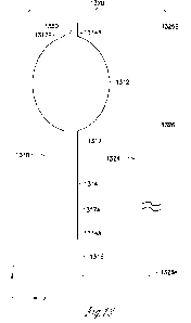
device 1310 or 1410) to the tissue by receiving an energy transmitted using a
push wire (e.g., the push wire 1324, 1524, or 1624). In various further
embodiments, the fixation mechanism can also release the implantable device
from the tissue by receiving another energy transmitted using the push wire.
1.1. Using Longitudinal Movements of Push Wire
100841 In various embodiments, the fixation mechanism can
anchor the
implantable device to the tissue (i.e., be activated) by engaging a portion of
the
tissue using a longitudinal movement of the push wire in a forward direction
and
trapping the engaged portion of the tissue using a longitudinal movement of
the
push wire in a reverse direction. The longitudinal movement of the push wire
in
the forward direction results from pushing the push wire, i.e., applying a
force
toward the conduit front end. The longitudinal movement of the push wire in
the
reverse direction can result from pulling the push wire and/or stopping the
pushing, depending the type of the fixation mechanism. In various further
embodiments, the fixation mechanism can also release the implantable device
from the tissue (i.e., be deactivated) by expelling the trapped portion of the
tissue
using additional longitudinal movements of the push wire without re-trapping
another portion of the tissue. In some embodiments, the fixation mechanism can
be kept in a deactivated state after the implantable device is released from
the
tissue to prevent it from unintentionally engaging and trapping another
portion
of the tissue while the implantable device is still in the patient. In various
embodiments, the implantable device can be released from the tissue by being
pulled away from the trapped portion of the tissue without using the push wire
to
operate the fixation mechanism (i.e., without deactivating the fixation
mechanism), a method referred to as a "pull-out release". The implantable
device can be configured to allow for the pull-out release without causing
unacceptable tissue and/or device damage. For example, the amount of pulling
23
CA 03183882 2022- 12- 21
WO 2021/263254
PCT/US2021/070512
force required for pulling the implantable device from the trapped portion of
the
tissue while the fixation mechanism remains activated (referred to as the
"pull-
out force") is to be small enough to prevent the implantable device from being
broken inside the patient.
100851 In various embodiments, the amount of the pull-out force can be
experimentally determined and used as a constraint in the design of the
fixation
mechanism. The amount of the pull-out force is to be larger than the force
required for preventing migration of the implantable device in the tissue but
smaller than the pulling force that would cause the unacceptable tissue and/or
device damages. For example, an excessive pull-out force can break the conduit
of the implantable device, leaving a front portion of the device in the
patient that
may require surgical removal. Experiments with a prototype implantable device
showed a pulling force of about 5 pounds could break the conduit, while a
pulling force of about 1 pound is required to prevent the implantable device
from
an unacceptable degree of migration in the tissue. Thus, the fixation
mechanism
is to be designed to provide a pull-out force within the range of 2 to 4
pounds to
allow for the pull-out release.
[0086] FIGS. 17A-17B are illustrations of a front end
portion of an
implantable device 1710 used with the push wire 1324, according to an
embodiment of present subject matter. The implantable device 1710 can
represent an example of implantable device 1310 includes a fixation mechanism
1750, which can represent an example of the fixation mechanism 1350. The
front end portion of the implantable device 1710 as shown in FIG. 17 includes
a
front portion of an elongate conduit 1714 having a conduit front end 1714B. A
push wire lumen 1717 extends longitudinally in the conduit 1714 and has a
lumen front end 1717B.
[0087] The fixation mechanism 1750 includes a coil spring
1752 and a
device tip 1753. FIG. 17A shows the spring 1752 in its extended position. FIG.
17B shows the spring 1752 in its resting position (i.e., natural length). The
spring 1752 has a spring rear end 1752A coupled to the conduit front end 1714B
and a spring front end 1752B. The device tip 1753 is coupled to the spring
front
end 1752B to receive the push wire front end 1326B to be pushed by the push
wire 1324 in the forward direction for extending the spring 1752. The spring
1752 can anchor the implantable device 1710 to the tissue by being extended
(as
24
CA 03183882 2022- 12- 21
WO 2021/263254
PCT/US2021/070512
illustrated in FIG. 17A) to engage a portion of the tissue using the
longitudinal
movement of the push wire 1324 in the forward direction and returning to its
resting position (as illustrated in FIG. 17B) to trap the engaged portion of
the
tissue using the longitudinal movement of the push wire 1324 in the reverse
direction. Repetitive longitudinal movements of the push wire 1324 in the
forward and reverse directions may be needed sometimes to reach a desirable
stability of the anchoring. The spring 1752 can also release the implantable
device 1710 from the tissue by expelling the trapped portion of the tissue
using
additional longitudinal movements of the push wire 1324 without re-trapping
another portion of the tissue. To prevent the spring 1752 from engaging and
trapping another portion of the tissue after the release, the implantable
device
1710 can be moved away from the trapped portion of the tissue while keeping
the spring 1752 in its extended position. In various embodiments, the
implantable device 1710 can be released from the tissue by being pulled away
from the trapped portion of the tissue without using the push wire 1324 (to
extend the spring 1752)(the pull-out release). The implantable device 1710 can
be configured to allow for the pull-out release without causing unacceptable
tissue and/or device damage.
100881 FIGS. 18A-18C are each an illustration of a wire used
for making the
spring 1752, according to various embodiments of present subject matter. In
various embodiments, the spring 1752 is made of metal and includes multiple
coils with space between the coils in the resting state to maintain viability
of the
portion of the tissue trapped in the spring 1752. In various embodiments, a
metal wire is formed into the spring 1752. Examples of the metal wire include
a
wire 1852A as illustrated in FIG. 18A, a wire 1852B as illustrated in FIG.
18B,
and a wire 1852C as illustrated in FIG. 18C. The metal wire 1852A has a
semicircular cross-section. The flat side of a wire having a semicircular
cross-
section provides for better grip on tissue when compared to a wire having a
circular cross-section. The metal wire I852B has generally a rectangular cross-
section with dents 1855 and/or 1856 on the wire to provide for the space
between the coils. The rectangular cross-section of the wire prevents nesting
of
the coils when the spring is in its resting or compressed position. The metal
wire
1852C has generally a semicircular cross-section with bumps 1857 on the wire
to provide the space between the coils. Other examples of the metal wire
CA 03183882 2022- 12- 21
WO 2021/263254
PCT/US2021/070512
include a metal wire having a circular cross-section and a braided wire. In
various embodiments, the metal wire can have any cross-section and/or features
that facilitate engaging a portion of the tissue in the extended position of
the
spring and/or trapping of the engaged portion of the tissue in the resting
position
of the spring, as well as maintaining viability of the trapped tissue in the
resting
position of the spring throughout the use of the implantable device.
100891 In various other embodiments, the spring 1752 may
also be a lattice-
cut tube (e.g., a structure similar to an intravascular stent) or a high
durometer
spiral-cut silicone tube. The spring 1752 can be any structure that is
biocompatible and can engage and trap the portion of the tissue using one or
more cycles of extension and returning to resting position.
100901 FIG. 19 is an illustration of a front end portion of
the implantable
device 1710 used with the push wire 1624, according to an embodiment of
present subject matter, according to an embodiment of present subject matter.
FIG. 19 differs from FIG. 17 in that the push wire 1624 replaces the push wire
1324 in FIG. 17. The push wire 1624 is used to facilitate the engaging of the
portion of the tissue into the spring 1752 during the anchoring of the
implantable
device 1710 to the tissue, and/or to facilitate the expelling of the portion
of the
tissue from the spring during the release of the implantable device 1710 from
the
tissue, when the spring is extended using the push wire 1624. During the
anchoring, a syringe 1958 can be used to draw fluid (e.g., air) through the
front
openings 1652B of the core lumen 1652 of the push wire 1624. This lowers the
pressure in the lumen 1652, which draws additional tissue into the space
between the coils of the spring 1752. During the release, the syringe 1958 can
be used to inject fluid (e.g., saline) through the front openings 1652B of the
core
lumen 1652 of the push wire 1624. This expels the portion of the tissue in the
space between the coils of the spring 1752 from the spring 1752.
100911 FIGS. 20A-20C are illustrations of a front end
portion of an
implantable device 2010 used with a push wire 2024, according to an
embodiment of present subject matter. The implantable device 2010 can
represent an example of implantable device 1310 including a fixation
mechanism 2050, which can represent another example of the fixation
mechanism 1350. The front end portion of the implantable device 2010 as
shown in FIG. 20 includes a front portion of an elongate conduit 2014 having a
26
CA 03183882 2022- 12- 21
WO 2021/263254
PCT/US2021/070512
conduit front end 2014B. A push wire lumen 2017 extends longitudinally in the
conduit 2014 and has a lumen front end 2017B. The push wire 2024 can
represent an example of the push wire 1324 having an elongate push wire body
2026 and a push wire front end 2026B that is configured to operate the
fixation
mechanism 2050.
100921 The fixation mechanism 2050 includes pincers 2064
coupled to the
conduit front end 2014B. FIG. 20A is a cross-sectional side view showing the
pincers 2064 open. FIG. 20B is a cross-sectional side view showing the pincers
2064 closed. FIG. 20C is an end view showing the pincers 2064 closed. The
pincers 2064 can anchor the implantable device 2010 to the tissue by being
opened (as illustrated in FIG. 20A) to engage a portion of the tissue using
longitudinal movement of the push wire 2024 in the forward direction and being
closed (as illustrated in FIG. 20B) to trap the engaged portion of the tissue
using
longitudinal movement of the push wire 2024 in the reverse direction. The
pincers 2064 can also release the implantable device 2010 from the tissue by
expelling the trapped portion of the tissue using additional longitudinal
movements of the push wire 2024 without re-trapping another portion of the
tissue. To prevent the pincers 2064 from engaging and trapping another portion
of the tissue after the release, the implantable device 2010 can be moved away
from the trapped portion of the tissue while the pincers 2064 are open. In
various embodiments, the implantable device 2010 can be released from the
tissue by being pulled away from the trapped portion of the tissue without
using
the push wire 2024 (the pull-out release). The implantable device 2010 can be
configured to allow for the pull-out release without causing unacceptable
tissue
and/or device damage.
00931 The push wire front end 2026B is shaped to be
suitable for opening
the pincers 2064. In various embodiments, the pincers 2064 can include a
radiopaque material, such as tantalum or Nitinol (alloy of nickel and
titanium), to
function as a marker on X-ray.
1.2. Using Rotational Movements of Push Wire
100941 In various embodiments, the fixation mechanism can
anchor the
implantable device to the tissue (i.e., be activated) by rotating the push
wire in a
Lightening rotational direction. In various further embodiments, the fixation
27
CA 03183882 2022- 12- 21
WO 2021/263254
PCT/US2021/070512
mechanism can also release the implantable device from the tissue (i.e., be
deactivated) by rotating the push wire in a loosening rotational direction. In
one
embodiment, the tightening rotational direction is a clockwise direction, and
the
loosening rotational direction is a counterclockwise direction. In another
embodiment, the tightening rotational direction is a counterclockwise
direction,
and the loosening rotational direction is a clockwise direction. In various
embodiments, the implantable device can be released from the tissue by being
pulled away from the trapped portion of the tissue without using the push wire
to
operate the fixation mechanism (i.e., the pull-out release, without
deactivating
the fixation mechanism). The implantable device can be configured to allow for
the pull-out release without causing unacceptable tissue and/or device damage.
For example, the amount of force required for pulling the implantable device
from the trapped portion of the tissue while the fixation mechanism remains
activated is to be small enough to prevent the implantable device from being
broken inside the patient.
100951 FIG. 21 is an illustration of a front end portion of
an implantable
device 2110 used with a push wire 2124, according to an embodiment of present
subject matter. The implantable device 2110 can represent an example of the
implantable device 1310 or 1410 and includes a fixation mechanism 2150, which
can represent another example of the fixation mechanism 1350 or 1450. The
front end portion of the implantable device 2110 as shown in FIG. 21 includes
a
front portion of an elongate conduit 2114 having a conduit front end 2114B. A
lumen 2117, which can represent an example of the push wire lumen 1317 or the
inflation lumen 1415, extends longitudinally in the conduit 2114 and has a
lumen front end 2117B. The push wire 2124 can represent an example of the
push wire 1324 having an elongate push wire body 2126 and a push wire front
end 2126B that is configured to operate the fixation mechanism 2150.
100961 The fixation mechanism 2150 is a helix assembly that
can include a
base 2167 and a helix 2166 coupled to the base 2167. The base 2167 is coupled
to the lumen 2117 at the conduit front end 2114B and configured to engage the
push wire 2124 at the push wire front end 2126B. The implantable device 2110
can be anchored to the tissue by rotating the push wire 2124 (engaged to the
base
2167) in the tightening rotational direction such that the helix 2166 enters
the
tissue. After being anchored, the implantable device 2110 can be released from
28
CA 03183882 2022- 12- 21
WO 2021/263254
PCT/US2021/070512
the tissue by rotating the push wire 2124 in the loosening rotational
direction. In
some embodiments, the helix 2166 is configured to allow the implantable device
2110 to be released from the tissue by simply pulling the implantable device
2110 from the tissue (the pull-out release). For example, the helix 2166 can
include a number of turns and/or be sized to limit the anticipated damage to
the
tissue and/or the implantable device 2110 caused by the pull-out release to an
acceptable extent. In various embodiments, the helix 2166 include a number of
turns determined to allow for control of the amount of pull-out force by
controlling the extent of the tightening rotational movement. The amount of
the
pull-out force depends on the type of tissue to which the implantable device
is to
be anchored on (e.g., fat, muscle, or scar tissue) and hence, can differ from
patient to patient. During the implantation procedure, the physician
performing
the procedure can decide the number of turns of the helix 2166 that actually
enters the tissue, for example by making the tightening rotational movement
incrementally while pulling the implantable device to feel for the pull-out
force.
100971 In various embodiments, the base 2167 can be coupled
to the lumen
2117 in a fluid-tight manner to allow the implantable device 2110 to be a
single-
lumen implantable device in which the lumen 2117 functions as both the push
wire lumen and the inflation lumen. In various other embodiments, the base
2167 can be coupled to the push wire lumen of a multi-lumen implantable device
that includes a separate inflation lumen.
100981 Examples of the implantable device 2110 with the
fixation device
2150 are discussed below with reference to FIGS. 22-24. In various
embodiments, considerations in choosing material(s) for constructing the
fixation device 2150 (including its various examples discussed below with
reference to FIGS. 22-24) can include, for example, biocompatibility for long-
term implantation, magnetic resonance imaging (MRI) safety, radiopacity, and
resistance to galvanic corrosion when more than one material is used. Examples
of suitable materials include titanium, nitinol (nickel-titanium), tantalum,
and
platinum-iridium
1.2.1. Helix with Base Rotating in Threaded Sleeve
100991 FIG. 22A is an illustration of a front end portion of
an implantable
device 2210 used with the push wire 2124, according to an embodiment of
29
CA 03183882 2022- 12- 21
WO 2021/263254
PCT/US2021/070512
present subject matter. The implantable device 2210 can represent an example
of implantable device 2110 and includes a fixation mechanism 2250, which is a
helix assembly and can represent an example of the fixation mechanism 2150.
The front end portion of the implantable device 2210 as shown in FIG. 22A
includes a front portion of an elongate conduit 2214 having a conduit front
end
2214B. A lumen 2217, which can function as a push wire lumen (e.g., the push
wire lumen 1317) or function as both the push wire lumen and the inflation
lumen (e.g., the inflation lumen 1415), extends longitudinally in the conduit
2214 and has a lumen front end 2217B. The push wire 2124 can represent an
example of the push wire 1324 having the elongate push wire body 2126 and the
push wire front end 2126B that is configured to operate the fixation mechanism
2250.
[00100] The conduit 2214 includes a threaded sleeve 2270 affixed onto the
surface of the lumen 2217 at the conduit front end 2214B. The fixation
mechanism 2250 includes a threaded base 2267 and a helix 2266 coupled to the
threaded base 2267. The threaded base 2267 and the threaded sleeve 2270 are
configured to mate each other to allow the threaded based 2267 to move
longitudinally by rotating within the threaded sleeve 2270. The threaded base
2267 is configured to engage the push wire 2124 at the push wire front end
2126B for the helix 2266 to exit from the lumen 2217 and enter the tissue by
rotating the push wire 2124 in the tightening rotational direction and for the
helix 2266 to be released from the tissue and retract into the lumen 2217 by
rotating the push wire 2124 in the loosening rotational direction. The helix
2266
can also be configured to allow for its release from the tissue by simply
pulling
the implantable device 2210 (the pull-out release) from the tissue without
causing unacceptable damage to the tissue and/or the implantable device 2210.
The threaded sleeve 2270 can be sized and positioned in the conduit 2214 to
allow the helix 2266 to be entirely inside the conduit 2214. When desirable,
this
allows the helix 2266 to be kept from the outer surface of the implantable
device
2210 during its insertion into the patient.
[00101] In various embodiments, the threaded base 2267 and the threaded
sleeve 2270 can be configured in a fluid-tight manner to allow the implantable
device 2210 to be a single-lumen implantable device in which the lumen 2217
functions as both the push wire lumen and the inflation lumen. In various
other
CA 03183882 2022- 12- 21
WO 2021/263254
PCT/US2021/070512
embodiments, the threaded sleeve 2270 can be affixed to the push wire lumen of
a multi-lumen implantable device in which the lumen 2217 is the push wire
lumen (which is separate from the inflation lumen).
[00102] FIG. 22B is another illustration of the front end portion of an
implantable device 2210 used with the push wire 2124, according to an
embodiment of present subject matter. FIG. 22A can be seen as a top view
illustration of the front end portion of the implantable device 2210 with the
implantable device 2210 being either the single-lumen implantable device or
the
multi-lumen implantable device. FIG. 22B can be seen as a side-view
illustration of the front end portion of the implantable device 2210 that
shows an
inflation lumen 2215 having a lumen front opening 2215B in fluid
communication with the chamber of the adjustable membrane element (not
shown) of the implantable device 2210. The inflation lumen 2115 is fluid-tight
by either ending within the conduit 2214 by manufacturing or plugged at the
conduit front end 2214B.
1.2.2. Helix with Base Rotating in Bushing
[00103] FIG. 23 is an illustration of a front end portion of an implantable
device 2310 used with the push wire 2124, according to an embodiment of
present subject matter. The implantable device 2310 can represent another
example of implantable device 2110 and includes a fixation mechanism 2350,
which is a helix assembly and can represent another example of the fixation
mechanism 2150. The front end portion of the implantable device 2310 as
shown in FIG. 23 includes a front portion of an elongate conduit 2314 having a
conduit front end 2314B. A lumen 2317, which can function as a push wire
lumen (e.g., the push wire lumen 1317) or function as both the push wire lumen
and the inflation lumen (e.g., the inflation lumen 1415), extends
longitudinally in
the conduit 2314 and has a lumen front end 2317B. The push wire 2124 can
represent an example of the push wire 1324 having the elongate push wire body
2126 and the push wire front end 2126B that is configured to operate the
fixation
mechanism 2350.
[00104] The conduit 2314 includes a bushing 2371 affixed onto the surface of
the lumen 2317 at the conduit front end 2314B. The fixation mechanism 2350
includes a base 2367 and a helix 2366 coupled to the base 2367. The base 2367
31
CA 03183882 2022- 12- 21
WO 2021/263254
PCT/US2021/070512
is configured to allow the base 2367 to rotate within the bushing 2371 without
longitudinal movement of the fixation mechanism 2350 relative to the conduit
2314. The base 2367 is configured to engage the push wire 2124 at the push
wire front end 2126B for the helix 2366 to enter the tissue by rotating the
push
wire 2124 in the tightening rotational direction and for the helix 2366 to be
released from the tissue by rotating the push wire 2124 in the loosening
rotational direction. The helix 2366 can also be configured to allow for its
release from the tissue by simply pulling the implantable device 2310 from the
tissue (the pull-out release) without causing unacceptable damage to the
tissue
and/or the implantable device 2310. The fixation mechanism 2350 can be
configured for the helix 2366 to be positioned completely or substantially
outside of the conduit 2314. The diameter of the helix 2366 is not limited by
the
diameter of the lumen 2317. In the illustrated embodiment, the diameter of the
helix 2366 is larger than the diameter of the lumen 2317, and can be
substantially identical to the diameter of the conduit 2314 when the lumen
front
end 2317B is centered at the front end of conduit 2314. In various
embodiments,
the diameter of the helix 2366 can be larger than, substantially equal to, or
smaller than the diameter of the lumen 2317, and can be determined based on an
amount of force for holding the implantable device 2310 in place after the
implantation.
[00105] In various embodiments, the base 2367 and the bushing 2371 can be
configured in a fluid-tight manner to allow the implantable device 2310 to be
a
single-lumen implantable device in which the lumen 2317 functions as both the
push wire lumen and the inflation lumen. In various other embodiments, the
bushing 2371 can be affixed to the push wire lumen of a multi-lumen
implantable device in which the lumen 2317 functions as the push wire lumen in
addition to the inflation lumen. The relative positions of the push wire lumen
and the inflation lumen in these multi-lumen embodiments of the implantable
device 2310 can be the same as or similar to what is illustrated in FIG. 22B
for
the implantable device 2210.
1.2.3. Helix with Base Fixed to Implantable Device
[00106] FIG. 24 is an illustration of a front end portion of an implantable
device 2410 used with the push wire 2124, according to an embodiment of
32
CA 03183882 2022- 12- 21
WO 2021/263254
PCT/US2021/070512
present subject matter. The implantable device 2410 can represent another
example of implantable device 2110 and includes a fixation mechanism 2450,
which is a helix assembly and can represent another example of the fixation
mechanism 2150. The front end portion of the implantable device 2410 as
shown in FIG. 24 includes a front portion of an elongate conduit 2414 having a
conduit front end 2414B. A lumen 2417, which can function as a push wire
lumen (e.g., the push wire lumen 1317) or function as both the push wire lumen
and the inflation lumen (e.g., the inflation lumen 1415), extends
longitudinally in
the conduit 2414 and has a lumen front end 2417B. The push wire 2124 can
represent an example of the push wire 1324 having the elongate push wire body
2126 and the push wire front end 2126B that is configured to operate the
fixation
mechanism 2450.
[00107] The fixation mechanism 2450 includes a base 2467 and a helix 2466
coupled to the base 2467. The base 2467 is affixed to the lumen 2417 at the
conduit front end 2414B through an affixation 2472 (e.g., an adhesive layer).
The base 2467 is configured to engage the push wire 2124 at the push wire
front
end 2126B for the helix 2466 to enter the tissue by rotating the push wire
2124
in the tightening rotational direction and for the helix 2466 to be released
from
the tissue by rotating the push wire 2124 in the loosening rotational
direction.
Because the base 2467 is affixed to the lumen 2417, when the push wire 2124 is
engaged to the base 2467 and rotated, the entire implantable device 2410
rotates
with the helix 2466. The helix 2466 can also be configured to allow for its
release from the tissue by simply pulling the implantable device 2410 from the
tissue (the pull-out release) without causing unacceptable damage to the
tissue
and/or the implantable device 2410. The diameter of the helix 2466 is not
limited by the diameter of the lumen 2417. In the illustrated embodiment, the
diameter of the helix 2466 is substantially identical to the diameter of the
lumen
2417. In various embodiments, the diameter of the helix 2466 can be larger
than, substantially equal to, or smaller than the diameter of the lumen 2417,
and
can be determined based on an amount of force for holding the implantable
device 2410 in place after the implantation.
[00108] In various embodiments, the affixation 2472 can affix and seal the
base 2467 to the lumen 2417 in a fluid-tight manner to allow the implantable
device 2410 to be a single-lumen implantable device in which the lumen 2417
33
CA 03183882 2022- 12- 21
WO 2021/263254
PCT/US2021/070512
functions as both the push wire lumen and the inflation lumen. In various
other
embodiments, the base 2467 can be affixed to the push wire lumen of a multi-
lumen implantable device in which the lumen 2417 functions as the push wire
lumen in addition to the inflation lumen. The relative positions of the push
wire
lumen and the inflation lumen in these multi-lumen embodiments of the
implantable device 2410 can be the same as or similar to what is illustrated
in
FIG. 22B for the implantable device 2210. When compared to the multi-lumen
implantable device, the single-lumen implantable device allows for the push
wire lumen to have a larger diameter, thereby accommodating a push wire of a
larger diameter. The push wire having the larger diameter can provide more
torque for rotating the implantable device 2410 during anchoring or release.
In
various embodiments in which the implantable device 2410 is a single-lumen
implantable device (e.g., an example of the implantable device 1410), the
implantable device 2410 can be removed from the patient by (1) using the push
wire 2124 reinserted through the septum (e.g., the septum 1418) to rotate in
the
loosening rotational direction, thereby leaving the implantable device 2410
intact, (2) cutting off the rear port (e.g., the rear port 1416) and using the
push
wire 2124 reinserted directly into the lumen 2417 to rotate in the loosening
rotational direction, or (3) pulling the implantable device 2410 out without
using
the push wire 2124 (the pull-out release).
2. To Anchor/Release via Lumen
[001091 In various embodiments, a fixation mechanism (e.g., fixation
mechanism 1350 or 1450) can anchor an implantable device (e.g., implantable
device 1310 or 1410) to the tissue by receiving an energy transmitted using a
lumen (e.g., the push wire lumen 1315 of implantable device 1310, the
inflation
lumen 1415 of the implantable device 1410, the core lumen 1552 of the push
wire 1524, or the core lumen 1652 of the push wire 1624). In various further
embodiments, the fixation mechanism can also release the implantable device
from the tissue by receiving another energy transmitted using the lumen. In
various embodiments, the fixation mechanism can be hydraulically controllable
(e.g., hydraulically activated and/or hydraulically deactivated) by passing
one or
more fluids via the lumen.
34
CA 03183882 2022- 12- 21
WO 2021/263254
PCT/US2021/070512
2.1. Using Pressure Transmitted via Lumen
[00110] In various embodiments, the anchoring of the implantable device to
the tissue can be facilitated by drawing a fluid from a lumen of the
implantable
device or a push wire that provides for access to a portion of the tissue,
thereby
creating a low pressure or vacuum to engage that portion of the tissue into
the
fixation mechanism to be trapped in the fixation mechanism. The lumen has one
or more front openings allowing for access to the tissue. Examples of such a
lumen includes a push wire lumen of the implantable device that includes one
or
more front openings and a core lumen of a hollow-core push wire that includes
one or more front openings. In various further embodiments, the release the
implantable device from the tissue can also be facilitated by injecting a
fluid into
the lumen, thereby creating a hydraulic pressure to expel the trapped portion
of
the tissue from the fixation mechanism. A syringe can be coupled to the lumen
to draw the fluid from the lumen or to inject the fluid into the lumen.
[00111] This pressure method can be used in conjunction with another
fixation method that is configured to anchor the implantable device to the
tissue
by engaging a portion of the tissue into the fixation mechanism and trapping
the
engaged portion of the tissue using the push wire, and to release the
implantable
device from the tissue by expelling the trapped portion of the tissue using
the
push wire, to facilitate the engaging and the expelling of the portion of the
tissue.
One example is discussed above with reference to FIG. 19.
2.2. Using Energy Transmitted via Lumen
[00112] In various embodiments, the fixation mechanism can anchor the
implantable device to the tissue by receiving a liquid via the lumen. The
liquid
causes a thermal or chemical response of the fixation mechanism to engage a
portion of the tissue into the fixation mechanism to be trapped in the
fixation
mechanism. In various further embodiments, the fixation mechanism can also
release the implantable device from the tissue by receiving another liquid via
the
lumen. The liquid causes another thermal or chemical response of the fixation
mechanism to expel the trapped portion of the tissue from the fixation
mechanism. A syringe can be coupled to the lumen inject the liquid.
[00113] In one embodiment, the fixation mechanism includes pincers that can
anchor the implantable device to the tissue by opening to receive a portion of
the
CA 03183882 2022- 12- 21
WO 2021/263254
PCT/US2021/070512
tissue in response to delivery of a liquid having a hot temperature exceeding
an
opening threshold temperature, and closing to trap the received portion of the
tissue in response to delivery of a liquid having a cold temperature under a
closing threshold temperature. One embodiment includes the implantable device
2010, as illustrated in FIG. 20, with the pincers 2064 made of thermally
controllable material, such that the state of the pincers 2064 can be
controlled by
varying their temperature, in place of or in addition to using the push wire
2024.
In this embodiment, the pincers 2064 open in response to a hot liquid being
injected into the lumen and close in response to a cold liquid being injected
into
the lumen. The liquid having the hot temperature and the liquid having the
cold
temperature can be a common liquid (e.g., saline) at different temperatures.
FIXATION METHOD EXAMPLES
[00114] FIG. 25 is a flow chart illustrating a method 2580 for anchoring an
implantable device to tissue, according to an embodiment of present subject
matter. In various embodiments, the implantable device provides for
controllable
coaptation of a body lumen in tissue of a living body, such as to treat
urinary
incontinence of a patient. Examples of the implantable devices include, but
are
not limited to, all the implantable devices including their various
embodiments
as discussed in this document (e.g., implantable devices 110, 910, 1010, 1310,
1410, 1710, 2010, 2110, 2210, 2310, and 2410). By way of example, but not by
way of limitation, the method 2580 as discussed below can use a push wire
(that
is used as a surgical tool for placing the implantable device) to operate a
fixation
mechanism, including its activation and optionally deactivation.
1001151 At 2581, an implantable device is provided. The implantable device
includes an adjustable membrane element (also referred to as a balloon) and an
elongate conduit. The adjustable membrane element includes a continuous wall
having an inner surface defining a chamber. The elongate conduit includes a
conduit peripheral surface, a conduit rear end, a conduit front end, and a
push
wire lumen. The conduit peripheral surface is connected to and sealed to the
adjustable membrane element at or near the conduit front end. The push wire
lumen extends longitudinally in the conduit and has an inlet to receive a
portion
of the push wire and a diameter suitable for accommodating the received
portion
of the push wire. The implantable device can also include a rear port
36
CA 03183882 2022- 12- 21
WO 2021/263254
PCT/US2021/070512
permanently or detachably coupled to the conduit at the conduit rear end.
Examples of the implantable device provided at 2281 can include, but are not
limited to, implantable devices 110, 910, and 1010 and their various
embodiments as discussed in this document.
[00116] At 2582, the implantable device is provided with the fixation
mechanism to limit displacement of the implantable device after being
implanted
in the tissue. Thus, the implantable device also includes the fixation
mechanism.
Examples of such an implantable device can include, but are not limited to,
implantable devices 1310, 1410, 1710, 2010, 2110, 2210, 2310, 2410 and their
various embodiments as discussed in this document.
[00117] At 2583, the implantable device is anchored to the tissue by
activating the fixation mechanism. In various embodiments, the implantable
device may need to be re-positioned in the tissue to improve efficacy of the
treatment or removal from the tissue. Thus, optionally at 2584, the
implantable
device is released from the tissue by deactivating the fixation mechanism. In
some embodiments, the implantable device can be released from the tissue by
pulling the implantable device without deactivating the fixation mechanism
(the
pull-out release). In such embodiments, the fixation mechanism is configured
to
limit possible damage to the tissue and/or the implantable device to an
acceptable extent. For example, the anchoring force is to be limited to an
amount that will not break the conduit of the implantable device when the
implantable device is being pulled.
[00118] In various embodiments, the implantable device is anchored to the
tissue at 2583 by activating the fixation mechanism to trap a portion of the
tissue
in the fixation mechanism. The implantable device is released from the tissue
at
2584 by deactivating the fixation mechanism to release the trapped portion of
the
tissue from the fixation mechanism or by simply pulling the implantable device
(the pull-out release). In various other embodiments, the implantable device
is
anchored to the tissue at 2583 by activating the fixation mechanism to extend
an
anchoring member of the fixation mechanism into the tissue. The implantable
device is released from the tissue at 2584 by deactivating the fixation
mechanism
to retract the anchoring member of the fixation mechanism from the tissue or
by
simply pulling the implantable device (the pull-out release).
37
CA 03183882 2022- 12- 21
WO 2021/263254
PCT/US2021/070512
[00119] In various embodiments, an energy is transmitted using the push wire
to activate the fixation mechanism at 2583. Optionally, another energy is
transmitted using the push wire to deactivate the fixation mechanism at 2584.
The energy transmitted at 2583 and the energy transmitted at 2584 can include
the same type of energy or different types of energy. In one embodiment, the
fixation mechanism is activated using longitudinal movements of the push wire
and optionally deactivated using additional longitudinal movements of the push
wire In another embodiment, the fixation mechanism is activated using a
rotational movement of the push wire in a rotational direction and optionally
deactivated the using a rotational movement of the push wire in an opposite
rotational direction. In yet another embodiment, the fixation mechanism is
activated by delivering a non-mechanical energy to the fixation mechanism
using the push wire and optionally deactivated delivering another non-
mechanical energy to the fixation mechanism using the push wire. In these
embodiments, the deactivation of the fixation mechanism is optional, and the
implantable device can be released by simply pulling the implantable device
(the
pull-out release) without deactivating the fixation mechanism.
[00120] In various embodiments, a fluid is passed through a lumen to activate
the fixation mechanism at 2583. Optionally, another fluid is passed using the
lumen to deactivate the fixation mechanism at 2584. The lumen can be the push
wire lumen of the conduit of the implantable device and/or a core lumen of the
push wire. In these embodiments, the deactivation of the fixation mechanism is
optional, and the implantable device can be released by simply pulling the
implantable device (the pull-out release) without deactivating the fixation
mechanism.
[00121] This application is intended to cover adaptations or variations of the
present subject matter. It is to be understood that the above detailed
description
is intended to be illustrative, and not restrictive. Other embodiments will be
apparent to those of skill in the art upon reading and understanding the above
description. The scope of the present subject matter should be determined with
reference to the appended claims, along with the full scope of legal
equivalents
to which such claims are entitled.
38
CA 03183882 2022- 12- 21