Note: Descriptions are shown in the official language in which they were submitted.
CA 03184812 2022-11-24
WO 2021/258017 PCT/US2021/038136
HEAVY PEPTIDE APPROACH TO ACCURATELY MEASURE UNPROCESSED C-
TERMINAL LYSINE
CROSS-REFERENCE TO RELATED APPLICATIONS
[0001] This application claims priority to and the benefit of U.S.
Provisional Patent
Application No. 63/041,015, filed June 18, 2020 which is herein incorporated
by reference.
FIELD
[0002] This application relates to methods for identifying and quantifying
a post-
translational modification (PTM) in a protein of interest using heavy isotopic
standards in liquid
chromatography-mass spectrometry analysis.
BACKGROUND
[0003] Therapeutic monoclonal antibodies (mAbs) and bispecific antibodies
(bsAbs) play a
key role in treating many disorders. The advantages of this class of drugs,
including high
specificity and affinity to an expansive variety of molecular targets, warrant
their continued
development and have led to approvals for treatment of health conditions like
asthma,
rheumatoid arthritis, and elevated low density lipoprotein cholesterol, among
many others.
While the commercial and scientific success of therapeutic antibodies is
unprecedented, their
inherent benefits are tempered by their large size, complexity, and chemical
heterogeneity,
necessitating that a host of methods be used to evaluate their safety and
efficacy.
[0004] A significant fraction of these methods is often devoted to
evaluating post-
translational modifications (PTMs), a product quality attribute and major
source of mass and
charge heterogeneity. The integration of highly sensitive mass spectrometer
detectors with an
ever-increasing number of liquid chromatography column chemistries and
enzymatic treatment
conditions has resulted in a mature suite of PTM characterization methods. A
mass spectrometry
(MS)-based peptide mapping assay allows for identification, localization, and
quantification of
all relevant PTMs with a detection limit of, for example, less than 0.1% under
optimal
conditions.
CA 03184812 2022-11-24
WO 2021/258017 PCT/US2021/038136
[0005] The advantages of PTM quantification by extracted ion chromatogram
(XIC) are
accompanied by some challenges that affect the method's accuracy and
precision. Many of these
issues are related to differences in the ionization of an unmodified peptide
versus the modified
form. For example, the C-terminal K truncation (des-K) value can be
particularly impacted due
to differences in ionization efficiencies and a reduction in the peptide's
predominant charge state
to 1+ compared to the unprocessed form (K, z = 2+). The percent relative
abundance of
unprocessed C-terminal K is typically calculated in relation to the sum of K
and des-K, but
previous efforts have found that the percentage of K can be overestimated
during peptide
mapping quantification because, for example, the additional K on the C-
terminus of the peptide
sequence increases the ionization efficiency relative to the des-K peptide.
[0006] Some attempts to minimize this error include using only the most
abundant charge
state to calculate the XIC area under the curve (AUC) for each peptide, or
using a correction
factor determined by injecting equal molar amounts of each peptide onto the LC
column and
gauging the mass spectrometer response. However, while the first method may
decrease the
magnitude of the unprocessed C-terminal K value, it does so with no or little
empirical
knowledge of how much this value should be decreased by. The correction factor
method
assumes that the correction factor remains static across the possible
concentration range of
unprocessed C-terminal K, in the presence of potentially coeluting peptides,
and among different
mass spectrometers. Variation in these factors will lead to greater
inaccuracies in the
measurement processed versus unprocessed C-terminal K that may not be
compensated for when
using a static correction factor.
[0007] Thus, there exists a need for a method to accurately quantify the
presence of a PTM
in a protein of interest that corrects for differences in ionization
efficiency of modified and
unmodified peptides.
SUMMARY
[0008] The present invention provides methods for the accurate and precise
characterization of PTMs in proteins, such as antibodies, using a heavy
peptide approach. More
specifically, the present disclosure provides methods for quantification of
unprocessed C-
terminal lysine (K) in antibodies. Methods of quantifying C-terminal K using a
heavy peptide
approach are provided.
2
CA 03184812 2022-11-24
WO 2021/258017 PCT/US2021/038136
[0009] In some exemplary embodiments, the method comprises (a) contacting a
sample
including said protein of interest to a digestive enzyme to obtain a peptide
digest; (b) adding to
said peptide digest a set of heavy peptide standards, wherein at least one
heavy peptide standard
includes said post-translational modification and at least one heavy peptide
standard does not
include said post-translational modification; (c) subjecting said peptide
digest with said added
heavy peptide standards to analysis using liquid chromatography-mass
spectrometry to acquire a
signal corresponding to each peptide of the peptide digest and heavy peptide
standards; (d)
generating a calibration curve using a relative signal of the at least one
heavy peptide standard
including said post-translational modification compared to the at least one
heavy peptide
standard not including said post-translational modification; (e) quantifying a
post-translational
modification of said protein of interest using the relative signal of at least
one peptide from said
protein of interest including said post-translational modification compared to
at least one peptide
from said protein of interest not including said post-translational
modification; and (f) correcting
the result of (e) using the calibration curve of (d) to further quantify said
post-translational
modification of said protein of interest.
[0010] In one aspect, the protein of interest is a therapeutic protein. In
a specific aspect,
said therapeutic protein is selected from a group consisting of an antibody, a
soluble receptor, an
antibody-drug conjugate, and an enzyme.
[0011] In one aspect, said protein of interest is a monoclonal antibody. In
another aspect,
said protein of interest is a bispecific antibody.
[0012] In one aspect, said post-translational modification is the presence
of an unprocessed
C-terminal lysine.
[0013] In one aspect, said heavy peptide standards are present at molar
ratios between
about 1:1 and about 1:1000 relative to at least one other heavy peptide
standard. In another
aspect, said heavy peptide standards comprise between about 1 and about 16
heavy isotopes. In
a further aspect, said heavy peptide standards comprise Cn, N'5, or a
combination thereof.
[0014] In one aspect, said digestive enzyme is trypsin.
[0015] In one aspect, said liquid chromatography method comprises reverse
phase liquid
chromatography, ion exchange chromatography, size exclusion chromatography,
affinity
3
CA 03184812 2022-11-24
WO 2021/258017 PCT/US2021/038136
chromatography, hydrophobic interaction chromatography, hydrophilic
interaction
chromatography, mixed-mode chromatography, or a combination thereof
[0016] In one aspect, said mass spectrometer is an electrospray ionization
mass
spectrometer, nano-electrospray ionization mass spectrometer, such as an
Orbitrap mass
spectrometer, a Q-TOF mass spectrometer or a triple quadruopole mass
spectrometer, wherein
said mass spectrometer is coupled to said liquid chromatography system, and
wherein said mass
spectrometer is capable of performing LC-MS, LC-MRM-MS, and/or LC-MS/MS
analyses.
[0017] The present disclosure additionally provides kits for carrying out
the method of the
present invention. In some exemplary embodiments, the kit comprises a first
composition
including at least one heavy peptide standard including a post-translational
modification; and a
second composition including at least one heavy peptide standard not including
said post-
translational modification, wherein a relative signal of the at least one
heavy peptide standard
including said post-translational modification compared to the at least one
heavy peptide
standard not including said post-translational modification can be used to
generate a calibration
curve when analyzed by mass spectrometry, wherein said calibration curve can
be used to
quantify a post-translational modification of a protein of interest.
[0018] In one aspect, said post-translational modification is the presence
of an unprocessed
C-terminal lysine.
[0019] In one aspect, said heavy peptide standards are present at molar
ratios between
about 1:1 and about 1:1000 relative to at least one other heavy peptide
standard. In another
aspect, said heavy peptide standards comprise between about 1 and about 16
heavy isotopes. In
a further aspect, said heavy peptide standards comprise Cn, 1\1'5, or a
combination thereof.
[0020] In one aspect, the kit further comprises at least one light peptide
standard.
[0021] In one aspect, the compositions of the kit may be packaged either in
aqueous media
or in lyophilized form.
[0022] In one aspect, the compositions can be provided in a container. The
container
means of the kit will generally include at least one vial, test tube, flask,
bottle, syringe or other
container means, into which a component may be placed, and preferably,
suitably aliquoted. In
another aspect, the compositions of the kit may be provided as dried
powder(s). When reagents
4
CA 03184812 2022-11-24
WO 2021/258017 PCT/US2021/038136
and/or components are provided as a dry powder, the powder can be
reconstituted by the addition
of a suitable solvent. It is envisioned that the solvent may also be provided
in another container
means.
[0023] These, and other, aspects of the invention will be better
appreciated and understood
when considered in conjunction with the following description and accompanying
drawings.
The following description, while indicating various embodiments and numerous
specific details
thereof, is given by way of illustration and not of limitation. Many
substitutions, modifications,
additions, or rearrangements may be made within the scope of the invention.
BRIEF DESCRIPTION OF THE DRAWINGS
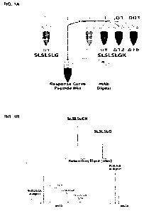
[0024] FIG. 1A shows a schematic of the assay of the present invention
according to an
exemplary embodiment. A "clipped" peptide (corresponding to the C-terminus of
a protein
lacking a C-terminal lysine) is mixed with four "unclipped" peptides
(corresponding to the C-
terminus of a protein having a C-terminal lysine) to form a response curve
(calibration curve)
peptide mix. This response curve peptide mix is then mixed with a sample
representing a
potential antibody manufacturing sample (mAb Digest) to accurately quantitate
the amount of C-
terminal lysine present in the antibody sample.
[0025] FIG. 1B shows the analytical peaks observed for each of the peptide
species when
subjected to liquid chromatography-mass spectrometry, according to an
exemplary embodiment.
[0026] FIGs. 2A-2E show the structures of four SLSLSLGK (SEQ ID NO:1)
"unclipped"
heavy peptides standards containing '3C and '5N isotopes (indicated by *),
according to an
exemplary embodiment. Isotopic heavy chain (HC) C-terminal peptide standards
shown in FIGs.
2A-2D are A4, A8, Al2, and A16 K peptides, respectively. FIG. 2E illustrates
the heavy
SLSLSLG (SEQ ID NO:1) standard A4 des-K, containing '3C and '5N isotopes
(indicated by *).
[0027] FIG. 3 shows a calibration curve (CC) exhibiting a proportional
relationship
between "clipped" and "unclipped" peptides according to an exemplary
embodiment.
[0028] FIG. 4 shows an exemplary response curve using heavy chain (HC) C-
terminal
peptides according to an exemplary embodiment.
CA 03184812 2022-11-24
WO 2021/258017 PCT/US2021/038136
[0029] FIG. 5A shows a UV chromatogram of an equimolar mixture of
"unclipped"
SLSLPGK (SEQ ID NO:4) and "clipped" SLSLSPG (SEQ ID NO:3) according to an
exemplary
embodiment.
[0030] FIG. 5B shows an extracted ion chromatogram (XIC) of equimolar
amounts of the
SLSLPGK (SEQ ID NO:4) and SLSLSPG (SEQ ID NO:3) reagent set according to an
exemplary embodiment.
DETAILED DESCRIPTION
[0031] Therapeutic monoclonal antibodies (mAbs) and bispecific antibodies
(bsAbs) play a
key role in treating many disorders. The advantages of this class of drugs,
including high
specificity and affinity to an expansive variety of molecular targets, warrant
their continued
development and have led to approvals for treatment of health conditions like
asthma,
rheumatoid arthritis, and elevated low density lipoprotein cholesterol, among
many others.
While the commercial and scientific success of therapeutic antibodies is
unprecedented, their
inherent benefits are tempered by their large size, complexity, and chemical
heterogeneity,
necessitating that a host of methods be used to evaluate their safety and
efficacy.
[0032] A significant fraction of these methods is devoted to evaluating
PTMs, a product
quality attribute and major source of mass and charge heterogeneity. The PTM
complement of a
single antibody is diverse, but common modifications are shared among almost
all mAbs and
bsAbs, such as C-terminal lysine truncation, glycosylation, N-terminal pyro-
Glu formation,
oxidation, amidation, deamidation, succinimide intermediate formation,
glycation, isomerization,
cysteinylation, and trisulfide bonding.
[0033] Careful monitoring of these PTM levels enables their control through
predefined
acceptance criteria and has become a common strategy for two distinct reasons.
Firstly,
numerous reports have shown that PTMs, especially when located in a
complementarity
determining region (CDR), can affect the stability and bioactivity of an
antibody. Secondly,
variability in PTM levels could indicate a lack of process control.
[0034] Post-translational modifications are assayed at the global level
with
chromatographic and electrophoretic techniques, including methods like size
exclusion
chromatography multi angle laser light scanning (SEC-MALLS), capillary
electrophoresis
sodium dodecyl sulfate (CE-SDS), imaged capillary isoelectric focusing
(iCIEF), and cation
6
CA 03184812 2022-11-24
WO 2021/258017 PCT/US2021/038136
exchange chromatography (CEX). Such methods have enjoyed wide acceptance, but
typically
identify only the most abundant modifications without determining their
specific locations within
the amino acid sequence.
[0035] For example, the acidic species in a CEX chromatogram will most
likely contain
PTMs like deamidation, glycation, and cysteinylation, and the basic species
will be comprised of
modifications like unprocessed C-terminal K, oxidation, and isomerization.
However, as the
amino acid locations of these PTMs are indeterminable in global analyses, it
is challenging to
determine if they are located in a CDR and at what abundance.
[0036] The integration of highly sensitive mass spectrometer detectors with
an ever-
increasing number of liquid chromatography column chemistries and enzymatic
treatment
conditions has resulted in a mature suite of PTM characterization methods.
Intact mass analysis
of an antibody via liquid chromatography mass spectrometry (LC-MS) does not
yield site-
specific PTM data, but it requires minimal sample preparation and can provide
an analysis of
larger PTMs with the additional benefit of mass identification.
[0037] Disulfide bond reduction and/or limited digestion with enzymes like
IdeS, papain,
GingisKHAN , and FabALACTICA marginally increase sample preparation time, but
enable
subunit level resolution of PTM localization that can be further increased by
fragmenting each
subunit using electron transfer dissociation (ETD) or another tandem mass
spectrometry
(MS/MS or M52) approach. However, site-specific localization and
quantification of PTMs
across a wide dynamic range are most commonly performed from the "bottom-up"
using a
technique called peptide mapping.
[0038] Peptide mapping methods require enzymatic digestion of the antibody,
yielding a
peptide mixture that is separated by liquid chromatography and detected by
ultraviolet/visible
(UV/Vis) absorbance before being ionized and infused into a mass spectrometer.
Full MS
spectra are acquired, and peptides are selected and fragmented to produce
MS/MS spectra that
are used to validate a peptide's identity or to localize a PTM on a peptide
containing more than
one potential modification site. While peptide mapping can potentially
contribute preparation-
related artifacts to the antibody sequence and significantly increases the
time and complexity of
an experiment, it is generally the most sensitive PTM characterization method
as well as site
specific.
7
CA 03184812 2022-11-24
WO 2021/258017 PCT/US2021/038136
[0039] Quantification of each modification can be performed using UV, or
using extracted
ion chromatograms (XICs) from mass spectrometry. UV quantification is
obfuscated by
coeluting peptides and is inherently less sensitive than modern mass
spectrometers. Because of
this, XIC-based quantification is routinely performed, and an MS-based peptide
mapping assay
allows for identification, localization, and quantification of all relevant
PTMs with a detection
limit of less than about 0.1% under optimal conditions.
[0040] The advantages of PTM quantification by XIC are accompanied by some
unique
challenges that affect the method's accuracy and precision. Many of these
issues are related to
differences in the ionization of an unmodified peptide versus the modified
form for a number of
potential reasons. There may be ion suppression of one or both peptide forms
from coeluting
peptide peaks. There may be a difference in solvent environment between the
two peptide forms
eluting at separate retention times. There may be a disparity in ionization
efficiency between the
modified peptide relative to the unmodified peptide. And, finally, there may
be variability
between mass spectrometers. Peptide mapping quantification of all PTMs is
influenced by these
factors, but C-terminal K truncation (des-K) quantification can be
particularly impacted due to
differences in ionization efficiencies and a reduction in the peptide's
predominant charge state to
1+ compared to the unprocessed form (K), which has a predominant charge state
of 2+.
[0041] C-terminal K truncation readily occurs because of carboxypeptidase
activity during
production from mammalian tissue culture cells. As a result, the predominant
form in a
recombinant mAb or bsAb is des-K. The percent relative abundance of
unprocessed C-terminal
K is typically calculated in relation to the sum of K and des-K. Unprocessed C-
terminal K is not
thought to be an efficacy or safety concern in antibodies since it is not in a
CDR and has been
shown to be rapidly lost upon injection with a half-life of roughly one hour.
However, careful
monitoring of this PTM demonstrates process control, and it has been reported
that antibodies
with more basic pI values may also have increased tissue uptake and blood
clearance.
[0042] For these reasons, unprocessed C-terminal K measurement is still
important, and
previous efforts have found that the percentage of K may be overestimated
during peptide
mapping quantification because the additional K on the C-terminus of the
peptide sequence
increases the ionization efficiency relative to the des-K peptide.
8
CA 03184812 2022-11-24
WO 2021/258017 PCT/US2021/038136
[0043] Some attempts to minimize this error include using only the most
abundant charge
state to calculate the XIC area under the curve (AUC) for each peptide, or
using a correction
factor determined by injecting equal molar amounts of each peptide onto the LC
column and
gauging the mass spectrometer response. However, while the first method
decreases the
magnitude of the unprocessed C-terminal K value, it does so with no or little
empirical
knowledge of how much this value should be decreased by. The correction factor
method
assumes that the correction factor remains static across the possible
concentration range of
unprocessed C-terminal K, in the presence of potentially coeluting peptides,
and among different
mass spectrometers. Variation in these factors will lead to greater
inaccuracies in the
measurement processed versus unprocessed C-terminal K that may not be
compensated for when
using a static correction factor.
[0044] To address the challenges of accurately quantifying PTMs in
recombinant proteins,
for example unprocessed C-terminal lysine of a recombinant antibody, described
herein are
methods and kits for using heavy isotope peptide standards to generate a
calibration curve to
normalize detection differences between modified and unmodified peptides.
[0045] An exemplary embodiment of the invention is illustrated in FIG. 1A
and FIG. 1B,
where five heavy peptides are coincubated in the presence of an antibody
digest to produce a
detectable signal. The detectable signal can indicate an accurate measure of
the "clipped" and
"unclipped" C-terminal lysine (K).
[0046] The assay of the present invention, using a novel set of heavy
peptides and
analytical chemistries (for example, liquid chromatography and mass
spectrometry) can be
calibrated to provide highly accurate measurements. This assay fidelity is key
for the
manufacture of complex protein molecules, for example, therapeutic antibodies
designed to be
introduced into human patients.
[0047] The present invention also provides kits for carrying out the assay
of the present
invention. In an exemplary embodiment, a key step in the assay for determining
accurate and
true measures of the presence of a PTM, for example, C-terminal lysines (K),
is the use of one or
more heavy peptides of sufficient plurality such that, when admixed with
appropriate standards
and a sample, provide a readable signal. The signal is typically measured
using analytical
chemistries, for example, liquid chromatography-mass spectrometry (LC-MS).
9
CA 03184812 2022-11-24
WO 2021/258017 PCT/US2021/038136
[0048] Accordingly, exemplary components of a kit for carrying out the
method of the
present invention may include standard peptides without a PTM of interest, for
example,
"clipped" of a C-terminal lysine; standard peptides with a PTM of interest,
for example,
"unclipped"; standard heavy peptides ("clipped" and "unclipped") including,
for example, one or
more of the following exemplary peptides disclosed herein; and instructions
for use, including,
for example, instructions for calibration, data extraction, analysis, and
interpretation.
[0049] Accordingly, the present invention provides for a convenient test
kit and
instructions for improving an important antibody manufacturing chemistry,
manufacturing, and
controls (CMC) endpoint.
[0050] It should be appreciated that the present invention provides for the
accurate
determination of the fine structure and exact amino acid sequence of a
therapeutic protein, such
as a therapeutic antibody. Accordingly, the present invention complements and
improves the
CMC (Chemistry, Manufacturing, and Controls) of any commercially produced
therapeutic
protein, such as a therapeutic antibody.
[0051] For example, the present invention allows for improving the
manufacture and
safeguarding of a number of antibody therapies. Such antibody therapies
include: abciximab,
adalimumab, adalimumab-atto, ado-trastuzumab emtansine, alemtuzumab,
alirocumab,
atezolizumab, avelumab, basiliximab, belimumab, bevacizumab, bezlotoxumab,
blinatumomab,
brentuximab vedotin, brodalumab, canakinumab, capromab pendeti de,
certolizumab pegol,
cetuximab, daclizumab (Zenapax), daclizumab (Zinbryta), daratumumab,
denosumab,
dinutuximab, dupilumab, durvalumab, eculizumab, elotuzumab, evolocumab,
golimumab,
golimumab, ibritumomab tiuxetan, idarucizumab, infliximab, infliximab-abda,
infliximab-dyyb,
ipilimumab ixekizumab, mepolizumab, natalizumab, necitumumab, nivolumab,
obiltoxaximab,
obinutuzumab, ocrelizumab, ofatumumab, olaratumab, omalizumab, palivizumab,
panitumumab,
pembrolizumab, pertuzumab, ramucirumab, ranibizumab, raxibacumab, reslizumab,
rituximab,
secukinumab, siltuximab, tocilizumab, tocilizumab, trastuzumab, ustekinumab,
vedolizumab,
sarilumab, rituximab, hyaluronidaseguselkumab, inotuzumab ozogamicin,
adalimumab-adbm,
gemtuzumab ozogamicin, bevacizumab-awwb, benralizumab, emicizumab-kxwh,
trastuzumab-
dkst, infliximab-qbtx, ibalizumab-uiyk, tildrakizumab-asmn, burosumab-twza,
and erenumab-
aooe.
CA 03184812 2022-11-24
WO 2021/258017 PCT/US2021/038136
[0052] Other therapeutic antibodies of interest for various indications
subject to the present
invention include: aflibercept, for treating eye disorders; rilonacept, for
treating blindness and
metastatic colorectal cancer; alirocumab, for treating familial
hypercholesterolemia or
clinical atherosclerotic cardiovascular disease (ASCVD); dupilumab, for
treating atopic
dermatitis; sarilumab, for treating rheumatoid arthritis and COVID-19;
cemiplimab, for treating
PD-1 related disease; and antibodies for treating Ebola.
[0053] Unless described otherwise, all technical and scientific terms used
herein have the
same meaning as commonly understood by one of ordinary skill in the art to
which this invention
belongs. Methods and materials similar or equivalent to those described herein
known to the
skilled artisan can be used in the practice of particular embodiments
described herein. All
publications mentioned are hereby incorporated by reference in their entirety.
[0054] The term "a" should be understood to mean "at least one" and the
terms "about"
and "approximately" should be understood to permit standard variation as would
be understood
by those of ordinary skill in the art and where ranges are provided, endpoints
are included. As
used herein, the terms "include," "includes," and "including" are meant to be
non-limiting and
are understood to mean "comprise," "comprises," and "comprising" respectively.
[0055] As used herein, the term "protein" or "protein of interest" can
include any amino
acid polymer having covalently linked amide bonds. Proteins comprise one or
more amino acid
polymer chains, generally known in the art as "polypeptides." "Polypeptide"
refers to a polymer
composed of amino acid residues, related naturally occurring structural
variants, and synthetic
non-naturally occurring analogs thereof linked via peptide bonds, related
naturally occurring
structural variants, and synthetic non-naturally occurring analogs thereof.
"Synthetic peptides or
polypeptides" refers to a non-naturally occurring peptide or polypeptide.
Synthetic peptides or
polypeptides can be synthesized, for example, using an automated polypeptide
synthesizer.
Various solid phase peptide synthesis methods are known to those of skill in
the art. A protein
may comprise one or multiple polypeptides to form a single functioning
biomolecule. In another
exemplary aspect, a protein can include antibody fragments, nanobodies,
recombinant antibody
chimeras, cytokines, chemokines, peptide hormones, and the like. A protein of
interest can
include any of bio-therapeutic proteins, recombinant proteins used in research
or therapy, trap
proteins and other chimeric receptor Fc-fusion proteins, chimeric proteins,
antibodies,
11
CA 03184812 2022-11-24
WO 2021/258017 PCT/US2021/038136
monoclonal antibodies, polyclonal antibodies, human antibodies, and bispecific
antibodies.
Proteins may be produced using recombinant cell-based production systems, such
as the insect
bacculovirus system, yeast systems (e.g., Pichia sp.), or mammalian systems
(e.g., CHO cells and
CHO derivatives like CHO-Kl cells). For a recent review discussing
biotherapeutic proteins and
their production, see Ghaderi et at., "Production platforms for biotherapeutic
glycoproteins.
Occurrence, impact, and challenges of non-human sialylation," (Darius Ghaderi
et at.,
Production platforms for biotherapeutic glycoproteins. Occurrence, impact, and
challenges of
non-human sialylation, 28 BIOTECHNOLOGY AND GENETIC ENGINEERING REVIEWS
147-176 (2012), the entire teachings of which are herein incorporated by
reference). Proteins
can be classified on the basis of compositions and solubility and can thus
include simple
proteins, such as, globular proteins and fibrous proteins; conjugated
proteins, such as,
nucleoproteins, glycoproteins, mucoproteins, chromoproteins, phosphoproteins,
metalloproteins,
and lipoproteins; and derived proteins, such as, primary derived proteins and
secondary derived
proteins.
[0056] In some exemplary embodiments, the protein of interest can be a
recombinant
protein, an antibody, a bi specific antibody, a multispecific antibody,
antibody fragment,
monoclonal antibody, fusion protein, scFv and combinations thereof.
[0057] As used herein, the term "recombinant protein" refers to a protein
produced as the
result of the transcription and translation of a gene carried on a recombinant
expression vector
that has been introduced into a suitable host cell. In certain exemplary
embodiments, the
recombinant protein can be an antibody, for example, a chimeric, humanized, or
fully human
antibody. In certain exemplary embodiments, the recombinant protein can be an
antibody of an
isotype selected from group consisting of: IgG, IgM, IgAl, IgA2, IgD, or IgE.
[0058] The term "antibody" as used herein includes immunoglobulin molecules
comprising four polypeptide chains, two heavy (H) chains and two light (L)
chains inter-
connected by disulfide bonds, as well as multimers thereof (e.g., IgM). Each
heavy chain
comprises a heavy chain variable region (abbreviated herein as HCVR or VH) and
a heavy chain
constant region. The heavy chain constant region comprises three domains: CH1,
CH2 and CH3.
Each light chain comprises a light chain variable region (abbreviated herein
as LCVR or VL) and
a light chain constant region. The light chain constant region comprises one
domain: CL1. The
12
CA 03184812 2022-11-24
WO 2021/258017
PCT/US2021/038136
VH and VL regions can be further subdivided into regions of hypervariability,
termed
complementarity determining regions (CDRs), interspersed with regions that are
more
conserved, termed framework regions (FR). Each VH and VL is composed of three
CDRs and
four FRs, arranged from amino-terminus to carboxy-terminus in the following
order: FR1,
CDR1, FR2, CDR2, FR3, CDR3, and FR4. In different embodiments of the
invention, the FRs
of the anti-big-ET-1 antibody (or antigen-binding portion thereof) may be
identical to the human
germline sequences or may be naturally or artificially modified. An amino acid
consensus
sequence may be defined based on a side-by-side analysis of two or more CDRs.
The term
"antibody," as used herein, also includes antigen-binding fragments of full
antibody molecules.
The terms "antigen-binding portion" of an antibody, "antigen-binding fragment"
of an antibody,
and the like, as used herein, include any naturally occurring, enzymatically
obtainable, synthetic,
or genetically engineered polypeptide or glycoprotein that specifically binds
an antigen to form a
complex. Antigen-binding fragments of an antibody may be derived, for example,
from full
antibody molecules using any suitable standard techniques such as proteolytic
digestion or
recombinant genetic engineering techniques involving the manipulation and
expression of DNA
encoding antibody variable and optionally constant domains. Such DNA is known
and/or is
readily available from, for example, commercial sources, DNA libraries
(including, e.g., phage-
antibody libraries), or can be synthesized. The DNA may be sequenced and
manipulated
chemically or by using molecular biology techniques, for example, to arrange
one or more
variable and/or constant domains into a suitable configuration, or to
introduce codons, create
cysteine residues, modify, add or delete amino acids, etc.
[0059] As
used herein, an "antibody fragment" includes a portion of an intact antibody,
such as, for example, the antigen-binding or variable region of an antibody.
Examples of
antibody fragments include, but are not limited to, a Fab fragment, a Fab'
fragment, a F(ab')2
fragment, a scFv fragment, a Fv fragment, a dsFy diabody, a dAb fragment, a
Fd' fragment, a Fd
fragment, and an isolated complementarity determining region (CDR) region, as
well as
triabodies, tetrabodies, linear antibodies, single-chain antibody molecules,
and multi specific
antibodies formed from antibody fragments. Fv fragments are the combination of
the variable
regions of the immunoglobulin heavy and light chains, and ScFv proteins are
recombinant single
chain polypeptide molecules in which immunoglobulin light and heavy chain
variable regions
are connected by a peptide linker. In some exemplary embodiments, an antibody
fragment
13
CA 03184812 2022-11-24
WO 2021/258017 PCT/US2021/038136
comprises a sufficient amino acid sequence of the parent antibody of which it
is a fragment that
it binds to the same antigen as does the parent antibody; in some exemplary
embodiments, a
fragment binds to the antigen with a comparable affinity to that of the parent
antibody and/or
competes with the parent antibody for binding to the antigen. An antibody
fragment may be
produced by any means. For example, an antibody fragment may be enzymatically
or
chemically produced by fragmentation of an intact antibody and/or it may be
recombinantly
produced from a gene encoding the partial antibody sequence. Alternatively, or
additionally, an
antibody fragment may be wholly or partially synthetically produced. An
antibody fragment
may optionally comprise a single chain antibody fragment. Alternatively, or
additionally, an
antibody fragment may comprise multiple chains that are linked together, for
example, by
disulfide linkages. An antibody fragment may optionally comprise a multi-
molecular complex.
A functional antibody fragment typically comprises at least about 50 amino
acids and more
typically comprises at least about 200 amino acids.
[0060] The term "bispecific antibody" includes an antibody capable of
selectively binding
two or more epitopes. Bispecific antibodies generally comprise two different
heavy chains with
each heavy chain specifically binding a different epitope¨either on two
different molecules
(e.g., antigens) or on the same molecule (e.g., on the same antigen). If a
bispecific antibody is
capable of selectively binding two different epitopes (a first epitope and a
second epitope), the
affinity of the first heavy chain for the first epitope will generally be at
least one to two or three
or four orders of magnitude lower than the affinity of the first heavy chain
for the second
epitope, and vice versa. Bispecific antibodies can be made, for example, by
combining heavy
chains that recognize different epitopes of the same antigen. For example,
nucleic acid
sequences encoding heavy chain variable sequences that recognize different
epitopes of the same
antigen can be fused to nucleic acid sequences encoding different heavy chain
constant regions
and such sequences can be expressed in a cell that expresses an immunoglobulin
light chain.
[0061] A typical bispecific antibody has two heavy chains each having three
heavy chain
CDRs, followed by a CHI domain, a hinge, a CH2 domain, and a CH3 domain, and
an
immunoglobulin light chain that either does not confer antigen-binding
specificity but that can
associate with each heavy chain, or that can associate with each heavy chain
and that can bind
one or more of the epitopes bound by the heavy chain antigen-binding regions,
or that can
associate with each heavy chain and enable binding or one or both of the heavy
chains to one or
14
CA 03184812 2022-11-24
WO 2021/258017 PCT/US2021/038136
both epitopes. BsAbs can be divided into two major classes, those bearing an
Fc region (IgG-
like) and those lacking an Fc region, the latter normally being smaller than
the IgG and IgG-like
bispecific molecules comprising an Fc. The IgG-like bsAbs can have different
formats such as,
but not limited to, triomab, knobs into holes IgG (kih IgG), crossMab, orth-
Fab IgG, Dual-
variable domains Ig (DVD-Ig), two-in-one or dual action Fab (DAF), IgG-single-
chain Fv (IgG-
scFv), or la-bodies. The non-IgG-like different formats include tandem scFvs,
diabody format,
single-chain diabody, tandem diabodies (TandAbs), Dual-affinity retargeting
molecule (DART),
DART-Fc, nanobodies, or antibodies produced by the dock-and-lock (DNL) method
(Gaowei
Fan, Zujian Wang & Mingju Hao, Bispecific antibodies and their applications, 8
JOURNAL OF
HEMATOLOGY & ONCOLOGY 130; Dafne MUller & Roland E. Kontermann, Bispecific
Antibodies, HANDBOOK OF THERAPEUTIC ANTIBODIES 265-310 (2014), the entire
teachings of which are herein incorporated by reference). The methods of
producing bsAbs are
not limited to quadroma technology based on the somatic fusion of two
different hybridoma cell
lines, chemical conjugation, which involves chemical cross-linkers, and
genetic approaches
utilizing recombinant DNA technology. Examples of bsAbs include those
disclosed in the
following patent applications, which are hereby incorporated by reference:
U.S. Ser. No.
12/823838, filed June 25, 2010; U.S. Ser. No. 13/ 488628, filed June 5, 2012;
U.S. Ser. No.
14/031075, filed September 19, 2013; U.S. Ser. No. 14/808171, filed July 24,
2015; U.S. Ser.
No. 15/713574, filed September 22, 2017; U.S. Ser. No. 15/713569, field
September 22, 2017;
U.S. Ser. No. 15/386453, filed December 21, 2016; U.S. Ser. No. 15/386443,
filed December 21,
2016; U.S. Ser. No. 15/22343 filed July 29, 2016; and U.S. Ser. No. 15814095,
filed November
15, 2017. Low levels of homodimer impurities can be present at several steps
during the
manufacturing of bispecific antibodies. The detection of such homodimer
impurities can be
challenging when performed using intact mass analysis due to low abundances of
the homodimer
impurities and the co-elution of these impurities with main species when
carried out using a
regular liquid chromatographic method.
[0062] As used herein, the term "multispecific antibody" refers to an
antibody with binding
specificities for at least two different antigens. While such molecules
normally will only bind
two antigens (i.e., bispecific antibodies, bsAbs), antibodies with additional
specificities such as
trispecific antibody and KIH Trispecific can also be addressed by the system
and method
disclosed herein.
CA 03184812 2022-11-24
WO 2021/258017 PCT/US2021/038136
[0063] The term "monoclonal antibody" as used herein is not limited to
antibodies
produced through hybridoma technology. A monoclonal antibody can be derived
from a single
clone, including any eukaryotic, prokaryotic, or phage clone, by any means
available or known
in the art. Monoclonal antibodies useful with the present disclosure can be
prepared using a
wide variety of techniques known in the art including the use of hybridoma,
recombinant, and
phage display technologies, or a combination thereof
[0064] The phrase "recombinant host cell" (or simply "host cell") includes
a cell into
which a recombinant expression vector coding for a protein of interest has
been introduced. It
should be understood that such a term is intended to refer not only to a
particular subject cell but
to a progeny of such a cell. Because certain modifications may occur in
succeeding generations
due to either mutation or environmental influences, such progeny may not, in
fact, be identical to
the parent cell, but are still be included within the scope of the term "host
cell" as used herein. In
an embodiment, host cells include prokaryotic and eukaryotic cells selected
from any of the
kingdoms of life. In one aspect, eukaryotic cells include protist, fungal,
plant and animal cells.
In a further aspect, host-cells include eukaryotic cells such as plant and/or
animal cells. The cells
can be mammalian cells, fish cells, insect cells, amphibian cells or avian
cells. In a particular
aspect, the host cell is a mammalian cell. A wide variety of mammalian cell
lines suitable for
growth in culture are available from the American Type Culture Collection
(Manassas, Va.) and
other depositories as well as commercial vendors. Cells that can be used in
the processes of the
invention include, but not limited to, MK2.7 cells, PER-C6 cells, Chinese
hamster ovary cells
(CHO), such as CHO-Kl (ATCC CCL-61), DG44 (Chasin et at., 1986, Som. Cell
Molec.
Genet., 12:555-556; Kolkekar et at., 1997, Biochemistry, 36: 10901-10909; and
WO 01/92337
A2), dihydrofolate reductase negative CHO cells (CH0/-DHFR, Urlaub and Chasin,
1980, Proc.
Natl. Acad. Sci. USA, 77:4216), and dp12.CHO cells (U.S. Pat. No. 5,721,121);
monkey kidney
cells (CV1, ATCC CCL-70); monkey kidney CV1 cells transformed by 5V40 (COS
cells, COS-
7, ATCC CRL-1651); HEK 293 cells, and 5p2/0 cells, 5L8 hybridoma cells, Daudi
cells, EL4
cells, HeLa cells, HL-60 cells, K562 cells, Jurkat cells, THP-1 cells, 5p2/0
cells, primary
epithelial cells (e.g., keratinocytes, cervical epithelial cells, bronchial
epithelial cells, tracheal
epithelial cells, kidney epithelial cells and retinal epithelial cells) and
established cell lines and
their strains (e.g., human embryonic kidney cells (e.g., 293 cells, or 293
cells subcloned for
growth in suspension culture, Graham et at., 1977, 1 Gen. Virol., 36:59); baby
hamster kidney
16
CA 03184812 2022-11-24
WO 2021/258017 PCT/US2021/038136
cells (BHK, ATCC CCL-10); mouse sertoli cells (TM4, Mather, 1980, Biol.
Reprod., 23:243-
251); human cervical carcinoma cells (HELA, ATCC CCL-2); canine kidney cells
(MDCK,
ATCC CCL-34); human lung cells (W138, ATCC CCL-75); human hepatoma cells (HEP-
G2,
HB 8065); mouse mammary tumor cells (MMT 060562, ATCC CCL-51); buffalo rat
liver cells
(BRL 3A, ATCC CRL-1442); TRI cells (Mather, 1982, Annals 1VY Acad. Sc., 383:44-
68); MCR
cells; FS4 cells; PER-C6 retinal cells, MDBK (NBL-1) cells, 911 cells, CRFK
cells, MDCK
cells, BeWo cells, Chang cells, Detroit 562 cells, HeLa 229 cells, HeLa S3
cells, Hep-2 cells, KB
cells, LS 180 cells, LS 174T cells, NCI-H-548 cells, RPMI 2650 cells, SW-13
cells, T24 cells,
WI-28 VA13, 2RA cells, WISH cells, BS-C-I cells, LLC-MK2 cells, Clone M-3
cells, 1-10 cells,
RAG cells, TCMK-1 cells, Y-1 cells, LLC-PKi cells, PK(15) cells, GEL cells,
GH3 cells, L2 cells,
LLC-RC 256 cells, MfliCi cells, XC cells, MDOK cells, VSW cells, and TH-I, B1
cells, or
derivatives thereof), fibroblast cells from any tissue or organ (including but
not limited to heart,
liver, kidney, colon, intestines, esophagus, stomach, neural tissue (brain,
spinal cord), lung,
vascular tissue (artery, vein, capillary), lymphoid tissue (lymph gland,
adenoid, tonsil, bone
marrow, and blood), spleen, and fibroblast and fibroblast-like cell lines
(e.g., TRG-2 cells, IMR-
33 cells, Don cells, GHK-21 cells, citrullinemia cells, Dempsey cells, Detroit
551 cells, Detroit
510 cells, Detroit 525 cells, Detroit 529 cells, Detroit 532 cells, Detroit
539 cells, Detroit 548
cells, Detroit 573 cells, HEL 299 cells, IMR-90 cells, MRC-5 cells, WI-38
cells, WI-26 cells,
MiCli cells, CV-1 cells, COS-1 cells, COS-3 cells, COS-7 cells, African green
monkey kidney
cells (VERO-76, ATCC CRL-1587; VERO, ATCC CCL-81); DBS-FrhL-2 cells, BALB/3T3
cells, F9 cells, SV-T2 cells, M-MSV-BALB/3T3 cells, K-BALB cells, BLO-11
cells, NOR-10
cells, C3H/IOTI/2 cells, HSDM1C3 cells, KLN205 cells, McCoy cells, Mouse L
cells, Strain 2071
(Mouse L) cells, L-M strain (Mouse L) cells, L-MTK (Mouse L) cells, NCTC
clones 2472 and
2555, SCC-PSA1 cells, Swiss/3T3 cells, Indian muntac cells, SIRC cells, CH
cells, and Jensen
cells, or derivatives thereof) or any other cell type known to one skilled in
the art.
[0065] As used herein, the term "therapeutic protein" refers to any protein
that can be
administered to a subject for the treatment of a disease or disorder. A
therapeutic protein may be
any protein with a pharmacological effect, for example, an antibody, a soluble
receptor, an
antibody-drug conjugate, or an enzyme.
[0066] As used herein, the term "liquid chromatography" refers to a process
in which a
biological/chemical mixture carried by a liquid can be separated into
components as a result of
17
CA 03184812 2022-11-24
WO 2021/258017 PCT/US2021/038136
differential distribution of the components as they flow through (or into) a
stationary liquid or
solid phase. Non-limiting examples of liquid chromatography include reverse
phase liquid
chromatography, ion-exchange chromatography, size exclusion chromatography,
affinity
chromatography, mixed-mode chromatography, hydrophobic chromatography or mixed-
mode
chromatography.
[0067] As used herein, the term "mass spectrometer" includes a device
capable of
identifying specific molecular species and measuring their accurate masses.
The term is meant
to include any molecular detector into which a polypeptide or peptide may be
characterized. A
mass spectrometer can include three major parts: the ion source, the mass
analyzer, and the
detector. The role of the ion source is to create gas phase ions. Analyte
atoms, molecules, or
clusters can be transferred into gas phase and ionized either concurrently (as
in electrospray
ionization) or through separate processes. The choice of ion source depends on
the application.
[0068] In some exemplary embodiments, the mass spectrometer can be a tandem
mass
spectrometer. As used herein, the term "tandem mass spectrometry" includes a
technique where
structural information on sample molecules is obtained by using multiple
stages of mass
selection and mass separation. A prerequisite is that the sample molecules be
transformed into a
gas phase and ionized so that fragments are formed in a predictable and
controllable fashion after
the first mass selection step. Multistage MS/MS, or MS, can be performed by
first selecting and
isolating a precursor ion (MS2), fragmenting it, isolating a primary fragment
ion (MS3),
fragmenting it, isolating a secondary fragment (MS4), and so on, as long as
one can obtain
meaningful information, or the fragment ion signal is detectable. Tandem MS
has been
successfully performed with a wide variety of analyzer combinations. What
analyzers to
combine for a certain application can be determined by many different factors,
such as
sensitivity, selectivity, and speed, but also size, cost, and availability.
The two major categories
of tandem MS methods are tandem-in-space and tandem-in-time, but there are
also hybrids
where tandem-in-time analyzers are coupled in space or with tandem-in-space
analyzers. A
tandem-in-space mass spectrometer comprises an ion source, a precursor ion
activation device,
and at least two non-trapping mass analyzers. Specific m/z separation
functions can be designed
so that in one section of the instrument ions are selected, dissociated in an
intermediate region,
and the product ions are then transmitted to another analyzer for m/z
separation and data
acquisition. In tandem-in-time, mass spectrometer ions produced in the ion
source can be
18
CA 03184812 2022-11-24
WO 2021/258017 PCT/US2021/038136
trapped, isolated, fragmented, and m/z separated in the same physical device.
The peptides
identified by the mass spectrometer can be used as surrogate representatives
of the intact protein
and their post-translational modifications. They can be used for protein
characterization by
correlating experimental and theoretical MS/MS data, the latter generated from
possible peptides
in a protein sequence database. The characterization includes, but is not
limited, to sequencing
amino acids of the protein fragments, determining protein sequencing,
determining protein de
novo sequencing, locating post-translational modifications, or identifying
post translational
modifications, or comparability analysis, or combinations thereof.
[0069] As used herein, the term "database" refers to a compiled collection
of protein
sequences that may possibly exist in a sample, for example in the form of a
file in a FASTA
format. Relevant protein sequences may be derived from cDNA sequences of a
species being
studied. Public databases that may be used to search for relevant protein
sequences included
databases hosted by, for example, Uniprot or Swiss-prot. Databases may be
searched using what
are herein referred to as "bioinformatics tools". Bioinformatics tools provide
the capacity to
search uninterpreted MS/MS spectra against all possible sequences in the
database(s), and
provide interpreted (annotated) MS/MS spectra as an output. Non-limiting
examples of such
tools are Mascot (www.matrixscience.com), Spectrum Mill
(www.chem.agilent.com), PLGS
(www.waters.com), PEAKS (www.bioinformaticssolutions.com), Proteinpilot
(download.appliedbiosystems.com//proteinpilot), Phenyx (www.phenyx-ms.com),
Sorcerer
(www.sagenresearch.com), OMS SA (www.pubchem.ncbi.nlm.nih.gov/omssa/), X!
Tandem
(www.thegpm.org/TANDEM/), Protein Prospector
(prospector.ucsfedu/prospector/mshome.htm), Byonic
(www.proteinmetrics.com/products/byonic) or Sequest
(fields.scripps.edu/sequest).
[0070] In some exemplary embodiments, the mass spectrometer can be coupled
to a liquid
chromatography system. In some exemplary embodiments, the mass spectrometer
can be
coupled to a liquid chromatography-multiple reaction monitoring system. More
generally, a
mass spectrometer may be capable of analysis by selected reaction monitoring
(SRM), including
consecutive reaction monitoring (CRM) and parallel reaction monitoring (PRM).
[0071] As used herein, "multiple reaction monitoring" or "MiRM" refers to a
mass
spectrometry-based technique that can precisely quantify small molecules,
peptides, and proteins
19
CA 03184812 2022-11-24
WO 2021/258017 PCT/US2021/038136
within complex matrices with high sensitivity, specificity and a wide dynamic
range (Paola
Picotti & Ruedi Aebersold, Selected reaction monitoring¨based proteomics:
workflows,
potential, pitfalls and future directions, 9 NATURE METHODS 555-566 (2012)).
MRM can be
typically performed with triple quadrupole mass spectrometers wherein a
precursor ion
corresponding to the selected small molecules/ peptides is selected in the
first quadrupole and a
fragment ion of the precursor ion was selected for monitoring in the third
quadrupole (Yong
Seok Choi et al., Targeted human cerebrospinal fluid proteomics for the
validation of multiple
Alzheimers disease biomarker candidates, 930 JOURNAL OF CHROMATOGRAPHY B 129-
135 (2013)).
[0072] In some aspects, the mass spectrometer in the method or system of
the present
application can be an electrospray ionization mass spectrometer, nano-
electrospray ionization
mass spectrometer, or a triple quadrupole mass spectrometer, wherein the mass
spectrometer can
be coupled to a liquid chromatography system, wherein the mass spectrometer is
capable of
performing LC-MS (liquid chromatography-mass spectrometry) or LC-MRM-MS
(liquid
chromatography-multiple reaction monitoring-mass spectrometry) analyses.
[0073] As used herein, the term "digestion" refers to hydrolysis of one or
more peptide
bonds of a protein. There are several approaches to carrying out digestion of
a protein in a
sample using an appropriate hydrolyzing agent, for example, enzymatic
digestion or non-
enzymatic digestion.
[0074] As used herein, the term "digestive enzyme" refers to any of a large
number of
different agents that can perform digestion of a protein. Non-limiting
examples of hydrolyzing
agents that can carry out enzymatic digestion include protease from
Aspergillus Saitoi, elastase,
subtilisin, protease XIII, pepsin, trypsin, Tryp-N, chymotrypsin,
aspergillopepsin I, LysN
protease (Lys-N), LysC endoproteinase (Lys-C), endoproteinase Asp-N (Asp-N),
endoproteinase
Arg-C (Arg-C), endoproteinase Glu-C (Glu-C) or outer membrane protein T
(OmpT),
immunoglobulin-degrading enzyme of Streptococcus pyogenes (IdeS), thermolysin,
papain,
pronase, V8 protease or biologically active fragments or homologs thereof or
combinations
thereof. For a recent review discussing the available techniques for protein
digestion see
Switazar et al., "Protein Digestion: An Overview of the Available Techniques
and Recent
Developments" (Linda Switzar, Martin Giera & Wilfried M. A. Niessen, Protein
Digestion: An
CA 03184812 2022-11-24
WO 2021/258017 PCT/US2021/038136
Overview of the Available Techniques and Recent Developments, 12 JOURNAL OF
PROTEOME RESEARCH 1067-1077 (2013)).
[0075] The amount of digestive enzyme and the time required for digestion
can be
appropriately selected. When the enzyme to substrate ratio is unsuitably high,
the
correspondingly high digestion rate will not allow sufficient time for the
peptides to be analyzed
by mass spectrometer, and sequence coverage will be compromised. On the other
hand, a low
enzyme to substrate ratio would need a long digestion time and thus a long
data acquisition time.
The enzyme to substrate ratio can range from about 1:0.5 to about 1:200.
[0076] As used herein, the general term "post-translational modifications"
or "PTMs" refer
to covalent modifications that polypeptides undergo, either during (co-
translational modification)
or after (post-translational modification) their ribosomal synthesis. PTMs are
generally
introduced by specific enzymes or enzyme pathways. Many occur at the site of a
specific
characteristic protein sequence (signature sequence) within the protein
backbone. Several
hundred PTMs have been recorded, and these modifications invariably influence
some aspect of
a protein's structure or function (Walsh, G. "Proteins" (2014) second edition,
published by Wiley
and Sons, Ltd., ISBN: 9780470669853). The various post-translational
modifications include,
but are not limited to, cleavage, N-terminal extensions, protein degradation,
acylation of the N-
terminus, biotinylation (acylation of lysine residues with a biotin),
amidation of the C-terminal,
glycosylation, iodination, covalent attachment of prosthetic groups,
acetylation (the addition of
an acetyl group, usually at the N-terminus of the protein), alkylation (the
addition of an alkyl
group (e.g. methyl, ethyl, propyl) usually at lysine or arginine residues),
methylation,
adenylation, ADP-ribosylation, covalent cross links within, or between,
polypeptide chains,
sulfonation, prenylation, Vitamin C dependent modifications (proline and
lysine hydroxylations
and carboxy terminal amidation), Vitamin K dependent modification wherein
Vitamin K is a
cofactor in the carboxylation of glutamic acid residues resulting in the
formation of a y-
carboxyglutamate (a glu residue), glutamylation (covalent linkage of glutamic
acid residues),
glycylation (covalent linkage glycine residues), glycosylation (addition of a
glycosyl group to
either asparagine, hydroxylysine, serine, or threonine, resulting in a
glycoprotein), isoprenylation
(addition of an isoprenoid group such as farnesol and geranylgeraniol),
lipoylation (attachment
of a lipoate functionality), phosphopantetheinylation (addition of a 4'-
phosphopantetheinyl
moiety from coenzyme A, as in fatty acid, polyketide, non-ribosomal peptide
and leucine
21
CA 03184812 2022-11-24
WO 2021/258017 PCT/US2021/038136
biosynthesis), phosphorylation (addition of a phosphate group, usually to
serine, tyrosine,
threonine or histidine), and sulfation (addition of a sulfate group, usually
to a tyrosine residue).
The post-translational modifications that change the chemical nature of amino
acids include, but
are not limited to, citrullination (the conversion of arginine to citrulline
by deimination), and
deamidation (the conversion of glutamine to glutamic acid or asparagine to
aspartic acid).
The post-translational modifications that involve structural changes include,
but are not limited
to, formation of disulfide bridges (covalent linkage of two cysteine amino
acids) and proteolytic
cleavage (cleavage of a protein at a peptide bond). In an exemplary
embodiment, a post-
translational modification is cleavage of a lysine at a protein C-terminus.
Certain post-
translational modifications involve the addition of other proteins or
peptides, such as ISGylation
(covalent linkage to the ISG15 protein (Interferon-Stimulated Gene)),
SUMOylation (covalent
linkage to the SUMO protein (Small Ubiquitin-related MOdifier)) and
ubiquitination (covalent
linkage to the protein ubiquitin). See European Bioinformatics
InstituteProtein Information
ResourceSIB Swiss Institute of Bioinformatics, EUROPEAN BIOINFORMATICS
INSTITUTE DRS -
DROSOMYCIN PRECURSOR - DROSOPHILA MELANOGASTER (FRUIT FLY) - DRS GENE &
PROTEIN,
http://www.uniprot.org/docs/ptmlist for a more detailed controlled vocabulary
of PTMs curated
by UniProt.
[0077] As used herein, the term "C terminal lysine (K)" or "K peptide"
refers to an amino
acid lysine residue or "K" residue that can be present or absent on the end of
an amino acid
sequence. In an exemplary embodiment, a C terminal lysine is on the heavy
chain of an
antibody. The term "truncated peptide" or "(des-K)" refers to a representative
portion of a
protein having the C-terminal amino acid sequence missing a C-terminal lysine
(K).
[0078] As used herein, the term "unclipped" refers to a protein C-terminal
sequence
wherein the C-terminal sequence has a terminal lysine (K) amino acid residue.
As used herein,
the term "clipped" refers to a protein C-terminal sequence wherein the C-
terminal sequence is
missing a terminal lysine (K) amino acid residue.
[0079] As used herein, the term "analyzing and quantifying the percentage
of K peptide"
refers to comparing the difference between a first and second assay signal
sufficient to ascertain
the relative presence or absence of a C-terminal lysine of a protein sequence.
In an exemplary
embodiment, the protein sequence is an antibody heavy chain.
22
CA 03184812 2022-11-24
WO 2021/258017 PCT/US2021/038136
[0080] As used herein, the term "heavy peptides" refers to any peptide of
the invention, or
equivalents thereof, wherein at least one or more carbon or nitrogen atoms of
the peptide is a
heavy isotope thereof, for example, '3C and 15N isotopes.
[0081] As used herein, the term "peptide digest" refers to a mix of
peptides resulting from
contacting a protein with one or more enzymes capable of digesting a protein
sequence. In an
exemplary embodiment, a peptide digest includes a polypeptide sequence
representative of the
C-terminus of the digested protein.
[0082] It is understood that the present invention is not limited to any of
the aforesaid
protein(s) of interest, therapeutic protein(s), recombinant protein(s),
recombinant host cell(s),
antibody(s), liquid chromatography system(s), mass spectrometer(s),
database(s), bioinformatics
tool(s), digestive enzyme(s), post-translational modification(s), or heavy
peptide(s), and any
protein(s) of interest, therapeutic protein(s), recombinant protein(s),
recombinant host cell(s),
antibody(s), liquid chromatography system(s), mass spectrometer(s),
database(s), bioinformatics
tool(s), digestive enzyme(s), post-translational modification(s), or heavy
peptide(s) can be
selected by any suitable means.
[0083] The present invention will be more fully understood by reference to
the following
Examples. They should not, however, be construed as limiting the scope of the
invention.
EXAMPLES
[0084] Materials and Methods. The present invention, when practiced by the
person
skilled in the art, may make use of conventional techniques in the field of
pharmaceutical
chemistry, immunology, molecular biology, cell biology, recombinant DNA
technology, and
assay techniques, as described in, for example, Sambrook et at. "Molecular
Cloning: A
Laboratory Manual", 3rd ed. 2001; Ausubel et at. "Short Protocols in Molecular
Biology", 5th ed.
1995; "Methods in Enzymology", Academic Press, Inc.; MacPherson, Hames and
Taylor (eds.).
"PCR 2: A practical approach", 1995; "Harlow and Lane (eds.) "Antibodies, a
Laboratory
Manual" 1988; Freshney (ed.) "Culture of Animal Cells", 4th ed. 2000; "Methods
in Molecular
Biology" vol. 149 ("The ELISA Guidebook" by John Crowther) Humana Press 2001,
and later
editions of these treatises (e.g., "Molecular Cloning" by Michael Green (4th
Ed. 2012) and
"Culture of Animal Cells" by Freshney (7th Ed., 2015), as well as current
electronic versions.
23
CA 03184812 2022-11-24
WO 2021/258017 PCT/US2021/038136
[0085] Methods useful for quantifying and analyzing PTMs in proteins are
provided within
the disclosure. More specifically, the present disclosure provides methods for
quantifying and
analyzing C-terminal lysine (K) in proteins, for example, antibodies. The
methods include
applying a set of heavy C-terminal peptide standards to a digested protein.
The protein may be
digested by proteases such as trypsin and other suitable enzymes.
[0086] The method of the present invention may involve spiking calibration
curves into
antibody digests and injecting approximately equimolar amounts of heavy des-K
peptide to
digested des-K peptide onto a column in each LC-MS/MS run. Unprocessed C-
terminal K may
be quantified in a single LC-MS/MS peptide mapping experiment.
[0087] The method of the present invention may involve generating a
calibration curve
spanning a ratio range of about 1:1000-1:1 K to des-K peptide. The calibration
curve may have
an error of less than about 10%, less than about 9%, or less than about 8%.
[0088] Mass spectra may be quantified using various spectrometers, such as,
for example,
Thermo Q-Exactive Plus 3, Q-Exactive Plus 4 or Orbitrap Fusion Lumos mass
spectrometers.
[0089] The following working examples demonstrate exemplary methods for
identifying
and quantifying PTMs in recombinant proteins.
Example 1. Assay Design and Methods for Calibration
[0090] This example shows the experimental design of the assay of the
invention for
generating a calibration curve to accurately assess the ratio of a peptide
modified with a PTM
and the same peptide without said PTM.
[0091] All light and heavy isotopic peptide standards were purchased from
New England
Peptide (Gardner, MA). Trifluoroacetic acid (TFA), formic acid (FA), tris [2-
carboxylethyl]
phosphine hydrochloride (TCEP-HC1), and Optima LC/MS grade acetonitrile (ACN)
were
obtained from Thermo Fisher Scientific (Rockford, IL) while glacial acetic
acid and
iodoacetamide (TAM) were procured from Sigma-Aldrich (St. Louis, MO).
Sequencing grade
modified trypsin, ultrapure urea, and ultrapure 1 M
Tris(hydroxymethyl)aminomethane
hydrochloride (Tris-HC1) were purchased from Promega (Madison, WI), Alfa Aesar
(Haverhill,
MA), and Invitrogen (Carlsbad, CA), respectively. Milli-Q water was purified
by a Millipore
Milli-Q Advantage A10 Water Purification System.
24
CA 03184812 2022-11-24
WO 2021/258017 PCT/US2021/038136
[0092] Isotopic HC C-terminal peptide standards were used to normalize the
mass
spectrometer response between corresponding light unprocessed and processed
peptides. Peptide
standards included SLSLSLG (SEQ ID NO:1), SLSLSLGK (SEQ ID NO:2), SLSLSPG (SEQ
ID
NO:3) and SLSLSPGK (SEQ ID NO:4).
[0093] Heavy isotopic SLSLSLGK (SEQ ID NO:2) standards are shown in FIG. 1.
13C
and 15N are indicated by.. A4 (e.g., comprising 4 heavy isotopes of carbon or
nitrogen), A8,
Al2, and A16 K peptides are shown in FIG. 1A, 1B, 1C and 1D, respectively. The
heavy
isotopic SLSLSLG (SEQ ID NO:1) standard, A4 des-K, is shown in FIG. 2. 13C and
15N are
indicated by..
[0094] The peptide standards were dissolved in 10% ACN, 0.1% TFA and
combined into
two calibration curve sets according to the C-terminal sequence (LGK or PGK).
Each set
contained equimolar concentrations of A4 des-K and K peptide as well as A8,
Al2, and A16 K
peptides at molar ratios of 1:10, 1:100, and 1:1000 K to des-K, respectively.
The mixture was
analyzed by XIC as shown in FIG. 3.
[0095] An equimolar mixture of SLSLSPGK (SEQ ID NO:4) and SLSLSPG (SEQ ID
NO:3) was quantified by UV chromatography, as shown in FIG. 4A. Corresponding
K
AUC/des-K AUC values for PGK peptides were 1.08. Similarly, K AUC/des-K AUC
values for
LGK peptides were 1.07. Equimolar amounts of the SLSLSPGK (SEQ ID NO:4) and
SLSLSPG
(SEQ ID NO:3) reagent set were quantified by XIC, as shown in Figure 4B. Heavy
AUC/light
AUC values for PGK and LGK peptides are shown in Table 1.
Table 1.
Peptide Heavy/light isotope Heavy AUC/light AUC
A4/A0 0.98
A8/A0 1.00
SLSLSPGK
Al2/A0 0.99
A16/A0 1.00
SLSLSPG A4/A0 0.99
SLSLSLGK A4/A0 1.02
CA 03184812 2022-11-24
WO 2021/258017 PCT/US2021/038136
A8/A0 1.04
Al2/A0 1.06
A16/A0 1.03
SLSLSLG A4/A0 1.00
[0096] As shown in Table 1, the signals of the heavy peptides were
approximately equal to
the corresponding light peptides, validating the use of the heavy peptides in
generating a
calibration curve that can be applied to light peptides.
[0097] To determine the accuracy of the method, known quantities of light
des-K and K
were spiked into the reagent sets across the 1:10 ¨ 1:1000 K to des-K peptide
ratio range and
measured using the calibration curve corrected method. As shown in Table 2,
the calibration
curve corrected values (or "normalized" values) were closely aligned with the
expected percent
lysine.
Table 2.
Expected SLSLSLGK (SEQ ID NO:2) SLSLSPGK (SEQ ID NO:4)
% K CC Corrected % K % Difference CC Corrected % K % Difference
50.0 49.6 0.8 50.8 1.5
9.1 8.9 2.0 9.3 2.5
1.0 0.9 8.9 1.0 7.1
0.1 0.1 2.0 0.1 3.5
Example 2. Unprocessed C-terminal Lysine Quantification of mAbs
[0098] This
example shows the experimental design of the assay of the invention for
accurate quantitation of C-terminus lysine (K) of a recombinant protein.
[0099] For antibody analysis, the calibration curves were spiked into
antibody digests so
that an approximately equimolar amount of heavy des-K peptide to digested des-
K peptide was
injected onto the column in each LC- MS/MS run.
26
CA 03184812 2022-11-24
WO 2021/258017 PCT/US2021/038136
[0100] Antibody Digestion. Equal weights of five IgG4 mAb samples were
buffer
exchanged into 5 mM acetic acid and 5 mM TCEP-HC1 before denaturation and
reduction at 80
C for ten minutes. The samples were further denatured in 4 M urea/0.1 M Tris-
HC1, pH 7.5, and
alkylated with 5 mM JAM at room temperature in the dark for 30 minutes. Urea
concentration
was lowered to 1 M by adding 0.1 M Tris-HC1, pH 7.4, and the antibodies were
digested at a
1:20 antibody to trypsin ratio at 37 C for 4 hours. Enzymatic activity was
quenched by
acidifying the samples in 0.2% TFA.
[0101] LC-MS and LC-MS/MS Parameters. Aliquots of 5 pg of antibody digest
were
injected onto a 2.1 mm x 150 mm Waters Acquity Ultra Performance Liquid
Chromatography
(UPLC) Charged Surface Hybrid (CSH) C18 column with 1.7 pm particles. Peptides
were
separated on this column with a Waters Acquity I-Class UPLC set to a flow rate
of 250 pL/min
and column temperature of 40 C. The gradient consisted of a 0.1 ¨ 35%
increase of organic
mobile phase (ACN and 0.1% FA) relative to water and 0.1% FA over 95 minutes.
[0102] Mass data was acquired using a Thermo Q-Exactive Plus using QE Plus
3 and 4
systems and/or Orbitrap Fusion Lumos mass spectrometer. Full mass scans were
performed on
the Q-Exactive Plus acquiring an m/z range of 300 ¨ 2000 at 140,000 resolution
(m/z 200) for an
ion population limited by an automatic gain control (AGC) target set to 1 x
106 or a maximum
ion injection time (max IT) of 50 ms.
[0103] For experiments requiring MS/MS identification by data dependent
acquisition
(DDA), a single dd-MS/MS loop began by isolating and fragmenting each of the
five most
intense peptide ions with a 1.5 Th window using higher energy collisional
dissociation (HCD) at
a normalized collision energy of 30.
[0104] Fragment ion population data was collected using an AGC target of 1
x 105 or a
max IT of 100 ms and then scanned at 17,500 resolution, at which point the
sampled precursor
was placed on an exclusion list for 10 seconds to ensure the analysis of less
intense ions.
[0105] Orbitrap Fusion Lumos parameters for MS acquisition were the same as
for the QE-
Plus, with the exceptions being resolution set to 120,000 (m/z 200) and ACG
target to 5 x 10⁵.
Differences in MS/MS settings were as follows: limiting DDA by a cycle time of
one second
instead of by number of precursors; setting AGC target to 2 x 10⁴; controlling
max IT with 50
27
CA 03184812 2022-11-24
WO 2021/258017 PCT/US2021/038136
ms but allowing for continued injection if parallelizable time was available;
and scanning at
15,000 resolution (m/z 200).
[0106] Relevant LC-MS/MS raw files were analyzed with Byonic 3.0 using
custom fasta
files for each antibody according to the following parameters: (1) Cleavage
Sites: R, K; (2)
Cleavage Side: C-terminal; (3) Digestion Specificity: Fully Specific; (4)
Precursor Mass
Tolerance: 10 ppm; (5) Fragmentation Type: QTOF/HCD; (6) Fragment Mass
Tolerance: 20
ppm; (7) Fixed and Variable Modifications: Fixed C Carbamidomethyl, Variable M
Oxidation,
Variable E/Q to pE, and Variable C-term K Loss; and (8) Glycan Modifications:
50 common
biantennary N-glycans. Ion chromatograms for the 1+ and 2+ charge states of
light and heavy C-
terminal peptides were extracted in Thermo Xcalibur 3.1 by the Genesis
algorithm set to a 10
ppm m/z tolerance. Quantitative AUC measurements were exported to Microsoft
Excel, where
calibration curves ranging from 1:1000 ¨ 1:1 K to des-K were constructed to
calculate the
percentage of unprocessed C-terminal K in each sample.
[0107] Table 3 shows the results obtained using the calibration curve
correction method
compared to normal, uncorrected peptide mapping. As shown in Table 3, the
percentage of C-
terminal lysine is overestimated during peptide quantification using
uncorrected peptide mapping
in comparison to the calibration curve (CC) corrected method of the present
disclosure. Thus,
the method of the present invention provides a critical correction for
quantification of a
recombinant protein PTM, for example, C-terminal lysine of an antibody.
Table 3.
CC Corrected C-term Lys % Uncorrected C-term Lys %
Antibody Standard Standard
Mean
Mean Deviation Deviation
mAb 1 5.7 0.2 10.4 0.4
mAb 2 6.9 0.7 11.5 0.7
mAb 3 6.3 0.2 12.0 0.3
mAb 4 9.8 0.4 15.8 0.2
mAb 5 11.8 0.4 19.1 1.1
28
CA 03184812 2022-11-24
WO 2021/258017
PCT/US2021/038136
Example 3. Unprocessed C-terminal Lysine Quantification of Bispecific
Antibodies
(BsAbs)
[0108] This example shows the experimental design of the assay of the
invention for
accurately quantifying PTMs of bispecific antibodies (bsAbs).
[0109] Seven IgG4-based bsAbs, containing both SLSLSLGK (SEQ ID NO:2) and
SLSLSPGK (SEQ ID NO:4) C-terminal sequences, were digested as described above.
Calibration curves were spiked into the antibody digests and approximately
equimolar amount of
heavy des-K peptide to digested des-K peptide was injected onto the column in
each LC-MS/MS
run. Control bsAb digests were subjected to traditional, uncorrected peptide
mapping.
[0110] Table 4 shows the results obtained using the calibration curve
correction method
compared to normal, uncorrected peptide mapping of the PGK C-terminal
sequences. As shown
in Table 4, the percentage of C-terminal lysine is significantly overestimated
during peptide
quantification using uncorrected peptide mapping in comparison to the CC
corrected method of
the present disclosure.
Table 4.
Antibody CC Corrected C-term Lys % Uncorrected C-term Lys %
(PGK) Mean Standard Deviation Mean
Standard Deviation
bsAb 1 14.3 0.1 23.9 0.3
bsAb 2 15.3 0.0 23.5 0.6
bsAb 3 15.8 0.0 27.3 1.0
bsAb 4 16.4 0.2 27.0 0.5
bsAb 5 16.9 0.2 25.7 1.2
bsAb 6 20.0 0.1 30.1 0.6
bsAb 7 26.4 0.3 37.3 0.3
29
CA 03184812 2022-11-24
WO 2021/258017
PCT/US2021/038136
1 1 1] Table 5
shows the results obtained using the calibration curve correction method
compared to uncorrected peptide mapping of the LGK C-terminal sequences. As
shown in Table
5, the percentage of C-terminal lysine is significantly overestimated during
peptide quantification
using uncorrected peptide mapping in comparison to the CC corrected method of
the present
disclosure.
Table 5.
Antibody CC Corrected C-term Lys % Uncorrected C-term Lys %
(LGK) Mean Standard Deviation Mean
Standard Deviation
bsAb 1 2.0 0.1 3.5 0.1
bsAb 2 2.5 0.1 4.1 0.1
bsAb 3 2.2 0.1 3.7 0.2
bsAb 4 2.5 0.2 4.2 0.1
bsAb 5 2.7 0.0 4.7 0.1
bsAb 6 3.4 0.1 5.7 0.1
bsAb 7 5.1 0.1 8.4 0.2
[0112] Further experiments were carried out to investigate variation in
quantitation
between instruments when employing the method of the invention. Five IgG4 mAbs
and one
IgG1 mAb were digested as described above. Calibration curves were spiked into
the antibody
digests and approximately equimolar amount of heavy des-K peptide to digested
des-K peptide
was injected onto the column in each LC-MS/MS run. Control mAb digests were
subjected to
traditional, uncorrected peptide mapping. Mass data were acquired using a
Thermo Q-Exactive
Plus and an Orbitrap Fusion Lumos mass spectrometer.
[0113] As shown in Table 6, when using the CC corrected method, there was
zero to little
difference in percent lysine when quantified using either the QE-Plus or
Fusion mass
spectrometer. However, greater variability of percent lysine was seen across
instruments when
uncorrected peptide mapping was used.
Table 6.
CA 03184812 2022-11-24
WO 2021/258017
PCT/US2021/038136
Antibody CC Corrected C-term Lys %
Uncorrected C-term Lys %
(C-term) QE-Plus Fusion % RSD QE-Plus Fusion %
RSD
IgG4 mAb
5.5 5.5 0.4 10.0 9.1 6.5
1
IgG4 mAb
6.5 6.5 0.2 10.9 9.8 7.5
2
IgG4 mAb
6.6 6.5 0.7 12.2 10.0 14.0
3
IgG4 mAb
9.7 9.6 0.7 16.0 14.8 5.5
4
IgG4 mAb
12.0 11.6 2.6 20.0 17.7 8.8
IgG1 mAb
0.7 0.6 8.6 1.1 0.8 19.2
1 (PGK)
[0114]
Additional experiments were carried out to investigate signal variability due
to
instruments using the method of the invention when applied to bsAbs. Seven
IgG4-based bsAbs
(containing both SLSLSLGK (SEQ ID NO:2) and SLSLSPGK (SEQ ID NO:4) C-terminal
sequences) were digested as described above. Calibration curves were spiked
into the antibody
digests and approximately equimolar amounts of heavy des-K peptide to digested
des-K peptide
was injected onto the column in each LC- MS/MS run. Control bsAb digests were
subjected to
traditional, uncorrected peptide mapping.
[0115]
Mass data were acquired using a Thermo Q-Exactive Plus and an Orbitrap Fusion
Lumos mass spectrometer. As shown in Table 7, when using the CC corrected
method, there
was zero to little difference in percent lysine when quantified using either
the QE-Plus or Fusion
mass spectrometer. However, greater variability of percent lysine was seen
across instruments
when uncorrected peptide mapping was used.
Table 7.
31
CA 03184812 2022-11-24
WO 2021/258017
PCT/US2021/038136
CC Corrected C-term Lys % Uncorrected C-term Lys %
Antibody C-terminal QE- QE-
Fusion % RSD Fusion % RSD
Plus Plus
PGK 14.2 14.8 2.7 24.1 18.9 17.0
bsAb 1
LGK 2.1 2.1 2.6 3.6 3.2 9.2
PGK 15.3 15.5 1.2 24.2 19.2 16.3
bsAb 2
LGK 2.6 2.5 2.5 4.0 4.2 3.3
PGK 15.8 16.3 2.2 28.1 20.8 20.9
bsAb 3
LGK 2.3 2.2 3.8 3.7 3.3 7.7
PGK 16.3 16.9 2.2 28.1 20.8 20.9
bsAb 4
LGK 2.6 2.5 2.1 4.3 3.9 6.2
PGK 16.7 17.3 2.2 24.8 21.1 11.4
bsAb 5
LGK 2.7 2.7 1.2 4.6 4.3 4.2
PGK 20.1 20.4 1.3 30.6 25.1 14.0
bsAb 6
LGK 3.5 3.4 2.6 5.8 5.3 6.0
PGK 26.1 26.9 2.0 37.6 31.9 11.8
bsAb 7
LGK 5.2 5.1 0.9 8.6 8.0 5.0
32