Note: Descriptions are shown in the official language in which they were submitted.
CA 03185035 2022-11-25
WO 2021/242983 PCT/US2021/034455
1
A METHOD TO DETECT WHHE BLOOD CELLS AND/OR WHITE BLOOD CELL
SUBTYPES FROM NON-INVASIVE CAPILLARY VIDEOS
RELATED APPLICATIONS
This application claims benefit of and priority to U.S. Serial No, 17/331,893
filed
May 27, 2021, under 35 U.S.C. 119, 120, 363, 365, and 37 C.F.R. 1,55 and
1,78,
and that application and this application also claim benefit of and priority
U.S.
Provisional Application Serial No. 63/031,117 filed May 28, 2020 under 35
U.S.C.
119, 120, 363, 365, and 37 C.F.R. 1.55 and /.78, and each of U.S, Patent
Application Serial No. 17/331,893 and U.S. Provisional Application No.
63/031,117 are
incorporated herein by this reference.
FIELD OF THE INVENTION
The subject invention relates to a method to detect white blood cells and/or
white
blood cell subtypes from non-invasive capillary videos. The subject invention
also
relates to a method to determine a density of red blood cells from non-
invasive capillary
videos.
BACKGROUND OF THE INVENTION
There is an acute clinical need for an improved, non-invasive, fast, accurate
and
reliable way to measure patients' white blood cells and white blood cell
subtypes,
including the identification of patients with dangerously low levels of white
blood cells.
CA 03185035 2022-11-25
WO 2021/242983 PCT/US2021/034455
White blood cells, also called leukocytes, include, inter alia, the white
blood cell subtypes
of neutrophils, lymphocytes, monorytes, eosinophils, and basophils. According
to the
Centers for Disease Control and Prevention, 10,000 of the 650,000 cancer
patients
treated with chemotherapy in the U.S. every year are hospitalized due to
chemotherapy-
induced febrile neutropenia, a clinically low level of neutrophils. See, e.g.,
Tai et al., Cost
of Cancer-Related Neatropenia or Fever Hospitalizations, Journal of Oncology
Practice,
13(6) (2017), incorporated by reference herein. Such hospitalizations
typically average
8.5 days, may have admission costs of about $25,000, and have a mortality rate
of about
seven percent, making neutropenia one of the most severe side effects of
chemotherapy.
Sec, cog., Truong et aL, Interpreting Febrile Neatropenia Rates From
Randomized
Controlled Trials for Consideration of Primary Prophylaxis in The Real World:
A
Systematic Review and Meta-Analysis, Annals of Oncology, 27(4) (2015), and
Lyman, et
al., Cost of Hospitalization in Patients With Cancer and Febrile Nentropenia
and Impact
of Comorhid Conditions, Am. Soc. Hematology (2015), both incorporated by
reference
herein. There are also many other diseases and conditions associated with
dangerously
low levels of white blood cells, including Acquired Immunodeficiency Syndrome
(AIDS), autoimmune diseases, organ transplantation, patients treated with
immunosuppressarit drugs for various conditions and the like,
One conventional technique which may be used to identify patients with
dangerously low levels of white blood cells is a Complete Blood Count (CRC),
The CBC
can monitor white blood cells differentials and neutropenia. The invasive CBC
requires
drawing more than about 3 ntLs of blood in a clinical setting. The subsequent
lab
CA 03185035 2022-11-25
WO 2021/242983 PCT/US2021/034455
3
analysis typically takes hours to several days for the results. The CBC is
challenging and
costly to perform, and potentially requires immunocompromised patients to
visit a
hospital, putting them at increased risk for developing an infection. See,
e.g., Weinstein,
Nosocomial Infection Update, Emerging Infectious Diseases, 4(3), (1998),
incorporated by reference herein.
Alternative conventional technologies based on finger pricks may have
fundamental
limitations because of a lack of repeatability between successive drops of
blood, elevated
leukocyte counts from fingertip blood at the site of puncture, and the blood
obtained with
such a method may include interstitial fluid. See e.g., Bond, et al., Drop-to-
Drop
Variation in the Cellular Components of Fin gerprick Blood: implications for
Point-of-
are Diagnostic Development, Am. J. Clin. Pathol., 144(6) (2015), Yang et al.,
Comparison of Blood Counts in Various Fingertip and Arterial Blood and Their
Measurement Variation, Clin. Lab. Haematol. 23(3) (2001), Daae et at., A
Comparison
Between Haematological Parameters in 'Capillary' and Venous Blood From Healthy
Adults, 48(7) (1988), all incorporated by reference herein. With such
limitations, finger-
prick approaches may poorly represent systemic cell blood count when performed
outside
the clinical setting. See, e.g., Hollis et al., Comparison of Venous and
capillary
Differential Leutkocyte Counts Using a Standard Hematology Analyzer and a
Novel
Microfluidic Impedance Cytometer, PloS one. 7(9) (2012), and Ghai, C.L., A
Textbook of
Practical Physiology, LP. Medical Ld. (2012), both incorporated by reference
herein.
Consequently, there are currently no devices for at-home, self-administered
monitoring of
white blood cell count, such as neutrophil count.
CA 03185035 2022-11-25
WO 2021/242983 PCT/US2021/034455
4
Conventional in vivo cell imaging systems and methods which may be portable,
inexpensive, and practical for point of care typically have insufficient depth
of focus,
contrast, or field of view to detect white blood cell subtypes. Conventional
capiliaroscopes may be utilized to collect videos or images of nailfold
capillaries of
healthy subjects. See e.g., Maldonado et al., Nai/fold Capillaroscopy in
Diabetes
Mellitus, Micro vascular Research, 112.41-46 (2017) and Mertgko et alõ
Morphological
Characterization of Nailfold Capillaries, Intelligent Technology and Its
Applications
(ISITIA) International Seminar, IEEE (2016), both incorporated by reference
herein.
Such conventional systems and methods may allow imaging of the capillary
geometry
and optical absorption gaps (0AGs) in microcirculation but may have technical
limitations including, inter alia, depth of focus, contrast to neutrophils,
and stability that
may prevent the acquired videos from subsequent analysis. See, e.g., Bourquard
et al.,
Analysis of White Blood Cell Dynamics in Nailfold Capillaries, 37th Annual
International
Conference of the IEEE Engineering in Medicine and Biology Society (EMBC),
IEEE,
(2015) and Bourquarcl et aL, Non-Invasive Detection of Severe Netaropenia in
Chemotherapy Patients By Optical Imaging of Naillold lidricrocirculation, Sci.
Rep,
8(0:5301 (2018), both incorporated by reference herein. As defined herein, an
"optical
absorption gap" (OAG) is as an area within a capillary that is depleted of red
blood cells
and does not absorb light at the wavelengths at which absorption occurs in
hemoglobin
(e.g. about 400 am to about 600 am). An OAG may be created by the presence of
any
white blood cell subtype or by a plasma gap. See, e.g., U.S. Patent No.
9,984,277 and
U.S. Pun No. 2019/0139221, both incorporated by reference herein. As disclosed
in the
CA 03185035 2022-11-25
WO 2021/242983
PCT/US2021/034455
'277 patent and the '221 patent application, videos or images of one or more
capillaries
may be used to show the frequency of OAGs flowing in a capillary correlates to
white
blood cells flowing in the capillary and may be used to determine white blood
cell count.
However, the '277 patent and the '221 patent application are limited to
utilizing
absorption signals and white blood cell subtypes within the OAGs cannot be
identified
and plasma gaps may also contribute to false positives or inaccurate
quantitative
measurement of white blood cell counts. See, e.g., Pablo-Trinidad et al.,
Automated
Detection of Neutropenia Using Noninvasive Video Microscopy of Supeificial
Capillaries, American Journal of Hematology, 94(8) (2019), McKay et al.,
Visualization
of Blood cell Contrast in Nailfold Capillaries With High-speed Reverse Lens
Mobile
Phone Microscopy, Biomedical Optical Express, 11(4) (2020), and McKay et al.,
Optimizing White Blood Cell Contrast in Graded -Field-Capillaroscopy Using
Capillary
Tissue Phantoms, Imaging, Manipulation, and Analysis of Biomolecules, Cells,
and
Tissues, International Society for Optics and Photonics XVIII, Vol. 11243
(2020) , all
incorporated by reference herein.
The conventional in vivo cell imaging systems and methods, the '277 patent,
and
the '221 patent application discussed above are also unable to determine
density of red
blood cells which may be used to non-invasively determine RBC count.
SUMMARY OF THE INVENTION
In one aspect,. a method to detect white blood cells and/or white blood cell
subtypes from non-invasive capillary videos is featured. The method includes
acquiring
CA 03185035 2022-11-25
WO 2021/242983 PCT/US2021/034455
6
a first plurality of images of a region of interest including one or more
capillaries of a
predetermined area of a human subject from non-invasive capillary videos
captured with
an optical device, processing the first plurality of images to determine one
or more
optical absorption gaps located in said capillary and annotating the first
plurality of
images with an indication of any optical absorption gap detected in the first
plurality of
images. The method also includes acquiring a second plurality of images of the
same
region of interest of the same capillary with an advanced optical device
capable of
resolving cellular structure of white blood cells and white blood cell
subtypes and
spatioternporally annotating the second plurality of images with an indication
of any
white blood cell detected and/or a subtype of any white blood cell detected in
the second
plurality of images. The method also includes inputting the first plurality of
images and
annotated information from the first plurality of images and annotated
information from
the spatiotemporally annotated second plurality of images into a machine
learning
subsystem configured to determine a presence of white blood cells and/or the
subtype of
any white blood cells present in the one or more optical absorption gaps in
the first
plurality of images.
In one embodiment, the machine learning subsystem may be further configured to
determine a white blood cell subtype for any optical absorption gap detected
in the first
plurality of images. The machine learning subsystem may be further configured
to
determine full white blood cell differential measurements and/or partial White
blood cell
differential measurements. The method may further include temporally aligning
the first
plurality of images to the spatiotemporally annotated second plurality of
images. The
CA 03185035 2022-11-25
WO 2021/242983 PCT/US2021/034455
7
temporal aligning may include creating said region of interest and said same
region of
interest by using a same objective lens on the optical device and the advanced
optical
device. The temporally aligning may include creating said region of interest
and said same
region of interest by focusing the optical device and the advanced optical
device at a
same location in the capillary. The method may further include generating
optical
absorption gap reference data including a frame identifier and indication of
any optical
absorption gap detected in the first plurality of images. The method may
further include
generating spatiotemporally annotated lookup data including a frame identifier
and
indication of the subtype of any white blood cell present. Temporall::õ,
aligning the first
plurality of images to the spatiotemporally annotated second plurality of
images may
include temporally aligning the frame identifier of the first plurality of
images to the frame
identifier of the visually spatiotemporally annotated second plurality of
images. The
method may further include inputting the first plurality of images, the
optical absorption
gap reference data, and the spatiotemporally annotated lookup data into the
machine
learning subsystem. The machine learning subsystem may be configured to output
results
data of any white blood cells detected and/or the subtype of any white blood
cells detected
and compare the results table to ground truth data. The machine leanaing
subsystem may be
configured to output results data of any white blood cells detected and/or a
subtype of any
white blood cells detected for each optical absorption gap in the first
plurality images and
compare the results data to a ground truth data. Spatiotemporally annotating
the second
plurality of images may further include indicating one or more of: a size, a
granularity, a
brightness, a speed, an elongation, and/or a margination of the white blood
cells and/or a
CA 03185035 2022-11-25
WO 2021/242983 PCT/US2021/034455
8
change of density of red blood cells located upstream or downstream from a
location of
white blood cells detected. The subtype of the white blood cell may include a
granulocyte, a neutrophil, a lymphocyte, a rnonocyte, an eosinophil or a
basoplail. The
optical device may include a high-resolution camera. The advanced imaging
device may
include, inter alia, one or more of: a spectrally-encoded confocal microscopy
(SECM)
device, a swept confocally-aligned planar excitation (SCAPE) microscopy
device, a
scattering confocally aligned oblique plane imaging (SCOPI) device, or oblique
back-
illumination microscopy (OBM) device, The predetermined area of the human
subject
may include, inter alia, one or more of: a finger, a nailfold, a toe, a
tongue, a gum, a lip, a
retina, and/or an earlobe. The optical device may be configured to output at
least one
optical absorption gap signal. The advanced optical device may be configured
to output
an advanced optical signal. Spatiotemporally annotating the second plurality
of images
may be performed by a human. Spatiotemporally annotating the second plurality
of
images may be performed by a processing subsystem. The method may further
include
determining the presence of white blood cells and/or the subtype of any white
blood cells
present in the one or more optical absorption gaps using the first plurality
of images and
annotated information from the first plurality of images and information from
the
machine learning subsystem which has learned and determined the presence of
white
blood cells and/or the subtype of white blood cells present in one or more
optical
absorption gaps using the annotated information from the second plurality of
images
acquired with the advanced optical device.
In another aspect, a method to detect white blood cells and/or white blood
cell
CA 03185035 2022-11-25
WO 2021/242983 PCT/US2021/034455
9
subtypes from non-invasive capillary videos is featured. The method includes
acquiring
a first plurality of images of a region of interest including one or more
capillaries of a
predetermined area of a human subject from non-invasive capillary videos
captured with
an optical device, processing the first plurality of images to determine one
or more
optical absorption gaps located in said capillary, and annotating the first
plurality of
images with an indication of any optical absorption gap detected in the first
plurality of
images. The method also includes determining a presence of white blood cells
and/or the
subtype of any white blood cells present in the one or more optical absorption
gaps using
the first plurality of images and annotated information from the first
plurality of images
and information from a machine learning subsystem which has learned and
determined
the presence of white blood cells and/or the subtype of white blood cells
present in one of
more optical absorption gaps using annotated information from a second
plurality of
images acquired with the advanced optical device.
In yet another aspect, a method to determine a density of red blood cells from
non-invasive capillary videos is featured. The method includes acquiring a
first plurality
of images of a region of interest including one or more capillaries or a
predetermined area
of a human subject from non-invasive capillary videos captured with an optical
device,
processing the first plurality of images to determine one or more areas of
hemoglobin
optical absorption located in the capillary, and annotating the first
plurality of images
with an indication of any areas of hemoglobin optical absorption detected in
the plurality
of images. The method also includes acquiring a second plurality of images of
the same
region of interest of the same capillary with an advanced optical device
capable of
CA 03185035 2022-11-25
WO 2021/242983 PCT/US2021/034455
resolving cellular structure of red blood cells, spatiotemporally annotating
the second
plurality of images with an indication of a density of any red blood cells
detected in the
second plurality of images, and inputting the first plurality of images and
annotated
information from the first plurality of images and annotated information from
the
spatiotemporaily annotated second plurality of images into a machine learning
subsystem
configured to determine the density of any red blood cells present in the one
or more optical
absorption gaps in the first plurality of images.
In one embodiment, a red blood cell count may be determined from the density
of
red blood cells,
BRIEF DESCRIPTION OF THE SEVERAL VIEWS OF THE DRAWINGS
Other objects, features and advantages will occur to those skilled in the art
from
the following description of a preferred embodiment and the accompanying
drawings, in
which:
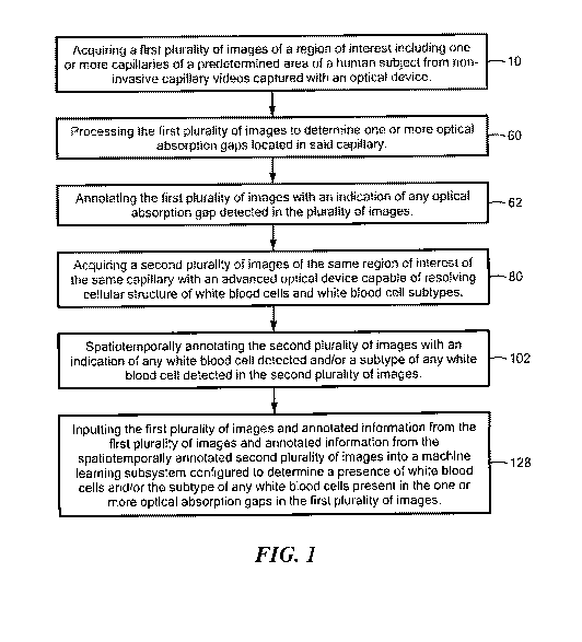
Fig. I is a flowchart showing the primary steps of one embodiment of the
method
to detect white blood cells and/or white blood cell subtypes from non-invasive
capillary
videos;
Fig, 2 shows in further detail examples of images of frames of the first
plurality of
images and the second plurality of images and additional components which may
be
utilized by the method shown in Fig. 1;
CA 03185035 2022-11-25
WO 2021/242983 PCT/US2021/034455
11
Fig. 3 is a schematic diagram showing one example of a nailfold of a finger of
a
human subject for the predetermined area of the human subject for the method
shown in
one or more of Figs. I and 2;
Fig. 4 is a schematic diagram showing in further detail examples of additional
components utilized by the method shown in one or more of Figs. 1-3 and an
example of
a region of interest;
Fig. 5 shows examples of additional areas of the predetermined area of a human
subject utilized for the method shown in one or more of Figs. 1-4;
Fig. 6 shows in further detail an example of one or more capillaries of the
nailfold
shown in Fig. 3 that may be detected by an optical device for the method shown
in one or
more of Figs. 1-5;
Fig. 7 shows an example of an OAG reference table for images detected with an
optical device and then annotated and an example of a spatiotemporally
annotated lookup
table for images detected by an advanced optical device and then
spatiotemporally
annotated for the method shown in one or more of Figs. 1-6;
Fig. 8 shows examples of plots of OAG signals output by an optical device and
plots of advanced optical output signals output by an advanced optical device
for the
method shown in one or more of Figs. 1-7;
Fig. 9 shows in further detail examples of the first plurality of images with
frame
identifiers and the second plurality of images with frame identifiers that are
temporally
aligned for the method shown in one or more of Figs. 1-8;
CA 03185035 2022-11-25
WO 2021/242983 PCT/US2021/034455
1.2
Fig. 10 is a flowchart showing another embodiment of the method to detect
white
blood cells and/white blood cell subtypes from non-invasive capillaries; and
Fig. Ills a flowchart showing one embodiment of the method to determine the
density of any red blood cells present in one or more adsorption gaps.
DETAILED DESCRIPTION OF THE INVENTION
Aside from the preferred embodiment or embodiments disclosed below, this
invention is capable of other embodiments and of being practiced or being
carried out in
various ways. Thus, it is to be understood that the invention is not limited
in its
application to the details of construction and the arrangements of components
set forth in
the following description or illustrated in the drawings. If only one
embodiment is
described herein, the claims hereof are not to be limited to that embodiment.
Moreover,
the claims hereof are not to be read restrictively unless there is clear and
convincing
evidence manifesting a certain exclusion, restriction, or disclaimer.
There is shown in Fig. 1, one embodiment of the method to detect white blood
cells and/or white blood cell subtypes from non-invasive capillary videos. The
method
includes acquiring a first. plurality of images of a region of interest
including one or more
capillaries of a predetermined area of a human subject from non-invasive
capillary videos
captured with an optical device, step 10, Fig. I. First plurality of images
12, Fig, 2, are
preferably derived from non-invasive capillary videos acquired or captured
with optical
device 14, e.g., a high-resolution camera, an imager or imaging device as
disclosed in the
'277 patent and/or the '221 patent application cited supra and incorporated by
reference,
CA 03185035 2022-11-25
WO 2021/242983 PCT/US2021/034455
13
or similar type device. In this example, first plurality of images 12 includes
images or
frames 16, 18, 20, 22 and 24 of region of interest (ROI) 26, Figs, 3 and 4,
which includes
one or more capillaries, e.g,, capillary 28, Figs, 2 and 4 of a predetermined
area of a
human subject. In this example, first plurality of images 12 includes five
images or
frames 16, 18, 20, 22, and It In other examples, first plurality of images 12
may include
more or less than five images or frames 16-24 as depicted in this example. In
one
example, the predetermined area of the human subject may be the nailfold of a
finger,
nailfold 40, Fig, 3, of finger 42 of human subject 44, Fig, 5. Nailfold 40,
Fig, 3, is
one preferred area of the human subject because one or more capillaries are
more easily
detected by optical device 14 because the capillaries are in a more
longitudinal position,
as shown by capillary 28, Fig, 6, In other examples, the predetermined area of
human subject 44, Fig, 5, may include a toe, a tongue, a gum, a lip, a retina,
an earlobe,
or any similar body part determined area of human subject 44.
Fig, 4 shows in further detail one example of ROI 26 of a predetermined area
of
the human subject where images of one or more capillaries may be acquired or
captured
with optical device 14. As disclosed in the '221 patent application and/or the
'277 patent,
in one design, a light source 50 emits light 52 which is reflected by mirror
54 such that
light 52 penetrates nailfold 56 of 40 in ROI 26 and reflected light 60 is
detected by
optical device 14 coupled to processing subsystem 70, Figs. 2 and 4, to create
non-
invasive capillary videos which include first plurality of images 12, Fig, 2,
with images
or frames 16-24.
The method to detect white blood cells and/or white blood cell subtypes from
CA 03185035 2022-11-25
WO 2021/242983 PCT/US2021/034455
14
non-invasive capillary videos also includes processing first plurality of
images 12 to
determine one or more OAGs located in the capillary, step 60. Fig, 1. As
discussed in the
Background Section above, an OAG is an area within a capillary that is
depleted of red
blood cells and does not absorb light at the wavelengths at which absorption
occurs in
hemoglobin (eg., about 400 nm to about 600 nm) and indicates the presence of
one or
more white blood cells, e.g,, as disclosed in the 221 patent application
and/or the 277
patent, In one example, processing subsystem 70 coupled to optical device 14,
similar to
the processor disclosed in the 221 patent application and/or the 277 patent,
or similar
type processing subsystem, processes first plurality of images 12 and detects
one or more
0A,Gs, e,gõ OAGs 64, Fig, 2, in capillary 28 in images or frames /6, 18, 20,
22, and 24.
The method to detect white blood cells and/or white blood cell subtypes from
non-invasive capillary videos also includes annotating the first plurality of
images 12
with an indication of any OAG detected in the plurality of images, step 62,
Fig. 1, In one
example, first plurality of images 12, Fig, 2, are input to processing
subsystem 70 which
outputs annotated information 72 associated with any OAG detected, in one
example,
annotated information 72 preferably includes OAG reference data, e.g., OAG
reference
table 74. Fig. 7, or similar type OAG reference data, that preferably includes
frame
identifier 76 and an indication of any optical gap detected in each of images
or frames 16,
18, 20, 22, and 24 of the first plurality of images 12, indicated at 78. In
this example,
OAG reference table 74 includes frame identifiers to, t, 0, t3,14,õt,,, for
each image or
frame and an OAG identifier for each image or frame, e,g, a I to indicate an
OAG has
been detected. Annotating first plurality of images 12, Figs, 2 and 7, with an
indication
CA 03185035 2022-11-25
WO 2021/242983 PCT/US2021/034455
of any OAG detected in first plurality of images 12 to generate annotated
information 72
may be performed by a trained human operator or by processing subsystem 70,
Figs. 2
and 4.
The method to detect white blood cells and/or white blood cell subtypes from
non-invasive capillary videos also includes acquiring a second plurality of
images of the
same region of interest of the same capillary with an advanced optical device
capable of
resolving cellular structure of white blood cells and white blood cell
subtypes, step 80,
Fig. 1.
Fig. 2 shows one example of second plurality of images 82 which includes
images
or frames 84, 86, 88, 90, 92, and 94 of the same ROI 26, Figs. 3 and 4, which
includes
one or more capillaries, e.g., capillaries 28 of a predetermined area of the
human subject,
e.g., nailfold 40, acquired or captured with advanced optical device 96, Figs.
2 and 6,
capable of resolving cellular structure of white blood cells and white blood
cell subtypes.
In one example, advanced optical device 96 may include a spectrally-encoded
confocal
microscopy (SECM) device, a swept confocally aligned planar excitation (SCAP)
microscopy device, a scattering confocally aligned oblique plane imaging
(SCOFF)
device, or an oblique black illumination microscopy (0BM) device, e.g., as
disclosed in
Golan et al., Noninvasive Imaging of Flowing Blood Cells Using Label-Free
Spectrally
Encoded Flow Cytometry, Biomedical Optics Express, Vol. 3 No. 6 (2012),
Bouchard et
al, Swept ConfOcal-Aligned Planar Excitation (SAP,) Microscopy for High-Speed
Volumetric Imaging of Behaving Organisms, Nature Photonics, Vol. 9 (2015),
McKay et
al., High-Speed Imaging of Scattering Particles Flowing Through Turbid Media
With
CA 03185035 2022-11-25
WO 2021/242983 PCT/US2021/034455
16
Confonciodly Aligned, Oblique Plane Illumination, SHE Bias, San Francisco, CA
(2019),
McKay et al., Imaging Human Blood Cells In Vivo With Oblique Back-Illumination
Capillaroscopy, Biomedical Optics Express, Vol. 11(5) (2020), and Ford, T., N.
and
Mertz, J., Video-Rate Imaging of Microcirculation With Single-Exposed Oblique
Black
Illumination Microscopy, Journal of Biomedical Optics, Vol. 18(6) (2013), all
incorporated by reference herein.
In one example, the cellular structure of white blood cells and white blood
cell
subtypes resolved by advanced optical device 96 may include the subtype of any
white
blood cells detected, e.g., a granulocyte, a neutrophil, a lymphocyte, a
monocyte, an
eosinophil, or a basophil. Image 100, Fig. 2, shows one example of the
cellular structure
of a white blood cell and/or white blood cell subtype in images 86 and 88
resolved by
advanced optical device 96, e.gõ in this example, resolved by a spectrally-
encoded
confocal microscopy (SECM) or similar type advanced optical device.
The method to detect white blood cells and/or white blood cell subtypes from
non-invasive capillary videos also includes spatiotemporally annotating second
plurality
of images with an indication of any white blood cell detected and/or a subtype
of any
white blood cell detected in the second plurality of image, step 102, Fig. 1.
Spatiotemporally annotating the second plurality of images 82, Fig. 2, may
include
indicating one or more of a size, a granularity, a brightness, a speed, an
elongation, and/or
a margination of any white blood cells detected and/or a change in density of
red blood
cells located upstream or downstream from the location of any white blood
cells detected,
In one example, second plurality of images 82, Fig. 2, is spatially annotated
using
CA 03185035 2022-11-25
WO 2021/242983 PCT/US2021/034455
17
spatiotemporally annotated data, e.g., spatiotemporally annotated look-up
table 108, Fig.
7, that includes frame identifier 110, e.g., to, ti, 12, t3, ti.. .t for each
of images or frames
84, 86, 88, 90, 92, and an indication of the white blood cell subtype
associated with each
frame identifier, e.g., a granulocyte, a neutrophil, a lymphocyte, a monocyte,
an
easinophil, or a basophil, exemplary indicated at 112. Spatiotemporally
annotating
second plurality of images 82 with an indication of any white blood cell
detected and/or a
subtype of any white blood cell detected in the second plurality of image 82
may be
performed by a trained human operator or by processing subsystem 70. Figs. 2
and 4, and
preferably generates annotated information 120, Fig. 2, associated with second
plurality
of images 82, and spatiotemporally annotated second plurality of images 122.
The method to detect white blood cells and/or white blood cell subtypes also
includes inputting first plurality of images 12, Fig. 2, annotated information
72 from the
first plurality of images 12 and annotated information 120 from
spatiotemporally
annotated second plurality of images 122 into machine learning subsystem 124
configured to determine a presence of white blood cells and/or subtype of any
white
blood cells present in the one or more optical absorption gaps in the first
plurality of
images 12, step 128, Fig. 1. In one example, machine learning subsystem 124
may be
neural network a support vector machine, a machine learning subsystem
utilizing a
Random Forest learning method. an AdaBoost meta-algorithm, a Naïve Bayes
classifier,
or deep learning, as known by those skilled in the art. Preferably, machine
learning
subsystem 122 may be configured to determine the presence of white blood cells
in
OAGs and determine a full white blood cell differential measurements and/or
partial
CA 03185035 2022-11-25
WO 2021/242983 PCT/US2021/034455
18
white blood cell differential measurements.
In one example, first plurality of images 12, Fig, 2, is preferably temporally
aligned with spatiotemporally annotated second plurality of images 122. In
this example,
temporally aligning includes creating the same region of interest, e.g., ROI
26, Figs, 2
and 4, using the same objective lens 170 on both optical device 14 and
advanced optical
device 96, Fig. 4. In other examples, temporally aligning first plurality of
images 12 with
spatiotemporaliy annotated second plurality of images 122 includes creating
the same
ROI 26 for optical device 14 and advanced optical device 96, Fit!. 4, e.g., by
focusing
optical device 28 and advanced optical device 90 at the same location in the
capillary,
e.g., focusing on ROI 26 and capillary 28, as shown. In other examples,
temporally
aligning first plurality of images 12 with spatiotemporally annotated second
plurality of
images 122 may use image alignment processing methods, e.g., registration or
similar
image alignment processing methods as known by those skilled in the art. See
e.g.,
Oliveira, F.P. and Travares, J.M.R., et al., Medical Image Registration: A
Review,
Computer Methods in Biomeehanics and Biomedical Engineering, 17(2) (2014),
incorporated by reference herein.
In one embodiment, the method to detect white blood cells and/or white blood
cell
subtypes from non-invasive capillary videos preferably includes aligning first
plurality of
images 12. Fig.. 2, to spatiotemporally annotated second plurality of images
122. In one
example, temporally aligning first plurality of images 12 with
spatiotemporally annotated
second plurality of images 122 includes aligning each frame identifier 76,
Fig. 7, e.g., to,
t. t.. in optical absorption gap reference table 74 to each frame
identifier 110, e.g.,
CA 03185035 2022-11-25
WO 2021/242983 PCT/US2021/034455
19
to, ti, t, t3, t in spatiotemporally annotated look-up table 108,
Plots 129, Fig. 8, show one example of OAG signal 132 output by optical device
14, Figs, 2 and 4, and input to processing subsystem 70. In this example, OAG
signal
132 includes peaks 140, 142, and 144 which each indicate the presence of an
OAG
indicative of a white blood cell in a capillary e.g., OAG 64, Figs, 2 and 9,
in capillary 28,
Plots 129, Fig. 8, also show an example of advanced optical output signals
134, 136, and
138 output by advanced optical device 96, in this example a SECM device, which
are
input to processing subsystem 70, Figs, 2 and 4. Each of advanced optical
signals 134,
136, and 138 preferably include a peak that indicates the subtype of a white
blood cell
that corresponds to the presence or detection OAG in a capillary. For example,
peak 146
of advanced optical signal 134 indicates a white blood cell subtype of a
rnonocyte, peak
148 indicates a white blood cell subtype of a lymphocyte, and peak 150
indicates a white
blood cell subtype of a granulocyte, a neutrophil. Peaks 146, 148, and 150 of
advanced
optical signals 134, 136, and 138, respectively, arc for exemplary purposes
only, as
advanced optical signals 134, 136, and 138 may have peaks which represent
other types
of white blood cell subtypes. Plots 129 may also include additional advanced
optical
signals with peaks indicating additional white blood cell subtypes, e.g.,
eosinophils,
basophils, or other white blood cellular structures. In this example,
processing subsystem
70 temporarily aligns peak 146 of advanced optical signal 134 with peak 140 of
OAG
signal 132 as shown which indicates a monocyte is present in OAG 64, Figs, 2
and 9, in
capillary 28. Similarly, processing subsystem 70 temporarily aligns peak 148
of
advanced optical signal 136 with peak 142 of OAG signal 132 as shown which, in
this
CA 03185035 2022-11-25
WO 2021/242983
PCT/US2021/034455
example, indicates a lymphocyte is present in OAG 64 in capillary 28.
Processing
subsystem 70 also temporarily aligns peak 150 of advanced optical signal 138
with peak
144 of OAG signal 132 as shown which indicates a neutnaphil is present in OAG
64 in
capillary 28. In a similar manner, processing subsystem 70 may temporarily
align a peak
of one or more additional advanced optical signals each having a peak
indicating
additional white blood cell subtypes, e.g., granulocyte, eosinophils,
basophils, or other "
white blood cell structures with additional peaks on OAG signal 132.
Fig. 9 shows one example of first plurality of images 12 with images of frames
16, IS, 20, 22, and 24 at frame identifiers, to, t0+68mv, t0+136rets, t0+20,
and to.272m,
respectively, and second plurality of images 82 with images or frames 84, 86,
88, 90, and
92 at frame identifies to, to+68m, ba+)36ms, 10+204ms, and to..272,,
respectively, which are
temporarily aligned as shown. In this example advanced optical device 96,
Figs. 2 and 4,
acquires second plurality of images 82, Fig. 9, using an SECM device that
utilizes a line
scan of capillary 28, indicated at 180. Other advanced optical devices may he
used as
disclosed above.
In one example, first plurality of images 12, Fig. 2, annotated information 72
from
the first plurality of images, e.g.. OAG reference data 74. Fig. 7, e.g., a
table or similar
type data and annotated information from the second plurality of images 120.
Fig. 2, e.g.,
spatiotemporally annotated look-up data 108, Fig. 7, e.g., a table or similar
type data are
input to machine learning subsystem 124, Fig. 2, which outputs results data
170, e.g., a
table of similar type results data, which indicates any white blood cell
detected and/or the
subtype of any white blood cell detected. Machine learning subsystem 124 then
CA 03185035 2022-11-25
WO 2021/242983 PCT/US2021/034455
21
preferably compares results data 170 to ground truth data 172, e.g., a table
or similar type
data to determine and improve the accuracy of the white blood cells detected
and/or the
white blood cell subtypes determined. As known by those skilled in the art,
"ground
truth" is a term relative to the knowledge of the truth concerning an ideal
expected result,
In one embodiment, machine learning subsystem 122 may output results data 174,
e.g., a table of similar type data, that includes any white blood cells
detected and/or a
subtype of any white blood cells detected for each OAG in first plurality of
images 12
and compares results data 174 to ground truth data 172 data to determine and
improve the
accuracy of the white blood cells detected and/or the white blood cell
subtypes
determined.
Once machine learning subsystem 124, Fig. 2, efficiently and effectively
learns
and determines the presence of white blood cells and/or the subtype of white
blood cells
present in one or more optical absorption gaps using the annotated information
from the
second plurality of images acquired with the advanced optical device, the
method to
detect white blood cells and/or white blood cell subtypes from non-invasive
capillaries of
another embodiment using similar techniques as discussed above with reference
to one or
more of Figs. 1-9, may include acquiring a first plurality of images of a
region of interest
including one or more capillaries of a predetermined area of a human subject
from non-
invasive capillary videos captured with an optical device, step 190, Fig. 10.
The method
may also include processing the first plurality of images to determine one or
more optical
gaps located in the capillary, step 192. The method may also include
annotating the first
plurality of images with an indication of any optical gap detected in the
first plurality of
CA 03185035 2022-11-25
WO 2021/242983
PCT/US2021/034455
22
images, step 92, and determining a presence of white blood cells and/or the
subtype of
any white blood cells present in the one or more optical absorption gaps using
the first
plurality of images and annotated information from the first plurality of
images and
information from a machine learning subsystem which has learned and determined
the
presence of white blood cells and/or the subtype of white blood cells present
in one or
more optical absorption gaps using annotated information from a second
plurality of
images acquired with an advanced optical device, step 194,
The result is the method to detect white blood cells and/or white blood cell
subtypes from non-invasive capillary videos accurately, efficiently, and
quantitatively
determines white blood cell differential measurements and/or partial white
blood cell
differential measurements to assist medical personnel in treating various
diseases and
conditions associated with dangerously low levels of white blood cells, e.gõ
neutropenia,
AIDs, autoimmune diseases, organ transplantation, patients treated with
immunosuppressant drugs for various conditions, and the like. Once the
machine.
learning subsystem efficiently and effectively learns and determines the
presence of
white blood cells and/or the subtype of white blood cells present in one or
more optical
absorption gaps using the annotated information from the second plurality of
images
acquired with the advanced optical device, the claimed method can then utilize
a simple,
portable and cost-effective imaging device, e.g., a capillaroscope to
determine the
presence of white bloods in OAGs and the subtype of the white blood cells and
does not
need to further utilize the advanced and expensive optical imaging system,
e.g., SECM,
SCAP, SCOPI, OPBM, and the likeõ
CA 03185035 2022-11-25
WO 2021/242983 PCT/US2021/034455
23
Using similar techniques as discussed above with reference to one or more of
Figs. 1-9, the method to determine density of red blood cells from non-
invasive capillary
videos of one embodiment of this invention includes acquiring a first
plurality of images
of a region of interest including one or more capillaries of a predetermined
area of a
human subject from non-invasive capillary videos captured with an optical
device. step
200, Fig. 10. The first plurality of images is processed to determine one or
more areas of
hemoglobin optical absorption located in the capillary, step 202. The first
plurality of
images is annotated with an indication of any areas of hemoglobin optical
absorption
detected in the first plurality of images, step 204. A second plurality of
images of the
same region of interest of the same capillary is acquired with an advanced
optical device
capable of resolving cellular structure of red blood cells, step 206. The
second plurality
of images with an indication of a density of any red blood cell detected is
spatiotemporally annotated in the second plurality of images, step 208. The
first plurality
of images and annotated information from the first plurality of images and
annotated
information from the spatiotemporally annotated second plurality of images are
input into
a machine learning subsystem configured to determine the density of any red
blood cells
present in the one or more optical absorption gaps in the first plurality of
images, step 210.
In one example, red blood cell count may be determined from the density of red
blood cells.
Although specific features of the invention are shown in some drawings and not
in others, this is for convenience only as each feature may be combined with
any or all of
the other features in accordance with the invention. The words "including",
CA 03185035 2022-11-25
WO 2021/242983 PCT/US2021/034455
24
"comprising", 'having", and "with" as used herein are to be interpreted
broadly and
comprehensively and are not limited to any physical interconnection. Moreover,
any
embodiments disclosed in the subject application are not to be taken as the
only possible
embodiments. Other embodiments will occur to those skilled in the art and are
within the
=
following claims.
In addition, any amendment presented during the prosecution of the patent.
application for this patent is not a disclaimer of any claim element presented
in the
application as filed: those skilled in the art cannot reasonably be expected
to draft a claim
that would literally encompass all possible equivalents, many equivalents will
be
unforeseeable at the time of the amendment and are beyond a fair
interpretation of what
is to be surrendered (if anything), the rationale underlying the amendment may
bear no
more than a tangential relation to many equivalents, and/or there are many
other reasons
the applicant cannot be expected to describe certain insubstantial substitutes
for any claim
element amended.
What is claimed is: