Note: Descriptions are shown in the official language in which they were submitted.
CA 03185434 2022-11-28
WO 2021/240176
PCT/GB2021/051321
Amniotic-like epithelial cell generation
The present invention relates to a method for producing amniotic-like
epithelial cells, using a new
methodology. The invention also relates to a composition and the use of said
composition comprising
amniotic-like epithelial cells prepared according to the method disclosed.
Such cells may have a
particular utility in research, and therapy including regenerative medicine
and for cosmetic
preparations. Alternatively, compositions derived from the cells, such as
membranes, cells in matrices
or scaffolds and/or cell extracts may be used. Such amniotic like epithelial
cells exhibit low expression
levels of human leukocyte antigens (including HLA-A, HLA-B, and HLA-C and HLA-
DR), which are key
antigens involved in recipient rejection, meaning that allogenic cell transfer
is possible. They are,
therefore, a desirable cell phenotype for use in therapy. Optionally, the
cells can be further
differentiated in vitro.
BACKGROUND TO THE INVENTION
The amnion is an extraembryonic epithelial tissue that forms a membrane
surrounding the developing
embryo. In primates including humans, amniotic epithelium originates from
pluripotent epiblast
during implantation. During post-implantation development, amnion functions to
mechanically
protect the embryo, produce growth factors, cytokines and hormones, maintain
the pH in amniotic
fluid. Furthermore, in contrast to rodents, early nascent amnion in primates
was suggested as a source
of primordial germ cells (PGC), secreting growth factors for their
differentiation in an autocrine
fashion, therefore amnion serves as a unique self-organising centre of PGC
specification.
Amniotic membrane is an attractive source for tissue engineering and
regenerative therapies, because
of its anti-inflammatory and immunomodulatory properties, ability to induce
epithelialisation, and
lack of tumorigenicity and ethical issues in clinical application. Amniotic
membrane collected from
term placenta has been successfully applied in patients for ocular surface
reconstruction and
treatments of burns and wounds. Despite their fundamental and clinical
importance, the properties
of amnion cells remain poorly characterised and the current approaches of
their clinical application
suffer from very limited expansion of amniotic epithelial cells in vitro.
There is thus a pressing need of
improved sources of human amniotic epithelial cells, and new methods to
generate and expand
populations of amniotic epithelial cells in vitro.
Amniotic epithelial cells (AECs) are extracted from the lining of the inner
membrane of the term
placenta. Amniotic epithelial cells show low immunogenicity, in addition to
immunomodulatory and
anti-inflammatory behaviours. Because of these qualities, AECs have been
proposed for and indeed
used in regenerative medicine. However, since there are regional restrictions
on the ability of
clinicians to use placental material, particularly in the US, and there are
technical limitations on
propagating cells from placental material, alternative sources of AECs are
desirable. Further, cells
from the placenta risk the transmission of infectious diseases and bacterial
contamination.
Therefore it is desirable to be able to generate a stable and robust source of
AECs.
1
CA 03185434 2022-11-28
WO 2021/240176
PCT/GB2021/051321
Regenerative medicine involves the generation of healthy cells to replace
diseased cells, or to produce
factors stimulating endogenous regenerative mechanisms. Stem cells can be
guided into becoming
specific cells that can be used to regenerate and repair diseased or damaged
tissues in people. Most
regenerative medicines require the use of pluripotent stem cells, such as
embryonic stem cells (ESCs)
or induced pluripotent stem cells (iPSCs), with the latter being cells
generated by the use of particular
reprograming factors or conditions on non-pluripotent cell types.
Pluripotent cells can give rise to all of the cell types that make up the
body; embryonic stem cells are
considered pluripotent. Pluripotency is defined as the capacity of single
cells to produce differentiated
progeny of the three principal germ layers and the germline. In the human
embryo, pluripotency is a
characteristic of epiblast cells from the early pre-implantation stage until
lineage specification during
gastrulation, lasting for at least 10 days. During this window, the epiblast
cells progress through
several distinct developmental phases and therefore, pluripotency is a generic
property of cells with
different identities. As such, two extreme states of pluripotency have been
defined: naive cells
correspond to the early pre-implantation epiblast and primed cells are
reminiscent of the pre-
gastrulation stage.
There have been many attempts to capture and evaluate human naive
pluripotency, with some
studies successfully establishing multiple protocols to generate human naive
pluripotent stem cells by
direct derivation from embryos, reprogramming from somatic cells or conversion
from conventional
primed pluripotent stem cells. Several groups have shown conversion of primed
state human
pluripotent stem cells to the naive state, using overexpression of transgenes,
or by treatment with
specific media (Theunissen et al (2014) Cell Stem Cell, 15(4): 471-487;
Takashima et al (2014) Cell,
158(6): 1254-1269; Guo et al (2017) Development, 144(15): 2748-2763). Shiozawa
et al. has shown
that using transgenes you can convert primed state embryonic stem cells from
common marmoset
(monkey) to the naive state (Shiozawa et al. Stem Cells Dev. 2020,
https://doi.org/10.1089/scd.2019.0259). Guo et al. and Boroviak et al.
provided a useful model for
mechanistic studies of pluripotency regulation and lineage differentiation by
establishing human and
mouse naive pluripotent stem cells from the epiblasts of preimplantation
blastocysts (Guo et al (2016)
Stem Cell Reports, 6(4): 437-446.). Naive pluripotency only exists for a short
period of time during
mammalian embryonic development (Nakamura et al (2016) Nature, 537(7618): 57-
62; Boroviak et al
(2014) Nat Cell Biol, 16(6): 516-528). Naive cells have an unlimited self-
renewal capacity when grown
under appropriate conditions and are able to differentiate into tissues of all
three germ layers in vitro.
An experimental in vitro system for conversion of human naive pluripotent stem
cells to the primed
state has been recently established (Rostovskaya et al (2019) Development,
146(7): dev172916). The
conversion of naive to primed pluripotent stem cells, also called
capacitation, or formative transition,
has been shown to recapitulate features of pen-implantation progression of
embryonic epiblast.
Naive and primed hPSC have distinct signalling requirements for sustained self-
renewal in vitro
(Theunissen et al (2014) Cell Stem Cell, 15(4): 471-487; Takashima et al
(2014) Cell, 158(6): 1254-1269).
The maintenance of naive hPSC requires the inhibition of the mitogen-activated
protein kinase (MAPK)
pathway, whereas the propagation of primed hPSC depends upon the activity of
this pathway (Vallier
et al (2005) JCS, 118: 4495-4509). The mitogen-activated protein kinase (MAPK)
pathway is a chain of
proteins in a cell which results in the communication of a signal from a
receptor on the surface of the
2
CA 03185434 2022-11-28
WO 2021/240176
PCT/GB2021/051321
cell to the DNA in the nucleus of the cell. The proteins convert extracellular
stimuli into a wide range
of cellular responses. All eukaryotic cells possess complex branched highly
pleiotropic MAPK
pathways. These co-ordinately regulate gene expression, mitosis, metabolism,
motility, survival,
apoptosis and differentiation. The central protein within these pathways are
protein Ser/Thr kinases
called mitogen-activated protein kinases (MAPK). The dysregulated signalling
of the MAPK proteins in
the pathway can result in excessive cell proliferation and survival, which may
play a role in specific
malignancies.
The TGF beta signalling pathway is also involved in many of the cells
processes in both embryonic
development and adult organisms. These cellular processes can include cell
growth, cell
differentiation, apoptosis and cellular homeostasis. The TGF beta superfamily
of ligands includes
Growth and differentiation factors (GDFs), Anti-mullerian hormone (AMH), Nodal
and TG93s, as well
as others. Signalling begins with the binding of a ligand to a TGF beta type
II receptor. This receptor
recruits and phosphorylates a type I receptor. The type I receptor will then
phosphorylate receptor-
regulated SMADs which can bind and form a complex with coSMAD that accumulates
in the nucleus.
This complex accumulation can act as transcription factors and participates in
the regulation of target
gene expression.
None of the previous work performed with pluripotent stem cells, as far as the
present inventors are
aware, has resulted in the purposive differentiation of cells into amnion-like
epithelial cells
recapitulating their developmental pathway. Work has been performed in
analysing early
embryogenesis to determine where and when the amnion arises during development
(Luckett (1975)
Dev Dynam 144(2): 149-167; Enders et al (1986) Am J Anat 177(2): 161-185;
Nakamura et al (2016)
Nature 537(7618): 57-62; The Virtual Human Embryo Atlas) concluding that
amnion emerges during
the pen-implantation period. Pluripotent stem cells have been used in attempts
to identify whether
naïve or primed pluripotent stem cells are the predecessors of potential
amnion fate. Such earlier
works include Guo eta! (Guo et al (2021) Cell Stem Cell doi:
10.1016/j.stem.2021.02.025) and lo eta!
(lo et al (2021) Cell Stem Cell doi: 10.1016/j.stem.2021.03.013). However,
both of these works did not
use the appropriate starting pluripotent stem cell state (which is in between
naive and primed states
that corresponds to pen-implantation embryonic state). The authors of both
works conclude that only
primed cells are capable of differentiation into cells that express putative
markers of amnion ¨ these
markers are BAM 61, ISL1, ITGB6, SEMA3C and IGFBP3 ¨ none of which are
established as specific
markers for amnion cells. Moreover, their work did not analyse the cells
produced in terms of the
appearance or development of any form of epithelium or three dimensional
cavitating structures.
Further, others have experimented upon the physical environment of primed
pluripotent stem cells
to see if this is of use in determining the development of tissues forming the
amnion. W02018/106997
discloses methods for deriving amnion tissues from stem cells using scaffolds
and devices, these act
as a biomimetic post-implantation niche. Such devices aim to recapitulate
amniogenesis shortly after
implantation (embryogenesis). This is further described in Shao et al (Nat
Mater 2017 16(4); 419-425).
In all three cases, research has been done using primed pluripotent stem cells
and none of these works
were performed using hPSC in capacitating conditions. Further, in all three
cases a BMP-signalling
dependent in vitro differentiation pathway is investigated.
The present inventors have devised a novel method to derive amnion-like
epithelial cells with high
efficiency from pluripotent stem cells, which recapitulates developmental
events in the embryo,
3
CA 03185434 2022-11-28
WO 2021/240176
PCT/GB2021/051321
permitting the establishment of a robust and effective source of amnion-like
epithelial cells that will
be of great use therapeutically and for research purposes.
SUMMARY OF THE INVENTION
The present invention provides a method for differentiating pluripotent stem
cells into amniotic-like
epithelial cells, said method comprising culturing said cells with an
inhibitor of the MAPK pathway and
an inhibitor of the TGF pathway.
Thus, the present invention is a method which involves the culturing of
pluripotent stem cells in
particular conditions which permits the differentiation of the pluripotent
stem cells into amniotic-like
epithelial cells. The method can therefore be described as ex vivo or in
vitro, since the method takes
place outside the human or animal body.
The method may comprise amniotic-like epithelial cells that form a continuous
layer of cells. The
continuous layer of cells may form a membrane or a 3D structure. The cells may
be human cells, or
animal cells.
Therefore, the amniotic-like epithelial cells of the invention can be observed
to form a continuous
layer of cells once cultured under appropriate conditions. Alternatively put,
the amniotic-like cells
form an epithelium.
The method of the present invention involves the culturing of a pluripotent
stem cell. Said pluripotent
stem cell may be any suitable pluripotent stem cell. The cells may be isolated
from an embryo, isolated
from a parthenote, or taken from an established embryonic stem line, or be an
induced pluripotent
stem cell. It is preferred that the pluripotent stem cell is obtained without
destruction of an embryo.
Optionally, the pluripotent stem cells cultured according to the method of the
present invention are
any one or more of:
a. naive pluripotent stem cells;
b. naive pluripotent stem cells cultured under capacitating conditions;
c. primed pluripotent stem cells cultured under conditions reverting them
to naive
pluripotent stem cells; and/or
d. pluripotent stem cells representing intermediate states between the
naive and the
primed pluripotent states.
The cells of section (d) optionally include, but are not limited to, formative
cells, cells that correspond
to the intermediates during the formative transition.
Those skilled in the art will appreciate that the term "pluripotent" actually
covers a variety of cell types
between naive cells and primed cells.
Optionally, the pluripotent stem cell according to the present invention is
any pluripotent stem cell
with the exception of a primed pluripotent stem cell.
4
CA 03185434 2022-11-28
WO 2021/240176
PCT/GB2021/051321
The method of the present invention may also optionally include culturing the
pluripotent stem cell
with a BMP inhibitor.
The method of the present invention comprises the use of a MAPK pathway
inhibitor. This MAPK
pathway inhibitor can be any suitable inhibitor of any member of this pathway,
and those skilled in
the art will be aware of suitable inhibitors. The inhibitor can be direct
inhibitor, i.e. have a direct effect
on the MAPK pathway component, or be indirect, for example induce an
inhibiting effect within the
cell. Optionally the MAPK pathway inhibitor may be a chemical inhibitor,
neutralising antibody,
aptamer, ligand trap, antisense nucleotide, protein inhibitor, and engineered
peptide targeting any
one from the list comprising: receptor tyrosine kinases, Ras, Src, Raf,
MEK1/2, p38 MAP kinases,
ERK1/2; or activators or agonists of AKT and PI3K. Optionally, the MAPK
pathway inhibitor may be an
indirect inhibitor of the MAPK pathway. For example, the MAPK inhibitor could
be a compound or
agent which induces expression of components required for gene knockdown or
knockout of a MAPK
pathway component. Examples of such a system may be DNA or RNA editing
inducible programmable
nucleases, notably the CRISPR/Cas9 system, small interfering RNAs, epigenetic
editing systems.
It may be preferred that the inhibitor targets (directly or indirectly) any
component of the MAPK/ERK
pathway, such as RAS, RAF, MEK and/or ERK (also called MAPK). In one
embodiment, the inhibitor
may target MEK (MEK1 and/or MEK2). In one embodiment, the inhibitor may target
MAPK (ERK1/2).
The method of the present invention comprises the use of a TGF pathway
inhibitor. This TGF pathway
inhibitor can be any suitable inhibitor of any pathway member, and those
skilled in the art will be
aware of suitable inhibitors. The inhibitor can be direct inhibitor, i.e. have
a direct effect on the TGF
pathway component, or be indirect and for example to induce an inhibiting
effect within the cell.
Optionally the TGF pathway inhibitor includes a chemical inhibitor,
neutralising antibody, ligand trap,
aptamer, antisense nucleotide, protein inhibitor, engineered peptide targeting
any one from the list
comprising: ligands TGF beta, Activin, Nodal; TGF beta type I receptors
TGFBR1, ACVR1, ACVRL1,
ACVR1B, ACVR1C; TGF beta type ll receptors TGFBR2, ACVR2A, ACVR2B; signal
transducers Smad2,
Smad3, Smad4; TGF ligand processing enzyme furin. Optionally, the TGF pathway
inhibitor may be an
indirect inhibitor of the TGF pathway. For example, the TGF inhibitor could be
a compound or agent
which induces expression of components required for gene knockdown or knockout
of a TGF pathway
component. Examples of such a system may be DNA or RNA editing inducible
programmable
nucleases, notably the CRISPR/Cas9 system, small interfering RNAs, epigenetic
editing systems.
It may be preferred that the inhibitor targets (directly or indirectly) any
component of the TGF beta
pathway, such as TGF beta type I receptors TGFBR1, ACVR1, ACVRL1, ACVR1B,
ACVR1C; TGF beta type
ll receptors TGFBR2, ACVR2A, ACVR2B; signal transducers SMAD2, SMAD3, SMAD4;
TGF ligand
processing enzyme furin. In one embodiment, the inhibitor may target TGF beta
type I and/or TGF
beta type ll receptors. It may be preferred that the inhibitor is capable of
inhibiting SMAD signalling
but not BMP signalling.
Optionally, the method of the present invention may comprise the use of a BMP
inhibitor. This
inhibitor is additional to those described above. This BMP inhibitor may be
any suitable inhibitor, and
those skilled in the art will be aware of suitable inhibitors. The inhibitor
can be direct BMP inhibitor,
or be indirect, for example induce an inhibiting effect within the cell.
Optionally the BMP inhibitor
5
CA 03185434 2022-11-28
WO 2021/240176
PCT/GB2021/051321
includes can be a chemical inhibitor, neutralising antibody, ligand trap,
aptamer, antisense nucleotide,
protein inhibitor, engineered peptide targeting any one from the list
comprising: ligands BMP2, BMP4,
BMP7; BMP type I receptors BM PRIA, BMPRIB; BMP type ll receptor BM PR2,
Smad1, Smad5, Smad8.
Optionally, the BMP inhibitor may be an indirect inhibitor of BMP. For
example, the BMP inhibitor
could be a compound or agent which induces expression of components required
for gene knockdown
or knockout of BMP pathway component. Examples of such a system may be DNA or
RNA editing
inducible programmable nucleases, notably the CRISPR/Cas9 system, small
interfering RNAs,
epigenetic editing systems.
The cells prepared according to the method of the invention are unique. A
second aspect of the
present invention, therefore, provides a composition comprising amniotic-like
epithelial cells
prepared according to the method as described herein. The composition can be a
pharmaceutical
preparation. The composition may include a scaffold such as a decellularized
biological matrix or
synthetic structure. It will be understood that the amniotic-like cells are
applied to the matrix or
.. scaffold after they have been prepared according to the methods of the
invention, rather than being
prepared in situ. The scaffold may be composed of any suitable material, and
the scaffold chosen may
depend upon the use to which the cells will be put. Suitable materials may or
may not be
biodegradable, and may include plastic polymers and metal. The composition may
include a
membrane, such as a biodegradable membrane, or a macroporous membrane made of
polymers. The
composition may include a gel such as a collagen gel, MatrigelTM or hydrogel.
The composition may
therefore comprise cells suspended in a gel. The composition can be a
preparation for research
purposes. The composition may be a cosmetic preparation.
The invention further extends to a composition prepared using the cells of the
present invention. The
cells are releasing compounds, factors and other chemicals that may be useful
in the field of
regenerative medicine. Therefore, the invention may usefully extend to a
preparation derived from
the cells of the invention, such as conditioned media, fractionated material
from the media, extract
of the cells, a homogenised preparation of cells, and extracellular extracts.
As such, this aspect of the
invention need not comprise live cells. Such may be useful where there are
restrictions on the use of
live cells for therapeutic uses, or if the use of live cells is undesirable.
The composition and/or cells of the present invention may be put to a variety
of uses in relation to
regenerative medicine and the like, in any human or animal subjects. The uses
described herein are
equally applicable to therapy in humans and veterinary medicine in animals.
The compositions and/or
.. cells of the present invention may be used in therapy. The compositions
and/or cells of the present
invention may be used in a method of treatment of the human or animal body in
need of such
treatment. The treatment may be any of those disclosed below.
The compositions and/or cells of the present invention may also have uses in
cosmetic applications,
such as in cosmetic surgery, in topical preparations such as creams. The
compositions and/or cells of
the present application may be used in methods of ameliorating or improving
the appearance of
wrinkles, fine lines, creases, crow's feet, sagging skin, age spots and/or
blemishes.
6
CA 03185434 2022-11-28
WO 2021/240176
PCT/GB2021/051321
The composition and/or cells of the present invention may be used for wound
healing and/or tissue
repair, optionally skin repair or repair of muscle or connective tissue
damage, such as a hernia or pelvic
floor repair.
The composition and/or cells of the present invention may also be used for the
treatment of ocular
conditions or for ocular surface repair.
The composition and/or cells of the present invention may also be used for the
treatment of burns,
ulcers or surgical wounds.
The composition and/or cells of the present invention may also be used for
treating diabetes or liver
disease.
The composition and/or cells of the present invention may also be used for the
treatment of
congenital conditions, optionally epidermolysis bullosa.
The composition and/or cells of the present invention may also be used for the
treatment of skin
necrosis, optionally Stevens Johnson syndrome.
The composition and/or cells of the present invention may also be used for the
treatment of urological
and/or gynaecological conditions.
The composition and/or cells of the present invention may also be used as an
anti-inflammatory.
Thus, there are a multitude of uses to which the cells prepared according to
the methods of the
present invention, as described herein, can be put. Most of these uses are in
regenerative medicine.
A third aspect of the present invention provides amniotic epithelium prepared
with cells differentiated
according to the method described herein. This amniotic epithelium may be used
therapeutically as
described herein. The cells may also or alternatively be for use as a research
tool. The amniotic
epithelium may be supported on a scaffold such as a decellularized biological
matrix or synthetic
structure. The amniotic epithelium may be supported on a membrane, such as a
polymer membrane.
The amniotic epithelium may be suspended within a gel, such as a hydrogel.
A fourth aspect of the present invention provides a membrane prepared with
cells differentiated
according to the method described herein. The membrane may be used
therapeutically as described
herein. The membrane may additionally or alternatively be for use as a
research tool. The membrane
may be supported on a scaffold such as a decellularized biological matrix or
synthetic structure.
A fifth aspect of the present invention provides a three dimensional (3D)
structure, such as a hollow
sphere or hollow spheroid, prepared with cells differentiated according to the
method described
herein. The 3D structure may be for use as a research tool.
A sixth aspect of the present invention provides a method of treatment of the
human or animal body
using the cells, compositions, epithelium or membranes as described herein.
The method of
7
CA 03185434 2022-11-28
WO 2021/240176
PCT/GB2021/051321
treatment may include any therapeutic use of the amniotic-like epithelial
cells, including wound
healing or tissue repair, optionally skin repair.
A seventh aspect of the present invention is a method of preparing amniotic-
like cells in suspension.
The amniotic-like cells prepared in suspension according to the method
described herein may form or
provide a membrane or a three dimensional (3D) structure, such as a hollow
sphere or hollow
spheroid. Such a suspension-based method is suitable for commercial scale-up.
DESCRIPTION OF THE FIGURES
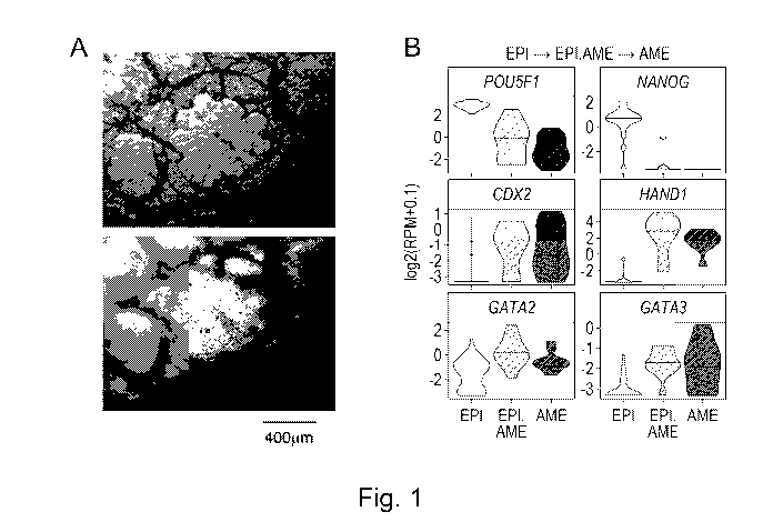
Figure 1 (A to l). Characterisation of hALEC (human Amniotic Like Epithelial
Cells):
Human pluripotent stem cells (hPSC) (HNES1 line) after 3 days of capacitation
in the presence of
XAV939, followed by 5 days of differentiation in AP-containing media.
Figure 1 (A) is bright field microscopy, two focal planes of the same field of
view. Figure 1(B) shows
diagnostic markers of pluripotency (POU5F1 and NANOG) and amnion (CDX2, HAND1,
GATA2 and
GATA3) during progression of amniotic lineage in ex vivo cultured human pre-
gastrulation embryos
(EP1 is epiblast, EPI.AME is an intermediate stage between epiblast and
amnion, AME is amnion); by
single-cell RNAseq (Xiang et al. (2020) Nature, 577: 537-542). Figure 1(C)
shows the same markers
during in vitro differentiation of hPSC to hALEC, assayed by qRT-PCR. Figure
1(D) is a bright field
microscopy of hALEC differentiated in suspension and figure 1(E) shows qRT-PCR
for diagnostic
markers during differentiation in suspension as compared to monolayer
induction. Figure 1 (F and G)
show immunostaining for markers GATA3, E-cadherin, CDX2, POU5F1, and
fluorescently labelled
Phalloidin is applied for counterstaining; and figure 1(H) depicts flow
cytometry of hALEC, obtained
from HNES1 capacitated for 5 days in XAV939 and then treated by AP for 4 days.
Figure 1 (I) shows
time-lapse imaging of hALEC self-assembly to epithelial bubbles.
Figure 2 (A to D). Comparison of hALEC to amnion cells of human and macaque
embryos:
Transcriptome of hALEC derived from HNES1 cells after 3-5 days of capacitation
in the presence of
XAV939, followed by differentiation in AP-containing media, was characterised
by bulk and single-cell
.. RNA sequencing. hALEC expression profile was compared to the cells of ex
vivo cultured human pre-
gastrulation embryos (Xiang et al. Nature 2020) and macaque gastrulating
embryos (Ma et al. (2019)
Science 366(6467): eaax7890, doi: 10.1126/science.aax7890). Figure 2(A) shows
average expression
of pluripotent epiblast, early amnion and late amnion markers of embryos,
during in vitro
differentiation to hALEC. Figure 2(B) shows clustering analysis of single
cells in hALEC population,
amnion-like cells are highlighted in black. Figure 2(C) is analysis of
fractions of identity (Gong et al
(2013) Bioinformatics 29: 1083-1085) of embryonic cell populations in hALEC.
Figure 2(D) shows PCA
of human and macaque embryo single cells, and undifferentiated cells and
amnion-like cells in vitro.
Respective lineages and cell types are highlighted in black. Abbreviations:
hsAME.E ¨ early amnion
from human embryos; hsPostEPI ¨ post-implantation epiblast from human embryos;
hsTE ¨
trophectoderm from human embryos; hsSTB ¨ syncytiotrophoblast from human
embryos; cyAME.L ¨
late amnion from macaque (cynomolgus monkey) embryos.
Figure 3 (A to l). Signalling requirement for hALEC differentiation:
Figure 3(A) depicts experimental outline; naïve hPSC were capacitated in
different conditions for 3
days (2uM XAV939 in N2B27 basal medium ("XAV939"), N2B27 basal medium only
("N2B27"), 1uM
8
CA 03185434 2022-11-28
WO 2021/240176
PCT/GB2021/051321
A8301 in N2B27 basal medium ("A8301"), or medium E8 for culturing primed hPSCs
("E8")), and then
differentiated to hALEC in AP media. Figure 3(B) is whole-well view of cells
after hALEC induction
stained with fluorescently labelled phalloidin. Figure 3(C) shows qRT-PCR for
diagnostic markers in
cells before and after hALEC induction in 2 independent experiments. Figure
3(D) shows brightfield
images of hPSC (cR-H9-EOS line) that were capacitated for 3 days in N2B27 and
then transferred to
basal media either: without inhibitors ("None"), or with A8301 ("A"), or with
PD03 ("P"), or with their
combination ("AP"); or with both inhibitors and LDN193189 ("DAP"). Figure 3(E)
shows qRT-PCR
results for characteristic markers in 2 independent experiments. Figure 3(F)
and (G) show images of
cells stained with fluorescently labelled phalloidin and qRT-PCR results,
respectively, of hALEC
differentiated in the presence of alternative MAPK inhibitors (1uM PD0325901,
or 5nM, 10nM or
30nM Trametinib). Figure 3(H) and (I) show images of cells stained with
fluorescently labelled
phalloidin and qRT-PCR results, respectively, for hALEC differentiated in the
presence of alternative
TGFb pathway inhibitors (1uM A8301; 10 or 20uM SB431542; 1uM or 5uM LY2109761;
5uM or 10uM
or 20uM LY364947).
Figure 4 (A to G). A competence window for hALEC differentiation during the
formative transition:
Figure 4(A) depicts the experimental outline. hPSC (HNES1 line) were
capacitated in XAV939 and
analysed for their ability to form hALEC each day of capacitation, by
treatment with AP of DAP for 4
days. Figure 4(B) shows stitched images showing whole wells of a 24-well
plate, and figure 4(C) shows
individual fields of view. Figure 4(D) is a qRT-PCR for markers in hALEC
differentiated using AP medium
after various length of capacitation. Figure 4(E) depicts the
immunofluorescence for markers (OCT4,
CDX2 and E-cadherin) during the time course of hALEC differentiation using
naive and partially
capacitated HNES1. Figure 4(F) shows images of hALEC obtained from hPSCs
capacitated for 8 days
and then differentiated in AP medium in the presence of various BMP inhibitors
("LDN", LDN193189;
"Dorso", Dorsomorphin; or K02288). Figure 4(G) is the flow cytometry analysis.
DETAILED DESCRIPTION OF THE INVENTION
The present invention relates to a method for differentiating pluripotent stem
cells into amniotic-like
epithelial cells, said method comprising culturing said cells with an
inhibitor of the MAPK pathway and
an inhibitor of the TGF pathway. This culturing may be described as ex vivo or
in vitro and not in the
human or animal body.
"Differentiation", also known as "cellular differentiation", involves a cell
changing into another cell
type; usually, but not always to a more specialised cell type. Differentiation
occurs multiple times
during the development of a multicellular organism. This process also carries
on after the
development of said organism, with focus on stem cells dividing to create
fully differentiated daughter
cells during tissue repair and during normal cell turnover. A cell's size,
shape, membrane
potential, metabolic activity and responsiveness to signals can all change
dramatically during
differentiation due to highly controlled modifications in gene expression.
"Cell culturing" involves the removal of cells from an animal or plant which
will then grow in favourable
controlled conditions outside their natural environment, or "ex vivo". The
cell culture can then be used
for in vitro assays. The cell culture can also be used to produce biological
compounds such as
antibodies or recombinant proteins. The conditions under which particular
cells are cultured are
9
CA 03185434 2022-11-28
WO 2021/240176
PCT/GB2021/051321
important, particularly in relation to stem cell technologies. Culturing
conditions vary for each cell
type, but generally include the use of a suitable vessel with a medium that
supplies the essential
nutrients, growth factors, hormones, and gases, and regulates the physio-
chemical environment.
Most cells require a surface or an artificial substrate or a layer of feeder
cells providing extracellular
matrix and soluble factors (adherent or monolayer culture) whereas others can
be grown free floating
in culture medium (suspension culture).
Human amnion consists of AECs on a basement collagenous membrane, an acellular
compact layer, a
fibroblast layer, and a highly hygroscopic spongy layer. Amniotic epithelial
cells (AEC) are usually
extracted from the lining of the inner membrane of the placenta, using
enzymatic digestion of the
amnion membrane after it is separated from the underlying chorion. During
development, the AEC
are formed from epiblasts between day 7 and 9 after fertilization. AEC form
squamous epithelium and
express epithelial marker E-cadherin. AEC in human cultured pre-gastrulation
embryos express the
markers GATA2, GATA3, TFAP2A, TFAP2C, CDX2 and lack embryonic pluripotency
markers NANOG,
SOX2 and POU5F1 (Xiang et al (2020) Nature, 577: 537-542). AEC in term
placentas express a specific
combination of major histocompatibility complex antigens, including classical
HLA-la and non-
classical HLA-lb (HLA-E and placental-specific HLA-G) (Hammer et al (1997) Am
J Reprod lmmunol,
37(2): 161-171; Houlihan et al (1995) J lmmunol, 154(11): 5665-5674). HLA-G is
known to provide
immunosuppressive properties to placenta (Le Bouteiller et al (1999) Hum
Reprod Update, 5(3): 223-
233).
The present invention relates to AECs which are derived in vitro/ex vivo from
pluripotent stem cells.
It is conventional, where cells have been derived effectively artificially,
that the term "like" is applied
to such cells. Thus, the cells of the present invention are "amniotic-like"
epithelial cells. The AECs of
the present invention are considered to be similar to those isolated from
nature.
The cells of the present invention are amniotic-like epithelial cells
generated from pluripotent stem
cells. As such, the cells may have one or more of the following
characteristics:
= the cells are flat squamous epithelial cells;
= the cells form a continuous layer of cells (an epithelium);and/or
= the cells express one or more marker associated with amniotic epithelial
cells such as E-
cadherin (CDH1), CDX2, HAND1, TFAP2C, TFAP2A, GATA2, GATA3.
"Human amnion-like epithelial cells" (hALEC) are epithelial cells expressing
amniotic epithelial cell
markers generated from human pluripotent stem cells using culturing according
to the method of the
invention.
In general, the present invention relates to amniotic-like epithelial cells
that form a continuous layer
of cells, wherein said continuous layer of cells forms a membrane or a 3D
structure. These cells can be
human cells. These human amnion-like epithelial cells (hALECs) can have
epithelial morphology and
form large (up to 2mm) hollow cysts in culture, reminiscent of amnion
structure in the embryo. This
is a 3D structure formed by the cells. The cells express genes characteristic
of amnion cells, such as E-
cadherin (CDH1), CDX2, HAND1, TFAP2C, TFAP2A, GATA2, GATA3. The inventors' new
approach
therefore provides an expandable, standardised and potentially unlimited
source of much sought-
CA 03185434 2022-11-28
WO 2021/240176
PCT/GB2021/051321
after proliferative human amniotic epithelial cells, all of which is an
advantage over amnion cells from
term placenta.
The amniotic-like cells of the present invention can form a continuous layer
of cells. This continuous
layer may be a layer of single cells. The layer is continuous, unbroken or
whole layer of cells. Thus,
the layer is composed of cells that are packed together. Epithelia are
continuous sheets or layers of
tightly linked cells that constitute the surfaces (such as the epidermis and
corneal epithelium) and
linings (such as the digestive, respiratory, and uro-genital epithelia) of the
body. Thus, the amnion-
like cells of the present invention may alternatively be described as being
able to form an epithelial
layer.
In nature, amniotic epithelial cells form part of the amniotic membrane. The
epithelium cells,
basement membrane and a stromal layer are the three major components of the
amniotic membrane.
The amniotic-like epithelial cells of the present invention may therefore
require a biological matrix
such as a decellularized matrix or a synthetic scaffold for use in certain
embodiments. The cells may
be applied after preparation according to the methods of the present
invention. Decellularized or
synthetic extracellular matrix (ECM) has emerged as a promising tool in the
fields of tissue engineering
or regenerative medicine. ECM provides a native cellular environment that
combines its unique
composition and architecture. It can be widely obtained from native organs of
different species after
being decellularized and provides necessary cues to cells homing. Biological
scaffolds derived from
extracellular matrix (ECM) have been widely utilised in regenerative medicine.
These structures can
be also created from synthetic components. Alternatively, a membrane such as a
biodegradable
membrane may be used to provide a support. Other options include using the
cells in a gel, such as
collagen or hydrogel.
The method of the present invention relies upon the culturing of pluripotent
stem cells. These
pluripotent stem cells may be any suitable pluripotent stem cell from any
source. Pluripotent stem
cells have the ability to undergo self-renewal and to give rise to all cells
of the tissues of the body.
The pluripotent stem cells for use in the present invention may be any
suitable source of cells,
including embryonic stem (ES) cells, cells from parthenotes, embryonic stem
(ES) cell lines, and
induced pluripotent stem (iPS) cells. The cells may be human or animal. The
pluripotent stem cells
are preferably obtained without destruction of an embryo. It is possible to
remove a single blastomere
without embryo destruction. Induced pluripotent stem cells involve the
reprogramming of somatic
cells such as skin fibroblasts or blood cells using a variety of techniques,
both genetic and chemical.
The advantage of using somatic cells is that it enables autologous cells to be
prepared, which will
reduce the risk of rejection of the cells once transferred. However, due to
the potential low expression
levels of human leukocyte antigens (including HLA-A, HLA-B, and HLA-C and HLA-
DR), which are key
antigens involved in recipient rejection, allogenic preparations of cells from
donor cells are also
contemplated herein.
Pluripotency is defined as the ability of single cells to produce all lineages
of the embryo. Pluripotency
exists from emergence of the epiblast in pre-implantation blastocyst until
lineage specification during
gastrulation. This period lasts from ¨4 days in rodents including mouse, to 8-
10 days or longer in
primates, including humans, and in many other mammals. Over this time,
pluripotent epiblast cells
11
CA 03185434 2022-11-28
WO 2021/240176
PCT/GB2021/051321
change their properties from the initial naïve character to a primed state
that is competent for
differentiation. Both naive and primed are states of pluripotency, but exhibit
slightly different
characteristics. The naïve state represents the cellular state of the
preimplantation blastocyst inner
cell mass, while the primed state is representative of the post-implantation
epiblast cells. These two
cell types exhibit clearly distinct developmental potential, as evidenced by
the fact that naive cells are
able to contribute to blastocyst chimeras, while primed cells cannot. Those in
the art consider that
there may be a continuum of intermediate states between naïve and primed
states in vivo, and thus
a spectrum of cell types exist between these two extremes.
Naïve and primed states can be classified on the basis of multiple
characteristics that each state can
retain in vitro. Different combinations of exogenous factors confer distinct
characteristics to
pluripotent stem cells in vitro. As a result, cells acquire a distinct set of
naive and primed properties.
It is possible that cells beyond the primed state are still pluripotent, as
used herein "primed"
encompasses cells beyond the point of primed.
Molecular criteria for defining the naïve human pluripotent state are
described in Theunissen et al
(2016) Cell Stem Cell, 19: 502-515, October 6,2016, herein incorporated by
reference.
Naïve cells can be generated by resetting conventional primed stem cells, by
somatic cell
reprogramming, or by derivation directly from dissociated human inner cell
mass (ICM) cells. They
exhibit transcriptome correlation with the preimplantation epiblast and show
protein expression of
naïve epiblast-specific transcription factors such as KLF4, KLF17 and TFCP2L1.
Naive cells are proposed to gain competence for lineage induction through a
process of capacitation.
The present inventor has previously established that the human naïve
pluripotent stem cells lack
competence to respond productively to inductive cues for lineage specification
(Rostovskaya et al
(2019) Development, 146(7): dev172916, herein incorporated by reference).
Naïve hPSCs can be
capacitated for somatic lineage induction.
The pluripotent stem cells as used in the method of the present invention may
be any one or more of:
a. naïve pluripotent stem cells;
b. naïve pluripotent stem cells cultured under capacitating conditions;
c. primed pluripotent stem cells cultured under conditions reverting them
to naive pluripotent
stem cells; and/or
d. pluripotent stem cells representing intermediate states between the naive
and the primed
pluripotent states.
The cells of section (d) optionally include, but are not limited to, formative
cells, cells that correspond
to intermediates of the formative transition.
The pluripotent stem cell used in the method of the invention is preferably
any pluripotent stem cell
that is not in the primed state. The present inventors consider that it is
possible that these cells are
too far advanced down the capacitation pathway to enable the amnion-like
epithelial cells to develop
consistently and robustly. During embryogenesis, the inventors hypothesize
than amnion-like
epithelial cells are generated in advance of the primed state being achieved.
However, it is possible to
12
CA 03185434 2022-11-28
WO 2021/240176
PCT/GB2021/051321
revert primed pluripotent stem cells towards the naive state, and these
reverted or partially reverted
cells may be used in the method of the invention.
A "naive pluripotent stem cell" is a pluripotent stem cell that can undergo
differentiation into any of
the three germ layers. These cells have the ability to generate chimeras in
vivo due to their
pluripotency. Naive pluripotent stem cells do not respond to lineage induction
or differentiation cues.
Markers of naive pluripotent stem cells include, but are not limited to, KLF4,
TFCP2L1, DN MT3L, FGF4,
KLF17, DPPA3 and DPPA5. Naive pluripotent stem cells also express general
pluripotency markers such
as POU5F1, NANOG and SOX2. In addition, naive pluripotent stem cells have low
DNA CpG methylation
levels (about 20-30%), in contrast to primed pluripotent stem cells and
somatic cells (80-90%
methylated CpG).
A "primed pluripotent stem cell" is a capacitated naive pluripotent stem cell.
It is possible to obtain
these cells directly from human embryos, or alternatively they can be obtained
via cell
reprogramming. Such cells can be reliably induced to undergo productive
differentiation into
endodermal, mesodermal and neuronal cell types. These cells may exhibit
dependence on exogenous
FGF and activin/FGF for continued expansion. Primed pluripotent stem cells may
express post-
implantation markers, such as TCF7L1, TCF15, FGF2, SOX11, DUSP6, ZIC2 and H
E51, in addition to
general markers of pluripotency such as POU5F1, 50X2 and NANOG.
In vitro capacitation for multi-lineage differentiation may occur without
exogenous growth factor
stimulation and, under the specific conditions examined here, is facilitated
by inhibition of Wnt
signalling. Following capacitation, these cells can be induced to undergo
productive differentiation
into endodermal, mesodermal and neuronal cell types. The capacitation process
of the cells may take
up to about 10 days.
Pluripotent stem cells acquire the full spectrum of properties of primed cells
after they have been
capacitated for at least 10 days and then transferred to primed cell media
with conditions suited for
priming cells, herein referred to as primed cell media or primed cell
conditions. The primed cell
conditions, suitable for expanding such cells, may contain FGF2 and activin A,
or alternative molecules
activating the same signalling pathways, for further passaging. The global
gene expression profile of
the capacitated naive cells becomes most similar to primed pluripotent stem
cells after 10 days of
capacitation and an additional 10 days of growth in primed cell conditions
(Rostovskaya et al (2019)).
During embryogenesis, naive cells go through a process of formative transition
in order to reach the
primed state, the late epiblast stage of development. The inventors consider
that culturing naive cells
under capacitating conditions allows them to track the process of formative
transition. In the human
embryo, pluripotency is a characteristic of epiblast cells from the early pre-
implantation stage until
lineage specification during gastrulation, lasting for at least 10 days.
During this window, the epiblast
cells progress through several distinct developmental phases and therefore,
pluripotency is a generic
property of cells with different identities. As such, two extreme states of
pluripotency have been
defined: naive cells correspond to the early pre-implantation epiblast and
primed cells are reminiscent
of the pre-gastrulation stage. By using specific culture conditions, human
pluripotent stem cells (hPSC)
resembling these distinct states can be isolated and propagated in vitro
retaining their properties. The
inventors have previously established previously a culture system for the
controlled transition of naive
13
CA 03185434 2022-11-28
WO 2021/240176
PCT/GB2021/051321
cells toward the primed state in vitro, in a process termed formative
transition or capacitation
(Rostovskaya et al (2019) Development, 146(7): dev172916). Importantly, gene
expression analysis
confirmed that the formative transition in vitro recapitulates the
transcriptional changes that occur
during the in utero development of primate embryos (Rostovskaya et al (2019)).
During capacitation, the cells pass through a developmental continuum. The
expression of various
markers start associated with the naive state start to decline, and the
expression of markers associated
with post-implantation start to increase. Capacitation is a process that is
continuous and seamless,
with the cells leaving the naive state and moving towards the primed state. It
is during this process of
capacitance that the inventors have developed a process to stably produce
amnion-like cells. It is
thought that the pluripotent stem cells are competent to produce amnion-like
cells during the
progression from pre-implantation (naive) state to post-implantation (primed)
state, reflecting the
properties of the pen-implantation epiblast.
Human naive and primed pluripotent cells have distinct signalling requirements
for sustained self-
renewal in vitro (Takashima et al (2014) Cell, 158(6): 1254-1269; Theunissen
et al (2014) Cell Stem Cell,
15(4): 471-487). The maintenance of naive hPSC requires the inhibition of the
mitogen-activated
protein kinase (MAPK) pathway, whereas the propagation of primed hPSC depends
upon the activity
of this pathway (Vallier et al (2005) JCS, 118: 4495-4509). Furthermore,
active TGFb/Activin/Nodal
signalling facilitates the stable maintenance of naive hPSC (unpublished data)
and is strictly necessary
for primed hPSC to self-renew (Vallier et al (2009) Development, 136(8): 1339-
1349). Since the
discovery of the system for formative transition, the inventors have sought to
identify the stage during
the progression from naive to primed state when hPSC switch their signalling
requirements for self-
renewal. Excitingly, and unexpectedly, they have found that upon the
simultaneous inhibition of the
TGFb/Activin/Nodal and MAPK pathways, hPSC at several stages along the
capacitation process form
epithelial cells that rapidly self-assemble into adherent, 3D hollow spherical
structures. The character
of these cells were analysed and it was unexpectedly revealed that they
possessed amnion epithelium
identity.
It may be preferred that the pluripotent stem cells have exited from the naive
state before the cells
are differentiated into amniotic-like epithelial cells. Pluripotent stem cells
may be exited from the
naive stage by culturing under capacitation conditions. However, as described
below, naive cells can
be used to generate amniotic-like cells, but they may require additional time
compared to cells
cultured under capacitating conditions.
It may be preferred that the pluripotent stem cells have not reached the
primed state before the cells
are differentiated into amniotic-like stem cells. Primed stem cells may be
reverted to an earlier state
using various previous methods (Theunissen et al (2014) Cell Stem Cell, 15(4):
471-487; Takashima et
al (2014) Cell, 158(6): 1254-1269; Guo et al (2017) Development, 144(15): 2748-
2763).
There may be a window during the progression from naive to primed stem cells
during which the cells
are optimally positioned to differentiate into amnion-like cells.
To identify a possible window during the progression from naive to primed
pluripotency where hPSC
have the competence to form amnion-like cells, systematic testing of the
ability of hPSC at different
14
CA 03185434 2022-11-28
WO 2021/240176
PCT/GB2021/051321
stages of capacitation to respond to hALEC-inducing cues (Figure 4C) was
carried out. Interestingly,
naive hPSC without prior capacitation (day 0) produced epithelial spheres,
however, the emergence
of these spheres was delayed by at least one day (Figure 4E) and the
efficiency of sphere formation
was reduced. The observed delay in the response by naive hPSC suggests that
the exit from naive
pluripotency may be required prior to hALEC formation. Notably, if hALEC
differentiation was induced
at any time point of hPSC capacitation beyond day 0, the spheres that emerged
did so simultaneously
when comparing between these cell populations and the differentiation showed
similar dynamics. It
is important to note that hPSC strongly downregulate the pluripotency markers
that define the naive
state, for example KLF4, after only 1 day of capacitation. hPSC only gain the
transcriptional signature
most similar to primed hPSC after 10 days of capacitation and further
passaging in primed cell media
(under the conditions described herein).
hPSC gain the transcriptional signature most similar to primed hPSC only by
about day 10 of
capacitation. Hence, the competence window to produce amniotic epithelium may
be seen to
encompass a period of formative transition that occurs after the exit from the
naive state and before
the acquisition of the primed state.
The pluripotent stem cells may be cultured under capacitating conditions.
These conditions may vary
according to the conditions selected to maintain the cells in a naive state.
Capacitating conditions
generally involve the withdrawal of self-renewal conditions. Self-renewal
conditions may require the
presence of growth factors, chemical inhibitors and other components promoting
self-renewal. In the
Examples, the following components are considered to aid self-renewal and are
withdrawn:
PD0325901, Go6983, XAV939 and LIE. However, those skilled in the art will
appreciate that there are
various protocols to culture naive hPSC, so this combination of components can
differ as appropriate.
Conditions may include an absence of exogenous growth factor stimulation.
Alternatively put, the
cells are cultured in basal conditions. In some situations additional
components may be added that
do not interfere with capacitation, such as FGF2, activin A or TGFb inhibitors
(example - A8301), or
BMP inhibitors (example - LDN193189). Optionally, the cells can be contacted
with an inhibitor of
Wnt. Such conditions allow the cells to gain competence over about 7-10 days
for efficient
differentiation into neuroectoderm, definitive endoderm and mesoderm lineages.
As used herein, a pluripotent stem cell may be at any stage of capacitation,
from naive to primed, but
is preferably between these two stages. Naive cells are capable of
differentiation, but their
development is delayed by about 24 hours. Primed pluripotent stem cells are
cells derived directly
from embryos or by reprogramming of somatic cells using conditions that
include FGF2 and activin A
(or related growth factors activating the same pathways) and further expanded
in these conditions,
or by capacitation of naive pluripotent stem cells for at least 10 days and
then grown in media
containing FGF2 and activin A for a further 10 days.
The pluripotent stem cell has, therefore, preferably exited the naive state
but not yet reached the
primed state. Cultured according to the capacitating conditions disclosed
here, this can be correlated
with cells that have been cultured under such conditions and not transferred
to the conditions for
maintenance of the primed pluripotent stem cells.
CA 03185434 2022-11-28
WO 2021/240176
PCT/GB2021/051321
The skilled person would be able to use any suitable method of capacitating
the pluripotent stem cells,
and different methods may take different number of days before the cells reach
the point of being
capacitated. The final step of acquisition of primed phenotype occurs after
the capacitated cells have
been transferred to primed media. If the capacitating conditions as described
in the Examples or
similar conditions used for the capacitation, then formative transition
requires (about) at least 10
days, prior to transfer to primed media. The period of culture in capacitation
conditions can be
extended beyond 10 days, in this case the capacitated cells can't be
maintained indefinitely and do
not acquire all properties of primed cells. In the capacitation conditions
similar described here,
pluripotent stem cells can be cultured for up to 18 days before they begin to
spontaneously to
differentiate. Thus, the pluripotent stem cells may have therefore been
cultured under capacitating
conditions for any one of 1, 2, 3, 4, 5, 6, 7, 8, 9, 10, 11, 12, 13, 14, 15,
16, 17, 18 days. Optimally, the
cells are cultured under capacitating conditions for 2 to 12 days, 2 to 10
days, 2 to 6 days, optionally
2 to 5 days.
Use of a BMP inhibitor may allow the window to be extended up until shortly
before the primed state
is acquired. Thus, when the method of the invention involves the culturing of
cells with a BMP
inhibitor, this permits the pluripotent stem cells to be previously cultured
in capacitating conditions
for longer, extending the window of maximal efficiency of differentiation.
Optimally, the cells may be
subjected to capacitating conditions for 2 to 9 days, suitably 2 to 8 days,
optionally 2 to 7 days.
Such windows in the formative transition are demonstrated in Figures 4A to 4G.
Those skilled in the art will appreciate that using different capacitating
conditions will result in
different timescales for capacitance. Therefore, the invention preferably uses
a pluripotent stem cell
that can be defined as one cell type on the developmental continuum between
the naive and primed
states. It is preferred that the pluripotent stem cell is not in the primed
state, but can be a primed
stem cell that has reverted to an earlier cell type in the developmental
continuum.
In order to differentiate the pluripotent cells into amnion-like cells, the
pluripotent cells may be
cultured with various inhibitors in order to direct the cells to an amnion-
like state.
The method of the present invention may comprise the use of a MAPK pathway
inhibitor. This MAPK
pathway inhibitor can be a chemical inhibitor, neutralising antibody, aptamer,
ligand trap, antisense
nucleotide, protein inhibitor, and engineered peptide, targeting any one of
the pathway components
selected from the list comprising: receptor tyrosine kinases, Ras, Src, Raf,
MEK1/2, p38 MAP kinases,
ERK1/2; or activators or agonists of AKT and PI3K. Optionally, the MAPK
pathway inhibitor may be an
indirect inhibitor of the MAPK pathway. For example, the MAPK inhibitor could
be a compound or
agent which induces expression of components required for gene knockdown or
knockout of a MAPK
pathway component. Examples of such a system may be DNA or RNA editing
inducible programmable
nucleases, notably the CRISPR/Cas9 system, small interfering RNAs, epigenetic
editing systems.
In an embodiment, the MAPK pathway inhibitor may inhibit any one or more of
the direct components
of the MAPK pathway, including RAS, RAF, MEK1/2 and/or ERK1/2 (MAPK).
Inhibition of MEK1/MEK2
may be particularly desirable.
16
CA 03185434 2022-11-28
WO 2021/240176
PCT/GB2021/051321
The mitogen-activated protein (MAP) kinases are ubiquitous intracellular
signalling proteins that
respond to a variety of extracellular signals and regulate most cellular
functions including
proliferation, apoptosis, migration, differentiation, and secretion. The four
major MAP kinase family
members, which include the ERK1/2, JNK, p38, and ERK5 proteins, coordinate
cellular responses by
phosphorylating and regulating the activity of dozens of substrate proteins
involved in transcription,
translation, and changes in cellular architecture. Many inhibitors of the MAPK
pathways are under
investigation, notably as they are being developed as cancer therapeutics.
Exemplary chemical inhibitors of this pathway include:
Receptor tyrosine kinase inhibitors targeting EGFR : Gefitinib (Iressa6),
targeting VEGFR: Erlotinib
(Tarceva6), Lapatinib (Tykerb6), targeting PDGFR : Sunitinib (Sutent6),
Sorafenib (Nexavar6), targeting
FGFR: PD173074, SU5402.
Non-receptor and receptor tyrosine kinase inhibitors targeting Bcr-Abl:
Nilotinib (Tasigna6), targeting
Bcr-Abl, c-Src: Dasatinib (Spryce16), targeting Bcr-Abl, c-SCT, c-Kit, PDGFR:
Imatinib (Gleevec6).
G-protein inhibitors, targeting Ras: Tipifarnib (ZarnestraTm).
MAPKKK inhibitors, targeting Raf: Sorafenib (Nexavar6), Sorafenib Tosylate,
Dabrafenib, Regorafenib,
RAF265, PLX-4720, LY3009120, RAF709, GDC-0879,
MAPKK inhibitors targeting MEK1/2: PD0325901, GSK1120212, PD98059, U0126,
PD184352, and
AZD6244; targeting MEK5: BIX02188, BIX02189.
MAPK inhibitors targeting p38: SB203580, SB202190, BIRB-796, Doramapimod
In the Examples, PD0325901 (MEK1/2 inhibitor) and Trametinib (GSK112021,
MEK1/2 inhibitor) are
used.
Antisense nucleotides are available that target components of the MAPK
pathway. Further, it is
possible to obtain blocking peptides and neutralising antibodies to MAPK
pathway components.
The method of the present invention may comprise the use of a TGF pathway
inhibitor. This TGF
pathway inhibitor can be a chemical inhibitor, neutralising antibody, ligand
trap, antisense nucleotide,
protein inhibitor, or engineered peptide, targeting any one of the pathway
components from the list
comprising: ligands TGF beta, Activin, Nodal; TGF beta type I receptors
TGFBR1, ACVR1, ACVRL1,
ACVR1B, ACVR1C; TGF beta type ll receptors TGFBR2, ACVR2A, ACVR2B; signal
transducers Smad2,
Smad3, Smad4; TGF ligand processing enzyme furin. Optionally, the TGF pathway
inhibitor may be an
indirect inhibitor of the TGF pathway. For example, the TGF inhibitor could be
a compound or agent
which induces expression of components required for gene knockdown or knockout
of a TGF pathway
component. Examples of such a system may be DNA or RNA editing inducible
programmable
nucleases, notably the CRISPR/Cas9 system, small interfering RNAs, epigenetic
editing systems.
The inhibitor may be active against a TGF beta-receptor type I or TGF beta-
receptor type II.
Alternatively or additionally, the inhibitor may inhibit the activin A
receptor (ACVR1C or ALK-7) and/or
activin receptor type-1B (ACVR1B or ALK-4). Alternatively or additionally, the
inhibitor is one which
inhibits SMAD signalling but optionally does not inhibit BM P signalling.
The transforming growth factor beta (TGF) signalling pathway is involved in
many cellular processes
in both the adult organism and the developing embryo including cell growth,
cell differentiation,
17
CA 03185434 2022-11-28
WO 2021/240176
PCT/GB2021/051321
apoptosis, cellular homeostasis and other cellular functions. TGFB superfamily
ligands bind to a type
ll receptor, which recruits and phosphorylates a type I receptor. The type I
receptor then
phosphorylates receptor-regulated SMADs (R-SMADs) which can now bind the
coSMAD SMAD4. R-
SMAD/coSMAD complexes accumulate in the nucleus where they act as
transcription factors and
participate in the regulation of target gene expression.
Exemplary chemical inhibitors of the TGF signalling pathway include:
Pan TGF-13 inhibitors: 2G7, SR-2F, ID11, GC-1008.
TGF-132 inhibitors: Metelimumab (CAT-192),
TGF-132/3 inhibitors: Lerdelimumab (CAT-152)
TGFBRI & RII kinase inhibitors: LY-2109761
TGFBRI kinase inhibitors: LY-550410, LY-580276, LY-2157299, LY-573636,
LY364947, SB-505124, SB-
431542, SD-208, Ki-26894, Sm16, NPC-30345, A-83-01, SX-007, IN-1130.
In the Examples, A-83-01 (TGFB receptor type I kinase inhibitor), SB431542
(TGFB receptor type I
inhibitor), LY2109761 (dual TGFB receptor type I and type ll inhibitor) and
LY364947 (Selective TGFB
receptor type I inhibitor) are used.
Exemplary antisense oligonucleotides of components of the TGF signalling
pathway include:
AP-12009 targeting mRNA TGF-132
AP-11014 targeting mRNA TGF-131
NovaRx antisense targeting TGF-131 & TGF-132
Exemplary interacting peptide aptamers targeting Smads: Trx-xFoxH1b.
Preferably, the method of the invention comprises the use of a MAPK pathway
inhibitor and a TGF
pathway inhibitor. Optionally, the method of the invention may also comprise
the use of a BMP
inhibitor. The method of the invention therefore comprises culturing the
pluripotent stem cells with
a MAPK pathway inhibitor, a TGF pathway inhibitor and optionally a BMP
inhibitor.
The method of the present invention may also comprise the use of a BMP
inhibitor. This BMP inhibitor
can be a chemical inhibitor, neutralising antibody, ligand trap, antisense
nucleotide, protein inhibitor,
engineered peptide targeting any one from the list comprising: ligands BMP2,
BMP4, BMP7; BMP type
I receptors BMPRIA, BMPRIB; BMP type ll receptor BMPR2, Smad1, 5mad5, 5mad8.
Bone
morphogenetic proteins (BMP) are embryonic proteins that are part of the
transforming growth factor
(TGFB) superfamily. Optionally, the BMP inhibitor may be an indirect inhibitor
of BMP. For example,
the BMP inhibitor could be a compound or agent which induces expression of
components required
for gene knockdown or knockout of BMP pathway component. Examples of such a
system may be
DNA or RNA editing inducible programmable nucleases, notably the CRISPR/Cas9
system, small
interfering RNAs, epigenetic editing systems.
The inhibitor may target any one or more of: bone morphogenetic protein
receptor type IA (BMPR1A
or ALK3), activin A receptor type I (ACVR1 or ALK-2 (activin receptor-like
kinase-2)), Bone
morphogenetic protein receptor type-1B (CDw293, BMPR1B or ALK6), and/or
serine/threonine-
protein kinase receptor R3 (ACVRL1 or ALK1).
18
CA 03185434 2022-11-28
WO 2021/240176
PCT/GB2021/051321
Exemplary inhibitors of BMP include:
K02288, DMH1, DMH2, LDN 193189 hydrochloride, dorsomorphin and analogues
thereof, LDN 212854
trihydrochloride and Noggin.
Exemplified here are LDN193189 (ALK2, 3 and 6 inhibitor), dosomorphin (ALK2, 3
and 6 inhibitor) and
K02288 (ALK1, 2, 3 and 6 inhibitor).
Inhibition of a MAPK pathway component, TGF pathway component or BMP pathway
component may
be indirect, for example through inducible gene/DNA/RNA/epigenetic editing to
knock-out or knock-
down a suitable component, such as BMP. Inducible gene editing generally makes
use of inducible
promoters that are "switched on" in the presence or absence of a compound
(such as a drug) and then
allow the production of a component required for the gene or RNA editing. Such
inducible promoters
include the Tet-on/off system which requires doxycycline for induction, or
lactose (Lac)/repressor
(Lac!) system which requires isopropyl 3-D-1-thiogalactopyranoside (IPTG), or
ER/ERT2 system which
requires tamoxifen.
Various methods are available for gene (DNA) or RNA editing that would allow
for temporary or
permanent knock-down or knockout of gene function. RNA editing is by nature a
temporary way of
knocking out gene expression. DNA or gene editing can be both temporary and
permanent.
Various methods of gene, DNA or RNA editing exist. RNA editing can be achieved
by using pre-existing
ADAR (adenosine deaminases acting on RNA) enzymes in the cell, and providing a
guide RNA. RNA
editing may also be achieved with a modified CRISPR/Cas9 system described
further below.
CRISPR gene editing uses a guide RNA to direct an enzyme called Cas9 to a
complementary DNA strand,
or RNA strand in the case of RNA editing. Many modifications of Cas9 are
available to alter various
properties, including removing its ability to cleave nucleic acid entirely.
For example, modified
CRISPR/Cas 9 systems have been designed that allow for different effects.
CRISPRi (CRISPR
interference) and CRISPRa (CRISPR activation) are two such modifications.
CRISPRi silences genes at
the transcriptional level, whilst CRISPRa can be utilised to upregulate gene
expression. In the CRISPRi
system, a catalytically dead Cas 9 (dCas9) is expressed, lacking endonuclease
activity, with the guide
RNA (gRNA). The gRNA is complementary to the gene of interest.
Gene editing can also be achieved using other systems such as zinc-finger
nucleases, transcription
activator-like effector nucleases (TALENs), and meganucleases. Such techniques
rely on cellular DNA¨
repair mechanisms in order to effect the gene editing.
Aptamers may also be used as inhibitors of the various pathway components. The
entities are
oligonucleotide or peptide molecules that bind to a specific target molecule.
Aptamers are usually
created by selecting them from a large random sequence pool, but natural
aptamers also exist.
Indirect inhibition of MAPK, TGFb or BMP pathway can be achieved by inducible
protein degradation,
to eliminate components of the respective pathways. Examples of inducible
protein degradation
systems include AID degron system and TRIM-away. AID degron inducible protein
degradation system
19
CA 03185434 2022-11-28
WO 2021/240176
PCT/GB2021/051321
employs tagging protein of interest with a small peptide (AID) and expression
of TIR1 protein in the
same cell; adding plant hormone auxin causes degradation of the respective
protein. TRIM-away
system involves expression of TRIM 21 protein in the cells, delivery of an
antibody into the cell causes
degradation of proteins that carry the epitope.
Thus, the inventors have established that the minimal conditions required to
induce human amnion-
like epithelial cell development is the use of a TGF pathway inhibitor with a
MAPK pathway inhibitor.
A BMP inhibitor may also be added to the induction culture, and may have the
effect of changing the
window in which the cells may be differentiated. In the Examples, it is shown
that blocking BMP
signalling does not interfere with hALEC differentiation (Figure 4C). This,
therefore, shows the
discovery of a unique, BMP-independent, route to human amnion differentiation
in vitro. The
inhibition of the BMP pathway potentiated the inhibition of the MAPK and TGFb
pathways, at least at
the later stages of capacitation, and extended the window of competence.
The pluripotent stem cells may be cultured with a TGF pathway inhibitor with a
MAPK pathway
inhibitor, and optionally a BMP inhibitor under any suitable conditions,
notably in an adherent culture
or in suspension. Adherent cells are cells which must be attached to a surface
to grow, and are
commonly used in laboratory environments. However, to produce commercial
scales of cells, the
preference has been to use suspensions of cells. Thus, the pluripotent stem
cells may be cultured in
suspension using non-adhesive tissue culture plates or bioreactors. Using
bioreactors permits large
quantities of cells to be produced under cGMP (current Good Manufacturing
Practices) conditions.
Preferably, the culture conditions are serum-free. Preferably, the method
involves dissociating the
pluripotent stem cells, transferring to non-adhesive culture plates or culture
bags, suitably at a seeding
density of about 4x10⁵cells/ml in a differentiation medium. Suitably, the
differentiation medium may
comprise ROCK inhibitor, suitably at a concentration of about 10p.M,
optionally for about the first 24
hours of differentiation. The cells maybe cultured under appropriate
conditions, such as a CO2
incubator. Such a method is described in the Examples.
The cells as prepared herein may be further differentiated to other cell types
if desirable, using
appropriate conditions.
The inventors consider that these amniotic-like cells derived from pluripotent
stem cells have been
generated for the first time. Therefore, these cells developed in the lab are
new. Cells derived by the
method described herein form part of the present invention. These cells are
amniotic-like, and are
thus similar to the natural cells. The cells can be supplied in a
substantially pure preparation, such
that the cells present are at least 90%, at least 95%, 96%, 97%, 98% or 99%
pure, such that cells of
other types are not present.
The present invention also relates to a composition comprising amniotic-like
epithelial cells prepared
according to the method of the invention. Alternatively, the composition may
comprise a preparation
derived from the amniotic-like cells of the invention, including homogenised
cells, cell extracts, cell
culture medium and extracts thereof. These compositions may be a
pharmaceutical preparation. The
composition may include a scaffold. These compositions may be a cosmetic
preparation.
CA 03185434 2022-11-28
WO 2021/240176
PCT/GB2021/051321
The present invention further relates to the use of the cells or compositions
disclosed here in therapy
and/or methods of treatment of the human or animal body in need thereof. The
cells may be
autologous (derived from the patient) or allogenic (derived from a donor).
The present invention further relates to the use of the disclosed composition
and/or cells in
regenerative medicine. Examples of potential uses of the cells or composition
include wound healing
and/or tissue repair, optionally skin repair. The composition and/or cells can
also be used for the
treatment of ocular conditions or for ocular surface repair. Additionally, the
composition and/or cells
can be used for the treatment of burns, ulcers, surgical wounds, diabetes, and
liver disease. The
composition and/or cells can also be used for the treatment of congenital
conditions, optionally
epidermolysis bullosa, or skin necrosis, optionally Steven Johnson syndrome.
Furthermore, the
composition and/or cells can be used for the treatment of urological and/or
gynaecological conditions.
The composition and/or cells can also be used as an anti-inflammatory.
The term "pharmaceutical preparation" in the context of this invention means a
composition
comprising an active agent and comprising additionally one or more
pharmaceutically acceptable
carriers. These carriers may be a gel (such as a collagen gel or hydrogel), a
membrane (such as a
biodegradable membrane or thin polymer membrane) or a scaffold.
The term "cosmetic preparation" in the context of this invention means a
composition comprising an
active agent and comprising additionally one or more cosmetically acceptable
carriers. Said cosmetic
preparation may comprise one or more excipients or carriers suitable for
carrying the cells of the
invention to the application site, typically the skin, such as the face, or it
may comprise a cosmetic
formulation containing one or more further components with a cosmetic action.
Typically, the
cosmetic formulation or base wherein the complex of stem cells of the
invention can be dispersed
may comprise one or more excipients or substances commonly used for cosmetic
applications and for
the formulation of creams, for example glycerin, substances with a fatty base
such as fatty acids and
derivatives thereof, triglycerides, oils, emulsions, thickeners, liposomes,
glycols, alcohols,
preservatives, silicones, humectants, emollients, and also active principles
or vitamins commonly used
in the cosmetic field such as vitamin C, vitamin E and derivatives thereof,
hyaluronic acid, sunscreens,
fructose, peptides, ribonucleic acids and derivatives thereof.
The present invention may also relate to amniotic epithelium prepared with
cells differentiated
according to the method of the invention. The present invention may further
relate to a membrane
prepared with cells differentiated according to the method already disclosed.
The present invention
also relates to a three-dimensional structure, such as a hollow sphere or
hollow spheroid, prepared
with cells differentiated according to the method of the invention. The cells,
membranes or structures
disclosed in the present invention can also be used as a research tool.
The present invention may relate to a method of treatment comprising use of
the cells, composition,
membrane or epithelia as described herein. The method of treatment may be for
wound healing or
tissue repair, optionally skin. The method of treatment may be the treatment
of ocular conditions or
for ocular surface repair, the treatment of burns, ulcers, surgical wounds,
diabetes, and liver disease.
The method of treatment may be the treatment of congenital conditions,
optionally epidermolysis
bullosa, or skin necrosis, optionally Steven Johnson syndrome. The method of
treatment may be the
21
CA 03185434 2022-11-28
WO 2021/240176
PCT/GB2021/051321
treatment of urological and/or gynaecological conditions. The method of
treatment may be the
treatment of inflammation.
The present invention may relate to a method of cosmetic treatment comprising
use of the cells,
composition, membrane or epithelia as described herein. The methods of
treatment may ameliorate
or improve the appearance of wrinkles, fine lines, crow's feet, creases,
sagging skin, age spots and
blemishes.
All references to publications made herein are incorporated by reference for
the purposes of US
patent prosecution.
The invention as described herein is now exemplified by the following non-
limiting examples:
EXAMPLES
1. Human pluripotent stem cells can produce amnion-like epithelial cells
To initiate formative transition, naïve hPSC were treated for 3 days with the
tankyrase inhibitor
XAV939 that suppresses WNT signalling (Rostovskaya et al. Development 2019).
These partially
capacitated cells were then transferred to a medium containing two inhibitors
termed "AP" consisting
of: A8301, which blocks the activation of TGFb receptors (ALK-4, -5, -7); and
PD0325901 (PD03
thereafter), which is an inhibitor of MAPK/ERK kinase (MEK). Inhibitor of Rho
kinase (ROCK) was added
during the first 24 hours of differentiation to improve cell viability, then
could be omitted. After 5 days
in AP-containing medium, the cells spontaneously formed numerous 3D structures
with the
appearance of hollow bubbles that grew out of the monolayer remaining attached
to the culture dish
(Figure 1A). Formation of bubbles was observed when the cells were plated at a
density allowing them
to grow to monolayer, optimally 105/cm2 (however as low as 104/cm2 density
resulted in spheres
formation).
To investigate the identity of the epithelial bubbles generated by partially
capacitated hPSC in
response to AP, we characterised the expression of diagnostic mRNA and protein
markers in these
cultures. Because our previous work demonstrated that hPSC gain the capacity
to differentiate into
somatic lineages during the formative transition (Rostovskaya et al.
Development 2019), we
investigated the expression of somatic lineage markers, such as ectoderm
(S0X1, PAX6), endoderm
(.50X17, GATA4) and mesoderm (TBRA), but these genes were not detected in the
AP-treated cells
(results not shown), thus ruling out a possibility that they belong to
embryonic germ layers. In primate
embryos, including human, prior to differentiation to somatic lineages
pluripotent epiblast cells form
amniotic epithelial cells during implantation (Luckett Dev Dynam 1975; Enders
et al. Am J Anat 1986;
Nakamura et al. Nature 2016; The Virtual Human Embryo Atlas). This period of
embryo development
in utero corresponds closely to an intermediate stage of the formative
transition of hPSC in vitro.
Therefore, we tested for the presence of markers that are characteristic of
amnion in primate and
human development such as CDX2, HAND1, GATA2 and GATA3 (Shao et al. Nat Mater
2017; Shao et
al. Nat Commun 2017; Xiang et al. Nature 2020) and for the loss of
pluripotency markers POU5F1 and
NANOG. We detected consistent dynamics of these genes expression in amniotic
lineage of ex vivo
cultured pre-gastrulation human embryos according to the published single cell
RNAseq dataset (Xiang
et al. Nature 2020) and during our differentiation in AP conditions (Figure 1B
and C).
22
CA 03185434 2022-11-28
WO 2021/240176
PCT/GB2021/051321
The spheres were also formed when partially capacitated cells were cultured in
AP medium in
suspension, in non-adhesive tissue culture plates (Figures 1D and 1E).
Suspension-based
differentiation of hPSC to hALEC was achieved as follows. hPSC were
dissociated to single cells using
TrypLE Express and counted. The cells were plated to non-adhesive tissue
culture plates (such as
Corning Costar Ultra-Low Attachment plates, Cat. CLS3471) at a seeding density
of 4x10⁵/m1 in
differentiation medium with 10 M ROCK inhibitor, and further cultured on a
rocker platform in a CO2
incubator. Differentiation medium was prepared as following: N2627 basal
medium, 1u.M PD0325901
and 1u.M A8301 (Cat. 2939, Tocris Bio-Techne). The medium was changed daily.
ROCK inhibitor was
required for the first 24 hours of differentiation to improve cell viability,
then can be omitted.
In addition, the majority of cells expressed proteins that are present in
amnion including GATA3 and
CDX2 but not the pluripotent epiblast marker OCT4 (Figures 1F and G). The cell
surface marker E-
CADHERIN was detected in >90% of the cells, thereby confirming their
epithelial identity (Figures 1F
and H). These data demonstrate that hPSC that have undergone 3 days of the
formative transition,
generate epithelial cells expressing amniotic epithelial cell markers in
response to AP treatment. Based
on these properties, we have termed the cells "human amnion-like epithelial
cells" (hALEC).
2. Tracking of hALEC self-assembly into epithelial spheres
Time-lapse microscopy was used to investigate the morphological changes during
hALEC
differentiation (Figure 1H). Within 24 hours after plating the partially
capacitated hPSC into AP-
containing media, the cells acquired clear epithelial cell morphology. Over
the next 16 hours, these
cells formed epithelial islands with distinct borders separating them from the
surrounding cells. The
islands then began to lift away from the surface. Most of the 3D structures,
now resembling bubbles
on a dish, emerged within a 6-hour window, typically beginning ¨40 hours after
the application of AP.
The spherical structures grew by enlarging the size of the constituent cells,
by engaging more epithelial
cells from the 2D monolayer, and by fusion with other spheres (results not
shown). After this time
period, the spheres sometimes collapsed and reformed, however, the emergence
of new spheres was
rarely observed. The spheres grew rapidly and they typically reached their
maximum size by 96-120
hours. Sphere diameter is variable and can reach 1-2mm (Figure 1A, 1F, 1G, 36,
3D, 3F, 3G, 46, 4C, 4E,
4F). The exact timing of these events slightly depended on the starting cell
density, however, overall,
the process is remarkably consistent between experiments (currently >30
independent experiments
using 4 hPSC lines).
3. Comparison of hALEC to amnion cells in human and macaque embryos
To validate the identity of hALEC, we compared the transcriptome of hALEC
(obtained by bulk
population and single cell RNA sequencing) to the gene expression profile of
ex vivo cultured human
and macaque embryos (Xiang et al. Nature 2020; Ma et al. Nature 2019). First,
we confirmed that
pluripotent epiblast-specific genes are globally downregulated during hALEC
differentiation, markers
of early amnion are upregulated and peak on day 3, whereas late amnion markers
peak on day 5 of
hALEC induction (Figure 2A). Single cell analysis revealed that about 87.2% of
hALEC population have
characteristics of amnion (Figure 26), the average expression of this
subpopulation was used for
further examination. Analysis of fractions of identity (Gong et al.
Bioinformatics 2013) showed that
these cells are most similar to amnion cells in embryos (>75% of human amnion
identity) and exhibit
features of amnion of both human and macaque embryos (Figure 2C). PCA
identified trajectories of
epiblast, amnion and trophectoderm progression in embryos (Figure 2D). As
expected,
23
CA 03185434 2022-11-28
WO 2021/240176
PCT/GB2021/051321
undifferentiated hPSC were positioned on the trajectory of epiblast, whereas
hALEC were clearly
aligned with amnion cells in PCA. Therefore, our comparison of hALEC to human
and macaque
embryos evidently validated their identity as amniotic epithelial cells.
4. Competence for hALEC formation is an intrinsic property of partially
capacitated hPSC
WNT inhibition is beneficial but not essential for the formative transition of
hPSC, which is rather
guided by their autocrine signalling (Rostovskaya et al. Development 2019). We
induced formative
transition by the range of conditions: (1) WNT inhibitor XAV939 in N2B27 basal
medium; (2) simple
withdrawal of the factors (PD0325901, Go6983, XAV939, LIE) for naïve hPSC
maintenance from the
medium and culturing in basal N2B27 conditions; (3) TGFb inhibitor A8301 in
N2B27 basal medium;
(4) E8 medium containing TGFb and FGF2 for culturing primed hPSC (Chen et al.
Nat Methods 2011);
all for 3 days, and then applied AP conditions for differentiation (Figure
3A). In all conditions hPSCs
successfully differentiated to hALEC as indicated by characteristic morphology
(Figure 3B) and markers
expression (Figure 3C). Efficiency of hALEC differentiation was slightly more
variable after capacitation
in E8 because this condition is known to be suboptimal for cell fitness during
the formative transition
(Rostovskaya et al. Development 2019), however it was consistently high in the
other three conditions.
Therefore, the ability to form hALEC capable of self-assembly into spheroids,
is an intrinsic property
of hPSC that is established during capacitation, rather than a property that
is acquired in response to
exogenous WNT inhibition.
5. Signalling requirements to generate hALEC
We next assessed whether the joint inhibition of the TGFb/Activin/Nodal and
MAPK pathways is
required for the formation of hALEC. We tested this using hPSC after 3-5 days
of the capacitation in
independent experiments by supplementing their media with either A8301 only
("A"), PD03 only ("P"),
neither of the inhibitors ("none"), or both ("AP"). Only the cells treated by
both inhibitors were able
to efficiently form the 3D bubble-like epithelial structures (Figure 3D) and
consistently upregulated
characteristic amnion markers (Figure 3E). These results demonstrate that the
TGFb/Activin/Nodal
and MAPK pathways must jointly be inhibited for hALEC differentiation.
Furthermore, we found that successful induction of hALEC can be achieved not
only by using a
combination of PD0325901 and A8301, but also by alternative inhibitors of
MAPK, such as Trametinib
(Figures 3F and 3G), and TGFb pathway, such as SB431542, LY2109761, LY364947
(Figures 3H and 31).
Therefore, hALEC differentiation is induced specifically by inhibition of MAPK
and TGFb pathways and
not by other effects of PD0325901 and A8301.
The formation of cells expressing a subset of amnion markers has been
previously reported after
treating conventional primed hPSC with the growth factor BM P4 (Shao et al.
Nat Mater 2017; Shao et
al. Nat Commun 2017; Zheng et al. Nature 2019). However, this finding is at
odds with the timing of
events that occur during embryo development. In particular, primed hPSC
closely resemble late post-
implantation epiblast cells just prior to the onset of gastrulation (Nakamura
et al. Nature 2016), a
developmental stage that occurs in humans at around 11-12dpf (Carnegie stage,
CS, Sc), which is
several days after the emergence of the amnion in the implanting embryo at
7dpf (CS 5a) (The Virtual
Human Embryo Atlas). Therefore, BMP-dependent route of differentiation
reported in the
aforementioned works can't explain the mechanism of formation of amnion and
amniotic cavity in
primate embryos. Nevertheless, we tested whether BMP signalling was required
for hALEC
24
CA 03185434 2022-11-28
WO 2021/240176
PCT/GB2021/051321
differentiation by adding a selective BMP receptor (ALK-2, -3, -6) inhibitor
called LDN193189 to the
AP-containing media (a combination referred to as "DAP" hereafter). Blocking
BMP signalling did not
interfere with hALEC differentiation (Figure 3D). These results, therefore,
show that we have
discovered a unique, BMP-independent, route to human amnion differentiation in
vitro that has not
been reported previously.
6. hPSC gain transient competence for differentiation to hALEC during the
formative transition
The embryological evidence described above indicates that amnion cells are
produced by epiblast
during the time of embryo implantation; a stage that also corresponds to when
the epiblast cells exit
from a naïve pluripotent state, manifested by the loss of the diagnostic naive
markers (Nakamura et
al. Nature 2016; Zhou et al. Nature 2019; Xiang et al. Nature 2020). To
identify a window during the
progression from naive to primed pluripotency where hPSC have the competence
to form amnion, we
systematically tested the ability of hPSC at different stages of capacitation
to respond to hALEC-
inducing cues (Figure 4A). In these experiments, hALEC differentiation was
induced using two
alternative media compositions ¨ AP and DAP ¨ in order to assess whether the
requirement for BMP
pathway activity is altered this window of competence. As an additional
control, conventional hESC
line H9 (whereby hESC are considered as conventional if they were derived and
maintained in the
primed state) were also included as a starting cell type. The efficiency of
hALEC formation was
evaluated visually by their efficiency to form 3D epithelial spheres (Figures
4B and 4C), and markers
expression (Figure 4D). In AP-containing conditions, the stage with the
highest potential for amnion
differentiation was observed when hPSC were induced to hALEC between day 2 and
5 of capacitation.
Interestingly, naïve hPSC without prior capacitation (day 0) also produced
epithelial spheres in AP
conditions, however, the emergence of these spheres was delayed by at least
one day and the
efficiency of sphere formation was reduced (Figure 4E), moreover AP-treated
naive hPSC contained a
considerable subpopulation of cells that failed to downregulate pluripotency
markers such as OCT4.
The observed delay in the response by naive hPSC and their recalcitrance to
differentiation suggest
that the exit from naïve pluripotency is required prior to hALEC formation.
The cells at 1 day of
capacitation showed a slightly reduced capacity for hALEC differentiation, as
compared to the cells
that were capacitated for 2-5 days (data not shown). Notably, if hALEC
differentiation was induced at
any time point of hPSC capacitation beyond day 0, the spheres that emerged did
so simultaneously
when comparing between these cell populations and the differentiation showed
similar dynamics
(results not shown). After day 5 of capacitation, the ability of hPSC to
produce hALEC rapidly declined
and was lost from day 7-8 onwards. Conventional hESC line H9 did not produce
hALEC in these
conditions; instead, a large fraction of the cells formed PAX6-positive
neuroepithelial cells (Figure 4G).
Thus, during the progression from naïve to primed pluripotency hPSC have a
transient competence to
differentiate with high efficiency into amnion-like cells.
When testing the role of BMP signalling, we found that hALEC differentiation
in DAP-containing media
was slightly delayed as compared to AP conditions (result not shown). By day 5
of DAP treatment,
however, the cells produced the characteristic spheres with high efficiency
and this efficiency was
comparable to cells in AP conditions. The time window defined by high
competency spanned days 2
to 6, and, therefore, was slightly extended as compared to cells in AP media
(Figure 4C). Moreover,
the spheres were readily formed by hPSC even after 7-9 days of capacitation,
albeit with a lower
efficiency. This extension of the window of competence was observed also in
the presence of
alternative inhibitors of BMP pathway, such as Dorsomorphin and K02288 (Figure
4F). The ability to
CA 03185434 2022-11-28
WO 2021/240176
PCT/GB2021/051321
produce hALEC then declined sharply and spheres were not observed when the
cells were induced at
day 10 of capacitation. Instead, a substantial fraction of induced cells
formed PAX6 positive,
neuroepithelial cells (Figure 4G). Primed hPSC also differentiated to the
neural lineage and not to
amnion when treated with DAP, as confirmed by flow cytometry analysis for
PAX6. These results
further demonstrate that hALEC differentiation is independent of BMP
signalling in our system.
Moreover, the inhibition of the BMP pathway potentiated the inhibition of the
MAPK and TGFb
pathways, at least at the later stages of capacitation, and extended the
window of competence.
It is important to note that hPSC strongly downregulate pluripotency markers
that define the naive
state, such as KLF4, after 1 day of capacitation, and most of the cells have
irreversibly lost the naive
properties by day 3 (Rostovskaya et al. Development 2019). Moreover, hPSC gain
the transcriptional
signature most similar to primed hPSC only by day 10 of capacitation. Hence,
the competence to
produce amniotic epithelium encompasses a period of formative transition that
occurs after the exit
from the naive state and before the acquisition of the primed state.
26