Note: Descriptions are shown in the official language in which they were submitted.
WO 2022/016190 PCT/US2021/070881
DESCRIPTION
ANTIBODIES AGAINST THE MUC1-C/EXTRACELLULAR DOMAIN (MUC1-
C/ECD)
This application claims benefit of priority to U.S. Provisional Applications
Serial No.
63/052,599, filed July 16, 2020, the entire contents of which are hereby
incorporated by
reference.
The sequence listing that is contained in the file named GENUP0047W0 ST25.txt,
which is 39 KB (as measured in Microsoft Windows ) and was created on July 6,
2021, is
filed herewith by electronic submission and is incorporated by reference
herein.
BACKGROUND
1. Field
The present disclosure relates generally to the fields of medicine, oncology
and
immunotherapeutics. More particularly, it concerns the development of
immunoreagents for
use in detecting and treating MUC1 -positive cancers.
2. Background
Mucins are extensively 0-glycosylated proteins that are predominantly
expressed by
epithelial cells. The secreted and membrane-bound mucins form a physical
barrier that
protects the apical borders of epithelial cells from damage induced by toxins,
microorganisms
and other forms of stress that occur at the interface with the external
environment. The
transmembrane mucin 1 (MUC1) can also signal to the interior of the cell
through its
cytoplasmic domain. MUC1 has no sequence similarity with other membrane-bound
mucins,
except for the presence of a sea urchin sperm protein-enterokinase-agrin (SEA)
domain
(Duraisamy et at., 2006). In that regard, MUC1 is translated as a single
polypeptide and then
undergoes autocleavage at the SEA domain (Macao et at., 2006).
MUC1 has been studied extensively by the inventors and others for its role in
cancer.
As discussed above, human MUC1 is heterodimeric glycoprotein, translated as a
single
polypeptide and cleaved into N- and C-terminal subunits (1VIIJC1-N and MUC1-C)
in the
endoplasmic reticulum (Ligtenberg et al., 1992; Macao et al., 2006; Levitin et
al., 2005).
Aberrant overexpression of MUC1, as found in most human carcinomas (Kufe et
al., 1984),
confers anchorage-independent growth and tumorigenicity (Li et al., 2003a;
Huang et al.,
2003; Schroeder et al, 2004; Huang et al, 2005) Other studies have
demonstrated that
1
CA 03186181 2023- 1- 16
WO 2022/016190
PCT/US2021/070881
overexpression of MUC1 confers resistance to apoptosis induced by oxidative
stress and
genotoxic anti-cancer agents (Yin and Kufe, 2003; Ren et al., 2004; Raina et
al., 2004; Yin et
al., 2004; Raina et al., 2006; Yin et al., 2007).
The family of tethered and secreted mucins functions in providing a protective
barrier
of the epithelial cell surface. With damage to the epithelial layer, the tight
junctions between
neighboring cells are disrupted, and polarity is lost as the cells initiate a
heregulin-induced
repair program (Vermeer et al., 2003). MUC1-N is shed from the cell surface
(Abe and Kufe,
1989), leaving MUC1-C to function as a transducer of environmental stress
signals to the
interior of the cell. In this regard, MUC1-C forms cell surface complexes with
members of
the ErbB receptor family, and MUC1-C is targeted to the nucleus in the
response to heregulin
stimulation (Li et al., 2001; Li et al., 2003c) MUC1-C also functions in
integrating the ErbB
receptor and Wnt signaling pathways through direct interactions between the
MUC1
cytoplasmic domain (CD) and members of the catenin family (Huang et al, 2005;
Li et al.,
2003c, Yamamoto et al., 1997; Li et al., 1998; Li et al., 2001; Li and Kufe,
2001). Other
studies have demonstrated that MUC1-CD is phosphorylated by glycogen synthase
kinase 313,
c-Src, protein kinase C6, and c-Abl (Raina et al., 2006; Li et al., 1998; Li
et al., 2001; Ren et
al., 2002). Inhibiting any of the foregoing interactions represents a
potential point of
therapeutic intervention for MUC1-related cancers.
2
CA 03186181 2023- 1- 16
WO 2022/016190
PCT/US2021/070881
SUMMARY
Thus, in accordance with the present disclosure, there is provided an antibody
or
fragment that binds selectively to MUC1-C extracellular domain (MUCI-C/ECD)
defined by
SEQ ID NO: 1, wherein said antibody comprises a variable heavy chain
comprising CDR1,
CDR2 and CDR3 regions of SEQ ID NOS: 3, 4, and 5, or 6, 7 and 8, and a
variable light
chain comprising CDR1, CDR2 and CDR3 regions comprising SEQ ID NOS: 9, 10 and
11,
or 12, 13 and 14, respectively. The antibody or fragment thereof may comprise
variable
heavy chain having 80% or more homology to SEQ ID NO: 15, 17, or 19, and a
variable light
chain having 80% or more homology to SEQ ID NO: 16, 18, or 20/25/26,
respectively, or
may comprise a variable heavy chain encoded by a nucleic acid having 70% or
more
homology to SEQ ID NO: 21, 23, or 27, and a variable light chain encoded by a
nucleic acid
having 70% or more homology to SEQ ID NO: 22, 24 or 28/29/30, respectively.
The antibody may be a single chain antibody, a single domain antibody, a
bispecific
antibody or a chimeric antibody. The antibody fragment may a Fab fragment. The
antibody
or fragment thereof may be a recombinant antibody or fragment thereof having
specificity for
the MUC1-C/ECD and a distinct cancer cell surface antigen. The antibody may be
murine
antibody, an IgG, a humanized antibody, or a humanized IgG antibody. The
antibody or
fragment thereof may further comprise a label. The label may be a peptide tag,
an enzyme, a
magnetic particle, a chromophore, a fluorescent molecule, a chemilluminescent
molecule, or
a dye. The antibody or fragment thereof may further comprise an antitumor drug
linked
thereto, such as where antitumor drug is linked to said antibody or fragment
thereof through a
photolabile linker, or said antitumor drug is linked to said antibody or
fragment thereof
through an enzymatically cleaved linker. The antitumor drug may be a toxin, a
radioisotope,
a cytokine or an enzyme.
The heavy and light chains may have 85%, 90%, 95% or 99% homology to to SEQ ID
NO: 15, 17, or 19, and SEQ ID NO: 16, 18, or 20/25/26, respectively, or may be
encoded by
nucleic acids having 85%, 90%, 95% or 99% homology to SEQ ID NO: 21, 23, or
27, and
SEQ ID NO: 22, 24 or 28/29/30, respectively. The antibody or fragment thereof
may be
conjugated to a nanoparticle or a liposome. The antibody or fragment thereof
may induce of
cell death comprises antibody-dependent cell cytotoxicity or complement-
mediated
cytoxocity.
CA 03186181 2023- 1- 16
WO 2022/016190
PCT/US2021/070881
Also provided is a method of treating cancer comprising contacting a MUCl-
positive
cancer cell in a subject with the antibody or fragment thereof that binds
selectively to MUC1-
C extracellular domain (MUC1-C/ECD) defined by SEQ ID NO: 1, wherein said
antibody
comprises a variable heavy chain comprising CDR1, CDR2 and CDR3 regions of SEQ
ID
NOS: 3, 4, and 5, or 6, 7 and 8, and a variable light chain comprising CDR1,
CDR2 and
CDR3 regions comprising SEQ ID NOS: 9, 10 and 11, or 12, 13 and 14,
respectively. The
antibody or fragment thereof may comprise variable heavy chain having 80% or
more
homology to SEQ ID NO: 15, 17, or 19, and a variable light chain having 80% or
more
homology to SEQ ID NO: 16, 18, or 20/25/26, respectively, or may comprise a
variable
heavy chain encoded by a nucleic acid having 70% or more homology to SEQ ID
NO: 21, 23,
or 27, and a variable light chain encoded by a nucleic acid having 70% or more
homology to
SEQ ID NO: 22, 24 or 28/29/30, respectively.
The antibody may be a single chain antibody, a single domain antibody, a
bispecific
antibody or a chimeric antibody. The antibody fragment may a Fab fragment. The
antibody
or fragment thereof may be a recombinant antibody or fragment thereof having
specificity for
the MUC1-C/ECD and a distinct cancer cell surface antigen. The antibody may be
murine
antibody, an IgG, a humanized antibody, or a humanized IgG antibody. The
antibody or
fragment thereof may further comprise a label. The label may be a peptide tag,
an enzyme, a
magnetic particle, a chromophore, a fluorescent molecule, a chemilluminescent
molecule, or
a dye. The antibody or fragment thereof may further comprise an antitumor drug
linked
thereto, such as where antitumor drug is linked to said antibody or fragment
thereof through a
photolabile linker, or said antitumor drug is linked to said antibody or
fragment thereof
through an enzymatically cleaved linker. The antitumor drug may be a toxin, a
radioisotope,
a cytokine or an enzyme.
The heavy and light chains may have 85%, 90%, 95% or 99% homology to to SEQ ID
NO: 15, 17, or 19, and SEQ ID NO: 16, 18, or 20/25/26, respectively, or may be
encoded by
nucleic acids having 85%, 90%, 95% or 99% homology to SEQ ID NO: 21, 23, or
27, and
SEQ ID NO: 22, 24 or 28/29/30, respectively. The antibody or fragment thereof
may be
conjugated to a nanoparticle or a liposome. The antibody or fragment thereof
may induce of
cell death comprises antibody-dependent cell cytotoxicity or complement-
mediated
cytoxocity.
The MUC 1-positive cancer cell may a solid tumor cell, such as a lung cancer
cell,
brain cancer cell, head & neck cancer cell, breast cancer cell, skin cancer
cell, liver cancer
4
CA 03186181 2023- 1- 16
WO 2022/016190
PCT/US2021/070881
cell (such as hepatocellular carcinoma), pancreatic cancer cell, stomach
cancer cell, colon
cancer cell, rectal cancer cell, uterine cancer cell, cervical cancer cell,
ovarian cancer cell,
testicular cancer cell, skin cancer cell, or esophageal cancer cell. The MUCl-
positive cancer
cell may be a leukemia or myeloma, such as acute myeloid leukemia, chronic
myelogenous
leukemia or multiple myeloma. The MUCl-positive cancer cell may be a
metastatic cancer
cell, a multiply drug resistant cancer cell or a recurrent cancer cell.
The method may further comprise contacting said MUCI-positive cancer cell with
a
second anti-cancer agent or treatment, such as chemotherapy, radiotherapy,
immunotherapy,
hormonal therapy, or toxin therapy. The second anti-cancer agent or treatment
may inhibit an
intracellular MUC1 function. The second anti-cancer agent or treatment may be
given at the
same time as said first agent or given before and/or after said first agent.
In a further embodiment, there is provided a method of treating a cancer
involving
human papilloma virus, such as cervical cancer, or involving H. pylori, such
as gastric
cancer, comprising administering to a subject an antibody or fragment thereof
that binds
selectively to MUC1-C extracellular domain (MUC1-C/ECD) defined by SEQ ID NO:
1 as
defined herein.
In a further embodiment, there is provided a method of treating an
inflammatory
condition comprising administering to a subject an antibody or fragment
thereof that binds
selectively to MUC1-C extracellular domain (MUC1-C/ECD) defined by SEQ ID NO:
1 as
defined herein. Such inflammatory conditions include acute and chronic
inflammatory
conditions, such as colitis, IBD, and IPF. Inflammatory conditions would also
include
bacterial, viral, fungal and parasitic infections, such as SARS-Cov-2, human
papilloma virus,
and H. pylori.
Also provide is a method of diagnosing a MUC 1-positive cancer in a subject
comprising contacting the subject or a cell-containing sample therefrom with
an antibody or
fragment thereof that binds selectively to MUC1-C extracellular domain (MUC1-
C/ECD)
defined by SEQ ID NO: 1 as defined herein. The MUC 1-positive cancer may be a
solid
tumor cancer, such as a lung cancer, brain cancer, head & neck cancer, breast
cancer, skin
cancer, liver cancer, pancreatic cancer, stomach cancer, colon cancer, rectal
cancer, uterine
cancer, cervical cancer, ovarian cancer, testicular cancer, skin cancer, or
esophageal cancer.
The MUCl-positive cancer may be a leukemia or myeloma, such as acute myeloid
leukemia,
chronic myelogenous leukemia or multiple myeloma. The MUC-1 positive cancer
may be
hepatocellular carcinoma or cervical cancer caused by human papilloma virus.
5
CA 03186181 2023- 1- 16
WO 2022/016190
PCT/US2021/070881
The method may further comprise administering to said subject an anti-cancer
agent
or treatment, such as chemotherapy, radiotherapy, immunotherapy, hormonal
therapy, or
toxin therapy, including an antibody or fragment thereof that binds
selectively to MUC1-C
extracellular domain (MUC1-C/ECD) defined by SEQ ID NO: 1 as defined herein.
The
MUCl-positive cancer may be is a metastatic cancer, a multiply drug resistant
cancer or a
recurrent cancer. The cell-containing sample may be a solid tissue sample,
such as a biopsy,
or a fluid sample, such as urine, semen, sputum, saliva, nipple aspirate, or
blood.
Further embodimennts included (a) a pharmaceutical formulation comprising an
antibody or fragment thereof that binds selectively to MUC1-C extracellular
domain (MUC1-
C/ECD) defined by SEQ ID NO: 1 as defined herein and a pharmaceutically
acceptable
carrier, buffer or diluent. The pharmaceutical formulation may be further
defined as vaccine
formulation, optionally further comprising an adjuvant, or an
immunohistochemistry reagent
or a radioimaging agent The formulation may further comprise an additional
therapeutic
agent.
In another embodiment, there is provided a fusion protein comprising (i) a
first single
chain antibody that binds selectively to MUC1-C/extracellular domain (ECD)
defined by
SEQ ID NO:1, wherein said antibody comprises a variable heavy chain comprising
CDR1,
CDR2 and CDR3 regions of SEQ ID NOS: 3, 4, and 5, or 6, 7 and 8, and a
variable light
chain comprising CDR1, CDR2 and CDR3 regions comprising SEQ ID NOS: 9, 10 and
11,
or 12, 13 and 14, respectively; and (ii) a second single chain antibody that
binds to a T or B
cell. The second single chain antibody may bind to CD3, CD16, PD1, PD-L1,
CD33, Her-2,
EGFR, CTLA-4, 0X40, FcyRI (CD64), FcyRIIIa (CD16A), FcecRI (CD89), CD163,
CD68,
CD89 Mab. The fusion protein may further comprise a label or a therapeutic
moiety. The
heavy and light chains may have 85%, 90%, 95% or 99% homology to to SEQ ID NO.
15,
17, or 19, and SEQ ID NO: 16, 18, or 20/25/26, respectively, or encoded by
nucleic acids
having 85%, 90%, 95% or 99% homology to SEQ ID NO: 21, 23, or 27, and SEQ ID
NO: 22,
24 or 28/29/30, respectively.
In yet another embodiment, there is provided a chimeric antigen receptor
comprising
(i) an ectodomain comprising single chain antibody variable region that binds
selectively to
MUC1-C/extracellular domain (MUC1-C/ECD) defined by SEQ ID NO:1, wherein said
antibody comprises a variable heavy chain comprising CDR1, CDR2 and CDR3
regions of
SEQ ID NOS: 3, 4, and 5, or 6, 7 and 8õ and a variable light chain comprising
CDR1, CDR2
and CDR3 regions comprising SEQ ID NOS: 9, 10 and 11, or 12, 13 and 14,
respectively,
6
CA 03186181 2023- 1- 16
WO 2022/016190
PCT/US2021/070881
with a flexible hinge attached at the C-terminus of said single chain antibody
variable region;
(ii) a transmembrane domain; and (iii) an endodomain, wherein said endodomain
comprises a
signal transduction function when said single-chain antibody variable region
is engaged with
MUC1. The transmembrane and endodomains may be derived from the same molecule.
The
endodomain amy comprise a CD3-zeta domain or a high affinity FcgRI. The
flexible hinge
may be from CD8a or Ig. The heavy and light chains may have 85%, 90%, 95% or
99%
homology to to SEQ ID NO: 15, 17, or 19, and SEQ ID NO: 16, 18, or 20/25/26,
respectively, or may have 85%, 90%, 95% or 99% homology to SEQ ID NO: 21, 23,
or 27,
and SEQ ID NO: 22, 24 or 28/29/30, respectively.
Additionally, there is provided a cell expressing the chimeric antigen
receptor as
described above. The transmembrane and endodomains may be derived from the
same
molecule. The endodomain may comprise a CD3-zeta domain or a high affinity
fcgRI. The
flexible hinge may be from CD8a or Ig.
It is contemplated that any method or composition described herein can be
implemented with respect to any other method or composition described herein.
The use of the word "a" or "an" when used in conjunction with the term
"comprising"
in the claims and/or the specification may mean "one," but it is also
consistent with the
meaning of "one or more," "at least one," and "one or more than one." The word
"about"
means plus or minus 5% of the stated number.
Other objects, features and advantages of the present disclosure will become
apparent
from the following detailed description. It should be understood, however,
that the detailed
description and the specific examples, while indicating specific embodiments
of the
disclosure, are given by way of illustration only, since various changes and
modifications
within the spirit and scope of the disclosure will become apparent to those
skilled in the art
from this detailed description.
7
CA 03186181 2023- 1- 16
WO 2022/016190
PCT/US2021/070881
BRIEF DESCRIPTION OF THE DRAWINGS
The following drawings form part of the present specification and are included
to
further demonstrate certain aspects of the present disclosure. The disclosure
may be better
understood by reference to one or more of these drawings in combination with
the detailed
description of specific embodiments presented herein.
FIGS. 1A-D. Antibody sequences. (FIG. 1A) GO-701m sequences. (FIG. 1B) GO-
702m sequences. (FIG. 1C) GO-702h amino acid sequences. (FIG. 1D) GO-702h
nucleic acid
sequences.
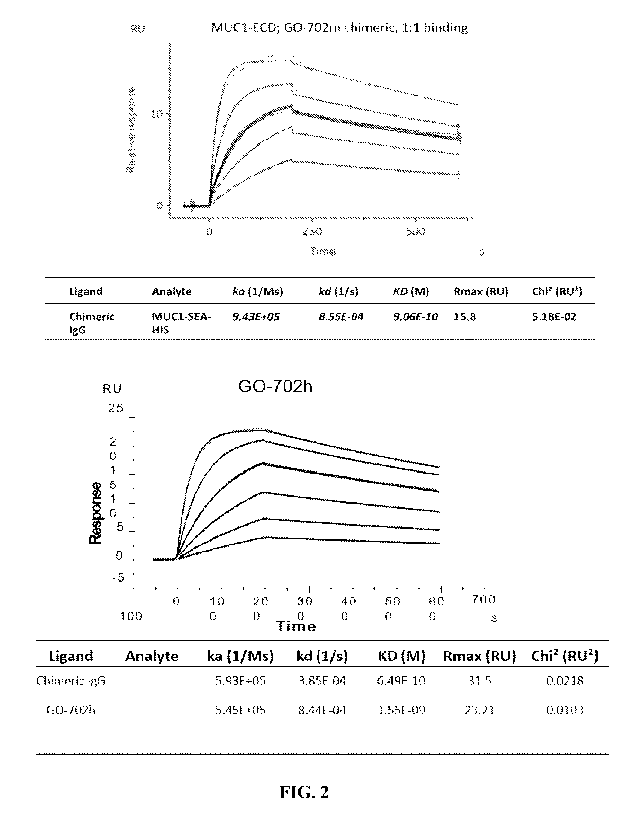
FIG. 2. Affinity measurement of chimeric antibody and humanized antibiody.
Real-time responses were shown with curves. Fitting of Biacore experimental
data to 1:1
interaction model was shown in black. The antigen concentrations used for the
top panel were
3 125 nM, 625 nM, 12S nM, 25 nM, 50 nM, respectively The antigen
concentrations used for
the bottom panel were 1.875 nM, 3.75 nM, 7.5 nM, 15 nM, 30 nM, 60 nM
respectively.
FIG. 3. SDS-PAGE results of selected antibody under non-reducing conditions.
Reducing conditions: Lane M (marker); Lane 1 (VH1+VL3); Lane 3 (VI-11+VL4);
Lane 5
(VH2+VL3); Lane 7 (VH5+VL1); Lane 9 (VH5+VL2); Lane 11 (VH5+VL3); Lane 13
(VH5+VH4). Non-reducing conditions. Lane 2 (VH1+VL3), Lane 4 (VH1+VL4), Lane 6
(VH2+VL3); Lane 8 (VH5+VL1); Lane 10 (VH5+VL2); Lane 12 (VH5+VL3); Lane 14
(VH5+VH4); Lane 15 (mouse IgG).
FIG. 4. Affinity measurement of Chimeric IgG and humanized IgGs. Real-time
responses were shown with colored curves. Fitting of Biacore experimental data
to 1:1
interaction modelwas shown in black. The antigen concentrations were 1.875 nM,
3.75 nM,
7.5 nM, 15 nM, 30 nM, 60 nM, respectively.
FIG. 5. FIG. 5. Affinity comparison of Chimeric IgG and humanized IgGs by
flow cytometry. Antibodies were incubated with HCT116/MUC1 cells and followed
by
incubation with secondary antibodies. Binding was analyzed by flow cytometry.
FIG. 6. Concentration dependent binding of mAbs to HCT116/MUC1. Wild-type
and afucosylated (AF) forms of both GO-702m and GO-702m/hFc chimera that
contains Fc
from human IgG1 were incubated with the cells at different concentrations (as
shown above)
followed by incubation with anti-hIgG-Biotin +Streptavidin-PE or anti-mouse
IgGk-FITC as
secondary reagents. Binding efficiency was indicated as mean fluorescence
Intensity (MFI) in
a concentration dependent manner.
FIG. 7. Unstained cells used as negative control for FIG. 6.
8
CA 03186181 2023- 1- 16
WO 2022/016190
PCT/US2021/070881
FIGS. 8A-B. GO-702m targets the alpha-4 helix. (FIG. 8A) The aa sequences of
the 58-aa human MUC1-C (SEQ ID NO: 2), cynomolgus monkey (SEQ ID NO: 38), and
mouse (SEQ ID NO: 39) Mudl-C extracellular domains. The a3 and a4 helices are
highlighted. Localization of the mAb GO-702m epitope to the GO helix, as shown
by NMR
spectroscopy of the p62/p58 heterodimer (Macao et al., 2006). (FIG. 8B) The
indicated
concentrations of mAb GO-702m were incubated with HCT116/vector or HCT116/MUC1
cells. Mean fluorescence intensity (MFI) was determined by flow cytometry.
Binding of
mAb GO-702m (middle bar in each column set) by ELISA to WT p58/p62 heterodimer
and
the S33A, R34G, Y35A, N36A mutant proteins for alpha-4 helix OR D19E/V20A/T22A
mutant proteins for alpha-3 helix. mAb CD1 (right bar in each column set) was
used as a
control. MAb 3D1 (left bar in each column set) was used as a control for alpha-
3-positive
binding. The results are expressed as percentage control binding as compared
with that
obtained with the WT protein (>3.0 OD units)
FIG. 9. Binding of GO-702mFc chimeric mAbs to HCT116/MUC1. Wild-type
chimera (G0-702m/hFc) and afucosylated form of the same that contains Fc from
human
IgG1 were incubated with the cells followed by incubation with anti-hIgG-
Biotin
+Streptavidin-PE or anti-mouse IgGk-FITC as secondary reagents. Binding
efficiency was
indicated as mean fluorescence Intensity (MFI).
FIGS. 10A-B. Concentration dependent binding of GO-702h to mouse Fc
receptor (FcRIV). (FIG. 10A) Wild-type and afucosylated (AF) forms of GO-702h
were
incubated with the cells at different concentrations (as shown above) followed
by incubation
with anti-human IgG-FITC as secondary reagents. Binding efficiency was
indicated as mean
fluorescence Intensity (MFI) in a concentration dependent manner. (FIG. 10B)
Wild-type
(diamond) and afucosylated (AF) form (square) of GO-702h were incubated with
the cells at
different concentrations followed by incubation with anti-human IgG-FITC as
secondary
reagents. Binding efficiency was indicated as mean fluorescence Intensity
(MFI) in a
concentration dependent manner. Right; a graphic representation of binding.
FIG. 11. Blood chemistry analysis of MUCLTg mice treated with the MAb (GO-
702m-AF). Five mg/kg MAb afucosylated GO-702m was injected i.p in MUC1 Tg mice
bearing MC-38/MUC1 tumors. Complete blood chemistry was performed to evaluate
any
toxicity of afucosylated GO-702m antibody.
FIG. 12. Hematology analysis of MUCT1-Tg mice treated with MAb (G0-702m-
AF). Five mg/kg MAb afucosylated GO-702m was injected i.p in MUC 1.Tg mice
bearing
9
CA 03186181 2023- 1- 16
WO 2022/016190
PCT/US2021/070881
MC-38/MUC1 tumors. Complete hematology analysis was performed to evaluate any
toxicity of afucosylated GO-702m antibody.
FIG. 13. Binding of GO-702m, GO-702h and GO-701m antibodies to HCT116-
MUC1 cells. Antibody binding to HCT116 cells over-expressed with human MUC1
was
analyzed by flow cytometry. Five ug of indicated anti-MUC1 antibody or isotype
control was
incubated with the cells for 60 minutes on ice. FITC-conjugated goat F(a1:02
anti-mouse or
anti-human immunoglobulin (depending on the primary antibody) was used as the
secondary
reagent. Antibody binding to the cell surface was analyzed using FACS Canto
II.
FIG. 14. Binding of wild-type and afucosylated GO-702h with HCT116/MUC1
cells. Antibody binding to HCT116 cells over-expressed with human MUC1 was
analyzed by
flow cytometry. GO-702h wild-type, GO-702h afucosylated or as negative control
CD1 anti-
MUC1 antibodies were incubated with the cells for 60 minutes on ice. FITC-
conjugated goat
F(ab')2 anti-human immunoglobulin was used as the secondary reagent Antibody
binding to
the cell surface was analyzed using FACS Canto II.
FIG. 15. Flow Cytometry of GO-702m with HCT116 + MUC-1. Antibody binding
to HCT116 cells with no MUC1 (Black) and HCT116 cells over-expressed with
human
MUC1 (grey) was analyzed by flow cytometry. GO-702m anti-MUC1 antibody was
incubated with the cells for 60 minutes on ice. FITC-conjugated goat F(a1302
anti-mouse
immunoglobulin was used as the secondary reagent. Antibody binding to the cell
surface was
analyzed using FACS Canto II.
FIG. 16. Flow Cytometry of GO-702m in ZR-75-1 cells. Antibody binding to
ZR-75-1 breast cancer cell line. GO-702m anti-MUC1 antibody (grey) or as
negative
control 1VIUC1 CD1 antibody (black) was incubated with the cells for 60
minutes on ice.
FITC-conjugated goat F(alp')2 anti-mouse immunoglobulin was used as the
secondary reagent.
Antibody binding to the cell surface was analyzed using FACS Canto II.
FIG. 17. Flow Cytometry of GO-702m in MCF-7/CshRNA vs MCF-
7/1VIUC 1 shRNA. Antibody binding to MCF-7/MUC1shRNA (black) or MCF-7/CshRNA
(grey) cells was analyzed by flow cytometry. GO-702m anti-MUC1 antibody was
incubated
with the cells for 60 minutes on ice. FITC-conjugated goat F(ab1)2 anti-mouse
immunoglobulin was used as the secondary reagent. Antibody binding to the cell
surface was
analyzed using FACS Canto II.
FIG. 18. Flow Cytometry of GO-702m in II-1975 NSCLC cells. Antibody binding
to H-1975 NSCLC cell line. GO-702m anti-MUC1 antibody (grey) or IgG as
negative control
CA 03186181 2023- 1- 16
WO 2022/016190
PCT/US2021/070881
(black) was incubated with the cells for 60 minutes on ice. FITC-conjugated
goat F(a13')2 anti-
mouse immunoglobulin was used as the secondary reagent. Antibody binding to
the cell
surface was analyzed using FACS Canto II.
FIG. 19. Flow Cytometry of GO-702m in MDA-MB-468 CshRNA/MUClshRNA.
Antibody binding to MDA-MB-468/MUCshRNA (right side grey peak) or MDA-MB-
468/CshRNA (left said grey peak) cells was analyzed by flow cytometry. IgG
used as
negative control (left side black peak). GO-702m anti-MUC1 antibody was
incubated with
the cells for 60 minutes on ice. FITC-conjugated goat F(abr)2 anti-mouse
immunoglobulin
was used as the secondary reagent. Antibody binding to the cell surface was
analyzed using
FACS Canto II.
FIG. 20. ADCC Activity of GO-702m (circles) and GO-702m-AF (HCT-1VITIC1;
squares). HCT116/MUC1 cells in a 96-well plate was incubated with Jurkat cells
(in which
antibody binding to FcRIV is linked to NFAT-mediated luciferase expression) as
the effector
cells at the E:T ratio of 20:1 for 6 hrs in the presence of indicated
antibodies starting from 1
[tg/m1 with 3-fold serial dlution. Luciferase activity was measured using
luciferin as the
substrate and plotted against concentration using Microsoft Excel.
FIG. 21. ADCC Activity of GO-702m-IgG2a on BT549. BT549 cells in a 96-well
plate was incubated with Jurkat cells (in which antibody binding to FcRIV is
linked to
NFAT-mediated luciferase expression) as the effector cells at the E:T ratio of
20:1 for 6 hrs
in the presence of indicated antibodies starting from 1 p.g/m1 with 3-fold
serial dlution.
Luciferase activity was measured using luciferin as the substrate and plotted
against
concentration using Microsoft Excel. Squares = afuscoylated GO-702m antibody;
triangles ¨
wt GO-702m antibody.
FIG. 22. Efficacy of afucosylated GO-702m in MUCLTg mice with MC-
38/1VIUCI tumors. MC-38 overexpressing MUCI cells were injected into MUCl.Tg
mice.
After 10-12 days, the mice were randomized into 2 different groups. Group 1:
vehicle
control; group 2: afucosylated GO-702m antibody 5 mg/kg once a week x 3 weeks
1P.
Tumor measurements were taken every other day. Diamonds: vehicle control group
(curve
shown as mean tumors for the group) and Circles: afucosylated GO-702m (curves
shown for
individual mouse). There was no significant change in the body weights.
Efficacy is shown
up to 84 days.
FIG. 23. ADCC in vitro studies with hG0702-AF antibody in comparison with
hG0-702 in HCT116/MUC1 colon carcinoma cells. HCT116/MUC1 cells in a 96-well
11
CA 03186181 2023- 1- 16
WO 2022/016190
PCT/US2021/070881
plate was incubated with Jurkat cells (in which antibody binding to FcRIV is
linked to
NEAT-mediated luciferase expression) as the effector cells at the E:T ratio of
20:1 for 6 hrs
in the presence of indicated antibodies starting from 1 p.g/m1 with 3-fold
serial dlution.
Luciferase activity was measured using luciferin as the substrate and plotted
against
concentration.
FIG. 24. ADCC in vivo studies with hG0702-AF antibody in comparison with
hG0-702. Six- to eight-week-old C57BL/6 mice were injected subcutaneously in
the flank
with 5 x 10⁵ MC38/MUC1, mouse colon carcinoma cells expressing human MUC1
(MC38/MUC1) in 100 pl of DMEM culture medium. Mice were randomized into two
treatment groups (6 mice for hG0-702-WT group and 7 mice for hG0-702
afucosylated (AF)
group). When the mean tumor volume reached 70-130 mm3, mice were treated with
5 mg/kg
of afucosylated humanized GO-702 (hG0-702-AF) or wild-type humanized GO-702
(hG0-
702-WT) once a week for 3 weeks IP Tumor measurements and body weights were
recorded
every other day. Mice were sacrificed when tumors reached >2,000 mm3 as
calculated by the
following formula: (width)2 X length/2. The results are expressed as tumor
volumes (mean +
SEM) against days of treatment.
12
CA 03186181 2023- 1- 16
WO 2022/016190
PCT/US2021/070881
DESCRIPTION OF ILLUSTRATIVE EMBODIMENTS
The inventors have generated antibodies against a 58 amino acid non-shed
portion of
the external domain of the MUC1-C protein. These antibodies have ben
demonstrated to
bind selectively to this portion of 1VIUC1-C, and as such, present an
opportunity to block the
activity of MUC1 following cleavage of the N-terminal region They also can be
used to
deliver therapeutic payloads to MUC1-expressing cancer cells even following
the cleavage of
the N-terminal MUC1 domain. These and other aspects of the disclosure are
described in
greater detail below.
I. MUC1
A. Structure
MUC 1 is a mucin-type glycoprotein that is expressed on the apical borders of
normal
secretory epithelial cells (Kufe et at., 1984). MUC1 forms a heterodimer
following synthesis
as a single polypeptide and cleavage of the precursor into two subunits in the
endoplasmic
reticulum (Ligtenberg et at., 1992). The cleavage may be mediated by an
autocatalytic
process (Levitan et at., 2005). The >250 kDa 1VIUC1 N-terminal (MUC1 N-ter,
MUC I-N)
subunit contains variable numbers of 20 amino acid tandem repeats that are
imperfect with
highly conserved variations and are modified by 0-linked glycans (Gendler et
at., 1988;
Siddiqui et at., 1988). MUC1-N is tethered to the cell surface by dimerization
with the -23
kDa C-terminal subunit (MUC1 C-ter, MUC1-C), which includes a 72-amino acid
cytoplasmic domain (MUC1-C/CD), a 28 amino acid tran sm em b ran e domain
(MUC1-
C/TMD), a 58 amino acid extracellular domain (MUC1-C/ECD) followed by a 62
amino acid
region the dimerizes together to form a SEA domain (Merlo et at., 1989). It is
the 58 amino
acid portion of the MUC1-C/ECD (italics) plays a major role in binding to the
antibodies of
the present disclosure. The human MUC1-C sequence is shown below:
SVVVOLTLAFREGTINVHDVETOFNOYKTEAASRYNLTISDVSVSDVPFPFSAOSGAGVPG
WGIALLVLVCVLVALAIVYLIALAVCQCRRKNYGOLDIFPARDTYHPMSEYPTYHT
HGRYVPP SSTDRSPYEKVSAGNGGSSLSYTNPAVAATSANL (SEQ ID NO: 1)
The bold sequence indicates the CD, and the underlined portion is an oligomer-
inhibiting
peptide. With transformation of normal epithelia to carcinomas, MUC1 is
aberrantly
overexpressed in the cytosol and over the entire cell membrane (Kufe et at.,
1984; Perey et
13
CA 03186181 2023- 1- 16
WO 2022/016190
PCT/US2021/070881
at., 1992). Cell membrane-associated MUC1 is targeted to endosomes by clathrin-
mediated
endocytosis (Kinlough et at., 2004). In addition, MUC1-C, but not MUC1-N, is
targeted to
the nucleus (Baldus et al, 2004; Huang et al., 2003; Li et al, 2003a; Li et
al., 2003b; Li et al.,
2003c; Wei et at., 2005; Wen et at., 2003) and mitochondria (Ren et at.,
2004).
B. Function
MUC1-C interacts with members of the ErbB receptor family (Li et at., 2001b;
Li et
at., 2003c; Schroeder et at., 2001) and with the Wnt effector, 13-catenin
(Yamamoto et at.,
1997). The epidermal growth factor receptor and c-Src phosphorylate the MUC1
cytoplasmic domain (MUC1-CD) on Y-46 and thereby increase binding of 1VIUC1
and 13-
catenin (Li et at., 2001a; Li et al, 2001b). Binding of MUC1 and f3-catenin is
also regulated
by glycogen synthase kinase 313 and protein kinase Co (Li et at., 1998; Ren et
at., 2002).
MUC1 colocalizes with 13-catenin in the nucleus (Baldus et at., 2004; Li et
aL, 2003a; Li et
at., 2003c; Wen et at., 2003) and coactivates transcription of Wnt target
genes (Huang et at.,
2003). Other studies have shown that MUC1 also binds directly to p53 and
regulates
transcription of p53 target genes (Wei et at., 2005). Notably, overexpression
of MUC1-C is
sufficient to induce anchorage-independent growth and tumorigenicity (Huang et
at., 2003;
Li et at., 2003b; Ren et at., 2002; Schroeder et at., 2004).
II. Producing Monoclonal Antibodies
The term "monoclonal antibody" as used herein refers to an antibody obtained
from a
population of substantially homogeneous antibodies, i.e., the individual
antibodies
comprising the population are identical except for possible naturally
occurring mutations that
may be present in minor amounts. Monoclonal antibodies are highly specific,
being directed
against a single antigenic site. Furthermore, in contrast to polyclonal
antibody preparations
that include different antibodies directed against different determinants
(epitopes), each
monoclonal antibody is directed against a single determinant on the antigen.
In addition to
their specificity, the monoclonal antibodies are advantageous in that they may
be synthesized
uncontaminated by other antibodies. The modifier "monoclonal" is not to be
construed as
requiring production of the antibody by any particular method. For example,
the monoclonal
antibodies useful in the present disclosure may be prepared by the hybridoma
methodology
first described by Kohler et at., Nature, 256:495 (1975), or may be made using
recombinant
DNA methods in bacterial, eukaryotic animal or plant cells (see, e.g., U.S.
Patent 4,816,567)
14
CA 03186181 2023- 1- 16
WO 2022/016190
PCT/US2021/070881
after single cell sorting of an antigen specific B cell, an antigen specific
plasmablast
responding to an infection or immunization, or capture of linked heavy and
light chains from
single cells in a bulk sorted antigen specific collection. The "monoclonal
antibodies" may
also be isolated from phage antibody libraries using the techniques described
in Clackson et
at., Nature, 352:624-628 (1991) and Marks et al., J. Mol. Biol., 222:581-597
(1991), for
example.
An "isolated antibody" is one that has been separated and/or recovered from a
component of its natural environment. Contaminant components of its natural
environment
are materials that would interfere with diagnostic or therapeutic uses for the
antibody, and
may include enzymes, hormones, and other proteinaceous or non-proteinaceous
solutes. In
particular embodiments, the antibody is purified: (1) to greater than 95% by
weight of
antibody as determined by the Lowly method, and most particularly more than
99% by
weight; (2) to a degree sufficient to obtain at least 15 residues of N-
terminal or internal amino
acid sequence by use of a spinning cup sequenator; or (3) to homogeneity by
SDS-PAGE
under reducing or non-reducing conditions using Coomassie blue or silver
stain. Isolated
antibody includes the antibody in situ within recombinant cells since at least
one component
of the antibody's natural environment will not be present. Ordinarily,
however, isolated
antibody will be prepared by at least one purification step.
The basic four-chain antibody unit is a heterotetrameric glycoprotein composed
of
two identical light (L) chains and two identical heavy (H) chains. An IgM
antibody consists
of 5 basic heterotetramer units along with an additional polypeptide called J
chain, and
therefore contain 10 antigen binding sites, while secreted IgA antibodies can
polymerize to
form polyvalent assemblages comprising 2-5 of the basic 4-chain units along
with J chain. In
the case of IgGs, the 4-chain unit is generally about 150,000 daltons. Each L
chain is linked
to an H chain by one covalent disulfide bond, while the two H chains are
linked to each other
by one or more disulfide bonds depending on the H chain isotype. Each H and L
chain also
has regularly spaced intrachain disulfide bridges. Each H chain has at the N-
terminus, a
variable region (VH) followed by three constant domains (CH) for each of the
alpha and
gamma chains and four CH domains for mu and isotypes. Each L chain has at the
N-terminus,
a variable region (VL) followed by a constant domain (CO at its other end. The
VL is aligned
with the VH and the CL is aligned with the first constant domain of the heavy
chain (CHO.
Particular amino acid residues are believed to form an interface between the
light chain and
heavy chain variable regions. The pairing of a VII and VL together forms a
single antigen-
CA 03186181 2023- 1- 16
WO 2022/016190
PCT/US2021/070881
binding site. For the structure and properties of the different classes of
antibodies, see, e.g.,
Basic and Clinical Immunology, 8th edition, Daniel P. Stites, Abba I. Terr and
Tristram G.
Parslow (eds.), Appleton & Lange, Norwalk, Conn., 1994, page 71, and Chapter
6.
The L chain from any vertebrate species can be assigned to one of two clearly
distinct
types, called kappa and lambda based on the amino acid sequences of their
constant domains
(CL). Depending on the amino acid sequence of the constant domain of their
heavy chains
(CH), immunoglobulins can be assigned to different classes or isotypes. There
are five classes
of immunoglobulins: IgA, IgD, IgE, IgG, and IgM, having heavy chains
designated alpha,
delta, epsilon, gamma and mu, respectively. They gamma and alpha classes are
further
divided into subclasses on the basis of relatively minor differences in CH
sequence and
function, humans express the following subclasses: IgG1 IgG2, IgG3, IgG4,
IgAl, and IgA2.
The term "variable" refers to the fact that certain segments of the V domains
differ
extensively in sequence among antibodies The V domain mediates antigen binding
and
defines specificity of a particular antibody for its particular antigen.
However, the variability
is not evenly distributed across the 110-amino acid span of the variable
regions. Instead, the
V regions consist of relatively invariant stretches called framework regions
(FRs) of 15-30
amino acids separated by shorter regions of extreme variability called
"hypervariable regions"
that are each 9-12 amino acids long. The variable regions of native heavy and
light chains
each comprise four FRs, largely adopting a beta-sheet configuration, connected
by three
hypervariable regions, which form loops connecting, and in some cases forming
part of, the
beta-sheet structure. The hypervariable regions in each chain are held
together in close
proximity by the FRs and, with the hypervariable regions from the other chain,
contribute to
the formation of the antigen-binding site of antibodies (see Kabat et al.,
Sequences of
Proteins of Immunological Interest, 5th Ed. Public Health Service, National
Institutes of
Health, Bethesda, Md. (1991)). The constant domains are not involved directly
in binding an
antibody to an antigen, but exhibit various effector functions, such as
participation of the
antibody in antibody dependent cellular cytotoxicity (ADCC), antibody-
dependent cellular
phagocytosis (ADCP), antibody-dependent neutrophil phagocytosis (ADNP), and
antibody-
dependent complement deposition (ADCD).
The term "hypervariable region" when used herein refers to the amino acid
residues of
an antibody that are responsible for antigen binding. The hypervariable region
generally
comprises amino acid residues from a "complementarity determining region" or
"CDR" (e.g.,
around about residues 24-34 (L1), 50-56 (L2) and 89-97 (L3) in the VL, and
around about 31-
16
CA 03186181 2023- 1- 16
WO 2022/016190
PCT/US2021/070881
35 (H1), 50-65 (H2) and 95-102 (H3) in the VH when numbered in accordance with
the Kabat
numbering system; Kabat et at., Sequences of Proteins of Immunological
Interest, 5th Ed.
Public Health Service, National Institutes of Health, Bethesda, Md. (1991));
and/or those
residues from a "hypervariable loop" (e.g., residues 24-34 (L1), 50-56 (L2)
and 89-97 (L3) in
the VL, and 26-32 (H1), 52-56 (H2) and 95-101 (H3) in the VH when numbered in
accordance
with the Chothia numbering system; Chothia and Lesk, J. Mol. Biol. 196:901-917
(1987));
and/or those residues from a "hypervariable loop"/CDR (e.g., residues 27-38
(L1), 56-65 (L2)
and 105-120 (L3) in the VL, and 27-38 (HI), 56-65 (H2) and 105-120 (H3) in the
VH when
numbered in accordance with the IMGT numbering system; Lefranc, M. P. et at.
Nucl. Acids
Res. 27:209-212 (1999), Ruiz, M. et at. Nucl. Acids Res. 28:219-221 (2000)).
Optionally the
antibody has symmetrical insertions at one or more of the following points 28,
36 (L1), 63,
74-75 (L2) and 123 (L3) in the VL, and 28, 36 (H1), 63, 74-75 (H2) and 123
(H3) in the
VsubH when numbered in accordance with AHo; Honneger, A and Plunkthun, A J Mol
Biol
309:657-670 (2001).
By "germline nucleic acid residue" is meant the nucleic acid residue that
naturally
occurs in a germline gene encoding a constant or variable region. "Germline
gene" is the
DNA found in a germ cell (i.e., a cell destined to become an egg or in the
sperm). A
"germline mutation" refers to a heritable change in a particular DNA that has
occurred in a
germ cell or the zygote at the single-cell stage, and when transmitted to
offspring, such a
mutation is incorporated in every cell of the body. A germline mutation is in
contrast to a
somatic mutation which is acquired in a single body cell. In some cases,
nucleotides in a
germline DNA sequence encoding for a variable region are mutated (i.e., a
somatic mutation)
and replaced with a different nucleotide.
A. General Methods
Antibodies to the MUC1-C/ECD may be produced by standard methods as are well
known in the art (see, e.g., Antibodies: A Laboratory Manual, Cold Spring
Harbor Laboratory,
1988; U.S. Patent 4,196,265). The methods for generating monoclonal antibodies
(MAbs)
generally begin along the same lines as those for preparing polyclonal
antibodies. The first
step for both these methods is immunization of an appropriate host or
identification of
subjects who are immune due to prior natural infection. As is well known in
the art, a given
composition for immunization may vary in its immunogenicity. It is often
necessary therefore
to boost the host immune system, as may be achieved by coupling a peptide or
polypeptide
17
CA 03186181 2023- 1- 16
WO 2022/016190
PCT/US2021/070881
immunogen to a carrier. Exemplary and preferred carriers are keyhole limpet
hemocyanin
(KLH) and bovine serum albumin (BSA). Other albumins such as ovalbumin, mouse
serum
albumin or rabbit serum albumin can also be used as carriers. Means for
conjugating a
polypeptide to a carrier protein are well known in the art and include
glutaraldehyde,
m-maleimidobencoyl-N-hydroxysuccinimide ester, carbodiimide and bis-biazotized
benzidine. As also is well known in the art, the immunogenicity of a
particular immunogen
composition can be enhanced by the use of non-specific stimulators of the
immune response,
known as adjuvants. Exemplary and preferred adjuvants include complete
Freund's adjuvant
(a non-specific stimulator of the immune response containing killed
Mycobacterium
tuberculosis), incomplete Freund's adjuvants and aluminum hydroxide adjuvant.
The amount of immunogen composition used in the production of polyclonal
antibodies varies upon the nature of the immunogen as well as the animal used
for
immunization A variety of routes can be used to administer the immunogen
(subcutaneous,
intramuscular, intradermal, intravenous and intraperitoneal). The production
of polyclonal
antibodies may be monitored by sampling blood of the immunized animal at
various points
following immunization. A second, booster injection, also may be given. The
process of
boosting and titering is repeated until a suitable titer is achieved. When a
desired level of
immunogenicity is obtained, the immunized animal can be bled and the serum
isolated and
stored, and/or the animal can be used to generate MAbs.
Following immunization, somatic cells with the potential for producing
antibodies,
specifically B lymphocytes (B cells), are selected for use in the MAb
generating protocol.
These cells may be obtained from biopsied spleens or lymph nodes, or from
circulating blood.
The antibody-producing B lymphocytes from the immunized animal are then fused
with cells
of an immortal myeloma cell, generally one of the same species as the animal
that was
immunized or human or human/mouse chimeric cells. Myeloma cell lines suited
for use in
hybridoma-producing fusion procedures preferably are non-antibody-producing,
have high
fusion efficiency, and enzyme deficiencies that render then incapable of
growing in certain
selective media which support the growth of only the desired fused cells
(hybridomas)
Any one of a number of myeloma cells may be used, as are known to those of
skill in
the art (Goding, pp. 65-66, 1986; Campbell, pp. 75-83, 1984). For example,
where the
immunized animal is a mouse, one may use P3-X63/Ag8, X63-Ag8.653, NS1/1.Ag 4
1,
Sp210-Ag14, FO, NSO/U, MPC-11, MPC11-X45-GTG 1.7 and S194/5XXO Bul; for rats,
one may use R210.RCY3, Y3-Ag 1.2.3, IR983F and 4B210; and U-266, GM1500-GRG2,
18
CA 03186181 2023- 1- 16
WO 2022/016190
PCT/US2021/070881
LICR-LON-HIVIy2 and UC729-6 are all useful in connection with human cell
fusions. One
particular murine myeloma cell is the NS-1 myeloma cell line (also termed P3-
NS-1-Ag4-1),
which is readily available from the NIGMS Human Genetic Mutant Cell Repository
by
requesting cell line repository number GM3573. Another mouse myeloma cell line
that may
be used is the 8-azaguanine-resistant mouse murine myeloma SP2/0 non-producer
cell line.
More recently, additional fusion partner lines for use with human B cells have
been described,
including KR12 (ATCC CRL-8658; K6H6/B5 (ATCC CRL-1823 SHIVI-D33 (ATCC CRL-
1668) and H1V1IVIA2.5 (Posner et al., 1987). The antibodies in this disclosure
were generated
using the SP2/0/mIL-6 cell line, an IL-6 secreting derivative of the SP2/0
line.
Methods for generating hybrids of antibody-producing spleen or lymph node
cells and
myeloma cells usually comprise mixing somatic cells with myeloma cells in a
2:1 proportion,
though the proportion may vary from about 20:1 to about 1:1, respectively, in
the presence of
an agent or agents (chemical or electrical) that promote the fusion of cell
membranes Fusion
methods using Sendai virus have been described by Kohler and Milstein (1975;
1976), and
those using polyethylene glycol (PEG), such as 37% (v/v) PEG, by Gefter et al.
(1977). The
use of electrically induced fusion methods also is appropriate (Goding, pp. 71-
74, 1986).
Fusion procedures usually produce viable hybrids at low frequencies, about 1 x
10-6 to
1 x 10⁻⁸. However, this does not pose a problem, as the viable, fused hybrids
are
differentiated from the parental, infused cells (particularly the infused
myeloma cells that
would normally continue to divide indefinitely) by culturing in a selective
medium. The
selective medium is generally one that contains an agent that blocks the de
novo synthesis of
nucleotides in the tissue culture media. Exemplary and preferred agents are
aminopterin,
methotrexate, and azaserine. Aminopterin and methotrexate block de 110V0
synthesis of both
purines and pyrimidines, whereas azaserine blocks only purine synthesis. Where
aminopterin
or methotrexate is used, the media is supplemented with hypoxanthine and
thymidine as a
source of nucleotides (HAT medium). Where azaserine is used, the media is
supplemented
with hypoxanthine. Ouabain is added if the B cell source is an Epstein Barr
virus (EBV)
transformed human B cell line, in order to eliminate EBV transformed lines
that have not
fused to the myeloma.
The preferred selection medium is HAT or HAT with ouabain. Only cells capable
of
operating nucleotide salvage pathways are able to survive in HAT medium. The
myeloma
cells are defective in key enzymes of the salvage pathway, e.g., hypoxanthine
phosphoribosyl
transferase (HPRT), and they cannot survive. The B cells can operate this
pathway, but they
19
CA 03186181 2023- 1- 16
WO 2022/016190
PCT/US2021/070881
have a limited life span in culture and generally die within about two weeks.
Therefore, the
only cells that can survive in the selective media are those hybrids formed
from myeloma and
B cells. When the source of B cells used for fusion is a line of EBV-
transformed B cells, as
here, ouabain is also used for drug selection of hybrids as EBV-transformed B
cells are
susceptible to drug killing, whereas the myeloma partner used is chosen to be
ouabain
resistant.
Culturing provides a population of hybridomas from which specific hybridomas
are
selected. Typically, selection of hybridomas is performed by culturing the
cells by
single-clone dilution in microtiter plates, followed by testing the individual
clonal
supernatants (after about two to three weeks) for the desired reactivity. The
assay should be
sensitive, simple and rapid, such as radioimmunoassays, enzyme immunoassays,
cytotoxicity
assays, plaque assays dot immunobinding assays, and the like.
The selected hybridomas are then serially diluted or single cell sorted by
flow
cytometric sorting and cloned into individual antibody-producing cell lines,
which clones can
then be propagated indefinitely to provide mAbs. The cell lines may be
exploited for MAb
production in two basic ways. A sample of the hybridoma can be injected (often
into the
peritoneal cavity) into an animal (e.g., a mouse). Optionally, the animals are
primed with a
hydrocarbon, especially oils such as pristane (tetramethylpentadecane) prior
to injection.
When human hybridomas are used in this way, it is optimal to inject
immunocompromised
mice, such as SCID mice, to prevent tumor rejection. The injected animal
develops tumors
secreting the specific monoclonal antibody produced by the fused cell hybrid.
The body
fluids of the animal, such as serum or ascites fluid, can then be tapped to
provide MAbs in
high concentration. The individual cell lines could also be cultured in vitro,
where the MAbs
are naturally secreted into the culture medium from which they can be readily
obtained in
high concentrations. Alternatively, human hybridoma cells lines can be used in
vitro to
produce immunoglobulins in cell supernatant. The cell lines can be adapted for
growth in
serum-free medium to optimize the ability to recover human monoclonal
immunoglobulins of
high purity.
MAbs produced by either means may be further purified, if desired, using
filtration,
centrifugation and various chromatographic methods such as FPLC or affinity
chromatography. Fragments of the monoclonal antibodies of the disclosure can
be obtained
from the purified monoclonal antibodies by methods which include digestion
with enzymes,
such as pepsin or papain, and/or by cleavage of disulfide bonds by chemical
reduction.
CA 03186181 2023- 1- 16
WO 2022/016190
PCT/US2021/070881
Alternatively, monoclonal antibody fragments encompassed by the present
disclosure can be
synthesized using an automated peptide synthesizer.
It also is contemplated that a molecular cloning approach may be used to
generate
monoclonals. For this, RNA can be isolated from the hybridoma line and the
antibody genes
obtained by RT-PCR and cloned into an immunoglobulin expression vector.
Alternatively,
combinatorial immunoglobulin phagemid libraries are prepared from RNA isolated
from the
cell lines and phagemids expressing appropriate antibodies are selected by
panning using
viral antigens. The advantages of this approach over conventional hybridoma
techniques are
that approximately 104 times as many antibodies can be produced and screened
in a single
round, and that new specificities are generated by H and L chain combination
which further
increases the chance of finding appropriate antibodies.
Other U.S. patents, each incorporated herein by reference, that teach the
production of
antibodies useful in the present disclosure include U.S. Patent 5,565,332,
which describes the
production of chimeric antibodies using a combinatorial approach; U.S. Patent
4,816,567
which describes recombinant immunoglobulin preparations; and U.S. Patent
4,867,973 which
describes antibody-therapeutic agent conjugates.
B. Antibodies of the Present Disclosure
Antibodies according to the present disclosure may be defined, in the first
instance, by
their binding specificity, i.e., the epitope to which the antibody binds. The
term "epitope"
refers to a site on an antigen to which B and/or T cells respond. B-cell
epitopes can he formed
both from contiguous amino acids or noncontiguous amino acids juxtaposed by
tertiary
folding of a protein . Epitopes formed from contiguous amino acids are
typically retained on
exposure to denaturing solvents, whereas epitopes formed by tertiary folding
are typically
lost on treatment with denaturing solvents. An epinve typically includes at
least 3, and more
usually, at least 5 or 8-10 amino acids in a unique spatial conformation.
In this case from the p58/p62 heterodimer, the major part of the epitope is
found in
MUC1-C/ECD, in particular:
SVVVQLTLAFREGTINVHDVETOFNQYKTEAASRYNLTISDVSVSDVPFPF SAQ S GA
21
CA 03186181 2023- 1- 16
WO 2022/016190
PCT/US2021/070881
(SEQ ID NO: 2). Those of skill in the art, by assessing the binding
specificity/affinity of a
given antibody using techniques well known to those of skill in the art, can
determine
whether such antibodies fall within the scope of the instant claims.
Modification-Assisted Profiling (MAP), also known as Antigen Structure-based
Antibody Profiling (ASAP) is a method that categorizes large numbers of
monoclonal
antibodies (mAbs) directed against the same antigen according to the
similarities of the
binding profile of each antibody to chemically or enzymatically modified
antigen surfaces
(see US 2004/0101920, herein specifically incorporated by reference in its
entirety). Each
category may reflect a unique epitope either distinctly different from or
partially overlapping
with epitope represented by another category. This technology allows rapid
filtering of
genetically identical antibodies, such that characterization can be focused on
genetically
distinct antibodies. When applied to hybridoma screening, MAP may facilitate
identification
of rare hybridoma clones that produce IllAbs having the desired
characteristics. MAP may be
used to sort the antibodies of the disclosure into groups of antibodies
binding different
epitopes.
The present disclosure includes antibodies that may bind to the same epitope,
or a
portion of the epitope. Likewise, the present disclosure also includes
antibodies that compete
for binding to a target or a fragment thereof with any of the specific
exemplaty antibodies
described herein. One can easily determine whether an antibody binds to the
same epitone
as, or competes for binding with, a reference antibody by using routine
methods known in the
art. For example, to determine if a test antibody binds to the same epitope as
a reference, the
reference antibody is allowed to bind to target under saturating conditions.
Next, the ability of
a test antibody to bind to the target molecule is assessed. If the test
antibody is able to bind to
the target molecule following saturation binding with the reference antibody,
it can be
concluded that the test antibody binds to a different epitope than the
reference antibody. On.
the other hand, if the test antibody is not able to bind to the target
molecule following
saturation binding with the reference antibody, then the test antibody may
bind to the same
epitope as the epitope bound by the reference antibody.
To determine if an antibody competes for binding with a reference anti-MUC1
antibody, the above-described binding methodology is performed in two
orientations: In a
first orientation, the reference antibody is allowed to bind to the MUC1
antigen under
saturating conditions followed by assessment of binding of the test antibody
to the MUC1
molecule. In a second orientation, the test antibody is allowed to bind to the
MUC1 antigen
22
CA 03186181 2023- 1- 16
WO 2022/016190
PCT/US2021/070881
molecule under saturating conditions followed by assessment of bindin.g of the
reference
antibody to the MUC1 molecule. If, in both orientations, only the first
(saturating) antibody is
capable of binding to the MUC1, then it is concluded that the test antibody
and the reference
antibody compete for binding to the MUCl. As will be appreciated by a person
of ordinary
skill in the art, an antibody that competes for binding with. a reference
antibody may not
necessarily bind to the identical epitope as the reference antibody but may
sterically block
binding of the reference antibody by binding an overlapping or adjacent
epitope.
Two antibodies bind to the same or overlapping epitope if each competitively
inhibits
(blocks) binding of the other to the antigen. That is, a 1-, 5-, 10-, 20- or
100-fold excess of
one antibody inhibits binding of the other by at least 50% but preferably 75%,
90% or even
99% as measured in a competitive binding assay (see, e.g., Junghans et al.,
Cancer Res. 1990
5014954502). Alternatively, two antibodies have the same epitope if
essentially all amino
acid mutations in the antigen that reduce or eliminate binding of one antibody
reduce or
eliminate binding of the other. Two antibodies have overl a.ppin.g epitopes if
some amino acid
mutations that reduce or eliminate binding of one antibody reduce or eliminate
binding of the
other.
Additional routine experimentation (e.g., peptide mutation and binding
analyses) can
then be carried out to confirm whether the observed lack of binding of the
test antibody is in
fact due to binding to the same epitope as the reference antibody or if Aerie
blocking (or
another phenomenon) is responsible for the lack of observed binding.
Experiments of this sort
can be performed using ELISA, :MA., surface plasmon resonance, flow cytometry
or any
other quantitative or qualitative antibody-binding assay available in the art.
Structural studies
with EM or crystallography also can demonstrate whether or not two antibodies
that compete
for binding recognize the same epitope.
In one embodiment, the antibody is an Immunoglobulin G (IgG) antibody isotype.
Representing approximately 75% of serum immunoglobulins in humans, IgG is the
most
abundant antibody isotype found in the circulation. IgG molecules are
synthesized and
secreted by plasma B cells. There are four IgG subclasses (IgGl, 2, 3, and 4)
in humans,
named in order of their abundance in serum (IgG1 being the most abundant). The
range from
having high to no affinity for the Fc receptor.
IgG is the main antibody isotype found in blood and extracellular fluid
allowing it to
control infection of body tissues. By binding many kinds of
pathogens¨representing viruses,
bacteria, and fungi¨IgG protects the body from infection. It does this via
several immune
23
CA 03186181 2023- 1- 16
WO 2022/016190
PCT/US2021/070881
mechanisms: IgG-mediated binding of pathogens causes their immobilization and
binding
together via agglutination; IgG coating of pathogen surfaces (known as
opsonization) allows
their recognition and ingestion by phagocytic immune cells; IgG activates the
classical
pathway of the complement system, a cascade of immune protein production that
results in
pathogen elimination; IgG also binds and neutralizes toxins. IgG also plays an
important role
in antibody-dependent cell-mediated cytotoxicity (ADCC) and intracellular
antibody-
mediated proteolysis, in which it binds to TRIIV121 (the receptor with
greatest affinity to IgG
in humans) in order to direct marked virions to the proteasome in the cytosol.
IgG is also
associated with Type II and Type III Hypersensitivity. IgG antibodies are
generated
following class switching and maturation of the antibody response and thus
participate
predominantly in the secondary immune response. IgG is secreted as a monomer
that is small
in size allowing it to easily perfuse tissues. It is the only isotype that has
receptors to facilitate
passage through the human placenta_ Along with IgA secreted in the breast
milk, residual IgG
absorbed through the placenta provides the neonate with humoral immunity
before its own
immune system develops. Colostrum contains a high percentage of IgG,
especially bovine
colostrum. In individuals with prior immunity to a pathogen, IgG appears about
24-48 hours
after antigenic stimulation.
In another aspect, the antibodies may be defined by their variable sequences
that
determine their binding specificity. Examples are provided below:
Table 1 ¨ Antibody CDR Sequences
Antibody Heavy Chain Light Chain
CDR1 - SYWMH (SEQ ID NO: 3) CDR1 - KASENVGTYVS
(SEQ ID
GO-7011m CDR2 - EINPSNGRTYYNENFKT (SEQ NO: 9)
ID NO: 4)
CDR2 - GASNRYT (SEQ ID NO: 10)
CDR3 - DGDYVSGFAY (SEQ ID NO: CDR3 - GQSYSYPWT (SEQ ID NO:
5) 11)
CDR1 - GFITNYFW (SEQ ID NO: 6) CDR' - CRASES V QY
SGISLMH
GO-702m CDR2 - ILPGTGST (SEQ ID NO: 7) (SEQ ID NO: 12)
CDR3 - RYDYTSSMDY (SEQ ID NO: CDR2 - GASNVET (SEQ ID NO: 13)
g)
CDR3 - QQNWKVPWT (SEQ ID NO.
14)
CDR1 - GFTFNYFW (SEQ ID NO: 6) CDR1 -
CRASESVQYSGTSLMH
GO-702h CDR2 - ILPGTGST (SEQ ID NO: 7) (SEQ ID NO: 12)
CDR3 - RYDYTSSMDY (SEQ ID NO: CDR2 - GASNVET (SEQ ID NO: 13)
8)
CDR3 - QQNWKVPWT (SEQ ID NO:
14)
24
CA 03186181 2023- 1- 16
WO 2022/016190
PCT/US2021/070881
Furthermore, the antibodies sequences may vary from the sequences provided
above,
optionally using methods discussed in greater detail below. For example, amino
sequences
may vary from those set out above in that (a) the variable regions may be
segregated away
from the constant domains of the light chains, (b) the amino acids may vary
from those set
out above while not drastically affecting the chemical properties of the
residues thereby (so-
called conservative substitutions), (c) the amino acids may vary from those
set out above by a
given percentage, e.g., 80%, 85%, 90%, 91%, 92%, 93%, 94%, 95%, 96%, 97%, 98%
or 99%
homology. Alternatively, the nucleic acids encoding the antibodies may (a) be
segregated
away from the constant domains of the light chains, (b) vary from those set
out above while
not changing the residues coded thereby, (c) may vary from those set out above
by a given
percentage, e.g., 70%, 75%, 80%, 85%, 90%, 91%, 92%, 93%, 94%, 95%, 96%, 97%,
98%
or 99% homology, or (d) vary from those set out above by virtue of the ability
to hybridize
under high stringency conditions, as exemplified by low salt and/or high
temperature
conditions, such as provided by about 0.02 M to about 0.15 M NaCl at
temperatures of about
50 C to about 70 C.
In making conservative changes in amino acid sequence, the hydropathic index
of
amino acids may be considered. The importance of the hydropathic amino acid
index in
conferring interactive biologic function on a protein is generally understood
in the art (Kyte
and Doolittle, 1982). It is accepted that the relative hydropathic character
of the amino acid
contributes to the secondary structure of the resultant protein, which in turn
defines the
interaction of the protein with other molecules, for example, enzymes,
substrates, receptors,
DNA, antibodies, antigens, and the like.
It also is understood in the art that the substitution of like amino acids can
be made
effectively on the basis of hydrophilicity. U.S. Patent 4,554,101,
incorporated herein by
reference, states that the greatest local average hydrophilicity of a protein,
as governed by the
hydrophilicity of its adjacent amino acids, correlates with a biological
property of the protein.
As detailed in U.S. Patent 4,554,101, the following hydrophilicity values have
been assigned
to amino acid residues: basic amino acids: arginine (+3.0), lysine (+3.0), and
histidine (-0.5);
acidic amino acids: aspartate (+3.0 1), glutamate (+3.0 1), asparagine
(+0.2), and
glutamine (+0.2); hydrophilic, nonionic amino acids: serine (+0.3), asparagine
(+0.2),
glutamine (+0.2), and threonine (-0.4), sulfur containing amino acids:
cysteine (-1.0) and
methionine (-1.3); hydrophobic, nonaromatic amino acids: valine (-1.5),
leucine (-1.8),
CA 03186181 2023- 1- 16
WO 2022/016190
PCT/US2021/070881
isoleucine (-1.8), proline (-0.5 1), alanine (-0.5), and glycine (0);
hydrophobic, aromatic
amino acids: tryptophan (-3.4), phenylalanine (-2.5), and tyrosine (-2.3).
It is understood that an amino acid can be substituted for another having a
similar
hydrophilicity and produce a biologically or immunologically modified protein.
In such
changes, the substitution of amino acids whose hydrophilicity values are
within 2 is
preferred, those that are within 1 are particularly preferred, and those
within 0.5 are even
more particularly preferred.
As outlined above, amino acid substitutions generally are based on the
relative
similarity of the amino acid side-chain substituents, for example, their
hydrophobicity,
hydrophilicity, charge, size, and the like. Exemplary substitutions that take
into consideration
the various foregoing characteristics are well known to those of skill in the
art and include:
arginine and lysine; glutamate and aspartate; serine and threonine; glutamine
and asparagine;
and valine, leucine and isoleucine.
Optimal alignment of sequences for comparison may be conducted using the
Megalign program in the Lasergene suite of bioinformatics software (DNASTAR,
Inc.,
Madison, Wis.), using default parameters. This program embodies several
alignment schemes
described in the following references: Dayhoff, M. 0. (1978) A model of
evolutionary
change in proteins--Matrices for detecting distant relationships. In Dayhoff,
M. 0. (ed.) Atlas
of Protein Sequence and Structure, National Biomedical Research Foundation,
Washington
D.C. Vol. 5, Suppl. 3, pp. 345-358; Hein J. (1990) Unified Approach to
Alignment and
Phylogeny pp. 626-645 Methods in Enzymology vol. 183, Academic Press, Inc.,
San Diego,
Calif.; Higgins, D. G. and Sharp, P.M. (1989) CABIOS 5:151-153; Myers, E. W.
and Muller
W. (1988) CABIOS 4:11-17; Robinson, E. D. (1971) Comb. Theor 11:105; Santou,
N. Nes,
M. (1987) Mol. Biol. Evol. 4:406-425; Sneath, P. H. A. and Sokal, R. R. (1973)
Numerical
Taxonomy--the Principles and Practice of Numerical Taxonomy, Freeman Press,
San
Francisco, Calif.; Wilbur, W. J. and Lipman, D. J. (1983) Proc. Natl. Acad.,
Sci. USA
80:726-730.
Alternatively, optimal alignment of sequences for comparison may be conducted
by
the local identity algorithm of Smith and Waterman (1981) Add. APL Math 2:482,
by the
identity alignment algorithm of Needleman and Wunsch (1970) J. Mol. Biol.
48:443, by the
search for similarity methods of Pearson and Lipman (1988) Proc. Natl. Acad.
Sci. USA 85:
2444, by computerized implementations of these algorithms (GAP, BESTFIT,
BLAST,
26
CA 03186181 2023- 1- 16
WO 2022/016190
PCT/US2021/070881
FASTA, and TFASTA in the Wisconsin Genetics Software Package, Genetics
Computer
Group (GCG), 575 Science Dr., Madison, Wis.), or by inspection.
One particular example of algorithms that are suitable for determining percent
sequence identity and sequence similarity are the BLAST and BLAST 2.0
algorithms, which
are described in Altschul et aL (1977) Nucl. Acids Res. 25:3389-3402 and
Altschul et al.
(1990) J. Mol. Biol. 215:403-410, respectively. BLAST and BLAST 2.0 can be
used, for
example, with the parameters described herein, to determine percent sequence
identity for the
polynucleotides and polypeptides of the disclosure. Software for performing
BLAST analyses
is publicly available through the National Center for Biotechnology
Information. The
rearranged nature of an antibody sequence and the variable length of each gene
requires
multiple rounds of BLAST searches for a single antibody sequence. Also, manual
assembly
of different genes is difficult and error-prone. The sequence analysis tool
IgBLAST (world-
wide-web at ncbi.nlm.nih.gov/igblast/) identifies matches to the germline V, D
and J genes,
details at rearrangement junctions, the delineation of Ig V domain framework
regions and
complementarity determining regions. IgBLAST can analyze nucleotide or protein
sequences
and can process sequences in batches and allows searches against the germline
gene
databases and other sequence databases simultaneously to minimize the chance
of missing
possibly the best matching germline V gene.
In one illustrative example, cumulative scores can be calculated using, for
nucleotide
sequences, the parameters M (reward score for a pair of matching residues;
always >0) and N
(penalty score for mismatching residues; always <0). Extension of the word
hits in each
direction are halted when: the cumulative alignment score falls off by the
quantity X from its
maximum achieved value; the cumulative score goes to zero or below, due to the
accumulation of one or more negative-scoring residue alignments; or the end of
either
sequence is reached. The BLAST algorithm parameters W, T and X determine the
sensitivity
and speed of the alignment. The BLASTTN program (for nucleotide sequences)
uses as
defaults a wordlength (W) of 11, and expectation (E) of 10, and the BLOSUM62
scoring
matrix (see Henikoff and Henikoff (1989) Proc. Natl. Acad. Sci. USA 89:10915)
alignments,
(B) of 50, expectation (E) of 10, M=5, N=-4 and a comparison of both strands.
For amino acid sequences, a scoring matrix can be used to calculate the
cumulative
score. Extension of the word hits in each direction are halted when: the
cumulative alignment
score falls off by the quantity X from its maximum achieved value; the
cumulative score goes
to zero or below, due to the accumulation of one or more negative-scoring
residue
27
CA 03186181 2023- 1- 16
WO 2022/016190
PCT/US2021/070881
alignments; or the end of either sequence is reached. The BLAST algorithm
parameters W, T
and X determine the sensitivity and speed of the alignment.
In one approach, the "percentage of sequence identity" is determined by
comparing
two optimally aligned sequences over a window of comparison of at least 20
positions,
wherein the portion of the polynucleotide or polypeptide sequence in the
comparison window
may comprise additions or deletions (i.e., gaps) of 20 percent or less,
usually 5 to 15 percent,
or 10 to 12 percent, as compared to the reference sequences (which does not
comprise
additions or deletions) for optimal alignment of the two sequences. The
percentage is
calculated by determining the number of positions at which the identical
nucleic acid bases or
amino acid residues occur in both sequences to yield the number of matched
positions,
dividing the number of matched positions by the total number of positions in
the reference
sequence (i.e., the window size) and multiplying the results by 100 to yield
the percentage of
sequence identity
Yet another way of defining an antibody is as a "derivative" of any of the
below-
described antibodies and their antigen-binding fragments. The term
"derivative" refers to an
antibody or antigen-binding fragment thereof that immunospecifically binds to
an antigen but
which comprises, one, two, three, four, five or more amino acid substitutions,
additions,
deletions or modifications relative to a "parental" (or wild-type) molecule.
Such amino acid
substitutions or additions may introduce naturally occurring (i.e., DNA-
encoded) or non-
naturally occurring amino acid residues. The term "derivative" encompasses,
for example, as
variants having altered CHL hinge, CH2, CH3 or CH4 regions, so as to form, for
example,
antibodies, etc., having variant Fc regions that exhibit enhanced or impaired
effector or
binding characteristics. The term "derivative" additionally encompasses non-
amino acid
modifications, for example, amino acids that may be glycosylated (e.g., have
altered
mannose, 2-N-acetylglucosamine, galactose, fucose, glucose, sialic acid, 5-N-
acetylneuraminic acid, 5-glycolneuraminic acid, etc. content), acetyl ated,
pegylated,
phosphorylated, amidated, derivatized by known protecting/blocking groups,
proteolytic
cleavage, linked to a cellular ligand or other protein, etc. In some
embodiments, the altered
carbohydrate modifications modulate one or more of the following:
solubilization of the
antibody, facilitation of subcellular transport and secretion of the antibody,
promotion of
antibody assembly, conformational integrity, and antibody-mediated effector
function. In a
specific embodiment, the altered carbohydrate modifications enhance antibody
mediated
effector function relative to the antibody lacking the carbohydrate
modification.
28
CA 03186181 2023- 1- 16
WO 2022/016190
PCT/US2021/070881
Carbohydrate modifications that lead to altered antibody mediated effector
function are well
known in the art (for example, see Shields, R. L. et al. (2002), J. Biol.
Chem. 277(30): 26733-
26740; Davies J. et al. (2001), Biotechnology & Bioengineering 74(4): 288-
294). Methods of
altering carbohydrate contents are known to those skilled in the art, see,
e.g., Wallick, S. C. et
at. (1988), J. Exp. Med. 168(3): 1099-1109; Tao, M. H. et at. (1989), J.
Immunol. 143(8):
2595-2601; Routledge, E. G. et at. (1995), Transplantation 60(8):847-53;
Elliott, S. et al
(2003), Nature Biotechnol. 2L44-2; Shields, R. L. et at. (2002), J. Biol.
Chem. 277(30):
26733-26740).
A derivative antibody or antibody fragment can be generated with an engineered
sequence or glycosylation state to confer preferred levels of activity in
antibody dependent
cellular cytotoxi city (ADCC), antibody-dependent cellular phagocytosis
(ADCP), antibody-
dependent neutrophil phagocytosis (ADNP), or antibody-dependent complement
deposition
(ADCD) functions as measured by bead-based or cell-based assays or in vivo
studies in
animal models.
A derivative antibody or antibody fragment may be modified by chemical
modifications using techniques known to those of skill in the art, including,
but not limited to,
specific chemical cleavage, acetylation, formulation, metabolic synthesis of
tunicamycin, etc.
In one embodiment, an antibody derivative will possess a similar or identical
function as the
parental antibody. In another embodiment, an antibody derivative will exhibit
an altered
activity relative to the parental antibody. For example, a derivative antibody
(or fragment
thereof) can bind to its epitope more tightly or be more resistant to
proteolysis than the
parental antibody.
C. Engineering of Antibody Sequences
In various embodiments, one may choose to engineer sequences of the identified
antibodies for a variety of reasons, such as improved expression, improved
cross-reactivity,
diminished off-target binding or abrogation of one or more natural effector
functions, such as
activation of complement or recruitment of immune cells (e.g., T cells). In
particular, IgM
antibodies may be converted to IgG antibodies. The following is a general
discussion of
relevant techniques for antibody engineering.
Hybridomas may be cultured, then cells lysed, and total RNA extracted. Random
hexamers may be used with RT to generate cDNA copies of RNA, and then PCR
performed
using a multiplex mixture of PCR primers expected to amplify all human
variable gene
29
CA 03186181 2023- 1- 16
WO 2022/016190
PCT/US2021/070881
sequences. PCR product can be cloned into pGEM-T Easy vector, then sequenced
by
automated DNA sequencing using standard vector primers. Assay of binding and
neutralization may be performed using antibodies collected from hybridoma
supernatants and
purified by FPLC, using Protein G columns. Recombinant full-length IgG
antibodies can be
generated by subcloning heavy and light chain Fv DNAs from the cloning vector
into a
Lonza pConIgG1 or pConK2 plasmid vector, transfected into 293 Freestyle cells
or Lonza
CHO cells, and collected and purified from the CHO cell supernatant.
The rapid availability of antibody produced in the same host cell and cell
culture
process as the final cGMP manufacturing process has the potential to reduce
the duration of
process development programs. Lonza has developed a generic method using
pooled
transfectants grown in CDACF medium, for the rapid production of small
quantities (up to 50
g) of antibodies in CHO cells. Although slightly slower than a true transient
system, the
advantages include a higher product concentration and use of the same host and
process as
the production cell line. Example of growth and productivity of GS-CHO pools,
expressing a
model antibody, in a disposable bioreactor: in a disposable bag bioreactor
culture (5 L
working volume) operated in fed-batch mode, a harvest antibody concentration
of 2 g/L was
achieved within 9 weeks of transfection.
pCon Vectors are an easy way to re-express whole antibodies. The constant
region
vectors are a set of vectors offering a range of immunoglobulin constant
region vectors
cloned into the pEE vectors. These vectors offer easy construction of full-
length antibodies
with human constant regions and the convenience of the GS SystemTM.
Antibody molecules will comprise fragments (such as F(ab'), F(ab')2) that are
produced, for example, by the proteolytic cleavage of the mAbs, or single-
chain
immunoglobulins producible, for example, via recombinant means. Such antibody
derivatives
are monovalent. In one embodiment, such fragments can be combined with one
another, or
with other antibody fragments or receptor ligands to form "chimeric" binding
molecules.
Significantly, such chimeric molecules may contain sub stituents capable of
binding to
different epitopes of the same molecule.
It may be desirable to "humanize" antibodies produced in non-human hosts in
order to
attenuate any immune reaction when used in human therapy. Such humanized
antibodies may
be studied in an in vitro or an in vivo context. Humanized antibodies may be
produced, for
example by replacing an immunogenic portion of an antibody with a
corresponding, but non-
immunogenic portion (i.e., chimeric antibodies). PCT Application
PCT/US86/02269; EP
CA 03186181 2023- 1- 16
WO 2022/016190
PCT/US2021/070881
Application 184,187; EP Application 171,496; EP Application 173,494; PCT
Application
WO 86/01533; EP Application 125,023; Sun et al. (1987); Wood et al. (1985);
and Shaw et
al. (1988); all of which references are incorporated herein by reference.
General reviews of
"humanized" chimeric antibodies are provided by Morrison (1985); also
incorporated herein
by reference. "Humanized" antibodies can alternatively be produced by CDR or
CEA
substitution. Jones et at. (1986); Verhoeyen et at. (1988); Beidler et at.
(1988); all of which
are incorporated herein by reference.
In related embodiments, the antibody is a derivative of the disclosed
antibodies, e.g.,
an antibody comprising the CDR sequences identical to those in the disclosed
antibodies (e.g.,
a chimeric, humanized or CDR-grafted antibody). In yet a further embodiment,
the antibody
is a fully human recombinant antibody.
Fc modification. The present disclosure also contemplates isotype
modification. By
modifying the Fc region to have a different isotype, different functionalities
can be achieved
For example, changing to IgGi can increase antibody dependent cell
cytotoxicity, switching
to class A can improve tissue distribution, and switching to class M can
improve valency.
Alternatively or additionally, it may be useful to combine amino acid
modifications
with one or more further amino acid modifications that alter Clq binding
and/or the
complement dependent cytotoxicity (CDC) function of the Fc region of an IL-
23p19 binding
molecule. The binding polypeptide of particular interest may be one that binds
to Clq and
displays complement dependent cytotoxicity. Polypeptides with pre-existing Clq
binding
activity, optionally further having the ability to mediate CDC may be modified
such that one
or both of these activities are enhanced. Amino acid modifications that alter
Clq and/or
modify its complement dependent cytotoxicity function are described, for
example, in
WO/0042072, which is hereby incorporated by reference.
One can design an Fc region of an antibody with altered effector function,
e.g., by
modifying Cl q binding and/or FcyR binding and thereby changing CDC activity
and/or
ADCC activity. "Effector functions" are responsible for activating or
diminishing a biological
activity (e.g., in a subject). Examples of effector functions include, but are
not limited to: Clq
binding; complement dependent cytotoxicity (CDC); Fc receptor binding;
antibody-
dependent cell-mediated cytotoxicity (ADCC); phagocytosis; down regulation of
cell surface
receptors (e.g., B cell receptor; BCR), etc. Such effector functions may
require the Fc region
to be combined with a binding domain (e.g., an antibody variable domain) and
can be
assessed using various assays (e.g., Fc binding assays, ADCC assays, CDC
assays, etc.).
31
CA 03186181 2023- 1- 16
WO 2022/016190
PCT/US2021/070881
For example, one can generate a variant Fc region of an antibody with improved
Clq
binding and improved FcyRIII binding (e.g., having both improved ADCC activity
and
improved CDC activity). Alternatively, if it is desired that effector function
be reduced or
ablated, a variant Fc region can be engineered with reduced CDC activity
and/or reduced
ADCC activity. In other embodiments, only one of these activities may be
increased, and,
optionally, also the other activity reduced (e.g., to generate an Fc region
variant with
improved ADCC activity, but reduced CDC activity and vice versa).
FcRn binding. Fc mutations can also be introduced and engineered to alter
their
interaction with the neonatal Fc receptor (FcRn) and improve their
pharmacokinetic
properties. A collection of human Fc variants with improved binding to the
FcRn have been
described (Shields et al., (2001). High resolution mapping of the binding site
on human IgG1
for FcyRI, FcyRII, FcyRIII, and FcRn and design of IgG1 variants with improved
binding to
the FcyR, (J Biol. Chem 276-6591-6604) A number of methods are known that can
result in
increased half-life (Kuo and Aveson, (2011)), including amino acid
modifications may be
generated through techniques including alanine scanning mutagenesis, random
mutagenesis
and screening to assess the binding to the neonatal Fc receptor (FcRn) and/or
the in vivo
behavior. Computational strategies followed by mutagenesis may also be used to
select one
of amino acid mutations to mutate.
The present disclosure therefore provides a variant of an antigen binding
protein with
optimized binding to FcRn. In a particular embodiment, the said variant of an
antigen binding
protein comprises at least one amino acid modification in the Fc region of
said antigen
binding protein, wherein said modification is selected from the group
consisting of 226, 227,
228, 230, 231, 233, 234, 239, 241, 243, 246, 250, 252, 256, 259, 264, 265,
267, 269, 270,
276, 284, 285, 288, 289, 290, 291, 292, 294, 297, 298, 299, 301, 302, 303,
305, 307, 308,
309, 311, 315, 317, 320, 322, 325, 327, 330, 332, 334, 335, 338, 340, 342,
343, 345, 347,
350, 352, 354, 355, 356, 359, 360, 361, 362, 369, 370, 371, 375, 378, 380,
382, 384, 385,
386, 387, 389, 390, 392, 393, 394, 395, 396, 397, 398, 399, 400, 401 403, 404,
408, 411, 412,
414, 415, 416, 418, 419, 420, 421, 422, 424, 426, 428, 433, 434, 438, 439,
440, 443, 444,
445, 446 and 447 of the Fc region as compared to said parent polypeptide,
wherein the
numbering of the amino acids in the Fc region is that of the EU index in
Kabat. In a further
aspect of the disclosure the modifications are M252Y/S254T/T256E.
Additionally, various publications describe methods for obtaining
physiologically
active molecules whose half-lives are modified, see for example Kontermann
(2009) either
32
CA 03186181 2023- 1- 16
WO 2022/016190
PCT/US2021/070881
by introducing an FcRn-binding polypeptide into the molecules or by fusing the
molecules
with antibodies whose FcRn-binding affinities are preserved but affinities for
other Fc
receptors have been greatly reduced or fusing with FcRn binding domains of
antibodies.
Derivatized antibodies may be used to alter the half-lives (e.g., serum half-
lives) of
parental antibodies in a mammal, particularly a human. Such alterations may
result in a half-
life of greater than 15 days, preferably greater than 20 days, greater than 25
days, greater than
30 days, greater than 35 days, greater than 40 days, greater than 45 days,
greater than 2
months, greater than 3 months, greater than 4 months, or greater than 5
months. The
increased half-lives of the antibodies of the present disclosure or fragments
thereof in a
mammal, preferably a human, results in a higher serum titer of said antibodies
or antibody
fragments in the mammal, and thus reduces the frequency of the administration
of said
antibodies or antibody fragments and/or reduces the concentration of said
antibodies or
antibody fragments to be administered Antibodies or fragments thereof having
increased in
vivo half-lives can be generated by techniques known to those of skill in the
art. For example,
antibodies or fragments thereof with increased in vivo half-lives can be
generated by
modifying (e.g., substituting, deleting or adding) amino acid residues
identified as involved in
the interaction between the Fc domain and the FcRn receptor.
Beltramello et al. (2010) previously reported the modification of neutralizing
mAbs,
due to their tendency to enhance dengue virus infection, by generating in
which leucine
residues at positions 1.3 and 1.2 of CH2 domain (according to the IMGT unique
numbering
for C-domain) were substituted with alanine residues. This modification, also
known as
"LALA" mutation, abolishes antibody binding to FcyRI, FcyRII and FcyRIIIa, as
described
by Hesse11 et al. (2007). The variant and unmodified recombinant mAbs were
compared for
their capacity to neutralize and enhance infection by the four dengue virus
serotypes. LALA
variants retained the same neutralizing activity as unmodified mAb but were
completely
devoid of enhancing activity. LALA mutations of this nature are therefore
contemplated in
the context of the presently disclosed antibodies.
Altered Glycosylation. A particular embodiment of the present disclosure is an
isolated monoclonal antibody, or antigen binding fragment thereof, containing
a substantially
homogeneous glycan without sialic acid, galactose, or fucose. The monoclonal
antibody
comprises a heavy chain variable region and a light chain variable region,
both of which may
be attached to heavy chain or light chain constant regions respectively. The
aforementioned
33
CA 03186181 2023- 1- 16
WO 2022/016190
PCT/US2021/070881
substantially homogeneous glycan may be covalently attached to the heavy chain
constant
region.
Another embodiment of the present disclosure comprises a mAb with a novel Fc
glycosylation pattern. The isolated monoclonal antibody, or antigen binding
fragment thereof,
is present in a substantially homogenous composition represented by the GNGN
or G1/G2
glycoform. Fc glycosylation plays a significant role in anti-viral and anti-
cancer properties of
therapeutic mAbs. The disclosure is in line with a recent study that shows
increased anti-
lentivirus cell-mediated viral inhibition of a fucose free anti-HIV mAb in
vitro. This
embodiment of the present disclosure with homogenous glycans lacking a core
fucose,
showed increased protection against specific viruses by a factor greater than
two-fold.
Elimination of core fucose dramatically improves the ADCC activity of mAbs
mediated by
natural killer (NK) cells but appears to have the opposite effect on the ADCC
activity of
polymorphonucl ear cells (PMNs)
The isolated monoclonal antibody, or antigen binding fragment thereof,
comprising a
substantially homogenous composition represented by the GNGN or G1/G2
glycoform
exhibits increased binding affinity for Fc gamma RI and Fc gamma RIII compared
to the
same antibody without the substantially homogeneous GNGN glycoform and with
GO, G1F,
G2F, GNF, GNGNF or GNGNFX containing glycoforms. In one embodiment of the
present
disclosure, the antibody dissociates from Fc gamma RI with a Kd of 1 x 10⁻⁸ M
or less and
from Fc gamma RIII with a Kd of 1 x 10⁻⁷M or less.
Glycosylation of an Fc region is typically either N-linked or 0-linked. N-
linked refers
to the attachment of the carbohydrate moiety to the side chain of an
asparagine residue. 0-
linked glycosylation refers to the attachment of one of the sugars N-
acetylgalactosamine,
galactose, or xylose to a hydroxyamino acid, most commonly serine or
threonine, although 5-
hydroxyproline or 5-hydroxylysine may also be used. The recognition sequences
for
enzymatic attachment of the carbohydrate moiety to the asparagine side chain
peptide
sequences are asparagine-X-serine and asparagine-X-threonine, where X is any
amino acid
except proline. Thus, the presence of either of these peptide sequences in a
polypeptide
creates a potential glycosylation site.
The glycosylation pattern may be altered, for example, by deleting one or more
glycosylation site(s) found in the polypeptide, and/or adding one or more
glycosylation site(s)
that are not present in the polypeptide. Addition of glycosylation sites to
the Fc region of an
antibody is conveniently accomplished by altering the amino acid sequence such
that it
34
CA 03186181 2023- 1- 16
WO 2022/016190
PCT/US2021/070881
contains one or more of the above-described tripeptide sequences (for N-linked
glycosylation
sites). An exemplary glycosylation variant has an amino acid substitution of
residue Asn 297
of the heavy chain. The alteration may also be made by the addition of, or
substitution by,
one or more serine or threonine residues to the sequence of the original
polypeptide (for 0-
linked glycosylation sites). Additionally, a change of Asn 297 to Ala can
remove one of the
glycosylation sites.
In certain embodiments, the antibody is expressed in cells that express beta
(1,4)-N-
acetylglucosaminyltransferase III (GnT III), such that GnT III adds GlcNAc to
the IL-23p19
antibody. Methods for producing antibodies in such a fashion are provided in
WO/9954342,
WO/03011878, patent publication 20030003097A1, and Umana etal., Nature
Biotechnology,
17:176-180, February 1999. Cell lines can be altered to enhance or reduce or
eliminate
certain post-translational modifications, such as glycosylation, using genome
editing
technology such as Clustered Regularly Interspaced Short Palindromic Repeats
(CRISPR)
For example, CRISPR technology can be used to eliminate genes encoding
glycosylating
enzymes in 293 or CHO cells used to express recombinant monoclonal antibodies.
Elimination of monoclonal antibody protein sequence liabilities. It is
possible to
engineer the antibody variable gene sequences obtained from human B cells to
enhance their
manufacturability and safety. Potential protein sequence liabilities can be
identified by
searching for sequence motifs associated with sites containing:
1) Unpaired Cys residues,
2) N-linked glycosylation,
3) Asn deamidation,
4) Asp isomerization,
5) SYE truncation,
6) Met oxidation,
7) Trp oxidation,
8) N-terminal glutamate,
9) Integrin binding,
10) CD11c/CD18 binding, or
11) Fragmentation
Such motifs can be eliminated by altering the synthetic gene for the cDNA
encoding
recombinant antibodies.
CA 03186181 2023- 1- 16
WO 2022/016190
PCT/US2021/070881
Protein engineering efforts in the field of development of therapeutic
antibodies
clearly reveal that certain sequences or residues are associated with
solubility differences
(Fernandez-Escamilla et al., Nature Biotech., 22 (10), 1302-1306, 2004;
Chennamsetty et al.,
PNAS, 106 (29), 11937-11942, 2009; Voynov et at., Biocon. Chem., 21(2), 385-
392, 2010)
Evidence from solubility-altering mutations in the literature indicate that
some hydrophilic
residues such as aspartic acid, glutamic acid, and serine contribute
significantly more
favorably to protein solubility than other hydrophilic residues, such as
asparagine, glutamine,
threonine, lysine, and arginine.
Stability. Antibodies can be engineered for enhanced biophysical properties.
One can
use elevated temperature to unfold antibodies to determine relative stability,
using average
apparent melting temperatures. Differential Scanning Cal orimetry (DSC)
measures the heat
capacity, Cp, of a molecule (the heat required to warm it, per degree) as a
function of
temperature One can use DSC to study the thermal stability of antibodies DSC
data for
mAbs is particularly interesting because it sometimes resolves the unfolding
of individual
domains within the mAb structure, producing up to three peaks in the
thermogram (from
unfolding of the Fab, Ci42, and C143 domains). Typically unfolding of the Fab
domain
produces the strongest peak. The DSC profiles and relative stability of the Fe
portion show
characteristic differences for the human IgGi, IgG2, IgG3, and IgG4 subclasses
(Garber and
Demarest, Biochem. Biophys. Res. Commun. 355, 751-757, 2007). One also can
determine
average apparent melting temperature using circular dichroism (CD), performed
with a CD
spectrometer. Far-UV CD spectra will be measured for antibodies in the range
of 200 to 260
nm at increments of 0.5 nm. The final spectra can be determined as averages of
20
accumulations. Residue ellipticity values can be calculated after background
subtraction.
Thermal unfolding of antibodies (0.1 mg/mL) can be monitored at 235 nm from 25-
95 C and
a heating rate of 1 C/min. One can use dynamic light scattering (DLS) to
assess for
propensity for aggregation. DLS is used to characterize size of various
particles including
proteins. If the system is not disperse in size, the mean effective diameter
of the particles can
be determined. This measurement depends on the size of the particle core, the
size of surface
structures, and particle concentration Since DLS essentially measures
fluctuations in
scattered light intensity due to particles, the diffusion coefficient of the
particles can be
determined. DLS software in commercial DLA instruments displays the particle
population at
different diameters. Stability studies can be done conveniently using DLS. DLS
measurements of a sample can show whether the particles aggregate over time or
with
36
CA 03186181 2023- 1- 16
WO 2022/016190
PCT/US2021/070881
temperature variation by determining whether the hydrodynamic radius of the
particle
increases. If particles aggregate, one can see a larger population of
particles with a larger
radius. Stability depending on temperature can be analyzed by controlling the
temperature in
situ. Capillary electrophoresis (CE) techniques include proven methodologies
for determining
features of antibody stability. One can use an iCE approach to resolve
antibody protein
charge variants due to deamidation, C-terminal lysines, sialylation,
oxidation, glycosylation,
and any other change to the protein that can result in a change in pI of the
protein. Each of the
expressed antibody proteins can be evaluated by high throughput, free solution
isoelectric
focusing (IEF) in a capillary column (cIEF), using a Protein Simple Maurice
instrument.
Whole-column UV absorption detection can be performed every 30 seconds for
real time
monitoring of molecules focusing at the isoelectric points (pIs). This
approach combines the
high resolution of traditional gel IEF with the advantages of quantitation and
automation
found in column-based separations while eliminating the need for a
mobilization step The
technique yields reproducible, quantitative analysis of identity, purity, and
heterogeneity
profiles for the expressed antibodies. The results identify charge
heterogeneity and molecular
sizing on the antibodies, with both absorbance and native fluorescence
detection modes and
with sensitivity of detection down to 0.7 mg/mL.
Solubility. One can determine the intrinsic solubility score of antibody
sequences.
The intrinsic solubility scores can be calculated using CamSol Intrinsic
(Sormanni et al., J
Mol Biol 427, 478-490, 2015). The amino acid sequences for residues 95-102
(Kabat
numbering) in HCDR3 of each antibody fragment such as a scFy can be evaluated
via the
online program to calculate the solubility scores. One also can determine
solubility using
laboratory techniques. Various techniques exist, including addition of
lyophilized protein to a
solution until the solution becomes saturated and the solubility limit is
reached, or
concentration by ultrafiltration in a microconcentrator with a suitable
molecular weight cut-
off. The most straightforward method is induction of amorphous precipitation,
which
measures protein solubility using a method involving protein precipitation
using ammonium
sulfate (Trevino et al., J Mol Biol, 366: 449-460, 2007). Ammonium sulfate
precipitation
gives quick and accurate information on relative solubility values. Ammonium
sulfate
precipitation produces precipitated solutions with well-defined aqueous and
solid phases and
requires relatively small amounts of protein. Solubility measurements
performed using
induction of amorphous precipitation by ammonium sulfate also can be done
easily at
37
CA 03186181 2023- 1- 16
WO 2022/016190
PCT/US2021/070881
different pH values. Protein solubility is highly pH dependent, and pH is
considered the most
important extrinsic factor that affects solubility.
Autoreactivity. Generally, it is thought that autoreactive clones should be
eliminated
during ontogeny by negative selection, however it has become clear that many
human
naturally occurring antibodies with autoreactive properties persist in adult
mature repertoires,
and the autoreactivity may enhance the antiviral function of many antibodies
to pathogens. It
has been noted that HCDR3 loops in antibodies during early B cell development
are often
rich in positive charge and exhibit autoreactive patterns (Wardemann et at.,
Science 301,
1374-1377, 2003). One can test a given antibody for autoreactivity by
assessing the level of
binding to human origin cells in microscopy (using adherent HeLa or HEp-2
epithelial cells)
and flow cytometric cell surface staining (using suspension Jurkat T cells and
293S human
embryonic kidney cells) Autoreactivity also can be surveyed using assessment
of binding to
tissues in tissue arrays
Preferred residues ("Human Likeness"). B cell repertoire deep sequencing of
human B cells from blood donors is being performed on a wide scale in many
recent studies.
Sequence information about a significant portion of the human antibody
repertoire facilitates
statistical assessment of antibody sequence features common in healthy humans.
With
knowledge about the antibody sequence features in a human recombined antibody
variable
gene reference database, the position specific degree of "Human Likeness" (HL)
of an
antibody sequence can be estimated. HL has been shown to be useful for the
development of
antibodies in clinical use, like therapeutic antibodies or antibodies as
vaccines. The goal is to
increase the human likeness of antibodies to reduce potential adverse effects
and anti-
antibody immune responses that will lead to significantly decreased efficacy
of the antibody
drug or can induce serious health implications. One can assess antibody
characteristics of the
combined antibody repertoire of three healthy human blood donors of about 400
million
sequences in total and created a novel "relative Human Likeness" (rHL) score
that focuses on
the hypervariable region of the antibody. The rEIL score allows one to easily
distinguish
between human (positive score) and non-human sequences (negative score).
Antibodies can
be engineered to eliminate residues that are not common in human repertoires
Modified antibodies may be made by any technique known to those of skill in
the art,
including expression through standard molecular biological techniques, or the
chemical
synthesis of polypeptides. Methods for recombinant expression are addressed
elsewhere in
this document.
38
CA 03186181 2023- 1- 16
WO 2022/016190
PCT/US2021/070881
D. Expression
Nucleic acids according to the present disclosure will encode antibodies,
optionally
linked to other protein sequences. As used in this application, the term -a
nucleic acid
encoding a MUC1-C antibody" refers to a nucleic acid molecule that has been
isolated free of
total cellular nucleic acid. In certain embodiments, the disclosure concerns
antibodies that
are encoded by any of the sequences set forth herein.
TABLE 2- CODONS
Amino Acids Codons
Alanine Ala A GCA GCC GCG GCU
Cysteine Cys C UGC UGU
Aspartic acid Asp D GAC GAU
Glutamic acid Glu E GAA GAG
Phenylalanine Phe F UUC UUU
Glycine Gly G GGA GGC GGG GGU
Histidine His H CAC CAU
Isoleucine Ile I AUA AUC AUU
Lysine Lys K AAA AAG
Leucine Leu L UUA UUG CUA CUC CUG CUU
Methionine Met M AUG
Asparagine Asn N AAC AAU
Proline Pro P CCA CCC CCG CCU
Glutamine Gln Q CAA CAG
Arginine Arg R AGA AGG CGA CGC CGG CGU
Serine Ser S AGC AGU UCA UCC UCG UCU
Threonine Thr T ACA ACC ACG ACU
Valine Val V GUA GUC GUG GUU
Tryptophan Trp W UGG
Tyrosine Tyr Y UAC UAU
The DNA segments of the present disclosure include those encoding biologically
functional equivalent proteins and peptides of the sequences described above.
Such
sequences may wise as a consequence of codon redundancy and amino acid
functional
equivalency that are known to occur naturally within nucleic acid sequences
and the proteins
thus encoded. Alternatively, functionally equivalent proteins or peptides may
be created via
the application of recombinant DNA technology, in which changes in the protein
structure
may be engineered, based on considerations of the properties of the amino
acids being
39
CA 03186181 2023- 1- 16
WO 2022/016190
PCT/US2021/070881
exchanged. Changes designed by man may be introduced through the application
of site-
directed mutagenesis techniques or may be introduced randomly and screened
later for the
desired function, as described below.
Within certain embodiments, expression vectors are employed to express a MUC1-
C
ligand trap in order to produce and isolate the polypeptide expressed
therefrom. In other
embodiments, the expression vectors are used in gene therapy. Expression
requires that
appropriate signals be provided in the vectors, and which include various
regulatory elements,
such as enhancers/promoters from both viral and mammalian sources that drive
expression of
the genes of interest in host cells. Elements designed to optimize messenger
RNA stability
and translatability in host cells also are defined. The conditions for the use
of a number of
dominant drug selection markers for establishing permanent, stable cell clones
expressing the
products are also provided, as is an element that links expression of the drug
selection
markers to expression of the polypeptide
Throughout this application, the term "expression construct" is meant to
include any
type of genetic construct containing a nucleic acid coding for a gene product
in which part or
all of the nucleic acid encoding sequence is capable of being transcribed. The
transcript may
be translated into a protein, but it need not be. In certain embodiments,
expression includes
both transcription of a gene and translation of mRNA into a gene product. In
other
embodiments, expression only includes transcription of the nucleic acid
encoding a gene of
interest.
The term "vector" is used to refer to a carrier nucleic acid molecule into
which a
nucleic acid sequence can be inserted for introduction into a cell where it
can be replicated.
A nucleic acid sequence can be "exogenous," which means that it is foreign to
the cell into
which the vector is being introduced or that the sequence is homologous to a
sequence in the
cell but in a position within the host cell nucleic acid in which the sequence
is ordinarily not
found. Vectors include plasmids, cosmids, viruses (bacteriophage, animal
viruses, and plant
vinises), and artificial chromosomes (e.g., YACs). One of skill in the art
would be well
equipped to construct a vector through standard recombinant techniques, which
are described
in Sambrook et al (1989) and Ausubel et al. (1994), both incorporated herein
by reference.
The term "expression vector" refers to a vector containing a nucleic acid
sequence
coding for at least part of a gene product capable of being transcribed. In
some cases, RNA
molecules are then translated into a protein, polypeptide, or peptide. In
other cases, these
sequences are not translated, for example, in the production of antisense
molecules or
CA 03186181 2023- 1- 16
WO 2022/016190
PCT/US2021/070881
ribozymes. Expression vectors can contain a variety of "control sequences,-
which refer to
nucleic acid sequences necessary for the transcription and possibly
translation of an operably
linked coding sequence in a particular host organism. In addition to control
sequences that
govern transcription and translation, vectors and expression vectors may
contain nucleic acid
sequences that serve other functions as well and are described infra.
1. Regulatory Elements
A "promoter" is a control sequence that is a region of a nucleic acid sequence
at
which initiation and rate of transcription are controlled. It may contain
genetic elements at
which regulatory proteins and molecules may bind such as RNA polymerase and
other
transcription factors. The phrases "operatively positioned," "operatively
linked," "under
control," and "under transcriptional control" mean that a promoter is in a
correct functional
location and/or orientation in relation to a nucleic acid sequence to control
transcriptional
initiation and/or expression of that sequence. A promoter may or may not be
used in
conjunction with an "enhancer," which refers to a cis-acting regulatory
sequence involved in
the transcriptional activation of a nucleic acid sequence.
A promoter may be one naturally associated with a gene or sequence, as may be
obtained by isolating the 5' non-coding sequences located upstream of the
coding segment
and/or exon. Such a promoter can be referred to as "endogenous." Similarly, an
enhancer
may be one naturally associated with a nucleic acid sequence, located either
downstream or
upstream of that sequence. Alternatively, certain advantages will be gained by
positioning
the coding nucleic acid segment under the control of a recombinant or
heterologous promoter,
which refers to a promoter that is not normally associated with a nucleic acid
sequence in its
natural environment.
A recombinant or heterologous enhancer refers also to an enhancer not normally
associated with a nucleic acid sequence in its natural environment. Such
promoters or
enhancers may include promoters or enhancers of other genes, and promoters or
enhancers
isolated from any other prokaryotic, viral, or eukaryotic cell, and promoters
or enhancers not
"naturally-occurring," i.e., containing different elements of different
transcriptional
regulatory regions, and/or mutations that alter expression. In addition to
producing nucleic
acid sequences of promoters and enhancers synthetically, sequences may be
produced using
recombinant cloning and/or nucleic acid amplification technology, including
PCRTM, in
connection with the compositions disclosed herein (see U.S. Patent 4,683,202,
U.S. Patent
41
CA 03186181 2023- 1- 16
WO 2022/016190
PCT/US2021/070881
5,928,906, each incorporated herein by reference). Furthermore, it is
contemplated the
control sequences that direct transcription and/or expression of sequences
within non-nuclear
organelles such as mitochondria, chloroplasts, and the like, can be employed
as well.
Naturally, it will be important to employ a promoter and/or enhancer that
effectively
directs the expression of the DNA segment in the cell type, organelle, and
organism chosen
for expression. Those of skill in the art of molecular biology generally know
the use of
promoters, enhancers, and cell type combinations for protein expression, for
example, see
Sambrook et at. (1989), incorporated herein by reference. The promoters
employed may be
constitutive, tissue-specific, inducible, and/or useful under the appropriate
conditions to
direct high-level expression of the introduced DNA segment, such as is
advantageous in the
large-scale production of recombinant proteins and/or peptides. The promoter
may be
heterologous or endogenous.
Table 3 lists several elements/promoters that may be employed, in the context
of the
present disclosure, to regulate the expression of a gene. This list is not
intended to be
exhaustive of all the possible elements involved in the promotion of
expression but, merely,
to be exemplary thereof Table 4 provides examples of inducible elements, which
are regions
of a nucleic acid sequence that can be activated in response to a specific
stimulus.
TABLE 3
Promoter and/or Enhancer
Promoter/Enhancer References
Immunoglobulin Heavy Chain Banerji et at., 1983; Gilles et at.,
1983; Grosschedl
et at., 1985; Atchinson et at., 1986, 1987; Imler et
at., 1987; Weinberger et at., 1984; Kiledjian et at.,
1988; Porton et at.; 1990
Immunoglobulin Light Chain Queen et al., 1983; Picard et at., 1984
T-Cell Receptor Luria et at., 1987; Winoto et at.,
1989; Redondo et
at.; 1990
EILA DQ a and/or DQ 13 Sullivan et at., 1987
I3-Interferon Goodbourn et at., 1986; Fujita et at.,
1987;
Goodbourn et at., 1988
Interleukin-2 Greene et at., 1989
42
CA 03186181 2023- 1- 16
WO 2022/016190
PCT/US2021/070881
TABLE 3
Promoter and/or Enhancer
Promoter/Enhancer References
Interleukin-2 Receptor Greene et at., 1989; Lin et al., 1990
MHC Class II 5 Koch et at., 1989
MHC Class II HLA-DRa Sherman et at., 1989
13-Actin Kawamoto et al., 1988; Ng et al.; 1989
Muscle Creatine Kinase (MCK) Jaynes et at., 1988; Horlick et at.,
1989; Johnson et
at., 1989
Prealbumin (Transthyretin) Costa et at., 1988
Elastase I Ornitz et al, 1987
Metallothionein (MTII) Karin et at., 1987; Culotta et at.,
1989
Collagenase Pinkert et at., 1987; Angel et at.,
1987
Albumin Pinkert et al., 1987; Tronche et al.,
1989, 1990
cc-Fetoprotein Godbout et al., 1988; Campere et aL,
1989
t-Globin Bodine et al., 1987; Perez-Stable et
al., 1990
13-Globin Trude] etal., 1987
c-fos Cohen et al., 1987
c-HA-ras Triesman, 1986; Deschamps et at., 1985
Insulin Edlund etal., 1985
Neural Cell Adhesion Molecule Hirsh et at., 1990
(NC AM)
cci-Antitrypain Latimer et al., 1990
H2B (TH2B) Histone Hwang c/at., 1990
Mouse and/or Type I Collagen Ripe et at., 1989
Glucose-Regulated Proteins Chang et at., 1989
(GRP94 and GRP78)
Rat Growth Hormone Larsen etal., 1986
Human Serum Amyloid A (SAA) Edbrooke c/at., 1989
Troponin I (TN I) Yutzey et at., 1989
43
CA 03186181 2023- 1- 16
WO 2022/016190
PCT/US2021/070881
TABLE 3
Promoter and/or Enhancer
Promoter/Enhancer References
Platelet-Derived Growth Factor Pech et at., 1989
(PDGF)
Duchenne Muscular Dystrophy Klamut et at., 1990
SV40 Banerji et at., 1981; Moreau et al,
1981; Sleigh et
at., 1985; Firak et al, 1986; Herr et al., 1986; Imbra
et at., 1986; Kadesch et at., 1986; Wang et at., 1986;
Ondek et al., 1987; Kuhl et al., 1987; Schaffner et
at., 1988
Polyoma Swartzendruber et at., 1975; Vasseur et
at., 1980;
Katinka et at., 1980, 1981; Tyndell et at., 1981;
Dandolo et at., 1983; de Villiers et at., 1984; Hen et
at., 1986; Satake et at., 1988; Campbell and/or
Villarreal, 1988
Retroviruses Kriegler et at., 1982, 1983; Levinson
et at., 1982;
Kriegler et at., 1983, 1984a, b, 1988; Bosze et at.,
1986; Miksicek et at., 1986; Celander et at., 1987;
Thiesen et al., 1988; Celander et al., 1988; Choi et
at., 1988; Reisman et al., 1989
Papilloma Virus Campo et at., 1983; Lusky et at., 1983;
Spandidos
and/or Wilkie, 1983; Spalholz et at., 1985; Lusky et
at., 1986; Cripe et at., 1987; Gloss et at., 1987;
Hirochika et at., 1987; Stephens et at., 1987; Glue
et al., 1988
Hepatitis B Virus Bulla et at., 1986; Jameel et at.,
1986; Shaul et at.,
1987; Spandau et al., 1988; Vannice et al, 1988
Human Immunodeficiency Virus Muesing et at., 1987; Hauber et at., 1988;
Jakobovits
et at., 1988; Feng et at., 1988; Takebe et at., 1988;
Rosen et at., 1988; Berkhout et at., 1989; Laspia et
at., 1989; Sharp et al, 1989; Braddock et al, 1989
44
CA 03186181 2023- 1- 16
WO 2022/016190
PCT/US2021/070881
TABLE 3
Promoter and/or Enhancer
Promoter/Enhancer References
Cytomegalovirus (CMV) Weber et at., 1984; Boshart el at.,
1985; Foecking et
at., 1986
Gibbon Ape Leukemia Virus Holbrook et at., 1987; Quinn et at.,
1989
TABLE 4
Inducible Elements
Element Inducer References
MT II Phorbol Ester (TFA) Palmiter et at.,
1982;
Heavy metals Haslinger et at.,
1985;
Searle et at., 1985; Stuart et
at., 1985; Imagawa et at.,
1987, Karin et at., 1987;
Angel et at., 1987b;
McNeall et al., 1989
MMTV (mouse mammary Glucocorticoids Huang et at., 1981;
Lee et
tumor virus) at., 1981; Majors
et at.,
1983; Chandler et at., 1983;
Lee et al., 1984; Ponta et
at., 1985; Sakai et al., 1988
I3-Interferon poly(rI)x Tavernier et at.,
1983
poly(rc)
Adenovirus 5 E2 ElA Imperiale et al.,
1984
Collagenase Phorbol Ester (TPA) Angel et al., 1987a
Strom el ysi n Phorbol Ester (TPA) Angel et al, 1987b
SV40 Phorbol Ester (TPA) Angel et al., 1987b
Murine MX Gene Interferon, Newcastle Hug et at., 1988
Disease Virus
GRP78 Gene A23187 Resendez et al.,
1988
CA 03186181 2023- 1- 16
WO 2022/016190
PCT/US2021/070881
TABLE 4
Inducible Elements
Element Inducer References
a-2-Macroglobulin IL-6 Kunz el at., 1989
Vimentin Serum Riffling et at.,
1989
MHC Class I Gene H-21(b Interferon Blanar et al., 1989
HSP70 ElA, SV40 Large T Taylor et at., 1989,
1990a,
Antigen 1990b
Proliferin Phorbol Ester-TPA Mordacq et at.,
1989
Tumor Necrosis Factor PMA Hensel c/at., 1989
Thyroid Stimulating Thyroid Hormone Chatterjee et at.,
1989
Hormone a Gene
The identity of tissue-specific promoters or elements, as well as assays to
characterize their
activity, is well known to those of skill in the art. Examples of such regions
include the
human LIMK2 gene (Nomoto et at. 1999), the somatostatin receptor 2 gene (Kraus
et at.,
1998), murine epididymal retinoic acid-binding gene (Lareyre et at., 1999),
human CD4
(Zhao-Emonet et at., 1998), mouse a1pha2 (XI) collagen (Tsumaki, et at.,
1998), DILA
dopamine receptor gene (Lee, et at., 1997), insulin-like growth factor II (Wu
et at., 1997),
human platelet endothelial cell adhesion molecule-1 (Almendro et at., 1996).
Tumor
specific promoters also will find use in the present disclosure. Some such
promoters are set
forth in Table 5.
46
CA 03186181 2023- 1- 16
WO 2022/016190
PCT/US2021/070881
TABLE 5- CANDIDATE TISSUE-SPECIFIC PROMOTERS FOR CANCER GENE
THERAPY
Tissue-specific promoter Cancers in which promoter Normal cells in
which
is active promoter is
active
Carcinoembryonic antigen Most colorectal carcinomas; Colonic
mucosa.
(CEA)* 50% of lung carcinomas; 40- gastric
mucosa; lung
50% of gastric carcinomas; epithelia;
eccrine
most pancreatic carcinomas; sweat glands;
cells in
many breast carcinomas testes
Prostate-specific antigen Most prostate carcinomas Prostate
epithelium
(PSA)
Vasoactive intestinal peptide Majority of non-small cell Neurons;
lymphocytes;
(VIP) lung cancers mast cells,
eosinophils
Surfactant protein A (SP-A) Many lung adenocarcinomas Type II
pneumocytes,
cells Clara
Human achaete-scute Most small cell lung cancers
Neuroendocrine cells
homolog (hASH) in lung
Mucin-1 (MUC1)** Most adenocarcinomas Glandular
epithelial
(originating from any tissue) cells in breast
and in
respiratory,
gastrointestinal, and
genitourinary tracts
Alpha-fetoprotein Most hepatocellular Hepatocytes
(under
carcinomas; possibly many certain
conditions);
testicular cancers testis
Albumin Most hepatocellular Hepatocytes
carcinomas
Tyrosinase Most melanomas Melanocytes;
astrocytes; Schwann
cells; some neurons
Tyrosine-binding protein Most melanomas Melanocytes;
(TRP) astrocytes,
Schwann
cells; some neurons
Keratin 14 Presumably many squamous Keratinocytes
cell carcinomas (e.g., Head
and neck cancers)
EBV LD-2 Many squamous cell Keratinocytes
of upper
carcinomas of head and neck digestive
Keratinocytes of upper
digestive tract
Glial fibrillary acidic protein Many astrocytomas Astrocytes
(GF AP)
Myelin basic protein (MBP) Many gliomas
Oligodendrocytes
Testis-specific angiotensin- Possibly many testicular Spermatazoa
converting enzyme (Testis- cancers
specific ACE)
Osteocalcin Possibly many osteosarcomas Osteoblasts
E2F-regulated promoter Almost all cancers Proliferating
cells
47
CA 03186181 2023- 1- 16
WO 2022/016190
PCT/US2021/070881
HLA-G Many colorectal carcinomas;
Lymphocytes;
many melanomas; possibly monocytes;
many other cancers spermatocytes;
trophoblast
FasL Most melanomas; many Activated
leukocytes:
pancreatic carcinomas; most neurons;
endothelial
astrocytomas possibly many cells;
keratinocytes;
other cancers cells in
immunoprivileged
tissues; some cells in
lungs, ovaries, liver,
and prostate
Myc-regulated promoter Most lung carcinomas (both
Proliferating cells
small cell and non-small cell); (only some cell-types):
most colorectal carcinomas mammary
epithelial
cells (including non-
proliferating)
MAGE-1 Many melanomas; some non- Testis
small cell lung carcinomas;
some breast carcinomas
VEGF 70% of all cancers Cells at sites
of
(constitutive overexpression in neovascularization
many cancers) (but unlike in
tumors,
expression is transient,
less strong, and never
constitutive)
bFGF Presumably many different Cells at
sites of
cancers, since bFGF ischemia (but
unlike
expression is induced by tumors,
expression is
ischemic conditions transient, less
strong,
and never constitutive)
COX-2 Most colorectal carcinomas; Cells at
sites of
many lung carcinomas; inflammation
possibly many other cancers
IL-10 Most colorectal carcinomas; Leukocytes
many lung carcinomas; many
squamous cell carcinomas of
head and neck; possibly many
other cancers
GRP78/BiP Presumably many different Cells at
sites of
cancers, since GRP7S ishemia
expression is induced by
tumor-specific conditions
CarG elements from Egr-1 Induced by ionization Cells exposed
to
radiation, so conceivably most ionizing radiation;
tumors upon irradiation leukocytes
48
CA 03186181 2023- 1- 16
WO 2022/016190
PCT/US2021/070881
A specific initiation signal also may be required for efficient translation of
coding sequences.
These signals include the ATG initiation codon or adjacent sequences.
Exogenous
translational control signals, including the ATG initiation codon, may need to
be provided.
One of ordinary skill in the art would readily be capable of determining this
and providing the
necessary signals. It is well known that the initiation codon must be "in-
frame" with the
reading frame of the desired coding sequence to ensure translation of the
entire insert. The
exogenous translational control signals and initiation codons can be either
natural or synthetic.
The efficiency of expression may be enhanced by the inclusion of appropriate
transcription
enhancer elements.
2. IRES
In certain embodiments of the disclosure, the use of internal ribosome entry
sites
(IRES) elements are used to create multigene, or polycistronic, messages. IRES
elements are
able to bypass the ribosome scanning model of 5'-methylated Cap dependent
translation and
begin translation at internal sites (Pelletier and Sonenberg, 1988). IRES
elements from two
members of the picomavirus family (polio and encephalomyocarditis) have been
described
(Pelletier and Sonenberg, 1988), as well an IRES from a mammalian message
(Macejak and
Sarnow, 1991). IRES elements can be linked to heterologous open reading
frames. Multiple
open reading frames can be transcribed together, each separated by an IRES,
creating
polycistronic messages. By virtue of the IRES element, each open reading frame
is
accessible to ribosomes for efficient translation. Multiple genes can be
efficiently expressed
using a single promoter/enhancer to transcribe a single message (see U.S
Patents 5,925,565
and 5,935,819, herein incorporated by reference).
3. Multi-Purpose Cloning Sites
Vectors can include a multiple cloning site (MCS), which is a nucleic acid
region that
contains multiple restriction enzyme sites, any of which can be used in
conjunction with
standard recombinant technology to digest the vector. See Carbonelli et al.,
1999, Levenson
et al., 1998, and Cocea, 1997, incorporated herein by reference. "Restriction
enzyme
digestion" refers to catalytic cleavage of a nucleic acid molecule with an
enzyme that
functions only at specific locations in a nucleic acid molecule. Many of these
restriction
enzymes are commercially available. Use of such enzymes is widely understood
by those of
skill in the art. Frequently, a vector is linearized or fragmented using a
restriction enzyme
that cuts within the MCS to enable exogenous sequences to be ligated to the
vector.
49
CA 03186181 2023- 1- 16
WO 2022/016190
PCT/US2021/070881
"Ligation- refers to the process of forming phosphodiester bonds between two
nucleic acid
fragments, which may or may not be contiguous with each other. Techniques
involving
restriction enzymes and ligation reactions are well known to those of skill in
the art of
recombinant technology.
4. Splicing Sites
Most transcribed eukaryotic RNA molecules will undergo RNA splicing to remove
introns from the primary transcripts. Vectors containing genomic eukaryotic
sequences may
require donor and/or acceptor splicing sites to ensure proper processing of
the transcript for
protein expression (see Chandler el al., 1997, herein incorporated by
reference).
5. Termination Signals
The vectors or constructs of the present disclosure will generally comprise at
least one
termination signal. A "termination signal" or "terminator" is comprised of the
DNA
sequences involved in specific termination of an RNA transcript by an RNA
polymerase.
Thus, in certain embodiments a termination signal that ends the production of
an RNA
transcript is contemplated. A terminator may be necessary in vivo to achieve
desirable
message levels
In eukaryotic systems, the terminator region may also comprise specific DNA
sequences that permit site-specific cleavage of the new transcript so as to
expose a
polyadenylation site. This signals a specialized endogenous polymerase to add
a stretch of
about 200 A residues (polyA) to the 3' end of the transcript. RNA molecules
modified with
this polyA tail appear to more stable and are translated more efficiently.
Thus, in other
embodiments involving eukaryotes, it is preferred that that terminator
comprises a signal for
the cleavage of the RNA, and it is more preferred that the terminator signal
promotes
polyadenylation of the message. The terminator and/or polyadenylation site
elements can
serve to enhance message levels and/or to minimize read through from the
cassette into other
sequences.
Terminators contemplated for use in the disclosure include any known
terminator of
transcription described herein or known to one of ordinary skill in the art,
including but not
limited to, for example, the termination sequences of genes, such as for
example the bovine
growth hormone terminator or viral termination sequences, such as for example
the SV40
terminator. In certain embodiments, the termination signal may be a lack of
transcribable or
translatable sequence, such as due to a sequence truncation
CA 03186181 2023- 1- 16
WO 2022/016190
PCT/US2021/070881
6. Polyadenylation Signals
In expression, particularly eukaryotic expression, one will typically include
a
polyadenylation signal to effect proper polyadenylation of the transcript. The
nature of the
polyadenylation signal is not believed to be crucial to the successful
practice of the disclosure,
and/or any such sequence may be employed. Preferred embodiments include the
SV40
polyadenylation signal and/or the bovine growth hormone polyadenylation
signal, convenient
and/or known to function well in various target cells. Polyadenylation may
increase the
stability of the transcript or may facilitate cytoplasmic transport.
7. Origins of Replication
In order to propagate a vector in a host cell, it may contain one or more
origins of
replication sites (often termed "on"), which is a specific nucleic acid
sequence at which
replication is initiated. Alternatively, an autonomously replicating sequence
(ARS) can be
employed if the host cell is yeast.
8. Selectable and Screenable Markers
In certain embodiments of the disclosure, cells containing a nucleic acid
construct of
the present disclosure may be identified in vitro or in vivo by including a
marker in the
expression vector. Such markers would confer an identifiable change to the
cell permitting
easy identification of cells containing the expression vector. Generally, a
selectable marker is
one that confers a property that allows for selection. A positive selectable
marker is one in
which the presence of the marker allows for its selection, while a negative
selectable marker
is one in which its presence prevents its selection. An example of a positive
selectable
marker is a drug resistance marker.
Usually the inclusion of a drug selection marker aids in the cloning and
identification
of transformants, for example, genes that confer resistance to neomycin,
puromycin,
hygromycin, DE1FR, GPT, zeocin and histidinol are useful selectable markers.
In addition to
markers conferring a phenotype that allows for the discrimination of
transformants based on
the implementation of conditions, other types of markers including screenable
markers such
as GFP, whose basis is colorimetric analysis, are also contemplated.
Alternatively,
screenable enzymes such as herpes simplex virus thymidine kinase (tk) or
chloramphenicol
acetyltransferase (CAT) may be utilized. One of skill in the art would also
know how to
employ immunologic markers, possibly in conjunction with FACS analysis. The
marker used
51
CA 03186181 2023- 1- 16
WO 2022/016190
PCT/US2021/070881
is not believed to be important, so long as it is capable of being expressed
simultaneously
with the nucleic acid encoding a gene product. Further examples of selectable
and screenable
markers are well known to one of skill in the art.
9. Viral Vectors
The capacity of certain viral vectors to efficiently infect or enter cells, to
integrate into
a host cell genome and stably express viral genes, have led to the development
and
application of a number of different viral vector systems (Robbins et al.,
1998). Viral
systems are currently being developed for use as vectors for ex vivo and in
vivo gene transfer.
For example, adenovirus, herpes-simplex virus, retrovirus and adeno-associated
virus vectors
are being evaluated currently for treatment of diseases such as cancer, cystic
fibrosis,
Gaucher disease, renal disease and arthritis (Robbins and Ghivizzani, 1998;
Imai et al., 1998;
U.S. Patent 5,670,488). The various viral vectors described below, present
specific
advantages and disadvantages, depending on the particular gene-therapeutic
application.
Adenoviral Vectors. In particular embodiments, an adenoviral expression vector
is
contemplated for the delivery of expression constructs. "Adenovirus expression
vector" is
meant to include those constructs containing adenovirus sequences sufficient
to (a) support
packaging of the construct and (b) to ultimately express a tissue or cell-
specific construct that
has been cloned therein.
Adenoviruses comprise linear, double-stranded DNA, with a genome ranging from
30
to 35 kb in size (Reddy et at., 1998; Morrison et at., 1997; Chillon et at.,
1999). An
adenovirus expression vector according to the present disclosure comprises a
genetically
engineered form of the adenovirus. Advantages of adenoviral gene transfer
include the
ability to infect a wide variety of cell types, including non-dividing cells,
a mid-sized genome,
ease of manipulation, high infectivity and the ability to be grown to high
titers (Wilson, 1996).
Further, adenoviral infection of host cells does not result in chromosomal
integration because
adenoviral DNA can replicate in an episomal manner, without potential
genotoxicity
associated with other viral vectors. Adenoviruses also are structurally stable
(Marienfeld et
at., 1999) and no genome rearrangement has been detected after extensive
amplification
(Parks et al., 1997; Bett et al., 1993).
Salient features of the adenovirus genome are an early region (El, E2, E3 and
E4
genes), an intermediate region (pIX gene, Iva2 gene), a late region (L1, L2,
L3, L4 and L5
genes), a major late promoter (MLP), inverted-terminal-repeats (ITRs) and a
klf sequence
52
CA 03186181 2023- 1- 16
WO 2022/016190
PCT/US2021/070881
(Zheng, et at., 1999; Robbins et at., 1998; Graham and Prevec, 1995). The
early genes El,
E2, E3 and E4 are expressed from the virus after infection and encode
polypeptides that
regulate viral gene expression, cellular gene expression, viral replication,
and inhibition of
cellular apoptosis. Further on during viral infection, the MLP is activated,
resulting in the
expression of the late (L) genes, encoding polypeptides required for
adenovirus encapsidation.
The intermediate region encodes components of the adenoviral capsid.
Adenoviral inverted
terminal repeats (ITRs; 100-200 bp in length), are cis elements, and function
as origins of
replication and are necessary for viral DNA replication. The ill sequence is
required for the
packaging of the adenoviral genome.
A common approach for generating adenoviruses for use as a gene transfer
vectors is the
deletion of the El gene (Er), which is involved in the induction of the E2, E3
and E4
promoters (Graham and Prevec, 1995). Subsequently, a therapeutic gene or genes
can be
inserted recombinantly in place of the El gene, wherein expression of the
therapeutic gene(s)
is driven by the El promoter or a heterologous promoter. The El-, replication-
deficient virus
is then proliferated in a "helper" cell line that provides the El polypeptides
in trans (e.g., the
human embryonic kidney cell line 293). Thus, in the present disclosure it may
be convenient
to introduce the transforming construct at the position from which the El-
coding sequences
have been removed. However, the position of insertion of the construct within
the adenovirus
sequences is not critical to the disclosure. Alternatively, the E3 region,
portions of the E4
region or both may be deleted, wherein a heterologous nucleic acid sequence
under the
control of a promoter operable in eukaryotic cells is inserted into the
adenovirus genome for
use in gene transfer (U.S. Patent 5,670,488; U.S. Patent 5,932,210, each
specifically
incorporated herein by reference).
Although adenovirus-based vectors offer several unique advantages over other
vector
systems, they often are limited by vector immunogenicity, size constraints for
insertion of
recombinant genes and low levels of replication. The preparation of a
recombinant
adenovirus vector deleted of all open reading frames, comprising a full-length
dystrophin
gene and the terminal repeats required for replication (Haecker et at., 1996)
offers some
potentially promising advantages to the above mentioned adenoviral
shortcomings. The
vector was grown to high titer with a helper virus in 293 cells and was
capable of efficiently
transducing dystrophin in mdx mice, in myotubes in vitro and muscle fibers in
vivo. Helper-
dependent viral vectors are discussed below.
53
CA 03186181 2023- 1- 16
WO 2022/016190
PCT/US2021/070881
A major concern in using adenoviral vectors is the generation of a replication-
competent virus during vector production in a packaging cell line or during
gene therapy
treatment of an individual. The generation of a replication-competent virus
could pose
serious threat of an unintended viral infection and pathological consequences
for the patient.
Armentano et al. (1990), describe the preparation of a replication-defective
adenovirus vector,
claimed to eliminate the potential for the inadvertent generation of a
replication-competent
adenovirus (U.S. Patent 5,824,544, specifically incorporated herein by
reference). The
replication-defective adenovirus method comprises a deleted El region and a
relocated
protein IX gene, wherein the vector expresses a heterologous, mammalian gene.
Other than the requirement that the adenovirus vector be replication
defective, or at
least conditionally defective, the nature of the adenovirus vector is not
believed to be crucial
to the successful practice of the disclosure. The adenovirus may be of any of
the 42 different
known serotypes and/or subgroups A-F Adenovirus type 5 of subgroup C is the
preferred
starting material in order to obtain the conditional replication-defective
adenovirus vector for
use in the present disclosure. This is because adenovirus type 5 is a human
adenovirus about
which a great deal of biochemical and genetic information is known, and it has
historically
been used for most constructions employing adenovirus as a vector.
As stated above, the typical vector according to the present disclosure is
replication
defective and will not have an adenovirus El region. Adenovirus growth and
manipulation is
known to those of skill in the art and exhibits broad host range in vitro and
in vivo (U.S.
Patent 5,670,488; U.S. Patent 5,932,210; U.S. Patent 5,824,544). This group of
viruses can
be obtained in high titers, e.g., 109 to 1011 plaque-forming units per ml, and
they are highly
infective. The life cycle of adenovirus does not require integration into the
host cell genome.
The foreign genes delivered by adenovirus vectors are episomal and, therefore,
have low
genotoxicity to host cells. Many experiments, innovations, preclinical studies
and clinical
trials are currently under investigation for the use of adenoviruses as gene
delivery vectors.
For example, adenoviral gene delivery-based gene therapies are being developed
for liver
diseases (Han et al., 1999), psychiatric diseases (Lesch, 1999), neurological
diseases (Smith,
1998; Hermens and Verhaagen, 1998), coronary diseases (Feldman et al., 1996),
muscular
diseases (Petrof, 1998), gastrointestinal diseases (Wu, 1998) and various
cancers such as
colorectal (Fujiwara and Tanaka, 1998; Dorai et al., 1999), pancreatic,
bladder (me et al.,
1999), head and neck (Blackwell et al., 1999), breast (Stewart et al., 1999),
lung (Batra et al.,
1999) and ovarian (Vanderkwaak et al., 1999).
54
CA 03186181 2023- 1- 16
WO 2022/016190
PCT/US2021/070881
Retroviral Vectors. In certain embodiments of the disclosure, the uses of
retroviruses for gene delivery are contemplated. Retroviruses are RNA viruses
comprising an
RNA genome. When a host cell is infected by a retrovirus, the genomic RNA is
reverse
transcribed into a DNA intermediate which is integrated into the chromosomal
DNA of
infected cells. This integrated DNA intermediate is referred to as a provirus.
A particular
advantage of retroviruses is that they can stably infect dividing cells with a
gene of interest
(e.g., a therapeutic gene) by integrating into the host DNA, without
expressing immunogenic
viral proteins. Theoretically, the integrated retroviral vector will be
maintained for the life of
the infected host cell, expressing the gene of interest.
The retroviral genome and the proviral DNA have three genes: gag, pol, and
env,
which are flanked by two long terminal repeat (LTR) sequences. The gag gene
encodes the
internal structural (matrix, capsid, and nucleocapsid) proteins; the pol gene
encodes the RNA-
directed DNA polymerase (reverse transcriptase) and the env gene encodes viral
envelope
glycoproteins. The 5' and 3' LTRs serve to promote transcription and
polyadenylation of the
virion RNAs. The LTR contains all other cis-acting sequences necessary for
viral replication.
A recombinant retrovirus of the present disclosure may be genetically modified
in
such a way that some of the structural, infectious genes of the native virus
have been
removed and replaced instead with a nucleic acid sequence to be delivered to a
target cell
(U.S. Patent 5,858,744; U.S. Patent 5,739,018, each incorporated herein by
reference). After
infection of a cell by the virus, the virus injects its nucleic acid into the
cell and the retrovirus
genetic material can integrate into the host cell genome. The transferred
retrovirus genetic
material is then transcribed and translated into proteins within the host
cell. As with other
viral vector systems, the generation of a replication-competent retrovirus
during vector
production or during therapy is a major concern. Retroviral vectors suitable
for use in the
present disclosure are generally defective retroviral vectors that are capable
of infecting the
target cell, reverse transcribing their RNA genomes, and integrating the
reverse transcribed
DNA into the target cell genome, but are incapable of replicating within the
target cell to
produce infectious retroviral particles (e.g., the retroviral genome
transferred into the target
cell is defective in gag, the gene encoding virion structural proteins, and/or
in pol, the gene
encoding reverse transcriptase). Thus, transcription of the provirus and
assembly into
infectious virus occurs in the presence of an appropriate helper virus or in a
cell line
containing appropriate sequences enabling encapsidation without coincident
production of a
contaminating helper virus.
CA 03186181 2023- 1- 16
WO 2022/016190
PCT/US2021/070881
The growth and maintenance of retroviruses is known in the art (U.S. Patent
5,955,331; U.S. Patent 5,888,502, each specifically incorporated herein by
reference). Nolan
et at. describe the production of stable high titre, helper-free retrovirus
comprising a
heterologous gene (U.S. Patent 5,830,725, specifically incorporated herein by
reference).
Methods for constructing packaging cell lines useful for the generation of
helper-free
recombinant retroviruses with amphoteric or ecotrophic host ranges, as well as
methods of
using the recombinant retroviruses to introduce a gene of interest into
eukaryotic cells in vivo
and in vitro are contemplated in the present disclosure (U.S. Patent
5,955,331).
Currently, the majority of all clinical trials for vector-mediated gene
delivery use
murine leukemia virus (MLV)-based retroviral vector gene delivery (Robbins et
at., 1998;
Miller et at., 1993). Disadvantages of retroviral gene delivery include a
requirement for
ongoing cell division for stable infection and a coding capacity that prevents
the delivery of
large genes However, recent development of vectors such as lentivirus (e.g.,
HIV), simian
immunodeficiency virus (SIV) and equine infectious-anemia virus (EIAV), which
can infect
certain non-dividing cells, potentially allow the in vivo use of retroviral
vectors for gene
therapy applications (Amado and Chen, 1999; Klimatcheva et at., 1999; White et
at., 1999;
Case et at., 1999). For example, HIV-based vectors have been used to infect
non-dividing
cells such as neurons (Miyatake et at., 1999), islets (Leibowitz et at., 1999)
and muscle cells
(Johnston et al., 1999). The therapeutic delivery of genes via retroviruses
are currently being
assessed for the treatment of various disorders such as inflammatory disease
(Moldawer et at.,
1999), AIDS (Amado and Chen, 1999; Engel and Kohn, 1999), cancer (Clay et at.,
1999),
cerebrovascular disease (Weihl et al., 1999) and hemophilia (Kay, 1998).
Herpes viral Vectors. Herpes simplex virus (HSV) type I and type II contain a
double-stranded, linear DNA genome of approximately 150 kb, encoding 70-80
genes. Wild
type HSV are able to infect cells lytically and to establish latency in
certain cell types (e.g.,
neurons). Similar to adenovirus, HSV also can infect a variety of cell types
including muscle
(Yeung et at., 1999), ear (Derby et al., 1999), eye (Kaufman et at., 1999),
tumors (Yoon et al.,
1999; Howard et al., 1999), lung (Kohut et al., 1998), neuronal (Garrido et
al., 1999;
Lachmann and Efstathiou, 1999), liver (Miytake et at., 1999; Kooby et at.,
1999) and
pancreatic islets (Rabinovitch et at., 1999).
HSV viral genes are transcribed by cellular RNA polymerase II and are
temporally
regulated, resulting in the transcription and subsequent synthesis of gene
products in roughly
three discernable phases or kinetic classes. These phases of genes are
referred to as the
56
CA 03186181 2023- 1- 16
WO 2022/016190
PCT/US2021/070881
Immediate Early (IE) or a, genes, Early (E) or 13 genes and Late (L) or y
genes. Immediately
following the arrival of the genome of a virus in the nucleus of a newly
infected cell, the IE
genes are transcribed. The efficient expression of these genes does not
require prior viral
protein synthesis. The products of 1E genes are required to activate
transcription and regulate
the remainder of the viral genome.
For use in therapeutic gene delivery, HSV must be rendered replication
defective
Protocols for generating replication-defective HSV helper virus-free cell
lines have been
described (U.S. Patent 5,879,934; U.S. Patent 5,851,826, each specifically
incorporated
herein by reference in its entirety). One IE protein, ICP4, also known as a4
or Vmw175, is
absolutely required for both virus infectivity and the transition from IE to
later transcription.
Thus, due to its complex, multifunctional nature and central role in the
regulation of HSV
gene expression, ICP4 has typically been the target of HSV genetic studies.
Phenotypic studies of HSV viruses deleted of ICP4 indicate that such viruses
will be
potentially useful for gene transfer purposes (Krisky et at., 1998a). One
property of viruses
deleted for ICP4 that makes them desirable for gene transfer is that they only
express the five
other FE genes: ICP0, ICP6, ICP27, ICP22 and ICP47 (DeLuca et al., 1985),
without the
expression of viral genes encoding proteins that direct viral DNA synthesis,
as well as the
structural proteins of the virus. This property is desirable for minimizing
possible deleterious
effects on host cell metabolism or an immune response following gene transfer.
Further
deletion of FE genes ICP22 and ICP27, in addition to ICP4, substantially
improve reduction
of HSV cytotoxicity and prevented early and late viral gene expression (Krisky
et at., 1998b).
The therapeutic potential of HSV in gene transfer has been demonstrated in
various in
vitro model systems and in vivo for diseases such as Parkinson's (Yamada et
al., 1999),
retinoblastoma (Hayashi et at., 1999), intracerebral and intradermal tumors
(Moriuchi et at.,
1998), B-cell malignancies (Suzuki et al., 1998), ovarian cancer (Wang et al.,
1998) and
Duchenne muscular dystrophy (Huard et at., 1997).
Adeno-Associated Viral Vectors. Adeno-associated virus (AAV), a member of the
parvovirus family, is a human virus that is increasingly being used for gene
delivery
therapeutics. AAV has several advantageous features not found in other viral
systems. First,
AAV can infect a wide range of host cells, including non-dividing cells.
Second, AAV can
infect cells from different species. Third, AAV has not been associated with
any human or
animal disease and does not appear to alter the biological properties of the
host cell upon
integration. For example, it is estimated that 80-85% of the human population
has been
57
CA 03186181 2023- 1- 16
WO 2022/016190
PCT/US2021/070881
exposed to AAV. Finally, AAV is stable at a wide range of physical and
chemical conditions
which lends itself to production, storage and transportation requirements.
The AAV genome is a linear, single-stranded DNA molecule containing 4681
nucleotides. The AAV genome generally comprises an internal non-repeating
genome
flanked on each end by inverted terminal repeats (ITRs) of approximately 145
bp in length.
The ITRs have multiple functions, including origins of DNA replication, and as
packaging
signals for the viral genome. The internal non-repeated portion of the genome
includes two
large open reading frames, known as the AAV replication (rep) and capsid (cap)
genes. The
rep and cap genes code for viral proteins that allow the virus to replicate
and package the
viral genome into a virion. A family of at least four viral proteins is
expressed from the AAV
rep region, Rep 78, Rep 68, Rep 52, and Rep 40, named according to their
apparent
molecular weight. The AAV cap region encodes at least three proteins, VP1,
VP2, and VP3.
AAV is a helper-dependent virus requiring co-infection with a helper virus
(e.g.,
adenovirus, herpesvirus or vaccinia) in order to form AAV virions. In the
absence of co-
infection with a helper virus, AAV establishes a latent state in which the
viral genome inserts
into a host cell chromosome, but infectious virions are not produced.
Subsequent infection
by a helper virus "rescues" the integrated genome, allowing it to replicate
and package its
genome into infectious AAV virions. Although AAV can infect cells from
different species,
the helper virus must be of the same species as the host cell (e.g., human AAV
will replicate
in canine cells co-infected with a canine adenovirus).
AAV has been engineered to deliver genes of interest by deleting the internal
non-
repeating portion of the AAV genome and inserting a heterologous gene between
the ITRs.
The heterologous gene may be functionally linked to a heterologous promoter
(constitutive,
cell-specific, or inducible) capable of driving gene expression in target
cells. To produce
infectious recombinant AAV (rAAV) containing a heterologous gene, a suitable
producer cell
line is transfected with a rAAV vector containing a heterologous gene. The
producer cell is
concurrently transfected with a second plasmid harboring the AAV rep and cap
genes under
the control of their respective endogenous promoters or heterologous
promoters. Finally, the
producer cell is infected with a helper virus.
Once these factors come together, the heterologous gene is replicated and
packaged as
though it were a wild-type AAV genome. When target cells are infected with the
resulting
rAAV virions, the heterologous gene enters and is expressed in the target
cells. Because the
58
CA 03186181 2023- 1- 16
WO 2022/016190
PCT/US2021/070881
target cells lack the rep and cap genes and the adenovirus helper genes, the
rAAV cannot
further replicate, package or form wild-type AAV.
The use of helper virus, however, presents a number of problems. First, the
use of
adenovirus in a rAAV production system causes the host cells to produce both
rAAV and
infectious adenovirus. The contaminating infectious adenovirus can be
inactivated by heat
treatment (56 C. for 1 hour). Heat treatment, however, results in
approximately a 50% drop
in the titer of functional rAAV virions. Second, varying amounts of adenovirus
proteins are
present in these preparations. For example, approximately 50% or greater of
the total protein
obtained in such rAAV virion preparations is free adenovirus fiber protein. If
not completely
removed, these adenovirus proteins have the potential of eliciting an immune
response from
the patient. Third, AAV vector production methods which employ a helper virus
require the
use and manipulation of large amounts of high titer infectious helper virus,
which presents a
number of health and safety concerns, particularly in regard to the use of a
herpesvirus.
Fourth, concomitant production of helper virus particles in rAAV virion
producing cells
diverts large amounts of host cellular resources away from rAAV virion
production,
potentially resulting in lower rAAV virion yields.
Lentiviral Vectors. Lentiviruses are complex retroviruses, which, in addition
to the
common retroviral genes gag, pot, and env, contain other genes with regulatory
or structural
function. The higher complexity enables the virus to modulate its life cycle,
as in the course
of latent infection. Some examples of lentivirus include the Human
Immunodeficiency
Viruses: HIV-1, HIV-2 and the Simian Immunodeficiency Virus: Sly. Lentiviral
vectors
have been generated by multiply attenuating the HIV virulence genes, for
example, the genes
env, WI vpr, vpu and nef are deleted making the vector biologically safe.
Recombinant lentiviral vectors are capable of infecting non-dividing cells and
can be
used for both in vivo and ex vivo gene transfer and expression of nucleic acid
sequences. The
lentiviral genome and the proviral DNA have the three genes found in
retroviruses: gag, poi
and env, which are flanked by two long terminal repeat (LTR) sequences. The
gag gene
encodes the internal structural (matrix, capsid and nucleocapsid) proteins;
the poi gene
encodes the RNA-directed DNA polymerase (reverse transcriptase), a protease
and an
integrase; and the env gene encodes viral envelope glycoproteins. The 5' and
3' LTR's serve
to promote transcription and polyadenylation of the virion RNA's. The LTR
contains all
other cis-acting sequences necessary for viral replication. Lentiviruses have
additional genes
including vif vpr, tat, rev, vpu, nef and vpx.
59
CA 03186181 2023- 1- 16
WO 2022/016190
PCT/US2021/070881
Adjacent to the 5' LTR are sequences necessary for reverse transcription of
the
genome (the tRNA primer binding site) and for efficient encapsidation of viral
RNA into
particles (the Psi site). If the sequences necessary for encapsidation (or
packaging of
retroviral RNA into infectious virions) are missing from the viral genome, the
cis defect
prevents encapsidation of genomic RNA. However, the resulting mutant remains
capable of
directing the synthesis of all virion proteins.
Lentiviral vectors are known in the art, see Naldini et al., (11996); Zufferey
et al.,
(1997); U.S. Patents 6,0113,5116; and 5,994,136. In general, the vectors are
plasmid-based or
virus-based, and are configured to carry the essential sequences for
incorporating foreign
nucleic acid, for selection and for transfer of the nucleic acid into a host
cell. The gag, pol
and env genes of the vectors of interest also are known in the art. Thus, the
relevant genes
are cloned into the selected vector and then used to transform the target cell
of interest.
Recombinant lentivirus capable of infecting a non-dividing cell wherein a
suitable
host cell is transfected with two or more vectors carrying the packaging
functions, namely
gag, pol and env, as well as rev and tat is described in U.S. Patent
5,994,136, incorporated
herein by reference. This describes a first vector that can provide a nucleic
acid encoding a
viral gag and a pol gene and another vector that can provide a nucleic acid
encoding a viral
env to produce a packaging cell. Introducing a vector providing a heterologous
gene, such as
the STAT- let gene in this disclosure, into that packaging cell yields a
producer cell which
releases infectious viral particles carrying the foreign gene of interest. The
env preferably is
an amphotropic envelope protein which allows transduction of cells of human
and other
species
One may target the recombinant virus by linkage of the envelope protein with
an
antibody or a particular ligand for targeting to a receptor of a particular
cell-type. By
inserting a sequence (including a regulatory region) of interest into the
viral vector, along
with another gene which encodes the ligand for a receptor on a specific target
cell, for
example, the vector is now target-specific.
The vector providing the viral env nucleic acid sequence is associated
operably with
regulatory sequences, e.g., a promoter or enhancer. The regulatory sequence
can be any
eukaryotic promoter or enhancer, including for example, the Moloney murine
leukemia virus
promoter-enhancer element, the human cytomegalovirus enhancer or the vaccinia
P7.5
promoter. In some cases, such as the Moloney murine leukemia virus promoter-
enhancer
element, the promoter-enhancer elements are located within or adjacent to the
LTR sequences.
CA 03186181 2023- 1- 16
WO 2022/016190
PCT/US2021/070881
The heterologous or foreign nucleic acid sequence, such as the STAT-la
encoding
polynucleotide sequence herein, is linked operably to a regulatory nucleic
acid sequence.
Preferably, the heterologous sequence is linked to a promoter, resulting in a
chimeric gene.
The heterologous nucleic acid sequence may also be under control of either the
viral LTR
promoter-enhancer signals or of an internal promoter and retained signals
within the
retroviral T,TR can still bring about efficient expression of the transgene
Marker genes may
be utilized to assay for the presence of the vector, and thus, to confirm
infection and
integration. The presence of a marker gene ensures the selection and growth of
only those
host cells which express the inserts. Typical selection genes encode proteins
that confer
resistance to antibiotics and other toxic substances, e.g., histidinol,
puromycin, hygromycin,
neomycin, methotrexate, etc., and cell surface markers.
The vectors are introduced via transfection or infection into the packaging
cell line.
The packaging cell line produces viral particles that contain the vector
genome. Methods for
transfection or infection are well known by those of skill in the art. After
cotransfection of
the packaging vectors and the transfer vector to the packaging cell line, the
recombinant virus
is recovered from the culture media and titered by standard methods used by
those of skill in
the art. Thus, the packaging constructs can be introduced into human cell
lines by calcium
phosphate transfection, lipofection or electroporation, generally together
with a dominant
selectable marker, such as neo, DEEM, Gln synthetase or ADA, followed by
selection in the
presence of the appropriate drug and isolation of clones. The selectable
marker gene can be
linked physically to the packaging genes in the construct.
Lentiviral transfer vectors Naldini et al. (1996), have been used to infect
human cells
growth-arrested in vitro and to transduce neurons after direct injection into
the brain of adult
rats. The vector was efficient at transferring marker genes in vivo into the
neurons and long
term expression in the absence of detectable pathology was achieved. Animals
analyzed ten
months after a single injection of the vector showed no decrease in the
average level of
transgene expression and no sign of tissue pathology or immune reaction
(Blomer et al.,
1997). Thus, in the present disclosure, one may graft or transplant cells
infected with the
recombinant lentivirus ex vivo, or infect cells in vivo.
Other Viral Vectors. The development and utility of viral vectors for gene
delivery
is constantly improving and evolving. Other viral vectors such as poxvirus;
e.g., vaccinia
virus (Gnant et al., 1999; Gnant et al., 1999), alpha virus; e.g., sindbis
virus, Semliki forest
virus (Lundstrom, 1999), reovirus (Coffey et al., 1998) and influenza A virus
(Neumann et
61
CA 03186181 2023- 1- 16
WO 2022/016190
PCT/US2021/070881
al., 1999) are contemplated for use in the present disclosure and may be
selected according to
the requisite properties of the target system.
In certain embodiments, vaccinia viral vectors are contemplated for use in the
present
disclosure. Vaccinia virus is a particularly useful eukaryotic viral vector
system for
expressing heterologous genes. For example, when recombinant vaccinia virus is
properly
engineered, the proteins are synthesized, processed and transported to the
plasma membrane.
Vaccinia viruses as gene delivery vectors have recently been demonstrated to
transfer genes
to human tumor cells, e.g., EMAP-II (Gnant et al., 1999), inner ear (Derby et
al., 1999),
glioma cells, e.g., p53 (Timiryasova et al., 1999) and various mammalian
cells, e.g., P450 (U.S.
Patent 5,506,138). The preparation, growth and manipulation of vaccinia
viruses are
described in U.S. Patent 5,849,304 and U.S. Patent 5,506,138 (each
specifically incorporated
herein by reference).
In other embodiments, sindbis viral vectors are contemplated for use in gene
delivery
Sindbis virus is a species of the alphavirus genus (Garoff and Li, 1998) which
includes such
important pathogens as Venezuelan, Western and Eastern equine encephalitis
viruses (Sawai
et al., 1999; Mastrangelo et al., 1999). In vitro, sindbis virus infects a
variety of avian,
mammalian, reptilian, and amphibian cells. The genome of sindbis virus
consists of a single
molecule of single-stranded RNA, 11,703 nucleotides in length. The genomic RNA
is
infectious, is capped at the 5' terminus and polyadenylated at the 3 terminus
and serves as
mRNA. Translation of a vaccinia virus 26S mRNA produces a polyprotein that is
cleaved
co- and post-translationally by a combination of viral and presumably host-
encoded proteases
to give the three virus structural proteins, a capsid protein (C) and the two
envelope
glycoproteins (El and PE2, precursors of the virion E2).
Three features of sindbis virus suggest that it would be a useful vector for
the
expression of heterologous genes. First, its wide host range, both in nature
and in the
laboratory. Second, gene expression occurs in the cytoplasm of the host cell
and is rapid and
efficient. Third, temperature-sensitive mutations in RNA synthesis are
available that may be
used to modulate the expression of heterologous coding sequences by simply
shifting cultures
to the non-permissive temperature at various time after infection. The growth
and
maintenance of sindbis virus is known in the art (U.S. Patent 5,217,879,
specifically
incorporated herein by reference).
Chimeric Viral Vectors. Chimeric or hybrid viral vectors are being developed
for
use in therapeutic gene delivery and are contemplated for use in the present
disclosure.
62
CA 03186181 2023- 1- 16
WO 2022/016190
PCT/US2021/070881
Chimeric poxviral/retroviral vectors (Holzer et al., 1999),
adenoviral/retroviral vectors (Feng
et at., 1997; Bilbao et at., 1997; Caplen et at., 1999) and adenoviral/adeno-
associated viral
vectors (Fisher et al., 1996; U.S. Patent 5,871,982) have been described.
These "chimeric" viral gene transfer systems can exploit the favorable
features of two
or more parent viral species. For example, Wilson et at., provide a chimeric
vector construct
which comprises a portion of an adenovirus, AAV 5' and 3' ITR sequences and a
selected
transgene, described below (U.S. Patent 5,871,983, specifically incorporate
herein by
reference).
The adenovirus/AAV chimeric virus uses adenovirus nucleic acid sequences as a
shuttle to deliver a recombinant AAV/transgene genome to a target cell. The
adenovirus
nucleic acid sequences employed in the hybrid vector can range from a minimum
sequence
amount, which requires the use of a helper virus to produce the hybrid virus
particle, to only
selected deletions of adenovirus genes, which deleted gene products can be
supplied in the
hybrid viral production process by a selected packaging cell. At a minimum,
the adenovirus
nucleic acid sequences employed in the pAdA shuttle vector are adenovirus
genomic
sequences from which all viral genes are deleted and which contain only those
adenovirus
sequences required for packaging adenoviral genomic DNA into a preformed
capsid head.
More specifically, the adenovirus sequences employed are the cis-acting 5' and
3' inverted
terminal repeat (ITR) sequences of an adenovirus (which function as origins of
replication)
and the native 5' packaging/enhancer domain, that contains sequences necessary
for
packaging linear Ad genomes and enhancer elements for the El promoter. The
adenovirus
sequences may be modified to contain desired deletions, substitutions, or
mutations, provided
that the desired function is not eliminated.
The AAV sequences useful in the above chimeric vector are the viral sequences
from
which the rep and cap polypeptide encoding sequences are deleted. More
specifically, the
AAV sequences employed are the cis-acting 5' and 3' inverted terminal repeat
(ITR)
sequences. These chimeras are characterized by high titer transgene delivery
to a host cell
and the ability to stably integrate the transgene into the host cell
chromosome (U.S. Patent
5,871,983, specifically incorporate herein by reference). In the hybrid vector
construct, the
AAV sequences are flanked by the selected adenovirus sequences discussed
above. The 5'
and 3' AAV ITR sequences themselves flank a selected transgene sequence and
associated
regulatory elements, described below. Thus, the sequence formed by the
transgene and
flanking 5' and 3' AAV sequences may be inserted at any deletion site in the
adenovirus
63
CA 03186181 2023- 1- 16
WO 2022/016190 PCT/US2021/070881
sequences of the vector. For example, the AAV sequences are desirably inserted
at the site of
the deleted Ela/Elb genes of the adenovirus. Alternatively, the AAV sequences
may be
inserted at an E3 deletion, E2a deletion, and so on. If only the adenovirus 5'
ITR/packaging
sequences and 3' ITR sequences are used in the hybrid virus, the AAV sequences
are inserted
between them.
The transgene sequence of the vector and recombinant virus can be a gene, a
nucleic
acid sequence or reverse transcript thereof, heterologous to the adenovirus
sequence, which
encodes a protein, polypeptide or peptide fragment of interest. The transgene
is operatively
linked to regulatory components in a manner which permits transgene
transcription. The
composition of the transgene sequence will depend upon the use to which the
resulting hybrid
vector will be put. For example, one type of transgene sequence includes a
therapeutic gene
which expresses a desired gene product in a host cell. These therapeutic genes
or nucleic
acid sequences typically encode products for administration and expression in
a patient in
vivo or ex vivo to replace or correct an inherited or non-inherited genetic
defect or treat an
epigenetic disorder or disease.
10. Non-Viral Transformation
Suitable methods for nucleic acid delivery for transformation of an organelle,
a cell, a
tissue or an organism for use with the current disclosure are believed to
include virtually any
method by which a nucleic acid (e.g., DNA) can be introduced into an
organelle, a cell, a
tissue or an organism, as described herein or as would be known to one of
ordinary skill in
the art Such methods include, but are not limited to, direct delivery of DNA
such as by
injection (U.S. Patents 5,994,624, 5,981,274, 5,945,100, 5,780,448, 5,736,524,
5,702,932,
5,656,610, 5,589,466 and 5,580,859, each incorporated herein by reference),
including
microinjection (Harland and Weintraub, 1985; U.S. Patent 5,789,215,
incorporated herein by
reference); by electroporation (U.S. Patent 5,384,253, incorporated herein by
reference); by
calcium phosphate precipitation (Graham and Van Der Eb, 1973; Chen and
Okayama, 1987;
Rippe et al., 1990); by using DEAE-dextran followed by polyethylene glycol
(Gopal, 1985);
by direct sonic loading (Fechheimer et at., 1987); by liposome mediated
transfection
(Nicolau and Sene, 1982; Fraley et at., 1979; Nicolau et at., 1987; Wong et
al., 1980; Kaneda
et at., 1989; Kato et at., 1991); by microprojectile bombardment (PCT
Application Nos. WO
94/09699 and 95/06128; U.S. Patents 5,610,042; 5,322,783, 5,563,055,
5,550,318, 5,538,877
and 5,538,880, and each incorporated herein by reference); by agitation with
silicon carbide
64
CA 03186181 2023- 1- 16
WO 2022/016190
PCT/US2021/070881
fibers (Kaeppler et at., 1990; U.S. Patents 5,302,523 and 5,464,765, each
incorporated herein
by reference); or by PEG-mediated transformation of protoplasts (Omirulleh et
at., 1993; U.S.
Patents 4,684,611 and 4,952,500, each incorporated herein by reference); by
desiccation/inhibition-mediated DNA uptake (Potrykus et at., 1985). Through
the application
of techniques such as these, organelle(s), cell(s), tissue(s) or organism(s)
may be stably or
transiently transformed.
Injection. In certain embodiments, a nucleic acid may be delivered to an
organelle, a
cell, a tissue or an organism via one or more injections (i.e., a needle
injection), such as, for
example, either subcutaneously, intradermally, intramuscularly, intervenously
or
intraperitoneally. Methods of injection of vaccines are well known to those of
ordinary skill
in the art (e.g., injection of a composition comprising a saline solution).
Further
embodiments of the present disclosure include the introduction of a nucleic
acid by direct
microinjection Direct microinjection has been used to introduce nucleic acid
constructs into
Xenopus oocytes (Harland and Weintraub, 1985).
Electroporation. In certain embodiments of the present disclosure, a nucleic
acid is
introduced into an organelle, a cell, a tissue or an organism via
electroporation.
Electroporation involves the exposure of a suspension of cells and DNA to a
high-voltage
electric discharge. In some variants of this method, certain cell wall-
degrading enzymes,
such as pectin-degrading enzymes, are employed to render the target recipient
cells more
susceptible to transformation by electroporation than untreated cells (U.S.
Patent 5,384,253,
incorporated herein by reference). Alternatively, recipient cells can be made
more
susceptible to transformation by mechanical wounding.
Transfection of eukaryotic cells using electroporation has been quite
successful.
Mouse pre-B lymphocytes have been transfected with human K-immunoglobulin
genes
(Potter et at., 1984), and rat hepatocytes have been transfected with the
chloramphenicol
acetyltransferase gene (Tur-Kaspa et at., 1986) in this manner.
To effect transformation by electroporation in cells such as, for example,
plant cells,
one may employ either friable tissues, such as a suspension culture of cells
or embryogenic
callus or alternatively one may transform immature embryos or other organized
tissue
directly. In this technique, one would partially degrade the cell walls of the
chosen cells by
exposing them to pectin-degrading enzymes (pectolyases) or mechanically
wounding in a
controlled manner.
Examples of some species which have been transformed by
electroporation of intact cells include maize (U.S. Patent 5,384,253; Rhodes
et at., 1995;
CA 03186181 2023- 1- 16
WO 2022/016190
PCT/US2021/070881
D'Halluin et al., 1992), wheat (Zhou et al., 1993), tomato (Hou and Lin,
1996), soybean
(Christou et al., 1987) and tobacco (Lee et al., 1989).
One also may employ protoplasts for electroporation transformation of plant
cells
(Bates, 1994; Lazzeri, 1995). For example, the generation of transgenic
soybean plants by
electroporation of cotyledon-derived protoplasts is described by Dhir and
Widholm in
International Patent Application No. WO 92/17598, incorporated herein by
reference. Other
examples of species for which protoplast transformation has been described
include barley
(Lazerri, 1995), sorghum (Battraw et al, 1991), maize (Bhattacharjee et at.,
1997), wheat
(He et al., 1994) and tomato (Tsukada, 1989).
Calcium Phosphate. In other embodiments of the present disclosure, a nucleic
acid
is introduced to the cells using calcium phosphate precipitation. Human KB
cells have been
transfected with adenovirus 5 DNA (Graham and Van Der Eb, 1973) using this
technique.
Also in this manner, mouse L(A9), mouse C127, CHO, CV-1, BHK, NIH3T3 and HeLa
cells
were transfected with a neomycin marker gene (Chen and Okayama, 1987), and rat
hepatocytes were transfected with a variety of marker genes (Rippe et al.,
1990).
DEAE-Dextran: In another embodiment, a nucleic acid is delivered into a cell
using
DEAE-dextran followed by polyethylene glycol. In this manner, reporter
plasmids were
introduced into mouse myeloma and erythroleukemia cells (Gopal, 1985).
Sonication Loading. Additional embodiments of the present disclosure include
the
introduction of a nucleic acid by direct sonic loading. LTK- fibroblasts have
been transfected
with the thymidine kinase gene by sonication loading (Fechheimer et at.,
1987).
Liposome-Mediated Transfection. In a further embodiment of the disclosure, a
nucleic acid may be entrapped in a lipid complex such as, for example, a
liposome.
Liposomes are vesicular structures characterized by a phospholipid bilayer
membrane and an
inner aqueous medium. Multilamellar liposomes have multiple lipid layers
separated by
aqueous medium. They form spontaneously when phospholipids are suspended in an
excess
of aqueous solution. The lipid components undergo self-rearrangement before
the formation
of closed structures and entrap water and dissolved solutes between the lipid
bilayers (Ghosh
and Bachhawat, 1991). Also contemplated is an nucleic acid complexed with
Lipofectamine
(Gibco BRL) or Superfect (Qiagen).
Liposome-mediated nucleic acid delivery and expression of foreign DNA in vitro
has
been very successful (Nicolau and Sene, 1982; Fraley et al., 1979; Nicolau et
al., 1987). The
66
CA 03186181 2023- 1- 16
WO 2022/016190
PCT/US2021/070881
feasibility of liposome-mediated delivery and expression of foreign DNA in
cultured chick
embryo, HeLa and hepatoma cells has also been demonstrated (Wong et al.,
1980).
In certain embodiments of the disclosure, a liposome may be complexed with a
hemagglutinating virus (HVJ). This has been shown to facilitate fusion with
the cell
membrane and promote cell entry of liposome-encapsulated DNA (Kaneda et at.,
1989). In
other embodiments, a liposome may be complexed or employed in conjunction with
nuclear
non-histone chromosomal proteins (HMG-1) (Kato et at., 199i1). In yet further
embodiments,
a liposome may be complexed or employed in conjunction with both HVJ and 11MG-
1. In
other embodiments, a delivery vehicle may comprise a ligand and a liposome.
Receptor-Mediated Transfection. Still further, a nucleic acid may be delivered
to a
target cell via receptor-mediated delivery vehicles. These take advantage of
the selective
uptake of macromolecules by receptor-mediated endocytosis that will be
occurring in a target
cell In view of the cell type-specific distribution of various receptors, this
delivery method
adds another degree of specificity to the present disclosure.
Certain receptor-mediated gene targeting vehicles comprise a cell receptor-
specific
ligand and a nucleic acid-binding agent. Others comprise a cell receptor-
specific ligand to
which the nucleic acid to be delivered has been operatively attached. Several
ligands have
been used for receptor-mediated gene transfer (Wu and Wu, 1987; Wagner et at.,
1990;
Perales et at., 1994; Myers, EPO 0273085), which establishes the operability
of the technique.
Specific delivery in the context of another mammalian cell type has been
described (Wu and
Wu, 1993; incorporated herein by reference). In certain aspects of the present
disclosure, a
ligand will be chosen to correspond to a receptor specifically expressed on
the target cell
population.
In other embodiments, a nucleic acid delivery vehicle component of a cell-
specific
nucleic acid targeting vehicle may comprise a specific binding ligand in
combination with a
liposome. The nucleic acid(s) to be delivered are housed within the liposome
and the specific
binding ligand is functionally incorporated into the liposome membrane. The
liposome will
thus specifically bind to the receptor(s) of a target cell and deliver the
contents to a cell. Such
systems have been shown to be functional using systems in which, for example,
epidermal
growth factor (EGF) is used in the receptor-mediated delivery of a nucleic
acid to cells that
exhibit upregulation of the EGF receptor.
In still further embodiments, the nucleic acid delivery vehicle component of a
targeted
delivery vehicle may be a liposome itself, which will preferably comprise one
or more lipids
67
CA 03186181 2023- 1- 16
WO 2022/016190 PCT/US2021/070881
or glycoproteins that direct cell-specific binding. For example, lactosyl-
ceramide, a
galactose-terminal asialganglioside, have been incorporated into liposomes and
observed an
increase in the uptake of the insulin gene by hepatocytes (Nicolau et al.,
1987). It is
contemplated that the tissue-specific transforming constructs of the present
disclosure can be
specifically delivered into a target cell in a similar manner.
11. Expression Systems
Numerous expression systems exist that comprise at least a part or all of the
compositions discussed above. Prokaryote- and/or eukaryote-based systems can
be employed
for use with the present disclosure to produce nucleic acid sequences, or
their cognate
polypeptides, proteins and peptides. Many such systems are commercially and
widely
available.
The insect cell/baculovirus system can produce a high level of protein
expression of a
heterologous nucleic acid segment, such as described in U.S. Patents 5,871,986
and
4,879,236, both herein incorporated by reference, and which can be bought, for
example,
under the name MaxBac 2.0 from Invitrogen and BacPackTm Baculovirus
Expression
System From Clontech .
Other examples of expression systems include Stratagene s Complete Controlim
Inducible
Mammalian Expression System, which involves a synthetic ecdysone-inducible
receptor, or
its pET Expression System, an E. coil expression system. Another example of an
inducible
expression system is available from Invitrogee, which carries the T-RexTm
(tetracycline-
regulated expression) System, an inducible mammalian expression system that
uses the full-
length CMV promoter. Invitrogen also provides a yeast expression system
called the Pichia
methanol/ca Expression System, which is designed for high-level production of
recombinant
proteins in the methylotrophic yeast Pichia methanolica. One of skill in the
art would know
how to express a vector, such as an expression construct, to produce a nucleic
acid sequence
or its cognate polypeptide, protein, or peptide.
Primary mammalian cell cultures may be prepared in various ways. In order for
the
cells to be kept viable while in vitro and in contact with the expression
construct, it is
necessary to ensure that the cells maintain contact with the correct ratio of
oxygen and carbon
dioxide and nutrients but are protected from microbial contamination
Cell culture
techniques are well documented.
68
CA 03186181 2023- 1- 16
WO 2022/016190
PCT/US2021/070881
One embodiment of the foregoing involves the use of gene transfer to
immortalize
cells for the production of proteins. The gene for the protein of interest may
be transferred as
described above into appropriate host cells followed by culture of cells under
the appropriate
conditions. The gene for virtually any polypeptide may be employed in this
manner. The
generation of recombinant expression vectors, and the elements included
therein, are
discussed above. Alternatively, the protein to be produced may be an
endogenous protein
normally synthesized by the cell in question.
Examples of useful mammalian host cell lines are Vero and HeLa cells and cell
lines
of Chinese hamster ovary, W138, BHK, COS-7, 293, HepG2, NIH3T3, KIN and MDCK
cells.
In addition, a host cell strain may be chosen that modulates the expression of
the inserted
sequences, or modifies and process the gene product in the manner desired.
Such
modifications (e.g., glycosylation) and processing (e.g., cleavage) of protein
products may be
important for the function of the protein Different host cells have
characteristic and specific
mechanisms for the post-translational processing and modification of proteins.
Appropriate
cell lines or host systems can be chosen to ensure the correct modification
and processing of
the foreign protein expressed.
A number of selection systems may be used including, but not limited to, HSV
thymidine
kinase, hypoxanthine-guanine phosphoribosyltransferase and
adenine
phosphoribosyltransferase genes, in tk-, hgprt- or aprt- cells, respectively.
Also, anti-
metabolite resistance can be used as the basis of selection for dhfr, that
confers resistance to;
gpt, that confers resistance to mycophenolic acid; neo, that confers
resistance to the
aminoglycoside G418; and hygro, that confers resistance to hygromycin.
E. Purification
In certain embodiments, the antibodies of the present disclosure may be
purified. The
term "purified," as used herein, is intended to refer to a composition,
isolatable from other
components, wherein the protein is purified to any degree relative to its
naturally obtainable
state. A purified protein therefore also refers to a protein, free from the
environment in which
it may naturally occur. Where the term "substantially purified" is used, this
designation will
refer to a composition in which the protein or peptide forms the major
component of the
composition, such as constituting about 50%, about 60%, about 70%, about 80%,
about 90%,
about 95% or more of the proteins in the composition.
69
CA 03186181 2023- 1- 16
WO 2022/016190
PCT/US2021/070881
Protein purification techniques are well known to those of skill in the art.
These
techniques involve, at one level, the crude fractionation of the cellular
milieu to polypeptide
and non-polypeptide fractions. Having separated the polypeptide from other
proteins, the
polypeptide of interest may be further purified using chromatographic and
electrophoretic
techniques to achieve partial or complete purification (or purification to
homogeneity).
Analytical methods particularly suited to the preparation of a pure peptide
are ion-exchange
chromatography, exclusion chromatography; polyacrylamide gel electrophoresis;
isoelectric
focusing. Other methods for protein purification include, precipitation with
ammonium
sulfate, PEG, antibodies and the like or by heat denaturation, followed by
centrifugation; gel
filtration, reverse phase, hydroxylapatite and affinity chromatography; and
combinations of
such and other techniques.
In purifying an antibody of the present disclosure, it may be desirable to
express the
polypeptide in a prokaryotic or eukaryotic expression system and extract the
protein using
denaturing conditions. The polypeptide may be purified from other cellular
components using
an affinity column, which binds to a tagged portion of the polypeptide. As is
generally known
in the art, it is believed that the order of conducting the various
purification steps may be
changed, or that certain steps may be omitted, and still result in a suitable
method for the
preparation of a substantially purified protein or peptide.
Commonly, complete antibodies are fractionated utilizing agents (i.e., protein
A) that
bind the Fc portion of the antibody. Alternatively, antigens may be used to
simultaneously
purify and select appropriate antibodies. Such methods often utilize the
selection agent bound
to a support, such as a column, filter or bead. The antibodies are bound to a
support,
contaminants removed (e.g., washed away), and the antibodies released by
applying
conditions (salt, heat, etc.).
Various methods for quantifying the degree of purification of the protein or
peptide
will be known to those of skill in the art in light of the present disclosure
These include, for
example, determining the specific activity of an active fraction, or assessing
the amount of
polypeptides within a fraction by SDS/PAGE analysis. Another method for
assessing the
purity of a fraction is to calculate the specific activity of the fraction, to
compare it to the
specific activity of the initial extract, and to thus calculate the degree of
purity. The actual
units used to represent the amount of activity will, of course, be dependent
upon the
particular assay technique chosen to follow the purification and whether or
not the expressed
protein or peptide exhibits a detectable activity.
CA 03186181 2023- 1- 16
WO 2022/016190
PCT/US2021/070881
It is known that the migration of a polypeptide can vary, sometimes
significantly, with
different conditions of SDS/PAGE (Capaldi et al., 1977). It will therefore be
appreciated that
under differing electrophoresis conditions, the apparent molecular weights of
purified or
partially purified expression products may vary.
F. Single Chain/Single Domain Antibodies
A Single Chain Variable Fragment (scFv) is a fusion of the variable regions of
the
heavy and light chains of immunoglobulins, linked together with a short
(usually serine,
glycine) linker. This chimeric molecule, also known as a single domain
antibody, retains the
specificity of the original immunoglobulin, despite removal of the constant
regions and the
introduction of a linker peptide. This modification usually leaves the
specificity unaltered.
These molecules were created historically to facilitate phage display where it
is highly
convenient to express the antigen binding domain as a single peptide.
Alternatively, scFv can
be created directly from subcloned heavy and light chains derived from a
hybridoma. Single
domain or single chain variable fragments lack the constant Fc region found in
complete
antibody molecules, and thus, the common binding sites (e.g., protein A/G)
used to purify
antibodies (single chain antibodies include the Fc region). These fragments
can often be
purified/immobilized using Protein L since Protein L interacts with the
variable region of
kappa light chains.
Flexible linkers generally are comprised of helix- and turn-promoting amino
acid
residues such as alaine, serine and glycine. However, other residues can
function as well.
Tang et al (1996) used phage display as a means of rapidly selecting tailored
linkers for
single-chain antibodies (scFvs) from protein linker libraries. A random linker
library was
constructed in which the genes for the heavy and light chain variable domains
were linked by
a segment encoding an 18-amino acid polypeptide of variable composition. The
scFv
repertoire (approx. 5 x 10⁶ different members) was displayed on filamentous
phage and
subjected to affinity selection with hapten. The population of selected
variants exhibited
significant increases in binding activity but retained considerable sequence
diversity.
Screening 1054 individual variants subsequently yielded a catalytically active
scFv that was
produced efficiently in soluble form. Sequence analysis revealed a conserved
proline in the
linker two residues after the VH C terminus and an abundance of arginines and
prolines at
other positions as the only common features of the selected tethers.
71
CA 03186181 2023- 1- 16
WO 2022/016190
PCT/US2021/070881
The recombinant antibodies of the present disclosure may also involve
sequences or
moieties that permit dimerization or multimerization of the receptors. Such
sequences include
those derived from IgA, which permit formation of multimers in conjunction
with the J-chain.
Another multimerization domain is the Gal4 dimerization domain. In other
embodiments, the
chains may be modified with agents such as biotin/avidin, which permit the
combination of
two antibodies.
In a separate embodiment, a single-chain antibody can be created by joining
receptor
light and heavy chains using a non-peptide linker or chemical unit. Generally,
the light and
heavy chains will be produced in distinct cells, purified, and subsequently
linked together in
an appropriate fashion (i.e., the N-terminus of the heavy chain being attached
to the C-
terminus of the light chain via an appropriate chemical bridge).
Cross-linking reagents are used to form molecular bridges that tie functional
groups of
two different molecules, e.g., a stablizing and coagulating agent However, it
is contemplated
that dimers or multimers of the same analog or heteromeric complexes comprised
of different
analogs can be created. To link two different compounds in a step-wise manner,
hetero-
bifunctional cross-linkers can be used that eliminate unwanted homopolymer
formation.
An exemplary hetero-bifunctional cross-linker contains two reactive groups:
one
reacting with primary amine group (e.g., N-hydroxy succinimide) and the other
reacting with
a thiol group (e.g., pyridyl disulfide, maleimides, halogens, etc.). Through
the primary amine
reactive group, the cross-linker may react with the lysine residue(s) of one
protein (e.g., the
selected antibody or fragment) and through the thiol reactive group, the cross-
linker, already
tied up to the first protein, reacts with the cysteine residue (free
sulfhydryl group) of the other
protein (e.g., the selective agent).
It is preferred that a cross-linker having reasonable stability in blood will
be employed.
Numerous types of disulfide-bond containing linkers are known that can be
successfully
employed to conjugate targeting and therapeutic/preventative agents. Linkers
that contain a
disulfide bond that is stericaIly hindered may prove to give greater stability
in vivo,
preventing release of the targeting peptide prior to reaching the site of
action. These linkers
are thus one group of linking agents.
Another cross-linking reagent is SMPT, which is a bifunctional cross-linker
containing a disulfide bond that is "sterically hindered" by an adjacent
benzene ring and
methyl groups. It is believed that steric hindrance of the disulfide bond
serves a function of
protecting the bond from attack by thiolate anions such as glutathione which
can be present in
72
CA 03186181 2023- 1- 16
WO 2022/016190
PCT/US2021/070881
tissues and blood, and thereby help in preventing decoupling of the conjugate
prior to the
delivery of the attached agent to the target site.
The SMPT cross-linking reagent, as with many other known cross-linking
reagents,
lends the ability to cross-link functional groups such as the SH of cysteine
or primary amines
(e.g., the epsilon amino group of lysine). Another possible type of cross-
linker includes the
hetero-bifunctional photoreactive phenylazides containing a cleavable
disulfide bond such as
sulfosuccinimidy1-2-(p-azido salicylamido) ethyl-1,3'-dithiopropionate. The N-
hydroxy-
succinimidyl group reacts with primary amino groups and the phenylazide (upon
photolysis)
reacts non-selectively with any amino acid residue.
In addition to hindered cross-linkers, non-hindered linkers also can be
employed in
accordance herewith. Other useful cross-linkers, not considered to contain or
generate a
protected disulfide, include SATA, SPDP and 2-iminothiolane (Wawrzynczak &
Thorpe,
1987) The use of such cross-linkers is well understood in the art Another
embodiment
involves the use of flexible linkers.
U.S. Patent 4,680,338, describes bifunctional linkers useful for producing
conjugates
of ligands with amine-containing polymers and/or proteins, especially for
forming antibody
conjugates with chelators, drugs, enzymes, detectable labels and the like.
U.S. Patents
5,141,648 and 5,563,250 disclose cleavable conjugates containing a labile bond
that is
cleavable under a variety of mild conditions. This linker is particularly
useful in that the agent
of interest may be bonded directly to the linker, with cleavage resulting in
release of the
active agent. Particular uses include adding a free amino or free sulfhydryl
group to a protein,
such as an antibody, or a drug.
U.S. Patent 5,856,456 provides peptide linkers for use in connecting
polypeptide
constituents to make fusion proteins, e.g., single chain antibodies. The
linker is up to about 50
amino acids in length, contains at least one occurrence of a charged amino
acid (preferably
argi nine or lysine) followed by a proline, and is characterized by greater
stability and reduced
aggregation. U.S. Patent 5,880,270 discloses aminooxy-containing linkers
useful in a variety
of immunodiagnostic and separative techniques.
G. Modified Antibodies
1. CARs
Artificial T cell receptors (also known as chimeric T cell receptors, chimeric
immunoreceptors, chimeric antigen receptors (CARs)) are engineered receptors,
which graft
73
CA 03186181 2023- 1- 16
WO 2022/016190
PCT/US2021/070881
an arbitrary specificity onto an immune effector cell. Typically, these
receptors are used to
graft the specificity of a monoclonal antibody onto a T cell, with transfer of
their coding
sequence facilitated by retroviral vectors. In this way, a large number of
cancer-specific T
cells can be generated for adoptive cell transfer. Phase I clinical studies of
this approach show
efficacy.
The most common form of these molecules are fusions of single-chain variable
fragments (scFv) derived from monoclonal antibodies, fused to CD3-zeta
transmembrane and
endodomain. Such molecules result in the transmission of a zeta signal in
response to
recognition by the scFv of its target. An example of such a construct is 14g2a-
Zeta, which is
a fusion of a scFv derived from hybridoma 14g2a (which recognizes
disialoganglioside GD2).
When T cells express this molecule (usually achieved by oncoretroviral vector
transduction),
they recognize and kill target cells that express GD2 (e.g., neuroblastoma
cells) To target
malignant B cells, investigators have redirected the specificity of T cells
using a chimeric
immunoreceptor specific for the B-lineage molecule, CD19.
The variable portions of an immunoglobulin heavy and light chain are fused by
a
flexible linker to form a scFv. This scFv is preceded by a signal peptide to
direct the nascent
protein to the endoplasmic reticulum and subsequent surface expression (this
is cleaved). A
flexible spacer allows to the scFv to orient in different directions to enable
antigen binding.
The transmembrane domain is a typical hydrophobic alpha helix usually derived
from the
original molecule of the signalling endodomain which protrudes into the cell
and transmits
the desired signal.
Type I proteins are in fact two protein domains linked by a transmembrane
alpha helix
in between. The cell membrane lipid bilayer, through which the transmembrane
domain
passes, acts to isolate the inside portion (endodomain) from the external
portion (ectodomain).
It is not so surprising that attaching an ectodomain from one protein to an
endodomain of
another protein results in a molecule that combines the recognition of the
former to the signal
of the latter.
Ectodomain. A signal peptide directs the nascent protein into the endoplasmic
reticulum. This is essential if the receptor is to be glycosylated and
anchored in the cell
membrane. Any eukaryotic signal peptide sequence usually works fine.
Generally, the signal
peptide natively attached to the amino-terminal most component is used (e.g.,
in a scFv with
orientation light chain - linker - heavy chain, the native signal of the light-
chain is used
74
CA 03186181 2023- 1- 16
WO 2022/016190
PCT/US2021/070881
The antigen recognition domain is usually an scFv. There are however many
alternatives. An antigen recognition domain from native T-cell receptor (TCR)
alpha and beta
single chains have been described, as have simple ectodomains (e.g., CD4
ectodomain to
recognize HIV infected cells) and more exotic recognition components such as a
linked
cytokine (which leads to recognition of cells bearing the cytokine receptor).
In fact almost
anything that binds a given target with high affinity can be used as an
antigen recognition
region.
A spacer region links the antigen binding domain to the transmembrane domain.
It
should be flexible enough to allow the antigen binding domain to orient in
different directions
to facilitate antigen recognition. The simplest form is the hinge region from
IgG1 .
Alternatives include the CI-T2CH3 region of immunoglobulin and portions of
CD3. For most
scFv based constructs, the IgG1 hinge suffices. However the best spacer often
has to be
determined empirically.
Transmembrane domain. The transmembrane domain is a hydrophobic alpha helix
that spans the membrane. Generally, the transmembrane domain from the most
membrane
proximal component of the endodomain is used. Interestingly, using the CD3-
zeta
transmembrane domain may result in incorporation of the artificial TCR into
the native TCR
a factor that is dependent on the presence of the native CD3-zeta
transmembrane charged
aspartic acid residue. Different transmembrane domains result in different
receptor stability.
The CD28 transmembrane domain results in a brightly expressed, stable
receptor.
Endodomain. This is the "business-end" of the receptor. After antigen
recognition,
receptors cluster and a signal is transmitted to the cell. The most commonly
used endodomain
component is CD3-zeta which contains 3 ITAMs. This transmits an activation
signal to the T
cell after antigen is bound. CD3-zeta may not provide a fully competent
activation signal and
additional co-stimulatory signaling is needed. For example, chimeric CD28 and
0X40 can be
used with CD3-Zeta to transmit a proliferative / survival signal, or all three
can be used
together.
"First-generation" CARs typically had the intracellular domain from the CD3
chain,
which is the primary transmitter of signals from endogenous TCRs. "Second-
generation"
CARs add intracellular signaling domains from various costimulatory protein
receptors (e.g.,
CD28, 41BB, ICOS) to the cytoplasmic tail of the CAR to provide additional
signals to the T
cell. Preclinical studies have indicated that the second generation of CAR
designs improves
the antitumor activity of T cells. More recent, "third-generation" CARs
combine multiple
CA 03186181 2023- 1- 16
WO 2022/016190
PCT/US2021/070881
signaling domains, such as CD3z-CD28-41BB or CD3z-CD28-0X40, to further
augment
potency.
Adoptive transfer of T cells expressing chimeric antigen receptors is a
promising anti-
cancer therapeutic as CAR-modified T cells can be engineered to target
virtually any tumor
associated antigen. There is great potential for this approach to improve
patient-specific
cancer therapy in a profound way. Following the collection of a patient's T
cells, the cells are
genetically engineered to express CARs specifically directed towards antigens
on the patient's
tumor cells, then infused back into the patient. Although adoptive transfer of
CAR-modified
T-cells is a unique and promising cancer therapeutic, there are significant
safety concerns.
Clinical trials of this therapy have revealed potential toxic effects of these
CARs when
healthy tissues express the same target antigens as the tumor cells, leading
to outcomes
similar to graft-versus-host disease (GVHD) A potential solution to this
problem is
engineering a suicide gene into the modified T cells In this way,
administration of a prodrug
designed to activate the suicide gene during GVHD triggers apoptosis in the
suicide gene-
activated CAR T cells. This method has been used safely and effectively in
hematopoietic
stem cell transplantation (HSCT). Adoption of suicide gene therapy to the
clinical application
of CAR-modified T cell adoptive cell transfer has potential to alleviate GVHD
while
improving overall anti-tumor efficacy.
2. ADCs
Antibody Drug Conjugates or ADCs are a new class of highly potent
biopharmaceutical drugs designed as a targeted therapy for the treatment of
people with
cancer. ADCs are complex molecules composed of an antibody (a whole mAb or an
antibody
fragment such as a single-chain variable fragment, or scFv) linked, via a
stable chemical
linker with labile bonds, to a biological active cytotoxic (anticancer)
payload or drug.
Antibody Drug Conjugates are examples of bioconjugates and immunoconjugates.
By combining the unique targeting capabilities of monoclonal antibodies with
the
cancer-killing ability of cytotoxic drugs, antibody-drug conjugates allow
sensitive
discrimination between healthy and diseased tissue. This means that, in
contrast to traditional
chemotherapeutic agents, antibody-drug conjugates target and attack the cancer
cell so that
healthy cells are less severely affected.
In the development ADC-based anti-tumor therapies, an anticancer drug (e.g., a
cell
toxin or cytotoxin) is coupled to an antibody that specifically targets a
certain tumor marker
76
CA 03186181 2023- 1- 16
WO 2022/016190
PCT/US2021/070881
(e.g., a protein that, ideally, is only to be found in or on tumor cells; in
this case MUC1).
Antibodies track these proteins down in the body and attach themselves to the
surface of
cancer cells. The biochemical reaction between the antibody and the target
protein (antigen)
triggers a signal in the tumor cell, which then absorbs or internalizes the
antibody together
with the cytotoxin. After the ADC is internalized, the cytotoxic drug is
released and kills the
cancer. Due to this targeting, ideally the drug has lower side effects and
gives a wider
therapeutic window than other chemotherapeutic agents.
A stable link between the antibody and cytotoxic (anti-cancer) agent is a
crucial
aspect of an ADC. Linkers are based on chemical motifs including disulfides,
hydrazones or
peptides (cleavable), or thioethers (noncleavable) and control the
distribution and delivery of
the cytotoxic agent to the target cell. Cleavable and noncleavable types of
linkers have been
proven to be safe in preclinical and clinical trials. Brentuximab vedotin
includes an enzyme-
sensitive cleavable linker that delivers the potent and highly toxic
antimicrotubule agent
Monomethyl auristatin E or MMAE, a synthetic antineoplastic agent, to human
specific
CD30-positive malignant cells. Because of its high toxicity M1VIAE, which
inhibits cell
division by blocking the polymerization of tubulin, cannot be used as a single-
agent
chemotherapeutic drug. However, the combination of M1VIAE linked to an anti-
CD30
monoclonal antibody (cAC10, a cell membrane protein of the tumor necrosis
factor or TNF
receptor) proved to be stable in extracellular fluid, cleavable by cathepsin
and safe for
therapy. Trastuzumab emtansine, the other approved ADC, is a combination of
the
microtubule-formation inhibitor mertansine (DM-1), a derivative of the
Maytansine, and
antibody trastuzumab (Hercepting/Genentech/Roche) attached by a stable, non-
cleavable
linker.
The availability of better and more stable linkers has changed the function of
the
chemical bond. The type of linker, cleavable or noncleavable, lends specific
properties to the
cytotoxic (anti-cancer) drug. For example, a non-cleavable linker keeps the
drug within the
cell. As a result, the entire antibody, linker and cytotoxic (anti-cancer)
agent enter the
targeted cancer cell where the antibody is degraded to the level of an amino
acid. The
resulting complex ¨ amino acid, linker and cytotoxic agent ¨ now becomes the
active drug. In
contrast, cleavable linkers are catalyzed by enzymes in the cancer cell where
it releases the
cytotoxic agent. The difference is that the cytotoxic payload delivered via a
cleavable linker
can escape from the targeted cell and, in a process called "bystander killing,-
attack
neighboring cancer cells.
77
CA 03186181 2023- 1- 16
WO 2022/016190
PCT/US2021/070881
Another type of cleavable linker, currently in development, adds an extra
molecule
between the cytotoxic drug and the cleavage site. This linker technology
allows researchers to
create ADCs with more flexibility without worrying about changing cleavage
kinetics.
Researchers are also developing a new method of peptide cleavage based on
Edman
degradation, a method of sequencing amino acids in a peptide. Future direction
in the
development of ADCs also include the development of site-specific conjugation
(TDCs) to
further improve stability and therapeutic index and a emitting
immunoconjugates and
antibody-conjugated nanoparticles.
3. BitES
Bi-specific T-cell engagers (BiTEs) are a class of artificial bispecific
monoclonal
antibodies that are investigated for the use as anti-cancer drugs. They direct
a host's immune
system, more specifically the T cells' cytotoxic activity, against cancer
cells. BiTE is a
registered trademark of Micromet AG.
BiTEs are fusion proteins consisting of two single-chain variable fragments
(scFvs) of
different antibodies, or amino acid sequences from four different genes, on a
single peptide
chain of about 55 kilodaltons. One of the scFvs binds to T cells via the CD3
receptor, and the
other to a tumor cell via a tumor specific molecule, in this case MUC1.
Like other bispecific antibodies, and unlike ordinary monoclonal antibodies,
BiTEs
form a link between T cells and tumor cells. This causes T cells to exert
cytotoxic activity on
tumor cells by producing proteins like perforin and granzymes, independently
of the presence
of MI-IC I or co-stimulatory molecules These proteins enter tumor cells and
initiate the cell's
apoptosis. This action mimics physiological processes observed during T cell
attacks against
tumor cells.
BiTEs that were in clinical trials as of July 2010 include Blinatumomab
(MT103) for
the treatment of non-Hodgkin's lymphoma and acute lymphoblastic leukemia,
directed
towards CD19, a surface molecule expressed on B cells; and MT110 for the
treatment of
gastrointestinal and lung cancers, directed towards the EpCAM antigen.
Utilizing the same technology, melanoma (with MCSP specific BiTEs) and acute
myeloid leukemia (with CD33 specific BiTEs) can be targeted. Research in this
area is
currently ongoing. Another avenue for novel anti-cancer therapies is re-
engineering some of
the currently used conventional antibodies like trastuzumab (targeting
HER2/neu), cetuximab
78
CA 03186181 2023- 1- 16
WO 2022/016190
PCT/US2021/070881
and panitumumab (both targeting the EGF receptor), using the BiTE approach.
BiTEs against
CD66e and EphA2 are being developed as well.
4. ADCC
Therapeutic antibodies have been used for the treatment of malignancies in
different
ways such as antibody-drug conjugate (ADC), induction of antibody-dependent
cell-mediated
cytotoxicity (ADCC). The antibody-dependent cell-mediated cytotoxicity (ADCC)
is a
mechanism of cell-mediated immune defense whereby an effector cell of the
immune system
actively lyses a target cell, whose membrane-surface antigens have been bound
by specific
antibodies.
Over the last decades, ADCC has been identified as one of the critical
mechanisms
underlying the clinical efficacy of therapeutic anticancer antibodies. It is a
key effector
mechanism by which therapeutic antibodies directed against cell surface
targets on cancer
cells exert their clinical effect. The process is mediated through the binding
of IgG to Fc
receptors on the effector cells of the immune system, including natural killer
(NK) cells,
monocytes, macrophages, and eosinophils. Fc of IgG binds to FCRI, FcRII and
predominantly FcRIIIa
The Fc region of IgG possesses a conserved glycosylation site at Asn-297 in
each of
the CH2 domains. The N-linked oligosaccharides expressed at this site have a
significant
effect on the effector functions of IgG. With regard to the clinical
applications of
glycoengineered antibodies, the removal of the core fucose residue from the N-
glycans of
IgG-Fc results in dramatic enhancement (>50-fold) of antibody-dependent
cellular
cytotoxicity (ADCC) through improved IgG binding to FcR receptor Ma (FcRIIIa)
(Yamane-
Ohnuki et al., 2004; Iida et al., 2009). Several studies demonstrated that the
presence of
fucose residues can lead to severely reduced ADCC efficiency. Several academic
groups and
pharmaceutical companies are presently focusing on the development of new cell
lines
capable of producing defucosylated mAbs, such as CHO cell lines deleted of the
FUT8 gene
coding for the enzyme a-1,6-fucosyltransferase, or over-expressing a
recombinant b-1,4-N-
acetylglucosaminyl-transferase III leading to antibodies enriched in bisected
and non-
fucosylated oligosaccharides.
79
CA 03186181 2023- 1- 16
WO 2022/016190
PCT/US2021/070881
III. Pharmaceutical Formulations and Treatment of Disease
A. Cancers
Cancer results from the outgrowth of a clonal population of cells from tissue.
The
development of cancer, referred to as carcinogenesis, can be modeled and
characterized in a
number of ways. An association between the development of cancer and
inflammation has
long-been appreciated. The inflammatory response is involved in the host
defense against
microbial infection, and also drives tissue repair and regeneration.
Considerable evidence
points to a connection between inflammation and a risk of developing cancer,
i.e., chronic
inflammation can lead to dysplasia.
Cancer cells to which the methods of the present disclosure can be applied
include
generally any cell that expresses MUC1, and more particularly, that
overexpresses MUC1.
An appropriate cancer cell can be a breast cancer, lung cancer, colon cancer,
pancreatic
cancer, renal cancer, stomach cancer, liver cancer, bone cancer, hematological
cancer (e.g.,
leukemia or lymphoma), neural tissue cancer, melanoma, ovarian cancer,
testicular cancer,
prostate cancer, cervical cancer, vaginal cancer, or bladder cancer cell. In
addition, the
methods of the disclosure can be applied to a wide range of species, e.g.,
humans, non-human
primates (e.g., monkeys, baboons, or chimpanzees), horses, cattle, pigs,
sheep, goats, dogs,
cats, rabbits, guinea pigs, gerbils, hamsters, rats, and mice. Cancers may
also be recurrent,
metastatic and/or multi-drug resistant, and the methods of the present
disclosure may be
particularly applied to such cancers so as to render them resectable, to
prolong or re-induce
remission, to inhibit angiogenesis, to prevent or limit metastasis, and/or to
treat multi-drug
resistant cancers. At a cellular level, this may translate into killing cancer
cells, inhibiting
cancer cell growth, or otherwise reversing or reducing the malignant phenotype
of tumor cells.
B. Inflammatory Disease States and Conditions
A role for MUC1 in disease states other than cancer has been well established.
Recently, Kufe et al. (2020) reported on the role of MUC1 in colitis and
progression to
colorectal cancer (ICI Insight, 5(12):137112), while Alimova et al. (2020)
reported on the
role of MUC1 in SARS-CoV-2 infections and lung damage (doi
:
10.1101/2020.06.30.180380). The following is a general discussion of
inflammatory
potential disease states and disorders in which the antibodies and fragments
thereof described
herein may be employed.
CA 03186181 2023- 1- 16
WO 2022/016190
PCT/US2021/070881
1. Sepsis
Sepsis is a serious medical condition characterized by a whole-body
inflammatory
state caused by infection. Traditionally the term sepsis has been used
interchangeably with
septicaemia and septicemia ("blood poisoning"). However, these terms are no
longer
considered synonymous; septicemia is considered a subset of sepsis.
Symptoms of sepsis are often related to the underlying infectious process.
When the
infection crosses into sepsis, the resulting symptoms are that of systemic
inflammatory
response syndrome (SIRS): general inflammation, fever, elevated white blood
cell count
(leukocytosis), and raised heart rate (tachycardia) and breathing rate
(tachypnea). Secondary
to the above, symptoms also include flu like chills.
The immunological response that causes sepsis is a systemic inflammatory
response
causing widespread activation of inflammation and coagulation pathways. This
may progress
to dysfunction of the circulatory system and, even under optimal treatment,
may result in the
multiple organ dysfunction syndrome and eventually death.
Sepsis is considered present if infection is highly suspected or proven and
two or
more of the following systemic inflammatory response syndrome (SIRS) criteria
are met:
heart rate > 90 beats per minute
body temperature < 36 (96.8 F) or > 38 C (100.4 F)
hyperventilation (high respiratory rate) > 20 breaths per minute or, on blood
gas, a
PaCO2 less than 32 mm Hg
white blood cell count < 4000 cells/mm3 or > 12000 cells/mm3 (<4 x 10⁹ or > 12
x 109 cells/L), or greater than 10% band forms (immature white blood
cells).
Consensus definitions however continue to evolve with the latest expanding the
list of signs
and symptoms of sepsis to reflect clinical bedside experience.
The more critical subsets of sepsis are severe sepsis (sepsis with acute organ
dysfunction) and septic shock (sepsis with refractory arterial hypotension).
Alternatively,
when two or more of the systemic inflammatory response syndrome criteria are
met without
evidence of infection, patients may be diagnosed simply with "SIRS." Patients
with SIRS and
acute organ dysfunction may be termed "severe SIRS."
Patients are defined as having "severe sepsis" if they have sepsis plus signs
of
systemic hypoperfusion; either end organ dysfunction or a serum lactate
greater than 4
mmol/dL. Patient are defined as having septic shock if they have sepsis plus
hypotension
81
CA 03186181 2023- 1- 16
WO 2022/016190
PCT/US2021/070881
after an appropriate fluid bolus (typically 20 ml/kg of crystaloid). The
criteria for diagnosing
an adult with sepsis do not apply to infants under one month of age. In
infants, only the
presence of infection plus a "constellation" of signs and symptoms consistent
with the
systemic response to infection are required for diagnosis.
The therapy of sepsis rests on antibiotics, surgical drainage of infected
fluid
collections, fluid replacement and appropriate support for organ dysfunction.
This may
include hemodialysis in kidney failure, mechanical ventilation in pulmonary
dysfunction,
transfusion of blood products, and drug and fluid therapy for circulatory
failure. Ensuring
adequate nutrition, if necessary by parenteral nutrition, is important during
prolonged illness.
A problem in the adequate management of septic patients has been the delay in
administering therapy after sepsis has been recognized Published studies have
demonstrated
that for every hour delay in the administration of appropriate antibiotic
therapy there is an
associated 7% rise in mortality A large international collaboration was
established to educate
people about sepsis and to improve patient outcomes with sepsis, entitled the
"Surviving
Sepsis Campaign." The Campaign has published an evidence-based review of
management
strategies for severe sepsis, with the aim to publish a complete set of
guidelines in subsequent
years.
Most therapies aimed at the inflammatory process itself have failed to improve
outcome, however drotrecogin alfa (activated protein C, one of the coagulation
factors) has
been shown to decrease mortality from about 31% to about 25% in severe sepsis.
To qualify
for drotrecogin alfa, a patient must have severe sepsis or septic shock with
an APACHE II
score of 25 or greater and a low risk of bleeding. Low dose hydrocortisone
treatment has
shown promise for septic shock patients with relative adrenal insufficiency as
defined by
ACTH stimulation testing.
Standard treatment of infants with suspected sepsis consists of supportive
care,
maintaining fluid status with intravenous fluids, and the combination of a 13-
lactam antibiotic
(such as ampicillin) with an aminoglycoside such as gentamicin.
2. Trauma
Physical trauma is a serious and body-altering physical injury, such as the
removal of
a limb Blunt force trauma, a type of physical trauma caused by impact or other
force applied
from or with a blunt object, whereas penetrating trauma is a type of physical
trauma in which
the skin or tissues are pierced by an object. Trauma can also be described as
both unplanned,
82
CA 03186181 2023- 1- 16
WO 2022/016190
PCT/US2021/070881
such as an accident, or planned, in the case of surgery. Both can be
characterized by mild to
severe tissue damage, blood loss and/or shock, and both may lead to subsequent
infection,
including sepsis. The present invention provides to treatment of trauma,
including both pre-
treatment (in the case of a medical procedure) and treatment after trauma
injury as occurred.
Surgery. Surgery uses operative manual and instrumental techniques on a
patient to
investigate and/or treat a pathological condition such as disease or injury,
to help improve
bodily function or appearance, or sometimes for some other reason. The present
invention
can address trauma resulting from surgeries, as defined further below.
As a general rule, a procedure is considered surgical when it involves cutting
of a
patient's tissues or closure of a previously sustained wound. Other procedures
that do not
necessarily fall under this rubric, such as angioplasty or endoscopy, may be
considered
surgery if they involve common surgical procedure or settings, such as use of
a sterile
environment, anesthesia, antiseptic conditions, typical surgical instruments,
and suturing or
stapling. All forms of surgery are considered invasive procedures; so-called
noninvasive
surgery usually refers to an excision that does not penetrate the structure
being addressed
(e.g., laser ablation of the cornea) or to a radiosurgical procedure (e.g.,
irradiation of a tumor).
Surgery can last from minutes to hours.
Surgical procedures are commonly categorized by urgency, type of procedure,
body
system involved, degree of invasiveness, and special instrumentation. Elective
surgery is
done to correct a non-life-threatening condition, and is carried out at the
patient's request,
subject to the surgeon's and the surgical facility's availability. Emergency
surgery is surgery
which must be done quickly to save life, limb, or functional capacity.
Exploratory surgery is
performed to aid or confirm a diagnosis. Therapeutic surgery treats a
previously diagnosed
condition.
Amputation involves cutting off a body part, usually a limb or digit.
Replantation
involves reattaching a severed body part Reconstructive surgery involves
reconstruction of
an injured, mutilated, or deformed part of the body. Cosmetic surgery is done
to improve the
appearance of an otherwise normal structure. Excision is the cutting out of an
organ, tissue, or
other body part from the patient. Transplant surgery is the replacement of an
organ or body
part by insertion of another from different human (or animal) into the
patient. Removing an
organ or body part from a live human or animal for use in transplant is also a
type of surgery.
When surgery is performed on one organ system or structure, it may be classed
by the
organ, organ system or tissue involved. Examples include cardiac surgery
(performed on the
83
CA 03186181 2023- 1- 16
WO 2022/016190
PCT/US2021/070881
heart), gastrointestinal surgery (performed within the digestive tract and its
accessory organs),
and orthopedic surgery (performed on bones and/or muscles).
Minimally invasive surgery involves smaller outer incision(s) to insert
miniaturized
instruments within a body cavity or structure, as in laparoscopic surgery or
angioplasty. By
contrast, an open surgical procedure requires a large incision to access the
area of interest.
Laser surgery involves use of a laser for cutting tissue instead of a scalpel
or similar surgical
instruments. Microsurgery involves the use of an operating microscope for the
surgeon to see
small structures. Robotic surgery makes use of a surgical robot, such as Da
Vinci or Zeus
surgical systems, to control the instrumentation under the direction of the
surgeon.
Traumatic Hemorrhage. Traumatic hemorrhage accounts for much of the wide
ranging international impact of injury, causing a large proportion of deaths
and creating great
morbidity in the injured. Despite differences in pre-hospital care, the acute
management of
traumatic hemorrhage is similar around the world and follows well accepted
published
guidelines. A critically injured patient's care occurs as four, often
overlapping segments: the
resuscitative, operative, and critical care phases. The diagnosis and control
of bleeding
should be a high priority during all of the phases of trauma care and is
especially important in
the patient who is in hemorrhagic shock. Early attempts at hemorrhage control
include direct
control of visible sources of severe bleeding with direct pressure, pressure
dressings, or
tourniquets; stabilization of long bone and pelvic fractures; and keeping the
patient warm.
During the resuscitative phase, warmed intravenous fluids, hypotensive
resuscitation prior to
surgical control of hemorrhage, and appropriate transfusion of blood and blood
products are
provided. In the operative phase, surgical control of the hemorrhage and any
other injury,
and additional transfusion is provide. Finally, the critical care phase
provides for post-
operative support and tissue perfusion.
3. Acute Pancreatitis
Acute pancreatitis is rapidly-onset inflammation of the pancreas. Depending on
its
severity, it can have severe complications and high mortality despite
treatment. While mild
cases are often successfully treated with conservative measures or
laparoscopy, severe cases
require invasive surgery (often more than one intervention) to contain the
disease process.
84
CA 03186181 2023- 1- 16
WO 2022/016190
PCT/US2021/070881
4. Acute Respiratory Distress Syndrome
Acute respiratory distress syndrome (ARDS), also known as respiratory distress
syndrome (RDS) or adult respiratory distress syndrome (in contrast with IRDS)
is a serious
reaction to various forms of injuries to the lung. This is the most important
disorder resulting
in increased permeability pulmonary edema.
ARDS is a severe lung disease caused by a variety of direct and indirect
insults. It is
characterized by inflammation of the lung parenchyma leading to impaired gas
exchange with
concomitant systemic release of inflammatory mediators causing inflammation,
hypoxemia
and frequently resulting in multiple organ failure. This condition is life
threatening and often
lethal, usually requiring mechanical ventilation and admission to an intensive
care unit. A
less severe form is called acute lung injury (ALT).
ARDS can occur within 24 to 48 hours of an injury or attack of acute illness.
In such a
case the patient usually presents with shortness of breath, tachypnea, and
symptoms related to
the underlying cause, i.e., shock. Long term illnesses can also trigger it,
such as malaria. The
ARDS may then occur sometime after the onset of a particularly acute case of
the infection.
An arterial blood gas analysis and chest X-ray allow formal diagnosis by
inference
using the aforementioned criteria. Although severe hypoxemia is generally
included, the
appropriate threshold defining abnormal Pa02 has never been systematically
studied. Any
cardiogenic cause of pulmonary edema should be excluded. This can be done by
placing a
pulmonary artery catheter for measuring the pulmonary artery wedge pressure.
However, this
is not necessary and is now rarely done as abundant evidence has emerged
demonstrating that
the use of pulmonary artery catheters does not lead to improved patient
outcomes in critical
illness including ARDS. Plain chest X-rays are sufficient to document
bilateral alveolar
infiltrates in the majority of cases. While CT scanning leads to more accurate
images of the
pulmonary parenchyma in ARDS, its has little utility in the clinical
management of patients
with ARDS, and remains largely a research tool.
Acute respiratory distress syndrome is usually treated with mechanical
ventilation in
the Intensive Care Unit. Ventilation is usually delivered through oro-tracheal
intubation, or
tracheostomy whenever prolonged ventilation (> 2 weeks) is deemed inevitable.
The
possibilities of non-invasive ventilation are limited to the very early period
of the disease or,
better, to prevention in individuals at risk for the development of the
disease (atypical
pneumonias, pulmonary contusion, major surgery patients). Treatment of the
underlying
cause is imperative, as it tends to maintain the ARDS picture. Appropriate
antibiotic therapy
CA 03186181 2023- 1- 16
WO 2022/016190
PCT/US2021/070881
must be administered as soon as microbiological culture results are available.
Empirical
therapy may be appropriate if local microbiological surveillance is efficient.
More than 60%
ARDS patients experience a (nosocomial) pulmonary infection either before or
after the onset
of lung injury. The origin of infection, when surgically treatable, must be
operated on. When
sepsis is diagnosed, appropriate local protocols should be enacted.
5. Ischemia-Reperfusion Injury
Reperfusion injury refers to damage to tissue caused when blood supply returns
to the
tissue after a period of ischemia. The absence of oxygen and nutrients from
blood creates a
condition in which the restoration of circulation results in inflammation and
oxidative
damage through the induction of oxidative stress rather than restoration of
normal function.
The damage of reperfusion injury is due in part to the inflammatory response
of
damaged tissues. White blood cells carried to the area by the newly returning
blood release a
host of inflammatory factors such as interleukins as well as free radicals in
response to tissue
damage. The restored blood flow reintroduces oxygen within cells that damages
cellular
proteins, DNA, and the plasma membrane. Damage to the cell's membrane may in
turn cause
the release of more free radicals. Such reactive species may also act
indirectly in redox
signaling to turn on apoptosis. Leukocytes may also build up in small
capillaries, obstructing
them and leading to more ischemia.
Reperfusion injury plays a part in the brain's ischemic cascade, which is
involved in
stroke and brain trauma. Repeated bouts of ischemia and reperfusion injury
also are thought
to be a factor leading to the formation and failure to heal of chronic wounds
such as pressure
sores and diabetic foot ulcers. Continuous pressure limits blood supply and
causes ischemia,
and the inflammation occurs during reperfusion. As this process is repeated,
it eventually
damages tissue enough to cause a wound.
In prolonged ischemia (60 min or more), hypoxanthine is formed as breakdown
product of ATP metabolism. The enzyme xanthine dehydrogenase is converted to
xanthine
oxidase as a result of the higher availability of oxygen. This oxidation
results in molecular
oxygen being converted into highly reactive superoxide and hydroxyl radicals.
Xanthine
oxidase also produces uric acid, which may act as both a prooxidant and as a
scavenger of
reactive species such as peroxinitrite. Excessive nitric oxide produced during
reperfusion
reacts with superoxide to produce the potent reactive species peroxynitrite.
Such radicals and
reactive oxygen species attack cell membrane lipids, proteins, and
glycosaminoglycans,
86
CA 03186181 2023- 1- 16
WO 2022/016190
PCT/US2021/070881
causing further damage. They may also initiate specific biological processes
by redox
signaling.
6. Cardiovascular Disease
Cardiovascular disease refers to the class of diseases that involve the heart
or blood
vessels (arteries and veins). While the term technically refers to any disease
that affects the
cardiovascular system, it is usually used to refer to those related to
atherosclerosis (arterial
disease). These conditions have similar causes, mechanisms, and treatments.
Treatment of
cardiovascular disease depends on the specific form of the disease in each
patient, but
effective treatment always includes preventive lifestyle changes discussed
above.
Medications, such as blood pressure reducing medications, aspirin and the
statin cholesterol-
lowering drugs may be helpful. In some circumstances, surgery or angioplasty
may be
warranted to reopen, repair, or replace damaged blood vessels
Most Western countries face high and increasing rates of cardiovascular
disease. Each
year, heart disease kills more Americans than cancer. Diseases of the heart
alone caused 30%
of all deaths, with other diseases of the cardiovascular system causing
substantial further
death and disability. Up until the year 2005, it was the number 1 cause of
death and disability
in the United States and most European countries. A large histological study
(PDAY) showed
vascular injury accumulates from adolescence, making primary prevention
efforts necessary
from childhood.
Some biomarkers are thought to offer a more detailed risk of cardiovascular
disease.
However, the clinical value of these biomarkers is questionable Currently,
biomarkers which
may reflect a higher risk of cardiovascular disease include:
higher fibrinogen and PAT-1 blood concentrations
hlevated homocysteine, or even upper half of normal
elevated blood levels of asymmetric dimethylarginine
high inflammation as measured by C-reactive protein
levated blood levels of B-type natriuretic peptide (BNP)
Various forms of cardiovascular disease include aneurysms, angina, arrhythmia,
atherosclerosis, cardiomyopathy, cerebrovascular disease, congenital heart
disease,
congestive heart failure, myocarditis, valve disease, coronary artery disease,
dilated
cardiomyopathy, diastolic dysfunction, endocarditis, high blood pressure
(hypertension),
87
CA 03186181 2023- 1- 16
WO 2022/016190
PCT/US2021/070881
hypertrophic cardiomyopathy, nitral valve prolapse, myocardial infarction, and
venous
thromboembolism.
7. AutoimmunetInflammtory Disease
The present invention contemplates the treatment of a variety of autoimmune
and/or
inflammatory disease states such as spondyloarthropathy, ankylosing
spondylitis, psoriatic
arthritis, reactive arthritis, enteropathic arthritis, ulcerative colitis,
Crohn's disease, irritable
bowel disease, inflammatory bowel disease, rheumatoid arthritis, juvenile
rheumatoid
arthritis, familial Mediterranean fever, am y otroph i c lateral sclerosis, Sj
ogren' s syndrome,
early arthritis, viral arthritis, multiple sclerosis, idiopathic pulmonary
fibrosis or psoriasis.
The diagnosis and treatment of these diseases are well documented in the
literature.
8. Chemotherapy, Radiotherapy and Cytokine Therapy Toxicity
Various forms of cancer therapy, including chemotherapy, radiation, and
cytokines,
are associated with toxicity, sometimes severe, in the cancer patient. To the
extent that the
toxicity is caused at least in part by the extracellular actions of hi stones,
the present invention
seeks to reduce this toxicity using the pharmaceutical compositions of the
present invention,
thereby reducing or alleviating discomfort on the part of the patient, as well
as permitting
higher doses of the therapy.
9. Burns
In medicine, a burn may be an injury caused by heat, cold, electricity,
chemicals,
friction or radiation. First-degree burns are usually limited to redness
(erythema), a white
plaque, and minor pain at the site of injury. These burns usually extend only
into the
epidermis. Second-degree burns additionally fill with clear fluid, have
superficial blistering
of the skin, and can involve more or less pain depending on the level of nerve
involvement.
Second-degree burns involve the superficial (papillary) dermis and may also
involve the deep
(reticular) dermis layer. Third-degree burns additionally have charring of the
skin, and
produce hard, leather-like eschars. An eschar is a scab that has separated
from the unaffected
part of the body. Frequently, there is also purple fluid These types of burns
are often painless,
because nerve endings have been destroyed in the burned areas. Serious bums,
especially if
they cover large areas of the body, can cause death; any hint of burn injury
to the lungs (e.g.,
through smoke inhalation) is a medical emergency.
88
CA 03186181 2023- 1- 16
WO 2022/016190
PCT/US2021/070881
Burns that injure the tissues underlying the skin, such as the muscles or
bones, are
sometimes categorized as fourth-degree burns. These burns are broken down into
three
additional degrees: fourth-degree burns result in the skin being irretrievably
lost, fifth-degree
burns result in muscle being irretrievably lost, and sixth-degree burns result
in bone being
charred.
A newer classification of -Superficial Thickness," -Partial Thickness" (which
is
divided into superficial and deep categories) and "Full Thickness" relates
more precisely to
the epidermis, dermis and subcutaneous layers of skin and is used to guide
treatment and
predict outcome.
Chemical burns are usually caused by chemical compounds, such as sodium
hydroxide (lye), silver nitrate, and more serious compounds (such as sulfuric
acid). Most
chemicals (but not all) that can cause moderate to severe chemical burns are
strong acids or
bases Nitric acid, as an oxidizer, is possibly one of the worst burn-causing
chemicals
Hydrofluoric acid can eat down to the bone and its burns are often not
immediately evident.
Most chemicals that can cause moderate to severe chemical burns are called
caustic.
Electrical burns are generally symptoms of electric shock, being struck by
lightning,
being defibrillated or cardioverted without conductive gel, etc. The internal
injuries sustained
may be disproportionate to the size of the "burns" seen - as these are only
the entry and exit
wounds of the electrical current.
Burns are assessed in terms of total body surface area (TBSA), which is the
percentage affected by partial thickness or full thickness burns (superficial
thickness burns
are not counted). The rule of nines is used as a quick and useful way to
estimate the affected
TBSA. The first step in managing a person with a burn is to stop the burning
process. With
dry powder burns, the powder should be brushed off first. With other burns,
the affected area
should be rinsed with a large amount of clean water to remove foreign bodies
and help stop
the burning process. Cold water should never be applied to any person with
extensive burns,
as it may severely compromise the burn victim's temperature status. At this
stage of
management, it is also critical to assess the airway status. If the patient
was involved in a fire,
then it must be assumed that he or she has sustained inhalation injury until
proven otherwise,
and treatment should be managed accordingly.
Once the burning process has been stopped, and airway status is ensured, the
patient
should be volume resuscitated according to the Parkland formula. This formula
dictates that
89
CA 03186181 2023- 1- 16
WO 2022/016190
PCT/US2021/070881
the amount of Lactated Ringer's solution to deliver in the first twenty four
hours after time of
injury is:
fluid = 4cc x % TB SA x weight in kg
% TB SA excludes any first degree burn
Half of this fluid should be given in the first eight hours post injury and
the rest in the
subsequent sixteen hours. The formula is a guide only and infusions must be
tailored to urine
output and central venous pressure. Inadequate fluid resuscitation causes
renal failure and
death. Severe edema in full thickness burns may be treated by escharotomy.
C. Infectious Disease
Another category of inflammatory disease is that of infections, including
viral,
bacterial, fungal and pathogen. Specific infectious diseases in which MUC1 has
been shown
to play a role are SARS-CoV-2, human papilloma virus and H. pylori infections.
D. Formulation and Administration
The present disclosure provides pharmaceutical compositions comprising anti-
MUC1-
C antibodies. In a specific embodiment, the term "pharmaceutically acceptable"
means
approved by a regulatory agency of the Federal or a state government or listed
in the U.S.
Pharmacopeia or other generally recognized pharmacopeia for use in animals,
and more
particularly in humans. The term "carrier" refers to a diluent, excipient, or
vehicle with which
the therapeutic is administered. Such pharmaceutical carriers can be sterile
liquids, such as
water and oils, including those of petroleum, animal, vegetable or synthetic
origin, such as
peanut oil, soybean oil, mineral oil, sesame oil and the like. Other suitable
pharmaceutical
excipients include starch, glucose, lactose, sucrose, saline, dextrose,
gelatin, malt, rice, flour,
chalk, silica gel, sodium stearate, glycerol monostearate, talc, sodium
chloride, dried skim
milk, glycerol, propylene glycol, water, ethanol and the like.
The compositions can be formulated as neutral or salt forms. Pharmaceutically
acceptable salts include those formed with anions such as those derived from
hydrochloric,
phosphoric, acetic, oxalic, tartaric acids, etc., and those formed with
cations such as those
derived from sodium, potassium, ammonium, calcium, ferric hydroxides,
isopropylamine,
triethylamine, 2-ethylamino ethanol, histidine, procaine, etc.
The antibodies of the present disclosure may include classic pharmaceutical
preparations Administration of these compositions according to the present
disclosure will
be via any common route so long as the target tissue is available via that
route This includes
CA 03186181 2023- 1- 16
WO 2022/016190
PCT/US2021/070881
oral, nasal, buccal, rectal, vaginal or topical. Alternatively, administration
may be by
intradermal, subcutaneous, intramuscular, intraperitoneal or intravenous
injection. Such
compositions would normally be administered as pharmaceutically acceptable
compositions,
described supra. Of particular interest is direct intratumoral administration,
perfusion of a
tumor, or admininstration local or regional to a tumor, for example, in the
local or regional
vasculature or lymphatic system, or in a resected tumor bed.
The active compounds may also be administered parenterally or
intraperitoneally.
Solutions of the active compounds as free base or pharmacologically acceptable
salts can be
prepared in water suitably mixed with a surfactant, such as
hydroxypropylcellulose.
Dispersions can also be prepared in glycerol, liquid polyethylene glycols, and
mixtures
thereof and in oils. Under ordinary conditions of storage and use, these
preparations contain
a preservative to prevent the growth of microorganisms
E. Combination Cancer Therapies
In the context of the present disclosure, it also is contemplated that anti-
MUC1-C
antibodies described herein could be used similarly in conjunction with chemo-
or
radiotherapeutic intervention, or other treatments. It also may prove
effective, in particular,
to combine anti-MUC1-C/ECD antibodies with other therapies that target
different aspects of
MUC1 function, such as peptides and small molecules that target the MUC1
cytoplasmic
domain.
To kill cells, inhibit cell growth, inhibit metastasis, inhibit angiogenesis
or otherwise
reverse or reduce the malignant phenotype of tumor cells, using the methods
and
compositions of the present disclosure, one would generally contact a "target"
cell with an
anti-MUC1-C antibody according to the present disclosure and at least one
other agent.
These compositions would be provided in a combined amount effective to kill or
inhibit
proliferation of the cell. This process may involve contacting the cells with
the anti-MUC1-C
antibody according to the present disclosure and the other agent(s) or
factor(s) at the same
time. This may be achieved by contacting the cell with a single composition or
pharmacological formulation that includes both agents, or by contacting the
cell with two
distinct compositions or formulations, at the same time, wherein one
composition includes
the anti-MUC1-C antibody according to the present disclosure and the other
includes the
other agent.
91
CA 03186181 2023- 1- 16
WO 2022/016190
PCT/US2021/070881
Alternatively, the anti-MUC1-C antibody therapy may precede or follow the
other
agent treatment by intervals ranging from minutes to weeks. In embodiments
where the other
agent and the anti-MUC1 antibody are applied separately to the cell, one would
generally
ensure that a significant period of time did not expire between the time of
each delivery, such
that the agent and expression construct would still be able to exert an
advantageously
combined effect on the cell. In such instances, it is contemplated that one
would contact the
cell with both modalities within about 12-24 hours of each other and, more
preferably, within
about 6-12 hours of each other, with a delay time of only about 12 hours being
most preferred.
In some situations, it may be desirable to extend the time period for
treatment significantly,
however, where several days (2, 3, 4, 5, 6 or 7) to several weeks (1, 2, 3, 4,
5, 6, 7 or 8) lapse
between the respective administrations.
It also is conceivable that more than one administration of either anti-MUC1
antibody
or the other agent will be desired Various combinations may be employed, where
an anti-
MUC1-C antibody according to the present disclosure therapy is "A" and the
other therapy is
"B", as exemplified below:
A/B/A B/A/B B/B/A A/A/B B/A/A A/B/B B/B/B/A B/B/A/B
A/A/B/B A/B/A/B A/B/B/A B/B/A/A B/A/B/A B/A/A/B B/B/B/A
A/A/A/B B/A/A/A A/B/A/A A/A/B/A A/B/B/B B/A/B/B B/B/A/B
Other combinations are contemplated. Again, to achieve cell killing, both
agents are
delivered to a cell in a combined amount effective to kill the cell
Agents or factors suitable for cancer therapy include any chemical compound or
treatment method that induces DNA damage when applied to a cell. Such agents
and factors
include radiation and waves that induce DNA damage such as, irradiation,
microwaves,
electronic emissions, and the like. A variety of chemical compounds, also
described as
"chemotherapeutic" or "genotoxic agents," may be used. This may be achieved by
irradiating the localized tumor site; alternatively, the tumor cells may be
contacted with the
agent by administering to the subject a therapeutically effective amount of a
pharmaceutical
composition.
Various classes of chemotherapeutic agents are comtemplated for use with the
present
disclosure. For example, selective estrogen receptor antagonists ("SERMs"),
such as
92
CA 03186181 2023- 1- 16
WO 2022/016190
PCT/US2021/070881
Tamoxifen, 4-hydroxy Tamoxifen (Afimoxfene), Falsodex, Raloxifene,
Bazedoxifene,
Clomifene, Femarelle, Lasofoxifene, Ormeloxifene, and Toremifene.
Chemotherapeutic agents contemplated to be of use, include, e.g.,
camptothecin,
actinomycin-D, mitomycin C. The disclosure also encompasses the use of a
combination of
one or more DNA damaging agents, whether radiation-based or actual compounds,
such as
the use of X-rays with cisplatin or the use of cisplatin with etoposide. The
agent may be
prepared and used as a combined therapeutic composition, or kit, by combining
it with a
MUC1 peptide, as described above.
Heat shock protein 90 is a regulatory protein found in many eukaryotic cells.
HSP90
inhibitors have been shown to be useful in the treatment of cancer. Such
inhibitors include
Gel danamycin, 17-(Al 1 ylamino)-17-dem ethoxygel danamycin, PU-H71 and
Rifabutin .
Agents that directly cross-link DNA or form adducts are also envisaged. Agents
such
as cisplatin, and other DNA alkylating agents may be used Cisplatin has been
widely used to
treat cancer, with efficacious doses used in clinical applications of 20 mg/m2
for 5 days every
three weeks for a total of three courses. Cisplatin is not absorbed orally and
must therefore
be delivered via injection intravenously, subcutaneously, intratumorally or
intraperitoneally.
Agents that damage DNA also include compounds that interfere with DNA
replication, mitosis and chromosomal segregation. Such chemotherapeutic
compounds
include adriamycin, also known as doxorubicin, etoposide, verapamil,
podophyllotoxin, and
the like. Widely used in a clinical setting for the treatment of neoplasms,
these compounds
are administered through bolus injections intravenously at doses ranging from
25-75 mg/m2
at 21 day intervals for doxorubicin, to 35-50 mg/m2 for etoposide
intravenously or double the
intravenous dose orally. Microtubule inhibitors, such as taxanes, also are
contemplated.
These molecules are diterpenes produced by the plants of the genus Tctxus, and
include
paclitaxel and docetaxel.
Epidermal growth factor receptor inhibitors, such as Iressa, mTOR, the
mammalian
target of rapamycin, also known as FK506-binding protein 12-rapamycin
associated protein 1
(FRAPI) is a serine/threonine protein kinase that regulates cell growth, cell
proliferation, cell
motility, cell survival, protein synthesis, and transcription. Rapamycin and
analogs thereof
("rapalogs") are therefore contemplated for use in cancer therapy in
accordance with the
present disclosure.
Another possible therapy is TNF-a. (tumor necrosis factor-alpha), a cytokine
involved
in systemic inflammation and a member of a group of cytokines that stimulate
the acute
93
CA 03186181 2023- 1- 16
WO 2022/016190
PCT/US2021/070881
phase reaction. The primary role of TNF is in the regulation of immune cells.
TNF is also
able to induce apoptotic cell death, to induce inflammation, and to inhibit
tumorigenesis and
viral replication.
Agents that disrupt the synthesis and fidelity of nucleic acid precursors and
subunits
also lead to DNA damage. As such a number of nucleic acid precursors have been
developed.
Particularly useful are agents that have undergone extensive testing and are
readily available.
As such, agents such as 5-fluorouracil (5-FU), are preferentially used by
neoplastic tissue,
making this agent particularly useful for targeting to neoplastic cells.
Although quite toxic,
5-FU, is applicable in a wide range of carriers, including topical, however
intravenous
administration with doses ranging from 3 to 15 mg/kg/day being commonly used.
Other factors that cause DNA damage and have been used extensively include
what
are commonly known as y-rays, x-rays, and/or the directed delivery of
radioisotopes to tumor
cells Other forms of DNA damaging factors are also contemplated such as
microwaves and
UV-irradiation. It is most likely that all of these factors effect a broad
range of damage DNA,
on the precursors of DNA, the replication and repair of DNA, and the assembly
and
maintenance of chromosomes. Dosage ranges for x-rays range from daily doses of
50 to 200
roentgens for prolonged periods of time (3 to 4 weeks), to single doses of
2000 to 6000
roentgens. Dosage ranges for radioisotopes vary widely, and depend on the half-
life of the
isotope, the strength and type of radiation emitted, and the uptake by the
neoplastic cells.
In addition, it also is contemplated that immunotherapy, hormone therapy,
toxin
therapy and surgery can be used. In particular, one may employ targeted
therapies such as
Avastin, Erbitux, Gleevec, Herceptin and Rituxan.
One particularly advantageous approach to combination therapy is to select a
second
agent that targets MUC1. In copending application filed by the present
inventors, there are
disclosed methods of inhibiting a MUC1-positive tumor cell in a subject
comprising
administering to said subject a MUC1 peptide of at least 4 consecutive MUC1
residues and
no more than 20 consecutive MUC1 residues and comprising the sequence CQC,
wherein the
amino-terminal cysteine of CQC is covered on its NH2-terminus by at least one
amino acid
residue that need not correspond to the native MUC-1 transmembrane sequence.
The peptide
may comprise at least 5 consecutive MUC1 residues, at least 6 consecutive MUC1
residues,
at least 7 consecutive MUC1 residues, at least 8 consecutive MUC1 residues and
the
sequence may more specifically comprise CQCR (SEQ ID NO: 31), CQCRR (SEQ ID
NO:
32), CQCRRR (SEQ ID NO: 33), CQCRRRR (SEQ ID NO: 34), CQCRRK (SEQ ID NO:
94
CA 03186181 2023- 1- 16
WO 2022/016190
PCT/US2021/070881
35), CQCRRKN (SEQ ID NO: 36), or RRRRRRRRRCQCRRKN (SEQ ID NO: 37). The
peptide may contain no more than 10 consecutive residues, 11 consecutive
residues, 12
consecutive residues, 13 consecutive residues, 14 consecutive residues, 15
consecutive
residues, 16 consecutive residues, 17 consecutive residues, 18 consecutive
residues or 19
consecutive residues of MUC1. The peptide may be fused to a cell delivery
domain, such as
poly-D-R, poly-D-P or poly-D-K. The peptide may comprise all L amino acids,
all D amino
acids, or a mix of L and D amino acids. See U.S. Patent No. 8,524,669.
A variation on this technology is described in U.S. Patent Application Serial
No.
13/026,858. In that application, methods of inhibiting a MUC1-positive cancer
cell are
disclosed comprising contacting the cell with a MUC1 peptide of at least 4
consecutive
MUC1 residues and no more than 20 consecutive MUC1 residues and comprising the
sequence CQC, wherein (i) the amino-terminal cysteine of CQC is covered on its
NH2-
terminus by at least one amino acid residue that need not correspond to the
native MUC1
transmembrane sequence; and (ii) the peptide comprises 3-5 consecutive
positively-charged
amino acid residues in addition to those positively-charged amino acid
residues
corresponding to native MUC1 residues. The MUC1-positive cell may be a solid
tumor cell,
such as a lung cancer cell, a brain cancer cell, a head & neck cancer cell, a
breast cancer cell,
a skin cancer cell, a liver cancer cell, a pancreatic cancer cell, a stomach
cancer cell, a colon
cancer cell, a rectal cancer cell, a uterine cancer cell, a cervical cancer
cell, an ovarian cancer
cell, a testicular cancer cell, a skin cancer cell or a esophageal cancer
cell. The MUC1-
positive cell may be a leukemia or myeloma cell, such as acute myeloid
leukemia, chronic
myelogenous leukemia or multiple myeloma. The peptide may be a stapled
peptide, a
cyclized peptide, a peptidomimetic or peptoid. The method may further comprise
contacting
the cell with a second anti-cancer agent, such as where the second anti-cancer
agent is
contacted prior to the peptide, after the peptide or at the same time as the
peptide. Inhibiting
may comprise inhibiting cancer cell growth, cancer cell proliferation or
inducing cancer cell
death, such as by apoptosis.
Another technology advanced by the inventors (see U.S. Patent Application
Serial No.
13/045,033) involves methods of inhibiting inflammatory signaling in a cell
comprising
contacting said cell with a flavone having the structure of:
CA 03186181 2023- 1- 16
WO 2022/016190
PCT/US2021/070881
R7
Ri = 0 2
R6
1
3
9
4
8 6
R2
7 5 R5
R3 R4
or a salt thereof, wherein:
RI is H, ¨OH, =0, substituted or unsubstituted alkyl(Ci-s), alkoxy(Ci-s),
haloalkyl(Ci-s), substituted phenyl or unsubstituted phenyl, wherein if RI is
=0, C7¨C8
is a double bond;
R2 is H,¨OH, alkyl(Ci-s), substituted phenyl, unsubstituted phenyl, phenyl,
phenyl thiazole, imidazole, pyrazole or furan;
R3 is H, ¨OH, =0, halogen, haloalkyl(Ci-s), substituted or unsubstituted
alkyl(Ci-s), substituted phenyl or unsubstituted phenyl, wherein if R3 is =0,
C8¨C9 is a
double bond;
R4 is H or ¨OH;
R5 is H,¨OH, substituted or unsubstituted alkyl(Ci-s) or alkoxy(Ci-s), or ORs,
wherein Rs is alkyl(Ci-s), an ester or an amide;
R6 is H,¨OH, substituted or unsubstituted alkyl(Ci-s) or alkoxy(Ci-s), or ORs,
wherein Rs is alkyl(Ci-s), an ester or an amide; and
R7 is H, ¨OH, or substituted or unsubstituted alkyl(Ci-s),
with the proviso that Ri and R3 cannot both be =O.
RI may be =0. R3 may be =0. The flavone in Morin, Apigenin, Kaempferol,
Fisetin,
PD98059, 7-(benzyloxy)-4-(trifluoromethyl)-2H-chromen-2-one or 7-[(3-oxobutan-
2-
yl)oxy]-4-pheny1-2H-chromen-2-one, or a salt of any of the foregoing.
The skilled artisan is directed to "Remington's Pharmaceutical Sciences" 15th
Edition,
Chapter 33, in particular pages 624-652. Some variation in dosage will
necessarily occur
depending on the condition of the subject being treated. The person
responsible for
administration will, in any event, determine the appropriate dose for the
individual subject.
Moreover, for human administration, preparations should meet sterility,
pyrogenicity, general
safety and purity standards as required by FDA Office of Biologics standards.
96
CA 03186181 2023- 1- 16
WO 2022/016190
PCT/US2021/070881
IV. Antibody Conjugates
Antibodies may be linked to at least one agent to form an antibody conjugate.
In order
to increase the efficacy of antibody molecules as diagnostic or therapeutic
agents, it is
conventional to link or covalently bind or complex at least one desired
molecule or moiety.
Such a molecule or moiety may be, but is not limited to, at least one effector
or reporter
molecule. Effector molecules comprise molecules having a desired activity,
e.g.,
immunosuppression/anti-inflammation. Non-limiting examples of such molecules
are set out
above. Such molecules are optionally attached via cleavable linkers designed
to allow the
molecules to be released at or near the target site.
By contrast, a reporter molecule is defined as any moiety which may be
detected
using an assay. Non-limiting examples of reporter molecules which have been
conjugated to
antibodies include enzymes, radiolabels, haptens, fluorescent labels,
phosphorescent
molecules, chemiluminescent molecules, chromophores, photoaffinity molecules,
colored
particles or ligands, such as biotin.
Antibody conjugates are generally preferred for use as diagnostic agents.
Antibody
diagnostics generally fall within two classes, those for use in in vitro
diagnostics, such as in a
variety of immunoassays, and those for use in vivo diagnostic protocols,
generally known as
"antibody-directed imaging." Many appropriate imaging agents are known in the
art, as are
methods for their attachment to antibodies (see, for e.g., U.S. Patents
5,021,236, 4,938,948,
and 4,472,509). The imaging moieties used can be paramagnetic ions,
radioactive isotopes,
fluorochrom es, NMR-detectable substances, and X-ray imaging agents
In the case of paramagnetic ions, one might mention by way of example ions
such as
chromium (III), manganese (II), iron (III), iron (II), cobalt (II), nickel
(II), copper (II),
neodymium (III), samarium (III), ytterbium (III), gadolinium (III), vanadium
(II), terbium
(III), dysprosium (III), holmium (III) and/or erbium (III), with gadolinium
being particularly
preferred. Ions useful in other contexts, such as X-ray imaging, include but
are not limited to
lanthanum (III), gold (III), lead (II), and especially bismuth (III).
In the case of radioactive isotopes for therapeutic and/or diagnostic
application, one
might mention astatine211, 14carbon, 51chromium, 'chlorine, 'cobalt, 58coba1t,
copper', 152Eu,
gallium67, 3hydrogen, iodine123, iodine125, iodine131, indium", 59ir0n,
32,hosphorus,
rhenium186, rhenium188, 75se1enium, 35su1phur, technicium" and/or yttrium'.
125I is often
being preferred for use in certain embodiments, and technicium' and/or indium'
are also
97
CA 03186181 2023- 1- 16
WO 2022/016190
PCT/US2021/070881
often preferred due to their low energy and suitability for long range
detection. Radioactively
labeled monoclonal antibodies may be produced according to well-known methods
in the art.
For instance, monoclonal antibodies can be iodinated by contact with sodium
and/or
potassium iodide and a chemical oxidizing agent such as sodium hypochlorite,
or an
enzymatic oxidizing agent, such as lactoperoxidase. Monoclonal antibodies may
be labeled
with technetium99m by ligand exchange process, for example, by reducing
pertechnate with
stannous solution, chelating the reduced technetium onto a Sephadex column and
applying
the antibody to this column. Alternatively, direct labeling techniques may be
used, e.g., by
incubating pertechnate, a reducing agent such as SNC12, a buffer solution such
as sodium-
potassium phthalate solution, and the antibody. Intermediary functional groups
are often used
to bind radioisotopes to antibody and exist as metallic ions are
diethylenetriaminepentaacetic
acid (DTPA) or ethylene diaminetetracetic acid (EDTA).
Among the fluorescent labels contemplated for use as conjugates include Alexa
350,
Alexa 430, AMCA, BODIPY 630/650, BODIPY 650/665, BODIPY-FL, BODIPY-R6G,
BODIPY-TMR, BODIPY-TRX, Cascade Blue, Cy3, Cy5,6-FAIVI, Fluorescein
Isothiocyanate,
HEX, 6-JOE, Oregon Green 488, Oregon Green 500, Oregon Green 514, Pacific
Blue, REG,
Rhodamine Green, Rhodamine Red, Renographin, ROX, TAMRA, TET,
Tetramethylrhodamine, and/or Texas Red.
Another type of antibody conjugates contemplated are those intended primarily
for
use in vitro, where the antibody is linked to a secondary binding ligand
and/or to an enzyme
(an enzyme tag) that will generate a colored product upon contact with a
chromogenic
substrate. Examples of suitable enzymes include urease, alkaline phosphatase,
(horseradish)
hydrogen peroxidase or glucose oxidase. Preferred secondary binding ligands
are biotin and
avidin and streptavidin compounds. The use of such labels is well known to
those of skill in
the art and are described, for example, in U.S. Patents 3,817,837, 3,850,752,
3,939,350,
3,996,345, 4,277,437, 4,275,149 and 4,366,241.
Yet another known method of site-specific attachment of molecules to
antibodies
comprises the reaction of antibodies with hapten-based affinity labels.
Essentially, hapten-
based affinity labels react with amino acids in the antigen binding site,
thereby destroying
this site and blocking specific antigen reaction. However, this may not be
advantageous since
it results in loss of antigen binding by the antibody conjugate.
Molecules containing azido groups may also be used to form covalent bonds to
proteins through reactive nitrene intermediates that are generated by low
intensity ultraviolet
98
CA 03186181 2023- 1- 16
WO 2022/016190
PCT/US2021/070881
light (Potter and Haley, 1983). In particular, 2- and 8-azido analogues of
purine nucleotides
have been used as site-directed photoprobes to identify nucleotide binding
proteins in crude
cell extracts (Owens & Haley, 1987; Atherton et al., 1985). The 2- and 8-azido
nucleotides
have also been used to map nucleotide binding domains of purified proteins
(Khatoon et at.,
1989; King et al., 1989; Dholakia et al., 1989) and may be used as antibody
binding agents.
Several methods are known in the art for the attachment or conjugation of an
antibody
to its conjugate moiety. Some attachment methods involve the use of a metal
chelate complex
employing, for example, an organic chelating agent such a
diethylenetriaminepentaacetic acid
anhydride (DTPA); ethylenetriaminetetraacetic acid; N-chloro-p-
toluenesulfonamide; and/or
tetrachloro-3a-6cc-diphenylglycouril-3 attached to the antibody (U.S. Patents
4,472,509 and
4,938,948). Monoclonal antibodies may also be reacted with an enzyme in the
presence of a
coupling agent such as glutaraldehyde or periodate. Conjugates with
fluorescein markers are
prepared in the presence of these coupling agents or by reaction with an
isothiocyanate. In
U.S. Patent 4,938,948, imaging of breast tumors is achieved using monoclonal
antibodies and
the detectable imaging moieties are bound to the antibody using linkers such
as methyl-p-
hy droxyb enzimid ate or N-succinimidy1-3-(4-hydroxyphenyl)propionate.
In other embodiments, derivatization of immunoglobulins by selectively
introducing
sulfhydryl groups in the Fc region of an immunoglobulin, using reaction
conditions that do
not alter the antibody combining site are contemplated. Antibody conjugates
produced
according to this methodology are disclosed to exhibit improved longevity,
specificity and
sensitivity (U.S. Patent 5,196,066, incorporated herein by reference). Site-
specific attachment
of effector or reporter molecules, wherein the reporter or effector molecule
is conjugated to a
carbohydrate residue in the Fc region have also been disclosed in the
literature (O'Shannessy
et al., 1987). This approach has been reported to produce diagnostically and
therapeutically
promising antibodies which are currently in clinical evaluation.
V. Immunodetection Methods
In still further embodiments, there are immunodetection methods for binding,
purifying, removing, quantifying and otherwise generally detecting MUC1 and
its associated
antigens. Some immunodetection methods include enzyme linked immunosorbent
assay
(ELISA), radioimmunoassay (RIA), immunoradiometric assay, fluoroimmunoassay,
chemiluminescent assay, bioluminescent assay, and Western blot to mention a
few. In
particular, a competitive assay for the detection and quantitation of MUC 11-C
antibodies also
99
CA 03186181 2023- 1- 16
WO 2022/016190
PCT/US2021/070881
is provided. The steps of various useful immunodetection methods have been
described in the
scientific literature, such as, e.g., Doolittle and Ben-Zeev (1999), Gulbis
and Galand (1993),
De Jager et al. (1993), and Nakamura et al. (1987). In general, the
immunobinding methods
include obtaining a sample and contacting the sample with a first antibody in
accordance with
embodiments discussed herein, as the case may be, under conditions effective
to allow the
formation of immunocomplexes.
Contacting the chosen biological sample with the antibody under effective
conditions
and for a period of time sufficient to allow the formation of immune complexes
(primary
immune complexes) is generally a matter of simply adding the antibody
composition to the
sample and incubating the mixture for a period of time long enough for the
antibodies to form
immune complexes with, i.e., to bind to MUC1 present. After this time, the
sample-antibody
composition, such as a tissue section, ELISA plate, dot blot or Western blot,
will generally be
washed to remove any non-specifically bound antibody species, allowing only
those
antibodies specifically bound within the primary immune complexes to be
detected.
In general, the detection of immunocomplex formation is well known in the art
and
may be achieved through the application of numerous approaches. These methods
are
generally based upon the detection of a label or marker, such as any of those
radioactive,
fluorescent, biological and enzymatic tags. Patents concerning the use of such
labels include
U.S. Patents 3,817,837, 3,850,752, 3,939,350, 3,996,345, 4,277,437, 4,275,149
and
4,366,241. Of course, one may find additional advantages through the use of a
secondary
binding ligand such as a second antibody and/or a biotin/avidin ligand binding
arrangement,
as is known in the art.
The antibody employed in the detection may itself be linked to a detectable
label,
wherein one would then simply detect this label, thereby allowing the amount
of the primary
immune complexes in the composition to be determined. Alternatively, the first
antibody that
becomes bound within the primary immune complexes may be detected by means of
a second
binding ligand that has binding affinity for the antibody. In these cases, the
second binding
ligand may be linked to a detectable label. The second binding ligand is
itself often an
antibody, which may thus be termed a "secondary" antibody. The primary immune
complexes are contacted with the labeled, secondary binding ligand, or
antibody, under
effective conditions and for a period of time sufficient to allow the
formation of secondary
immune complexes. The secondary immune complexes are then generally washed to
remove
100
CA 03186181 2023- 1- 16
WO 2022/016190
PCT/US2021/070881
any non-specifically bound labeled secondary antibodies or ligands, and the
remaining label
in the secondary immune complexes is then detected.
Further methods include the detection of primary immune complexes by a two
step
approach. A second binding ligand, such as an antibody that has binding
affinity for the
antibody, is used to form secondary immune complexes, as described above.
After washing,
the secondary immune complexes are contacted with a third binding ligand or
antibody that
has binding affinity for the second antibody, again under effective conditions
and for a period
of time sufficient to allow the formation of immune complexes (tertiary immune
complexes).
The third ligand or antibody is linked to a detectable label, allowing
detection of the tertiary
immune complexes thus formed. This system may provide for signal amplification
if this is
desired.
One method of immunodetection uses two different antibodies A first
biotinylated
antibody is used to detect the target antigen, and a second antibody is then
used to detect the
biotin attached to the complexed biotin. In that method, the sample to be
tested is first
incubated in a solution containing the first step antibody. If the target
antigen is present, some
of the antibody binds to the antigen to form a biotinylated antibody/antigen
complex. The
antibody/antigen complex is then amplified by incubation in successive
solutions of
streptavidin (or avidin), biotinylated DNA, and/or complementary biotinylated
DNA, with
each step adding additional biotin sites to the antibody/antigen complex. The
amplification
steps are repeated until a suitable level of amplification is achieved, at
which point the sample
is incubated in a solution containing the second step antibody against biotin.
This second step
antibody is labeled, as for example with an enzyme that can be used to detect
the presence of
the antibody/antigen complex by histoenzymology using a chromogen substrate.
With
suitable amplification, a conjugate can be produced which is macroscopically
visible.
Another known method of immunodetection takes advantage of the immuno-PCR
(Polymerase Chain Reaction) methodology. The PCR method is similar to the
Cantor method
up to the incubation with biotinylated DNA, however, instead of using multiple
rounds of
streptavidin and biotinylated DNA incubation, the
DNA/biotin/streptavidin/antibody complex
is washed out with a low pH or high salt buffer that releases the antibody.
The resulting wash
solution is then used to carry out a PCR reaction with suitable primers with
appropriate
controls. At least in theory, the enormous amplification capability and
specificity of PCR can
be utilized to detect a single antigen molecule.
101
CA 03186181 2023- 1- 16
WO 2022/016190
PCT/US2021/070881
A. ELISAs
Immunoassays, in their most simple sense, are binding assays. Certain
preferred
immunoassays are the various types of enzyme linked immunosorbent assays
(ELISAs) and
radioimmunoassays (RIA) known in the art. Immunohistochemical detection using
tissue
sections is also particularly useful. However, it will be readily appreciated
that detection is
not limited to such techniques, and western blotting, dot blotting, FACS
analyses, and the like
may also be used.
In one exemplary ELISA, the antibodies of the disclosure are immobilized onto
a
selected surface exhibiting protein affinity, such as a well in a polystyrene
microtiter plate.
Then, a test composition suspected of containing the MUCI is added to the
wells. After
binding and washing to remove non-specifically bound immune complexes, the
bound
antigen may be detected. Detection may be achieved by the addition of another
anti-MUC1-C
antibody that is linked to a detectable label This type of ELISA is a simple
"sandwich
ELISA." Detection may also be achieved by the addition of a second anti-MUCI-C
antibody,
followed by the addition of a third antibody that has binding affinity for the
second antibody,
with the third antibody being linked to a detectable label.
In another exemplary ELISA, the samples suspected of containing the MUC1
antigen
are immobilized onto the well surface and then contacted with anti-MUCI-C
antibody. After
binding and washing to remove non-specifically bound immune complexes, the
bound anti-
MUCI-C antibodies are detected. Where the initial anti-MUC 1-C antibodies are
linked to a
detectable label, the immune complexes may be detected directly. Again, the
immune
complexes may be detected using a second antibody that has binding affinity
for the first
anti-MUC1-C antibody, with the second antibody being linked to a detectable
label.
Irrespective of the format employed, ELISAs have certain features in common,
such
as coating, incubating and binding, washing to remove non-specifically bound
species, and
detecting the bound immune complexes These are described below.
In coating a plate with either antigen or antibody, one will generally
incubate the
wells of the plate with a solution of the antigen or antibody, either
overnight or for a specified
period of hours. The wells of the plate will then be washed to remove
incompletely adsorbed
material. Any remaining available surfaces of the wells are then "coated" with
a nonspecific
protein that is antigenically neutral with regard to the test antisera. These
include bovine
serum albumin (BSA), casein or solutions of milk powder. The coating allows
for blocking of
102
CA 03186181 2023- 1- 16
WO 2022/016190
PCT/US2021/070881
nonspecific adsorption sites on the immobilizing surface and thus reduces the
background
caused by nonspecific binding of antisera onto the surface.
In ELISAs, it is probably more customary to use a secondary or tertiary
detection
means rather than a direct procedure. Thus, after binding of a protein or
antibody to the well,
coating with a non-reactive material to reduce background, and washing to
remove unbound
material, the immobilizing surface is contacted with the biological sample to
be tested under
conditions effective to allow immune complex (antigen/antibody) formation.
Detection of the
immune complex then requires a labeled secondary binding ligand or antibody,
and a
secondary binding ligand or antibody in conjunction with a labeled tertiary
antibody or a third
binding ligand.
"Under conditions effective to all ow immune complex (antigen/antibody)
formation"
means that the conditions preferably include diluting the antigens and/or
antibodies with
solutions such as BSA, bovine gamma globulin (BGG) or phosphate buffered
saline
(PBS)/Tween. These added agents also tend to assist in the reduction of
nonspecific
background.
The "suitable- conditions also mean that the incubation is at a temperature or
for a
period of time sufficient to allow effective binding. Incubation steps are
typically from about
1 to 2 to 4 hours or so, at temperatures preferably on the order of 25 C to
27 C, or may be
overnight at about 4 C or so.
Following all incubation steps in an ELISA, the contacted surface is washed so
as to
remove non-complexed material. A preferred washing procedure includes washing
with a
solution such as PBS/Tween, or borate buffer. Following the formation of
specific immune
complexes between the test sample and the originally bound material, and
subsequent
washing, the occurrence of even minute amounts of immune complexes may be
determined.
To provide a detecting means, the second or third antibody will have an
associated
label to allow detection. Preferably, this will be an enzyme that will
generate color
development upon incubating with an appropriate chromogenic substrate. Thus,
for example,
one will desire to contact or incubate the first and second immune complex
with a urease,
glucose oxidase, alkaline phosphatase or hydrogen peroxidase-conjugated
antibody for a
period of time and under conditions that favor the development of further
immune complex
formation (e.g., incubation for 2 hours at room temperature in a PBS-
containing solution such
as PBS-Tween).
103
CA 03186181 2023- 1- 16
WO 2022/016190
PCT/US2021/070881
After incubation with the labeled antibody, and subsequent to washing to
remove
unbound material, the amount of label is quantified, e.g., by incubation with
a chromogenic
substrate such as urea, or bromocresol purple, or 2,2'-azino-di-(3-ethyl-
benzthiazoline-6-
sulfonic acid (ABTS), or H202, in the case of peroxidase as the enzyme label.
Quantification
is then achieved by measuring the degree of color generated, e.g., using a
visible spectra
spectrophotometer.
B. Western Blot
The Western blot (alternatively, protein immunoblot) is an analytical
technique used
to detect specific proteins in a given sample of tissue homogenate or extract.
It uses gel
electrophoresis to separate native or denatured proteins by the length of the
polypeptide
(denaturing conditions) or by the 3-D structure of the protein (native/non-
denaturing
conditions). The proteins are then transferred to a membrane (typically
nitrocellulose or
PVDF), where they are probed (detected) using antibodies specific to the
target protein.
Samples may be taken from whole tissue or from cell culture. In most cases,
solid
tissues are first broken down mechanically using a blender (for larger sample
volumes), using
a homogenizer (smaller volumes), or by sonication. Cells may also be broken
open by one of
the above mechanical methods. However, it should be noted that bacteria, virus
or
environmental samples can be the source of protein and thus Western blotting
is not restricted
to cellular studies only. Assorted detergents, salts, and buffers may be
employed to encourage
lysis of cells and to solubilize proteins. Protease and phosphatase inhibitors
are often added to
prevent the digestion of the sample by its own enzymes Tissue preparation is
often done at
cold temperatures to avoid protein denaturing.
The proteins of the sample are separated using gel electrophoresis. Separation
of
proteins may be by isoelectric point (pI), molecular weight, electric charge,
or a combination
of these factors. The nature of the separation depends on the treatment of the
sample and the
nature of the gel. This is a very useful way to determine a protein. It is
also possible to use a
two-dimensional (2-D) gel which spreads the proteins from a single sample out
in two
dimensions. Proteins are separated according to isoelectric point (pH at which
they have
neutral net charge) in the first dimension, and according to their molecular
weight in the
second dimension.
In order to make the proteins accessible to antibody detection, they are moved
from
within the gel onto a membrane made of nitrocellulose or polyvinylidene
difluoride (PVDF).
104
CA 03186181 2023- 1- 16
WO 2022/016190
PCT/US2021/070881
The membrane is placed on top of the gel, and a stack of filter papers placed
on top of that.
The entire stack is placed in a buffer solution which moves up the paper by
capillary action,
bringing the proteins with it. Another method for transferring the proteins is
called
electroblotting and uses an electric current to pull proteins from the gel
into the PVDF or
nitrocellulose membrane. The proteins move from within the gel onto the
membrane while
maintaining the organization they had within the gel. As a result of this
blotting process, the
proteins are exposed on a thin surface layer for detection (see below). Both
varieties of
membrane are chosen for their non-specific protein binding properties (i.e.,
binds all proteins
equally well). Protein binding is based upon hydrophobic interactions, as well
as charged
interactions between the membrane and protein. Nitrocellulose membranes are
cheaper than
PVDF, but are far more fragile and do not stand up well to repeated probings.
The uniformity
and overall effectiveness of transfer of protein from the gel to the membrane
can be checked
by staining the membrane with Coomassie Brilliant Blue or Ponceau S dyes Once
transferred,
proteins are detected using labeled primary antibodies, or unlabeled primary
antibodies
followed by indirect detection using labeled protein A or secondary labeled
antibodies
binding to the Fc region of the primary antibodies.
C. Immunohistochemistry
The antibodies may also be used in conjunction with both fresh-frozen and/or
formalin-fixed, paraffin-embedded tissue blocks prepared for study by
immunohistochemistry (IHC). The method of preparing tissue blocks from these
particulate
specimens has been successfully used in previous MC studies of various
prognostic factors,
and is well known to those of skill in the art (Brown et al., 1990; Abbondanzo
et al., 1990;
Allred et al., 1990).
Briefly, frozen-sections may be prepared by rehydrating 50 ng of frozen
"pulverized"
tissue at room temperature in phosphate buffered saline (PBS) in small plastic
capsules;
pelleting the particles by centrifugation; resuspending them in a viscous
embedding medium
(OCT); inverting the capsule and/or pelleting again by centrifugation; snap-
freezing in -70 C
isopentane; cutting the plastic capsule and/or removing the frozen cylinder of
tissue; securing
the tissue cylinder on a cryostat microtome chuck; and/or cutting 25-50 serial
sections from
the capsule. Alternatively, whole frozen tissue samples may be used for serial
section cuttings.
Permanent-sections may be prepared by a similar method involving rehydration
of the
50 mg sample in a plastic microfuge tube; pelleting; resuspending in 10%
formalin for 4
105
CA 03186181 2023- 1- 16
WO 2022/016190
PCT/US2021/070881
hours fixation; washing/pelleting; resuspending in warm 2.5% agar; pelleting;
cooling in ice
water to harden the agar; removing the tissue/agar block from the tube;
infiltrating and/or
embedding the block in paraffin; and/or cutting up to 50 serial permanent
sections. Again,
whole tissue samples may be substituted.
D. Immunodetection Kits
In still further embodiments, there are immunodetection kits for use with the
immunodetection methods described above.. The immunodetection kits will thus
comprise, in
suitable container means, a first antibody that binds to MUC1 antigen, and
optionally an
immunodetection reagent.
In certain embodiments, the MUC1-C antibody may be pre-bound to a solid
support,
such as a column matrix and/or well of a microtitre plate. The immunodetection
reagents of
the kit may take any one of a variety of forms, including those detectable
labels that are
associated with or linked to the given antibody. Detectable labels that are
associated with or
attached to a secondary binding ligand are also contemplated. Exemplary
secondary ligands
are those secondary antibodies that have binding affinity for the first
antibody.
Further suitable immunodetection reagents for use in the present kits include
the two-
component reagent that comprises a secondary antibody that has binding
affinity for the first
antibody, along with a third antibody that has binding affinity for the second
antibody, the
third antibody being linked to a detectable label. As noted above, a number of
exemplary
labels are known in the art and all such labels may be employed in connection
with
embodiments discussed herein
The kits may further comprise a suitably aliquoted composition of the MUC1
antigen,
whether labeled or unlabeled, as may be used to prepare a standard curve for a
detection
assay. The kits may contain antibody-label conjugates either in fully
conjugated form, in the
form of intermediates, or as separate moieties to be conjugated by the user of
the kit. The
components of the kits may be packaged either in aqueous media or in
lyophilized form.
The container means of the kits will generally include at least one vial, test
tube, flask,
bottle, syringe or other container means, into which the antibody may be
placed, or preferably,
suitably aliquoted. The kits will also include a means for containing the
antibody, antigen,
and any other reagent containers in close confinement for commercial sale.
Such containers
may include injection or blow-molded plastic containers into which the desired
vials are
retained.
106
CA 03186181 2023- 1- 16
WO 2022/016190
PCT/US2021/070881
VI. Examples
The following examples are included to demonstrate preferred embodiments. It
should be appreciated by those of skill in the art that the techniques
disclosed in the examples
which follow represent techniques discovered by the inventors to function well
in the practice
of embodiments, and thus can be considered to constitute preferred modes for
its practice.
However, those of skill in the art should, in light of the present disclosure,
appreciate that
many changes can be made in the specific embodiments which are disclosed and
still obtain a
like or similar result without departing from the spirit and scope of the
disclosure.
EXAMPLE 1¨ METHODS
Sequencing method. Total RNA was isolated from the hybridoma cells following
the
technical manual of TRIzole Reagent. Total RNA was then reverse-transcribed
into cDNA
using either isotype-specific anti-sense primers or universal primers
following the technical
manual of PrimeScripfrm 1st Strand cDNA Synthesis Kit. Antibody fragments of
heavy chain
and light chain were amplified according to the standard operating procedure
(SOP) of rapid
amplification of cDNA ends (RACE) of GenScript. Amplified antibody fragments
were
cloned into a standard cloning vector separately. Colony PCR was performed to
screen for
clones with inserts of correct sizes. The consensus sequence was provided.
Humanization materials and equipment. pTT5 expression vector and HEK293-6E
cell (prepared by GenScript); 37 C CO2 incubator (Thermo Scientific, Model.
3951);
Biological safety cabinet (Thermo Scientific, Model. 1384); Orbital shaker
(Thermo
Scientific, Model. 416); Polyethylenimine (Polysciences, Cat. No. 23966);
FreeStyle 293
medium (lifetechnologies, Cat. No.12338-018); 'TN1 (Organotechnie, Cat. No.
19553); 125-
ml shake flask (Corning, Cat. No. 430421); 500-ml shake flask (Corning, Cat.
No. 421145);
Protein-A resin (GenScript, Cat. No. L00210); Binding buffer: OAS M NaCl, 20
mM
Na2HPO4, pH 7.0; Elution buffer: 0.1 M Glycine-HCl, pH 3.2; Neutralization
buffer: 1 M
Tris-HC1, pH 9.0; Biacore T200 (GE Healthcare); Series S Sensor Chip CM5 (GE
Healthcare, Cat. No.: BR-1005-30); HBS-EP: 10 mM HEPES, 150 mM NaCl, 3 mM
EDTA,
0.005% Tween 20, pH 7.4; Capture antibody: Anti-human Fc gamma specific
antibody
(Jackson ImmunoResearch Cat.No.109- 005; NHS: 100 mM N-hydroxysuccinimide in
H20,
EDC: 400 mM 1-ethyl-3-(3-dimethylaminopropyl) carbodiimide in H20;
Ethanolamine: 1 M
ethanolamine hydrochloride, adjusted to pH 8.5 with NaOH; 10 mM sodium
acetate, pH 4.5;
50 mM HC1; Coating buffer: 0.05 M NaHCO3, pH 9.6; Blocking buffer: PBS with 5%
skim
107
CA 03186181 2023- 1- 16
WO 2022/016190
PCT/US2021/070881
Milk; Tetramethylbenzidine (TMB, GenScript); 1M HC1 (GenScript); HCT116/MUC1
cell
line (U0920DE100-A); Sample: purified antibody (diluted from 300 nM,
3*dilution, 10
dilution); Isotype control: Human IgG (diluted from 300 nM, 3*dilution, 10
dilution);
Secondary antibody: Goat anti Human IgG(H-FL) iFluor 647 (3 ug/m1).
Antibody humanization by CDR grafting: selection of acceptor frameworks. The
variable domain sequences of parental antibody were searched in the database
of human
germlines using NCBI Ig-Blast. Five diverse human acceptors (i.e., human
variable domains
with high homology to the parental antibody) for each heavy chain and light
chain were
chosen. The CDRs of human acceptors were replaced with their mouse
counterparts, resulting
in the humanized variable domain sequences. The humanized variable domains of
light
chains were named VL1, VL2, VL3, VL4 and VL5. Similarly, the humanized
variable
domains of heavy chains were named VH1, VH2, VH3, VH4 and VH5. The sequences
of
humanized light chains are shown in Appendix I.
Binding confirmation of chimeric antibody. The affinity of antibody binding to
Ag
MUC1-ECD was determined using a Surface Plasmon Resonance (SPR) biosensor,
Biacore
T200 (GE Healthcare). Antibody was immobilized on the sensor chip through Fc
capture
method. Antigen MUC1-ECD was used as the analyte. The data of dissociation
(kd) and
association (ka) rate constants were obtained using Biacore T200 evaluation
software. The
apparent equilibrium dissociation constants (KD) were calculated from the
ratio of kd over
ka.
Construction and production of humanized antibodies. The DNA sequences
encoding humanized IgG heavy and light chains were synthesized and inserted
into pTT5
vector to construct expression plasmids of full-length IgGs. Twenty-five
humanized
antibodies were expressed in HEK 293 cell culture and purified. Binding
confirmation and
affinity ranking were tested by Surface Plasmon Resonance (SPR) using Biacore
T200.
Affinity ranking of humanized antibodies. Anti-human Fc gamma specific
antibody
was immobilized onto the sensor chip using amine coupling method. Twenty-five
humanized
antibodies secreted to the culture medium plus the parental antibody were
injected and
captured by anti-human Fc antibody via Fc (capture phase) individually. After
equilibration,
Ag MUC1-ECD was injected for 200 seconds (association phase) followed by the
injection
of running buffer for 600s (dissociation phase). Responses of reference flow
cell (flow cell 1)
were subtracted from those of humanized antibodies flow cells during each
cycle. The surface
was regenerated before the injection of other humanized antibodies. The
process was
repeated until all antibodies are analyzed. The off-rates of humanized
antibodies were
108
CA 03186181 2023- 1- 16
WO 2022/016190
PCT/US2021/070881
obtained from fitting the experimental data locally to 1:1 interaction model
using the Biacore
T200 evaluation software. The antibodies were ranked by their dissociation
rate constants
(off-rates, kd). The binders that interact with Ag MUC1-ECD with similar
affinity to parental
antibody were selected.
Production and affinity measurement of selected antibodies. The top 7 binders
were selected to express in HEK293 cell culture The recombinant TgGs secreted
to the
medium were purified using protein A affinity chromatography. The affinities
of purified
antibodies binding to MUC1-ECD were determined using a Surface Plasmon
Resonance
(SPR) biosensor, Biacore T200. Antibodies were immobilized on the sensor chip
through an
amine coupling method. Antigen MUC1-ECD was used as the analyte. The rate of
dissociation (kd) and association (ket) rate constants were obtained using
Biacore T200
evaluation software. The equilibrium constants (KD) were calculated from the
ratio of kd or
ka.
FACS titration of humanized antibodies. For affinity ranking of humanized
antibodies to HCT116/MUC1 cells (a human colon cancer cell line engineered to
express
MUC1)õ the purified antibodies were subject to FACs titration. In brief,
HCT116/MUC1
cells were cultured and harvested by centrifugation. About 2.5 x 10⁵ cells per
well were
washed with PBS twice and incubated in 200 IA of serial diliutions of
antibodies for 30 min at
4 C. After washing with PBS., secondary antibody (3 jig Goat anti-Human
IgG(H+L) I Fluor
647) was added to the cells and incubated for 30 min at 4 C. After washing
with PBS, cells
were analyzed for binding (EC50) by using FACS Calibur (BD Bioscience, San
Jose, CA) and
Flowjo software.
EXAMPLE 2¨ RESULTS
Production and affinity measurement of selected antibodies. Seven selected
humanized antibodies were expressed and purified. There is a small amount of
protein
precipitation under conventional conditions. Evaluating from the SDS-PA GE,
the purity of
humanized IgGs are all over 90%. The yields of seven purified IgGs are listed
in Table 6.
Binding data of each antibody was processed and fitted to 1:1 interaction
model using
Biacore T200 evaluation software. All experimental data could be well fitted
to the model
(FIG. 4). As listed in Table 7, seven humanized antibodies retain comparable
antigen-binding
affinities to the parent chimeric antibody.
109
CA 03186181 2023- 1- 16
WO 2022/016190 PCT/US2021/070881
FACS titration of humanized antibodies. Each of the 7 antibodies was titrated
for
its binding capacity to HCT116/MUC1 cells at various concntrations and the
results are
mentioned in Table 8 and with a graphical representation in FIG. 5.
Conclusion. In this proj ect, parental antibody was successfully humanized.
Fivehumanized heavy chains and fivehumanized light chains were designed,
synthesized
andinserted into expression vector. The humanized antibodies were expressed,
and then used
for affinity ranking test. Finally, three humanized antibodies with similar
binding affinity to
chimeric antibody were purified for the delivery.
Table 6- Purity and Yields of Purified IgGs
Sarripie Con ,-(rritirril) Armatintg) Purity
VH1+,43- a 570 1.25 97%
Vii1-i-V.14 1.3,42 7.95 98%
vitzvaa 0,711 1.55 .99%
Iii-154-VLI. 1.243 2.73 95%
M-15-W12 a374 0.8:2 90%
vH54-v.t3. ct.414 0,91 .91%
V:-#.54-VL4 0,27 0.59 99%
Table 7- Affinity Measurement of Chimeric and Humanized Antibodies
,
Ligand Anal-0e ka (111Ms) kd (1/s) KO. OM
Prrzax (PLO .C:hil (Rt.)2)
Chinmericig.G S.93E+05 3.85E-04 6,49E-10
31.5 0õ.0218
VI-15443 5,45E 05 8.44E-04 1.55E-09
23;71 0.0103
V1-15-i-VL4 4.39E405 1.18E-03 2.58E-09
41,9.9 0.0177
V1-15-EVL1 4,36E4'05 1,20E-03 2_75E-09
47.31 0,0238
MUC1-.E.CD
V1-151-µ112 4.55E +05 1,2SE-03 2_75E-09
35..67 0,0153
VI-12+VB 4,04E+05 1.45:E-03 3.50E-09
25.87 0.0118
Viii+VL3 4.23E+05 2,22E-03 5.23E-09
37.14 0.0287
VIII+VL4 4,42E+05 3.15E-03 7.13E-09
42,92 0,0538
110
CA 03186181 2023- 1- 16
n
>
o
u,
oD"
cn
oD"
..
r.,
o
r.,
'V
Table 8. FACS titration binding test of chimeric and humanized antibodies
o
N
0
N
N
0
I-,
0
.,
0
0
Conc.(nM) log(nM) #3 #4 #8 #21 #22 #23
#24 chimeric Human IgG
300 2.477121 42.00 53.90 44.10 52.10
46.30 56.70 68.30 49.40 8.68
100 2.000000 41.70 56.20 4330 56.50
48.90 56.30 68.60 49.20 6.06
33.33333 1.522879 41.00 57.80 43.00 55.70
48.10 56.00 67.50 50.20 5.57
11.11111 1.045757 20.20 32.30 21.50 44.00
33.40 30.40 36.50 41.70 5.35
3.703704 0.568636 10.40 16.60 13.90 20.10
17.30 15.50 19.80 19.80 5.53
1-, 1.234568 0.091515 7.09 7.61 10.60 12.20
11.80 11.40 13.20 13.30 5.74
1-,
1-,
0.411523 -0.385610 6.22 10.10 6.48 9.70 9.71
10.60 11.20 10.60 6.12
0.137174 -0.862730 9.47 5.50 9.04 6.80 9.20
10.10 10.40 10.20 9.22
0.045725 -1.339850 8.96 5.99 5.46 10.00
6.76 10.10 9.81 9.83 6.46
0.015242 -1.816970 5.76 5.73 8.96 4.98 5.88
9.64 9.62 9.42 5.70
EC50 13.23 9.804 13.31 6.162
7.689 12.17 11.73 6.054 - 0.2919
ro
n
Cl)
N
0
N
I-,
0-
--1
0
00
00
I-,
WO 2022/016190
PCT/US2021/070881
* * * * * * * * * * * * * * * * *
All of the compositions and methods disclosed and claimed herein can be made
and
executed without undue experimentation in light of the present disclosure.
While the
compositions and methods of this disclosure have been described in terms of
preferred
embodiments, it will be apparent to those of skill in the art that variations
may be applied to
the compositions and methods and in the steps or in the sequence of steps of
the method
described herein without departing from the concept, spirit and scope of the
disclosure. More
specifically, it will be apparent that certain agents which are both
chemically and
physiologically related may be substituted for the agents described herein
while the same or
similar results would be achieved. All such similar substitutes and
modifications apparent to
those skilled in the art are deemed to be within the spirit, scope and concept
of the disclosure
as defined by the appended claims.
112
CA 03186181 2023- 1- 16
WO 2022/016190
PCT/US2021/070881
VII. REFERENCES
The following references, to the extent that they provide exemplary procedural
or
other details supplementary to those set forth herein, are specifically
incorporated herein by
reference.
U.S. Patent 3,817,837
U.S. Patent 3,850,752
U.S. Patent 3,939,350
U.S. Patent 3,996,345
U.S. Patent 4,196,265
U.S. Patent 4,275,149
U.S. Patent 4,277,437
U.S. Patent 4,366,241
U.S. Patent 4,472,509
U.S. Patent 4,554,101
U.S. Patent 4,680,338
U.S. Patent 4,683,202
U.S. Patent 4,684,611
U.S. Patent 4,816,567
U.S. Patent 4,867,973
U.S. Patent 4,879,236
U.S. Patent 4,938,948
U.S. Patent 4,952,500
U.S. Patent 5,021,236
U.S. Patent 5,141,648
U.S. Patent 5,196,066
U.S. Patent 5,217,879
U.S. Patent 5,302,523
U.S. Patent 5,322,783
U.S. Patent 5,384,253
U.S. Patent 5,464,765
U.S. Patent 5,506,138
U.S. Patent 5,538,877
113
CA 03186181 2023- 1- 16
WO 2022/016190
PCT/US2021/070881
U.S. Patent 5,538,880
U.S. Patent 5,550,318
U.S. Patent 5,563,055
U.S. Patent 5,563,250
U.S. Patent 5,565,332
U.S. Patent 5,580,859
U.S. Patent 5,589,466
U.S. Patent 5,610,042
U.S. Patent 5,656,610
U.S. Patent 5,670,488
U.S. Patent 5,702,932
U.S. Patent 5,736,524
U.S. Patent 5,739,018
U.S. Patent 5,780,448
U.S. Patent 5,789,215
U.S. Patent 5,824,544
U.S. Patent 5,830,725
U.S. Patent 5,849,304
U.S. Patent 5,851,826
U.S. Patent 5,856,456
U.S. Patent 5,858,744
U.S. Patent 5,871,982
U.S. Patent 5,871,983
U.S. Patent 5,871,986
U.S. Patent 5,879,934
U.S. Patent 5,880,270
U.S. Patent 5,888,502
U.S. Patent 5,925,565
U.S. Patent 5,928,906
U.S. Patent 5,932,210
U.S. Patent 5,935,819
U.S. Patent 5,945,100
U.S. Patent 5,955,331
114
CA 03186181 2023- 1- 16
WO 2022/016190
PCT/US2021/070881
U.S. Patent 5,981,274
U.S. Patent 5,994,136
U.S. Patent 5,994,624
U.S. Patent 6,013,516
U.S. Patent Serial No. 12/789,127
U.S. Patent Serial No. 13/026,858
U.S. Patent Serial No. 13/045,033
"Antibodies: A Laboratory Manual," Cold Spring Harbor Press, Cold Spring
Harbor, NY,
1988.
Abbondanzo et at., Am. .1 Pediatr. Hematol. Oncol., 12(4), 480-489, 1990.
Allred et at., Arch. Surg., 125(1), 107-113, 1990.
Almendro et al .,1 Immunol., 157(12):5411-5421, 1996.
Amado and Chen, Science, 285(5428):674-676, 1999.
Angel et al., Cell, 49:729, 1987a.
Angel et al., Cell, 49:729, 1987b.
Armentano et at., Proc. Natl. Acad. Sci. USA, 87(16):6141-6145, 1990.
Atchison and Perry, Cell, 46:253, 1986.
Atchison and Perry, Cell, 48:121, 1987.
Atherton et at., Biol. of Reproduction, 32, 155-171, 1985.
Ausubel et at., Current Protocols in Molecular Biology, Greene Publishing
Associates and
Wiley Interscience, N.Y., 1994.
Baldus et at., Clin. Cancer Res., 10(8):2790-2796, 2004.
Banerji et al., Cell, 27:299, 1981.
Banerji et al., Cell, 33(3):729-740, 1983.
Bates, Mot. Biotechnol, 2(2):135-145, 1994.
Batra et at., Am. I Respir. Cell 11/fol. Biol., 21(2):238-245, 1999.
Battraw and Hall, Theor. App. Genet., 82(2):161-168, 1991.
Beidler et al., I Immunol 141(10:4053-4060, 1988.
Berkhout et al., Cell, 59:273-282, 1989.
Bett et al.,1 Virololgy, 67(10):5911-5921, 1993.
Bhattacharjee et at., I Plant Bloch. Biotech., 6(2):69-73. 1997.
Bilbao et at., FASEB 1., 11(8):624-634, 1997.
Blackwell et at., Arch. Otolatyngol Head Neck Surg., 125(8):856-863, 1999.
115
CA 03186181 2023- 1- 16
WO 2022/016190
PCT/US2021/070881
Blanar et al., EMBO 1, 8:1139, 1989.
Blomer et al., J. Virol., 71(9):6641-6649, 1997.
Bodine and Ley, EMBO 1, 6:2997, 1987.
Boshart et al., Cell, 41:521, 1985.
Bosze et al, EMBO 5(7):1615-1623, 1986.
Braddock et al, Cell, 58:269, 1989.
Brown et al,' Immunol. Meth., 12;130(1), :111-121, 1990.
Bulla and Siddiqui, J. Virol., 62:1437, 1986.
Campbell and Villarreal, Mol. Cell. Biol., 8:1993, 1988.
Campbell, In: Monoclonal Antibody lechnology, Laboratory lechniques in
Biochemistry and
Molecular Biology, Vol. 13, Burden and Von Knippenberg, Eds. pp. 75-83,
Amsterdam, Elsevier, 1984.
Campere and Tilghman, Genes and Dev., 3:537, 1989.
Campo et al., Nature, 303:77, 1983.
Capaldi et at., Biochem. Biophys. Res. Comm., 74(2):425-433, 1977.
Caplen et al., Gene Ther., 6(3):454-459, 1999.
Carbonelli et at., FEMS Microbiol. Lett., 177(1):75-82, 1999.
Case et al., Proc. Natl. Acad. Sci. USA, 96(6):2988-2993, 1999.
Celander and Haseltine, J. Virology, 61:269, 1987.
Celander et al., Virology, 62:1314, 1988.
Chandler et al., Proc. Natl. Acad. Sci. USA, 94(8):3596-601, 1997.
Chang et al., Mol. Cell. Biol., 9:2153, 1989.
Chatterjee et al., Proc. Natl. Acad. Sci. USA, 86:9114, 1989.
Chen and Okayama, Mol. Cell Biol., 7(8):2745-2752, 1987.
Chillon et al., Virol., 73(3):2537-2540, 1999.
Choi et al., 11/fol. Biol., 262(2):151-167,
1996.
Christou et al., Proc. Natl. Acad. Sci. USA, 84(12):3962-3966, 1987.
Clay et al., J. Immunol., 162:1749, 1999.
Cocea, Biotechniques, 23(5):814-816, 1997.
Coffey et al., Science, 282(5392):1332-1334, 1998.
Cohen et at., J. Cell. Physiol, 5:75, 1987.
Costa et al., Mol. Cell. Biol., 8:81-90, 1988.
Cripe et al., Ell/MO 1, 6:3745, 1987.
116
CA 03186181 2023- 1- 16
WO 2022/016190
PCT/US2021/070881
Culotta and Hamer, MoL Cell. Biol., 9:1376-1380, 1989.
D'Halluin et al., Plant Cell, 4(12):1495-1505, 1992.
Dandolo et al, I Virology, 47:55-64, 1983.
De Jager et al, Semin. Nucl. Med. 23(2), 165-179, 1993.
DeLuca et al.,I Prot., 56(2).558-570, 1985.
Derby et al., Hear Res, 134(1-2):1-8, 1999.
Deschamps et al., Science, 230:1174-1177, 1985.
Dholakia et al., J. Biol. Chem., 264, 20638-20642, 1989.
Doolittle and Ben-Zeev, Methods MoL Biol., 109, :215-237, 1999.
Dorai et al., InL J. Cancer, 82(6):846-52, 1999.
Duraisamy et al., Gene, 373:28-34, 2006.
Edbrooke et al.,Mol. Cell. Biol., 9:1908-1916, 1989.
Edlund et al õccience, 230:912-916, 1985.
Engel and Kohn, Front Biosci, 4:e26-33, 1999.
EP Application 125,023
EP Application 171,496
EP Application 173,494
EP Application 184,187
EPO 0273085
Fechheimer et al., Proc Natl. Acad. Sci. USA, 84:8463-8467, 1987.
Feldman et al., Cardiovasc. Res., 32(2):194-207, 1996.
Feldman et al, Semin. Interv. Cardiol, 1(3):203-208,1996.
Feng and Holland, Nature, 334:6178, 1988.
Feng et al, Nat. Biotechnol, 15(9):866-870, 1997.
Firak and Subramanian, Mot. Cell. Biol., 6:3667, 1986.
Fisher et aL, Hum. Gene Ther., 7(17):2079-2087, 1996.
Foecking and Hofstetter, Gene, 45(1):101-105, 1986.
Fraley et al., Proc. Natl. Acad. Sci. USA, 76:3348-3352, 1979.
Fujita et at, Cell, 49:357, 1987.
Fujiwara and Tanaka, Nippon Geka Gakkai Zasshi, 99(7).463-468, 1998.
Garoff and Li, Cum Opin. Biotechnol., 9(5):464-469, 1998.
Garrido et al., I Neztrovirol, 5(3):280-288, 1999.
Gefter et al., Somatic Cell Genet., 3:231-236, 1977.
117
CA 03186181 2023- 1- 16
WO 2022/016190
PCT/US2021/070881
Gendler et al., 1 Biol. Chem., 263:12820-12823, 1988.
Ghosh and Bachhawat, In: Liver Diseases, Targeted Diagnosis and Therapy Using
Specific
Receptors and Ligands, Wu et al. (Eds.), Marcel Dekker, NY, 87-104, 1991.
Gillies et al., Cell, 33:717, 1983.
Gloss et al, EIVIBO 1, 6:3735, 1987.
Gnant et at., Cancer Res., 59(14):3396-403, 1999.
Gnant et al., J. Natl. Cancer Inst., 91(20):1744-1750, 1999.
Godbout et al., Ma Cell. Biol., 8:1169, 1988.
Goding, In: Monoclonal Antibodies: Principles and Practice, 2d ed., Orlando,
Fla.,
Academic Press, 60-61, 65-66, 71-74, 1986.
Goodbourn and Maniatis, Cell, 41(2):509-520, 1985.
Goodboum et al., Cell, 45:601, 1986.
Gopal, Mot Cell Biol., 5:1188-1190, 1985.
Graham and Prevec, Mol Biotechnol, 3(3).207-220, 1995.
Graham and Van Der Eb, Virology, 52:456-467, 1973.
Greene et al., Immunology Today, 10:272, 1989.
Grosschedl and Baltimore, Cell, 41:885, 1985.
Gulbis and Galand, Hum. Pathol. 24(12), 1271-1285, 1993.
Haecker et al., Hum. Gene Ther., 7(15):1907-1914, 1996.
Han et al., J. Infect. Dis., 179:230-233, 1999.
Harland and Weintraub, J. Cell Biol., 101(3):1094-1099, 1985.
Haslinger and Karin, Proc. Natl. Acad. Sci. USA, 82:8572, 1985.
Hauber and Cullen, J. Virology, 62:673, 1988.
Hayashi et al., Neurosci. Lett., 267(1):37-40, 1999.
He et al., Plant Cell Reports, 14 (2-3):192-196, 1994.
Hen et al., Nature, 321:249, 1986.
Hensel et al., Lymphokine Res., 8:347, 1989.
Hermens and Verhaagen, Frog. Neurobiol., 55(4):399-432, 1998.
Herr and Clarke, Cell, 45:461, 1986.
Hirochika et al., J. Virol., 61:2599, 1987.
Hirsch et al., Mol. Cell. Biol., 10:1959, 1990.
Holbrook et al., Virology, 157:211, 1987.
Holzer et al. Virology, 253(1):107-114, 1999.
118
CA 03186181 2023- 1- 16
WO 2022/016190
PCT/US2021/070881
Horlick and Benfield, Mol. Cell. Biol., 9:2396, 1989.
Hou and Lin, Plant Physiology, 111:166, 1996.
Howard et al., Ann. 1VY Acad. Sci., 880:352-365, 1999.
Huang et al., Cell, 27:245, 1981.
Huard et al., Neuromuscul Disord, 7(5).299-313, 1997.
Hug et al., Mol. Cell. Biol., 8:3065-3079, 1988.
Hwang et al., Mol. Cell. Biol., 10:585, 1990.
Iida et al., BMC Cancer. 9: 58, 2009.
Imagawa et Cell, 51:251, 1987.
Imai et al., Nephrologie, 19(7):397-402, 1998.
Imbra and Karin, Nature, 323:555, 1986.
Imler et al., Mal. Cell. Biol., 7:2558, 1987.
Imperiale and Nevins, 11/161. Cell. Biol., 4:875, 1984.
Irie et al., Antisense Nucleic Acid Drug Dev., 9(4).341-349, 1999.
Jakobovits et al., Mol. Cell. Biol., 8:2555, 1988.
Jameel and Siddiqui, Mol. Cell. Biol., 6:710, 1986.
Jaynes et al., Mol. Cell. Biol., 8:62, 1988.
Johnson et al., Mol. Cell. Biol., 9(8):3393-3399, 1989.
Johnston et al., J. Virol, 73(6):4991-5000, 1999.
Jones et al., Nature, 321:522-525, 1986.
Kadesch and Berg, Mol. Cell. Biol., 6:2593, 1986.
Kaeppler et al, Plant Cell Rep., 8:415-418, 1990.
Kaneda et al., Science, 243:375-378, 1989.
Karin et al., Mol. Cell. Biol., 7:606, 1987.
Karin et al., Mol. Cell. Biol., 7:606, 1987.
Katinka et aL, Cell, 20:393, 1980.
Katinka et al., Nature, 290:720, 1981.
Kato et al, J. Biol. Chem., 266:3361-3364, 1991.
Kaufman et al., Arch. Ophthalmol.,117(7):925-928, 1999.
Kawamoto et al., Mol. Cell. Biol., 8:267, 1988.
Kay, Haemophilia, 4(4):389-392, 1998.
Khatoon et al, Ann. of Neurology, 26, 210-219, 1989.
Kiledjian et al., Mol. Cell. Biol., 8:145, 1988.
119
CA 03186181 2023- 1- 16
WO 2022/016190
PCT/US2021/070881
King et al., J. Biol. Chem., 269, 10210-10218, 1989.
Kinlough et al., J. Biol. Chem., 279(51):53071-53077, 2004.
Kinoshita et al., Biochem. Biophys. Res. Commun., 394:205-210, 2010.
Klamut et at., Mol. Cell. Biol., 10:193, 1990.
Klimatcheva et al., Front Biosci, 4:D481-496, 1999.
Koch et al,Mol. Cell. Biol., 9:303, 1989.
Kohler and Milstein, Eur. J. Immunol., 6, 511-519, 1976.
Kohler and Milstein, Nature, 256, 495-497, 1975.
Kohut et cd., Am. J. Physiol., 275(6Pt1):L1089-1094, 1998.
Kooby et al., LASEB .1, 13(11):1325-34, 1999.
Kraus et at. FEBS Lett., 428(3):165-170, 1998.
Kriegler and Botchan, Mol. Cell. Biol., 3:325, 1983.
Kriegler et al, Cell, 38:483, 1984a.
Kriegler et at., In: Cancer Cells 2/Oncogenes and Viral Genes, Van de Woude et
at. eds,
Cold Spring Harbor, Cold Spring Harbor Laboratory, 1984b.
Krisky et al., Gene Ther, 5(10:1517-1530, 1998a.
Krisky et al., Gene Ther, 5(12):1593-1603, 1998b.
Kuhl et al, Cell, 50:1057, 1987.
Kunz et al, Nucl. Acids Res., 17:1121, 1989.
Kyte and Doolittle, J. Mol. Biol., 157(1):105-132, 1982.
Lachmann and Efstathiou, Cum Opin. Mol. Ther., 1(5):622-632, 1999.
Lareyre et at., J. Biol. Chem., 274(12):8282-8290, 1999.
Larsen et at., Proc. Natl. Acad. Sci. USA, 83:8283, 1986.
Laspia et al, Cell, 59:283, 1989.
Latimer et at., Mot. Cell. Biol., 10:760, 1990.
Lazzeri, Methods 11/fol. Biol., 49:95-106, 1995.
Lee et al., Environ. Mol. Mutagen 13(1):54-59, 1989.
Lee et al., Nature, 294:228, 1981.
Lee et at., Nuckic Acids Res., 12:4191-206, 1984.
Lee et al., DNA Cell Biol., 16(11):1267-1275, 1997.
Leibowitz et at., Diabetes, 48(4):745-753, 1999.
Lesch, Blot Psychiatry, 45(3):247-253, 1999.
Levenson et at., Hum. Gene Ther., 9(8):1233-1236, 1998.
120
CA 03186181 2023- 1- 16
WO 2022/016190
PCT/US2021/070881
Li et al., Cancer Biol. Ther., 2:187-193, 2003b.
Lin et al., Mol. Cell. Biol., 10:850, 1990.
Lundstrom, I Recept Signal Transduct. Res., 19(1-4):673-686, 1999.
Luria et al., EA/B0 6:3307, 1987.
Lusky and Botchan, Proc. Natl. Acad. Sci. USA, 83:3609, 1986.
Lusky et al., Mol. Cell. Biol., 3:1108, 1983.
Macejak and Sarnow, Nature, 353:90-94, 1991.
Macao et al., Nat Struct Mol Biol. 13(1):71-76, 2006.
Majors and Varmus, Proc. Natl. Acad. Sci. USA, 80:5866, 1983.
Marienfeld et al., Gene Ther., 6(6):1101-1113, 1999.
Mastrangelo et al., Biotechnol. Bioeng., 65(3):298-305, 1999.
McNeall et al, Gene, 76:81, 1989.
Miksicek et al, Cell, 46:203, 1986.
Miller et al., I Pharmacol. Exp. Ther., 264:11-16, 1993.
Miyatake et al., Gene Ther., 6:564-572, 1999.
Moldawer et al., Shock, 12(2):83-101, 1999.
Mordacq and Linzer, Genes and Dev., 3:760, 1989.
Moreau et al., Nucl. Acids Res., 9:6047, 1981.
Moriuchi et al., Cancer Res, 58(24):5731-5737, 1998.
Morrison et al., I. Gen. Virol., 78(Pt 4):873-878, 1997.
Morrison, Science, 229(4719):1202-1207, 1985.
Muesing et al., Cell, 48:691, 1987.
Nakamura et al., In: Enzyme Immunoassays: Heterogeneous and Homogeneous
Systems,
Chapter 27, 1987.
Naldini et al, Science, 272(5259):263-267, 1996.
Neuberger et al., Nucleic Acids Res., 16(14B):6713-6724, 1988.
Neumann et al., Proc. Natl. Acad. Sci. USA, 96(16):9345-9350, 1999.
Ng et al., NliC. Acids Res., 17:601, 1989.
Nicolau and Sene, Biochim. Biophys. Ac/a, 721:185-190, 1982.
Nicolau et al., Methods Enzymol, 149:157-176, 1987.
Nomoto et al., Gene, 236(2):259-271, 1999.
Omirulleh et al., Plant Mol. Biol., 21(3):415-428, 1993.
Omitz et al., Mol. Cell. Biol., 7:3466, 1987.
121
CA 03186181 2023- 1- 16
WO 2022/016190
PCT/US2021/070881
Ondek et al., EMBO 1, 6:1017, 1987.
O'Shannessy et al., J. Immun. Meth., 99, 153-161, 1987.
Owens and Haley, J. Biol. Chem., 259, 14843-14848, 1987.
Palmiter et at., Cell, 29:701, 1982.
Parks et al., J. Virol., 71(4):3293-8, 1997.
PCT Application PCT/US86/02269
PCT Application WO 86/01533
PCT Appin. WO 92/17598
PCT Appin. WO 94/09699
PCT Appin. WO 95/06128
Pech et al., MOl. Cell. Biol., 9:396, 1989.
Pelletier and Sonenberg, Nature, 334(6180):320-325, 1988.
Perales et al, Proc. Natl. Acad. Sci. USA, 91:4086-4090, 1994.
Perez-Stable and Constantini, Mal Cell. Biol., 10:1116, 1990.
Petrof, Eur Respir J, 11(2):492-497, 1998.
Picard and Schaffner, Nature, 307:83, 1984.
Pinkert et at., Genes and Dev., 1:268, 1987.
Ponta et al., Proc. Natl. Acad. Sci. USA, 82:1020, 1985.
Posner et at., Hybridomsi 6, 611-625, 1987.
Potrykus et al., Mol. Gen. Genet., 199(2):169-177, 1985.
Potter and Haley, Meth. Enzymol., 91, 613-633, 1983.
Potter et al., Proc. Natl. Acad. Sci. USA, 81:7161-7165, 1984.
Queen and Baltimore, Cell, 35:741, 1983.
Quinn et at., Mol. Cell. Biol., 9:4713, 1989.
Rabinovitchet at., Diabetes, 48(6):1223-1229, 1999.
Reddy et al., Virology, 251(2):414-26, 1998.
Redondo et al., Science, 247:1225, 1990.
Reisman and Rotter, Mol. Cell. Biol., 9:3571, 1989.
Remington's Pharmaceutical Sciences, 15th Ed., 33:624-652, 1990.
Resendez Jr. c/at., Mol. Cell. Biol., 8:4579, 1988.
Rhodes et al., Methods Mol. Biol., 55:121-131, 1995.
Rippe et al., Mol. Cell. Biol., 9(5):2224-22277, 1989.
Rippe, et al., Mol. Cell Biol., 10:689-695, 1990.
122
CA 03186181 2023- 1- 16
WO 2022/016190
PCT/US2021/070881
Rittling et al., Nucl. Acids Res., 17:1619, 1989.
Robbins and Ghivizzani, Pharmacol Ther, 80(1):35-47, 1998.
Robbins et al., Trends Biotechnol., 16(1):35-40, 1998.
Sambrook et at., In: Molecular cloning: a laboratory manual, 2' Ed., Cold
Spring Harbor
Laboratory Press, Cold Spring Harbor, NY, 1989.
Sawai et al. Mol. Genet. Metab., 67(1):36-42, 1999.
Schaffner et al., J. Mol. Biol., 201:81, 1988.
Searle c/at., Mol. Cell. Biol., 5:1480, 1985.
Sharp and Marciniak, Cell, 59:229, 1989.
Shaul and Ben-Levy, EMBO .1., 6:1913, 1987.
Shaw et al.õI. Natl. Cancer Inst., 80(19): 1553-1559, 1988.
Sherman et al., Mol. Cell. Biol., 9:50, 1989.
Sleigh and Lockett, I RAIRO, 4:3831, 1985.
Smith et al., Neuron., 20:1093-1102, 1998.
Spalholz et al., Cell, 42:183, 1985.
Spandau and Lee, J. Virology, 62:427, 1988.
Spandidos and Wilkie, Ell/IBO J., 2:1193, 1983.
Stephens and Hentschel, Biochem. J., 248:1, 1987.
Stewart c/at., Arch. Biochem. Biophys. 365:71-74; 1999.
Stuart et al., Nature, 317:828, 1985.
Sullivan and Peterlin, Mol. Cell. Biol., 7:3315, 1987.
Sun et al, J. Steroid Biochem., 26(1):83-92, 1987.
Suzuki et al., Biochein Biophys Res Commun, 252(3):686-90, 1998.
Swartzendruber and Lehman, J. Cell. Physiology, 85:179, 1975.
Takebe et al., Mol. Cell. Biol., 8:466, 1988.
Tavernier et al., Nature, 301:634, 1983.
Taylor and Kingston, Mol. Cell. Biol., 10:165, 1990a.
Taylor and Kingston, Mol. Cell. Biol., 10:176, 1990b.
Taylor et al., I Biol. Chem., 264:15160, 1989.
Thiesen et al.,' Virology, 62:614, 1988.
Timiryasova et al., Int. J. Oncol, 14(5):845-854, 1999.
Treisman, Cell, 42:889, 1985.
Tronche et al., Mol. Biol. Med., 7:173, 1990.
123
CA 03186181 2023- 1- 16
WO 2022/016190
PCT/US2021/070881
Tronche et al., Mol. Cell. Biol., 9:4759, 1989.
Trudel and Constantini, Genes and Dev., 6:954, 1987.
Tsukada et al., Plant Cell Physiol., 30(4)599-604, 1989.
Tsumaki et al., J. Biol. Chem., 273(36):22861-22864, 1998.
Tur-Kaspa et aL,Mol Cell Biol., 6:716-718, 1986.
Tyndall et al., Nuc. Acids. Res., 9:6231, 1981.
Vanderkwaak et al., Gynecol Oncol, 74(2):227-234, 1999.
Vasseur et al., Proc. Natl. Acad. Sci. USA, 77:1068, 1980.
Verhoeyen et al., Science, 239(4847):1534-1536, 1988.
Wagner et al., Proc. Natl. Acad. Sci. USA 87(9):3410-3414, 1990.
Wang and Calame, Cell, 47:241, 1986.
Wang et al., Infect. Immun., 66:4193-202, 1998.
Wawrzynczak & Thorpe, Cancer Treat Res., 37:239-51, 1988.
Weber et al, Cell, 36:983, 1984.
Wei et al., Cancer Cell, 7:167-178, 2005.
Weihl et al., Neurosurgery, 44(2):239-252, 1999.
Wen et al., J. Biol. Chem., 278:38029-38039, 2003.
White et al. J. Virol, 73(4):2832-2840, 1999.
Wilson, J. Clin. Invest., 98(11):2435, 1996.
Winoto and Baltimore, Cell, 59:649, 1989.
Wong et al., Gene, 10:87-94, 1980.
Wood et al., J. Clin. Lab. Immunol., 17(4):167-171, 1985.
Wu and Wu, Adv. Drug Delivery Rev., 12:159-167, 1993.
Wu and Wu, J. Biol. Chem., 262:4429-4432, 1987.
Wu et al, Biochem. Biophys. Res. Commun., 233(1):221-226, 1997.
Wu et al., Cancer Res., 58(8): 1605-8, 1998.
Yamada et al., Brain Res., 833(2):302-307, 1999.
Yamane-Ohnuki et al., Biotechnol Bioeng 87: 614-622, 2004.
Yeung et al., Gene Ther., 6(9):1536-1544, 1999.
Yoon et al., I Gastrointest. Surg., 3(1):34-48, 1999.
Yutzey et al. Mol. Cell. Biol., 9:1397, 1989.
Zhao-Emonet et al., Biochim. Biophys. Acta, 1442(2-3):109-119, 1998.
Zheng et al., J. Gen. Virol., 80(Pt 7):1735-1742, 1999.
124
CA 03186181 2023- 1- 16
WO 2022/016190
PCT/US2021/070881
Zhou et al., Nature, 361(6412):543-547, 1993.
Zufferey et al., Nat. Biotechnol., 15(9):871-875, 1997.
125
CA 03186181 2023- 1- 16