Note: Descriptions are shown in the official language in which they were submitted.
CA 03186989 2022-12-12
Specification
Anti- SARS-CoV-2-infection Protein and Vaccine
Field of the Invention
The present invention relates to an anti- SARS-CoV-2-infection protein and
vaccine, and belongs to the field of medicine.
Back2round of the Invention
SARS-CoV-2 is a novel beta coronavirus (13-CoV) named by the World Health
Organization (WHO). The virus has an envelope and is present in round or oval
(often
pleomorphic) particles, with a diameter of 60-140nm. With obviously different
gene
characteristics from SARS-CoV and MERS-CoV, it is an unprecedented human novel
coronavirus branch. Bats may be the natural host of SARS-CoV-2, and pangolins
have
also been suggested as a possible animal source of the virus. At present, the
novel
coronavirus SARS-CoV-2 has already infected tens of thousands of people, but
there
are still no definitely effective antiviral drugs for prevention and
treatment. Therefore,
the research and development of the related virus vaccine is significantly
important
for the disease prevention and treatment.
Main structural proteins of SARS-CoV-2 include spike (S), envelop (E),
membrane (M) and nucleocapsid (N), among which S protein plays the key role in
virus infection and virulence. Angiotensin-converting enzyme 2 (ACE2) is the
functional receptor of SARS coronavirus, while recent research shows that
SARS-CoV-2 enters the host cell through binding with the ACE2 receptor for
virus
infection and replication. SARS-CoV-2 S protein consists of two domains, Si
and S2.
Si protein, as the receptor-binding domain (RBD) that binds with the ACE2
receptor,
is responsible for binding of virus with the host cell receptor, fusion with
the cell
membrane, and virus invasion and infection.
Summary of the Invention
The present invention is intended to solve one of the technical problems of
the
prior art. Therefore, the purpose of the present invention is to provide an
1
Date Recue/Date Received 2022-12-12
4
CA 03186989 2022-12-12
anti-SARS-CoV-2-infection protein. Another purpose of the present invention is
to
provide a vaccine containing the protein for SARS-CoV-2 infection prevention
and/or
treatment.
The present invention provides an anti-SARS-CoV-2-infection protein, which
contains a domain that binds with the angiotensin-converting enzyme 2 (ACE2)
receptor as contained in the SARS-CoV-2 S protein.
Furthermore, the structure basis of the domain is the 319th-541th amino acids
of
RBD in S protein and the 319th amino acid is dispensable.
Furthermore, the amino acid sequence of the domain is SEQ ID No.1 or SEQ ID
No.2.
Furthermore, the amino acid sequence of the protein is at least one of the SEQ
ID
No.1, SEQ ID No.2, SEQ ID No.3, and SEQ ID No.4.
SEQ ID No.1:
RVQPTESIVRFPNITNLCPFGEVFNATRFASVYAWNRKRISNCVADYSVLYNSA
SF STFKCYGVSPTKLNDLCFTNVYADSFVIRGDEVRQIAPGQTGKIADYNYKL
PDDFTGCVIAWNSNNLDSKVGGNYNYLYRLFRKSNLKPFERDISTEIYQAGST
PCNGVEGFNCYFPLQSYGFQPTNGVGYQPYRVVVLSFELLHAPATVCGPKKST
NLVKNKCVNF
SEQ ID No.2:
VQPTESIVRFPNITNLCPFGEVFNATRFASVYAWNRKRISNCVADYSVLYNSAS
FSTFKCYGVSPTKLNDLCFTNVYADSFVIRGDEVRQIAPGQTGKIADYNYKLP
DDFTGCVIAWNSNNLDSKVGGNYNYLYRLFRKSNLKPFERDISTEIYQAGSTP
CNGVEGFNCYFPLQSYGFQPTNGVGYQPYRVVVLSFELLHAPATVCGPKKST
NLVKNKCVNF
SEQ ID No.3:
RVQPTESIVRFPNITNLCPFGEVFNATRFASVYAWNRKRISNCVADYSVLYNSA
SF STFKCYGVSPTKLNDLCFTNVYADSFVIRGDEVRQIAPGQTGKIADYNYKL
PDDFTGCVIAWNSNNLDSKVGGNYNYLYRLFRKSNLKPFERDISTEIYQAGST
PCNGVEGFNCYFPLQSYGFQPTNGVGYQPYRVVVLSFELLHAPATVCGPKKST
NLVKNKCVNFNFNG
2
Date Recue/Date Received 2022-12-12
CA 03186989 2022-12-12
SEQ ID No.4:
VQPTESIVRFPNITNLCPFGEVFNATRFASVYAWNRKRISNCVADYSVLYNSAS
FSTFKCYGVSPTKLNDLCFTNVYADSFVIRGDEVRQIAPGQTGKIADYNYKLP
DDFTGCVIAWNSNNLD SKVGGNYNYLYRLFRKSNLKPFERDISTEIYQAGSTP
CNGVEGFNCYFPLQ SYGFQPTNGVGYQPYRVVVLSFELLHAPATVCGPKKST
NLVKNKC VNFNFNG
Extracellular domain composition of the SARS-CoV-2 S protein is shown in Fig.
13; wherein, SP stands for the signal peptide, NTD the N-terminal domain, RBD
the
receptor-binding domain, FP the fusion peptide, IFP the internal fusion
peptide, HR1
the heptad repeat 1, HR2 the heptad repeat 2, PTM the proximal transmembrane
domain, and TM the transmembrane domain.
The protein shown in SEQ ID No.1 is an anti- SARS-CoV-2-infection drug
designed based on the RBD (the 319th-541th amino acids). Protein tags are
inserted
in the amino acid sequence of the protein herein, and the 1st amino acid R in
the
sequence is dispensable, as shown in SEQ ID No.2.
Preferably, the protein as shown in SEQ ID No.3 has four additional amino
acids
NFNG after the 541th amino acid (that is, there are actually the 319th-545th
amino
acids in the RBD), which can enhance the stability of the anti- SARS-CoV-2-
infection
protein described in the present invention. Protein tags are inserted in the
amino acid
sequence of the protein herein, and the 1st amino acid R in the sequence is
dispensable, as shown in SEQ ID No.4.
Furthermore, the 8xHis protein tag is fused at the C-terminal, which can
felicitate
the protein purification.
The present invention provides the precursor of the protein which links the
anti-
SARS-CoV-2-infection protein with a signal peptide and/or protein tag.
Preferably, the protein tag is at least one of the histidine tag, thioredoxin
tag,
glutathione transferase tag, ubiquitin-like modified protein tag, maltose-
binding
protein tag, c-Myc protein tag, Avi tag protein tag, and nitrogen source
utilization
substance A protein tag.
Furthermore, the precursor also links the anti-SARS-CoV-2-infection protein
3
Date Recue/Date Received 2022-12-12
CA 03186989 2022-12-12
with a protease recognition area for protein tag removal.
Preferably, the protease is at least one of the enterokinase, TEV protease,
thrombin, coagulation factor Xa, carboxypeptidase A, and rhinovirus 3c
protease.
Furthermore, the amino acid sequence is at least one of the SEQ ID No.5, SEQ
ID No.6, SEQ ID No.7 and SEQ ID No.14.
SEQ ID No.5 (Insect cell signal peptide + S protein + His tag amino acid
sequence):
MLLVNQSHQGFNKEHTSKMVSAIVLYVLLAAAAHSAFAADVQPTESIVRFPNI
TNLCPF GEVFNATRFAS VYAWNRKRI SNC VADY SVLYN SA SF S TFKCYGVS PT
KLNDLCFTNVYADSFVIRGDEVRQ1APGQTGKIADYNYKLPDDFTGCVIAWNS
NNLD SKVGGNYNYLYRLFRKSNLKPFERDISTEIYQAGSTPCNGVEGFNCYFP
LQ SYGF QPTNGVGYQPYRVVVL SF ELLHAPATVC GPKKSTNLVKNKCVNFNF
NGHHHHHHHH
SEQ ID No.6 (Insect cell signal peptide + Escherichia coli Trx (thioredoxin) +
S
protein RBD amino acid sequence):
MLLVNQSHQGFNKEHTSKMVSAIVLYVLLAAAAHSAFAAD SDKIIHLTDD SFD
TDVLKAD GAILVDFWAEWC GPCKMIAPILDEIADEYQGKLTVAKLNIDQNP GT
APKYGIRGIPTLLLFKNGEVAATKVGAL SKGQLKEFLDANLAGS GSGHMHHH
HHH S SGDDDDKVQPTESIVRFPNITNLCPF GEVFNATRFAS VYAWNRKRI SNC V
ADYSVLYNSASFSTFKCYGVSPTKLNDLCFTNVYAD SF VIRGDEVRQIAPGQT
GKIADYNYKLPDDFTGCVIAWNSNNLD SKVGGNYNYLYRLFRKSNLKPFERD
I S TEIY QAG S TP CNGVE GFNC YFPL Q SYGF QPTNGVGYQPYRVVVL SF ELLH AP
ATVCGPKKSTNLVKNKCVNFNFNG
SEQ ID No.7 (Insect cell signal peptide + insect Trx (thioredoxin) + S protein
RBD
amino acid sequence):
MLLVNQSHQGFNKEHTSKMVSAIVLYVLLAAAAHSAFAAD SIHIKDSDDLKN
RLAEAGDKLVVIDFMATWC GPCKMIGPKLDEMANEM SD CIVVLKVDVDECE
DIATEYNINSMPTFVFVKNSKKIEEFS GANVDKLRNTIIKLKLAGSGSGHMHH
HHHHS SGDDDDKVQPTESIVRFPNITNLCPF GEVFNATRFASVYAWNRKRISN
CVADY SVLYN SA SF S TFKCYGV SP TKLND LC FTNVYAD SF VIRGDEVRQ IAP G
QTGKIADYNYKLPDDFTGCVIAWNSNNLD SKVGGNYNYLYRLFRKSNLKPFE
4
Date Recue/Date Received 2022-12-12
CA 03186989 2022-12-12
RDI S TEIY QAGSTPCNGVEGFNCYFPLQ SYGFQPTNGVGYQPYRVVVL S F ELL
HAPATVCGPKKSTNLVKNKC VNFNFNG
SEQ ID No.14 (Human IL-6 protein signal peptide + 8xHis signal peptide + EK
restriction enzyme cutting site +RBD 320-545 226aa amino acid sequence):
MNSF STS AF GPVAF S LGLLLVLPAAFPAPHHHHHHHHDDDDKVQPTE SIVRFP
NITNLCPFGEVFNATRFASVYAWNRKRISNCVADYSVLYNSASFSTFKCYGVSP
TKLNDLCFTNVYADSFVIRGDEVRQIAPGQTGKIADYNYKLPDDFTGCVIAW
NSNNLDSKVGGNYNYLYRLFRKSNLKPFERDISTEIYQAGSTPCNGVEGFNCY
FPLQ SYGFQPTNGVGYQPYRVVVLSFELLHAPA'TVCGPKKSTNLVKNKCVNF
NFNG
The present invention provides the use of the protein and/or the precursor in
preparing the SARS-CoV-2 infection prevention and/or treatment drugs.
The present invention provides a vaccine for SARS-CoV-2 infection prevention
and/or treatment, which comprises the protein and/or the precursor as well as
the
pharmaceutically acceptable excipient or auxiliary ingredient.
Furthermore, the auxiliary ingredient is the immunologic adjuvant.
Preferably, the immunologic adjuvant is at least one of the aluminum salt,
calcium salt, plant saponin, plant polysaccharide, monophosphate-lipid A,
murinyl
dipeptide, murinyl tripeptide, squalene oil-in-water emulsion (MF59),
recombinant
cholera toxin (rCTB), GM-CSF cytokine, lipid, cationic liposome material, and
CpG
ODN (nucleotide sequence with non-methylated cytosine and guanine
dinucleotides
as the core sequence, and synthetic CpG).
Furthermore, the aluminum salt is at least one of the aluminum hydroxide and
alum.
Furthermore, the calcium salt is tricalcium phosphate.
Furthermore, the plant saponin is QS ¨21 or ISCOM.
Furthermore, the plant polysaccharide is astragalus polysaccharide (APS).
Furthermore, the lipid is at least one of the phosphatidyl ethanolamine (PE),
phosphatidyl choline (PC), cholesterol (Chol), and dioleylphosphatidyl
ethanolamine
(DOPE).
Date Recue/Date Received 2022-12-12
CA 03186989 2022-12-12
Furthermore, the cationic liposome material is at least one of the
(2,3 -Di oleoyloxy -propy1)-trimethy lammonium-chloride (DOTAP),
N-[1-2,3-dioleyoxy, propy1]-n,n,n-trimethylammonium chloride (DOTMA), cationic
cholesterol (DC-Chol), trifluoroacetic acid dimethy1-2,
3-dioleoxy
propy1-2-(2-spermine formyl amino) ethyl ammonium (DOSPA), dodecyl trimethyl
ammonium bromide (DTAB), tetradecyl trimethyl ammonium bromide (TTAB),
cetyl-methyl-ammoniumbromide (C TAB), and dimethyldioctadecylammonium
bromide (DDAB).
Furthermore, the vaccine is an injection preparation.
Preferably, the vaccine is an intramuscular injection preparation.
The present invention provides a polynucleotide, which encodes the protein
and/or the precursor.
Furthermore, the nucleotide sequence of the polynucleotide is at least one of
the
SEQ ID No.8, SEQ ID No.9, SEQ ID No.10, SEQ ID No.11, SEQ ID No.12 and SEQ
ID No.13.
SEQ ID No.8 (Insect cell signal peptide + S protein RBD + His tag optimized
corresponding nucleotide sequence):
GGATCCATGCTGCTGGTCAACCAATCTCATCAGGGCTTCAACAAAGAACAT
ACTTCAAAAATGGTCTCCGCTATCGTGCTGTACGTGCTCCTCGCTGCTGCTG
CTCAC TC TGC TTTC GC TGCTGAC GAATTCAGGGTGCAGC CAACC GAATC TA
TCGTCAGATTCCCAAACATCACTAACCTGTGCCCTTTCGGAGAGGTGTTCA
ACGCTAC CAGGTTC GC CAGC GTC TAC GCTTGGAAC C GCAAGC GTATCAGCA
ACTGCGTCGCCGACTACTCTGTGCTGTACAACTCCGCTAGCTTCTCTACTTT
CAAGTGCTACGGCGTGTCACCTACCAAGCTGAACGACCTGTGCTTCACTAA
C GTCTAC GC C GACTCC TTC GTGATCC GC GGAGAC GAAGTCC GTCAGATC GC
TCCTGGACAGACCGGAAAGATCGCTGACTACAACTACAAGCTGCCAGACG
ACTTCACTGGCTGCGTGATCGCTTGGAACTCAAACAACCTGGACTCCAAG
GTCGGTGGCAACTACAACTACCTGTACAGGCTGTTCAGAAAGTCAAACCT
GAAGC CTTTC GAGC GC GACATC TCAACC GAAATCTAC CAGGC TGGTTC CAC
TCC CTGCAAC GGTGTGGAGGGC TTCAAC TGC TACTTC CCC CTGCAGTCC TA
6
Date Recue/Date Received 2022-12-12
CA 03186989 2022-12-12
C GGTTTC CAGCCAAC CAAC GGAGTC GGTTAC CAGC C TTAC CGTGTGGTC GT
GCT GAGC TTC GAAC TGC TC CAC GC TC CTGC TAC TGTGTGC GGTCCCAAGAA
GTCTACTAACC TGGTCAAAAACAAATGTGTCAACTTCAACTTCAACGGTCA
CCACCACCACCACCACCACCACTGATAAGCTT
SEQ ID No.9 (Insect cell signal peptide + escherichia coli Trx (thioredoxin) +
S
protein RBD corresponding nucleotide sequence):
GGATCCATGCTGCTGGTCAACCAGAGCCACCAGGGCTTCAACAAGGAACA
CAC TTCCAAGATGGTGTCC GC CATC GTC CTGTAC GTGCTGC TGGCC GC C GC
TGCTCACAGC GC TTTC GCC GCTGACAGCGACAAGATCATCCACCTGACTGA
CGACAGCTTCGACACTGACGTGCTGAAGGCTGACGGTGCTATCCTGGTCG
ACTTCTGGGCCGAGTGGTGCGGCCCTTGCAAGATGATCGCTCCCATCCTGG
ACGAGATC GC CGAC GAGTACCAGGGTAAACTGAC TGTGGCCAAGCTGAAC
ATC GACCAGAAC CC C GGTACTGCTCC TAAGTAC GGCATCC GTGGTATCC CC
ACTCTGCTGC TGTTCAAGAAC GGTGAGGTGGCC GC TAC CAAGGTC GGTGC
TCTGAGCAAGGGCCAGCTGAAGGAGTTCCTGGACGCTAACCTGGCTGGTT
CC GGCAGC GGCCACATGCAC CACCAC CAC CATCACAGCAGC GGC GAC GAC
GACGACAAGGTGCAGCCAACCGAATCTATCGTCAGATTCCCAAACATCACT
AACCTGTGCCCTTTCGGAGAGGTGTTCAACGCTACCAGGTTCGCCAGCGTC
TAC GC TTGGAAC CGCAAGC GTATCAGCAACTGC GTC GC C GAC TAC TC TGTG
CTGTACAACTCCGCTAGCTTCTCTACTTTCAAGTGCTACGGCGTGTCACCTA
CCAAGCTGAACGACCTGTGCTTCACTAACGTCTAC GCCGACTCCTTC GTGA
TCC GC GGAGAC GAAGTCC GTCAGATC GCTCC TGGACAGACC GGAAAGATC
GCTGACTACAACTACAAGCTGCCAGACGACTTCACTGGCTGCGTGATCGCT
TGGAACTCAAACAACCTGGACTCCAAGGTCGGTGGCAACTACAACTACCT
GTACAGGC TGTTCAGAAAGTCAAACC TGAAGCCTTTC GAGC GC GACATC T
CAACCGAAATCTACCAGGCTGGTTCCACTCCCTGCAACGGTGTGGAGGGC
TTCAACTGCTAC TTCC CC CTGCAGTCC TAC GGTTTC CAGCCAAC CAAC GGA
GTCGGTTACCAGCCTTACCGTGTGGTCGTGCTGAGCTTCGAACTGCTCCAC
GCTCCTGCTACTGTGTGCGGTCCCAAGAAGTCTACTAACCTGGTCAAAAAC
AAATGTGTCAACTTCAACTTCAACGGT TAAAAGCTT
7
Date Recue/Date Received 2022-12-12
CA 03186989 2022-12-12
SEQ ID No.10 (Insect cell signal peptide + insect Trx (thioredoxin) + S
protein RBD
corresponding nucleotide sequence):
GGATCCATGCTGCTGGTCAACCAGAGCCACCAGGGTTTCAACAAGGAACA
CAC CAGCAAGAT GGTGAGC GCTATC GTGC TGTACGTCC TGCTGGCCGCTGC
TGCTCACAGC GC TTTC GCTGC TGAC TCC ATCCACATCAAGGACAGC GAC GA
CC TGAAGAAC C GTC TGGCC GAGGC C GGTGACAAGCTGGTC GTCATC GACT
TCATGGC CAC TTGGTGC GGTC CTTGCAAGATGATC GGCCC TAAGCTGGAC G
AGATGGCTAACGAGATGTCCGACTGCATCGTGGTCCTGAAGGTGGACGTCG
ACGAGTGCGAGGACATCGCCACCGAATACAACATCAACAGCATGCCCACC
TTCGTGTTC GTGAAGAACAGCAAGAAGATC GAGGAATTTTCC GGC GC TAA
CGTCGACAAGCTGCGTAACACCATCATCAAGCTGAAGCTGGCCGGCTCCG
GCTCCGGCCACATGCATCACCACCACCACCATTCCTCCGGTGACGACGACG
ACAAGGTGCAGCCAACCGAATCTATCGTCAGATTCCCAAACATCACTAACC
TGTGC CC TTTC GGAGAGGT GTTCAAC GC TAC CAGGTTC GC CAGC GTCTAC G
CTTGGAACCGCAAGCGTATCAGCAACTGCGTCGCCGACTACTCTGTGCTGT
ACAAC TC C GC TAGC TTC TC TACTTTCAAGTGC TACGGCGTGTCACCTACCA
AGCTGAACGACCTGTGCTTCACTAACGTCTACGCCGACTCCTTCGTGATCC
GCGGAGACGAAGTCCGTCAGATCGCTCCTGGACAGACCGGAAAGATCGCT
GACTACAACTACAAGCTGCCAGACGACTTCACTGGCTGCGTGATCGCTTGG
AACTCAAACAACCTGGACTCCAAGGTCGGTGGCAACTACAACTACCTGTA
CAGGC TGTTCAGAAAGTCAAACCTGAAGC CTTTC GAGC GC GACATCTCAA
CC GAAATCTACCAGGC TGGTTCCACTCC CTGCAAC GGTGTGGAGGGC TTCA
ACT GCTACTTC C CC CTGCAGTCCTAC GGTTTC CAGCCAACCAAC GGAGTC G
GTTACCAGC C TTAC C GTGTGGTC GTGCTGAGC TTC GAACTGCTC CAC GCTC
CTGCTACTGTGTGCGGTCCCAAGAAGTCTACTAACCTGGTCAAAAACAAAT
GTGTCAACTTCAACTTCAACGGT TAA AAGCTT
SEQ ID No.11, including restriction enzyme cutting site BglII (1-6) + EK
restriction
enzyme cutting site (7-21) + S-RBD(aa320-545) optimized by Escherichia coli
biased
codons + restriction enzyme cutting site Xho I (last 6 bits):
AAGCTTGAC GAC GAC GACAAGGTGCAGCC GAC C GAAAGCATTGTGC GC TT
8
Date Recue/Date Received 2022-12-12
CA 03186989 2022-12-12
TCC GAACATTACCAAC C TTTGTC CTTTC GGTGAGGTATTCAATGCAACAC GC
TTTGCTTCAGTTTATGCTTGGAACCGCAAACGCATTTCAAACTGTGTTGCTG
ATTATTCAGTTCTTTATAACTCAGCTTCATTCTCCACCTTTAAATGTTATGGC
GTTTCACCTACAAAGCTGAATGATCTTTGTTTCACCAATGTTTATGCTGATTC
ATTTGTTATTC GC GGC GATGAAGTTC GCCAGATT GCTCC TGGC CAGACAGG
CAAGATAGCCGATTATAACTATAAACTTCCTGATGATTTCACGGGATGTGTTA
TTGCTTGGAACTCAAACAACC TT GATTCAAAGGTC GGTGGCAACTATAACT
ATCTTTATC GC CTGTTC C GGAAGTCAAACC TTAAAC CTTTC GAGAGAGATAT
TTCAACAGAAATTTATCAGGCTGGCTCAACACCTTGTAACGGCGTTGAAGG
CTTTAACTGTTATTTCCCACTGCAAAGCTATGGCTTTCAGCCTACAAACGGC
GTTGGCTATCAGCCTTATCGCGTTGTTGTTCTTTCATTTGAACTTCTTCATGC
TCCTGCTACAGTTTGTGGCCCTAAGAAAAGCACTAATCTGGTGAAAAACAA
ATGTGTGAAC TTTAAC TTTAAC GGC TGATAACTC GAG
SEQ ID No.12, including restriction enzyme cutting site XhoI (1-6) + signal
peptide
cleavage site (7-21) + S-RBD(aa320-545) optimized by Escherichia coli biased
codons + restriction enzyme cutting site Xba I (last 6 bits):
CTCGAGAAAAGAGTTCAACCTACAGAATCAATCGTTAGATTTCCTAACATC
ACAAACCTTTGTCCTTTCGGCGAGGTCTTCAATGCCACAAGATTTGCATCA
GTTTATGCATGGAACAGAAAGCGTATATCAAACTGTGTTGCAGATTATTCAG
TTCTTTATAACTCAGCATCATTCTCTACCTTTAAATGTTATGGAGTTTCACCT
ACAAAGCTCAATGATCTTTGTTTCACTAATGTTTATGCAGATTCATTTGTTAT
CAGAGGAGATGAAGTTAGACAAATCGCACCTGGACAAACAGGAAAGATTG
CC GATTATAAC TATAAAC TTCC TGATGATTTCACC GGC TGTGTTATC GCATGG
AACTCAAACAATCTCGACAGCAAAGTAGGTGGGAATTACAATTACTTGTAC
C GGCTATTTAGGAAGTCCAAC CTCAAGC CGTTC GAGC GC GATATCTCAACA
GAAATCTATCAAGCAGGATCAACACCTTGTAACGGAGTTGAAGGATTTAAC
TGTTATTTC CC GC TACAATCATATGGATTTCAACCTACAAAC GGAGTTGGATA
TCAACCTTATAGAGTTGTTGTTCTTTCATTTGAACTTCTTCATGCACCTGCA
ACAGTTTGTGGACCTAAGAAGTCTACGAACCTTGTTAAGAATAAGTGTGTT
AACTTTAACTTTAACGGATGATAATCTAGA
9
Date Recue/Date Received 2022-12-12
CA 03186989 2022-12-12
SEQ ID No.13 (human IL6 protein signal peptide + 8xHis signal peptide + EK
restriction enzyme cutting site + RBD 320-545 226aa):
ATGAACAGCTTCAGCAC CAGC GC CTTC GGCC CC GT GGC CTTCAGCCTGGGC
CTGCTGCTGGTGCTGCCCGCCGCCTTCCCCGCCCCCCACCACCACCACCAC
CAC CACCAC GAC GAC GAC GACAAGGTGCAGCC CACC GAGAGCATC GTGA
GGTTC CC CAACATCAC CAACC TGTGC CC CTTC GGC GAGGT GTTCAAC GC CA
CCAGGTTCGC CAGC GTGTACGC CTGGAACAGGAAGAGGATCAGCAACT GC
GTGGCCGACTACAGCGTGCTGTACAACAGCGCCAGCTTCAGCACCTTCAA
GTGCTACGGCGTGAGCCCCACCAAGCTGAACGACCTGTGCTTCACCAACG
TGTACGCCGACAGCTTCGTGATCAGGGGCGACGAGGTGAGGCAGATCGCC
CC C GGCCAGACC GGCAAGATC GCC GAC TACAACTACAAGC TGC CC GAC GA
CTTCACC GGC TGC GTGATC GC C TGGAACAGCAACAAC CTGGACAGCAAGG
TGGGCGGCAACTACAACTACCTGTACAGGCTGTTCAGGAAGAGCAACCTG
AAGCCCTTCGAGAGGGACATCAGCACCGAGATCTACCAGGCCGGCAGCAC
CC C CTGCAAC GGC GTGGAGGGC TTCAACTGC TACTTCC CC CTGCAGAGCTA
CGGCTTCCAGCCCACCAACGGCGTGGGCTACCAGCCCTACAGGGTGGTGG
TGCTGAGCTTC GAGCTGCTGCAC GCC CC CGCCACC GTGTGC GGCC CCAAG
AAGAGCACCAACCTGGTGAAGAACAAGTGCGTGAACTTCAACTTCAACGG
CTGA
The present invention provides a recombinant vector, which contains the
polynucleotide.
Furthermore, the recombinant vector is at least one of the insect baculovirus
expression vector, mammalian cell expression vector, Escherichia coli
expression
vector and yeast expression vector.
Preferably, the insect baculovirus expression vector is pFastBacl.
Preferably, the Escherichia coli expression vector is pET32a.
Preferably, the yeast expression vector is pPICZaA.
Preferably, the mammalian cell expression vector is a CHO cell expression
vector.
Furthermore, the CHO cell expression vector is preferably pTT5 or FTP-002.
Date Recue/Date Received 2022-12-12
CA 03186989 2022-12-12
The present invention provides a host cell, which contains the recombinant
vector.
Furthermore, the hose cell is at least one of the insect cell, mammalian cell,
escherichia coli, and yeast.
Preferably, the insect cell is at least one of the 09 cell, sf21 cell, and Hi5
cell.
Preferably, the mammalian cell is a CHO cell.
The present invention provides the preparation method for the protein, which
comprises the following step: culturing the host cell to express the protein
or
precursor and then recovering the protein.
The present invention provides the preparation method for the protein, which
comprises the following step: constructing the recombinant vector containing
the
polynucleotide to realize human immunity and thus generating the protein.
Furthermore, the vector is at least one of the mRNA, DNA vaccine, adenovirus,
vaccinia Ankara virus, and adeno-associated virus.
The present invention provides the anti-SARS-CoV-2-infection protein and
vaccine, particularly the S protein targeted at the SARS-CoV-2 virus, which
particularly block the ACE2 receptor-binding domain of the S protein to induce
the
production of antibodies in the body for immunoreaction and block the binding
the
SARS-CoV-2 S protein and the ACE2 receptor of the host cell, thus helping the
host
to fight against the corona virus infection.
Brief Description of the Drawings
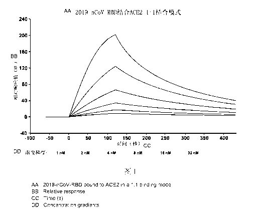
Fig. 1 shows the results of affinity test for the protein disclosed in the
present
invention and the ACE2 protein in Test Example 1;
Fig. 2 shows the results of antibody titer test in Test Example 2;
Fig. 3 shows the results of response between the antibody in the patient with
SARS-CoV-2 and the protein disclosed in the present invention as determined by
ELISA in Test Example 3;
Fig. 4 shows the test results of blocking the binding between the RBD protein
and
ACE2 receptor in Test Example 4;
Fig. 5 shows the results of neutralizing antibody detection in Test Example 5;
11
Date Recue/Date Received 2022-12-12
CA 03186989 2022-12-12
Fig. 6 shows the results of copy detection for virus gRNA and sgRNA in lung
tissue in
Test Example 5;
Fig. 7 shows the results of copy detection for virus gRNA and sgRNA in throat
swab
in Test Example 5;
Fig. 8 shows the results of copy detection for virus gRNA and sgRNA in anal
swab in
Test Example 5;
Fig. 9 shows the section staining results for the lung tissue in Test Example
5;
Fig. 10 shows the results of mouse challenge experiment against SARS-CoV-2
infection in Test Example 6;
Fig. 11 shows the detection results of cytokines INF-y and IL-4 in Test
Example 7;
Fig. 12 shows the results of cytokine level detection in Test Example 8;
Fig. 13 is a schematic diagram for extracellular domain composition of the
SARS-CoV-2 S protein;
Fig. 14 is a spectrogram of Escherichia coli expression vector pET32a in
Embodiment
3;
Fig. 15 is a spectrogram of yeast expression vector pPICZaA in Embodiment 4;
Detailed Description of the Preferred Embodiments
The technical solution of the present invention will be described in
combination
with the embodiments. Those skilled in the art will understand that the
following
embodiments are used only to describe the present invention, but not to limit
the
scope of the present invention. It should be noted that, where no specific
technologies
or conditions are indicated in the embodiments of the present invention, the
technologies or conditions described by the literature in the present art or
specified in
the product specification shall apply. The reagents or apparatuses, where no
specific
manufacturer is indicated, are all commercially available conventional
products.
S protein is a glycosylated protein and preferably, the insect baculovirus
expression system or mammalian cell expression system (CHO expression system)
is
used for better gaining the natural S protein. The specific preparation method
is
described as bellow:
Embodiment 1 Preparing the anti-SARS-CoV-2-infection protein disclosed
12
Date Recue/Date Received 2022-12-12
CA 03186989 2022-12-12
in the present invention by use of the insect baculovirus expression system
Vector construction: Recombinant proteins produced by use of the insect
baculovirus expression system mainly utilize the S protein receptor-binding
domain
(RBD). SARS-CoV-2 S protein is a protein located on the virus envelope. In
order to
simulate the secretion process of the SARS-CoV-2 S protein, a GP67 signal
peptide is
added to the N-terminal during the construction of S protein RBD to facilitate
the
secretion expression of the protein. This signal peptide will be spontaneously
excised
by insect cells during the secretion process of the protein. At the same time,
in order
to facilitate purification and increase the water solubility of the protein, a
thioredoxin
tag and an enterokinase (EK) restriction enzyme cutting site are also
introduced into
the sequence. The complete nucleotide sequence is shown in SEQ ID No.8 or SEQ
ID
No.10. The expression vector of S protein RBD is constructed based on the
pFastBacl
vector (amicillin resistance), BamHI and HindIII restriction enzyme cutting
sites are
inserted into the pFast-bacI vector, and escherichia coli biased codons are
used for
optimization.
Amplification of recombinant baculovirus: The Bac-to-Bac expression system
is used to construct recombinant bacmids in Escherichia coli (DH10Bac,
containing
bacmid (kanamycin resistance) and the helper plasmid (tetracycin resistance))
by
generating site-specific transposition through the Tn7 transposition element
using the
principle of bacterial transposon. The successfully recombined bacmids are
extracted
and transfected into 09 insect cells with Cellfectin II to generate the
recombinant
baculoviruses that can express the target gene. The first generation of
viruses are
collected 72 h after the transfection, and then amplified from P2 to P4. P3 or
P4
viruses are used to express the protein.
Protein expression: Hi5 insect cells (or 09 and sf21 cells) are infected with
the
P3 or P4 viruses, with a multiplicity of infection (MOI) of 0.5-10, and the
supernatant
is collected after 48-72 hours of culture. The optimal harvest time may vary
according to the amount of virus and cell status, and it is generally
appropriate when
about 50% of the cells get infected as observed by the microscopic
examination.
Protein purification: The harvested culture supernatant is centrifuged at 4 C
at
13
Date Recue/Date Received 2022-12-12
CA 03186989 2022-12-12
a high speed and filtered with an 0.22 gm filter membrane. The recombinant
protein is
initially purified by the affinity purification method (Histrap nickel
column). Then,
the recombinant protein is further purified by the MonoQ ion column and
Superdex
200 10/300GL molecular sieve. The protein purity is required to reach more
than 95%
as determined by the SDS-PAGE detection. The prepared protein is dissolved or
diluted to 1-5 mg/ml with enzyme digestion buffer. A corresponding amount of
enterokinase (EK enzyme) is added at a proportion of 1U enterokinase for 50
jig
recombinant protein, mixed, and left standing for digestion at 25 C for 16
hours to
remove the tag of recombinant protein. The nucleotide shown in SEQ ID No.8 is
used
to express the protein SEQ ID No.3, and the nucleotide shown in SEQ ID No.10
is
used to express the protein SEQ ID No.4. The obtained recombinant protein can
be
used for subsequent studies, such as animal immunization.
Embodiment 2 Preparing the anti-SARS-CoV-2-infection protein disclosed
in the present invention by use of the CHO cell expression system
Recombinant protein vaccines produced by use of CHO cells are mainly targeted
at the S protein receptor-binding domain (RBD). These fragments are
genetically
synthesized according to the codon preference, and polyhistidine is used as
the
purification tag (6His). The complete nucleotide sequence is shown in SEQ ID
No.13.
Then, it is constructed into the high expression vector pTT5 and the expressed
amino
acid sequence is the precursor protein as shown in SEQ ID No.14.
Embodiment 3 Expressing the anti-SARS-CoV-2-infection protein in the
Escherichia coli
pET32a from Novagen is used as the expression vector (see the plasmid profile
in Fig. 14), which contains a T7 promoter and where the transcription of
downstream
target genes is regulated by the IPTG. The N-terminal of the expressed product
is
fused with thioredoxin (Trx) and purified by the metal chelate affinity
chromatography (MCAC) in a single step. In order to remove Trx after
purification of
the target protein, the enterokinase (EK) restriction enzyme cutting site is
added after
Trx. The complete nucleotide sequence is shown in SEQ ID No.11.
The recombinant plasmids are expressed in Escherichia coli strain BL21 (DE3)
14
Date Recue/Date Received 2022-12-12
CA 03186989 2022-12-12
respectively. The electrophoresis results show that the size of the obtained
target
protein is similar to the predicted size and verified by Western Blot. The
content of the
target protein is more than 30% of the total protein, mainly in the form of
inclusion
bodies. Under denaturing conditions, the protein can be purified by the metal
chelate
affinity chromatography (MCAC) in a single step to allow for a purity of
higher than
95% and a yield of 200-400mg/L. The target protein can be renatured by
dialysis,
with a renaturation efficiency of higher than 50%.
To reduce the production cost, metal chelate columns can be substituted with
reversed-phase columns for single-step purification, which can also produce
the
high-purity target protein with a purity of higher than 95%.
Embodiment 4 Expressing the anti-SARS-CoV-2-infection protein in the
yeast
The nucleotide sequence as shown in SEQ ID No.12 is cloned into the double
enzyme (Xho I/Xba I) site of the yeast expression vector pPICZaA (Invitrogen;
see
the profile of yeast expression vector pPICZaA in Fig. 15) to secrete and
express the
protein using the factor a secretion signal in the methylotrophic yeast.
pPICZaA plasmid is used to mediate and integrate the S-RBD gene into the
methylotrophic yeast chromosome, and then methanol is used to induce the
expression of the target protein. The expressed target protein is present in a
soluble
form in the culture medium of the methylotrophic yeast, which could be
purified in a
single step by reverse-phase column chromatography, with the expression
quantity
reaching 200-400mg/L.
Embodiment 5 Preparing the anti-SARS-CoV-2-infection vaccine
Antigens are prepared under sterile conditions and the purified recombinant
protein antigens (prepared according to embodiments 1-4) are diluted with
5mmol/L
phosphate buffer (pH7.2) to a concentration of 80 mcg/mL. Adjuvants are
prepared
under aseptic conditions and the aluminum hydroxide adjuvants (with a content
of
14.55mg/mL) are diluted with 5mmo1/L phosphate buffer (pH7.2) to a
concentration
of 2.0 mg/mL. Antigen-adjuvant adsorption is carried out under sterile
conditions at a
speed of 20 mL/min, the diluted protein antigen liquid is added dropwise to
the
Date Recue/Date Received 2022-12-12
CA 03186989 2022-12-12
diluted aluminum hydroxide adjuvant working solution at a volume ratio (V/V)
of 1:1,
so that the final concentration of recombinant protein antigens in the mixed
solution is
40 mcg/mL, and the final concentration of aluminum adjuvants is 1.0 mg/mL. The
reaction temperature is kept at 25 C and the stirring speed at 800rpm. After
dropping,
the adsorption is performed for 60min at the temperature of 25 C and the
stirring
speed of 800rpm. The pH of the mixed solution is adjusted to 7.2. The solution
is
stored at 4 C away from light. The adsorbed vaccine preparations are
characterized,
including particle size, site position, antigen content, adjuvant content,
adsorption rate,
pH value, endotoxin, adjuvant and antigen adsorption rate, adsorption strength
and its
hold status, and antigen integrity and stability after adsorption. For
filling, the
qualified vaccine preparations refilled into the lmL sterile penicillin
bottles or
ampoule bottles in lmL/vial. Continuous stirring is kept when filling to make
the
filled liquid even. The filled vials are capped immediately after filling,
attached with
the serial number labels, and stored at 4 C away from light.
The advantageous effects of the present invention are demonstrated by the
following test examples.
Test Example 1 Affinity test for the protein disclosed in present invention
and the ACE2 protein by surface plasmon resonance (SPR) analysis
Surface plasmon resonance detection was performed using a macromolecular
interactometer Biacore 8K (GE Healthcare, Sweden). The ACE2-Fc was pre-
anchored
on the surface of Sensor Chip Protein A chip with a capture response unit (RU)
value
of ¨100RU. For kinetic analysis, the RBD protein of the present invention,
whose
amino acid sequence is shown in SEQ ID No.3, was handled to pass through the
chip
surface with a concentration gradient (1, 2, 4, 6, 8, 16, 32 nM) diluted by a
double
equal proportion respectively, and another channel is set as the blank control
group.
Antigen dissociation was performed for 300 seconds using HBS-EP+ dissociation
solution at a flow rate of 30 mL/min, followed by 60 seconds of chip
regeneration
using glycine solution with pH 1.5 as the regeneration solution. In this
process, the
binding constant (Ka) and dissociation constant (I() of ACE2-Fc antibody and
RBD
protein of the present invention were respectively detected, and their
affinity (I(D)
16
Date Recue/Date Received 2022-12-12
CA 03186989 2022-12-12
was calculated.
The results, as shown in Fig. 1, showed that the RBD protein of the present
invention can bind with the ACE2 receptor protein efficiently and
specifically, and
suggested that the RBD protein of the present invention may maintain an
integral
spatial structure and have the same ACE2 receptor ability as the S protein RBD
of the
virus, providing a strong support for taking the in-vitro recombinant S
protein RBD as
vaccines. Its affinity KD was 1.52x 10' M (mol/L), dissociation constant Ka
was
4.41x10² (s-i), and binding constant Ka was 3.85x 10⁶ (Ms-1).
Test example 2 Inducing the RBD-specific antibodies in mice vaccinated
with the vaccine disclosed in the present invention
Animal immunization test: BALB/c or C57BL/6 mice were injected with the
recombinant proteins (with the amino acid sequence as shown in SEQ ID No.3) at
doses ranging from 0.1 to 10.0 g per mouse; each group was assigned with five
to ten
mice. Each mouse was injected with a volume of 50 L of vaccine (prepared
according
to Embodiment 5) intramuscularly (im) in the right hind leg. Two immunization
regimens were used: vaccination on days 1, 7, and 21, and vaccination on days
1, 14,
and 21.
Determination of mouse serum antibody by enzyme linked immunosorbent
assay (ELISA): On the 7th day after each immunization, the plasma of mice was
collected by capillary orbital blood sampling from 5 mice in each group. After
coagulating at room temperature for 1-2h, and centrifugation at 3000rpm/min
for
10min at 4 C, the upper layer of serum was taken and stored at -20 C for later
use.
For the determination of serum IgG and subtype by the ELISA, a 1 Kg/m1
solution of
recombinant protein S-Fc or RBD-Fc was prepared in 50 mM carbonate coating
buffer (PH9.6), and added at 100 I/well into a 96-well plate (Thermo
Scientific,
NUNC-MaxiSorp) for coating overnight at 4 C. For the preparation of 50m1\'l
carbonate coating buffer (PH9.6), 0.15g Na2CO3 and 0.293g NaHCO3 were weighed
and dissolved in double distilled water, the PH was adjusted to 9.6, then the
volume
was fixed to 100 ml and stored at 4 C for later use. The next day, the mice
plasma was
washed 3 times with PBS solution containing 0.1% Tween20 (PBST), blocked with
17
Date Recue/Date Received 2022-12-12
CA 03186989 2022-12-12
blocking solution containing 1%BSA or 5% skim milk (prepared in PBST) for lh
at
room temperature, and then washed once with PBST. The mice serum was diluted
with blocking solution in different proportions, then added at 1041 well for
incubation for lh-2h at 37 C, and washed with PBST for 3 times. Then the
plasma
was added with HRP-goat anti-mouse IgG or HRP-anti-mouse IgGl, IgM or other
subtype antibodies at 100 111/well (diluted in blocking solution at 1:5000),
incubated
at 37 C for lh, and then washed with PBST for 5 times. Finally, 3,3',5,5'
-tetramethylbiphenyl diamine (TMB) was added at 100 1/well, and after 10-15min
of
color development in the dark, 1M H2SO4 stop solution was added at 50 1/well,
and
the reading was performed on the microplate reader at 450nm wavelength after
mixing. To prepare the 1 M H2SO4 stop solution, 2.7mL of concentrated sulfuric
acid
(98%) was added drop by drop to 47.3mL of double distilled water.
The test results are shown in Fig. 2. In order to measure the titer of RBD-
specific
antibody induced by the recombinant protein, serum was continuously diluted in
different proportions and measured by titration, and the A450 optical density
value
was measured. As shown in Fig. 2, the recombinant protein vaccine elicited
significant S protein RBD-specific antibodies. Serum collected 7 days after
vaccination showed a strong antibody response, with IgG (Fig. 2A) and IgM
(Fig. 2B)
increased in different ratios, while the A450 optical density was
significantly lower in
the control group vaccinated with the normal saline, suggesting that the
vaccine can
rapidly induce immune responses and is important for the prevention of SARS-
COV-2.
These results indicated that the recombinant S protein RBD vaccine was highly
immunogenic in mice.
Test Example 3 Reaction determination between the antibody in the patient
with SARS-CoV-2 and the protein disclosed in the present invention by ELISA
In this experiment, 16 serum samples from patients infected with SARS-CoV-2
were collected to investigate the immunogenicity of the RBD protein of the
present
invention in human bodies. ELISA was used for determination as follows:
A 96-well plate was coated with the RBD protein (with the amino acid sequence
shown in SEQ ID No.3) at a concentration of 0.2 g/well, 100111/ well, at 4 C
18
Date Recue/Date Received 2022-12-12
CA 03186989 2022-12-12
overnight. A negative control well was set up during coating. The 96-well
plate was
removed the next day, and in half an hour after rewarming to the room
temperature,
the plate was washed with PBS for 4 times, 1 min each time. The 96-well plate
was
blocked with 1%BSA at 100 1/ well, incubated at 37 C for 30 min, and then
washed
again with PBS for 4 times, 1 min each time. The serum was diluted by 5 folds,
namely adding 80 1 of PBS for every 20 1 of serum in each well. Then, the
plate was
incubated at 37 C for 30 min, washed with PBS for 4 times, 1 min each time,
then
added with the HRP-labeled secondary antibody (anti-human IgG/IgM antibody)
diluted in a proportion of 1:2000 at 100u1/well, incubated at 37 C for 30 min,
and
washed with PBS for 4 times, 1 min each time. For color development, each well
was
added with 50 1 of liquid A and then 50 1 of liquid B and left standing at
room
temperature for 15 min. To stop, each well was added with 100 1 of stop
solution.
Colorimetry with a microplate reader was conducted within 10 min. In this
assay, for
the detection of IgM antibodies, 15 1 of serum in each well was added with 15
1 PBS
and then 150 1 IgG adsorbent, and centrifuged at 10000rpm for 10 min; 100 1 of
supernatant was taken for detection.
As shown in Fig. 3, serum from the 16 patients infected with SARS-CoV-2 had
obvious response to the RBD protein of the present invention, and both IgM and
IgG
reactions were positive, while the serum from 10 healthy people showed
negative
reaction to the antigen, indicating that the SARS-CoV-2 S protein RBD had high
immunogenicity as a vaccine in patients. The RBD protein prepared by the
present
invention can be recognized by the human immune system.
Test Example 4 Blocking test for the binding between the RBD protein and
ACE2 receptor
In this experiment, cell-expressed ACE2, a protein thought to retain its
native
conformation, was used to allow RBD binding activity to be measured by flow
cytometry. Specific operations are as follows:
The in-vitro cultured cell strains with high expression of ACE2 (lung cancer
A549) were digested and collected into flow cytometry tubes at 106 cells/tube
and
washed with PBS/HBSS several times. Recombinant RBD-Fc protein at a final
19
Date Recue/Date Received 2022-12-12
CA 03186989 2022-12-12
concentration of 1 ug/ml was added to each tube of cells and the serum from
immunized anti-RBD mice was then added (after the mouse serum obtained from
Test
Example 2 was diluted by 50 folds) for incubation for 30 min at room
temperature.
For the positive control tube, no antiserum was added or normal serum from
unimmunized mice was added. After washing with PBS/HBSS for several times,
Anti-Human IgG (Fc specific)-FITC (SIGMA) fluorescent secondary antibody
(1:100-1:200) was added for incubation at room temperature for 30 min in the
dark.
After washing with PBS/HBSS for several times and fixation by adding 500 I
PBS
containing 1% paraformaldehyde, detection was performed by flow cytometry.
As shown in Fig. 4, the added RBD-Fc protein could significantly bind with the
ACE2-expressing cells, while only background signal was detected if RBD-Fc
protein
was not added (negative control). Mouse antiserum effectively blocked the
binding of
the RBD-Fc protein with ACE2-expressing cells, while the unimmunized or
pre-immunized serum of the same dilution showed no inhibitory activity.
Test Example 5 Challenge experiment on non-human primates (such as
rhesus monkey) with live SARS-CoV-2 virus
1. Experimental method
All research procedures involving nonhuman primates were reviewed and
approved by the Institutional Animal Care and Use Committee of the Institute
of
Medical Biology, Chinese Academy of Medical Sciences, and were performed in
the
Animal BioSafety Level 4 (ABSL-4) facility at the National Kunming High-level
Biosafety Primate Research Center in Yunan, China. The RBD protein used in
this
experiment was the protein of this prevention whose amino acid sequence is
shown in
SEQ ID No.4, and the vaccine was prepared according to Embodiment 5. Twelve
nonhuman primates (rhesus monkeys) (aged 5-9 years) were used in live
SARS-CoV-2 challenge experiments, and grouped as follows: (a) group 1 with 4
rhesus monkeys (n = 4), which were vaccinated with the vaccine comprising 40
g
RBD protein plus aluminum hydroxide adjuvant each dose; (b) group 2 with 3
rhesus
monkeys (n = 3), which were vaccinated with the vaccine comprising 20 g RBD
protein plus aluminum hydroxide adjuvant each dose; (c) group 3 with 2 rhesus
Date Recue/Date Received 2022-12-12
CA 03186989 2022-12-12
monkeys (n = 2), which was the normal saline control group; (d) group 4 with
rhesus
monkeys (n = 2), which was the aluminum hydroxide adjuvant control group. The
nonhuman primates were immunized by intramuscular injection on days 0 and 7,
followed by nasal challenge with SARS-CoV-2 (0.5 ml, 106 pfu/ ml) 28 days
after the
initial immunization. To assess the neutralizing effect of SARS-CoV-2
infection, sera
were collected on days 28 and 35 (5 days after virus vaccination) after the
first
immunization for neutralizing antibody assay. Vero E6 cells (5x 10⁴/well) were
inoculated in a 96-well plate and cultured overnight. SARS-CoV-2 with 100-fold
TCID50 (50% tissue culture infective dose) was preincubated with an equal
volume
of diluted serum, and after incubation for 1 h at 37 C, the mixture was added
to Vero
E6 cells. On day 3 after infection, cytopathic effect (CPE) was recorded under
a
microscope, and neutralization titers were calculated for serum diluents
producing EC
50 inhibition (50% neutralization). The control groups included the monkey
serum
treated with normal saline or aluminum hydroxide alone.
The contents of viral genomic RNA (gRNA) and viral subgenomic RNA (sgRNA,
representing virus replication) were determined by quantitative real-time
reverse
transcription PCR (qRT-PCR). Viral loads in lung tissue, throat swabs, and
anal swabs
were determined by qRT-PCR based on sequences recommended by WHO and
Chinese Center for Disease Control and Prevention, using primers and probes
from
the NP gene.
Forward: 5'-GGGGAACTTCTCCTGCTAGAAT-3' (SEQ ID No.15);
Reserve: 5'-CAGACATTTTGCTCTCAAGCTG-3' (SEQ ID No.16);
Probe: 5'-FAM-TTGCTGCTGCTTGACAGATT-TAMRA-3' (SEQ ID No.17)
According to the instructions for TaqMan Fast Virus 1-Step Master Mix (Article
No.: 4444434), the reaction system was 10 L: 1 .1_, forward primer, 1 .1_,
reverse
primer, 0.25 .1_, probe, 2.5 .1_, mRNA template, 2.5 .1_, Master Mix, and
2.75 .1_,
RNase-Free H20. For PCR program setting and operation, operation instructions
for
BioRad CFX384 Real Time PCR System were consulted: reverse transcription
(incubated at 25 C for 2 min and 50 C for 15 min); initiation (incubated at 95
C for 2
min); two-step amplification, 40 cycles (incubated at 95 C for 5s and 58 C for
31s).
21
Date Recue/Date Received 2022-12-12
CA 03186989 2022-12-12
The content of SARS-CoV-2 E gene subgenomic mRNA (sgmRNA) indicating
virus replication was determined using the primer and probe with the following
sequences:
Forward: 5'-GCTAGAGAACATCTAGACAAGAG-3' (SEQ ID No.18);
Reverse: 5'-ACACACGCATGACGACGTTATA-3' (SEQ ID No.19);
Probe: 5'-FAM-TGTGATCGGTAGGAATGACGCGAAGC-Quencher-3' (SEQ
ID No.20);
The reaction system and PCR procedure were consistent with gRNA assay.
For paraffin embedding of slices, tissues were collected and fixed with 10%
neutral formalin and embedded in paraffin. Sections were 5 gm thick and
stained with
hematoxylin and eosin (HE).
2. Experimental results
Neutralizing antibodies against live SARS-CoV-2 were detected in all
vaccinated
nonhuman primates but not in either control group, as shown in Fig. 5.
Quantitative real-time reverse transcription PCR (qRT-PCR) was used to detect
viral genomic RNA (gRNA) and viral subgenomic RNA (sgRNA, representing virus
replication). Lung tissues from nonhuman primates were collected on day 7
after
challenge to assess virus replication status. The lung tissue of control group
(normal
saline group and aluminum hydroxide adjuvant group) showed excessive copies of
viral gRNA and sgRNA. In contrast, there was no detectable virus replication
in the
groups vaccinated with 20 or 40 gg RBD protein plus adjuvant (Fig. 6). In
addition,
peak viral gRNA loads in throat swabs were observed 3 days after vaccination
in the
control groups (normal saline group and aluminum hydroxide adjuvant group),
and
these viral peaks could be blocked with vaccine, while the viral loads were
only 1.6
and 3.8 percent per million in the 20 gg and 40 ug vaccine groups.
Importantly, no
detectable sgRNA was observed in throat swabs after viral challenge in both
the 20
and 40 gg vaccine groups, whereas a high amount of sgRNA was observed in the
control groups (normal saline group and aluminum hydroxide adjuvant group),
indicating virus replication (Fig. 7). On days 5 and 6 after inoculation, peak
viral
gRNA and gRNA loads in anal swabs were observed in the control groups (normal
22
Date Recue/Date Received 2022-12-12
CA 03186989 2022-12-12
saline group and aluminum hydroxide adjuvant group), while only an extremely
low
level was detected in the vaccinated groups, without detectable sgRNA in the
anal
swabs of nonhuman primates vaccinated with the 20 lag and lig vaccine (Fig.
8). The
above results indicate that vaccination with the RBD protein vaccine of the
present
invention can prevent SARS-CoV-2 infection.
Lung tissues from the two control groups (normal saline group and aluminum
hydroxide adjuvant group) showed the histopathologic changes typical of
SARS-CoV-2 viral interstitial pneumonia, a key feature of COVID-19. As shown
in
Fig. 9, the alveolar walls were significantly thickened as observed under the
microscope, and a large number of interstitial mononuclear inflammatory cells
infiltrated. There were also numerous inflammatory infiltrates and serous
exudates in
the alveolar space, accompanied by the recognizable loss of lung tissue
structures. In
addition, diffuse bleeding and type II pneumonocyte hyperplasia were observed.
In
contrast, nonhuman primates vaccinated with the RBD protein (20 g or 40 g)
showed
no significant histopathologic changes and had a normal lung tissue
appearance.
Test Example 6 Mice challenge experiment against SARS-CoV-2 infection
BALB/c or C57BL/6 mice aged between 6 to 8 weeks were immunized by
intramuscular injection of the recombinant RBD protein vaccine (i.e., the
protein with
amino acid sequence as shown in SEQ ID No.3; the vaccine prepared according to
Embodiment 5) at different doses (0.1-20Kg each). For example, mice received
an
injection on day 0, serum was collected on day 7, and mice in the control
group were
injected with either aluminum hydroxide immune adjuvant or normal saline
alone.
Serum was collected again on day 7 after immunization. The serum was stored at
4 C
for used in the later experiment. The SPF hACE2 transgenic mice established by
the
Institute of Laboratory Animal Science, Chinese Academy of Medical Sciences
and
Peking Union Medical College were used in animal experiments of SARS-CoV-2
infection. Seven days after the first vaccination, 0.8m1 of serum was
collected. Serum
from vaccine-immunized mice was used as the experimental group, and normal
serum
from normal saline treated mice was used as the control group. One day before
SARS-CoV-2 virus challenge (intranasal infection, 105 TCID50), hACE2
transgenic
23
Date Recue/Date Received 2022-12-12
CA 03186989 2022-12-12
mice were injected with the serum intraperitoneally. In addition, mice
infected with
the virus but not received the serum injection served as controls. Five days
after the
virus challenge, the mice were killed and their lungs and other organs were
harvested.
Lung tissue was used to detect viral replication or fixed with 10% buffered
formalin
solution for histopathologic analysis. Real-time quantitative reverse
transcriptase
polymerase chain reaction (qRT-PCR) was performed with PowerUp SYBG Green
Master Mix Kit (Applied Biosystems, USA) to determine viral RNA copy number in
lung tissues of mice challenged with SARS-COV-2, expressed in the RNA copy
number/ml of lung tissue. The primer sequence used for qRT-PCR was the
envelope
(E) gene against SARS-cov-2 as follows:
Forward: 5'-TCGTTTCGGAAGAGACAGGT-3' (SEQ ID No.21);
Reverse: 5'-GCGCAGTAAGGATGGCTAGT-3' (SEQ ID No.22).
The slices were stained with hematoxylin and eosin, and the histopathological
changes were observed under the light microscope.
This experiment tested whether early humoral immunity through vaccination can
prevent mice from being infected with the SARS-CoV-2 virus. Human ACE-2
transgenic mice were challenged with the SARS-CoV-2 virus, and lung tissues of
the
mice were collected 5 days after virus challenge to measure the virus
replication status
of the serum receiving immunization (the serum 7 days after the first
immunization)
or the control serum. As shown in Fig. 10, no viral replication was detected
by
quantitative real-time reverse transcriptase polymerase chain reaction (qRT-
PCR) in
mice treated with the immune serum induced by the RBD protein vaccine, whereas
the level of viral replication was higher in lung tissues of the control mice.
Accordingly, the lung tissues of the control mice showed significant
interstitial
pneumonia histopathological changes, including significant alveolar wall
thickening,
extensive interstitial monocyte and lymphocyte infiltration, embolism, and
serum
exudate in the alveolar space. In contrast, no histopathological changes or
slight
exudation were observed in mice treated with the serum from mice immunized
with
the recombinant RBD protein vaccine. In addition, mice treated with the immune
serum gained a slight amount of weight (approximately 8%) during the first 5
days
24
Date Recue/Date Received 2022-12-12
CA 03186989 2022-12-12
after infection, while no weight gain in the control group and an 8% weight
loss in the
untreated group were observed. This experiment further confirmed that the
antibody
induced by the RBD protein vaccine could completely block the virus infection.
Test Example 7 Induction of cellular immune response by the RBD protein
vaccine disclosed in the present invention
BALB/c or C57BL/6 mice aged between 6 to 8 weeks were immunized by
intramuscular injection of the recombinant RBD protein vaccine (i.e., the
protein with
amino acid sequence as shown in SEQ ID No.3; the vaccine prepared according to
Embodiment 5) at different doses (0.1-20m each). For example, mice received an
injection on day 0, serum was collected on day 7, and mice in the control
group were
injected with either aluminum hydroxide immune adjuvant or normal saline
alone.
Spleen T lymphocytes were collected again 7 days after immunization. To
investigate
the cellular immune response, mice immunized with S protein RBD or PBS were
killed and their lymphocytes were extracted for IL-4 and IFN-y detection by
ELISA.
In brief, mouse spleen lymphocytes (1 x 10⁶/mL) were cultured in RPMI 1640
medium
(containing 10% fetal bovine serum, 100U/mL penicillin, 100 g/mL streptomycin,
1mM pyruvate, 500/1 13-mercaptoethanol, 20U/mL IL-2). At the same time, 1
i.tg/mL
RBD protein was added for culture and stimulation for 72 hours. Cells without
RBD
protein stimulation were used as the negative control. The supernatant was
collected
for ELISA.
Because cellular immune responses may play a role in clearing SARS-CoV-2
infection, in which both CD4 and CD8 positive T cells are involved in immune
responses against SARS virus infection, the potential cellular immune
responses to
the vaccine were also examined. Lymphocytes were collected on day 7 after the
first
vaccination, and cytokines produced by the lymphocytes such as INF-y (gamma
interferon) and IL-4 (interleukin-4) were measured by ELISA. Stimulation of
the
isolated mouse lymphocytes with the recombinant RBD protein showed that
vaccine-immunized mice produced more IFN-y and IL-4 in the lymphocytes,
whereas
only IFN-y and IL-4 at the background level were detected in mice treated with
normal saline after the lymphocytes were stimulated by the recombinant RBD
protein
Date Recue/Date Received 2022-12-12
CA 03186989 2022-12-12
(Fig. 11).
Test Example 8 Safety experiment for the vaccine disclosed in the present
invention
Mice were immunized with the vaccine of the present invention. BALB/c or
C57BL/6 mice aged between 6 to 8 weeks were immunized by intramuscular
injection
of the recombinant RBD protein vaccine (i.e., the protein with amino acid
sequence as
shown in SEQ ID No.3; the vaccine prepared according to Embodiment 5) at
different
doses (0.1-20Kg each). No pathological changes were found in heart, brain,
liver,
spleen, lung, kidney and other organs. No changes in blood cells or blood
biochemical
indicators were found. For example, this experiment measured the cytokine
level in
the blood to see whether vaccination caused changes in cytokines in the
systemic
inflammatory response. Mice received a single injection on day 0, serum was
collected on day 7, and control mice received aluminum hydroxide immune
adjuvant
or normal saline alone. On the 7th day after the first inoculation, serum was
collected
from the mice, and the cytokines of TNF-a, IFN-y, IFN-a, IFN-b, IL-6 and IL-4
were
detected by ELISA. Cytokine content in the serum was determined using Thermo
Fisher Scientific-eBioscience Elisa kit as follows: the antigen was coated,
diluted with
coating buffer at 200 L/well, sealed, and incubated overnight at 4 C; the
coating
solution was discarded after coating, and the plate was washed three times
with
washing buffer at 250 L/well or higher; 200 .1., ELISA/ELISPOT diluent (1x)
was
used for blocking for 1 h at room temperature; the standard was prepared
according to
the concentration requirements in the specification, and the standard with the
highest
concentration was diluted by two-fold dilution method, a total of 8 points,
and
ELISA/ELISPOT diluent (1x) was used as a control; the serum was diluted with
ELISA/ELISPOT diluent (1x) for later use; after the plate was washed, the
plate was
added with 100 L of standards and samples to be tested, sealed and incubated
for 2 h
at room temperature; the antibody to be detected was diluted with
ELISA/ELISPOT
diluent (1x), the plate was washed 3-5 times, the diluted antibody to be
detected was
added to each well at 100 L/well, and the plate was sealed and incubated for
1 hour
at room temperature; Hrp-labeled antibodies were diluted with ELISA/ELISPOT
26
Date Recue/Date Received 2022-12-12
CA 03186989 2022-12-12
diluent (1x), and the plate was washed 3-5 times, the diluted antibiotin
protein HRP
was added to each well at 100 jiL/well, and the plate was sealed and incubated
for 30
min at room temperature; the plate was washed 5-7 times, 1X TMB solution was
added to each well at 100 jiL/well, and color development was performed for 10-
15
min at room temperature; stop solution was added at 100 pt/well for
termination;
plate reading was carried out with the detection wavelength being 450nm and
the
reference wavelength being 570nm.
The experimental results showed that the no difference in the blood cytokine
level was found between vaccine-immunized mice and control mice (aluminum
hydroxide immunized adjuvant or normal saline only) (see Fig. 12). The above
experimental results indicated that vaccination with the vaccine may not cause
systemic inflammatory reaction.
It is important to note that the specific characteristics, structures,
materials or
features described in this specification may be combined in any one or more
embodiments in a suitable manner. In addition, as long as no mutual
contradiction is
caused, those skilled in the art may incorporate and combine the different
embodiments described in this specification and the characteristics of the
different
embodiments.
27
Date Recue/Date Received 2022-12-12