Note: Descriptions are shown in the official language in which they were submitted.
WO 2022/023528
PCT/EP2021/071399
1
CYTOSOLIC DELIVERY
Field of the Invention
The present invention relates to means for delivering functional
macromolecules to the cytosol. The
invention provides chimeric receptors comprising parts of DNGR-1, a
transmembrane receptor, as well as
cells expressing the chimeric receptors, means for producing the chimeric
receptors, and medical uses
thereof. Functional macromolecules, such as biopolymers, which bind and
trigger DNGR-1 and/or the
chimeric receptors of the invention, and constructs comprising said
macromolecules, are also provided.
Background
DNGR-1 (also known as CLEC9A) is a C-type lectin that has been previously
described as a key
mediator of cross-presentation by type 1 conventional dendritic cells (cDC1s)
(D Sancho et al. Nature
200912; W02009/013484A120, both of which are specifically incorporated by
reference herein). Cross-
presentation (XP) refers to a process, performed by antigen-presenting cells
(APCs), of presenting
exogenous antigens on MHC class I molecules to cytotoxic T lymphocytes (CTL).
XP is essential for the
induction of protective CTL responses against tumours and many viruses-I-7.
Macrophages, monocyte-derived dendritic cells and other myeloid cell types, as
well as non-immune
cells, have been used extensively to dissect some of the mechanisms involved
in XP8,21,38. While this has
led to the view that XP in many cases involves the cytosolic pathway
(phagosome to cytosolic transfer;
P2C), these studies have generally fallen short of explaining how P2C occurs,
especially for complex
substrates such as dead cells. Indeed, the actual mechanism underlying the
regulation of cross-
presentation of antigens from cellular corpses by DNGR-1 is reported to remain
a mystery28. XP in vivo,
notably in the context of virus infection and anti-tumour immunity, is
abrogated in cDC1-deficient Batf37-
mice39, pointing to cDC1 as a non-redundant cross-presenting APC.
Relatively few papers have focused on XP mechanisms specifically in
cDC1s7,30,36,40-42. Typically, dying
virally-infected or tumour cells are thought to be sources of exogenous
antigen for cross-presenting
cDC1s. The DNGR-1 expressed by these cells detects F-actin/myosin complexes
exposed on dead cell
debris and promotes XP of corpse-associated antigens12-16, thus coupling the
detection of cell death with
immunity.
A tyrosine-containing hemITAM motif in the cytoplasmic tail of DNGR-154 allows
binding of the tyrosine
kinase, Syk, upon tyrosine phosphorylation12. However, until now, the
subsequent intracellular signalling
events that allow DNGR-1 to facilitate the XP of bound antigens (and the
mechanisms underlying XP in
general) have remained poorly understood. Some previous disclosures have
examined DNGR-1 function
by generating chimeric proteins comprising the extracellular domain of DNGR-1
fused to the intracellular
domain of CD3(;=12, 20 In this context, detectable signalling is triggered
when a bivalent ligand binds the
DNGR-1 extracellular domain, causing the readily detectable signalling cascade
initiated by CD3
activation. However, the lack of understanding of the cellular mechanisms
responsible for XP has
inhibited research into whether DNGR-1 could provide the basis of useful
biomedical technologies.
CA 03187062 2023- 1-24
WO 2022/023528
PCT/EP2021/071399
2
Summary of the Invention
The inventors have uncovered the mechanism by which DNGR-1 mediates cross
presentation (XP). This
led the inventors to provide the chimeric receptors of the invention, which
provides a platform technology
to facilitate XP of a target antigen (which is not limited to any particular
type or class of antigen).
Surprisingly, this can be applied to achieve XP of the target antigen by a
range of cell types, not just
professional antigen presenting cells (APCs). This new understanding of the XP
pathway also leads to
the provision of a way of delivering macromolecules such as biopolymers to the
cytosol without
degradation. The invention and its underlying mechanism are explained in more
detail below.
The inventors found that DNGR-1 dependent XP proceeds via a cytosolic pathway.
DNGR-1 promotes
phagosomal rupture, which allows internalised antigens to be released into the
cytosol where they are
processed and presented via the conventional MHC class I antigen processing
pathway. The inventors
surprisingly found that the internalised antigens are not substantially
degraded before being released into
the cytosol. Using chimeric receptors, the inventors have now demonstrated
that the cytoplasmic tail of
DNGR-1 is a key mediator of this process, thus enabling XP. The inventors show
that this requires only
the DNGR-1 signalling domain, which recruits and activates spleen tyrosine
kinase (Syk) and NADPH
oxidase to cause lipid peroxidation and phagosomal membrane instability.
Notably, DNGR-1 signalling
can induce phagosomal membrane rupture and XP in heterologous cells, including
non-professional
APCs. These results show that phagosomal rupture is coupled to XP, providing a
simple mechanism for
access of exogenous antigens to the endogenous MHC I pathway. This mechanism
is non-selective and
does not require specific antigen transporters. Furthermore, the basic
machinery for phagosomal rupture
is not limited to DCs, thus other non-APCs have the potential to cross-present
exogenous antigens.
However, the effective engagement of this machinery requires a dedicated XP
signalling receptor such as
DNGR-1.
Thus, the invention provides chimeric proteins comprising the signalling
domain of the cytoplasmic tail of
DNGR-1. The chimeric proteins of the invention can facilitate XP. By
engineering cells (not limited to
DCs) to express the chimeric proteins, the invention provides a wide range of
cells that can recognise and
process a desired target (not limited to the actin/myosin II signal on dead
cells), and present antigens
from the target to CD8+ T cells, to elicit a robust immune response to the
target. The invention also
provides a way of delivering biopolymers (such as proteins and nucleic acids)
to the cytosol without being
degraded.
Accordingly, in a first aspect, the invention provides a method of delivering
a biopolymer to the cytosol of
a cell, wherein the cell expresses a transmembrane protein comprising an
intracellular domain that
comprises a Syk-binding sequence derived from the signalling domain of the
cytoplasmic tail of DNGR-1,
wherein the biopolymer comprises a binding domain that can specifically bind
an extracellular portion of
the transmembrane protein, and wherein the method comprises contacting the
cell with the biopolymer to
allow the binding domain to bind to the extracellular portion of the
transmembrane protein such that the
biopolymer is internalised and translocated to the cytosol without being
degraded in a phagosome.
Preferably, the biopolymer further comprises a nucleic acid that encodes a
gene product.
CA 03187062 2023- 1-24
WO 2022/023528
PCT/EP2021/071399
3
Thus, the invention provides a way of delivering payloads to the cytosol. The
payload may be the
biopolymer itself and/or an additional moiety, such as a nucleic acid encoding
a gene product, that is
covalently or non-covalently associated with the biopolymer. The delivery of
such payloads finds use in
research, diagnostic and medical applications. As such, the invention provides
the biopolymer defined
herein for use in medical methods, such as vaccination In vitro methods are
particularly suited to
research and/or diagnostic applications.
In some embodiments, the biopolymer comprises a second domain, which does not
directly bind to the
extracellular portion of the transmembrane protein. This second domain is
covalently joined to the
binding domain, e.g. via a linker. In some embodiments, the linker can be
cleaved by a protease present
in the cytosol of the cell, allowing the second domain to dissociate from the
binding domain in the cytosol.
(The second biopolymer domain may be referred to as a payload.) The second
domain may be a nucleic
acid that encodes a gene product. The gene product may be a pro-apoptotic
protein, an enzyme, or a
cytotoxic peptide as described herein.
In some embodiments, the (first) biopolymer (that comprises a binding domain)
is non-covalently
associated with a second biopolymer. This second biopolymer may be referred to
as a 'second domain'
herein, and may also be referred to as a payload. A non-covalent association
can be achieved e.g. by
fusing the first biopolymer to an avidin or streptavidin moiety, and
covalently linking a biotin 'tag' to the
second biopolymer, or vice-versa. The avidin/streptavidin non-covalently binds
the biotin tag, thus
associating the first and second biopolymers with each other.
Thus, the binding domain of the biopolymer may be coupled to a second
biopolymer domain, wherein the
coupling may be covalent or non-covalent.
Polypeptide biopolymers, polynucleotide biopolymers and polysaccharide
biopolymers are all envisaged.
The biopolymer may be a polypeptide and the binding domain may comprise at
least part of the antigen-
binding fragment of an antibody. For instance, the polypeptide may comprise an
antibody VH domain;
and this can pair with an antibody VL domain that is provided as a separate
polypeptide. Alternatively, the
polypeptide may comprise an antibody VL domain; and this can pair with an
antibody VH domain that is
provided as a separate polypeptide. In some embodiments, the polypeptide may
comprise both antibody
VH and VL domains, present on a single polypeptide chain, e.g. in the scFV
format.
The biopolymer may be a nucleic acid and the binding domain may be an aptamer.
Combinations of biopolymers of different classes, for the binding domain and
for the second domain, are
also envisaged. For instance, a binding domain that is an antibody (a
polypeptide) may be coupled to a
non-peptide second domain. Such coupling of biopolymer domains of different
classes can be achieved
covalently or non-covalently, e.g. as described herein. The term "biopolymer"
used herein, and the
methods of delivering said biopolymer, are intended to encompass biopolymer
pairings where the binding
domain is one type of biopolymer and the payload is another type of
biopolymer.
The biopolymer may comprise multiple binding domains. For instance, two or
more antibodies and one or
more second biopolymer domains can all be immobilised on a single carrier
particle, e.g. a latex bead.
CA 03187062 2023- 1-24
WO 2022/023528
PCT/EP2021/071399
4
In embodiments where the biopolymer comprises a polynucleotide, the
polynucleotide may encode a
gene product and is capable of expressing the gene product in a host cell. The
encoded gene product
may be a polypeptide disclosed herein. In other embodiments, the
polynucleotide is, or encodes, an RNA
sequence that interferes with the expression of another protein in the cell
(e.g. an RNAi molecule such as
an siRNA). The polynucleotide may be DNA_ The DNA may be capable of activating
the STING
pathway. The polynucleotide may be RNA. The RNA may be capable of activating
the RIG-I and/or
MDA5 pathways.
In some embodiments, the second domain comprises a cytotoxin or pro-apoptotic
protein. For instance,
the second domain may comprise cytochrome C, a caspase, a maytansinoid, a
dolastatin, an auristatin
drug analogue, a cryptophycin, a duocarmycin deriative, an enediyne
antibiotic, or a
pyrolobenodiazepine. In some embodiments, the second domain comprises an
antibody that binds to an
intracellular target. In some embodiments, the biopolymer comprises a tumour
antigen (second domain)
conjugated to an anti-DNGR-1 antibody (first domain). In some embodiments, the
second domain is not
a peptide antigen. In other words, in some embodiments, the second domain is a
whole protein or a
whole domain of a protein. In some embodiments, the second domain comprises
more than 50 amino
acid residues.
In some embodiments, the second domain is a nucleic acid that encodes a
cytotoxin or pro-apoptotic
protein. For instance, the second domain may encode cytochrome C, a caspase, a
maytansinoid, a
dolastatin, an auristatin drug analogue, a cryptophycin, a duocarmycin
deriative, an enediyne antibiotic, or
a pyrolobenodiazepine. In some embodiments, the second domain encodes an
antibody that binds to an
intracellular target. In some embodiments, the biopolymer encodes a tumour
antigen (second domain)
conjugated to an anti-DNGR-1 antibody (first domain). In some embodiments, the
second domain is not
a peptide antigen. In other words, in some embodiments, the second domain is a
whole protein or a
whole domain of a protein. In some embodiments, the second domain encodes more
than 50 amino acid
residues.
The one or more binding domains and/or second domains of the biopolymer may be
linked to each other
via a linker, e.g. a peptide linker. Peptide linkers are well known in the
art. The skilled person can
choose from a selection of linker sequences such as GGGSGGG (SEQ ID NO:92),
GGGGSGGGGS
(SEQ ID NO:93), PGPG (SEQ ID NO:94) and GSAGSAAGSGEF (SEQ ID NO:95).
Combinations and
repeats of these linker sequences may also be used.
The cell may be a tumour cell. The cell may be an immune cell. In some
embodiments, the cell
expresses an NADPH oxidase comprising NOX2.
The transmembrane protein expressed by the cell of the first aspect may be a
chimeric receptor of the
invention, as described in more detail elsewhere herein. The method of the
first aspect may comprise the
step of expressing the transmembrane protein in the cell before the cell is
contacted with the biopolymer.
The method of expression may comprise delivering a vector comprising a nucleic
acid to the cell. The
vector may be a non-viral gene therapy vector, e.g a plasmid (optionally
complexed with a liposome or
delivery agent) or the vector may be a viral gene therapy vector, e.g. a viral
vector. Lentiviral vectors,
adenoviral vectors and adenovirus associated virus (AAV) based vectors are all
envisaged.
CA 03187062 2023- 1-24
WO 2022/023528
PCT/EP2021/071399
In other embodiments, the transmembrane protein expressed by the cell is DNGR-
1. In these
embodiments, the binding domain of the biopolymer will bind the extracellular
domain of DNGR-1. For
instance, the binding domain of the biopolymer may be a polypeptide comprising
a DNGR-1 binding
fragment of the F-actin/myosin complex. Alternatively, the binding domain of
the biopolymer may be an
5 antibody or aptamer that specifically binds DNGR-1.
In a second aspect, the invention provides a method of treating a disease in a
patient in need thereof, by
delivering a biopolymer to the cytosol of a cell of the patient using a method
of the first aspect. In a
related third aspect, the invention provides a biopolymer for use in a method
of treating a disease in a
patient in need thereof, the method comprising delivering a biopolymer to the
cytosol of a cell of the
patient using a method of the first aspect. In a related fourth aspect, the
invention provides the use of a
biopolymer in the manufacture of a medicament for treating a disease in in a
patient in need thereof, the
treatment comprising delivering a biopolymer to the cytosol of a cell of the
patient using a method of the
first aspect.
The biopolymer can be delivered to the patient's cell while it is still in the
patient's body, by administering
the biopolymer to the patient. The cell may be a cancer cell. The biopolymer
may be administered by
injection to the cancer or surrounding tissue. Alternatively, the biopolymer
can be delivered to the
patient's cell ex vivo, following a step of harvesting the cell. The cell may
be an immune cell. In these
embodiments, the cell can be reintroduced to the patient as a cell therapy.
In some embodiments, the biopolymer is administered to a cancer patient to
elicit an anti-cancer Th1
response. In some embodiments, the biopolymer comprises an autoantigen and the
biopolymer is
administered to a patient who is suffering from an autoimnnune disease, to
elicit a tolerogenic response to
the autoantigen.
In a fifth aspect, the invention provides a biopolymer as described herein.
Nucleic acid molecules that
encode polypeptide biopolymers of the invention are also provided. In some
embodiments, these nucleic
acids form part of a vector.
In a sixth aspect, the invention provides a chimeric receptor comprising an
extracellular target binding
domain, a transmembrane domain, and an intracellular domain that comprises a
Syk-binding sequence
derived from the signalling domain of the cytoplasmic tail of DNGR-1, wherein
said Syk-binding sequence
contains a tyrosine residue. The Syk-binding sequence may comprise a hemITAM.
In some
embodiments, the hemITAM comprises the amino acid sequence set forth in SEQ ID
NO:14 (EXXYXXL;
wherein X represents any amino acid residue).
In some embodiments, the Syk-binding sequence comprises an amino acid sequence
as set forth in SEQ
ID NO:15 (MHAEXXYX)(LQWD), wherein 'X' represents any amino acid residue. In
some embodiments,
the Syk-binding sequence comprises an amino acid sequence as set forth in SEQ
ID NO:15
(MHAEXXYXXLQWD), wherein 'X' represents any amino acid residue, and wherein
one, two or all three
of the residues at the N-terminus can be removed or substituted with another
amino acid residue, and/or
wherein one, two or all three of the residues at the C-terminus can be removed
or substituted with
another amino acid residue. In some embodiments, the Syk-binding sequence
comprises an amino acid
sequence as set forth in SEQ ID NO:90 (MHEEXXYXXLQWD). In some embodiments,
the Syk-binding
CA 03187062 2023- 1-24
WO 2022/023528
PCT/EP2021/071399
6
sequence comprises an amino acid sequence as set forth in SEQ ID NO:90
(MHEEXXY)(XLQWD),
wherein 'X' represents any amino acid residue, and wherein one, two or all
three of the residues at the N-
terminus can be removed or substituted with another amino acid residue, and/or
wherein one, two or all
three of the residues at the C-terminus can be removed or substituted with
another amino acid residue.
In some embodiments, the Syk-binding sequence comprises an amino acid sequence
as set forth in SEQ
ID NO:11 (MHAEEIYTSLQWD), optionally wherein one, two or three amino acid
residues are substituted
with another amino acid residue. Preferably, the N-terminal amino acid residue
of the intracellular
signalling domain lies at the N-terminus of the chimeric receptor of the
invention. In some embodiments,
the Syk-binding sequence comprises an amino acid sequence as set forth in SEQ
ID NO:89
(MHEEEIYTSLQWD).
SEQ ID NO:11 is derived from mouse DNGR-1, whereas SEQ ID NO:69 is derived
from human DNGR-1.
These sequences differ from each other at the third amino acid residue, which
is alanine in mouse and is
glutamic acid in human. Thus, the Syk-binding sequence may comprises an amino
acid sequence as set
forth in SEQ ID NO:91, which is MHXEEIYTSLQVVD, wherein X represents any amino
acid. Preferably,
the X in SEQ ID NO:91 is alanine (A) or glutamic acid (E).
In some embodiments, the target binding domain binds a target that is present
on a pathogen, a
pathogenic cell, a dead cell or a diseased cell. The target antigen may be
present on a cancer cell, The
target antigen may be a tumour antigen, e.g. a neoantigen. For instance, the
tumour antigen may be
CEA, ERBB2, EGFR, GD2, mesothelin, MUC1, PSMA, CAIX,C0133,c-Met, EGFR,
EGFRvIll, Epcam,
EphA2, FRoc, CD19, CD20, GPC3, GUCY2C, HER1, HER2, ICAM-1, MAGE, or MET.
The target antigen may be present on a virally infected cell, e.g. at the cell
surface, The target antigen
may be a viral antigen, e.g. HCMV gB, influenza A hemagglutinin, influenza
matrix 2 protein M2e, RSV
glycoprotein F, SARS-Cov-2 Spike protein, HIV gp120 or HIV Env.
In some embodiments, the target binding domain of the chimeric receptor is
derived from a non-DNGR-1
lectin, or is derived from a transferrin receptor. One example of a non-DNGR-1
lectin is Dectin-1. For
instance, the target binding domain of the chimeric receptor can be derived
from mouse Dectin-1.
In other embodiments, the target binding domain comprises an antibody variable
region heavy chain (VH)
and/or variable region light chain (VL). For instance, the chimeric receptor
may comprise an antibody VH
domain; and this can pair with an antibody VL domain that is expressed
separately. Alternatively, the
chimeric receptor may comprise an antibody VL domain; and this can pair with
an antibody VH domain
that is expressed separately. In some embodiments, the chimeric receptor may
comprise both antibody
VH and VL domains, present on a single polypeptide chain, e.g. in the single-
chain variable fragment
(scFv) format.
The target binding domain of the chimeric receptor may comprise the ligand-
binding domain of a nucleic
acid receptor (for instance a pattern recognition receptor), enabling the
chimeric receptor to bind to a
nucleic acid target and facilitating transportation of the nucleic acid to the
cytosol.
The target binding domain, transmembrane domain and/or cytosolic domains of
the chimeric receptor
may be linked to each other via a linker, e.g. a peptide linker. Peptide
linkers are well known in the art.
The skilled person can choose from a selection of linker sequences such as
GGGSGGG (SEQ ID
CA 03187062 2023- 1-24
WO 2022/023528
PCT/EP2021/071399
7
NO:92), GGGGSGGGGS (SEQ ID NO:93), PGPG (SEQ ID NO:94) and GSAGSAAGSGEF (SEQ
ID
NO:95). Combinations and repeats of these linker sequences may also be used.
In some embodiments, the transmembrane domain of the chimeric receptor is from
a type I
transmembrane protein. In some embodiments, the extracellular domain of the
chimeric receptor is from
a type I transmembrane protein. In some embodiments, the transmembrane domain
of the chimeric
receptor is from a type II transmembrane protein. In some embodiments, the
extracellular domain of the
chimeric receptor is from a type 11 transmembrane protein.
A related aspect provides a cell comprising the chimeric receptor of the
invention. The chimeric receptor
may be expressed at the surface of a cell, i.e. at the cell membrane. The cell
may be a non-professional
APC, for instance a tumour cell. The cell may be an immune cell_ The cell may
be a professional APC,
such as a macrophage or DC. In some embodiments, the cell expresses an NADPH
oxidase comprising
NOX2.
Disclosed herein are amino acid sequences of transmembrane receptors and their
constituent domains.
In view of the disclosed invention, it will be apparent that the intracellular
domain that comprises a Syk-
binding sequence (derived from the signalling domain of the cytoplasmic tail
of DNGR-1) can be
combined with the transmembrane domain and extracellular / ligand-binding
domains from any of these
exemplary sequences, to provide a broad range of chimeric receptors. This
principle can be utilised to
provide a chimeric receptor of the desired target specificity, to facilitate
translocation of the target of
interest to the cytosol of the cell.
Non-limiting, exemplary amino acid sequences of the chimeric receptors of the
invention are provided
here:
MHAEEIYISLOVVDIPTSEASQKCQSPSKCSGAVGLGILCFVVVVVAAVLGALAFWRHNSGRNPEEKDSF
LSRNKENHKPTESSLDEKVAPSKASQTTGGFSQSCLPNWIMHGKSCYLFSFSGNSWYGSKRHCSQLGA
HLLKIDNSKEFEFIESQTSSHRINAFWIGLSRNQSEGPWFINEDGSAFFPNSFQVRNAVPQESLLHNCVWI
HGSEVYNQICNTSSYSICE (SEQ ID NO:96)
The exemplary chimeric receptor of SEQ ID NO:96 consists of the cytoplasmic
domain of mouse DNGR-1
(as set forth in SEQ ID NO:7) fused to the transmembrane and extracellular
domains of mouse Dectin-1.
SEQ ID NO:18 is the transmembrane domain. The extracellular domain comprises
the neck region (SEQ
ID NO:19) and the C-type lectin domain, CTLD (SEQ ID NO:20).
MHEEEIYTSLQWDSPAPDTYQKCLSSNKCSGACCLVMVISCVFCMGLLTASIFLGVCKGVEPKTECERLA
GTESPVREEPGEDFPAARRLYVVDDLKRKLSEKLDSTDFTGTIKLLNENSYVPREAGSQKDENLALYVEN
QFREFKLSKVINRDQHFVKIQVKDSAQNSVIIVDKNGRLVYLVENPGGYVAYSKAATVTGKLVHANFGTKK
DFEDLYTPVNGSIVIVRAGKITFAEKVANAESLNAIGVLIYMDQTKFPIVNAELSFFGHAHLGTGDPYTPGF
PSFNHTQFPPSRSSGLPNIPVQTISRAAAEKLFGNMEGDCPSDINKTDSTCRMVTSESKNVKLTVSNVLK
EIKILNIFGVIKGFVEPDHYVVVGAQRDAWGPGAAKSGVGTALLLKLAQMFSDMVLKDGFQPSRSIIFASW
SAGDFGSVGATEWLEGYLSSLHLKAFTYINLDKAVLGTSNFKVSASPLLYTLIEKTMQNVKHPVTGQFLYQ
DSNWASKVEKLTLDNAAFPFLAYSGIPAVSFCFCEDTDYPYLGTTMDTYKELIERIPELNKVARAAAEVAG
QFVIKLTHDVELNLDYERYNSQLLSFVRDLNQYRADIKEMGLSLOWLYSARGDFFRATSRLTTDFGNAEK
CA 03187062 2023- 1-24
WO 2022/023528
PCT/EP2021/071399
8
TDRFVMKKLNDRVMRVEYHFLSPYVSPKESPFRHVFWGSGSHTLPALLENLKLRKQNNGAFNETLFRNQ
LALATVVTIQGAANALSGDVWDIDNEF (SEQ ID NO:97)
The exemplary chimeric receptor of SEQ ID NO:97 consists of the cytoplasmic
and transmembrane
domains of human DNGR-1 (as set forth in SEQ ID NOs:2 and 3, respectively)
fused to the extracellular
domain of the human transferrin receptor, as set forth in SEQ ID NO:67.
MILTSFGDDMWLLTTLLLWVPVGGEVVNATKAVITLQPPWVSIFQKENVTLWCEGPHLPGDSSTQWFING
TAVQISTPSYSIPEASFQDSGEYRCQIGSSMPSDPVQLQIHNDWLLLQASRRVLTEGEPLALRCHGWKNK
LVYNVVFYRNGKSFQFSSDSEVAILKTNLSHSGIYHCSGTGRHRYTSAGVSITVKELFTTPVLRASVSSPF
PEGSLVTLNCETNLLLQRPGLQLHFSFYVGSKILEYRNTSSEYHIARAEREDAGFYVVCEVATEDSSVLKR
SPELELQVLGPQSSAPVVVFHILFYLSVGIMFSLNTVLYVGGGSGGGMHEEEIYTSLQWD (SEQ ID
NO :98)
The exemplary chimeric receptor of SEQ ID NO:98 consists of the extracellular
and transmembrane
domains of mouse FcyRI (as set forth in SEQ ID NOs:36 and 35, respectively)
fused to the Syk-binding
sequence of human DNGR-1 (as set forth in SEQ ID NO:69), separated by a short
linker sequence,
GGGSGGG (SEQ ID N092).
Peptide linkers are well known in the art. The skilled person can choose from
a selection of linker
sequences such as GGGSGGG (SEQ ID NO:92), GGGGSGGGGS (SEQ ID NO:93), PGPG (SEQ
ID
NO:94) and GSAGSAAGSGEF (SEQ ID NO:95). Combinations and repeats of these
linker sequences
may also be used.
In a further aspect, the invention provides nucleic acid molecules that encode
the chimeric receptor of the
invention. In some embodiments, this nucleic acid forms part of a vector.
The invention further provides host cells comprising the nucleic acid or
vector that encodes the chimeric
receptor. In preferred embodiments, the host cell expresses the chimeric
receptor and is thus capable of
cross-presenting an exogenous antigen that binds to the chimeric receptor.
In some embodiments, the cell that expresses the chimeric receptor cell is a
myeloid cell, e.g. a
macrophage, a monocyte, or a dendritic cell. In some embodiments, the cell
that expresses the chimeric
receptor is a lymphocyte. In some embodiments, the cell that expresses the
chimeric receptor is not a
professional antigen presenting cell. In some embodiments, the cell that
expresses the chimeric receptor
is not a dendritic cell.
The invention further provides a method of producing a cell that expresses the
chimeric receptor of the
invention, the method comprising:
a. providing a precursor cell;
b. introducing a nucleic acid or vector encoding the chimeric receptor into
the precursor cell
to produce a host cell; and
CA 03187062 2023- 1-24
WO 2022/023528
PCT/EP2021/071399
9
c.
propagating the host cell of step b. under conditions that promote
expression of the
chimeric receptor encoded by said nucleic acid, such that the host cell
expresses the chimeric receptor
and thus becomes capable of cross-presenting the exogenous antigen.
In some embodiments of the invention, the exogenous antigen that is presented
by the cell that
expresses the chimeric receptor is the target that is bound by the target
binding domain of the chimeric
receptor. In other embodiments, the exogenous antigen that is presented by the
cell that expresses the
chimeric receptor is associated with the target that is bound by the target
binding domain of the chimeric
receptor.
The invention further provides pharmaceutical compositions comprising the
vector of the invention. The
invention also provides pharmaceutical compositions comprising the cell that
expresses the chimeric
receptor of the invention.
The pharmaceutical compositions of the invention are suitable for use in
methods of treating cancer,
where the method comprising administering the pharmaceutical composition to a
cancer patient. The
cancer may be a solid tumour and the method may optionally comprise injecting
the pharmaceutical
composition into the solid tumour, or into the tissue immediately surrounding
the solid tumour. The
pharmaceutical compositions of the invention may be used in medicine.
The pharmaceutical compositions of the invention are suitable for use in
methods of treating infectious
diseases, where the method comprises administering the pharmaceutical
composition to the infected
patient.
The pharmaceutical compositions of the invention are also suitable for use as
vaccines or as part of
vaccination programmes.
DNGR-1 binding agents; DNGR-1 binding agent complexes and coniugates
The inventors have found that transmembrane receptors having an intracellular
domain comprising the
Syk-binding sequence of DNGR-1 can facilitate translocation of macromolecules
to the cytosol, from the
extracellular space, without sustaining substantial degradation. This provides
a way of transporting
functional macromolecules such as proteins and nucleic acids to the cytosol
via targeting to the
extracellular part of the transmembrane receptor. The transmembrane receptor
may be wild type DNGR-
1, or it may be a chimeric receptor of the invention. Thus the invention
provides binding agents
associated with a payload, wherein the binding agent is capable of binding the
extracellular domain of the
transmembrane receptor. This binding agent-payload complex/conjugate can
access the cytosol of cells
that express the transmembrane receptor.
The payload may be a biological macromolecule, for instance a protein or a
nucleic acid. The payload
may be associated with the binding agent via chemical conjugation (e.g. via
expression as a fusion
protein) or through high-affinity noncovalent interactions, such as that
between biotin and streptavidin or
avidin. The binding agent may be an anti-DNGR-1 antibody, a DNGR-1-binding
aptamer, or a DNGR-1
binding fragment of the F-actin/myosin complex which is the natural ligand of
DNGR-1. In some
embodiments, the binding agent acts as a DNGR-1 agonist, to strongly stimulate
the XP pathway
CA 03187062 2023- 1-24
WO 2022/023528
PCT/EP2021/071399
described herein. In some embodiments, the payload is associated with multiple
binding agents (e.g. two
or more antibodies). Such "polyvalent" conjugates can be particularly
effective at stimulating the XP
pathway described herein.
This aspect of the invention is useful for delivering biologically active
payloads into the cell, e.g. where the
5 uncomplexed payload does not readily penetrate the cell membrane. Thus,
in some embodiments, the
payload is a protein or cytotoxin, for instance a maytansinoid, a dolastatin,
an auristatin drug analogue, a
cryptophycin, duocarmycin, a duocarmycin deriative, an enediyne antibiotic, or
a pyrolobenodiazepine. In
some embodiments, the payload is a genetically active nucleic acid (i.e. a
nucleic acid that interacts with
the translational and/or transcriptional machinery). Thus, in some
embodiments, the nucleic acid does
10 not act as an adjuvant. In these embodiments, the nucleic acid is not a
TLR agonist. More particularly, in
these embodiments, the nucleic acid is not Poly I:C (polyinosine-polycytidylic
acid), which binds TLR3; is
not polyU RNA (1-(2-methylpropyI)-1H-imidazo(4,5-c)quinolin-4-amine), which
binds TLR7; and is not
CpG (DNA CpG motifs), which binds TLR9. Instead, the nucleic acid may encode a
gene product, or it
may encode (or be) an interfering RNA.
In embodiments where the nucleic acid encodes a gene product, the gene product
may be an antigen,
e.g. a tumour antigen or a viral antigen, and the binding agent/payload
complex can be used as a
vaccine.
Sequences
In view of the present disclosure, the amino acid sequences of the domains of
the following receptors can
be spliced together to form chimeric receptors of the invention:
Human DNGR-1 (CLEC9A)
Human DNGR-1 is a type II transmembrane protein. The full length amino acid
sequence of human
DNGR-1 (CLEC9A) is available on the public protein databases, e.g. on the NCB!
database with identifier
NP_997228.1, and is provided here by SEQ ID NO:1:
MHEEEIYTSLQVVDSPAPDTYQKCLSSNKCSGACCLVMVISCVFCMGLLTASIFLGVKLLQVSTIAMQQQE
KLIQQERALLNFTEVVKRSCALQMKYCQAFMQNSLSSAHNSSPCPNNWIQNRESCYYVSEIWSIVVHTSQE
NCLKEGSTLLQIESKEEMDFITGSLRKIKGSYDYVVVGLSQDGHSGRVVLWQDGSSPSPGLLPAERSQSAN
QVCGYVKSNSLLSSNCSTWKYFICEKYALRSSV (SEQ ID NO:1)
(Some splice variants of human DNGR-1 exist, however they do not vary with
respect to the cytoplasmic
domain.)
The cytoplasmic domain of human DNGR-1
The part of the amino acid sequence of human DNGR-1 that is the N-terminal
cytoplasmic domain is set
forth in SEQ ID NO:2:
MHEEEIYTSLQVVDSPAPDTYQKCLSSNKCSGA (SEQ ID NO:2)
The transmembrane domain of human DNGR-1
CA 03187062 2023- 1-24
WO 2022/023528
PCT/EP2021/071399
11
The part of the amino acid sequence of human DNGR-1 that is the transmembrane
domain is set forth in
SEQ ID NO:3:
CCLVMVISCVFCMGLLTASIFLGV (SEQ ID NO: 3)
The neck domain of human DNGR-1
The part of the amino acid sequence of human DNGR-1 that is the neck domain is
set forth in SEQ ID
NO:4:
KLLQVSTIAMQQQEKLIQQERALLNFTEWKRSCALQMKYCQAFMQNSLSSAHNSS (SEQ ID NO:4)
The C-type lectin domain (CTLD) of human DNGR-1
The part of the amino acid sequence of human DNGR-1 that is the CTLD is set
forth in SEQ ID NO:5:
PCPNNWIQNRESCYYVSEIWSIVVHTSQENCLKEGSTLLQIESKEEMDFITGSLRKIKGSYDYWVGLSQDG
HSGRWLWQDGSSPSPGLLPAERSQSANQVCGYVKSNSLLSSNCSTVVKYFICEKYA (SEQ ID NO:5)
Mouse DNGR-1 (CLEC9A)
Mouse DNGR-1 is a type II transmembrane protein. The full length amino acid
sequence of mouse
DNGR-1 (CLEC9A) is available on the public protein databases, e.g. on the NCB!
database with identifier
NP_001192292.1, and is provided here by SEQ ID NO:6:
MHAEEIYISLQVVDIPTSEASQKCQSPSKCSGAWCVVTMISCVVCMGLLATSIFLGIKFFQVSSLVLEQQE
RLIQQDTALVNLTQWQRKYTLEYCQALLQRSLHSGTDASTGPVLLTSPQMVPQTLDSKETGSDCSPCPH
NWIQNGKSCYYVFERWEMVVNISKKSCLKEGASLFQIDSKEEMEFISSIGKLKGGNKYVVVGVFQDGISGS
VVFWEDGSSPLSDLLPAERQRSAGQICGYLKDSTLISDKCDSVVKYFICEKKAFGSCI (SEQ ID NO:6)
The cytoplasmic domain of mouse DNGR-1
The part of the amino acid sequence of mouse DNGR-1 that is the N-terminal
cytoplasmic domain is set
forth in SEQ ID NO:7:
MHAEEIYTSLQWDIPTSEASQKCQSPSKCSGA (SEQ ID NO:7)
The transmembrane domain of mouse DNGR-1
The part of the amino acid sequence of mouse DNGR-1 that is the transmembrane
domain is set forth in
SEQ ID NO:8:
WCVVTMISCVVCMGLLATSIFLGI (SEQ ID NO:8)
The neck domain of mouse DNGR-1
The part of the amino acid sequence of mouse DNGR-1 that is the neck domain is
set forth in SEQ ID
NO:9:
KFFQVSSLVLEQQERLIQQDTALVNLTQWQRKYTLEYCQALLQRSLHSGTDASTGPVLLTSPQMVPQTL
DSKETGSDCS (SEQ ID NO:9)
The C-type lectin domain (CTLD) of mouse DNGR-1
CA 03187062 2023- 1-24
WO 2022/023528
PCT/EP2021/071399
12
The part of the amino acid sequence of mouse DNGR-1 that is the CTLD is set
forth in SEQ ID NO:10:
PCPHNWIQNGKSCYYVFERVVEMVVNISKKSCLKEGASLFQIDSKEEMEFISSIGKLKGGNKYWVGVFQDG
ISGSVVFVVEDGSSPLSDLLPAERQRSAGQICGYLKDSTLISDKCDSWKYFICEKKA (SEQ ID NO:10)
The sionallino domain of the cytoplasmic tail of DNGR-1
The present disclosure shows that the cytoplasmic tail of DNGR-1 is a key
mediator of XP and that its
capacity to mediate XP requires only the DNGR-1 signalling domain. This
sequence comprises a
"HemITAM"54 sequence that allows/mediates Syk binding when fused to mouse
Dectin-1.
The signalling portion of the DNGR-1 cytoplasmic domain subsists in the first
thirteen residues of the
cytoplasmic domain, as set forth in SEQ ID NO:11:
MHAEEIYTSLQVVD (SEQ ID NO:11). This sequence is the first thirteen residues of
mouse DNGR-1.
The signalling portion of human DNGR-1 cytoplasmic domain subsists in the
first thirteen residues of the
cytoplasmic domain, as set forth in SEQ ID NO:89:
MHEEEIYTSLQVVD (SEQ ID NO:89).
hemITAM domains
The above 13-amino acid portion of the DNGR-1 cytoplasmic domain comprises a
hemITAM domain, as
set forth in SEQ ID NO:12:
EEIYTSL (SEQ ID NO:12)
Other hemITAM sequences have been reported (Baur and Steinle, 2017, which is
hereby incorporated by
reference in its entirety) such as that set forth in SEQ ID NO:13
DEDGYXXL (SEQ ID NO:13)
The essential amino acid residues of the hemITAM sequence are the first
glutamic acid (E), the central
tyrosine (Y) and the leucine (L) because these residues are conserved in
hemITAM sequences across
different proteins and in different species. Thus, the signalling domain of
the chimeric receptor of the
invention may comprise a hemITAM as set forth in SEQ ID NO:14:
EXXYXXL (SEQ ID NO:14), where 'X' represents any amino acid residue.
Intracellular signalling domains
The inventors believe that the 1-3 amino acids on each side of the hemITAM of
DNGR-1 may be
important for promoting XP. Thus, the chimeric receptor of the invention may
comprise an intracellular
signalling domain (which may be denoted a "Syk-binding sequence derived from
the signalling domain of
the cytoplasmic tail of DNGR-1") that comprises a sequence as set forth in SEQ
ID NO:15:
MHAEXXYXXLQWD (SEQ ID NO:15), where 'X' represents any amino acid residue.
Mouse Dectin-1 (CLEC7A)
CA 03187062 2023- 1-24
WO 2022/023528
PCT/EP2021/071399
13
Mouse Dectin-1 is a type II transmembrane protein. The full length amino acid
sequence of mouse
Dectin-1 (CLEC7A) is available on the public protein databases, e.g. on the
NCB! database with identifier
AAS37670.1, and is provided here by SEQ ID NO:16:
MKYHSHIENLDEDGYTQLDFSTQDIHKRPRGSEKGSRAPSSPWRPIAVGLGILCFVVVVVAAVLGALAFW
RHNSGRNPEEKDSFLSRNKENHKPTESSLDEKVAPSKASQTTGGFSQSCLPNWIMHGKSCYLFSFSGN
SWYGSKRHCSQLGAHLLKIDNSKEFEFIESQTSSHRINAFWIGLSRNQSEGPWFWEDGSAFFPNSFQVR
NAVPQESLLHNCVWIHGSEVYNQICNTSSYSICEKEL (SEQ ID NO:16)
The cytoplasmic domain of mouse Dectin-1
The part of the amino acid sequence of mouse Dectin-1 that is the N-terminal
cytoplasmic domain is set
forth in SEQ ID NO:17:
MKYHSHIENLDEDGYTQLDFSTQDIHKRPRGSEKGSRAPSSPWR (SEQ ID NO:17)
The transmembrane domain of mouse Dectin-1
The part of the amino acid sequence of mouse Dectin-1 that is the
transmembrane domain is set forth in
SEQ ID NO:18:
VGLGILCFVVVVVAAVLGALAFW (SEQ ID NO:18)
The neck domain of mouse Dectin-1
The part of the amino acid sequence of mouse Dectin-1 that is the neck domain
is set forth in SEQ ID
NO:19:
RHNSGRNPEEKDSFLSRNKENHKPTESSLDEKVAPSKASQTTGGFSQSCLPNWIM (SEQ ID NO:19)
The C-type lectin domain (CTLD) of mouse Dectin-1
The part of the amino acid sequence of mouse Dectin-1 that is the CTLD is set
forth in SEQ ID NO:20:
HGKSCYLFSFSGNSWYGSKRHCSQLGAHLLKIDNSKEFEFIESQTSSHRINAFWIGLSRNQSEGPWF\NE
DGSAFFPNSFQVRNAVPQESLLHNCVWIHGSEVYNQICNTSSYSICE (SEQ ID NO:20)
Human FcR gamma chain
Human FcR gamma chain is type I transmembrane protein. The full length amino
acid sequence of
human FcR gamma chain is available on the public protein databases, e.g. on
the NCB! database with
identifier NP_004097, and is provided here by SEQ ID NO:21:
MIPAVVLLLLLLVEQAAALGEPQLCYILDAILFLYGIVLTLLYCRLKIQVRKAAITSYEKSDGVYTGLSTRNQE
TYETLKHEKPPQ (SEQ ID NO:21)
The cytoplasmic domain of human FcR gamma chain
The part of the amino acid sequence of human FcR gamma chain that is the C-
terminal cytoplasmic
domain is set forth in SEQ ID NO:22:
CA 03187062 2023- 1-24
WO 2022/023528
PCT/EP2021/071399
14
RLKIQVRKAAITSYEKSDGVYTGLSTRNQETYETLKHEKPPQ (SEQ ID NO:22)
The transmembrane domain of human FcR gamma chain
The part of the amino acid sequence of human FcR gamma chain that is the
transmembrane domain is
set forth in SEQ ID NO:23:
LCYILDAILFLYGIVLTLLYC (SEQ ID NO:23)
The extracellular domain of human FcR gamma chain
The part of the amino acid sequence of human FcR gamma chain that is the
extracellular domain is set
forth in SEQ ID NO:24:
LGEPQ (SEQ ID NO:24)
Mouse FcR gamma chain
Mouse FcR gamma chain is type I transmembrane protein. The full length amino
acid sequence of mouse
FcR gamma chain is available on the public protein databases, e.g. on the NCBI
database with identifier
NP_034315.1, and is provided here by SEQ ID NO:25:
MISAVILFLLLLVEQAAALGEPQLCYILDAVLFLYGIVLTLLYCRLKIQVRKAAIASREKADAVYTGLNT
RSQETYETLKHEKPPQ (SEQ ID NO:25)
The cytoplasmic domain of mouse FcR gamma chain
The part of the amino acid sequence of mouse FcR gamma chain that is the C-
terminal cytoplasmic
domain is set forth in SEQ ID NO:26:
RLKIQVRKAAIASREKADAVYTGLNTRSQETYETLKHEKPPQ (SEQ ID NO:26)
The transmembrane domain of mouse FcR gamma chain
The part of the amino acid sequence of mouse FcR gamma chain that is the
transmembrane domain is
set forth in SEQ ID NO:27:
LCYILDAVLFLYGIVLTLLYC (SEQ ID NO:27)
The extracellular domain of mouse FcR gamma chain
The part of the amino acid sequence of mouse FcR gamma chain that is the
extracellular domain is set
forth in SEQ ID NO:28:
LGEPQ (SEQ ID NO:28)
CA 03187062 2023- 1-24
WO 2022/023528
PCT/EP2021/071399
Human FcyRI
Human FcyRI is a type I transmembrane protein. The full length amino acid
sequence of human FcyRI is
available on the public protein databases, e.g. on the NCB! database with
identifier NP_001365733, and
is provided here by SEQ ID NO:29:
5
MVVFLTTLLLWVPVDGQVDTTKAVITLQPPVVVSVFQEETVTLHCEVLHLPGSSSTQWFLNGTATQTSTPS
YRITSASVNDSGEYRCQRGLSGRSDPIQLEIHRGWLLLQVSSRVFTEGEPLALRCHAWKDKLVYNVLYYR
NGKAFKFFHVVNSNLTILKTNISHNGTYHCSGMGKHRYTSAGISVIVKELFPAPVLNASVTSPLLEGNLVTL
SCETKLLLQRPGLQLYFSFYMGSKTLRGRNTSSEYQILTARREDSGLYVVCEAATEDGNVLKRSPELELQ
10 VLGLQLPTPVWFHVLFYLAVGI
MFLVNTVLVVVTIRKELKRKKKVVDLEISLDSGHEKKVISSLQEDRHLEEE
LKCQEQKEEQLQEGVHRKEPQGAT (SEQ ID NO:29)
The cytoplasmic domain of human FcyRI
The part of the amino acid sequence of human FcyRI that is the C-terminal
cytoplasmic domain is set
15 forth in SEQ ID NO:30:
RKELKRKKKWDLEISLDSGHEKKVISSLQEDRHLEEELKCQEQKEEQLQEGVHRKEPQGAT (SEQ ID
NO :30)
The transmembrane domain of human FcyRI
The part of the amino acid sequence of human FcyRI that is the transmembrane
domain is set forth in
SEQ ID NO:31:
VLFYLAVGIMFLVNTVLVVVTI (SEQ ID NO:31)
The extracellular domain of human FcyRl
The part of the amino acid sequence of human FORl that is the extracellular
domain is set forth in SEQ
ID NO:32:
QVDTTKAVITLQPPVVVSVFQEETVTLHCEVLHLPGSSSTQWFLNGTATQTSTPSYRITSASVNDSGEYRC
QRGLSGRSDPIQLEIHRGWLLLQVSSRVFTEGEPLALRCHAWKDKLVYNVLYYRNGKAFKFFHWNSNLTI
LKTNISHNGTYHCSGMGKHRYTSAGISVTVKELFPAPVLNASVTSPLLEGNLVTLSCETKLLLQRPGLQLY
FSFYMGSKTLRGRNTSSEYQILTARREDSGLYVVCEAATEDGNVLKRSPELELQVLGLQLPTPVWFH
(SEQ ID NO:32)
Mouse Fc7R1
Mouse FcyRI is a type I transmembrane protein. The full length amino acid
sequence of mouse FcyRI is
available on the public protein databases, e.g. on the NCB! database with
identifier NP_034316, and is
provided here by SEQ ID NO:33:
CA 03187062 2023- 1-24
WO 2022/023528
PCT/EP2021/071399
16
MI LTSFG DDMWLLTTLLLVVVPVGGEVVNATKAVI TLQPPWVSI FQKENVTLWCEG PH LPGDSSTQWF I
NG
TAVQ ISTPSYSI PEASFQDSGEYRCQIGSSMPSDPVQLQIHNDWLLLQASRRVLTEGEPLALRCHGWKNK
LVYNVVFYRNGKSFQFSSDSEVAI LKTN LSHSG IYHCSGTGRH RYTSAGVSITVKELFTTPVLRASVSSPF
PEGSLVTLNCETNLLLQRPGLQLHFSFYVGSKI LEYRNTSSEYH IARAEREDAGFYWCEVATEDSSVLKR
SPELELQVLGPQSSAPVVVFH ILFYLSVG IMFSLNTVLYVKIHRLQREKKYNLEVPLVSEQGKKANSFQQVR
SDGVYEEVTATASQTTPKEAPDGPRSSVGDCGPEQPEPLPPSDSTGAQTSQS (SEQ ID NO:33)
The cytoplasmic domain of mouse Fc-yRI
The part of the amino acid sequence of mouse FcyRI that is the C-terminal
cytoplasmic domain is set
forth in SEQ ID NO:34:
KIHRLQREKKYNLEVPLVSEQGKKANSFQQVRSDGVYEEVTATASQTTPKEAPDGPRSSVGDCGPEQP
EPLPPSDSTGAQTSQS (SEQ ID NO:34)
The transmembrane domain of mouse FcyRI
The part of the amino acid sequence of mouse FcyRI that is the transmembrane
domain is set forth in
SEQ ID NO:35:
VWFHILFYLSVGIMFSLNTVLYV (SEQ ID NO:35)
The extracellular domain of mouse FcyRI
The part of the amino acid sequence of mouse FORl that is the extracellular
domain is set forth in SEQ
ID NO:36:
EVVNATKAVITLQPPVVVS I FQKENVTLWC EGPHLPG DSSTQWFI NGTAVQ ISTPSYSI P EASFQDSG
EYR
CQIGSSMPSDPVQLQIHNDVVLLLQASRRVLTEGEPLALRCHGWKNKLVYNVVFYRNGKSFQFSSDSEVA
ILKTNLSHSGIYHCSGTGRHRYTSAGVSITVKELFTTPVLRASVSSPFPEGSLVTLNCETNLLLQRPGLQLH
FSFYVGSKILEYRNTSSEYHIARAEREDAGFYWCEVATEDSSVLKRSPELELQVLGPQSSAP (SEQ ID
NO:36)
Human FcyRI IA
Human FcyRI IA is a type I transmembrane protein. The full length amino acid
sequence of human
FcyRIIA is available on the public protein databases, e.g. on the NCB!
database with identifier
NP_001129691, and is provided here by SEQ ID NO:37:
MTMETQMSQNVCPRNLVVLLQPLTVLLLLASADSQAAAPPKAVLKLEPPWINVLQEDSVTLTCQGARSPE
SDSIQWFHNG N LI PTHTQPSYRFKANNNDSGEYTCQTGQTSLSDPVHLTVLSEVVLVLQTP HLEFQEGETI
MLRCHSWKDKPLVKVTFFQNGKSQKFSHLDPTFSIPQANHSHSGDYHCTGNIGYTLFSSKPVTITVQVPS
MGSSSPMGIIVAVVIATAVAAIVAAVVALIYCRKKRISANSTDPVKAAQFEPPGRQMIAIRKRQLEETNNDY
ETADGGYMTLNPRAPTDDDKNIYLTLPPNDHVNSNN (SEQ ID NO:37)
The cytoplasmic domain of human FcyRIIA
CA 03187062 2023- 1-24
WO 2022/023528
PCT/EP2021/071399
17
The part of the amino acid sequence of human FcyRIIA that is the C-terminal
cytoplasmic domain is set
forth in SEQ ID NO:38:
CRKKRISANSTDPVKAAQFEPPGRQMIAIRKRQLEETNNDYETADGGYMTLNPRAPTDDDKNIYLTLPPN
DHVNSNN (SEQ ID NO:38)
The transmembrane domain of human Fc-yRIIA
The part of the amino acid sequence of human FcyRIIA that is the transmembrane
domain is set forth in
SEQ ID NO:39:
IIVAVVIATAVAAIVAAVVALIY (SEQ ID NO:39)
The extracellular domain of human FcvRIIA
The part of the amino acid sequence of human FcyRIIA that is the extracellular
domain is set forth in SEQ
ID NO:40:
QAAAPPKAVLKLEPPWINVLQEDSVTLTCQGARSPESDSIQWFHNGNLIPTHTQPSYRFKANNNDSGEY
TCQTGQTSLSDPVHLTVLSEWLVLQTPHLEFQEGETIMLRCHSWKDKPLVKVTFFQNGKSQKFSHLDPT
FSIPQANHSHSGDYHCTGNIGYTLFSSKPVTITVQVPSMGSSSPMG (SEQ ID NO:40)
Human TIMD4
Human TIMD4 is a type I transmembrane protein. The full length amino acid
sequence of human TIMD4
is available on the public protein databases, e.g. on the NCB! database with
identifier NP_612388, and is
provided here by SEQ ID NO:41:
MSKEPLILWLMIEFVVVVLYLTPVISETVVTEVLGHRVTLPCLYSSWSHNSNSMCWGKDQCPYSGCKEALI
RTDGMRVTSRKSAKYRLQGTIPRGDVSLTILNPSESDSGVYCCRIEVPGVVFNDVKINVRLNLQRASTTTH
RTATTTTRRTTITSPITTROMTTTPAALPTIVVTTPDLTTGTPLQMITIAVFTTANTCLSLTPSTLPEEATG
LLTPEPSKEGPILTAESETVLPSDSWSSVESTSADTVLLTSKESKVVVDLPSTSHVSMVVKTSDSVSSPQPG
ASDTAVPEQNKTTKTGQMDGIPMSMKNEMPISQLLMI IAPSLGFVLFALFVAFLLRGKLMETYCSQKHTRL
DYIGDSKNVLNDVQHGREDEDGLFTL (SEQ ID NO:41)
The cytoplasmic domain of human TIMD4
The part of the amino acid sequence of human TIMD4 that is the C-terminal
cytoplasmic domain is set
forth in SEQ ID NO:42:
LRGKLMETYCSQKHTRLDYIGDSKNVLNDVQHGREDEDGLFTL (SEQ ID NO:42)
The transmembrane domain of human TIMD4
The part of the amino acid sequence of human TIMD4 that is the transmembrane
domain is set forth in
SEQ ID NO:43:
CA 03187062 2023- 1-24
WO 2022/023528
PCT/EP2021/071399
18
LLM I IAPSLGFVLFALFVAFL (SEQ ID NO:43)
The extracellular domain of human TIMD4
The part of the amino acid sequence of human TIM04 that is the extracellular
domain is set forth in SEQ
ID NO:44:
ETVVTEVLGHRVTLPCLYSSWSHNSNSMCWGKDOCPYSGCKEALIRTDGMRVTSRKSAKYRLOGTIPR
GDVSLTILNPSESDSGVYCCRIEVPGWFNDVKINVRLNLQRASTTTHRTATTTTRRTTTTSPTTTRQMTTT
PAALPTTVVTTPDLTTGTPLQMTTIAVFTTANTCLSLTPSTLPEEATGLLTPEPSKEGPILTAESETVLPSDS
WSSVESTSADTVLLTSKESKVWDLPSTSHVSMWKTSDSVSSPQPGASDTAVPEQ NKTTKTGQM DG I PM
SMKNEMPISQ (SEQ ID NO:44)
Mouse TIMD4
Mouse TIMD4 is a type I transmembrane protein. The full length amino acid
sequence of mouse TIMD4
is available on the public protein databases, e.g. on the NCB! database with
identifier NP_848874, and is
provided here by SEQ ID NO:45:
MSKGLLLLWLVTELVVWLYLTPAASEDTI IGFLGQPVTLPCHYLSWSQSRNSMCWGKGSCPNSKCNAELL
RTDGTRI ISRKSTKYTLLGKVQFGEVSLTISNTN RGDSGVYCC RIEVPGVVFNDVKKNVRLELRRATTTKKP
TTTTRPTTTPYVTTTTPELLPTTVMTTSVLPTTTPPQTLATTAFSTAVTTCPSTTPGSFSQETTKGSAFTTE
SETLPASNHSQRSMMTISTDIAVLRPTGSNPGILPSTSQLTTQKTTLTTSESLQKTTKSHQINSRQTILI IAC
CVGFVLMVLLFLAFLLRGKVTGANCLQRHKRPDNTEDSDSVLNDMSHGRDDEDGIFTL (SEQ ID NO:45)
The cytoplasmic domain of mouse TIMD4
The part of the amino acid sequence of mouse TIMD4_that is the C-terminal
cytoplasmic domain is set
forth in SEQ ID NO:46:
LRGKVTGANCLQRHKRPDNTEDSDSVLNDMSHGRDDEDGIFTL (SEQ ID NO:46)
The transmembrane domain of mouse TIMD4
The part of the amino acid sequence of
mouse TIMD4 that is the transmembrane domain is set forth in SEQ ID NO:88:
ILIIACCVGFVLMVLLFLAFL (SEQ ID NO:88)
The extracellular domain of mouse TIMD4
The part of the amino acid sequence of mouse TIMD4 that is the extracellular
domain is set forth in SEQ
ID NO:47:
ASEDTI IGFLGQPVTLPCHYLSWSQSRNSMCWGKGSCPNSKCNAELLRTDGTRI ISRKSTKYTLLGKVQF
GEVSLTISNTNRGDSGVYCCRIEVPGVVFNDVKKNVRLELRRATTTKKPTITTRPTTTPYVTTTTPELLPTT
CA 03187062 2023- 1-24
WO 2022/023528
PCT/EP2021/071399
19
VMTTSVLPTTTPPQTLATTAFSTAVTTCPSTTPGSFSQETTKGSAFTTESETLPASNHSQRSMMTISTDIA
VLRPTGSNPGILPSTSQLTTQKTTLTTSESLQKTTKSHQINSRQT (SEQ ID NO:47)
Human Medf10
Human Megf10 is a type I transmembrane protein. The full length amino acid
sequence of human
Megf10 is available on the public protein databases, e.g. on the NCB! database
with identifier
NP_001243474, and is provided here by SEQ ID NO:48:
MVISLNSCLSFICLLLCHVVIGTASPLNLEDPNVCSHVVESYSVTVQESYPHPFDQIYYTSCTDILNVVFKCTR
HRVSYRTAYRHGEKTMYRRKSQCCPG FYESGEMCVPHCADKCVHGRCIAPNTCQCEPGVVGGTNCSS
ACDGDHWGPHCTSRCQCKNGALCNPITGACHCAAGFRGWRCEDRCEQGTYGNDCHQRCQCQNGAT
CDHVTGECRCPPGYTGAFCEDLCPPGKHGPQCEQRCPCQNGGVCHHVTGECSCPSGWMGTVCGQP
CPEGRFGKNCSQECQCHNGGICDAATGQCHCSPGYTGERCQDECPVGTYGVLCAETCQCVNGGKCY
HVSGACLCEAGFAGERCEARLCPEGLYGIKCDKRCPCHLENTHSCH PMSGECACKPGWSGLYCNETC
SPGFYGEACQQICSCQNGADCDSVTGKCTCAPGFKGIDCSTPCPLGTYGINCSSRCGCKNDAVCSPVD
GSCTCKAGVVHGVDCS IRCPSGTWGFGCN LTCQCLNGGACNTLDGICTCAPGWRGEKCELPCQDGTY
GLNCAERCDCSHADGCHPTTGHCRCLPGWSGVHCDSVCAEGRWGPNCSLPCYCKNGASCSPDDGIC
ECAPGFRGTTCQRICSPGFYGHRCSQTCPQCVHSSGPCHHITGLCDCLPGFTGALCNEVCPSGRFGKN
CAGICTCTNNGTCNP IDRSCQCYPGWIGSDCSQ PCPPAH WGP NCI HTCNCHNGAFCSAYDGECKCTPG
VVTGLYCTQRCPLGFYGKDCALICQCQNGADCDHISGQCTCRTGFMGRHCEQKCPSGTYGYGCRQICD
CLN NSTCDHITGTCYCSPGVVKGARCDQAGVIIVGNLNSLSRTSTALPADSYQ IGAIAGI II LVLVVLFLLALF
I I
YRHKQKGKESSMPAVTYTPAMRVVNADYTISGTLPHSNGGNANSHYFTNPSYHTLTQCATSPHVNNRD
RMTVTKSKNNQLFVNLKNVNPGKRGPVGDCTGTLPADWKHGGYLNELGAFGLDRSYMGKSLKDLGKN
SEYNSSNCSLSSSEN PYATIKDPPVLIPKSSECGYVEMKSPARRDSPYAEI NNSTSANRNVYEVEPTVSV
VQGVFSNNGRLSQDPYDLPKNSHIPCHYDLLPVRDSSSSPKQEDSGGSSSNSSSSSE (SEQ ID NO:48)
The cytoplasmic domain of human Medf10
The part of the amino acid sequence of human Megf10_that is the C-terminal
cytoplasmic domain is set
forth in SEQ ID NO:49:
YRHKQKGKESSMPAVTYTPAMRVVNADYTISGTLPHSNGGNANSHYFTNPSYHTLTQCATSPHVNNRD
RMTVTKSKNNQLFVNLKNVNPGKRGPVGDCTGTLPADWKHGGYLNELGAFGLDRSYMGKSLKDLGKN
SEYNSSNCSLSSSEN PYATIKDPPVLIPKSSECGYVEMKSPARRDSPYAEINNSTSANRNVYEVEPTVSV
VQGVFSNNGRLSQDPYDLPKNSHIPCHYDLLPVRDSSSSPKQEDSGGSSSNSSSSSE (SEQ ID NO:49)
The transmembrane domain of human Medf10
The part of the amino acid sequence of human Megf10 that is the transmembrane
domain is set forth in
SEQ ID NO:50:
AIAGIIILVLVVLFLLALFII (SEQ ID NO:50)
The extracellular domain of human Megf10
The part of the amino acid sequence of human Megfl 0 that is the extracellular
domain is set forth in SEQ
ID NO:51:
CA 03187062 2023- 1-24
WO 2022/023528
PCT/EP2021/071399
LNLEDPNVCSHWESYSVTVQESYPH PFDQ IYYTSCTD I LNWFKCTRH RVSYRTAYRH GEKTMYRRKSQC
CPGFYESGEMCVPHCADKCVHGRCIAPNICQCEPGVVGGINCSSACDGDHWGPHCTSRCQCKNGALC
NPITGACHCAAGFRGWRCEDRCEQGTYGNDCHQRCQCQNGATCDHVTGECRCPPGYTGAFCEDLCP
PGKHGPQCEQRCPCQNGGVCHHVTGECSCPSGVVMGTVCGQPCPEGRFGKNCSQECQCHNGGTCD
5 AATGQCHCSPGYTGERCQDECPVGTYGVLCAETCQCVNGGKCYHVSGACLCEAGFAGERCEARLCPE
GLYG IKCDKRCPCHLENTHSCHPMSGECACKPGVVSGLYCNETCSPGFYGEACQQICSCQNGADCDSV
TGKCTCAPGF KG! DCSTPCP LGTYG INCSSRCGCKNDAVCSPVDGSCTCKAGVVHGVDCSIRCPSGTVVG
FGCNLTCQCLNGGACNTLDGTCTCAPGWRGEKCELPCQDGTYGLNCAERCDCSHADGCHPTTGHCR
CLPGVVSGVHCDSVCAEGRWGPNCSLPCYCKNGASCSPDDG ICECAPGFRGTTCQRICSPGFYGH RCS
10 QTCPQCVHSSGPCHH ITGLCDC LPGFTGALCNEVCPSGRFGKNCAGICTCTNNGTCNP
IDRSCQCYPG
WI GSDCSQPCP PAHVVGPNCIHTCNCHNGAFCSAYDGECKCTPGVVTG LYCTQRCPLGFYGKDCAL ICQ
CQNGADCDHISGQCTCRTGFMGRHCEQKCPSGTYGYGCRQICDCLNNSTCDH ITGTCYCSPGVVKGAR
CDQAGVIIVGNLNSLSRTSTALPADSYQIG (SEQ ID NO.51)
15 Mouse Meqf10
Mouse Megf10 is a type I transmembrane protein. The full length amino acid
sequence of mouse Megf10
is available on the public protein databases, e.g. on the NCB! database with
identifier NP_001001979,
and is provided here by SEQ ID NO:52:
20 MAISSSSCLGLICSLLCHVVVGTASSLNLEDPNVCSHWESYSVTVQESYPHPFDQ IYYTSCTD I
LNWFKCT
RHRISYRTAYRHGEKTMYRRKSQCCPGFYESRDMCVPHCADKCVHGRCIAPNTCQCEPGWGGTNCSS
ACDGDHWGPHCSSRCQCKNRALCNPITGACHCAAGYRGVVRCEDRCEQGTYGNDCHQRCQCCINGAT
CDHITGECRCSPGYTGAFCEDLCPPGKHGPHCEQRCPCQNGGVCHHVTGECSCPSGWMGTVCGQPC
PEGRFGKNCSQECQCHNGGTCDAATGQCHCSPGYTGERCQDECPVGSYGVRCAEACRCVNGGKCY
HVSGTCLCEAGFSGELCEARLCPEGLYG I KC DKRCPC H LDNTHSCHPMSGECGCKPGWSGLYCNETCS
PGFYGEACQQICSCQNGADCDSVTGRCACAPGFKGTDCSTPCPLGRYGINCSSRCGCKNDAVCSPVD
GSCICKAGVVHGVDCSIRCPSGTWG FGCNLTCQCLNGGACNTLDGTCTCAPGVVRGAKCEF PCQDGTY
GLNCAERCDCSHADGCHPTTGHCRCLPGWSGVHCDSVCAEGRWGP NCSLPCYCKNGASCSPDDG IC
ECAPGFRGTTCQRICSPGFYGHRCSQTCPQCVHSSGPCHHITGLCDCLPGFTGALCNEVCPSGRFGKN
CAGVCTCTNNGTCN PI DRSCQCYPGWIGSDCSQPCP PAHWGPNCI HTCNCHNGAFCSAYDGECKCTP
GWTGLYCTQRCPLGFYGKDCALICQCQNGADCDHISGQCTCRTGFMGRHCEQKCPAGTYGYGCRQIC
DCLN NSTCDH ITGTCYCSPGWKGARCDQAGVIIVGNLNSLSRTSTALPADSYQIGAIAGIVVLVLVVLFLLA
LF I IYRHKQ KRKESSM PAVTYTPAMRVINADYTIAETLP HSNGGNANSHYFTN PSYHTLSQCATSPHVNNR
DRMTIAKSKN NQLFVN LKNVN PGKRGTLVDCTGTLPADWKQGGYLN ELGAFG LDRSYMGKSLKDLGKN
SEYNSSTCSLSSSENPYATIKDPPALLPKSSECGYVEMKSPARRDSPYAEINNSTPANRNVYEVEPTVSV
VQGVFSNSGHVTQDPYDLPKNSHIPCHYDLLPVRDSSSSPKREDGGGSNSTSSNSTSSSSSSSE (SEQ
ID NO:52)
The cytoplasmic domain of mouse Meqf10
The part of the amino acid sequence of mouse Megf10 that is the C-terminal
cytoplasmic domain is set
forth in SEQ ID NO:53:
YRHKQKRKESSMPAVTYTPAMRVINADYTIAETLPHSNGGNANSHYFTNPSYHTLSQCATSPHVNNRDR
MTIAKSKNNQLFVNLKNVNPGKRGTLVDCTGTLPADVVKQGGYLNELGAFGLDRSYMGKSLKDLGKNSE
YNSSTCSLSSSENPYATIKDPPALLPKSSECGYVEMKSPARRDSPYAEI N NSTPAN RNVYEVEPTVSVVQ
GVFSNSGHVTQDPYDLPKNSHIPCHYDLLPVRDSSSSPKREDGGGSNSTSSNSTSSSSSSSE (SEQ ID
NO :53)
The transmembrane domain of mouse Meof10
CA 03187062 2023- 1-24
WO 2022/023528
PCT/EP2021/071399
21
The part of the amino acid sequence of mouse Megf10 that is the transmembrane
domain is set forth in
SEQ ID NO:54:
AIAGIVVLVLVVLFLLALFII (SEQ ID NO:54)
The extracellular domain of mouse Megf10
The part of the amino acid sequence of mouse Megf10 that is the extracellular
domain is set forth in SEQ
ID NO:55:
LNLEDPNVCSHWESYSVTVQESYPH PFDQ IYYTSCTD I LN WFKCTRH R ISYRTAYRH G EKTMYRRKSQC
CPGFYESRDMCVPHCADKCVHGRCIAPNICQCEPGVVGGINCSSACDGDHWGPHCSSRCQCKNRALC
NP ITGACHCAAGYRGWRCEDRCEQGTYGNDCHQRCQCQNGATCDH ITGECRCSPGYTGAFCEDLCP P
GKHGPHCEQRCPCQNGGVCHHVTGECSCPSGWMGTVCGQPCPEGRFGKNCSQECQCHNGGTCDAA
TGQCHCSPGYTGERCQDECPVGSYGVRCAEACRCVNGGKCYHVSGTCLCEAGFSGELCEARLCPEGL
YGIKCDKRCPCHLDNTHSCHPMSGECGCKPGWSGLYCNETCSPGFYGEACQQICSCQNGADCDSVTG
RCACAPGFKGTDCSTPC PLGRYGINCSSRCGCKNDAVCSPVDGSCICKAGWHGVDCSIR CPSGTWGF
GCNLICQCLNGGACNTLDGTCTCAPGVVRGAKCEFPCQDGTYGLNCAERCDCSHADGCHPTTGHCRC
LPGWSGVHCDSVCAEGRWGPNCSLPCYCKNGASCSPDDGICECAPGFRGTTCQRICSPGFYGHRCSQ
TCPQCVHSSGPCHH ITGLCDCLPGFTGALCNEVCPSGRFG KNCAGVCTCTNNGTCNP IDRSCQCYPGW
IGSDCSQPCPPAHWGPNCIHTCNCHNGAFCSAYDGECKCTPGWTGLYCTQRCPLGFYGKDCALICQCQ
NGADCDHISGQCTCRTGFMGRHCEQKCPAGTYGYGCRQICDCLNNSTCDHITGTCYCSPGWKGARCD
QAGVIIVGNLNSLSRTSTALPADSYQIG (SEQ ID NO:55)
Human CD3 zeta chain
Human CD3 zeta chain is a type I transmembrane protein. The full length amino
acid sequence of
human CD3 zeta chain is available on the public protein databases, e.g. on the
NCB! database with
identifier NP_932170, and is provided here by SEQ ID NO:56:
MKWKALFTAAI LQAQLPITEAQSFGLLDPKLCYLLDG IL FIYGVI LTALFLRVKFSRSADAPAYQQGQNQLY
NELNLGRREEYDVLDKRRGRDPEMGGKPQRRKNPQEGLYNELQKDKMAEAYSEIGMKGERRRGKGHD
GLYQGLSTATKDTYDALHMQALPPR (SEQ ID NO:56)
The cytoplasmic domain of human CD3 zeta chain
The part of the amino acid sequence of human CD3 zeta chain_that is the C-
terminal cytoplasmic domain
is set forth in SEQ ID NO:57:
RVKFSRSADAPAYQQGQNQLYNELNLGRREEYDVLDKRRGRDPEMGGKPQRRKNPQEGLYNELQKDK
MAEAYSEIGMKGERRRGKGHDGLYQGLSTATKDTYDALHMQALPPR (SEQ ID NO:57)
The transmembrane domain of human CO3 zeta chain
The part of the amino acid sequence of human CO3 zeta chain_that is the
transmembrane domain is set
forth in SEQ ID NO:58:
LCYLLDGILFIYGVILTALFL (SEQ ID NO:58)
CA 03187062 2023- 1-24
WO 2022/023528
PCT/EP2021/071399
22
The extracellular domain of human CD3 zeta chain
The part of the amino acid sequence of human CD3 zeta chain_that is the
extracellular domain is set forth
in SEQ ID NO:59:
QSFGLLDPK (SEQ ID NO:59)
Mouse 003 zeta chain
Mouse CD3 zeta chain is a type I transmembrane protein. The full length amino
acid sequence of mouse
CD3 zeta chain is available on the public protein databases, e.g on the NCB!
database with identifier
NP_001106862, and is provided here by SEQ ID NO:60:
MKVVKVSVLACILHVRFPGAEAQSFGLLDPKLCYLLDGILFIYGVIITALYLRAKFSRSAETAANLQDPNQLY
NELNLGRREEYDVLEKKRARDPEMGGKQQRRRNPQEGVYNALQKDKMAEAYSEIGTKGERRRGKGHD
GLYQGLSTATKDTYDALHMQTLAPR (SEQ ID NO:60)
The cytoplasmic domain of mouse CD3 zeta chain
The part of the amino acid sequence of mouse CD3 zeta chain that is the C-
terminal cytoplasmic domain
is set forth in SEQ ID NO:61:
RAKFSRSAETAANLQDPNQLYNELNLGRREEYDVLEKKRARDPEMGGKQQRRRNPQEGVYNALQKDK
MAEAYSEIGTKGERRRGKGHDGLYQGLSTATKDTYDALHMQTLAPR (SEQ ID NO:61)
The transmembrane domain of mouse CD3 zeta chain
The part of the amino acid sequence of mouse CD3 zeta chain that is the
transmembrane domain is set
forth in SEQ ID NO:62:
LCYLLDGILFIYGVIITALYL (SEQ ID NO:62)
The extracellular domain of mouse CD3 zeta chain
The part of the amino acid sequence of mouse CD3 zeta chain that is the
extracellular domain is set forth
in SEQ ID NO:63:
QSFGLLDPK (SEQ ID NO:63)
Human transferrin receptor
The human transferrin receptor is a type ll transmembrane protein. The full
length amino acid sequence
of the human transferrin receptor is available on the public protein
databases, e.g. on the NCB! database
with identifier NP_001121620, and is provided here by SEQ ID NO:64:
CA 03187062 2023- 1-24
WO 2022/023528
PCT/EP2021/071399
23
MMDQARSAFSNLFGGEPLSYTRFSLARQVDGDNSHVEM KLAVD EEENADNNTKANVTKPKRCSGSICY
GTIAVIVFF LIG FMI GYLGYCKGVEPKTECERLAGTESPVREEPGEDFPAARRLYVVDDLKRKLSEKLDSTD
FTGTIKLLNENSYVPREAGSQKDENLALYVENQFREFKLSKVVVRDQHFVKIQVKDSAQNSVIIVDKNGRL
VYLVENPGGYVAYSKAATVTGKLVHANFGTKKDFEDLYTPVNGSIVIVRAGKITFAEKVANAESLNAIGVLI
YMDQTKFPIVNAELSFFGHAHLGTGDPYTPGFPSFNHTQFPPSRSSGLPNIPVQTISRAAAEKLFGNMEG
DCPS DWKTDSTC RMVTSESKNVKLTVSNVLKE I KI LN I FGVIKGFVEPDHYVVVGAQRDAWG PGAAKSGV
GTALLLKLAQMFSDMVLKDGFQPSRSII FASWSAGDFGSVGATEVVLEGYLSSLHLKAFTYIN LDKAVLGTS
NFKVSASPLLYTLI EKTMQNVKHPVTGQFLYQDSNWASKVEKLTLDNAAFPFLAYSG IPAVSFCFCEDTD
YPYLGTTMDTYKEL I ER I PE LN KVARAAAEVAGQFVI KLTH DVELN LDYERYNSQLLSFVRD LN
QYRAD I KE
MGLSLQWLYSARGDFFRATSRLTTDFGNAEKTDRFVMKKLNDRVMRVEYHFLSPYVSPKESPFRHVFW
GSGSHTLPALLENLKLRKQNNGAFNETLFRNQLALATVVTIQGAANALSGDVWDIDNEF (SEQ ID NO:64)
The cytoplasmic domain of human transferrin receptor
The part of the amino acid sequence of human transferrin receptor that is the
C-terminal cytoplasmic
domain is set forth in SEQ ID NO:65:
MMDQARSAFSNLFGGEPLSYTRFSLARQVDGDNSHVEMKLAVDEEENADNNTKANVTKPKRCSGSIC
(SEQ ID NO:65)
The transmembrane domain of human transferrin receptor
The part of the amino acid sequence of human transferrin receptor that is the
transmembrane domain is
set forth in SEQ ID NO:66:
YGTIAVIVFFLIGFMIGYLGY (SEQ ID NO:66)
The extracellular domain of human transferrin receptor
The part of the amino acid sequence of human transferrin receptor that is the
extracellular domain is set
forth in SEQ ID NO:67:
CKGVEPKTECERLAGTESPVREEPGEDFPAARRLYVVDDLKRKLSEKLDSTDFTGTIKLLNENSYVPREA
GSQKDENLALYVENQFREFKLSKVVVRDQHFVKIQVKDSAQNSVI IVDKNGRLVYLVENPGGYVAYSKAAT
VTGKLVHAN FGTKKDFEDLYTPVNGSIVIVRAGKITFAEKVANAESLNAIGVLIYMDQTKFPIVNAELSFFGH
AHLGTGDPYTPGFPSFNHTQF PPSRSSG LPN IPVQTISRAAAEKLFGNMEGDCPSDVVKTDSTCRMVTSE
SKNVKLTVSNVLKE I KI LN I FGVI KG FVEPD
HYVVVGAQRDAVVGPGAAKSGVGTALLLKLAQMFSDMVLKD
GFQPSRSI IFASWSAGDFGSVGATEVVLEGYLSSLHLKAFTYINLDKAVLGTSNFKVSASPLLYTLIEKTMQ
NVKHPVTGQFLYQDSNWASKVEKLTLDNAAFPFLAYSGIPAVSFCFCEDTDYPYLGTTMDTYKELI ERIPE
LNKVARAAAEVAGQFVIKLTHDVELNLDYERYNSQ LLSFVRDLNQYRADIKEMGLSLQVVLYSARGDFFRA
TSRLTTDFGNAEKTDRFVMKKLNDRVMRVEYHFLSPYVSPKESPFRHVFWGSGSHTLPALLENLKLRKQ
NNGAFNETLFRNQLALATVVTIQGAANALSGDVWDIDNEF (SEQ ID NO:67)
The extracellular domain of human transferrin receptor
The part of the amino acid sequence of human transferrin receptor that is the
ligand binding domain is set
forth in SEQ ID NO:68:
EDTDYPYLGTTMDTYKELIERIPELNKVARAAAEVAGQFVI KLTHDVELNLDYERYNSQLLSFVRDLNQYR
ADIKEMGLSLQWLYSARGDFFRATSRLTTDFGNAEKTDRFVMKKLNDRVMRVEYHFLSPYVSPKESPFR
CA 03187062 2023- 1-24
WO 2022/023528
PCT/EP2021/071399
24
HVFWGSGSHTLPALLENLKLRKQNNGAFNETLFRNQLALATVVTIQGAANALSGDVWDIDNEF (SEQ ID
NO:68)
Human Clec-2 receptor
The human Clec-2 receptor is a type 11 transmembrane protein. The full length
amino acid sequence of
the human Clec-2 receptor is available on the public protein databases, e.g.
on the NCB! database with
identifier NP_057593 and is provided here by SEQ ID NO:69:
MQDEDGYITLNIKTRKPALISVGSASSSVVWRVMALILLILCVGMVVGLVALGIWSVMQRNYLQGENENRT
GTLQQLAKRFCQYWKQSELKGTFKGHKCSPCDTNVVRYYGDSCYGFFRHNLTVVEESKQYCTDMNATL
LKIDNRNIVEYIKARTHLIRVVVGLSRQKSNEVWKWEDGSVISENMFEFLEDGKGNMNCAYFHNGKMHPT
FCENKHYLMCERKAGMTKVDQLP (SEQ ID NO:69)
The cytoplasmic domain of human Clec-2 receptor
The part of the amino acid sequence of human Clec-2 receptor that is the C-
terminal cytoplasmic domain
is set forth in SEQ ID NO:70:
MQDEDGYITLNIKTRKPALISVGSASSSVWVRVM (SEQ ID NO: 70)
The transmembrane domain of human Clec-2 receptor
The part of the amino acid sequence of human Clec-2 receptor that is the
transmembrane domain is set
forth in SEQ ID NO:71:
ALILLILCVGMVVGLVALGIW (SEQ ID NO:71)
The extracellular domain of human Clec-2 receptor
The part of the amino acid sequence of human Clec-2 receptor that is the
extracellular domain is set forth
in SEQ ID NO:72:
SVMQRNYLQGENENRTGTLQQLAKRFCQYVVKQSELKGTFKGHKCSPCDTNWRYYGDSCY
GFFRHNLTWEESKQYCTDMNATLLKIDNRNIVEYIKARTHLIRWVGLSRQKSNEVVVKWED
GSVISENMFEFLEDGKGNMNCAYFHNGKMHPTFCENKHYLMCERKAGMTKVDQLP (SEQ ID NO:72)
Human KLRF1 receptor
The human KLRF1 receptor is a type 11 transmembrane protein. The full length
amino acid sequence of
the human KLRF1 receptor is available on the public protein databases, e.g. on
the NCB! database with
identifier Q9NZS2 and is provided here by SEQ ID NO:73:
MQDEERYMTLNVQSKKRSSAQTSQLTFKDYSVTLHVVYKILLGISGTVNGILTLTLISLILLVSQGVLLKCQK
GSCSNATQYEDTGDLKVNNGTRRNISNKDLCASRSADQTVLCQSEWLKYQGKCYWFSNEMKSWSDSY
VYCLERKSHLLIIHDQLEMAFIQKNLRQLNYVWIGLNFTSLKMTVVTWVDGSPIDSKIFFIKGPAKENSCAAI
KESKIFSETCSSVFKWICQY
(SEQ ID NO:73)
The cytoplasmic domain of human KLRF1 receptor
CA 03187062 2023- 1-24
WO 2022/023528
PCT/EP2021/071399
The part of the amino acid sequence of human KLRF1 receptor that is the C-
terminal cytoplasmic domain
is set forth in SEQ ID NO:74:
MQDEERYMTLNVQSKKRSSAQTSQLTFKDYSVTLHVVYK (SEQ ID NO: 74)
5
The transmembrane domain of human KLRF1 receptor
The part of the amino acid sequence of human KLRF1 receptor that is the
transmembrane domain is set
forth in SEQ ID NO:75:
10 ILLGISGTVNGILTLTLISLI (SEQ ID NO:75)
The extracellular domain of human KLRF1 receptor
The part of the amino acid sequence of human KLRF1 receptor that is the
extracellular domain is set forth
in SEQ ID NO:76:
LLVSQGVLLKCQKGSCSNATQYEDTGDLKVNNGTRRNISNKDLCASRSADQTVLCQSEVVL
KYQGKCYWFSNEMKSWSDSYVYCLERKSHLLIIHDQLEMAFIQKNLRQLNYVWIGLNFTS
LKMTVVTWVDGSPIDSKIFFIKGPAKENSCAAIKESKIFSETCSSVFKWICQY (SEQ ID NO:76)
Human Dectin-1 receptor
The human Dectin-1 receptor is a type ll transmembrane protein. The full
length amino acid sequence of
the human Dectin-1 receptor is available on the public protein databases, e.g.
on the NCBI database with
identifier NP_922938 and is provided here by SEQ ID NO:77:
MEYHPDLENLDEDGYTQLHFDSQSNTRIAVVSEKGSCAASPPWRLIAVILGILCLVILVIAVVLGTMAIWRS
NSGSNTLENGYFLSRNKENHSQPTQSSLEDSVIPTKAVKTTGVLSSPCPPNVVIIYEKSCYLFSMSLNSW
DGSKRQCWQLGSNLLKIDSSNELGFIVKQVSSQPDNSFWIGLSRPQTEVPWLVVEDGSTFSSNLFQIRTT
ATQENPSPNCVVVIHVSVIYDQLCSVPSYSICEKKFSM (SEQ ID NO:77)
The cytoplasmic domain of human Dectin-1 receptor
The part of the amino acid sequence of human Dectin-1 receptor that is the C-
terminal cytoplasmic
domain is set forth in SEQ ID NO:78:
MEYHPDLENLDEDGYTQLHFDSQSNTRIAVVSEKGSCAASPPVVR (SEQ ID NO:78)
The transmembrane domain of human Dectin-1 receptor
The part of the amino acid sequence of human Dectin-1 receptor that is the
transmembrane domain is set
forth in SEQ ID NO:79:
LIAVILGILCLVILVIAVVLG (SEQ ID NO:79)
The extracellular domain of human Dectin-1 receptor
CA 03187062 2023- 1-24
WO 2022/023528
PCT/EP2021/071399
26
The part of the amino acid sequence of human Dectin-1 receptor that is the
extracellular domain is set
forth in SEQ ID NO:80:
TMAIVVRSNSGSNTLENGYFLSRNKENHSQPTQSSLEDSVTPTKAVKTTGVLSSPCPPNVVI
IYEKSCYLFSMSLNSVVDGSKRQCWQLGSNLLKIDSSNELGFIVKQVSSQPDNSFWIGLSR
PQTEVPWLWEDGSTFSSNLFQIRTTATQENPSPNCVVVIHVSVIYDQLCSVPSYSICEKKF
SM (SEQ ID NO:80)
The sequence information disclosed herein provides exemplary cytosolic,
transmembrane and
extracellular domains that can be combined to make further exemplary chimeric
receptors of the
invention. However, the chimeric receptors of the invention are defined by the
claims and are not limited
to the exemplary sequences disclosed above, unless specified as such in the
claims.
Chimeric proteins
Chimeric proteins, such as those provided by the present invention, comprise
sequences derived from
more than one wildtype protein. For instance, the chimeric protein of the
invention has an intracellular
sequence that comprises an amino acid sequence derived from a DNGR-1 protein
fused to sequences
derived from other (non-DNGR-1) proteins. Thus the extracellular domain of the
chimeric protein of the
invention is functionally distinct from the extracellular domain of wild type
DNGR-1 in some respects. For
instance, the chimeric protein of the invention may not bind to the F-
actin/myosin complexes, which are
the natural ligands of wild type human DNGR-1 (and bind the CTLD at the
extracellular domain of DNGR-
1). For instance, anti-DNGR-1 antibodies, which specifically bind the
extracellular domain of wild type
human DNGR-1, may not specifically bind the chimeric proteins of the
invention. Besides these
functional differences, because the extracellular domain of the chimeric
protein of the invention is not
derived from DNGR-1, it has low sequence identity to the extracellular domain
of DNGR-1. For instance,
the extracellular domain of the chimeric protein of the invention may have
less than 50% sequence
identity to the extracellular domain of wild type human DNGR-1, as measured
across the length of the
DNGR-1 extracellular domain. In some embodiments, the extracellular domain of
the chimeric protein of
the invention has less than 40% sequence identity, less than 30% sequence
identity, less than 25%
sequence identity, or less than 20% sequence identity to the extracellular
domain of wild type human
DNGR-1, as measured across the length of the DNGR-1 extracellular domain.
Sequence relationships
It is routine within the fields of biochemistry and molecular biology to
identify related sequences. The
skilled person can readily determine whether a subject sequence is related to
a reference sequence. If
the sequences are related to each other, it could be said that one of the
sequences is "derived from" the
other.
Once the skilled person understands which amino acid residues are important to
the function of a first
protein, s/he can then determine whether a second sequence has these amino
acid residues,
appropriately positioned, and will therefore likely share the function of the
first protein; if so, the
sequences are structurally and functionally related. In the case of the amino
acid sequences of the
CA 03187062 2023- 1-24
WO 2022/023528
PCT/EP2021/071399
27
present invention, a "Syk-binding sequence derived from the signalling domain
of the cytoplasmic tail of
DNGR-1" is structurally and functionally related to the signalling domain of
the cytoplasmic tail of DNGR-1
because it shares the key amino acid sequence motif of the cytoplasmic tail of
DNGR-1 and it therefore
shares the ability to bind Syk and facilitate XP via the cytosolic pathway.
The tyrosine residue in the
middle of the Syk-binding sequence represented by SEQ ID NO:15 (MHAEXXYXXLQWD)
or SEQ ID
NO:90 (MHEEXXYXXLQVVD) is important for the Syk-binding function. In contrast,
the methionine
residue at position 1 is less likely to be important for function although it
is needed for translation of
constructs where the Syk-binding sequence sits at the N-terminus of the
chimeric protein. It may be
omitted from chimeric constructs in which an additional leader sequence or
linker is present at the N-
terminus. These constructs may therefore comprise SEQ ID NO:81
(HAE)OXYXXLQVVD) or SEQ ID
NO:82 (HEEX)(YX,XLQVVD). Additionally, the three amino acid residues at the C-
terminal end of the Syk-
binding sequence are not thought to be as important. Thus, the chimeric
proteins of the invention may
comprise SEQ ID NO:83 (MHAEXXYXXL), SEQ ID NO:84 (MHEEXXYXXL), SEQ ID NO:85
(HAEXXYXXL), or SEQ ID NO:86 (HEEXXYXXL). The chimeric protein of the
invention may comprise
SEQ ID NO:87 (HXEXXYXXL).
Additionally or alternatively, a degree of sequence identity can be specified
to define the structural
relationship between two sequences. For instance, a degree of sequence
identity may be specified from
at least 60% to 100% sequence identity. More preferably, the specified degree
of sequence identity may
be one of at least 65%, 70%, 75%, 80%, 85%, 86%, 87%, 88%, 89%, 90%, 91%, 92%,
93%, 94%, 95%,
96%, 97%, 98% or 99% identity.
The invention includes the combination of the aspects and preferred features
described except where
such a combination is clearly impermissible or expressly avoided.
Summary of the Figures
Embodiments and experiments illustrating the principles of the invention will
now be discussed with
reference to the accompanying figures in which:
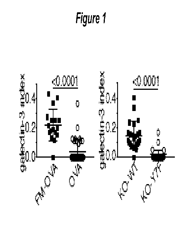
Figure 1. Phagosomal damage. mCherry-galectin-3 was expressed in DNGR-1
deficient MuTuDCs
reconstituted with either NWT or mutant (Y7F) DNGR-1 and cells were incubated
with FM-OVA beads.
Galectin-3 phagosome+ cells were counted and plotted as a ratio (index) to
bead+ cells. Galectin-3 binds
sugar moieties attached to membrane proteins on the luminal side of damaged
endosomes and
phagosomes. mCherry-galectin-3 was recruited to phagosomes in cells expressing
wildtype (WT) but not
the tyrosine-to-phenylalanine (Y7F) mutant.
Figure 2. Efficient XP in non-professional APCs expressing a chimeric receptor
of the invention, `C9/C7'
(shown as "C9::07"). IL-2 ELISA from B3Z hybridoma and HEK293T C7, C9/C7 or
C9(Y7F)/C7 (shown
as "C9(Y7F)::C7") cell co-culture supernatants stimulated with zymosan-OVA.
Data plotted as mean
standard deviation of an experimental triplicate.
CA 03187062 2023- 1-24
WO 2022/023528
PCT/EP2021/071399
28
Figure 3. Syk phosphorylation. Quantification of fold enrichment of phospho-
SYK-staining on
phagosomes (n > 50 phagosomes) in HEK293T C7, C7(Y15F), C9/C7 (shown as
"C9::C7") and
C9(Y7F)/C7 (shown as "C9(Y7F)::C7") cells. p values were calculated by two-
tailed Mann-Whitney test.
Representative plots (n = 2).
Detailed Description of the Invention
This disclosure shows that the superiority of cDC1s in XP is not fully
dependent on unique cell biology but
also on the expression of receptors such as DNGR-1 that detect relevant XP
substrates and initiate the
intracellular signalling that allows phagosome disruption and enables
efficient XP. The inventors show
that ligand-dependent DNGR-1 signalling at the level of phagosomes induces a
local NADPH-dependent
oxidative burst that destabilises the phagosomal membrane causing rupture and
wholesale access of
luminal contents to the cytoplasmic compartment where they can enter the
endogenous MHC class I
processing pathway. Notably, the ability of DNGR-1 to signal for phagosomal
rupture is intrinsic to its
cytoplasmic signalling domain and can be transplanted onto other receptors and
other cell types. Thus,
XP relies on ubiquitous machinery for reactive oxygen species production in
endosomes that can be
subverted by specialised receptors to deliberately provoke vacuolar membrane
damage and P2C.
These results do not exclude the fact that cDC1 possess cell biological
specialisations that favour
xp7,30,36,40-42. Indeed, we identify in these cells slowly maturing phagosomal
compartments that can retain
undegraded cargo for long periods, which is known to favour XP22,23. The fact
that DNGR-1 preferentially
localises to these early phagosomes but does not impact their maturation is
consistent with the notion
that the main function of the receptor is to survey vacuolar compartments for
the presence of exposed F-
actin/myosin complexes, indicative of putatively antigenic cargo that is
relatively intact. Receptor
engagement then leads to Syk-dependent local production of ROS and membrane
damage. Rupture of
any given phagosome is likely to be a stochastic event partly determined by
the extent of damage
possibly offset by membrane repair. Phagosomes that do not rupture can
continue to mature, generating
the LAMP + DNGR-1- degradative late phagosome pool that we also detect in our
assays. The limited
nature of the rupture event, and the fact that it is circumscribed to early
non-degradative endosomes, may
contribute to preventing cell toxicity that would be expected to be induced by
introduction of proteolytic
enzymes into the cyt050149. Yet, the probability of rupture is sufficiently
high that all C9/C7-expressing
HEK293T cells die upon overnight incubation with cytochrome c-soaked zymosan,
which indicates at
least one phagosome rupture event in each of the cells within the 24h period.
Other receptors that can
signal via Syk (e.g., integrins) might also plug into the phagosomal damage
pathway with varying
efficiency, which could explain instances of XP with ligands such as latex
beads that have been shown to
engage a Vav-Rac-NADPH oxidase-dependent XP pathway35. However, some receptors
that can target
antigens for XP by cDC1 , such as the mannose receptor, may employ a distinct
mechanism of P2044,50.
Furthermore, it is clear that not all Syk-activating receptors cause
phagosomal damage and P2C, arguing
for signal divergence at the level of Syk activation. Most notably, Dectin-1,
the canonical Syk-coupled
hemITAM-bearing receptor51, does not induce XP but, rather, promotes DC
activation and inflammatory
gene expression52. Conversely, DNGR-1 signaling triggers XP but does not
induce DC activation4, which
CA 03187062 2023- 1-24
WO 2022/023528
PCT/EP2021/071399
29
means that DNGR-1 acts exclusively as a receptor to decode the antigenicity of
internalised cargo. Thus,
additional signals emanating from dead cells are required to activate cross-
presenting cDC1 and render
them competent to prime CD8+ T cells (e.g., for anti-tumour immunity), as
previously noted in the context
of antibody-mediated antigen targeting to DNGR-1 where adjuvants are necessary
for inducing a
productive CTL response". Because activation signals can also impact XP53,
further understanding of
how they synergise with signals emanating from dedicated XP-promoting
receptors such as DNGR-1
offers great promise for the design of immunotherapies and vaccines that
harness the power of CD8+ T
cells.
Professional/non-professional antigen presenting cells
Some immune cells such as dendritic cells and macrophages are considered to be
"professional" antigen
presenting cells (professional APCs) because they are specialised to perform
this function. While many
cell types can perform some degree of antigen presentation upon MHC class I
molecules (under certain
conditions, specifically when the antigen has been synthesised
intracellularly), professional APCs can
present antigens upon MHC class ll molecules in addition to presenting
antigens on MHC class I. By
expressing DNGR-1 or the chimeric receptor of the invention, cells that are
not professional APCs, can be
enabled to cross-present antigens on MHC I. For instance, by expressing DNGR-1
or the chimeric
receptor of the invention in fibroblasts or muscle cells (which are non-
professional APCs), these cells can
be enabled to cross-present antigens on MHC I.
Biopolymers
Biopolymers are polymeric biomolecules, which are produced in nature in
biological systems such as
cells and which may also be produced by biotechnological systems, such as cell
free expression systems.
Biopolymers include polypeptides, polynucleotides and polysaccharides.
Molecules that comprise a
polypeptide, polynucleotide and/or polysaccharide domain are considered to be
biopolymers even if the
peptide, nucleotide, or saccharide sequence is not found in nature and/or if
the molecule comprises
additional non-biomolecule and/or non-polymer domains.
Pharmaceutical compositions
Pharmaceutical compositions may be prepared using a pharmaceutically
acceptable "carrier" composed
of materials that are considered safe and effective. "Pharmaceutically
acceptable" refers to molecular
entities and compositions that are "generally regarded as safe", e.g., that
are physiologically tolerable and
do not typically produce an allergic or similar untoward reaction, such as
gastric upset and the like, when
administered to a human. In some embodiments, this term refers to molecular
entities and compositions
approved by a regulatory agency of the US federal or a state government, as
the GRAS list under section
204(s) and 409 of the Federal Food, Drug and Cosmetic Act, that is subject to
premarket review and
approval by the FDA or similar lists, the U.S. Pharmacopeia or another
generally recognised
pharmacopeia for use in animals, and more particularly in humans.
CA 03187062 2023- 1-24
WO 2022/023528
PCT/EP2021/071399
The term "carrier" refers to diluents, binders, lubricants and disintegrants.
Those with skill in the art are
familiar with such pharmaceutical carriers and methods of compounding
pharmaceutical compositions
using such carriers.
The pharmaceutical compositions provided herein may include one or more
excipients, e.g., solvents,
5 solubility enhancers, suspending agents, buffering agents, isotonicity
agents, antioxidants or antimicrobial
preservatives. When used, the excipients of the compositions will not
adversely affect the stability,
bioavailability, safety, and/or efficacy of the active ingredients, i.e. the
vectors, cells and or chimeric
receptors, used in the composition. Thus, the skilled person will appreciate
that compositions are
provided wherein there is no incompatibility between any of the components of
the dosage form.
10 Excipients may be selected from the group consisting of buffering
agents, solubilizing agents, tonicity
agents, chelating agents, antioxidants, antimicrobial agents, and
preservatives.
Subject
The subject to be treated may be any animal or human. The subject is
preferably mammalian, more
preferably human. The subject may be a non-human mammal, but is more
preferably human. The
15 subject may be male or female. The subject may be a patient. Therapeutic
uses may be in human or
animals (veterinary use).
Cancers
A "cancer" can comprise any one or more of the following: acute lymphocytic
leukemia (ALL), acute
myeloid leukemia (AML), adrenocortical cancer, anal cancer, bladder cancer,
blood cancer, bone cancer,
20 brain tumor, breast cancer, cancer of the female genital system, cancer
of the male genital system,
central nervous system lymphoma, cervical cancer, childhood rhabdomyosarcoma,
childhood sarcoma,
chronic lymphocytic leukemia (CLL), chronic myeloid leukemia (CML), colon and
rectal cancer, colon
cancer, endometrial cancer, endometrial sarcoma, esophageal cancer, eye
cancer, gallbladder cancer,
gastric cancer, gastrointestinal tract cancer, hairy cell leukemia, head and
neck cancer, hepatocellular
25 cancer, Hodgkin's disease, hypopharyngeal cancer, Kaposi's sarcoma,
kidney cancer, laryngeal cancer,
leukemia, leukemia, liver cancer, lung cancer, malignant fibrous histiocytoma,
malignant thymoma,
melanoma, mesothelioma, multiple myeloma, myeloma, nasal cavity and paranasal
sinus cancer,
nasopharyngeal cancer, nervous system cancer, neuroblastoma, non-Hodgkin's
lymphoma, oral cavity
cancer, oropharyngeal cancer, osteosarcoma, ovarian cancer, pancreatic cancer,
parathyroid cancer,
30 penile cancer, pharyngeal cancer, pituitary tumor, plasma cell neoplasm,
primary CNS lymphoma,
prostate cancer, rectal cancer, respiratory system, retinoblastoma, salivary
gland cancer, skin cancer,
small intestine cancer, soft tissue sarcoma, stomach cancer, stomach cancer,
testicular cancer, thyroid
cancer, urinary system cancer, uterine sarcoma, vaginal cancer, vascular
system, Waldenstronn's
macroglobulinemia and Wilms' tumor.
Cancers may be of a particular type. Examples of types of cancer include
astrocytoma, carcinoma (e.g.
adenocarcinoma, hepatocellular carcinoma, medullary carcinoma, papillary
carcinoma, squamous cell
CA 03187062 2023- 1-24
WO 2022/023528
PCT/EP2021/071399
31
carcinoma), glioma, lymphoma, medulloblastoma, melanoma, myeloma, meningioma,
neuroblastoma,
sarcoma (e.g. angiosarcoma, chrondrosarcoma, osteosarcoma).
Some cancers cause solid tumours. Such solid tumours may be located in any
tissue, for example the
pancreas, lung, breast, uterus, stomach, kidney or testis. In contrast,
cancers of the blood, such as
leukaemias, may not cause solid tumours ¨ and may be referred to as liquid
tumours.
Vectors
A "vector" as used herein is an oligonucleotide molecule (DNA or RNA) used as
a vehicle to transfer
foreign genetic material into a cell. The vector may be an expression vector
for expression of the foreign
genetic material in the cell. Such vectors may include a promoter sequence
operably linked to the
nucleotide sequence encoding the gene sequence to be expressed. A vector may
also include a
termination codon and expression enhancers. Any suitable vectors, promoters,
enhancers and
termination codons known in the art may be used to express the chimeric
receptor of the invention in a
cell or tissue.
The skilled person will appreciate that a gene therapy vector can be used to
introduce the nucleic acid of
the invention into a recipient cell or tissue. In some embodiments, the gene
therapy vector is a viral
vector. The viral vector may be an adenoviral vector, an AAV or a lentiviral
vector. For some applications,
it is advantageous to use a viral vector that is pseudotyped with an envelope
protein that facilitates the
transduction of hematopoietic stem cells and/or progenitor cells. In some
embodiments, the nucleic acid
is introduced into the mammalian cell using the CRISPR-CAS9 system.
Antibody-based target binding domains
Antibodies which will bind to the targets discussed herein are already known.
In view of today's
techniques in relation to monoclonal antibody technology, antibodies can be
prepared to most antigens.
The target binding domain may be a part of an antibody (for example a Fab
fragment) or a synthetic
antibody fragment (for example a single chain Fv fragment [ScFv]) Suitable
monoclonal antibodies to
selected antigens may be prepared by known techniques, for example those
disclosed in "Monoclonal
Antibodies: A manual of techniques ", H Zola (CRC Press, 1988) and in
"Monoclonal Hybridoma
Antibodies: Techniques and Applications ", J G R Hurrell (CRC Press, 1982).
Chimeric antibodies are
discussed by Neuberger et al (1988, 8th International Biotechnology Symposium
Part 2, 792-799).
Monoclonal antibodies (mAbs) are useful in the methods of the invention and
are a homogenous
population of antibodies specifically targeting a single epitope on an
antigen. Suitable monoclonal
antibodies can be prepared using methods well known in the art (e.g. see
Kohler, G.; Milstein, C. (1975).
"Continuous cultures of fused cells secreting antibody of predefined
specificity". Nature 256 (5517): 495;
Siegel DL (2002). "Recombinant monoclonal antibody technology". Schmitz U,
Versmold A, Kaufmann P,
Frank HG (2000); "Phage display: a molecular tool for the generation of
antibodies--a review". Placenta.
21 Suppl A: S106-12. Helen E. Chadd and Steven M. Chamow; "Therapeutic
antibody expression
technology," Current Opinion in Biotechnology 12, no. 2 (April 1,2001): 188-
194; McCafferty, J.; Griffiths,
A.; Winter, G.; Chiswell, D. (1990). "Phage antibodies: filamentous phage
displaying antibody variable
CA 03187062 2023- 1-24
WO 2022/023528
PCT/EP2021/071399
32
domains". Nature 348 (6301): 552-554; "Monoclonal Antibodies: A manual of
techniques", H Zola (CRC
Press, 1988) and in "Monoclonal Hybridoma Antibodies: Techniques and
Applications ", J G R Hurrell
(CRC Press, 1982). Chimeric antibodies are discussed by Neuberger et al (1988,
8th International
Biotechnology Symposium Part 2, 792-799)).
Polyclonal antibodies are useful in the methods of the invention. Monospecific
polyclonal antibodies are
preferred. Suitable polyclonal antibodies can be prepared using methods well
known in the art.
Fragments of antibodies, such as Fab and Fab2 fragments may also be used as
can genetically
engineered antibodies and antibody fragments. The variable heavy (VH) and
variable light (VL) domains of
the antibody are involved in antigen recognition, a fact first recognised by
early protease digestion
experiments. Further confirmation was found by "humanisation" of rodent
antibodies. Variable domains
of rodent origin may be fused to constant domains of human origin such that
the resultant antibody
retains the antigenic specificity of the rodent parented antibody (Morrison et
al (1984) Proc. Natl. Acad.
Sd. USA 81, 6851-6855).
That antigenic specificity is conferred by variable domains and is independent
of the constant domains is
known from experiments involving the bacterial expression of antibody
fragments, all containing one or
more variable domains. These molecules include Fab-like molecules (Better et
al (1988) Science 240,
1041); Fv molecules (Skerra et al (1988) Science 240, 1038); single-chain Fv
(ScFv) molecules where the
VH and VL partner domains are linked via a flexible oligopeptide (Bird et al
(1988) Science 242, 423;
Huston et al (1988) Proc. Natl. Acad. Sd. USA 85, 5879) and single domain
antibodies (dAbs) comprising
isolated V domains (Ward et al (1989) Nature 341, 544). A general review of
the techniques involved in
the synthesis of antibody fragments which retain their specific binding sites
is to be found in Winter &
Milstein (1991) Nature 349, 293- 299.
By "ScFv molecules" we mean molecules wherein the VH and VL partner domains
are covalently linked,
e.g. directly, by a peptide or by a flexible oligopeptide. Fab, Fv, ScFv and
dAb antibody fragments can all
be expressed in and secreted from E. coil, thus allowing the facile production
of large amounts of the said
fragments.
Whole antibodies, and F(a13)2 fragments are "bivalent". By "bivalent" we mean
that the said antibodies
and F(ab.)2 fragments have two antigen combining sites. In contrast, Fab, Fv,
ScFv and dAb fragments
are monovalent, having only one antigen combining site. Synthetic antibodies
which bind to a target
discussed herein may also be made using phage display technology as is well
known in the art (e.g. see
"Phage display: a molecular tool for the generation of antibodies--a review".
Placenta. 21 Suppl A: S106-
12. Helen E. Chadd and Steven M. Chamow; "Phage antibodies: filamentous phage
displaying antibody
variable domains". Nature 348 (6301): 552-554).
Antibodies can be readily conjugated to peptide or protein moieties by
operably linking a nucleotide
sequence encoding the desired peptide or protein at the 3' end of a nucleotide
sequence that encodes
one of the antibody chains, e.g. the heavy chain. Thus a fusion antibody can
be expressed, where the C-
terminus of the antibody polypeptide is fused to the desired peptide or
protein, often via a linker
sequence. Antibody fusions are well known in the art.
CA 03187062 2023- 1-24
WO 2022/023528
PCT/EP2021/071399
33
Aptamer based target binding domains
Aptamers are short DNA/RNA/peptide molecules which can bind specifically to a
target molecule(Pan &
Clawson, 2009). Aptamers specific for a particular target are often selected
from a large pool of randomly
generated libraries of molecules, e.g. by using the Systematic Evolution of
Ligands by Exponential
Enrichment (SELEX) method. SELEX method involves exposing a random sequence
library to a specific
target and amplifying the bound molecules which are then subjected to
additional rounds of selection.
After multiple rounds of selection, specific aptamers identified for binding
to the target molecule can be
subjected to further rounds of modifications to improve their binding affinity
and stability. Aptamers can
be readily conjugated to additional nucleic acid moieties, thus facilitating
targeted binding of the additional
nucleic acid moiety to the specific binding target of the aptamer.
Aspects and embodiments of the present invention will now be discussed with
reference to the
accompanying figures. Further aspects and embodiments will be apparent to
those skilled in the art. All
documents mentioned in this text are incorporated herein by reference.
The features disclosed in the foregoing description, or in the following
claims, or in the accompanying
drawings, expressed in their specific forms or in terms of a means for
performing the disclosed function,
or a method or process for obtaining the disclosed results, as appropriate,
may, separately, or in any
combination of such features, be utilised for realising the invention in
diverse forms thereof.
While the invention has been described in conjunction with the exemplary
embodiments described above,
many equivalent modifications and variations will be apparent to those skilled
in the art when given this
disclosure. Accordingly, the exemplary embodiments of the invention set forth
above are considered to
be illustrative and not limiting. Various changes to the described embodiments
may be made without
departing from the spirit and scope of the invention.
For the avoidance of any doubt, any theoretical explanations provided herein
are provided for the
purposes of improving the understanding of a reader. The inventors do not wish
to be bound by any of
these theoretical explanations.
Any section headings used herein are for organizational purposes only and are
not to be construed as
limiting the subject matter described.
Throughout this specification, including the claims which follow, unless the
context requires otherwise, the
word "comprise" and "include", and variations such as "comprises",
"comprising", and "including" will be
understood to imply the inclusion of a stated integer or step or group of
integers or steps but not the
exclusion of any other integer or step or group of integers or steps.
It must be noted that, as used in the specification and the appended claims,
the singular forms "a," "an,"
and "the" include plural referents unless the context clearly dictates
otherwise. Ranges may be expressed
herein as from "about" one particular value, and/or to "about" another
particular value. When such a range
is expressed, another embodiment includes from the one particular value and/or
to the other particular
CA 03187062 2023- 1-24
WO 2022/023528
PCT/EP2021/071399
34
value. Similarly, when values are expressed as approximations, by the use of
the antecedent "about," it
will be understood that the particular value forms another embodiment. The
term "about" in relation to a
numerical value is optional and means for example +/- 10%.
Examples
Example 1 ¨ DNGR-1 knock-out cDC1 cells
To study the contribution of DNGR-1 to XP of dead cell-associated antigens by
cDC1 12'15, the inventors
made use of the cDC1 cell line MuTuDC1940 (henceforth termed MuTuDCs)17.
MuTuDCs were pulsed
with UV-irradiated ovalbumin (OVA)-expressing H-2Kbm1 mouse embryonic
fibroblasts (OVA dead cells)
and then cultured with pre-activated OVA-specific (OT-1) CD8+ T cells.
Interferon-y (IFN-y) accumulation
in the culture indicates 01-1 receptor triggering by H2Kb/OVA complexes and,
hence, is an indirect
measure of OVA XP by MuTuDCs. As reported16, OT-1T cultures with DNGR-1-
deficient MuTuDCs (KO)
accumulated lower levels of IFN-y than cultures with wild type MuTuDCs (WT).
This defect was corrected
by ectopic re-expression of the receptor in DNGR-1-deficient MuTuDCs (KO-VVT)
and was not attributable
to an effect on antigen uptake as KO, VVT and KO-WT MuTuDCs all displayed a
similar capacity to
internalise dye-labelled dead cell material. In contrast, when studying the
uptake of OVA-coated latex
beads (OVA beads), we noticed that additional coating with the physiological
ligand for DNGR-1, F-
actin/myosin 11 complexes16 (FM-OVA beads), did result in greater bead
internalisation (p<0.0001), which
enhanced OVA XP (measured by IFN-y production following co-culture with pre-
activated OT-1CD8+ T
cells, which enhanced OVA XP. Thus, DNGR-1 can serve as a phagocytic receptor
for ligand-bearing
particles but it is redundant for uptake of dead cell debris by cDC1, likely
because the latter contain
ligands that can engage additional cDC1 phagocytic receptors.
To separate the effect of DNGR-1 on XP from its contribution to ligand uptake,
the inventors fed
MuTuDCs with OVA beads or FM-OVA beads and sorted cells that had phagocytosed
a single bead
before testing them in the XP assayle. It was found that MuTuDCs containing
single FM-OVA beads
stimulated CD8' OT-1T cells more efficiently than cells with single OVA beads.
This effect was shown to
be specific for XP, because both sets of sorted MuTuDCs (FM-OVA bead-uptake;
and OVA bead-uptake)
stimulated CD4' OT-II T cells to the same extent.
DNGR-1-deficient MuTuDCs re-expressing either wildtype receptor (KO-WT), or a
mutant receptor that
cannot bind F-actin (W155A/VV250A; termed KO-2WA) were compared, which showed
a defect in XP of
dead cell-associated antigen in KO-2WA cells but unaltered capacity to cross-
present OVA beads or egg
white (a source of endotoxin-free soluble OVA antigen). KO-WA MuTuDC
internalised fewer FM-OVA
beads than KO-WT cells and displayed markedly diminished ability to stimulate
OT-1 cells. Therefore, as
above, taking single FM-OVA bead + MuTuDCs, the inventors asked whether the
mutation in the F-actin
binding region of DNGR-1 impacted XP independently of particle uptake. Again,
they observed a
decrease in XP in cells bearing mutant DNGR-1 unable to engage its ligand.
Together, these data
indicate a specific effect of DNGR-1 engagement on XP of ligand-associated
antigen, which can be
formally distinguished from receptor contribution to ligand uptake.
CA 03187062 2023- 1-24
WO 2022/023528
PCT/EP2021/071399
Example 2 ¨ The cytosolic pathway to cross-presentation
Two basic models for XP have emerged from studies in multiple cell types, one
in which antigen
processing and MHC class I molecule loading occurs entirely within the
phago/endosomal compartment
of APCs ("vacuolar" pathway) and another in which exogenous antigens somehow
gain access to the
5 ARC cytosol ("cytosolic" or "phagosome-to-cytosol" (P2C) pathway) and are
processed by the
proteasome as for endogenous antigens21. Inhibition of lysosomal proteases by
leupeptin or pepstatin,
which block the vacuolar pathway, did not diminish XP of soluble antigen, bead-
bound OVA or dead cell-
associated OVA. In contrast, the proteasome inhibitor lactacystin inhibited XP
of both bead-bound
antigen and dead cell-associated antigen, but had no effect on XP of soluble
antigen (except at high
10 concentrations) or adverse effects on T cells stimulated with pre-
processed OVA peptide SIINFEKL. In
line with previous reports22-24, blockade of lysosomal acidification by
chloroquine or blockade of cysteine
proteases by E64 enhanced XP of FM-OVA beads and dead cell-associated antigen.
Taken together, these results suggest that antigen degradation through
lysosomal proteases is
detrimental for DNGR-1-dependent XP and that DNGR-1 engages the cytosolic
rather than the vacuolar
15 XP pathway in cDC1.
Example 3 ¨ The DNGR-1+ phagosomal compartment
To investigate how DNGR-1 affects the properties of antigen-containing
phagosomes, we isolated FM-
OVA bead phagosomes from MuTuDCs and characterised them by flow cytometry
(PhagoFACS).
Interestingly, DNGR-1 and the lysosomal marker LAMP-2 marked two mutually
exclusive phagosome
20 populations, which, by microscopy were found to co-exist in individual
cells. Further analysis revealed
that DNGR-1+ phagosomes co-stained for MHC I and II whereas LAMP-2+ phagosomes
were MHC II+ but
contained lower levels of MHC I.
Importantly, the two phagosome populations showed differential capacity to
degrade antigen, as DNGR-
1+ phagosomes displayed high anti-OVA staining in both flow cytometry and
microscopy assays whereas
25 LAMP-2+ phagosomes stained only weakly. After long chase periods, some
DNGR-1+ MHC l+ OVAh'gh
phagosomes eventually lost DNGR-1 staining, acquired LAMP and degraded OVA,
indicating that they
were not fully arrested in maturation. DNGR-1+ MHC OVAh'oh phagosomes were not
an aberrant
compartment of MuTu DCs as they were also found in phagosomal preparations
derived from primary
cDC1s obtained from bone marrow cultured in Flt3L and in KID cells, a second
cDC1 cell line26.
30 Furthermore, ectopic expression of DNGR-1 in the macrophage cell line
RAVV264.7 also allowed for
identification of DNGR-1+ OVAhigh phagosomes distinct from LAMP-2+ OVAI'Dw
phagosomes. To ask if
DNGR-1 was required for formation of MHC OVAh'gh phagosomal compartments, the
inventors
compared FM-OVA bead phagosomes from DNGR-1 deficient (KO) and wildtype (WT)
MuTuDCs.
Interestingly, MHC I+ LAMP-2- and MHC I+ OVAhigh phagosomes were identified at
similar frequency in
35 both cells. We further compared DNGR-1+ phagosomes from wildtype MuTuDCs
that had been fed OVA
beads vs. FM-OVA beads and found no differences with respect to OVA
degradation or MHC I
recruitment.
CA 03187062 2023- 1-24
WO 2022/023528
PCT/EP2021/071399
36
Taken together, these data suggest that DNGR-1 marks an MHC I+ phagosomal
compartment in cDC1
(and, when ectopically expressed, in RAW264.7 macrophages) that has low
degradative potential and the
ability to preserve undegraded antigen, at least temporarily. However, the
presence of DNGR-1 or of its
ligand does not affect the ability of phagocytic cargo to access this poorly
degradative compartment.
Example 4 ¨ Phagosome to cytosol transfer (P2C)
We searched for subsequent steps in XP that might be modulated by DNGR-1
ligand. As DNGR-1
dependent XP is mediated through the cytosolic pathway (see above), we focused
on phagosome to
cytosol transfer of the antigen. We investigated if DNGR-1+ phagosomes showed
signs of membrane
damage, consistent with disruption, by measuring local recruitment of
cytosolic galectin-3 or 8, which bind
to sugar moieties attached to membrane proteins on the luminal side of damaged
endosomes and
phagosomes 27. Strikingly, by PhagoFACS, we found that the frequency of
galectin-8+ phagosomes in
MuTuDCs was higher for DNGR-1+ phagosomes after 4 hours compared to LAMP-2 *
phagosomes.
Similarly, by confocal microscopy, mCherry-galectin-3 could be found
decorating phagosomes when
MuTuDCs were fed FM-OVA beads but much less frequently with OVA beads that do
not trigger DNGR-
1.
DNGR-1 is a type II trans-membrane protein with a short N-terminal
intracellular tail bearing a single
hemITAM signalling motif28. To test the role of the DNGR-1 hemITAM, we
expressed mCherry-galectin-3
in DNGR-1-deficient MuTuDCs reconstituted with either wildtype (KO-VVT) or
hemITAM signalling
incompetent DNGR-1 (tyrosine to phenylalanine mutation at position 7 - KO-
Y7F). When cells were fed
FM-OVA beads, mCherry-galectin-3 was recruited to phagosomes in cells
expressing WT but not the Y7F
receptor (Figure 1).
To validate these observations, we used a different cytosolic sensor of
endosomal membrane damage.
Sphingomyelin is distributed asymmetrically in cellular membranes and is
exposed to the cytosol only
upon phagosomal damage. Therefore, cytosolic expression of a probe containing
a version of the
sphingomyelin-binding protein lysenin fused to mCherry allows for the specific
labelling of damaged
phagosomal membranes (Ellison et al, submitted). In line with the results with
galectins, the mCherry-
lysenin probe accumulated specifically on FM-OVA bead phagosomes when wildtype
DNGR-1, but not
Y7F DNGR-1, was expressed. The inventors conclude that ligand-dependent DNGR-1
signalling via its
hemITAM induces phagosomal membrane damage in MutuDCs.
Example 5 ¨ Chimeric receptors facilitate P2C
The inventors investigated the possibility that DNGR-1 hemITAM signalling
could mediate phagosomal
damage in a heterologous system. HEK293T cells were transfected with Dectin-1
(aka CLEC7A; a
receptor structurally homologous to DNGR-1) or chimeras comprising the
extracellular domain and
transmembrane region of Dectin-1 fused to variants of the cytoplasmic tail of
DNGR-1. Dectin-1 binds to
yeast beta-glucans and functions as a yeast phagocytic receptor, allowing us
to analyse uptake of
zymosan particles (i.e., yeast cell walls) instead of latex beads. Indeed,
Dectin-1 (C7), Dectin-1 fused
CA 03187062 2023- 1-24
WO 2022/023528
PCT/EP2021/071399
37
with wildtype (09/C7) or hemITAM tyrosine-mutated cytoplasmic tail of DNGR-1
(C9(Y7F)/C7) all
conferred upon HEK293T the ability to take up zymosan while a tail-less mutant
of Dectin-1 did not.
Notably, when co-expressing the nnCherry-lysenin probe and the chimeric
receptors in HEK293T, lysenin'
phagosomes were observed in cells expressing the C9/C7 chimera but not in
cells expressing wild type
C7 (p<0.0001) or the signaling incompetent chimera C9(Y7F)/C7 (p=0.03). In
contrast to latex beads,
zymosan particles are porous and do not have a solid core, therefore acting as
a sponge for any probe
that accesses the phagosomal lumen. Strikingly, we noticed that phagosomes in
HEK293T cells
expressing the C9/C7 chimera became positive for GFP that was expressed in the
cytosol. In contrast,
intra-phagosomal GFP was largely absent from zymosan-containing phagosomes in
HEK293T
expressing C7 or C9(Y7F)/C7, indicating a specific requirement for DNGR-1
hemITAM signalling.
Lysenin phagosomes showed a higher mean fluorescent intensity (MFI) for GFP
compared to lysenin
-
phagosomes, suggesting that access of cytosolic GFP to phagosomes was coupled
to membrane
damage. This was also apparent from live cell imaging, which showed that
lysenin recruitment was
predictive of but preceded GFP influx. These results suggest that DNGR-1
hemITAM signalling
permeabilises phagosomes, rendering their lumen accessible to cytosolic
proteins.
To assess whether permeability is bi-directional and permits release of
phagosomal cargo into the
cytosol, HEK293T expressing either C9/C7 or C9(Y7F)/C7 were pulsed with
zymosan soaked in
sulforhodamine B (SRB). A significant increase in SRB fluorescence was
detected in the cytosol of
HEK293T expressing 09/C7, but not those expressing C9(Y7F)/C7.
A previously-reported P2C assay29 was also used to confirm that the efficacy
with which zymosan-
adsorbed beta-lactannase could be released from phagosomes into the cytosol
was greater in HEK293T
cells expressing C9/C7 than C9(Y7F)/C7.
Zymosan soaked cytochrome c particles (zymosan-cyt. c particles) were added
the to C7, C9/C7 and
C9(Y7F)/C7 expressing HEK293T cells (Fig. 3g). When incubated with zymosan-
cyt. c particles,
HEK293T cells expressing C7 or C9(Y7F)/C7 internalised the particles but
survival was unaffected. In
stark contrast, all the 09/C7-expressing cells died within a 24h period,
demonstrating that DNGR-1
hemITAM-dependent P2C, essentially, occurs in all cells in which receptor
signalling is triggered.
Together, these results indicate that hemITAM signalling permeabilises
phagosomes so as to allow efflux
of luminal contents into the cytosol ¨ and that this effect is retained when
the hemITAM motif is present in
the cytoplasmic tail of other transmembrane proteins, besides DNGR-1.
Example 6 ¨ Characterising hemITAM-induced phagosome permeability
To examine the nature and durability of DNGR-1 hemITAM-induced phagosomal
permeability, the
inventors performed FRAP experiments on GFP. phagosomes stained with the
lysenin probe. After
photobleaching the GFP within the lumen of lysenin phagosomes, signal was re-
observed within 2
minutes, indicating continuous GFP influx and suggesting irreversible
phagosomal membrane damage.
As a control, bleaching of the lysenin signal did not lead to fluorescence
recovery.
CA 03187062 2023- 1-24
WO 2022/023528
PCT/EP2021/071399
38
Analysis of the ultrastructure of GFPlysenin+ zymosan phagosomes in C9/C7-
expressing cells by
correlative light and electron microscopy revealed that the phagosomal
membrane contained a large hole
with a diameter of roughly 1-1.5 pm. Thus, DNGR-1 signalling can cause large
scale rupture of
phagosomes, which would allow for even sizeable luminal contents to be
released into the cytosol.
Example 7 ¨ XP in HEK293T cells
HEK293T lines stably expressing murine H-2K' and beta-2-microglobulin were
further transfected to
express C7, C9/C7 or C9(Y7F)/C7 chimeras, single cell cloned and selected for
equal H-2Kb and Dectin-1
extracellular domain expression levels. All three cell lines showed equivalent
capacity to stimulate B3Z,
an OVA/H-2K'-specific T cell hybridoma, when pulsed with exogenous SIINFEKL
peptide and were
equally competent at presenting endogenous antigen when transfected to express
Venus-SIINFEKL, a
fusion protein mimic of endogenous OVA. These cells were exposed to zymosan
that had been soaked
in egg white (zymosan-OVA). C7, C9/C7and C9(Y7F)/C7 HEK293T lines all
internalised zymosan-OVA
with similar efficacy. However, efficient XP, as measured by B3Z activation
after HEK293T cell fixation,
was only observed with cells expressing the C9/C7 chimera (Figure 2).
HEK293T cells expressing 09/C7 were then fed with zymosan dually soaked in
both egg white and
cytochrome c. Exposure time was optimised to kill only a fraction of the
cells. This led to complete loss
of cross presentation activity when compared to feeding cells with zymosan
particles soaked with OVA
alone. Overall cytotoxicity of cytochrome c in the culture was excluded by the
fact that no decrease in
B3Z activation was observed when we incubated zymosan-cyt. c particles with
HEK293T cells expressing
the C9/C7 chimera and Venus-SIINFEKL. These data formally indicate that DNGR-1
hemITAM
signalling-induced phagosomal rupture is responsible for XP.
Example 8¨ Interaction of the hemITAM motif with the tyrosine kinase, Syk
The hemITAM motif of DNGR-1 can recruit and activate Syk or SHP-1 in response
to DNGR-1 ligand
engagement12,33. Accordingly, Syk phosphorylation at two distinct sites was
observed in wildtype
MuTuDCs treated with anti-DNGR-1 cross-linking antibody or F-actin/myosin ll
complexes (DNGR-1
ligand; DNGR-1L). The inventors now confirm that C9/C7 chimeras also induce
phosphorylation of Syk in
HEK293T in response to zymosan treatment. This is observed at the level of
phagosomes and is
hemITAM-dependent, as it was not observed in cells expressing C9(Y7F)/C7
(Figure 3).
To determine whether phosphorylation of Syk was upstream of phagosomal
rupture, a Syk-deficient
C9/C7-expressing HEK293T cell was generated using CRISPR/Cas9 (SylccRIsPR).
The influx of
cytoplasmic GFP into zymosan phagosomes was completely lost in SylccRIsPR
cells. However, this influx
was restored by reconstituting the Syk-deficient C9/C7-expressing HEK293T
cells with (CRISPR-
resistant) wild type mouse Syk (Syk VVT) but not a catalytically-deficient
mutant (K396R - kinase dead;
Syk KD). Similarly, phagosomal GFP influx was not observed when C9/C7-
expressing HEK293T cells
were treated with the Syk inhibitor R406, even though zymosan uptake was not
affected. In MuTu DCs,
both R406 and another Syk inhibitor, inhibitor IV, abrogated staining of
phagosomes with the lysenin
CA 03187062 2023- 1-24
WO 2022/023528
PCT/EP2021/071399
39
probe and blocked XP of FM-OVA beads. This suggests that Syk activation and
kinase activity
downstream of DNGR-1 are required for the induction of phagosomal rupture and
XP.
Example 9 ¨ Syk mediated membrane destabilisation and rupture via reactive
oxygen species
The inventors examined possible mechanisms downstream of Syk that might cause
membrane
destabilisation and rupture. Reactive oxygen species (ROS) produced by the
NADPH oxidase cause lipid
peroxidation leading to membrane destabilisation and endosomal content
leakage34-37. Moreover, Syk
activates NADPH oxidase activation via Vav and Rac, all of which have been
implicated in XP of
particulate antigens by myeloid Ce IIS34-37. The inventors noticed that
exposure to zymosan led to a very
potent oxidative burst in RAW264.7 cells ectopically expressing C9/C7 but not
C9(Y7F)/C7. Using a
fluorescent probe, the inventors confirmed that this burst occurred at the
level of individual phagosomes
and was diminished in RAW264.7 cells expressing C9(Y7F)/C7 or in cells treated
with NADPH inhibitor,
DPI. Similarly, in RAW264.7 cells expressing DNGR-1 (C9) and fed with fixed
and permeabilised sheep
red blood cells (a phagocytic target bearing exposed cortical F-actin that can
be recognised by DNGR-
114), the inventors observed an oxidative burst around phagosomes that was
diminished by SYK inhibition
or DPI treatment. Similar results were obtained in HEK293T cells expressing
C9/C7. Reactive oxygen
species produced by NADPH cause lipid peroxidation and lead to membrane
destabilisation and
endosomal content leakage37. Consistent with this notion, inhibition of NADPH
oxidase by DPI prevented
lysenin accumulation on phagosomes in RAW264.7 cells. Notably, it also
decreased XP of zymosan-
OVA by HEK293T cells whilst not affecting the presentation of Venus-SIINFEKL.
Finally, in MuTu DCs,
DPI, as well as the reactive oxygen species (ROS) scavenger, alpha-tocopherol
(vitamin E) abrogated XP
of FM-OVA beads or OVA-bearing dead cells but did not affect the presentation
of SIINFEKL peptide.
These results indicate that DNGR-1/Syk-dependent activation of NADPH causes
lipid peroxidation
allowing phagosomal rupture and P2C. This allows for the unselective release
of internalised ligand-
associated antigens into the cytosol of cDC1s and permits access to the
endogenous MHC class I
processing and presentation pathway.
Reactive oxygen species (ROS) produced by the NADPH oxidase can cause lipid
peroxidation and lead
to membrane destabilisation and endosomal content leakage37. Consistent with a
role for ROS in
membrane damage, inhibition of the NADPH oxidase by DPI blocked lysenin
accumulation on
phagosomes in RAW264.7 cells expressing C9/C7 to the same level as the Y7F
mutation. Furthermore,
DPI, as well as the ROS scavenger alpha-tocopherol (vitamin E), greatly
decreased XP of both FM-OVA
beads and OVA-bearing dead cells in MutuDCs without impacting presentation of
SIINFEKL peptide.
Similarly, DPI decreased XP of zymosan-OVA by HEK293T cells expressing C9/07
but did not diminish
presentation of endogenous Venus-SIINFEKL antigen. Finally, siRNA-mediated
silencing of NOX2
(CYBB), the predominant catalytic subunit of the NADPH oxidase in myeloid
cells, decreased XP of
zymosan-OVA by RAW264.7 expressing C9/C7 and H-2Kb but did not affect
presentation of SIINFEKL
peptide.
These data were extended to primary cDC1s by establishing Flt3L cultures with
bone marrow from wild-
type, DNGR-1 KO and NOX2 KO mice and purified cDC1s by magnetic enrichment.
NOX2 KO cDC1
CA 03187062 2023- 1-24
WO 2022/023528
PCT/EP2021/071399
were defective in phagosomal ROS production in response to DNGR-1 stimulation
and both DNGR-1 KO
and NOX2 KO cDC1s displayed a reduction in XP of OVA-bearing dead cells
relative to \NT cDC1s
despite being equally effective at presenting SIINFEKL peptide and
internalising dead cell material. To
assess the importance of NOX2 for DNGR-1-dependent XP in vivo, we first
immunised wild-type, DNGR-
5 1 KO, BATF3 KO or NOX2 KO mice with FM-OVA beads + poly I:C and measured
OVA-specific CD8* T
cells responses by H-2Kb/OVA-pentamer staining. As reported18, VVT mice
mounted a robust response to
FM-OVA beads + poly I:C that was significantly decreased in both DNGR-1 KO and
BATF3 KO mice.
Importantly, NOX2 KO mice also displayed a reduction in OVA-specific CD8* T
cell cross-priming. To
confirm that this reflected NOX2 function in cDC1s and to extend the data to
cross-priming to dead cell-
10 associated antigens, we generated radiation chimeras using bone marrow
from BATF3 KO CD45.1 mice
mixed at a ratio of 80:20 with bone marrow from either BATF3 KO, wild-type,
DNGR-1 KO or NOX2 KO
CD45.2 mice. The inventors used BATF3 KO CD45.1 mice as recipients, further
ensuring that the only
cDC1 that develop after reconstitution arise from the CD45.2 donor bone
marrow, and we analysed
exclusively the response of CD45.1 T cells to exclude any cell-intrinsic
effects of NOX2 deletion in
15 lymphocytes. Following immunisation with OVA+polyl:C-pulsed dead cells,
BATF3 KO:BATF3 KO
chimeras failed to generate OVA-specific CD8+ T cells, as expected18. In
contrast, robust cross-priming
to OVA was seen in BATF3 KO:VVT chimeras as measured by H-2Kb/OVA-tetramer
staining or by
intracellular staining for IFNy in response to ex vivo spleen cell
restimulation with SIINFEKL peptide.
Consistent with previous observations in DNGR-1 KO mice3,11,12,44, the OVA-
specific CD8+ T cell
20 response was significantly diminished in BATF3 KO:DNGR-1 KO chimeras.
Notably, it was also
significantly diminished in BATF3 KO:NOX2 KO chimeras.
Macrophages, monocyte-derived dendritic cells and other myeloid cell types, as
well as non-immune
cells, have been used extensively to dissect some of the mechanisms involved
in XP7'23'45. Relatively few
papers have focused on XP mechanisms specifically in cDC1. Here, the inventors
focus on the possibility
25 that the superiority of cDC1s in XP depends not only on unique cell
biology but also on the expression of
receptors such as DNGR-1 that detect relevant XP substrates. The inventors
show that ligand-dependent
DNGR-1 signalling at the level of phagosomes induces a local NADPH-dependent
oxidative burst that
destabilises the phagosomal membrane causing rupture and wholesale access of
luminal contents to the
cytoplasmic compartment where they can enter the endogenous MHC I processing
pathway. Notably, the
30 ability of DNGR-1 to signal for phagosomal rupture is intrinsic to its
cytoplasmic signalling domain and
can be transplanted onto other receptors and operate in other cell types,
including non-immune cells.
Thus, XP relies on the machinery for reactive oxygen species production in
endosomes. This machinery
can be subverted by specialised receptors to deliberately provoke vacuolar
membrane damage and P2C.
35 Numbered Paragraphs
1. A chimeric receptor comprising an extracellular target binding
domain, a transmembrane domain,
and an intracellular domain that comprises a Syk-binding sequence derived from
the signalling domain of
the cytoplasmic tail of DNGR-1, wherein said Syk-binding sequence contains a
tyrosine residue.
CA 03187062 2023- 1-24
WO 2022/023528
PCT/EP2021/071399
41
2. The chimeric receptor according to paragraph 1, wherein the Syk-binding
sequence comprises a
hem ITAM.
3. The chimeric receptor according to paragraph 2, wherein the hemITAM
comprises the amino acid
sequence set forth in SEQ ID NO:14 (EXXYXXL; wherein X represents any amino
acid residue).
4. The chimeric receptor according to paragraph 3, wherein the Syk-binding
sequence comprises an
amino acid sequence as set forth in SEQ ID NO:15 (MHAEXXYX)(LQWD) or as set
forth in SEQ ID
NO:90 (MHEE)OYXXLQVVD); optionally wherein one, two or all three of the amino
acid residues at the N-
terminal end of the MHAEXXYXXLQVVD (SEQ ID NO.: 15) or MHEEXXYXXLQVVD (SEQ ID
NO.: 90)
sequence are removed or substituted with another amino acid residue, or
wherein one, two or all three of
the amino acid residues at the C-terminal end of the MHAEXXYXXLQVVD (SEQ ID
NO.: 15) sequence
are removed or substituted with another amino acid residue.
5. The chimeric receptor according to any one of paragraphs 1-4, wherein
the wherein the Syk-
binding sequence comprises an amino acid sequence as set forth in SEQ ID NO:11
(MHAEEIYTSLQWD)
or an amino acid sequence as set forth in SEQ ID NO:89 (MHEEEIYTSLQWD),
optionally wherein one,
two or three amino acid residues are substituted with another amino acid
residue.
6. The chimeric receptor according to any one of the preceding paragraphs,
wherein the target
binding domain binds a target that is present on a pathogen, a pathogenic
cell, a dead cell or a diseased
cell.
7. The chimeric receptor according to paragraph 6, wherein the target is an
antigen present on a
tumour cell, optionally wherein the target is a tumour neoantigen.
8. The chimeric receptor according to paragraph 7, wherein the antigen
present on the tumour cell
is CEA, ERBB2, EGFR, GD2, mesothelin, MUC1, PSMA, CAIX,CD133,c-Met, EGFR,
EGFRvIll, Epcam,
EphA2, FRa, CD19, CD20, GPC3, GUCY2C,HER1, HER2, ICAM-1, MAGE, or MET.
9. The chimeric receptor according to paragraph 6, wherein the target is a
viral antigen present at
the surface of a viral particle or present on a virally infected cell.
10. The chimeric receptor according to paragraph 9, wherein the viral
antigen is HCMV gB, influenza
A hemagglutinin, influenza matrix 2 protein M2e, RSV glycoprotein F, SARS-Cov-
2 Spike protein, HIV
gp120 or HIV Env.
11. The chimeric receptor according to any one of the preceding paragraphs,
wherein the target
binding domain is derived from the ligand binding domain of a non-DNGR-1
lectin, a transferrin receptor,
an FcR, an FcyRI, an FcyRIIA, TIMD4, Megf10, or a 003 zeta chain.
12. The chimeric receptor according to paragraph 11, wherein the target
binding domain is derived
from mouse Dectin-1.
CA 03187062 2023- 1-24
WO 2022/023528
PCT/EP2021/071399
42
13. The chimeric receptor according to any one of the preceding paragraphs,
wherein the target
binding domain comprises an antibody variable region heavy chain (MI) and/or
light chain (VL).
14. The chimeric receptor according to paragraph 13, wherein the target
binding domain comprises a
single-chain variable fragment (scFv).
15. The chimeric receptor according to any one of the preceding paragraphs,
wherein the target
binding domain comprises the ligand-binding domain of a nucleic acid receptor,
and wherein the target is
a nucleic acid.
16. A nucleic acid encoding the chimeric receptor according to any one of
the preceding paragraphs.
17. A vector comprising the nucleic acid according to paragraph 16.
18. A host cell comprising the nucleic acid according to paragraph 16 or
the vector according to
paragraph 17.
19. A cell capable of cross-presenting an exogenous antigen, said cell
comprising the host cell
according to paragraph 18 and the chimeric receptor according to any one of
paragraphs 1-15 expressed
at the cell surface.
20. The cell according to paragraph 18 or paragraph 19, wherein the cell is
a myeloid cell.
21. The cell according to paragraph 20, wherein the cell is a macrophage, a
monocyte, or a dendritic
cell.
22. The cell according to paragraph 18 or paragraph 19, wherein the cell is
a lymphocyte.
23. The cell according to paragraph 18 or paragraph 19, wherein the cell is
not a dendritic cell.
24. The cell according to paragraph 19, wherein the cell is not a
professional antigen presenting cell.
25. The cell according to any one of paragraphs 19 to 24, wherein the cell
is a fibroblast or a muscle
cell.
26. A method of producing a cell according to any one of paragraphs 19-25,
the method comprising:
a. providing a precursor cell;
b. introducing the nucleic acid according to paragraph 16 or the vector
according to
paragraph 17 into the precursor cell to produce the host cell according to
paragraph 18
c. propagating the host cell of step b. under conditions that promote
expression of the
chimeric receptor encoded by said nucleic acid, such that the host cell
expresses the
chimeric receptor and thus becomes capable of cross-presenting the exogenous
antigen.
27. The method according to any one of paragraphs 19-26, wherein the
exogenous antigen is the
target that is bound by the target binding domain of the chimeric receptor.
CA 03187062 2023- 1-24
WO 2022/023528
PCT/EP2021/071399
43
28. The method according to any one of paragraphs 19-26, wherein the
exogenous antigen is
associated with the target that is bound by the target binding domain of the
chimeric receptor.
29. A pharmaceutical composition comprising the vector according to
paragraph 17 or the cell
according any one of paragraphs 19-28.
30. The pharmaceutical composition according to paragraph 29, for use in a
method of treating
cancer, the method comprising administering the pharmaceutical composition to
the patient.
31. The pharmaceutical composition according to paragraph 29, for
use in a method of treating an
infectious disease in a patient, the method comprising administering the
pharmaceutical composition to
the patient.
32. A method of treating a cancer in a patient in need thereof, the method
comprising administering
the pharmaceutical composition according to paragraph 29 to the patient.
33. A method of treating an infectious disease in a patient in need
thereof, the method comprising
administering the pharmaceutical composition according to paragraph 29 to the
patient.
34. The pharmaceutical composition for use according to paragraph 30, or
the method of treating a
cancer according to paragraph 32, wherein the cancer is a solid tumour.
35. The pharmaceutical composition for use, or the method of treating a
cancer according to
paragraph 34, wherein the method comprises injecting the pharmaceutical
composition into the solid
tumour or into the tissue immediately surrounding the solid tumour.
36. The pharmaceutical composition according to paragraph 29, for use as a
vaccine.
37. A method of vaccinating a subject, the method comprising administering
the pharmaceutical
composition according to paragraph 28 to a subject in need of vaccination.
38. A method of delivering a biopolymer to the cytosol of a cell, wherein
the cell expresses a
transmembrane protein comprising an intracellular domain that comprises a Syk-
binding sequence
derived from the signalling domain of the cytoplasmic tail of DNGR-1, wherein
the biopolymer comprises
a binding domain that can specifically bind an extracellular portion of the
transmembrane protein, and
wherein the method comprises contacting the cell with the biopolymer to allow
the binding domain to bind
to the extracellular portion of the transmembrane protein such that the
biopolymer is internalised and
translocated to the cytosol without being degraded in a phagosome.
39. The method according to paragraph 38, wherein the biopolymer comprises
a second domain
covalently joined to the binding domain.
40. The method according to paragraphs 39, wherein the binding domain is
covalently joined to the
second domain by a linker that can be cleaved by a protease present in the
cytosol of the cell.
CA 03187062 2023- 1-24
WO 2022/023528
PCT/EP2021/071399
44
41. The method according to paragraph 38, wherein the biopolymer is non-
covalently associated with
a second biopolymer, said second biopolymer constituting a second domain.
42. The method according to any one of paragraphs 38-41, wherein the
biopolymer is a protein.
43. The method according to any one of paragraphs 38-42, wherein the
binding domain is an
antibody.
44. The method according to any one of paragraphs 38-43, wherein the second
domain is a nucleic
acid, which optionally encodes an antigen.
45. The method according to paragraph 44, wherein the nucleic acid
comprises a DNA that is
capable of activating the cell via the STING pathway.
46. The method according to paragraph 44, wherein the nucleic acid
comprises an RNA that is
capable of activating the cell via the RIG-I and/or MDA5 pathways.
47. The method according to any one of paragraphs 38-42, wherein the second
domain is a pro-
apoptotic protein or a cytotoxin.
48. The method according to paragraph 47, wherein the pro-apoptotic protein
or cytotoxin is selected
from the group consisting of cytochrome C, a caspase, a maytansinoid, a
dolastatin, an auristatin drug
analogue, a cryptophycin, a duocarmycin deriative, an enediyne antibiotic, and
pyrolobenodiazepine.
49. The method according to any one of paragraphs 38-48, wherein the
transmembrane protein is a
chimeric receptor according to any one of paragraphs 1-15.
50. The method according to any one of paragraphs 38-49, wherein the cell
is a tumour cell.
51. The method according to any one of paragraphs 38-49, wherein the cell
is an immune cell.
52. The method according to any one of paragraphs 38-51, wherein the method
comprises the step
of expressing the transmembrane protein in the cell before the cell is
contacted with the biopolymer.
53. A method of treatment comprising the method according to any one of
paragraphs 38-52.
54. The method of treatment according to paragraph 53, wherein the
biopolymer comprises a tumour
antigen conjugated to an anti-DNGR-1 antibody.
55. The method of treatment according to paragraph 54, wherein the
biopolymer is administered to a
cancer patient to elicit an anti-cancer Th1 response.
56. The method of treatment according to paragraph 55, wherein the
biopolymer comprises an
autoantigen and wherein the biopolymer is administered to a patient who is
suffering from an autoimmune
disease to elicit a tolerogenic response to the autoantigen.
CA 03187062 2023- 1-24
WO 2022/023528
PCT/EP2021/071399
References
A number of publications are cited above in order to more fully describe and
disclose the invention and
the state of the art to which the invention pertains. Full citations for some
of these references are
provided below. The entirety of each references mentioned anywhere in this
document is expressly
5 incorporated by reference herein, as if each were reproduced in its
entirety.
1. Huang, A. Y. etal. Role of bone marrow-derived cells in presenting MHC
class I-restricted tumor
antigens. Science 264, 961-965 (1994).
2. Sigal, L. J., Crotty, S., Andino, R. & Rock, K. L. Cytotoxic T-cell
immunity to virus-infected non-
10 haematopoietic cells requires presentation of exogenous antigen. Nature
398, 77-80 (1999).
3. lborra, S. etal. The DC receptor DNGR-1 mediates cross-priming of CTLs
during vaccinia virus
infection in mice. J Clin Invest 122, 1628-1643 (2012).
4. Zelenay, S. et aL The dendritic cell receptor DNGR-1 controls endocytic
handling of necrotic cell
antigens to favor cross-priming of CTLs in virus-infected mice. J Clin Invest
122, 1615-1627 (2012).
15 5. Smed-Sorensen, A. etal. Influenza A virus infection of human
primary dendritic cells impairs their
ability to cross-present antigen to CD8 T cells. PLoS Pathog 8, e1002572
(2012).
6. Alloatti, A. et al. Critical role for Sec22b-dependent antigen cross-
presentation in antitumor
immunity. Journal of Experimental Medicine (2017). doi:10.1084/jem.20170229
7. Theisen, D. J. etal. WDFY4 is required for cross-presentation in
response to viral and tumor
20 antigens. Science 362, 694-699 (2018).
8. Theisen, D. & Murphy, K. The role of cDC1s in vivo: CD8 T cell priming
through cross-
presentation. F1000Res 6,98 (2017).
9. Huysamen, C., VVillment, J. A., Dennehy, K. M. & Brown, G. D. CLEC9A is
a novel activation C-
type lectin-like receptor expressed on BDCA3+ dendritic cells and a subset of
monocytes. J Biol Chem
25 283,16693-16701 (2008).
10. Caminschi, I. etal. The dendritic cell subtype-restricted C-type lectin
Clec9A is a target for
vaccine enhancement. Blood 112, 3264-3273 (2008).
11. Sancho, D. etal. Tumor therapy in mice via antigen targeting to a
novel, DC-restricted C-type
lectin. J Clin Invest 118, 2098-2110 (2008).
30 12. Sancho, D. etal. Identification of a dendritic cell receptor that
couples sensing of necrosis to
immunity. Nature 458, 899-903 (2009).
13. Ahrens, S. etal. F-actin is an evolutionarily conserved damage-
associated molecular pattern
recognized by DNGR-1, a receptor for dead cells. Immunity 36, 635-645 (2012).
14. Zhang, J.-G. etal. The dendritic cell receptor Clec9A binds damaged
cells via exposed actin
35 filaments. Immunity 36,646-657 (2012).
15. Hana, P. etal. Structure of the Complex of F-Actin and DNGR-1, a C-Type
Lectin Receptor
Involved in Dendritic Cell Cross-Presentation of Dead Cell-Associated
Antigens. Immunity 42, 839-849
(2015).
16. Schulz, 0. etal. Myosin ll Synergizes with F-Actin to Promote DNGR-1-
Dependent Cross-
40 Presentation of Dead Cell-Associated Antigens. Ce// Rep 24, 419-428
(2018).
CA 03187062 2023- 1-24
WO 2022/023528
PCT/EP2021/071399
46
17. Fuertes Marraco, S. A. etal. Novel murine dendritic cell lines: a
powerful auxiliary tool for
dendritic cell research. Front Immunol 3,331 (2012).
18. Hana, P. etal. A pH-and ionic strength-dependent conformational change
in the neck region
regulates DNGR-1 function in dendritic cells. EMBO J 35, 2484-2497 (2016).
19 Schnorrer, P. et aL The dominant role of CD8+ dendritic cells in cross-
presentation is not dictated
by antigen capture. Proc Natl Acad Sci USA 103, 10729-10734 (2006).
20. VV02009/013484A1
21. Cruz, F. M., Colbert, J. D., Merino, E., Kriegsman, B. A. & Rock, K. L.
The Biology and
Underlying Mechanisms of Cross-Presentation of Exogenous Antigens on MHC-I
Molecules. Annu Rev
Immunol (2017). doi:10.1146/annurev-immuno1-041015-055254
22. Accapezzato, D. etal. Chloroquine enhances human CD8+ T cell responses
against soluble
antigens in vivo. J Exp Med 202, 817-828 (2005).
23. Belizaire, R. & Unanue, E. R. Targeting proteins to distinct
subcellular compartments reveals
unique requirements for MHC class land II presentation. Proc Natl Acad Sc! USA
106, 17463-17468
(2009).
24. Han, D. etal. Anti-tumour immunity controlled through mRNA m6A
methylation and YTHDF1 in
dendritic cells. Nature 566, 270-274 (2019).
25. H Zola (CRC Press, 1988) Monoclonal Antibodies: A manual of techniques
26 Bottcher, J. P. etal. Oncogenic Transformation of Dendritic
Cells and Their Precursors Leads to
Rapid Cancer Development in Mice. The Journal of Immunology 195, 5066-5076
(2015).
27. Thurston, T. L. M., Muhlinen, von, N. & Randow, F. Galectin 8 targets
damaged vesicles for
autophagy to defend cells against bacterial invasion. Nature 482, 414-418
(2012).
28. Nano, P. etal. in (ed. Yamasaki, S.) 65-81 (Springer Japan, 2016).
doi:10.1007/978-4-431-
56015-9_5
29. Cebrian, I. etal. Sec22b regulates phagosomal maturation and antigen
crosspresentation by
dendritic cells. Ce// 147, 1355-1368 (2011).
30. Lin, M. L. etal. Selective suicide of cross-presenting CD8+
dendritic cells by cytochrome c
injection shows functional heterogeneity within this subset. Proc Nat! Acad
Sci USA 105, 3029-3034
(2008).
31. J G R Hurrell (CRC Press, 1982) Monoclonal Hybridoma Antibodies:
Techniques and
Applications
32. Neuberger et al (1988, 8th International Biotechnology Symposium Part
2,792-799) discusses
Chimeric antibodies
33. del Fresno, C. etal. DNGR-1 in dendritic cells limits tissue damage by
dampening neutrophil
recruitment. Science 362, 351-356 (2018).
34. Savina, A. etal. NOX2 controls phagosomal pH to regulate antigen
processing during
crosspresentation by dendritic cells. Cell 126, 205-218 (2006).
35. Graham, D. B. etal. An ITAM-signaling pathway controls cross-
presentation of particulate but not
soluble antigens in dendritic cells. Journal of Experimental Medicine 204,
2889-2897 (2007).
36. Savina, A. etal. The Small GTPase Rac2 Controls Phagosomal
Alkalinization and Antigen
Crosspresentation Selectively in CD8+ Dendritic Cells. Immunity 30, 544-555
(2009).
CA 03187062 2023- 1-24
WO 2022/023528
PCT/EP2021/071399
47
37. Dingjan, I. etal. Lipid peroxidation causes endosomal antigen release
for cross-presentation. Sci
Rep 6, 22064 (2016).
38. Gros, M. & Amigorena, S. Regulation of Antigen Export to the Cytosol
During Cross-Presentation.
Front Immunol 10, 1283-9 (2019).
39. Hildner, K. et aL Batf3 deficiency reveals a critical role for
CD8alpha+ dendritic cells in cytotoxic T
cell immunity. Science 322, 1097-1100 (2008).
40. VVeimershaus, M. etal. Conventional dendritic cells require IRAP-Rab14
endosomes for efficient
cross-presentation. The Journal of Immunology 188, 1840-1846 (2012).
41. Grotzke, J. E. et al. Sec61 blockade by mycolactone inhibits antigen
cross-presentation
independently of endosome-to-cytosol export. Proc Nat! Acad Sci USA (2017).
doi:10.1073/pnas.1705242114
42. Kretzer, N. M. etal. RA643 facilitates cross-presentation of cell-
associated antigens by CD8a+
dendritic cells. Journal of Experimental Medicine 213, 2871-2883 (2016).
43. Ackerman, A. L., Giodini, A. & Cresswell, P. A role for the endoplasmic
reticulum protein
retrotranslocation machinery during crosspresentation by dendritic cells.
Immunity 25, 607-617 (2006).
44. Zehner, M. etal. Mannose receptor polyubiquitination regulates
endosomal recruitment of p97
and cytosolic antigen translocation for cross-presentation. Proc Nat! Acad Sci
USA 108, 9933-9938
(2011).
45. Reis e Sousa, C. & Germain, R. N. Major histocompatibility complex
class I presentation of
peptides derived from soluble exogenous antigen by a subset of cells engaged
in phagocytosis. J Exp
Med 182, 841-851 (1995).
46. Ackerman, A. L., Kyritsis, C., Tamp& R. & Cresswell, P. Early
phagosomes in dendritic cells form
a cellular compartment sufficient for cross presentation of exogenous
antigens. Proc Nail Acad Sci USA
100, 12889-12894 (2003).
47. Guermonprez, P. et al. ER-phagosome fusion defines an MHC class I cross-
presentation
compartment in dendritic cells. Nature 425, 397-402 (2003).
48. Houde, M. etal. Phagosomes are competent organelles for antigen cross-
presentation. Nature
425, 402-406 (2003).
49. Turk, B. & Turk, V. Lysosomes as 'suicide bags' in cell death: myth or
reality? J Biol Chem 284,
21783-21787 (2009).
50. Zehner, M. etal. The translocon protein Sec61 mediates antigen
transport from endosomes in
the cytosol for cross-presentation to CD8(+) T cells. Immunity 42, 850-863
(2015).
51. Rogers, N. C. et al. Syk-dependent cytokine induction by Dectin-1
reveals a novel pattern
recognition pathway for C type lectins. Immunity 22, 507-517 (2005).
52. LeibundGut-Landmann, S. et al. Syk- and CARD9-dependent coupling of
innate immunity to the
induction of T helper cells that produce interleukin 17. Nat Immunol 8, 630-
638 (2007).
53. Nair-Gupta, P. etal. TLR signals induce phagosomal MHC-I delivery from
the endosomal
recycling compartment to allow cross-presentation. Cell 158, 506-521 (2014).
54. Robinson, M. J., Sancho, D., Slack, E. C., LeibundGut-Landmann, S. &
Reis e Sousa,
C.Myeloid C-type lectins in innate immunity. Nature Immunol. 7,1258-1265
(2006).
CA 03187062 2023- 1-24
WO 2022/023528
PCT/EP2021/071399
48
55. Baur and Steinle, HemITAM: A single tyrosine motif that packs a punch.
Science Signaling 05
Dec 2017: Vol. 10, Issue 508, eaan3676 DOI: 10.1126/scisignal.aan3676
56. Pan W, Clawson GA (2009) The shorter the better: reducing fixed primer
regions of
oligonucleotide libraries for aptamer selection. Molecules 14: 1353-1369
For standard molecular biology techniques, see Sambrook, J., Russel, D.W.
Molecular Cloning, A
Laboratory Manual. 3 ed. 2001, Cold Spring Harbor, New York: Cold Spring
Harbor Laboratory Press
CA 03187062 2023- 1-24