Note: Descriptions are shown in the official language in which they were submitted.
WO 2022/029732
PCT/IB2021/057295
KINETIC MODULATION FOR MAGNETIC ANALYTE DETECTION
CROSS-REFERENCE TO RELATED APPLICATIONS
This application claims the benefit of U.S. Provisional Patent Application No.
63/063,029 filed August 7, 2020 entitled
"KINETIC MODULATION FOR MAGNETIC ANALYTE DETECTION," which is incorporated by
reference herein in its
entirety.
TECHNICAL FIELD
The present invention relates to, inter alia, methods to detect presence,
absence, or amount of an analyte in a biological
sample with improved kinetics of the detection.
SEQUENCE LISTING
The instant application contains a Sequence Listing that has been submitted in
ASCII format via EFS-Web and is
hereby incorporated by reference in its entirety. Said ASCII copy, created on
August 6, 2021, is named 124436-
5012_Sequence_Listing_S125.txt and is 4,096 bytes in size.
BACKGROUND
To diagnose diseases and conditions, monitor and assess treatment progression,
and to perform various other
healthcare-related tasks, reliable tests are often required to detect and
quantify a diverse range of targets, including
but not limited to proteins, bacteria, whole cells, viruses, and small
molecules.
Analyte detection has various clinical and non-clinical applications in
industries ranging from medicine and biological
research to environmental science and beyond. Traditional methods for analyte
detection involve assays such as
enzyme-linked immunosorbent assays (ELISA), mass spectrometry, and high
pressure liquid chromatography (HPLC).
While HPLC and mass spectrometry may be used to detect analytes on the basis
of charge and/or size, ELISA may
be used to detect an analyte based on antigens on the analyte that are
recognizable by capture and detection agents
(e.g., antibodies, aptamers, etc.). In particular, ELISA assay has become a
common detection method. However,
conventional ELISA may be time-consuming as it involves various incubation and
washing steps. Also, parameters for
carrying out ELISA assays are highly variable, and traditional ELISA platforms
may not provide adequate sensitivity
and specificity.
Various diagnostic methods have been developed to detect antibodies and
antigens, including ELISA, agglutination,
precipitation, complement-fixation, fluorescent antibodies, and
chemiluminescence. For example, serological tests are
diagnostic methods that are used to identify antibodies and antigens in a
patient's sample. The knowledge of a
serological status of a person regarding a certain infectious disease,
autoimmune disease, allergy, etc. is useful for
various applications, including diagnosis, selection of treatment, monitoring
of treatment, establishing of quarantine,
1
CA 03187399 2023- 1- 26
WO 2022/029732
PCT/IB2021/057295
making decisions in forensics, biometric identification, etc. Serological
tests can also be applied to determining a
person's blood type.
Each of the existing approaches has its advantages and drawbacks, and problems
that remain to be solved relate to
the reliability, speed, and cost of the testing. Also, conventional
immunoassays may have long processing times and
often less than desirable sensitivity and specificity. The prolonged time
required to perform an analyte detection using
a conventional approach can be a significant limitation for many clinical
applications where it is desired to process
samples promptly. Decreased samples analysis times may be critical for
epidemiological applications. For example,
as the COVID-19 (SARS-CoV-2 or 2019-nCoV) pandemic has shown, quick and
reliable processing of large number
of samples can be a life-saving approach for identifying infected subjects,
for contact tracing, and for ultimate return to
normal.
Accordingly, there exist a need for quick and accurate diagnostic tests for
comprehensive analysis of a biological
sample.
SUMMARY
Accordingly, in various aspects, the present invention provides methods for
detecting the presence, absence, or
amount of an analyte in a biological sample, or kits to effect such methods.
The method allows for detection of one or
more analytes in a sample with greatly improved kinetics such that an entire
assay in accordance with embodiments
of the present disclosure can be performed in a range from about 1 minute to
about 20 minutes. For comparison,
traditional assays require from about 1.5 hours to about 6 hours.
In embodiments, a method for detecting the presence, absence, or amount of an
analyte in a biological sample is
provided. The method comprises (a) contacting the sample with a magnetic
conjugate comprising a magnetic particle
and a capture moiety configured to bind an analyte in the sample; (b)
contacting the magnetic conjugate with a reporter
binding moiety having a tag bound thereto, the reporter binding moiety being
configured to bind the analyte; (c)
contacting the magnetic conjugate with a reporter having a tag binding partner
that is configured to bind the tag thereby
optionally associating a reporter binding moiety bound to the tag with the
reporter, wherein a concentration of the
reporter binding moiety is substantially greater than a concentration of the
reporter; (d) applying a magnetic field to
separate the magnetic conjugate, optionally having an analyte that has the
reporter binding moiety associated with the
reporter bound thereto; and (e) detecting the presence, absence, or level of
the analyte based on detection of a signal
generated by the reporter. The reporter can be detected in various ways,
depending on a type of the reporter.
In some embodiments, the method for detecting the presence, absence, or amount
of an analyte in a biological sample
comprises (a) contacting the sample with a magnetic conjugate comprising a
magnetic particle and a capture moiety
configured to bind an analyte in the sample; (b) contacting the sample with a
reporter binding moiety having a tag
bound thereto, the reporter binding moiety being configured to bind the
analyte; (c) contacting the sample with a
2
CA 03187399 2023- 1- 26
WO 2022/029732
PCT/IB2021/057295
reporter having a tag binding partner bound thereto such that the tag binding
partner binds the tag thereby associating
a reporter binding moiety bound to the tag with the reporter, wherein a
concentration of the reporter binding moiety is
substantially greater than a concentration of the reporter; (d) separating the
analyte that has the magnetic conjugate
and the reporter binding moiety associated with the reporter bound thereto via
a tag-tag binding partner interaction
from the sample by applying a magnetic field; and (e) detecting the presence,
absence, or level of the analyte based
on detection of a signal generated by the reporter.
In embodiments of the present disclosure, instead of using a reporter
conjugate (i.e. a reporter binding moiety with a
reporter bound thereto) like in conventional immunoassays, a reporter binding
moiety has a tag bound thereto rather
than a reporter. The reporter binding moiety interacts with the reporter via
an interaction between a tag bound to that
reporter binding moiety and a corresponding tag binding partner bound to the
reporter. This system allows the use of
an increased concentration of the reporter binding moiety that is
substantially greater than a concentration of the
reporter. Thus, in some embodiments, the concentration of the reporter binding
moiety is at least about 5 times greater,
or at least about 10 times greater, or at least about 100 times greater, or at
least about 1000 times greater than the
concentration of the reporter. In some embodiments, the concentration of the
reporter binding moiety is about 1000
times greater than the concentration of the reporter.
In some embodiments, the concentration of the reporter is in a picomolar
range. For example, the concentration of the
reporter may be less than about 300 pM. In some embodiments, the concentration
of the reporter is from about 10 pM
to about 100 pM, optionally from about 40 pM to about 120 pM. In some
embodiments, the concentration of the reporter
is about 20 pM. In some embodiments, the concentration of the reporter is
about 120 pM.
In some embodiments, the concentration of the reporter binding moiety is in a
nanomolar range. For example, the
concentration of the reporter binding moiety may be greater than about 1 nm.
In some embodiments, the concentration
of the reporter binding moiety is from about 1 nm to about 60 nM, or from
about 1 nm to about 50 nM, or from about 1
nm to about 40 nM, or from about 1 nm to about 30 nM, or from about 1 nm to
about 20 nM, or from about 1 nm to
about 15 nM, or from about 1 nm to about 10 nM, or from about 1 nm to about 5
nM. In some embodiments, the
concentration of the reporter binding moiety is from about 100 nm to about 800
nM (e.g., about 600 nM).
In some embodiments, the concentration of the reporter binding moiety ranges
from about 1 nM to about 10 nM, and
the concentration of the reporter ranges from about 15 pM to about 25 pM. In
some embodiments, the concentration
of the reporter binding moiety is about 5 nM and the concentration of the
reporter is about 20 pM.
In some embodiments, the reporter comprises a metal core and a silica shell or
the reporter; wherein the silica shell is
optionally impregnated with a plurality of quantum dots; and wherein the metal
core optionally comprises gold. The
reporter may also comprise a plurality of quantum dots. In some embodiments,
the reporter is a fluorescent reporter, a
phosphorescent reporter, or a colorimetric reporter.
3
CA 03187399 2023- 1- 26
WO 2022/029732
PCT/IB2021/057295
In some embodiments, the tag comprises biotin and the tag binding partner
comprises streptavidin. Any other types of
tags and tag binding partners can be used.
Embodiments of the present disclosure allow detecting various types of
analytes. Thus, in some embodiments, the
analyte is selected from the group consisting of human chorionic gonadotropin
(hCG), luteinizing hormone
(LH)/Lutropin, prostate specific antigen (PSA), herpes simplex virus (HSV)
antibodies, estrone-3-glucuronide (E3G),
bacteria, hemoglobin A1C, C-reactive protein (CRP), an inflammation biomarker,
troponin, lyme disease antigen, lyme
disease antibodies, an LDL biomarker, an HDL biomarker, a total cholesterol
biomarker, thyroid stimulating hormone,
a hepatitis C virus biomarker, a rhino virus biomarker, an influenza virus
biomarker, a liver function biomarker, estrogen,
progesterone, lactic acid, and combinations thereof.
The sample may be whole blood, plasma, serum, bile, saliva, urine, tears,
perspiration, cerebrospinal fluid (CSF),
semen, mucus, sputum, menstrual blood, menstrual fluid, vaginal mucus,
amniotic fluid, synovial fluid, breast milk, ear
wax, preejaculate, lochia, Rheum, lymph, and pus, or any other types of a
sample.
In some embodiments, the analyte comprises an antibody, and the capture moiety
of the magnetic conjugate comprises
an antigen configured to bind the antibody. The reporter binding moiety may
comprise a secondary antibody configured
to bind the antigen. In some embodiments, the biological sample may be
obtained from a subject, and the method
indicates whether the subject is producing or not producing antibodies
directed against an antigen. In some
embodiments, the method provides an amount of antibodies in the sample.
In some embodiments, sensitivity of a method in accordance with embodiments of
the present disclosure increases as
the concentration of the reporter binding moiety increases and as the
concentration of the reporter decreases.
The method in accordance with embodiments of the present disclosure provides
various advantages as compared to
a method using an assay in which a concentration of the reporter binding
moiety is not substantially different from a
concentration of the reporter. For example, in some embodiments, the method
provides reduced background noise as
compared to a method using an assay in which a concentration of the reporter
binding moiety is not substantially
different from a concentration of the reporter. In some embodiments, the
method provides an increased signal-to-noise
ratio as compared to a method using an assay in which a concentration of the
reporter binding moiety is not substantially
different from a concentration of the reporter.
In some embodiments, the method provides better sensitivity and specificity
than a method using an assay in which a
concentration of the reporter binding moiety is not substantially different
from a concentration of the reporter.
In various aspects, the present invention provides a kit suitable for the
method of any of the embodiments disclosed
herein. The kit may comprise the magnetic conjugate, the reporter binding
moiety, and the reporter.
The details of the invention are set forth in the accompanying description
below. Although methods and materials
similar or equivalent to those described herein can be used in the practice or
testing of the present invention, illustrative
4
CA 03187399 2023- 1- 26
WO 2022/029732
PCT/IB2021/057295
methods and materials are now described. Other features, objects, and
advantages of the invention will be apparent
from the description and from the claims. In the specification and the
appended claims, the singular forms also include
the plural unless the context clearly dictates otherwise. Unless defined
otherwise, all technical and scientific terms used
herein have the same meaning as commonly understood by one of ordinary skill
in the art to which this invention
belongs.
Any aspect or embodiment disclosed herein can be combined with any other
aspect or embodiment as disclosed
herein.
BRIEF DESCRIPTION OF THE DRAWINGS
The patent or application file contains at least one drawing executed in
color. Copies of this patent or patent application
publication with color drawing(s) will be provided by the Office upon request
and payment of the necessary fee.
FIGs. 1A-1E depict schematically an example of an immunoassay in accordance
with some embodiments of the
present disclosure. FIG. 1A shows (left panel) components that are involved in
the assay, and (right panel) magnetic
particles each bound to a capture moiety (an antibody #1), an analyte, and
reporter binding moieties (an antibody #2)
bound to a tag. FIG. 1B shows (left panel) components that are involved in the
assay, and (right panel) that the
antibodies #1 and #2 can simultaneously bind the analyte and that excess
reporter binding moiety can be removed
when a magnetic field is applied. FIG. 1C depicts that the sample can be
resuspended with reporter particles. FIG. 1D
shows that reporter particles added as shown in FIG. 1B each become associated
with a reporter binding moiety to
form a detectable complex. FIG. lE illustrates that excess of the reporter
particles is removed when a magnetic field
is applied.
FIG. 2 is a schematic diagram illustrating an example of components,
intermediate complexes, and a final complex
formed in an immunoassay in accordance with some embodiments of the present
disclosure.
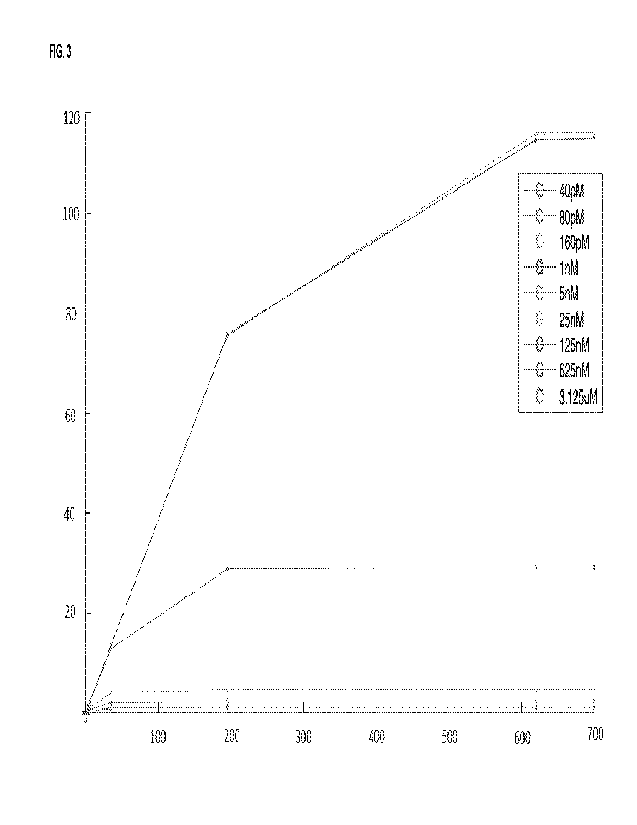
FIG. 3 is a graph showing results of a titration of a reporter binding moiety
(Ab) with a tag in an assay in accordance
with embodiments of the present disclosure, illustrating a resulting detected
signal (Y-axis, in relative fluorescence
units, MM) versus a concentration of an antibody of interest (hCG, in mIU/m1)
(X-axis), for different concentrations of a
reporter binding moiety (40 pM, 80 pM, 160 pM, 1 nM, 5nM, 25 nM, 125 nM, 625
nM, and 3.125 uM).
FIG. 4 is a graph showing results of a titration of the reporter in the assay,
showing background noise in relative
fluorescence units (MM) as it varies depending on a concentration of the
reporter. For each of Rep 1, Rep 2, Rep 3,
and AVG, there are four concentrations, left to right: 20 pM, 200 pM, 1000 pM,
and 2000 pM.
FIG. 5 is a graph showing the use of the present methods to achieve a limit of
detection ("LOD") of 7.7 fM for luteinizing
hormone ("LH") specific antibodies. Error bars represent the standard
deviation of replicates.
5
CA 03187399 2023- 1- 26
WO 2022/029732
PCT/IB2021/057295
FIG. 6 is a graph showing the use of the present methods to achieve a limit of
detection ("LOD") of 15.7 fM for prostate
specific antigen ("PSA") specific antibodies. Error bars represent the
standard deviation of replicates.
DETAILED DESCRIPTION
The present disclosure provides methods and systems that address the need for
accurate detection of analytes in
biological samples.
In various clinical and non-clinical applications related to diagnosing or
treating a subject, it is desirable to detect an
analyte in a sample quickly and accurately. However, conventional immunoassay
often require a prolonged sample
analysis time, which can be 6 hours or longer. Also, many conventional
immunoassays not only take a relatively long
time to detect an analyte, but also suffer from poor sensitivity (e.g., limit
of detections (LoDs) in the picomolar ¨
nanomolar range), poor sensitivity, and large sample volume requirements
(e.g., hundreds of microliters).
Accordingly, embodiments of the present disclosure provide an immunoassay
designed such that the kinetics of the
detection is modulated. The described approach allows achieving significantly
faster detection of an analyte ¨ e.g. the
detection may require from about 1 to about 20 minutes, on average about 15
minutes. In some embodiments, the
immunoassay can be completed in as low as about 1 minute, or about 2 minutes,
or about 3 minutes, or about 4, or
about 5 minutes. Also, an immunoassay in accordance with embodiments of the
present disclosure can have a low
background across different samples (e.g., bodily fluids) and high
sensitivity.
In various aspects, a method for detecting the presence, absence, or amount of
an analyte in a biological sample is
provided. In some embodiments, the method comprises(a) contacting the sample
with a magnetic conjugate comprising
a magnetic particle and a capture moiety configured to bind an analyte in the
sample; (b) contacting the magnetic
conjugate with a reporter binding moiety having a tag bound thereto, the
reporter binding moiety being configured to
bind the analyte; (c) contacting the magnetic conjugate with a reporter having
a tag binding partner that is configured
to bind the tag thereby optionally associating a reporter binding moiety bound
to the tag with the reporter, wherein a
concentration of the reporter binding moiety is substantially greater than a
concentration of the reporter; (d) applying a
magnetic field to separate the magnetic conjugate, optionally having an
analyte that has the reporter binding moiety
associated with the reporter bound thereto; and (e) detecting the presence,
absence, or level of the analyte based on
detection of a signal generated by the reporter.
The magnetic conjugate may have or not have an analyte associated therewith,
which can be detected by detecting a
signal generated by the reporter or by detecting the reporter, depending on
whether or not the analyte is present in the
sample. When an analyte is present in a biological sample, the magnetic
conjugate can have associated therewith an
analyte that has the reporter binding moiety associated with the reporter
bound thereto. When the analyte is not present
in a sample, the magnetic conjugate will not be associated with an analyte.
6
CA 03187399 2023- 1- 26
WO 2022/029732
PCT/IB2021/057295
In some embodiments, the method comprises (a) contacting the sample with a
magnetic conjugate comprising a
magnetic particle and a capture moiety configured to bind an analyte in the
sample; (b) contacting the sample with a
reporter binding moiety having a tag bound thereto, the reporter binding
moiety being configured to bind the analyte;
and (c) contacting the sample with a reporter having a tag binding partner
bound thereto such that the tag binding
partner binds the tag thereby associating a reporter binding moiety bound to
the tag with the reporter. A concentration
of the reporter binding moiety is substantially greater than a concentration
of the reporter. The method further comprises
(d) separating the analyte that has the magnetic conjugate and the reporter
binding moiety associated with the reporter
bound thereto via a tag-tag binding partner interaction from the sample by
applying a magnetic field, and (e) detecting
the presence, absence, or level of the analyte based on detection of a signal
generated by the reporter.
In the present method, a concentration of the reporter binding moiety is
substantially greater than a concentration of
the reporter. In some embodiments, the concentration of the reporter binding
moiety is at least about 5 times greater,
or at least about 10 times greater, or at least about 100 times greater, or at
least about 1000 times greater than the
concentration of the reporter. In some embodiments, the concentration of the
reporter binding moiety is about 1000
times greater than the concentration of the reporter.
In some embodiments, the concentration of the reporter is in a picomolar
range. For example, the concentration of the
reporter may be less than about 300 pM. In some embodiments, the concentration
of the reporter is from about 10 pM
to about 140 pM, or from about 40 pM to about 140 pM, from about 40 pM to
about 100 pM, or from about 60 pM to
about 100 pM, or from about 80 pM to about 100 pM, or from about 100 pM to
about 140 pM. In some embodiments,
the concentration of the reporter is about 20 pM, or about 40 pM, or about 60
pM, or about 80 pM, or about 100 pM. In
some embodiments, the concentration of the reporter is about 120 pM.
In some embodiments, the concentration of the reporter binding moiety is in a
nanomolar range. For example, the
concentration of the reporter binding moiety may be greater than about 1 nm.
In some embodiments, the concentration
of the reporter binding moiety is from about 1 nm to about 60 nM, or from
about 1 nm to about 50 nM, or from about 1
nm to about 40 nM, or from about 1 nm to about 30 nM, or from about 1 nm to
about 20 nM, or from about 1 nm to
about 15 nM, or from about 1 nm to about 10 nM, or from about 1 nm to about 5
nM. In some embodiments, the
concentration of the reporter binding moiety is from about 100 nm to about 700
nM, e.g., about 100 nM, or about 200
nM, or about 300 nM, or about 400 nM, or about 500 nM, or about 600 nM, or
about 600 nM.
In some embodiments, the concentration of the reporter binding moiety ranges
from about 1 nM to about 10 nM, and
the concentration of the reporter ranges from about 15 pM to about 25 pM. In
some embodiments, the concentration
of the reporter binding moiety is about 5 nM and the concentration of the
reporter is about 20 pM.
Various tags and corresponding tag binding partners can be used in accordance
with embodiments of the present
disclosure. In some embodiments, the tag comprises biotin and the tag binding
partner comprises avidin (e.g.
7
CA 03187399 2023- 1- 26
WO 2022/029732
PCT/IB2021/057295
streptavidin). In some embodiments, the tag comprises biotin and the tag
binding partner comprises streptavidin. In
some embodiments, the tag comprises fluorescein isothiocyanate (FITC) and the
tag binding partner comprises anti-
FITC antibody, or the tag comprises dinitrophenol (DNP) and the tag binding
partner comprises anti-DNP antibody, or
the tag comprises digoxigenin (DIG) and the tag binding partner comprises anti-
DIG antibody, or the tag comprises
Etag and the tag binding partner comprises an anti-Etag antibody
(GAPVPYPDPLEPR (SEQ ID NO: 1)), or the tag
comprises FLAG and the tag binding partner comprises an anti-FLAG antibody
(DYKDDDDK (SEQ ID NO: 2)), or the
tag comprises Myc and the tag binding partner comprises an anti-Myc antibody
(EQKLISEEDL (SEQ ID NO: 3)), or the
tag comprises HA and the tag binding partner comprises an anti-HA antibody
(YPYDVPDYA (SEQ ID NO: 4)), or the
tag comprises SNAP and the tag binding partner comprises a benzylguanine
derivative, or the tag comprises "CLIP"
and the tag binding partner comprises a benzylcytosine derivative.
Various tags can be used in methods and kits in accordance with embodiments of
the present disclosure. The tags
can be various peptide tags, covalent peptide tags, protein tags, and other
suitable types of tags.
Various reporters can be used in embodiments of the present disclosure. In
some embodiments, the reporter molecule
is a metal core and a silica shell or the reporter; wherein the silica shell
is optionally impregnated with a plurality of
quantum dots; and wherein the metal core optionally comprises gold. In some
embodiments, the reporter comprises a
plurality of quantum dots, quantum-dot-studded particles, a single quantum
dot, a single quantum-dot-studded particle,
organic dye, upconverting phosphors, and other types.
In some embodiments, the reporter is a fluorescent reporter, a phosphorescent
reporter, or a colorimetric reporter.
In some embodiments, the reporter may be a fluorescent reporter, a
phosphorescent reporter, or colorimetric reporter
such as a colored particle for measuring absorbance and/or scattering of light
(or, for example, the presence absence
of a certain color through colorimetric analysis). In some embodiments, any
suitable detectable reporter as is known in
the art can be used. For example, the detectable reporter can be a radioactive
reporter (such as, e.g. 3H, 1251, 35S,
14C, 32P, and 33P), an enzymatic reporter (such as, e.g. horseradish
peroxidase, alkaline phosphatase, glucose 6-
phosphate dehydrogenase, and the like), a chemiluminescent reporter (such as,
e.g., acridinium esters, thioesters, or
sulfonamides; luminol, isoluminol, phenanthridinium esters, and the like), a
fluorescent reporter (such as fluorescein
(e.g., 5-fluorescei n, 6-carboxyfluorescein, 3'6-carboxyfl uorescein, 5(6)-
carboxyfluorescei n, 6-hexachloro-fluorescein,
6-tetrachlorofluorescein, fluorescein isothiocyanate, and the like)),
rhodamine, phycobiliproteins, R-phycoerythrin,
quantum or metal containing (Mc) dots (e.g., zinc sulfide-capped cadmium
selenide), a thermometric reporter, or an
immuno-polymerase chain reaction reporter. In various embodiments, the
reporter can include, without limitation,
fluorophores, chromophores, radioisotopes, magnetic particles, gold particles,
enzyme substrates, and the like. In
some embodiments, the reporter is a chemiluminescent or fluorescent protein,
such as, for example, green fluorescent
protein (GFP), enhanced green fluorescent protein (EGFP), Renilla Reniformis
green fluorescent protein, GFPmut2,
GFPuv4, yellow fluorescent protein (YFP), enhanced yellow fluorescent protein
(EYFP), cyan fluorescent protein
8
CA 03187399 2023- 1- 26
WO 2022/029732
PCT/IB2021/057295
(CFP), enhanced cyan fluorescent protein (ECFP), enhanced blue fluorescent
protein (EBFP), citrine and red
fluorescent protein from discosoma (dsRED), luciferase, urn belliferone,
rhodamine, fluorescein, dichlorotriazinylamine
fluorescein, dansyl chloride, phycoerythrin, and the like. In some
embodiments, the reporter is a non-protein organic
fluorophore of any of the following families: xanthene derivatives, such as
fluorescein, rhodamine, Oregon green, eosin,
and Texas red; cyanine derivatives, such as cyanine, indocarbocyanine,
oxacarbocyanine, thiacarbocyanine, and
merocyanine; squaraine derivatives and ring-substituted squaraines, including
Seta, SeTau, and Square dyes;
naphthalene derivatives (dansyl and prodan derivatives); coumarin derivatives;
oxadiazole derivatives, such as
pyridyloxazole, nitrobenzoxadiazole and benzoxadiazole; anthracene
derivatives, such as anthraquinones, including
DRAQ5, DRAQ7 and CyTRAK Orange; pyrene derivatives, such as cascade blue,
etc.; oxazine derivatives, such as
Nile red, Nile blue, cresyl violet, oxazine 170, etc.; acridine derivatives,
such as proflavin, acridine orange, acridine
yellow, etc.; arylmethine derivatives, such as auramine, crystal violet,
malachite green; and tetrapyrrole derivatives,
such as porphin, phthalocyanine, bilirubin. In various embodiments, the
reporter includes without limitation enzymatic
reporters, e.g., enzymes such as horseradish peroxidase, alkaline phosphatase,
beta-galactosidase, glucose 6-
phosphate dehydrogenase, and the like. In some embodiments, the reporter may
be one or more quantum dots or
quantum-dot-studded particles. In some embodiments, the reporter may include a
metal core (i.e., gold core) with a
silica shell, wherein the silica shell is impregnated with a plurality (e.g.,
100-600) quantum dots (e.g. quantum-dot-
studded particles). Any other number of quantum dots can be used.
In embodiments, the method employs a relatively low amount of quantum dots or
quantum-dot-studded particles, e.g.
about 400 pM or less, or about 300 pM or less, or about 200 pM or less, or
about 100 pM or less, or about 50 pM or
less, or about 10 pM or less, e.g. about 400 pM, or about 300 pM, or about 200
pM or less, or about 100 pM, or about
50 pM, or about 10 pM. Various analytes can be detected using the methods in
accordance with embodiments of the
present disclosure. In some embodiments, the analyte comprises one or more of
human chorionic gonadotropin (hCG),
luteinizing hormone (LH)/Lutropin, prostate specific antigen (PSA), herpes
simplex virus (HSV) antibodies, estrone-3-
glucuronide (E3G), bacteria, hemoglobin A1C, C-reactive protein (CRP), an
inflammation biomarker, troponin, lyme
disease antigen, lyme disease antibodies, an LDL biomarker, an HDL biomarker,
a total cholesterol biomarker, thyroid
stimulating hormone, a hepatitis C virus biomarker, a rhino virus biomarker,
an influenza virus biomarker, a liver function
biomarker, estrogen, progesterone, lactic acid, and combinations thereof. In
some embodiments, the analyte
additionally or alternatively comprises one or more of N-terminal (NT)-pro
hormone BNP (NT-proBNP), C-reactive
protein (CRP), D-Dimer, Vitamin-D, Vitamin B12, 13, 14, Thyroid-stimulating
hormone (TSH), Parathyroid hormone
(PTH), Follicle stimulating hormone (FSH), Ferritin, luteinizing hormone (LH),
human chorionic gonadotropin (hCG),
Progesterone, Estradiol, Testosterone, Prostate-specific antigen (PSA), and
Homocysteine.
In some embodiments, an analyte can be or can comprise an antigen and/or a
biomarker for a biological event. In
some embodiments, the biological events may include a disease event (e.g.,
disease biomarker), an inflammation
9
CA 03187399 2023- 1- 26
WO 2022/029732
PCT/IB2021/057295
event (e.g., an inflammation biomarker), a reproduction event (e.g., a
reproduction biomarker), and/or an aging event
(e.g., an aging biomarker). Disease biomarkers may include one or more disease
biomarkers related to or associated
with the onset of disease, the offset of disease, and/or the presence of a
disease state in a patient. Disease biomarkers
may include one or more of a viral biomarker, a bacterial biomarker, a cancer
biomarker, or a symptom biomarker.
Viral biomarkers may include, but are not limited to biomarkers for common
cold (e.g. rhinovirus), influenza, herpes,
Zika, and/or HIV. In some embodiments, viral biomarkers may include herpes
simplex virus (HSV), one or more
rhinovirus proteins, one or more influenza A/B/C proteins, one or more HSF-1/2
proteins, and/or one or more HIV virus
proteins. Bacterial biomarkers may include, but are not limited to, biomarkers
for strep throat (i.e., Streptococcus-A
(Strep-A)), biomarkers for chlamydia, and/or biomarkers for gonorrhea. In some
embodiments, bacterial biomarkers
may include, but are not limited to, one or more streptococcus proteins, one
or more chlamydia trachomatis proteins,
and/or one or more Neisseria Gonorrhoeae proteins. Symptom biomarkers may
include, but are not limited to,
biomarkers for coughing, wheezing, runny nose, nausea, cramps, tightness of
the chest, light-headedness, sore throat,
and/or chest pain. Disease biomarkers may also include, but are not limited
to, biomarkers for cardiac distress and/or
diabetes. In some embodiments, disease biomarkers may include troponin, CRP,
and/or ha1c. Cancer biomarkers
may include biomarkers for prostate cancer, breast cancer, colorectal cancer,
gastric cancer, GIST,
leukemia/lymphoma, lung cancer, melanoma, and or pancreatic cancer. In some
embodiments, prostate cancer
biomarkers may include PSA. In some embodiments, breast cancer biomarkers may
include one or more of ER/PR
and HER-2/neu. In some embodiments, colorectal cancer biomarkers may include
one or more of EGFR, KRAS, and
UGT1A1. In some embodiments, gastric cancer biomarkers may include HER-2/neu.
In some embodiments GIST
biomarkers may include c-K IT. In some embodiments, leukemia/lymphoma
biomarkers may include one or more of
CD20 antigen, CD30, FIP1L1-PDGRFalpha, PDGFR, PML/RAR alpha, TPMT, and UGT1A1.
In some embodiments,
lung cancer biomarkers may include one or more of ALK, EGFR, and KRAS. In some
embodiments, melanoma
biomarkers may include BRAF. Inflammatory biomarkers, which may include anti-
inflammatory biomarkers, may
include one or more inflammatory biomarkers described in U.S. Patent
Application Publication No. 2010/0275282, the
entirety of which is incorporated herein by reference. Reproduction biomarkers
may include biomarkers for ovulation,
fertilization, implantation, and/or embryo development. In some embodiments,
reproduction biomarkers may include
B-human Chorionic Gonadotropin ([3-hCG or hCG), hyperglycosylated hCG,
luteinizing hormone (LH), estrone-3-
glucuronide (E3G), early pregnancy factor (EPF), and/or pre implantation
factor. Aging biomarkers or age-related
biomarkers include one or more biomarkers described in U.S. Patent Application
Publication No. 2008/0124752, the
entirety of which is incorporated herein by reference. Other
antigens/biomarkers of interest include, but are not limited
to, any antigens/biomarkers associated with SARS-CoV-2, MERS, SARS, Hand foot
and mouth disease, cardiac
biomarkers, thyroid hormone, obesity biomarkers, biomarkers relating to
bleeding disorders such as vVVF, Factor 8,
Factor 10, fifths disease, cold, flu, Ebola, E coli, Listeria, and Salmonella.
CA 03187399 2023- 1- 26
WO 2022/029732
PCT/IB2021/057295
In embodiments, the sample is or comprises whole blood, plasma, serum, bile,
saliva, urine, tears, perspiration,
cerebrospinal fluid (CSF), semen, mucus, sputum, menstrual blood, menstrual
fluid, vaginal mucus, amniotic fluid,
synovial fluid, breast milk, ear wax, preejaculate, lochia, Rheum, lymph, pus,
and combinations thereof. In some
embodiments, the sample is whole blood, plasma, serum, or urine. In addition,
the sample can be of any other nature,
as embodiments are not limited to any specific type of a sample in which
analyte(s) can be detected.
The methods in accordance with embodiments of the present disclosure allow
detection of various analytes in a sample.
In some embodiments, an analyte of interest comprises an antigen. In some
embodiments, a capture moiety of a
magnetic conjugate comprises an antibody configured to bind an analyte. Non-
limiting examples of antigens include
infectious disease antigens, such as, e.g., coronavirus antigens, influenza
antigens (e.g. surface proteins
hemagglutinin (H and neuraminidase (NA)), etc.
In some embodiments, an analyte of interest comprises an antibody. In
embodiments, non-limiting examples of
antibodies being detected include one or more of IgG, IgM, I gD, IgA, and IgE
antibodies.
In embodiments, the antibody being detected is an antibody directed against a
pathogen antigen, e.g., without
limitation, a coronavirus antigen. The coronavirus antigen can be, e.g., an
IgG antibody. In some embodiments,
antibodies of interest comprise I gG and/or IgM antibodies. The coronavirus is
a member of the Coronaviridae family,
optionally selected from (a) a betacoronavirus, optionally selected from
severe acute respiratory syndrome coronavirus
2 (SARS-CoV-2), severe acute respiratory syndrome coronavirus (SARS-CoV),
middle east respiratory syndrome
coronavirus (MERS-CoV), HCoV-HKU1, and HCoV-0043, and (b) an alphacoronavirus,
optionally selected from
HCoV-NL63 and HCoV-229E. In embodiments, the coronavirus is SARS-CoV-2.
n some embodiments, a capture moiety of the magnetic conjugate comprises a
first antibody configured to bind the
analyte, wherein a reporter binding moiety comprises a second antibody
configured to bind the analyte. In such
embodiments, the analyte can comprise an antibody, and a capture moiety of the
magnetic conjugate can comprise
an antigen configured to bind the antibody. The method may indicate whether
the subject is producing or not producing
antibodies directed against an antigen. In some embodiments, the method may
provide an amount of antibodies in the
sample.
In some embodiments, the capture moieties and the reporter binding moiety bind
different portions of the analyte. In
some embodiments, the capture moieties and the reporter binding moieties are
different. In some embodiments, the
capture moieties and the reporter binding moieties bind to different antigens
or epitopes.
In embodiments of the present disclosure, use of a reporter binding moiety
that has a tag bound thereto and that is
configured to associate with a reporter via a tag binding partner that can
interact with a tag (e.g. via an antigen-antibody
interaction), allows substantially increasing the speed of the detection. A
concentration of the reporter binding moiety
can be substantially greater than a concentration of the reporter ¨ e.g., the
concentration of the reporter binding moiety
11
CA 03187399 2023- 1- 26
WO 2022/029732
PCT/IB2021/057295
can be in a nanomolar range, whereas the concentration of the reporter can be
in a picomolar range. In some
embodiments, a concentration of the reporter binding moiety can be about 10-6
M, or about 10-7M, or about 10-8M. In
some embodiments, a concentration of the reporter binding moiety can be at
least about 10-6 M, or at least about 10-7
M, or at least about 10-8M. The concentration of the reporter can be less than
about 10-11 M, or no greater than 10-11
M. Thus, the kinetics of the creation of a detectable complex (i.e. the
complex comprising the analyte bound to a
magnetic particle and to a reporter) is dramatically improved, in some cases
by 1000 times faster.
Also, the proportion of bound, detectable analytes, as compared to the entire
amount of the analyte in a sample
(including detectable and undetected analytes present in the sample), can be
improved significantly, up to 100% in
some case. In some embodiments, the proportion of bound, detectable analytes
is at least 80%, or at least 85%, or at
least 90%, or at least 95%, or at least 98%, or at least 99%, or about 100%.
In embodiments, a signal-to-noise ratio is increased, background noise is
reduced, and specificity and sensitivity are
increased.
In some embodiments, the sample has a volume of about 1 microliter. In some
embodiments, the sample has a volume
of smaller than 1 microliter. In some embodiments, the sample has a volume of
about 2 microliters, or about 3
microliters, or about 4 microliters, or about 5 microliters.
In some embodiments, e.g. in which the detected analytes comprise antibodies,
the method further comprises a step
of pre-treating the sample with a magnetic conjugate comprising a magnetic
particle and a moiety configured to bind
contaminant antibodies and/or non-antibody moieties. In some embodiments, the
contaminant antibodies are not
directed against the antigen configured to bind the antibody or are
ineffective at generating an immune response
against the antigen configured to bind the antibody. In some embodiments,
wherein the pre-treating reduces or
eliminates one or more of: (a) heterophile antibodies; (b) antibodies that non-
specifically interact with the magnetic
particle; and (c) non-antibody moieties that non-specifically interact with
the magnetic particle.
In some embodiments, the method is suitable for point-of-care usage. In some
embodiments, the method is suitable
for field usage and/or the method is suitable for home usage.
In some embodiments, the methods are compatible with the World Health
Organization's ASSURED (affordable,
sensitive, specific, user-friendly, rapid and robust, equipment-free, and
deliverable) criteria.
In some embodiments, the method is substantially free of false positives. In
some embodiments, the method is
substantially free of false negatives.
In some embodiments, the method provides better (i.e. greater) sensitivity and
specificity than a solid phase
immunoassay method, a bead-based flow cytometry, or a lateral flow
immunochromatographic assay.
12
CA 03187399 2023- 1- 26
WO 2022/029732
PCT/IB2021/057295
In some embodiments, the method provides better sensitivity and specificity
than a method using a bead-based flow
cytometry-based assays, optionally bead-based, flow cytometry-based assays. In
some embodiments, the method
provides better sensitivity and specificity than a method using a lateral flow
immunochromatographic assay.
In some embodiments, the method provides an increased signal-to-noise ratio as
compared to a method using a solid
phase immunoassay.
In some embodiments, the method provides reduced background noise as compared
to a method using an assay in
which a concentration of the reporter binding moiety is not substantially
different from a concentration of the reporter.
In some embodiments, the method provides an increased signal-to-noise ratio as
compared to a method using an
assay in which a concentration of the reporter binding moiety is not
substantially different from a concentration of the
reporter.
In some embodiments, the method provides better sensitivity and specificity
than a method using an assay in which a
concentration of the reporter binding moiety is not substantially different
from a concentration of the reporter.
In various aspects, the present invention provides a kit suitable for the
method of any of the embodiments disclosed
herein. The kit may comprise a magnetic conjugate, a reporter binding moiety,
and a reporter. The kit can be configured
such that a concentration of the reporter binding moiety is substantially
greater than a concentration of the reporter.
In embodiments, the methods in accordance with the present disclosure employ a
nanoparticle-based immunoassay
configured to detect the presence, absence, or level of the antibody by
detecting the reporter. In some embodiments,
the immunoassay can be implemented similar to assays described in
PCT/US2018/015440 (published as
W02018140719), or as a variation or combination of those assays, the
disclosure of which is incorporated by reference
herein in its entirety.
Immunoassays
Methods described herein include methods for detecting the presence, absence,
or amount of an analyte in a biological
sample.
In certain embodiments, the methods described herein encompass a sandwich
method, a separate addition method,
a competitive assay method, a tertiary (three binding event) method, a whole
cell assay method, or any combinations
thereof.
For example, the sandwich method may be well suited for processing small fluid
sample volumes. The separate
addition method described herein may enable processing of larger fluid
volumes, with improved sensitivity. The
competitive assay method may be useful, e.g., for assaying analytes in
scenarios in which a matched pair of a capture
moiety and a corresponding reporter binding moiety that would bind to an
analyte simultaneously is not available. The
13
CA 03187399 2023- 1- 26
WO 2022/029732
PCT/IB2021/057295
tertiary assay method may encompass three binding events to enhance the
kinetics of a system employed for the
present method.
FIGs. 1A-1E illustrate schematically components that are involved in an assay
in accordance with embodiments of the
present disclosure. The assay shown in FIGs. 1A-1E is a sandwich assay, and an
analyte of interest is shown by way
of example only as HCG. It should however be appreciated that any other
analyte of interest can be detected using the
described methods.
Left panel of FIG. 1A illustrates an analyte of interest (HOG, shown only by
way of example, in green), a capture moiety
shown as an antibody #1 (in blue) that is specific to the analyte of interest,
and a reporter binding moiety shown as
antibody #2 (in orange) that is also specific to the analyte of interest. FIG.
1A (left panel) also shows that the antibodies
#1&2 are able to simultaneously bind to the analyte of interest. The assay
also includes a reporter (colored reporter,"
shown as a yellow sun symbol), a magnetic particle, and a tag shown by way of
example only as biotin. Although not
shown in FIGs. 1A-1E, the reporter has a tag binding partner bound thereto and
configured to bind the tag bound to
the reporter binding moiety.
FIG. 1A further shows, in right panel, magnetic particles bound to the
antibody #1, biotin bound to the antibody #2, and
the analyte.
FIG. 1B further illustrates (in right panel, the left panel is identical to
the left panel of FIG. 1A) that the antibodies #1
and #2 can simultaneously bind the analyte, thereby forming a complex
comprising the magnetic particle with the
antibody #1 bound thereto and the reporter binding moiety with a tag (biotin,
in which example) bound thereto. The
formation of the complex shown in FIG. 1B (right panel) can occur in one step
(when both the magnetic particle and
reporter binding moiety are added to a reaction mixture) or in two steps (when
the magnetic particle and reporter
binding moiety are separately added to a reaction mixture).
After the complex as shown in FIG. 1B is formed, a magnetic pull down can be
performed to separate the analyte from
the rest of the sample. The excess reporter binding moiety can be removed, and
the sample (including the analyte)
can be resuspended with a relatively low concentration of reporter, as shown
schematically in FIG. 10. A concentration
of the reporter binding moiety is substantially greater than a concentration
of the reporter. It should be appreciated that,
even though the present disclosure refers to "the sample" throughout the
assay, the sample may not be in the original
form as obtained from a subject or from another source. The sample will be in
the reaction mixture that includes other
ingredients and the mixture he sample can be subjected to sonication, removal
of a portion of the mixture,
resuspension, and other manipulations. The reaction mixture, including at
least a portion of the sample, may or may
not include an analyte of interest.
As shown in FIG. 1D, each reporter becomes associated with a reporter binding
moiety. The reporter may have a tag
binding partner (e.g., streptavidin). At the time the reporter is added to the
reaction mixture, the final complex (can be
14
CA 03187399 2023- 1- 26
WO 2022/029732
PCT/IB2021/057295
referred to as a "sandwich," in this example) is a limiting reagent, and the
reporter is in large excess relative to the
sandwich complexes, although the reporter's concentration is orders of
magnitude lower than that of a reporter binding
moiety.
FIG. lE illustrates that, when a magnetic field is applied to separate the
analyte (in the final, detectable complex with
the magnetic conjugate, reporter binding moiety, and reporter) from the
sample, excess of the reporter (i.e. unbound
reporter particles) is removed (washed away).
In the illustrated example, during the final complex formation, an analyte is
a limiting reagent and the higher
concentrations of the other binding moieties allow the fraction of the analyte
that is bound, as well as maximizing the
speed at which this binding happens.
In embodiments, a reporter binding moiety is bound to a tag and a reporter is
bound to a tag binding partner, and the
tag and the tag binding partner are configured to interact. A concentration of
the reporter binding moiety is substantially
greater than a concentration of the reporter.
In a conventional immunoassay assay, a magnetic particle with a capture moiety
(e.g., antigen) binds an analyte (e.g.,
an antibody) that is also bound to a reporter binding moiety of a reporter
conjugate that is formed from a reporter and
a reporter binding moiety. In this way, a complex (e.g., a "sandwich") is
formed. The kinetics of formation of the complex
in a traditional assay (e.g. ELISA) is described below.
A magnetic particle with a capture moiety (e.g., antibody) bound thereto (i.e.
a magnetic conjugate) can bind to an
analyte (e.g. an antigen) in a sample first, or a reporter conjugate ("RC")
can first bind to the analyte. In the illustration
below, it is assumed that the magnetic conjugate binds to the analyte first,
forming a complex ("C1") of the magnetic
conjugate with the analyte bound thereto. The reporter conjugate ("RC") can
then bind to the complex Cl. It should be
appreciated however that the reporter conjugate can alternatively bind to the
analyte before the magnetic conjugate
binds to the analyte.
d[Complex]
) is
As shown in Equation (1) below, in a typically immunoassay, the rate of
formation of a final complex (
dt
defined as a concentration of the magnetic conjugate with the analyte bound
thereto ([Cl]), multiplied by a
concentration of the reporter conjugate ("RC") and multiplied by an
association rate constant of a reporter binding
moiety (KoNR). The [Cl] concentration of the magnetic conjugate with the
analyte bound is proportional to the
concentration of the analyte in the sample, and this is the value that is
being measured in the assay. The [RC]
concentration of the reporter is typically 10-11 M, though other values can be
used (e.g., 10 M or 10-10 M). The
concentration of the reporter conjugate is limited by various factors, e.g.
colloidal stability of reporter particles. The
KoNR is typically 105M.
CA 03187399 2023- 1- 26
WO 2022/029732
PCT/IB2021/057295
The reporter conjugate may fall off the analyte with a certain off-rate (a
dissociation rate constant of a reporter binding
moiety (KoFFR), such that formed complexes will be lost at a concentration of
[Complex]KoFFR. Typically, 1/KoFF,
is 1.5-6 hours. In a typical immunoassay (e.g. ELISA), there are long
incubation steps and long wash steps, and
Ko FFR becomes meaningful. Equation (2) below illustrates an equilibrium state
(i.e. when no change in the complex
formation occurs). The dissociation constant of the reporter binding moiety,
KD, can then be expressed as shown by
Equation (3). The right part of Equation (3) is an equilibrium ratio that is
dictated by KoFFR, KoNR, or by KD of the
reporter binding moiety (e.g. antibody). This defines a fraction of an analyte
that is in a detectable form versus a fraction
of an analyte in an undetectable form.
d[Complex]
= [Ci][RC] KoNR ¨ [Complex]KoFFR
(1)
dt
d[Complex]
Equilibrium - = 0
(2)
KoFF [Cl][RC]
KD = ¨ = ____________________________________________________________________
(3)
KoN [Complex]
Equations (1) through (3) above illustrate generally limitations of a typical
immunoassay, including long incubation
times that are driven by a slow nature of the reporter binding moiety kinetics
and are limited by ability to increase a
concentration of the reporter binding moiety without decreasing quality of the
signal (i.e. getting nonspecific signal).
To overcome limitations of the present disclosure, in methods in accordance
with the embodiments of the present
disclosure, the step of binding the reporter conjugate to the analyte of
interest is replaced with a step of binding a
reporter binding moiety with a tag bound thereto and a step of binding a
reporter with a tag binding partner bound
thereto. FIG. 2 illustrates schematically components of an immunoassay in
accordance with some embodiments of the
present disclosure, as well as intermediate complexes and a final complex that
are formed as part of the immunoassay.
In this example, an analyte 100 comprises an antigen (which is shown by way of
example only). Both a capture moiety
of a magnetic conjugate 102 and a reporter binding moiety 103 (with a tag
bound thereto) are antibodies, which can
be the same or different antibodies.
As shown in FIG. 2, and as also illustrated in Equation (4) below, in the
example illustrated, the first step of the assay
may be the same as in the conventional assay - binding of a magnetic conjugate
102 with an analyte 100 bound thereto
to form a complex Cl (104), with the concentration expressed as [Cl]. The use
of the reporter binding moiety with a
tag bound thereto allows having the reporter binding moiety at a greater
concentration than a concentration of the
reporter (103 in FIG. 2). FIG. 2 further shows a reporter 105 with a tag
binding partner bound thereto. FIG. 2 also
illustrates complexes that can be formed in the reaction mixture, such as a
complex 107 comprising the analyte and
the reporter binding moiety with the tag, and a complex 109 comprising the
reporter binding moiety and the reporter
associated via the tag-tag binding partner interaction (without the analyte).
16
CA 03187399 2023- 1- 26
WO 2022/029732
PCT/IB2021/057295
As shown in Equation (4) below and in FIG. 2, the rate of formation of the
final complex (120 in FIG. 2) depends on a
concentration of a complex 106 ([03]) comprising the magnetic conjugate 102
and the reporter binding moiety 103
bound to the analyte 100. The formation of the complex 106 is expressed as
shown in Equation (5) where [TAB]
denotes a concentration of the tagged reporter binding moiety 103 (i.e., in
this example, a "tagged antibody"). The
concentration of 03 ([C3], in the reaction is proportional to the amount of
the analyte (i.e. antigen, in this example).
In Equation (4), instead of the KoNR and KoFFRterms, terms Komi, and K0FFL are
used which are association and
disassociation rate constants of a "linker" ¨ i.e. the interaction between the
tag of the reporter binding moiety 103 and
the tag binding partner of the reporter 105.
d[Complex]
¨ 1C31[RC] KoNL ¨ [Complex]KoFFL (4)
31(.1dt = [C1][TAB] KoNR ¨ [C3]KoFFR
(5)
With reference to Equation (5), like in the traditional assay (see Equation
(1)), KoNR can be about 105 M. However, in
Equation (4), while [RC] is about 10-11 M, for the linker, KoNL can be about
10' M. Accordingly, the formation of the
complex in accordance with Equation (4) is a thousand times faster than the
formation of the complex in accordance
with Equation (1) showing the kinetics of the conventional assay.
Thus, even though, in the illustrated method in accordance with embodiments of
the present disclosure, the
concentration of the reporter can be about the same as in the conventional
assay, the rate of formation of the complex
is dramatically faster than in the conventional assay. Furthermore, the
concentration of the tagged reporter binding
moiety 103 (FIG. 2) (shown as [TAB] in Equation (5)) can be increased as
compared to the conventional assay, e.g. it
can be about 10-6 M. In some embodiments, the concentration of the tagged
reporter binding moiety can be about 10-
/ M or 10-8M. Thus, in a method in accordance with embodiments of the present
disclosure, the concentration of the
tagged reporter binding moiety is much higher than the [RC] concentration of
the reporter (e.g. about 10-11 M) in the
conventional assay. Also, even though, instead of using a reporter conjugate
as in a conventional assay, the present
methods make use of separate binding events (of a reporter binding moiety
method having a tag bound thereto and of
a reporter having a tag binding partner bound thereto), these two separate
steps are an order of magnitude, or more,
faster than the step of binding a reporter conjugate in a conventional assay.
This boosted kinetic allows performing the
entire assay in a range from about 1 minute to about 20 minutes. Also, the
term [Complex]KoFFL in Equation (4)
and the term [C3]KoFFR in Equation (5) become negligible (approximately zero).
The assay can have about 100%
bound rate such that, with reference to FIG. 2, most or all of the analyte 100
is in the bound form in the final detectable
complex 120.
17
CA 03187399 2023- 1- 26
WO 2022/029732
PCT/IB2021/057295
In some embodiments, the concentration of a reporter binding moiety is about
from hundreds picomolar to hundreds
nanomolar. In some embodiments, the concentration of a reporter is from about
1 pM to about 600 pM, e.g., in some
embodiments, from about 40 pM to about 120 pM.
In embodiments, a concentration of magnetic particles used in an immunoassay
can be about the same as an expected
concentration of analyte molecules in a sample. In some embodiments, a
concentration of magnetic particles used in
an immunoassay can be greater than an expected concentration of analyte
molecules in a sample. Also, in
embodiments in which a magnetic particle has more than one capture moiety
bound thereto (e.g., 3-4 capture moieties,
or any other number of capture moieties such as, e.g., antibodies), a number
of the binding sites on the magnetic
particle can be higher than an expected concentration of analyte molecules in
a sample.
In some embodiments, more than one analyte can be detected. This can be done
in parallel, e.g., in separate reactions
(e.g., without limitations, separate wells of a well plate).
In some embodiments, methods in accordance with the present disclosure can be
used to detect the presence,
absence, or amount of a plurality of analytes in a biological sample
simultaneously, in the same sample. By tagging
each analyte with a reporter particle having a certain property (e.g., capable
of generating a signal of a specific color),
the described approach can be used for simultaneous detection of multiple
analytes in the same sample.
In some embodiments, such multiplexing method comprises (a) contacting the
sample with at least one magnetic
conjugate comprising a magnetic particle and a plurality of capture moieties
coupled to the magnetic particle and each
configured to bind a corresponding analyte of the plurality of analytes; (b)
contacting the magnetic conjugate with a
plurality of reporter binding moieties each having a corresponding tag bound
thereto, each reporter binding moiety
being configured to bind a corresponding analyte of the plurality of analytes;
(c) contacting the magnetic conjugate with
a plurality of reporters each having a corresponding tag binding partner bound
thereto that is configured to bind a
corresponding tag thereby optionally associating a reporter binding moiety
bound to the tag with a corresponding
reporter, wherein each reporter is configured to generate a corresponding
different signal; (d) applying a magnetic field
to separate the magnetic conjugate, optionally having an analyte of the
plurality of analytes that has the corresponding
reporter binding moiety associated with the corresponding reporter bound
thereto and (e) detecting the presence,
absence, or level of each analyte of the plurality of analytes based on
detection of a signal generated by each of the
reporters.
In multiplexing embodiments in which more than one analyte can be detected in
the same sample, tag-tag binding
partner pairs may be orthogonal, such that a tag binds only a corresponding
tag binding partner, and vice versa. Thus,
for detection of certain analytes in a sample, tag-tag binding partner pairs
can be selected such that no interaction can
occur between tag and tag binding partners from different pairs. In other
words, each tag-tag binding partner pair is
selected to be specific to a particular analyte of the plurality of analytes
to be detected.
18
CA 03187399 2023- 1- 26
WO 2022/029732
PCT/IB2021/057295
In multiplexing embodiments, the reporters, each bound to a respective analyte
(if the analyte is present in the sample),
generate corresponding signals of different properties, such as colors, and
the method thus allows to discriminate
between different analytes in the sample.
In embodiments, the methods described herein employ nanoparticle-based
immunoassays configured to perform the
present detection of the presence, absence, or amount of an analyte in a
biological sample. The nanoparticle-based
immunoassays can be implemented as part of a testing platform which can be a
portable system in some
implementations. The system can be in the form of a kit including all the
components necessary to perform the present
detection.
In some embodiments of the method implemented in accordance with a sandwich
immunoassay method, a reporter
can be, e.g., one or more gold core particles with a silica shell impregnated
with 100-600 quantum dots (e.g. from
nanoComposix, San Diego, CA). In some embodiments, the reporter comprises one
or more quantum dots or qu antum-
dot-studded particles, or another nanoparticle.
In some embodiments, a reporter can be used that is a bright reporter (i.e.
generates a signal of high quality), has high
surface area, and remains colloidally stable during the analysis. For example,
in some embodiments, such reporter
can be a particle that includes a large number (e.g., several hundred) of
highly fluorescent quantum dots or quantum-
dot-studded particles providing up to about 300x optical amplification of the
signal.
The sandwich immunoassay involves detection of whether a complex is formed
that comprises a magnetic conjugate,
an analyte of interest, and a reporter binding moiety associated with a
corresponding reporter that is bound thereto via
a tag-tag binding partner interaction. In embodiments, the magnetic conjugate
comprises a magnetic particle and a
capture moiety coupled to the magnetic particle and configured to bind an
analyte of interest in the sample.
The complex ("sandwich") is formed only in the presence of the analyte. When
the analyte is present, the resulting
complex can be attracted by a magnet and provides an optical signal that
increases its intensity as the analyte
concentration increases.
The sandwich complex cannot form in absence of the analyte, because the
reporter binding moiety/reporter system
does not bind with the magnetic conjugate. When not associated with a magnetic
particle, the reporter is washed away
and does not generate a signal when the sample is analyzed.
In some embodiments of the sandwich immunoassay method, a magnetic conjugate
(comprising a magnetic particle
and a capture moiety coupled to the magnetic particle and configured to bind
an analyte), a reporter binding moiety
(having a corresponding tag bound thereto, the reporter binding moiety being
configured to bind the analyte), and a
reporter (having a corresponding tag binding partner bound thereto such that
the tag binding partner can bind a
corresponding tag thereby associating a reporter binding moiety bound to the
tag with a corresponding reporter) may
be added to an analysis chamber and mixed with a biological sample which may
including an analyte of interest. A
19
CA 03187399 2023- 1- 26
WO 2022/029732
PCT/IB2021/057295
magnetic field may be applied (a "pulldown") by a magnet to separate the
analyte from the sample. The pulldown can
be performed by applying a magnetic field to the sample (with other
ingredients added) for a certain time period (e.g.,
about 1 minute, or about 2 minutes, or about 3 minutes, or about 4 minutes, or
about 5 minutes, or about 6 minutes, or
about 7 minutes). In some embodiments, the magnetic field is applied for about
5 minutes. It should be appreciated
however that the magnetic field can be applied to the sample for any suitable
duration of time.
If the reporter is a fluorescence signal reporter (e.g., an organic dye,
nanomaterial, or conjugated polymer), light may
then be transmitted through at least a portion of the analysis chamber to
cause the reporter to fluoresce. Such
fluorescence may be detected by a suitable detector. In the absence of the
analyte of interest, the reporter is not pulled
down with the analyte and no fluorescence occurs.
In some embodiments, the method described herein may comprise: (a) contacting
a sample with a magnetic conjugate
comprising a magnetic particle and a capture moiety coupled to the magnetic
particle and configured to bind an analyte
in the sample; (b) contacting the magnetic conjugate with a reporter binding
moiety having a tag bound thereto, the
reporter binding moiety being configured to bind the analyte; (c) contacting
the magnetic conjugate with a reporter
having a tag binding partner bound thereto that is configured to bind the tag
thereby optionally associating the reporter
binding moiety bound to the tag with the reporter, wherein a concentration of
the reporter binding moiety is substantially
greater than a concentration of the reporter; (d) applying a magnetic field to
separate the magnetic conjugate, optionally
having associated therewith the analyte and the reporter binding moiety
associated with the reporter bound thereto;
and (e) detecting the presence, absence, or level of the analyte based on
detection of a signal generated by the
reporter.
In the above embodiment, a magnetic conjugate comprises a magnetic particle
having a capture moiety coupled to the
magnetic particle and configured to bind an analyte in a sample. In some
embodiments, a magnetic conjugate
comprises a magnetic particle having more than one capture moiety coupled
thereto, such that more than one analyte
(e.g., antigen) can be bound to the same magnetic particle.
If the reporter is a fluorescence signal reporter, light may then be
transmitted through at least a portion of an analysis
chamber carrying the sample to cause the reporter to fluoresce, and the
emitted fluorescence is measured by a suitable
detector. A signal generated by the reporter can be detected using, e.g., a
light source and a photodetector.
In embodiments, a magnetic conjugate (comprising a magnetic particle and a
capture moiety coupled to the magnetic
particle), a reporter binding moiety, and a reporter can be added to the
analysis chamber simultaneously or at different
times. Thus, in some embodiments, a magnetic conjugate, a reporter binding
moiety, and a reporter may be added
separately. Furthermore, in some embodiments, a reporter binding moiety and a
reporter can be pre-bound to each
other.
CA 03187399 2023- 1- 26
WO 2022/029732
PCT/IB2021/057295
In some embodiments, an immunoassay method is a separate addition method,
which can be used for processing
larger volumes of samples (though small samples can be analyzed as well) and
which allows concentrating an analyte
of interest. This approach may allow detecting an analyte in a sample with
improved sensitivity. In the separate addition
method, a magnetic conjugate may be added to an analysis chamber and mixed
with a sample which may or may not
include an analyte of interest. A pulldown may be performed by activating a
magnetic field (such that an analyte, if
present, binds with a capture moiety of the magnetic conjugate), and a volume
(e.g. a portion) of the sample may be
removed. An additional volume of the sample may then be added. After (or, in
some cases, before) the additional
volume of the sample is added, the magnetic field may be deactivated and the
magnetic conjugate may again be mixed
with the sample. This process may be repeated a certain number of times (e.g.,
one, two, three, four, or more than four
times) to concentrate the analyte. After concentrating the analyte, the
reporter binding moiety and the reporter may be
added and mixed with the sample such that the reporter binding moiety binds
the analyte and a tag bound to the
reporter binding moiety is bound to a tag binding partner that is bound to a
reporter, thereby the reporter binding moiety
is associated with the reporter. A further magnetic pulldown may then be
performed to separate the analyte from the
sample, which (if present) is bound to the magnetic particle and the reporter
(via the reporter binding moiety). If the
reporter is a fluorescence signal reporter, light may then be transmitted
through at least a portion of the analysis
chamber to cause the reporter to fluoresce, and the emitted fluorescence is
measured by a suitable detector. In the
absence of analyte, the reporter will not be pulled down with the analyte and
no fluorescence is detected.
In some embodiments, a competitive immunoassay method is performed, which can
include two types of methods.
The method of a first type of the competitive immunoassay method can be used
in scenarios in which an analyte (e.g.,
without limitation, an antigen, antibody, cell, bacteria, virus, etc.) is too
small for simultaneously binding with a
corresponding capture moiety (coupled to a magnetic particle, as part of a
magnetic conjugate) and a reporter binding
moiety (which may or may not be coupled to a reporter, e.g., via a tag-tag
binding partner interaction). This method
may also be used where either of the capture moiety and the reporter binding
moiety is not available. In this method,
a magnetic conjugate may be added to an assay chamber and mixed with a
biological sample that may or may not
include an analyte of interest. A reporter-labeled second analyte (which is an
analyte, different form the analyte off
interest) configured to bind the magnetic conjugate in the absence of the
analyte of interest) may then be added and
mixed with the sample. When the analyte of interest is present in the sample,
the reporter-labeled second analyte does
not bind to the magnetic conjugate because a binding site of a capture moiety
of the magnetic conjugate is occupied
by the analyte of interest. However, if the analyte of interest is not present
in the analyzed sample, the magnetic
conjugate will bind to the reporter-labeled second analyte. A magnetic
pulldown is performed to separate the analyte
of interest (if present) from the sample. If the reporter is a fluorescence
signal reporter, light may then be transmitted
through at least a portion of the analysis chamber to cause the reporter to
fluoresce, and the emitted fluorescence is
21
CA 03187399 2023- 1- 26
WO 2022/029732
PCT/IB2021/057295
measured by a suitable detector. In the absence of the analyte of interest,
the reporter-labeled analyte will be pulled
down with the magnetic conjugate and no fluorescence is detected.
Accordingly, in some embodiments (e.g., in which a first type of the
competitive immunoassay method is implemented),
the method described herein may include the steps of: (a) contacting a sample
with a magnetic conjugate comprising
a magnetic particle and a capture moiety configured to bind an analyte of
interest the sample; (b) contacting the sample
with a reporter-labeled second analyte configured to bind the magnetic
conjugate in the absence of the analyte in the
sample; (d) applying a magnetic field to the analysis chamber to pull down the
magnetic conjugate, optionally with the
analyte associated therewith; and (e) detecting the presence, absence, or
level of the analyte by detecting the reporter
with a light source and photodetector. In the method, a concentration of the
reporter binding moiety can be substantially
greater than a concentration of the reporter.
In a second type of the competitive immunoassay method, a magnetic particle
(e.g., a magnetic bead) may be used
with a second, competitive analyte bound thereto (e.g., an antigen, antibody,
or another type). The second analyte can
be different from an analyte of interest. The second analyte can be, e.g., an
antigen configured to bind a reporter
binding moiety that is configured to bind the analyte of interest. In the
absence of the analyte of interest, the second
analyte (bound to the magnetic particle) binds the reporter binding moiety
associated with a reporter. The resulting
complex can be attracted by a magnet and provides an optical signal that
decreases in intensity as a concentration of
the analyte of interest in the sample increases. If the analyte of interest is
present in the sample, it blocks complex
formation. In particular, when the analyte of interest is present in the
sample, it can preemptively bind the reporter
binding moiety, competing for the formation of the complete complex with the
second analyte bound to the magnetic
particle. For example, when the analyte of interest is an antigen, it binds
the available binding sites on the reporter
binding moiety (i.e. an antibody) thus preventing the binding of the second
analyte (bound to the magnetic particle) to
the reporter binding moiety (bound to a reporter). In this way, the antibody
binding sites on the reporter binding moiety
are occupied by the analyte of interest and the analyte of interest is thus
competing for the formation of the complete
complex. Accordingly, when a magnetic field is applied, the second analyte,
which is bound to the magnetic particle
and not bound to the reporter, is pulled down. The analyte of interest that is
bound to the reporter via the reporter
binding moiety (the reporter and the reporter binding moiety may interact via
a tag-tag binding partner interaction) is
washed away. Therefore, an optical signal generated by the reporter decreases
in intensity as concentration of the
analyte of interest increases.
In the second type of the competitive immunoassay method, the magnetic
particle can be coupled to the second analyte
(to form what is referred to as a magnetic particle-labeled analyte) before
the assay is performed. In some
embodiments, the method involves the use of a reporter conjugate comprising a
reporter and a reporter binding moiety.
The reporter conjugate is added to an analysis chamber comprising a biological
sample, and mixed with the sample
which may include an analyte of interest. The magnetic particles with the
second analyte bound thereto are added to
22
CA 03187399 2023- 1- 26
WO 2022/029732
PCT/IB2021/057295
the analysis chamber and mixed with the sample. A magnetic pulldown is then
performed to separate the magnetic
particles with the second analyte bound thereto from the sample. If the
reporter is a fluorescence signal reporter, light
may then be transmitted through the sample to cause the reporter to fluoresce,
and the emitted fluorescence is
measured by a suitable detector. In the presence of the analyte of interest,
the magnetic particles having the second
analyte bound thereto but not being associated with a reporter, are pulled
down, resulting in the absence of
fluorescence. Depending on the concentration of the analyte of interest in the
sample, intensity of the signal generated
by the reporter decreases as the analyte concentration increases.
Accordingly, in some embodiments (e.g., in which a second type of the
competitive immunoassay method is
implemented), the methods described herein may include the steps of: (a)
contacting a sample with a reporter and a
reporter binding moiety configured to bind an analyte of interest the sample;
(b) contacting the sample with a magnetic
particle-labeled second analyte configured to bind the reporter conjugate in
the absence of the analyte of interest in
the sample; (c) separating the magnetic particle-labeled analyte from the
sample by applying a magnetic field to the
sample; and (d) detecting the presence, absence, or level of the analyte by
detecting the reporter. A concentration of
the reporter binding moiety is substantially greater than a concentration of
the reporter. The reporter can be detected,
e.g., as described in the above embodiments, using a light source and
photodetector.
As discussed above, in embodiments of the present disclosure, instead of using
a reporter conjugate (i.e. a reporter
binding moiety with a reporter bound thereto) as in a conventional
immunoassay, a detection method can involve use
of a reporter binding moiety having a tag bound thereto (instead of a
reporter). The reporter binding moiety having a
tag bound thereto and a reporter having a corresponding tag binding partner
bound thereto can be added to a reaction
mixture in two respective separate steps. The use of a reporter binding moiety
with a tag bound thereto (and the use
of the reporter with the corresponding tag binding partner) allows increasing
a concentration of a reporter binding
moiety, and a concentration of a reporter is substantially lower than the
concentration of the reporter binding moiety.
The speed of the final complex formation is increased dramatically, such that
the entire assay can be performed in less
than 20 minutes (e.g., in about 15 minutes), as compared to traditional assay
that may take as long as 6 hours.
In some embodiments, the detection method comprises: (a) contacting a sample
with a reporter binding moiety having
a tag bound thereto and being configured to bind an analyte of interest in the
sample; (b) contacting the sample with a
reporter having a tag binding partner bound thereto such that the tag binding
partner binds the tag thereby associating
the reporter binding moiety bound to the tag with the reporter, wherein a
concentration of the reporter binding moiety
is substantially greater than a concentration of the reporter; (c) contacting
the sample with a magnetic particle-labeled
second analyte configured to bind the reporter binding moiety in the absence
of the analyte of interest; (d) separating
the magnetic particle-labeled second analyte from the sample by applying a
magnetic field to the sample; and (e)
detecting the presence, absence, or level of the analyte of interest based on
detection of a signal generated by the
23
CA 03187399 2023- 1- 26
WO 2022/029732
PCT/IB2021/057295
reporter. The reporter can be detected, e.g., as described in the above
embodiments, using a light source and
photodetector.
In some embodiments, the tag comprises biotin and the tag binding partner
comprises streptavidin, or the tag comprises
fluorescein isothiocyanate (FITC) and the tag binding partner comprises anti-
FITC antibody, or the tag comprises
dinitrophenol (DNP) and the tag binding partner comprises anti-DNP antibody,
or the tag comprises digoxigenin (DIG)
and the tag binding partner comprises anti-DIG antibody, or the tag comprises
Etag and the tag binding partner
comprises an anti-Etag antibody (GAPVPYPDPLEPR (SEQ ID NO: 1)), or the tag
comprises FLAG and the tag binding
partner comprises an anti-FLAG antibody (DYKDDDDK (SEQ ID NO: 2)), or the tag
comprises Myc and the tag binding
partner comprises an anti-Myc antibody (EQKLISEEDL (SEQ ID NO: 3)), or the tag
comprises HA and the tag binding
partner comprises an anti-HA antibody (YPYDVPDYA (SEQ ID NO: 4)), or the tag
comprises SNAP and the tag binding
partner comprises a benzylguanine derivative, or the tag comprises "CLIP" and
the tag binding partner comprises a
benzylcytosine derivative.
In some embodiments, a method for detecting the presence, absence, or amount
of an analyte in a biological sample
may comprise: (a) contacting a sample with a reporter binding moiety, the
reporter binding moiety having a tag bound
thereto and being configured to bind an analyte in the sample; (b) contacting
the sample with a reporter having a tag
binding partner bound thereto such that the tag binding partner binds the tag
thereby associating the reporter binding
moiety bound to the tag with the reporter, wherein a concentration of the
reporter binding moiety is substantially greater
than a concentration of the reporter; (c) contacting the sample with a
magnetic particle having a second analyte bound
thereto, the second analyte being configured to bind the reporter binding
moiety; (d) separating the magnetic particle
having the second analyte bound thereto by applying a magnetic field to the
sample; and (e) detecting the presence,
absence, or level of the analyte by detecting the reporter. The reporter can
be detected, e.g., as described in the above
embodiments, using a light source and photodetector.
In some embodiments, a tertiary immunoassay method is implemented, which makes
use of three binding events to
enhance the kinetics of a system used in the present invention. The tertiary
binding method can be applied to or can
comprise the sandwich method, the separate addition method, and the
competitive assay (first and second methods).
The tertiary mode may involve the use of a reporter conjugate comprising a
reporter having a tag binding partner bound
thereto (e.g., fluorescent quantum dot functional ized with streptavidin) and
a reporter binding moiety having a tag bound
thereto (e.g., an antibody labeled with a biotin), and a magnetic conjugate
comprising a magnetic particle and a capture
moiety. The sample can be disposed in an analysis chamber.
The tertiary binding method may comprise adding the magnetic conjugate
(comprising a magnetic particle and a
capture moiety) to the analysis chamber and mixing the magnetic conjugate with
the sample that may include an
analyte of interest. A magnetic field may be applied (a "pulldown") to
separate the analyte of interest from the sample.
Volumes of sample may be removed and analyte concentration steps may be
performed one or more times, as
24
CA 03187399 2023- 1- 26
WO 2022/029732
PCT/IB2021/057295
described above. The magnetic field may be deactivated and the reporter
binding moiety (e.g., an antibody having a
tag bound thereto) may be added to the analysis chamber, which may bind to the
analyte of interest that is in turn
bound to the magnetic conjugate. The reporter (having a tag binding partner
bound thereto) may then be added to the
analysis chamber, which may then bind to the reporter binding moiety, e.g.,
via a tag binding partner ¨ tag interaction
(e.g., a streptavidin-biotin binding interaction). A magnetic pulldown may
then be performed to again separate the
analyte of interest from the sample. If the reporter is a fluorescence signal
reporter, light may then be transmitted
through at least a portion of the analysis chamber to cause the reporter to
fluoresce, and the emitted fluorescence is
measured by a suitable detector. In the absence of the analyte of interest, no
fluorescence occurs.
In some embodiments (e.g., in which a tertiary immunoassay method is
implemented), the methods described herein
may include steps of: (a) contacting a sample with a magnetic conjugate
comprising a magnetic particle and a capture
moiety configured to bind an analyte in a sample; (b) applying a magnetic
field to separate the analyte from the sample;
(c) contacting the magnetic conjugate with a reporter binding moiety having a
tag bound thereto, the reporter binding
moiety being configured to bind the analyte; (d) contacting the magnetic
conjugate with a reporter having a tag binding
partner bound thereto that is configured to bind the tag thereby optionally
associating the reporter binding moiety bound
to the tag with the reporter, wherein a concentration of the reporter binding
moiety is substantially greater than a
concentration of the reporter; (e) applying a magnetic field to separate the
magnetic conjugate, optionally having the
analyte and the reporter binding moiety associated with the reporter bound
thereto; and (f) detecting the presence,
absence, or level of the analyte based on detection of a signal generated by
the reporter.
In the above embodiments, after the step (b) (a pulldown by a magnet to
separate the analytes from the sample) is
performed, and before the step (c), a magnetic field may be deactivated. Also,
in some embodiments, after the step
(b) and before the step (c), a volume of a sample may be removed and analyte
concentration steps may be performed,
as discussed above.
In some embodiments, a detection method comprises a whole cell immunoassay
that targets surface analytes (e.g.,
biomarkers such as cell surface receptors) present on a cell of interest,
thereby detecting the entire cell (e.g., a
bacterium). The ability to measure whole cells can provide insights into
fitness, immune disorders, cancers, and
bacterial infections. In the case of strep throat, e.g. the detection of the
bacterium Streptococcus pyogenes is highly
valuable due to the prevalence of this infection in adults in children. The
whole cell detection can advantageously allow
detecting bacteria in complex samples, which finds use in in clinical,
epidemiological, and environmental applications.
In embodiments, the whole cell immunoassay involves the use of, for detection
of an analyte, a magnetic conjugate, a
reporter binding moiety, and a reporter. As in other embodiments described
herein, the magnetic conjugate comprises
a magnetic particle and a capture moiety coupled to the magnetic particle and
configured to bind an analyte. In some
implementations, a magnetic conjugate comprises a magnetic particle that has a
capture moiety coupled thereto that
is configured to bind the analyte. The reporter binding moiety has a
corresponding tag bound thereto and is configured
CA 03187399 2023- 1- 26
WO 2022/029732
PCT/IB2021/057295
to bind the analyte. The capture moiety of the magnetic conjugate and the
reporter binding moiety, which can be
antibodies specific to surface analytes on the cell of interest, can be
configured to bind markers, e.g. cell receptors, on
the surface of the cell analyte being detected. The capture moiety and the
reporter binding moiety can be configured
to bind to the same or different cell surface receptors.
The reporter has a tag binding partner bound thereto such that the tag binding
partner can bind a corresponding tag
thereby associating a reporter binding moiety bound to the tag with the
reporter. The assay can be designed such that,
for each type of a cell being detected, multiple magnetic particles and
reporters are associated with the cell (via
respective capture and reporter binding moieties).
In the whole cell immunoassay, when the cells of interest are present, the
assay components bind the cell receptors
on the surface of the cell. The resulting complex can be attracted by a magnet
and can provide an optical signal that
increases in intensity as the analyte concentration increases. If the cells of
interest are absent from the sample, the
complex does not form and the reporter system washes away during magnetic
pulldown, resulting in a negative signal.
In various embodiments, methods for detecting the presence, absence, or amount
of an analyte in a biological sample
can include any of the above-described sandwich method, separate addition
method, competitive method (comprising
two types), tertiary (three binding event) method, whole cell detection, or a
combination and/or variation of these
methods. In some embodiments, one or more of the sandwich, separate addition,
competitive, and tertiary methods
can be implemented similar to respective assays described in PCT/US2018/015440
(published as W02018140719),
the disclosure of which is incorporated by reference herein in its entirety.
Non-limiting examples of analytes that can be detected include one or more of
human chorionic gonadotropin (hCG),
luteinizing hormone (LH)/Lutropin, prostate specific antigen (PSA), herpes
simplex virus (HSV) antibodies, estrone-3-
glucuronide (E3G), bacteria, hemoglobin A1C, C-reactive protein (CRP), an
inflammation biomarker, troponin, lyme
disease antigen, lyme disease antibodies, an LDL biomarker, an HDL biomarker,
a total cholesterol biomarker, thyroid
stimulating hormone, a hepatitis C virus biomarker, a rhino virus biomarker,
an influenza virus biomarker, a liver function
biomarker, estrogen, progesterone, lactic acid, and combinations thereof. In
some embodiments, the analyte
additionally or alternatively comprises one or more of N-terminal (NT)-pro
hormone BNP (NT-proBNP), C-reactive
protein (CRP), D-Dimer, Vitamin-D, Vitamin B12, T3, T4, Thyroid-stimulating
hormone (TSH), Parathyroid hormone
(PTH), Follicle stimulating hormone (FSH), Ferritin, luteinizing hormone (LH),
human chorionic gonadotropin (hCG),
Progesterone, Estradiol, Testosterone, Prostate-specific antigen (PSA), and
Homocysteine.
In some embodiments, the analytes are or comprise whole cells, e.g., bacteria,
tumor cells, or any other types of cells.
Embodiments of the present disclosure can also be used to detect viruses.
It should be appreciated that embodiments of the present disclosure provide
techniques for detection of any suitable
analytes.
26
CA 03187399 2023- 1- 26
WO 2022/029732
PCT/IB2021/057295
In some embodiments, the methods for detecting the presence, absence, or
amount of an analyte of interest in a
biological sample can include concentrating the analyte of interest, as
described above in connection with a separate
addition method. In some embodiments, the methods for detecting the presence,
absence, or amount of an analyte of
interest in a biological sample can include some or all steps of one or both
of the competitive method, and tertiary
(three binding event) method.
The methods in accordance with embodiments of the present disclosure can be
implemented in a suitable analysis
chamber of an analytic device or system used herein. In some embodiments, the
analysis chamber comprises or is a
well plate including a suitable number of wells.
The analytic system can include an analysis chamber configured to receive a
biological sample therein, a magnet, a
light source, and a detector (e.g., a photodetector), among other components.
The analytic system can include or can
be associated with a magnet, which may be a permanent magnet that may be
separated from the analysis chamber in
order to apply a magnetic field to the analysis chamber. In some embodiments,
the magnet may be an electromagnet
that may be controlled to be activated or deactivated in order to apply a
magnetic field to the analysis chamber. It
should be appreciated that the analytic device or system in accordance with
embodiments of the present disclosure
can have any suitable configuration and any suitable components configured to
detect the presence, absence, or
amount of an analyte in a biological sample.
In some embodiments, the analytic system includes a light source connected to
or otherwise associated with the
analysis chamber. The light source is configured to transmit light through at
least a portion of the analysis chamber.
In some embodiments, the analysis chamber may be one chamber, or two chambers,
or three chambers, or four
chambers. The analysis chamber may include a plurality of chambers which can
be more than four chambers. In some
embodiments, the plurality of chambers may be in fluid communication with one
another. In some embodiments, one
or more of a biological sample, a reporter, a reporter binding moiety, and a
magnetic conjugate (comprising a magnetic
particle and a capture moiety associated therewith) may be mixed in a first
chamber of the plurality of chambers. In
some embodiments, the magnetic field may be applied in a second chamber of the
plurality of chambers, and a light
source coupled to the analysis chamber is configured to transmit light through
the second chamber. In some
embodiments, the method steps described herein may each be performed in
separate chambers of the analysis
chamber. In some embodiments, the analysis chamber may be one chamber and all
method steps may be performed
in the same chamber.
In some embodiments, a photodetector may be connected to or otherwise
associated with the analysis chamber (e.g.,
positioned as facing, in line with, opposite to the light source, or in other
ways) and may be configured to detect light
transmitted through the analysis chamber by the light source and thereby
measure transmittance and/or absorbance
of the light. In some embodiments, the photodetector may be positioned
relative to the analysis chamber such that the
27
CA 03187399 2023- 1- 26
WO 2022/029732
PCT/IB2021/057295
photodetector is orthogonal to the light source (orthogonal illumination), and
may be configured to detect fluorescence
and/or phosphorescence of a reporter in a portion of the analysis chamber. In
some embodiments, the photodetector
may be positioned relative to the analysis chamber such that the photodetector
is opposite to the light source (trans
illumination), and may be configured to detect fluorescence and/or
phosphorescence of a reporter in a portion of the
analysis chamber. In some embodiments, the photodetector may be connected to
the analysis chamber, in line with
the light source (e.g., by way of a dichroic mirror (cis illumination)), and
may be configured to detect fluorescence
and/or phosphorescence of a reporter in a portion of the analysis chamber.
The photodetectors used in embodiments of the present disclosure can have
various configurations. In some
embodiments, a photodetector may include one or more photomultiplier tube
detectors and photodiode detectors. As
used herein, the term "photomultiplier" or "photomultiplier tube" refers to
optical detection components that convert
incident photons into electrons via the photoelectric effect and secondary
electron emission. The term photomultiplier
tube is meant to include devices that include separate dynodes for current
multiplication as well as those devices that
include one or more channel electron multipliers. As used herein, the term
"optical detector" or "photodetector" refers
to a device that generates an output signal when irradiated with optical
energy. Thus, in its broadest sense, the term
optical detector system is taken to define a device for converting energy from
one form to another for the purpose of
measurement of a physical quantity or for information transfer. Optical
detectors include, but are not limited to,
photomultipliers and photodiodes. As used herein, the term "photodiode" refers
to a solid-state light detector type
including, but not limited to, PN, PIN, APD, CMOS, and CCD. In some
embodiments, the photodetector may include
one or more of a PN based detector, a PIN based detector, an APD based
detector, a CMOS based detector, and a
CCD based detector.
In some embodiments, the analysis chamber comprises a photodetector as
described herein. In some embodiments,
the analysis chamber comprises one or more of a PN based detector, a PIN based
detector, an APD based detector,
a CMOS based detector, and a CCD based detector.
In various embodiments, an analytic device or system, which can be in the form
of a kit, includes a sample collector
configured to receive a biological sample obtained from a subject. In various
embodiments, the methods in accordance
with the present disclosure may involve adding a sample acquired from a
subject to the analysis chamber. In some
embodiments, adding the sample to the analysis chamber may include delivering
the sample to the sample collector.
The sample can be blood, plasma or serum, and the sample collector can be or
can include a needle prick (e.g. lancet),
a syringe, or another form of a sample collector configured to access blood or
another bodily fluid. In some
implementations, the sample collector can be a retractable element (which can
be, e.g., spring-loaded) that can be
safely retracted into a sample collection tube or another compartment upon
collection of a biological sample (e.g. blood,
plasma or serum). In some embodiments, the device for safe sample collection
comprises a needle prick (e.g. lancet)
or a syringe, wherein the needle prick or the syringe is attached to a cap,
the cap is detachably attachable to a sample
28
CA 03187399 2023- 1- 26
WO 2022/029732
PCT/IB2021/057295
collection tube. In some embodiments, the needle prick, syringe, or another
type of a sample collector is disposed in a
removable cover (which can be attached to the device) configured to cover the
sample collector when not in use. The
removable cover and/or the sample collector can be coupled to a valve or
another component configured to be activated
to retract the sample collector or otherwise make the sample collector
available for sample collection. In some
embodiments, the cover can be configured to open automatically, and/or the
sample collector can be configured to
open automatically.
In some embodiments, the sample collector (e.g., a syringe) is separate from
the analytic device or system. The
separate sample collector can be part of a kit including the analytic system.
The needle prick, syringe, or another type of a sample collector can be in
fluid communication with the interior of the
analysis chamber. Thus, the sample collector may provide the sample received
therein to the analysis chamber. In
some embodiments, the sample collector may include an absorbent and/or wicking
material, or another type of a
material that facilitates delivery of the collected sample (e.g., blood,
plasma or serum) to the analysis chamber.
The analytic device or system can be disposable. In some embodiments, parts of
the system (e.g., a sample collector)
can be disposable while other parts can be reusable. In some embodiments, some
components of the system can be
removable and/or disposable.
In some embodiments, one or more of the magnetic conjugate, reporter, reporter
binding moiety, and reporter conjugate
(comprising a reporter and a reporter binding moiety) may be disposed at the
sample collector and the contacting the
sample with the magnetic conjugate, reporter, reporter binding moiety, or
reporter conjugate may occur at the sample
collector. In some embodiments, one or more of the magnetic conjugate,
reporter, reporter binding moiety, and reporter
conjugate may be imbedded in a portion of the sample collector before a sample
is added to the sample collector. In
some embodiments, the methods described herein may include contacting the
sample in the analysis chamber with a
magnetic conjugate, reporter, reporter binding moiety, and/or reporter
conjugate.
In some embodiments, the reporter may be a fluorescent reporter, a
phosphorescent reporter, or a colorimetric reporter
such as a colored particle that may be configured to measure absorbance or
scattering of light (or, for example, the
presence/absence of a certain color by colorimetric analysis). The reporter
can have a tag binding partner such as any
of the tag binding partners described hereinabove, or other binding partners,
configured to bind to respective tags.
In some embodiments, the methods described herein may further include
concentrating analytes of interest in the
sample by applying a magnetic field to the analysis chamber after contacting
the sample with the magnetic conjugate,
and then reducing the volume of the sample in the analysis chamber. In some
embodiments, the methods described
herein may further include deactivating the magnetic field before contacting
the sample with the reporter conjugate.
29
CA 03187399 2023- 1- 26
WO 2022/029732
PCT/IB2021/057295
In some embodiments, reducing the volume of the sample in the analysis chamber
may be performed by, for example,
syphoning of a fraction of the volume or by removing the entire sample and
resuspending the sample in a new, lesser
volume.
In embodiments, the methods described herein may further include the steps of
concentrating the analytes of interest
in the sample by applying a magnetic field to the analysis chamber after
contacting the sample with the magnetic
conjugate, removing a volume of the sample from the analysis chamber, and
adding a volume of buffer and/or an
additional volume of the sample to the analysis chamber. In some embodiments,
the methods described herein may
include the step of deactivating the magnetic field before contacting the
sample with the reporter conjugate.
In some embodiments, the methods described herein may include the step of
adding a volume of buffer and/or
additional volumes of sample to the analysis chamber.
In some embodiments, the methods described herein may include the step of
removing volumes of sample from the
analysis chamber after a pulldown of the magnetic conjugate (i.e., application
of a magnetic field) and before or after
contacting the sample with a reporter binding moiety.
In some embodiments, the reporter binding moiety comprises a reporter antibody
that is labeled with biotin and the
reporter is functionalized with streptavidin. In some embodiments, the
reporter binding moiety, such as, e.g., the
reporter antibody, is function alized with streptavidin and the reporter is
labeled with biotin.
In various embodiments, the methods described herein include or make use of
various detection techniques, e.g., for
detecting a reporter signal. The detection techniques may include use of a
microscope, a spectrophotometer, a
fluorimeter, a tube luminometer or plate luminometer, x-ray technology,
magnetic fields, a scintillator, a fluorescence
activated cell sorting (FACS) apparatus, a microfluidics apparatus, a bead-
based apparatus, etc.
In some embodiments, a magnetic particle of a magnetic conjugate is a
paramagnetic particle. In some embodiments,
the paramagnetic particle is a nanoparticle, which can be, e.g., a nanobead.
In some embodiments, the paramagnetic
particle is a microparticle. In some embodiments, the microparticle is a
microbead. The paramagnetic particle is, in
various embodiments, a magnetic nano- or microbead, which allows the particle
to be held and/or manipulated by
magnets. In some embodiments, the paramagnetic particle is a metallic
nanoparticles coated with a thin (e.g., about 2
nm in diameter) graphene-like carbon layer. In some embodiments the
paramagnetic particle is coated, e.g.
streptavidin- or PEG-coated. Examples magnetic particles that can be used are
DYNABEADs (THERMOFISHER),
MACS beads (MILTENYI BIOTEC), TURBOBEADS (TURBOBEADS), ABSOLUTE MAC ST
REPTAVIDI N MAGNETIC
PARTICLES (CREATIVE DIAGNOSTICS), and GOLD NANOPARTICLES (SIGMA ALDRICH).
In some embodiments, the magnetic beads are nanoparticles with a
superparamagnetic Fe2O3 core and a
biocompatible outer coating. The surface of the beads can be activated, e.g.,
with carboxyl groups.
CA 03187399 2023- 1- 26
WO 2022/029732
PCT/IB2021/057295
In some embodiments, the reporter particles described herein may include a
biocompatible coating that may be
activated with amine groups or carboxyl groups to facilitate amid coupling. In
some embodiments, the reporter particles
described herein may be activated with amine groups or carboxyl groups to
facilitate amid coupling.
In some embodiments, the particles described herein may be nanoparticles (e.g.
nanobeads), which are smaller than
1 micrometer in diameter (e.g. about 5 to about 500 nanometers, e.g. about 5
nanometers, or about 10 nanometers,
or about 50 nanometers, or about 100 nanometers, or about 250 nanometers, or
about 500 nanometers). In some
embodiments, the nanoparticles (e.g. nanobeads) have a mean particle diameter
of 25-500 nm-fl-5 nm, 25-500 nm-fl-
nm, 25-500 nm+/-15 nm, 25-500 nm+/-20 nm, 25-500 nm+/-25 nm, 25-500 nm+/-30
nm, 25-500 nm+/-35 nm, 25-
500 nm-fl-40 nm, 25-500 nm+/-45 nm, or 25-500 nm-fl-SO nm. In some
embodiments, the nanoparticles (e.g.
10 nanobeads) have a mean particle diameter of about 20 to about 200 nm.
In some embodiments, the nanoparticles (e.g. nanobeads) are smaller than 1
micrometer in diameter (e.g. about 5 to
about 500 nanometers, e.g. about 5 nanometers, or about 10 nanometers, or
about 50 nanometers, or about 100
nanometers, or about 250 nanometers, or about 500 nanometers). In some
embodiments, the nanoparticles (e.g.
nanobeads) have a mean particle diameter of 25-500 nm+/-5 nm, 25-500 nm+/-10
nm, 25-500 nm+/-15 nm, 25-500
nm-fl- 20 nm, 25-500 nm+/-25 nm, 25-500 nm4-30 nm, 25-500 nm+/-35 nm, 25-500
nm+/-40 nm, 25- 500 nm+/-45
nm, or 25-500 nm-fl-SO nm. In some embodiments, the nanoparticles (e.g.
nanobeads) have a mean particle diameter
of about 20 to about 200 nm. In some embodiments, the magnetic particle may be
a magnetic nanoparticle (e.g.
nanobead) that is composed of oxides, such as ferrites, maghemite, magnetite,
or iron oxide, optionally modified by
surfactants, silica, silicones or phosphoric acid derivatives. In some
embodiments, the nanoparticle (e.g. nanobead) is
composed of ferrites with a shell (e.g. a silica shell, optionally modified).
In some embodiments, the magnetic
nanoparticle is metallic (e.g. iron, cobalt, etc.). In some embodiments, the
magnetic nanoparticle is a metallic
nanoparticle comprising a shell (e.g. of gentle oxidation, surfactants,
polymers and metals (e.g. of gold, graphene,
palladium, platinum, etc.)).
In some embodiments, a particle described herein may be a nanoparticle that
comprises one or more quantum dots In
some embodiments, the nanoparticle comprises a metal core and one or more
quantum dots. In some embodiments,
the nanoparticle comprises a metal core that may be studded with one or more
quantum dots. In some embodiments,
the nanoparticle comprises a metal core that may be studded with a plurality
of quantum dots. Quantum dots are
discrete nanoparticles that have properties similar to bulk semiconductors
such that when exposed to electromagnetic
energy they in turn emit energy. Quantum dots can be engineered to be
sensitive to energy in the infrared region, the
visible spectrum, and even ultraviolet range through changes in size and
composition. Further, they can be designed
to be either photoluminescent or photovoltaic, producing either light or
energy, respectively.
In some embodiments, the reporter may be a nanoparticle (e.g. nanobead), which
may comprise one or more quantum
dots. In some embodiments, the reporter comprises a metal core and one or more
quantum dots. In some
31
CA 03187399 2023- 1- 26
WO 2022/029732
PCT/IB2021/057295
embodiments, the reporter comprises a metal core that may be studded with one
or more quantum dots. In some
embodiments, the reporter comprises a metal core that may be studded with a
plurality of quantum dots.
In some embodiments, the reporter may comprise one or more quantum dots or
quantum-dot-studded particles.. In
some embodiments, the reporter may comprise one or more quantum dots or
quantum-dot-studded particles.. In some
embodiments, the reporter may comprise a plurality of quantum dots or quantum-
dot-studded particles..
Examples of quantum dots, e.g. produced by colloidal methods, include, but are
not limited to, cadmium-selenide
(CdSe), cadmium- sulfide (CdS), indium-arsenide (InAs), and indium-phosphide
(InP) cadmium -tellurium-sulfide
(CdTeS). The number of atoms that comprise a quantum dot can range from 100 to
100,000, typically with a diameter
ranging from 2 to 20 nm (e.g., 2, 3, 4, 5, 6, 7, 8, 9, 10, 1 1, 12, 13, 14,
15, 16, 17, 18, 19, 20, 2.5, 3.5, 4.5, 5.5, 6.5, 7.5,
8.5, 9.5, 10.5, 1 1.5, 12.5, 13.5, 14.5, 15.5, 16.5, 17.5, 18.5, 19.5, 20.5
nm).
In some embodiments, particle materials, including quantum dot materials,
include, but are not limited to, carbon,
colloidal gold, germanium, indium arsenide, indium antimonide, gallium
arsenide, gallium nitride,
cadmium/selenium/telluride, lead, lead oxide, lead sulfide, lead selenide,
indium gallium phosphide, silicon, colloidal
silver, mercury cadmium telluride, iron, iron oxide, cobalt, graphene,
lanthanum, cerium, strontium carbonate,
manganese, manganese oxide, nickel oxide, platinum, lithium, lithium titanate,
tantalum, copper, palladium,
molybdenum, boron carbide, silicon carbide, titanium carbide, tungsten oxide,
aluminum, niobium, thulium, aluminum
nitride, tin, aluminum oxide, tin oxide, antimony, dysprosium, paseodynium,
anfinmony oxide, erbium, rhenium, barium,
ruthenium, beryllium, samarium, bismuth oxide, boron, gadolinium, boron
nitride, vanadium oxide, strontium, ytterbium,
zirconium, diamond (C), Silicon (Si), germanium (Ge), silicon carbide (SiC),
silicon-germanium (SiGe), aluminium
antimonide (AlSb), aluminium arsenide (AlAs), aluminium nitride (Al N),
aluminium phosphide (Al P), boron nitride (BN),
boron phosphide (BP), boron arsenide (BAs), gallium antimonide (GaSb), gallium
arsenide (GaAs), gallium nitride
(GaN), gallium phosphide (GaP), indium antimonide (InSb), indium arsenide
(InAs), indium nitride (InN), indium
phosphide (InP), aluminium gallium arsenide (AlGaAs), indium gallium arsenide
(InGaAs, InxGai_xAs), indium gallium
phosphide (InGaP), aluminum indium arsenide (AllnAs), aluminum indium
antimonide (AlInSb), gallium arsenide nitride
(GaAsN), gallium arsenide phosphide (GaAsP), aluminum gallium nitride (AlGaN),
aluminum gallium phosphide
(AlGaP), indium gallium nitride (InGaN), indium arsenide antimonide (InAsSb),
indium gallium antimonide (InGaSb),
aluminum gallium indium phosphide (AlGaInP, also InAlGaP, InGaAIP, AlInGaP),
aluminum gallium arsenide
phosphide (AlGaAsP), indium gallium arsenide phosphide (InGaAsP), aluminum
indium arsenide phosphide (AllnAsP),
aluminum gallium arsenide nitride (AlGaAsN), indium gallium arsenide nitride
(InGaAsN), indium aluminium arsenide
nitride (InAlAsN), gallium arsenide antimonide nitride (GaAsSbN), gallium
indium nitride arsenide antimonide
(GaInNAsSb), gallium indium arsenide antimonide phosphide (GalnAsSbP), cadmium
selenide (CdSe), cadmium
sulfide (CdS), cadmium telluride (CdTe), zinc oxide (Zn0), zinc selenide
(ZnSe), zinc sulfide (ZnS), zinc telluride
(ZnTe), cadmium zinc telluride (CdZnTe, "CZT"), mercury cadmium telluride
(HgCdTe), mercury zinc telluride
32
CA 03187399 2023- 1- 26
WO 2022/029732
PCT/IB2021/057295
(HgZnTe), mercury zinc selenide (HgZnSe), cuprous chloride (CuCI), lead
selenide (PbSe), lead sulfide (PbS), lead
telluride (PbTe), tin sulfide (SnS), tin telluride (SnTe), lead tin telluride
(PbSnTe), thallium tin telluride (Ti2SnTe5),
thallium germanium telluride (TI2GeTe5), bismuth telluride (Bi2Te3), cadmium
phosphide (Cd3P2), cadmium arsenide
(Cd3As2), cadmium antimonide (Cd3Sb2), zinc phosphide (Zn3P2), zinc arsenide
(Zn3As2), zinc antimonide
(Zn3Sb2), lead(II) iodide (Pb12), molybdenum disulfide (MoS2), gallium
selenide (GaSe), tin sulfide (SnS), bismuth
sulfide (Bi2S3), copper indium gallium selenide (CIGS), platinum silicide
(PtSi), bismuth(III) iodide (Bil3), mercury(II)
iodide (Hg12), thallium(I) bromide (TIBr), titanium dioxide: anatase (TiO2),
copper(I) oxide (Cu2O), copper(II) oxide
(Cu0), uranium dioxide (UO2), uranium trioxide (UO3), and the like.
In various embodiments, the magnetic field is applied using an external
magnet. In various embodiments, the magnet
is a permanent magnet (e.g. neodymium iron boron (NdFeB), samarium cobalt
(SmCo), alnico, and ceramic or ferrite
magnets). In various embodiments, the magnet is a temporary magnet. In various
embodiments, the magnet is an
electromagnet.
In various embodiments, the detection of the reporter is undertaken near the
magnetic field. In various embodiments,
the detection of the reporter is undertaken away from the magnetic field as
in, for example, performed in a chamber
that is separate from a chamber in which a magnetic pull down step is
performed.
In some embodiments, the method provides nanomolar, or picomolar, or
femtomolar scale sensitivity.
In various aspects, the present invention provides a kit suitable for the
method of any one of the embodiments disclosed
herein. The kit can be for an immunoassay in which a concentration of a
reporter binding moiety is substantially greater
than a concentration of a reporter. The kit may comprise a magnetic conjugate,
a reporter binding moiety, and a
reporter, and a concentration of the reporter binding moiety is substantially
greater than a concentration of the reporter.
Optionally, the above-described components of the kits of the present
technology are packed in suitable containers
and labeled for diagnosis of a corresponding disease or condition, or are
labeled for corresponding other purpose. The
above-mentioned components may be stored in unit or multi-use containers, for
example, sealed ampoules, vials,
bottles, syringes, and test tubes, as an aqueous, preferably sterile, solution
or as a lyophilized, preferably sterile,
formulation for reconstitution. For example, the assay reagents of the present
technology can be lyophilized to obviate
requirement for cold chain shipping and storage. For example, in some
embodiments, all reagents except the
Magnesium Acetate Mg(OH3000)2are lyophilized in the bottom of the assay tube
and the Mg(CH3000)2 is lyophilized
on the lid: this prevents the 5' to 3 DNA polynnerase from occurring until the
Mg(CH3000)2 is mixed with the other
assay reagents.
The kit may further comprise a second container which holds a buffer or other
ingredients suitable for diluting the
sample towards a higher volume. Furthermore, the kit may comprise instructions
for carrying out the assay. The
containers may be formed from a variety of materials such as glass or plastic
and may have a sterile access. The kit
33
CA 03187399 2023- 1- 26
WO 2022/029732
PCT/IB2021/057295
may further comprise more containers comprising an acceptable buffer. The kit
may further comprise a device for
collecting a biological sample. It may further include other materials
desirable from a commercial and user standpoint,
including other buffers, diluents, filters, needles, and syringes. The kits
may optionally include instructions customarily
included in commercial packages of diagnostic products, which include
information about, for example, the indications,
usage, manufacture, and/or warnings concerning the use of such diagnostic
products. The kits may further include a
color or fluorescent scale for comparison for diagnosis. The kit components
(e.g., reagents) can be packaged in a
suitable container. The kit can further comprise instructions for using the
kit.
In some embodiments, the kit further comprises a device that collects the
biological sample (e.g., blood, serum, plasma,
urine, or another type of sample) in a safe manner. In some embodiments (e.g.,
for analysis of blood content), the
device comprises a needle and a safety syringe (e.g. a Luer-type syringe). In
some embodiments, the device comprises
spring-loaded retractable needle and a syringe. It should be appreciated that
the device can be any type of a device
for collecting a sample that can be any of whole blood, plasma, serum, bile,
saliva, urine, tears, perspiration,
cerebrospinal fluid (CSF), semen, mucus, sputum, menstrual blood, menstrual
fluid, vaginal mucus, amniotic fluid,
synovial fluid, breast milk, ear wax, preejaculate, lochia, Rheum, lymph, and
pus. Also, in some embodiments, the kit
does not include a sample collection device.
In some embodiments, the kit can also comprise, e.g., a buffering agent, a
preservative or a protein- stabilizing agent.
The kit can further comprise components necessary for detecting the detectable-
label, e.g., an enzyme or a substrate.
The kit can also contain a control sample or a series of control samples,
which can be assayed and compared to the
test sample. Each component of the kit can be enclosed within an individual
container and all of the various containers
can be within a single package, along with instructions for interpreting the
results of the assays performed using the
kit. The kits of the present technology may contain a written product on or in
the kit container. The written product
describes how to use the reagents contained in the kit.
In various aspects, a sample can be obtained from a subject that is a human
subject. Additionally, in some
embodiments, a subject is a mammal different from a human.
Definitions
The following definitions are used in connection with the invention disclosed
herein. Unless defined otherwise, all
technical and scientific terms used herein have the same meaning as commonly
understood to one of skill in the art to
which this invention belongs.
As used herein, "a," "an," or "the" can mean one or more than one.
Further, the term "about" when used in connection with a referenced numeric
indication means the referenced numeric
indication plus or minus up to 10% of that referenced numeric indication. For
example, the language "about 50" covers
the range of 45 to 55.
34
CA 03187399 2023- 1- 26
WO 2022/029732
PCT/IB2021/057295
As used herein, something is "decreased" if a read-out of activity and/or
effect is reduced by a significant amount, such
as by at least about 10%, at least about 20%, at least about 30%, at least
about 40%, at least about 50%, at least
about 60%, at least about 70%, at least about 80%, at least about 90%, at
least about 95%, at least about 97%, at
least about 98%, or more, up to and including at least about 100%, in the
presence of an agent or stimulus relative to
the absence of such modulation. As will be understood by one of ordinary skill
in the art, in some embodiments, activity
is decreased and some downstream read-outs will decrease but others can
increase.
Conversely, activity is "increased" if a read-out of activity and/or effect is
increased by a significant amount, for example
by at least about 10%, at least about 20%, at least about 30%, at least about
40%, at least about 50%, at least about
60%, at least about 70%, at least about 80%, at least about 90%, at least
about 95%, at least about 97%, at least about
98%, or more, up to and including at least about 100% or more, at least about
2-fold, at least about 3-fold, at least
about 4-fold, at least about 5-fold, at least about 6-fold, at least about 7-
fold, at least about 8-fold, at least about 9-fold,
at least about 10-fold, at least about 50-fold, at least about 100-fold, in
the presence of an agent or stimulus, relative
to the absence of such agent or stimulus.
As referred to herein, all compositional percentages are by weight of the
total composition, unless otherwise specified.
As used herein, the word "include," and its variants, is intended to be non-
limiting, such that recitation of items in a list
is not to the exclusion of other like items that may also be useful in the
compositions and methods of this technology.
Similarly, the terms "can" and "may" and their variants are intended to be non-
limiting, such that recitation that an
embodiment can or may comprise certain elements or features does not exclude
other embodiments of the present
technology that do not contain those elements or features.
As used herein, the term "sample" may refer to a solution, suspension,
mixture, or undiluted amount of bodily or another
fluid that may or may not include an an alyte of interest. A sample, as used
herein, may include water and/or a buffer.
As used herein, the term "bodily fluid" may refer to any fluid that can be
isolated from the body of an individual and
includes, but is not limited to whole blood, plasma, serum, bile, saliva,
urine, tears, perspiration, cerebrospinal fluid
(CSF), semen, swabbed samples (e.g. cheek swabs, throat swabs, etc.), mucus,
sputum, menstrual blood, menstrual
fluid, vaginal mucus, amniotic fluid, synovial fluid, breast milk, ear wax,
preejaculate, lochia, Rheum, lymph, pus, and
the like. In some embodiments, bodily fluid may more particularly refer to
whole blood, serum, urine, saliva, swabbed
samples, mucus, or semen. In certain embodiments, bodily fluid may more
particularly refer to whole blood, serum,
urine, or saliva. In some embodiments, the bodily fluid may include an analyte
of interest (e.g., a biomarker).
Although the open-ended term "comprising," as a synonym of terms such as
including, containing, or having, is used
herein to describe and claim the invention, the present invention, or
embodiments thereof, may alternatively be
described using alternative terms such as "consisting of" or "consisting
essentially of."
CA 03187399 2023- 1- 26
WO 2022/029732
PCT/IB2021/057295
As used herein, the words "preferred" and "preferably" refer to embodiments of
the technology that afford certain
benefits, under certain circumstances. However, other embodiments may also be
preferred, under the same or other
circumstances. Furthermore, the recitation of one or more preferred
embodiments does not imply that other
embodiments are not useful, and is not intended to exclude other embodiments
from the scope of the technology.
EXAMPLES
The examples herein are provided to illustrate advantages and benefits of the
present technology and to further assist
a person of ordinary skill in the art with practicing the method for treating
a cancer of the present technology. The
examples herein are also presented in order to more fully illustrate the
certain aspects of the present technology. The
examples should in no way be construed as limiting the scope of the present
technology, as defined by the appended
claims. The examples can include or incorporate any of the variations, aspects
or embodiments of the present
technology described above. The variations, aspects or embodiments described
above may also further each include
or incorporate the variations of any or all other variations, aspects or
embodiments of the present technology.
Example 1. Dependence of Immunoassay Performance on Concentration of Reporter
Binding Moiety
FIG. 3 illustrates results of a titration of a reporter binding moiety (an
antibody, in this example) with a tag in an assay
in accordance with embodiments of the present disclosure. A detected signal (Y-
axis, in fluorescence units, MM) versus
a concentration of an analyte of interest (hCG, in mIU/m1) (X-axis), is shown
for different concentrations of a reporter
binding moiety (40 pM, 80 pM, 160 pM, 1 nM, 5nM, 25 nM, 125 nM, 625 nM, and
3.125 uM). As shown in FIG. 3, the
concentration of a reporter binding moiety of about 5 nM achieves the higher
signal of the entire range of concentrations
of the reporter. As also shown, there is a noticeable jump in the signal
values between the curves representing 160 pM
and 1 nM concentrations of the reporter binding moiety, respectively, and the
smaller jump between the curves
representing 1 nM and 5 nM concentrations of the reporter binding moiety,
respectively. FIG. 3 demonstrates that a
concentration of a reporter binding moiety in a nanomolar range dramatically
boosts performance (i.e. intensity of
fluorescence is increased).
Example 2. Dependence of Immunoassay Performance on Concentration of Reporter
FIG. 4 is illustrates results of a titration of the reporter in the assay,
showing a signal resulting from non-specific and/or
off-target binding (in fluorescence units, MM) as it varies depending on a
concentration of a reporter (quantum dots).
Three replicates and an average of the replicates are shown. In this example,
the concentration of the reporter that
provides the lowest background (i.e. best performance) is 20 pM. FIG. 4
demonstrates that the non-specific signal
(background noise) is greater (and thus worse) when the reporter is in a
nanomolar range (-5 MM fluorescence units)
than when the reporter is in a picomolar range (0.01-0.18 MM fluorescence
units). The data shown in FIG. 4 was
obtained using a Luteinizing hormone (LH) system on a negative control sample
which should have little to no LH.
Example 3: Improved Limits of Detection
36
CA 03187399 2023- 1- 26
WO 2022/029732
PCT/IB2021/057295
In this example, a pair of antibodies against LH were analyzed using the
concentration difference ("kinetic engineering"
methods described herein. The antibody pair has KDs of 2.3 pM and 6.3pM (KAs
of 4.4x10¹¹ and 1.6x10¹¹) respectively.
Relevant parameters include:
Kinetic parameters
AK-Kottationrt 1O /tvh-
Diggedeglon apeIngtent 1.3:x MsIJ
Affini4r.constant 44 KD g. 1012 M 2,3
p14.1)
Ddte.minetion method SPR analysi5 'Wtot.e=On XI>R38)
Datenninetkla antigen L11,.5criept Laixwatories (Ca
:L0815, Lot 231501.02)
Kinetic parameters
Anociajon torotagt 54 g 1C0 /flol's
Dwodatm tat t'Origtait .g ICO1A.
.Winikvowistent ,r414x I KM .t.,rg 63 pm)
DeterminatiOn method :SPR rolysis (PTote0n,104110)
Doterrningann antigen Liit Solon& Lanotnrieg :Kat: L081S,
1.4e.23solon
With these antibodies in a traditional equilibrium, non-kinetically
engineered, immunoassay one would expect
that one should not be able to achieve an LOD lower than 6.3pM.
Here, a LOD of 7.7 fM was achieved with these antibodies using the present
kinetic engineering methods.
This is nearly 1000x lower than the theoretical limit in a traditional
immunoassay and ¨5000x lower than the
practical limit in a traditional immunoassay. See FIG. 5.
37
CA 03187399 2023- 1- 26
WO 2022/029732
PCT/IB2021/057295
Further, in this example, a pair of antibodies against PSA were analyzed using
the concentration difference ("kinetic
engineering" methods described herein. The antibody pair has KDs of 7.5 pM and
11pM (KAs of 1.3x10¹¹ and 1x10¹¹)
respectively.
Relevant parameters include:
KIM& parameters
,ftwo4s:**4Ømago.$: IrtslE* Oft
%waoosm.443.**.wo. t .4.0,
it"ikeomo -, tOot;.1 .,i$3$.1W*4004.0t0w4-
0Ø4mgmft..044Ø4 SWAfttoo.*****14W:
Ostt000k:10t4x* kiiiti,*wavvoteftve,..g.A Pena; ttt
=,tstM444
lithretit paramaters
.ftlioavozio om:4*-vo:** mftokkixxosomx
mwuogRk. ***ko: .0:4*
-06ftft WM:ft :704
ts4,0ft*w:40.4g.E044 :**Wwoomozoio.
.P.SOk :06:tft ,M,14K:Isslot ÷.1:14
With these antibodies in a traditional equilibrium, non-kinetically
engineered, immunoassay one would expect
that one should not be able to achieve an LOD lower than 11pM.
Here, a LOD of 15.7 fM was achieved with these antibodies using the present
kinetic engineering methods.
This is nearly 1000x lower than the theoretical limit in a traditional
immunoassay and ¨5000x lower than the
practical limit in a traditional immunoassay. See FIG. 6.
INCORPORATION BY REFERENCE
All patents and publications referenced herein are hereby incorporated by
reference in their entireties.
The publications discussed herein are provided solely for their disclosure
prior to the filing date of the present
application. Nothing herein is to be construed as an admission that the
present invention is not entitled to antedate
such publication by virtue of prior invention.
38
CA 03187399 2023- 1- 26
WO 2022/029732
PCT/IB2021/057295
As used herein, all headings are simply for organization and are not intended
to limit the disclosure in any manner. The
content of any individual section may be equally applicable to all sections.
EQUIVALENTS
While the invention has been disclosed in connection with specific embodiments
thereof, it will be understood that it is
capable of further modifications and this application is intended to cover any
variations, uses, or adaptations of the
invention following, in general, the principles of the invention and including
such departures from the present disclosure
as come within known or customary practice within the art to which the
invention pertains and as may be applied to the
essential features hereinbefore set forth and as follows in the scope of the
appended claims.
39
CA 03187399 2023- 1- 26