Note: Descriptions are shown in the official language in which they were submitted.
WO 2022/051570
PCT/US2021/048976
METHODS OF ASSAYING A BIOLOGICAL CELL
[0001] This application is a non-provisional application claiming the benefit
under 35 U.S.C.
119(e) of U.S. Provisional Application No. 63/080,960, filed on September 21,
2020; U.S. Provisional
Application No. 63/075,269, filed on September 7, 2020; and U.S Provisional
Application No.
63/211,337, filed on June 16, 2021, each of which disclosures is herein
incorporated by reference in
its entirety.
[0002] This application is filed with a Sequence Listing in electronic format.
The Sequence
Listing is provided as a file entitled "01149-0018-00PCT ST25.txt" created on
August 27, 2021,
which is 17,595 bytes in size. The information in the electronic format of the
sequence listing is
incorporated herein by reference in its entirety.
INTRODUCTION AND SUMMARY
[0003] This application relates to methods of assaying a biological cell. This
application also
relates to methods of barcoding the 5' ends of RNA from a biological cell and
methods of preparation
of expression constructs from the barcoded RNA.
[0004] Over the past three decades, antibody therapies have been developed for
a host of
different diseases, ranging from autoimmune disorders to infectious diseases
and cancer. Cell-based
assays enable screening against native antigens and, therefore, may accelerate
therapeutic antibody
lead candidate selection. However, the time it takes to screen cells for lead
candidates using a typical
workflow significantly adds to the drug development timeline. For example,
after immunizing the
animal and harvesting the antibody-producing B lymphocytes (or B cells) from
the spleen, bone
marrow, or lymph node, it can take at least 12 weeks to produce a hybridoma
and screen through all
of the potential hits, prolonging the development process.
[0005] Recent development of on-chip screening systems allows more rapid
selection of lead
candidates. For example, several tens of thousands of cells can be cloned in
parallel in chambers of the
microfluidic device, and multiple assays can be performed for thorough
characterization of promising
lead candidates. Automated cell lysis and reverse transcription can be
performed on chip to generate
stable clJNA molecules, which can be subsequently recovered for paired
heavy/light chain
amplification and sequencing. Due to the short life span of antibody producing
cells (especially plasma
cells), however, the total number of sequences that can be recovered is
limited by export capacity
within that time frame. Further, validating antibody sequences obtained
requires cloning of the
exported cDNAs, re-expression of antibodies in culture, and off-chip assays.
Accomplishing this work
using traditional cloning and re-expression methods can be slow and labor
intensive. Accordingly, a
1
CA 03189006 2023- 2-9
WO 2022/051570
PCT/US2021/048976
need exists for antibody discovery workflows that allow for rapid selection
and/or re-expression of
antibodies.
[0006] Disclosed herein are methods for providing one or more barcoded cDNA
sequences
from a biological cell. Also, disclosed herein are methods of preparing an
expression construct for
protein expression from the captured barcoded cDNA sequences.
[0007] In some embodiments, a method of assaying for inhibition of a specific
binding
interaction between a first molecule and a second molecule is provided. In
some embodiments, the
method is performed within a microfluidic device having a chamber, the method
comprising:
introducing a micro-object into the chamber of the microfluidic device,
wherein the micro-object
comprises a plurality of first molecules; introducing a cell into the chamber,
wherein the cell is capable
of producing a molecule of interest; incubating the cell in the chamber, in
the presence of the micro-
obj ect, and under conditions conducive to production and secretion of the
molecule of interest; after
incubating the cell in the chamber, introducing the second molecule into the
chamber, wherein the
second molecule is bound to a detectable label; and monitoring an accumulation
of the second
molecule on the micro-object, wherein an absence or diminishment of
accumulation of the second
molecule on the micro-object indicates that the molecule of interest inhibits
binding of the first
molecule to the second molecule.
[0008] In some embodiments, introducing the micro-object into the chamber may
further
include selecting the single micro-object based on detecting a condition of
viability for the micro-
object. Detecting the condition of viability may further include employing a
machine-learning
algorithm to assign a probability of viability to the micro-object.
[0009] In some embodiments, a method of providing one or more barcoded cDNA
sequences
from a biological cell is provided. In some embodiments, the method includes
providing the biological
cell within a chamber; providing a capture object in the chamber, the capture
object comprising a label,
a plurality of first oligonucleotides, and a plurality of second
oligonucleotides, wherein each first
oligonucleotide of the plurality comprises a barcode sequence, and a sequence
comprising at least three
consecutive guanine nucleotides at a 3' end, wherein each second
oligonucleotide of the plurality
comprises a capture sequence, lysing the biological cell and allowing RNA
released from the lysed
biological cell to be captured by the capture sequences of the plurality of
second oligonucleotides,
thereby forming captured RNA; and reverse transcribing the captured RNA,
thereby producing one or
more barcoded cDNA sequences, each comprising an oligonucleotide sequence
complementary to a
corresponding one captured RNA covalently linked to the reverse complement of
the barcode sequence
of the first oligonucleotide.
2
CA 03189006 2023- 2-9
WO 2022/051570
PCT/US2021/048976
[0010] In some embodiments, introducing the biological cell into the chamber
may further
include selecting the biological cell based on detecting a condition of
viability for the biological cell.
Detecting the condition of viability may further include employing a machine-
learning algorithm to
assign a probability of viability to the biological cell.
[0011] In some embodiments, a capture object is provided, the capture object
comprising a
label, a plurality of first and second oligonucleotides wherein each first
oligonucl eoti de of the plurality
comprises a barcode sequence, and a sequence comprising at least three
consecutive guanine
nucleotides at a 3' end and wherein each second oligonucleotide of the
plurality comprises a capture
sequence. In some embodiments, a kit is provided, including a plurality of
capture objects described
herein. In some embodiments, a kit is provided, including a microfluidic
device having a plurality of
chambers, and a plurality of capture objects, each having a plurality of first
and second
oligonucleotides, according to any of the capture objects described herein.
[0012] In some embodiments, a method is provided for introducing a micro-
object into a
chamber of a microfluidic device, including: introducing one or more micro-
objects into a flow region
of a microfluidic device; determining a condition of viability of the one or
more micro-objects;
selecting at least one micro-object having viability from the one or more
micro-objects; and
introducing the at least one micro-object into a chamber of the microfluidic
device. In some
embodiments, the determining the condition of viability is performed without
labelling the one or more
micro-objects, e.g., the micro-object are label-free. In some embodiments,
determining the condition
of viability may further include employing a machine-learning algorithm to
assign a probability of
viability to each of the one or more micro-obj ects. In some embodiments, the
machine-learning
algorithm may include a trained machine-learning algorithm, where the training
may include imaging
micro-objects having a label demarking a condition of viability. The micro-
objects having the label
form a training set of molecules, and may be micro-objects of the same kind as
the one or more micro-
objects introduced to the flow channel of the microfluidic device.
[0013] These and other features and advantages of the disclosed methods will
be set forth or
will become more fully apparent in the description that follows and in the
appended claims. The
features and advantages may be realized and obtained by means of the objects
and combinations
particularly pointed out in the appended examples, partial listing of
embodiments, and claims.
Furthermore, the features and advantages of the described methods may be
learned by the practice or
will be obvious from the description, as set forth hereinafter.
[0014] It is to be understood that both the foregoing general description and
the following
detailed description are exemplary and explanatory only and are not
restrictive of the claims. The
accompanying drawings, which are incorporated in and constitute a part of this
specification, illustrate
3
CA 03189006 2023- 2-9
WO 2022/051570
PCT/US2021/048976
one (several) embodiment(s) and together with the description, serve to
explain the principles
described herein.
BRIEF DESCRIPTION OF THE DRAWINGS
[0015] FIG.1A illustrates a micro-fluidic device and a system with associated
control
equipment according to some embodiments of the disclosure.
[0016] FIG.1B illustrates a microfluidic device with sequestration pens
according to an
embodiment of the disclosure.
[0017] FIGS. 2A to 2B illustrate a microfluidic device having sequestration
pens according to
some embodiments of the disclosure.
[0018] FIG. 2C illustrates a sequestration pen of a microfluidic device
according to some
embodiments of the disclosure.
[0019] FIG. 3 illustrates a sequestration pen of a microfluidic device
according to some
embodiments of the disclosure.
[0020] FIGS. 4A to 4B illustrate electrokinetic features of a microfluidic
device according to
some embodiments of the disclosure.
[0021] FIG. 5A illustrates a system for use with a microfluidic device and
associated control
equipment according to some embodiments of the disclosure.
[0022] FIG. 5B illustrates an imaging device according to some embodiments of
the disclosure.
[0023] FIG. 6 illustrates a workflow for antibody discovery according to some
embodiments
of the disclosure.
[0024] FIG. 7 illustrates RNA capture and reverse transcription to generate a
barcoded cDNA
sequence according to certain embodiments of the present disclosure.
[0025] FIG. 8 shows formation of an expression construct for an antibody heavy
chain using
transcriptionally-active PCR (TAP) according to certain embodiments of the
present disclosure.
[0026] FIG. 9 illustrates a schematic representation of demultiplexing
barcoded cDNA
sequences according to certain embodiments of the present disclosure.
[0027] FIG. 10 is a schematic representation of an embodiment of a capture
object of the
present disclosure.
[0028] FIG. 11 is a schematic representation of a method for aligning sequence
fragments to
provide a V(D)J sequence of a plasma cell according to some embodiments of the
disclosure.
[0029] FIG. 12A is a graphical illustration of sequence alignment in a
reference-based
assembly algorithm according to some embodiments of the disclosure.
4
CA 03189006 2023- 2-9
WO 2022/051570
PCT/US2021/048976
[0030] FIG. 12B is a graphical illustration of sequence alignment in a
reference-based
assembly algorithm according to some embodiments of the disclosure.
[0031] FIG. 12C is a graphical illustration of sequence alignment in a
reference-based
assembly algorithm according to some embodiments of the disclosure.
[0032] FIG. 13 is a schematic representation of a method for aligning sequence
fragments to
provide oligonucleotide sequences of the heavy and light chains of a B cell
receptor sequence.
[0033] FIGS. 14A-B are graphical illustrations of sequence alignment in a
reference-based
assembly algorithm according to some embodiments of the disclosure.
[0034] FIG. 15 is a schematic representation of a Sanger sequencing-based
model for sequence
recognition.
[0035] FIGS. 16A-16C show multiple recombinant PD-Li bead binding assays,
performed
simultaneously or in parallel. The recombinant PD-Li bead binding assay
performed in-channel
(FIGS. 16A-16C, top row) down-selects for antibodies that bind to the PD-L1
coated beads. In the
examples shown, both the blocking and non-blocking antibodies bind the PD-Li
coated beads. The
cell binding assay performed in-pen (FIGS. 10A-10C, middle row) was performed
at the same time
as the recombinant PD-Li bead binding assay and identifies antibodies that
bind to native PD-Li
expressed by a reporter cell. In the examples shown, both the blocking and non-
blocking antibodies
bound the reporter cell. The ligand/receptor-blocking assay identifies
antibodies with the ability to
block the PD-1/PD-L1 interaction (FIGS. 16A-16C, bottom row). In the examples
shown, the
blocking antibodies are detected by non-fluorescent reporter cells, while the
non-blocking antibodies
result in fluorescent reporter cells.
[0036] FIG. 17 shows that deeper characterization enables down-selection of
high quality lead
candidates. Fewer than 2% of screened plasma B cells secreted antibodies that
bound recombinant
PD-Li. Of these 598 antibodies, only 273 antibodies (fewer than 1% of plasma B
cells screened)
bound to the cell-based PD-Li (as shown in CHO-Kl cell binding assay). Further
screening with the
ligand/receptor-blocking assay down-selected 46 lead candidates (0.1% of
plasma B cells screened).
[0037] FIG 18 shows a large number of functionally-active lead candidates are
identified by
screening B cells from multiple organs using the methods according to certain
embodiments of the
present disclosure. Three times (3x) more ligand/receptor blocking antibodies
were identified from
plasma B cells in the bone marrow as compared to the spleen (34 of the 46
candidates, or 74%).
[0038] FIGS. 19A-19D show that re-expressed antibodies exhibited the expected
functional
behavior when evaluated using conventional well-plate-based assays. 20 out of
24 of the lead
candidates that were cloned and re-expressed exhibited binding affinity to the
PD-Li extracellular
domain (ECD) in an ELISA (FIG. 19A) and to the full-length PD-Li protein
expressed by CHO-Kl
CA 03189006 2023- 2-9
WO 2022/051570
PCT/US2021/048976
cells in a FACS assay (FIG. 19B). The same 20 antibodies also bound to the
cynomolgus PD-Li
protein that would most likely be used in obligate animal studies during the
pre-clinical phase of drug
development (FIG. 19C). Finally, 20 of the purified antibodies effectively
blocked the PD-1/PD-L1
interaction (FIG. 19D). 20% of these antibodies had IC50 values comparable to
PD-1/PD-L1 blocking
antibodies currently in the clinic.
[0039] FIG. 20 is a photographic representation of stained cells disposed
within the
microfluidic device imaged at brightfield (top), FITC (calcein) and DAPI
(Zombie) (middle), and
CY5 (CD138) (bottom) cube channels (filter cubes). There are cells located
both in channel and in
chambers, which may be difficult to determine in the brightfield image (top).
As examples, circle
2010 circles three cells that are calcein-positive as shown in the middle
image; circle 2020 circles
another four cells, among which, three of them are Zombie-positive and one of
them is calcein-
positive as shown in the middle image.
[0040] FIG. 21 shows three boxplots illustrating the fluorescence levels
(brightness) of cells
stained with calcein (top), Zombie (middle), and CD138 (bottom) inside the
microfluidic device
respectively. The thresholds for each channel to determine whether cells are
stained positive are based
on the 2 standard deviations (stdev) above the average for each channel. n =
5837 cells.
[0041] FIG. 22 shows boxplots comparing the fluorescence levels (brightness)
of cells stained
with Zombie (top), calcein (middle), and CD138 (bottom) in-channel and in-pen.
Data collected from
three microfluidic devices (chips) are presented: D70161, n = 4403 in channel,
n = 3179 in cells;
D70163, n = 4698 cells in channel, n = 3561 cells in pen; D70169, n = 4523
cells in channel, 3563
cells in pen. Outliers were excluded by gating cell diameter (10 microns), and
cell debris/clump
verified in Image Analyzer 2.1 were also excluded. Each dot represents a
plasma cell in channels.
Whiskers extend to data within 1.5 times the IQR.
[0042] FIG. 23 shows a graph illustrating the subpopulation frequency
differences between in-
channel and in-pen cells stained with CD138 (top), Zombie (middle), and
calcein (bottom) based on
the threshold from unstained cells (328.9 AFU for calcein, 4101.7 AFU for
Zombie, 2024.6 AFU for
CD138).
[0043] FIG. 24 shows density scatter plots illustrating the relationship of
CD138, calcein,
Zombie expression levels of cells comparing in-channel and in-pen locations.
The data are shown in
log scale. From the plots showing the calcein and Zombie expression levels,
two subpopulations can
be clearly observed; while a major subpopulation was observed from the
comparison between Zombie
and CD138 expression levels. The density scatter plots demonstrate that
calcein separates the live and
dead subpopulations with the largest fluorescence separation.
6
CA 03189006 2023- 2-9
WO 2022/051570
PCT/US2021/048976
[0044] FIGS. 25A-25B show graphs illustrating the data from an off-chip FACS
analysis
showing the signal intensities of lives cells (FIG. 25A) or dead cells (FIG.
25B) (the scatter plot) and
the backgating analysis (the three plots on the right of each panel). The
graphs verify that the on-chip
data match very well with the off-chip flow cytometry data. The analysis was
performed on a BD
FACS Celesta Cell Analyzer, and the data was analyzed using the FlowJo v10
software.
[0045] FIGS. 26A-26B shows the scatter plots illustrating the data from an off-
chip FACS
analysis. Those scatter plots demonstrate the correlation between Zombie
(DAPI) vs. Calcein (FITC)
(FIG. 26A) and Zombie (DAPI) vs. CD138 (AF647) (FIG. 26B).
[0046] FIG. 27 demonstrates three typical morphologies of cells observed under
brightfield
that may be used to correlate with assigned values of viability of the cells.
[0047] FIG 28 shows the correlation between calcein intensity and the
morphologies of cells.
[0048] FIG. 29 shows a combined image taken at brightfield and FITC channel
(calcein).
[0049] FIG. 30 shows an image of B cells (denoted with "+") detected in FIG.
29, which was
used as the input to the live/dead classification model.
[0050] FIG. 31 shows an expected output for the live/dead classification
model. Each live cell
is denoted with a solid circle; while each dead cell is denoted with a '+µ.
[0051] FIG. 32 shows the detection of a stain-free sample performed by a
trained live/dead
classification model. The image in left shows the live cells (in solid white
circle) and dead cells (in
solid black circle) recognized by the algorithm. The image in right is a
brightfield image annotated
by human eyes verifying the algorithm was accurate.
[0052] FIG. 33 shows a combined image taken at brightfield and FITC channel
(calcein),
which demonstrates that the live/dead classification model is properly
classifying detected B cells as
live/dead based on only an OEP image. Each live cell is denoted with a solid
circle; while each dead
cell is denoted with a .
[0053] FIG. 34 shows the same image as FIG. 33 but with the OEP channel turned
off. Each
live cell is denoted with a solid circle ; while each dead cell is denoted
with a '+'.
[0054] FIGS. 35A-35B show two plots demonstrating how the setting of threshold
is affecting
the precision (FIG. 35A) and recall (FIG. 35B) rate of the live/dead
detection.
[0055] FIG. 36 shows a plot illustrating the Fl score, which is the harmonic
mean calculated
from the precision and recall data in FIGS 35A-358.
[0056] FIG. 37 is a graphical illustration of the frequency of amplicons with
the expected
barcode from PCR reactions using barcode specific forward primers to amplify
cDNA according to
some embodiments of the disclosure.
7
CA 03189006 2023- 2-9
WO 2022/051570
PCT/US2021/048976
[0057] FIG. 38 shows on-chip images of channels filled by Jurkat cells at a
density of
1.7x10A8 (upper) and by K562 cells at a density of lx10A8 (lower)
respectively.
[0058] FIG. 39 illustrates a generalized schematic of a receptor blocking
assay.
[0059] FIG. 40 illustrates a generalized schematic of a ligand blocking assay.
[0060] FIG. 41 illustrates a receptor blocking assay on chip. Secreting B
cells are shown as
"B" circles. Reporter cells are shown as "R" circles. Dye-labeled ligands are
shown as
rectangles. The upper panel demonstrates the case where the secreted
antibodies bind the reporter
and block ligand binding. The lower panel demonstrates the case where the
secreted antibodies are
non-blocking, allowing ligand to bind to the reporter.
[0061] FIG. 42 illustrates a ligand blocking assay on chip. Antibody-secreting
B cells are
shown as "B" circles. Reporter cells are shown as "R" circles. Dye-labeled
ligands are shown as
"L" rectangles. The top panel demonstrates the case where the secreted
antibodies bind the ligand
and block binding to the reporter. The middle panel demonstrates the case
where the secreted
antibodies are non-blocking, allowing the ligand to bind to the reporter. The
bottom panel
demonstrates the case where the secreted antibodies bind and block the ligand,
but because the
ligand concentration significantly exceeds the secreted antibody
concentration, some of the
ligand may reach and bind to the reporter.
[0062] FIG. 43 illustrates the design of a receptor blocking assay. CD3 is
endogenously
expressed on the surface of the Jurkat reporter cell and will bind both
secreted OKT3 antibody as
well as the dye-labeled HIT3a (ligand). Pens with OKT3 secreting hybridoma
cells should block
HIT3a binding and the reporter cells will appear dark in the ligand imaging
channel. Pens lacking
OKT3 secreting cells will be non-blocking, and HIT3 can freely bind to the
reporter cells, which
will appear bright in the ligand imaging channel.
[0063] FIG. 44A shows the intensity distribution of background
(MeanBackgroundBrightness) and reporter cells (MaxBrightness) as a function of
ligand
concentration.
[0064] FIG. 44B shows the median, 75th and 95th percentile of the background
subtracted
reporter cell intensity (Max - BG) as a function of ligand concentration.
[0065] FIG. 45A shows the intensity distribution of background
(MeanBackgroundBrightness) and reporter cells (MaxBrightness) as a function of
time.
[0066] FIG. 45B shows the median of the background subtracted reporter cell
intensity (Max
- BG) as a function of ligand concentration.
[0067] FIGS. 46A-46B show the distribution of Mean Background Brightness (
"BG") and
MaxBrightness ( "Max") just before (FIG. 46A) and 5 min after flushing (FIG.
46B) the chip
8
CA 03189006 2023- 2-9
WO 2022/051570
PCT/US2021/048976
with media. The black vertical line ("Threshold") indicates a cell detection
threshold defined by
the average background signal plus 2 standard deviations.
[0068] FIG. 47A shows background subtracted reporter cell intensity histograms
just before
and 5 min after flushing with media.
[0069] FIG. 47B shows background (MeanBackgroundBrightness) and the fraction
of
reporter cells above detection threshold () as a function of time.
[0070] FIG. 48 is a heatmap showing that pen-based false positive hit rates as
a function of
reporter detection rate and reporter cells loaded per pen. The original
heatmap was shown in
color and the black and white version is shown in Fig. 48.
[0071] FIGS. 49A-49B show the distribution of background fluorescence per pen
(MeanBackgroundBrightness), brightest reporter cell fluorescence per pen from
lgG-secreting
OKT3-loaded pens (OKT3 MaxBrightness), and brightest reporter cell
fluorescence per pen from
lgG-secreting OKT8-loaded pens (OKT8 MaxBrightness). FIG. 49B is a zoomed in
view of the
fluorescence distributions.
[0072] FIGS. 50A-50C show that OKT3 hits, OKT8 hits, and false positive hit
rate as a
function of signal threshold for pens with >=1 Jurkat reporter cells (FIG.
50A), >=3 Jurkat
reporter cells (FIG. 50B), and >=5 Jurkat reporter cells (FIG. 50C) per pen.
DETAILED DESCRIPTION OF CERTAIN EMBODIMENTS
[0073] This specification describes exemplary embodiments and applications of
the disclosure.
The disclosure, however, is not limited to these exemplary embodiments and
applications or to the
manner in which the exemplary embodiments and applications operate or are
described herein.
Moreover, the figures may show simplified or partial views, and the dimensions
of elements in the
figures may be exaggerated or otherwise not in proportion. In addition, as the
terms "on,- "attached
to," "connected to," "coupled to," or similar words are used herein, one
element (e.g., a material, a
layer, a substrate, etc.) can be "on," "attached to," "connected to," or
"coupled to" another element
regardless of whether the one element is directly on, attached to, connected
to, or coupled to the other
element or there are one or more intervening elements between the one element
and the other element.
Also, unless the context dictates otherwise, directions (e.g., above, below,
top, bottom, side, up, down,
under, over, upper, lower, horizontal, vertical, "x," "y," "z," etc.), if
provided, are relative and
provided solely by way of example and for ease of illustration and discussion
and not by way of
limitation. In addition, where reference is made to a list of elements (e.g.,
elements a, b, c), such
reference is intended to include any one of the listed elements by itself, any
combination of less than
9
CA 03189006 2023- 2-9
WO 2022/051570
PCT/US2021/048976
all of the listed elements, and/or a combination of all of the listed
elements. Section divisions in the
specification are for ease of review only and do not limit any combination of
elements discussed.
[0074] Where dimensions of microfluidic features are described as having a
width or an area,
the dimension typically is described relative to an x-axial and/or y-axial
dimension, both of which lie
within a plane that is parallel to the substrate and/or cover of the
microfluidic device. The height of a
microfluidic feature may be described relative to a z-axial direction, which
is perpendicular to a plane
that is parallel to the substrate and/or cover of the microfluidic device. In
some instances, a cross
sectional area of a microfluidic feature, such as a channel or a passageway,
may be in reference to a
x-axial/z-axial, a y-axial/z-axial, or an x-axial/y-axial area.
I. Definitions
[0075] Although the terms "first- and "second- may be used herein to describe
various
features/elements (including steps), these features/elements should not be
limited by these terms,
unless the context indicates otherwise. These terms may be used to distinguish
one feature/element
from another feature/element. Thus, a first feature/element discussed below
could be termed a
second feature/element, and similarly, a second feature/element discussed
below could be termed a
first feature/element without departing from the teachings of the present
invention.
[0076] Throughout this specification and the claims which follow, unless the
context requires
otherwise, the word "comprise", and variations such as "comprises" and
"comprising" means
various components can be co-jointly employed in the methods and articles
(e.g., compositions and
apparatuses including device and methods). For example, the term "comprising"
will be understood
to imply the inclusion of any stated elements or steps but not the exclusion
of any other elements or
steps.
[0077] As used herein in the specification and claims, including as used in
the examples and
unless otherwise expressly specified, all numbers may be read as if prefaced
by the word "about" or
"approximately," even if the term does not expressly appear. The phrase
"about" or
"approximately" may be used when describing magnitude and/or position to
indicate that the value
and/or position described is within a reasonable expected range of values
and/or positions. For
example, a numeric value may have a value that is +/- 0.1% of the stated value
(or range of values),
+/- 1% of the stated value (or range of values), +/- 2% of the stated value
(or range of values), +/-
5% of the stated value (or range of values), +/- 10% of the stated value (or
range of values), etc.
Any numerical values given herein should also be understood to include about
or approximately that
value, unless the context indicates otherwise. For example, if the value "10"
is disclosed, then
"about 10" is also disclosed. Any numerical range recited herein is intended
to include all sub-
CA 03189006 2023- 2-9
WO 2022/051570
PCT/US2021/048976
ranges subsumed therein. It is also understood that when a value is disclosed
that "less than or
equal to" the value, "greater than or equal to the value" and possible ranges
between values are also
disclosed, as appropriately understood by the skilled artisan. For example, if
the value -X" is
disclosed the -less than or equal to X" as well as -greater than or equal to
X" (e.g., where X is a
numerical value) is also disclosed. It is also understood that the throughout
the application, data is
provided in a number of different formats, and that this data, represents
endpoints and starting
points, and ranges for any combination of the data points. For example, if a
particular data point
"10" and a particular data point "15" are disclosed, it is understood that
greater than, greater than or
equal to, less than, less than or equal to, and equal to 10 and 15 are
considered disclosed as well as
between 10 and 15. It is also understood that each unit between two particular
units are also
disclosed. For example, if 10 and 15 are disclosed, then 11, 12, 13, and 14
are also disclosed.
[0078] As used herein, "substantially" means sufficient to work for the
intended purpose. The
term "substantially" thus allows for minor, insignificant variations from an
absolute or perfect state,
dimension, measurement, result, or the like such as would be expected by a
person of ordinary skill
in the field but that do not appreciably affect overall performance. When used
with respect to
numerical values or parameters or characteristics that can be expressed as
numerical values,
"substantially" means within ten percent.
[0079] The term "ones" means more than one. As used herein, the term
"plurality" can be 2,
3,4, 5, 6, 7, 8,9, 10, or more.
[0080] As used herein: !am means micrometer, ttm3 means cubic micrometer, pL
means
picoliter, nL means nanoliter, and ttL (or uL) means microliter.
[0081] As used herein, "air- refers to the composition of gases predominating
in the
atmosphere of the earth. The four most plentiful gases are nitrogen (typically
present at a
concentration of about 78% by volume, e.g., in a range from about 70-80%),
oxygen (typically present
at about 20.95% by volume at sea level, e.g. in a range from about 10% to
about 25%), argon (typically
present at about 1.0% by volume, e.g. in a range from about 0.1% to about 3%),
and carbon dioxide
(typically present at about 0.04%, e.g., in a range from about 0.01% to about
0.07%). Air may have
other trace gases such as methane, nitrous oxide or ozone, trace pollutants
and organic materials such
as pollen, diesel particulates and the like. Air may include water vapor
(typically present at about
or may be present in a range from about lOppm to about 5% by volume). Air may
be provided
for use in culturing experiments as a filtered, controlled composition and may
be conditioned as
described herein.
[0082] As used herein, the term "disposed" encompasses within its meaning
"located."
11
CA 03189006 2023- 2-9
WO 2022/051570
PCT/US2021/048976
[0083] As used herein, a "microfluidic device" or "microfluidic apparatus" is
a device that
includes one or more discrete microfluidic circuits configured to hold a
fluid, each microfluidic circuit
comprised of fluidically interconnected circuit elements, including but not
limited to region(s), flow
path(s), channel(s), chamber(s), and/or pen(s), and at least one port
configured to allow the fluid (and,
optionally, micro-objects suspended in the fluid) to flow into and/or out of
the microfluidic device.
Typically, a microfluidic circuit of a microfluidic device will include a flow
region, which may
include a microfluidic channel, and at least one chamber, and will hold a
volume of fluid of less than
about 1 mL, e.g., less than about 750, 500, 250, 200, 150, 100, 75, 50, 25,
20, 15, 10, 9, 8, 7, 6, 5, 4,
3, or 2 iitL. In certain embodiments, the microfluidic circuit holds about 1-
2, 1-3, 1-4, 1-5, 2-5, 2-8,
2-10, 2-12, 2-15, 2-20, 5-20, 5-30, 5-40, 5-50, 10-50, 10-75, 10-100, 20-100,
20-150, 20-200, 50-200,
50-250, or 50-300 L. The microfluidic circuit may be configured to have a
first end fluidically
connected with a first port (e.g., an inlet) in the microfluidic device and a
second end fluidically
connected with a second port (e.g., an outlet) in the microfluidic device.
[0084] As used herein, a "nanofluidic device" or "nanofluidic apparatus- is a
type of
microfluidic device having a microfluidic circuit that contains at least one
circuit element configured
to hold a volume of fluid of less than about 1 L, e.g., less than about 750,
500, 250, 200, 150, 100,
75, 50, 25, 20, 15, 10,9, 8, 7, 6, 5,4, 3,2, 1 nL or less. A nanofluidic
device may comprise a plurality
of circuit elements (e.g., at least 2, 3, 4, 5, 6, 7, 8, 9, 10, 15, 20, 25,
50, 75, 100, 150, 200, 250, 300,
400, 500, 600, 700, 800, 900, 1000, 1500, 2000, 2500, 3000, 3500, 4000, 4500,
5000, 6000, 7000,
8000, 9000, 10,000, or more). In certain embodiments, one or more (e.g., all)
of the at least one circuit
elements is configured to hold a volume of fluid of about 100 pL to 1 nL, 100
pL to 2 nL, 100 pL to
nL, 250 pL to 2 nL, 250 pL to 5 nL, 250 pL to 10 nL, 500 pL to 5 nL, 500 pL to
10 nL, 500 pL to
nL, 750 pL to 10 nL, 750 pL to 15 nL, 750 pL to 20 nL, Ito 10 nL, 1 to 15 nL,
1 to 20 nL, 1 to 25
nL, or 1 to 50 nL. In other embodiments, one or more (e.g., all) of the at
least one circuit elements are
configured to hold a volume of fluid of about 20 nL to 200nL, 100 to 200 nL,
100 to 300 nL, 100 to
400 nL, 100 to 500 nL, 200 to 300 nL, 200 to 400 nL, 200 to 500 nL, 200 to 600
nL, 200 to 700 nL,
250 to 400 nL, 250 to 500 nL, 250 to 600 nL, or 250 to 750 nL.
[0085] A microfluidic device or a nanofluidic device may be referred to herein
as a
"microfluidic chip" or a "chip"; or "nanofluidic chip" or "chip".
[0086] A "microfluidic channel" or "flow channel" as used herein refers to
flow region of a
microfluidic device having a length that is significantly longer than both the
horizontal and vertical
dimensions. The length of the channel is generally defined by the flow path of
the channel. In the case
of a straight channel, the length would be the "longitudinal axis" of the
channel. The "horizontal
dimension" or "width" of the channel is the horizontal dimension as observed
in a transverse section
12
CA 03189006 2023- 2-9
WO 2022/051570
PCT/US2021/048976
oriented perpendicular to the longitudinal axis of the channel (or, if the
channel is curved,
perpendicular to an axis tangential to the flow path of the channel at the
plane of the transverse
section). The -vertical dimension" or -height" of the channel is the vertical
dimension as observed in
a transverse section oriented perpendicular to the longitudinal axis of the
channel (or, if the channel
is curved, perpendicular to an axis tangential to the flow path of the channel
at the plane of the
transverse se cti on).
[0087] For example, the flow channel can be at least 5 times the length of
either the horizontal
or vertical dimension, e.g., at least 10 times the length, at least 25 times
the length, at least 100 times
the length, at least 200 times the length, at least 500 times the length, at
least 1,000 times the length,
at least 5,000 times the length, or longer. In some embodiments, the length of
a flow channel is about
100,000 microns to about 500,000 microns, including any value therebetween. In
some embodiments,
the horizontal dimension is about 100 microns to about 1000 microns (e.g.,
about 150 to about 500
microns) and the vertical dimension is about 25 microns to about 200 microns,
(e.g., from about 40
to about 150 microns). It is noted that a flow channel may have a variety of
different spatial
configurations in a microfluidic device, and thus is not restricted to a
perfectly linear element. For
example, a flow channel may be, or include one or more sections having, the
following configurations:
curve, bend, spiral, incline, decline, fork (e.g., multiple different flow
paths), and any combination
thereof. In addition, a flow channel may have different cross-sectional areas
along its path, widening
and constricting to provide a desired fluid flow therein. The flow channel may
include valves, and the
valves may be of any type known in the art of microfluidics. Examples of
microfluidic channels that
include valves are disclosed in U.S. Patents 6,408,878 and 9,227,200, each of
which is herein
incorporated by reference in its entirety.
[0088] For example, the flow channel can be at least 5 times the length of
either the horizontal
or vertical dimension, e.g., at least 10 times the length, at least 25 times
the length, at least 100 times
the length, at least 200 times the length, at least 500 times the length, at
least 1,000 times the length,
at least 5,000 times the length, or longer. In some embodiments, the length of
a flow channel is about
100,000 microns to about 500,000 microns, including any value therebetween. In
some embodiments,
the horizontal dimension is about 100 microns to about 1000 microns (e.g.,
about 150 to about 500
microns) and the vertical dimension is about 25 microns to about 200 microns,
(e.g., from about 40
to about 150 microns). It is noted that a flow channel may have a variety of
different spatial
configurations in a microfluidic device, and thus is not restricted to a
perfectly linear element. For
example, a flow channel may be, or include one or more sections having, the
following configurations:
curve, bend, spiral, incline, decline, fork (e.g., multiple different flow
paths), and any combination
thereof. In addition, a flow channel may have different cross-sectional areas
along its path, widening
13
CA 03189006 2023- 2-9
WO 2022/051570
PCT/US2021/048976
and constricting to provide a desired fluid flow therein. The flow channel may
include valves, and the
valves may be of any type known in the art of microfluidics. Examples of
microfluidic channels that
include valves are disclosed in U.S. Patents 6,408,878 and 9,227,200, each of
which is herein
incorporated by reference in its entirety.
[0089] The direction of fluid flow through the flow region (e.g., channel), or
other circuit
element (e.g., a chamber), dictates an "upstream" and a "downstream"
orientation of the flow region
or circuit element. Accordingly, an inlet will be located at an upstream
position, and an outlet will be
generally located at a downstream position. It will be appreciated by a person
of skill in the art, that
the designation of an "inlet" or an "outlet" may be changed by reversing the
flow within the device
or by opening one or more alternative aperture(s).
[0090] As used herein, the term "transparent" refers to a material which
allows visible light to
pass through without substantially altering the light as is passes through.
[0091] As used herein, "brightfield" illumination and/or image refers to white
light
illumination of the microfluidic field of view from a broad-spectrum light
source, where contrast is
formed by absorbance of light by objects in the field of view.
[0092] As used herein, "structured light" is projected light that is modulated
to provide one or
more illumination effects. A first illumination effect may be projected light
illuminating a portion of
a surface of a device without illuminating (or at least minimizing
illumination of) an adjacent portion
of the surface, e.g., a projected light pattern, as described more fully
below, used to activate DEP
forces within a DEP substrate. When using structured light patterns to
activate DEP forces, the
intensity, e.g., variation in duty cycle of a structured light modulator such
as a DMD, may be used to
change the optical power applied to the light activated DEP actuators, and
thus change DEP force
without changing the nominal voltage or frequency. Another illumination effect
that may be produced
by structured light includes projected light that may be corrected for surface
irregularities and for
irregularities associated with the light projection itself, e.g., fall-off at
the edge of an illuminated field.
Structured light is typically generated by a structured light modulator, such
as a digital mirror device
(DMD), a microshutter array system (MSA), a liquid crystal display (LCD), or
the like. Illumination
of a small area of the surface, e.g., a selected area of interest, with
structured light improves the signal-
to-noi se-ratio (SNR), as illumination of only the selected area of interest
reduces stray/scattered light,
thereby lowering the dark level of the image. An important aspect of
structured light is that it may be
changed quickly over time. A light pattern from the structured light
modulator, e.g., DMD, may be
used to autofocus on difficult targets such as clean mirrors or surfaces that
are far out of focus. Using
a clean mirror, a number of self-test features may be replicated such as
measurement of modulation
transfer function and field curvature/tilt, without requiring a more expensive
Shack-Hartmann sensor.
14
CA 03189006 2023- 2-9
WO 2022/051570
PCT/US2021/048976
In another use of structured light patterns, spatial power distribution may be
measured at the sample
surface with a simple power meter, in place of a camera. Structured light
patterns may also be used
as a reference feature for optical module/system component alignment as well
used as a manual
readout for manual focus. Another illumination effect made possible by use of
structured light patterns
is selective curing, e.g., solidification of hydrogels within the microfluidic
device.
[0093] As used herein, the term "micro-object" refers generally to any
microscopic object that
may be isolated and/or manipulated in accordance with the present disclosure.
Non-limiting examples
of micro-objects include. inanimate micro-objects such as microparticles;
microbeads (e.g.,
polystyrene beads, glass beads, amorphous solid substrates, LuminexTM beads,
or the like), magnetic
beads; microrods; microwires; quantum dots, and the like; biological micro-
objects such as cells;
biological organelles; vesicles, or complexes; synthetic vesicles; liposomes
(e.g., synthetic or derived
from membrane preparations); lipid nanorafts, and the like; or a combination
of inanimate micro-
objects and biological micro-objects (e.g., microbeads attached to cells,
liposome-coated micro-
beads, liposome-coated magnetic beads, or the like). Beads may include
moieties/molecules
covalently or non-covalently attached, such as fluorescent labels, proteins
(including receptor
molecules), carbohydrates, antigens, small molecule signaling moieties, or
other chemical/biological
species capable of use in an assay. In some variations, beads/solid substrates
including
moieties/molecules may be capture beads, e.g., configured to bind molecules
including small
molecules, peptides, proteins or nucleic acids present in proximity either
selectively or non-
selectively. In one non-limiting example, a capture bead may include a nucleic
acid sequence
configured to bind nucleic acids having a specific nucleic acid sequence or
the nucleic acid sequence
of the capture bead may be configured to bind a set of nucleic acids having
related nucleic acid
sequences. Either type of binding may be understood to be selective. Capture
beads containing
moieties/molecules may bind non-selectively when binding of structurally
different but physico-
chemically similar molecules is performed, for example, size exclusion beads
or zeolites configured
to capture molecules of selected size or charge. Lipid nanorafts have been
described, for example, in
Ritchie et al. (2009) "Reconstitution of Membrane Proteins in Phospholipid
Bilayer Nanodiscs,"
Methods Enzymol., 464:211-231.
[0094] As used herein, the term "cell" is used interchangeably with the term
"biological cell."
Non-limiting examples of biological cells include eukaryotic cells, plant
cells, animal cells, such as
mammalian cells, reptilian cells, avian cells, fish cells, or the like,
prokaryotic cells, bacterial cells,
fungal cells, protozoan cells, or the like, cells dissociated from a tissue,
such as muscle, cartilage, fat,
skin, liver, lung, neural tissue, and the like, immunological cells, such as T
cells, B cells, natural killer
cells, macrophages, and the like, embryos (e.g., zygotes), oocytes, ova, sperm
cells, hybridomas,
CA 03189006 2023- 2-9
WO 2022/051570
PCT/US2021/048976
cultured cells, cells from a cell line, cancer cells, infected cells,
transfected and/or transformed cells,
reporter cells, and the like. A mammalian cell can be, for example, from a
human, a mouse, a rat, a
horse, a goat, a sheep, a cow, a primate, or the like.
[0095] A colony of biological cells is "clonal" if all of the living cells in
the colony that are
capable of reproducing are daughter cells derived from a single parent cell.
In certain embodiments,
all the daughter cells in a clonal colony are derived from the single parent
cell by no more than 10
divisions. In other embodiments, all the daughter cells in a clonal colony are
derived from the single
parent cell by no more than 14 divisions. In other embodiments, all the
daughter cells in a clonal
colony are derived from the single parent cell by no more than 17 divisions.
In other embodiments,
all the daughter cells in a clonal colony are derived from the single parent
cell by no more than 20
divisions. The term "clonal cells" refers to cells of the same clonal colony.
[0096] As used herein, a "colony" of biological cells refers to 2 or more
cells (e.g. about 2 to
about 20, about 4 to about 40, about 6 to about 60, about 8 to about 80, about
10 to about 100, about
20 to about 200, about 40 to about 400, about 60 to about 600, about 80 to
about 800, about 100 to
about 1000, or greater than 1000 cells).
[0097] As used herein, the term "maintaining (a) cell(s)" refers to providing
an environment
comprising both fluidic and gaseous components and, optionally a surface, that
provides the
conditions necessary to keep the cells viable and/or expanding.
[0098] As used herein, the term "expanding" when referring to cells, refers to
increasing in cell
number.
[0099] As referred to herein, "gas permeable" means that the material or
structure is permeable
to at least one of oxygen, carbon dioxide, or nitrogen. In some embodiments,
the gas permeable
material or structure is permeable to more than one of oxygen, carbon dioxide
and nitrogen and may
further be permeable to all three of these gases.
[0100] A "component" of a fluidic medium is any chemical or biochemical
molecule present
in the medium, including solvent molecules, ions, small molecules,
antibiotics, nucleotides and
nucleosides, nucleic acids, amino acids, peptides, proteins, sugars,
carbohydrates, lipids, fatty acids,
cholesterol, metabolites, or the like.
[0101] As used herein in reference to a fluidic medium, "diffuse" and
"diffusion" refer to
thermodynamic movement of a component of the fluidic medium down a
concentration gradient.
[0102] The phrase "flow of a medium" means bulk movement of a fluidic medium
primarily
due to any mechanism other than diffusion, and may encompass perfusion. For
example, flow of a
medium can involve movement of the fluidic medium from one point to another
point due to a
pressure differential between the points. Such flow can include a continuous,
pulsed, periodic,
16
CA 03189006 2023- 2-9
WO 2022/051570
PCT/US2021/048976
random, intermittent, or reciprocating flow of the liquid, or any combination
thereof. When one fluidic
medium flows into another fluidic medium, turbulence and mixing of the media
can result. Flowing
can comprise pulling solution through and out of the microfluidic channel
(e.g., aspirating) or pushing
fluid into and through a microfluidic channel (e.g. perfusing).
[0103] The phrase "substantially no flow" refers to a rate of flow of a
fluidic medium that,
when averaged overtime, is less than the rate of diffusion of components of a
material (e.g., an analyte
of interest) into or within the fluidic medium. The ratio of a rate of flow of
a component in a fluidic
medium (i.e., advection) divided by the rate of diffusion of such component
can be expressed by a
dimensionless Peclet number. Thus, a region within a microfluidic device that
experiences
substantially no flow in one in which the Peclet number is less than 1. The
Peclet number associated
with a particular region within the microfluidic device can vary with the
component or components
of the fluidic medium being considered (e.g., the analyte of interest), as the
rate of diffusion of a
component or components in a fluidic medium can depend on, for example,
temperature, the size,
mass, and/or shape of the component(s), and the strength of interactions
between the component(s)
and the fluidic medium. In certain embodiments, the Peclet number associated
with a particular region
of the microfluidic device and a component located therein can be 0.95 or
less, 0.9 or less, 0.85 or
less, 0.8 or less, 0.75 or less, 0.7 or less, 0.65 or less, 0.6 or less, 0.55
or less, 0.5 or less, 0.4 or less,
0.3 or less, 0.2 or less, 0.1 or less, 0.05 or less, 0.01 or less, 0.005 or
less, or 0.001 or less.
[0104] As used herein in reference to different regions within a microfluidic
device, the phrase
"fluidically connected" means that, when the different regions are
substantially filled with fluid, such
as fluidic media, the fluid in each of the regions is connected so as to form
a single body of fluid. This
does not mean that the fluids (or fluidic media) in the different regions are
necessarily identical in
composition. Rather, the fluids in different fluidically connected regions of
a microfluidic device can
have different compositions (e.g., different concentrations of solutes, such
as proteins, carbohydrates,
ions, or other molecules) which are in flux as solutes move down their
respective concentration
gradients and/or fluids flow through the device.
[0105] As used herein, a "flow path" refers to one or more fluidically
connected circuit
elements (e.g., channel(s), region(s), chamber(s) and the like) that define,
and are subject to, the
trajectory of a flow of medium. A flow path is thus an example of a swept
region of a microfluidic
device. Other circuit elements (e.g., un swept regions) may be fluidically
connected with the circuit
elements that comprise the flow path without being subject to the flow of
medium in the flow path.
[0106] As used herein, "isolating a micro-object" confines a micro-object to a
defined area
within the microfluidic device.
17
CA 03189006 2023- 2-9
WO 2022/051570
PCT/US2021/048976
[0107] The defined area can be, for example, a chamber. As used herein, a
"chamber" is a
region within a microfluidic device (e.g., a circuit element) that allows one
or more micro-object(s)
to be isolated from other micro-objects located within the microfluidic
device. Examples of chambers
include microwells, which may be regions etched out of a substrate (e.g., a
planar substrate), as
described in U.S. Patent Application Publication Nos. 2013/0130232 (Weibel et
al.) and
2013/0204076 (Han et al), or a region formed in a multi-layer device, such as
the microfluidic devices
described in WO 2010/040851 (Dimov et al.) or U.S. Patent Application No.
2012/0009671 (Hansen
et al). Other examples of chambers include valved chambers, such as described
in WO 2004/089810
(McBride et al.) and U.S. Patent Application Publication No. 2012/0015347
(Singhal et al.). Other
examples of chambers include the chambers described in: Somaweera et al.
(2013), "Generation of a
Chemical Gradient Across an Array of 256 Cell Cultures in a Single Chip",
Analyst., Vol. 138(19),
pp 5566-5571; U.S. Patent Application Publication No. 2011/0053151 (Hansen
etal.); and U.S. Patent
Application Publication No. 2006/0154361 (Wikswo et al.). Still other examples
of chambers include
the sequestration pens described in detail herein. In certain embodiments, the
chamber can be
configured to hold a volume of fluid of about 100 pL to 1 nL, 100 pL to 2 nL,
100 pL to 5 nL, 250
pL to 2 nL, 250 pL to 5 nL, 250 pL to 10 nL, 500 pL to 5 nL, 500 pL to 10 nL,
500 pL to 15 nL, 750
pL to 10 nL, 750 pL to 15 nL, 750 pL to 20 nL, 1 to 10 nL, 1 to 15 nL, 1 to 20
nL, 1 to 25 nL, or 1 to
50 nL. In other embodiments, the chamber can be configured to hold a volume of
fluid of about 20
nL to 200nL, 100 to 200 nL, 100 to 300 nL, 100 to 400 nL, 100 to 500 nL, 200
to 300 nL, 200 to 400
nL, 200 to 500 nL, 200 to 600 nL, 200 to 700 nL, 250 to 400 nL, 250 to 500 nL,
250 to 600 nL, or
250 to 750 nL.
[0108] As used herein, "pen- or "penning- specifically refers to disposing
micro-objects within
a sequestration pen within the microfluidic device. Forces used to pen a micro-
object may be any
suitable force as described herein such as di electrophoresis (DEP), e.g., an
optically actuated
dielectrophoretic force (OEP); gravity; magnetic forces; locally actuated
fluid flow; or tilting. In some
embodiments, penning a plurality of micro-objects may reposition substantially
all the micro-objects.
In some other embodiments, a selected number of the plurality of micro-objects
may be penned, and
the remainder of the plurality may not be penned. In some embodiments, when
selected micro-objects
are penned, a DEP force, e.g., an optically actuated DEP force or a magnetic
force may be used to
reposition the selected micro-objects. Typically, micro-objects may be
introduced to a flow region,
e.g., a microfluidic channel, of the microfluidic device and thereafter
introduced into a chamber by
penning.
[0109] As used herein, "unpen" or "unpenning" refers to repositioning micro-
objects from
within a sequestration pen to a new location within a flow region, e.g., a
microfluidic channel, of the
18
CA 03189006 2023- 2-9
WO 2022/051570
PCT/US2021/048976
microfluidic device. Forces used to unpen a micro-object may be any suitable
force as described
herein such as dielectrophoresis, e.g., an optically actuated
dielectrophoretic force; gravity; magnetic
forces; locally actuated fluid flow; or tilting. In some embodiments,
unpenning a plurality of micro-
objects may reposition substantially all the micro-objects. In some other
embodiments, a selected
number of the plurality of micro-objects may be unpenned, and the remainder of
the plurality may
not be unpenned. In some embodiments, when selected micro-objects are
unpenned, a DEP force,
e.g., an optically actuated DEP force or a magnetic force may be used to
reposition the selected micro-
obj ects.
[0110] As used herein, "export" or "exporting" can include, consist of, or
consist essentially
of repositioning micro-objects from a location within a microfluidic device,
e.g., a flow region, a
microfluidic channel, a chamber, etc., to a location outside of the
microfluidic device, such as a well
plate, a tube, or other receiving vessel. In some embodiments, exporting a
micro-object comprises
withdrawing (e.g., micro-pipetting) a volume of medium containing the micro-
object from within the
microfluidic device and depositing the volume of medium in or upon the
location outside of the
microfluidic device. In some related embodiments, withdrawing the volume of
medium is preceded
by disassembling the microfluidic device (e.g., removing an upper layer, such
as a cover or lid, of the
microfluidic device from a lower layer, such as a base or substrate, of the
microfluidic device) to
facilitate access (e.g., of a micro-pipetted) to the internal regions of the
microfluidic device. In other
embodiments, exporting a micro-object comprises flowing a volume of fluid
containing the micro-
object through the flow region (including, e.g., a microfluidic channel) of
the microfluidic device, out
through an outlet of the microfluidic device, and depositing the volume of
medium in or upon the
location outside of the microfluidic device. In such embodiments, micro-
object(s) within the
microfluidic channel may be exported without requiring disassembly (e.g.,
removal of the cover of
the device) or insertion of a tool into an interior region of the microfluidic
device to remove micro-
objects for further processing. "Export" or "exporting" may further comprise
repositioning micro-
objects from within a chamber, which may include a sequestration pen, to a new
location within a
flow region, such as a microfluidic channel, as described above with regard to
"unpenning". A planar
orientation of the chamber(s) with respect to the microfluidic channel, such
that the chamber(s) opens
laterally from the microfluidic channel, as described herein with regard to
sequestration pens, permits
easy export of micro-objects that have been positioned or repositioned (e.g.,
unpenned from a
chamber) to be disposed within the microfluidic channel.
[0111] A microfluidic (or nanofluidic) device can comprise "swept" regions and
"unswept"
regions. As used herein, a "swept" region is comprised of one or more
fluidically interconnected
circuit elements of a microfluidic circuit, each of which experiences a flow
of medium when fluid is
19
CA 03189006 2023- 2-9
WO 2022/051570
PCT/US2021/048976
flowing through the microfluidic circuit. The circuit elements of a swept
region can include, for
example, regions, channels, and all or parts of chambers. As used herein, an
"unswept" region is
comprised of one or more fluidically interconnected circuit element of a
microfluidic circuit, each of
which experiences substantially no flux of fluid when fluid is flowing through
the microfluidic circuit.
An unswept region can be fluidically connected to a swept region, provided the
fluidic connections
are structured to enable diffusion but substantially no flow of media between
the swept region and
the unswept region. The microfluidic device can thus be structured to
substantially isolate an unswept
region from a flow of medium in a swept region, while enabling substantially
only diffusive fluidic
communication between the swept region and the unswept region. For example, a
flow channel of a
micro-fluidic device is an example of a swept region while an isolation region
(described in further
detail below) of a microfluidic device is an example of an unswept region.
[0112] As used herein, a "non-sweeping" rate of fluidic medium flow means a
rate of flow
sufficient to permit components of a second fluidic medium in an isolation
region of the sequestration
pen to diffuse into the first fluidic medium in the flow region and/or
components of the first fluidic
medium to diffuse into the second fluidic medium in the isolation region; and
further wherein the first
medium does not substantially flow into the isolation region.
[0113] As used herein, an "isolation region" refers to a region within a
microfluidic device that
is configured to hold a micro-object such that the micro-object is not drawn
away from the region as
a result of fluid flowing through the microfluidic device. Depending upon
context, the term "isolation
region" can further refer to the structures that define the region, which can
include a base/substrate,
walls (e.g., made from microfluidic circuit material), and a cover.
[0114] As used herein, "antibody- refers to an immunoglobulin (Ig) and
includes both
polyclonal and monoclonal antibodies; multichain antibodies, such as IgG, IgM,
IgA, IgE, and IgD
antibodies; single chain antibodies, such as camelid antibodies; mammalian
antibodies, including
primate antibodies (e.g., human), rodent antibodies (e.g., mouse, rat, guinea
pig, hamster, and the
like), lagomorph antibodies (e.g., rabbit), ungulate antibodies (e.g., cow,
pig, horse, donkey, camel,
and the like), and canidae antibodies (e.g., dog); primatized (e.g.,
humanized) antibodies; chimeric
antibodies, such as mouse-human, mouse-primate antibodies, or the like; and
may be an intact
molecule or a fragment thereof (such as alight chain variable region (VL),
heavy chain variable region
(VH), scFv, Fv, Fd, Fab, Fab' and F(ab)'2 fragments), or multi mers or
aggregates of intact molecules
and/or fragments; and may occur in nature or be produced, e.g., by
immunization, synthesis or genetic
engineering. An "antibody fragment," as used herein, refers to fragments,
derived from or related to
an antibody, which bind antigen. In some embodiments, antibody fragments may
be derivatized to
exhibit structural features that facilitate clearance and uptake, e.g., by the
incorporation of galactose
CA 03189006 2023- 2-9
WO 2022/051570
PCT/US2021/048976
residues. The capability of biological micro-objects (e.g., biological cells)
to produce specific
biological materials (e.g., proteins, such as antibodies) can be assayed in
such a microfluidic device.
In a specific embodiment of an assay, sample material comprising biological
micro-objects (e.g.,
cells) to be assayed for production of an analyte of interest can be loaded
into a swept region of the
microfluidic device. Ones of the biological micro-objects (e.g., mammalian
cells, such as human cells)
can be selected for particular characteristics and disposed in unswept
regions. The remaining sample
material can then be flowed out of the swept region and an assay material
flowed into the swept
region. Because the selected biological micro-objects are in unswept regions,
the selected biological
micro-objects are not substantially affected by the flowing out of the
remaining sample material or
the flowing in of the assay material. The selected biological micro-objects
can be allowed to produce
the analyte of interest, which can diffuse from the unswept regions into the
swept region, where the
analyte of interest can react with the assay material to produce localized
detectable reactions, each of
which can be correlated to a particular unswept region. Any unswept region
associated with a detected
reaction can be analyzed to determine which, if any, of the biological micro-
objects in the unswept
region are sufficient producers of the analyte of interest.
[0115] An antigen, as referred to herein, is a molecule or portion thereof
that can bind with
specificity to another molecule, such as an Ag-specific receptor. An antigen
may be any portion of a
molecule, such as a conformational epitope or a linear molecular fragment, and
often can be
recognized by highly variable antigen receptors (B-cell receptor or T-cell
receptor) of the adaptive
immune system. An antigen may include a peptide, polysaccharide, or lipid. An
antigen may be
characterized by its ability to bind to an antibody's variable Fab region.
Different antibodies have the
potential to discriminate among different epitopes present on the antigen
surface, the structure of
which may be modulated by the presence of a hapten, which may be a small
molecule.
[0116] In some embodiments, an antigen is a cancer cell- associated antigen.
The cancer cell-
associated antigen can be simple or complex; the antigen can be an epitope on
a protein, a
carbohydrate group or chain, a biological or chemical agent other than a
protein or carbohydrate, or
any combination thereof; the epitope may be linear or conformational.
[0117] The cancer cell-associated antigen can be an antigen that uniquely
identifies cancer
cells (e.g., one or more particular types of cancer cells) or is upregulated
on cancer cells as compared
to its expression on normal cells. Typically, the cancer cell-associated
antigen is present on the surface
of the cancer cell, thus ensuring that it can be recognized by an antibody.
The antigen can be
associated with any type of cancer cell, including any type of cancer cell
that can be found in a tumor
known in the art or described herein. In particular, the antigen can be
associated with lung cancer,
breast cancer, melanoma, and the like. As used herein, the term "associated
with a cancer cells," when
21
CA 03189006 2023- 2-9
WO 2022/051570
PCT/US2021/048976
used in reference to an antigen, means that the antigen is produced directly
by the cancer cell or results
from an interaction between the cancer cell and normal cells.
[0118] The terms -nucleic acid molecule", -nucleic acid" and -polynucleotide"
may be used
interchangeably and refer to a polymer of nucleotides. Such polymers of
nucleotides may contain
natural and/or non-natural nucleotides, and include, but are not limited to,
DNA, RNA, and PNA.
"Nucleic acid sequence" refers to the linear sequence of nucleotides that
comprise the nucleic acid
molecule or polynucleotide.
[0119] As used herein, "B" used to denote a single nucleotide, is a nucleotide
selected from G
(guanosine), C (cytidine) and T (thymidine) nucleotides but does not include A
(adenine).
[0120] As used herein, "H" used to denote a single nucleotide, is a nucleotide
selected from A,
C and T, but does not include G.
[0121] As used herein, "D" used to denote a single nucleotide, is a nucleotide
selected from A,
G, and T, but does not include C.
[0122] As used herein, "K" used to denote a single nucleotide, is a nucleotide
selected from G
and T.
[0123] As used herein, "M" used to denote a single nucleotide, is a nucleotide
selected from A
or C.
[0124] As used herein, "N" used to denote a single nucleotide, is a nucleotide
selected from A,
C, G, and T.
[0125] As used herein, "R" used to denote a single nucleotide, is a nucleotide
selected from A
and G.
[0126] As used herein, "S- used to denote a single nucleotide, is a nucleotide
selected from G
and C.
[0127] As used herein, "V" used to denote a single nucleotide, is a nucleotide
selected from A,
G, and C, and does not include T.
[0128] As used herein, "Y" used to denote a single nucleotide, is a nucleotide
selected from C
and T.
[0129] As used herein, "I" used to denote a single nucleotide is inosine.
[0130] As used herein, A, C, T, G followed by "*" indicates phosophorothioate
substitution in
the phosphate linkage of that nucleotide.
[0131] As used herein, IsoG is isoguanosine; IsoC is isocytidine; IsodG is an
isoguanosine
deoxyribonucleotide and IsodC is a isocytidine deoxyribonucleotide. Each of
the isoguanosine and
isocytidine ribo- or deoxyribo- nucleotides contain a nucleobase that is
isomeric to guanine
nucleobase or cytosine nucleobase, respectively, usually incorporated within
RNA or DNA.
22
CA 03189006 2023- 2-9
WO 2022/051570
PCT/US2021/048976
[0132] As used herein, rG denotes a ribonucleotide included within a nucleic
acid otherwise
containing deoxyribonucleotides. A nucleic acid containing all ribonucleotides
may not include
labeling to indicate that each nucleotide is a ribonucleotide, but is made
clear by context.
[0133] As used herein, a "priming sequence" is an oligonucleotide sequence
which can be part
of a larger oligonucleotide but, when separated from the larger
oligonucleotide such that the priming
sequence includes a free 3' end, can function as a primer in a DNA (or RNA)
polymerization reaction.
II. Methods for Antibody Discovery
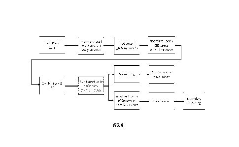
[0134] As mentioned above, the time needed in screening cells for lead
candidates using
macroscale workflows that are typically currently used, significantly adds to
the drug development
timeline. Thus, it is urgently needed to reduce the time needed for screening
cells capable of secreting
a desired antibody, to thereby accelerate antibody discovery. FIG. 6 shows a
general workflow which
is directed to providing acceleration of antibody discovery campaigns. The
method includes isolating
plasma B cells and importing the cells in a microfluidic device, preferably
the microfluidic device as
disclosed in the following sections. The cells can be loaded into the channel
or chamber of the
microfluidic device and cultured individually. In some embodiments, up to 50k
single plasma B cells
may be loaded. In some embodiments, cells that are determined to be healthy
(e.g., viable),
substantially healthy, or enriched in a proportion of cells that are healthy,
may be introduced
preferentially to the chamber(s) of the microfluidic device.
[0135] The method may also include conducting binding or functional assays,
which can be,
but is not limited to bead-based analyses for testing the IgG-antigen
specificity of the antibodies
secreted in each pen. The method may further include loading nucleic acid
capture objects, which
may be any nucleic acid capture object at described herein, and performing on-
chip lysis, nucleic acid
capture and reverse transcription. As explained in more detail in the
following sections, barcoded
cDNA sequences are generated through these steps by using the capture objects
of the present
disclosure. The nucleic acid capture objects additionally are labelled, to
permit correlation of the
binding/functional assay results with the specific nucleic acid isolated from
the cell(s) responsible for
the assay results. Detection of the labels may be performed at any point
during the workflow to
identify the label for each capture object in each chamber.
[0136] Subsequently, the barcoded cDNA sequences, which are captured on the
capture objects
and comprise the BCR sequence (i.e., barcoded BCR beads), may be exported to
an off-chip culture
plate. In some embodiments, barcoded BCR beads from over 1000 pens can be
unloaded to a single
96-well plate and permit multiplexing of subsequent processes.
23
CA 03189006 2023- 2-9
WO 2022/051570
PCT/US2021/048976
[0137] As explained in more detail in the following sections, the capture
objects of the present
disclosure enable the identification of the origin of the barcoded BCR beads
on the 96-well plate.
Last, subsequent analyses including sequencing and/or selective cloning of BCR
sequences,
performing bioinformatics visualization or re-expression of BCR sequences may
be performed.
Further, in some embodiments, secondary screenings can be conducted. In some
embodiments, the
method of the present disclosure aims at increasing screening throughput to up
to 50k single plasma
B cells and over 1000 exports of target B cell receptor (BCR) sequences.
Overall, this workflow
provides for high throughput antibody discovery methods
Methods for Identification of Healthy Cells Prior to Importation into a
Chamber.
[0138] Identifying healthy cells before importing the cells into a chamber can
offer benefits in
the methods of the present disclosure. As referred to herein, a healthy cell
is a cell demonstrating
characteristics of viability, e.g., is a viable cell and has the ability to
continue to grow and optionally,
produce either biomolecules of interest and/or produce daughter cells having
the same capabilities.
Disposing into the chambers, e.g., sequestration pens, of the microfluidic
device only, substantially
only or an increased proportion of healthy cells out of an imported population
can increase the
likelihood of identifying useful cells/clonal populations thereof. Further,
resources used during a
biomolecule production development/identification campaign are not expended
upon non-viable
cells, reducing waste and preserving the use of the pre-defined number of
chambers for cells that have
some possibility of expressing the biomolecule of interest.
[0139] Thus, another aspect of the present disclosure is to identify healthy
cells before
importing them into the chamber of the microfluidic device. However,
identifying healthy cells within
a microfluidic device can be difficult, due to the very nature of the small
scale of the microfluidic
device. Furthermore, for a single cell culture scheme, only a relatively small
number of cells may be
imported into the device, and staining of such small number of cells may not
be able to generate a
fluorescent intensity sufficient for meaningful detection. Additionally, for
some biomolecule
production methods, it may be desirable to not include any sort of dyes or
stains to the cells
themselves, depending on the downstream uses of the cells. Therefore, it is
useful to develop a
method of identifying and importing healthy cells which does not rely upon
staining every batch of
cells to be imported into sequestration pens.
[0140] In some embodiments, a staining method can be combined with a
brightfield image
observation for the purposes of identifying healthy cells.
24
CA 03189006 2023- 2-9
WO 2022/051570
PCT/US2021/048976
[0141] In some embodiments, identifying a healthy cell can involve the use of
a machine
learning algorithm to process image data. In some embodiment, the machine
learning algorithm is
capable of identifying healthy cells without staining. The machine learning
algorithm can include a
neural network, such as a convolutional neural network. A convolutional neural
network (CNN)
generally accomplishes an advanced form of image processing and
classification/detection by first
looking for low level features such as, for example, edges and curves, and
then advancing to more
abstract (e.g., unique to the type of images being classified) concepts
through a series of convolutional
layers. A CNN can do this by passing an image through a series of
convolutional, nonlinear, pooling
(or downsampling, as will be discussed in more detail below), and fully
connected layers, and get an
output. The output can be a single class or a probability of classes that best
describes the image or
detects objects on the image. Some examples of CNNs useful in these methods
include have been
described, for example, International Application Publication No. WO
2019/232473, entitled
"Automated Detection and Characterization of Micro-Object in Microfluidic
Devices", filed on May
31, 2019; and in International Application Publication No. W02018102748,
entitled "Automated
Detection and Characterization of Micro-Obj ect in Microfluidic Devices",
filed on December 1, 2017,
each of which disclosures are incorporated herein by reference.
[0142] In some embodiments, a training data used in establishing the CNN model
of the present
disclosure may include a fluorescent image having the cells of interest
stained, a brightfield image
having the cells of interest annotated, or combinations thereof The dyes
suitable in the present
disclosure may include but are not limited to calcein, zombie violet stain,
annexin, acridine orange,
propidium iodide, or combinations thereof. Any suitable stain that
discriminates between a healthy
cell and a dead/dying and/or non-viable cell, as is known to one of skill in
the art may be used. In
some embodiments, other dyes that are specific to a marker of interest can
also be used, for instance,
Alexa Fluor 647 anti-mouse CD138 (Syndecan-1) Antibody (BioLegend), which is
highly specific
for terminally differentiated live plasma cells and stains CD138 presented on
the surface. In some
embodiments, two or more dyes can be used in staining a sample to provide
cross-reference or
verification.
[0143] In a particular embodiment, a training data includes images of cells
stained with a
fluorescent dye in combination with images of the cells under brightfield A
healthy cell can be
identified, for example, by observing the morphologies of the cells under
brightfield In some
embodiments, a healthy cell, e.g., a viable cell, can be characterized as
having a clear cell boundary,
good contrast, round shape, or combinations thereof In some embodiment, a
healthy cell can be
determined by identifying the unhealthy ones. For instance, an unhealthy cell
can be characterized as
having debris-like appearance, unclear or different contrast, or combinations
thereof. In many
CA 03189006 2023- 2-9
WO 2022/051570
PCT/US2021/048976
embodiments, assessment of viability can be made in a relative manner by
comparing cells in a
sample. For instance, a healthy cell can have a larger diameter while other
cells having a smaller
diameter are more likely to be unhealthy/dead or merely cell debris.
[0144] In the training regime, cells may be first detected under brightfield
and then labeled as
live/dead based on the fluorescent intensity. In some embodiment, labeling of
live/dead cells is based
on a cutoff value of the fluorescent intensity, which can be selected in
accordance with the user's
likings or needs.
[0145] After training is accomplished with the type of cell under
investigation, the method of
penning healthy cells using the trained machine learning algorithm may be
employed to increase the
penning efficiency of healthy cells, and decrease the numbers of non-viable
cells penned. In some
embodiments, the percentage of healthy cells relative to non-viable cells
imported into the chambers,
e.g., sequestration pens, after identification by the algorithm, may be
improved by about 10%, 15%,
20%, 25%, 30%, 35%, 40%, 50%, 60%, 70%, 80%, 90%, 100%, or more.
IV.
A Method of Assaying For A Specific Binding Interaction Between A First
Molecule And A Second Molecule
[0146] Binding interactions between a first molecule and a second molecule can
be measured
in a chamber of a microfluidic chip. The chamber can be any of the chambers
described or referenced
herein, including a microwell or a sequestration pen, and the assay formats
can vary widely. For
example, the assay can be a "sandwich" assay in which a surface, such as a
bead or an internal surface
of a wall of the microfluidic device, is configured to capture and/or present
the first molecule; binding
of the second molecule is detected via a third molecule that is labeled and
capable of binding to the
complex formed by the second molecule binding to the first molecule, thereby
associating the label
of the third molecule with the surface in a detectable manner. In such assays,
the second molecule can
be produced by a biological cell. The assay surface can be in the chamber
(e.g., as described in U.S.
Patent Application Publication No. 2015/0165436 and PCT International
Publication No. WO
2010/040851) or proximal to the chamber, such as in a channel that the chamber
connects with (e.g.,
as described in U.S. Patent Application Publication No. 2015/0151298).
Alternatively, the assay can
be a diffusion gradient assay in which the second molecule has a label (which
can be linked to the
second molecule, or can be an intrinsic property of the second molecule, such
as auto-fluorescence)
and the diffusive properties of the labeled second molecule in the presence of
the first molecule can
be monitored, e.g., as described in PCT International Publication No. WO
2017/181135. In such
assays, the first molecule can be produced by a biological cell. Still other
assays can feature a blocking
interaction in which a molecule of interest binds to the first molecule and
thereby blocks an interaction
26
CA 03189006 2023- 2-9
WO 2022/051570
PCT/US2021/048976
of the first molecule with the second molecule. In such assays, the molecule
of interest can be
produced by a biological cell, and the second molecule can comprise a label.
As with the sandwich
assays, the blocking assays can feature the first molecule bound to a surface.
The surface can be
located, for example, in the chamber or a region proximal to the chamber, such
as a channel. Examples
of blocking assays are described below and elsewhere herein, including in the
examples and in the
claims.
[0147] In-channel Binding Assay. In some embodiments, a method of assay for a
specific
binding interaction between a first molecule and a second molecule is provided
The method can be
performed within a microfluidic device having a channel and a chamber, such as
a microwell or a
sequestration pen, fluidically connecting to the channel. The method can
include: introducing each of
a plurality of biological cells into a respective one of a plurality of
chambers, incubating the biological
cells and allowing the biological cell to produce and/or secrete a molecule of
interest; introducing a
micro-object including a plurality of first molecules into the channel; and
monitoring an accumulation
of the molecule of interest on the micro-object.
[0148] In some embodiments, monitoring an accumulating of the molecule of
interest on the
micro-object including introducing a third molecule that is labeled and
capable of binding to the
complex formed by the molecule of interest binding to the first molecule,
thereby associating the label
of the third molecule with the accumulation of the molecule of interest on the
micro-object. Some
aspects of an in-channel assay using micro-objects comprising beads having a
plurality of first
molecules are further described in an International Application filed on
October 22, 2014, and
published as International Publication W02015/061497.
[0149] In some embodiments, introducing a micro-object including a plurality
of first
molecules, e.g., a reporter cell, into the channel including introducing a
plurality of the micro-objects
and allowing the plurality of the micro-objects fill the channel at a density.
In some embodiments, an
optimal density is such that nearly the entire channel is filled with the
micro-objects. A density that
is below optimal might result in a sparse number of micro-objects in the
channel and an under-
sampling of the secreted molecule of interest, making unambiguous
identification of the secreting
chamber difficult. On the other hand, an overly concentrated density might
lead to higher risk of
channel blockages, poor uniformity across the chip, and might lead to the
micro-objects getting
pushed into the chambers. In some embodiments, the optimal density can vary
depending on the size
of the micro-objects introduced. In certain embodiments, the micro-objects are
biological cells, and
the density can be from about 107 to 109, or about 10g to 2x10⁸ cells/mL. In
some embodiments, the
micro-object including the plurality of first molecules, e.g., reporter cells,
may be cells that may be
cells that culture in suspension. In other embodiments, adherent cell types
may be used as reporter
27
CA 03189006 2023- 2-9
WO 2022/051570
PCT/US2021/048976
cells when detachment protocols are used. For example, adherent CHO cells may
be successfully used
when a detachment protocol may include: culturing to confluence prior to
importation, e.g. not
exceeding confluence; and treating with a detachment reagent such as Accutase
(ThermoFisher
Scientific, A1110501), TrypLE or the like, e.g. treatment at about 22 C for 10
min with no agitation.
The adherent CHO cells were then successfully importable as monodisperse cells
and the target cell
densities were achieved. Specific detachment protocols may be determined as
needed for other cell
types. The preparatory culture density may be varied, e.g., less than about
100% confluent, less than
about 90% confluent, less than about 80% confluent, less than about 60%
confluent, or less than about
50% confluent. The detachment reagent may be varied. The duration of the
detachment treatment may
be varied, e.g., from about 5 min to about lh, about 10 min, about 15 min,
about 20 min, about 30
min, about 45 min, about 60 min or any value therebetween. In some
embodiments, agitation is not
employed. In yet other embodiments, the cells may be agitated during the
detachment treatment.
Temperature may be varied in order to successfully detach the cells, and may
be varied from about
15 C to about 36 C, about 10 C to about 40 C, or any temperature therebetween.
Filtration through
a cell strainer may be useful to remove cell clumps or other large debris, and
may be performed before
concentrating the cells to target import concentration. The cells may be
concentrated by centrifuging
at 400 x g for 5 min, and resuspended to the desired concentration. The third
molecule, which is
labeled and capable of binding to the complex formed by the molecule of
interest binding to the first
molecule, e.g., a labelled antibody, may be added to the media when
resuspending the cells.
[0150] In a specific embodiment, as shown in FIG 38, Jurkat cells (upper) and
K562 cells
(lower) were used as the micro-objects at a density of 1.7x10A8 cells/mL and
1x10^8 cells/mL
respectively. The figures show that the channels were nearly filled by the
cells at an acceptable extent
for the in-channel binding assays.
[0151] In some embodiments, the first molecule and/or the molecule of interest
can be a
protein. The protein can be, for example, a cell surface protein or an
extracellular protein. The protein
can be a modified protein, such as a glycosylated protein, a lipid-anchored
protein, or the like. In
some embodiments, the molecule of interest can specifically bind to the first
molecule. In certain
embodiments, the first molecule and molecule of interest can be an antigen-
antibody pair. For
example, the biological cell can be a B cell producing an antibody of interest
(i.e., molecule of interest)
and the first molecule presented on the surface of the micro-objects can be an
antigen or epitope of
the produced antibody. In some embodiments, the third molecule can be a
secondary antibody binding
to the produced antibodies (i.e., the secreted second molecule), and the
detection thereof is associated
with the binding of the first molecule and the molecule of interest.
28
CA 03189006 2023- 2-9
WO 2022/051570
PCT/US2021/048976
[0152] In certain embodiments, the micro-object can be one or more beads or
cells that express
the first molecule. If a cell, the cell can express the first molecule
naturally or can be genetically
modified (e.g., stably or transiently transfected) to express the first
molecule. If a bead, the bead can
be created by conjugating the first molecule on its surface.
[0153] Blocking Assay. In some embodiments, a method of assaying for
inhibition of a
specific binding interaction between a first molecule and a second molecule is
provided. The method
can be performed within a microfluidic device having a chamber, such as a
microwell or a
sequestration pen, and can include: introducing each of a biological cell and
a micro-object that
comprises a plurality of first molecules into the chamber of the microfluidic
device, incubating the
biological cell in the presence of the micro-object and allowing the
biological cell to produce and/or
secrete a molecule of interest; introducing the second molecule into the
chamber, wherein the second
molecule is bound to a detectable label (or intrinsically produces a signal,
such as auto-fluorescence);
and monitoring an accumulation of the second molecule on the micro-object. One
or more micro-
objects can be loaded with the cell into the chamber. An absence or
diminishment of accumulation of
the second molecule on the one or more micro-objects indicates that the
molecule of interest produced
by the cell inhibits binding of the first molecule to the second molecule.
[0154] In some embodiments, monitoring an accumulation of the second molecule
on the
micro-object comprises comparing the accumulation to that observed on a
control micro-object in the
presence of a positive control molecule of interest and/or a negative control
molecule of interest. In
other embodiments, monitoring an accumulation of the second molecule on the
micro-object
comprises comparing the accumulation to that observed on a control micro-
object in the absence of a
control molecule of interest.
[0155] As used herein, the term "diminishment" indicates lower accumulation
compared to
that observed in one or more control chambers. In some embodiments, the
control chamber can be a
negative control chamber. Examples of negative control chambers may include,
but are not limited
to, chambers containing a control micro-object and a negative control cell.
The negative control cell
can be a cell producing a molecule that is known not to bind the first
molecule or the second molecule,
or a cell known not to produce a molecule of interest. Other examples of
negative control chambers
include chambers containing a control micro-object by itself (i.e., with no
control cell present).
Accordingly, in some embodiments, monitoring an accumulation of the second
molecule on the
micro-object comprises comparing the accumulation to that observed on a
control micro-object
incubated in the presence of one or more or no negative control cells.
[0156] In some embodiments, the control chamber can be a positive control
chamber.
Examples of positive control chambers may include, but are not limited to,
chambers containing a
29
CA 03189006 2023- 2-9
WO 2022/051570
PCT/US2021/048976
control micro-object and a positive control cell. The positive control cell
can be a cell producing a
molecule that is known to bind the first molecule or the second molecule and
thereby inhibit binding
of the first molecule to the second molecule.
[0157] Control cells (e.g., positive or negative control cells) may be
introduced into the same
microfluidic device as the biological cell that is capable of producing a
protein of interest, or into a
different microfluidic device. Control cells may be introduced into the
microfluidic device at a same
period or a different time period.
[0158] In some embodiments, the method is performed within a microfluidic
device having a
plurality of chambers, and monitoring an accumulation of the second molecule
on the micro-object
comprises comparing the accumulation to that observed in one or more other
chambers of the plurality
where one or more control cells are introduced. A control cell in another
chamber may be introduced
intentionally (i.e., when the control cell is known to be a positive or
negative control cell) or control
cells may be identified from the pool of introduced cells based on the fact
that a user will expect that
not all of the introduced cells will produce a molecule of interest capable of
impacting the
accumulation of the second molecule on the micro-object. In some embodiments,
monitoring an
accumulation of the second molecule on the micro-object comprises comparing
the accumulation to
that observed in one or more other chambers of the plurality where no cell is
introduced.
[0159] In some embodiments, comparison to a single control chamber is
sufficient (e.g., a
control chamber having a single well-characterized control cell or no control
cell at all). In other
embodiments, the method includes comparison to a plurality of control chambers
For example, the
comparison can comprise comparing accumulation of the second molecule on the
micro-object with
a statistical measure of the accumulation of the second molecule on the
control micro-objects in the
plurality of control chambers (e.g., an average accumulation or a level of
accumulation that is one,
two, or three standard deviations below the average accumulation on the
control micro-objects in the
plurality of control chambers). Alternatively, the comparison can comprise
comparing accumulation
of the second molecule on the micro-object with the minimum accumulation (or
maximum
accumulation) of the second molecule on the control micro-objects in the
plurality of control
chambers.
[0160] In some embodiments, the method is performed within a microfluidic
device having a
microfluidic channel and a plurality of chambers, and monitoring an
accumulation of the second
molecule on the micro-object comprises comparing the accumulation to that
observed in an area
outside the chamber where the cell is introduced (e.g., in the microfluidic
channel), in one or more
other chambers of the plurality where one or more control cells are
introduced, or in one or more
chambers where no cell is introduced.
CA 03189006 2023- 2-9
WO 2022/051570
PCT/US2021/048976
[0161] In certain embodiments, the first molecule and/or the second molecule
can be a protein.
The protein can be, for example, a cell surface protein or an extracellular
protein. The protein can be
a modified protein, such as a glycosylated protein, a lipid-anchored protein,
or the like. In certain
embodiments, the first molecule and second molecules can be a receptor-ligand
pair. The -ligand"
used herein refers to a molecule that has a region, structure, or motif that
can be recognized and bound
specifically by a receptor at a certain level of affinity. In some
embodiments, the level of affinity is
high enough to form and maintain a receptor-ligand complex during the
operation of the blocking
assay of the present disclosure but is lower than the level of affinity of the
molecule of interest to the
ligand or the receptor. In some embodiments, the first molecule is a receptor
molecule, and wherein
the second molecule is a ligand that specifically binds to the receptor
molecule. For example, the first
molecule can be a growth factor receptor, a cytokine receptor, a chemokine
receptor, an adhesion
receptor (e.g., an integrin or a cell adhesion molecule (CAM)), an ion
channel, a G protein-coupled
receptor (GPCR), or a fragment retaining activity of its respective full
length biomolecule of any of
the foregoing; and the ligand can be a growth factor, a cytokine, a chemokine,
an adhesive ligand, an
ion channel ligand, a GPCR ligand, a viral protein (e.g., a viral coat or
capsid protein, such as a fusion
protein), or a fragment retaining activity of its respective full length
biomolecule of any of the
foregoing. In some embodiments, the first molecule is a ligand, and the second
molecule is a receptor
that is specifically bound by the ligand as exemplified above. In certain
embodiments that the second
molecule is a receptor, the receptor can be a receptor molecule anchored on an
object such as a cell,
a bead, a lipid particle. Alternatively, when the second molecule is a
receptor, the receptor molecule
may be a soluble receptor molecule. The receptor molecule can be manufactured
by chemical
synthesis or a semi synthetic process.
[0162] In certain embodiments, the one or more micro-objects can be one or
more beads or
cells that expresses the first molecule. If a cell, the cell can express the
first molecule naturally or can
be genetically modified (e.g., stably or transiently transfected) to express
the first molecule.
[0163] The blocking assays described herein can be a receptor blocking assay
or a ligand
blocking assay. In an exemplary receptor blocking assay (FIG. 39), the
targeted antigen (i.e., the
first molecule) may be located on the surface of the reporter cell (i.e., the
micro-object), and the
secreted antibody binds to this surface-bound antigen "receptor", potentially
blocking the binding of
the dye-labeled, soluble "ligand" (i.e., the second molecule). Conversely, in
a ligand blocking assay
(FIG. 40), the targeted antigen (i.e., the second molecule) may be in
solution, and the secreted
antibody binds to this antigen "ligand", potentially blocking its binding to
the receptor (i.e., the first
molecule) of the reporter cell. In either design, if the secreted antibody is
an effective blocker, little
or no dye-labeled ligand (i.e., second molecule) will bind to the reporter
surface, and the reporter
31
CA 03189006 2023- 2-9
WO 2022/051570
PCT/US2021/048976
cell will be dark in the fluorescence channel associated with the ligand (the
"ligand channel"). If the
secreted antibody is non-blocking, the dye-labeled ligand will bind to and
accumulate on the
reporter surface, and the reporter will be visible in the fluorescence channel
associated with the
ligand.
[0164] In some embodiments of receptor blocking assays, an optional secondary
antibody
(i.e., the third molecule), labeled with a dye different than that of the
ligand, may be included to
confirm binding of the secreted antibody to the reporter (FIG 39). In this
design, if the secreted
antibody both binds to the receptor and blocks ligand binding, the reporter
will be visible in the
secondary channel and dark in the ligand channel. However, if the secreted
antibody binds the
receptor but does not block ligand binding, the reporter cell will be visible
in both the secondary and
ligand channels. In order to determine binding in the ligand blocking assay
design, a separate in-
channel assay should be performed.
[0165] In some embodiments of receptor blocking assays (FIG. 41), the
biological cells,
producing a molecule of interest, labeled as "B" may be penned first, followed
by introduction of
micro-objects including first molecules, e.g., reporter micro-objects, which
may be beads or cells.
The upper and low rows of FIG. 41 represent timepoints, e.g., each chamber,
left to right, along
each row, for two different types of receptor blocking assays. As shown in the
upper row of FIG.
41, an exemplary embodiment is shown where the molecules of interest, e.g., an
antibody produced
by cell "B", bind to the reporter micro-objects and block the ligands "L" from
binding to the
reporter micro-objects "R". Upon introduction to the chamber, the cells ("B")
producing the
molecule of interest, e.g., an antibody in this instance, are shown in the
first (left-hand side of the
upper row of FIG. 41) exemplary chamber. After introduction of the reporter
micro-objects, labelled
"R" (second from left, upper row of FIG. 41), the micro-objects may then be
incubated with the
secreting biological cells, permitting binding of the molecule of interest to
the reporter micro-object.
In the third chamber of the upper row of FIG. 41 is shown introduction of the
dye labeled ligands,
labelled "L" (i.e., the second molecule). In this embodiment, the secreted
antibody is capable of
binding to and saturating the receptor (i.e., the first molecule, which may
include an antigen binding
site) on the micro-objects, blocking the ligand from binding. The labelled
ligand therefore does not
label the reporter molecule and accumulation of signal on the reporter micro-
object is diminished or
eliminated.
[0166] In the lower row of FIG. 41, is shown a different embodiment. The
secreting
biological cell "B" is introduced to the chamber (first chamber, left hand of
lower row of FIG. 41)
and produces the molecule of interest, e.g., an antibody. In the second
chamber of lower row of FIG.
41, the reporter micro-objects "R" including the first molecules are
introduced, as before. In the
32
CA 03189006 2023- 2-9
WO 2022/051570
PCT/US2021/048976
third chamber of the lower row of FIG. 41, the ligand "L" is introduced and is
capable of binding to
the reporter micro-object and blocking the molecule of interest from binding
(or from stably
binding) to the first molecules of reporter micro-object -It", and in the
fourth chamber, is shown
the time point, where the ligand (e.g., the second molecule) has
accumulated on the reporter
micro-object "R", binding to the first molecule associated thereto, and
accumulation of signal is
observed.
[0167] The two cell types may be cultured together using a Pulse Culture
operation
consisting of alternating intervals of zero-flow incubation and short periods
of chip flushing
designed to allow binding of secreted antibodies and minimize pen-to-pen
diffusion. Flush volume,
flush rate, and incubation duration are tunable parameters and can be adjusted
based on user
selections. After this period of pulsed culture, typically 30 min, a solution
containing dye-labeled
ligand is imported and allowed to diffuse into the pens, where it can bind to
unblocked reporter
cells. Finally, flushes are performed to wash out unbound ligands and images
are acquired to assess
blocking.
[0168] In some embodiments of ligand blocking assays (FIG. 42), the biological
cells and the
micro-objects may be penned sequentially. In this type of assay, rather than
ensuring that reporter
cell receptors, e.g., first molecules, are saturated, since the ligand is, for
instance an antigen, a Pulse
Culture incubation period can be performed to let the micro-objects producing
a molecule of interest
"B", e.g., plasma cells, recover and resume secreting prior to importing the
antigen "ligand". Once
the B cells recover and resume secreting antibodies of interest, dye-labeled
ligand (i.e., the second
molecule, which is an antigen in this embodiment) is introduced and allowed to
diffuse into the
pens. If not blocked by the secreted antibody, the ligand can bind to the
reporter cells "Er. Blocking
of the ligand by the secreted antibody (molecule of interest) prevents ligand
binding to the reporters
(e.g., the micro-objects including the first molecule). In FIG. 42, the
chambers, left to right of each
row, demonstrate successive timepoints during a particular version of a ligand
blocking assay.
Labels are as for FIG. 41. In the top row, a ligand blocking assay is shown
where the secreted
molecules of interest, e.g., an antibody as shown here, bind to the introduced
ligand, e.g., second
molecule. Thus, accumulation of signal on the first molecules, e.g., a
receptor molecule, of reporter
micro-object "R" is diminished or inhibited. In the second embodiment, shown
in the second row of
FIG. 42, the secreted molecules of interest, e.g., antibody, are non-blocking
and do not prevent
accumulation of signal on the first molecules of the reporter micro-object
"R". Signal is therefore
observed to accumulate on the reporter micro-objects including the first
molecules. Thus, the
secreted molecules of interest, e.g., an antibody, does not bind to the first
molecules, e.g., a receptor
of the reporter micro-object, stably or with sufficient affinity to prevent
displacement by the ligand,
33
CA 03189006 2023- 2-9
WO 2022/051570
PCT/US2021/048976
e.g., second molecule. The bottom row of FIG. 42 represents an embodiment,
where, while the
secreted molecule of interest, e.g., an antibody, may be capable of binding to
the first molecule of
the reporter micro-objects -It", ligand -L" is introduced in a high
concentration, sufficiently high
that its concentration exceeds that of the secreted molecule of interest, and
thus can both bind to the
secreted molecule of interest and well as first molecules of the reporter
micro-objects. Thus, signal
may accumulate on the reporter micro-objects, and may lead to a false negative
result.
[0169] Ligand titration and incubation timing. In some embodiments, ligand
concentration (i.e., the concentration of the second molecule) can be
optimized prior to running a
blocking assay of the present disclosure. Too little ligand can result in low
signal accumulation on
the reporter cell, making discrimination between blocking and non-blocking
antibodies difficult.
Too much ligand can result in a large fraction of unbound ligand, resulting in
higher background
and potentially lower blocking assay sensitivity. In the case of the receptor
blocking assay,
excessively high ligand concentration can result in otherwise blocking
antibodies being displaced by
the concentrated ligand due to competition. In the ligand blocking design,
there may not be enough
secreted antibody to block all of a high concentration of ligand. In certain
embodiments, a
concentration of the second molecule is at least 5 nM, from about 5 to about
30 nM, at least 6 nM,
or from about 6 nM to about 30 nM.
[0170] Ligand Binding Specificity. In some embodiments, confirmation that
ligand binding
is specific to the surface-expressed receptor (i.e., the first molecule of the
micro-object), with
minimal non-specific binding to the reporter may be performed. In the case of
endogenously
expressing reporter cells, a knockout cell line, where receptor expression is
eliminated, can serves as
a useful negative control to confirm specificity of ligand binding. Similarly,
for transfected reporter
cell lines, both the parental and transfected cells can be screened to confirm
that ligand binding is
specific to transfection, with minimal binding to the parental cell line. This
measurement of
specificity can be performed off-chip using standard flow cytometry methods,
or on-chip by
importing and penning reporter cells and negative control reporter cells into
different regions of the
chip, followed by importing and incubating with the dye-labeled ligand. During
incubation, the chip
may be regularly imaged in the fluorescence channel of the ligand to detect
signal accumulation on
the reporter cell populations. The intensity difference between the two
reporter populations may be
clearly discernible, with little or no detectable signal on the negative
control reporters.
[0171] Reporter heterogeneity. In some embodiments, heterogeneity of the
reporter cell
(e.g., the micro-object) may be performed for in-pen blocking assays since
only a few reporter cells
are introduced into any single pen. Consistent with cell-binding assays, an
ideal reporter cell
population would have a high surface expression of receptor and low receptor
expression variation
34
CA 03189006 2023- 2-9
WO 2022/051570
PCT/US2021/048976
such that each cell has nearly the same level of expression. In such a case,
all the reporter cells
would bind the same amount of dye-labeled ligand in the absence of non-
blocking antibodies, and
all cells would be equally bright in the imaging channel of the ligand. In the
presence of blocking
antibodies, little to no dye-labeled ligand would bind the reporter, and it
would appear -dark" in the
ligand imaging channel. However, if the reporter population has a large
fraction of cells with low
receptor surface concentration, this subpopul ati on may appear "dark" even in
the absence of
blocking antibodies, leading to an increase in false positive blocking hits,
especially with few
reporter cells in each pen.
[0172] In certain embodiments, the micro-object is a cell, which is from a
transfected cell
line. In some embodiments, the transfected cell line may be stably or
transiently transfected to
express the plurality of first molecules. In certain embodiments, at least
60%, at least 65%, at least
70%, at least 75%, at least 80%, at least 85%, at least 90%, at least 95% or
more of the cells in the
transfected cell line may express the first molecules at detectable levels. In
certain embodiments, in
order to reduce the false positive rate, each pen is introduced with at least
two or at least three
micro-objects, where each micro-object includes the first molecule.
[0173] The blocking assays described herein can be coupled with any of the
other assays
described herein, including the sandwich and/or diffusion gradient assays.
Cells identified as
producing a molecule of interest that blocks the interaction between the first
and second molecules
can be further analyzed, for example, using the 5' barcoding methods described
herein.
V. Methods of Capturing the 5' Ends of RNA and Barcode Identification
[0174] Provided herein are methods of capturing the 5' ends of RNA. Also
provided here are
methods of providing one or more 5' barcoded cDNA sequences by reverse
transcribing RNA
captured from a biological cell.
[0175] In some embodiments, the methods comprise providing a biological cell
within a
chamber. The cell may be provided within a microwell of a microfluidic device.
The cell may be
provided within a sequestration pen located within an enclosure of a
microfluidic device. In some
embodiments, the methods comprise disposing the biological cell within a
sequestration pen located
within an enclosure of a microfluidic device. In some embodiments, a single
capture object is provided
in the chamber. The biological cell, reagents, time period (and optionally
other conditions),
sequestration pens, and microfluidic devices may be any of those described
herein.
[0176] In some embodiments, the methods comprise providing a capture object
within the
chamber. Further embodiments of disposing one or more biological cells and/or
capture objects within
CA 03189006 2023- 2-9
WO 2022/051570
PCT/US2021/048976
the chamber (e.g., microwell or sequestration pens of the microfluidic device)
are described in the
section entitled "Microfluidic Device and System".
[0177] A capture object described herein comprises a label, a plurality of
first oligonucleotides,
and a plurality of second oligonucleotides. In some embodiments, each first
oligonucleotide of the
plurality comprises a barcode sequence and a sequence comprising at least
three consecutive guanine
nucleotides at a 3' end. In some embodiments, each first oligonucleotide of
the plurality comprises a
barcode sequence and a sequence comprising three consecutive guanine
nucleotides at a 3' end In
some embodiments, each first oligonucleotide of the plurality comprises a
barcode sequence and a
sequence comprising at least three consecutive guanine nucleotides at a 3'
end, and each second
oligonucleotide of the plurality comprises a capture sequence. In some
embodiments, each first
oligonucleotide of the plurality further comprises a priming sequence that
corresponds to a first primer
sequence. In some embodiments, each second oligonucleotide of the plurality
further comprises a
priming sequence that corresponds to a second primer sequence. In some
embodiments, the first
oligonucleotide comprises a first priming sequence that corresponds to a first
primer sequence and
wherein the second oligonucleotide comprises a second priming sequence that
corresponds to a
second primer sequence. In some embodiments, the first and second primer
sequences are the same.
In some embodiments, the first oligonucleotide and the second oligonucleotide
are individually linked
to the capture object, e.g., the first oligonucleotide is part or all of a
first molecule linked to the capture
object and the second oligonucleotide is part or all of a second molecule
linked to the capture object,
where the first and second molecules are different and independently attached
to the capture object.
[0178] In some embodiments, the methods comprise lysing the biological cell.
In some
embodiments, the methods comprise allowing RNA molecules released from the
lysed biological cell
to be captured by the capture sequences of the plurality of second
oligonucleotides, e.g., comprised
by a capture object. In some embodiments, the methods comprise lysing the
biological cell and
allowing RNA released from the lysed biological cell to be captured by the
capture sequences of the
plurality of second oligonucleotides, thereby forming captured RNA. The
capture object, capture
sequences, priming sequences, and lysis procedures may be any of those
described herein.
[0179] In some embodiments, lysing the biological cell is
performed such that a plasma
membrane of the biological cell is degraded, releasing cytoplasmic RNA from
the biological cell. In
some embodiments, the lysing reagent may include at least one ribonuclease
inhibitor. An exemplary
lysis reagent is commercially available in the Single Cell Lysis Kit, Ambion
Catalog No. 4458235.
This reagent can be flowed into the microfluidic channel of a microfluidic
device and permitted to
diffuse into sequestration pens, followed by a suitable exposure period (e.g.,
10 minutes; shorter or
longer periods may be appropriate depending on cell type, temperature, etc.).
Lysis can be stopped by
36
CA 03189006 2023- 2-9
WO 2022/051570
PCT/US2021/048976
flowing in an appropriate stop lysis buffer, e.g., from the Single Cell Lysis
Kit, Ambion Catalog No.
4458235 and incubating for an appropriate time. Similar results can be
obtained using other lysis
buffers, including but not limited to Clontech lysis buffer, Cat #635013,
which does not require a stop
lysis treatment step Released mRNA can be captured by a capture object present
within the same
sequestration pen.
[0180] In some embodiments, the methods comprise reverse transcribing captured
RNA. In
some embodiments, one or more barcoded cDNA sequences is produced. In some
embodiments,
each cDNA sequence comprises an oligonucleotide sequence complementary to a
corresponding
one captured RNA covalently linked to the reverse complement of the barcode
sequence of the first
oligonucleotide. In some embodiments, the methods comprise reverse
transcribing the captured
RNA, thereby producing one or more barcoded cDNA sequences, each comprises an
oligonucleotide sequence complementary to a corresponding one captured RNA
covalently linked to
the reverse complement of the barcode sequence of the first oligonucleotide.
Reverse transcribing
RNA molecules may be performed according to any appropriate procedure
described herein. In
some embodiments, the capture sequence binds to, and thereby captures, RNA and
primes
transcription from the captured RNA. In some embodiments, a reverse
transcription (RT)
polymerase transcribes the captured RNA.
[0181] FIG. 7 shows a schematic representation of an exemplary process. A
biological cell
may be placed within a sequestration pen within a microfluidic device. A
capture object, which may
be configured as any capture object described herein, may be disposed into the
same sequestration
pen, which may be performed before or after disposing the cell into the
sequestration pen. The cell
may be lysed using a lysis reagent which lyses the outer cell membrane of cell
but not the nuclear
membrane. A lysed cell results from this process and releases RNA. The second
oligonucleotide of
capture object includes a priming sequence, which has a priming sequence
(e.g., corresponding to
P1 primer) and a capture sequence, which in this case includes a PolyT
sequence which can capture
the released nucleic acid having a PolyA sequence at its 3' end. The capture
sequence captures the
released nucleic acid. Next, the second oligonucleotide is extended through
reverse transcription
from the released nucleic acid while in the presence of template switching
oligonucleotide. As the
captured RNA is transcribed, the transcript is extended to include several C
(cytosine) nucleotides,
which brings the end of the RNA distal to the PolyA tail into alignment with
the rGrGrG terminus
of the oligonucleotide bearing the barcode (including a TSO). Identification
of the barcode may be
performed, using any of the methods described herein either before RNA capture
to the barcoded
beads; before reverse transcription of the RNA captured to the beads, or after
reverse transcription
of the RNA on the bead. In some embodiments, identification of the cell
specific barcode may be
37
CA 03189006 2023- 2-9
WO 2022/051570
PCT/US2021/048976
performed after reverse transcription of RNA captured to the bead. After both
reverse transcription
and identification of the barcode of the capture object has been achieved, the
cDNA captured by the
capture object is exported out of the chamber into e.g., a common receptacle.
A plurality of cDNA
capture objects may be exported at the same time and the amplification may be
performed, using a
common amplification primer (e.g., P1 primer).
[0182] In some embodiments, the methods further comprise identifying the
barcode sequence
of the plurality of first oligonucleotides while the capture object is located
within the chamber.
Identifying can include detecting the barcode with one or more labeled
antisense oligos (e.g., as
described in US Patent Application Publication No. 2019/0345488).
[0183] In some embodiments, identifying the barcode comprises detecting
fluorescence
emitted from the label, which may be an integral part of the capture object or
an extrinsic label capable
of binding to another molecule (e.g., an oligonucleotide) on the surface of
the capture object. In some
embodiments, the label comprises one or more fluorophores. In some
embodiments, the label
comprises a single fluorophore. In some embodiments, the label comprises
multiple fluorophores,
with each fluorophore present at one or more levels, resulting in unique
combinations of fluorophores
and fluorophore levels that constitute unique labels. The detectable label can
be, for example, a
fluorescent label, such as, but not limited to a fluorescein, a cyanine, a
rhodamine, a phenyl indole, a
coumarin, or an acridine dye. Some non-limiting examples include Alexa Fluor
dyes such as Alexa
Fluor 647, Alexa Fluor 405, Alexa Fluor 488; Cyanine dyes such as Cy 5 or
Cy 7, or any
suitable fluorescent label as known in the art. Any set of distinguishable
fluorophores may be selected
to be present on hybridization probes flowed into the microfluidic environment
for detection of the
barcode, as long as each dye's fluorescent signal is detectably
distinguishable. Alternatively, the
detectable label can be a luminescent agent such as a luciferase reporter, a
lanthanide tag or an
inorganic phosphor, or a Quantum Dot, which may be tunable and may include
semiconductor
materials. Other types of detectable labels may be incorporated such as FRET
labels which can
include quencher molecules along with fluorophore molecules. FRET labels can
include dark
quenchers such as Black Hole Quencher (Biosearch); Iowa Black Tm or dabsyl.
The FRET labels
may be any of TaqMane probes, hairpin probes, Scorpion probes, Molecular
Beacon probes and
the like. In some embodiments, barcodes of capture objects may be identified
or deconvolved as
follows. Capture objects are initially detected by brightfi el d imaging.
Fluorescence is then measured
in a plurality of fluorescence channels (e.g., two, three, or four channels,
such as channels
corresponding to two, three, or four of FITC, Cy5, DAPI, and Texas red (TRED))
with a plurality of
measurements being taken in each channel.
38
CA 03189006 2023- 2-9
WO 2022/051570
PCT/US2021/048976
[0184] In some embodiments, detecting the label of the capture object may
include
determining a signal observed for the capture obj ect in more than one
fluorescence channel, e.g., each
distinct label may be determined by observing/imaging a unique signature of
intensities across two,
three or four fluorescence channels (such as FITC, Cy5, DAPI and TRED).
Detecting each distinct
label yields the previously paired identity of the barcode associated with
that distinguishable label. In
some embodiments, the distinguishable lab el is integral to the capture obj
ect as described above. No
matter what type of label of the capture obj ect is, determining the identity
of the label permits
determining and correlating the origin pen of the cell with the sequencing
results obtained after nucleic
acid capture and sequencing, which may be performed via any suitable method,
including a massively
parallel sequencing method.
[0185] In some embodiments, the barcode sequence of the first oligonucleotide
corresponds to
the label of the capture object. For example, there can be a one-to-one
relationship between the
barcode sequence of the first oligonucleotide and the label of the capture
object. In one non-limiting
example, the barcode sequence of the first oligonucleotide corresponds to the
label of the capture
object, which is integral to the capture object, e.g., an integral
fluorescent, visible or luminescent color
of the capture obj ect. In some embodiments, the barcode sequence of the first
oligonucleotide is the
label of the capture object.
[0186] In some embodiments, one or more fluorophores are directly disposed on
the capture
object itself. In some embodiments, one or more fluorophores are inked via an
oligonucleotide that
binds to the barcode sequence or the reverse complement of the barcode
sequence.
[0187] In some embodiments, the first oligonucleotide comprises one or more
uridine
nucleotides 5' to the barcode sequence and, if present, the first priming
sequence. In some
embodiments, the first oligonucleotide comprises three uridine nucleotides 5'
to the barcode sequence
and, if present, the first priming sequence. In further embodiments, the one
or more uridine
nucleotides are adjacent to or comprise the 5' -most nucleotide(s) of the
first oligonucleotide. In some
embodiments, reverse transcribing the captured RNA is performed in the
presence of an enzyme that
cleaves a sequence containing one or more uridine nucleotides (e.g., a USER
enzyme).
[0188] In some embodiments, each of the one or more barcoded cDNA sequences is
associated
with the capture object. In some embodiments, the one or more barcoded cDNA
sequences are
produced in the chamber.
[0189] In some embodiments, the methods further comprise exporting the capture
object from
the chamber. Exporting the plurality of the capture obj ects may include
exporting each of the plurality
of the capture objects individually. In some embodiments, the method may
further include delivering
each capture object of the plurality to a separate destination container
outside of the microfluidic
39
CA 03189006 2023- 2-9
WO 2022/051570
PCT/US2021/048976
device. The destination container may be a common receptacle, a cell culture
flask, dish, petri dish,
multi-well plate, or the like.
[0190] In some embodiments, the methods further comprise storing the one or
more barcoded
cDNA sequences. In some embodiments, the one or more barcoded cDNA sequences
are stored at a
temperature at about 4 C.
[0191] In some embodiments, the methods further comprise amplifying the one or
more
barcoded cDNA sequences. In some embodiments, amplifying the one or more
barcoded cDNA
sequences comprises using a single primer (e.g., a P1 primer). In other
embodiments, amplifying the
one or more barcoded cDNA sequences comprises using a pair of primers (e.g.,
P7 and PS primers).
[0192] Where applicable, providing the capture object, providing the
biological cell,
lysing/transcribing captured RNA, and identifying the barcode sequence, of
methods disclosed herein
can be performed in the order in which they are written or in other orders,
with the limitation that the
rearrangement of the order of these activities does not violate logical order
(e.g., transcribing before
lysing, and so on). As an example, identification of the barcode sequence can
be performed after
providing the biological cell, after lysing the biological cell, or after
transcribing the captured RNA.
Likewise, the step of providing the capture object in the chamber can be
performed after providing
the biological cell in the chamber.
VI. Methods of Demultiplexing a Pool of Exported cDNA and
Preparation of
Expression Construct Therefrom
[0193] In some embodiments, the methods further comprise performing the method
on a
plurality of biological cells provided in a corresponding plurality of
chambers. In some embodiments,
a plurality of capture objects are provided to the plurality of chambers, each
capture object of the
plurality having (i) a unique label selected from a plurality of unique labels
(e.g., at least 12, 14, 16,
18, 20, 25, 30, 40, 50, 60, 70, 80, 90, 100, 150, 200, 250, 500, 1000, or more
different labels, or a
number of labels falling within a range defined by any two of the foregoing
values), and (ii) a plurality
of first oligonucleotides having a barcode sequence corresponding to the
unique label.
[0194] In some embodiments, the methods further comprise exporting the
plurality of capture
objects into a common receptacle; and amplifying the one or more barcoded cDNA
sequences from
each capture object of the plurality, thereby producing a plurality of
barcoded cDNA sequences, each
barcoded cDNA sequence having a barcode sequence corresponding to one of the
plurality of unique
labels.
[0195] In some embodiments, a plurality of barcoded cDNA sequences is produced
in the
chamber, each barcoded cDNA sequence of the plurality encoding a protein of
interest, corresponding
CA 03189006 2023- 2-9
WO 2022/051570
PCT/US2021/048976
to any one of a plurality of different proteins, linked to a corresponding
reverse complement barcode
sequence. For example, barcoded cDNA sequences corresponding to up to 12
unique labels are pooled
in single well. As barcoded cDNA sequences from specific exports can be
identified based on a
barcode sequence (-10 bp) on the capture object, without amplifying antibody
transcripts from
individual cells before TAP assembly, it may lead to the expression of non-
clonal antibodies and
make down stream characterization difficult. Accordingly, in some embodiments,
the methods further
comprise selectively amplifying the barcoded cDNA sequences to produce an
amplified cDNA
product (or further amplified cDNA product) encoding the protein of interest
or a fragment thereof.
[0196] In some embodiments, the methods further comprise:
a. optionally amplifying a plurality of barcoded cDNA sequences;
b. selectively amplifying the plurality of barcoded cDNA sequences (or
amplified cDNA
sequences) using a barcode-specific forward primer and a reverse primer
specific to the protein of
interest to produce an amplified cDNA product (or further amplified cDNA
product) encoding the
protein of interest or a fragment thereof;
c. annealing a 5' end of the amplified cDNA product (or further amplified cDNA
product) to
a 5' corresponding end of a DNA fragment for transcriptionally-active PCR
(TAP) to produce an
annealed TAP product; and
d. amplifying the annealed TAP product via overlap extension PCR using a TAP
adapter
primer to produce a construct for expression of the protein of interest.
[0197] In some embodiments, the reverse primer specific to the protein of
interest comprises a
sequence complementary to a sequence encoding a conserved region (e.g., a
constant portion) of the
protein of interest, or a sequence 3' to the conserved region (e.g., a 3' UTR
sequence). In some
embodiments, a 3' end of the amplified cDNA product (or further amplified cDNA
product)
comprises a region overlapping with a 3' corresponding end of the DNA fragment
for TAP.
[0198] In some embodiments, each barcoded cDNA sequence of the plurality
encoding a heavy
chain or a light chain sequence corresponding to any one of a plurality of
different antibodies, linked
to a corresponding reverse complement barcode sequence. In these embodiments,
the method further
comprising:
a. optionally amplifying the plurality of barcoded cDNA sequences;
b. selectively amplifying the plurality of barcoded cDNA sequences using a
barcode-specific
forward primer and a reverse primer targeting a conserved portion of the
corresponding constant
region sequence (e.g., a 5' end, or sequence adjacent thereto, of the constant
region) to produce an
41
CA 03189006 2023- 2-9
WO 2022/051570
PCT/US2021/048976
amplified cDNA product (or further amplified cDNA product) encoding the
barcode-specific variable
region;
c. annealing ends of the amplified cDNA product (or further amplified cDNA
product) to
corresponding ends of a DNA fragment for TAP to produce an annealed TAP
product; and
d. amplifying the annealed TAP product via overlap extension PCR using TAP
adapter primers
to produce an expression construct for expression of an antibody heavy chain
or light chain.
[0199] In some embodiments, amplifying the plurality of barcoded cDNA
sequences
comprises using a single primer (e.g., a P1 primer). In some embodiments,
amplifying the plurality
of barcoded cDNA sequences comprises using different forward and reverse
primers.
[0200] In some embodiments, in step b of selectively amplifying, the barcode-
specific forward
primer may be a sequence comprising one of SEQ ID NO: 13-24. In some
embodiments, in step b of
selectively amplifying, the reverse primer targeting a conserved portion may
be a sequence
comprising SEQ ID NO: 54 or 55.
[0201] Further, provided herein are methods of preparing a construct for
expression of the
protein of interest.
[0202] In some embodiments, the methods comprise providing a barcoded cDNA
sequence
and the barcoded cDNA sequence comprises a nucleic acid encoding a protein of
interest linked to
the reverse complement of the barcode sequence of the first oligonucleotide.
In some embodiments,
the barcode cDNA sequence is produced by the methods described herein.
[0203] In some embodiments, the methods comprise amplifying at least a portion
of the
barcoded cDNA sequence using a barcode-specific primer and a primer specific
to the nucleic acid
encoding the protein of interest, thereby producing an amplified cDNA product.
[0204] In some embodiments, the methods comprise providing a DNA fragment for
transcriptionally-active PCR (TAP) comprising:
i. a promoter sequence,
ii. a nucleic acid sequence complementary to a 5' end of the nucleic acid
encoding the protein
of interest (e.g., 5' end of the amplified cDNA product),
iii. a nucleic acid sequence complementary to a 3' end of the nucleic acid
encoding the protein
of interest (e.g., a 3' end of the amplified cDNA product), and
iv. a terminator sequence.
[0205] In some embodiments, the methods comprise incorporating the amplified
cDNA
product into the DNA fragment for TAP, thereby producing a construct for
expression of the protein
of interest.
42
CA 03189006 2023- 2-9
WO 2022/051570
PCT/US2021/048976
[0206] Transcriptionally-active PCR (TAP) as described in Clargo etal., mAbs
6:1, 143-159;
January/February 2014 may be used to prepare a construct for antibody
expression or, more generally,
for expression of a protein complex. With TAP, an expression construct for a
protein of interest (e.g.,
antibody heavy or light chain) can be directly generated without cloning genes
into expression vectors
or purifying fragments from the PCR reaction. In some embodiments, the
transcriptionally-active
PCRs (TAP) are used producing pairs of heavy and light chain variable domain
genes as shown in
FIG. 8, where a variable domain of a heavy chain of an antibody is amplified
via PCR using a barcode-
specific forward primer to bind to the barcode sequence at the 5' end and a 3'
reverse primer targeting
a conserved portion of the corresponding constant region sequence (e.g., a 5'
end, or sequence
adjacent thereto, of the constant region) to produce an amplified cDNA product
encoding the barcode-
specific variable region (Vh). The amplified cDNA product includes overlap
regions (-25 base pair)
at the 5' end overlapping with 3' end of the promoter sequence (e.g.,
cytomegalovirus (CMV)
promoter) and at the 3' end with the 5' end of a heavy or light chain constant
domain sequence linked
to a terminator sequence (such as a polyadenylation sequence). Then, the
annealed TAP product is
amplified via overlap extension PCR using TAP adapter primers to produce a
linear TAP product,
providing an expression construct for expression of an antibody heavy chain or
light chain.
[0207] Similarly, a TAP product encoding a light chain of the antibody is
generated via PCR
reactions with the primers specific to the light chain variable domain. The
pairs of separate TAP
products, one encoding the heavy chain and the other encoding light chain,
were subsequently used
directly in transfection of cells and production of recombinant antibody.
[0208] Accordingly, in some embodiments, the methods described herein are
provided for
preparing a construct for expression an antibody, or a fragment thereof, from
the barcoded cDNA
sequences, as shown in FIG. 9. In some embodiments, the methods of preparing a
construct for
antibody expression comprise:
a. providing a barcoded cDNA sequence produced by the method described herein,
wherein
the barcoded cDNA sequence comprises a nucleic acid encoding a heavy chain or
a light chain of an
antibody, or a fragment thereof, linked to the reverse complement of the
barcode sequence of the first
oligonucl eotide;
b. amplifying at least a portion of the barcoded cDNA sequence using a barcode-
specific
primer and a primer specific to the nucleic acid encoding the heavy chain or
the light chain of the
antibody, thereby producing an amplified cDNA product;
c. providing a DNA fragment for transcriptionally active PCR (TAP), the DNA
fragment
comprising:
i. a promoter sequence,
43
CA 03189006 2023- 2-9
WO 2022/051570
PCT/US2021/048976
ii. a nucleic acid sequence complementary to a 5' end of the nucleic acid
encoding the
heavy chain or light chain sequence (e.g., 5' end of the amplified cDNA
product),
iii. a nucleic acid sequence complementary to a 3' end of the nucleic acid
encoding the
heavy chain or light chain sequence (e.g., a 3' end of the amplified cDNA
product),
iv. a heavy or light chain constant domain sequence, and
v. a terminator sequence;
d. incorporating the amplified cDNA product into the DNA fragment for TAP,
thereby
producing a construct for expression of the heavy chain or light chain of the
antibody comprising a
variable domain and a constant domain.
[0209] In some embodiments, the barcoded cDNA sequence comprises a nucleic
acid encoding
a heavy chain or a light chain variable domain of an antibody linked to a
barcode sequence at a 5'
end.
[0210] In some embodiments, the amplified cDNA product comprises a heavy chain
or light
chain variable domain sequence.
[0211] In some embodiments, the DNA fragment for TAP comprises an antibody
sequence
encoding a heavy or light chain constant domain sequence 3' to a respective
variable domain.
[0212] In some embodiments, incorporating the amplified cDNA product into the
DNA
fragment for TAP comprises incorporating the amplified cDNA product encoding
the variable region
into the DNA fragment 3' to the promoter sequence and 5' to the sequence
encoding the heavy or
light chain constant domain sequence.
[0213] In some embodiments, the constant region sequence in the DNA fragment
for TAP is a
heavy chain constant region sequence. In some embodiments, wherein the heavy
chain constant
region sequence comprises one, two, or three tandem immunoglobulin domains. In
some
embodiments, the constant region sequence in the DNA fragment for TAP is a
light chain constant
region sequence.
[0214] In some embodiments, the promoter sequence comprises a cytomegalovirus
(CMV)
promoter sequence. In some embodiments, the promoter sequence provides
constitutive gene
expression. Any other known promoter suitable for constitutive gene expression
may be used.
[0215] In some embodiments, the DNA fragment for TAP further comprises a
sequence
encoding fluorescent reporter protein. In some embodiments, the DNA fragment
for TAP further a
sequence encoding a self-cleaving peptide 5' to the sequence encoding
fluorescent reporter protein.
In some embodiments, the self-cleaving peptide is T2A, P2A, E2A, or F2A. In
some embodiments,
the self-cleaving peptide is T2A.
44
CA 03189006 2023- 2-9
WO 2022/051570
PCT/US2021/048976
[0216] In some embodiments, amplifying the barcoded cDNA sequence occurs by
performing
polymerase chain reaction (PCR) selective for barcoded cDNA sequences using
the barcode-specific
primer.
[0217] In some embodiments, incorporating the amplified barcoded cDNA sequence
into the
DNA fragment for TAP occurs by using overlap extension PCR. The overlap
extension PCR
generates overlap regions (-25 base pairs, for example) at the 5' end with the
promoter sequence and
at the 3' end with the constant domain sequence.
[0218] In some embodiments, the methods further comprise amplifying the
expression
construct.
[0219] In some embodiments, providing one or more barcoded cDNA sequence
comprises
providing a mixture of barcoded cDNA sequences, each barcoded cDNA sequence of
the mixture
encoding a heavy chain or a light chain sequence, corresponding to any one of
a plurality of different
antibodies, linked to a corresponding reverse complement barcode sequence.
[0220] In some embodiments, the methods described herein are provided for
preparing
producing a pair of expression constructs for the heavy chain and the light
chain of an antibody from
the barcoded cDNA sequences.
[0221] In some embodiments, the methods comprise providing a first barcoded
cDNA
sequence, comprising a nucleic acid encoding a heavy chain of an antibody,
linked to a reverse
complement of a first barcode sequence at a 5' end; and providing a second
barcoded cDNA sequence,
comprising a nucleic acid en coding a light chain of the same antibody, linked
to a reverse complement
of a second barcode sequence at a 5' end. In some embodiments, the first and
second barcode
sequences are the same. In some embodiments, the first and second barcode
sequences are different.
[0222] In some embodiments, the methods comprise
a. providing a first DNA fragment for transcriptionally active PCR (TAP), the
DNA fragment
comprising
i. a promoter sequence,
a constant domain sequence 3' to a respective variable domain of the heavy
chain, and
i i i . a terminator sequence;
b. providing a second DNA fragment for transcriptionally active PCR (TAP), the
DNA
fragment comprising:
i. a promoter sequence,
a constant domain sequence 3' to a respective variable domain of the light
chain,
and
CA 03189006 2023- 2- 9
WO 2022/051570
PCT/US2021/048976
a terminator sequence.
[0223] In some embodiments, the methods comprise
a. providing a first barcoded cDNA sequence, comprising a nucleic acid
encoding a heavy
chain of an antibody, linked to a first barcode sequence at a 5' end;
b. providing a second barcoded cDNA sequence, comprising a nucleic acid
encoding a light
chain of the same antibody, linked to a second barcode sequence at a 5' end;
c amplifying at least a portion of the first barcoded cDNA sequence using a
first barcode-
specific primer;
d. amplifying at least a portion of the second barcoded cDNA sequence using a
second
barcode-specific primer;
e. providing a first DNA fragment for transcriptionally active PCR (TAP), the
DNA fragment
comprising:
i. a promoter sequence,
a constant domain sequence 3' to a respective variable domain of the heavy
chain, and
a terminator sequence;
f. providing a second DNA fragment for transcriptionally active PCR (TAP), the
DNA
fragment comprising:
i. a promoter sequence,
a constant domain sequence 3' to a respective variable domain of the light
chain,
and
a terminator sequence;
g. incorporating the amplified cDNA products encoding the respective variable
domain into
the DNA fragment 3' to the promoter sequence and 5' to the corresponding
constant domain sequence,
thereby producing a pair of expression constructs for the heavy chain and the
light chain of an
antibody.
VII. Capture Objects
[0224] A capture object described herein may comprise a label, a plurality of
first and second
oligonucleotides. Each of first oligonucleotides includes a barcode sequence
and a sequence
comprising at least three consecutive guanine nucleotides at a 3' end. Each of
first oligonucleotides
includes each second oligonucleotide of the plurality comprises a capture
sequence.
[0225] Each of the first oligonucleotides of the plurality may include a 5'-
most nucleotide and
a 3' -most nucleotide, where the priming sequence may be adjacent to or
comprise the 5'-most
46
CA 03189006 2023- 2-9
WO 2022/051570
PCT/US2021/048976
nucleotide, and where the barcode sequence may be located 3' to the priming
sequence and 5' to the
3' -most nucleotide.
[0226] Each of the first oligonucleotides of the plurality may include a 5'-
most nucleotide and
a 3'-most nucleotide, the priming sequence may be adjacent to or comprise the
5'-most nucleotide,
and where capture sequence may be adjacent to or comprise the 3'-most
nucleotide.
[0227] A schematic showing the construction of a plurality of capture objects
is shown in FIG.
10. Each capture object has a bead to which first oligonucleotides and second
oligonucleotides are
attached, for illustrative purpose only one each of first oligonucleotide
(top) and second
oligonucleotide (bottom) is attached. The 5' end of the first oligonucleotide,
and in particular to the
5' end of the first priming sequence is linked to the bead. The 5' end of the
second oligonucleotide,
and in particular to the 5' end of the second priming sequence, is attached to
the capture object.
Priming sequence (shown here as "Pl") are common to all oligonucleotides of
all capture objects in
this example, but in other embodiments, the linker and/or the priming sequence
may be different for
different oligonucleotides on a capture object or alternatively the linker
and/or the priming sequence
may be different for different capture objects in the plurality.
[0228] Priming sequence (shown here as "P1") are common to all second
oligonucleotides of
all capture objects in this example, but in other embodiments, the linker
and/or the priming sequence
may be different for different second oligonucleotides on a capture object or
alternatively the linker
and/or the priming sequence may be different for different capture objects in
the plurality.
[0229] Capture sequence of the second oligonucleotide is located at or
proximal to the 3' end
of the second oligonucleotide. In this non-limiting example, the capture
sequence is shown as a PolyT-
VN sequence, which generically captures released RNA. In some embodiments, the
capture sequence
is common to all second oligonucleotides of all of the capture objects of the
plurality of capture
objects. However, in other pluralities of capture objects, the capture
sequence on each second
oligonucleotide of the capture object may not necessarily be the same.
[0230] Barcode sequence (-10 bp in length) of the first oligonucleotide is 3'
to the priming
sequence. Each first oligonucleotide of the plurality on a single capture
object has an identical barcode
sequence, and the barcode sequence for the plurality of capture objects are
different for each of the
capture objects of the plurality.
[0231] In some embodiments, the ratio of the second oligonucleotide to the
first
oligonucleotide ranges from 1:10 to 10:1. In some embodiments, the ratio of
the capture sequence of
the second oligonucleotide to the first oligonucleotide sequence is about
1:10, about 1:9, about 1:8,
about 1:7, about 1:6, about 1:5, about 1:4, about 1:3, about 1:2, about 1:1,
about 2:1, about 3:1, about
4:1, about 5:1, about 6:1, about 7:1, about 8:1, about 9:1, or about 10:1. In
some embodiments, the
47
CA 03189006 2023- 2-9
WO 2022/051570
PCT/US2021/048976
ratio of the second oligonucleotide to the first oligonucleotide is about 11
(e.g., 95:100 to 100:95).
The ratio can be measured by methods known in the field; in one nonlimiting
example, two labeling
molecules binding to the first oligonucleotide and the second oligonucleotide
respectively can be
introduced to the beads and the ratio can be determined by detecting the
labeling molecules.
[0232] A plurality of capture objects. A plurality of capture objects is
provided for use in
multiplex nucleic acid capture. Each capture object of the plurality is a
capture object according to
any capture object described herein, wherein the barcode sequence of the first
oligonucleotide of each
capture object of the plurality is different from the barcode sequence of the
first oligonucleotide of a
capture object of the plurality having a different label. In some embodiments,
the plurality of capture
objects includes capture objects having at least 4 different types of barcodes
(e.g., at least 12, 14, 16,
18, 20, 25, 30, 40, 50, 60, 70, 80, 90, 100, 150, 200, 250, 500, 1000, or more
different barcodes). In
some embodiments, the plurality comprises at least 4 types of capture objects,
at least 8 types of
capture objects, at least 12 types of capture objects.
[0233] In some embodiments, the plurality of capture objects may include at
least 4 different
types of capture objects (e.g., at least 12, 14, 16, 18, 20, 25, 30, 40, 50,
60, 70, 80, 90, 100, 150, 200,
250, 500, 1000, or more different capture object types). In other embodiments,
the plurality of capture
objects may include at least 10,000 capture objects.
A. Template-switching oligos (TSO)
[0234] During reverse transcription, upon reaching the 5' end of the RNA, the
terminal
transferase activity of the reverse transcriptase adds a few additional
nucleotides (usually starting with
C, e.g., CCC). These additional nucleotides are used for priming the Template
Switching Oligo (TSO)
including at least three guanine nucleotides (e.g., GGG). In this template
switching step, the reverse
transcriptase switches from mRNA as a template to TSO as a template, as
depicted in FIG. 7.
[0235] Accordingly, in some embodiments, the first oligonucleotide comprises
at least three
guanine nucleotides at a 3' end. In some embodiments, the first
oligonucleotide comprises 3, 4, 5, 6,
7, 8, or more guanine nucleotides at a 3' end.
B. Capture Sequence
[0236] The second oligonucleotide includes a capture sequence configured to
capture RNA.
The capture sequence is an oligonucleotide sequence having from about 6 to
about 50 nucleotides. In
some embodiments, the capture sequence captures RNA by hybridizing to RNA
released from a cell
of interest. One non-limiting example includes polyT sequences, (having about
30 to about 40
nucleotides) which can capture and hybridize to RNA fragments having PolyA at
their 3' ends. The
polyT sequence may further contain two nucleotides VN or VI at its 3' end.
Other examples of capture
48
CA 03189006 2023- 2-9
WO 2022/051570
PCT/US2021/048976
sequences include random hexamers ("randomers") which may be used in a mixture
to hybridize to
and thus capture complementary nucleic acids. Alternatively, complements to
gene specific sequences
may be used for targeted capture of nucleic acids, such as B cell receptor or
T cell receptor sequences.
[0237] In various embodiments, the capture sequence of one or more (e.g., all
or substantially
all) of the plurality of second oligonucleotides may bind to one of the
released RNA and primes the
released RNA, thereby allowing a polymerase (e.g., reverse transcriptase) to
transcribe the captured
RNA.
[0238] In some embodiments, the capture sequence of the second oligonucleotide
of the
plurality of capture objects comprises an oligo-dT sequence. For example, the
oligo-dT sequence may
be a N(T)xVN sequence or an (T)VI sequence, wherein X is greater than 10, 15,
20, 25, or 30.
[0239] In other embodiments, the capture sequence of one or more (e.g., each)
of the plurality
of second oligonucleotides may include a gene-specific primer sequence. In
some embodiments, the
gene-specific primer sequence may target (or may bind to) an mRNA sequence
encoding a T cell
receptor (TCR) (e.g., a TCR alpha chain or TCR beta chain, particularly a
region of the mRNA
encoding a variable region or a region of the mRNA located 3' but proximal to
the variable region).
In other embodiments, the gene-specific primer sequence may target (or may
bind to) an mRNA
sequence encoding a B-cell receptor (BCR) (e.g., a BCR light chain or BCR
heavy chain, particularly
a region of the mRNA encoding a variable region or a region of the mRNA
located 3' but proximal
to the variable region).
C. Priming and other/additional sequences
[0240] The oligonucleotide of the capture object has a priming sequence, and
the priming
sequence may be adjacent to or comprises the 5'-most nucleotide of the
oligonucleotide(s). The
priming sequence may bind to a primer that, upon binding, primes a reverse
transcriptase.
[0241] In some embodiments, the first oligonucleotide comprises a first
priming sequence that
corresponds to a first primer sequence and/or wherein the second
oligonucleotide comprises a second
priming sequence that corresponds to a second primer sequence. In some
embodiments, the first and
second primer sequences are the same.
[0242] The priming sequence may be a generic or a sequence-specific priming
sequence.
[0243] In some embodiments, the generic priming sequence may correspond to a
P1 primer,
a P5 primer, or a P7 primer. primer. In some embodiments, the priming sequence
of the
oligonucleotides described herein may be a sequence comprising one of SEQ ID
NOs: 50-53.
[0244] In some embodiments, the first oligonucleotide comprises one or more
uridine
nucleotides 5' to the barcode sequence and, if present, the first priming
sequence. In some
49
CA 03189006 2023- 2-9
WO 2022/051570
PCT/US2021/048976
embodiments, the first oligonucleotide comprises three uridine nucleotides 5'
to the barcode sequence
and, if present, the first priming sequence. In some embodiments, the one or
more uridine nucleotides
are adjacent to or comprise the 5'-most nucleotide(s) of the first
oligonucleotide.
D. Modifications
[0245] The first and/or the second oligonucleotides contained on the capture
objects, as
described herein, may include modifications. These modifications may afford a
wide range of tunable
functionality for the first and second oligonucleotides. A modification of the
first or second
oligonucleotide may include non-natural nucleotide moieties or other small
organic molecular
moieties which provide for stable connection to a capture object as known in
the art. Exemplary
modifications include but are not limited to an amine-modified
oligonucleotide; thiol-modified
oligonucleotide, disulfide- modified oligonucleotide, hydrazide-modified
succinate-modified
oligonucleotide, or proprietary linker-modified oligonucleotide (commercially
available or otherwise)
which may be present at the 5' or 3' terminus of the first and/or second
oligonucleotides, depending
on the selected usage. Alternatively, the first and/or the second
oligonucleotide may include a biotin,
streptavidin, or other biomolecule capable of binding to a respective binding
molecule on the capture
object. Further the first and/or the second oligonucleotide may include an
azidyl-modification or
alkynyl-modification, permitting Click coupling to a reaction pair moiety on
the capture object Other
modifications may include other non-nucleotide containing moieties, proximal
to such terminal
modifications to reduce steric interference for priming sequences, capturing
sequences, barcoding
sequences, labelling sequences, or any other sequence module of the first and
second
oligonucleotides.
[0246] The first and/or the second oligonucleotide may include, within the
respective
nucleotide sequences, one or more modified nucleotide moieties which may
improve the stability of
the first and/or the second oligonucleotide to conditions used throughout the
methods as described
herein. The modifications may increase stability of the first and/or the
second oligonucleotide with
respect to one or more of melting temperature, affinity for a target
nucleotide, resistance to a nuclease,
and the like. In some alternative embodiments, modified first and/or second
oligonucleotides may
provide for enhanced susceptibility to one or more nucleases or selective
chemical, photochemical
and/or thermal cleavages along its length.
[0247] The first and/or second oligonucleotide can have various nucleic acid
residues, such as
for example, an unmodified nucleotide moiety, a modified nucleotide moiety, or
any other feature as
long as the polymerizing agent is capable of functioning on the primer as a
viable substrate.
CA 03189006 2023- 2-9
WO 2022/051570
PCT/US2021/048976
[0248] The first and/or second oligonucleotide may include one or more
modified nucleotides
capable of incorporation into a primer in the place of a ribosyl or
deoxyribosyl moiety. The modified
nucleotides may be modified at the 2' position of sugar moiety of the
nucleoside, which may include
substituted, unsubstituted, saturated, unsaturated, aromatic or non-aromatic
moieties Suitable
moieties at the 2' position include, but are not limited to, alkoxy (such as
methoxy, ethoxy, propoxy),
2'-oxy-3-deoxy, 2'-t-butyl dim ethyl silyloxy, furanyl, propyl, pyranosyl,
pyrene, acyclic moieties, and
the like. In other embodiments, a 2' modification may include a 2' fluoro-
modified nucleotide, a 2'
alkoxyalkyl (e.g., 2'0- methoxyethyl (MOE), or the like. Further the modified
nucleotide may be a
locked nucleic acid (LNA), an unlocked nucleic acid or an unnatural nucleotide
analog such as, but
not limited to, 5-nitroindole, 5-methyl dC, Super T (IDT), Super G (IDT)
and the like.
E. Other features of Capture Objects
[0249] A capture object may be of any suitable size, as long as it is small
enough to pass
through the flow channel(s) of the flow region and into/out of a sequestration
pen of the microfluidic
device with which it is being used, e.g., any microfluidic device as described
herein. Further, the
capture object may be selected to have a sufficiently large number of
oligonucleotides linked thereto,
such that nucleic acid may be captured in sufficient quantity to generate a
nucleic acid library useful
for sequencing. In various embodiments, the capture object may be a bead. For
example, the capture
object can be a bead (or similar object) having a core that includes a
paramagnetic material, a
polymeric material and/or glass. The polymeric material may be polystyrene or
any other plastic
material which may be functionalized to link the plurality of
oligonucleotides. In some embodiments,
the capture object may be a spherical or partially spherical bead and have a
diameter greater than
about 5 microns and less than about 40 microns. In some embodiments, the
spherical or partially
spherical bead may have a diameter of about 5, about 7, about 8, about 10,
about 12, about 14, about
16, about 18, about 20, about 22, about 24, or about 26 microns, or any range
defined by two of the
foregoing values.
[0250] In some embodiments, the capture object has a composition such that it
is amenable to
movement using a dielectrophoretic (DEP) force, such as a negative DEP force.
For example, the
capture object can be a bead (or similar object) having a core that includes a
paramagnetic material,
a polymeric material and/or glass. The polymeric material may be polystyrene
or any other plastic
material which may be functionalized to link the oligonucleotides. The core
material of the capture
object may be coated to provide a suitable material to attach linkers to the
oligonucleotides, which
may include functionalized polymers, although other arrangements are possible.
The linkers used to
link the oligonucleotides to the capture object may be any suitable linker as
is known in the art. The
51
CA 03189006 2023- 2-9
WO 2022/051570
PCT/US2021/048976
linker may include hydrocarbon chains, which may be unsubstituted or
substituted, or interrupted or
non-interrupted with functional groups such as amide, ether or keto- groups,
which may provide
desirable physicochemical properties. The linker may have sufficient length to
permit access by
processing enzymes to priming sites near the end of the oligonucleotide linked
to the linker. The
oligonucleotides may be linked to the linker covalently or non-covalently, as
is known in the art. A
nonlimiting example of a non-covalent linkage to the linker may be via a bi
otin/streptavi din pair.
[0251] In some embodiments, the first oligonucleotide is linked to the capture
object. In some
embodiments, the first oligonucleotide is covalently bound to the capture
object. In some
embodiments, the first oligonucleotide is linked to the capture object by
streptavidin-biotin binding.
[0252] In some embodiments, the second oligonucleotide is linked to the
capture object. In
some embodiments, the second oligonucleotide is covalently bound to the
capture object. In some
embodiments, the second oligonucleotide is linked to the capture object by
streptavidin-biotin
binding.
[0253] Additional priming and/or adaptor sequences. The second
oligonucleotide(s)
(sometimes referred herein as "capture oligonucleotide") may optionally have
one or more additional
priming/adaptor sequences, which either provide a landing site for primer
extension or a site for
immobilization to complementary hybridizing anchor sites within a massively
parallel sequencing
array or flow cell.
[0254] Optional oligonucleotide sequences. Each capture oligonucleotide of the
plurality of
capture oligonucleotides may optionally further include a unique molecule
identifier (UMI) sequence.
Each capture oligonucleotide of the plurality may have a different UMI from
the other capture
oligonucleotides of a capture object, permitting identification of unique
captures as opposed to
numbers of amplified sequences. In some embodiments, the U1\4I may be located
3' to the priming
sequence and 5' to the capture sequence. The LTMI sequence may be an
oligonucleotide having about
to about 20 nucleotides. In some embodiments, the oligonucleotide sequence of
the UMI sequence
may have about 10 nucleotides.
[0255] In some embodiments, each capture oligonucleotide of the plurality of
capture
oligonucleotides may also include a Notl restriction site sequence (GCGGCCGC,
SEQ ID NO: 56).
The Not I restriction site sequence may be located 5' to the capture sequence
of the capture
oligonucleotide. In some embodiments, the Notl restriction site sequence may
be located 3' to the
barcode sequence of the capture oligonucleotide.
[0256] In other embodiments, each capture oligonucleotide of the plurality of
capture
oligonucleotides may also include additional indicia such as a pool Index
sequence. The Index
sequence is a sequence of 4 to 10 oligonucleotides which uniquely identify a
set of capture objects
52
CA 03189006 2023- 2-9
WO 2022/051570
PCT/US2021/048976
belonging to one experiment, permitting multiplex sequencing combining
sequencing libraries from
several different experiments to save on sequencing run cost and time, while
still permitting
deconvolution of the sequencing data, and correlation back to the correct
experiment and capture
objects associated therein.
F. Exemplary Barcode Sequences, first and second oligonucleotides
[0257] Set of barcode sequences In various embodiments, the method may further
include.
selecting each barcode sequence from a set of 12 to 100 non-identical
oligonucleotide sequences. In
some embodiments, the set of barcode sequences may consist essentially of 2,
3, 4, 5, 6, 7, 8, 9, 10,
11, or 12 barcode sequences
[0258] An exemplary set of barcode sequences is provided in Table 8, which
includes 12 non-
identical barcode sequences (SEQ ID NOs. 1-12), each barcode sequence of the
set having a structure
according to any barcode as described herein. Examples of corresponding
barcode-specific forward
primers are provided in Table 8 (as SEQ ID NOs: 13-24). Examples of
corresponding demultiplexing
forward primers are provided in Table 8 (as SEQ ID NOs: 25-36).
[0259] Some exemplary, but not limiting first oligonucleotides are illustrated
in Table 8. In
some embodiments, the first oligonucleotides including a first priming
sequence, a barcode sequence,
and optionally UUU at the 5' end and at least three guanine nucleotides at the
3' end may be a
sequence comprising one of SEQ ID NOs: 37-48.
[0260] Some exemplary, but not limiting second oligonucleotides are
illustrated in Table 8. In
some embodiments, the second oligonucleotide comprising a second priming
sequence and a capture
sequence may be a sequence
comprising
/5Biosg/AAGCAGTGGTATCAACGCAGAGTACTTTTTTTTTTTTTTTTTTTTTTTTTTTTTTVI
(SEQ ID NO: 49).
VIII. Methods of Assembling Full Length V(D)J sequences from fragmented NGS
(Next Generation Sequencing, massively parallel sequencing) data
[0261] In another aspect, methods are provided to assemble complete V(D)J
sequences from
fragmented NGS data originating from a single antibody producing cell (e.g., a
B-Cell). Antibody
producing cells (e.g.,B-Cells) are expected to have one heavy chain and one
light chain sequence that
together form an antibody. The V(D)J region of the heavy and light chains is
also known as the
variable region and represent the part of an antibody responsible for binding
to a specific antigen.
[0262] In some cases, antibody producing cells are known to have more than one
heavy and
light chain. Additionally, there is always the possibility that single cell
NGS data might be
53
CA 03189006 2023- 2-9
WO 2022/051570
PCT/US2021/048976
contaminated, and an algorithm is needed that can identify all unique variable
region sequences in a
sample.
[0263] The variable region of heavy chains contains a V, D, and J region in
that order. The
variable region of light chains contains a V and J region in that order.
Unlike heavy chains there are
2 types of light chains, Kappa and Lambda. When light chains are formed
typically only one type of
light chain is retained by the cell, and the V and J will be from the same
type of light chain (i.e., V
and J from Kappa or Lambda, but no mixing of V and J alleles between Kappa and
Lambda). Each
of these genes have many possible alleles and all versions of those alleles
are well characterized.
[0264] When V, D, and J alleles are combined to form a given chain it is not
uncommon for
them to have inconsistent recombination sites causing the appearance of
deletions with regards to the
reference alleles or insertions between the alleles in the final assembly.
Additionally, antibody
producing cells (e.g., B-Cells) can go through a phase of Somatic
Hypermutation creating mismatches
with respect to the reference alleles. These variations are of critical
importance to the function of the
antibody that is produced. Therefore, it may not be sufficient to simply
identify the reference alleles
that the final sequence is constructed from, and the real sequence should be
identified with all of its
variation relative to the reference.
[0265] Accordingly, an assembly algorithm is provided to identify and
correlate the correct
sections of sequence fragment, and the overall approach is shown schematically
in FIG. 11.
[0266] Reference-Based Assembly. Many steps in the assembly algorithm may
include a
reference-based assembly. The reference-based assembly performs sequence
assemblies by aligning
the sequence reads obtained by massively parallel sequencing techniques with a
reference sequence.
The massively parallel sequencing may be a 75x 75 or a 150 x 150 sequencing
experiment. Speed and
the accuracy of sequence assembly can be improved using the reference-based
assembly methods
described herein, and computing demand can be decreased. The reference-based
assembly may be
conducted as follows:
[0267] All reads are aligned from the sample to a set of references. The
reference set may be
provided as described below, and shown schematically in FIG. 15.
[0268] All aligned reads are reviewed and the frequency of each type of base
that aligned to
each base of the reference is recorded, as well as the frequency and types of
insertions and deletions
relative to the reference. Alignment algorithms may have difficulty aligning
reads to a reference
sequence when the reads have mismatches close to the start or end of the
reference. When this can
be identified, the alignment may be extended to the end of the reference to
capture the mismatch. If a
read aligns to a reference with the aligned portion of the read starting or
ending close to the start or
end of the reference and the aligned read has unaligned base pairs that
overhang the start or end of
54
CA 03189006 2023- 2-9
WO 2022/051570
PCT/US2021/048976
the reference, the alignment can be extended to the end of the reference
sequence, as shown in FIG.
12A.
[0269] A new sequence may be constructed for each reference in the original
set by going
through each nucleotide of the sequence and adding the most frequently
occurring base from the
alignment data. The new sequences may be modified based on the insertions and
deletions recorded
from alignment. To include an insertion, it will preferably occur at least
half as frequently as the base
before and after the insertion. To include a deletion, it will preferably
occur more frequently than any
of the bases it is deleting
[0270] Partial references can be constructed if aligned reads do not cover the
entire sequence.
The example shown in FIG. 12B has two reference sequences that are very
similar and have reads
that align without mismatches, insertions, or deletions.
[0271] Consensus sequences may be built from references that can be combined
due to high
degree of similarity, as shown in FIG. 12C.
[0272] All final sequences that have more than 0.5% of total sample reads
supporting the
reference sequence may be reported.
[0273] Method of assembling sequences using the reference-based algorithms.
FIG. 13
shows how the reference-based assembly of each segment of heavy and light
chains may be
incorporated into the overall assembly algorithm. In some embodiments, the
segments may be
assembled from either 75x75 or 150x150 sequence fragments obtained from
massively parallel (NGS)
sequencing experiments.
[0274] The observed V and J sequences for heavy and light chains are
identified. This may be
performed by performing reference-based assembly on the following reference
sets, which may be
obtained from the IMGT database (International ImMunoGeneTics information
system for
immunoglobulins or antbodies): Heavy V alleles, Heavy J alleles, Light V
alleles, Light J alleles,
[0275] The observed set of "Extended Heavy CDR3 regions" are identified. This
may be
performed by the following operations: The terminal base pairs (e.g., the last
10, 15, 25, 30, 35, 40,
45, 50, 55, 60 or more base pairs) of all observed Heavy V alleles may be
extracted to create a set of
Heavy V ends. The initial base pairs (e.g., the first 10, 15, 25, 30, 35, 40,
45, 50, 55, 60 or more base
pairs) of all observed Heavy J alleles may be extracted to create a set of
Heavy J starts. If the Heavy
J allele has fewer than the pre-selected initial base pairs (e.g., 40 bases),
the entire sequence may be
used to create the set. All known Heavy D alleles are obtained. All possible
combinations of Heavy
V ends, Heavy D alleles, and Heavy J starts may be constructed, in that order,
to create an "Extended
Heavy CDR3" reference set, as shown in FIG. 14A.
CA 03189006 2023- 2-9
WO 2022/051570
PCT/US2021/048976
[0276] Reference-based assembly may be performed on this new set to find the
observed
"Extended Heavy CDR3s". In the example shown in FIG. 14B, there is an observed
sequence
between one of the V and D alleles.
[0277] The observed set of -Extended Light CDR3 regions" may then be
identified. This may
be accomplished by the following operations. The terminal base pairs (e.g.,
the last 10, 25, 30, 35, 40,
45, 50, 55, 60 or more base pairs) of all observed Light V alleles may be
extracted to create a set of
Light V ends. The initial base pairs (e.g., the first 10, 25, 30, 35, 40, 45,
50, 55, 60 or more base pairs)
of all observed Light J alleles may be extracted to create a set of Light J
starts. If the Light J allele
has fewer than the pre-selected initial base pairs (e.g., 40 bases), take the
entire sequence. All possible
combinations of Light V ends and Light J starts may be constructed, in that
order, to create an
"Extended Light CDR3" reference set. Reference-based assembly may be performed
on this new set
to find the observed "Extended Light CDR3s"
[0278] The observed full length variable sequences may then be identified, by
the following
operations:
[0279] Possible full length heavy chain references may be constructed for all
observed
"Extended Heavy CDR3s", by:
a. Identifying the observed Heavy V allele having a terminus that most
strongly overlaps
with the start of the "Extended Heavy CDR3".
b. Identifying the observed Heavy J allele having a terminus that most
strongly overlaps
with the end of the "Extended Heavy CDR3"
c. Constructing possible full length heavy chain variable sequences by using
the
observed Heavy V allele, the observed Heavy J allele, and the observed
extended
heavy CDR3 according to the overlapping sequences, giving preference to the
CDR3
when resolving mismatches or indels.
[0280] Possible full length light chain references may be constructed by the
following
operations:
a. Identifying the observed Light V allele having a terminus that most
strongly overlaps
with the start of the "Extended Light CDR3".
b. Identifying the observed Light J allele having a terminus that most
strongly overlaps
with the end of the "Extended Light CDR3".
c. Constructing possible full length light chain variable sequences by using
the observed
Light V allele, the observed Light J allele, and the observed extended light
CDR3
according to the overlapping sequences, giving preference to the CDR3 when
resolving mismatches or indels.
56
CA 03189006 2023- 2-9
WO 2022/051570
PCT/US2021/048976
[0281] A combined reference set may then be created.
[0282] Perform reference-based assembly may then be performed to find the
observed full
length variable sequences. This final reference-based assembly also fixes any
possible errors in
constructing the reference sequences.
IX. Sanger Sequencing based
Reference.
[0283] Sanger sequencing results were utilized to train a machine learning
algorithm to
identify sequences originating from individual pens using NGS sequence
results. As
shown in FIG. 15, Module A is used to develop a clonal model for experiment.
In the
operations of the training portion of the algorithm, comparisons including up
to 140
features were used to develop the clonal model (e.g., full model (merged model
+
nullable). While 135-140 features provided excellent accuracy and precision,
acceptable
accuracy was obtained using as few as 30 features selected from the full set.
A compact
set of features, having 50 features, provides accuracy and precision meeting,
and even
exceeding,the accuracy and precision found with the model using 135 features.
[0284] Table 1 contains a list of features having a feature importance of
greater than 0.008
for the full model (merged model + nullable).
[0285] Table 1.
No. Feature Importance No. Feature
Importance
1 percent assembly 16
reads 0.13488676 coy 113
0 01228165
2 barcode color 0.09732204 17 cov 37
0.01140315
3 totalChain 0.05011147 18 cov 87
0.01081742
4 coy 108 0.03178951 19 coy 117
0.01079252
cov 1 0.02741755 20 cov 0 0.01060491
6 total heavy 0.02420187 21 coy 50
0.01022766
7 coy 109 0.02208113 22 coy 106
0.01001359
8 nt length 0.01992039 23 cov 107
0.00999279
9 chimera score 0.01975148 24 cov 48
0.00998143
total light 0.01974904 25
NumCellsUnpenned 0.00978322
11 cdr2 aa length 0.01874339 26 coy 118
0.00899648
12 chain type 0.01513904 27 cov 97
0.00834949
13 cov 88 0.01501483 28 cov 60
0.00825807
57
CA 03189006 2023- 2-9
WO 2022/051570
PCT/US2021/048976
No. Feature Importance No. Feature
Importance
14 cov 51 0.01470301 29 coy 119
0.00808127
15 cdr3 aa length 0.01387441
[0286] Table 2 contains a list of features having a feature importance of less
than 0.0008 for
the full model (merged model + nullable)
[0287] Table 2.
No. Feature Importance No. Feature
Importance
30 coy 112 0.00793725 83 cov 7
0.00267119
31 UnverifiedUnpen 84
Success 0.00749018 cov 28
0.00263476
32 cov 49 0.00717805 85 cov 56
0.00259653
33 cov 53 0.00706491 86 cov 46
0.00256055
34 coy 59 0.00687551 87 cov 6
0.00249187
35 coy 99 0.00680053 88 cov 82
0.00242178
36 coy 93 0.00659014 89 cov 5
0.00238974
37 cov 98 0.00645001 90 cov 11
0.00236259
38 cov 61 0.00641327 91 cov 83
0.00236018
39 cdrl aa length 0.00636796 92 cov 3
0.00233374
40 coy 58 0.00635875 93 cov 31
0.00233178
41 coy 100 0.00614383 94 cov 13
0.00232144
42 chain index 0.00589706 95 cov 4
0.00227171
43 cov 25 0.00570587 96 cov 81
0.00224191
44 uniformity95 5 0.00566731 97 cov 71
0.00222566
45 cov 47 0.00554503 98 cov 15
0.00219491
46 cov 17 0.00552746 99 cov 54
0.00202219
47 coy _Ill 0.00546038 100 cov 57
0.00198246
48 coy 110 0.0053381 101 cov 24
0.00180943
49 cov 16 0.00533135 102 cov 19
0.00180237
50 coy 101 0.00531684 103 cov 14
0.00175177
51 cov 90 0.00523051 104 cov 96
0.00169785
52 coy 29 0.00517171 105 cov 44
0.00167004
58
CA 03189006 2023- 2-9
WO 2022/051570
PCT/US2021/048976
No. Feature Importance No. Feature
Importance
53 coy 103 0.00502226 106 coy 21
0.00166661
54 cov 35 0.00491194 107 coy 33
0.00163053
55 coy 36 0.00490216 108 coy 79
0.00162344
56 cov 9 0.00453705 109 coy 77
0.00161934
57 coy 114 0.00436248 110 coy 34
0.00157118
58 coy 116 0.00430934 111 coy 22
0.0015238
59 coy 104 0.0042527 112 coy 39
0.00143602
60 cov 95 0.00383903 113 coy 64
0.00143543
61 coy 73 0.00380893 114 coy 12
0.00140005
62 cov 26 0.00377299 115 coy 8
0.00139841
63 coy 115 0.00374886 116 coy 89
0.00134276
64 coy 91 0.00368684 117 coy 52
0.00130813
65 coy 102 0.00367221 118 coy 75
0.00130418
66 coy 105 0.00364628 119 coy 27
0.00125696
67 cov 74 0.00362533 120 coy 80
0.00120217
68 coy 86 0.00350795 121 coy 70
0.00113529
69 coy 85 0.00343147 122 coy 30
0.001073
70 cov 40 0.00337501 123 coy 23
0.0010349
71 coy 18 0.00322563 124 coy 43
0.00100188
72 coy 38 0.00322112 125 coy 78
0.00087833
73 cov 10 0.00313785 126 coy 76
0.00085684
74 coy 2 0.00313284 127 coy 69
0.00085654
75 cov 32 0.00309601 128 coy 68
0.00082919
76 coy 45 0.00308953 129 coy 41
0.00079478
77 coy 94 0.00302882 130 coy 65
0.00078142
78 cov 55 0.00287788 131 coy 42
0.00071942
79 coy 84 0.00285239 132 coy 63
0.0007127
80 coy 72 0.00283859 133 coy 66
0.00065801
81 cov 92 0.00282351 134 coy 20
0.00063121
82 coy 62 0.0026922 135 coy 67
0.00059034
[0288] In Table 3, a set of features is shown for the compact set.
59
CA 03189006 2023- 2-9
WO 2022/051570
PCT/US2021/048976
[0289] Table 3.
No. Feature Importance No. Feature
Importance
1 percent assembly 26
reads 0.14538409 cov 48
0.01202331
2 barcode color 0.13838243 27 cov 53
0.010517
3 totalChain 0.06649052 28 coy 106
0.00989941
4 chain type 0.05593161 29 cov 49
0.00911674
coy 108 0.03687575 30 cov 93 0.00873677
6 cov 1 0.03648595 31 coy 112
0.0084324
7 coy 109 0.03464852 32 coy 100
0.00836917
8 cov 0 0.02953576 33 cov 99
0.00802632
9 coy _Si 0.02567839 34 cov 17
0.00788566
coy 25 0.02561054 35 coy 101 0.00668119
11 coy 37 0.02497421 36 cov 58
0.00607603
12 total light 0.02326205 37 cdrl aa length
0.00514594
13 cdr3 aa length 0.01941174 38 coy 118
0.00503291
14 cov 88 0.01816266 39 cov 97
0.00446025
chain index 0.01768483 40 cov 60 0.00437193
16 nt length 0.01727029 41 cov 61
0.00432937
17 coy 107 0.01682552 42 coy _Ill
0.00424489
18 cdr2 aa length 0.01680029 43 cov 59
0.00401305
19 Unveri fi edUnp en 44
Success 0,016726 NumCellsUnpenned
0,00398344
cov 50 0.01621451 45 uniformity95 5 0.00366323
21 chimera score 0.01477338 46 cov 119
0.0034652
22 total heavy 0.01457163 47 coy _47
0.00308524
23 cov 110 0.01324299 48 cov 16
0.003004
24 coy _87 0.01273379 49 co v 117
0.0026794
cov 113 0.01247092 50 coy _98 0.00260878
[0290] Accuracy. Using the full set of 135 features, an accuracy of 83% and an
FT score of
87% was obtained. Three sets of data (size equal to 284, 285, 284
respectively) were
analyzed using the compact model of 50 features as shown in Table 6, as well
as
CA 03189006 2023- 2-9
WO 2022/051570
PCT/US2021/048976
tolerating some null column values A respective accuracy of 89% (Fl-score of
92%);
accuracy of 93% (Fl-score 96%); and accuracy 91% (F1 score 94%) were obtained,
showing excellent, even improved performance for the compact model.
[0291] Representative description of features is as shown in Table 4
[0292] Table 4.
Percent assembly reads Percent of reads of a sample aligned to
the assembly vs all
reads for that sample
B arcode color Color barcode (CFTD code)
totalChain Total number of assemblies of its chain
type (H or L) in a
sample
total heavy Total number of assemblies of heavy chains
nt-length Length of ig vdj nucleotide
chimera score The maximum coverage' divided by the
maximum coverage
dropd over an ig assembly
total light Total number of assemblies of light chains
in a sample
cdrl aa length The length of cdrl amino acid of the
assembly
Cdr2 aa length The length of cdr2 amino acid of the
assembly
Cdr3 aa length The length of cdr3 amino acid of the
assembly
chain type Heavy or Light chain of the assembly
NumCellsUnpenned Number of unpenned cells reported by instrument
algorithm
UnverifiedUnpenSuccess A Boolean value that indicates whether the
`NumCellsUnpenned' is non-zero
Chain index The Numeric index of assemblies of a
certain chain type (H or
L) ordered by descending Percent assembly reads, starting at
1
Uniformity95 5 The 95 percentile of uniformity scores'
over the 5 percentile of
uniformity scores'
Coy 108 The average of the coverage scores of vdj
loci 325, 326 and
327
61
CA 03189006 2023- 2-9
WO 2022/051570
PCT/US2021/048976
X. Microfluidic Device and System
[0293] Microfluidic device/system feature cross- applicability.
It should be appreciated
that various features of microfluidic devices, systems, and motive
technologies described herein may
be combinable or interchangeable. For example, features described herein with
reference to the
microfluidic device 100, 175, 200, 300, 320, 400, 450, 520 and system
attributes as described in FIGS.
1A-5B may be combinable or interchangeable.
[0294] Microfluidic devices. FIG. 1A illustrates an example of a
microfluidic device 100.
A perspective view of the microfluidic device 100 is shown having a partial
cut-away of its cover 110
to provide a partial view into the microfluidic device 100. The microfluidic
device 100 generally
comprises a microfluidic circuit 120 comprising a flow path 106 through which
a fluidic medium 180
can flow, optionally carrying one or more micro-objects (not shown) into
and/or through the
microfluidic circuit 120.
[0295] As generally illustrated in FIG. 1A, the microfluidic
circuit 120 is defined by an
enclosure 102. Although the enclosure 102 can be physically structured in
different configurations,
in the example shown in FIG. lA the enclosure 102 is depicted as comprising a
support structure 104
(e.g., a base), a microfluidic circuit structure 108, and a cover 110. The
support structure 104,
microfluidic circuit structure 108, and cover 110 can be attached to each
other. For example, the
microfluidic circuit structure 108 can be disposed on an inner surface 109 of
the support structure
104, and the cover 110 can be disposed over the microfluidic circuit structure
108. Together with the
support structure 104 and cover 110, the microfluidic circuit structure 108
can define the elements of
the microfluidic circuit 120, forming a three-layer structure.
[0296] The support structure 104 can be at the bottom and the
cover 110 at the top of the
microfluidic circuit 120 as illustrated in FIG. 1A. Alternatively, the support
structure 104 and the
cover 1 10 can be configured in other orientations. For example, the support
structure 104 can be at
the top and the cover 110 at the bottom of the microfluidic circuit 120.
Regardless, there can be one
or more ports 107 each comprising a passage into or out of the enclosure 102.
Examples of a passage
include a valve, a gate, a pass-through hole, or the like. As illustrated,
port 107 is a pass-through hole
created by a gap in the microfluidic circuit structure 108. However, the port
107 can be situated in
other components of the enclosure 102, such as the cover 110. Only one port
107 is illustrated in
FIG. 1A but the microfluidic circuit 120 can have two or more ports 107. For
example, there can be
a first port 107 that functions as an inlet for fluid entering the
microfluidic circuit 120, and there can
be a second port 107 that functions as an outlet for fluid exiting the
microfluidic circuit 120. Whether
62
CA 03189006 2023- 2-9
WO 2022/051570
PCT/US2021/048976
a port 107 function as an inlet or an outlet can depend upon the direction
that fluid flows through flow
path 106.
[0297] The support structure 104 can comprise one or more
electrodes (not shown) and a
substrate or a plurality of interconnected substrates. For example, the
support structure 104 can
comprise one or more semiconductor substrates, each of which is electrically
connected to an
electrode (e.g., all or a subset of the semiconductor substrates can be
electrically connected to a single
electrode). The support structure 104 can further comprise a printed circuit
board assembly
("PCBA"). For example, the semiconductor substrate(s) can be mounted on a PCBA
[0298] The microfluidic circuit structure 108 can define circuit
elements of the microfluidic
circuit 120. Such circuit elements can comprise spaces or regions that can be
fluidly interconnected
when microfluidic circuit 120 is filled with fluid, such as flow regions
(which may include or be one
or more flow channels), chambers (which class of circuit elements may also
include sub-classes
including sequestration pens), traps, and the like. Circuit elements can also
include barriers, and the
like. In the microfluidic circuit 120 illustrated in Figure 1A, the
microfluidic circuit structure 108
comprises a frame 114 and a microfluidic circuit material 116. The frame 114
can partially or
completely enclose the microfluidic circuit material 116. The frame 114 can
be, for example, a
relatively rigid structure substantially surrounding the microfluidic circuit
material 116. For example,
the frame 114 can comprise a metal material. However, the microfluidic circuit
structure need not
include a frame 114. For example, the microfluidic circuit structure can
consist of (or consist
essentially of) the microfluidic circuit material 116.
[0299] The microfluidic circuit material 116 can be patterned
with cavities or the like to
define the circuit elements and interconnections of the microfluidic circuit
120, such as chambers,
pens and microfluidic channels. The microfluidic circuit material 116 can
comprise a flexible
material, such as a flexible polymer (e.g., rubber, plastic, elastomer,
silicone, polydimethylsiloxane
("PDMS"), or the like), which can be gas permeable. Other examples of
materials that can form the
microfluidic circuit material 116 include molded glass, an etchable material
such as silicone (e.g.,
photo-pattemable silicone or "PPS"), photo-resist (e.g., SU8), or the like. In
some embodiments, such
materials¨and thus the microfluidic circuit material 116¨can be rigid and/or
substantially
impermeable to gas. Regardless, microfluidic circuit material 116 can be
disposed on the support
structure 104 and inside the frame 114.
[0300] The microfluidic circuit 120 can include a flow region in
which one or more
chambers can be disposed and/or fluidically connected thereto. A chamber can
have one or more
openings fluidically connecting the chamber with one or more flow regions. In
some embodiments, a
flow region comprises or corresponds to a microfluidic channel 122. Although a
single microfluidic
63
CA 03189006 2023- 2-9
WO 2022/051570
PCT/US2021/048976
circuit 120 is illustrated in FIG. 1A, suitable microfluidic devices can
include a plurality (e.g., 2 or 3)
of such microfluidic circuits. In some embodiments, the microfluidic device
100 can be configured
to be a nanofluidic device. As illustrated in FIG. 1A, the microfluidic
circuit 120 may include a
plurality of microfluidic sequestration pens 124, 126, 128, and 130, where
each sequestration pens
may have one or more openings. In some embodiments of sequestration pens, a
sequestration pen
may have only a single opening in fluidic communication with the flow path
106. In some other
embodiments, a sequestration pen may have more than one opening in fluidic
communication with
the flow path 106, e.g., n number of openings, but with n-1 openings that are
valved, such that all but
one opening is closable. When all the valved openings are closed, the
sequestration pen limits
exchange of materials from the flow region into the sequestration pen to occur
only by diffusion. In
some embodiments, the sequestration pens comprise various features and
structures (e.g., isolation
regions) that have been optimized for retaining micro-objects within the
sequestration pen (and
therefore within a microfluidic device such as microfluidic device 100) even
when a medium 180 is
flowing through the flow path 106.
[0301] The cover 110 can be an integral part of the frame 114
and/or the microfluidic circuit
material 116. Alternatively, the cover 110 can be a structurally distinct
element, as illustrated in
Figure 1A. The cover 110 can comprise the same or different materials than the
frame 114 and/or the
microfluidic circuit material 116. In some embodiments, the cover 110 can be
an integral part of the
microfluidic circuit material 116. Similarly, the support structure 104 can be
a separate structure from
the frame 114 or microfluidic circuit material 116 as illustrated, or an
integral part of the frame 114
or microfluidic circuit material 116. Likewise, the frame 114 and microfluidic
circuit material 116
can be separate structures as shown in FIG. lA or integral portions of the
same structure. Regardless
of the various possible integrations, the microfluidic device can retain a
three-layer structure that
includes a base layer and a cover layer that sandwich a middle layer in which
the microfluidic circuit
120 is located.
[0302] In some embodiments, the cover 110 can comprise a rigid
material. The rigid
material may be glass or a material with similar properties. In some
embodiments, the cover 110 can
comprise a deformable material. The deformable material can be a polymer, such
as PDMS. In some
embodiments, the cover 110 can comprise both rigid and deformable materials.
For example, one or
more portions of cover 110 (e.g., one or more portions positioned over
sequestration pens 124, 126,
128, 130) can comprise a deformable material that interfaces with rigid
materials of the cover 110.
Microfluidic devices having covers that include both rigid and deformable
materials have been
described, for example, in U.S. Patent No. 10,058,865 (Breinlinger et al.),
the contents of which are
incorporated herein by reference. In some embodiments, the cover 110 can
further include one or
64
CA 03189006 2023- 2-9
WO 2022/051570
PCT/US2021/048976
more electrodes. The one or more electrodes can comprise a conductive oxide,
such as indium-tin-
oxide (ITO), which may be coated on glass or a similarly insulating material.
Alternatively, the one
or more electrodes can be flexible electrodes, such as single-walled
nanotubes, multi-walled
nanotubes, nanowires, clusters of electrically conductive nanoparticles, or
combinations thereof,
embedded in a deformable material, such as a polymer (e.g., PDMS). Flexible
electrodes that can be
used in microfluidic devices have been described, for example, in U.S. Patent
No 9,227,200 (Chiou
et al.), the contents of which are incorporated herein by reference. In some
embodiments, the cover
110 and/or the support structure 104 can be transparent to light. The cover
110 may also include at
least one material that is gas permeable (e.g., PDMS or PPS).
[0303]
In the example shown in FIG. 1A, the microtluidic circuit 120 is
illustrated as
comprising a microfluidic channel 122 and sequestration pens 124, 126, 128,
130. Each pen
comprises an opening to channel 122, but otherwise is enclosed such that the
pens can substantially
isolate micro-objects inside the pen from fluidic medium 180 and/or micro-
objects in the flow path
106 of channel 122 or in other pens. The walls of the sequestration pen extend
from the inner surface
109 of the base to the inside surface of the cover 110 to provide enclosure.
The opening of the
sequestration pen to the microfluidic channel 122 is oriented at an angle to
the flow 106 of fluidic
medium 180 such that flow 106 is not directed into the pens. The vector of
bulk fluid flow in channel
122 may be tangential or parallel to the plane of the opening of the
sequestration pen, and is not
directed into the opening of the pen. In some instances, pens 124, 126, 128,
130 are configured to
physically isolate one or more micro-objects within the microfluidic circuit
120. Sequestration pens
in accordance with the present disclosure can comprise various shapes,
surfaces and features that are
optimized for use with DEP, OET, OEW, fluid flow, magnetic forces,
centripetal, and/or gravitational
forces, as will be discussed and shown in detail below.
[0304]
The microfluidic circuit 120 may comprise any number of microfluidic
sequestration
pens. Although five sequestration pens are shown, microfluidic circuit 120 may
have fewer or more
sequestration pens. As shown, microfluidic sequestration pens 124, 126, 128,
and 130 of microfluidic
circuit 120 each comprise differing features and shapes which may provide one
or more benefits
useful for maintaining, isolating, assaying or culturing biological micro-
objects. In some
embodiments, the microfluidic circuit 120 comprises a plurality of identical
microfluidic
sequestration pens.
[0305]
In the embodiment illustrated in FIG. 1A, a single flow path 106
containing a single
channel 122 is shown. However, other embodiments may contain multiple channels
122 within a
single flow path 106, as shown in FIG. 1B. The microfluidic circuit 120
further comprises an inlet
valve or port 107 in fluid communication with the flow path 106, whereby
fluidic medium 180 can
CA 03189006 2023- 2-9
WO 2022/051570
PCT/US2021/048976
access the flow path 106 (and channel 122). In some instances, the flow path
106 comprises a
substantially straight path. In other instances, the flow path 106 is arranged
in a non-linear or winding
manner, such as a zigzag pattern, whereby the flow path 106 travels across the
microfluidic device
100 two or more times, e.g., in alternating directions The flow in the flow
path 106 may proceed
from inlet to outlet or may be reversed and proceed from outlet to inlet.
[0306] One example of a multi-channel device, microfluidic device
175, is shown in FIG.
1B, which may be like microfluidic device 100 in other respects Microfluidic
device 175 and its
constituent circuit elements (e.g., channels 122 and sequestration pens 128)
may have any of the
dimensions discussed herein. The microfluidic circuit illustrated in FIG. 1B
has two inlet/outlet ports
107 and a flow path 106 containing four distinct channels 122. The number of
channels into which
the microfluidic circuit is sub-divided may be chosen to reduce fluidic
resistance. For example, the
microfluidic circuit may include 1, 2, 3, 4, 5, 6, 7, 8, 9, 10, or more
channels to provide a selected
range of fluidic resistance. Microfluidic device 175 further comprises a
plurality of sequestration pens
opening off of each channel 122, where each of the sequestration pens is
similar to sequestration pen
128 of FIG. 1A, and may have any of the dimensions or functions of any
sequestration pen as
described herein. However, the sequestration pens of microfluidic device 175
can have different
shapes, such as any of the shapes of sequestration pens 124, 126, or 130 of
FIG. lA or as described
anywhere else herein. Moreover, microfluidic device 175 can include
sequestration pens having a
mixture of different shapes. In some instances, a plurality of sequestration
pens is configured (e.g.,
relative to a channel 122) such that the sequestration pens can be loaded with
target micro-objects in
parallel.
[0307] Returning to FIG. 1A, microfluidic circuit 120 further may
include one or more
optional micro-object traps 132. The optional traps 132 may be formed in a
wall forming the
boundary of a channel 122, and may be positioned opposite an opening of one or
more of the
microfluidic sequestration pens 124, 126, 128, 130. The optional traps 132 may
be configured to
receive or capture a single micro-object from the flow path 106, or may be
configured to receive or
capture a plurality of micro-objects from the flow path 106. In some
instances, the optional traps 132
comprise a volume approximately equal to the volume of a single target micro-
object. In some
instances, the trap 132 comprises a side passage 134 that is smaller than the
target micro-object in
order to facilitate flow through the trap 132.
[0308] Sequestration pens. The microfluidic devices described
herein may include one or
more sequestration pens, where each sequestration pen is suitable for holding
one or more micro-
objects (e.g., biological cells, or groups of cells that are associated
together). The sequestration pens
may be disposed within and open to a flow region, which in some embodiments is
a microfluidic
66
CA 03189006 2023- 2-9
WO 2022/051570
PCT/US2021/048976
channel. Each of the sequestration pens can have one or more openings for
fluidic communication to
one or more microfluidic channels. In some embodiments, a sequestration pen
may have only one
opening to a microfluidic channel.
[0309] FIGS. 2A-2C show sequestration pens 224, 226, and 228 of a
microfluidic device
200, which may be like sequestration pen 128 of FIG. 1A. Each sequestration
pen 224, 226, and 228
can comprise an isolation region 240 and a connection region 236 fluidically
connecting the isolation
region 240 to a flow region, which may, in some embodiments include a
microfluidic channel, such
as channel 122. The connection region 236 can comprise a proximal opening 234
to the flow region
(e.g., microfluidic channel 122) and a distal opening 238 to the isolation
region 240. The connection
region 236 can be configured so that the maximum penetration depth of a flow
of a fluidic medium
(not shown) flowing in the microfluidic channel 122 past the sequestration pen
224, 226, and 228
does not extend into the isolation region 240, as discussed below for FIG. 2C.
In some embodiments,
streamlines from the flow in the microfluidic channel do not enter the
isolation region. Thus, due to
the connection region 236, a micro-object (not shown) or other material (not
shown) disposed in the
isolation region 240 of a sequestration pen 224, 226, and 228 can be isolated
from, and not
substantially affected by, a flow of fluidic medium 180 in the microfluidic
channel 122.
[0310] The sequestration pens 224, 226, and 228 of FIGS.2A-2C
each have a single opening
which opens directly to the microfluidic channel 122. The opening of the
sequestration pen may open
laterally from the microfluidic channel 122, as shown in FIG. 2A, which
depicts a vertical cross-
section of microfluidic device 200. FIG 2B shows a horizontal cross-section of
microfluidic device
200. An electrode activation substrate 206 can underlie both the microfluidic
channel 122 and the
sequestration pens 224, 226, and 228. The upper surface of the electrode
activation substrate 206
within an enclosure of a sequestration pen, forming the floor of the
sequestration pen, can be disposed
at the same level or substantially the same level of the upper surface the of
electrode activation
substrate 206 within the microfluidic channel 122 (or flow region if a channel
is not present), forming
the floor of the flow channel (or flow region, respectively) of the
microfluidic device. The electrode
activation substrate 206 may be featureless or may have an irregular or
patterned surface that varies
from its highest elevation to its lowest depression by less than about 3
micrometers (microns), 2.5
microns, 2 microns, 1.5 microns, 1 micron, 0.9 microns, 0.5 microns, 0.4
microns, 0.2 microns, 0.1
microns or less. The variation of elevation in the upper surface of the
substrate across both the
microfluidic channel 122 (or flow region) and sequestration pens may be equal
to or less than about
10%, 7%, 5%, 3%, 2%, 1%. 0.9%, 0.8%, 0.5%, 0.3% or 0.1% of the height of the
walls of the
sequestration pen. Alternatively, the variation of elevation in the upper
surface of the substrate across
both the microfluidic channel 122 (or flow region) and sequestration pens may
be equal to or less than
67
CA 03189006 2023- 2-9
WO 2022/051570
PCT/US2021/048976
about 2%, 1%. 0.9%, 0.8%, 0.5%, 0.3%, 0.2%, or 0.1% of the height of the
substrate. While described
in detail for the microfluidic device 200, this may also apply to any of the
microfluidic devices
described herein.
[0311] The microfluidic channel 122 and connection region 236 can
be examples of swept
regions, and the isolation regions 240 of the sequestration pens 224, 226, and
228 can be examples of
un swept regions Sequestration pens like 224, 226, 228 have isolation regions
wherein each isolation
region has only one opening, which opens to the connection region of the
sequestration pen. Fluidic
media exchange in and out of the isolation region so configured can be limited
to occurring
substantially only by diffusion. As noted, the microfluidic channel 122 and
sequestration pens 224,
226, and 228 can be configured to contain one or more fluidic media 180. In
the example shown in
Figures 2A-2B, ports 222 are connected to the microfluidic channel 122 and
allow the fluidic medium
180 to be introduced into or removed from the microfluidic device 200. Prior
to introduction of the
fluidic medium 180, the microfluidic device may be primed with a gas such as
carbon dioxide gas.
Once the microfluidic device 200 contains the fluidic medium 180, the flow 242
(see FIG. 2C) of
fluidic medium 180 in the microfluidic channel 122 can be selectively
generated and stopped. For
example, as shown, the ports 222 can be disposed at different locations (e.g.,
opposite ends) of the
flow region (microfluidic channel 122), and a flow 242 of the fluidic medium
can be created from
one port 222 functioning as an inlet to another port 222 functioning as an
outlet.
[0312] FIG. 2C illustrates a detailed view of an example of a
sequestration pen 224, which
may contain one or more micro-obj ects 246, according to some embodiments. The
flow 242 of -fluidic
medium 180 in the microfluidic channel 122 past the proximal opening 234 of
the connection region
236 of sequestration pen 224 can cause a secondary flow 244 of the fluidic
medium 180 into and out
of the sequestration pen 224. To sequester the micro-objects 246 in the
isolation region 240 of the
sequestration pen 224 from the secondary flow 244, the length Lon of the
connection region 236 of
the sequestration pen 224 (i.e., from the proximal opening 234 to the distal
opening 238) should be
greater than the penetration depth Dp of the secondary flow 244 into the
connection region 236. The
penetration depth Dp depends upon a number of factors, including the shape of
the microfluidic
channel 122, which may be defined by a width W - con of the connection region
236 at the proximal
opening 234; a width Weil of the microfluidic channel 122 at the proximal
opening 234; a height Hen
of the channel 122 at the proximal opening 234; and the width of the distal
opening 238 of the
connection region 236. Of these factors, the width W - con of the connection
region 236 at the proximal
opening 234 and the height Hch of the channel 122 at the proximal opening 234
tend to be the most
significant. In addition, the penetration depth Dp can be influenced by the
velocity of the fluidic
medium 180 in the channel 122 and the viscosity of fluidic medium 180.
However, these factors (i.e.,
68
CA 03189006 2023- 2-9
WO 2022/051570
PCT/US2021/048976
velocity and viscosity) can vary widely without dramatic changes in
penetration depth D. For
example, for a microfluidic chip 200 having a width w - con of the connection
region 236 at the proximal
opening 234 of about 50 microns, a height Hch of the channel 122 at the
proximal opening 122 of
about 40 microns, and a width Wen of the microfluidic channel 122 at the
proximal opening 122 of
about 100 microns to about 150 microns, the penetration depth Dp of the
secondary flow 244 ranges
from less than 1.0 times W .. eon (i.e., less than 50 microns) at a flow rate
of 0.1 microliters/sec to about
2.0 times Weon (i.e., about 100 microns) at a flow rate of 20 microliters/sec,
which represents an
increase in Dp of only about 2.5-fold over a 200-fold increase in the velocity
of the fluidic medium
180.
[0313] In some embodiments, the walls of the microfluidic channel
122 and sequestration
pen 224, 226, or 228 can be oriented as follows with respect to the vector of
the flow 242 of fluidic
medium 180 in the microfluidic channel 122: the microfluidic channel width Wen
(or cross-sectional
area of the microfluidic channel 122) can be substantially perpendicular to
the flow 242 of medium
180; the width Wes,' (or cross-sectional area) of the connection region 236 at
opening 234 can be
substantially parallel to the flow 242 of medium 180 in the microfluidic
channel 122; and/or the length
Lon of the connection region can be substantially perpendicular to the flow
242 of medium 180 in the
microfluidic channel 122. The foregoing are examples only, and the relative
position of the
microfluidic channel 122 and sequestration pens 224, 226 and 228 can be in
other orientations with
respect to each other.
[0314] In some embodiments, for a given microfluidic device, the
configurations of the
microfluidic channel 122 and the opening 234 may be fixed, whereas the rate of
flow 242 of fluidic
medium 180 in the microfluidic channel 122 may be variable. Accordingly, for
each sequestration
pen 224, a maximal velocity Vmax for the flow 242 of fluidic medium 180 in
channel 122 may be
identified that ensures that the penetration depth Dp of the secondary flow
244 does not exceed the
length LC011 of the connection region 236. When Vmax is not exceeded, the
resulting secondary flow
244 can be wholly contained within the connection region 236 and does not
enter the isolation region
240. Thus, the flow 242 of fluidic medium 180 in the microfluidic channel 122
(swept region) is
prevented from drawing micro-objects 246 out of the isolation region 240,
which is an unswept region
of the microfluidic circuit, resulting in the micro-objects 246 being retained
within the isolation region
240. Accordingly, selection of microfluidic circuit element dimensions and
further selection of the
operating parameters (e.g., velocity of fluidic medium 180) can prevent
contamination of the isolation
region 240 of sequestration pen 224 by materials from the microfluidic channel
122 or another
sequestration pen 226 or 228. It should be noted, however, that for many
microfluidic chip
configurations, there is no need to worry about Vmax per se, because the chip
will break from the
69
CA 03189006 2023- 2-9
WO 2022/051570
PCT/US2021/048976
pressure associated with flowing fluidic medium 180 at high velocity through
the chip before Vma,
can be achieved.
[0315] Components (not shown) in the first fluidic medium 180 in
the microfluidic channel
122 can mix with the second fluidic medium 248 in the isolation region 240
substantially only by
diffusion of components of the first medium 180 from the microfluidic channel
122 through the
connection region 236 and into the second fluidic medium 248 in the isolation
region 240. Similarly,
components (not shown) of the second medium 248 in the isolation region 240
can mix with the first
medium 180 in the microfluidic channel 122 substantially only by diffusion of
components of the
second medium 248 from the isolation region 240 through the connection region
236 and into the first
medium 180 in the microfluidic channel 122. In some embodiments, the extent of
fluidic medium
exchange between the isolation region of a sequestration pen and the flow
region by diffusion is
greater than about 90%, 91%, 92%, 93%, 94% 95%, 96%, 97%, 98%, or greater than
about 99% of
fluidic exchange.
[0316] In some embodiments, the first medium 180 can be the same
medium or a different
medium than the second medium 248. In some embodiments, the first medium 180
and the second
medium 248 can start out being the same, then become different (e.g., through
conditioning of the
second medium 248 by one or more cells in the isolation region 240, or by
changing the medium 180
flowing through the microfluidic channel 122).
[0317] As illustrated in FIG. 2C, the width W0
n of the connection region 236 can be
uniform from the proximal opening 234 to the distal opening 238. The width
Wcon of the connection
region 236 at the distal opening 238 can be any of the values identified
herein for the width Wcon of
the connection region 236 at the proximal opening 234. In some embodiments,
the width of the
isolation region 240 at the distal opening 238 can be substantially the same
as the width Wcon of the
connection region 236 at the proximal opening 234. Alternatively, the width
Wcon of the connection
region 236 at the distal opening 238 can be different (e.g., larger or
smaller) than the width
Wcon of
the connection region 236 at the proximal opening 234. In some embodiments,
the width Wcon of the
connection region 236 may be narrowed or widened between the proximal opening
234 and distal
opening 238. For example, the connection region 236 may be narrowed or widened
between the
proximal opening and the distal opening, using a variety of different
geometries (e.g., chamfering the
connection region, beveling the connection region). Further, any part or
subpart of the connection
region 236 may be narrowed or widened (e.g., a portion of the connection
region adjacent to the
proximal opening 234).
[0318] FIG. 3 depicts another exemplary embodiment of a
microfluidic device 300
containing microfluidic circuit structure 308, which includes a channel 322
and sequestration pen
CA 03189006 2023- 2-9
WO 2022/051570
PCT/US2021/048976
324, which has features and properties like any of the sequestration pens
described herein for
microfluidic devices 100, 175, 200, 400, 520 and any other microfluidic
devices described herein.
[0319] The exemplary microfluidic devices of FIG. 3 include a
microfluidic channel 322,
having a width Won, as described herein, and containing a flow 310 of first
fluidic medium 302 and
one or more sequestration pens 324 (only one illustrated in FIG. 3). The
sequestration pens 324 each
have a length Ls, a connection region 336, and an isolation region 340, where
the isolation region 340
contains a second fluidic medium 304. The connection region 336 has a proximal
opening 334,
having a width Wconi, which opens to the microfluidic channel 322, and a
distal opening 338, having
a width Wcon2, which opens to the isolation region 340. The width Wconi may or
may not be the same
as Woop2, as described herein. The walls of each sequestration pen 324 may be
formed of microfluidic
circuit material 316, which may further form the connection region walls 330.
A connection region
wall 330 can correspond to a structure that is laterally positioned with
respect to the proximal opening
334 and at least partially extends into the enclosed portion of the
sequestration pen 324. In some
embodiments, the length Loon of the connection region 336 is at least
partially defined by length L wall
of the connection region wall 330. The connection region wall 330 may have a
length Lwall, selected
to be more than the penetration depth Dp of the secondary flow 344. Thus, the
secondary flow 344
can be wholly contained within the connection region without extending into
the isolation region 340.
[0320] The connection region wall 330 may define a hook region
352, which is a sub-region
of the isolation region 340 of the sequestration pen 324. Since the connection
region wall 330 extends
into the inner cavity of the sequestration pen, the connection region wall 330
can act as a physical
barrier to shield hook region 352 from secondary flow 344, with selection of
the length of Lwall,
contributing to the extent of the hook region. In some embodiments, the longer
the length Lwan of the
connection region wall 330, the more sheltered the hook region 352.
[0321] In sequestration pens configured like those of FIGS. 2A-2C
and 3, the isolation
region may have a shape and size of any type, and may be selected to regulate
diffusion of nutrients,
reagents, and/or media into the sequestration pen to reach to a far wall of
the sequestration pen, e.g.,
opposite the proximal opening of the connection region to the flow region (or
microfluidic channel).
The size and shape of the isolation region may further be selected to regulate
diffusion of waste
products and/or secreted products of a biological micro-object out from the
isolation region to the
flow region via the proximal opening of the connection region of the
sequestration pen. In general,
the shape of the isolation region is not critical to the ability of the
sequestration pen to isolate micro-
objects from direct flow in the flow region.
[0322] In some other embodiments of sequestration pens, the
isolation region may have
more than one opening fluidically connecting the isolation region with the
flow region of the
71
CA 03189006 2023- 2-9
WO 2022/051570
PCT/US2021/048976
microfluidic device. However, for an isolation region having a number of n
openings fluidically
connecting the isolation region to the flow region (or two or more flow
regions), n-1 openings can be
valved. When the n-1 valved openings are closed, the isolation region has only
one effective opening,
and exchange of materials into/out of the isolation region occurs only by
diffusion.
[0323] Examples of microfluidic devices having pens in which
biological micro-objects can
be placed, cultured, and/or monitored have been described, for example, in
U.S. Patent No. 9,857,333
(Chapman, et al.), U.S. Patent No. 10,010,882 (White, et al), and U.S. Patent
No. 9,889,445
(Chapman, et al.), each of which is incorporated herein by reference in its
entirety.
[0324] Microfluidic circuit element dimensions. Various
dimensions and/or features of
the sequestration pens and the microfluidic channels to which the
sequestration pens open, as
described herein, may be selected to limit introduction of contaminants or
unwanted micro-objects
into the isolation region of a sequestration pen from the flow
region/microfluidic channel; limit the
exchange of components in the fluidic medium from the channel or from the
isolation region to
substantially only diffusive exchange; facilitate the transfer of micro-
objects into and/or out of the
sequestration pens; and/or facilitate growth or expansion of the biological
cells. Microfluidic
channels and sequestration pens, for any of the embodiments described herein,
may have any suitable
combination of dimensions, may be selected by one of skill from the teachings
of this disclosure.
[0325] For any of the microfluidic devices described herein, a
microfluidic channel may
have a uniform cross sectional height along its length that is a substantially
uniform cross sectional
height, and may be any cross sectional height as described herein. At any
point along the microfluidic
channel, the substantially uniform cross sectional height of the channel, the
upper surface of which is
defined by the inner surface of the cover and the lower surface of which is
defined by the inner surface
of the base, may be substantially the same as the cross sectional height at
any other point along the
channel, e.g., having a cross sectional height that is no more than about 10%,
about 9%, about 8%,
about 7%, about 6%, about 5%, about 4%, about 3%, about 2% or about 1% or
less, different from
the cross-sectional height of any other location within the channel.
[0326] Additionally, the chamber(s), e.g., sequestration pen(s),
of the microfluidic devices
described herein, may be disposed substantially in a coplanar orientation
relative to the microfluidic
channel into which the chamber(s) open. That is, the enclosed volume of the
chamber(s) is formed
by an upper surface that is defined by the inner surface of the cover, a lower
surface defined by the
inner surface of the base, and walls defined by the microfluidic circuit
material. Therefore, the lower
surface of the chamber(s) may be coplanar to the lower surface of the
microfluidic channel, e.g.,
substantially coplanar. The upper surface of the chamber may be coplanar to
the upper surface of the
microfluidic channel, e.g., substantially coplanar. Accordingly, the
chamber(s) may have a cross-
72
CA 03189006 2023- 2-9
WO 2022/051570
PCT/US2021/048976
sectional height, which may have any values as described herein, that is the
same as the channel, e.g.,
substantially the same, and the chamber(s) and microfluidic channel(s) within
the microfluidic device
may have a substantially uniform cross sectional height throughout the flow
region of the microfluidic
device, and may be substantially coplanar throughout the microfluidic device.
[0327] Coplanarity of the lower surfaces of the chamber(s) and
the microfluidic channel(s)
can offer distinct advantage with repositioning micro-objects within the
microfluidic device using
DEP or magnetic force. Penning and unpenning of micro-objects, and in
particular selective penning/
selective unpenning, can be greatly facilitated when the lower surfaces of the
chamber(s) and the
microfluidic channel to which the chamber(s) open have a coplanar orientation.
[0328] The proximal opening of the connection region of a
sequestration pen may have a
width (e.g., Wcon or Wconi) that is at least as large as the largest dimension
of a micro-object (e.g., a
biological cell, which may be a plant cell, such as a plant protoplast) for
which the sequestration pen
is intended. In some embodiments, the proximal opening has a width (e.g., Wcon
or Wconl) of about
20 microns, about 40 microns, about 50 microns, about 60 microns, about 75
microns, about 100
microns, about 150 microns, about 200 microns, or about 300 microns. The
foregoing are examples
only, and the width (e.g., Wcon or Wconl) of a proximal opening can be
selected to be a value between
any of the values listed above (e.g., about 20-200 microns, about 20-150
microns, about 20-100
microns, about 20-75 microns, about 20-60 microns, about 50-300 microns, about
50-200 microns,
about 50-150 microns, about 50-100 microns, about 50-75 microns, about 75-150
microns, about 75-
100 microns, about 100-300 microns, about 100-200 microns, or about 200-300
microns).
[0329] In some embodiments, the connection region of the
sequestration pen may have a
length (e.g., Lam) from the proximal opening to the distal opening to the
isolation region of the
sequestration pen that is at least 0.5 times, at least 0.6 times, at least 0.7
times, at least 0.8 times, at
least 0.9 times, at least LO times, at least 1.1 times, at least 1.2 times, at
least 1.3 times, at least 1.4
times, at least 1.5 times, at least 1.75 times, at least 2.0 times, at least
2.25. times, at least 2.5 times,
at least 2.75 times, at least 3.0 times, at least 3.5 times, at least 4.0
times, at least 4.5 times, at least
5.0 times, at least 6.0 times, at least 7.0 times, at least 8.0 times, at
least 9.0 times, or at least 10.0
times the width (e.g., Wean or Wconi) of the proximal opening. Thus, for
example, the proximal
opening of the connection region of a sequestration pen may have a width
(e.g.,
Wcon or Wconl) from
about 20 microns to about 200 microns (e.g., about 50 microns to about 150
microns), and the
connection region may have a length Lcon that is at least 1.0 times (e.g., at
least 1.5 times, or at least
2.0 times) the width of the proximal opening. As another example, the proximal
opening of the
connection region of a sequestration pen may have a width (e.g., \A/.. con or
Weald) from about 20 microns
to about 100 microns (e.g., about 20 microns to about 60 microns), and the
connection region may
73
CA 03189006 2023- 2-9
WO 2022/051570
PCT/US2021/048976
have a length Lcon that is at least LO times (e.g., at least L5 times, or at
least 2.0 times) the width of
the proximal opening.
[0330]
The microfluidic channel of a microfluidic device to which a
sequestration pen opens
may have specified size (e.g., width or height) In some embodiments, the
height (e.g., Hen) of the
microfluidic channel at a proximal opening to the connection region of a
sequestration pen can be
within any of the following ranges: 20-100 microns, 20-90 microns, 20-80
microns, 20-70 microns,
20-60 microns, 20-50 microns, 30-100 microns, 30-90 microns, 30-80 microns, 30-
70 microns, 30-
60 microns, 30-50 microns, 40-100 microns, 40-90 microns, 40-80 microns, 40-70
microns, 40-60
microns, or 40-50 microns. The foregoing are examples only, and the height
(e.g., flen) of the
microfluidic channel (e.g., 122) can be selected to be between any of the
values listed above.
Moreover, the height (e.g., Hen) of the microfluidic channel 122 can be
selected to be any of these
heights in regions of the microfluidic channel other than at a proximal
opening of a sequestration pen.
[0331]
The width (e.g., Wen) of the microfluidic channel at the proximal
opening to the
connection region of a sequestration pen can be within any of the following
ranges: about 20-500
microns, 20-400 microns, 20-300 microns, 20-200 microns, 20-150 microns, 20-
100 microns, 20-80
microns, 20-60 microns, 30-400 microns, 30-300 microns, 30-200 microns, 30-150
microns, 30-100
microns, 30-80 microns, 30-60 microns, 40-300 microns, 40-200 microns, 40-150
microns, 40-100
microns, 40-80 microns, 40-60 microns, 50-1000 microns, 50-500 microns, 50-400
microns, 50-300
microns, 50-250 microns, 50-200 microns, 50-150 microns, 50-100 microns, 50-80
microns, 60-300
microns, 60-200 microns, 60-150 microns, 60-100 microns, 60-80 microns, 70-500
microns, 70-400
microns, 70-300 microns, 70-250 microns, 70-200 microns, 70-150 microns, 70-
100 microns, 80-100
microns, 90-400 microns, 90-300 microns, 90-250 microns, 90-200 microns, 90-
150 microns, 100-
300 microns, 100-250 microns, 100-200 microns, 100-150 microns, 100-120
microns, 200-800
microns, 200-700 microns, or 200-600 microns. The foregoing are examples only,
and the width
(e.g., Weh) of the microfluidic channel can be a value selected to be between
any of the values listed
above. Moreover, the width (e.g., Wen) of the microfluidic channel can be
selected to be in any of
these widths in regions of the microfluidic channel other than at a proximal
opening of a sequestration
pen. In some embodiments, the width Wch of the microfluidic channel at the
proximal opening to the
connection region of the sequestration pen (e.g., taken transverse to the
direction of bulk flow of fluid
through the channel) can be substantially perpendicular to a width (e.g., Wcon
or o1, o. W f the proximal
¨ cn
opening.
[0332]
A cross-sectional area of the microfluidic channel at a proximal
opening to the
connection region of a sequestration pen can be about 500-50,000 square
microns, 500-40,000 square
microns, 500-30,000 square microns, 500-25,000 square microns, 500-20,000
square microns, 500-
74
CA 03189006 2023- 2-9
WO 2022/051570
PCT/US2021/048976
15,000 square microns, 500-10,000 square microns, 500-7,500 square microns,
500-5,000 square
microns, 1,000-25,000 square microns, 1,000-20,000 square microns, 1,000-
15,000 square microns,
1,000-10,000 square microns, 1,000-7,500 square microns, 1,000-5,000 square
microns, 2,000-
20,000 square microns, 2,000-15,000 square microns, 2,000-10,000 square
microns, 2,000-7,500
square microns, 2,000-6,000 square microns, 3,000-20,000 square microns, 3,000-
15,000 square
microns, 3,000-10,000 square microns, 3,000-7,500 square microns, or 3,000 to
6,000 square microns.
The foregoing are examples only, and the cross-sectional area of the
microfluidic channel at the
proximal opening can be selected to be between any of the values listed above.
In various
embodiments, and the cross-sectional area of the microfluidic channel at
regions of the microfluidic
channel other than at the proximal opening can also be selected to be between
any of the values listed
above. In some embodiments, the cross-sectional area is selected to be a
substantially uniform value
for the entire length of the microfluidic channel.
[0333] In some embodiments, the microfluidic chip is configured
such that the proximal
opening (e.g., 234 or 334) of the connection region of a sequestration pen may
have a width (e.g.,
WCOn or Wconl) from about 20 microns to about 200 microns (e.g., about 50
microns to about 150
microns), the connection region may have a length Leon (e.g., 236 or 336) that
is at least 1.0 times
(e.g., at least 1.5 times, or at least 2.0 times) the width of the proximal
opening, and the microfluidic
channel may have a height (e.g., Hai) at the proximal opening of about 30
microns to about 60
microns. As another example, the proximal opening (e.g., 234 or 334) of the
connection region of a
sequestration pen may have a width (e.g., w ¨ con or Wconl) from about 20
microns to about 100 microns
(e.g., about 20 microns to about 60 microns), the connection region may have a
length Lon (e.g., 236
or 336) that is at least 1.0 times (e.g., at least 1.5 times, or at least 2.0
times) the width of the proximal
opening, and the microfluidic channel may have a height (e.g., ILO at the
proximal opening of about
30 microns to about 60 microns. The foregoing are examples only, and the width
(e.g., Wcon or w v. coni)
of the proximal opening (e.g., 234 or 274), the length (e.g., Leon) of the
connection region, and/or the
width (e.g., Wch) of the microfluidic channel (e.g., 122 or 322), can be a
value selected to be between
any of the values listed above. Generally, however, the width (Wcon or Wconi)
of the proximal opening
of the connection region of a sequestration pen is less than the width (Wch)
of the microfluidic channel.
In some embodiments, the width (Wcon or Wconl) of the proximal opening is
about 8%, 9%, 10%,
11%, 12%, 13%, 14%, 15%, 16%, 17%, 18%, 19%, 20%, 22%, 24%, 25%, or 30% of the
width (Wen)
of the microfluidic channel. That is, the width (Wch) of the microfluidic
channel may be at least 2.5
times, 3.0 times, 3.5 times, 4.0 times, 4.5 times, 5.0 times, 6.0 times, 7.0
times, 8.0 times, 9.0 times
or at least 10.0 times the width (Wcon or Wconl) of the proximal opening of
the connection region of
the sequestration pen.
CA 03189006 2023- 2-9
WO 2022/051570
PCT/US2021/048976
[0334] In some embodiments, the size Wc (e.g., cross-sectional
width Wch, diameter, area,
or the like) of the channel 122, 322, 618, 718 can be about one and a quarter
(1.25), about one and a
half (1.5), about two, about two and a half (2.5), about three (3), or more
times the size Wo (e.g.,
cross-sectional width w ¨ con, diameter, area, or the like) of a chamber
opening, e.g., sequestration pen
opening 234, 334, and the like. This can reduce the extent of secondary flow
and the rate of diffusion
(or diffusion flux) through the opening 234, 334 for materials diffusing from
a selected chamber (e.g.,
like sequestration pens 224, 226 of FIG. 2B) into channel 122, 322, 618, 718
and subsequently re-
entering a downstream or adjacent chamber (e.g., like sequestration pen 228).
The rate of diffusion
of a molecule (e.g., an analyte of interest, such as an antibody) is dependent
on a number of factors,
including (without limitation) temperature, viscosity of the medium, and the
coefficient of diffusion
Do of the molecule. For example, the Do for an IgG antibody in aqueous
solution at about 20 C is
about 4.4x10⁻⁷ cm2/sec, while the kinematic viscosity of cell culture medium
is about 9x10⁴ m2/sec.
Thus, an antibody in cell culture medium at about 20 C can have a rate of
diffusion of about 0.5
microns/sec. Accordingly, in some embodiments, a time period for diffusion
from a biological micro-
object located within a sequestration pen such as 224, 226, 228, 324 into the
channel 122, 322, 618,
718 can be about 10 minutes or less (e.g., about 9, 8, 7, 6, 5 minutes, or
less). The time period for
diffusion can be manipulated by changing parameters that influence the rate of
diffusion. For
example, the temperature of the media can be increased (e.g., to a
physiological temperature such as
about 37 C) or decreased (e.g., to about 15 C, 10 C, or 4 C) thereby
increasing or decreasing the rate
of diffusion, respectively. Alternatively, or in addition, the concentrations
of solutes in the medium
can be increased or decreased as discussed herein to isolate a selected pen
from solutes from other
upstream pens.
[0335] Accordingly, in some variations, the width (e.g., Wch) of
the microfluidic channel at
the proximal opening to the connection region of a sequestration pen may be
about 50 to 500 microns,
about 50 to 300 microns, about 50 to 200 microns, about 70 to 500 microns,
about to 70-300 microns,
about 70 to 250 microns, about 70 to 200 microns, about 70 to 150 microns,
about 70 to 100 microns,
about 80 to 500 microns, about 80 to 300 microns, about 80 to 250 microns,
about 80 to 200 microns,
about 80 to 150 microns, about 90 to 500 microns, about 90 to 300 microns,
about 90 to 250 microns,
about 90 to 200 microns, about 90 to 150 microns, about 100 to 500 microns,
about 100 to 300
microns, about 100 to 250 microns, about 100 to 200 microns, or about 100 to
150 microns. In some
embodiments, the width Wch of the microfluidic channel at the proximal opening
to the connection
region of a sequestration pen may be about 70 to 250 microns, about 80 to 200
microns, or about 90
to 150 microns. The width W
¨ con of the opening of the chamber (e.g., sequestration pen) may be about
20 to 100 microns; about 30 to 90 microns; or about 20 to 60 microns. In some
embodiments, Wch is
76
CA 03189006 2023- 2-9
WO 2022/051570
PCT/US2021/048976
about 70-250 microns and w about 20 to 100 microns; Wch is about 80 to
200 microns and W ¨ con .S con
is about 30 to 90 microns; Weil is about 90 to 150 microns, and con s w
about 20 to 60 microns; or any
¨
combination of the widths of Wch and w ¨ con thereof.
[0336]
In some embodiments, the proximal opening (e.g., 234 or 334) of the
connection
region of a sequestration pen has a width (e.g., w ¨ con or Won) that is 2.0
times or less (e.g., 2.0, 1.9,
1.8, 1.5, 1.3, 1.0, 0.8, 05, or 0.1 times) the height (e.g., Hai) of the flow
region/ mi croflui di c channel
at the proximal opening, or has a value that lies within a range defined by
any two of the foregoing
values.
[0337]
In some embodiments, the width Wconi of a proximal opening (e.g., 234
or 334) of a
connection region of a sequestration pen may be the same as a width Wcon2 of
the distal opening (e.g.,
238 or 338) to the isolation region thereof. In some embodiments, the width
Wcont of the proximal
opening may be different than a width Wcon2 of the distal opening, and Wconi
and/or Wcon2 may be
selected from any of the values described for w con or Wconi. In some
embodiments, the walls
(including a connection region wall) that define the proximal opening and
distal opening may be
substantially parallel with respect to each other. In some embodiments, the
walls that define the
proximal opening and distal opening may be selected to not be parallel with
respect to each other.
[0338]
The length (e.g., Leon) of the connection region can be about 1-600
microns, 5-550
microns, 10-500 microns, 15-400 microns, 20-300 microns, 20-500 microns, 40-
400 microns, 60-300
microns, 80-200 microns, about 100-150 microns, about 20-300 microns, about 20
-250 microns,
about 20-200 microns, about 20-150 microns, about 20-100 microns, about 30-250
microns, about
30-200 microns, about 30- 150 microns, about 30-100 microns, about 30-80
microns, about 30-50
microns, about 45-250 microns, about 45-200 microns, about 45-100 microns,
about 45- 80 microns,
about 45-60 microns, about 60-200 microns, about 60-150 microns, about 60-100
microns or about
60-80 microns. The foregoing are examples only, and length (e.g., Lon) of a
connection region can
be selected to be a value that is between any of the values listed above.
[0339]
The connection region wall of a sequestration pen may have a length
(e.g., Lwall) that
is at least 0.5 times, at least 0.6 times, at least 0.7 times, at least 0.8
times, at least 0.9 times, at least
1.0 times, at least 1.1 times, at least 1.2 times, at least 1.3 times, at
least 1.4 times, at least 1.5 times,
at least 1.75 times, at least 2.0 times, at least 2.25 times, at least 2.5
times, at least 2.75 times, at least
3.0 times, or atleast 3.5 times the width (e.g., Wcon or Wconl) of the
proximal opening of the connection
region of the sequestration pen. In some embodiments, the connection region
wall may have a length
Lwall of about 20-200 microns, about 20-150 microns, about 20-100 microns,
about 20-80 microns, or
about 20-50 microns. The foregoing are examples only, and a connection region
wall may have a
length Lwan selected to be between any of the values listed above.
77
CA 03189006 2023- 2-9
WO 2022/051570
PCT/US2021/048976
[0340] A sequestration pen may have a length Ls of about 40-600
microns, about 40-500
microns, about 40-400 microns, about 40-300 microns, about 40-200 microns,
about 40-100 microns
or about 40-80 microns. The foregoing are examples only, and a sequestration
pen may have a length
Ls selected to be between any of the values listed above.
[0341] According to some embodiments, a sequestration pen may
have a specified height
(e.g., Hs). In some embodiments, a sequestration pen has a height Hs of about
20 microns to about
200 microns (e.g., about 20 microns to about 150 microns, about 20 microns to
about 100 microns,
about 20 microns to about 60 microns, about 30 microns to about 150 microns,
about 30 microns to
about 100 microns, about 30 microns to about 60 microns, about 40 microns to
about 150 microns,
about 40 microns to about 100 microns, or about 40 microns to about 60
microns). The foregoing are
examples only, and a sequestration pen can have a height Hs selected to be
between any of the values
listed above.
[0342] The height Ham of a connection region at a proximal
opening of a sequestration pen
can be a height within any of the following heights: 20-100 microns, 20-90
microns, 20-80 microns,
20-70 microns, 20-60 microns, 20-50 microns, 30-100 microns, 30-90 microns, 30-
80 microns, 30-
70 microns, 30-60 microns, 30-50 microns, 40-100 microns, 40-90 microns, 40-80
microns, 40-70
microns, 40-60 microns, or 40-50 microns. The foregoing are examples only, and
the height H., of
the connection region can be selected to be between any of the values listed
above. Typically, the
height Ham of the connection region is selected to be the same as the height
Hal of the microfluidic
channel at the proximal opening of the connection region. Additionally, the
height Hs of the
sequestration pen is typically selected to be the same as the height Hcon of a
connection region and/or
the height Hch of the microfluidic channel. In some embodiments, Hs, Hcon, and
Ha may be selected
to be the same value of any of the values listed above for a selected
microfluidic device.
[0343] The isolation region can be configured to contain only
one, two, three, four, five, or
a similar relatively small number of micro-objects. In other embodiments, the
isolation region may
contain more than 10, more than 50 or more than 100 micro-objects.
Accordingly, the volume of an
isolation region can be, for example, at least 1x10⁴, 1X10⁵, 5X10⁵, 8X10⁵,
1X10⁶, 2X10⁶, 4x10⁶, 6x10⁶,
lx107, 3x10⁷, 5x107 lx105, 5x10⁸, or 8x10⁸cubic microns, or more. The
foregoing are examples only,
and the isolation region can be configured to contain numbers of micro-objects
and volumes selected
to be between any of the values listed above (e.g., a volume between l x105
cubic microns and 5x10⁵
cubic microns, between 5x10⁵ cubic microns and 1x10⁶ cubic microns, between
1x10⁶ cubic microns
and 2x10⁶ cubic microns, or between 2x10⁶ cubic microns and 1x10⁷ cubic
microns).
[0344] According to some embodiments, a sequestration pen of a
microfluidic device may
have a specified volume. The specified volume of the sequestration pen (or the
isolation region of the
78
CA 03189006 2023- 2-9
WO 2022/051570
PCT/US2021/048976
sequestration pen) may be selected such that a single cell or a small number
of cells (e.g., 2-10 or 2-
5) can rapidly condition the medium and thereby attain favorable (or optimal)
growth conditions. In
some embodiments, the sequestration pen has a volume of about 5x10⁵, 6x10⁵,
8x10⁵, 1x10⁶, 2x10⁶,
4x10⁶, 8x10⁶, 1x10⁷, 3x10⁷, 5x10⁷, or about 8x10⁷ cubic microns, or more. In
some embodiments,
the sequestration pen has a volume of about 1 nanoliter to about 50
nanoliters, 2 nanoliters to about
25 nanoliters, 2 nanoliters to about 20 nanoliters, about 2 nanoliters to
about 15 nanoliters, or about 2
nanoliters to about 10 nanoliters. The foregoing are examples only, and a
sequestration pen can have
a volume selected to be any value that is between any of the values listed
above.
[0345] According to some embodiments, the flow of fluidic medium
within the microfluidic
channel (e.g., 122 or 322) may have a specified maximum velocity (e.g., Vmax).
In some embodiments,
the maximum velocity (e.g., Vmax) may be set at around 0.2, 0.5, 0.7, 1.0,
1.3, 1.5, 2.0, 2.5, 3.0, 3.5,
4.0, 4.5, 5.0, 5.5, 6.0, 6.7, 7.0, 7.5, 8.0, 8.5, 9.0, 10, 11, 12, 13, 14, 15,
16, 17, 18, 19, 20, 22, 24, or
25 microliters/sec. The foregoing are examples only, and the flow of fluidic
medium within the
microfluidic channel can have a maximum velocity (e.g., Vmax) selected to be a
value between any of
the values listed above. The flow of fluidic medium within the microfluidic
channel typically may be
flowed at a rate less than the Vmax. While the Vmax may vary depending on the
specific size and
numbers of channel and sequestration pens opening thereto, a fluidic medium
may be flowed at about
0.1 microliters/sec to about 20 microliters/sec; about 0.1 microliters/sec to
about 15 microliters/sec;
about 0.1 microliters/sec to about 12 microliters/sec, about 0.1
microliters/sec to about 10
microliters/sec; about 0.1 microliter/sec to about 7 microliters/sec without
exceeding the Vmax. In
some portions of a typical workflow, a flow rate of a fluidic medium may be
about 0.1 microliters/sec;
about 0.5 microliters/sec; about 1.0 microliters/sec; about 2.0
microliters/sec; about 3.0
microliters/sec; about 4.0 microliters/sec; about 5.0 microliters/sec; about
6.0 microliters/sec; about
7.0 microliters/sec; about 8.0 microliters/sec; about 9.0 microliters/sec;
about 10.0 microliters/sec;
about 11.0 microliters/sec; or any range defined by two of the foregoing
values, e.g., 1-5
microliters/sec or 5-10 microliters/sec. The flow rate of a fluidic medium in
the microfluidic channel
may be equal to or less than about 12 microliters/sec; about 10
microliters/sec; about 8 microliters/sec,
or about 6 microliters/sec.
[0346] In various embodiment, the microfluidic device has
sequestration pens configured
as in any of the embodiments discussed herein where the microfluidic device
has about 5 to about 10
sequestration pens, about 10 to about 50 sequestration pens, about 25 to about
200 sequestration pens,
about 100 to about 500 sequestration pens, about 200 to about 1000
sequestration pens, about 500 to
about 1500 sequestration pens, about 1000 to about 2500 sequestration pens,
about 2000 to about
5000 sequestration pens, about 3500 to about 7000 sequestration pens, about
5000 to about 10,000
79
CA 03189006 2023- 2-9
WO 2022/051570
PCT/US2021/048976
sequestration pens, about 7,500 to about 15,000 sequestration pens, about
12,500 to about 20,000
sequestration pens, about 15,000 to about 25,000 sequestration pens, about
20,000 to about 30,000
sequestration pens, about 25,000 to about 35,000 sequestration pens, about
30,000 to about 40,000
sequestration pens, about 35,000 to about 45,000 sequestration pens, or about
40,000 to about 50,000
sequestration pens. The sequestration pens need not all be the same size and
may include a variety
of configurations (e.g., different widths, different features within the
sequestration pen).
[0347] Coating solutions and coating agents. In some embodiments,
at least one inner
surface of the microfluidic device includes a coating material that provides a
layer of organic and/or
hydrophilic molecules suitable for maintenance, expansion and/or movement of
biological micro-
object(s) (i.e., the biological micro-object exhibits increased viability,
greater expansion and/or
greater portability within the microfluidic device). The conditioned surface
may reduce surface
fouling, participate in providing a layer of hydration, and/or otherwise
shield the biological micro-
objects from contact with the non-organic materials of the microfluidic device
interior.
[0348] In some embodiments, substantially all the inner surfaces
of the microfluidic device
include the coating material. The coated inner surface(s) may include the
surface of a flow region
(e.g., channel), chamber, or sequestration pen, or a combination thereof. In
some embodiments, each
of a plurality of sequestration pens has at least one inner surface coated
with coating materials. In
other embodiments, each of a plurality of flow regions or channels has at
least one inner surface
coated with coating materials. In some embodiments, at least one inner surface
of each of a plurality
of sequestration pens and each of a plurality of channels is coated with
coating materials. The coating
may be applied before or after introduction of biological micro-object(s), or
may be introduced
concurrently with the biological micro-object(s). In some embodiments, the
biological micro-
object(s) may be imported into the microfluidic device in a fluidic medium
that includes one or more
coating agents. In other embodiments, the inner surface(s) of the microfluidic
device (e.g., a
microfluidic device having an electrode activation substrate such as, but not
limited to, a device
including dielectrophoresis (DEP) electrodes) may be treated or "primed" with
a coating solution
comprising a coating agent prior to introduction of the biological micro-
object(s) into the microfluidic
device. Any convenient coating agent/coating solution can be used, including
but not limited to:
serum or serum factors, bovine serum albumin (BSA), polymers, detergents,
enzymes, and any
combination thereof.
[0349] Synthetic polymer-based coating materials. The at least
one inner surface may
include a coating material that comprises a polymer. The polymer may be non-
covalently bound (e.g.,
it may be non-specifically adhered) to the at least one surface. The polymer
may have a variety of
structural motifs, such as found in block polymers (and copolymers), star
polymers (star copolymers),
CA 03189006 2023- 2-9
WO 2022/051570
PCT/US2021/048976
and graft or comb polymers (graft copolymers), all of which may be suitable
for the methods disclosed
herein. A wide variety of alkylene ether containing polymers may be suitable
for use in the
microfluidic devices described herein, including but not limited to Pluronic
polymers such as
Pluronic L44, L64, P85, and F127 (including F127NF). Other examples of
suitable coating
materials are described in US2016/0312165, the contents of which are herein
incorporated by
reference in their entirety.
[0350] Covalently linked coating materials. In some embodiments,
the at least one inner
surface includes covalently linked molecules that provide a layer of organic
and/or hydrophilic
molecules suitable for maintenance/expansion of biological micro-object(s)
within the microfluidic
device, providing a conditioned surface for such cells. The covalently linked
molecules include a
linking group, wherein the linking group is covalently linked to one or more
surfaces of the
microfluidic device, as described below. The linking group is also covalently
linked to a surface
modifying moiety configured to provide a layer of organic and/or hydrophilic
molecules suitable for
maintenance/ expansion/ movement of biological micro-object(s).
[0351] In some embodiments, the covalently linked moiety
configured to provide a layer of
organic and/or hydrophilic molecules suitable for maintenance/expansion of
biological micro-
object(s) may include alkyl or fluoroalkyl (which includes perfluoroalkyl)
moieties; mono- or
polysaccharides (which may include but is not limited to dextran); alcohols
(including but not limited
to propargyl alcohol); polyalcohols, including but not limited to polyvinyl
alcohol; alkylene ethers,
including but not limited to polyethylene glycol; polyelectrolytes ( including
but not limited to
polyacrylic acid or polyvinyl phosphonic acid); amino groups (including
derivatives thereof, such as,
but not limited to alkylated amines, hydroxyalkylated amino group,
guanidinium, and heterocylic
groups containing an unaromatized nitrogen ring atom, such as, but not limited
to morpholinyl or
piperazinyl); carboxylic acids including but not limited to propiolic acid
(which may provide a
carboxylate anionic surface), phosphonic acids, including but not limited to
ethynyl phosphonic acid
(which may provide a phosphonate anionic surface); sulfonate anions;
carboxybetaines;
sulfobetaines; sulfamic acids; or amino acids.
[0352] In various embodiments, the covalently linked moiety
configured to provide a layer
of organic and/or hydrophilic molecules suitable for maintenance/expansion of
biological micro-
object(s) in the microfluidic device may include non-polymeric moieties such
as an alkyl moiety,
amino acid moiety, alcohol moiety, amino moiety, carboxylic acid moiety,
phosphonic acid moiety,
sulfonic acid moiety, sulfamic acid moiety, or saccharide moiety.
Alternatively, the covalently linked
moiety may include polymeric moieties, which may include any of these
moieties.
81
CA 03189006 2023- 2-9
WO 2022/051570
PCT/US2021/048976
[0353] In some embodiments, a microfluidic device may have a
hydrophobic layer upon the
inner surface of the base which includes a covalently linked alkyl moiety. The
covalently linked
alkyl moiety may comprise carbon atoms forming a linear chain (e.g., a linear
chain of at least 10
carbons, or at least 14, 16, 18, 20, 22, or more carbons) and may be an
unbranched alkyl moiety. In
some embodiments, the alkyl group may include a substituted alkyl group (e.g.,
some of the carbons
in the alkyl group can be fluorinated or perfluorinated). In some embodiments,
the alkyl group may
include a first segment, which may include a perfluoroalkyl group, joined to a
second segment, which
may include a non-substituted alkyl group, where the first and second segments
may be joined directly
or indirectly (e.g., by means of an ether linkage). The first segment of the
alkyl group may be located
distal to the linking group, and the second segment of the alkyl group may be
located proximal to the
linking group.
[0354] In other embodiments, the covalently linked moiety may
include at least one amino
acid, which may include more than one type of amino acid. Thus, the covalently
linked moiety may
include a peptide or a protein. In some embodiments, the covalently linked
moiety may include an
amino acid which may provide a zwitterionic surface to support cell growth,
viability, portability, or
any combination thereof.
[0355] In other embodiments, the covalently linked moiety may
further include a
streptavidin or biotin moiety. In some embodiments, a modified biological
moiety such as, for
example, a biotinylated protein or peptide may be introduced to the inner
surface of a microfluidic
device bearing covalently linked streptavi din, and couple via the covalently
linked streptavi din to the
surface, thereby providing a modified surface presenting the protein or
peptide.
[0356] In other embodiments, the covalently linked moiety may
include at least one
alkylene oxide moiety and may include any alkylene oxide polymer as described
above. One useful
class of alkylene ether containing polymers is polyethylene glycol (PEG Mw
<100,000Da) or
alternatively polyethylene oxide (PEO, Mw>100,000). In some embodiments, a PEG
may have an
Mw of about 1000Da, 5000Da, 10,000Da or 20,000Da. In some embodiments, the PEG
polymer may
further be substituted with a hydrophilic or charged moiety, such as but not
limited to an alcohol
functionality or a carboxylic acid moiety.
[0357] The covalently linked moiety may include one or more
saccharides. The covalently
linked saccharides may be mono-, di-, or polysaccharides. The covalently
linked saccharides may be
modified to introduce a reactive pairing moiety which permits coupling or
elaboration for attachment
to the surface. One exemplary covalently linked moiety may include a dextran
polysaccharide, which
may be coupled indirectly to a surface via an unbranched linker.
82
CA 03189006 2023- 2-9
WO 2022/051570
PCT/US2021/048976
[0358] The coating material providing a conditioned surface may
comprise only one kind
of covalently linked moiety or may include more than one different kind of
covalently linked moiety.
For example, a polyethylene glycol conditioned surface may have covalently
linked alkylene oxide
moieties having a specified number of alkylene oxide units which are all the
same, e.g., having the
same linking group and covalent attachment to the surface, the same overall
length, and the same
number of alkylene oxide units. Alternatively, the coating material may have
more than one kind of
covalently linked moiety attached to the surface. For example, the coating
material may include the
molecules having covalently linked alkylene oxide moieties having a first
specified number of
alkylene oxide units and may further include a further set of molecules having
bulky moieties such as
a protein or peptide connected to a covalently attached alkylene oxide linking
moiety having a greater
number of alkylene oxide units. The different types of molecules may be varied
in any suitable ratio
to obtain the surface characteristics desired. For example, the conditioned
surface having a mixture
of first molecules having a chemical structure having a first specified number
of alkylene oxide units
and second molecules including peptide or protein moieties, which may be
coupled via a
biotin/streptavidin binding pair to the covalently attached alkylene linking
moiety, may have a ratio
of first molecules: second molecules of about 99:1; about 90:10; about 75:25;
about 50:50; about
30:70; about 20:80; about 10:90; or any ratio selected to be between these
values. In this instance,
the first set of molecules having different, less sterically demanding termini
and fewer backbone
atoms can help to functionalize the entire substrate surface and thereby
prevent undesired adhesion
or contact with the silicon/silicon oxide, hafnium oxide or alumina making up
the substrate itself The
selection of the ratio of mixture of first molecules to second molecules may
also modulate the surface
modification introduced by the second molecules bearing peptide or protein
moieties.
[0359] Conditioned surface properties. Various factors can alter
the physical thickness
of the conditioned surface, such as the manner in which the conditioned
surface is formed on the
substrate (e.g., vapor deposition, liquid phase deposition, spin coating,
flooding, and electrostatic
coating). In some embodiments, the conditioned surface may have a thickness of
about mm to about
10nm. In some embodiments, the covalently linked moieties of the conditioned
surface may form a
monolayer when covalently linked to the surface of the microtluidic device
(which may include an
electrode activation substrate having di el ectroph ore si s (DEP) or el
ectrowetting (EW) electrodes) and
may have a thickness of less than 10 nm (e.g., less than 5 nm, or about 1.5 to
3.0 nm). These values
are in contrast to that of a surface prepared by spin coating, for example,
which may typically have a
thickness of about 30nm. In some embodiments, the conditioned surface does not
require a perfectly
formed monolayer to be suitably functional for operation within a DEP-
configured microfluidic
83
CA 03189006 2023- 2-9
WO 2022/051570
PCT/US2021/048976
device. In other embodiments, the conditioned surface formed by the covalently
linked moieties may
have a thickness of about 10 nm to about 50 nm.
[0360] Unitary or Multi-part conditioned surface. The covalently
linked coating material
may be formed by reaction of a molecule which already contains the moiety
configured to provide a
layer of organic and/or hydrophilic molecules suitable for
maintenance/expansion of biological
micro-object(s) in the microfluidic device, and may have a structure of
Formula I, as shown below.
Alternatively, the covalently linked coating material may be formed in a two-
part sequence, having a
structure of Formula II, by coupling the moiety configured to provide a layer
of organic and/or
hydrophilic molecules suitable for maintenance and/or expansion of biological
micro-object(s) to a
surface modifying ligand that itself has been covalently linked to the
surface. In some embodiments,
the surface may be formed in a two-part or three-part sequence, including a
streptavidin/biotin binding
pair, to introduce a protein, peptide, or mixed modified surface.
moiety
moiety
(On
coating materi CG
al (L),
LG LG coating
material
0
01
DEP substrate DEP substrate
or _____________________________________________________________
Formula I Formula IT
[0361] The coating material may be linked covalently to oxides of
the surface of a DEP-
configured or EW- configured substrate. The coating material may be attached
to the oxides via a
linking group ("LG"), which may be a siloxy or phosphonate ester group formed
from the reaction of
a siloxane or phosphonic acid group with the oxides. The moiety configured to
provide a layer of
organic and/or hydrophilic molecules suitable for maintenance/expansion of
biological micro-
object(s) in the microfluidic device can be any of the moieties described
herein. The linking group
LG may be directly or indirectly connected to the moiety configured to provide
a layer of organic
and/or hydrophilic molecules suitable for maintenance/expansion of biological
micro-object(s) in the
microfluidic device. When the linking group LG is directly connected to the
moiety, optional linker
("L") is not present and n is 0. When the linking group LG is indirectly
connected to the moiety,
linker L is present and n is 1. The linker L may have a linear portion where a
backbone of the linear
portion may include 1 to 200 non-hydrogen atoms selected from any combination
of silicon, carbon,
nitrogen, oxygen, sulfur and/or phosphorus atoms, subject to chemical bonding
limitations as is
known in the art. It may be interrupted with any combination of one or more
moieties, which may be
84
CA 03189006 2023- 2-9
WO 2022/051570
PCT/US2021/048976
chosen from ether, amino, carbonyl, amido, and/or phosphonate groups, arylene,
heteroarylene, or
heterocyclic groups. In some embodiments, the coupling group CG represents the
resultant group
from reaction of a reactive moiety Rx and a reactive pairing moiety Rpx (i.e.,
a moiety configured to
react with the reactive moiety R). CG may be a carboxamidyl group, a
triazolylene group, substituted
triazolylene group, a carboxamidyl, thioamidyl, an oxime, a mercaptyl, a
disulfide, an ether, or
alkenyl group, or any other suitable group that may be formed upon reaction of
a reactive moiety with
its respective reactive pairing moiety. In some embodiments, CG may further
represent a
streptavidin/biotin binding pair.
[0362] Further details of suitable coating treatments and
modifications, as well as methods
of preparation, may be found at U.S. Patent Application Publication No.
US2016/0312165 (Lowe,
Jr., et al.), U.S. Patent Application Publication No US2017/0173580 (Lowe,
Jr., et al), International
Patent Application Publication W02017/205830 (Lowe, Jr., et al.), and
International Patent
Application Publication W02019/01880 (Beemiller et al.), each of which
disclosures is herein
incorporated by reference in its entirety.
[0363] Microfluidic device motive technologies. The microfluidic
devices described
herein can be used with any type of motive technology. As described herein,
the control and
monitoring equipment of the system can comprise a motive module for selecting
and moving objects,
such as micro-objects or droplets, in the microfluidic circuit of a
microfluidic device. The motive
technology(ies) may include, for example, dielectrophoresis (DEP),
electrowetting (EW), and/or
other motive technologies. The microfluidic device can have a variety of
motive configurations,
depending upon the type of object being moved and other considerations.
Returning to FIG. 1A, for
example, the support structure 104 and/or cover 110 of the microfluidic device
100 can comprise DEP
electrode activation substrates for selectively inducing motive forces on
micro-objects in the fluidic
medium 180 in the microfluidic circuit 120 and thereby select, capture, and/or
move individual micro-
objects or groups of micro-objects.
[0364] In some embodiments, motive forces are applied across the
fluidic medium 180 (e.g.,
in the flow path and/or in the sequestration pens) via one or more electrodes
(not shown) to
manipulate, transport, separate and sort micro-objects located therein. For
example, in some
embodiments, motive forces are applied to one or more portions of microfluidic
circuit 120 in order
to transfer a single micro-object from the flow path 106 into a desired
microfluidic sequestration pen.
In some embodiments, motive forces are used to prevent a micro-object within a
sequestration pen
from being displaced therefrom. Further, in some embodiments, motive forces
are used to selectively
remove a micro-object from a sequestration pen that was previously collected
in accordance with the
embodiments of the current disclosure.
CA 03189006 2023- 2-9
WO 2022/051570
PCT/US2021/048976
[0365] In some embodiments, the microfluidic device is configured
as an optically-actuated
electrokinetic device, such as in optoelectronic tweezer (OET) and/or
optoelectrowetting (OEW)
configured device. Examples of suitable OET configured devices (e.g.,
containing optically actuated
dielectrophoresis electrode activation substrates) can include those
illustrated in U.S. Patent No. RE
44,711 (Wu, et al.) (originally issued as U.S. Patent No. 7,612,355), U.S.
Patent No. 7,956,339 (Ohta,
et al), U.S. Patent No. 9,908,115 (Hobbs et al.), and U.S. Patent No.
9,403,172 (Short et al), each of
which is incorporated herein by reference in its entirety. Examples of
suitable OEW configured
devices can include those illustrated in U.S. Patent No. 6,958,132 (Chiou, et
al.), and U.S. Patent
Application No. 9,533,306 (Chiou, et al.), each of which is incorporated
herein by reference in its
entirety. Examples of suitable optically-actuated electrokinetic devices that
include combined
OET/OEW configured devices can include those illustrated in U.S. Patent
Application Publication
No. 2015/0306598 (Khandros, et al.), U.S. Patent Application Publication No
2015/0306599
(Khandros, et al.), and U.S. Patent Application Publication No. 2017/0173580
(Lowe, et al.), each of
which is incorporated herein by reference in its entirety.
[0366] It should be understood that, for purposes of simplicity,
the various examples of
FIGS. 1-5B may illustrate portions of microfluidic devices while not depicting
other portions. Further,
Figures 1-5B may be part of, and implemented as, one or more microfluidic
systems. In one non-
limiting example, FIGS. 4A and 4B show a side cross-sectional view and a top
cross-sectional view,
respectively, of a portion of an enclosure 102 of the microfluidic device 400
having a region/chamber
402, which may be part of a fluidic circuit element having a more detailed
structure, such as a growth
chamber, a sequestration pen (which may be like any sequestration pen
described herein), a flow
region, or a flow channel. For instance, microfluidic device 400 may be
similar to microfluidic devices
100, 175, 200, 300, 520 or any other microfluidic device as described herein.
Furthermore, the
microfluidic device 400 may include other fluidic circuit elements and may be
part of a system
including control and monitoring equipment 152, described above, having one or
more of the media
module 160, motive module 162, imaging module 164, optional tilting module
166, and other modules
168. Microfluidic devices 175, 200, 300, 520 and any other microfluidic
devices described herein
may similarly have any of the features described in detail for FIGS. 1A-1B and
4A-4B.
[0367] As shown in the example of FIG. 4A, the microfluidic
device 400 includes a support
structure 104 having a bottom electrode 404 and an electrode activation
substrate 406 overlying the
bottom electrode 404, and a cover 110 having a top electrode 410, with the top
electrode 410 spaced
apart from the bottom electrode 404. The top electrode 410 and the electrode
activation substrate 406
define opposing surfaces of the region/chamber 402. A fluidic medium 180
contained in the
region/chamber 402 thus provides a resistive connection between the top
electrode 410 and the
86
CA 03189006 2023- 2-9
WO 2022/051570
PCT/US2021/048976
electrode activation substrate 406. A power source 412 configured to be
connected to the bottom
electrode 404 and the top electrode 410 and create a biasing voltage between
the electrodes, as
required for the generation of DEP forces in the region/chamber 402, is also
shown. The power source
412 can be, for example, an alternating current (AC) power source.
[0368] In certain embodiments, the microfluidic device 200
illustrated in FIGS. 4A and 4B
can have an optically-actuated DEP electrode activation substrate.
Accordingly, changing patterns of
light 418 from the light source 416, which may be controlled by the motive
module 162, can
selectively activate and deactivate changing patterns of DEP electrodes at
regions 414 of the inner
surface 408 of the electrode activation substrate 406. (Hereinafter the
regions 414 of a microfluidic
device having a DEP electrode activation substrate are referred to as "DEP
electrode regions.") As
illustrated in Figure 4B, a light pattern 418 directed onto the inner surface
408 of the electrode
activation substrate 406 can illuminate select DEP electrode regions 414a
(shown in white) in a
pattern, such as a square. The non-illuminated DEP electrode regions 414
(cross-hatched) are
hereinafter referred to as "dark- DEP electrode regions 414. The relative
electrical impedance
through the DEP electrode activation substrate 406 (i.e., from the bottom
electrode 404 up to the inner
surface 408 of the electrode activation substrate 406 which interfaces with
the fluidic medium 180 in
the flow region 106) is greater than the relative electrical impedance through
the fluidic medium 180
in the region/chamber 402 (i.e., from the inner surface 408 of the electrode
activation substrate 406
to the top electrode 410 of the cover 110) at each dark DEP electrode region
414. An illuminated
DEP electrode region 414a, however, exhibits a reduced relative impedance
through the electrode
activation substrate 406 that is less than the relative impedance through the
fluidic medium 180 in the
region/chamber 402 at each illuminated DEP electrode region 414a.
[0369] With the power source 412 activated, the foregoing DEP
configuration creates an
electric field gradient in the fluidic medium 180 between illuminated DEP
electrode regions 414a and
adjacent dark DEP electrode regions 414, which in turn creates local DEP
forces that attract or repel
nearby micro-objects (not shown) in the fluidic medium 180. DEP electrodes
that attract or repel
micro-objects in the fluidic medium 180 can thus be selectively activated and
deactivated at many
different such DEP electrode regions 414 at the inner surface 408 of the
region/chamber 402 by
changing light patterns 418 projected from a light source 416 into the
microfluidic device 400.
Whether the DEP forces attract or repel nearby micro-objects can depend on
such parameters as the
frequency of the power source 412 and the dielectric properties of the fluidic
medium 180 and/or
micro-objects (not shown). Depending on the frequency of the power applied to
the DEP
configuration and selection of fluidic media (e.g., a highly conductive media
such as PBS or other
media appropriate for maintaining biological cells), negative DEP forces may
be produced. Negative
87
CA 03189006 2023- 2-9
WO 2022/051570
PCT/US2021/048976
DEP forces may repel the micro-objects away from the location of the induced
non-uniform electrical
field. In some embodiments, a microfluidic device incorporating DEP technology
may generate
negative DEP forces.
[0370] The square pattern 420 of illuminated DEP electrode
regions 414a illustrated in FIG.
4B is an example only. Any pattern of the DEP electrode regions 414 can be
illuminated (and thereby
activated) by the pattern of light 418 projected into the microfluidic device
400, and the pattern of
illuminated/activated DEP electrode regions 414 can be repeatedly changed by
changing or moving
the light pattern 418.
[0371] In some embodiments, the electrode activation substrate
406 can comprise or consist
of a photoconductive material. In such embodiments, the inner surface 408 of
the electrode activation
substrate 406 can be featureless. For example, the electrode activation
substrate 406 can comprise or
consist of a layer of hydrogenated amorphous silicon (a-Si:H). The a-Si:H can
comprise, for example,
about 8% to 40% hydrogen (calculated as 100 * the number of hydrogen atoms /
the total number of
hydrogen and silicon atoms). The layer of a-Si:H can have a thickness of about
500 nm to about 2.0
p.m. In such embodiments, the DEP electrode regions 414 can be created
anywhere and in any pattern
on the inner surface 408 of the electrode activation substrate 406, in
accordance with the light pattern
418. The number and pattern of the DEP electrode regions 214 thus need not be
fixed, but can
correspond to the light pattern 418. Examples of microfluidic devices having a
DEP configuration
comprising a photoconductive layer such as discussed above have been
described, for example, in
U.S. Patent No. RE 44,711 (Wu, et al.) (originally issued as U.S. Patent No.
7,612,355), each of which
is incorporated herein by reference in its entirety.
[0372] In other embodiments, the electrode activation substrate
406 can comprise a
substrate comprising a plurality of doped layers, electrically insulating
layers (or regions), and
electrically conductive layers that form semiconductor integrated circuits,
such as is known in
semiconductor fields. For example, the electrode activation substrate 406 can
comprise a plurality of
phototransistors, including, for example, lateral bipolar phototransistors,
with each phototransistor
corresponding to a DEP electrode region 414. Alternatively, the electrode
activation substrate 406
can comprise electrodes (e.g., conductive metal electrodes) controlled by
phototransistor switches,
with each such electrode corresponding to a DEP electrode region 414. The
electrode activation
substrate 406 can include a pattern of such phototransistors or
phototransistor-controlled electrodes.
The pattern, for example, can be an array of substantially square
phototransistors or phototransistor-
controlled electrodes arranged in rows and columns. Alternatively, the pattern
can be an array of
substantially hexagonal phototransistors or phototransistor-controlled
electrodes that form a
88
CA 03189006 2023- 2-9
WO 2022/051570
PCT/US2021/048976
hexagonal lattice. Regardless of the pattern, electric circuit elements can
form electrical connections
between the DEP electrode regions 414 at the inner surface 408 of the
electrode activation substrate
406 and the bottom electrode 404, and those electrical connections (i.e.,
phototransistors or
electrodes) can be selectively activated and deactivated by the light pattern
418, as described above.
[0373] Examples of microfluidic devices having electrode
activation substrates that
comprise phototransistors have been described, for example, in U.S. Patent No.
7,956,339 (Ohta et
al.) and U.S. Patent No. 9,908,115 (Hobbs et al.), the entire contents of each
of which are incorporated
herein by reference. Examples of microfluidic devices having electrode
activation substrates that
comprise electrodes controlled by phototransistor switches have been
described, for example, in U.S.
Patent No. 9,403,172 (Short et al.), which is incorporated herein by reference
in its entirety.
[0374] In some embodiments of a DEP configured microfluidic
device, the top electrode
410 is part of a first wall (or cover 110) of the enclosure 402, and the
electrode activation substrate
406 and bottom electrode 404 are part of a second wall (or support structure
104) of the enclosure
102. The region/chamber 402 can be between the first wall and the second wall.
In other
embodiments, the electrode 410 is part of the second wall (or support
structure 104) and one or both
of the electrode activation substrate 406 and/or the electrode 410 are part of
the first wall (or cover
110). Moreover, the light source 416 can alternatively be used to illuminate
the enclosure 102 from
below.
[0375] With the microfluidic device 400 of FIGS. 4A-4B having a
DEP electrode activation
substrate, the motive module 162 of control and monitoring equipment 152, as
described for FIG. lA
herein, can select a micro-object (not shown) in the fluidic medium 180 in the
region/chamber 402
by projecting a light pattern 418 into the microfluidic device 400 to activate
a first set of one or more
DEP electrodes at DEP electrode regions 414a of the inner surface 408 of the
electrode activation
substrate 406 in a pattern (e.g., square pattern 420) that surrounds and
captures the micro-object. The
motive module 162 can then move the in situ-generated captured micro-object by
moving the light
pattern 418 relative to the microfluidic device 400 to activate a second set
of one or more DEP
electrodes at DEP electrode regions 414. Alternatively, the microfluidic
device 400 can be moved
relative to the light pattern 418.
[0376] In other embodiments, the microfluidic device 400 may be a
DEP configured device
that does not rely upon light activation of DEP electrodes at the inner
surface 408 of the electrode
activation substrate 406. For example, the electrode activation substrate 406
can comprise selectively
addressable and energizable electrodes positioned opposite to a surface
including at least one
electrode (e.g., cover 110). Switches (e.g., transistor switches in a
semiconductor substrate) may be
selectively opened and closed to activate or inactivate DEP electrodes at DEP
electrode regions 414,
89
CA 03189006 2023- 2-9
WO 2022/051570
PCT/US2021/048976
thereby creating a net DEP force on a micro-object (not shown) in
region/chamber 402 in the vicinity
of the activated DEP electrodes. Depending on such characteristics as the
frequency of the power
source 412 and the dielectric properties of the medium (not shown) and/or
micro-objects in the
region/chamber 402, the DEP force can attract or repel a nearby micro-object.
By selectively
activating and deactivating a set of DEP electrodes (e.g., at a set of DEP
electrodes regions 414 that
forms a square pattern 420), one or more micro-objects in region/chamber 402
can be selected and
moved within the region/chamber 402. The motive module 162 in FIG. lA can
control such switches
and thus activate and deactivate individual ones of the DEP electrodes to
select, and move particular
micro-objects (not shown) around the region/chamber 402. Microfluidic devices
having a DEP
electrode activation substrate that includes selectively addressable and
energizable electrodes are
known in the art and have been described, for example, in U.S. Patent No.
6,294,063 (Becker, et al.)
and U.S. Patent No. 6,942,776 (Medoro), each of which is incorporated herein
by reference in its
entirety.
[0377] Regardless of whether the microfluidic device 400 has a
dielectrophoretic electrode
activation substrate, an electrowetting electrode activation substrate or a
combination of both a
dielectrophoretic and an electrowetting activation substrate, a power source
412 can be used to
provide a potential (e.g., an AC voltage potential) that powers the electrical
circuits of the microfluidic
device 400. The power source 412 can be the same as, or a component of, the
power source 192
referenced in Fig. 1A. Power source 412 can be configured to provide an AC
voltage and/or current
to the top electrode 410 and the bottom electrode 404. For an AC voltage, the
power source 412 can
provide a frequency range and an average or peak power (e.g., voltage or
current) range sufficient to
generate net DEP forces (or electrowetting forces) strong enough to select and
move individual micro-
objects (not shown) in the region/chamber 402, as discussed above, and/or to
change the wetting
properties of the inner surface 408 of the support structure 104 in the
region/chamber 202, as also
discussed above. Such frequency ranges and average or peak power ranges are
known in the art. See,
e.g., U.S. Patent No. 6,958,132 (Chiou, et al.), US Patent No. RE44,711 (Wu,
et al.) (originally issued
as US Patent No. 7,612,355), and U.S. Patent Application Publication Nos.
2014/0124370 (Short, et
al.), 2015/0306598 (Khandros, et al.), 2015/0306599 (Khandros, et al.), and
2017/0173580 (Lowe, Jr.
et al), each of which disclosures are herein incorporated by reference in its
entirety.
[0378] Other forces may be utilized within the microfluidic
devices, alone or in
combination, to move selected micro-objects. Bulk fluidic flow within the
microfluidic channel may
move micro-objects within the flow region. Localized fluidic flow, which may
be operated within the
microfluidic channel, within a sequestration pen, or within another kind of
chamber (e.g., a reservoir)
can also be used to move selected micro-objects. Localized fluidic flow can be
used to move selected
CA 03189006 2023- 2-9
WO 2022/051570
PCT/US2021/048976
micro-objects out of the flow region into a non-flow region such as a
sequestration pen or the reverse,
from a non-flow region into a flow region. The localized flow can be actuated
by deforming a
deformable wall of the microfluidic device, as described in U.S. Patent No.
10,058,865 (Breinlinger,
et al), which is incorporated herein by reference in its entirety.
[0379] Gravity may be used to move micro-objects within the
microfluidic channel, into a
sequestration pen, and/or out of a sequestration pen or other chamber, as
described in US. Patent No.
9,744,533 (Breinlinger, et al.), which is incorporated herein by reference in
its entirety. Use of gravity
(e.g., by tilting the microfluidic device and/or the support to which the
microfluidic device is attached)
may be useful for bulk movement of cells into or out of the sequestration pens
from/to the flow region.
Magnetic forces may be employed to move micro-objects including paramagnetic
materials, which
can include magnetic micro-objects attached to or associated with a biological
micro-object.
Alternatively, or in additional, centripetal forces may be used to move micro-
objects within the
microfluidic channel, as well as into or out of sequestration pens or other
chambers in the microfluidic
device.
[0380] In another alternative mode of moving micro-objects, laser-
generated dislodging
forces may be used to export micro-objects or assist in exporting micro-
objects from a sequestration
pen or any other chamber in the microfluidic device, as described in
International Patent Publication
No. W02017/117408 (Kurz, et al.), which is incorporated herein by reference in
its entirety.
[0381] In some embodiments, DEP forces are combined with other
forces, such as fluidic
flow (e.g., bulk fluidic flow in a channel or localized fluidic flow actuated
by deformation of a
deformable surface of the microfluidic device, laser generated dislodging
forces, and/or gravitational
force), so as to manipulate, transport, separate and sort micro-objects and/or
droplets within the
microfluidic circuit 120. In some embodiments, the DEP forces can be applied
prior to the other
forces. In other embodiments, the DEP forces can be applied after the other
forces. In still other
instances, the DEP forces can be applied in an alternating manner with the
other forces. For the
microfluidic devices described herein, repositioning of micro-objects may not
generally rely upon
gravity or hydrodynamic forces to position or trap micro-objects at a selected
position. Gravity may
be chosen as one form of repositioning force, but the ability to reposition of
micro-objects within the
microfluidic device does not rely solely upon the use of gravity. While fluid
flow in the microfluidic
channels may be used to introduce micro-objects into the microfluidic channels
(e.g., flow region),
such regional flow is not relied upon to pen or unpen micro-objects, while
localized flow (e.g., force
derived from actuating a deformable surface) may, in some embodiments, be
selected from amongst
the other types of repositioning forces described herein to pen or unpen micro-
objects or to export
them from the microfluidic device.
91
CA 03189006 2023- 2-9
WO 2022/051570
PCT/US2021/048976
[0382] When DEP is used to reposition micro-objects, bulk fluidic
flow in a channel is
generally stopped prior to applying DEP to micro-objects to reposition the
micro-objects within the
microfluidic circuit of the device, whether the micro-objects are being
repositioned from the channel
into a sequestration pen or from a sequestration pen into the channel. Bulk
fluidic flow may be
resumed thereafter.
[0383] System. Returning to FIG. 1A, a system 150 for operating
and controlling
microfluidic devices is shown, such as for controlling the microfluidic device
100. The electrical
power source 192 can provide electric power to the microfluidic device 100,
providing biasing
voltages or currents as needed. The electrical power source 192 can, for
example, comprise one or
more alternating current (AC) and/or direct current (DC) voltage or current
sources.
[0384] System 150 can further include a media source 178. The
media source 178 (e.g., a
container, reservoir, or the like) can comprise multiple sections or
containers, each for holding a
different fluidic medium 180. Thus, the media source 178 can be a device that
is outside of and
separate from the microfluidic device 100, as illustrated in FIG. 1A.
Alternatively, the media source
178 can be located in whole or in part inside the enclosure 102 of the
microfluidic device 100. For
example, the media source 178 can comprise reservoirs that are part of the
microfluidic device 100.
[0385] FIG. 1A also illustrates simplified block diagram
depictions of examples of control
and monitoring equipment 152 that constitute part of system 150 and can be
utilized in conjunction
with a microfluidic device 100. As shown, examples of such control and
monitoring equipment 152
can include a master controller 154 comprising a media module 160 for
controlling the media source
178, a motive module 162 for controlling movement and/or selection of micro-
objects (not shown)
and/or medium (e.g., droplets of medium) in the microfluidic circuit 120, an
imaging module 164 for
controlling an imaging device (e.g., a camera, microscope, light source or any
combination thereof)
for capturing images (e.g., digital images), and an optional tilting module
166 for controlling the
tilting of the microfluidic device 100. The control equipment 152 can also
include other modules 168
for controlling, monitoring, or performing other functions with respect to the
microfluidic device 100.
As shown, the monitoring equipment 152 can further include a display device
170 and an input/output
device 172.
[0386] The master controller 154 can comprise a control module
156 and a digital memory
158. The control module 156 can comprise, for example, a digital processor
configured to operate in
accordance with machine executable instructions (e.g., software, firmware,
source code, or the like)
stored as non-transitory data or signals in the memory 158. Alternatively, or
in addition, the control
module 156 can comprise hardwired digital circuitry and/or analog circuitry.
The media module 160,
motive module 162, imaging module 164, optional tilting module 166, and/or
other modules 168 can
92
CA 03189006 2023- 2-9
WO 2022/051570
PCT/US2021/048976
be similarly configured. Thus, functions, processes acts, actions, or steps of
a process discussed
herein as being performed with respect to the microfluidic device 100 or any
other microfluidic
apparatus can be performed by any one or more of the master controller 154,
media module 160,
motive module 162, imaging module 164, optional tilting module 166, and/or
other modules 168
configured as discussed above. Similarly, the master controller 154, media
module 160, motive
module 162, imaging module 164, optional tilting module 166, and/or other
modules 168 may be
communicatively coupled to transmit and receive data used in any function,
process, act, action or
step discussed herein.
[0387] The media module 160 controls the media source 178. For
example, the media
module 160 can control the media source 178 to input a selected fluidic medium
180 into the enclosure
102 (e.g., through an inlet port 107). The media module 160 can also control
removal of media from
the enclosure 102 (e.g., through an outlet port (not shown)). One or more
media can thus be
selectively input into and removed from the microfluidic circuit 120. The
media module 160 can also
control the flow of fluidic medium 180 in the flow path 106 inside the
microfluidic circuit 120. The
media module 160 may also provide conditioning gaseous conditions to the media
source 178, for
example, providing an environment containing 5% CO2 (or higher). The media
module 160 may also
control the temperature of an enclosure of the media source, for example, to
provide feeder cells in
the media source with proper temperature control.
[0388] Motive module. The motive module 162 can be configured to
control selection and
movement of micro-objects (not shown) in the microfluidic circuit 120. The
enclosure 102 of the
microfluidic device 100 can comprise one or more electrokinetic mechanisms
including a
dielectrophoresis (DEP) electrode activation substrate, optoelectronic
tweezers (OET) electrode
activation substrate, electrowetting (EW) electrode activation substrate,
and/or an opto-electrowetting
(OEW) electrode activation substrate, where the motive module 162 can control
the activation of
electrodes and/or transistors (e.g., phototransistors) to select and move
micro-objects and/or droplets
in the flow path 106 and/or within sequestration pens 124, 126, 128, and 130.
The electrokinetic
mechanism(s) may be any suitable single or combined mechanism as described
within the paragraphs
describing motive technologies for use within the microfluidic device. A DEP
configured device may
include one or more electrodes that apply a non-uniform electric field in the
microfluidic circuit 120
sufficient to exert a di el ectrophoreti c force on micro-objects in the
microfluidic circuit 120. An OET
configured device may include photo-activatable electrodes to provide
selective control of movement
of micro-objects in the microfluidic circuit 120 via light-induced
dielectrophoresis.
[0389] The imaging module 164 can control the imaging device. For
example, the imaging
module 164 can receive and process image data from the imaging device. Image
data from the
93
CA 03189006 2023- 2-9
WO 2022/051570
PCT/US2021/048976
imaging device can comprise any type of information captured by the imaging
device (e.g., the
presence or absence of micro-objects, droplets of medium, accumulation of
label, such as fluorescent
label, etc.). Using the information captured by the imaging device, the
imaging module 164 can
further calculate the position of objects (e.g., micro-objects, droplets of
medium) and/or the rate of
motion of such objects within the microfluidic device 100.
[0390]
The imaging device (part of imaging module 164, discussed below) can
comprise a
device, such as a digital camera, for capturing images inside microfluidic
circuit 120. In some
instances, the imaging device further comprises a detector having a fast frame
rate and/or high
sensitivity (e.g., for low light applications). The imaging device can also
include a mechanism for
directing stimulating radiation and/or light beams into the microfluidic
circuit 120 and collecting
radiation and/or light beams reflected or emitted from the microfluidic
circuit 120 (or micro-objects
contained therein). The emitted light beams may be in the visible spectrum and
may, e.g., include
fluorescent emissions. The reflected light beams may include reflected
emissions originating from
an LED or a wide spectrum lamp, such as a mercury lamp (e.g., a high-pressure
mercury lamp) or a
Xenon arc lamp. The imaging device may further include a microscope (or an
optical train), which
may or may not include an eyepiece.
[0391]
Support Structure. System 150 may further comprise a support structure
190
configured to support and/or hold the enclosure 102 comprising the
microfluidic circuit 120. In some
embodiments, the optional tilting module 166 can be configured to activate the
support structure 190
to rotate the microfluidic device 100 about one or more axes of rotation The
optional tilting module
166 can be configured to support and/or hold the microfluidic device 100 in a
level orientation (i.e.,
at 0 relative to x- and y-axes), a vertical orientation (i.e., at 90
relative to the x-axis and/or the y-
axis), or any orientation therebetween. The orientation of the microfluidic
device 100 (and the
microfluidic circuit 120) relative to an axis is referred to herein as the
"tilt" of the microfluidic device
100 (and the microfluidic circuit 120). For example, support structure 190 can
optionally be used to
tilt the microfluidic device 100 (e.g., as controlled by optional tilting
module 166) to 0.1 , 0.2 , 0.3 ,
0.4 , 0.5 , 0.6 , 0.7 , 0.8 , 0.90,
207 307 4,), 5,), 10 ,15 , 20 , 25 , 30 , 35 , 40 , 45 , 50 , 55 , 60
,
65 , 70 , 75 , 80 , 90 relative to the x-axis or any degree therebetween.
When the microfluidic
device is tilted at angles greater than about 15, tilting may be performed to
create bulk movement of
micro-objects into/out of sequestration pens from/into the flow region (e.g.,
mi croflui die channel). In
some embodiments, the support structure 190 can hold the microfluidic device
100 at a fixed angle
of 0.1 , 0.2 , 0.3 , 0.4 , 0.5 , 0.6 , 0.7 , 0.8 , 0.9 , 1 , 2', 3 , 4 , 5 ,
or 10 relative to the x-axis
(horizontal), so long as DEP is an effective force to move micro-objects out
of the sequestration pens
into the microfluidic channel. Since the surface of the electrode activation
substrate is substantially
94
CA 03189006 2023- 2-9
WO 2022/051570
PCT/US2021/048976
flat, DEP forces may be used even when the far end of the sequestration pen,
opposite its opening to
the microfluidic channel, is disposed at a position lower in a vertical
direction than the microfluidic
channel.
[0392] In some embodiments where the microfluidic device is
tilted or held at a fixed angle
relative to horizontal, the microfluidic device 100 may be disposed in an
orientation such that the
inner surface of the base of the flow path 106 is positioned at an angle above
or below the inner
surface of the base of the one or more sequestration pens opening laterally to
the flow path. The term
"above" as used herein denotes that the flow path 106 is positioned higher
than the one or more
sequestration pens on a vertical axis defined by the force of gravity (i.e.,
an object in a sequestration
pen above a flow path 106 would have a higher gravitational potential energy
than an object in the
flow path), and inversely, for positioning of the flow path 106 below one or
more sequestration pens.
In some embodiments, the support structure 190 may be held at a fixed angle of
less than about 50
,
about 40, about 3 or less than about 2 relative to the x-axis (horizontal),
thereby placing the
sequestration pens at a lower potential energy relative to the flow path. In
some other embodiments,
when long term culturing (e.g., for more than about 2, 3, 4, 5, 6, 7 or more
days) is performed within
the microfluidic device, the device may be supported on a culturing support
and may be tilted at a
greater angle of about 10 , 15 , 20 , 25 , 30 , or any angle therebetween to
retain biological micro-
objects within the sequestration pens during the long-term culturing period.
At the end of the culturing
period, the microfluidic device containing the cultured biological micro-
objects may be returned to
the support 190 within system 150, where the angle of tilting is decreased to
values as described
above, affording the use of DEP to move the biological micro-objects out of
the sequestration pens.
Further examples of the use of gravitational forces induced by tilting are
described in U.S. Patent No.
9,744,533 (Breinlinger et al.), the contents of which are herein incorporated
by reference in its
entirety.
[0393] Nest. Turning now to FIG. 5A, the system 150 can include a
structure (also referred
to as a "nest") 500 configured to hold a microfluidic device 520, which may be
like microfluidic
device 100, 200, or any other microfluidic device described herein. The nest
500 can include a socket
502 capable of interfacing with the microfluidic device 520 (e.g., an
optically actuated electrokinetic
device 100, 200, etc.) and providing electrical connections from power source
192 to microfluidic
device 520. The nest 500 can further include an integrated electrical signal
generation subsystem
504. The electrical signal generation subsystem 504 can be configured to
supply a biasing voltage to
socket 502 such that the biasing voltage is applied across a pair of
electrodes in the microfluidic device
520 when it is being held by socket 502. Thus, the electrical signal
generation subsystem 504 can be
part of power source 192. The ability to apply a biasing voltage to
microfluidic device 520 does not
CA 03189006 2023- 2-9
WO 2022/051570
PCT/US2021/048976
mean that a biasing voltage will be applied at all times when the microfluidic
device 520 is held by
the socket 502. Rather, in most cases, the biasing voltage will be applied
intermittently, e.g., only as
needed to facilitate the generation of electrokinetic forces, such as
dielectrophoresis or electro-
wetting, in the microfluidic device 520.
[0394] As illustrated in FIG. 5A, the nest 500 can include a
printed circuit board assembly
(PCBA) 522 The electrical signal generation subsystem 504 can be mounted on
and electrically
integrated into the PCBA 522 The exemplary support includes socket 502 mounted
on PCBA 522,
as well.
[0395] In some embodiments, the nest 500 can comprise an
electrical signal generation
subsystem 504 configured to measure the amplified voltage at the microfluidic
device 520 and then
adjust its own output voltage as needed such that the measured voltage at the
microfluidic device 520
is the desired value. In some embodiments, the waveform amplification circuit
can have a +6.5V to
-6.5V power supply generated by a pair of DC-DC converters mounted on the PCBA
322, resulting
in a signal of up to 13 Vpp at the microfluidic device 520.
[0396] In certain embodiments, the nest 500 further comprises a
controller 508, such as a
microprocessor used to sense and/or control the electrical signal generation
subsystem 504. Examples
of suitable microprocessors include the ArduinoTM microprocessors, such as the
Arduino NanoTM.
The controller 508 may be used to perform functions and analysis or may
communicate with an
external master controller 154 (shown in FIG 1A) to perform functions and
analysis. In the
embodiment illustrated in FIG 5A the controller 508 communicates with the
master controller 154 (of
FIG 1A) through an interface (e.g., a plug or connector).
[0397] As illustrated in FIG. 5A, the support structure 500
(e.g., nest) can further include a
thermal control subsystem 506. The thermal control subsystem 506 can be
configured to regulate the
temperature of microfluidic device 520 held by the support structure 500. For
example, the thermal
control subsystem 506 can include a Peltier thermoelectric device (not shown)
and a cooling unit (not
shown). In the embodiment illustrated in FIG 5A, the support structure 500
comprises an inlet 516
and an outlet 518 to receive cooled fluid from an external reservoir (not
shown) of the cooling unit,
introduce the cooled fluid into the fluidic path 514 and through the cooling
block, and then return the
cooled fluid to the external reservoir. In some embodiments, the Peltier
thermoelectric device, the
cooling unit, and/or the fluidic path 514 can be mounted on a casing 512 of
the support structure 500.
In some embodiments, the thermal control subsystem 506 is configured to
regulate the temperature
of the Peltier thermoelectric device so as to achieve a target temperature for
the microfluidic device
520. Temperature regulation of the Peltier thermoelectric device can be
achieved, for example, by a
thermoelectric power supply, such as a PololuTM thermoelectric power supply
(Pololu Robotics and
96
CA 03189006 2023- 2-9
WO 2022/051570
PCT/US2021/048976
Electronics Corp.). The thermal control subsystem 506 can include a feedback
circuit, such as a
temperature value provided by an analog circuit. Alternatively, the feedback
circuit can be provided
by a digital circuit.
[0398] The nest 500 can include a serial port 524 which allows
the microprocessor of the
controller 508 to communicate with an external master controller 154 via the
interface. In addition,
the microprocessor of the controller 508 can communicate (e.g., via a Plink
tool (not shown)) with
the electrical signal generation subsystem 504 and thermal control subsystem
506. Thus, via the
combination of the controller 508, the interface, and the serial port 524, the
electrical signal generation
subsystem 504 and the thermal control subsystem 506 can communicate with the
external master
controller 154. In this manner, the master controller 154 can, among other
things, assist the electrical
signal generation subsystem 504 by performing scaling calculations for output
voltage adjustments.
A Graphical User Interface (GUI) (not shown) provided via a display device 170
coupled to the
external master controller 154, can be configured to plot temperature and
waveform data obtained
from the thermal control subsystem 506 and the electrical signal generation
subsystem 504,
respectively. Alternatively, or in addition, the GUI can allow for updates to
the controller 508, the
thermal control subsystem 506, and the electrical signal generation subsystem
504.
[0399] Optical sub-system. FIG. 5B is a schematic of an optical
sub-system 550 having an
optical apparatus 510 for imaging and manipulating micro-objects in a
microfluidic device 520, which
can be any microfluidic device described herein. The optical apparatus 510 can
be configured to
perform imaging, analysis and manipulation of one or more micro-objects within
the enclosure of the
microfluidic device 520.
[0400] The optical apparatus 510 may have a first light source
552, a second light source
554, and a third light source 556. The first light source 552 can transmit
light to a structured light
modulator 560, which can include a digital mirror device (DMD) or a
microshutter array system
(MSA), either of which can be configured to receive light from the first light
source 552 and
selectively transmit a subset of the received light into the optical apparatus
510. Alternatively, the
structured light modulator 560 can include a device that produces its own
light (and thus dispenses
with the need for a light source 552), such as an organic light emitting diode
display (OLED), a liquid
crystal on silicon (LCOS) device, a ferroelectric liquid crystal on silicon
device (FLCOS), or a
transmissive liquid crystal display (LCD). The structured light modulator 560
can be, for example,
a projector. Thus, the structured light modulator 560 can be capable of
emitting both structured and
unstructured light. In certain embodiments, an imaging module and/or motive
module of the system
can control the structured light modulator 560.
97
CA 03189006 2023- 2-9
WO 2022/051570
PCT/US2021/048976
[0401] In embodiments when the structured light modulator 560
includes a mirror, the
modulator can have a plurality of mirrors. Each mirror of the plurality of
mirrors can have a size of
about 5 microns x 5 microns to about 10 microns x10 microns, or any values
therebetween. The
structured light modulator 560 can include an array of mirrors (or pixels)
that is 2000 x 1000, 2580 x
1600, 3000 x 2000, or any values therebetween. In some embodiments, only a
portion of an
illumination area of the structured light modulator 560 is used. The
structured light modulator 560
can transmit the selected subset of light to a first dichroic beam splitter
558, which can reflect this
light to a first tube lens 562.
[0402] The first tube lens 562 can have a large clear aperture,
for example, a diameter larger
than about 40 mm to about 50 mm, or more, providing a large field of view.
Thus, the first tube lens
562 can have an aperture that is large enough to capture all (or substantially
all) of the light beams
emanating from the structured light modulator 560.
[0403] The structured light 515 having a wavelength of about 400
nm to about 710 nm, may
alternatively or in addition, provide fluorescent excitation illumination to
the microfluidic device.
[0404] The second light source 554 may provide unstructured
brightfield illumination. The
brightfield illumination light 525 may have any suitable wavelength, and in
some embodiments, may
have a wavelength of about 400 nm to about 760 nm. The second light source 554
can transmit light
to a second dichroic beam splitter 564 (which also may receive illumination
light 535 from the third
light source 556), and the second light, brightfield illumination light 525,
may be transmitted
therefrom to the first dichroic beam splitter 558. The second light,
brightfield illumination light 525,
may then be transmitted from the first dichroic beam splitter 558 to the first
tube lens 562.
[0405] The third light source 556 can transmit light through a
matched pair relay lens (not
shown) to a mirror 566. The third illumination light 535 may therefrom be
reflected to the second
dichroic beam splitter 5338 and be transmitted therefrom to the first beam
splitter 5338, and onward
to the first tube lens 5381. The third illumination light 535 may be a laser
and may have any suitable
wavelength. In some embodiments, the laser illumination 535 may have a
wavelength of about 350
nm to about 900 nm. The laser illumination 535 may be configured to heat
portions of one or more
sequestration pens within the microfluidic device. The laser illumination 535
may be configured to
heat fluidic medium, a micro-object, a wall or a portion of a wall of a
sequestration pen, a metal target
disposed within a microfluidic channel or sequestration pen of the micro-
fluidic channel, or a
photoreversible physical barrier within the microfluidic device, and described
in more detail in U. S.
Application Publication Nos. 2017/0165667 (Beaumont, et al.) and 2018/0298318
(Kurz, et al.), each
of which disclosure is herein incorporated by reference in its entirety. In
other embodiments, the laser
illumination 535 may be configured to initiate photocleavage of surface
modifying moieties of a
98
CA 03189006 2023- 2-9
WO 2022/051570
PCT/US2021/048976
modified surface of the microfluidic device or photocleavage of moieties
providing adherent
functionalities for micro-objects within a sequestration pen within the
microfluidic device. Further
details of photocleavage using a laser may be found in International
Application Publication No.
W02017/205830 (Lowe, Jr. et al.), which disclosure is herein incorporated by
reference in its entirety.
[0406] The light from the first, second, and third light sources
(552, 554, 556) passes
through the first tube lens 562 and is transmitted to a third dichroic beam
splitter 568 and filter changer
572. The third dichroic beam splitter 568 can reflect a portion of the light
and transmit the light
through one or more filters in the filter changer 572 and to the objective
570, which may be an
objective changer with a plurality of different objectives that can be
switched on demand. Some of
the light (515, 525, and/or 535) may pass through the third dichroic beam
splitter 568 and be
terminated or absorbed by a beam block (not shown). The light reflected from
the third dichroic beam
splitter 568 passes through the objective 570 to illuminate the sample plane
574, which can be a
portion of a microfluidic device 520 such as the sequestration pens described
herein.
[0407] The nest 500, as described in FIG. 5A, can be integrated
with the optical apparatus
510 and be a part of the apparatus 510. The nest 500 can provide electrical
connection to the enclosure
and be further configured to provide fluidic connections to the enclosure.
Users may load the
microfluidic apparatus 520 into the nest 500. In some other embodiments, the
nest 500 can be a
separate component independent of the optical apparatus 510.
[0408] Light can be reflected off and/or emitted from the sample
plane 574 to pass back
through the objective 570, through the filter changer 572, and through the
third dichroic beam splitter
568 to a second tube lens 576. The light can pass through the second tube lens
576 (or imaging tube
lens 576) and be reflected from a mirror 578 to an imaging sensor 580. Stray
light baffles (not shown)
can be placed between the first tube lens 562 and the third dichroic beam
splitter 568, between the
third dichroic beam splitter 568 and the second tube lens 576, and between the
second tube lens 576
and the imaging sensor 580.
[0409] Objective. The optical apparatus can comprise the
objective lens 570 that is
specifically designed and configured for viewing and manipulating of micro-
objects in the
microfluidic device 520. For example, conventional microscope objective lenses
are designed to view
micro-objects on a slide or through 5mm of aqueous fluid, while micro-objects
in the microfluidic
device 520 are inside the plurality of sequestration pens within the viewing
plane 574 which have a
depth of 20, 30, 40, 50, 60 70, 80 microns or any values therebetween. In some
embodiments, a
transparent cover 520a, for example, glass or ITO cover with a thickness of
about 750 microns, can
be placed on top of the plurality of sequestration pens, which are disposed
above a microfluidic
substrate 520c. Thus, the images of the micro-objects obtained by using the
conventional microscope
99
CA 03189006 2023- 2-9
WO 2022/051570
PCT/US2021/048976
objective lenses may have large aberrations such as spherical and chromatic
aberrations, which can
degrade the quality of the images. The objective lens 570 of the optical
apparatus 510 can be
configured to correct the spherical and chromatic aberrations in the optical
apparatus 1350. The
objective lens 570 can have one or more magnification levels available such
as, 4X, 10X, 20X.
[0410] Modes of illumination. In some embodiments, the structured
light modulator 560
can be configured to modulate light beams received from the first light source
552 and transmits a
plurality of illumination light beams 515, which are structured light beams,
into the enclosure of the
microfluidic device, e.g., the region containing the sequestration pens. The
structured light beams can
comprise the plurality of illumination light beams. The plurality of
illumination light beams can be
selectively activated to generate a plurality of illuminations patterns. In
some embodiments, the
structured light modulator 560 can be configured to generate an illumination
pattern, similarly as
described for FIGS. 4A-4B, which can be moved and adjusted. The optical
apparatus 560 can further
comprise a control unit (not shown) which is configured to adjust the
illumination pattern to
selectively activate the one or more of the plurality of DEP electrodes of a
substrate 520c and generate
DEP forces to move the one or more micro-objects inside the plurality of
sequestration pens within
the microfluidic device 520. For example, the plurality of illuminations
patterns can be adjusted over
time in a controlled manner to manipulate the micro-objects in the
microfluidic device 520. Each of
the plurality of illumination patterns can be shifted to shift the location of
the DEP force generated
and to move the structured light for one position to another in order to move
the micro-objects within
the enclosure of the microfluidic apparatus 520
[0411] In some embodiments, the optical apparatus 510 may be
configured such that each
of the plurality of sequestration pens in the sample plane 574 within the
field of view is simultaneously
in focus at the image sensor 580 and at the structured light modulator 560. In
some embodiments, the
structured light modulator 560 can be disposed at a conjugate plane of the
image sensor 580. In
various embodiments, the optical apparatus 510 can have a confocal
configuration or confocal
property. The optical apparatus 510 can be further configured such that only
each interior area of the
flow region and/or each of the plurality of sequestration pens in the sample
plane 574 within the field
of view is imaged onto the image sensor 580 in order to reduce overall noise
to thereby increase the
contrast and resolution of the image.
[0412] In some embodiments, the first tube lens 562 can be
configured to generate
collimated light beams and transmit the collimated light beams to the
objective lens 570. The objective
570 can receive the collimated light beams from the first tube lens 562 and
focus the collimated light
beams into each interior area of the flow region and each of the plurality of
sequestration pens in the
sample plane 574 within the field of view of the image sensor 580 or the
optical apparatus 510. In
100
CA 03189006 2023- 2-9
WO 2022/051570
PCT/US2021/048976
some embodiments, the first tube lens 562 can be configured to generate a
plurality of collimated light
beams and transmit the plurality of collimated light beams to the objective
lens 570. The objective
570 can receive the plurality of collimated light beams from the first tube
lens 562 and converge the
plurality of collimated light beams into each of the plurality of
sequestration pens in the sample plane
574 within the field of view of the image sensor 580 or the optical apparatus
510.
[0413] In some embodiments, the optical apparatus 510 can be
configured to illuminate the
at least a portion of sequestration pens with a plurality of illumination
spots. The objective 570 can
receive the plurality of collimated light beams from the first tube lens 562
and project the plurality of
illumination spots, which may form an illumination pattern, into each of the
plurality of sequestration
pens in the sample plane 574 within the field of view. For example, each of
the plurality of
illumination spots can have a size of about 5 microns X 5 microns; 10 microns
X 10 microns; 10
microns X 30 microns, 30 microns X 60 microns, 40 microns X 40 microns, 40
microns X 60 microns,
60 microns X 120 microns, 80 microns X 100 microns, 100 microns X 140 microns
and any values
there between. The illumination spots may individually have a shape that is
circular, square, or
rectangular. Alternatively, the illumination spots may be grouped within a
plurality of illumination
spots (e.g., an illumination pattern) to form a larger polygonal shape such as
a rectangle, square, or
wedge shape. The illumination pattern may enclose (e.g., surround) an
unilluminated space that may
be square, rectangular or polygonal. For example, each of the plurality of
illumination spots can have
an area of about 150 to about 3000, about 4000 to about 10000, or 5000 to
about 15000 square
microns. An illumination pattern may have an area of about 1000 to about 8000,
about 4000 to about
10000, 7000 to about 20000, 8000 to about 22000, 10000 to about 25000 square
microns and any
values there between.
[0414] The optical system 510 may be used to determine how to
reposition micro-objects
and into and out of the sequestration pens of the microfluidic device, as well
as to count the number
of micro-objects present within the microfluidic circuit of the device.
Further details of repositioning
and counting micro-objects are found in U. S. Application Publication No.
2016/0160259 (Du); U. S.
Patent No. 9,996,920 (Du et al.); and International Application Publication
No. W02017/102748
(Kim, et al.). The optical system 510 may also be employed in assay methods to
determine
concentrations of reagents/assay products, and further details are found in U.
S. Patent Nos. 8,921,055
(Chapman), 10,010,882 (White et al.), and 9,889,445 (Chapman et al.);
International Application
Publication No. W02017/181135 (Lionberger, et al.); and International
Application Serial No.
PCT/US2018/055918 (Lionberger, et al.). Further details of the features of
optical apparatuses
suitable for use within a system for observing and manipulating micro-objects
within a microfluidic
101
CA 03189006 2023- 2-9
WO 2022/051570
PCT/US2021/048976
device, as described herein, may be found in W02018/102747 (Lundquist, et al),
the disclosure of
which is herein incorporated by reference in its entirety.
[0415] Additional system components for maintenance of viability of cells
within the
sequestration pens of the microfluidic device In order to promote growth
and/or expansion of cell
populations, environmental conditions conducive to maintaining functional
cells may be provided by
additional components of the system. For example, such additional components
can provide nutrients,
cell growth signaling species, pH modulation, gas exchange, temperature
control, and removal of
waste products from cells.
A. Disposing biological cells/capture object within chamber
[0416] In some embodiments, the method may further include disposing one or
more biological
cells within the one or more sequestration pens of the microfluidic device. In
some embodiments,
each one of the one or more biological cells may be disposed in a different
one of the one or more
sequestration pens. The one or more biological cells may be disposed within
the isolation regions of
the one or more sequestration pens of the microfluidic device. In some
embodiments of the method,
at least one of the one or more biological cells may be disposed within a
sequestration pen having one
of the one or more capture objects disposed therein. In some embodiments, the
one or more biological
cells may be a plurality of biological cells from a clonal population. In
various embodiments of the
method, disposing the one or more biological cells may be performed before
disposing the one or
more capture objects.
[0417] In various embodiments, the capture object may be any capture object as
described
herein. In some embodiments, the capture object may include a magnetic
component (e.g., a magnetic
bead). Alternatively, the capture object can be non-magnetic.
[0418] In some embodiments, a single biological cell is disposed in a
sequestration pen. In
some embodiments, a plurality of biological cells, for example, 2 or more, 2
to 10, 3 to 8, 4 to 6, or
the like, are disposed within said sequestration pen.
[0419] In various embodiments, disposing the biological cell may further
include marking the
biological cell (e.g., with a marker for nucleic acids, such as Dapi or
Hoechst stain).
[0420] In some embodiments, disposing said biological cell within said
sequestration pen is
performed before disposing said capture object within said sequestration pen.
In some embodiments,
disposing said capture object within said sequestration pen is performed
before disposing said
biological cell within said sequestration pen.
[0421] In some embodiments, said enclosure of said microfluidic device
comprises at least one
coated surface. In some embodiments, the coated surface comprises a covalently
linked surface. In
102
CA 03189006 2023- 2-9
WO 2022/051570
PCT/US2021/048976
some embodiments, the coated surface comprises a hydrophilic or a negatively
charged coated
surface. The coated surface can be coated with Tris and/or a polymer, such as
a PEG-PPG block co-
polymer. In yet other embodiments, the enclosure of the microfluidic device
may include at least one
conditioned surface.
[0422] The at least one conditioned surface may include a covalently bound
hydrophilic moiety
or a negatively charged moiety. A covalently bound hydrophilic moiety or
negatively charged moiety
can be a hydrophilic or negatively charged polymer.
[0423] In some embodiments, said enclosure of the microfluidic device further
comprises a
dielectrophoretic (DEP) configuration, and wherein disposing said biological
cell and/or disposing
said capture object is performed by applying a dielectrophoretic (DEP) force
on or proximal to said
biological cell and/or said capture object.
[0424] In some embodiments, said microfluidic device further comprises a
plurality of
sequestration pens. Optionally, the method further comprises disposing a
plurality of said biological
cells within said plurality of sequestration pens.
[0425] A plurality of said biological cells disposed within said plurality of
sequestration pens
may have substantially only one biological cell disposed within sequestration
pens of said plurality.
Thus, each sequestration pen of the plurality having a biological cell
disposed therein will generally
contain a single biological cell. For example, less than 10%, 7%, 5%, 3% or 1%
of sequestration pens
occupied by a cell may contain more than one biological cell. In some
embodiments, the plurality of
biological cells may be a clonal population of biological cells.
[0426] A plurality of said capture objects disposed within said plurality of
sequestration pens
may have substantially only one capture object disposed within sequestration
pens of said plurality.
Thus, each sequestration pen of the plurality having a capture object disposed
therein will generally
contain a single capture object. For example, less than 10%, 7%, 5%, 3% or 1%
of sequestration pens
occupied by a capture object may contain more than one capture object.
[0427] A plurality of said biological cells and a plurality of capture objects
disposed within
said plurality of sequestration pens may have substantially only one
biological cell and substantially
only one capture object disposed within sequestration pens of said plurality.
Thus, each sequestration
pen of the plurality having a biological cell and a capture object disposed
therein will generally
contain a single biological cell and a single capture object. For example,
less than 10%, 7%, 5%, 3%
or 1% of sequestration pens occupied by a cell and a capture object may
contain more than one
biological cell or more than one capture object. In some embodiments, the
plurality of biological cells
may be a clonal population of biological cells.
103
CA 03189006 2023- 2-9
WO 2022/051570
PCT/US2021/048976
XI. Biological cell
[0428] In various embodiments, the biological cell may be a single biological
cell.
Alternatively, the biological cell can be a plurality of biological cells,
such as a clonal population.
Biological cells include eukaryotic cells, plant cells, animal cells, such as
mammalian cells, reptilian
cells, avian cells, fish cells, or the like, or prokaryotic cells, bacterial
cells, fungal cells, protozoan
cells, or the like.
[0429] In some embodiments involving first and second biological cells, the
first and second
biological cells are of the same cell type (e.g., differentiation status). In
some embodiments, the first
and second biological cells are of the same biological species. In some
embodiments, the first and
second biological cells are isolated from the same subject, sample, or cell
line. In some embodiments,
the first and second biological cells are members of the same clonal
population.
[0430] In some embodiments, the biological cell is from a cell line.
[0431] In some embodiments, the biological cell is a primary cell isolated
from a tissue, such
as blood, muscle, cartilage, fat, skin, liver, lung, neural tissue, and the
like.
[0432] In some embodiments, the biological cell may be an immune cell, for
example a T cell,
B cell, NK cell, macrophage, dendritic cell, and the like.
[0433] In some embodiments, the biological cell may be a cancer cell, such as
a melanoma
cancer cell, breast cancer cell, neurological cancer cell, etc.
[0434] In other embodiments, the biological cell may be a stem cell (e.g.,
embryonic stem cell,
induced pluripotent (iPS) stem cell, etc.) or a progenitor cell.
[0435] In yet other embodiments, the biological cell is an embryo (e.g., a
zygote, a 2 to 200
cell embryo, a blastula, etc.), an oocyte, ovum, sperm cell, hybridoma,
cultured cell, infected cell,
transfected and/or transformed cell, or reporter cell.
XII. Kits
[0436] A kit is also provided for use in methods of assaying a biological cell
such as any of
those disclosed herein. In some embodiments, the kit includes a plurality of
capture objects described
herein. In some embodiments, the kit includes: a microfluidic device
comprising an enclosure, where
the enclosure includes a flow region and a plurality of sequestration pens
opening off of the flow
region; and capture objects described herein.
[0437] In some embodiments, the kit includes: (i) a microfluidic device having
a plurality of
chambers, and (ii) a plurality of capture objects, each having a plurality of
first and second
oligonucleotides described herein. In some embodiments, the plurality of
capture objects includes
104
CA 03189006 2023- 2-9
WO 2022/051570
PCT/US2021/048976
capture objects having at least 10 different barcodes (e.g., at least 12, 14,
16, 18, 20, 25, 30, 40, 50,
60, 70, 80, 90, 100, 150, 200, 250, 500, 1000, or more different barcodes).
[0438] Further materials that may be included in the kits include reverse
transcription enzyme,
USER enzyme, a lytic agent (e.g., a lysis buffer), one or more surface
conditioning agents (e.g., for
conditioning the inner surfaces of the chip), or any combination thereof.
[0439] In some embodiments, the plurality of capture objects are in a solution
comprising an
RNAse inhibitor. In some embodiments, the RNAse inhibitor is a chemical base
RNAse inhibitor. In
some embodiments, the plurality of capture objects are stored at a temperature
of about 4 C.
XIII. EXAMPLES
Example 1: Optimization of receptor blocking assays.
[0440] Materials and Methods
[0441] System and Microfluidic device: The Beacon system and microfluidic
device used in
the Example were manufactured by Berkeley Lights, Inc. The system included at
least a flow
controller, temperature controller, fluidic medium conditioning and pump
component, light source for
light activated DEP configurations, mounting stage for the microfluidic
device, and a camera. The
microfluidic device was an OptoSelectTM chip configured with
OptoElectroPositioning (0EPT1)
technology. The microfluidic device included a microfluidic channel and a
plurality of NanoPenTM
chambers fluidically connected thereto.
[0442] Cell preparation and assay reagents: As shown in FIG. 43, CD3 present
on Jurkat
cells was used as a model antigen (i.e., the first molecule in this
experiment), Jurkat cells (ATCC,
TIB-152) were selected as an endogenously expressing reporter cell (i.e., the
micro-object, e.g.,
reporter cells, having the first molecule,in this experiment). OKT3 hybridoma
cells (ATCC, CRL-
8001) were selected as the anti-CD3 secreting cell and OKTS hybridoma cells
(ATCC, CRL-8014)
were used as a negative control cell. A different anti-CD3 antibody clone
(11IT3a) (Alexa Fluor 647)
was selected as the model ligand (i.e., the second molecule, in this
experiment).
[0443] Ligand Titration and Incubation Timing. In this experiment, the dye-
labeled ligand
(AF647 HIT3a) was titrated from low to high concentration and incubated with
previously penned
Jurkat cells to determine the optimal ligand concentration giving the highest
reporter signal with
minimal unbound fraction. Briefly, Jurkat cells were imported and penned in
selected fields of view
(F0Vs). Anti-CD3 antibodies (HIT3a) were imported at a concentration of 3.1 nM
(0.5 ug/mL), 6.3
nM (0.9 ug/mL), 12.5 nM (1.9 ug/mL), 25 nM (3.8 ug/mL), 50 nM (7.5 ug/mL), and
100 nM (15
ug/mL) respectively in different experiments of this example. Then, time lapse
images were taken in
the CY5 channel for analyzing the mean pixel intensity of the background area
surrounding the
105
CA 03189006 2023- 2- 9
WO 2022/051570
PCT/US2021/048976
detected cells (MeanBackgroundBrightness) and the maximum pixel intensity of
the detected cells
(MaxBrightness) by using TPS (Target and Pen Selection) analysis of the Beacon
system. Additional
details of TPS and associated detection methods are described in International
Application
Publication No. W02016/094459, filed on December 8, 2015; W02018/102748, filed
on December
1, 2017; and W02019/232473, filed on Mary 31, 2019, each of which disclosures
are herein
incorporated by reference in its entirety for any purpose. An additional
metric "Max - BG" was
calculated as the difference between MaxBrightness and
MeanBackgroundBrightness to determine
the background subtracted brightness of the reporter cell.
[0444] The results are shown in FIGS. 44A-44B. As the concentration of the
ligand
increases, both the background and reporter cell signals increase (FIG. 44A).
However, the
background subtracted signal (Max - BG) plateaus when HIT3A concentration was
increased above
6 nM (FIG. 44B) . Due to the Jurkat cell heterogeneity observed above, the
median, 75th, and 95th
percentile of the background subtracted signal were included in this analysis,
and all show the same
plateau at around 6 nM. This suggests that above 6 nM, the fraction of unbound
ligands is increasing,
which serves only to decrease signal to noise.
[0445] Reporter Expression and Heterogeneity. In this experiment, reporter
expression
and heterogeneity were explored more thoroughly at the optimized ligand
concentration determined
as above. Additionally, to increase reporter cell loading density and improve
uniformity, the Jurkat
cell import density was increased from 5.6x10^6 to 1.1x10^7 cells/ml, and the
standard import was
replaced with the "Well Import". The reporter cells were penned en masse,
since all FOVs were to
receive the same treatment with ligand. After the reporter cells were
imported, the chip was placed in
a vertical orientation for 5 min such that the cells would passively settle
into the pens, and then the
chip re-established in a horizontal orientation for the remainder of the
experiment. At the 1.7x10^7
cells/mL import density used, 20% (2218) of the pens did not receive a
reporter cell, and 45% (5045)
of the pens received 2 or more reporter cells.
[0446] After loading the reporter cells, the AF647 HIT3a ligand (1 ug/ml, 6.7
nM) was
imported and incubated with time lapse imaging as previously described. After
30 minutes of
incubation, the chip was flushed with 500 uL of culture media followed
immediately by Pulse
Culture, described in earlier sections of this application, for an additional
25 minutes. The flush and
Pulse Culture were performed to determine if removing unbound ligand would
result in decreased
background and improved reporter cell signal. Because the ligand is an lgG
antibody, in this model
system, the 25 minutes of Pulse Culture allowed the ligand to diffuse out of
the pens.
[0447] TPS was used to process the image sequences, as described above. FIG.
45A shows a
time course of background (MeanBackgroundBrightness) and reporter cell
intensity
106
CA 03189006 2023- 2-9
WO 2022/051570
PCT/US2021/048976
(MaxBrightness). The background subtracted reporter cell signal "BG-Max" was
also calculated in
FIG. 45B. The background subtracted reporter cell signal increases rapidly in
the first 15 minutes,
then gradually stabilizes. At 30 minutes the chip was flushed, and the
background rapidly dropped
off, with only a small temporary increase in background-subtracted cell
signal.
[0448] Mean Background Brightness (CY5) and MaxBrightness (CY5) distributions
are
plotted in FIGS. 46A-46B, just before and just after the flush at the 30 min
timepoint. There is
significant overlap between the two distributions which indicates a large
fraction of the reporter cells
is indistinguishable from background. Furthermore, the background subtracted
signal distributions
from just before and just after the flush are plotted in FIG. 47A. The
population looks bimodal, with
a sizable fraction likely undetectable above background, in agreement with
what was observed
visually in FIG. 46 above. A reporter cell detection threshold was set by
adding 2 standard deviations
to the average background signal at each timepoint. The average background and
fraction of
detectable cells are plotted as a function of incubation time (FIG. 47B). The
fraction of detectable
Jurkat cells increased rapidly to 56% during the pre-flush incubation. After
flushing, the background
dropped, resulting in a transiently high 89% of detectable Jurkat cells.
Further flushing resulted in a
stabilized detectable fraction of 66%.
[0449] As discussed previously, reporter cell populations with a sizable
fraction of low or
undetectable signal can result in an increased false positive blocking hit
rate, since positive blocking
is indicated by dark reporter cells. At the stabilized 66% detection rate
above, one would expect a
34% false positive blocking hit rate if only 1 reporter cell was added to each
pen. Increasing the
reporter cells to 2 cells per pen, at a 66% detection rate, reduces the
expected false positive rate for
each pen from 34% to 12%, since the probability of both cells being below the
detection threshold is
0.342 = 0.12, or 12%. Generally, the false positive rate can be described by
the formula:
FP = ¨
where FP is the false positive rate, d is the detection rate of a ligand-bound
reporter cell, and n
is the number of reporter cells per pen.
[0450] FIG. 48 shows the expected false positive blocking hit rates for a
range of reporter
cell detection rates and the number of reporter cells per pen. Thus, a
reporter cell population with a
high detection rate is preferred, while more heterogeneous reporter
populations can be used if the in-
pen reporter density is increased.
Example 2: Receptor Blocking Antibody Screening
[0451] System and Microfluidic device: Same as in Example 1.
107
CA 03189006 2023- 2-9
WO 2022/051570
PCT/US2021/048976
[0452] Cell preparation and assay reagents: Same as in Example 1, except that
the Jurkat
cell import density was further increased to 2.5x10A7 cells/ml, to target a
greater per-pen load
density.
[0453] An Opto Select ilk chip was loaded and primed with hybridoma load
media,
according to standard procedures. OKT8 (2.3x10A6 cells/ml) and OKT3 (2.0x10A6
cells/m1)
hybridoma cells were sequentially imported and penned. For demonstration
purposes, the penning
parameters were selected such that the two cell types were loaded in
alternating pens with an empty
pen between all cells. After penning, the chip was primed with hybridoma
culture media and cultured
overnight at 36 C to allow for some expansion of single cell loads and
increased secretion for the
pending assays.
[0454] The Well Import and Off Instrument Loading were again used for loading
the reporter
cells. The chip was imaged in brightfield, and cells counted to assess the
reporter cell load
distribution on chip. 70% of the pens had 3 or more Jurkat cells per pen,
which, assuming the same
66% detection rate measured previously, would result in a predicted false
positive blocking rate of
4% or less. 17% of the pens had 2 Jurkat cells, which would have a false
positive rate of 12%. 10%
of the pens had only 1 Jurkat cell, which would result in a 34% false positive
rate.
[0455] After loading, the reporter cells were incubated for 30 min with the
already penned
hybridoma cells, using Pulse Culture at 36 C to minimize pen-to-pen diffusion
of secreted
antibodies. Default pulsed-culture settings were used: 36 C culture
temperature and 4 uL flush every
2 min. This incubation period allows secreted antibodies to bind the reporter
cells prior to
introduction of the dye-labeledligand.
[0456] After the 30 min incubation, the AF647 HIT3a ligand (1 ug/ml, 6.3 nM)
was imported
and incubated with time lapse imaging as previously described. After an
additional 30 min
incubation, the chip was flushed with 500 uL of culture media followed
immediately by Pulse
Culture for 25 min. Upon completion of the blocking assay, an IgG binding
assay was performed to
confirm hybridoma lgG secretion.
[0457] TPS analysis was used to process the image sequences, as described
above.
Background (MeanBackgroundBrightness) and reporter cell intensity
(MaxBrightness) were
tabulated, and their distributions plotted as a function of hybridoma type and
Jurkat cell load (FIGS.
49A-49B). As shown in FIGS. 49A-49B, the reporter cell signal (MaxBrightness)
from secreting
OKT3 pens was only slightly higher than background, indicating blocking of
HIT3a anti-CD3, as
expected. Reporter cell signal from secreting OKT8 pens, on the other hand,
was significantly higher
than both background and the OKT3 pens, indicating a negative blocking result.
A gradual decrease
in reporter cell signal was observed in the non-blocking OKT8 pens with
decreasing reporter cell
108
CA 03189006 2023- 2-9
WO 2022/051570
PCT/US2021/048976
load. This is most likely due to the heterogeneity in the reporter cell
population, as discussed above.
When five Jurkat cells were loaded, the probability of a "low signal" for the
pen was less than 1 %.
However, when only one Jurkat cell was loaded in a pen, the probability of
getting a "low signal" for
the pen is ¨34%, for this reporter cell preparation.
[0458] There is no specific method for establishing an exact signal threshold
for the highest
true positive hit recovery with the lowest possible false positive rate.
Lowering the signal threshold
and/or limiting the candidate pens to those with a higher number of reporter
cells will lower the risk
of including false positives. However, this comes with the cost of excluding
some true positives.
Conversely, raising the signal threshold and/or including pens with fewer
reporter cells will increase
both the number of true and false positives. This concept is demonstrated for
the model blocking
assay in FIGS. 50A-50B.
[0459] Regardless of reporter cell load, increasing the signal threshold
results in an increase
in both the number of true positive hits and the false positive hit rate.
However, as discussed above,
limiting the candidate pens to those with a higher number of reporter cells
decreases the false
positive rate at the cost of a total number of true positive hits. The
following table shows the number
of true positives and false positive rate for this data set, at a fixed signal
threshold of 1400.
[0460] Table 5:
Jurkat Cells Per Pen Total Hits Selected True Hits False
Positive Rate (%)
>=1 843 762 9.6
>=3 440 415 5.7
>-5 93 90 3.2
[0461] A general approach to generating a hit list is to sort the pens based
on the
MaxBrightness of the reporter cells. Pens with lower reporter cell signals are
most likely to be true
blockers. Pens with higher signals are most likely non-blockers. If the
reporter cell characterization
has been performed as recommended, the false positive risk as a function of
reporter cell load count
can be determined beforehand. For an assay with a very low hit rate, it would
be reasonable to
include pens at higher risk of false positive hits (fewer reporter cells) in
order to unload and recover
as many real hits as possible. On the other hand, if the assay results in a
high hit rate, it would be
reasonable to exclude pens with higher risk of being a false positive, since
there are plenty of pens
from which to choose.
Example 3. Ligand/Receptor Blocking Antibody Screening
[0462] Materials and Methods
109
CA 03189006 2023- 2-9
WO 2022/051570
PCT/US2021/048976
[0463] System and Mierofluidic device: the system and microfluidic device used
in the
Example were manufactured by Berkeley Lights, Inc. The system included at
least a flow controller,
temperature controller, fluidic medium conditioning and pump component, light
source for light
activated DEP configurations, mounting stage for the microfluidic device, and
a camera. The
microfluidic device was an OptoSel eCti'M chip configured with
OptoElectroPositioning (0EPT1)
technology. The microfluidic device included a microfluidic channel and a
plurality of NanoPenTM
chambers fluidically connected thereto.
[0464] Cell preparation and assay reagents: Primary plasma cells were isolated
from the
bone marrow and spleen of Balb/c mice immunized with Fe-fused PD-Li
extracellular domain
(huPD-L1 ECD-FC) using a CD138+ plasma cell isolation kit (Miltenyi Biotech).
PD-1-AF488 was
prepared by labeling a recombinant PD-1-Fc fusion protein (ChemPartner) using
an AF488 labeling
kit (Thermo Fisher Scientific). Recombinant PD- Li beads were prepared by
coupling biotinylated
PD-Li (ChemPartner) to streptavidin polystyrene particles (Spherotech Inc.).
Finally, CHO-K1 cells
were engineered to over-express human PD-Li (ChemPartner).
[0465] Antibody screening assays: Single plasma cells were loaded into
individual
NanoPenTM chambers on OptoSelectTM 1 lk chips using Berkeley Lights' OEPTM
technology. CHO-
K I -PD-Ll cells were then bulk loaded into individual NanoPen chambers so
that an average of 4 cells
were loaded per pen. An assay mixture of antigen-coated beads and secondary
antibody were loaded
to simultaneously perform a recombinant PD-Li bead binding assay (in-channel)
and cell binding
assay (in- pen). The assay mixture was then flushed out of the chip to perform
the ligand/receptor-
blocking assay. Cell-based assays were scored by human verification.
[0466] Recombinant PD-Li bead binding assay (in-channel): PD-Li coated beads,
in
suspension with a fluorescently labeled anti-mouse secondary antibody (AF568),
were imported into
the main channel of the OptoS elect ilk chip so that beads were concentrated
around the mouth of
each NanoPen chamber. Secreted antibodies diffused from the NanoPen chambers
into the channel
where binding of the secreted antibody was detected optically as in-channel
"blooms" in the TRED
imaging channel. Blooms observed over the center of the NanoPen indicated
positive PD-Li binding.
[0467] Cell binding assay (in-pen): The in-pen cell binding assay was
performed by first co-
incubating plasma B cells and CHO-K 1 -PD-L 1 cells for 1 hour to allow for
secreted antibodies to
saturate the receptors. A fluorescently labeled anti-mouse secondary antibody
(AF568) was then
perfused through the OptoSelect ilk chip and allowed to diffuse into the
NanoPen chambers. Anti-
PD-Li cell-binding antibodies were identified by locating pens with
fluorescent CHO-K1- PD-Li
cells when imaged on the Beacon system using a TRED filter cube.
110
CA 03189006 2023- 2-9
WO 2022/051570
PCT/US2021/048976
[0468] Ligand/receptor-blocking assay (in-pen): After completing the in-pen
cell binding
assay, a fluorescently labeled, soluble PD-1-Fc fusion protein (AF488) was
perfused through the
OptoSelect ilk chip. PD-1 binding to the reporter cells was detected in the
FITC imaging channel.
NanoPen chambers containing CHO-Kl-PD-L1 cells that are positive in both the
TRED and FITC
channels confirm the presence of secreted antibodies that have PD-Li binding,
but no blocking
activity. NanoPen chambers that contained CHO-K 1 -PD-L1 cells that were
positive in TRED but
negative in FITC contained secreted antibodies that had both PD-Li binding and
PD-1/PD-L1
blocking activity.
[0469] Sequence recovery and functional confirmation: Cells secreting PD-Li/PD-
1
blocking antibodies were exported from specific NanoPen chambers to a 96-well
PCR plate. Antibody
heavy and light chain sequences were amplified and recovered using components
of the OptoTM
Plasma B Discovery cDNA Synthesis Kit and the OptoTM Plasma B Discovery Sanger
Prep Kit,
Mouse (Berkeley Lights). Sample preparation and sequencing was performed as
described in
International Application Publication No. W02019191459, entitled "Methods for
Preparation of
Nucleic Acid Sequencing Libraries, filed on March 28, 2019, the disclosure of
which is herein
incorporated by reference in its entirety. Recovered sequences were cloned
into expression constructs,
and antibodies were re- expressed and purified. Antigen binding and blocking
activity was confirmed
using plate-based ELISA and FACS measurements.
[0470] Results:
[0471] Identifying blocking antibodies using a ligand/receptor blocking assay:
An in-
channel recombinant protein binding assay (FIGS. 16A-16C, top row) and an in-
pen cell binding
assay (FIGS. 16A-16C, middle row) were first performed simultaneously to
identify antibodies that
bound recombinant PD-L1 and native PD-Li expressed on the cell surface of a
reporter cell,
respectively. Following the recombinant and cell-based binding assays, a PD-
1/PD-L1
ligand/receptor-blocking assay was performed in-pen (FIGS. 16A-16C, bottom
row). Fluorescent
imaging clearly revealed antibodies that effectively blocked the ability of
the fluorescently labeled
PD-1 to bind PD-L1 expressed on CHO-K1 cells (FIG. 16B, bottom panel) as well
as antibodies that
were not effective blockers (FIG. 16C, middle panel) despite binding to PD-Li
in the recombinant
and cell-based binding assays (FIG I 6A-C, top and bottom rows).
[0472] Of the 33,377 mouse plasma 13 cells screened (16,500 cells from the
spleen and 16,877
cells from the bone marrow), 598 (1.8%) cells generated antibodies that bound
to the PD-Li coupled
beads. The cell binding assay allowed us to down-select further to 273 (0.8%)
cells that secreted
antibodies that bound to PD-Li expressed on the surface of the CHO-K1 cells
(Figure 17). The
ligand/receptor- blocking assay identified 46 (0.1%) lead candidates that both
bound PD-Li and were
111
CA 03189006 2023- 2-9
WO 2022/051570
PCT/US2021/048976
able to block the interaction between fluorescently labeled PD-1 and PD-LL The
ability to down-
select to 46 lead candidates eliminated the need to sequence, clone, re-
express, and purify nearly 600
antibodies.
[0473] Discovering more blocking antibodies by accessing bone marrow plasma B
cells:
Opto Plasma B Discovery is uniquely capable of accessing plasma B cells from
multiple organs,
including spleen, bone marrow, and lymph nodes. 3x more blocking antibodies
were identified by
screening plasma B cells from bone marrow compared to the spleen plasma B
cells (FIG. 18),
suggesting that this B cell compartment could be an important source for
therapeutic molecules The
plasma B cells secreting PD-1/PD-L1 blocking antibodies were unloaded from the
chip for cDNA
recovery and amplification of antibody heavy/light chain genes for sequencing.
Sequencing the PD-
1/PD-L1 blocking antibodies confirmed that the lead candidates that were
identified using the Beacon
instrument were unique antibodies compared to commercially-approved antibodies
currently in the
clinic (not shown).
[0474] Identifying antibodies with performance comparable to commercially-
approved
antibodies: Twenty-four (24) blocking antibodies were selected to clone, re-
express and purify for
characterization using orthogonal assays (FIGS. 19A-19D). Twenty (20) out of
24 antibodies (83%)
of antibodies bound the extracellular domain (ECD) of human PD-Li as confirmed
by ELISA (FIG.
19A). This binding was not limited to just recombinant proteins, as 20 of 24
antibodies also bound to
CHO-K1 cells expressing the PD-Li protein (FIG. 19B). It was determined that
these candidates
bound the cynomolgus PD-L1 variant (FIG. 19C), an important requirement for
pre-clinical animal
toxicological studies. Finally, it was confirmed the lead candidates had
functional ligand/receptor
blocking activity in wellplate-based assays (FIG. 19D). Of the 20 antibodies
tested, 5 had IC50 values
comparable to commercially-available therapeutic antibodies and 2 had sub-
nanomolar affinities
based on results generated using a Biacore instrument (GE Healthcare, data not
shown).
Example 4: Enhanced penning of live plasma cells using machine learning
algorithm.
[0475] A. Cell stain selection to distinguish live and dead cells: Primary
plasma cells were
isolated from dissected spleens derived from immunized Balb/C mice. Enrichment
of the plasma cells
from the splenocytes was performed by density gradient centrifugation followed
by magnetic-
activated cell sorting (MACS) using a commercially available Mouse CDI38+
Plasma Cell Isolation
Kit (Miltenyi, 130-092-530). The plasma cells were stained with calcein-AM
(Bio-Legend, 425201),
an esterase activity indicator, as per the manufacturer's instructions. The
cells were also labeled with
a PE conjugated anti-mouse CD138 antibody (Miltenyi, 130-120-810) and Zombie
Violet Fixable
Viability Dye (Biolegend, 423113) at optimized concentrations. The cell
staining procedures for each
of the stains used were performed as follows:
112
CA 03189006 2023- 2-9
WO 2022/051570
PCT/US2021/048976
= calcein Stain (Live staining). 1) Resuspend in 50 microliter PBS. 2) Add
0.5 microliter of
calcein-AM (calcein: BioLegend 76084, Lot B255562, From 7/19/2020). 3)
Incubate at room
temperature incubation protected from light, 20 minutes. 4) Pellet cells and
resuspend in warm
culture media, allow to incubate 10 minutes at RT to ensure optimal retention
of calcein-AM. 5)
After incubation, calcein-AM labeled cells are ready for downstream
applications or analysis.
= Zombie Violet Stain for Cells (Dead staining). 1) 1:100 in 50 microliter
PBS. 2) In PBS,
room temperature incubation for 10min
= CD138 Stain. 1) Dilute AF647 anti-mouse CD138 (Biolegend, 142526) 1:20 in
FACS
Buffer. 2) Resuspend cells in 200uL of diluted CD138 stain. 3) Incubate at 4C
protected from light,
30 minutes. 4) After incubation, Zombie Violet labeled cells are ready for
downstream applications
or analysis
[0476] After staining, cells were imported into an OptoSelectTM device
(Berkeley Lights, Inc.),
configured with OptoElectroPositioning (OEPTM) technology operated on a Beacon
system (Berkeley
Lights, Inc.) and imaged at FITC (calcein), DAPI (Zombie), CY5 (CD138) cube
channels. FIG. 20
shows cells stained with calcein, Zombie, CD138. To test whether Beacon system
can be able to
distinguish live and dead plasma cells as accurately as possible, mean
fluorescence levels of cells
imported in chip channels were compared between unstained and stained plasma
cells. Looking at the
negative control (plasma cells without stains), background signals were found
in the stains. Out of all
three stains, calcein had the most minimal mean background signals ( < 1000
AFU). The thresholds
for each channel to determine whether cells are stained positive were based on
the 2 standard
deviations (stdev) above the average for each channel. n = 5837 cells (FIG.
21).
[0477] Fluorescence was then examined from the stained plasma cells in channel
and in pen
after the cell load with OEP. With the same imaging exposure times and
setting, boxplots of each
stain were examined. Any outliers were eliminated by gating cell diameter (10
micron) and any cell
debris/clump verified in Image Analyzer 2.1. Each dot represented a plasma
cell in channels.
Whiskers extended to data within 1.5 times the IQR. Since dielectrophoretic
force from OEP is
expected to be higher in live cells than dead cells, cells in the pens would
fluoresce high in
calcein/CD138 and low in Zombie. Across 3 chips (D70161, n = 4403 in channel,
n = 3179 in cells;
D70163, n = 4698 cells in channel, n = 3561 cells in pen; D70169, n = 4523
cells in channel, 3563
cells in pen), it was observed that the in-pen cells appear to have higher
calcein and Zombie expression
levels than the in-channel cells, while CD138 expression level is similar
between in-pen and in-
channel. FIG. 22 suggests that calcein would be the optimal stain in Beacon
system to distinguish
live and dead cells.
113
CA 03189006 2023- 2-9
WO 2022/051570
PCT/US2021/048976
104781 Subpopulation frequency comparison between in-pen vs in-channel cells
[0479] Next, subpopulation frequencies difference between in-channel and in-
pen cells were
examined. Based on the threshold from unstained cells, as shown in FIG. 23,
the CD138+ in-channel
subpopulation is lower than the CD138+ in-pen subpopulation. Similarly, the
calcein+ (Live) in-
channel subpopulation is lower than the calcein+ in-pen subpopulation. Also,
Zombie-I- (Dead) in-pen
subpopul ati on was seen to be lower than the Zombie-I- in-channel subpopulati
on. The boxplots suggest
that enrichment process of live cells by penning cells in pens can be observed
with the cell stains with
Beacon system.
[0480] Association among CD138, calcein, Zombie stains: Next, relationships
among
CD138, calcein, Zombie expression levels were examined. In log scale, it was
seen that in the density
scatter plot (FIG. 24) that Zombie (dead) and calcein (live) expression levels
were separable into 2
subpopulations (one subpopulation with high Zombie and low calcein, the other
with low Zombie and
high calcein). In addition, comparing between Zombie and CD138, a major
subpopulation was
observed with high CD138 and low Zombie expression levels. Comparing between
calcein and
CD138, a major subpopulation with high calcein and high CD138 expression
levels was observed.
Both in-pen and in-channel samples have similar trends. Within the Beacon
system, calcein was seen
to separate the live and dead subpopulations with the largest fluorescence
separation. The on-chip
data match very well with the off-chip flow cytometry data (see the following
paragraphs.
[0481] Off-Chip FACS analysis of CD138, calcein, Zombie stains: The stained
cells were
analyzed on a BD FACS Celesta Cell Analyzer, and the data was further analyzed
using the FlowJo
v10 software. The data from the FACS analysis showed that cells with strong
calcein signal had very
low to no signal for Zombie Violet, which only stains dead or dying cells.
Cells that are expressing
CD138, a known plasma cell surface marker, also had strong calcein signal.
[0482] The scatter plots show the signal intensities of live cells or dead
cells for CD138
(AF647) and calcein (FITC) (FIGS. 25A-25B). The 3 plots on the right of each
panel shows the
backgating analysis to show where the target population (enclosed by the solid
lines) is located in the
parent populations. The table at the bottom shows the Median Fluorescent
Intensities (MFI) of
Zombie Violet (Comp-BV421-A), calcein (Comp-FITC-A), and CD138 (Comp-AF647-A).
FIGS.
26A-26B show the correlation between Zombie Violet (DAPI) vs calcein-AM (FITC)
(FIG. 26A) and
CD138 (AF647) (FIG. 26B).
104831 Association between brightfield and fluorescent (calcein stain) image
sequences.
[0484] It was then attempted to determine whether the live-stain and
brightfield images of
plasma cells in Beacon system have good association with each other. In
brightfield image sequences,
cells with different morphologies (FIG. 27) were seen. Investigation was
performed to determine
114
CA 03189006 2023- 2-9
WO 2022/051570
PCT/US2021/048976
whether these differences in morphologies were relatable with the live cell
stain (calcein). With the
dataset of cells stained with calcein (FIG. 28), it was validated that cells
with the low calcein
fluorescence were associated with cells with non-clear boundaries and smaller
cell diameter. Fresh
samples (a pool of 3 chips): each dot denoted a cell. Although OEP median
brightness (and also other
TPS (Target and Pen Selection) parameters) is not particularly effective in
distinguishing live cells
from dead cells, it can be visually observed that live cells have clear
outlines, while dead cells have
unclear outlines.
[0485] In summary, It was demonstrated that that Beacon system can be used to
assess live
and dead cells based on cell stains (calcein, Zombie). Furthermore, the live-
stain is associated with
the brightfield images and matches with off-chip flow cytometry data, proving
it is a reliable source
for training a convolutional neural network.
[0486] B. Training of the Convolutional Neural Network. A CNN B cell live/dead
classification model was trained and established. The B cell live/dead
classification model can be an
additional neural network feature that utilizes the output from a B cell
detection model of the CNN.
Since the live / dead classification model is a separate module from the B
cell detection model, the
live/dead classification model can be turned on and off without affecting cell
detection. Once B cells
are detected, new cell images are produced based on centroid locations of
cells and are passed into
the live/dead classification model. The output of this classification model is
a probability of the cell
being live.
[0487] Training the model: Training data consisted of cells stained with
calcein using FITC
dye, in combination with images of the cells under OEP (brightfield). Cells
were first detected using
the B cell detection model under OEP. Afterwards, cells were labeled as live /
dead based on
fluorescent intensity under the FITC fluorescent cube (via TPS). A
classification model was then
trained using a combination of cell images under OEP and the labels gathered
using the FITC dye
(see Generating Input Data and Generating Labels paragraphs below). Table 6
shows six different
microfluidic chips were utilized for training a live / dead cell
classification model.
Table 6
Nest # Device ID Experiment Tool ID
Script Revision
Nest 1 D71954 Fresh Cells: Mouse plasma cells BSNO025
CAS1.5
Nest 2 D71956 with calcein(live), Zombie (dead), .. B SNO025
CAS1.5
Nest 3 D71977 Annexin (dead) staining. IgG bead BSNO025
CAS1.5
capture assay afterwards
115
CA 03189006 2023- 2-9
WO 2022/051570
PCT/US2021/048976
Nest 1 D71961 Frozen/Thawed Cells: Mouse BSNO025 CAS1.5
Nest 2 D71967 plasma cells with calcein (live), B SNO025
CAS1.5
Nest 3 D73451 Zombie (dead), Annexin (dead) BSNO025
CAS1.5
staining. IgG bead capture assay
afterwards
[0488] FIG 29, FIG 30, FIG 31 were obtained from the same image, and
demonstrated how
training data was generated. FIG 29 shows raw data used for training, with an
OEP (brightfield) and
a FITC (calcein) channel overlay. Green glowing cells denoted a cell stained
with calcein, whereas
other cells did not have this stain.
[0489] Generating Input Data. FIG 30 shows cells detected using the B cell
detection model
on FIG 29 under brightfield. Each cell was used as the input to the live/dead
classification model.
Each detected B cell is denoted with a +' . The vertical lines divide the
channel into segments, each
corresponding to a destination pen. The number indicated at the opening of
each pen represents the
number of B cells detected in each segment of the channel.
[0490] Generating Labels for live/dead cells. Live/dead cell labels for
detected B cells
from FIG 31 were gathered based on fluorescent intensities via TPS, using a
cutoff based on the
FITC channel's Mean Brightness value of-10000 (16 bit unsigned int). A solid
circle denoted a live
cell label. A
denoted a dead cell label. FIG 31 was used as the expected output for
the live/dead
classification model.
[0491] The trained live/dead classification model was used in a stain-free
sample to identify
live B cells from dead B cells. The result is shown in FIG 32. The image in
left shows the live cells
(in solid white) and dead cells (in solid back) recognized by the algorithm
The image in right is a
brightfield image annotated by human eyes verifying the algorithm was
accurate.
[0492] Qualitative Metrics: Six different devices as shown in Table 7 are
utilized for
evaluation FIG 33 and FIG 34 were obtained from the same image, and
demonstrated how
evaluation data was analyzed. These images demonstrate that the model properly
classified detected
B cells as live/dead based on only an OEP image. This was validated by using
the calcein stain (FITC
channel) to denote where the true live cells were present.
Table 7
Nest # Device ID Experiment Tool ID
Script Revision
Nest 1 D73449 BSNO025 1.5
116
CA 03189006 2023- 2-9
WO 2022/051570
PCT/US2021/048976
Nest 2 D73720 Fresh Cells with calcein: Pen FOV 0- BSN0025
1.5
13: with calcein stain gating; Pen FOV
14-27: without calcein stain gating.
IgG bead capture assay afterwards
Nest 3 D74778 Frozen/Thawed Cells with calcein: BSNO025
1.5
Nest 4 D74779 Pen FOV 0-13: with calcein stain BSN0025
1.5
gating; Pen FOV 14-27: without
calcein stain gating. IgG bead capture
assay afterwards
Nest 3 D73474 Cells with calcein (poor quality cells B
SNO025 1.5
Nest 4 D73722 from a well are added to decrease BSNO025
1.5
cell viability): Pen FOV 0-13: with
calcein stain gating; Pen FOV 14-27:
without calcein stain gating. IgG bead
capture assay afterwards
[0493] Evaluation data, OEP and FITC (calcein) channel overlay. FIG 33 shows
the
output of unseen evaluation data (withheld from training a model, to avoid
biasing the output). All B
cells detected from the B cell detection model (under OEP) were labeled with a
(aqua and red).
The green glowing B cells (denoting calcein stain) with a solid circle were
predicted as live cells. Cell
with a ' +' were predicted as dead. These cells have no calcein staining since
they do not glow green.
[0494] Evaluation data, FITC (calcein) channel only. FIG. 34 is the same as
above, but with
the OEP channel turned off (no brightfield) to provide another view of the
same information. All cells
detected from the B cell detection model (under OEP) were labeled with a ' '
(aqua and red). The
green glowing B cells (denoting calcein stain) with a solid circle were
predicted as live cells. Cell
with a '+' were predicted as dead and cannot be seen under the FITC cube due
to lack of calcein
staining.
[0495] Quantitative Metrics: The following plots provide insight into
quantitative metrics for
experiment D74779. A threshold set to 0 is the same as turning this live! dead
classification feature
off. Tuning a proper cutoff can be done by the user to their likings that
trades off precision and recall
of live/dead cells. As shown in FIGS. 35A-35B, setting a higher threshold
cutoff will increase the
percentage of truly live cells (increased precision), at the cost of the
number of total live cells retrieved
117
CA 03189006 2023- 2-9
WO 2022/051570
PCT/US2021/048976
(decreased recall). The Fl score (FIG. 36) is a measure of a test's accuracy,
which is the harmonic
mean between precision and recall.
Example 5. On-Chip Lysis, RNA capture, Label Detection and Export.
[0496] System, in-pen assay reagents, and cells are similar to the materials
in Example 1.
Labelled and barcoded nucleic acid capture beads included 12 sets of
differently labelled (e.g.,
detectably distinguishable labelled), barcoded nucleic acid capture objects as
described herein. Each
set of detectably distinguishable nucleic acid capture beads has an integral
bead color (commercially
available from Spherotech) that is distinct from any of the other eleven sets
of nucleic acid capture
beads. Further, each capture bead of each set of detectably distinguishable
nucleic acid capture beads
includes a barcode sequence (e.g. oligonucleotide sequence) that is paired to
that specific integral
bead color. The label and the barcode sequence are each the same for each
nucleic acid capture bead
of each set of detectably distinguishable nucleic acid capture beads. The
twelve distinct barcode
sequences are the sequences shown in SEQ ID NOs: 1-12 in Table 8.
[0497] Label Detection. Bead type/barcode detection used a maximum entropy
classification
model with stochastic dual coordinate ascent (SDCA). This model used an input
of the normalized
fluorescent intensity based on 4 filter cubes (scale from 0 to 1.0): Cy5,
DAPI, FITC, TRED, and
output a probability of the bead belonging to a particular bead barcode (e.g.
CODOFOT1, where C
denotes Cy5, D denotes DAPI, F denotes FITC, T denotes TRED; 0 and I are on
and off binary
numbers). During training, the model used the same input features as described
above (Cy5, DAPI,
FITC, TRED), and an expected bead barcode output ground truth was provided
based on bead
import data. The ground truth dataset was created by importing each bead type
from a well plate via
an export/import needle on the instrument controlling the microfluidic chip.
Each bead type was
penned to specific fields of view and assigned to a specific pen ID. Bead
types were spatially
separated across the fields of views in a chip. The ground truth dataset of
pen ID, field of view
number, and fluorescent images of all cubes were used to train and test the
accuracy of the bead
classification model.
[0498] Cells were imported into the microfluidic device, and individual cells
were imported
using DEP forces into individual sequestration pens. Individual healthy cells
were selected for
penning based on the trained CNN methods described above in Example 2, but
penning can be
accomplished in other manners, such as manual penning, cell staining followed
by selective
importation, and bulk penning. Antibody binding/functional assays were
performed as described in
Example 1.
118
CA 03189006 2023- 2-9
WO 2022/051570
PCT/US2021/048976
[0499] On chip lysis, nucleic acid capture and RT. After completion of assays
designed to
detect antibodies secreted by the cells, the plurality of 12 distinct sets of
labelled, barcoded nucleic
acid capture beads, described in the previous paragraph, were imported into
the flow channel of the
microfluidic device. The labelled barcoded nucleic acid beads were imported
into sequestration pens
containing a single cell or single clonal population, to deliver one labelled
barcode nucleic acid bead
per sequestration pen. The process of importation of the labelled barcoded
beads into the
sequestration pen included the use of DEP forces to select a desired labelled
barcoded bead for each
sequestration pen Typically, but not required, importation of the labelled
barcoded beads was
performed to import a different color, and hence, barcode, for a set of
adjacent sequestration pens.
[0500] After importation of the distinguishably labelled, barcoded nucleic
acid capture beads,
on-chip cell lysis was then performed by importing a lysis reagent including a
detergent-based cell
lysis buffer (24 microliters); PBS including magnesium, calcium chloride,
F127, and RNase inhibitor
(31.8 microliters); PEG 4000 (1.2 microliter) and RNase OUT' (3 microliter,
Invitrogen), at a
perfusion rate of 0.1 microliter/sec. The lysis reagent diffused into the
sequestration pens, and the
cells were exposed to lysis reagent for 10 min at 25 C. The microfluidic chip
was then flushed with
a wash buffer including saline sodium citrate buffer. During the lysing and
flush period, RNA from
the lysed cells was captured to the nucleic acid capture object within that
individual pen.
[0501] On-chip reverse transcription was performed by lowering the temperature
of the
microfluidic device to 16 C. Reverse transcription reagent (15 microliters),
including water; 5x RT
Buffer; dNTPs; PEG 4000; and RT enzyme was imported onto the chip, and the
reagent diffused into
the sequestration pens. On-chip reverse transcription was performed by cycling
the microfluidic chip
temperature as follows: 10 min at 20C; 10 min at 30C; 90 min at 42 C; 10 min
at 30 C; and 10 min at
20C. The chip was then cooled to 18C for bead classification and subsequent
export.
[0502] For bead barcode/type detection, the beads were imaged in multiple
fluorescent
channels using the maximum entropy classification model with stochastic dual
coordinate ascent
(SDCA) described above. The identity of the label was stored with the identity
of the sequestration
pen. This permitted correlation of the antibody binding/functional assay
results for that pen to the
nucleic acid capture object imported there, allowing for correlation of
binding/functional assays to
sequencing results for the cell/clonal population in that sequestration pen.
[0503] While in this experiment determination of the identity of the label of
the barcoded
nucleic acid capture object was performed after reverse transcription of the
captured RNA, detection
of the label may be performed at other points during any of the processes
performed while the nucleic
acid capture object is disposed within the sequestration pen.
119
CA 03189006 2023- 2-9
WO 2022/051570
PCT/US2021/048976
[0504] After nucleic acid capture object classification, beads were selected
for export. Exports
were performed by selecting one bead of each color type, e.g., one of each
distinct set of labelled,
barcoded nucleic acid capture objects, and exporting each set of twelve
differently labelled capture
objects to a single well in a 96 well plate. This was repeated sequentially
across the chip. Up to 1152
exports of target BCR sequences were achieved by exporting 12 distinct types
of beads as one batch
per well.
[0505] The exported beads with cDNA were processed for downstream sequencing
and,
optionally, re-expression as described in Example 1.
Example 6. Demultiplexing barcoded export cDNA.
[0506] Amplification of a specific antibody variable domain from barcoded
export cDNA may
be accomplished by preparing a PCR with a single barcoded forward primer
matching the desired
barcode and a common reverse primer designed to bind to either the He or Lc
constant region.
[0507] Barcoded heavy chain and light chain variable domain amplicons from
pooled plasma
cell export cDNA were amplified using the KAPA HiFi HotStart ReadyMix.
Barcoded amplicons
were amplified in independent reactions using barcode specific forward
primers, and common
reverse primers targeting either the heavy or light constant domains.
[0508] The PCR was run with the following conditions:
98 C 3 min; followed by:
24 cycles including 98 C for 20 sec; 70 C for 15 sec; 72 C for 45 sec.
After completing the 24 cycles of PCR, the reaction was incubated at 72 C for
a further 3
min;
Final Hold at 4 C.
[0509] The frequency of amplicons with the expected barcode from PCR reactions
using
barcode specific forward primers as determined by NGS sequencing is shown in
FIG. 37. A series
of 12 histograms, one for each expected barcode, depicting the amount of the
observed barcodes on
amplicons from export cDNA amplified using barcode-specific primers. Template
from 12 export
wells, each containing cDNA from 12 barcoded-bead exports were amplified as
described using
barcode specific primers. These variable domain amplicons were indexed and
sequenced using
standard NGS library using standard protocols. The fraction of reads
containing all barcodes, both
expected and unexpected, was analyzed for each sample to determine the
specificity of barcode
primer amplification. Some small fraction of reads for non-expected barcodes
in some samples were
observed. The original histograms were in color to differentiate frequencies
of 12 different
barcodes and the black and white version of the histograms are shown in Fig.
37. For example, the
histogram shows a small fraction of reads for different barcodes (as
represented by bars centered
120
CA 03189006 2023- 2-9
WO 2022/051570
PCT/US2021/048976
about the major bar) for barcode 8, none of which have a frequency greater
than a few percent of the
correct barcode read. This demonstrates that there is some non-correct
amplification when the bead
having barcode 8 is amplified. Amplicons with the expected barcode 8 represent
more than 87.5%
of reads, demonstrating the specificity of amplification for each barcoded set
of cDNA captured
from the on-chip lysis and RNA capture. Barcode 9 shows fewer cross-
amplification events but one
specific non-correct read is present up to about 5% of the total reads, and
the expected barcode 9 is
present in more than about 88% of the reads captured using beads having
barcode 9. Barcode 10
and barcode 4 show very small incidences of cross-reads of incorrect barcoded
sequences, providing
greater than about 95% of the expected barcode 10 or barcode 4 in reads from
the barcoded set of
cDNA captured from the respective set of beads having barcode 10 and barcode
4.
* * *
[0510] In addition to any previously indicated modification, numerous other
variations and
alternative arrangements may be devised by those skilled in the art without
departing from the spirit
and scope of this description, and appended claims are intended to cover such
modifications and
arrangements. Thus, while the information has been described above with
particularity and detail in
connection with what is presently deemed to be the most practical and
preferred aspects, it will be
apparent to those of ordinary skill in the art that numerous modifications,
including, but not limited
to, form, function, manner of operation, and use may be made without departing
from the principles
and concepts set forth herein. Also, as used herein, the examples and
embodiments, in all respects,
are meant to be illustrative only and should not be construed to be limiting
in any manner.
Furthermore, where reference is made herein to a list of elements (e.g.,
elements a, b, c), such
reference is intended to include any one of the listed elements by itself, any
combination of less than
all of the listed elements, and/or a combination of all of the listed
elements. Also, as used herein, the
terms a, an, and one may each be interchangeable with the terms at least one
and one or more. It
should also be noted, that while the term step is used herein, that term may
be used to simply draw
attention to different portions of the described methods and is not meant to
delineate a starting point
or a stopping point for any portion of the methods, or to be limiting in any
other way.
XIV. ADDITIONAL EMBODIMENTS
[0511] Embodiment 1. A method of assaying for inhibition of a specific binding
interaction
between a first molecule and a second molecule, wherein the method is
performed within a
microfluidic device having a chamber, the method comprising: introducing a
micro-object into the
121
CA 03189006 2023- 2- 9
WO 2022/051570
PCT/US2021/048976
chamber of the microfluidic device, wherein the micro-object comprises a
plurality of first
molecules; introducing a cell into the chamber, wherein the cell is capable of
producing a molecule
of interest; incubating the cell in the chamber, in the presence of the micro-
object, and under
conditions conducive to production and secretion of the molecule of interest;
after incubating the cell
in the chamber, introducing the second molecule into the chamber, wherein the
second molecule is
bound to a detectable label; and monitoring an accumulation of the second
molecule on the micro-
object, wherein an absence or diminishment of accumulation of the second
molecule on the micro-
object indicates that the molecule of interest inhibits binding of the first
molecule to the second
molecule.
[0512] Embodiment 2: The method of embodiment 1, wherein the molecule of
interest binds
to first molecules on the micro-object and thereby inhibits binding of the
second molecule to the
micro-object.
[0513] Embodiment 3. The method of embodiment 1, wherein the molecule of
interest binds
to second molecules and thereby inhibits binding of the second molecules to
the first molecules on
the micro-object.
[0514] Embodiment 4. The method of any one of embodiments 1 to 3, wherein the
first
molecule is a receptor molecule, and wherein the second molecule is a ligand
that specifically binds
to the receptor molecule.
[0515] Embodiment 5. The method of any one of embodiments 1 to 3, wherein the
first
molecule is a ligand, and wherein the second molecule is a receptor that is
specifically bound by the
ligand.
[0516] Embodiment 6. The method of embodiment 4 or 5, wherein the receptor
molecule is a
protein, and, optionally, a glycosylated protein.
[0517] Embodiment 7. The method of embodiment 6, wherein the receptor is a
growth factor
receptor, a cytokine receptor, a chemokine receptor, an adhesion receptor
(e.g., an integrin or a cell
adhesion molecule (CAM)), an ion channel, a G protein-coupled receptor (GPCR),
or a fragment
retaining activity of its respective full length biomolecule of any of the
foregoing.
[0518] Embodiment 8. The method of any one of embodiments 4 to 7, wherein the
ligand is a
protein.
[0519] Embodiment 9. The method of any one of embodiments 4 to 8, wherein the
ligand is a
growth factor, a cytokine, a chemokine, an adhesive ligand, an ion channel
ligand, a GPCR ligand, a
viral protein (e.g., a viral fusion protein), or a fragment retaining activity
of its respective full length
biomolecule of any of the foregoing.
122
CA 03189006 2023- 2-9
WO 2022/051570
PCT/US2021/048976
[0520] Embodiment 10. The method of any one of embodiments 1 to 9, wherein the
micro-
object comprising the plurality of first molecules is a cell.
[0521] Embodiment 11. The method of embodiment 10, wherein the cell comprising
the
plurality of first molecules is from a transfected cell line (e.g., stably or
transiently transfected).
[0522] Embodiment 12. The method of embodiment 11, wherein at least 60% (e.g.,
at least
65%, at least 70%, at least 75%, at least 80%, at least 85%, at least 90%, at
least 95%, or more) of
the cells in the transfected cell line express the first molecules at
detectable levels.
[0523] Embodiment 13. The method of any one of embodiments 10 to 12, wherein
the cell
that comprises the plurality of first molecules comprises an exogenous nucleic
acid molecule
encoding the first molecule.
[0524] Embodiment 14. The method of any one of embodiments 1 to 13, wherein
the
plurality of first molecules comprised by the micro-object is sufficient to
bind at least 50,000 second
molecules (e.g., at least 60,000, at least 70,000, at least 80,000, at least
90,000, at least 100,000, at
least 110,000, at least 120,000, at least 130,000, at least 140,000, at least
150,000, or more second
molecules).
[0525] Embodiment 15. The method of any one of embodiments 1 to 14, wherein
the
molecule of interest is an antibody.
[0526] Embodiment 16. The method of embodiment 15, wherein the cell capable of
producing the molecule of interest is an antibody producing cell (APC).
[0527] Embodiment 17. The method of embodiment 15, wherein the cell capable of
producing the molecule of interest is a B cell, and, optionally, a plasma
cell.
[0528] Embodiment 18. The method of embodiment 15, wherein the cell capable of
producing the molecule of interest is a memory B cell and, optionally, wherein
incubating the cell
capable of producing the molecule of interest under conditions conducive to
production and secretion
of the molecule of interest comprises contact the cell capable of producing
the molecule of interest
with one or more memory B cell activating agents.
[0529] Embodiment 19. The method of any one of embodiments 1 to 18, wherein
introducing
the micro-object into the chamber of the microfluidic device comprises
introducing a single micro-
object into the chamber of the microfluidic device.
[0530] Embodiment 20. The method of embodiment 19, wherein the single micro-
object is
selectively introduced into the chamber, optionally using dielectrophoresis
(DEP) force.
[0531] Embodiment 21. The method of any one of embodiments 1 to 18, wherein
introducing
the micro-object into the chamber of the microfluidic device comprises
introducing a plurality of
micro-objects into the chamber of the microfluidic device.
123
CA 03189006 2023- 2-9
WO 2022/051570
PCT/US2021/048976
[0532] Embodiment 22. The method of embodiment 21, wherein introducing the
plurality of
micro-objects into the chamber of the microfluidic device comprises
introducing three, four, or five
micro-objects into the chamber of the microfluidic device.
[0533] Embodiment 23. The method of embodiment 21 or 22, wherein the plurality
of micro-
objects are introduced into the chamber using DEP force or gravity.
[0534] Embodiment 24. The method of any one of embodiments 1-23, wherein
introducing
the micro-object into the chamber comprises selectively introducing the micro-
object based on
detecting a condition of viability for the micro-object, optionally using
dielectrophoresis (DEP)
force.
[0535] Embodiment 25. The method of embodiment 24, wherein detecting the
condition of
viability further comprises employing a machine-learning algorithm to assign a
probability of
viability to the single micro-object or the plurality of micro-objects.
[0536] Embodiment 26. The method of embodiment 25, wherein the machine-
learning
algorithm comprises a trained machine-learning algorithm, wherein the trained
machine-learning
algorithm comprises training a machine-learning algorithm by imaging micro-
objects comprising a
label demarking a condition of viability.
[0537] Embodiment 27. The method of embodiment 26, wherein the micro-objects
comprising the label demarking viability are a same type of cells as the
single micro-object or the
plurality of micro-objects to be selected for introduction to the chamber or
plurality of chambers.
[0538] Embodiment 28. The method of embodiment 26 or 27, wherein the label
demarking
viability comprises a live/dead stain comprising calcein, zombie violet stain,
annexin, acridine
orange, propidium iodide, or any combination thereof.
[0539] Embodiment 29. The method of any one of embodiments 26 to 28, wherein
the
training further comprises imaging the micro-objects comprising the label
demarking viability under
brightfield conditions.
[0540] Embodiment 30. The method of any one of embodiments 1 to 29, wherein
the
chamber is a microwell.
[0541] Embodiment 31. The method of any one of embodiments 1 to 29, wherein
the
chamber is a sequestration pen.
[0542] Embodiment 32. The method of embodiment 31, wherein the microfluidic
device
comprises a microfluidic channel, wherein the sequestration pen comprises an
isolation region and a
connection region, and wherein the connection region has a proximal opening to
the microfluidic
channel and a distal opening to the isolation region.
124
CA 03189006 2023- 2-9
WO 2022/051570
PCT/US2021/048976
[0543] Embodiment 33. The method of embodiment 32, wherein the isolation
region
comprises a single opening to the connection region.
[0544] Embodiment 34. The method of embodiment 32 or 33, wherein the
sequestration pen
has a single opening to the microfluidic channel.
[0545] Embodiment 35. The method of any one of embodiments 1 to 34, wherein
the
chamber comprises a volume of about 200 pL to about 10 nL (e.g., about 200 pL
to about 5 nL, or
about 250 pL to about 2 nL).
[0546] Embodiment 36. The method of any one of embodiments 1 to 35, wherein
introducing
the second molecule into the chamber comprises flowing a medium comprising the
second molecule
into a microfluidic channel which is fluidically connected to the chamber and
allowing the second
molecule to diffuse into the chamber.
[0547] Embodiment 37. The method of any one of embodiments 1 to 36, wherein
the
microfluidic device comprises a plurality of chambers, and wherein the method
further comprises:
introducing a micro-object into each chamber of the plurality of chambers,
wherein the micro-object
comprises a plurality of first molecules; introducing a cell into each chamber
of the plurality of
chambers, wherein the cell is capable of producing a molecule of interest;
incubating the cells in the
plurality of chambers, in the presence of the micro-objects, and under
conditions conducive to
production and secretion of the molecule of interest; after incubating the
cells in the plurality of
chambers, introducing the second molecule into each chamber of the plurality
of chambers, wherein
the second molecule is bound to a detectable label; and monitoring an
accumulation of the second
molecule on the micro-objects.
[0548] Embodiment 38. The method of any one of embodiments 1- 37, wherein
monitoring
an accumulation of the second molecule on each of the micro-objects comprises
comparing the
accumulation to that observed in the presence of a positive control molecule
of interest and/or a
negative control molecule of interest.
[0549] Embodiment 39. A method of providing one or more barcoded cDNA
sequences from
a biological cell, comprising: providing the biological cell within a chamber;
providing a capture
object in the chamber, the capture object comprising a label, a plurality of
first oligonucleotides, and
a plurality of second oligonucleotides, wherein each first oligonucleotide of
the plurality comprises a
barcode sequence, and a sequence comprising at least three consecutive guanine
nucleotides at a 3'
end of each first oligonucleotide, wherein each second oligonucleotide of the
plurality comprises a
capture sequence, lysing the biological cell and allowing RNA released from
the lysed biological cell
to be captured by the capture sequences of the plurality of second
oligonucleotides, thereby forming
captured RNA; and reverse transcribing the captured RNA, thereby producing one
or more barcoded
125
CA 03189006 2023- 2-9
WO 2022/051570
PCT/US2021/048976
cDNA sequences, each comprising an oligonucleotide sequence complementary to a
corresponding
one of the captured RNA and covalently linked to the reverse complement of the
barcode sequence
of the first oligonucleotide.
[0550] Embodiment 40. The method of embodiment 39, wherein the chamber
comprises a
microwell.
[0551] Embodiment 41. The method of embodiment 39, wherein the chamber
comprises a
sequestration pen of a microfluidic device.
[0552] Embodiment 42. The method of any one of embodiments 39 to 41, wherein a
single
capture object is provided in the chamber.
[0553] Embodiment 43. The method of any one of embodiments 39 to 42, wherein
the first
oligonucleotide comprises a first priming sequence that corresponds to a first
primer sequence and/or
wherein the second oligonucleotide comprises a second priming sequence that
corresponds to a
second primer sequence.
[0554] Embodiment 44. The method of embodiment 43, wherein the first and
second primer
sequences are the same.
[0555] Embodiment 45. The method of any one of embodiments 39 to 44, wherein
the
capture sequence binds to, and thereby, captures RNA and primes transcription
from the captured
RNA.
[0556] Embodiment 46. The method of embodiment 45, wherein a reverse
transcription (RT)
polym erase transcribes captured RNA.
[0557] Embodiment 47. The method of any one of embodiments 39 to 46, wherein
the
barcode sequence of the first oligonucleotide corresponds to the label of the
capture object.
[0558] Embodiment 48. The method of embodiment 47, wherein the label is an
integral color
of the capture object.
[0559] Embodiment 49. The method of any one of embodiments 39 to 46, wherein
the
barcode sequence of the first oligonucleotide is the label of the capture
object.
[0560] Embodiment 50. The method of any one of embodiments 39 to 49, further
comprising
identifying the barcode sequence of the plurality of first oligonucleotides
while the capture object is
located within the chamber.
[0561] Embodiment 51. The method of embodiment 50, wherein identifying the
barcode
comprises detecting fluorescence emitted from the label.
[0562] Embodiment 52. The method of any one of embodiments 39 to 51, wherein
the label
comprises one or more fluorophores.
126
CA 03189006 2023- 2-9
WO 2022/051570
PCT/US2021/048976
[0563] Embodiment 53. The method of embodiment 52, wherein the label comprises
a single
fluoroph ore.
[0564] Embodiment 54. The method of embodiment 52, wherein the label comprises
multiple
fluorophores.
[0565] Embodiment 55. The method of any one of embodiments 39 to 54, wherein
the first
oligonucleotide comprises one or more uridine nucleotides 5' to the barcode
sequence and, if present,
the first priming sequence.
[0566] Embodiment 56. The method of any one of embodiments 39 to 54, wherein
the first
oligonucleotide comprises three uridine nucleotides 5' to the barcode sequence
and, if present, the
first priming sequence.
[0567] Embodiment 57. The method of embodiment 55 or 56, wherein the one or
more
uridine nucleotides are adjacent to or comprise the 5'-most nucleotide(s) of
the first oligonucleotide.
[0568] Embodiment 58. The method of any one of embodiments 39 to 57, wherein
reverse
transcribing the captured RNA is performed in the presence of an enzyme that
cleaves a sequence
containing one or more uridine nucleotides (e.g., a USER enzyme).
[0569] Embodiment 59. The method of any one of embodiments 39 to 58, wherein
the first
oligonucleotide comprises three guanine nucleotides at a 3' end.
[0570] Embodiment 60. The method of any one of embodiments 39 to 59, wherein
the
capture sequence of the second oligonucleotide of the plurality of capture
objects comprises an oligo-
dT sequence (e.g., a (T)x VN sequence or an (T)x VI sequence, wherein X is
greater than 10, 15, 20,
25, or 30).
[0571] Embodiment 61. The method of any one of embodiments 39 to 60, wherein
the ratio
of the second oligonucleotide to the first oligonucleotide on the capture
object ranges from 1:10 to
10:1.
[0572] Embodiment 62. The method of any one of embodiments 39 to 61, wherein
the ratio
of the second oligonucleotide to the first oligonucleotide on the capture
object is about 1:1 (e.g.,
95:100 to 100:95).
[0573] Embodiment 63. The method of any one of embodiments 39 to 62, wherein
the first
oligonucleotide comprises, consists of, or consists essentially of RNA.
[0574] Embodiment 64. The method of any one of embodiments 39 to 63, wherein
the first
oligonucleotide comprises at least one modified base.
[0575] Embodiment 65. The method of embodiment 64, where the at least one
modified base
independently comprises a 2'-0-methyl base, 0-methoxy-ethyl (MOE) base, or a
locked nucleic acid
base.
127
CA 03189006 2023- 2-9
WO 2022/051570
PCT/US2021/048976
[0576] Embodiment 66. The method of any one of embodiments 39 to 65, wherein
the first
oligonucleotide comprises at least one phosphorothioate bond.
[0577] Embodiment 67. The method of any one of embodiments 39 to 66, wherein
the first
oligonucleotide is linked to the capture object.
[0578] Embodiment 68. The method of any one of embodiments 39 to 66, wherein
the first
oligonucleotide is covalently bound to the capture object
[0579] Embodiment 69. The method of embodiment 68, wherein the first
oligonucleotide is
linked to the capture object by streptavidin-biotin binding.
[0580] Embodiment 70. The method of any one of embodiments 39 to 69, wherein
the
second oligonucleotide is linked to the capture object.
[0581] Embodiment 71. The method of any one of embodiments 39 to 69, wherein
the
second oligonucleotide is covalently bound to the capture object.
[0582] Embodiment 72. The method of embodiment 70, wherein the second
oligonucleotide
is linked to the capture object by streptavidin-biotin binding.
[0583] Embodiment 73. The method of any one of embodiments 39 to 72, wherein
each of
the one or more barcoded cDNA sequences is associated with the capture object.
[0584] Embodiment 74. The method of any one of embodiments 39 to 73, wherein
the one or
more barcoded cDNA sequences are produced in the chamber.
[0585] Embodiment 75. The method of any one of embodiments 39 to 74, further
comprising
exporting the capture object from the chamber.
[0586] Embodiment 76. The method of any one of embodiments 39 to 75, further
comprising
storing the one or more barcoded cDNA sequences.
[0587] Embodiment 77. The method of any one of embodiments 39 to 76, wherein
the one or
more barcoded cDNA sequences are stored at a temperature of about 4 C.
[0588] Embodiment 78. The method of any one of embodiments 39 to 77, further
comprising
amplifying the one or more barcoded cDNA sequences.
[0589] Embodiment 79. The method of the embodiment 78, wherein amplifying the
one or
more barcoded cDNA sequences comprises using a single primer (e.g., a P1
primer).
[0590] Embodiment 80. The method of any one of embodiments 39 to 79, further
comprising
performing the method on a plurality of biological cells provided in a
corresponding plurality of
chambers.
[0591] Embodiment 81. The method of embodiment 80, wherein a plurality of
capture
objects are provided to the plurality of chambers, each capture object of the
plurality having (i) a
unique label selected from a plurality of unique labels (e.g., at least 12,
14, 16, 18, 20, 25, 30, 40, 50,
128
CA 03189006 2023- 2-9
WO 2022/051570
PCT/US2021/048976
60, 70, 80, 90, 100, 150, 200, 250, 500, 1000, or more different labels), and
(ii) a plurality of first
oligonucleotides having a barcode sequence corresponding to the unique label.
[0592] Embodiment 82. The method of embodiment 81, further comprising:
exporting the
plurality of capture objects into a common receptacle; and amplifying the one
or more barcoded
cDNA sequences from each capture object of the plurality, thereby producing a
plurality of barcoded
cDNA sequences, each barcoded cDNA sequence having a barcode sequence
corresponding to one
of the plurality of unique labels.
[0593] Embodiment 83. The method of any one of embodiments 39 to 82, wherein
providing
one or more barcoded cDNA sequences comprises providing a plurality of
barcoded cDNA
sequences, each barcoded cDNA sequence of the plurality encoding a protein of
interest,
corresponding to any one of a plurality of different proteins, linked to a
corresponding reverse
complement barcode sequence; and the method further comprising: optionally
amplifying the
plurality of barcoded cDNA sequences; selectively amplifying the plurality of
barcoded cDNA
sequences (or amplified cDNA sequences) using a barcode-specific forward
primer and a reverse
primer specific to the protein of interest to produce an amplified cDNA
product (or further amplified
cDNA product) encoding the protein of interest or a fragment thereof;
annealing a 5' end of the
amplified cDNA product (or further amplified cDNA product) to a 5'
corresponding end of a DNA
fragment for transcriptionally-active PCR (TAP) to produce an annealed TAP
product; and
amplifying the annealed TAP product via overlap extension PCR using a TAP
adapter primer to
produce a construct for expression of the protein of interest.
[0594] Embodiment 84. The method of embodiment 83, wherein the reverse primer
specific
to the protein of interest comprises a sequence complementary to a sequence
encoding a conserved
region (e.g., a constant portion) of the protein of interest, or a sequence 3'
to the conserved region
(e.g., a 3' UTR sequence).
[0595] Embodiment 85. The method of embodiment 83 or 84, wherein a 3' end of
the
amplified cDNA product (or further amplified cDNA product) comprises a region
overlapping with
a 3' corresponding end of the DNA fragment for TAP.
[0596] Embodiment 86. The method of any one of embodiments 39 to 82, wherein
providing
one or more barcoded cDNA sequences comprises providing a plurality of
barcoded cDNA
sequences, each barcoded cDNA sequence of the plurality encoding a heavy chain
or a light chain
sequence corresponding to any one of a plurality of different antibodies,
linked to a corresponding
reverse complement barcode sequence; the method further comprising: optionally
amplifying the
plurality of barcoded cDNA sequences; selectively amplifying the plurality of
barcoded cDNA
sequences using a barcode-specific forward primer and a reverse primer
targeting a conserved
129
CA 03189006 2023- 2-9
WO 2022/051570
PCT/US2021/048976
portion of the corresponding constant region sequence (e.g., a 5' end, or
sequence adjacent thereto,
of the constant region) to produce an amplified cDNA product (or further
amplified cDNA product)
encoding the barcode-specific variable region; annealing ends of the amplified
cDNA product (or
further amplified cDNA product) to corresponding ends of a DNA fragment for
TAP to produce an
annealed TAP product; and amplifying the annealed TAP product via overlap
extension PCR using
TAP adapter primers to produce an expression construct for expression of an
antibody heavy chain
or light chain.
[0597] Embodiment 87. The method of any one of embodiments 83-86, wherein
amplifying
the plurality of barcoded cDNA sequences comprises using a single primer
(e.g., a P1 primer).
[0598] Embodiment 88. The method of any one of embodiments 83-87, wherein
amplifying
the plurality of barcoded cDNA sequences comprises using different forward and
reverse primers.
[0599] Embodiment 89. The method of any one of embodiments 39 to 82, wherein
providing
one or more barcoded cDNA sequence comprises providing a mixture of barcoded
cDNA sequences,
each barcoded cDNA sequence of the mixture encoding a heavy chain or a light
chain sequence,
corresponding to any one of a plurality of different antibodies, linked to a
corresponding reverse
complement barcode sequence.
[0600] Embodiment 90. The method of any one of embodiments 39 to 82, wherein
the
method comprises: providing a first barcoded cDNA sequence, comprising a
nucleic acid encoding a
heavy chain of an antibody, linked to a reverse complement of a first barcode
sequence at a 5' end;
and providing a second barcoded cDNA sequence, comprising a nucleic acid
encoding a light chain
of the same antibody, linked to a reverse complement of a second barcode
sequence at a 5' end.
[0601] Embodiment 91. The method of embodiment 90, wherein the first and
second barcode
sequences are the same.
[0602] Embodiment 92. The method of embodiment 90, wherein the first and
second barcode
sequences are different.
[0603] Embodiment 93. The method of any one of embodiments 90 to 92, wherein
the
method comprises: providing a first DNA fragment for transcriptionally active
PCR (TAP), the DNA
fragment comprising: a promoter sequence, a constant domain sequence 3' to a
respective variable
region of the heavy chain of the antibody, and a terminator sequence;
providing a second DNA
fragment for transcriptionally active PCR (TAP), the DNA fragment comprising:
a promoter
sequence, a constant domain sequence 3' to a respective variable region of the
light chain of the
antibody, and a terminator sequence.
[0604] Embodiment 94. The method of any one of embodiments 90 to 93, wherein
the
method comprises: providing a first barcoded cDNA sequence, comprising a
nucleic acid encoding a
130
CA 03189006 2023- 2-9
WO 2022/051570
PCT/US2021/048976
heavy chain of an antibody, linked to a first barcode sequence at a 5' end;
providing a second
barcoded cDNA sequence, comprising a nucleic acid encoding a light chain of
the same antibody,
linked to a second barcode sequence at a 5' end; amplifying at least a portion
of the first barcoded
cDNA sequence using a first barcode-specific primer; amplifying at least a
portion of the second
barcoded cDNA sequence using a second barcode-specific primer; providing a
first DNA fragment
for transcriptionally active PCR (TAP), the DNA fragment comprising: a
promoter sequence, a
constant domain sequence 3' to a respective variable region of the heavy
chain, and a terminator
sequence; providing a second DNA fragment for transcriptionally active PCR
(TAP), the DNA
fragment comprising. a promoter sequence, a constant domain sequence 3' to a
respective variable
region of the light chain, and a terminator sequence, incorporating the
amplified cDNA products
encoding the respective variable region into the DNA fragment 3' to the
promoter sequence and 5' to
the corresponding constant domain sequence, thereby producing a pair of
expression constructs for
the heavy chain and the light chain of an antibody.
[0605] Embodiment 95. The method of any one of embodiments 39 to 94, wherein
providing
the biological cell within the microwell or sequestration pen is performed
before providing the
capture object within the microwell or sequestration pen.
[0606] Embodiment 96. The method of any one of embodiments 39 to 94, wherein
providing
the capture object within the chamber is performed before providing the
biological cell within the
chamber.
[0607] Embodiment 97. The method of any one of embodiments 39 to 96, further
comprising
providing each of one or more capture objects to each of a corresponding one
of one or more
chambers within the microfluidic device.
[0608] Embodiment 98. The method of one of embodiments 39 to 97, further
comprising
providing each of one or more biological cells to each of a corresponding one
or more chambers of
the microfluidic device.
[0609] Embodiment 99. The method of embodiment 98, wherein each one of the one
or more
biological cells are provided in a different one of the one or more chambers.
[0610] Embodiment 100. The method of any one of embodiments 98 to 99, wherein
the one
or more biological cells are provided within isolation regions of the one or
more chambers of the
microfluidic device, when the chambers comprise sequestration pens
[0611] Embodiment 101. The method of any one of embodiments 98 to 100, wherein
at least
one of the one or more biological cells is provided within a chamber having
one of the one or more
capture objects provided therein.
131
CA 03189006 2023- 2-9
WO 2022/051570
PCT/US2021/048976
[0612] Embodiment 102. The method of any one of embodiments 39 to 101, wherein
the one
or more biological cells is a plurality of biological cells from a clonal
population.
[0613] Embodiment 103. The method of any one of embodiments 39 to 102, wherein
providing the one or more biological cells is performed before providing the
one or more capture
objects.
[0614] Embodiment 104. The method of any one of embodiments 39 to 103, wherein
the
biological cell is an immune cell.
[0615] Embodiment 105. The method of any one of embodiments 39 to 103, wherein
the
biological cell is a cancer cell.
[0616] Embodiment 106. The method of any one of embodiments 39 to 103, wherein
the
biological cell is a stem cell or progenitor cell.
[0617] Embodiment 107. The method of any one of embodiments 39 to 103, wherein
the
biological cell is an embryo.
[0618] Embodiment 108. The method of any one of embodiments 39 to 107, wherein
the
biological cell is a single biological cell.
[0619] Embodiment 109. The method of any one of embodiments 39 to 108, wherein
providing the biological cell further comprises marking the biological cell.
[0620] Embodiment 110. The method of any one of embodiments 39 to 109, wherein
the
microfluidic device further comprises a flow region for containing a flow of a
first fluidic medium,
and a microfluidic channel comprising at least a portion of the flow region.
[0621] Embodiment 111. The method of any one of embodiments 39 to 109, wherein
the
microfluidic device further comprises a flow region for containing a flow of a
first fluidic medium;
and a sequestration pen comprising an isolation region for containing a second
fluidic medium, the
isolation region having a single opening, wherein the isolation region of the
sequestration pen is an
unswept region of the microfluidic device; and a connection region fluidically
connecting the
isolation region to the flow region.
[0622] Embodiment 112. The method of any one of embodiments 39 to 111, wherein
each of
the one or more chambers of the microfluidic device has at least one inner
surface coated with a
coating material that provides a layer of organic and/or hydrophilic
molecules.
[0623] Embodiment 113. The method of embodiment 110 or 111, wherein the flow
region or
channel of the microfluidic device has at least one inner surface coated with
coating materials.
[0624] Embodiment 114. The method of embodiment 112 or 113, wherein the at
least one
coated surface comprises a hydrophilic or a negatively charged coated surface.
132
CA 03189006 2023- 2-9
WO 2022/051570
PCT/US2021/048976
[0625] Embodiment 115. The method of any one of embodiments 39 to 114, wherein
the
enclosure of the microfluidic device further comprises a dielectrophoretic
(DEP) configuration.
[0626] Embodiment 116. The method of embodiment 115, wherein providing the
biological
cell and/or providing the capture object is performed by applying a
dielectrophoretic (DEP) force on
or proximal to the biological cell and/or the capture object.
[0627] Embodiment 117. A method of preparing a construct for expression of the
protein of
interest, comprising: providing a barcoded cDNA sequence produced by the
method of any one of
embodiments 39 to 82, wherein the barcoded cDNA sequence comprises a nucleic
acid encoding a
protein of interest linked to the reverse complement of the barcode sequence
of the first
oligonucleotide; amplifying at least a portion of the barcoded cDNA sequence
using a barcode-
specific primer and a primer specific to the nucleic acid encoding the protein
of interest, thereby
producing an amplified cDNA product; providing a DNA fragment for
transcriptionally active PCR
(TAP), the DNA fragment comprising: a promoter sequence, a nucleic acid
sequence complementary
to a 5' end of the nucleic acid encoding the protein of interest (e.g., 5' end
of the amplified cDNA
product), a nucleic acid sequence complementary to a 3' end of the nucleic
acid encoding the protein
of interest (e.g., a 3' end of the amplified cDNA product), and a terminator
sequence; and
incorporating the amplified cDNA product into the DNA fragment for TAP,
thereby producing a
construct for expression of the protein of interest.
[0628] Embodiment 118. A method of preparing a construct for expression of an
antibody,
comprising: providing a barcoded cDNA sequence produced by the method of any
one of
embodiments 39 to 117, wherein the barcoded cDNA sequence comprises a nucleic
acid encoding a
heavy chain or a light chain of an antibody, or a fragment thereof, linked to
the reverse complement
of the barcode sequence of the first oligonucleotide; amplifying at least a
portion of the barcoded
cDNA sequence using a barcode-specific primer and a primer specific to the
nucleic acid encoding
the heavy chain or the light chain of the antibody, thereby producing an
amplified cDNA product;
providing a DNA fragment for transcriptionally active PCR (TAP), the DNA
fragment comprising: a
promoter sequence, a nucleic acid sequence complementary to a 5' end of the
nucleic acid encoding
the heavy chain or light chain sequence (e.g., 5' end of the amplified cDNA
product), a nucleic acid
sequence complementary to a 3' end of the nucleic acid encoding the heavy
chain or light chain
sequence (e.g., a 3' end of the amplified cDNA product), a heavy or light
chain constant domain
sequence, and a terminator sequence; incorporating the amplified cDNA product
into the DNA
fragment for TAP, thereby producing a construct for expression of the heavy
chain or light chain of
the antibody comprising a variable domain and a constant domain.
133
CA 03189006 2023- 2-9
WO 2022/051570
PCT/US2021/048976
[0629] Embodiment 119. The method of embodiment 118, wherein the barcoded cDNA
sequence comprises a nucleic acid encoding a heavy chain variable domain or a
light chain variable
domain of an antibody linked to a barcode sequence at a 5' end.
[0630] Embodiment 120. The method of any one of embodiments 118 to 119,
wherein the
amplified cDNA product comprises a heavy chain variable domain or light chain
variable domain
sequence.
[0631] Embodiment 121. The method of any one of embodiments 118 to 120,
wherein the
DNA fragment for TAP comprises an antibody sequence encoding a heavy or light
chain constant
domain sequence 3' to a respective variable region.
[0632] Embodiment 122. The method of any one of embodiments 118 to 121,
wherein
incorporating the amplified cDNA product into the DNA fragment for TAP
comprises incorporating
the amplified cDNA product encoding the variable region into the DNA fragment
3' to the promoter
sequence and 5' to the sequence encoding the heavy or light chain constant
domain sequence.
[0633] Embodiment 123. The method of any one of embodiments 118 to 122,
wherein the
constant region sequence in the DNA fragment for TAP is a heavy chain constant
region sequence.
[0634] Embodiment 124. The method of embodiment 123, wherein the heavy chain
constant
region sequence comprises one, two, or three tandem immunoglobulin domains.
[0635] Embodiment 125. The method of any one of embodiments 118 to 124,
wherein the
constant region sequence in the DNA fragment for TAP is a light chain constant
region sequence.
[0636] Embodiment 126. The method of any one of embodiments 118 to 125,
wherein the
promoter sequence comprises a cytomegalovirus (CMV) promoter sequence.
[0637] Embodiment 127. The method of any one of embodiments 118 to 126,
wherein the
promoter sequence provides constitutive gene expression.
[0638] Embodiment 128. The method of any one of embodiments 118 to 127,
wherein the
DNA fragment for TAP further comprises a sequence encoding fluorescent
reporter protein.
[0639] Embodiment 129. The method of embodiment 128, wherein the DNA fragment
for
TAP further comprises a sequence encoding a self-cleaving peptide 5' to the
sequence encoding
fluorescent reporter protein.
[0640] Embodiment 130. The method of embodiment 129, wherein the self-cleaving
peptide
is T2A, P2A, E2A, or F2A.
[0641] Embodiment 131. The method of embodiment 129, wherein the self-cleaving
peptide
is T2A.
134
CA 03189006 2023- 2-9
WO 2022/051570
PCT/US2021/048976
[0642] Embodiment 132. The method of any one of embodiments 118 to 131,
wherein
amplifying the barcoded cDNA sequence occurs by performing polymerase chain
reaction (PCR)
selective for barcoded cDNA sequences using the barcode-specific primer.
[0643] Embodiment 133. The method of any one of embodiments 118 to 132,
wherein
incorporating the amplified barcoded cDNA sequence into the DNA fragment for
TAP occurs by
using overlap extension PCR.
[0644] Embodiment 134. The method of any one of embodiments 118 to 133,
further
comprising amplifying the expression construct.
[0645] Embodiment 135. A capture object comprising a label, a plurality of
first and second
oligonucleotides wherein each first oligonucleotide of the plurality comprises
a barcode sequence,
and a sequence comprising at least three consecutive guanine nucleotides at a
3' end of each first
oligonucleotide and wherein each second oligonucleotide of the plurality
comprises a capture
sequence.
[0646] Embodiment 136. The capture object of embodiment 135, wherein the first
oligonucleotide comprises a first priming sequence that corresponds to a first
primer sequence and/or
wherein the second oligonucleotide comprises a second priming sequence that
corresponds to a
second primer sequence.
[0647] Embodiment 137. The capture object of embodiment 136, wherein the first
and
second primer sequences are the same.
[0648] Embodiment 138. The capture object of any one of embodiments 135 to
137, wherein
the barcode sequence of the first oligonucleotide corresponds to the label of
the capture object.
[0649] Embodiment 139. The capture object of embodiment 138, wherein the label
is an
integral color of the capture object.
[0650] Embodiment 140. The capture object of any one of embodiments 135 to
139, wherein
the barcode sequence of the first oligonucleotide is the label of the capture
object.
[0651] Embodiment 141. The capture object of any one of embodiments 135 to
140, wherein
the label of the capture object comprises one or more fluorophores.
[0652] Embodiment 142. The capture object of embodiment 141, wherein the label
comprises a single fluorophore
[0653] Embodiment 143. The capture object of embodiment 141, wherein the label
comprises multiple fluorophores.
[0654] Embodiment 144. The capture object of any one of embodiments 135 to
143, wherein
the first oligonucleotide comprises one or more uridine nucleotides 5' to the
barcode sequence and, if
present, the first priming sequence.
135
CA 03189006 2023- 2-9
WO 2022/051570
PCT/US2021/048976
[0655] Embodiment 145. The capture object of any one of embodiments 135 to
144, wherein
the first oligonucleotide comprises three uridine nucleotides 5' to the
barcode sequence and, if
present, the first priming sequence.
[0656] Embodiment 146. The capture object of embodiment 144 or 145, wherein
the one or
more uridine nucleotides are adjacent to or comprise the 5'-most nucleotide(s)
of the first
ol i gonucl eoti de.
[0657] Embodiment 147. The capture object of any one of embodiments 135 to
146, wherein
the capture sequence of the second oligonucleotide of the plurality of capture
objects comprises an
oligo-dT sequence (e.g., a (T)xVN sequence or a (T)xVI sequence, wherein X is
greater than 10, 15,
20, 25, or 30).
[0658] Embodiment 148. The capture object of any one of embodiments 135 to
147, wherein
the ratio of the second oligonucleotide to the first oligonucleotide on the
capture object ranges from
1:10 to 10:1.
[0659] Embodiment 149. The capture object of any one of embodiments 135 to
148, wherein
the ratio of the second oligonucleotide to the first oligonucleotide on the
capture object is about 1:1
(e.g., 95:100 to 100:95).
[0660] Embodiment 150. The capture object of any one of embodiments 135 to
149, wherein
the first oligonucleotide comprises, consists of, or consists essentially of
RNA.
[0661] Embodiment 151. The capture object of any one of embodiments 135 to
150, wherein
the first oligonucleotide comprises at least one modified base.
[0662] Embodiment 152. The capture object of embodiment 151, where the at
least one
modified base independently comprises a 2'-0-methyl base, 0-methoxy-ethyl
(MOE) base, or a
locked nucleic acid base.
[0663] Embodiment 153. The capture object of any one of embodiments 135 to
152, wherein
the first oligonucleotide comprises at least one phosphorothioate bond.
[0664] Embodiment 154. The capture object of any one of embodiments 135 to
153, wherein
the first oligonucleotide is linked to the capture object.
[0665] Embodiment 155. The capture object of any one of embodiments 135 to
154, wherein
the first oligonucleotide is covalently bound to the capture object.
[0666] Embodiment 156. The capture object of embodiment 154, wherein the first
oligonucleotide is linked to the capture object by streptavidin-biotin
binding.
[0667] Embodiment 157. The capture object of any one of embodiments 135 to
156, wherein
the second oligonucleotide is linked to the capture object.
136
CA 03189006 2023- 2-9
WO 2022/051570
PCT/US2021/048976
[0668] Embodiment 158. The capture object of any one of embodiments 135 to
157, wherein
the second oligonucleotide is covalently bound to the capture object.
[0669] Embodiment 159. The capture object of embodiment 157, wherein the
second
oligonucleotide is linked to the capture object by streptavidin-biotin
binding.
[0670] Embodiment 160. The capture object of any one of embodiments 135 to
159, wherein
each of the one or more barcoded cDNA sequences is associated with the capture
object (e.g. a
capture object associated with one or more barcoded cDNA sequence can be a
produced obtained by
the method of any one of embodiments 39 to 116).
[0671] Embodiment 161. A plurality of capture objects, wherein each capture
object of the
plurality is a capture object according to any one of embodiment 135 to 160,
wherein the barcode
sequence of the first oligonucleotide of each capture object of the plurality
is different from the
barcode sequence of the first oligonucleotide of a capture object of the
plurality having a different
label.
[0672] Embodiment 162. The plurality of capture objects of embodiment 161,
wherein the
plurality comprises at least 4 types of capture objects.
[0673] Embodiment 163. The plurality of capture objects of embodiment 161,
wherein the
plurality comprises at least 8 types of capture objects.
[0674] Embodiment 164. The plurality of capture objects of embodiment 161,
wherein the
plurality comprises at least 12 types of capture objects.
[0675] Embodiment 165. A kit comprising a plurality of capture objects
according to any one
of embodiments 161 to 164.
[0676] Embodiment 166. A kit comprising (i) a microfluidic device having a
plurality of
chambers, and (ii) a plurality of capture objects, each having a plurality of
first and second
oligonucleotides, according to any one of embodiments 161 to 164.
[0677] Embodiment167. The kit according to embodiment 165 or 166, wherein the
plurality
of capture objects includes capture objects having at least 4 different
barcodes (e.g., at least 4, 5, 6, 7,
8, 9, 10, 11, 12, 14, 16, 18, 20, 25, 30, 40, 50, 60, 70, 80, 90, 100, 150,
200, 250, 500, 1000, or more
different barcodes).
[0678] Embodiment 168. The kit according to any one of embodiments 165 to 166,
further
comprising reverse transcription enzyme, USER enzyme, a lytic agent (e.g., a
lysis buffer), one or
more surface conditioning agents (e.g., for conditioning the inner surfaces of
the chip), or any
combination thereof
[0679] Embodiment 169. The kit according to any one of embodiments 165 to 168,
wherein
the plurality of capture objects are in a solution comprising an RNAse
inhibitor.
137
CA 03189006 2023- 2-9
WO 2022/051570
PCT/US2021/048976
[0680] Embodiment 170. The kit according to embodiment 169, wherein the RNAse
inhibitor
is a chemical base RNAse inhibitor.
[0681] Embodiment 171. The kit according to any one of embodiments 165 to 170,
wherein
the plurality of capture objects are stored at a temperature of about 4 C.
[0682] Embodiment 172. A method of introducing a micro-object into a chamber
of a
microfluidic device, comprising: introducing one or more micro-objects into a
flow region of a
microfluidic device; determining a condition of viability of the one or more
micro-objects; selecting
at least one micro-object having viability from the one or more micro-objects;
and introducing the at
least one micro-object into a chamber of the microfluidic device.
[0683] Embodiment 173. The method of embodiment 172, wherein introducing the
at least
one micro-object into the chamber comprises using DEP force.
[0684] Embodiment 174. The method of embodiment 172 or 173, wherein
determining the
condition of viability comprises employing a machine-learning algorithm to
assign a probability of
viability to each of the one or more micro-objects.
[0685] Embodiment 175. The method of embodiment 174, wherein the machine-
learning
algorithm comprises a trained machine-learning algorithm, wherein training the
machine-learning
algorithm comprises imaging micro-objects comprising a label demarking a
condition of viability.
[0686] Embodiment 176. The method of embodiment 175, wherein the micro-objects
comprising the label demarking viability are a same type of cells as the one
or more micro-objects.
[0687] Embodiment 177. The method of embodiment 175 or 176, wherein the label
comprises a live/dead stain comprising calcein, zombie violet stain, annexin,
acridine orange,
propidium iodide, or any combination thereof.
[0688] Embodiment 178. The method of any one of embodiments 175 to 177,
wherein the
training further comprises imaging the micro-objects comprising the label
under brightfield
conditions.
[0689] Embodiment 179. The method of any one of embodiments 172 to 178,
wherein the
one or more micro-objects comprises a plurality of micro-objects and the at
least one micro-object
introduced to the chamber comprises a sub-set of the plurality of micro-
objects.
[0690] Embodiment 180. The method of any one of embodiments 172- 179, wherein
the
chamber comprises a sequestration pen.
[0691] Embodiment 181. A method for assembling a sequence from sequence
fragments for
a nucleic acid obtained from a biological micro-object or a clonal population
thereof,
comprising:obtaining a plurality of sequence fragments, wherein a subset of
the plurality of sequence
138
CA 03189006 2023- 2-9
WO 2022/051570
PCT/US2021/048976
fragments is derived from the nucleic acid obtained from the biological micro-
object or a clonal
population thereof;
aligning the subset of sequence fragments with a reference sequence;
determining, from alignments between each sequence fragment of the subset of
sequence
fragments and the reference sequence, a matching frequency between each base
of each
sequence fragment of the subset of sequence fragments and each corresponding
base of the
reference sequence; and
constructing a sequence by selecting a base having the highest matching
frequency at each
position of the constructed sequence.
[0692] Embodiment 182. The method of embodiment 181, further comprising
determining,
from an alignment between each sequence fragment of the subset of sequence
fragments and the
reference sequence, a mismatching frequency between each base of each sequence
fragment of the
subset of sequence fragments and each corresponding base of the reference
sequence.
[0693] Embodiment 183. The method of embodiment 181 or 182, further comprising
determining an alternation and a corresponding alternation frequency from
alignments between each
sequence fragment of the subset of sequence fragments and the reference
sequence; wherein the
alternation comprises an insertion and/or a deletion.
[0694] Embodiment 184. The method of embodiment 183, wherein constructing the
sequence
comprises modifying the constructed sequence based on the alternation.
[0695] Embodiment 185. The method of embodiment 184, wherein the alternation
comprises
an insertion; and wherein the constructed sequence is modified with the
insertion provided that the
alternation frequency for the insertion is at least half a frequency value of
a base prior to (e.g.,
immediately 5' to) the insertion and abase following (e.g., immediately 3' to)
the insertion.
[0696] Embodiment 186. The method of embodiment 184 or 185, wherein the
alternation
comprises a deletion; and wherein the constructed sequence is modified with
the deletion provided
that the alternation frequency of the deletion is greater than the frequency
of any base removed by
the deletion.
[0697] Embodiment 187. The method of any one of embodiments 181 to 186,
wherein the
reference sequence comprises a plurality of reference sequences.
[0698] Embodiment 188. The method of any one of embodiments 181 to 187,
wherein the
subset of sequence fragments is derived from a heavy chain of an antibody; and
further wherein the
subset of heavy chain sequence fragments comprises a plurality of heavy V
allele sequence
fragments, a plurality of heavy D allele sequence fragments, and a plurality
of heavy J allele
sequence fragments.
139
CA 03189006 2023- 2-9
WO 2022/051570
PCT/US2021/048976
[0699] Embodiment 189. The method of embodiment 188, wherein aligning the
plurality of
heavy chain sequence fragments with a reference sequence further comprises:
aligning the subset of sequence fragments with each of a set of heavy V
reference
sequences, thereby identifying one or more observed heavy V allele sequences,
and
aligning the subset of sequence fragments with each of a set of heavy J
reference
sequence, thereby identifying one or more observed heavy J allele sequences.
[0700] Embodiment 190. The method of embodiment 189, wherein the set of heavy
V
reference sequence comprises more than one distinct heavy V reference
sequence.
[0701] Embodiment 191.The method of embodiment 189 or 190, wherein the set of
heavy J
reference sequence comprises more than one distinct heavy J reference
sequence.
[0702] Embodiment 192. The method of any one of embodiments 189 to 191,
further
comprising creating a set of heavy CDR3 reference sequences; wherein the set
of heavy CDR3
reference sequences comprises at least one extended heavy CDR3 sequence
region; and further
wherein each of the at least one extended heavy CDR3 sequence region comprises
a combination of:
a heavy V allele end sequence (e.g., 3' end sequence) derived from one of the
one or
more observed heavy V allele sequences;
one of the plurality of heavy D allele sequence fragments; and
a heavy J allele starting sequence (e.g., 5' start sequence) derived from one
of the one or
more observed heavy J allele sequences; wherein the combined sequences are
provided in
an order of V allele, D allele, J allele in each sequence, and optionally,
wherein the set of
heavy CDR3 reference sequences comprises a plurality (e.g., 2, 3, 4, 5 or
more, 10 or
more, 15 or more, 20 or more, or all possible) of combinations of the
foregoing heavy V
allele end sequences, plurality of heavy D allele sequence fragments, and
heavy J allele
start sequences.
[0703] Embodiment 193. The method of embodiment 192, wherein the heavy V
allele ending
sequence comprises at least the last 10, (or 15, 25, 30, 35, 40, 45, 50, 55,
60 or more) bases of one of
the one or more plurality of observed heavy V allele sequences; and wherein
the heavy J allele
starting sequence comprises at least the first 10, (or 15, 25, 30, 35, 40, 45,
50, 55, 60 or more) bases
of one of the one or more of observed heavy J allele sequences.
[0704] Embodiment 194. The method of any one of embodiments 192 to 193,
wherein
aligning the plurality of sequence fragments with a reference sequence
comprises: aligning the
plurality of sequence fragments with each sequence of the set of the heavy
CDR3 reference
sequences; and constructing a sequence comprises assembling a set of observed
extended heavy
CDR3 sequences.
140
CA 03189006 2023- 2-9
WO 2022/051570
PCT/US2021/048976
[0705] Embodiment 195. The method of embodiment 194, further comprising
assembling a
possible full-length variable heavy chain sequence, comprising:
aligning each of the one or more observed heavy V allele sequences with each
sequence
of the set of observed extended heavy CDR3 sequences, and thereby identifying
one of
the one or more observed heavy V allele sequences comprising a 3' terminus
sequence
that most strongly overlaps with a 5' end sequence of one of the set of
observed extended
heavy CDR3 sequences;
aligning each of one or more observed heavy J allele sequences with each of
the set of
observed extended heavy CDR3 sequences, and thereby identifying one of the one
or
more observed heavy J allele sequences comprising a 5' terminus sequence that
most
strongly overlaps with a 3'end sequence of one of the set of observed extended
heavy
CDR3 sequences; and
in accordance with the most strongly overlapping sequences, constructing the
possible
full-length variable heavy chain sequence from the identified one of the one
or more
observed heavy V allele sequences, the identified one of the one or more
observed heavy
J allele sequence, and the one of the set of observed extended heavy CDR3
sequences
used for such identifying.
[0706] Embodiment 196. The method of any one of embodiments 181 to 195,
wherein the
subset of sequence fragments is derived from a light chain of an antibody and
wherein the subset of
sequence fragments comprises a plurality of light V allele sequence fragments,
and a plurality of
light J allele sequence fragments.
[0707] Embodiment 197. The method of embodiment 196, wherein aligning the
subset of
light chain sequence fragments with a reference sequence further comprises:
aligning the subset of sequence fragments with each of a set of light V
reference
sequences, thereby identifying one or more observed light V allele sequences,
and
aligning the plurality of sequence fragments with each of a set of light J
reference
sequences, thereby identifying one or more observed light J allele sequences.
[0708] Embodiment 198. The method of embodiment 197, wherein the set of light
V
reference sequences comprises more than one distinct light V reference
sequence.
[0709] Embodiment 199. The method of embodiment 197 or 198, wherein the set of
light J
reference sequences comprises more than one distinct light J reference
sequence.
[0710] Embodiment 200. The method of any one of embodiments 196 to 199,
further
comprising creating a set of light CDR3 reference sequences; wherein the set
of light CDR3
141
CA 03189006 2023- 2-9
WO 2022/051570
PCT/US2021/048976
reference sequences comprises at least one extended light CDR3 sequence
region; and further
wherein each of the at least one extended light CDR3 sequence region comprises
a combination of:
a light V allele end sequence (e.g., 3' end sequence) derived from one of the
one or more
of observed light V allele sequences; and
a light J allele start sequence (e.g., 5' start sequence) derived from one of
the one or more
of observed light J allele sequences; wherein the combined sequences are
provided in an
order of V allele, J allele in each sequence, and optionally wherein the set
of light CDR3
reference sequences comprises a plurality of (e.g., 2, 3, 4, 5 or more, 10 or
more, 15 or
more, 20 or more, or all possible) combinations of the foregoing light V
allele end
sequences and light J allele start sequences.
[0711] Embodiment 201. The method of embodiment 200, wherein the light V
allele end
sequence comprises at least the last 10 (or 15, 25, 30, 35, 40, 45, 50, 55, 60
or more)bases of one of
the plurality of observed light V allele sequences; and wherein the light J
allele start sequence
comprises at least the first 10 (or 15, 25, 30, 35, 40, 45, 50, 55, 60 or
more) bases of one of the one or
more of observed light J allele sequences.
[0712] Embodiment 202. The method of any one of embodiments 200 to 201,
wherein
aligning the plurality of sequence fragments with a reference sequence
comprises: aligning the
plurality of sequence fragments with each sequence of the set of light CDR3
reference sequences;
and constructing a sequence comprises assembling a set of observed extended
light CDR3 sequences.
[0713] Embodiment 203. The method of embodiment 202, further comprising:
assembling a
possible full-length variable light chain sequence, comprising:
aligning each of the one or more observed light V allele sequences with each
sequence of
the set of observed extended light CDR3 sequences, and thereby identifying one
of the
one or more observed heavy V allele sequences comprising a 3' terminus
sequence that
most strongly overlaps with a 5' end sequence of one of the set of observed
extended light
CDR3 sequences;
aligning each of the one or more observed light J allele sequences with each
sequence of
the set of observed extended light CDR3 sequences, and thereby identifying one
of the
one or more observed light J allele sequences comprising a 5' terminus
sequence that
most strongly overlaps with a 3' end sequence of one of the set of observed
extended light
CDR3 sequences; and
in accordance with the most strongly overlapping sequences, constructing the
possible
full-length variable light chain sequence from the identified one of the one
or more
observed light V allele sequences, the identified one of the one or more
observed light J
142
CA 03189006 2023- 2-9
WO 2022/051570
PCT/US2021/048976
allele sequences, and the one of the set of observed extended light CDR3
sequences used
for such identifying
[0714] Embodiment 204. The method of embodiment 203, further comprising
constructing a
possible heavy and light chain sequences for the nucleic acid obtained from
the biological micro-
object or the clonal population thereof by obtaining a combined reference set,
comprising the
possible full-length variable heavy chain sequences and the possible full-
length variable light chain
sequences.
XV. DESCRIPTION OF SEQUENCES
[0715] Table 8 provides a listing of certain sequences referenced herein.
SEQ
Description Sequence
ID No.
Exemplary barcode TGGTAGGCTG
1
Exemplary barcode GTTAGCTGCT
2
Exemplary barcode TACATAAAGA
3
Exemplary barcode AGCCCTATCA
4
Exemplary barcode ACCTACCGCC
5
Exemplary barcode TCTCCAAGAC
6
Exemplary barcode GTATACATTA
7
Exemplary barcode AGACTCGATT
8
Exemplary barcode CCAGGATTAA
9
Exemplary barcode CTCCTTCAAG
10
Exemplary barcode ACTACTTCTG
11
Exemplary barcode GCCTTGTTGT
12
Exemplary demultiplexing
13
CTTCCGATCTTGGTAGGCTG
forward primer
Exemplary demultiplexing
14
CTTCCGATCTGTTAGCTGCT
forward primer
Exemplary demultiplexing
15
CTTCCGATCTTACATAAAGA
forward primer
Exemplary demultiplexing
16
CTTCCGATCTAGCCCTATCA
forward primer
143
CA 03189006 2023- 2-9
WO 202/(051570
PCT/US2021/048976
Exemplary demultiplexing
17
CTTCCGATCTACCTACCGCC
forward primer
Exemplary demultiplexing
18
CTTCCGATCTTCTCCAAGAC
forward primer
Exemplary demultiplexing
19
CTTCCGATCTGTATACATTA
forward primer
Exemplary demultiplexing
20
CTTCCGATCTAGACTCGATT
forward primer
Exemplary demultiplexing
21
CTTCCGATCTCCAGGATTAA
forward primer
Exemplary demultiplexing
22
CTTCCGATCTCTCCTTCAAG
forward primer
Exemplary demultiplexing
23
CTTCCGATCTACTACTTCTG
forward primer
Exemplary demultiplexing
24
CTTCCGATCTGCCTTGTTGT
forward primer
Exemplary barcode-specific
25
CTCACACGACGCTCTTCCGATCTTGGTAGGCTG
primer
Exemplary barcode-specific
26
CTCACACGACGCTCTTCCGATCT GTTAGCTGCT
primer
Exemplary barcode-specific
27
CTCACACGACGCTCTTCCGATCT TACATAAAGA
primer
Exemplary barcode-specific
28
CTCACACGACGCTCTTCCGAT CTAGCCCTATCA
primer
Exemplary barcode-specific
29
CTCACACGACGCTCTTCCGATCTACCTACCGCC
primer
Exemplary barcode-specific
30
CTCACACGACGCTCTTCCGATCTTCTCCAAGAC
primer
Exemplary barcode-specific
31
CTCACACGACGCTCTTCCGATCT GTATACAT TA
primer
Exemplary barcode-specific
32
CTCACACGACGCTCTTCCGAT CTAGACTCGATT
primer
Exemplary barcode-specific
33
CTCACACGACGCTCTTCCGATCT CCAGGATTAA
primer
Exemplary barcode-specific
34
CTCACACGACGCTCTTCCGATCTCTCCTTCAAG
primer
Exemplary barcode-specific
35
CTCACACGACGCTCTTCCGAT CTACTACTTCTG
primer
144
CA 03189006 2023- 2-9
WO 202/(051570
PCT/US2021/048976
Exemplary barcode-specific
36
CTCACACGACGCTCTTCCGATCTGCCTTGTTGT
primer
Exemplary first /52-
37
oligonucleotide Bio/TATATAUUUGTGGTATCAACGCAGAGTACACGAC
GCTCTTCCGATCTTGGTAGGCTGmG*mG*mG*
Exemplary first /52-
38
oligonucleotide Bio/TATATAUUUGTGGTATCAACGCAGAGTACACGAC
GCTCTTCCGATCTGTTAGCTGCTmG*mG*mG*
Exemplary first /52-
39
oligonucleotide Bio /
TATATAUUUGTGGTATCAACGCAGAGTACACGAC
GCTCTTCCGATCTTACATAAAGAmG*mG*mG*
Exemplary first /52-
40
oligonucleotide Bio/TATATAUUUGTGGTATCAACGCAGAGTACACGAC
GCTCTTCCGATCTAGCCCTATCAmG-kmG-kmG*
Exemplary first /52-
41
oligonucleotide Bio/TATATAUUUGTGGTATCAACGCAGAGTACACGAC
GCTCTTCCGATCTACCTACCGCCmG4-mG4-mG*
Exemplary first /52-
42
oligonucleotide Bio/TATATAUUUGTGGTATCAACGCAGAGTACACGAC
GCTCTTCCGATCTTCTCCAAGACmG*mG*mG*
Exemplary first /52-
43
oligonucleotide Bio/TATATAUUUGTGGTATCAACGCAGAGTACACGAC
GCTCTTCCGATCT GTATACATTAmG*mG*mG*
Exemplary first /52-
44
oligonucleotide Bio/TATATAUUUGTGGTATCAACGCAGAGTACACGAC
GCTCTTCCGATCTAGACTCGATTmG*mG*mG*
Exemplary first /52-
45
oligonucleotide Bio/TATATAUUUGTGGTATCAACGCAGAGTACACGAC
GCTCTTCCGAT CT CCAGGATTAAmG mG mG*
Exemplary first /52-
46
oligonucleotide Bio/TATATAUUUGTGGTATCAACGCAGAGTACACGAC
GCTCTTCCGATCTCTCCTTCAAGmG*mG*mG*
Exemplary first /52-
47
oligonucleotide Bio/TATATAUUUGTGGTATCAACGCAGAGTACACGAC
GCTCTTCCGATCTACTACTTCTGmG*mG*mG*
Exemplary first /52-
48
oligonucleotide Bio/TATATAUUUGTGGTATCAACGCAGAGTACACGAC
GCTCTTCCGATCTGCCTTGTTGTmG*mG*mG*
145
CA 03189006 2023- 2-9
WO 2022/051570
PCT/US2021/048976
Exemplary second / 5Bi os
g/AAGCAGTGGTATCAACGCAGAGTACTTTTT 49
oligonucleotide including a TTTTTTTTTTTTTTTTTTTTTTTTTVT
capture sequence
Exemplary priming sequence AAGCAGTGGTATCAACGCAGAGTAC
50
Exemplary priming sequence ACACTCTTTCCCTACACGACGCTCTTCCGATC
51
Exemplary priming sequence AATGATACGGCGACCACCGAGATCTACACTCTTTCCCT
52
ACACGA
Exemplary priming sequence CAAGCAGAAGACGGCATACGAGAT
53
Exemplary heavy chain ACAGTCACTGAGCTGCT
54
constant region reverse
primer
Exemplary light chain GACTGAGGCACCTCCAGATG
55
constant region reverse
primer
Not1 restriction site GCGGCCGC
56
sequence
Exemplary barcode-specific cttccgatct tggtaggctg
57
primer
Exemplary cDNA sequence acacgacgct cttccgatct tggtaggctg
58
Exemplary cDNA sequence cagcctacca agatcggaag agcgtcgtgt
59
Exemplary TAP adapter primer agagtacacg acgctcttcc gatcttggta ggctg
60
Exemplary cDNA sequence ctottccgat cttggtaggc tg
61
Exemplary cDNA sequence cagcctacca agatcggaag ag
62
Exemplary TAP backbone tatatatttg tggtatcaac gcagagtaca
63
sequence cgacgctott ccgatct
Exemplary cDNA sequence tctcatgtgc tgcgagaagg ctagaaccat ccgac
64
* = PS linkage; m = 2'-0-methyl
146
CA 03189006 2023- 2-9