Note: Descriptions are shown in the official language in which they were submitted.
WO 2022/040147
PCT/US2021/046246
METHODS FOR SELECTIVE CELL-FREE NUCLEIC ACID ANALYSIS
CROSS REFERENCE
[0001] This patent application claims the benefit of U.S. Provisional
Application No. 63/067,800, filed
August 19, 2020, which is incorporated by reference herein in its entirety.
BACKGROUND
[0002] Nucleic acid variation often includes differences in protein binding,
such as nucleic acid binding
proteins including but not limited to transcription factors, as well as
nucleic acid modifications such as
methylation. This variation is sometimes associated with disease or with risk
of developing a disease.
Accordingly, analysis of nucleic acids bound to a specific protein or having a
specific alteration may be
useful in disease diagnosis and treatment.
SUMMARY
[0003] In an aspect, provided herein are methods for processing a plurality of
nucleic acid molecules
derived from a cell-free biological sample. In some cases, methods herein
comprise (a) bringing the
plurality of nucleic acid molecules or derivatives thereof in contact with a
plurality of binding agents, to
provide a first subset of the plurality of nucleic acid molecules coupled to
the plurality of binding agents
and a second subset of the plurality of nucleic acid molecules; (b) separating
the first subset of the
plurality of nucleic acid molecules coupled to the plurality of binding agents
from the second subset of the
plurality of nucleic acid molecules; (c) subsequent to (b), circularizing a
nucleic acid molecule derived
from the first subset of the plurality of nucleic acid molecules to obtain a
circularized nucleic acid
molecule; and (d) identifying the circularized nucleic acid molecule or
derivative thereof. In some cases,
the plurality of nucleic acid molecules comprise a deoxyribonucleic acid (DNA)
molecule or a ribonucleic
acid (RNA) molecule. In some cases, circularizing comprises ligating a 5' end
and a 3' end of the nucleic
acid molecule to one another. In some cases, circularizing comprises coupling
an adapter to a 3' end, a 5'
end, or both a 5' end and a 3' end of the nucleic acid molecule. In some
cases, the method further
comprises subjecting the circularized nucleic acid molecule to nucleic acid
amplification to generate a
plurality of amplification products of the circularized nucleic acid molecule,
wherein (d) comprises
identifying the plurality of nucleic acid amplification products. In some
cases, the nucleic acid
amplification is effected by a polymerase having strand-displacement activity.
In some cases, the nucleic
acid amplification is effected by a polymcrase that does not have strand-
displacement activity. In some
cases, the nucleic acid amplification comprises contacting the circularized
nucleic acid molecule to an
amplification reaction mixture comprising random primers. In some cases, the
nucleic acid amplification
comprises contacting the circularized nucleic acid molecule to an
amplification reaction mixture
comprising one or more primers, each of which specifically hybridizes to a
different target sequence via
sequence complementarity. In some cases, the binding agent comprises an
antibody, or fragment thereof
In some cases, the antibody, or fragment thereof specifically binds to a
nucleic acid binding protein. In
some cases, the nucleic acid binding protein is a chromatin protein. In some
cases, the nucleic acid
1
CA 03189709 2023- 2- 15
WO 2022/040147
PCT/US2021/046246
binding protein is a histone. In some cases, the nucleic acid binding protein
is a methyl CpG binding
protein. In some cases, the nucleic acid binding protein is a transcription
factor. In some cases, the
nucleic acid binding protein is an RNA binding protein. In some cases, the
antibody, or fragment thereof
specifically binds to a nucleic acid sequence. In some cases, the nucleic acid
sequence is methylated. In
some cases, the binding agent comprises a polypeptide or a nucleic acid. In
some cases, the polypeptide
comprises streptavidin. In some cases, the method further comprises
determining a size of each cell-free
nucleic acid molecule of the plurality of cell-free nucleic acid molecules. In
some cases, (d) comprises
sequencing the circularized nucleic acid molecule or derivative thereof. In
some cases, the sequencing
comprises a method selected from one or more of sequencing by synthesis,
sequencing by ligation,
nanoporc sequencing, nanoball sequencing, ion detection, sequencing by
hybridization, polymerized
colony (POLONY) sequencing, nanogrid rolling circle sequencing (ROLONY), and
ion torrent
sequencing. In some cases, the cell-free biological sample comprises less than
75 nanograms of nucleic
acids. In some cases, the cell-free biological sample comprises a bodily
fluid. In some cases, the bodily
fluid is urine, saliva, blood, serum, plasma, tears, sputum, cerebrospinal
fluid, synovial fluid, mucus, bile,
semen, lymph, amniotic fluid, menstrual fluid, or combinations thereof In some
cases, (d) comprises
sequencing the circularized nucleic acid molecule or derivative thereof. In
some cases, the method further
comprises processing the sequence against a plurality of reference sequences
to identify the sequence as
corresponding to at least a subset of the plurality of reference sequences,
thereby determining that a
subject has or is at risk of having a disease. In some cases, the disease is
cancer. In some cases, the
cancer is selected from the group consisting of colon cancer, non-small cell
lung cancer, small cell lung
cancer, breast cancer, hepatocellular carcinoma, liver cancer, skin cancer,
malignant melanoma,
endometrial cancer, esophageal cancer, gastric cancer, ovarian cancer,
pancreatic cancer, brain cancer,
leukemia, lymphoma, and myeloma. In some cases, the method further comprises
using the sequence
identified to output an electronic report indicating that the subject has or
is at risk of having a disease. In
some cases, the method further comprises using the sequence identified to
provide a therapeutic
intervention to the subject for a disease. In some cases, the method further
comprises using the sequence
identified to treat the subject for the disease. In some cases, the subject is
treated by administering a
chemotherapy or immunotherapy to the subject. In some cases, the method
further comprises using the
sequence identified to monitor the subject for a progression or regression of
the subject. In some cases,
(c) the nucleic acid molecule is coupled to a binding agent of the plurality
of binding agents.
[0004] In another aspect, there are provided methods for processing a
plurality of nucleic acid molecules
derived from a cell-free biological sample, comprising (a) determining a
methylation state for a nucleic
acid molecule of the plurality of nucleic acid molecules; (b) determining a
size for the nucleic acid
molecule of the plurality of nucleic acid molecules; and (c) processing (i)
the methylation state for the
nucleic acid molecule of the plurality of nucleic acid molecules against a
first database, and (ii) the size
for the nucleic acid molecule of the plurality of nucleic acid molecules
against a second database, to
identify an association of the methylation state and of the size with at least
a disease. In some cases,
determining the methylation state comprises sequencing the nucleic acid
molecule of the plurality of
2
CA 03189709 2023- 2- 15
WO 2022/040147
PCT/US2021/046246
nucleic acid molecules. In some cases, the sequencing comprises a method
selected from one or more of
sequencing by synthesis, sequencing by ligation, nanopore sequencing, nanoball
sequencing, ion
detection, sequencing by hybridization, polymerized colony (POLONY)
sequencing, nanogrid rolling
circle sequencing (ROLONY), and ion torrent sequencing. In some cases,
determining the methylation
state comprises contacting the nucleic acid molecule to a binding agent that
binds specifically to
methylated nucleic acids or a derivative thereof In some cases, determining
the methylation state
comprises (a) bringing the plurality of nucleic acid molecules in contact with
a plurality of binding agents,
to provide a first subset of the plurality of nucleic acid molecules coupled
to the plurality of binding
agents and a second subset of the plurality of nucleic acid molecules; (b)
separating the first subset of the
plurality of nucleic acid molecules coupled to the plurality of binding agents
from the second subset of the
plurality of nucleic acid molecules; (c) subsequent to (b), circularizing a
nucleic acid molecule derived
from the first subset of the plurality of nucleic acid molecules to obtain a
circularized nucleic acid
molecule; and (d) identifying the circularized nucleic acid molecule or
derivative thereof. In some cases,
the binding agent comprises an antibody, or fragment thereof. In some cases,
the binding agent comprises
a polypeptide or a nucleic acid. In some cases, the polypeptide comprises
streptavidin. In some cases, the
plurality of nucleic acid molecules comprise a deoxyribonucleic acid (DNA)
molecule or a ribonucleic
acid (RNA) molecule. In some cases, circularizing comprises ligating a 5' end
and a 3' end of the nucleic
acid molecule to one another. In some cases, circularizing comprises coupling
an adapter to a 3' end, a 5'
end, or both a 5' end and a 3' end of the nucleic acid molecule. In some
cases, the method further
comprises subjecting the circularized nucleic acid molecule to nucleic acid
amplification to generate a
plurality of amplification products of the circularized nucleic acid molecule,
wherein (d) comprises
identifying the plurality of nucleic acid amplification products. In some
cases, the nucleic acid
amplification is effected by a polymerase having strand-displacement activity.
In some cases, the nucleic
acid amplification is effected by a polymerase that does not have strand-
displacement activity. In some
cases, the nucleic acid amplification comprises contacting the circularized
nucleic acid molecule to an
amplification reaction mixture comprising random primers. In some cases, the
nucleic acid amplification
comprises contacting the circularized nucleic acid molecule to an
amplification reaction mixture
comprising one or more primers, each of which specifically hybridizes to a
different target sequence via
sequence complementarity. In some cases, (d) comprises sequencing the
circularized nucleic acid
molecule or derivative thereof In some cases, the sequencing comprises a
method selected from one or
more of sequencing by synthesis, sequencing by ligation, nanopore sequencing,
nanoball sequencing, ion
detection, sequencing by hybridization, polymerized colony (POLONY)
sequencing, nanogrid rolling
circle sequencing (ROLONY), and ion torrent sequencing. In some cases, the
cell-free biological sample
comprises less than 75 nanograms of nucleic acids. In some cases, the cell-
free biological sample
comprises a bodily fluid. In some cases, the bodily fluid is urine, saliva,
blood, serum, plasma, tears,
sputum, cerebrospinal fluid, synovial fluid, mucus, bile, semen, lymph,
amniotic fluid, menstrual fluid, or
combinations thereof In some cases, the method further comprises processing
the methylation state and
against a plurality of reference methylation states and processing the size
against a plurality of reference
3
CA 03189709 2023- 2- 15
WO 2022/040147
PCT/US2021/046246
sizes to identify the methylation state as corresponding to at least a subset
of the plurality of reference
methylation states and the size as corresponding to at least a subset of the
reference sizes, thereby
deten-nining that a subject has or is at risk of 'having a disease. In some
eases, the disease is cancer. In
some cases, the cancer is selected from the group consisting of colon cancer,
non-small cell lung cancer,
small cell lung cancer, breast cancer, hepatocellular carcinoma, liver cancer,
skin cancer, malignant
melanoma, endometrial cancer, esophageal cancer, gastric cancer, ovarian
cancer, pancreatic cancer, brain
cancer, leukemia, lymphoma, and myeloma. In some cases, the method further
comprises using the
methylation state and the size identified to output an electronic report
indicating that the subject has or is
at risk of having a disease. In some cases, the method further comprises using
the methylation state and
the size identified to provide a therapeutic intervention to the subject for a
disease. In some cases, the
method further comprises using the methylation state and the size identified
to treat the subject for the
disease. In some cases, the subject is treated by administering a chemotherapy
or immunotherapy to the
subject. In some cases, the method further comprises using the methylation
state and the size identified to
monitor the subject for a progression or regression of the subject.
[0005] In another aspect, there are provided methods for processing a
plurality of nucleic acid molecules
derived from a cell-free biological sample of a subject, comprising (a)
bringing the plurality of nucleic
acid molecules or derivatives thereof in contact with a plurality of binding
agents, to provide a first subset
of the plurality of nucleic acid molecules coupled to the plurality of binding
agents and a second subset of
the plurality of nucleic acid molecules; (b) separating the first subset of
the plurality of nucleic acid
molecules coupled to the plurality of binding agents from the second subset of
the plurality of nucleic acid
molecules; (c) subsequent to (b) circularizing nucleic acid molecules derived
from the first subset of the
plurality of nucleic acid molecules to obtain a first subset of circularized
nucleic acid molecules; (d)
subsequent to (b) circularizing nucleic acid molecules derived from the second
subset of the plurality of
nucleic acid molecules to obtain a second subset of circularized nucleic acid
molecules; (e) sequencing the
first subset of circularized nucleic acid molecules or derivatives thereof to
obtain a first size and the
second subset of circularized nucleic acid molecules or derivatives thereof to
obtain a second size; (f)
compare the first size with the second size to determine a disease level of
the subject.
[0006] In another aspect, there are provided methods for processing a
plurality of nucleic acid molecules
derived from a cell-free biological sample of a subject, comprising (a)
bringing the plurality of nucleic
acid molecules or derivatives thereof in contact with a plurality of binding
agents, to provide a first subset
of the plurality of nucleic acid molecules coupled to the plurality of binding
agents and a second subset of
the plurality of nucleic acid molecules; (b) separating the first subset of
the plurality of nucleic acid
molecules coupled to the plurality of binding agents from the second subset of
the plurality of nucleic acid
molecules; (c) subsequent to (b) circularizing nucleic acid molecules derived
from the first subset of the
plurality of nucleic acid molecules to obtain a first subset of circularized
nucleic acid molecules; (d)
subsequent to (b) circularizing nucleic acid molecules derived from the second
subset of the plurality of
nucleic acid molecules to obtain a second subset of circularized nucleic acid
molecules; (e) sequencing the
first subset of circularized nucleic acid molecules or derivatives thereof to
obtain a first end sequence and
4
CA 03189709 2023- 2- 15
WO 2022/040147
PCT/US2021/046246
the second subset of circularized nucleic acid molecules or derivatives
thereof to obtain a second end
sequence; (f) compare the first end sequence with the second end sequence to
determine a disease level of
the subject.
INCORPORATION BY REFERENCE
[0007] All publications, patents, and patent applications mentioned in this
specification are herein
incorporated by reference to the same extent as if each individual
publication, patent, or patent application
was specifically and individually indicated to be incorporated by reference.
BRIEF DESCRIPTION OF THE DRAWINGS
[0008] The novel features of the invention are set forth with
particularity in the appended claims. A
better understanding of the features and advantages of the present invention
will be obtained by reference
to the following detailed description that sets forth illustrative
embodiments, in which the principles of the
invention are utilized, and the accompanying drawings (also "Figure- and "FIG.-
herein), of which:
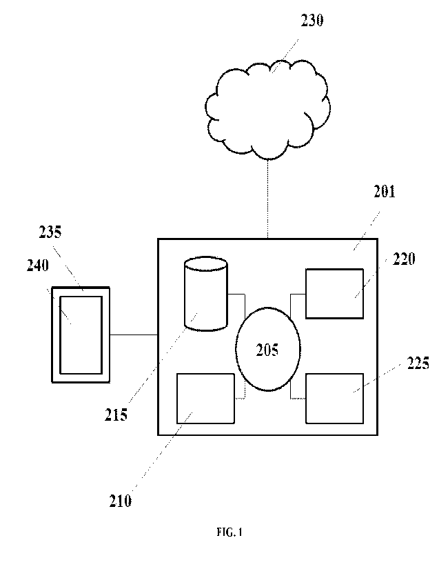
[0009] FIG. 1 schematically illustrates a computer system that may be
programmed or otherwise
configured to implement methods of the present disclosure.
DETAILED DESCRIPTION
[0010] While various embodiments of the invention have been shown and
described herein, it will be
obvious to those skilled in the art that such embodiments are provided by way
of example only.
Numerous variations, changes, and substitutions may occur to those skilled in
the art without departing
from the invention. It should be understood that various alternatives to the
embodiments of the invention
described herein may be employed.
[0011] As used herein the term "about" or "approximately" means within an
acceptable error range for
the particular value as determined by one of ordinary skill in the art, which
may depend in part on how the
value is measured or determined, i.e., the limitations of the measurement
system. For example, "about"
can mean within 1 or more than 1 standard deviation, per the practice in the
art. As another example,
"about" can mean a range of up to 20%, up to 10%, up to 5%, or up to 1% of a
given value. With respect
to biological systems or processes, the term "about" can mean within an order
of magnitude, such as
within 5-fold or within 2-fold of a value. Where particular values are
described in the application and
claims, unless otherwise stated, the term "about" means within an acceptable
error range for the particular
value.
[0012] As used herein, the term "polynucleotide" generally refers to
a polymeric form of nucleotides of
any length, either deoxyribonucleotides (DNA) or ribonucleotides (RNA), or
analogs thereof A
polynucleotide may be a nucleic acid molecule. A polynucleotide (or
oligonucleotide) may have a
nucleotide or nucleic acid sequence. Polynucleotides may have any three-
dimensional structure, and may
perform any function. The following are non-limiting examples of
polynucleotides: cell-free nucleic
acids, cell-free DNA (cfDNA), cell-free RNA (cfRNA), circulating tumor DNA
(ctDNA), circulating
tumor RNA (ctRNA), coding or non-coding regions of a gene or gene fragment,
loci (locus) defined from
linkage analysis, exons, introns, messenger RNA (mRNA), transfer RNA (tRNA),
ribosomal RNA
CA 03189709 2023- 2- 15
WO 2022/040147
PCT/US2021/046246
(rRNA), short interfering RNA (siRNA), short-hairpin RNA (shRNA), micro-RNA
(miRNA), ribozymes,
cDNA, recombinant polynucleotides, branched polynucleotides, plasmids,
vectors, isolated DNA of any
sequence, isolated RNA of any sequence, nucleic acid probes, and primers. A
polynucleotide may
comprise one or more modified nucleotides, such as methylated nucleotides and
nucleotide analogs. If
present, modifications to the nucleotide structure may be imparted before or
after assembly of the
polymer. The sequence of nucleotides may be interrupted by non-nucleotide
components. A
polynucleotide may be further modified after polymerization, such as by
conjugation with a labeling
component.
[0013] The term "subject," as used herein, generally refers to a
vertebrate, such as a mammal (e.g., a
human). Mammals include, but are not limited to, murincs, simians, humans,
farm animals, sport animals,
and pets (e.g., a dog or a cat). Tissues, cells, and their progeny of a
biological entity obtained in vivo or
cultured in vitro are also encompassed. The subject may be a patient. The
subject may be symptomatic
with respect to a disease (e.g., cancer). Alternatively, the subject may be
asymptomatic with respect to the
disease.
[0014] The term "biological sample," as used herein, generally
refers to a sample derived from or
obtained from a subject, such as a mammal (e.g., a human). Biological samples
may include, but are not
limited to, hair, finger nails, skin, sweat, tears, ocular fluids, nasal swab
or nasopharyngeal wash, sputum,
throat swab, saliva, mucus, blood, serum, plasma, placental fluid, amniotic
fluid, cord blood, emphatic
fluids, cavity fluids, earwax, oil, glandular secretions, bile, lymph, pus,
microbiota, meconium, breast
milk, bone marrow, bone, CNS tissue, cerebrospinal fluid, adipose tissue,
synovial fluid, stool, gastric
fluid, urine, semen, vaginal secretions, stomach, small intestine, large
intestine, rectum, pancreas, liver,
kidney, bladder, lung, and other tissues and fluids derived from or obtained
from a subject. The biological
sample may be a cell-free (or cell free) biological sample.
[0015] The term cell-free biological sample," as used herein,
generally refers to a sample derived
from or obtained from a subject that is free from cells. A cell-free
biological sample may include one or
more cell-free molecules, such as cell-free DNA, cell-free RNA, or a cell-free
polypeptide (e.g., protein).
Cell-free biological samples may include, but are not limited to, blood,
serum, plasma, nasal swab or
nasopharyngeal wash, saliva, urine, gastric fluid, tears, stool, mucus, sweat,
earwax, oil, glandular
secretion, bile, lymph, cerebrospinal fluid, tissue, semen, vaginal fluid,
interstitial fluids, including
interstitial fluids derived from tumor tissue, ocular fluids, spinal fluid,
throat swab, breath, hair, finger
nails, skin, biopsy, placental fluid, amniotic fluid, cord blood, emphatic
fluids, cavity fluids, sputum, pus,
microbiota, meconium, breast milk and/or other excretions.
[0016] As used herein, the term "binding agent" generally refers to
a material that binds to a nucleic
acid. A binding agent may include but is not limited to one or more of an
antibody or fragment thereof, a
polypeptide, a streptavidin, a nucleic acid, a DNA probe, or an RNA probe.
[0017] The terms "early stage cancer" and "non-metastatic cancer,"
as used herein, generally refer to a
cancer that may not have metastasized in a subject (i.e., the cancer has not
left its initial location to spread
6
CA 03189709 2023- 2- 15
WO 2022/040147
PCT/US2021/046246
to other locations). An early stage cancer may metastasize in a subject.
Alternatively, an early stage
cancer may not metastasize in a subject. The exact staging may depend upon the
type of cancer.
[0018] The tennis "tumor burden" and "tumor load," as used herein,
generally refer to the size of a
tumor or the amount of cancer in the body of the subject.
[0019] Whenever the term -at least," -greater than," or -greater
than or equal to- precedes the first
numerical value in a series of two or more numerical values, the term "at
least," "greater than" or "greater
than or equal to" applies to each of the numerical values in that series of
numerical values. For example,
greater than or equal to I, 2, or 3 is equivalent to greater than or equal to
1, greater than or equal to 2, or
greater than or equal to 3.
[0020] Whenever the term "no more than," "less than," or "less than or equal
to" precedes the first
numerical value in a series of two or more numerical values, the term "no more
than," "less than," or "less
than or equal to- applies to each of the numerical values in that series of
numerical values. For example,
less than or equal to 3, 2, or 1 is equivalent to less than or equal to 3,
less than or equal to 2, or less than or
equal to 1.
[0021] Provided herein are methods, systems, and compositions for analyzing
nucleic acids from a cell-
free biological sample comprising selection of at least a subset of nucleic
acids in the cell-free biological
sample. Some such methods can comprise immunoprecipitation of nucleic acids,
such as methylated
nucleic acids or nucleic acids bound to a protein, such as a nucleic acid
binding protein (e.g., a
transcription factor) using binding agent, such as an antibody or fragment
thereof and isolating bound
nucleic acids for further analysis, such as sequence or size analysis.
Methods for Selective Nucleic Acid Analysis
[0022] Provided herein are methods for selective nucleic acid analysis. In
some cases, methods for
processing a plurality of nucleic acid molecules derived from a cell-free
biological sample comprise
bringing the plurality of nucleic acid molecules in contact with a plurality
of binding agents, such as
antibodies or fragments thereof. This provides a first subset of the plurality
of nucleic acid molecules
coupled to the plurality of antibodies or fragments thereof and a second
subset of the plurality of nucleic
acid molecules. Next the first subset of the plurality of nucleic acid
molecules coupled to the plurality of
antibodies or fragments thereof can be separated from the second subset of the
plurality of nucleic acid
molecules. Next, a nucleic acid molecule derived from the first subset of the
plurality of nucleic acid
molecules can be circularized to obtain a circularized nucleic acid molecule.
Then, the method involves
identifying the circularized nucleic acid molecule or derivative thereof.
10023] In aspects of methods for selective nucleic acid analysis provided
herein, in some cases, the
plurality of nucleic acid molecules comprises a deoxyribonucleic acid (DNA)
molecule. In some cases,
the plurality of nucleic acid molecules comprises a ribonucleic acid (RNA)
molecule. In some cases, the
plurality of nucleic acid molecules comprises a mixture of RNA molecules and
DNA molecules.
[0024] In aspects of methods for selective nucleic acid analysis provided
herein, in some cases,
circularizing comprises ligating a 5' end and a 3' end of the nucleic acid
molecule to one another. In
7
CA 03189709 2023- 2- 15
WO 2022/040147
PCT/US2021/046246
some cases, circularizing comprises coupling an adapter to a 3' end, a 5' end,
or both a 5' end and a 3'
end of the nucleic acid molecule.
[0025] In aspects of methods for selective nucleic acid analysis provided
herein, in some cases, methods
further comprise subjecting the circularized nucleic acid molecule to nucleic
acid amplification to
generate a plurality of amplification products of the circularized nucleic
acid molecule, wherein the
identification step comprises identifying the plurality of nucleic acid
amplification products. In some
cases, nucleic acid amplification is effected by a polymerase having strand-
displacement activity. In some
cases, nucleic acid amplification is effected by a polymerase that does not
have strand-displacement
activity. In some cases, nucleic acid amplification comprises contacting the
circularized nucleic acid
molecule to an amplification reaction mixture comprising random primers. In
some cases, nucleic acid
amplification comprises contacting the circularized nucleic acid molecule to
an amplification reaction
mixture comprising one or more primers, each of which specifically hybridizes
to a different target
sequence via sequence complementarity. In some cases, nucleic acid
amplification comprises contacting
the circularized nucleic acid molecule to an amplification mixture comprising
one or more random
primers.
[0026] In aspects of methods for selective nucleic acid analysis provided
herein, in some cases, the
binding agent, such as an antibody, or fragment thereof specifically binds to
a nucleic acid. In some
cases, the antibody, or fragment thereof specifically binds to a nucleic acid
binding protein. In some
cases, the nucleic acid binding protein is a chromatin protein. In some cases,
the nucleic acid binding
protein is a histone. In some cases, the nucleic acid binding protein is a
methyl CpG binding protein. In
some cases, the nucleic acid binding protein is a transcription factor. In
some cases, the nucleic acid
binding protein is a polymerase. In some cases, the nucleic acid binding
protein is a nuclease. In some
cases, the nucleic acid binding protein comprises a zinc finger motif, a helix-
turn-helix motif, or a leucine
zipper motif. In some cases, the nucleic acid binding protein is an RNA
binding protein. In some cases,
the nucleic acid binding protein is a splicing protein or RNA transport
protein. In some cases, the
antibody, or fragment thereof specifically binds to a nucleic acid sequence.
In some cases, the antibody,
or fragment thereof specifically binds to a methylated nucleic acid sequence.
In some cases, the antibody,
or fragment thereof specifically binds to a phosphorylated nucleic acid
sequence. In some cases, the
antibody, or fragment thereof specifically binds to a acetylated nucleic acid
sequence. In some cases, the
nucleic acid molecule is coupled to an antibody or fragment thereof of the
plurality of antibodies or
fragments thereof.
10027] In aspects of methods for selective nucleic acid analysis provided
herein, in some cases, methods
further comprise determining a size of each cell-free nucleic acid molecule of
the plurality of cell-free
nucleic acid molecules. In some cases, determining a size of each cell-free
nucleic acid molecule
comprises sequencing, electrophoresis, or a combination thereof.
[0028] In aspects of methods for selective nucleic acid analysis provided
herein, in some cases,
identifying the circularized nucleic acid comprises sequencing the
circularized nucleic acid molecule or
derivative thereof. In some cases, sequencing comprises a method selected from
one or more of
8
CA 03189709 2023- 2- 15
WO 2022/040147
PCT/US2021/046246
sequencing by synthesis, sequencing by ligation, nanopore sequencing, nanoball
sequencing, ion
detection, sequencing by hybridization, polymerized colony (POLONY)
sequencing, nanogrid rolling
circle sequencing (ROLONY), and ion torrent sequencing.
[0029] In aspects of methods for selective nucleic acid analysis provided
herein, in some cases, the cell-
free biological sample comprises less than 100 nanograms of nucleic acids. In
some cases, the cell-free
biological sample comprises less than 90 nanograms of nucleic acids. In some
cases, the cell-free
biological sample comprises less than 80 nanograms of nucleic acids. In some
cases, the cell-free
biological sample comprises less than 75 nanograms of nucleic acids. In some
cases, the cell-free
biological sample comprises less than 70 nanograms of nucleic acids. In some
cases, the cell-free
biological sample comprises less than 60 nanograms of nucleic acids. In some
cases, the cell-free
biological sample comprises less than 50 nanograms of nucleic acids.
10030] In aspects of methods for selective nucleic acid analysis provided
herein, in some cases, the cell-
free biological sample comprises a bodily fluid. In some cases, the bodily
fluid is urine, saliva, blood,
serum, plasma, tears, sputum, cerebrospinal fluid, synovial fluid, mucus,
bile, semen, lymph, amniotic
fluid, menstrual fluid, or combinations thereof.
Methods for determining whether a subiect has or is at risk of having a
disease
10031] Provided herein are methods of determining whether a subject has or is
at risk of having a disease
comprising selective nucleic analysis herein. In some cases, some such methods
comprise bringing the
plurality of nucleic acid molecules in contact with a plurality of binding
agents, such as antibodies or
fragments thereof, to provide a first subset of the plurality of nucleic acid
molecules coupled to the
plurality of antibodies or fragments thereof and a second subset of the
plurality of nucleic acid molecules.
Next, in some cases, methods comprise separating the first subset of the
plurality of nucleic acid
molecules coupled to the plurality of antibodies or fragments thereof from the
second subset of the
plurality of nucleic acid molecules. Next, the method comprises circularizing
a nucleic acid molecule
derived from the first subset of the plurality of nucleic acid molecules to
obtain a circularized nucleic acid
molecule. Then, the method comprises identifying a sequence of the
circularized nucleic acid molecule or
derivative thereof and processing the sequence against a plurality of
reference sequences to identify the
sequence as corresponding to at least a subset of the plurality of reference
sequences, thereby determining
that a subject has or is at risk of having a disease. In some cases, the
plurality of nucleic acid molecules
comprise a deoxyribonucleic acid (DNA) molecule or a ribonucleic acid (RNA)
molecule. In some cases,
the nucleic acid molecule is coupled to an antibody or fragment thereof of the
plurality of antibodies or
fragments thereof
[0032] In aspects of methods for selective nucleic acid analysis provided
herein, in some cases,
circularizing comprises ligating a 5' end and a 3' end of the nucleic acid
molecule to one another. In
some cases, circularizing comprises coupling an adapter to a 3' end, a 5' end,
or both a 5' end and a 3'
end of the nucleic acid molecule.
10033] In aspects of methods for diagnosis provided herein, in some cases,
methods further comprise
subjecting the circularized nucleic acid molecule to nucleic acid
amplification to generate a plurality of
9
CA 03189709 2023- 2- 15
WO 2022/040147
PCT/US2021/046246
amplification products of the circularized nucleic acid molecule, wherein the
identification step comprises
identifying the plurality of nucleic acid amplification products. In some
cases, nucleic acid amplification
is effected by a polymerase having strand-displacement activity. In some
cases, nucleic acid amplification
is effected by a polymerase that does not have strand-displacement activity.
In some cases, nucleic acid
amplification comprises contacting the circularized nucleic acid molecule to
an amplification reaction
mixture comprising random primers. In some cases, nucleic acid amplification
comprises contacting the
circularized nucleic acid molecule to an amplification reaction mixture
comprising one or more primers,
each of which specifically hybridizes to a different target sequence via
sequence complementarity. In
some cases, nucleic acid amplification comprises contacting the circularized
nucleic acid molecule to an
amplification mixture comprising one or more random primers.
[0034] In aspects of methods for diagnosis provided herein, in some cases, the
binding agent, such as an
antibody, or fragment thereof specifically binds to a nucleic acid. In some
cases, the antibody, or
fragment thereof specifically binds to a nucleic acid binding protein. In some
cases, the nucleic acid
binding protein is a chromatin protein. In some cases, the nucleic acid
binding protein is a histone. In
some cases, the nucleic acid binding protein is a methyl CpG binding protein.
In some cases, the nucleic
acid binding protein is a transcription factor. In some cases, the nucleic
acid binding protein is a
polymerase. In some cases, the nucleic acid binding protein is a nuclease. In
some cases, the nucleic acid
binding protein comprises a zinc finger motif, a helix-turn-helix motif, or a
leucine zipper motif In some
cases, the nucleic acid binding protein is an RNA binding protein. In some
cases, the nucleic acid binding
protein is a splicing protein or RNA transport protein. In some cases, the
antibody, or fragment thereof
specifically binds to a nucleic acid sequence. In some cases, the antibody, or
fragment thereof specifically
binds to a methylated nucleic acid sequence. In some cases, the antibody, or
fragment thereof specifically
binds to a phosphorylated nucleic acid sequence. In some cases, the antibody,
or fragment thereof
specifically binds to a acetylated nucleic acid sequence. In some cases, the
nucleic acid molecule is
coupled to an antibody or fragment thereof of the plurality of antibodies or
fragments thereof.
[0035] In aspects of methods for diagnosis provided herein, in some cases,
methods further comprise
determining a size of each cell-free nucleic acid molecule of the plurality of
cell-free nucleic acid
molecules. In some cases, determining a size of each cell-free nucleic acid
molecule comprises
sequencing, electrophoresis, or a combination thereof
[0036] In aspects of methods for diagnosis provided herein, in some cases,
identifying the circularized
nucleic acid comprises sequencing the circularized nucleic acid molecule or
derivative thereof. In some
cases, sequencing comprises a method selected from one or more of sequencing
by synthesis, sequencing
by ligation, nanopore sequencing, nanoball sequencing, ion detection,
sequencing by hybridization,
polymerized colony (POLONY) sequencing, nanogrid rolling circle sequencing
(ROLONY), and ion
torrent sequencing.
[0037] In aspects of methods for diagnosis provided herein, in some cases, the
cell-free biological sample
comprises less than 100 nanograms of nucleic acids. In some cases, the cell-
free biological sample
comprises less than 90 nanograms of nucleic acids. In some cases, the cell-
free biological sample
CA 03189709 2023- 2- 15
WO 2022/040147
PCT/US2021/046246
comprises less than 80 nanograms of nucleic acids. In some cases, the cell-
free biological sample
comprises less than 75 nanograms of nucleic acids. In some cases, the cell-
free biological sample
comprises less than 70 nanograms of nucleic acids. In sonic cases, the cell-
free biological sample
comprises less than 60 nanograms of nucleic acids. In some cases, the cell-
free biological sample
comprises less than 50 nanograms of nucleic acids.
[0038] In aspects of methods for diagnosis provided herein, in some cases, the
cell-free biological sample
comprises a bodily fluid. In some cases, the bodily fluid is urine, saliva,
blood, serum, plasma, tears,
sputum, cerebrospinal fluid, synovial fluid, mucus, bile, semen, lymph,
amniotic fluid, menstrual fluid, or
combinations thereof.
[0039] In aspects of methods for diagnosis provided herein, in some cases the
disease is cancer. In some
cases, the cancer is selected from the group consisting of colon cancer, non-
small cell lung cancer, small
cell lung cancer, breast cancer, hepatocellular carcinoma, liver cancer, skin
cancer, malignant melanoma,
endometrial cancer, esophageal cancer, gastric cancer, ovarian cancer,
pancreatic cancer, brain cancer,
leukemia, lymphoma, and myeloma.
[0040] In aspects of methods for diagnosis provided herein, in some cases the
method further comprise
using the sequence identified to output an electronic report indicating that
the subject has or is at risk of
having a disease. In some cases, the method further comprises using the
sequence identified to provide a
therapeutic intervention to the subject for a disease. In some cases, the
method further comprises using
the sequence identified to treat the subject for the disease.
[0041] In aspects of methods for diagnosis provided herein, in some cases the
subject is treated by
administering a chemotherapy or immunotherapy to the subject.
[0042] In aspects of methods for diagnosis provided herein, in some cases the
method further comprises
using the sequence identified to monitor the subject for a progression or
regression of the subject.
[0043] Further provided herein are methods of determining whether a subject
has or is at risk of having a
disease comprising selective nucleic analysis herein. In some cases, methods
comprise determining a
methylation state for a nucleic acid molecule of the plurality of nucleic acid
molecules and determining a
size for the nucleic acid molecule of the plurality of nucleic acid molecules.
Then, the method comprises
processing the methylation state for the nucleic acid molecule of the
plurality of nucleic acid molecules
against a first database and processing the size for the nucleic acid molecule
of the plurality of nucleic
acid molecules against a second database. Next, the method comprises
identifying an association of the
methylation state and of the size with at least a disease, thereby determining
that a subject has or is at risk
of having a disease.
[0044] In aspects of methods for diagnosis provided herein, in some cases, the
disease is cancer. In some
cases, the cancer is selected from the group consisting of colon cancer, non-
small cell lung cancer, small
cell lung cancer, breast cancer, hepatocellular carcinoma, liver cancer, skin
cancer, malignant melanoma,
endometrial cancer, esophageal cancer, gastric cancer, ovarian cancer,
pancreatic cancer, brain cancer,
leukemia, lymphoma, and myeloma.
11
CA 03189709 2023- 2- 15
WO 2022/040147
PCT/US2021/046246
[0045] In aspects of methods for diagnosis provided herein, in some cases, the
method comprises using
the methylation state and the size identified to output an electronic report
indicating that the subject has or
is at risk of having a disease. In some cases, the method further comprises
using the methylation state and
the size identified to provide a therapeutic intervention to the subject for a
disease. In some cases, the
method further comprises using the methylation state and the size identified
to treat the subject for the
disease. In some cases, the subject is treated by administering a chemotherapy
or immunotherapy to the
subject. In some cases, the method further comprises using the methylation
state and the size identified to
monitor the subject for a progression or regression of the subject.
Methods for Methylation and Size Nucleic Acid Analysis
[0046] Provided herein arc methods for nucleic acid analysis using methylation
state and size. In some
cases, methods comprise determining a methylation state for a nucleic acid
molecule of the plurality of
nucleic acid molecules and determining a size for the nucleic acid molecule of
the plurality of nucleic acid
molecules. Next, the method comprises processing the methylation state for the
nucleic acid molecule of
the plurality of nucleic acid molecules against a first database and
processing the size for the nucleic acid
molecule of the plurality of nucleic acid molecules against a second database.
Then, the method comprise
identifying an association of the methylation state of the nucleic acid
molecule and of the size of the
nucleic acid molecule with at least a disease.
[0047] In aspects of methods for nucleic acid analysis herein, in some cases,
determining the methylation
state comprises sequencing the nucleic acid molecule of the plurality of
nucleic acid molecules. In some
cases, sequencing comprises a method selected from one or more of sequencing
by synthesis, sequencing
by ligation, nanopore sequencing, nanoball sequencing, ion detection,
sequencing by hybridization,
polymerized colony (POLONY) sequencing, nanogrid rolling circle sequencing
(ROLONY), and ion
torrent sequencing. In some cases, determining the methylation state comprises
bisulfite sequencing.
[0048] In aspects of methods for nucleic acid analysis herein, in some cases,
determining the methylation
state comprises contacting the nucleic acid molecule to an antibody or
fragment thereof that binds
specifically to methylated nucleic acids. In some cases, the antibody or
fragment thereof binds
specifically to methylated nucleic acid binding proteins or fragments thereof.
[0049] In aspects of methods for nucleic acid analysis herein, in some cases,
determining the methylation
state comprises bringing the plurality of nucleic acid molecules in contact
with a plurality of antibodies or
fragments thereof, to provide a first subset of the plurality of nucleic acid
molecules coupled to the
plurality of antibodies or fragments thereof and a second subset of the
plurality of nucleic acid molecules.
Next, the method comprises separating the first subset of the plurality of
nucleic acid molecules coupled
to the plurality of antibodies or fragments thereof from the second subset of
the plurality of nucleic acid
molecules. Then, the method comprises circularizing a nucleic acid molecule
derived from the first subset
of the plurality of nucleic acid molecules to obtain a circularized nucleic
acid molecule and identifying the
circularized nucleic acid molecule or derivative thereof.
10050] In aspects of methods for nucleic acid analysis herein, in some cases,
the plurality of nucleic acid
molecules comprises a deoxyribonucleic acid (DNA) molecule. In some cases, the
plurality of nucleic
12
CA 03189709 2023- 2- 15
WO 2022/040147
PCT/US2021/046246
acid molecules comprises a ribonucleic acid (RNA) molecule. In some cases, the
plurality of nucleic acid
molecules comprises a mixture of RNA molecules and DNA molecules.
[0051] In aspects of methods for nucleic acid analysis herein, in some cases,
circularizing comprises
ligating a 5' end and a 3' end of the nucleic acid molecule to one another. In
some cases, circularizing
comprises coupling an adapter to a 3' end, a 5' end, or both a 5' end and a 3'
end of the nucleic acid
molecule.
[0052] In aspects of methods for nucleic acid analysis herein, in some cases,
methods further comprise
subjecting the circularized nucleic acid molecule to nucleic acid
amplification to generate a plurality of
amplification products of the circularized nucleic acid molecule, wherein the
identification step comprises
identifying the plurality of nucleic acid amplification products. In some
cases, nucleic acid amplification
is effected by a polymerase having strand-displacement activity. In some
cases, nucleic acid amplification
is effected by a polymerase that does not have strand-displacement activity.
In some cases, nucleic acid
amplification comprises contacting the circularized nucleic acid molecule to
an amplification reaction
mixture comprising random primers. In some cases, nucleic acid amplification
comprises contacting the
circularized nucleic acid molecule to an amplification reaction mixture
comprising one or more primers,
each of which specifically hybridizes to a different target sequence via
sequence complementarity. In
some cases, nucleic acid amplification comprises contacting the circularized
nucleic acid molecule to an
amplification mixture comprising one or more random primers.
[0053] In aspects of methods for nucleic acid analysis herein, in some cases,
the antibody. or fragment
thereof specifically binds to a nucleic acid. In some cases, the antibody, or
fragment thereof specifically
binds to a nucleic acid binding protein. In some cases, the nucleic acid
binding protein is a chromatin
protein. In some cases, the nucleic acid binding protein is a histone. In some
cases, the nucleic acid
binding protein is a methyl CpG binding protein. In some cases, the nucleic
acid binding protein is a
transcription factor. In some cases, the nucleic acid binding protein is an
RNA binding protein. In some
cases, the antibody, or fragment thereof specifically binds to a nucleic acid
sequence. In some cases, the
antibody, or fragment thereof specifically binds to a methylated nucleic acid
sequence. In some cases, the
nucleic acid molecule is coupled to an antibody or fragment thereof of the
plurality of antibodies or
fragments thereof
[0054] In aspects of methods for nucleic acid analysis herein, in some cases,
methods further comprise
determining a size of each cell-free nucleic acid molecule of the plurality of
cell-free nucleic acid
molecules.
10055] In aspects of methods for nucleic acid analysis herein, in some cases,
identifying the circularized
nucleic acid comprises sequencing the circularized nucleic acid molecule or
derivative thereof. In some
cases, sequencing comprises a method selected from one or more of sequencing
by synthesis, sequencing
by ligation, nanopore sequencing, nanoball sequencing, ion detection,
sequencing by hybridization,
polymerized colony (POLONY) sequencing, nanogrid rolling circle sequencing
(ROLONY), and ion
torrent sequencing.
13
CA 03189709 2023- 2- 15
WO 2022/040147
PCT/US2021/046246
[0056] In aspects of methods for nucleic acid analysis herein, in some cases,
the cell-free biological
sample comprises less than 100 nanograms of nucleic acids. In some cases, the
cell-free biological
sample comprises less than 90 nanograms of nucleic acids. In some cases, the
cell-free biological sample
comprises less than 80 nanograms of nucleic acids. In some cases, the cell-
free biological sample
comprises less than 75 nanograms of nucleic acids. In some cases, the cell-
free biological sample
comprises less than 70 nanograms of nucleic acids. In some cases, the cell-
free biological sample
comprises less than 60 nanograms of nucleic acids. In some cases, the cell-
free biological sample
comprises less than 50 nanograms of nucleic acids.
[0057] In aspects of methods for nucleic acid analysis herein, in some cases,
the cell-free biological
sample comprises a bodily fluid. In some cases, the bodily fluid is urine,
saliva, blood, scrum, plasma,
tears, sputum, cerebrospinal fluid, synovial fluid, mucus, bile, semen, lymph,
amniotic fluid, menstrual
fluid, or combinations thereof
Systems and Computer Assisted Methods
[0058] Provided herein are systems for selective nucleic acid analysis methods
provided herein. In some
cases, systems herein comprises a computer configured to receive a user
request for processing a plurality
of nucleic acid molecules derived from a cell-free biological sample and a
processing unit for bringing the
plurality of nucleic acid molecules in contact with a plurality of binding
agents, such as antibodies or
fragments thereof to obtain a first subset of the plurality of nucleic acid
molecules coupled to the plurality
of antibodies or fragments thereof and a second subset of the plurality of
nucleic acid molecules. In some
cases, the system further comprises an isolation unit for separating the first
subset of the plurality of
nucleic acid molecules coupled to the plurality of antibodies or fragments
thereof from the second subset
of the plurality of nucleic acid molecules. In some cases, the system further
comprises a circularization
unit for circularizing a nucleic acid molecule derived from the first subset
of the plurality of nucleic acid
molecules to obtain a circularized nucleic acid molecule. In some cases, the
system further comprises an
identification unit for identifying the circularized nucleic acid molecule or
derivative thereof In some
cases the system additionally comprises a report generator that sends a report
to a recipient containing the
identity of the circularized nucleic acid molecule or derivative thereof.
[0059] A computer for use in the system can comprise one or more processors.
Processors may be
associated with one or more controllers, calculation units, and/or other units
of a computer system, or
implanted in firmware as desired. If implemented in software, the routines may
be stored in any computer
readable memory such as in RAM, ROM, flash memory, a magnetic disk, a laser
disk, or other suitable
storage medium. Likewise, this software may be delivered to a computing device
via any known delivery
method including, for example, over a communication channel such as a
telephone line, the internet, a
wireless connection, etc., or via a transportable medium, such as a computer
readable disk, flash drive,
etc. The various steps may be implemented as various blocks, operations,
tools, modules and techniques
which, in turn, may be implemented in hardware, firmware, software, or any
combination of hardware,
firmware, and/or software. When implemented in hardware, some or all of the
blocks, operations,
techniques, etc. may be implemented in, for example, a custom integrated
circuit (IC), an application
14
CA 03189709 2023- 2- 15
WO 2022/040147
PCT/US2021/046246
specific integrated circuit (ASIC), a field programmable logic array (FPGA), a
programmable logic array
(PLA), etc. A client-server, relational database architecture can be used in
embodiments of the system. A
client-server architecture is a -network architecture in which each computer
or process on the network is
either a client or a server. Server computers are typically powerful computers
dedicated to managing disk
drives (file servers), printers (print servers), or network traffic (network
servers). Client computers
include PCs (personal computers) or workstations on which users run
applications, as well as example
output devices as disclosed herein. Client computers rely on server computers
for resources, such as files,
devices, and even processing power. in some embodiments, the server computer
handles all of the
database functionality. The client computer can have software that handles all
the front-end data
management and can also receive data input from users.
[0060] The system can be configured to receive a user request to
perform a detection reaction on a
sample. The user request may be direct or indirect. Examples of direct request
include those transmitted
by way of an input device, such as a keyboard, mouse, or touch screen.
Examples of indirect requests
include transmission via a communication medium, such as over the internet
(either wired or wireless).
[0061] The system can further comprise an amplification system that
performs a nucleic acid
amplification reaction on the sample or a portion thereof in response to the
user request. A variety of
methods of amplifying polynucleotides (e.g. DNA and/or RNA) are available.
Amplification may be
linear, exponential, or involve both linear and exponential phases in a multi-
phase amplification process.
Amplification methods may involve changes in temperature, such as a heat
denaturation step, or may be
isothermal processes that do not require heat denaturation. Non-limiting
examples of suitable
amplification processes are described herein, such as with regard to any of
the various aspects of the
disclosure. In some embodiments, amplification comprises rolling circle
amplification (RCA). A variety
of systems for amplifying polynucleotides are available, and may vary based on
the type of amplification
reaction to be performed. For example, for amplification methods that comprise
cycles of temperature
changes, the amplification system may comprise a thermocycler. An
amplification system can comprise a
real-time amplification and detection instrument, such as systems manufactured
by Applied Biosystems,
Roche, and Stratagene. In some embodiments, the amplification reaction
comprises the steps of (i)
circularizing individual polynucleotides to form a plurality of circular
polynucleotides, each of which
having a junction between the 5' end and 3' end; and (ii) amplifying the
circular polynucleotides.
Samples, polynucleotides, primers, polymerases, and other reagents can be any
of those described herein,
such as with regard to any of the various aspects. Non-limiting examples of
circularization processes (e.g.
with and without adapter oligonucleotides), reagents (e.g. types of adapters,
use of ligases), reaction
conditions (e.g. favoring self-joining), optional additional processing (e.g.
post-reaction purification), and
the junctions formed thereby are provided herein, such as with regard to any
of the various aspects of the
disclosure. Systems can be selected and or designed to execute any such
methods.
[0062] Systems may further comprise a sequencing system that
generates sequencing reads for
polynucleotides amplified by the amplification system, identifies sequence
differences between
sequencing reads and a reference sequence, and calls a sequence difference
that occurs in at least two
CA 03189709 2023- 2- 15
WO 2022/040147
PCT/US2021/046246
circular polynucleotides having different junctions as the sequence variant.
The sequencing system and
the amplification system may be the same, or comprise overlapping equipment.
For example, both the
amplification system and sequencing system may utilize the same thennocycler.
A variety of sequencing
platforms for use in the system are available, and may be selected based on
the selected sequencing
method. Examples of sequencing methods are described herein. Amplification and
sequencing may
involve the use of liquid handlers. Several commercially available liquid
handling systems can be utilized
to run the automation of these processes (see for example liquid handlers from
Perkin-Elmer, Beckman
Coulter, Caliper Life Sciences, Tecan, Eppendorf, Apricot Design, Velocity 11
as examples). A variety of
automated sequencing machines are commercially available, and include
sequencers manufactured by
Life Technologies (SOLiD platform, and pH-based detection), Roche (454
platform), Illumina (e.g. flow
cell based systems, such as Genome Analyzer devices). Transfer between 2, 3,
4, 5, or more automated
devices (e.g. between one or more of a liquid handler and a sequencing device)
may be manual or
automated.
[0063] The system can further comprise a report generator that sends
a report to a recipient, wherein
the report contains results for detection of the sequence variant. A report
may be generated in real-time,
such as during processing or while data is being analyzed, with periodic
updates as the process progresses.
In addition, or alternatively, a report may be generated at the conclusion of
the analysis. The report may
be generated automatically, such when the system completes the step of
identifying the circular nucleic
acids. In some embodiments, the report is generated in response to
instructions from a user. In addition
to the results of identifying the nucleic acid, a report may also contain an
analysis based on the identified
nucleic acids. For example, where one or more sequence variants are associated
with a particular
contaminant or phenotype, the report may include information concerning this
association, such as a
likelihood that the contaminant or phenotype is present, at what level, and
optionally a suggestion based
on this information (e.g. additional tests, monitoring, or remedial measures).
The report can take any of a
variety of forms. It is envisioned that data relating to the present
disclosure can be transmitted over such
networks or connections (or any other suitable approach for transmitting
information, including but not
limited to mailing a physical report, such as a print-out) for reception
and/or for review by a receiver. The
receiver can be but is not limited to an individual, or electronic system
(e.g. one or more computers,
and/or one or more servers).
[0064] A machine readable medium comprising computer-executable code may take
many forms,
including but not limited to, a tangible storage medium, a carrier wave medium
or physical transmission
medium. Non-volatile storage media include, for example, optical or magnetic
disks, such as any of the
storage devices in any computers) or the like, such as may be used to
implement the databases, etc.
Volatile storage media include dynamic memory, such as main memory of such a
computer platform.
Tangible transmission media include coaxial cables; copper wire and fiber
optics, including the wires that
comprise a bus within a computer system. Carrier-wave transmission media may
take the form of electric
or electromagnetic signals, or acoustic or light waves such as those generated
during radio frequency (RF)
and infrared (IR) data communications. Common forms of computer-readable media
therefore include for
16
CA 03189709 2023- 2- 15
WO 2022/040147
PCT/US2021/046246
example: a floppy disk, a flexible disk, hard disk, magnetic tape, any other
magnetic medium, a CD-
ROM, DVD or DVD-ROM, any other optical medium, punch cards paper tape, any
other physical storage
medium with patterns of holes, a RAM, a ROM, a PROM and EPROM, a FLASH-EPROM,
any other
memory chip or cartridge, a carrier wave transporting data or instructions,
cables or links transporting
such a carrier wave, or any other medium from which a computer may read
programming code and/or
data. Many of these forms of computer readable media may be involved in
carrying one or more
sequences of one or more instructions to a processor for execution.
[0065] The subject computer-executable code can be executed on ally
suitable device comprising a
processor, including a server, a PC, or a mobile device such as a smartphone
or tablet. Any controller or
computer optionally includes a monitor, which can be a cathode ray tube
("CRT") display, a flat panel
display (e.g., active matrix liquid crystal display, liquid crystal display,
etc.), or others. Computer
circuitry is often placed in a box, which includes numerous integrated circuit
chips, such as a
microprocessor, memory, interface circuits, and others. The box also
optionally includes a hard disk
drive, a floppy disk drive, a high capacity removable drive such as a
writeable CD-ROM, and other
common peripheral elements. Inputting devices such as a keyboard, mouse, or
touch -sensitive screen,
optionally provide for input from a user. The computer can include appropriate
software for receiving
user instructions, either in the form of user input into a set of parameter
fields, e.g., in a GUI, or in the
form of preprogrammed instructions, e.g., preprogrammed for a variety of
different specific operations.
Methods of Library Preparation and Amplification
[0066] Methods herein comprise, in certain cases, amplification of
polynucleotides present in a sample
from a subject. Methods of amplification used herein often comprise rolling-
circle amplification.
Alternatively or in combination, methods of amplification used herein comprise
PCR. In some cases,
methods of amplification herein comprise linear amplification. Often
amplification is not targeted to one
gene or set of genes and the entire nucleic acid sample is amplified. In some
cases, the method comprises
(a) circularizing individual polynucleotides of the plurality to form a
plurality of circular polynucleotides,
each of which having a junction between the 5' end and the 3' end; and (b)
amplifying the circular
polynucleotides of (a) to produce amplified polynucleotides. In additional
cases, methods of
amplification comprise (c) shearing the amplified polynucleotides to produce
sheared polynucleotides,
each sheared polynucleotide comprising one or more shear points at a 5' end
and/or 3' end. In some
cases, the method does not comprise enriching for a target sequence.
[0067] In general, joining ends of a polynucleotide to one-another
to form a circular polynucleotide
(either directly, or with one or more intermediate adapter oligonucleotides)
produces a junction having a
junction sequence. Where the 5' end and 3' end of a polynucleotide are joined
via an adapter
polynucleotide, the term -junction" can refer to a junction between the
polynucleotide and the adapter
(e.g. one of the 5 end junction or the 3' end junction), or to the junction
between the 5' end and the 3' end
of the polynucleotide as formed by and including the adapter polynucleotide.
Where the 5' end and the 3'
end of a polynucleotide are joined without an intervening adapter (e.g. the 5'
end and 3' end of a single-
stranded DNA), the term -junction" refers to the point at which these two ends
are joined. A junction
17
CA 03189709 2023- 2- 15
WO 2022/040147
PCT/US2021/046246
may be identified by the sequence of nucleotides comprising the junction (also
referred to as the "junction
sequence").
[0068] Samples herein comprise polynucleotides having a mixture of
ends formed by natural
degradation processes (such as cell lysis, cell death, and other processes by
which polynucleotides such as
DNA and RNA are released from a cell to its surrounding environment in which
it may be further
degraded, e.g., cell-free polynucleotides, e.g., cell-free DNA and cell-free
RNA), fragmentation that is a
byproduct of sample processing (such as fixing, staining, and/or storage
procedures), and fragmentation
by methods that cleave DNA without restriction to specific target sequences
(e.g. mechanical
fragmentation, such as by sonication; non-sequence specific nuclease
treatment, such as DNase I,
fragmentase). Where samples comprise polynucleotides having a mixture of ends,
the likelihood of two
polynucleotides having the same 5' end or 3' end is low, and the likelihood
that two polynucleotides will
independently have both the same 5' end and 3' end is lower. Accordingly, in
some embodiments,
junctions may be used to distinguish different polynucleotides, even where the
two polynucleotides
comprise a portion having the same target sequence. Where polynucleotide ends
are joined without an
intervening adapter, a junction sequence may be identified by alignment to a
reference sequence. For
example, where the order of two component sequences appears to be reversed
with respect to the
reference sequence, the point at which the reversal appears to occur may be an
indication of a junction at
that point. Where polynucleotide ends are joined via one or more adapter
sequences, a junction may be
identified by proximity to the known adapter sequence, or by alignment as
above if a sequencing read is
of sufficient length to obtain sequence from both the 5' and 3' ends of the
circularized polynucleotide. In
some embodiments, the formation of a particular junction is a sufficiently
rare event such that it is unique
among the circularized polynucleotides of a sample.
[0069] In some embodiments, circularizing individual polynucleotides
is effected by subjected the
plurality of polynucleotides to a ligation reaction. The ligation reaction may
comprise a ligase enzyme. In
some cases, the ligase enzyme is a single strand DNA or RNA ligase. In some
cases, the ligase enzyme is
a double strand DNA ligase. In some embodiments, the ligase enzyme is degraded
prior to amplifying in
(b). Degradation of ligase prior to amplifying in (b) can increase the
recovery rate of amplifiable
polynucleotides. In some embodiments, the plurality of circularized
polynucleotides are not purified or
isolated prior to (b). In some embodiments, uncircularized, linear
polynucleotides are degraded prior to
amplifying. In some cases, the plurality of polynucleotides are denatured to
create single stranded
polynucleotides prior to circularization; in some cases, the plurality of the
polynucleotides are not
denatured prior to circularization.
[0070] In some cases, circularizing in (a) comprises the step of
joining and adapter polynucleotide to
the 5' end, the 3' end, or both the 5' end and the 3' end of a polynucleotide
in the plurality of
polynucleotides. As previously described, where the 5' end and/or 3' end of a
polynucleotide are joined
via an adapter polynucleotide, the term "junction" can refer to the junction
between the polynucleotide
and the adapter (e.g., one of the 5 end junction or the 3' end junction), or
to the junction between the 5'
end and the 3' end of the polynucleotide as formed by and including the
adapter polynucleotide.
18
CA 03189709 2023- 2- 15
WO 2022/040147
PCT/US2021/046246
[0071] The circularized polynucleotides are amplified, in some
cases, for example, after degradation of
the ligase enzyme, to yield amplified polynucleotides. Amplifying the circular
polynucleotides can be
effected by a polymerase. In sonic cases, the polym erase is a polymerase
having strand-displacement
activity. In some cases, the polymerase is a Phi29 DNA polymerase.
Alternatively, the polymerase is a
polymerase that does not have strand-displacement activity. In some cases, the
polymerase is a 14 DNA
polymerase or a T7 DNA polymerase. Alternately or in combination, the
polymerase is a Taq
polymerase, or polymerase in the Taq polymerase family. In some cases, the
polymerase is a reverse
transcriptase. In some cases, amplification comprises rolling circle
amplification (RCA). The amplified
polynucleotides resulting from RCA can comprise linear concatemers, or
polynucleotides comprising
more than one copy of a target sequence (e.g., subunit sequence) from a
template polynucleotidc. In some
embodiments, amplifying comprises subjecting the circular polynucleotides to
an amplification reaction
mixture comprising random primers. In some cases, amplifying comprises
subjecting the circular
polynucleotides to an amplification reaction mixture comprising one or more
primers, each of which
specifically hybridizes to a different target sequence via sequence
complementarity. In some cases,
amplifying comprises subjecting the circular polynucleotides to an
amplification reaction mixture
comprising inverse primers.
10072] The amplified polynucleotides are sheared, in some cases, to
produce sheared polynucleotides
that are shorter in length relative to the unsheared polynucleotides. Two or
more sheared polynucleotides
originating from the same linear concatemer may have the same junction
sequence but can have different
5' and/or 3' ends (e.g., shear ends).
[0073] Cell-free polynucleotides from a sample may be any of a
variety of polynucleotides, including
but not limited to, DNA, RNA, ribosomal RNA (rRNA), transfer RNA (tRNA), micro
RNA (miRNA),
messenger RNA (mRNA), small interfering RNA (siRNA), fragments of any of
these, or combinations of
any two or more of these. In some embodiments, samples comprise DNA. In some
embodiments,
samples comprise cell-free genomic DNA. In some embodiments, the samples
comprise DNA generated
by amplification, such as by primer extension reactions using any suitable
combination of primers and a
DNA polymerase, including but not limited to polymerase chain reaction (PCR),
reverse transcription, and
combinations thereof Where the template for the primer extension reaction is
RNA, the product of
reverse transcription is referred to as complementary DNA (cDNA). Primers
useful in primer extension
reactions can comprise sequences specific to one or more targets, random
sequences, partially random
sequences, and combinations thereof. In general, sample polynucleotides
comprise any polynucleotide
present in a sample, which may or may not include target polynucleotides. The
polynucleotides may be
single-stranded, double-stranded, or a combination of these. In some
embodiments, polynucleotides
subjected to a method of the disclosure are single-stranded polynucleotides,
which may or may not be in
the presence of double-stranded polynucleotides. In some embodiments, the
polynucleotides are single-
stranded DNA. Single-stranded DNA (ssDNA) may be ssDNA that is isolated in a
single-stranded form,
or DNA that is isolated in double-stranded form and subsequently made single-
stranded for the purpose of
one or more steps in a method of the disclosure.
19
CA 03189709 2023- 2- 15
WO 2022/040147
PCT/US2021/046246
[0074] In some embodiments, polynucleotides are subjected to
subsequent steps (e.g. circularization
and amplification) without an extraction step, and/or without a purification
step. For example, a fluid
sample may be treated to remove cells without an extraction step to produce a
purified liquid sample and a
cell sample, followed by isolation of DNA from the purified fluid sample. A
variety of procedures for
isolation of polynucleotides are available, such as by precipitation or non-
specific binding to a substrate
followed by washing the substrate to release bound polynucleotides. Where
polynucleotides are isolated
from a sample without a cellular extraction step, polynucleotides will largely
be extracellular or "cell-
free" polynucleotides, such as cell-free DNA and cell-free RNA, which may
correspond to dead or
damaged cells. The identity of such cells may be used to characterize the
cells or population of cells from
which they arc derived, such as tumor cells (e.g. in cancer detection), fetal
cells (e.g. in prenatal
diagnostic), cells from transplanted tissue (e.g. in early detection of
transplant failure), or members of a
microbial community.
10075] If a sample is treated to extract polynucleotides, such as
from cells in a sample, a variety of
extraction methods are available. For example, nucleic acids can be purified
by organic extraction with
phenol, phenol/chloroform/isoamyl alcohol, or similar formulations, including
TRIzol and TriReagent.
Other non-limiting examples of extraction techniques include: (1) organic
extraction followed by ethanol
precipitation, e.g., using a phenol/chloroform organic reagent (Ausubel et
al., 1993, which is entirely
incorporated herein by reference), with or without the use of an automated
nucleic acid extractor, e.g., the
Model 341 DNA Extractor available from Applied Biosystems (Foster City,
Calif.); (2) stationary phase
adsorption methods (U.S. Pat. No. 5,234,809; Walsh et al., 1991, each of which
is entirely incorporated
herein by reference); and (3) salt-induced nucleic acid precipitation methods
(Miller et al., (1988) which is
entirely incorporated herein by reference), such precipitation methods being
typically referred to as
"salting-out" methods. Another example of nucleic acid isolation and/or
purification includes the use of
magnetic particles to which nucleic acids can specifically or non-specifically
bind, followed by isolation
of the beads using a magnet, and washing and eluting the nucleic acids from
the beads (see e.g. U.S. Pat.
No. 5,705,628, which is entirely incorporated herein by reference). In some
embodiments, the above
isolation methods may be preceded by an enzyme digestion step to help
eliminate unwanted protein from
the sample, e.g., digestion with proteinase K, or other like proteases. See,
e.g., U.S. Pat. No. 7,001,724,
which is entirely incorporated herein by reference. If desired, RNase
inhibitors may be added to the lysis
buffer. For certain cell or sample types, it may be desirable to add a protein
denaturation/digestion step to
the protocol. Purification methods may be directed to isolate DNA, RNA, or
both. When both DNA and
RNA are isolated together during or subsequent to an extraction procedure,
further steps may be employed
to purify one or both separately from the other. Sub-fractions of extracted
nucleic acids can also be
generated, for example, purification by size, sequence, or other physical or
chemical characteristic. In
addition to an initial nucleic acid isolation step, purification of nucleic
acids can be performed after any
step in the disclosed methods, such as to remove excess or unwanted reagents,
reactants, or products. A
variety of methods for determining the amount and/or purity of nucleic acids
in a sample are available,
such as by absorbance (e.g. absorbance of light at 260 urn, 280 nm, and a
ratio of these) and detection of a
CA 03189709 2023- 2- 15
WO 2022/040147
PCT/US2021/046246
label (e.g. fluorescent dyes and intercalating agents, such as SYBR green,
SYBR blue, DAPI, propidium
iodine, Hoechst stain, SYBR gold, ethidium bromide).
[0076] In some cases, methods herein comprise preparation of a DNA
library from polynucleotides.
For example, methods herein comprise preparation of a single stranded DNA
library. Any suitable
method of preparing a single stranded DNA library may be used in methods
herein. For example, the
method of preparing a single stranded DNA library comprises denaturing the DNA
sample to create a
plurality of ssDNA; ligating an adapter to the 3' end of the ssDNA molecules
or extending the 3' end of
the ssDNA molecules through a non-template synthesis; synthesizing a second
strand using a primer
complementary to the adapter or the 3' extended sequence: ligating a double
stranded adapter to the
extension products; amplifying the second strand using primers targeting the
first and second adapters (for
example, using PCR); and sequencing the library on a sequencer. An additional
method of single stranded
library preparation comprises denaturing the DNA sample to create a plurality
of ssDNA; ligating an
adapter to the 3' end of the ssDNA molecules; synthesizing the second strand
by using a primer
complementary to the adapter; ligating a double stranded adapter to the
extension products; amplifying the
second strand (for example, by PCR) using primers targeting the first and
second adapters; optionally
enriching for the regions of interest using hybridization with capture probes;
amplifying (for example, by
PCR) the captured products; and sequencing the library on a sequencer.
[0077] Further examples of single stranded library preparation
include a method comprising the steps
of treating the DNA with a heat labile phosphatase to remove residual
phosphate groups from the 5' and
3' ends of the DNA strands; removal of deoxyuracils derived from cytosine
deamination from the DNA
strands; ligation of a 5'-phosphorylated adapter oligonucleotide having about
10 nucleotides and a long 3'
biotinylated spacer arm to the 3' ends of the DNA strands; immobilization of
adapter-ligated molecules on
streptavidin beads; copying the template strand using a 5' -tailed primer
complementary to the adapter
using Bst polymerase; washing away excess primers; removal of 3' overhangs
using 14 DNA polymerase;
joining a second adapter to the newly synthesized strands using blunt-end
ligation; washing away excess
adapter; releasing library molecules by heat denaturation; adding full-length
adapter sequences including
bar codes through amplification using tailed primers; and sequencing the
library, as described in
Gansauge et al. 2013. Nature Protocols. 8(4) 737-748, which is entirely
incorporated herein by reference.
[0078] In additional embodiments, methods herein comprise preparation of a
double stranded DNA
library. Any suitable method of preparing a double stranded DNA library may be
used in methods herein.
For example, the method of preparing a double stranded DNA library comprises
ligating sequencing
adapters to the 5' and 3' ends of a plurality of DNA fragments and sequencing
the library on a sequencer.
An additional method of double stranded DNA library preparation comprises
ligating adapters to the 5'
and 3' ends of a plurality of DNA fragments; attaching the full adapter
sequences to the ligated fragments
through PCR using primers that are complementary to the ligated adapters; and
sequencing the library on
a sequencer. A further method comprises ligating adapters to the 5' and 3'
ends of a plurality of DNA
fragments; amplifying the ligated product through PCR that are complementary
to the ligated adapters;
optionally enriching for the regions of interest through hybridization with
capture probes; PCR amplifying
21
CA 03189709 2023- 2- 15
WO 2022/040147
PCT/US2021/046246
the captured products; and sequencing the library on a sequencer. An
additional method of double
stranded library preparation comprises ligating adapters to the 5- and 3' ends
of a plurality of DNA
fragments; amplifying the ligated product through PCR using primers that are
complementary to the
ligated adapters; circularizing the double stranded PCR products or denature
and circularize the single
stranded PCR products; optionally enriching for the regions of interest by PCR
using primers targeting
specific genes; and sequencing the library on a sequencer.
[0079] Further examples of double stranded library preparation
include the Safe-Sequencing System
described in Kinde et al. (Kinde et al. 2011. Proc. Natl. Acad. Sci., USA,
108(23) 9530-9535, which is
entirely incorporated herein by reference) which comprises assignment of a
unique identifier (UID) to
each template molecule; amplification of each uniquely tagged template
molecule to create UID families;
and redundant sequencing of the amplification products. An additional example
comprises the circulating
single-molecule amplification and resequencing technology (cSMART) described
in Lv et al. (Lv et al.
2015. Clin. Chem., 61(1) 172-181, which is entirely incorporated herein by
reference) which tags single
molecules with unique barcodes, circularizes, targets alleles for replication
by inverse PCR, then
sequencing the prepared library and counts the alleles present.
[0080] In additional library preparation methods, cfDNA fragments
having certain features are selected
using an antibody. In some cases, cfDNA fragments that are methylated or
hypermethylated are selected
using an antibody. Selected cfDNA fragments are then used in any library
preparation method described
herein, including circularization, single stranded DNA library preparation,
and double stranded DNA
library preparation. Sequencing such isolated cfDNA fragments provides
information as to the features
present in the cfDNA, including modifications such as methylation or
hypermethylation.
[0081] According to some embodiments, polynucleotides among the plurality of
polynucleotides from
a sample are circularized. Circularization can include joining the 5' end of a
polynucleotide to the 3' end
of the same polynucleotide, to the 3' end of another polynucleotide in the
sample, or to the 3' end of a
polynucleotide from a different source (e.g. an artificial polynucleotide,
such as an oligonucleotide
adapter). In some embodiments, the 5' end of a polynucleotide is joined to the
3' end of the same
polynucleotide (also referred to as "self-joining"). In some embodiment,
conditions of the circularization
reaction are selected to favor self-joining of polynucleotides within a
particular range of lengths, so as to
produce a population of circularized polynucleotides of a particular average
length. For example,
circularization reaction conditions may be selected to favor self-joining of
polynucleotides shorter than
about 5000, 2500, 1000, 750, 500, 400, 300, 200, 150, 100, 50, or fewer
nucleotides in length. In some
embodiments, fragments having lengths between 50-5000 nucleotides, 100-2500
nucleotides, or 150-500
nucleotides are favored, such that the average length of circularized
polynucleotides falls within the
respective range. In some embodiments, 80% or more of the circularized
fragments are between 50-500
nucleotides in length, such as between 50-200 nucleotides in length. Reaction
conditions that may be
optimized include the length of time allotted for a joining reaction, the
concentration of various reagents,
and the concentration of polynucleotides to be joined. In some embodiments, a
circularization reaction
preserves the distribution of fragment lengths present in a sample prior to
circularization. For example,
22
CA 03189709 2023- 2- 15
WO 2022/040147
PCT/US2021/046246
one or more of the mean, median, mode, and standard deviation of fragment
lengths in a sample before
circularization and of circularized polynucleotides are within 75%, 80%, 85%,
90%, 95%, or more of one
another.
[0082] In some cases, rather than preferentially forming self-
joining circularization products, one or
more adapter oligonucleotides are used, such that the 5' end and 3' end of a
polynucleotide in the sample
are joined by way of one or more intervening adapter oligonucleotides to form
a circular polynucleotide.
For example, the 5- end of a polynucleotide can be joined to the 3- end of an
adapter, and the 5' end of the
same adapter can be joined to the 3' end of the same polynucleotide. An
adapter oligonucleotide includes
any oligonucleotide having a sequence, at least a portion of which is known,
that can be joined to a
sample polynucleotide. Adapter oligonucleotides can comprise DNA, RNA,
nucleotide analogues, non-
canonical nucleotides, labeled nucleotides, modified nucleotides, or
combinations thereof. Adapter
oligonucleotides can be single-stranded, double-stranded, or partial duplex.
In general, a partial-duplex
adapter comprises one or more single-stranded regions and one or more double-
stranded regions. Double-
stranded adapters can comprise two separate oligonucleotides hybridized to one
another (also referred to
as an "oligonucleotide duplex"), and hybridization may leave one or more blunt
ends, one or more 3'
overhangs, one or more 5' overhangs, one or more bulges resulting from
mismatched and/or unpaired
nucleotides, or any combination of these. When two hybridized regions of an
adapter are separated from
one another by a non-hybridized region, a "bubble" structure results. Adapters
of different kinds can be
used in combination, such as adapters of different sequences. Different
adapters can be joined to sample
polynucleotides in sequential reactions or simultaneously. In some
embodiments, identical adapters are
added to both ends of a target polynucleotide. For example, first and second
adapters can be added to the
same reaction. Adapters can be manipulated prior to combining with sample
polynucleotides. For
example, terminal phosphates can be added or removed.
[0083] Where adapter oligonucleotides are used, the adapter oligonucleotides
can contain one or more
of a variety of sequence elements, including but not limited to, one or more
amplification primer
annealing sequences or complements thereof, one or more sequencing primer
annealing sequences or
complements thereof, one or more barcode sequences, one or more common
sequences shared among
multiple different adapters or subsets of different adapters, one or more
restriction enzyme recognition
sites, one or more overhangs complementary to one or more target
polynucleotide overhangs, one or more
probe binding sites (e.g. for attachment to a sequencing platform, such as a
flow cell for massive parallel
sequencing, such as flow cells as developed by Illumina, Inc.), one or more
random or near-random
sequences (e.g. one or more nucleotides selected at random from a set of two
or more different
nucleotides at one or more positions, with each of the different nucleotides
selected at one or more
positions represented in a pool of adapters comprising the random sequence),
and combinations thereof.
In some cases, the adapters may be used to purify those circles that contain
the adapters, for example by
using beads (particularly magnetic beads for ease of handling) that are coated
with oligonucleotides
comprising a complementary sequence to the adapter, that can "capture" the
closed circles with the correct
adapters by hybridization thereto, wash away those circles that do not contain
the adapters and any
23
CA 03189709 2023- 2- 15
WO 2022/040147
PCT/US2021/046246
unligated components, and then release the captured circles from the beads. In
addition, in some cases,
the complex of the hybridized capture probe and the target circle can be
directly used to generate
concatemers, such as by direct rolling circle amplification (RCA). In sonic
embodiments, the adapters in
the circles can also be used as a sequencing primer. Two or more sequence
elements can be non-adjacent
to one another (e.g. separated by one or more nucleotides), adjacent to one
another, partially overlapping,
or completely overlapping. For example, an amplification primer annealing
sequence can also serve as a
sequencing primer annealing sequence. Sequence elements can be located at or
near the 3' end, at or near
the 5' end, or in the interior of the adapter oligonucleotide. A sequence
element may be of any suitable
length, such as fewer than or equal to about 50, 45, 40, 35, 30, 25, 20, 15,
10, 9, 8, 7, 6, 5, 4, 3, or fewer
nucleotides in length. Adapter oligonucleotides can have any suitable length,
at least sufficient to
accommodate the one or more sequence elements of which they are comprised. In
some embodiments,
adapters are fewer than or equal to about 200, 100, 90, 80, 75, 70, 65, 60,
55, 50, 45, 40, 35, 30, 25, 20,
15, 10, or fewer nucleotides in length. In some embodiments, an adapter
oligonucleotide is in the range of
about 12 to 40 nucleotides in length, such as about 15 to 35 nucleotides in
length.
[0084] In some embodiments, the adapter oligonucleotides joined to fragmented
polynucleotides from
one sample comprise one or more sequences common to all adapter
oligonucleotides and a barcode that is
unique to the adapters joined to polynucleotides of that particular sample,
such that the barcode sequence
can be used to distinguish polynucleotides originating from one sample or
adapter joining reaction from
polynucleotides originating from another sample or adapter joining reaction.
In some embodiments, an
adapter oligonucleotide comprises a 5' overhang, a 3' overhang, or both that
is complementary to one or
more target polynucleotide overhangs. Complementary overhangs can be one or
more nucleotides in
length, including but not limited to 1, 2, 3, 4, 5, 6, 7, 8, 9, 10, 11, 12,
13, 14, 15, or more nucleotides in
length. Complementary overhangs may comprise a fixed sequence. Complementary
overhangs of an
adapter oligonucleotide may comprise a random sequence of one or more
nucleotides, such that one or
more nucleotides are selected at random from a set of two or more different
nucleotides at one or more
positions, with each of the different nucleotides selected at one or more
positions represented in a pool of
adapters with complementary overhangs comprising the random sequence. In some
embodiments, an
adapter overhang is complementary to a target polynucleotide overhang produced
by restriction
endonuclease digestion. In some embodiments, an adapter overhang consists of
an adenine or a thymine.
[0085] A variety of methods for circularizing polynucleotides are
available. In some embodiments,
circularization comprises an enzymatic reaction, such as use of a ligase (e.g.
an RNA or DNA ligase). A
variety of ligases are available, including, but not limited to, CircligaseTM
(Epicentre; Madison, WI), RNA
ligase, T4 RNA Ligase 1 (ssRNA Ligase, which works on both DNA and RNA). In
addition, T4 DNA
ligase can also ligate ssDNA if no dsDNA templates are present, although this
is generally a slow
reaction. Other non-limiting examples of ligases include NAD-dependent ligases
including Taq DNA
ligase, Thernms filiforrnis DNA ligase, Escherichia coil DNA ligase, Tth DNA
ligase, The rnms
scotoductus DNA ligase (I and II), themiostable ligase, Ampligase thermostable
DNA ligase, VanC-type
ligase, 9' N DNA Ligase, Tsp DNA ligase, and novel ligases discovered by
bioprospecting; ATP-
24
CA 03189709 2023- 2- 15
WO 2022/040147
PCT/US2021/046246
dependent ligases including T4 RNA ligase, T4 DNA ligase, T3 DNA ligase, T7
DNA ligase, Pfu DNA
ligase, DNA ligase 1; DNA ligase III, DNA ligase IV, and novel ligases
discovered by bioprospecting;
and wild-type, mutant isofonns, and genetically engineered variants thereof.
Where self-joining is desired,
the concentration of polynucleotides and enzyme can be adjusted to facilitate
the formation of
intramolecular circles rather than intermolecular structures. Reaction
temperatures and times can be
adjusted as well. In some embodiments, 60 C is used to facilitate
intramolecular circles. In some
embodiments, reaction times are between 12-16 hours. Reaction conditions may
be those specified by the
manufacturer of the selected enzyme. In some embodiments, an exonuclease step
can be included to
digest any unligaied nucleic acids after the circularization reaction. That
is, closed circles do not contain a
free 5' or 3' end, and thus the introduction of a 5' or 3' exonucicase will
not digest the closed circles but
will digest the unligated components. This may find particular use in
multiplex systems.
100861 In general, joining ends of a polynucleotide to one-another to form a
circular polynucleotide
(either directly, or with one or more intermediate adapter oligonucleotides)
produces a junction having a
junction sequence. Where the 5' end and 3' end of a polynucleotide are joined
via an adapter
polynucleotide, the term "junction" can refer to a junction between the
polynucleotide and the adapter
(e.g. one of the 5' end junction or the 3' end junction), or to the junction
between the 5' end and the 3' end
of the polynucleotide as formed by and including the adapter polynucleotide.
Where the 5' end and the 3'
end of a polynucleotide are joined without an intervening adapter (e.g. the 5'
end and 3' end of a single-
stranded DNA), the term -junction" refers to the point at which these two ends
are joined. A junction
may be identified by the sequence of nucleotides comprising the junction (also
referred to as the "junction
sequence"). In some embodiments, samples comprise polynucleotides having a
mixture of ends formed
by natural degradation processes (such as cell lysis, cell death, and other
processes by which DNA is
released from a cell to its surrounding environment in which it may be further
degraded, such as in cell-
free polynucleotides, such as cell-free DNA and cell-free RNA), fragmentation
that is a byproduct of
sample processing (such as fixing, staining, and/or storage procedures), and
fragmentation by methods
that cleave DNA without restriction to specific target sequences (e.g.
mechanical fragmentation, such as
by sonication; non-sequence specific nuclease treatment, such as DNase I,
fragmentase). Where samples
comprise polynucleotides having a mixture of ends, the likelihood that two
polynucleotides will have the
same 5. end or 3' end is low, and the likelihood that two polynucleotides will
independently have both the
same 5' end and 3' end is extremely low. Accordingly, in some embodiments,
junctions may be used to
distinguish different polynucleotides, even where the two polynucleotides
comprise a portion having the
same target sequence. Where polynucleotide ends are joined without an
intervening adapter, a junction
sequence may be identified by alignment to a reference sequence. For example,
where the order of two
component sequences appears to be reversed with respect to the reference
sequence, the point at which the
reversal appears to occur may be an indication of a junction at that point.
Where polynucleotide ends are
joined via one or more adapter sequences, a junction may be identified by
proximity to the known adapter
sequence, or by alignment as above if a sequencing read is of sufficient
length to obtain sequence from
both the 5' and 3' ends of the circularized polynucleotide. In some
embodiments, the formation of a
CA 03189709 2023- 2- 15
WO 2022/040147
PCT/US2021/046246
particular junction is a sufficiently rare event such that it is unique among
the circularized polynucleotides
of a sample.
Methods of Seouencinz
[0087] According to some embodiments, linear and/or circularized
polynucleotides (or amplification
products thereof, which may have optionally been enriched) are subjected to a
sequencing reaction to
generate sequencing reads. Sequencing reads produced by such methods may be
used in accordance with
other methods disclosed herein. A variety of sequencing methodologies are
available, particularly high-
throughput sequencing methodologies. Examples include, without limitation,
sequencing systems
manufactured by Illumina (sequencing systems such as HiSeq and MiSeq ), Life
Technologies (Ion
Torrent , SOLiD , etc.), Roche's 454 Life Sciences systems, Pacific
Biosciences systems, Oxford
Nanopore Technologies, nanoball sequencing, sequencing by hybridization,
polymerized colony
(POLONY) sequencing, nanogrid rolling circle sequencing (ROLONY), etc. In some
embodiments,
sequencing comprises use of HiSeq and MiSeq systems to produce reads of
about or more than about
50, 75, 100, 125, 150, 175, 200, 250, 300, or more nucleotides in length. In
some embodiments,
sequencing comprises a sequencing by synthesis process, where individual
nucleotides are identified
iteratively, as they are added to the growing primer extension product.
Pyrosequencing is an example of a
sequence by synthesis process that identifies the incorporation of a
nucleotide by assaying the resulting
synthesis mixture for the presence of by-products of the sequencing reaction,
namely pyrophosphate. In
particular, a primer/template/polymerase complex is contacted with a single
type of nucleotide. If that
nucleotide is incorporated, the polymerization reaction cleaves the nucleoside
triphosphate between the a
and 13 phosphates of the triphosphate chain, releasing pyrophosphate. The
presence of released
pyrophosphate is then identified using a chemiluminescent enzyme reporter
system that converts the
pyrophosphate, with AMP, into ATP, then measures ATP using a luciferase enzyme
to produce
measurable light signals. Where light is detected, the base is incorporated,
where no light is detected, the
base is not incorporated. Following appropriate washing steps, the various
bases are cyclically contacted
with the complex to sequentially identify subsequent bases in the template
sequence. See, e.g., U.S. Pat.
No. 6,210,891.
[0088] In related sequencing processes, the
primer/template/polymerase complex is immobilized upon
a substrate and the complex is contacted with labeled nucleotides. The
immobilization of the complex
may be through the primer sequence, the template sequence and/or the
polymerase enzyme, and may be
covalent or noncovalent. For example, immobilization of the complex can be via
a linkage between the
polymerase or the primer and the substrate surface. In alternate
configurations, the nucleotides are
provided with and without removable terminator groups. Upon incorporation, the
label is coupled with
the complex and is thus detectable. In the case of terminator bearing
nucleotides, all four different
nucleotides, bearing individually identifiable labels, are contacted with the
complex. Incorporation of the
labeled nucleotide arrests extension, by virtue of the presence of the
terminator, and adds the label to the
complex, allowing identification of the incorporated nucleotide. The label and
terminator are then
removed from the incorporated nucleotide, and following appropriate washing
steps, the process is
26
CA 03189709 2023- 2- 15
WO 2022/040147
PCT/US2021/046246
repeated. In the case of non-terminated nucleotides, a single type of labeled
nucleotide is added to the
complex to determine whether it will be incorporated, as with pyrosequencing.
Following removal of the
label group on the nucleotide and appropriate washing steps, the various
different nucleotides are cycled
through the reaction mixture in the same process. See, e.g.. U.S. Pat. No.
6,833,246, incorporated herein
by reference in its entirety for all purposes. For example, the Illumina
Genome Analyzer System is based
on technology described in WO 98/44151, wherein DNA molecules are bound to a
sequencing platform
(flow cell) via an anchor probe binding site (otherwise referred to as a flow
cell binding site) and
amplified in situ on a glass slide. A solid surface on which DNA molecules are
amplified typically
comprise a plurality of first and second bound oligonucleotides, the first
complementary to a sequence
near or at one end of a target polynucicotide and the second complementary to
a sequence near or at the
other end of a target polynucleotide. This arrangement permits bridge
amplification, such as described in
U520140121116. The DNA molecules are then annealed to a sequencing primer and
sequenced in
parallel base-by-base using a reversible terminator approach. Hybridization of
a sequencing primer may
be preceded by cleavage of one strand of a double-stranded bridge
polynucleotide at a cleavage site in one
of the bound oligonucleotides anchoring the bridge, thus leaving one single
strand not bound to the solid
substrate that may be removed by denaturing, and the other strand bound and
available for hybridization
to a sequencing primer. Typically, the Illumina Genome Analyzer System
utilizes flow-cells with 8
channels, generating sequencing reads of 18 to 36 bases in length, generating
>1.3 Gbp of high quality
data per run (see www.illumina.com).
[0089] In yet a further sequence by synthesis process, the
incorporation of differently labeled
nucleotides is observed in real time as template dependent synthesis is
carried out. An individual
immobilized primer/template/polymerase complex may be observed as
fluorescently labeled nucleotides
are incorporated, permitting real time identification of each added base as it
is added. In this process,
label groups may be attached to a portion of the nucleotide that is cleaved
during incorporation. For
example, by attaching the label group to a portion of the phosphate chain
removed during incorporation,
i.e., a I3,y, or other terminal phosphate group on a nucleoside polyphosphate,
the label is not incorporated
into the nascent strand, and instead, natural DNA is produced. Observation of
individual molecules may
involve the optical confinement of the complex within a very small
illumination volume. By optically
confining the complex, a monitored region may be created, in which randomly
diffusing nucleotides may
be present for a very short period of time, while incorporated nucleotides may
be retained within the
observation volume for longer as they are being incorporated. This may result
in a characteristic signal
associated with the incorporation event, which is also characterized by a
signal profile that is
characteristic of the base being added. Interacting label components, such as
fluorescent resonant energy
transfer (FRET) dye pairs, may be provided with the polymerase or other
portion of the complex and the
incorporating nucleotide, such that the incorporation event puts the labeling
components in interactive
proximity, and a characteristic signal results, that is again, also
characteristic of the base being
incorporated (See, e.g., U.S. Pat. Nos. 6,917,726, 7,033,764, 7,052,847,
7,056,676, 7,170,050, 7,361,466,
and 7,416,844; and US 20070134128, each of which is entirely incorporated
herein by reference).
27
CA 03189709 2023- 2- 15
WO 2022/040147
PCT/US2021/046246
[0090]
In some embodiments, the nucleic acids in the sample can be sequenced by
ligation. This
method typically uses a DNA ligase enzyme to identify the target sequence, for
example, as used in the
polony method and in the SOLiD technology (Applied Biosystems, now
Invitrogen). In general, a pool of
all possible oligonucleotides of a fixed length is provided, labeled according
to the sequenced position.
Oligonucleotides are annealed and ligated; the preferential ligation by DNA
ligase for matching sequences
results in a signal corresponding to the complementary sequence at that
position.
[0091]
Sequencing methods of the present disclosure may provide information
useful for various
applications, such as, for example, identifying a disease (e.g., cancer) in a
subject or determining that the
subject is at risk of having (or developing) the disease. Sequencing may
provide a sequence of a
polymorphic region. Sequencing may provide a length of a polynucleotide, such
as a DNA (e.g., cfDNA).
Further, sequencing may provide a sequence of a breakpoint or end of a DNA,
such as a cfDNA.
Sequencing may provide a sequence of a border of a protein binding site or a
border of a DNase
hypersensitive site.
Samples
[0092] In some embodiments of the various methods described herein, the sample
is from a subject. A
subject may be any animal, including but not limited to, a cow, a pig, a
mouse, a rat, a chicken, a cat, a
dog, etc., and is usually a mammal, such as a human. Sample polynucleotides
are often isolated from a
cell-free sample from a subject, such as a tissue sample, bodily fluid sample,
or organ sample, including,
for example, blood sample, or fluid sample containing nucleic acids (e.g.
saliva). In some cases, the
sample is treated to remove cells, or polynucleotides are isolated without a
cellular extractions step (e.g. to
isolate cell-free polynucleotides, such as cell-free DNA). Other examples of
sample sources include those
from blood, urine, feces, nares, the lungs, the gut, other bodily fluids or
excretions, materials derived
therefrom, or combinations thereof. In some embodiments, the sample is a blood
sample or a portion
thereof (e.g. blood plasma or serum). Serum and plasma may be of particular
interest, due to the relative
enrichment for tumor DNA associated with the higher rate of malignant cell
death among such tissues. In
some embodiments, a sample from a single individual is divided into multiple
separate samples (e.g. 2, 3,
4, 5, 6, 7, 8, 9, 10, or more separate samples) that are subjected to methods
of the disclosure
independently, such as analysis in duplicate, triplicate, quadruplicate, or
more. Where a sample is from a
subject, the reference sequence may also be derived from the subject, such as
a consensus sequence from
the sample under analysis or the sequence of polynucleotides from another
sample or tissue of the same
subject. For example, a blood sample may be analyzed for cfDNA mutations,
while cellular DNA from
another sample (e.g. buccal or skin sample) is analyzed to determine the
reference sequence.
[0093] Polynucleotides may be extracted from a sample according to any
suitable method. A variety of
kits are available for extraction of polynucleotides, selection of which may
depend on the type of sample,
or the type of nucleic acid to be isolated. Examples of extraction methods are
provided herein, such as
those described with respect to any of the various aspects disclosed herein.
In one example, the sample
may be a blood sample, such as a sample collected in an EDTA tube (e.g. BD
Vacutainer). Plasma can be
separated from the peripheral blood cells by centrifugation (e.g. 10 minutes
at 1900xg at 4 C). Plasma
28
CA 03189709 2023- 2- 15
WO 2022/040147
PCT/US2021/046246
separation performed in this way on a 6mL blood sample will typically yield
2.5 to 3 mL of plasma.
Circulating cell-free DNA can be extracted from a plasma sample, such as by
using a QIAmp Circulating
Nucleic Acid Kit (Qiagene), according the manufacturer's protocol. DNA may
then be quantified (e.g. on
an Agilent 2100 Bioanalyzer with High Sensitivity DNA kit (Agilent)). As an
example, yield of
circulating DNA from such a plasma sample from a healthy person may range from
lng to lOng per mL
of plasma, with significantly more in disease (e.g., cancer) patient samples.
[0094] In some embodiments, the plurality of polynucleotides
comprises cell-free polynucleotides,
such as cell-free DNA (cfDNA), cell-free RNA (cfRNA), circulating tumor DNA
(ctDNA), or circulating
tumor RNA (ctRNA). Cell-free DNA circulates in both healthy and diseased
individuals. Cell-free RNA
circulates in both healthy and diseased individuals. cfDNA from tumors (ctDNA)
is not confined to any
specific cancer type, but appears to be a common finding across different
malignancies. According to
some measurements, the free circulating DNA concentration in plasma is about
14-18 ng/ml in control
subjects and about 180-318 ng/ml in patients with neoplasia. Apoptotic and
necrotic cell death contribute
to cell-free circulating DNA in bodily fluids. For example, significantly
increased circulating DNA levels
have been observed in plasma of prostate cancer patients and other prostate
diseases, such as Benign
Prostate Hyperplasia and Prostatitis. In addition, circulating tumor DNA is
present in fluids originating
from the organs where the primary tumor occurs. Thus, breast cancer detection
can be achieved in ductal
lavages; colorectal cancer detection in stool; lung cancer detection in
sputum, and prostate cancer
detection in urine or ejaculate. Cell-free DNA may be obtained from a variety
of sources. One common
source is blood samples of a subject. However, cfDNA or other fragmented DNA
may be derived from a
variety of other sources. For example, urine and stool samples can be a source
of cfDNA, including
ctDNA. Cell-free RNA may be obtained from a variety of sources.
[0095] In some embodiments, polynucleotides are subjected to
subsequent steps (e.g. circularization
and amplification) without an extraction step, and/or without a purification
step. For example, a fluid
sample may be treated to remove cells without an extraction step to produce a
purified liquid sample and a
cell sample, followed by isolation of DNA from the purified fluid sample. A
variety of procedures for
isolation of polynucleotides are available, such as by precipitation or non-
specific binding to a substrate
followed by washing the substrate to release bound polynucleotides. Where
polynucleotides are isolated
from a sample without a cellular extraction step, poly-nucleotides will
largely be extracellular or -cell-
free- polynucleotides. For example, cell-free polynucleotides may include cell-
free DNA (also called
"circulating" DNA). In some embodiments, the circulating DNA is circulating
tumor DNA (ctDNA) from
tumor cells, such as from a body fluid or excretion (e.g. blood sample). Cell-
free polynucleotides may
include cell-free RNA (also called "circulating" RNA). In some embodiments,
the circulating RNA is
circulating tumor RNA (ctRNA) from tumor cells. Tumors may show apoptosis or
necrosis, such that
tumor nucleic acids are released into the body, including the blood stream of
a subject, through a variety
of mechanisms, in different forms and at different levels. Typically; the size
of the ctDNA can range
between higher concentrations of smaller fragments, generally 70 to 200
nucleotides in length, to lower
concentrations of large fragments of up to thousands kilobases.
29
CA 03189709 2023- 2- 15
WO 2022/040147
PCT/US2021/046246
Cancer
[0096] Methods herein provide for detection of cancer or detection
risk of cancer. Staging of cancer is
dependent on cancer type where each cancer type has its own classification
system. Examples of cancer
staging or classification systems are described in more detail below.
Table 1: Colon Cancer Primary Tumor (T)
TX Primary tumor cannot be assessed
TO No evidence of primary tumor
Tis Carcinoma in situ: intraepithelial or intramucosal carcinoma
(involvement of lamina propria with
no extension through the muscularis mucosa)
Ti Tumor invades submucosa (through the muscularis mucosa but not
into the muscularis propria)
T2 Tumor invades muscularis propria
T3 Tumor invades through the muscularis propria into the
pericolorectal tissues
T4 Tumor invades the visceral peritoneum or invades or adheres to
adjacent organ or structure
T4a Tumor invades through the visceral peritoneum (including gross
perforation of the bowel
through tumor and continuous invasion of tumor through areas of inflammation
to the surface of
the visceral peritoneum)
T4b Tumor directly invades or is adherent to other organs or
structures
Colon Cancer Regional Lymph Notes (N)
NX Regional lymph nodes cannot be assessed
NO No regional lymph node metastasis
Ni Metastasis in 1-3 regional lymph nodes (tumor in lymph nodes
measuring >0.2 mm) or any
number of tumor deposits are present and all identifiable nodes are negative
N 1 a Metastasis in 1 regional lymph node
Nib Metastasis in 2-3 regional lymph nodes
N 1 c Tumor deposit(s) in the subserosa, mesentery, or
nonperitonealized, pericolic, or perirectal/
mesorectal tissues without regional nodal metastasis
N2 Metastasis in 4 or more lymph nodes
N2a Metastasis in 4-6 regional lymph nodes
N2b Metastasis in 7 or more regional lymph nodes
Colon Cancer Distant Metastasis (M)
MO No distant metastasis by imaging or other studies, no evidence
of tumor in distant sites or organs.
(This category is not assigned by pathologists.)
M1 Metastasis to one or more distant sites or organs or
peritoneal metastasis
Mla Metastasis confined to 1 organ or site (e.g., liver, lung,
ovary, nonregional node) without
peritoneal metastasis
Mlb Metastasis to two or more sites or organs without peritoneal
metastasis
Mlc Metastasis to the peritoneal surface alone or with other site
or organ metastases
Table 2: Colon Cancer Anatomic stage/prognostic groups
Stage T N M Dukes MAC
0 Tis NO MO
Ti NO MO A A
T2 NO MO A B1
IIA T3 NO MO B B2
IIB T4a NO MO B B2
IIC T4b NO MO B B3
IIIA T 1 -T2 N1/N1c MO C Cl
CA 03189709 2023- 2- 15
WO 2022/040147
PCT/US2021/046246
Ti N2a MO C Cl
IIIB T3-T4a N1/N1c MO C C2
T2-T3 N2a MO C
C1/C2
T 1 -T2 N2b MO C Cl
IIIC T4a N2a MO C C2
T3-T4a N2b MO C C2
T4b N 1 -N2 MO C C3
IVA Any T Any N Mla
1VB Any T Any N Mlb
IVC Any T Any N Mlc
Table 3: Malignant Melanoma Primary Tumor (T)
TX Primary tumor cannot be assessed (i.e. curettaged melanoma)
TO No evidence of primary tumor
Tis Melanoma in situ
Ti Thickness <1.0mm
T la: <0.8 mm without ulceration
Tlb: <0.8 mm with ulceration, or 0.8-1.0 mm with or without ulceration
T") Thickness >1.0-2.0 mm
T2a: Without ulceration
T2b: With ulceration
T3 Thickness >2.0-4.0 mm
T3a: Without ulceration
T3b: With ulceration
T4 Thickness >4.0 mm
T4a: Without ulceration
T4b: With ulceration
Malignant Melanoma Regional Lymph Notes (N)
NX Regional lymph nodes cannot be assessed
NO No regional metastasis detected
Ni One tumor-involved lymph node or in-transit, satellite, and/or
microsatellite metastases with no
tumor-involved nodes
N la: One clinically occult (i.e., detected by sentinel lymph node biopsy
[SLNB]; no in-transit,
satellite, or microsatellite metastases
Nib: One clinically detected; no in-transit, satellite, or microsatellite
metastases
N lc: No regional lymph node disease; in-transit, satellite, and/or
microsatellite metastases found
N2 Two or three tumor-involved nodes; or in-transit, satellite,
or microsatellite metastases
N2a: Two or three clinically occult (i.e., detected by SLNB); no in-transit,
satellite, or
microsatellite metastases
N2b: Two or three clinically detected; no in-transit, satellite, or
microsatellite metastases
N2c: One clinically occult or clinically detected; in-transit, satellite,
and/or microsatellite
metastases found
N3 >4 tumor-involved nodes or in -transit, satellite, and/or
microsatellite metastases with >2 tumor-
involved nodes or any number of matted nodes without or with in-transit,
satellite, and/or
microsatellite metastases
N3a: >4 clinically occult (i.e., detected by SLNB); no in-transit, satellite,
or microsatellite
metastases
N3b: >4, at least one of which was clinically detected, or presence of any
matted nodes; no in-
31
CA 03189709 2023- 2- 15
WO 2022/040147
PCT/US2021/046246
transit, satellite, or mierosatellite metastases
N3c: >2 clinically occult or clinically detected and/or presence of any matted
nodes, with
presence of in-transit, satellite, and/or microsatellite metastases
Malignant Melanoma Distant Metastasis (M)
MO No detectable evidence of distant metastases
M la Metastases to skin, soft tissue (including muscle), and/or
nonregional lymph nodes
M lb Lung metastasis, with or
without Mla involvement
M lc Distant metastasis to non¨central nervous system (CNS)
visceral sites with or without Mla or
MI b involvement
Mid Distant metastasis to CNS, with or without M1 a or Mlb
involvement
Table 4: Malignant Melanoma Anatomic stage/prognostic groups
Stage
0 Tis NO MO
IA T la NO MO
TB T lb NO MO
T2a NO MO
IIA T2b NO MO
T3a NO MO
JIB T3b NO MO
T4a NO MO
TIC T4b NO MO
III Any T, Tis Ni, N2, or N3 MO
IV Any T Any N M1
Table 5: Hepatocellular Carcinoma Primary tumor (T)
TX Primary tumor cannot be assessed
TO No evidence of primary tumor
Ti Solitary tumor 2 cm
without vascular invasion
T 1 a Solitary tumor <2 cm
T 1 b Solitary tumor > 2 cm without vascular invasion
T2 Solitary tumor >2 cm with vascular invasion; or multiple
tumors, non >5 cm
T3 Multiple tumors, at least one of which is >5 cm
T4 Single tumor or tumors of any size involving a major branch
of the portal vein or
hepatic vein, or tumor(s) with direct invasion of adjacent organs other than
the
gallbladder or with perforation of visceral peritoneum
Hepatocellular Carcinoma Regional Lymph Nodes (N)
NX Regional lymph node(s) cannot be assessed
NO No regional lymph node metastasis
Ni Regional lymph node metastasis
Hepatocellular Carcinoma Distant Metastasis (M)
MO No distant metastasis
M1 Distant metastasis
32
CA 03189709 2023- 2- 15
WO 2022/040147
PCT/US2021/046246
Table 6: Hepatocellular Carcinoma Anatomic stage/prognostic
groups
Stage
IA T 1 a NO MO
TB T lb NO MO
II T2 NO MO
IIIA T3 NO MO
IIIB T4 NO MO
1VA Any T Ni MO
IVB Any T Any N M1
Table 7: Hepatocellular Carcinoma Histologic grade
GX Grade cannot be accessed
G1 Well differentiated
G2 Moderately differentiated
G3 Poorly differentiated
G4 Undifferentiated
Table 8: Barcelona-Clinic Liver Cancer staging system
Performance Okuda
Stage Status Tumor Stage Stage
Liver function
A: Early HCC
No portal
hypertension, normal
Al 0 Single, <5 cm I
bilirubin
Single, <5 cm Portal hypertension,
A2 0 I
normal bilirubin
Single, <5 cm Portal hypertension,
A3 0 I
normal bilirubin
A4 0 3 tumors, <3 cm 1-11
Child-Pugh A-B
Stage B: Intermediate Large,
HCC 0 multinodular
Child-Pugh A-B
Vascular invasion
Stage C: Advanced or extrahepatic
HCC 1-2 spread
Child-Pugh A-B
Stage D: End-Stage
HCC 3-4 Any
Child-Pugh C
Table 9. Ishak Fibrosis score
Architectural Change Score
No fibrosis 0
Fibrous expansion of some portal areas, with or 1
without short fibrous septa
Fibrous expansion of most portal areas, with or 2
without short fibrous septa
Fibrous expansion of portal areas with occasional 3
portal-to-portal bridging
Fibrous expansion of portal areas with marked 4
33
CA 03189709 2023- 2- 15
WO 2022/040147
PCT/US2021/046246
bridging as well as portal-central
Marked bridging (portal-to-portal and/or portal- 5
central) with occasional nodule (incomplete
cirrhosis)
Cirrhosis, probable or definite 6
Table 10: Gastric Cancer Primary tumor (T)
TX Primary tumor cannot be assessed
TO No evidence of primary tumor
Tis Carcinoma in situ: intraepithelial tumor without invasion of
the lamina propria
Ti Tumor invades lamina propria, muscularis mucosae, or
submucosa
Tla Tumor invades lamina propria or muscularis mucosae
Tlb Tumor invades submucosa
T2 Tumor invades muscularis propria
T3 Tumor penetrates sub serosal connective tissue without
invasion of visceral peritoneum
or adjacent structures.
T4 Tumor invades serosa (visceral peritoneum) or adjacent
structures
T4a Tumor invades serosa (visceral peritoneum)
T4b Tumor invades adjacent structures
Regional Lymph Nodes (N)
NX Regional lymph node(s) cannot be assessed
NO No regional lymph node metastasis
Ni Metastasis in 1-2 regional lymph nodes
N2 Metastasis in 3-6 regional lymph nodes
N3 Metastasis in seven or more regional lymph nodes
N3a Metastasis in 7-15 regional lymph nodes
N3b Metastasis in 16 or more regional lymph nodes
Distant Metastasis (M)
MO No distant metastasis
M1 Distant metastasis
Table 11: Gastric Cancer Clinical stage/prognostic groups (cTNM)
Stage
0 Tis NO MO
Ti NO MO
T2 NO MO
ITA T1 N1, N2, N3 MO
T2 N1, N2, N3 MO
JIB T3 NO MO
T4 NO MO
III T N1, N2, N3 MO
T4a N1, N2, N3 MO
IVA Any T Any N MO
IVB Any T Any N MI
34
CA 03189709 2023- 2- 15
WO 2022/040147
PCT/US2021/046246
Table 12: Gastric Cancer Pathological stage (pTNM)
Stage
0 Tis NO MO
Ti NO MO
T1 Ni MO
TB T2 NO MO
Ti N2 MO
II A T2 Ni MO
T3 NO MO
Ti N3 MO
T2 N2 MO
Table 13: Gastric Cancer Post-neoadjuvant therapy staging and overall survival
(ypTNM)
Stage T N M 3-year survival 5-year
survival (%)
(%)
TI, T2 NO MO 81.4
76.5
Ti Ni MO
Ti N2, N3 MO
T2 N1, N2 MO
II T3 NO, Ni MO 54.8
46.3
T4a NO MO
T2 N3 MO
T3 M2, N3 MO
III T4a Ni, N2, N3 MO
T4b NO, Ni, N2, MO
N3 28.8
18.3
IV Any T Any N MI 10.2
5.7
Table 14: Esophageal Cancer Primary tumor (T)
TX Primary tumor cannot be assessed
TO No evidence of primary tumor
Tis High-grade dysplasia,* defined as malignant cells confined
by the basement membrane
Ti Tumor invades lamina propria, muscularis mucosae, or
submucosa
Tla Tumor invades lamina propria or muscularis mucosae
T lb Tumor invades submucosa
T2 Tumor invades muscularis propria
T3 Tumor invades adventitia
14 Tumor invades adjacent structures
T4a Resectable tumor invading pleura, pericardium, azygos vein,
diaphragm or peritoneum
T4b Unresectable tumor invading other adjacent structures, such as the
aorta, vertebral body,
and trachea
Esophageal Cancer Regional Lymph Nodes (N)
NX Regional lymph node(s) cannot be assessed
NO No regional lymph node metastasis
Ni Metastasis in 1-2 regional lymph nodes
N2 Metastasis in 3-6 regional lymph nodes
CA 03189709 2023- 2- 15
WO 2022/040147
PCT/US2021/046246
N3 Metastasis in 7 or more regional lymph nodes
Esophageal Cancer Distant Metastasis (M)
MO No distant metastasis
M1 Distant metastasis
Table 15. Esophageal Cancer Histologic grade
Histologic grade (G)
GX Grade cannot be assessed ¨ stage grouping as G1
G1 Well differentiated
G2 Moderately differentiated
G3 Poorly differentiated or undifferentiated*
Table 16. Squamous cell carcinoma location
X Location unknown
Upper Cervical esophagus to lower border of azygos vein
Middl Lower border of azygos vein to lower border of inferior
pulmonary vein
Lower Lower border of inferior pulmonary vein to stomach, including
gastroesophageal
junction
Table17: Esophageal Cancer Clinical stage groups
Stage Group cT cN cM
Sqaarnons cell carcinoma
0 us NO MO
T1 NO-1 MO
T2 NO-1 MO
IT T3 NO MO
T3 Ni MO
T1-3 N2 MO
T4 NO-2 MO
IVA T1-4 N3 MO
IVB T1-4 NO-3 M1
Adenocarcinoma
0 Tis NO MO
1 Ti NO MO
IIA Ti Ni MO
JIB T2 NO MO
T2 Ni MO
T3-4a NO-1 MO
T1-4a N2 MO
TVA T4b NO-2 MO
36
CA 03189709 2023- 2- 15
WO 2022/040147
PCT/US2021/046246
T1-4 N3 MO
1VB T1-4 NO-3 M1
Table 18: Pathologic stage groups (Open Table in a new window)
Stage Group pT pN pM Grade Location
Squamous cell carcinoma
0 Tis NO MO N/A Any
IA T 1 a NO MO G-1, X Any
T lb NO MO G 1 -3 , X Any
TB T 1 a NO MO G2-3 Any
T2 NO MO G 1 Any
T2 NO MO G2 -3 , X Any
11A 13 NO MO Any Lower
T34 NO MO G1 Upper/middle
T3 NO MO G2-3 Upper/middle
T3 NO MO GX Any
JIB T3 NO MO Any X
Ti Ni MO Any Any
IIIA Ti N2 MO Any Any
T2 Ni MO Any Any
T4a NO-1 MO Any Any
111B 13 N 1 MO Any Any
12-3 N2 MO Any Any
T4a N2 MO Any Any
TVA T4b NO-2 MO Any Any
T 1 -4 N3 MO Any Any
IVB T1-4 NO-3 M1 Any Any
Adenocarcinoma
0 Tis NO MO N/A
IA T 1 a NO MO G 1 , X
TB T 1 a NO MO G2
T lb NO MO G1 -2, X
Ti NO MO G3
IC 12 NO MO G1-2
IIA T2 NO MO G3 , X
Ti Ni MO Any
JIB 13 NO MO Any
Ti N2 MO Any
IIIA 12 Ni MO Any
T4a NO-1 MO Any
IIIB T3 Ni MO Any
12-3 N2 MO Any
IVA T4a N2 MO Any
T4b NO-2 MO Any
T1-4 N3 MO Any
R1-4 NO-3 M 1 Any
37
CA 03189709 2023- 2- 15
WO 2022/040147
PCT/US2021/046246
Table 19: Postneoadjuvant therapy staging (Open Table in a new window)
Stage Group ypT ypN YPM
Squamous cell carcinoma
TO-2 NO MO
II T3 NO MO
ITIA TO-2 Ni MO
T4a NO MO
IIIB T3 Ni MO
TO-3 N2 MO
T4a Ni-2,X MO
IVA T4b NO-2 MO
T1-4 N3 MO
IVB T1-4 NO-3 MI
Table 20: Endometrial Cancer Primary Tumor (T)
TNAI FIGO stages Surgical-pathologic findings
TX Primary tumor cannot be assessed
TO No evidence of primary tumor
Tis Carcinoma in situ (preinvasive carcinoma)
Ti I Tumor confined to corpus uteri
T la IA Tumor linked to endometrium or invades
less than one half of
the myometrium
T1 b TB Tumor invades one half or more of the
myometrium
T2 II Tumor invades stromal connective tissue of
the cervix but does
not extend beyond uterus**
T3a IIIA Tumor involves serosa and/or adnexa
(direct extension or
metastasis)
T3b IIIB Vaginal involvement (direct extension or
metastasis) or
parametrial involvement
1IIC Metastases to pelvic and/or para-aortic
lymph nodes
IV Tumor invades bladder mucosa and/or bowel
mucosa, and/or
distant metastases
T4 IVA Tumor invades bladder mucosa and/or bowel
mucosa (bullous
edema is not sufficient to classify a tumor as T4)
Endometrial Cancer Regional Lymph Nodes (N)
TNNI FIGO Surgical-pathologic findings
stages
NX Regional lymph nodes cannot be assessed
NO No regional lymph node metastasis
Ni IIIC 1 Regional lymph node metastasis to pelvic
lymph nodes
N2 IIIC2 Regional lymph node metastasis to para-
aortic lymph nodes,
with or without positive pelvic lymph nodes
Endometrial Cancer Distant Metastasis
TNIVE FIGO Surgical-pathologic findings
38
CA 03189709 2023- 2- 15
WO 2022/040147
PCT/US2021/046246
stages
MO No distant metastasis
M1 Distant metastasis (includes metastasis to
inguinal lymph nodes,
intraperitoneal M1 IVB disease, or lung, liver, or bone
metastases; it excludes metastasis to para-aortic lymph nodes,
vagina, pelvic serosa, or adnexa)
Table 21: Non-Small Cell Lung Cancer Primary tumor (T)
TX Primary tumor cannot be assessed, or tumor is proven by
the presence of malignant cells in
sputum or bronchial washings but not visualized by imaging or bronchoscopy
TO No evidence of primary tumor
Tis Carcinoma in situ
Squamous cell carcinoma in situ (SC1S)
Adenocarcinoma in situ (AIS): adenocarcinoma with pure lepidic pattern, < 3 cm
in
greatest dimension
Ti Tumor < 3 cm in greatest dimension, surrounded by lung
or visceral pleura, without
bronchoscopic evidence of invasion more proximal than the lobar bronchus
(i.e., not in the
main bronchus)
Tlmi Minimally invasive adenocarcinoma: adenocarcinoma (< 3
cm in greatest dimension) with
a predominantly lepidic pattern and < 5 mm invasion in greatest dimension
T 1 a Tumor < 1 cm in greatest dimension. A superficial,
spreading tumor of any size whose
invasive component is limited to the bronchial wall and may extend proximal to
the main
bronchus also is classified as T1 a, but those tumors are uncommon.
T 1 b Tumor > 1 cm but < 2 cm in greatest dimension
T1 c Tumor > 2 cm but < 3 cm in greatest dimension
T2 Tumor > 3 cm but < 5 cm or having any of the following
features:
= Involves the main bronchus regardless of distance to the carina, but
without
involvement of the canna
= Invades visceral pleura (PL1 or PL2)
= Associated with atelectasis or obstructive pneumonitis extending to the
hilar
region, involving part or all of the lung
T2 tumors with these features are classified as T2a if < 4 cm or if the size
cannot be
determined and T2b if > 4 cm but < 5 cm
T2a Tumor > 3 cm but < 4 cm in greatest dimension
T2b Tumor > 4 cm but < 5 cm in greatest dimension
T3 Tumor > 5 cm but < 7 cm in greatest dimension or
directly invading any of the following:
parietal pleural (PL3), chest wall (including superior sulcus tumors), phrenic
nerve,
parietal pericardium; or separate tumor nodule(s) in the same lobe as the
primary
T4 Tumor > 7 cm or tumor of any size that invades one or
more of the following: diaphragm,
mediastinum, heart, great vessels, trachea, recurrent laryngeal nerve,
esophagus, vertebral
body, or canna; or separate tumor nodule(s) in an ipsilateral lobe different
from that of the
primary
Non-Small Cell Lung Cancer Regional lymph nodes (N)
NX Regional lymph nodes cannot be assessed
NO No regional node metastasis
NI Metastasis in ipsilateral peribronchial and/or
ipsilateral hilar lymph nodes and
intrapulmonary nodes, including involvement by direct extension
N2 Metastasis in ipsilateral mediastinal and/or subcarinal
lymph node(s)
N3c Metastasis in the contralateral mediastinal,
contralateral hilar, ipsilateral or contralateral
scalene, or supraclavicular lymph node(s)
Non-Small Cell Lung Cancer Distant metastasis (M)
MO No distant metastasis
M1 Distant metastasis
M la Separate tumor nodule(s) in a contralateral lobe tumor;
tumor with pleural or pericardial
39
CA 03189709 2023- 2- 15
WO 2022/040147
PCT/US2021/046246
nodules or malignant pleural or pericardial effusion. Most pleural
(pericardial) effusion
with lung cancer are a result of the tumor. In a few patients, however,
multiple microscopic
examinations of pleural (pericardial) fluid are negative for tumor, and the
fluid is
nonbloody- and not an exudate. If these elements and clinical judgment dictate
that the
effusion is not related to the tumor, the effusion should be excluded as a
staging descriptor.
M lb Single extrathoracic metastasis in a single organ and
involvement of a single nonregional
node
M lc Multiple extrathoracic metastases in a single organ or
in multiple organs
Table 22 : Non-Small Cell Lung Cancer Anatomic stage/prognostic groups
Stage
0 Tis NO MO
Tlmi NO MO
IA1 T1 a NO MO
IA2 T 1 b NO MO
IA3 Tic NO MO
TB T2a NO MO
IIA T2b NO MO
JIB T 1 a Ni MO
T 1 b Ni MO
Tie Ni MO
T2a Ni MO
'12b Ni MO
T3 NO MO
T 1 a N2 MO
T1 b N2 MO
Tic N2 MO
T2a N2 MO
IIIA T2b N2 MO
T3 Ni MO
T4 NO MO
T4 Ni MO
IIIB T1 a N3 MO
T 1 b N3 MO
Tic N3 MO
T2a N3 MO
T2b N3 MO
T3 N2 MO
T4 N2 MO
CA 03189709 2023- 2- 15
WO 2022/040147
PCT/US2021/046246
T3 N3 MO
111C T4 N3 MO
IVA T Any N Any M la
T Any N Any M lb
IVB T Any N Any M lc
Table 23: Small Cell Lung Cancer Primary tumor (T)
TX Primary tumor cannot be assessed, or tumor is proven by
the presence of malignant cells in
sputum or bronchial washings but not visualized by imaging or bronchoscopy
TC No evidence of primary tumor
Tis
Carcinoma in situ
Squamous cell carcinoma in situ (SCIS)
Adenocarcinoma in situ (A1S): adenocarcinoma with pure lepidic pattern, < 3 cm
in
greatest dimension
Ti Tumor < 3 cm in greatest dimension, surrounded by lung
or visceral pleura, without
bronchoscopic evidence of invasion more proximal than the lobar bronchus
(i.e., not in the
main bronchus)
Tlmi Minimally invasive adenocarcinoma: adenocarcinoma (< 3
cm in greatest dimension) with
a predominantly lepidic pattern and < 5 mm invasion in greatest dimension
T 1 a Tumor < 1 cm in greatest dimension. A superficial,
spreading tumor of any size whose
invasive component is limited to the bronchial wall and may extend proximal to
the main
bronchus also is classified as T I a, but those tumors are uncommon.
T 1 b Tumor > 1 cm but < 2 cm in greatest dimension
Tic Tumor > 2 cm but < 3 cm in greatest dimension
T2 Tumor > 3 cm but < 5 cm or having any of the following
features:
= Involves the main bronchus regardless of distance to the canna, but
without
involvement of the carina
= Invades visceral pleura (PL1 or PL2)
= Associated with atelectasis or obstructive pneumonitis extending to the
hilar
region, involving part or all of the lung
T2 tumors with these features are classified as T2a if < 4 cm or if the size
cannot be
determined and T2b if > 4 cm but < 5 cm
T2a Tumor > 3 cm but < 4 cm in greatest dimension
T2b Tumor > 4 cm but < 5 cm in greatest dimension
T3 Tumor > 5 cm but < 7 cm in greatest dimension or
directly invading any of the following:
parietal pleural (PL3), chest wall (including superior sulcus tumors), phrenic
nerve,
parietal pericardium; or separate tumor nodule(s) in the same lobe as the
primary
T4 Tumor > 7 cm or tumor of any size that invades one or
more of the following: diaphragm,
mediastinum, heart, great vessels, trachea, recurrent laryngeal nerve,
esophagus, vertebral
body, or canna; or separate tumor nodule(s) in an ipsilateral lobe different
from that of the
primary
Small Cell Lung Cancer Regional lymph nodes (N)
NX Regional lymph nodes cannot be assessed
NO No regional lymph node metastasis
N1 Metastasis to ipsilateral peribronchial and/or
ipsilateral hilar lymph nodes and
intrapulmonary nodes, including involvement by direct extension
N2 Metastases in ipsilateral mediastina1 and/or subearinal
lymph node(s)
N3 Metastasis in contralateral mediastinal, contralateral
hilar, ipsilateral or contralateral
scalene, or supraclavicular lymph node(s)
Small Cell Lung Cancer Distant metastasis (M)
MO No distant metastasis
41
CA 03189709 2023- 2- 15
WO 2022/040147
PCT/US2021/046246
MI Distant metastases
M la Separate tumor nodule(s) in a contralateral lobe tumor;
tumor with pleural or pericardial
nodules or malignant pleural or pericardial effusion. Most pleural
(pericardial) effusion
with lung cancer are a result of the tumor. In a few patients, however,
multiple microscopic
examinations of pleural (pericardial) fluid are negative for tumor, and the
fluid is
nonbloody and not an exudate. If these elements and clinical judgment dictate
that the
effusion is not related to the tumor, the effusion should be excluded as a
staging descriptor.
M lb Single extrathoracic metastasis in a single organ and
involvement of a single nonregional
node
M lc Multiple extrathoracic metastases in a single organ or
in multiple organs
Table 24: Small Cell Lung Cancer Anatomic stage/prognostic groups
Stage
Limited disease
0 Tis NO MO
Tlmi NO MO
IA1 T la NO MO
IA2 T lb NO MO
IA3 T lc NO MO
TB T2a NO MO
IIA T2b NO MO
JIB T la NI MO
T lb N1 MO
T lc N1 MO
T2a Ni MO
T2b N1 MO
T3 NO MO
T la N2 MO
T lb N2 MO
T lc N2 MO
IIIA T2a N2 MO
T2b N2 MO
T3 NJ MO
T4 NO MO
T4 N1 MO
IIIB T la N3 MO
T 1 b N3 MO
T lc N3 MO
T2a N3 MO
T2b N3 MO
T3 N2 MO
T4 N2 MO
IIIC T3 N3 MO
Extensive disease
I VA T Any N Any M 1 a
T Any N Any M lb
IVB T Any N Any M1 c
42
CA 03189709 2023- 2- 15
WO 2022/040147
PCT/US2021/046246
Table 25: Breast Cancer Primary tumor (T)
TX Primary tumor cannot be assessed
TO No evidence of primary tumor
Tis
Carcinoma in situ
Tis (DCIS) Ductal carcinoma in situ
Tis Paget disease of the nipple NOT associated with invasive
carcinoma and/or carcinoma in
(Paget) situ (DCIS) in the underlying breast parenchyma.
Carcinomas in the breast parenchyma
associated with Paget disease are categorized on the basis of the size and
characteristics of
the parenchymal disease, although the presence of Paget disease should still
be noted
Ti Tumor < 20 mm in greatest dimension
Tlmi Tumor < 1 mm in greatest dimension
Tla Tumor > 1 mm but < 5 mm in greatest dimension (round any
measurement >1.0-1.9 mm to
2 mm)
Tlb Tumor > 5 mm but < 10 mm in greatest dimension
Tic Tumor > 10 mm but < 20 mm in greatest dimension
T2 Tumor > 20 mm but < 50 mm in greatest dimension
T3 Tumor > 50 mm in greatest dimension
T4 Tumor of any size with direct extension to the chest
wall and/or to the skin (ulceration or
skin nodules), not including invasion of dermis alone
T4a Extension to chest wall, not including only pcctoralis
muscle adherence/invasion
T4b Ulceration and/or ipsilateral satellite nodules and/or
edema (including peaud'orange) of
the skin, which do not meet the criteria for inflammatory carcinoma
T4c Both T4a and T4b
T4d Inflammatory carcinoma
Breast Cancer Regional lymph nodes (N)
Clinical
cNX Regional lymph nodes cannot be assessed (e.g.,
previously removed)
cNO No regional lymph node metastasis (on imaging or
clinical examination)
cN1 Metastasis to movable ipsilatcral level I, II axillary
lymph node(s)
cN lmi Micrometastases (approximately 200 cells, larger than
0.2 mm, but none larger than 2.0
mm)
cN2 Metastases in ipsilateral level 1, IT axillary lymph
nodes that are clinically fixed or matted;
or in ipsilateral internal mammary nodes in the absence of clinically evident
axillary
lymph node metastases
cN2a Metastases in ipsilateral level I, II axillary lymph
nodes fixed to one another (matted) or to
other structures
cN2b Metastases only in ipsilateral internal mammary nodes
and in the absence of axillary
lymph node metastases
cN3 Metastases in ipsilateral infraclavicular (level 111
axillary) lymph node(s), with or without
level 1, II axillary node involvement, or in ipsilateral internal mammary
lymph node(s)
with level I, II axillary lymph node metastasis; or metastases in ipsilateral
supraclavicular
lymph node(s), with or without axillary or internal mammary lymph node
involvement
cN3a Metastasis in ipsilateral infraclavicular lymph node(s)
43
CA 03189709 2023- 2- 15
WO 2022/040147
PCT/US2021/046246
cN3b Metastasis in ipsilateral internal mammary lymph node(s)
and axillary lymph node(s)
cN3c Metastasis in ipsilateral supraclavicular lymph node(s)
Breast Cancer Pathologic (pN)
pNX Regional lymph nodes cannot be assessed (for example,
previously removed, or not
removed for pathologic study)
pN0 No regional lymph node metastasis identified
histologically, or isolated tumor cell clusters
(ITCs) only. Note: ITCs are defined as small clusters of cells < 0.2 mm, or
single tumor
cells, or a cluster of < 200 cells in a single histologic cross-section; ITCs
may be detected
by routine histology or by immunohistochemical (IHC) methods; nodes containing
only
ITCs are excluded from the total positive node count for purposes of N
classification but
should be included in the total number of nodes evaluated
pN0(i) No regional lymph node metastases histologically,
negative IHC
pN0(i+) ITCs only in regional lymph node(s)
pN0(mol-) No regional lymph node metastases histologically,
negative molecular findings (reverse
transcriptasc polymerase chain reaction [RT-PCR])
pN0(mol+) Positive molecular findings by RT-PCR; no ITCs detected
pN 1 Micrometastases; or metastases in 1-3 axillary lymph
nodes and/or in internal mammary
_nodes, and/or in clinically negative internal mammary _nodes with
micrometastases or
macrometastases by sentinel lymph node biopsy
pN 1 mi Micrometastases (200 cells, > 0.2 mm but none > 2.0 mm)
pN la Metastases in 1-3 axillary lymph nodes (at least 1
metastasis > 2.0 mm)
pN lb Metastases in ipsilateral internal mammary lymph nodes,
excluding ITCs, detected by
sentinel lymph node biopsy
pNlc Metastases in 1-3 axillary lymph nodes and in internal
mammary sentinel nodes (i.e., pNla
and pNlb combined)
pN2 Metastases in 4-9 axillary lymph nodes; or positive
ipsilateral internal mammary lymph
nodes by imaging in the absence of axillary lymph node metastases
pN2a Metastases in 4-9 axillary lymph nodes (at least 1 tumor
deposit > 2.0 mm)
pN2b Clinically detected*1 metastases in internal mammary
lymph nodes with or without
microscopic confirmation; with pathologically negative axillary lymph nodes
pN3 Metastases in > 10 axillary lymph nodes; or in
infraclavicular (level III axillary) lymph
nodes; or positive ipsilateral internal mammary lymph nodes by imaging in the
presence of
one or more positive level 1, II axillary lymph nodes; or in > 3 axillary
lymph nodes and
micrometastases or macrometastases by sentinel lymph node biopsy in clinically
negative
ipsilateral internal mammary lymph nodes; or in ipsilateral supraclavicular
lymph nodes
pN3a Metastases in > 10 axillary lymph nodes (at least 1
tumor deposit > 2.0 mm); or metastases
to the infraclavicular (level III axillary lymph) nodes
pN3b pN la or pN2a in the presence of cN2b (positive internal
mammary nodes by imaging) or
pN2a in the presence of pNlb
pN 3c Metastases in ipsilateral supraclavicular lymph nodes
Breast Cancer Distant metastasis (M)
MO No clinical or radiographic evidence of distant
metastasis
44
CA 03189709 2023- 2- 15
WO 2022/040147
PCT/US2021/046246
cM0(i+) No clinical or radiographic evidence of distant
metastases in the presence of tumor cells or
deposits no larger than 0.2 mm detected microscopically or by molecular
techniques in
circulating blood, bone marrow, or other nomegional nodal tissue in a patient
without
symptoms or signs of metastasis
cM 1 Distant metastases detected by clinical and radiographic
approaches
p M 1 Any histologically proven metastases in distant organs;
or if in non-regional nodes,
metastases > 0.2 mm
Table 26: Breast Cancer Histologic grade (G)
GX Grade cannot be assessed
G1 Low combined histologic grade (favorable)
G2 Intermediate combined histologic grade (moderately
favorable)
G3 High combined histologic grade (unfavorable)
Table 27: Breast Cancer Anatomic stage/prognostic groups
Stage
0 Tis NO MO
IA T1 NO MO
TB TO Nlmi MO
Ti Nlmi MO
IIA TO Ni MO
Ti Ni MO
T2 NO MO
IIB T2 Ni MO
T3 NO MO
IIIA TO N2 MO
Ti N2 MO
T2 N2 MO
T3 Ni MO
T3 N2 MO
IIIB T4 NO MO
T4 Ni MO
T4 N2 MO
IBC Any T N3 MO
IV Any T Any N M1
[0097] Methods provided herein, in certain aspects, allow for early
detection cancer or for detection of
non-metastatic cancer. Examples of cancers that may be detected in accordance
with a method disclosed
herein include, without limitation, Acanthoma, Acinic cell carcinoma, Acoustic
neuroma, Acral
CA 03189709 2023- 2- 15
WO 2022/040147
PCT/US2021/046246
lentiginous melanoma, Acrospiroma, Acute eosinophilic leukemia, Acute
lymphoblastic leukemia, Acute
mcgakaryoblastic leukemia, Acute monocytic leukemia, Acute mycloblastic
leukemia with maturation,
Acute myeloid dcndritic cell leukemia, Acute myeloid leukemia, Acute
promyclocytic leukemia,
Adamantinoma, Adenocarcinoma, Adenoid cystic carcinoma, Adenoma, Adenomatoid
odontogenic
tumor, Adrenocortical carcinoma, Adult T-cell leukemia, Aggressive NK-cell
leukemia, AIDS-Related
Cancers, AIDS-related lymphoma, Alveolar soft part sarcoma, Ameloblastic
fibroma, Anal cancer,
Anaplastic large cell lymphoma, Anaplastic thyroid cancer, Angioimmunoblastic
T-cell lymphoma,
Angiomyolipoma, Angiosarcoma, Appendix cancer, Astrocytoma, Atypical teratoid
rhabdoid tumor,
Basal cell carcinoma, Basal-like carcinoma, B-cell leukemia, B-cell lymphoma,
Bellini duct carcinoma,
Biliary tract cancer, Bladder cancer, Blastoma, Bone Cancer, Bone tumor, Brain
Stem Glioma, Brain
Tumor, Breast Cancer, Brenner tumor, Bronchial Tumor, Bronchioloalveolar
carcinoma, Brown tumor,
Burkitt's lymphoma, Cancer of Unknown Primary Site, Carcinoid Tumor,
Carcinoma, Carcinoma in situ,
Carcinoma of the penis, Carcinoma of Unknown Primary Site, Carcinosarcoma,
Castleman's Disease,
Central Nervous System Embryonal Tumor, Cerebellar Astrocytoma, Cerebral
Astrocytoma, Cervical
Cancer, Cholangiocarcinoma, Chondroma, Chondrosarcoma, Chordoma,
Choriocarcinoma, Choroid
plexus papilloma, Chronic Lymphocytic Leukemia, Chronic monocytic leukemia,
Chronic myelogenous
leukemia, Chronic Mycloprolifcrative Disorder, Chronic ncutrophilic leukemia,
Clear-cell tumor, Colon
Cancer, Colorectal cancer, Craniopharyngioma, Cutaneous T-cell lymphoma, Degos
disease,
Dermatofibrosarcoma protuberans, Dermoid cyst, Desmoplastic small round cell
tumor, Diffuse large B
cell lymphoma, Dyscmbryoplastic ncurocpithelial tumor, Embryonal carcinoma,
Endodcrmal sinus tumor,
Endome trial cancer, Endome trial Uterine Cancer, Endometrioid tumor,
Enteropathy-associated T-cell
lymphoma, Ependymoblastoma, Ependymoma, Epithelioid sarcoma,
Erythroleukemia,Esophageal cancer,
Esthesioneuroblastoma, Ewing Family of Tumor, Ewing Family Sarcoma, Ewing's
sarcoma, Extracranial
Germ Cell Tumor, Extragonadal Germ Cell Tumor, Extrahepatic Bile Duct Cancer,
Extramammary
Paget's disease, Fallopian tube cancer, Fetus in fetu, Fibroma, Fibrosarcoma,
Follicular lymphoma,
Follicular thyroid cancer, Gallbladder Cancer, Gallbladder cancer,
Ganglioglioma, Ganglioneuroma,
Gastric Cancer, Gastric lymphoma, Gastrointestinal cancer, Gastrointestinal
Carcinoid Tumor,
Gastrointestinal Stromal Tumor, Gastrointestinal stromal tumor, Germ cell
tumor, Germinoma,
Gestational choriocareinoma, Gestational Trophoblastic Tumor, Giant cell tumor
of bone, Glioblastoma
multiforme, Glioma, Gliomatosis cerebri, Glomus tumor, Glucagonoma,
Gonadoblastoma, Granulosa cell
tumor, Hairy Cell Leukemia, Hairy cell leukemia, Head and Neck Cancer, Head
and neck cancer, Heart
cancer, Hemangioblastoma, Hemangiopericytoma, Hemangiosarcoma, Hematological
malignancy,
Hepatocellular carcinoma, Hepatosplenic T-cell lymphoma, Hereditary breast-
ovarian cancer syndrome,
Hodgkin Lymphoma, Hodgkin's lymphoma, Hypopharyngeal Cancer, Hypothalamic
Glioma,
Inflammatory breast cancer, Intraocular Melanoma, Islet cell carcinoma, Islet
Cell Tumor, Juvenile
myelomonocytic leukemia, Kaposi Sarcoma, Kaposi's sarcoma, Kidney Cancer,
Klatskin tumor,
Krukenbcrg tumor, Laryngeal Cancer, Laryngeal cancer, Lentigo maligna
melanoma, Leukemia,
Leukemia, Lip and Oral Cavity Cancer. Liposarcoma, Lung cancer, Luteoma,
Lymphangioma,
46
CA 03189709 2023- 2- 15
WO 2022/040147
PCT/US2021/046246
Lymphangiosarcoma, Lymphoepithelioma, Lymphoid leukemia, Lymphoma,
Macroglobulinemia,
Malignant Fibrous Histiocytoma, Malignant fibrous histiocytoma, Malignant
Fibrous Histiocytoma of
Bone, Malignant Glioma, Malignant Mesothelioma, Malignant peripheral nerve
sheath tumor, Malignant
rhabdoid tumor, Malignant triton tumor, MALT lymphoma, Mantle cell lymphoma,
Mast cell leukemia,
Mediastinal germ cell tumor, Mediastinal tumor, Medullary thyroid cancer,
Medulloblastoma,
Medulloblastoma, Medulloepithelioma, Melanoma, Melanoma, Meningioma, Merkel
Cell Carcinoma,
Mesothelioma, Mesothelioma, Metastatic Squamous Neck Cancer with Occult
Primary, Metastatic
urothelial carcinoma, Mixed Mullerian tumor, Monocytie leukemia, Mouth Cancer,
Mucinous tumor,
Multiple Endocrine Neoplasia Syndrome, Multiple Myeloma, Multiple myeloma,
Mycosis Fungoides,
Mycosis fungoides, Myelodysplastic Disease, Myelodysplastic Syndromes, Myeloid
leukemia, Myeloid
sarcoma, Myeloproliferative Disease, Myxoma, Nasal Cavity Cancer,
Nasopharyngeal Cancer,
Nasopharyngeal carcinoma, Neoplasm, Neurinoma, Neuroblastoma, Neuroblastoma,
Neurofibroma,
Neuroma, Nodular melanoma, Non-Hodgkin Lymphoma, Non-Hodgkin lymphoma,
Nonmelanoma Skin
Cancer, Non-Small Cell Lung Cancer, Ocular oncology, Oligoastrocytoma,
Oligodendroglioma,
Oncocytoma, Optic nerve sheath men ingioma, Oral Cancer, Oral cancer,
Oropharyngeal Cancer,
Osteosarcoma, Osteosarcoma, Ovarian Cancer, Ovarian cancer, Ovarian Epithelial
Cancer, Ovarian Germ
Cell Tumor, Ovarian Low Malignant Potential Tumor, Paget's disease of the
breast, Pancoast tumor,
Pancreatic Cancer, Pancreatic cancer, Papillary thyroid cancer,
Papillomatosis, Paraganglioma, Paranasal
Sinus Cancer, Parathyroid Cancer, Penile Cancer, Perivascular epithelioid cell
tumor, Pharyngeal Cancer,
Pheochromocytoma, Pineal Parenchymal Tumor of Intermediate Differentiation,
Pincoblastoma,
Pituicytoma, Pituitary adenoma, Pituitary tumor, Plasma Cell Neoplasm,
Pleuropulmonary blastoma,
Polvembryoma, Precursor T-lymphoblastic lymphoma, Primary central nervous
system lymphoma,
Primary effusion lymphoma, Primary Hepatocellular Cancer, Primary Liver
Cancer, Primary peritoneal
cancer, Primitive neuroectodermal tumor. Prostate cancer, Pseudomyxorna
peritonei, Rectal Cancer,
Renal cell carcinoma, Respiratory Tract Carcinoma Involving the NUT Gene on
Chromosome 15,
Retinoblastoma, Rhabdomyoma, Rhabdomyosarcoma, Richter's transformation,
Sacrococcygeal teratoma,
Salivary Gland Cancer, Sarcoma, Schwannomatosis, Sebaceous gland carcinoma,
Secondary neoplasm,
Seminoma, Serous tumor, Sertoli-Leydig cell tumor, Sex cord-stromal tumor,
Sezary Syndrome, Signet
ring cell carcinoma, Skin Cancer, Small blue round cell tumor, Small cell
carcinoma, Small Cell Lung
Cancer, Small cell lymphoma, Small intestine cancer, Soft tissue sarcoma,
Somatostatinoma, Soot wart,
Spinal Cord Tumor, Spinal tumor, Splenic marginal zone lymphoma, Squamous cell
carcinoma, Stomach
cancer, Superficial spreading melanoma, Supratentorial Primitive
Neuroectodermal Tumor, Surface
epithelial-stromal tumor, Synovial sarcoma, T-cell acute lymphoblastic
leukemia, T-cell large granular
lymphocyte leukemia, T-cell leukemia, T-cell lymphoma, T-cell prolymphocytic
leukemia, Teratoma,
Terminal lymphatic cancer, Testicular cancer, Thecoma, Throat Cancer, Thymic
Carcinoma, Thymoma,
Thyroid cancer, Transitional Cell Cancer of Renal Pelvis and Ureter,
Transitional cell carcinoma, Urachal
cancer, Urethral cancer, Urogcnital neoplasm, Uterine sarcoma, Uveal melanoma,
Vaginal Cancer, Verner
47
CA 03189709 2023- 2- 15
WO 2022/040147
PCT/US2021/046246
Morrison syndrome, Verrucous carcinoma, Visual Pathway Glioma, Vulvar Cancer,
Waldenstrom's
macroglobulinemia, Warthin's tumor, Wilms' tumor, and combinations thereof.
Computer systems
[0098] The present disclosure provides computer systems that are programmed to
implement methods
of the disclosure. FIG. 1 shows a computer system 201 that is programmed or
otherwise configured to
implement methods of the present disclosure. The computer system 201 can
regulate various aspects of
methods of the present disclosure, such as, for example, methods for
determining that a subject has or is at
risk of having a disease (e.g., cancer).
[0099] The computer system 201 includes a central processing unit
(CPU, also "processor" and
"computer processor" herein) 205, which can be a single core or multi core
processor, or a plurality of
processors for parallel processing. The computer system 201 also includes
memory or memory location
210 (e.g., random-access memory, read-only memory, flash memory), electronic
storage unit 215 (e.g.,
hard disk), communication interface 220 (e.g., network adapter) for
communicating with one or more
other systems, and peripheral devices 225, such as cache, other memory, data
storage and/or electronic
display adapters. The memory 210, storage unit 215, interface 220 and
peripheral devices 225 are in
communication with the CPU 205 through a communication bus (solid lines), such
as a motherboard. The
storage unit 215 can be a data storage unit (or data repository) for storing
data. The computer system 201
can be operatively coupled to a computer network ("network") 230 with the aid
of the communication
interface 220. The network 230 can be the Internet, an interne and/or
extranet, or an intranet and/or
extranet that is in communication with the Internet. The network 230 in some
cases is a
telecommunication and/or data network. The network 230 can include one or more
computer servers,
which can enable distributed computing, such as cloud computing. The network
230, in some cases with
the aid of the computer system 201, can implement a peer-to-peer network,
which may enable devices
coupled to the computer system 201 to behave as a client or a server.
[00100] The CPU 205 can execute a sequence of machine-readable instructions,
which can be embodied
in a program or software. The instructions may be stored in a memory location,
such as the memory 210.
The instructions can be directed to the CPU 205, which can subsequently
program or otherwise configure
the CPU 205 to implement methods of the present disclosure. Examples of
operations performed by the
CPU 205 can include fetch, decode, execute, and writeback.
1001011 The CPU 205 can be part of a circuit, such as an integrated circuit.
One or more other
components of the system 201 can be included in the circuit. In some cases,
the circuit is an application
specific integrated circuit (ASIC).
[00102] The storage unit 215 can store files, such as drivers, libraries and
saved programs. The storage
unit 215 can store user data, e.g., user preferences and user programs. The
computer system 201 in some
cases can include one or more additional data storage units that are external
to the computer system 201,
such as located on a remote server that is in communication with the computer
system 201 through an
intranet or the Internet.
48
CA 03189709 2023- 2- 15
WO 2022/040147
PCT/US2021/046246
[00103] The computer system 201 can communicate with one or more remote
computer systems
through the network 230. For instance, the computer system 201 can communicate
with a remote
computer system of a user (e.g., a healthcare provider or patient). Examples
of remote computer systems
include personal computers (e.g., portable PC), slate or tablet PC's (e.g.,
Apple iPad, Samsung Galaxy
Tab), telephones, Smart phones (e.g., Apple iPhone, Android-enabled device,
Blackberry ), or personal
digital assistants. The user can access the computer system 201 via the
network 230.
[00104] Methods as described herein can be implemented by way of machine
(e.g., computer processor)
executable code stored on an electronic storage location of the computer
system 201, such as, for
example, on the memory 210 or electronic storage unit 215. The machine
executable or machine readable
code can be provided in the form of software. During use, the code can be
executed by the processor 205.
In some cases, the code can be retrieved from the storage unit 215 and stored
on the memory 210 for
ready access by the processor 205. In some situations, the electronic storage
unit 215 can be precluded,
and machine-executable instructions are stored on memory 210.
[00105] The code can be pre-compiled and configured for use with a machine
having a processer
adapted to execute the code, or can be compiled during runtime. The code can
be supplied in a
programming language that can be selected to enable the code to execute in a
pre-compiled or as-
compiled fashion.
[00106] Aspects of the systems and methods provided herein, such as the
computer system 201, can be
embodied in programming. Various aspects of the technology may be thought of
as -products" or
"articles of manufacture- typically in the form of machine (or processor)
executable code and/or
associated data that is carried on or embodied in a type of machine readable
medium. Machine-
executable code can be stored on an electronic storage unit, such as memory
(e.g., read-only memory,
random-access memory, flash memory) or a hard disk. "Storage" type media can
include any or all of the
tangible memory of the computers, processors or the like, or associated
modules thereof, such as various
semiconductor memories, tape drives, disk drives and the like, which may
provide non-transitory storage
at any time for the software programming. All or portions of the software may
at times be communicated
through the Internet or various other telecommunication networks. Such
communications, for example,
may enable loading of the software from one computer or processor into
another, for example, from a
management server or host computer into the computer platform of an
application server. Thus, another
type of media that may bear the software elements includes optical, electrical
and electromagnetic waves,
such as used across physical interfaces between local devices, through wired
and optical landline
networks and over various air-links. The physical elements that carry such
waves, such as wired or
wireless links, optical links or the like, also may be considered as media
bearing the software. As used
herein, unless restricted to non-transitory, tangible -storage" media, terms
such as computer or machine
"readable medium- refer to any medium that participates in providing
instructions to a processor for
execution.
1001071 Hence, a machine readable medium, such as computer-executable code,
may take many forms,
including but not limited to, a tangible storage medium, a carrier wave medium
or physical transmission
49
CA 03189709 2023- 2- 15
WO 2022/040147
PCT/US2021/046246
medium. Non-volatile storage media include, for example, optical or magnetic
disks, such as any of the
storage devices in any computer(s) or the like, such as may be used to
implement the databases, etc.
shown in the drawings. Volatile storage media include dynamic memory, such as
main memory of such a
computer platform. Tangible transmission media include coaxial cables; copper
wire and fiber optics,
including the wires that comprise a bus within a computer system. Carrier-wave
transmission media may
take the form of electric or electromagnetic signals, or acoustic or light
waves such as those generated
during radio frequency (RF) and infrared (IR) data communications. Common
forms of computer-
readable media therefore include for example: a floppy disk, a flexible disk,
hard disk, magnetic tape, any
other magnetic medium, a CD-ROM, DVD or DVD-ROM, any other optical medium,
punch cards paper
tape, any other physical storage medium with patterns of holes, a RAM, a ROM,
a PROM and EPROM, a
FLASH-EPROM, any other memory chip or cartridge, a carrier wave transporting
data or instructions,
cables or links transporting such a carrier wave, or any other medium from
which a computer may read
programming code and/or data. Many of these forms of computer readable media
may be involved in
carrying one or more sequences of one or more instructions to a processor for
execution.
[00108] The computer system 201 can include or be in communication with an
electronic display 235
that comprises a user interface (UI) 240 for providing, for example, results
of methods of the present
disclosure. Examples of UI' s include, without limitation, a graphical user
interface (GUI) and web-based
user interface.
[00109] Methods and systems of the present disclosure can be implemented by
way of one or more
algorithms. An algorithm can be implemented by way of software upon execution
by the central
processing unit 205. The algorithm can be, for example, a trained algorithm
(or trained machine learning
algorithm), such as, for example, a support vector machine or neural network.
EXAMPLES
[00110] The following examples are given for the purpose of illustrating
various embodiments of the
invention and are not meant to limit the present invention in any fashion. The
present examples, along
with the methods described herein are presently representative of preferred
embodiments, are exemplary,
and are not intended as limitations on the scope of the invention. Changes
therein and other uses which
are encompassed within the spirit of the invention as defined by the scope of
the claims will occur to those
skilled in the art.
Example 1: Methods for Selective Sequencing
1001111A biological sample is obtained from a subject and cell-free nucleic
acids are isolated from the
sample. An antibody that binds specifically to methylated nucleic acids is
used to separate the nucleic
acids having methylated sequences from those that do not have methylated
sequences. The nucleic acids
bound to the antibody are purified from the antibody then circularized and
sequenced, thereby obtaining
the sequences of cell-free nucleic acids having methylated sequences in the
subject.
1001121 While preferred embodiments of the present invention have been shown
and described herein, it
will be obvious to those skilled in the art that such embodiments are provided
by way of example only.
CA 03189709 2023- 2- 15
WO 2022/040147
PCT/US2021/046246
Numerous variations, changes, and substitutions will now occur to those
skilled in the art without
departing from the invention. It should be understood that various
alternatives to the embodiments
described herein may be employed. It is intended that the following claims
define the scope of the
invention and that methods and structures within the scope of these claims and
their equivalents be
covered thereby.
51
CA 03189709 2023- 2- 15