Note: Descriptions are shown in the official language in which they were submitted.
WO 2022/037682
PCT/CN2021/113823
Functional Screening Using Droplet-Based Microfluidics
BACKGROUND OF THE INVENTION
Cancer immunotherapies harness the power of the immune system to treat tumor,
and
has become an important part of cancer treatment. The first generations of
cancer
immunotherapy agents consist primarily of antagonist antibodies that block
negative immune
checkpoints, such as programmed cell death protein 1 (PD-1) (1-3).
Nevertheless,
co-stimulatory receptor agonist antibodies and bi specific T-cell engager
(BiTE) antibodies are
becoming increasingly important in driving anticancer immunity (4-7).
Although early attempts to develop CD28 superagonists had met with
unacceptable
toxic effects, the areas of co-stimulatory receptor agonist has been reignited
over the last
decade thanks to the substantial advances in the field of immunoncology. The
co-stimulatory
receptors are expressed on a number of immune cell types, including T cells, B
cells and
natural killer (NK) cells, as well as APCs, and engagement of these receptors
can promote
immune cell function, proliferation and survival. Nevertheless, there are no
general rules
that guide the screening of agonist antibodies. For example, a panel of
antibodies that bind
to the same or similar epitopes of Fas receptor led to different biological
effects, with some
acting as agonists while others as antagonists (4, 26). The intrinsic
complexity of agonist
antibody required screening as many antibodies as possible in order to
maximize the chance
of discovering potent agonist antibodies.
Meanwhile, bispecific T or NK cell engager (BiTE or BiKE) also hold great
promise
for cancer treatment, and a growing number of BiTE and BiKE are making their
way through
various stages of development (6). To obtain the optimal BiTE or BiKE,
however, a
bi specific antibody library needs to be constructed to cover the complexity
of the array of
tumor antigen-targeting antibodies. The difficulties arise, however, with the
large number of
bi specific antibodies in the library that exceeds the throughput of the
existing methods.
Phage display is one of the in vitro display technology that allows one to
select
antibody binders from a large combinatorial library (8-13). However,
analogously powerful
approaches are lagging for isolating antibodies whose function goes beyond
simple binding,
which is the case for agonist antibody and BiTE. One underlying reason is that
one has to
produce and test the activity of each individual antibody, an inherently slow
process that is
difficult to scale up for high throughput screening. Therefore, such
conventional methods
don't allow one to assay more than a few thousands antibodies at one time.
1
CA 03190249 2023- 2- 21
WO 2022/037682
PCT/CN2021/113823
Hongkai et ciL has developed an autocrine based methods for selection of
functional
antibodies (14, 15). In that approach, reporter cells are infected with a
lentiviral antibody
expression library. The cell activated by the antibody secreted by itself is
sorted, and the
antibody can be identified by sequencing the gene of antibody in the cell.
However, this method doesn't allow iterative screening, and cannot be easily
adapted
for the assays involving more than one cell. The problem is particularly acute
for screening
bispecific antibodies, which need to be screened in an effector cell and
target cell coculture
system.
The advent of droplet microfluidics technology allows screening of antibody-
secreting
cells at single cell level, which could not be obtained using the population-
based assays. The
microfluidics droplet system can encapsulate cells in the water-in-oil
droplets at the rate of
thousands of droplets per second (16). Antibodies generated by the cells are
contained in the
droplet, enabling the maintenance of phenotype and genotype linkage in the
droplet (17-19).
Finally the droplets containing desirable cell can be sorted by fluorescence
activated droplet
sorting (FADS). Bachir et al. described application of microfluidics droplet
system to screen
hundreds of thousands of hybridoma cells for antibody that inhibit enzyme ACE-
1 or bind to
target cells (20, 21). Recently Klaus et al. simultaneously analyzing antibody
secretion rate
and affinity of millions of individual antibody secreting cells (22), and
Annabelle et al.
screened millions of plasma cells for antibodies bound to vaccine or cancer
target using a
sophisticated microfluidics droplet system (23).
SUMMARY OF THE INVENTION
The invention described herein provides a droplet-based microfluidics platform
for
functional screening of interacting molecules. The platform is particularly
useful for high
throughput screening of millions of interacting proteins, such as antibodies,
that trigger a
detectable downstream signaling event.
For example, for antibody screening, antibody genes are delivered into cells
using
appropriate vectors, such as lentiviral vectors, and the resulting antibody-
producing cells are
co-compartmentalized with a reporter cell in droplets using microfluidics
system. Droplets
in which the reporter cell is activated by the co-encapsulated antibody-
producing cell are
sorted, and antibody-secreting cells are expanded for further rounds of
selection (if desired).
The enriched antibodies are identified by next generation sequencing (NGS) of
antibody
genes in the sorted cells.
2
CA 03190249 2023- 2- 21
WO 2022/037682
PCT/CN2021/113823
With this approach, an anti-Her2/anti-CD3 bispecific antibody and several
potent
CD40 agonist antibodies were discovered, most of which were too rare (<0.02%
frequency) to
be discovered by using the conventional method. Results described herein
demonstrates the
technical capability and versatility of the platform, which may revolutionize
antibody drug
development.
Thus the invention provides a method of identifying an agonist or antagonist
polypeptide of a biological function, the method comprising: (1) providing a
plurality of nano-
or pico-liter droplets, each comprising: (i) no more than one library cell
that (if present)
expresses or is capable of expressing a candidate agonist or antagonist
polypeptide from a
library of candidate agonist or antagonist polypeptides; (ii) a reporter cell
that, upon
contacting the agonist or antagonist polypeptide of the biological function,
produces a
detectable signal as a marker of said biological function; (2) maintaining the
plurality of nano-
or pico-liter droplets under a suitable condition to permit said agonist or
antagonist
polypeptide to contact the report cell to trigger the biological function,
thereby producing said
detectable signal; (3) isolating or enriching nano- or pico-liter droplets
manifesting said
detectable signal, thereby identifying the agonist or antagonist polypeptide
of said biological
function, within the isolated or enriched nano- or pico-liter droplets.
BRIFF DESCRIPTION OF THE DRAWINGS
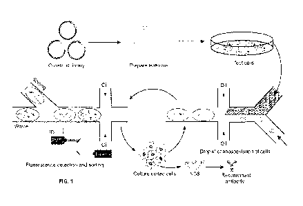
FIG 1 is a schematic overview of one embodiment of an activity-based antibody
selection method using droplet-based microfluidics. Briefly, the antibody
genes were cloned
into lentiviral vectors. Eukaryotic cells were infected by the lentiviral
antibody library and
individual transduced cells were co-encapsulated with the reporter cell into
droplets using
microfluidics system. The resulting emulsion was incubated off-chip overnight
and injected
into the sorting chip. Droplets containing antibody secreting cells and
activated reporter cell
were sorted. The sorted cells were cultured for the next round of selection.
After multiple
rounds of iteration, antibody genes were amplified from the sorted cells and
analyzed by
Sanger Sequencing or Next Generation Sequencing (NGS). The enriched antibodies
were
synthesized and recombinant antibodies were expressed and tested for function.
FIG. 2A shows an exemplary embodiment of a droplet maker microfluidics chip,
which can be used to generate picoliter droplets to co-encapsulate antibody
secreting cells
with reporter cells.
FIG 2B shows an exemplary embodiment of a sorting chip that can be used to
collect
3
CA 03190249 2023- 2- 21
WO 2022/037682
PCT/CN2021/113823
picoliter droplets based on the intensity of fluorescence inside the droplets.
The functions of
various inlets is indicated, and pictures of outlets for droplet generation
and sorting are shown.
FIGs. 3A-3E show a typical screening of bispecific antibodies from a spike-in
library
with microfluidics system.
FIG 3A is an image of nano- or pico-liter droplets generated by microfluidic
device
during their 24-hr incubation at 37 C.
FIG 3B shows the double Poisson distribution of cell number in the droplets.
K562-1Ier2 cells were stained with CellTrace Violet and Jurkat cells were
stained with
CellTrace Yellow. Cell loading was evaluated by counting the labeling signals
within each
droplet.
FIG 3C shows comparison of plate-based culture and droplet-based culture.
K562-Her2 cells were infected with anti-Her2/anti-CD3 positive control
lentivirus at low MOI
Half of the infected K562-Her2 cells were cocultured with Jurkat / plL2-eGFP
reporter cell in
plate well, and the other half of infected K562-Her2 cells were coencapsulated
with the
reporter cell (with a mean X, of 0.5 protein-secreting K562 cell per droplet).
After 16 hrs of
incubation, the activation of reporter cells for both conditions were analyzed
(e.g., using flow
cytometry).
FIG 3D shows representative images of the droplets after sorting for anti-Her2
/
anti-CD3 bispecific antibody. K562-Her2 cells were infected with anti-Her2 /
anti-CD3
bispecific antibody lentivirus library. The antibody secreting K562-Her2 cells
were stained
with CellTrace Violet and the Jurkat / plL2-eGFP cells were stained with
CellTrace Yellow.
Individual K562-Her2 cells were coencapsulated with the Jurkat / pIL2-eGFP
reporter cells
into droplets. After 16 hrs of incubation, the droplets were sorted.
FIG 3E shows activation of reporter cells by the identified bispecific
antibodies. The
K562-Her2 cells were cocultured with Jurkat/pIL2-eGFP reporter cells in the
presence of
selected bispecific antibodies. Expression of GFP by the reporter cell was
analyzed by flow
cytometry.
FIG 3F shows cell-mediated cytotoxicity of BiTEl. PBMCs and SK-BR-3 cells
were cocultured for 48 hrs in the presence of BiTE1 or control antibody CD3-
HEL. Cell
lysis was determined by measuring the release of Lactic Acid Dehydrogenase
(LDH) from
tumor cells.
FIGs. 3G and 31 show T cell activation in the response to BiTE. SK-BR-3 cells
were
4
CA 03190249 2023- 2- 21
WO 2022/037682
PCT/CN2021/113823
cocultured with PBMCs for 48 hours in the presence of BiTE1 or a control
antibody. The
activation of T cells was investigated by detecting CD69 expression on T cells
using flow
cytometry analysis (FIG 3G). The level of IFN-y (FIG 3H) and IL-2 (FIG 31) in
culture
supernatant was measured by ELISA.
FIGs. 4A and 4B show screening of CD40 agonist from a spike-in library with
microfluidics system. In FIG. 4A, RFP-positive hexameric CD4OL protein-
secreting cells were
spiked into a 10-fold excess of BFP-positive irrelevant anti-HEL antibody-
secreting cells, and
the mixture of cells were co-encapsulated with the reporter cells. After
incubation, the
droplets containing activated reporter cells were sorted based on the green
fluorescence. The
proportion of droplets containing RFP-positive or BFP-positive cells before
and after sorting
was analyzed. FIG 4B shows bright field and fluorescence images of droplets
before and after
sorting
FIGs. 5A-5F show screening of CD40 agonist antibody from the combinatorial
antibody library. HEK293T cells were infected with a lentivirus antibody
library and
individually coencapsulated with CellTrace Yellow prestained Jurkat reporter
cell and
fluorescence conjugated secondary antibody in droplets. After incubation, the
droplets
containing secreting antibody that bind and activate the co-encapsulated
reporter cell were
sorted. The sorted cells were then expanded for the 2nd round of selection.
The enriched
antibodies were identified by NGS (next generation sequencing). Specifically,
FIG 5A is a
schematic of possible time traces. From left to right: droplets without
reporter cells, droplets
containing reporter cells but secreted antibody can't bind to target, droplets
containing
reporter cells and secreted antibodies can bind to reporter cell target but
have no function,
droplets containing reporter cells and secreted antibodies can activate the
reporter cell. FIG
5B shows the proportions of different types of droplets for each rounds of
selection that were
analyzed. FIG 5C shows bright field and fluorescence images of the sorted
droplets after the
second round of selection. FIG 5D is a Bar plot for the top 20 scFy clusters
and their
frequencies during the selection. Besides the top 20 scFy clusters, the sum
frequency of
other scFvs are represented in gray at the bottom of bars. FIG 5E shows the
change of
frequencies of the selected antibodies during the selection. In FIG 5F,
agonist activity of the
selected antibodies was tested using the CD40 reporter cell line.
FIGs. 6A-6D show characterization of antibody in in vitro and in vivo models.
FIG
6A shows the FcyRI1B dependency of antibody C04. Jurkat / NF-KB-GFP-hCD40
reporter
cells were stimulated by antibody C04 or anti-HEL control in the presence of
FcyRIII3
CA 03190249 2023- 2- 21
WO 2022/037682
PCT/CN2021/113823
overexpressing HEK293T cells. The activation of the reporter cell line was
analyzed by
flow cytometry. FIG 6B shows the activation of DCs or B cells induced by C04.
Human
DC cells or B cells were stimulated by C04 in the absence or presence of anti-
Fe antibody.
The expression of CD86 was analyzed by flow cytometry. FIG 6C shows OVA-
specific
CD8+ T cell response induced by C04 in the CD40 / FcgR humanized mice. The
transgenic
mice were adoptively transfered with OVA-specific OT-I cells and treated with
DEC-OVA
together with C04 or isotype control antibody. Mice were euthanized for the
analysis of T
cells. Each circle represented an individual mouse. FIG 6D shows antitumor
effect of C04
in syngeneic mouse model. CD40 / FcgR humanized mice were s.c. engrafted with
MC38
tumor cells. When MC38 tumors were established (-100 mm3), mice were treated
with C04,
CP-870,893 or isotype control anti-FEEL antibody. The tumor volume and body
weight were
measured every three days until the end of the experiment. Data are
represented as mean
SEM
FIG 7A shows development and validation of the Jurkat / p1L2-eGFP reporter
cells.
The Jurkat/pIL2-eGFP reporter cells were stimulated with anti-CD3 and anti -
CD28 antibody
overnight. GFP fluorescence was obtained by flow cytometry. FIG. 7B shows K562-
Her2 cells
infected with anti-Her2xanti-CD3 BiTElentivirus or noninfected K562-Her2 cells
(control)
were cocultured with Jurkat/pIL2-eGFP reporter cells in the presence or
absence of anti-CD28
antibody overnight. GFP expression was analyzed by flow cytometry.
FIG. 8 shows the gating strategy for the analysis of cell viability. The
droplets were
first gated (gate 1) to eliminate coalesced droplets and retain only droplets
of the desired size.
The droplet in gate 2 defines droplet containing K562-Her2 cells; gate 3
defines the droplet
containing Jurkat cells. Gate 4 defines the viable cells with low fluorescent
nuclear staining,
indicating the live cells after 16 hrs of incubation time.
FIG 9A shows the gating strategies for screening of anti-Her2 / anti-CD3
bispecific
antibody. The droplets were first gated to eliminate coalesced droplets and
retain only
droplets of the desired size. The droplets containing K562 cell were gated
based on
CellTrace Violet fluorescence signal (gate 1). CellTrace Yellow fluorescence
signal peak
showed the presence of reporter cell in the droplet (gate 2). GFP fluorescence
signal peak
indicated activation of the reporter cell(gate 3). Lastly colocalization 2/3
and
non-colocalization 1/2 were used to sort droplets where GFP was from Jurkat
rather than
K562 (gate 4 and gate 5).
FIG 9B shows discrimination of strong and weak anti-Her2xanti-CD3 BiTE in
plate
6
CA 03190249 2023- 2- 21
WO 2022/037682
PCT/CN2021/113823
well-based or droplet-based coculture systems. K562-Her2 cells infected with
anti-Her2xanti-CD3, BiTE1 or BiTE3 lentivirus were cocultured with Jurkat/pIL2-
eGFP
reporter cells in plate wells (left panel) or individually coencapsulated with
reporter cells
(right panel) and incubated overnight. The activation of reporter cells in
both conditions was
compared.
FIGs. 10A-10C show development and validation of Jurkat / NF-KB-GFP-hCD40
reporter cells. FIG 10A is a schematic diagram of the hexameric CD4OL. Three
receptor
binding domains of CD40 ligand were tandemly linked to form a trivalent
protein. IgGl-Fc is
then used to link two of the trivalent proteins together, creating six
receptor binding domains
in a single agonist. In FIG. 10B, the Jurkat / NF-KB-GFP-hCD40 reporter cells
were
stimulated with hexameric form of CD4OL overnight. GFP fluorescence was
obtained using
flow cytometry. In FIG IOC, Jurkat/NF-KB-GFP-hCD40 reporter cells were
stimulated with
CD4OL in the presence or absence of DyLight 650 anti-Fe. GFP expression was
analyzed
using flow cytometry.
FIG 11 shows the Gating strategy for the analysis of the Jurkat / NF-KB-GFP-
hCD40
activation in droplet. The droplets were first gated to eliminate coalesced
droplets and retain
only droplets of the desired size. Two gates were assigned using drop code
DY638. Gate 1
defines droplets from negative emulsion where HEL cells were encapsulated,
gate 2 defines
droplets from positive emulsion where CD4OL cells were encapsulated. Gate 3
defines the
droplet containing HEL cells according to BFP signal and gate 4 defines the
CD4OL cells
according to RFP signal. Gate 5 defines droplets where reporter cells were
activated and
emitted GFP fluorescence. After 16 hrs of incubation, 24% of the CD4OL cell
containing
droplets exhibited GFP fluorescence signal while the HEL cell and reporter
cell
co-encapsulating droplets showed clean background of the activation of
reporter cell (0.5%).
FIG 12 shows the gating strategies for screening of CD40 agonist antibody. The
droplets were first gated to eliminate coalesced droplets and retain only
droplets of the desired
size. The droplets of the screening population were first gated based on the
intensity of
DY405. CellTrace Yellow fluorescence signal peak showed the presence of
reporter cell in
the droplet (gate 2). The Dylight647 fluorescence peak signal indicated
binding of the
secreting antibodies to CD40 on the reporter cell (gate 3) and GFP
fluorescence signal peak
indicated activation of the reporter cell (gate 4).
FIG 13A shows experimental time traces recorded for droplets analyzed at ¨ 900
Hz
and corresponding to the examples schematized in FIG 6A. Droplets are sorted
if they
7
CA 03190249 2023- 2- 21
WO 2022/037682
PCT/CN2021/113823
display a green peak (green line, GFP signal), a red peak (red line, rabbit
anti-human IgG Fc
DL650 gathering around reporter cells) and a yellow peak (yellow line,
reporter cells) that
co-localize. Blue fluorescence (blue line) signal is used to identify droplets
population.
FIG 13B shows the Fe-dependency of the hits in droplet-based coculture
systems.
FIEK293FT cells infected with CO1 or C04 lentivirus were individually
coencapsulated with
reporter cells in the presence or absence of crosslinking secondary antibody.
FIGs. 14A-14C show binding of C04 to human, rhesus and cynomolgus monkey
CD40. FIG 14A shows binding of C04 to human and rhesus CD40 determined by flow
cytometry. 293FT cells were transient transfected with human or rhesus CD40
and incubated
with different concentration of antibody C04 and goat anti-human IgG Fc Alexa
Fluor 488.
The cells were analyzed by flow cytometry. FIG 14B shows binding of C04 to
cynomolgus
monkey CD40 determined by SPR analysis. Anti-His antibody was immobilized on
Series S
CM5 chip, His-tagged cynomolgus monkey CD40 were captured by the immobilized
anti-his
antibody. Different concentrations of CD40 antibodies were injected through
flow cells and
Kd values were calculated using the 1:1 binding kinetics model. FIG 14C shows
binding of
C04 to different tumor necrosis factor superfamily (TNF SF) receptors
determined by ELISA.
DETAILED DESCRIPTION OF THE INVENTION
/. 0 erview
The invention described herein provides an efficient technology platform to
simultaneously screen binding and agonistic or antagonistic activity of
interacting
polypeptides, such as antibody and antigen, by combining the strength of
libraries (such as
those carried by alentiviral vector system) with the high throughout
capability of
microfluidics droplet system.
As used herein, an "antigen" is a molecule or a portion of a molecule capable
of being
bound by an antibody (or antigen binding polypeptide). In general, an antigen
includes
cpitopes consist of chemically active surface groupings of molecules, for
example, amino
acids or sugar side chains, and have specific three-dimensional structural
characteristics as
well as specific charge characteristics Epitope, as used herein, refers to the
antigenic
determinant recognized by the CDRs of the antibody (or an antigen-binding
portion thereof).
In other words, epitope refers to that portion of any molecule capable of
being recognized by,
and bound by, an antibody.
8
CA 03190249 2023- 2- 21
WO 2022/037682
PCT/CN2021/113823
The term "antibody," as used herein, may refer to an intact immunoglobulin
having
two light and two heavy chains that binds to an antigen of interest, or any
portion or fragment
thereof that binds to the antigen of interest.
The technical capabilities of the technology platform have been demonstrated
in the
examples herein, which show successful screening of potent co-stimulatory
receptor CD40
agonist antibodies and anti-Her2/anti-CD3 bispecific antibodies from large
combinatorial
antibody libraries. The streamlined technology enables efficient discovery of
active antibodies
that may be useful for numerous biological and therapeutic utilities, such as
next generation
immunotherapy to treat diseases including cancer.
Merely to illustrate, FIG 1 shows one exemplary embodiment of the subject
function-based screening / selection process using droplet-based
microfluidics.
Specifically, cells are infected by an antibody-expression library, such as a
lentiviral-based antibody expression library, at low multiplicity of infection
(MOT, such as 0.1
to 0.3) such that each cell is infected by no more than one lentivirus and
thus expresses no
more than one unique antibody. The antibody-encoding lentivirus is integrated
into the cell
genome in the form of provirus, resulting in cell secreting the corresponding
antibody. For
microfluidics system based screening, individual antibody-secreting cell is co-
encapsulated
with a reporter cell into a droplet by using microfluidic drop-maker. The
resulting emulsion
is incubated off-chip overnight, and injected into the sorting chip. The
droplets containing
activated reporter cells are then sorted by FADS. The cells are recovered from
the sorted
droplets, and the functional antibodies are identified by sequencing antibody
genes in the
cells.
The data presented herein illustrates the use of a droplet-based microfluidics
platform
for the selection of functional antibody, such as co-stimulatory receptor
agonist antibody and
bispecific T cell engager. The platform shared some key features with the most
efficient
selection methods to date such as phage display (//). First, the genotype and
phenotype
linkage was maintained through the whole process. Second, the product from one
round can
be directly amplified and used as the input of the next round of selection
Thus, multiple
rounds of iteration allowed enrichment of rare hits. Compared to the
conventional method to
individually express and assay thousands of antibodies, the throughput of this
platform
increased to 10 million. The usefulness of this platform has been demonstrated
in the
discovery of both bispecific antibodies and agonist antibodies, which are two
emerging drug
modalities for cancer immunotherapy.
9
CA 03190249 2023- 2- 21
WO 2022/037682
PCT/CN2021/113823
Indeed, the superiority of the subject platform is evidenced by the fact that
the few
potent CD40 agonist antibodies identified by the screen were too rare (<0.02%
frequency) to
be discovered by using the conventional method. Further, the ability of the
platform to
simultaneously screen large number of candidate bispecific antibodies provides
significant
opportunities to identify optimal BiTE or BiKE from large bispecific antibody
libraries.
The activity-based selection method described herein also has broad
applicability to
the high throughput analysis of cell-cell interactions. For example, DC cells
infected with
lentivirus library of neoantigens can be co-encapsulated with tumor
infiltrating T cells to map
the pairs of cognate antigens and T cell receptors (TCR) (27).
The activity-based selection method can also be adapted to screen different
types of
molecules such as cytokines (28-30), such as in cytokine engineering. For
example, a library
of cytokine-encoding polynucleotides can be produced through, e.g., random
mutagenesis
and/or rational design. The library can then be expressed using the lentiviral
vector of the
invention in individual cells transduced by the vector, and the system and
method of the
invention can be used to identify engineered cytokines with altered property,
such as altered
binding specificity / affinity, such that they either bind to new cytokine
receptors, or bind to
the native receptors with fine-tuned downstream signaling and/or cellular
responses (including
proliferation, differentiation, activation, apoptosis, cell fate
determination, etc.).
However, the invention described herein is not limited to the specific
illustrative
embodiments above. The methods and systems of the invention can be applied to
any
functional screening using a library of candidates with a reporter that
generates a detectable
signal which signifies a functional activity of the candidate.
Thus one aspect of the invention provides a method of identifying an agonist
or
antagonist polypeptide of a biological function, the method comprising: (1)
providing a
plurality of nano- or pico-liter droplets, each comprising: (i) no more than
one library cell that
(if present) expresses or is capable of expressing a candidate agonist or
antagonist polypeptide
from a library of candidate agonist or antagonist polypeptides; (ii) a
reporter cell that, upon
contacting the agonist or antagonist polypeptide of the biological function,
produces a
detectable signal as a marker of said biological function; (2) maintaining the
plurality of nano-
or pico-liter droplets under a suitable condition to permit said agonist or
antagonist
polypeptide to contact the report cell to trigger the biological function,
thereby producing said
detectable signal; (3) isolating or enriching nano- or pico-liter droplets
manifesting said
detectable signal, thereby identifying the agonist or antagonist polypeptide
of said biological
CA 03190249 2023- 2- 21
WO 2022/037682
PCT/CN2021/113823
function, within the isolated or enriched nano- or pico-liter droplets.
As used herein, a "reporter cell" includes any cell that can generate a
detectable signal
(e.g., a light signal, such as a fluorescent signal) upon contacting a desired
candidate molecule
that can trigger a biological function of interest. For example, in some
embodiment, the
reporter cell may comprise a reporter gene encoding a fluorescent protein
under the control of
a promoter, which promoter is activated upon triggering the biological
function of interest.
Therefore, a functional antibody as a candidate molecule may bind to the
surface of a reporter
cell to crosslink a cell surface receptor on the reporter cell, and initiate a
downstream
signaling event that includes activating the promoter of the reporter gene.
In certain embodiments, the biological function is cell death, e.g., the
reporter cell is
dead upon triggering of the biological function. For example, the candidate
molecule (e.g.,
functional antibody) may induce ADCC of the reporter cell, and the ability of
the each
candidate antibody to induce ADCC of the reporter cell may be detected by a
fluorescent
signal generated by the dead reporter cell.
There are many ways to detect dead cells using fluorescent signal. In one
embodiment, the presence of dead cells is measured by taking advantage of the
loss of
membrane integrity in the dead reporter cell, and the ability of indicator
molecules to partition
into a compartment not achievable if the cell membrane is intact. For example,
the reporter
cell may encode an enzyme (such as lactate dehydrogenase or LDH) that is only
leaked
outside the cell into the nano- or pico-liter droplet to catalyze a chemical
reaction that
generates detectable color in the nano- or pico-liter droplet. For example,
LDH catalyzes the
conversion of pyruvate to lactate, and in the process, converts NAD-' to NADH,
the reducing
capacity of which can be used to reduce a variety of substrate molecules into
products that are
either colored (e.g., tetrazolium compound as the di aphorase substrate which
is converted into
an intensely colored formazan product), fluorescent (e.g., resazurin being
converted into the
fluorogenic product resorufin), or luminogenic (e.g, a pro-luciferin substrate
is converted into
a luciferin product that is linked to a firefly luciferase reaction to
generate a luminescent
signal).
In other embodiments, enzymes that do not use the NADH cycling assay chemistry
can also be used as markers of dead cells, such as adenylate kinase (AK) and
glyceraldehyde-3-phosphate dehydrogenase (GAPDH) that can produce ATP by
providing a
reaction cocktail containing the necessary ingredients to generate a cycling
assay chemistry.
In another embodiment, the enzyme leaked from a dead reporter cell is a
protease,
11
CA 03190249 2023- 2- 21
WO 2022/037682
PCT/CN2021/113823
such as aminopeptidase, which activity can be measured using substrates
containing a short
sequence of amino acids (alanine-alanine-phenylalanine) conjugated via a
peptide bond to
either rhodamine 110 or aminoluciferin. Enzymatic removal of the amino acids
can generate
free rhodamine-110 for a fluorescent assay or free aminoluciferin which can be
used by firefly
luciferase to generate light.
In a further embodiment, the reporter cell is pre-loaded with a measurable
marker,
such as pro-fluorescent Calcein-AM or radioactive 51Cr which has been used to
measure
ADCC. Reporter cells incubated with 51Cr take up the radioactive marker which
becomes
bound as protein complexes in the cytoplasm of live cells. Similarly, calcein-
AM is taken up
by live reporter cells where cytoplasmic esterase activity removes the AM
group to generate
fluorescent calcein which is retained in live cells. Upon reporter cell death
and loss of
membrane integrity, the fluorescent calcein or the radioactive 51Cr is
released from the
cytoplasm into the nano- or pico-liter droplet medium encompassing the
reporter cell, where
they can be identified as diffused signals (as opposed to concentrated peak
signals)
colocalizing with the position of the reporter cell
In yet another embodiment, the reporter cell expresses a marker enzyme such as
luciferase, which produces reduced luminescence signal when the reporter cell
dies and
cytoplasmic components are released outside the cell.
In another embodiment, the nano- or pico-liter droplet encompassing the
reporter cell
may comprise a dye (such as trypan blue or nucleic acid binding dye such as
CellTox Green,
YO-PRO-1, Hoechst 33342, propidium iodide, SYTOX Green nucleic acid stain,
YOYO-1
Iodide, TO-PRO-3 Iodide, DRAQ7 far-red fluorescent DNA dye, and GelRed) that
is not
permeable through live cell membrane, but is able to permeate into a dead
cell's membrane.
As used herein, a "picoliter droplet" or "pico-liter droplet" can be produced
by a
microfluidic device (such as those described herein). It typically has a
volume of from about
0.002-500 picoliter (pL), 0.01-400 pL, 0.1-300 pL, 1-200 pL, 10-150 pL, 50-100
pL, 50-150
pL, 50-180 pL, 50-200 pL, 60-100 pL, 60-120 pL, 60-150 pL, 60-200 pL though
large (e.g.,
nL) or smaller (e.g., fL) volume droplets can be controllably produced by
adjusting settings
and/or microfluidic device architect, and are within the scope of the
invention described
herein. As used herein, a "nano-liter droplet- can be produced by a
microfluidic device
(such as those described herein). A nano-liter droplet may have a volume of
from about 0.2
nanoliter (nL), 0.3 nL, 0.4 nL, 0.5 nL, 1 nL, 2 nL, 3 nL, 5 nL, 10 nL, 20 nL,
to 50 nL.
Such droplets can be produced by microfluidic devices at a very high rate of
between,
12
CA 03190249 2023- 2- 21
WO 2022/037682
PCT/CN2021/113823
for example, 0.1 ¨ 10k Hz, or about 5-10k, or more droplets per second.
As used herein, a "reporter cell" is a cell that can generate a detectable
signal in
response to the presence or absence of the biological function, such that the
nano- or pico-liter
droplet containing the reporter cell with the detectable signal can be
identified or
distinguished from a reporter cell without the detectable signal.
In certain embodiments, the detectable signal is a light signal, such as
fluorescent light
signal, that permits the nano- or pico-liter droplets containing such light
signal be sorted in,
for example, a fluorescence-activated cell sorting (FACS) device or an
equivalent thereof.
One salient feature of the present invention is that the method can be used
for
functional screening of a library of molecules (e.g., polypeptides) that can
be produced /
expressed by library cells.
As used herein, "library cells" includes a population of cells that each
produces /
expresses ideally one member of a heterogeneous library of candidate
molecules. "A library
cell" is a cell from the library cells. Although two library cells may produce
/ express the
same candidate molecule, collectively, the library cells together produce /
express all members
of the candidate library, or a substantial portion of the candidate library.
In certain
embodiments, each library cell produces / expresses one candidate molecule
from the
candidate library. In other embodiments, each library cell produces /
expresses more than
one candidate molecule from the candidate library.
In certain embodiments, the library cell is a eukaryotic cell, such as a plant
cell, an
animal cell, a mammalian cell, a unicellular organism, an insect cell (e.g.,
sf9), or a yeast (S.
cerevisiae, S. pombe, C. albicans etc.).
In certain embodiments, the library cell is a stem cell, a cancer cell (e.g.,
cancer cell
line or isolated cancer cell), an immune cell, a lymphocyte, a B cell, a T
cell, a CD4+ T cell, a
CD8+ T cell, a Treg cell, a NK cell, a NKT cell, a macrophage, a neutrophil,
an eosinophil, a
basophil, a monocyte, a mast cell, or a myoblast cell.
In certain embodiments, the library cell is from a relatively homogenous
established
cell line. In certain embodiments, the library cell is from a heterogeneous
population of cells
obtained from the same source, such as a tissue sample, a tumor, or an
individual.
In certain embodiments, the library cell is a prokaryotic cell, such as a
bacteria cell.
In certain embodiments, the library cell expresses the candidate molecule on
the cell
surface. In certain embodiments, the library cell produces or secrets the
candidate molecule
13
CA 03190249 2023- 2- 21
WO 2022/037682
PCT/CN2021/113823
extracellularly (e.g-., into the medium in which the library cell grows).
The library of candidate molecules can be any molecules that can be produced
or
expressed by the library cells. Exemplary candidate molecules include
polypeptides or
proteins, small molecules, nucleic acids, lipids, polysaccharides, etc.
In certain embodiments, the candidate molecule is an agonist of a biological
function,
such as a growth factor, a cytokine, a chemokine, a hormone, a stimulator of
cell surface
receptor such as GPCR, TCR, BCR, immune checkpoint receptor, an antibody that
engages a
cell surface receptor and activates the receptor, or triggers an antibody-
mediated downstream
event such as ADCC (antibody-dependent cell-mediated cytotoxicity) or CDC
(complement-dependent cytotoxicity), etc.
In certain embodiments, the candidate molecule is an antagonist of a
biological
function, such as a blocking antibody that prevents the binding of a natural
ligand to a cell
surface receptor and inhibits the natural ligand-induce signaling
In certain embodiments, the candidate molecules are proteins or polypeptides,
including, without limitation, antibodies, bi-specific antibodies, tri-
specific antibodies, a
heavy chain of an antibody, a light chain of an antibody, a functional portion
of an antibody,
an antigen-binding portion / fragment of an antibody (including antibodies or
antigen-binding
fragment thereof having similar CDR sequence except for random mutations in
the CDR
sequences for affinity maturation), a cytokine, or a chemokine, or a
derivative thereof.
The library of cells can each produce / express a candidate member from a
library
through, for example, introducing an expression vector into the library cells.
Any expression
vectors can be used with the method of the invention.
In certain embodiments, the expression vector is a viral vector, such as a
retroviral
vector, a sindbis viral vector, a lentiviral vector, an adenoviral vector, an
AAV vector, a plant
viral vector (such as tobacco mosaic virus or TMV vector), or a hybrid
thereof. In certain
embodiments, the expression vector is a plasmid that can be introduced into
the library cells
by transfection. The library cells can be infected or transfected by the
expression vectors,
each encoding a unique candidate molecule, such that expression or production
of the
candidate molecules by the library cells can be screened using the method of
the invention
In certain embodiments, the library constructed into the viral vector (e.g.,
lentiviral
vector) originates from a larger library having more non-redundant library
members, such as
(an scFv) phage display library having more than 108, 109, 1010
,
1011, 1012 or more members.
The phage display library can be pre-panned against an antigen of interest to
enrich for
14
CA 03190249 2023- 2- 21
WO 2022/037682
PCT/CN2021/113823
members having a threshold binding affinity for the antigen, before the
enriched members are
introduced into the viral vector for use in the methods of the invention.
In certain embodiments, the expression or production of the candidate molecule
can be
controlled (e.g., induced or suppressed). This can be advantageous since it
allows more
precise control about the timing and/or extent of expression / production of
the candidate
molecule to be screened, and/or the timing of detection of expression /
production by the
reporter cell
Thus, in certain embodiments, the expression of the candidate agonist or
antagonist
polypeptide from the library is under the control of an inducible promoter
inducible by an
activator or an activating condition.
In certain embodiments, the inducible promoter is a positive inducible
promoter, and
wherein an activator for said positive inducible promoter is introduced into
the plurality of
nano- or pico-liter droplets subsequent to the formation of the plurality of
nano- or pi co-liter
droplets.
A "positive inducible promoter" is one that inactive in the OFF state,
because, for
example, an activator protein of the promoter, though maybe present, cannot
bind to the
promoter. Only after an inducer or activator binds to the activator protein
can the activator
protein be able to bind to the positive inducible promoter, thus turning it ON
and initiating
transcription from the promoter. In certain embodiments, the activator is a
small molecule
activator.
In certain embodiments, the positive inducible promoter is a Tet-ON promoter,
and
wherein the activator is tetracycline or a derivative thereof capable of
binding to activator
rtTA (reverse tetracycline-controlled transactivator).
In certain embodiments, the positive inducible promoter is an alcohol-
regulated
promoter (such as the AlcA promoter), and wherein the activator is AlcR or
AlcA.
In certain embodiments, the positive inducible promoter is a steroid-regulated
promoter (such as the LexA promoter), and wherein the activator is XVE.
In certain embodiments, the inducible promoter is a negative inducible
promoter, and
wherein an activator for the negative inducible promoter is introduced into
the plurality of
nano- or pico-liter droplets subsequent to the formation the plurality of nano-
or pico-liter
droplets.
A "negative inducible promoter" is one that is inactive in the OFF state
because a
CA 03190249 2023- 2- 21
WO 2022/037682
PCT/CN2021/113823
bound repressor protein actively prevents transcription. Once an inducer binds
the repressor
protein, the repressor protein is removed from the DNA. With the repressor
protein absent,
transcription is turned ON.
In certain embodiments, the negative inducible promoter is a pLac piontotet,
and
wherein the activator is lactose or a derivative thereof (such as lPTG)
capable of binding to
lac repressor (lad I protein).
In certain embodiments, the negative inducible promoter is a pBad promoter,
and
wherein the activator is arabinose capable of binding to AraC.
In certain embodiments, the inducible promoter is a temperature sensitive
promoter,
and the expression of the candidate agonist or antagonist polypeptide from the
library is under
the control of a temperature change as the activating condition that activates
the inducible
promoter.
Temperature sensitive expression systems are typically less leaky, and can
have
near-zero expression at regular temperatures but can be induced by heat or
cold exposure.
Examples include the heat shock-inducible Hsp70 or Hsp90-derived promoters, in
which a
gene of interest (such as the candidate agonist or antagonist in the candidate
library) is only
expressed following exposure to a brief heat shock. In the case of Hsp70, the
heat shock
releases heat shock factor 1 (HSF-1), which subsequently binds to heat shock
elements in the
promoter, thereby activating transcription.
In certain embodiments, the inducible promoter is a light inducible promoter
(such as
the FixK2 promoter), and the expression of the candidate agonist or antagonist
polypeptide
from the library is under the control of a light signal as the activating
condition that activates
the light inducible promoter.
For example, an exemplary light inducible promoter can be regulated by the
blue-light
sensing protein YFI. When light is absent, '(FT phosphorylates FixJ, which
binds to the
FixK2 light inducible promoter to induce transcription When light is present,
'(Fl is
inactive, preventing transcription from the light inducible promoter.
In certain embodiments, the inducible promoter is a pH sensitive promoter, and
the
expression of the candidate agonist or antagonist polypeptide from the library
is under the
control of a pH change as the activating condition that activates the
inducible promoter.
For example, the E. coil asr (acid shock RNA) gene encodes small RNA that is
inducible by low external pH, and asr gene may be regulated by the two
component system
16
CA 03190249 2023- 2- 21
WO 2022/037682
PCT/CN2021/113823
PhoBR2. In this two-component system, the protons from the environment (H+)
activate the
sensory part (PhoR-) of the two-component system, which then transduces the
signal to the
activator protein PhoB, which can bind promoter of asr to initiate
transcription. The
promoter region of asr contains a sequence similar to the pho-box, which is a
consensus
sequence able to bind to PhoB.
In certain embodiments, the pH sensitive promoter is Pasr or PgadA.
In any of the embodiments herein, one or more additional components, such as
an
activator, an inducer, an additional cell, or a bolus of liquid value with
acid or base used for
pH change may need to be introduced into the nano- or pico-liter droplets,
which can be
accomplished by any of many means known in the art, such as by injection or
fusion.
For example, the one or more additional components can be directly injected
into each
of the plurality of nano- or pico-liter droplets in a high throughput fashion.
Alternatively, the
one or more additional components can be encapsulated in their own nano- or pi
co-liter
droplets that can be similarly generated by microfluidic devices, and such
nano- or pico-liter
droplet having the one or more additional components can be used to each of
the plurality of
nano- or pico-liter droplets containing the library cell and/or the reporter
cell. In certain
embodiments, the fusion is mediated by geometrical constraint, mechanical
force, surface
property change, electrical, laser, acoustic force, or any art-recognized
methods. See, for
example, Ahn et aI., AppL Phys. Lett. 88:3, 2006; Priest et al., Appl Phys.
Lett. 89:134101,
2006; and Songet al., Appl Phys. Lett. 83: 4664, 2003, all incorporated herein
by reference.
In certain embodiments, prior to step (1), a first plurality of nano- or pico-
liter droplets
each comprising the no more than one library cell have been maintained under a
pre-determined condition for a pre-determined period of time to allow said
candidate agonist
or antagonist polypeptide to express, before said reporter cell is introduced
into each said first
plurality of nano- or pico-liter droplets to provide the plurality of nano- or
pico-liter droplets
in step (1). This may be advantageous because expression! production of the
candidate
molecule by the library cells can be separately controlled, either through
induction,
suppression, and/or time period for expression, until a desired concentration
of the candidate
molecule in the nano- or pico-liter droplets is reached, before the reporter
cell is introduced
into the nano- or pico-liter droplet for detection.
Substantially the same means can be used to introduce the reporter cells into
each of
the nano- or pico-liter droplets containing the library cells, including by
injection or fusion.
The fusion may be mediated by geometrical constraint, mechanical force,
surface property
17
CA 03190249 2023- 2- 21
WO 2022/037682
PCT/CN2021/113823
change, electrical, laser, acoustic force, or any art-recognized methods.
In certain embodiments, the library cell that expresses or is capable of
expressing a
candidate agonist or antagonist polypeptide is pre-stained with a first
tracking signal (e.g.,
CellTi ace Violet), and said reporter cell is pre-stained with a second,
different, tracking signal
(e.g., CellTrace Yellow) prior to step (1), and wherein step (3) is carried
out by retrieving
nano- or pico-liter droplets that: (I) contain both the first and the second
tracking signals; (II)
produce said detectable signal (e.g., GFP) after step (2); and, (III) exhibit
colocalization of the
second (reporter cell) tracking signal and the detectable signal.
In certain embodiments, steps (1)-(3) are repeated more than once, using the
library
cells isolated or enriched in step (3) of a previous repeat. That is, after
the detection and
sorting of positive nano- or pico-liter droplets containing the detectable
signal that signifies
the presence or absence of the biological function of interest, the positive
nano- or pico-liter
droplets can be collected, optionally with the library cells within these
positive nano- or
pi co-liter droplets pooled, expanded and/or further cultured, before the
pooled, expended
and/or cultured library cells from the first round is again encapsulated with
the same or a
different reporter cell in a new plurality of nano- or pico-liter droplets for
a further round of
screening, for the same biological function or for a different biological
function.
After one or more rounds of screening, the nano- or pico-liter droplets having
the
positive detectable signal can be collected and the library cells within
obtained, in order to
determine the identity of the candidate library member that gives right to the
detectable
biological function, then identifying the candidate molecule as the agonist or
antagonist, as the
case may be.
The methods of the invention can be implemented in numerous specific settings
to
screen large number of candidate molecules that may exhibit a desired
biological function.
In certain embodiments, the library of candidate molecules has more than 103,
104, 105, 106,
107, 10 1-10
8 , , 109, u 1011 or more non-redundant members (e.g.,
millions of non-redundant
antibody coding sequences). In certain embodiments, the plurality of nano- or
pico-liter
droplets comprises more than 103, io, i05, 106, 107, 108, 00, 1010, 10"
droplets
The method of the invention can be used in numerous functional screenings.
For example, in one embodiment, the agonist or antagonist polypeptide is a
bispecific
T cell engager (BiTE) comprising a first antigen-binding fragment (such as a
14 scFv) specific
for a first antigen fused to a second antigen-binding fragment (such as a 2nd
scFv) specific for
a second antigen.
18
CA 03190249 2023- 2- 21
WO 2022/037682
PCT/CN2021/113823
In certain embodiments, the first antigen is a T cell antigen (such as CD3),
and the
second antigen is a surface antigen on a target cell (such as a cancer antigen
(e.g., 1-1ER2) on a
target cancer cell).
In certain embodiments, in each nano- or pico-litei droplet having said one
library cell
that expresses or is capable of expressing said candidate agonist or
antagonist polypeptide,
said candidate agonist or antagonist polypeptide is a BiTE from a library of
candidate BiTEs
each encoded by a lentiviral vector from a lentiviral vector library encoding
said library of
candidate BiTEs, and wherein said one cell is the target cell that expresses
said target cancer
antigen (e.g., I-IER2).
In certain embodiments, the reporter cell is a T cell-derived cell line (e.g.,
Jurkat cell)
that produces a fluorescent protein (e.g., GFP), the transcription of which
encoding RNA is
under the transcriptional control of a promoter (e.g., IL-2 promoter)
activated by T cell
activation upon binding of the BiTE to the TCR of the reporter cell and the
target cancer
antigen on the target cell.
In certain embodiments, the library of candidate agonist or antagonist
polypeptides is a
library of candidate BiTEs encoded by a lentiviral vector-based library, and
wherein coding
sequence for each of said second antigen-binding fragment (such as a 2nd scFv)
specific for
the second antigen has been pre-selected from a phage display library based on
biopanning
against said second antigen.
In certain embodiments, the complexity of the phage display library is about
1010
members, and wherein the complexity of the library of candidate Bi l'Es with
respect to the
second antigen-binding fragment is 105 members.
In certain embodiments, the one library cell that expresses or is capable of
expressing
the candidate agonist or antagonist polypeptide (BiTE) is produced by
infection at low MOT,
by a lentiviral vector-based library encoding said library of candidate
agonist or antagonist
polypeptides (BiTEs), to ensure that each cell produces no more than one type
of the
candidate agonist or antagonist polypeptide (BiTE).
In certain other embodiments, the agonist or antagonist polypeptide is an
agonist or
antagonist antibody or an antigen-binding fragment thereof specific for a cell
surface receptor
(e.g., CD40) that triggers said biological function.
In certain embodiments, in each nano- or pico-liter droplet having said one
cell that
expresses or is capable of expressing said candidate agonist or antagonist
polypeptide, said
candidate agonist or antagonist polypeptide is an scFv-IgG1 Fc fusion from a
library of
19
CA 03190249 2023- 2- 21
WO 2022/037682
PCT/CN2021/113823
candidate scFv-IgG1 Fc fusions each encoded by a lentiviral vector from a
lentiviral vector
library encoding said library of candidate scFv-IgG1 Fc fusions, optionally,
wherein said cell
surface receptor is CD40 and wherein said biological function is NFKB
signaling.
In certain embodiments, the reporter cell is a cell line (e.g., Jinkat cell)
that produces a
fluorescent protein (e.g., GFP), the transcription of which encoding RNA is
under the
transcriptional control of a promoter (e.g., NEKB promoter) activated by
activation of said cell
surface receptor (e.g., CD40) upon binding of the agonist antibody or antigen-
binding
fragment thereof to the cell surface receptor (e.g., CD40) of the reporter
cell.
In certain embodiments, the coding sequence for each of said scFv in said
library of
candidate scFv-IgG1 Fc fusions has been pre-selected from a phage display
library based on
biopanning against said cell surface receptor (e.g., CD40).
In certain embodiments, the complexity of the phage display library is about
1010
members, and wherein the complexity of the library of candidate scFv-IgG1 Fc
fusions with
respect to the second antigen-binding fragment is 105 members.
In certain embodiments, the one library cell that expresses or is capable of
expressing
the candidate agonist or antagonist polypeptide is produced by infection at
low MOI, by a
lentiviral vector-based library encoding said library of candidate agonist or
antagonist
polypeptides, to ensure that each cell produces no more than one type of the
candidate agonist
or antagonist polypeptide.
In certain embodiments, a secondary antibody specific for said candidate
agonist or
antagonist polypeptide is labeled with a first tracking signal (e.g.,
Dylight647-conjugated) and
co-encapsulated into the nano- or pico-liter droplets in step (1), and said
reporter cell is
pre-stained with a second, different, tracking signal (e.g., CellTrace Yellow)
prior to step (1),
and wherein step (3) is carried out by retrieving nano- or pico-liter droplets
that: (I) contain
both the first and the second tracking signals; (II) produce said detectable
signal (e.g., GFP)
after step (2); and, (III) exhibit colocalization of the first (CD40 agonist
antibodies) and the
second (reporter cell) tracking signals and the detectable signal.
In yet another embodiment, the agonist or antagonist polypeptide is an
engineered or
modified cytokine for a cytokine receptor that triggers said biological
function.
In certain embodiments, the engineered or modified cytokine has altered
specificity
and/or affinity towards the cytokine receptor compared to the cognate wild-
type cytokine.
In certain embodiments, the engineered or modified cytokine binds to and
activates a
CA 03190249 2023- 2- 21
WO 2022/037682
PCT/CN2021/113823
cytokine receptor to which a cognate wild-type cytokine does not bind.
In certain embodiments, the engineered or modified cytokine stimulates or
inhibits a
downstream signaling pathway that is not stimulated by a cognate wild-type
cytokine.
In certain embodiments, the engineered or modified cytokine commits a cell to
a
differentiation, proliferation, activation, and/or apoptotic process that is
not stimulated or
inhibited by a cognate wild-type cytokine, or is not stimulated or inhibited
by the cognate
wild-type cytokine to the same degree.
In certain embodiments, the method of the invention can be used to screen for
agonist
or antagonist polypeptide (e.g, antibody) that can induce ADCC against an
antigen expressed
on the reporter cell. For example, a library of antibodies can be expressed by
the library
cells, each can be encapsulated with a reporter cell in a nano- or pico-loiter
droplet using the
method of the invention. When / if the antibody recognizes the the antigen
expressed on the
reporter cell, an effector cell (such as NK cell) also encapsulated in the
nano- or pico-liter
droplet can kill the reporter cell. The presence of the dead reporter cell can
be detected by a
light-generating reaction using any of the suitable methods described herein
(such as
generating a detectable fluorescent signal by an LDH enzyme leaked outside the
reporter cell).
The NK / effector cell can be introduced into the nano- or pico-liter droplet
using injection or
fusion as described herein, or be included in the initial nano- or pico-liter
droplet with the
library cell and the reporter cell.
For antagonistic assay, the library cell expresses a candidate polypeptide
that can
potential block effector / NK cell-mediated ADCC on the target cell, and the
absence /
reduction of killing of the reporter cell can be measured by the fluorescent
signal generated by
the reporter cell. The NK / effector cell can be introduced into the nano- or
pico-leter droplet
at a later time through inhection or fusion to permit accumulation of the
antagonistic
polypeptide inside the droplet to reach a critical concentration.
In certain embodiments, the method of the invention can be used to screen for
agonist
or antagonist polypeptide (e.g., antibody) that can induce CDC against an
antigen expressed
on the reporter cell. For example, the library cell may express a candidate
antibody
recognizing an antigen expressed on the surface of the reporter cell, and the
complement
pathway components required for CDC can either be provided in the medium for
encapsulating cells into the nano- or pico-liter droplets or expressed by the
library or reporter
cells. Dead reporter cells due to CDC can be detected using any of the methods
described
herein for generating fluorescent or other detectable signals in dead cells.
21
CA 03190249 2023- 2- 21
WO 2022/037682
PCT/CN2021/113823
In yet other embodiments, the method of the invention can be used for high
throughput
screening of cell-cell interaction, wherein, for example, dendritic cells or
other
antigen-presenting cells (APCs) can be infected by a library of neoantigens in
lentiviral or
other suitable vectors. Such APCs are then co-encapsulated with tumor
infiltrating T cells
(Tits) as reporter cells capable of generating fluorescent signals upon TCR
activation, in
order to map the pairs of cognate antigens and T cell receptors (TCR).
In certain embodiments, step (1) is carried out with a nano- or pico-liter
droplet-producing microfluidic device comprising: (a) a first inlet for an oil
to form a
continuous oil phase; (b) a second inlet for an aqueous suspension of a
population of said
reporter cell; (c) a third inlet for an aqueous suspension of a population of
said cell that
expresses or is capable of expressing a candidate agonist polypeptide; (d) an
outlet for
retrieving said nano- or pico-liter droplets dispersed in said continuous oil
phase; and, (e) a
junction area where the first, the second, and the third inlets converge to
form nano- or
pico-liter droplets in the continuous oil phase before exiting through the
outlet.
In certain embodiments, step (3) is carried out in a nano- or pico-liter
droplet-sorting
microfluidic device comprising: (A) a first inlet of spacing oil and a second
inlet of bias oil;
(B) a third inlet of retrieved nano- or pico-liter droplets after step (2);
(C) a first outlet for
retrieving nano- or pico-liter droplets manifesting said detectable signal;
(D) a second outlet
for collecting waste not retrieved by the first outlet; (E) a sorting actuator
that directs a
passing nano- or pi co-liter droplet to the first outlet when the passing nano-
or pi co-liter
droplet manifests the detectable signal, and directs the passing nano- or pico-
liter droplet to
the second outlet otherwise; and, (F) a junction area where the first, the
second, and the third
inlets converge to form a stream of passing nano- or pico-liter droplets
before the sorting
actuator, and where the first and second outlets diverge to separate said nano-
or pico-liter
droplets manifesting said detectable signal from the waste.
In certain embodiments, the agonist or antagonist polypeptide is identified
through
identifying the coding sequence thereof from said cell that expresses or is
capable of
expressing said agonist or antagonist polypeptide retrieved from said nano- or
pico-liter
droplets manifesting said detectable signal. In certain embodiments, the
coding sequence is
identified through high throughput sequencing, such as next-generation
sequencing (NGS) of
the antibody encoding sequences from the isolated cells.
In certain embodiments, said cell that expresses or is capable of expressing
said
agonist or antagonist polypeptide can be retrieved for further processing to
identify the coding
22
CA 03190249 2023- 2- 21
WO 2022/037682
PCT/CN2021/113823
sequence(s) of the polypeptide of interest. Such further processing may
include the
droplet-based transcriptome analysis of said cell, e.g., as described in PCT
international patent
application published as W02017/097939 and Gerard, A. et al. (2020). High-
throughput
single-cell activity-based screening and sequencing of antibodies using
droplet microfluidics.
Nature Biotechnology, 38, 715-721.
In certain embodiments, the method further comprises verifying that said
agonist or
antagonist polypeptide leads to said biological function, including activation
of said biological
function by said agonist or antagonist polypeptide in a manner depending on
binding by said
agonist or antagonist polypeptide.
2. Design of the tnicrofinidics platform
Two microfluidics devices are particularly useful in the methods of the
invention: (i) a
droplet formation microfluidic device (or "droplet formation device") that
compartmentalizes
the library cells (e.g., library of lentivirus infected cells) with the
reporter cells and/or
detection reagents; and (ii) a droplet sorting device that sorts droplets
based on reporter cell
activation and receptor binding signals using a chosen mechanism, such as
surface acoustic
wave based sorter (see FIGs. 2A-2C) (24).
In certain embodiments, droplet formation and fluorescence analysis are
performed on
a dedicated droplet microfluidics platform reported previously in Gerard A. el
al (23),
incorporated herein by reference. Briefly, the microfluidic chips can be
fabricated in
polydimethylsiloxane (PDMS) polymer (Sylgard 184 elastomer kit; Dow Corning
Corp) using
the standard soft lithography as described (31). Masters are made using one
layer of SU-8
photoresist (MicroChem). The depth of the two devices is 40+/-ljtm to allow
the droplet
generating or flowing in a monolayer format. For sorting device, the PDMS is
bonded to a
piezoelectric substrate (Y128-cut Lithium niobate wafer) where an golden
interdigital
electrode is patterned with standard lift-off technology and aligned with the
fluidic channel
above. Microfluidics devices are treated before use with 1% v/v
1H,1H,2H,2H-perfluorodecyltrichlorosilane (Alfa Aesar) in Novec HFE7500
fluorinated oil
(3M) to prevent droplets wetting the channel walls.
However, the microfluidic devices suitable for use with the methods of the
invention is
not so limited.
In certain embodiments, the droplet formation device comprises a T-junction or
a
flow-focusing device or portion or mechanism that forms monodisperse (e.g., <1-
3% disperity)
23
CA 03190249 2023- 2- 21
WO 2022/037682
PCT/CN2021/113823
droplets at rates up to around 10 kHz. See, for example, Thorsen et at., Phys.
Rev. Lett
86:4163, 2001; Nisisako et at., Lab Chip 2:24, 2002; and Link et at., Phys.
Rev. Lett.
92:054503, 2004, incorporated herein by reference.
In certain embodiments, the droplet formation device utilizes jetting to form
droplets
at a rate of 10's of kHz. See, for example, Utada et at., Phys. Rev. Lett 99:
094502, 2007;
Utada et al., Phys. Rev. Lett 100:014502, 2008; and Humphry et al., Phys. Rev.
E 79:056310,
2009, incorporated herein by reference.
In certain embodiments, the droplet formation device utilizes membranes or
sieves to
produce multiple droplets simultaneously. Such membrane emulsification devices
produce
droplets by dispersing one fluid into a continuous phase through a membrane or
sieve,
essentially forming an array of T-junctions to increase rate of droplet
formation by orders of
magnitude. See Sugiura et at., Langmuir 17:5562, 2001, incorporated herein by
reference.
In certain embodiments, the droplet formation device is an on-demand droplet
formation device which ensures that only perfectly monodisperse droplets enter
the device.
Such on-demand droplet formation devices utilize sudden changes in applied
pressure, in
combination with narrow channels at the flow-focusing section of the device
and rapid
withdrawal of the water flow from a budding droplet. See Lorenz et at., Anal.
Chem.
78:6433, 2006; and He et at., Anal. Chem. 77:1539, 2005, incorporated herein
by reference.
In certain embodiments, the droplet formation device comprises an on-chip
piezo-electric actuator to provide previse control over droplet formation.
In certain embodiments, the droplet formation device comprises a heating
element that
can, for example, provide a heat change for inducible expression of genes in
the library cells
under the control of a heat-sensitive promoter. The heating element can also
provide
additional control over droplet size, independent of device geometry and flow
rates, by
changing interfacial tension and viscosity to modulate droplet diameter formed
in a flow
focusing device. See Nguyen et at., Appl. Phys. Lett. 91:084102, 2007, and Tan
et at., J.
Phys. D 41:165501, 2008, incorporated herein by reference.
In certain embodiments, the droplet formation device comprises a cell-
triggered
Rayleigh-Plateau instability in a flow-focusing geometry to maximize single-
cell
compartmentalization during jet break-up. In this embodiments, up to about 70-
80 % of the
injected cell population is encapsulated into drops containing one and only
one cell, with
<1 % contamination by empty droplets. Chabert et at., Proc. Natl. Acad. Sd.
USA 105:3191,
2008; and Edd et al., Lab chip 8:1262, 2008, incorporated herein by reference.
24
CA 03190249 2023- 2- 21
WO 2022/037682
PCT/CN2021/113823
Although droplet formation does not necessarily require surfactants, in
certain
embodiment, the droplet formation device of the invention utilizes a
stabilizing agent to
prevent or inhibit rapid coalescence of the formed droplets inside the droplet
formation
devices. In certain embodiments, the continuous phase is a hydrocarbon or
fluorocarbon oils
for forming water-in-oil emulsion. In certain embodiments, the continuous
phase is a
mineral oil (including fluorinated oil), optionally comprising commercially
available
surfactants such as Span 80, Abil EM, and Krytox (DuPont) (which contains a
perfluoropolyether (PFPE) tail and a carboxylic acid hydrophilic head group)
for fluorous oil
continuous phases. In certain embodiments, the continuous phase comprises a
fluorinated
oil, and optionally comprises a fluorinated surfactant.
In certain embodiments, the continuous phase comprises a oligoethylene glycol
(OEG)-terminated surfactant. In certain embodiments, the OEG surfactant is an
OEG
fluorinated surfactant.
In certain embodiments, the surfactant comprises a hydrophilic head group,
such as a
PFPE surfactant. In certain embodiments, the surfactant comprises an ammonium
salt of
carboxy-PFPE and/or poly-L-lysine. In certain embodiments, the surfactant
comprises
polyethyleneg,lycol (PEG) and dimorpholinophosphate (DNIP), or DMP¨PFPE
surfactant.
Such surfactant provides excellent stability in addition to biocompatibility,
in that with
DMP¨PFPE surfactant in fluorinated oil, cells encapsulated in droplets on a
microfluidic chip
as emulsion can be stored for up to 14 days off chip, and then re-injected
into a mi croflui di c
device with minimal coalescence (<10 % after 14 days).
In certain embodiments, the surfactant has a critical micelle concentration
(CMC) of
104 mol or greater.
In certain embodiments, the surfactant is synthesized from 600 g/mol PEG and
6000
g/mol PFPE.
In certain embodiments, the surfactant facilitates droplet fusion through
controlling
droplet surface chemistry by tuning the surfactant concentration and
surfactant accumulation
time on chip (see Mazutis et al., Lab Chip 9:2665, 2009, incorporated herein
by reference).
In certain embodiments, the methods of the invention call for droplet
injection to
introduce additional reagents and/or cells into pre-formed nano- or pico-liter
droplets, which
can be achieved using a varieties of mechanisms.
In certain embodiments, the droplet formation device comprises a picoinjector,
a
robust device to add controlled volumes of reagent using electro-microfluidics
at kHz rates.
CA 03190249 2023- 2- 21
WO 2022/037682
PCT/CN2021/113823
It can also perform multiple injections for serial and combinatorial
additions. See Abate et
al., Proc Natl Acad Sci USA 107(45): 19163-19166. 2010, incorporated herein by
reference.
In certain embodiments, the additional reagents and/or cells are introduced
into the
nano- or pico-liter droplets through printed droplet microfluidics (PDM),
which can be used to
construct defined reactions with chemicals and cells incubated under air on an
open array.
See Siltanen et al., Sc/Rep 8:7913, 2018, incorporated herein by reference.
In certain embodiments, the methods of the invention call for droplet fusion,
which
can be achieved using a varieties of mechanisms.
In certain embodiments, the droplet formation device is a passive fusion
devices which
relies on channel properties and/or surface properties of the channels to
induce droplet
coalescence. For example, in-channel droplet fusion is initiated when two or
more droplets
are brought into close contact by draining of the continuous phase, and
imbalances in surface
tension leads to droplets coalescence (see Tan et al, Lab Chip 4:292, 2004,
incorporated
herein by reference).
In certain embodiments, the droplet formation device facilitates the fusion or
more
than two droplets by controlling surface energy patterns inside microfluidic
channels. For
example, the channels can be designed to disrupt the flow of the droplets,
trapping them to the
pattern and only releasing them after coalescence. Varying channel and pattern
dimensions
as well as fluid flows provide full control over droplet fusion, allowing the
incorporation of
several components into a single, large droplet by coalescence of multiple
droplets. See
Fidalgo et al., Lab chip 7: 984, 2007, incorporated herein by reference).
In certain embodiments, the droplet formation device facilitates in-channel
fusion by
incorporating rows of pillars within the channels that act as passive merging
elements. The
pillars trap droplets while allowing the continuous phase to drain when a
second droplet enters
the pillar area. The fusion process is independent of the inter-droplet
separation but is rather
controlled by droplet size. Moreover, the number of droplets that can be fused
at any time
can also be controlled by the mass flow rate and volume ratio between the
droplets and the
merging chamber. See Niu et al, Lab chip 8:1837, 2008.
In certain embodiments, the droplet formation device facilitates fusion by
relying on
transient states in the stabilization of the droplet interface by surfactant,
coupled to a proper
geometrical design of a coalescence module, to induce the selective fusion of
a droplet
stabilized by surfactant (re-injected) with a droplet which is not fully
stabilized (generated
on-chip). See Mazutis et al, Lab chip 9(18): 2665-72, 2009, incorporated
herein by
26
CA 03190249 2023- 2- 21
WO 2022/037682
PCT/CN2021/113823
reference.
In certain embodiments, the droplet formation device comprises an active
fusion
mechanism that can be controlled (e.g., be switched on or off). The device has
the ability for
achieving accurate droplet synchronization to put the droplets in very close
proximity. See
Thiam et at., Phys. Rev. Lett. 102:188304, 2009.
In certain embodiments, the active fusion mechanism relies on an external
trigger to
induce coalescence.
In certain embodiments, the external trigger is a strong electric field (see
Link et at,
Angew. Chem. 118:2618, 2006 and Mazutis eta!, Lab chip 9:2902, 2009,
incorporated herein
by reference). In certain embodiments, a smaller droplet is fused with a
larger droplet
because smaller drops move faster due to the Poiseuille flow, thus ensuring
that they are in
contact in the region where they are coalesced with an electric field.
In certain embodiments, the external trigger is an alternating current (AC)
field (see
Chabert et at., https://doi.org/10.1002/elps.200500109, 2005).
In certain embodiments, the external trigger is a magnetic field. See Varma et
at., Sc!
Rep 6:37671, 2016, incorporated herein by reference. Specifically, a uniform
magnetic field
is used to induce merging of ferrofluid based droplets, and control of droplet
velocity and
merging can be achieved through uniform magnetic field and flow rate ratio.
In certain embodiments, the external trigger is a localized heating induced by
laser
pulse (see Baroud etal., Lab chip 7:1029, 2007, incorporated herein by
reference).
In certain embodiments, the droplet formation device hydrodynamically couples
the
two droplet-formation channels to ensure perfectly alternating droplet
sequences. See Frenz
et al., Langmuir 24:12073, 2008, incorporated herein by reference. For
example,
hydrodynamic resistance can be exploited to synchronize droplets in two
parallel channels by
using passive devices such as loops or ladders to ensure perfect alternation
of droplets into
two channels at a T-junction, thus leading to symmetric splitting of droplet
trains.
In certain embodiments, the droplet formation device facilitates droplet
fusion with a
surface through a chemistrode device. See Liu et at, Langmuir 25:2854, 2009,
incorporated
herein by reference.
Additional fusion mechanisms that can be used with the methods of the
invention are
known in the art, such as those described in Xu et at., Droplet Coalescence in
Microfluidic
Systems (core.ac.uk/download/pdf/143869049.pdf, incorporated herein by
reference).
27
CA 03190249 2023- 2- 21
WO 2022/037682
PCT/CN2021/113823
In certain embodiments, the library cells can be incubated in the nano- or
pico-liter
droplets for a pre-determined time with the reporter cell, or before being in
contact with the
reporter cell in the nano- or pico-liter droplet.
In certain embodiments, the pre-determined time is minutes to hours, 1-5
hours, 10-24
hrs, 1-5 days, 5-10 days, or up to 2-3 weeks. The droplets can be incubated in
the
microfluidic device (e.g., in a storage reservoir) or collected from the
device after the droplet
formation, and incubated under a different condition (e.g., temperature).
In certain embodiments, the droplet sorting device is an integral part of the
droplet
formation device.
In certain embodiments, the droplet sorting device is separate from the
droplet
formation device.
In certain embodiments, the sorting device utilizes electric field to isolate
droplets
having the desired detectable signal. See Ahn et al., App!. Phys. Lett. 88:
024104, 2006,
incorporated herein by reference.
In certain embodiments, the sorting device utilizes surface acoustic wave to
isolate
droplets having the desired detectable signal. See Franke et at., Lab chip
9:2625, 2009,
incorporated herein by reference. Surface acoustic wave devices are capable of
deflecting
droplets or particles by locally compressing fluids.
In certain embodiments, the sorting device utilizes magnetic fields to isolate
droplets
loaded with magnetic particles and have the desired detectable signal. See
Zhang et at., Lab
Chip 9:2992, 2009, incorporated herein by reference.
In certain embodiments, the sorting device utilizes laser-induced localized
heating to
isolate droplets having the desired detectable signal. See Baroud etal., Phys.
Rev. E
75:046302, 2007, incorporated herein by reference. An added benefit of
integrating a
microheater element in the device offers precise control over droplet motion
(leading to both
splitting and sorting of droplets) at Y-junctions through changes in fluidic
resistance and the
thermocapillarity in one of the branches.
In certain embodiments, the detectable signal is a fluorescent light. The
electric
signal obtained from fluorescence light detected at a photon multiplier tube
(PMT) can be
used to trigger further droplet manipulations in, for example, in the sorting
device in a manner
similar to a flow cytometer. The fluorescence activated sorting, or FADS
(fluorescence-activated droplet sorting), combines fluorescence intensity
detection with
28
CA 03190249 2023- 2- 21
WO 2022/037682
PCT/CN2021/113823
selective emulsion separation to extract target droplets into a continuous
aqueous stream for
collection or further manipulation.
Unless specifically disclaimed or improper, any one embodiment of the
invention can
be combined with one or more additional embodiments of the invention.
EXAMPLES
Example 1 Development and Optimization of Droplet-Based Assay
First, the droplets were generated by using microfluidics chip. The droplets
were
incubated 24 h at 37 C. No merging of droplets were observed, indicating that
the droplets
were stable (FIG. 3A).
Next, anti-Her2/anti-CD3 BiTE antibody was used as a model for the development
and
optimization of the droplet-based assay. The BiTE antibodies were created by
linking single
chain antibodies, one binding to CD3 on T cells and the other to a surface
antigen on the target
cell (25).
Specifically, trastuzumab-derived anti-Her2 scFy were fused to blinatumomab-
derived
anti-CD3 scFv to generate an anti-Her2/anti-CD3 positive control. Her2-
overexpressing
K562-Her2 cells were then infected with the anti-Her2/anti-CD3 positive
control lentivirus at
a low MOI (<0.1) so that less than 10% of the cells were infected. The K562-
Her2 cells
played a dual role to express the BiTE antibody and to provide Her2-mediated
crosslinking of
the secreted antibodies. Individual infected K562-I-Ter2 cells were co-
encapsulated with a
Jurkat / p11,2-eGFP reporter cell (FIGs. 7A-7B) with X, of 0.5 antibody
secreting cell and 1
reporter cell per droplet. The number of cells per droplet was analyzed based
on the image,
and the results are in good agreement with double Poisson distribution (FIG
3B).
After 16 hrs of incubation, the droplets were re-injected to the sort chip to
analyze the
activation of reporter cell in the droplet. About 9.5% of the reporter cells
were activated,
suggesting high efficiency of reporter cell activation by the secreted
antibody in the droplet.
This may be due to the extreme high antibody concentration in the very small
volume of each
droplet. In contrast, when the anti-Her2/anti-CD3 lentivirus infected K562-
Her2 cells were
cocultured with the reporter cells in the plate well, 72.7% of the reporter
cells were activated
after 16 hrs of incubation (FIG 3C).
The difference between the two conditions demonstrated that antibody secreted
by
cells in the droplet can not transfer between different droplets, thus the
system can be used for
29
CA 03190249 2023- 2- 21
WO 2022/037682
PCT/CN2021/113823
library screening. In addition, NucGreen Dead 488 were used to track the cell
viability in
droplets. Both K562 and Jurkat cells in droplets have high viability around
90% after 16 hrs
of incubation (FIG. 8).
Example 2 Functional Screening of Anti-Her2 / Anti-CD3 Bispecific Antibody
The method of the invention was used here to conduct a function-based
screening for
anti-Her2/anti-CD3 bispecific antibodies.
Because phage display technology can interrogate a much larger diversity space
than is
possible in eukaryotic systems, Her2-binding antibodies were first selected
from an antibody
phage display library. A naive human single chain Fv (scFv) library with size
of 1010
members was panned against the Her2 protein. One rounds of panning were
performed, and
the scFv genes in phagemid were subcloned into a lentiviral vector, which
contained fixed
blinatumomab derived anti-CD3 scFv gene to express anti-Her2/anti-CD3 BiTE
antibody.
The size of the combinatorial bispecific antibody library is about 105
members.
K562-Her2 cells were infected with the Bi TE antibody lentivirus library at
low MOT to
ensure most cells were infected by only one virus and produced a single type
of bispecific
antibody. Prior to cell encapsulation, K562-Her2 cells were stained with
CellTrace
and Jurkat/pIL2-eGFP cells were stained with CellTrace Yellow. Each
individually infected
K562-Her2 cell was co-encapsulated with a Jurkat/pIL2-eGFP reporter cell into
the same
droplet. After 16 hrs of incubation, the droplets were sorted based the
following gating
strategy (FIG 9A): first, select droplets for the presence of K562-Her2 and
Jurkat/pIL2-eGFP
cells based on the cell staining fluorescence signals; then select droplets if
the reporter cell
inside was activated based on GFP signal; and finally, select the droplets if
the signal of GFP
was colocalized with the reporter Jurkat cell staining signal.
Eleven millions of droplets were imaged and about 0.26% of the droplets were
sorted,
and a representative image of the sorted droplet were shown (FIG. 3D). The
anti-Her2 scFv
genes were amplified from the sorted cells and cloned into mammalian
expression plasmid.
Twenty clones were picked for Sanger sequencing, and five bispecific
antibodies appeared
more than once. The purified bispecific antibodies were added into the
Jurkat/pIL2-eGFP
reporter cell in coculture with K562-Her2. Three out of five antibodies (BiTE-
1, Bi 1E-2,
BiTE-3) can activate the reporter cell in the presence of K562-Her2 (FIG. 3E).
To assess the antitumor activity of BiTE1, we performed in vitro cytotoxicity
assays
by coculturing PBMCs and HER2-expressing SK-BR-3 breast cancer cells. The
results
CA 03190249 2023- 2- 21
WO 2022/037682
PCT/CN2021/113823
showed that lysis of SK-BR-3 cells only occurred when they were incubated with
BiTE1, not
with the control antibody anti-CD3xanti-HEL (hen egg lysozyme, HEL) (FIG. 3F).
Flow
cytometry analysis of T cells in the coculture system showed that BiTE1
stimulated
expression of the activation marker CD69 on T cells (FIG. 3G). Moreover, BiTE1
induced
dose-dependent increases in IFN-y and IL-2 in the supernatants (FIGs. 3H and
31).
To validate the potential of the method to discriminate between strong and
weak hits,
trastuzumab-derived anti-Her2xanti-CD3 positive control, BiTE1 and BiTE3 in
descending
order of potency were each cloned into lentiviruses. Cells infected with each
of these
bispecific antibodies encoding lentivirus were coencapsulated with the
reporter cell, and
droplets were analyzed for the activation of the reporter cell. The order of
droplet intensity
was as follows: trastuzumab-derived anti-Her2xanti-CD3 positive control >
(FIG. 9B). This work demonstrates the capability of the technology to
discriminate between
weak and strong hits, which enables fast enrichment of high potency hits using
a more
stringent gating strategy.
Overall, the results demonstrate that this technology allows combinatorial
screening
and profiling of large numbers of bispecific antibodies.
Example 3 Screening of CD40 Agonist from a Spike-in Library
This experiment further validates the utility of this function-based screening
method,
by identifying CD40 agonistic antibodies.
CD40 is a promising drug target for cancer immune therapy (5). Activation of
CD40
on antigen presenting cells (APCs) results in improved antigen processing and
presentation,
and cytokine release, which enhances T cell response.
Human Jurkat T cells were engineered to constitutively express human CD40 and
express GFP controlled by NF-KB response elements. The reporter cell line
express GFP
when the CD40 is activated (FIG 10B).
FEEK293T cells co-expressing red fluorescence protein (RFP) and hexameric form
of
CD4OL was used as positive control (CD4OL cell). HEK293T cells co-expressing
blue
fluorescence protein (BFP) and a non-related hen egg lysozyme (HEL) antibody
was used as
negative control (HEL cell).
The CD4OL or HEL cells were co-encapsulated with the Jurkat / NF-KB-GFP-hCD40
reporter cells into droplets. After 16 hrs of incubation, 24% of the CD4OL
cell-containing
droplets exhibited GFP fluorescence signal while the HEL cell and reporter
cell
31
CA 03190249 2023- 2- 21
WO 2022/037682
PCT/CN2021/113823
co-encapsulating droplets showed clean background of activation of the
reporter cell (0.5%)
(FIG 11).
CD4OL cells were spiked into a 10-fold excess of HEL cells. The mixture of
CD4OL
cells and HEL cells were co-encapsulated with the Jurkat / NF-KB-GFP-11CD40
reporter cells
with a mean A, of 0.5 protein secreting cell per droplet. The droplets which
contained activated
reporter cell were sorted based on green fluorescence. Before sorting, only
0.94% of the
mixed cell population were RFP positive, 16.58% of cell were BFP positive, and
the rest
droplets were empty or contained only reporter cell, whereas after the sort
the percentage of
droplets containing RFP positive CD4OL cell increased to 52.57%, where 49.48%
RFP
positive and 3.09% RFP and BFP double positive (FIG 4A). Most droplets
containing both
RFP positive CD4OL cells and activated GFP reporter cell after sorting (FIG
4B).
Example 4 Screening of CD40 Agonist Antibody using the Function-Based
Screening
Method
CD40 binding antibodies were first selected from an antibody library using
phage
display technology. The scFy genes in phagemid were subcloned into lentiviral
vector that
contained IgG1 Fc gene to express scFy Fc fusion protein. The library size was
about 105.
HEK293T cells were infected with lentivirus library at low MOI to ensure most
cells were
infected by only one virus and produced one type of monoclonal antibody.
To screen CD40 agonist antibodies, the antibody-producing cells were co-
encapsulated
with CellTrace Yellow prestained Jurkat / NF-KB-GFP-hCD40 reporter cell, and
Dylight647-conjugated secondary antibody in droplets. The reporter cells were
also
co-encapsulated with soluble hexameric CD4OL protein and anti-HEL antibody as
positive
and negative controls. Droplets of different populations (positive control,
negative control,
and the screening population) were coded by different concentrations of DY405.
After 16
hrs of incubation, the droplets were sorted based on the following criteria
(FIG 12). The
droplets of the screening population were first gated based on the intensity
of DY405.
CellTrace Yellow fluorescence signal showed the presence of reporter cell in
the droplet
The Dylight647 fluorescence signal indicated binding of the secreting
antibodies to CD40 on
the surface of the reporter cell, and GFP fluorescence signal peak indicated
activation of the
reporter cell (FIG 12). The droplets displayed different patterns based on
their fluorescence
signals (FIG 5A, and FIG 13A). 13 million and 4.8 million droplets were imaged
for the 1st
round and the 2nd round of screening, respectively. Whereas only 0.29%
droplets were
32
CA 03190249 2023- 2- 21
WO 2022/037682
PCT/CN2021/113823
Dylight647 and green fluorescence double positive for the 14- round of
screening (out of the
droplet containing Jurkat cell signal), this value increased to 12.5% for the
2' round of
screening (FIG 5B). The droplets before and after sorting were imaged and the
number of
Dylight647 and green fluorescence double positive droplets dramatically
increased after the
sorting (FIG 5C).
The scFv genes were amplified from the cells after each round of sorting and
were
subject to the third-generation sequencing. Circular consensus sequences from
these reads
were generated, filtered by quality. Full-length scFvs were identified in
41,971 reads.
Considering the errors introduced by PCR or sequencing, the similar (at 95%
similarity) scFv
sequences were grouped into 602 scFv clusters and consensus scFv sequences of
each cluster
were created. Comparison of the sequence frequency in each round revealed that
some scFvs
were enriched while some scFvs were eliminated during the selection (FIG. SD).
The top 6
scFv clusters were chosen after the 2'd round of selection based on enrichment
factor in each
rounds. The frequency of scFv clusters C01, CO3, C04, COS, and C06 were low
before
sorting, and showed roundwi se increase during the selection process In
contrast, scFv cluster
CO2 were highly abundant before sorting, but its frequency was significantly
reduced after
sorting (FIG. 5E).
The scFv sequences of C01, CO3, C04, C05, and C06 are listed below, with the
heavy
chain CDR sequences double underlined and in bold font, and the light chain
CDR sequences
double underlined and in italic font.
C01:
MAQVQLVE SGGGLVQ PGRSLR I SCAGSGFTFGDSAMHWVRQAPGKGL EWVSGISRNSDTIVYADSVKG
RFT I S RDNAKNSLYLQMNSLRAE DTALYYCARRSGDHHAMDVWGPGTIVIVS SGGGGGGS ET TLTQ S P
AT L SL SPGERATLSCRAS QSVNTYLAWYQQKPGQAPRLLMYDSSSRATGI PDRF SGSG SGTD FT LT
IS
RL E PE DFAVY YC QOYS TVPL TFGGGT KVDI KR
CO3:
MAQVQLVESGAEVKKPGASVKVSCKASGYTFTGYYMHINVRQAPGQGLEWMGWISAYNGNTNYAQKLQG
RVILT RDT ST STVYMELSSLRSEDTAVYYCARAKKIRGYSYGGFDYWGQGTTVTVS SGGGGGGSQSAL
TQPASASGSPGQSVT I SCTGT SSDVGGYNYVSWYQQH PGKAPKLL I Y EV/VKRP SGVPDRF
SGSKSDNT
ASLTVSGLQAE DEADYYC SS YAGSDNS YVFGTGTKLTVLG
C 04:
MAQVQLVQSGAEVKKPGASVKVSCKASGYTFTGYYMHWVRQAPGQGLEWMGWINPNSGGTNYAQKFQG
RV= RDT S I STAYMELSRLRSDDTAVYY CARERVGATPTYYYYMDVWGKGT TVTV S SGGGG SGGGGS
33
CA 03190249 2023- 2- 21
WO 2022/037682
PCT/CN2021/113823
GGGGS DVVMTQSP FS L PVT PGE PAS I SCRS S QSLLHSNGHNYLDWYVQKPGQ SPQLL I
HLGSNRASGV
PDRFSGGGSGTDFTLKI SRVEAE DVGVY YCMOALOTPFTFGPGT KVD I KR
C05:
MAQVQLQQSGPGLVKPSQTLSLTCAI SGDSVSSNTAAWNWI RQS PS RGLEWL GRTYYRSKWYNDYAVS
VKSRI T INPDT SKNQ FSLQLNSVTPEDTAVYYCARQVQLERRHAFDIWGQGTLVTVSSGGGGGSQSAL
TQ PAS VSGS PGQS III SCTGT SSDVGGYNYVSWYQQH PGKAFKLMI Y
EVSNRPSGVSNRFSGSKSGNT
ASLT I SGLQAE DEADY FC SSY-TSSS TVVIFGGGTKVTVLG
C 06:
MAQVQLLQSGGGVVQ FGRSLRL SCAASGFTFSSYGMHWVRQAFGKGL EWVAVISYDGSNKYYADSVKG
RFT IS RDNSKNTLYLQMNSLRAE DTAVYYCAKVIRGSSGWSDAFDIWGQGTMVTVS SGGGGSGGGGSG
GGGSQAVLTQ P S SAS GT PGQRVT I SC SGS SSNIGSHTVSWYQQL PGTAPKLL IY
STDQRPSGVPDRLS
GS KSGT SASLT I SGLQ S EDEAHY YCAAWDDSQNKSLVFGGGT QLTVLG
Table!: EC50 of selected CD40 agonist antibodies.
CD40 agonist CO1 CO2 CO3 C04 C05
C06
EC50 without crosslinker (nM) 7 6 3
EC50 with crosslinker (nM) 6 5 3 30
5
Based on the HCVR and LCVR CDR sequences, the genes of full length IgG C01,
CO2, CO3, C04, C05, and C06 were synthesized and recombinant antibodies were
expressed
and purified. The activity of the antibodies was tested using the Jurkat / NF-
KB-GFP-hCD40
reporter cell. The reporter cells were stimulated with different
concentrations of antibody in
the presence or absence of the crosslinking secondary antibody. Antibodies
C01, CO3, C04,
C05, and C06 activated the reporter cell line. The activity of COL CO3, and
C05 was
independent of crosslinking, while C04 and C06 activated the reporter cell in
a crosslinking
dependent manner (FIG 5F). Signal intensities between Fc-dependent C04 and
Fc-independent CO1 in the droplet-based assay were compared, and the results
supported that
the differential Fc-dependency of these hits (FIG 13B). The Fc receptor
independent activity
of CD40 agonist antibody is of concern because this feature underlies possible
systemic
adverse events. Therefore, the potent Fc receptor-dependent antibody C04 was
chosen for
further characterization.
Flow cytometry results showed that antibody C04 bound to human, rhesus macaque
34
CA 03190249 2023- 2- 21
WO 2022/037682
PCT/CN2021/113823
and cynomolgus monkey CD40 with similar affinity (FIG. 14A and 14B). In
addition,
ELISA results confirmed that antibody C04 specifically bound to CD40 rather
than other TNF
receptor superfamily members such as GITR, 0X40 and 4-1BB (FIG 14C).
Since FcyRIM expressed on tumor infiltrating myeloid cells is required for
agonistic
activity of the crosslinking-dependent CD40 agonist antibodies, the FcyRIEB
dependency of
antibody C04 was assessed. The Jurkat / NF-KB-GFP-hCD40 reporter can be
stimulated by
C04 in the presence of FcyRIM-overexpressing cells. The results revealed that
the agonism
of C04 was FcyRIIb dependent (FIG 6A).
To investigate whether C04 can promote the activation of CD40-positive APCs,
human
dendritic cells (DCs) and B cells isolated from PBMC were stimulated by C04 in
the presence
or absence of the crosslinking antibody. Flow cytometry analysis showed that
C04
stimulated the expression of activation marker CD86 on DCs and B cells, and
anti-Fc
mediated crosslinking could further enhance the agonistic activity (FIG. 6B).
The immunostimulatory activity of antibody C04 was further tested in mouse
humanized for CD40 and FcyRs. The recipient CD40/Fc1R humanized mice were
adoptively transferred with ovalbumin (OVA)-specific OT-I cells, and OVA were
delivered to
dendritic cells by i.p. injection of the chimeric anti-DEC205 antibody
conjugated to OVA
(DEC-OVA) together with either C04 or anti-HEL antibody. Mice treated with C04
displayed significantly increased percentage of OT-1 cells among CD8-' T cells
compared to
the mice treated with control antibody, demonstrating C04 had robust adjuvant
effect (FIG
6C).
In addition, antitumor efficacy of C04 was assessed in syngeneic tumor models.
When MC38 tumors were established (-100 mm3), mice were treated with C04,
CP-870,893(Pfizer) or anti-HEL antibodies. C04 displayed comparable anti-tumor
activity
as CP-870,893, which is the most potent CD40 agonistic antibody among those
tested in
clinical trials to date. In addition, treatment with C04 didn't cause severe
reduction of body
weight as CP-870,893 did, suggesting that C04 had a favorable toxicity profile
(FIG 6D).
Example 5 Discussions
Here, a droplet-based microfluidics platform for the functional screening of
millions of
antibodies was described. The platform shares some key features with the most
efficient
selection methods, such as phage display. First, the genotype and phenotype
linkage was
maintained throughout the whole process. Second, the product from one round
can be directly
CA 03190249 2023- 2- 21
WO 2022/037682
PCT/CN2021/113823
amplified and used as the input in the next round of selection. Thus, multiple
rounds of
iteration allow the enrichment of rare hits. Compared to the conventional
method of
individually expressing and assaying thousands of antibodies, the throughput
of this platform
increased that limit to 10 million. This is especially useful for the
development of
next-generation cancer immunotherapies, such as agonist antibodies or
bispecific antibodies,
when simple binding assays may be inadequate.
To demonstrate the usefulness of this platform, it was first applied to
discover
bispecific antibodies and agonist antibodies, whose development was limited by
low diversity
and/or low throughput and potentially biased screening.
Bispecific T or NK cell engagers (BiTEs or BiKEs) hold great promise for
cancer
treatment, and a growing number of BiTEs and BiKEs are making their way
through various
stages of development. To identify the optimal BiTE or BiKE, a bispecific
antibody library
was constructed to address the complexity of the array of tumor antigen
targeting antibodies.
The large number of bispecific antibodies in a given library can exceed the
throughput of
existing methods. The described approach provides significant opportunities to
screen
unprecedented numbers of molecules of different formats and compositions, such
as
antibodies and nonantibody protein scaffolds. However, the method by itself
cannot guarantee
the generation of functional hits of high potency, and the successful
isolation of potent
antibodies also depends on the existence of such hits in the library. Similar
to other in vitro
display methods, the efficiency of drug discovery will scale with library
size.
A single round of screening was performed to identify anti-Her2x anti-CD3
antibodies.
Such a single round could be potentially sufficient for some targets, but the
false positive rate
could be high in certain screenings. A few reasons could contribute to the
false positive rate.
First, the local concentration of antibody in the droplets is very high, and
may result in an
increased tendency to form high-molecular-weight aggregates and nonspecific
activation of
the reporter cell. Second, the gating strategy may not be stringent enough to
exclude all-false
positive events. As used in well-established methods, such as phage display,
iterative rounds
of screening may help to enrich the true hits and eliminate false positive
hits. In addition,
more stringent gating strategies can be applied in screening.
Research into costimulatory receptor agonists has been reignited over the last
decade
due to substantial advances in the field of immunoncology. Costimulatory
receptors are
expressed on a number of immune cell types, including T cells, B cells and
natural killer (NK)
cells, as well as APCs, and engagement of these receptors promotes immune cell
function,
36
CA 03190249 2023- 2- 21
WO 2022/037682
PCT/CN2021/113823
proliferation and survival. Nevertheless, there are no general rules to guide
the screening of
agonist antibodies. For example, a panel of antibodies binds to the same or
similar epitopes of
the Fas receptor but results in different biological effects, with some acting
as agonists and
others as antagonists. The intrinsic complexity of agonist antibodies requires
screening as
many antibodies as possible. The instant unique platform of the disclosure was
used to screen
for CD40 agonist antibody and discovered a few potent CD40 agonist antibodies,
most of
which were too rare (<0.02% frequency) to be discovered using a conventional
screening
platform.
The instant method of the disclosure can also be applied to high-throughput
analysis of
cell-cell interactions. For example, this method is applicable where DC cells
infected with a
lentivirus library encoding neoantigens are coencapsulated with tumor
infiltrating T cells to
map the pairs of cognate antigens and T cell receptors (TCRs). This method can
also be
adapted to screen different types of molecules, such as cytokines. In addition
to application in
drug discovery, identification of intercellular signaling pathways has become
an actively
growing field Combining our technology with the power of CRISPR/Cas9 library
screening
could enable the deciphering of cell¨cell communications at scale. The
innovative
applications of this activity-based selection method have been limited only by
the imagination
of the users.
In summary, the instant disclosure describes a unique high-throughput platform
for
function-based screening of up to millions of antibodies. With the capability
to screen millions
of antibody-producing cells without any presumptions other than the key
function used for
screening, this may revolutionize next-generation cancer immunotherapy drug
discovery and
development, as well as advance basic research involving cell-to-cell
interactions.
Materials and certain experimental details for the Examples above are provided
herewith.
Cell culture
HEK293FT cells (Thermo Fisher Scientific, R70007) were cultured in DMEM
(Thermo Fisher Scientific). Jurkat cells (ATCC, TIB-152) were cultured in RPMI
1640
medium (Thermo Fisher Scientific). SK-BR-3 cells (ATCC, HTB-30) were cultured
in
McCoy's 5A (modified) medium (Biological Industries). All the culture medium
was
supplemented with 10% fetal bovine serum (Biological Industries), lx
nonessential amino
acids, 100 U/ml penicillin, 1001.igiml streptomycin and 12.5 mM HEPES (Thermo
Fisher
37
CA 03190249 2023- 2- 21
WO 2022/037682
PCT/CN2021/113823
Scientific). HEK293F cells (Thermo Fisher Scientific, R79007) were suspended
and cultured
in FreeStyleTM 293 Expression Medium (Thermo Fisher Scientific). All cells
were maintained
in a CO2 incubator at 37 C.
IL2 reporter cell line
Jurkat / plL2-eGFP was developed to monitor activation of T cells. The
immortalized T cell leukemia Jurkat cell line was transfected with vector
carrying eGFP
reporter gene under the control of full-length IL-2 promoter (from -648 to -1
upstream of IL-2
translation initiation codon). After co-stimulation by anti-CD3 and anti-CD28
antibodies,
cells expressing high level of GFP were sorted into wells of a 96-well plate
using FACS, and
individual clones of Jurkat / pIL2-eGFP cell line were characterized.
CD40 reporter cell line
Jurkat / NF-KB-GFP-hCD40 reporter cell was developed to monitor activation of
CD40. The Jurkat cell was transfected with a vector carrying an eGFP reporter
gene under
the transcriptional control of NF-KB response element. After stimulation by 10
ng/mL TNFcc
(Sino Biological), cells expressing high level of eGFP were sorted by FACS,
and the Jurkat /
NF-KB-eGFP cell line were characterized.
The resulting Jurkat / NF-KB-eGFP cell line was then infected with lentivirus
expressing full-length human CD40. After stimulation by 100 nM hexameric CD4OL
Fc
fusion protein, individual cells with high level of GFP signal were sorted
into the wells of a
96-well plate by FACS, and individual clones of the Jurkat / NF-KB-GFP-hCD40
cell were
characterized.
Microfluidic chip fabrication
All microfluidic chips were fabricated in polydimethylsiloxane (PDMS) polymer
(Sylgard 184 elastomer kit; Dow Coming Corp) using the standard soft
lithography as
described (3 1 ) . Masters were made using one layer of SU-8 photoresist
(MicroChem). The
depth of the two devices is 40 1 pm to allow the droplet generating or
flowing in a
monolayer format. For device ii, the PDMS is bonded to a piezoelectric
substrate (Y1-28-cut
Lithium niobate wafer) where an golden interdigital electrode is patterned
with standard
lift-off technology and aligned with the fluidic channel above. Microfluidics
devices were
treated before use with 1% v/v 1H,114,214,2H-perfluorodecyltrichlorosilane
(Alfa Aesar) in
Novec HFE7500 fluorinated oil (3M) to prevent droplets from wetting the
channel walls.
38
CA 03190249 2023- 2- 21
WO 2022/037682
PCT/CN2021/113823
Droplet production, collection, and incubation
Aqueous phases containing infected cells and reporter cells were co-flowed and
partitioned into droplets with hydrodynamic flow focusing in dripping mode on
a microfluidic
chip (Fig. 2A). The nozzle is 15 pm wide, 40 pm deep and 10 pm long. The
continuous phase
was Novec HFE-7500 fluorinated oil (3M) containing 2% w/w 008-FluoroSurfactant
(RAN
Biotechnologies). Pressure pumps (Fluigent) were used to generate
monodispersed droplets of
100 pl- 10 pL at 5000 Hz. The droplets were collected into a 10 mL tube and
incubated at
37 C in 5% CO2 to allow antibody secretion and subsequent activity to occur
within each
droplet prior to screening.
Illicrofluidic droplet screening and recovery
Droplet fluorescence analysis and sorting operations were performed on a
dedicated
droplet microfluidic station, similar to that described by Mazutis et al.
(15).
Pressure pumps (Fluigent) were used to inject the collected droplets into the
sorter device (Fig.
2C) at a frequency of 1000-3000 Hz. The sorter device was mounted on an
inverted
microscope (ASI Microscope) equipped with a 940 nm LED illumination source
(Thorlabs
M940L3) and a fixed focus laser line (solid-state laser of wavelength 405 nm,
488 nm, 561
nm or 635 nm, Omicron) with photomultiplier tube bandpass filters of 440/40-25
nm,
525/40-25 nm, 593/46-25 nm and 708/75-25 nm (Hamamatsu).
The fluorescence of each droplet was measured as the droplet flowed past an
observation constraint in the microfluidic channel where the laser line was
positioned. The
emitted fluorescence was detected with PMTs, converted into corresponding
signal output
voltages, and recorded by the data acquisition card (FPGAPCIe-7842R). These
voltages were
then processed by the card and custom Lab View software to identify droplets
according to
their fluorescence intensity and size. These characteristics were used to
determine whether
each droplet should be sorted.
Droplets were sorted based on surface-acoustic wave deflection as described by
Frank et al.
with a GHz-signal generator (Wavetek, Model 3010) (21, 42). Sorted droplets
were collected
in a 1.5 mL Eppendorf tube. Cells were recovered by adding 100 p.L DMEM
culture medium,
followed by 100 pL of 1H, 1H, 2H, 2H-perfluoro-1-octanol (Sigma, 37053), and
then cells
were pooled and centrifuged at 400 g for 5 min at 4 'V for subsequent steps,
such as
39
CA 03190249 2023- 2- 21
WO 2022/037682
PCT/CN2021/113823
subculture or DNA sequencing.
Phage display
A human naïve scFv library was constructed from the PBMC of 30 healthy donors
with standard protocols. The phage library was incubated with biotinylated
CD4O-Fc fusion
protein or Her2 recombinant protein (Acro biosystems) for 2 hours at room
temperature (RT),
and the phage-antigen complex was captured by Dynabeads M280 (Life
technologies). The
bound phages were eluted by Glycin-HC1 (pH 2.2) for 10 min. at RT, and
neutralized with
Tris-HC1 (pH 8.0) to adjust pH to 7.5. The phagemid DNA was isolated using
plasmid
miniprep kit (Qiagen).
Lentivirus libraty construction
Both phagemids and lentiviral vector pLV-efla-ScFv-Fc were digested with
enzyme
õViI The lentiviral vector and scFy genes were isolated after electrophoresis.
ScFy genes
were then ligated into the lentiviral vector. The product of ligation reaction
were
transformed into XL1-Blue competent cells by electroporation, and most of the
transformed
bacteria were plated on LB Agar plates. The remaining bacteria were serially
diluted and
plated to estimate the size of the library. Lentiviral plasmid was prepared
using plasmid
midiprep kit (Qiagen) for lentivirus preparation.
Lentivirus preparation
When confluency reached 80%, HEK293T cells were transfected with lentiviral
backbone plasmid and packaging plasmids using PEI transfection reagent. The
medium was
then changed to fresh complete culture medium 6 hrs post transfection.
Supernatant
containing lentivirus was harvested after 48 hours, centrifuged at 300 g for 5
min. at 4 C, and
filtered by 0.45 um filter to remove cell debris. Viral titer was measured
using P24 ELISA
kit (Clontech). The virus was aliquoted and stored at -80 C.
Function based screening of anti-Her2/anti-CD3 bispecific antibody using
microfluidics
Aqueous phase I: Jurkat / NF-1(13-eGFP reporter cells were washed with PBS and
stained with 1 !AM CellTrace Yellow dye for 10 min. at 37 C. The stained
Jurkat /
NF-KB-eGFP reporter cells were washed twice with RPMI 1640, and then
resuspended in cell
culture medium (RPMI 1640, 5% FBS, 25 mM HEPES, and 0.1% Pluronic F-68)
containing 1
g/m L anti-CD28 antibody ( Invitrogen).
Aqueous phase II: The stable Her2 expressing K562 cells were infected with the
CA 03190249 2023- 2- 21
WO 2022/037682
PCT/CN2021/113823
lentiviral antibody library. The resulting antibody-secreting K562 cells were
washed with
PBS, and stained with 11.tM CellTrace Violet dye for 10 min. at 37 C. The
stained cells
were washed twice with RPMI 1640, and resuspended with cell culture medium
containing
200 nM DY647. K562 cells infected with positive control lentivirus were
resuspended with
culture medium containing 1.5 laM DY647.
Aqueous phases I and II were injected into the droplet generation chip from
different
inlets and used as the disperse phase. Novec HFE7500 fluorinated oil (3M)
containing 2%
w/w fluoro-surfactant (RAN Biotechnologies) was used as the continue phase to
produce
droplets with average size of 100 pL. The flow rates of aqueous phase I,
aqueous phase II,
and oil phase were adjusted, so on average one reporter cell and 0.5 antibody
secreting cell
were co-encapsulated per droplet. During droplet production, the cell
suspension was cooled
using ice-water to inhibit antibody secretion. The droplets were collected and
incubated at
37 C for 16 hrs.
The droplets were first gated to eliminate coalesced droplets and retain only
droplets
of desired size. Positive control and the screening population droplets were
distinguished
based on the different intensity of the fluorescence of DY647. For the
screening population,
the droplets were selected for the presence of Jurkat reporter cells based on
CellTrace Yellow
signal, and K562 cells based on CellTrace Violet signal. The FADS was
performed to sort
the droplets containing Jurkat emitting GFP fluorescence. Finally, droplets
with GFP signal
colocalized with Jurkat staining signal, but not with K562 signal, were gated
and sorted.
The cells were recovered from the sorted droplets by adding 200 j.iL RPMI 1640
medium supplemented with 10% Fetal Bovine Serum (FBS) and 24% Nycodenz,
followed by
adding 50 [IL 1H,1H,2H,211-Perfluoro-1-octanol (Sigma). After mixing the
droplets
thoroughly and centrifuging them at 300 g for 5 min. at 4 C, the aqueous layer
was
completely separated and washed with RPMI 1640 medium. The recovered cells
were lysed,
and their antibody genes were amplified from the cells.
Anti-Her2/anti-CD3 bispecific antibody in vitro experiment/ Jurkat / IL-2-GFP
reporter cell
assay
For detecting the activity of anti-Her2 / anti-CD3 BiTE antibody candidates,
the Jurkat
/ IL-2-GFP reporter cells were stimulated with different concentrations of
anti-Her2 /
anti-CD3 antibodies and 1 [(g/mL anti-CD28 antibody in the presence of K562
cells or
41
CA 03190249 2023- 2- 21
WO 2022/037682
PCT/CN2021/113823
K562-Her2 cells. After 16 hrs of incubation, GFP expression in the reporter
cells was
detected by flow cytometry.
Anti-Her2 / anti-CD3 bispecific antibody ex vivo assay
Primary T cells were isolated from PBMCs using CD3 MicroBeads (Miltenyi,
130-050-101). lx105 T cells were cocultured with target cells (SKBR3 cells,
MDA-MB-231
cells, or BEK293 cells) at a 1:1 ratio. Different concentrations of anti-Her2
/ anti-CD3
antibody or control antibody, together with 1 pg/mL anti-CD28 antibody were
then added.
After 48 hrs of incubation, cells were collected and stained with anti-CD3-
FITC (BioLengend,
300406) and anti-CD69-APC (BioLegend, 310910) for 30 min. at 4 C. T cell
activation was
determined by flow cytometry. Flow cytometry results were analyzed with
software Flowjo
X.
Cell supernatant was collected to quantify cytokine release and cytotoxicity.
IL-2
and INF-y were measured with ELISA kit according to the manufacturer's
instructions.
Cytotoxicity was analyzed by measuring levels of released lactate
dehydrogenase (LDH)
using the CytoTox 96 non-radioactive cytotoxicity assay protocol (Promega).
Function-based screening of CD40 agonist antibody using microfluidics
Aqueous phase I: Jurkat-CD4O-NEKB reporter cells were washed with PBS and
stained with 1 uM CellTrace Yellow dye for 10 min. at 37 C. The stained
Jurkat-CD40-NFKB reporter cells were washed twice with DMEM, and then
resuspended at
20 million cells/mL with cell culture medium (DMEM, 5% FBS, 25 mM HEPES, and
0.1%
Pluronic F-68) containing 16.67 nM Dylight647-conjugated goat anti-human Fc
IgG and 24%
Nycodenz. The secondary antibody DyLight 650-conjugated goat anti-human Fc IgG
was
used to mimic the crosslinking action by the Fc receptor.
Aqueous phase II: For the screening population, the HEK293 cells were infected
with
the lentiviral antibody library and resuspended with cell culture medium
containing 500 nM
DY405. For positive control droplets, HEK293 cells were resuspended with cell
culture
medium containing soluble hexameric CD4OL protein and 1,500 nM DY405. For
negative
control droplets, I-fEK293T cells were resuspended with cell culture medium
containing
anti -HEL antibody and 2,500 nM DY405
Aqueous phases I and II were then injected into the droplet generation chip
from
different inlets and used as the disperse phase. Novec I-EFE7500 fluorinated
oil (3M)
containing 2% w/w fluoro-surfactant (RAN Biotechnologies) was used as the
continuous
42
CA 03190249 2023- 2- 21
WO 2022/037682
PCT/CN2021/113823
phase to produce droplets with an average size of about 100 pL. The flow rates
of aqueous
phase I, aqueous phase II, and oil phase were adjusted, such that on average,
1 reporter cell
and 0.5 antibody-secreting cell were co-encapsulated per droplet. During
droplet production,
cell suspension was cooled using ice-water to inhibit antibody secretion.
Droplets were
collected and incubated at 37 C for 16 hrs.
The droplets were first gated to eliminate coalesced droplets and retain only
droplets
of the desired size. Negative control droplets, positive control droplets and
screening
droplets were distinguished based on their different intensity of the blue
fluorescent dye
DY405. For screening population, the droplets were selected for the presence
of Jurkat
reporter cells in the droplet based on the yellow fluorescence of CellTrace
Yellow dye.
Finally the FADS was performed to sort the droplets containing Jurkat emitting
fluorescence
of Dylight647 and GFP. The cells were recovered from the sorted droplets by
adding 200 [IL
DMEM medium supplemented with 10% FBS and 24% Nycodenz, followed by adding 50
[IL
114,114,214,214-Perfluoro-1-octanol (370533, Sigma). After mixing thoroughly
and
centrifugation at 300 g for 5 min at 4 C, the aqueous layer was completely
separated and
washed with DMEM medium. The cells were then resuspended with DMEM medium
containing 10% FBS and 1% PS (Penicillin Streptomycin) and then cultured for 1-
2 weeks.
Bioinformatic analysis of the PacBio sequencing results
Single molecule real-time (SMRT) sequencing platform (Pacific Biosciences)
generates long sequencing read with an average read length of ¨20 kb, which
can sequence an
scFv (subreads) more than 10 times. Circular consensus sequencing program from
PacBio
SMRTportal software (version 4.1.0) takes multiple subreads of the same SMRT
bell sequence
and combines them, employs a statistical model, and produces one high quality
circular
consensus sequence (CCS). After CCS from long sequencing reads were generated,
scFy
flanking sequences were trimmed, scFy DNA sequences were translated into
protein using
CLC genomics workbench (version 11Ø1), and CDR1-3 regions for heavy and
light chains
were identified by IgBLAST (version 1.15.0). CD-hit (version 4.8.1) was used
to group
scFvs at protein similarity 95%. Frequency of scFvs for each round were
calculated in java
program, respectively. ScFvs that appeared in only one sample were removed
because those
are likely PCR artifact products. The barplot were draw by R packages ggp1ot2
(version
3.2.1).
Protein expression and purification offill length IgG
43
CA 03190249 2023- 2- 21
WO 2022/037682
PCT/CN2021/113823
Equal amounts of heavy chain and light chain expression plasmids were co-
transfected
into 293F cells. Five days after transfection, the transfected cells were
centrifuged at 3,000
rpm for 10 min at 4 C, and the supernatants were harvested and passed through
a 0.45 p.m
filter. Antibodies were purified with HiTrap Protein A column (GE) using AKTA
purifier
chromatography system.
CD40 in vitro experiment
Species Cross Reactivity of antibody
Cross-reactivity of CO3 was assessed by flow cytometry analysis. Briefly,
HEK293T
cells were transiently transfected with human or rhesus macaque CD40
expressing vector
using PEI (polyscience). After 48 hours, HEK293T-hCD40 or HEK293T-rCD40 cells
were
incubated with different concentrations of antibody at RT for 30 min. Then the
cells were
stained with AlexFuor488-conjugated goat anti human Fc (Life technologies) at
RT for 30
min., and analyzed by flow cytometry. Fluorescence intensity equals to the
percentage of
GFP positive cells multiplied by Median Fluorescence Intensity(MFI). The
fluorescence
intensity was plotted against the antibody concentrations using software
GraphPad Prism.
Surface plasmon resonance (SPR) analysis.
SPR experiments were performed with a Biacore T200 SPR system (GE Healthcare).
In brief, experiments were performed at 20 C in HBS-P+ buffer (0.01 M HEPES,
0.15 M
NaCl, and 0.05% v/v Surfactant P20). Anti-his antibody was immobilized on
Series S CM5
chip by amine coupling, his-tagged cynomolgus monkey CD40 were captured by the
immobilized anti-his antibody with a flow rate of 10 pL min-1- for 60s. Two-
fold serially
diluted CD40 antibodies were injected through flow cells for 120 s followed by
a 130 s
dissociation phase at a flow rate of 30 tit min-1. Prior to next assay cycle,
the sensor surface
was regenerated with Glycine-HC1 (pH 1.5) for 30 s at a flow rate of 30 pL
Background binding to blank immobilized flow cells was subtracted, and KD
values were
calculated using the 1:1 binding kinetics model built in the BIAcore T200
Evaluation
Software (version 3.2).
CD40 antibody selectivity
Human CD40 (Acrobiosystems), GITR (Acrobiosystems), 0X40 (Acrobiosystems),
4-1BB (Acrobiosystems) or BSA (Solarbio) were plated onto a microtiter plate
at 4 C
overnight. The coated wells were blocked by 0.5% BSA in PBS at 37 C for 1 hr.
Serially
44
CA 03190249 2023- 2- 21
WO 2022/037682
PCT/CN2021/113823
diluted antibodies were added and incubated at 37 C for 1 hr, washed 8 times,
before goat
anti-human IgG-HRP (SouthernBiotech) was added and incubated at 37 C for 30
min. After
8 times of washes, ABTS substrate solution (Thermofisher) was added and the OD
at 405 nm
were measured with a plate reader.
Jurkat / NF-KB-GFP-hCD40 reporter cell assays
For CD40 agonists activity detection, Jurkat / NF-KB-GFP-hCD40 reporter cells
were
incubated with different concentrations of CD40 agonist antibodies with or
without goat
anti-human Fc antibody (SouthernBiotech) for 24 hours. GFP expression was
detected by
flow cytometry.
For FcyRIBE3 dependency experiment, HEK293T cells were transiently transfected
with FcyRIM expressing vector using PEI. After 36 hours, HEK293T-FcyRIIB cells
were
plated in 48-well plate and cultured overnight at 37 C. Then Jurkat / NF-KB-
GFP-hCD40
reporter cells and different concentrations of CO3 or HEL were cocultured with
EIEK293FT-FcyRIM cells for 24 hours. GFP expression was detected by flow
cytometry.
For data analysis of the Jurkat / NF-KB-GFP-hCD40 reporter cell assays, flow
cytometry results were analyzed with software Flowjo X, fluorescence intensity
equals to the
percentage of GFP positive cells mutiplied by MFI. Fluorescent intensity of
the cells was
plotted against antibody concentrations calculated using software GraphPad
Prism.
CD40 ex vivo experiment
Following thawing and recovery of human PBMCs, monocytes were selected by
adhering to plastic and then cultured for 8 days in RPMI containing 10% FBS
(Gibco), 100
ng/mL GM-CSF (R&D Systems) and 10 ng/mL IL-4 (R&D Systems). Suspended cells
were
harvested and confirmed to be dendritic cells by CD11c expression. B cells
were isolated from
PBMCs by magnetic selection using CD19 beads (Miltenyi). 1 >c 05 dendritic
cells or B cells
were incubated with different concentrations of CO3 with or without goat anti-
human Fc
antibody(SouthernBiotech) for 48 hours. Upregulation of the activation markers
CD86 was
analyzed by flow cytometry(Biolegend). Flow cytometry results were analyzed
with software
Flowjo X. The MFI of cells was plotted against the antibody concentrations
using software
GraphPad Prism
CD40 in vivo experiment
All animal experimental procedures were conducted in accordance with the
relevant
CA 03190249 2023- 2- 21
WO 2022/037682
PCT/CN2021/113823
governmental / ethical guides for the care and use of laboratory animals, and
were performed
according to the institutional ethical guidelines for animal experiment. All
experimental
procedures were approved by the relevant ethics committee and/or regulatory
authorities.
All mice were housed under SPF condition.
0VA-specific CD8+ T-cell response model
CD40/FcyR humanized mice were adoptively transferred with CD45.1+ splenic OT-I
cells (2 x 10⁶ cells in 200 [.11 PBS per mouse) via tail vein injection one
day before immunized
with 2 [tg of DEC-OVA, in the presence of the CD40 agonist antibody or the
isotype control
by intraperitoneal injection. On day 6, spleen cells were harvested. After red
blood cells
lysis, the single-cell suspension was stained with anti-CD4 (clone RM4-5),
anti-CD8 (clone
53-6.7), anti-CD45.1 (A20), anti-TCR-Vc.t2 (B20.1) to quantify OVA-specific OT-
I CD8+ T
cells. OT-I CD8+ T cell is defined as CD45.1+CD8+ TCR-Va2+ cells.
Syngeneie mouse model
CD40/FcyR humanized mice were inoculated subcutaneously with 2x 10⁶ MC38
cells.
When tumor volumes reached 50 to 100 mm3, mice were randomly assigned to
different
groups (n=5). MC38-bearing CD40/Fc7R humanized mice were treated
intraperitoneally
with CO3, CP870893 or HEL (3 mg/kg, q3dx2). Tumor growth was monitored every 3
days
by measuring the length (L) and width (W) with calipers and tumor volume was
calculated
with the formula, (LxW2)/2.
REFERENCES
1. L. P. Andrews, H. Yano, D. A. A. Vignali, Inhibitory receptors and
ligands beyond
PD-1, PD-L1 and CTLA-4: breakthroughs or backups. Nature immunology 20, 1425
(Nov, 2019).
2. J. Tang etal., Trial watch: The clinical trial landscape for PD1/PDL1
immune
checkpoint inhibitors. Nature reviews. Drug discovery 17, 854 (Nov 28, 2018).
3. A. Hoos, Development of immuno-oncology drugs - from CTLA4 to PD1 to the
next
generations Nature reviews. Drug discovery 15, 235 (Apr, 2016)
4. P. A. Mayes, K. W. Hance, A. Hoos, The promise and challenges of immune
agonist
antibody development in cancer. Nature reviews. Drug discovery 17, 509 (Jul,
2018).
5. R. H. Vonderheide, CD40 Agonist Antibodies in Cancer Immunotherapy.
Annual
46
CA 03190249 2023- 2- 21
WO 2022/037682
PCT/CN2021/113823
review of medicine 71, 47 (Jan 27, 2020).
6. A. F. Labrijn, M. L. Janmaat, J. M. Reichert, P. Parren, Bispecific
antibodies: a
mechanistic review of the pipeline. Nature reviews. Drug discovery 18, 585
(Aug,
2019).
7. H. Li, P. Er Saw, E. Song, Challenges and strategies for next-generation
bispecific
antibody-based antitumor therapeutics. Cellular & molecular immunology 17, 451
(May, 2020).
8. G. P. Smith, Filamentous fusion phage: novel expression vectors that
display cloned
antigens on the virion surface. Science 228, 1315 (Jun 14, 1985).
9. R. W. Roberts, J. W. Szostak, RNA-peptide fusions for the in vitro
selection of
peptides and proteins. Proceedings of the National Academy of Sciences of the
United
States of America 94, 12297 (Nov 11, 1997).
10. J. McCafferty, A. D. Griffiths, G Winter, D. J. Chiswell, Phage
antibodies: filamentous
phage displaying antibody variable domains. Nature 348, 552 (Dec 6, 1990).
11. R. A. Lerner, Manufacturing immunity to disease in a test tube: the
magic bullet
realized. Angew Chem Int Ed Engl 45, 8106 (Dec 11, 2006).
12. W. D. Huse et al., Generation of a large combinatorial library of the
immunoglobulin
repertoire in phage lambda. Science 246, 1275 (Dec 8, 1989).
13. C. F. Barbas, 3rd, A. S. Kang, R. A. Lerner, S. J. Benkovic, Assembly
of combinatorial
antibody libraries on phage surfaces: the gene III site. Proceedings of the
National
Academy of Sciences of the United States of America 88, 7978 (Sep 15, 1991).
14. H. Zhang et al., Autocrine selection of a GLP-1R G-protein biased
agonist with potent
antidiabetic effects. Nat Commun 6, 8918 (Dec 1, 2015).
15. H. Zhang, IA. Wilson, R. A. Lerner, Selection of antibodies that
regulate phenotype
from intracellular combinatorial antibody libraries. Proceedings of the
National
Academy of Sciences of the United States of America 109, 15728 (Sep 25, 2012).
16. S. L. Anna., N. Bontoux., H. A. Stone., Formation of dispersions using
"flow
focusing" in microchannels. Applied Physics Letters 82, 364 (2003).
17. L. Mazutis et al., Single-cell analysis and sorting using droplet-based
microfluidics.
Nat Protoc 8,870 (May, 2013).
18. N. Shembekar, C. Chaipan, R. Utharala, C. A. Merten, Droplet-based
microfluidics in
drug discovery, transcriptomics and high-throughput molecular genetics. Lab
Chip 16,
47
CA 03190249 2023- 2- 21
WO 2022/037682
PCT/CN2021/113823
1314 (Apr 21, 2016).
19. G Georgiou et al, The promise and challenge of high-throughput
sequencing of the
antibody repertoire. Nat Biotechnol 32, 158 (Feb, 2014).
20. B. El Debs, R. Utharala, I. V. Balyasnikova, A. D. Griffiths, C. A.
Merten, Functional
single-cell hybridoma screening using droplet-based microfluidics. Proceedings
of the
National Academy of Sciences of the United States of America 109, 11570 (Jul
17,
2012).
21. N. Shembekar, II. Hu, D. Eustace, C. A. Merten, Single-Cell Droplet
Microfluidic
Screening for Antibodies Specifically Binding to Target Cells. Cell reports
22, 2206
(Feb 20, 2018).
22. K. Eyer, R. C. L. Doineau, C. E. Castrillon, L. Briserio-Roa, Single-
cell deep
phenotyping of IgG-secreting cells for high-resolution immune monitoring. Nat
Biotechnol 35, 977 (Oct, 2017).
23. A. Gerard, A. Woolfe, G Mottet, M. Reichen, C. Castrillon, High-
throughput
single-cell activity-based screening and sequencing of antibodies using
droplet
microfluidics. Nat Biotechnol 38, 715 (Jun, 2020).
24. L. Schmid, D. A. Weitz, T. Franke, Sorting drops and cells with
acoustics: acoustic
microfluidic fluorescence-activated cell sorter. Lab chip 14, 3710 (Oct 7,
2014).
25. R. Lutterbuese et al., T cell-engaging BiTE antibodies specific for
EGFR potently
eliminate KRAS- and BRAF-mutated colorectal cancer cells. Proceedings of the
National Academy of Sciences of the United States of America 107, 12605 (Jul
13,
2010).
26. M. Chodorge et al., A series of Fas receptor agonist antibodies that
demonstrate an
inverse correlation between affinity and potency. Cell death and
differentiation 19,
1187 (Jul, 2012).
27. T. Kula et al, T-Scan: A Genome-wide Method for the Systematic
Discovery of T Cell
Epitopes. Cell 178, 1016 (Aug 8, 2019).
28. J. B. Spangler, I. Moraga, J. L. Mendoza, K. C. Garcia, Insights into
cytokine-receptor
interactions from cytokine engineering. Annual review of immunology 33, 139
(2015).
29. J. T. Sockolosky et al., Selective targeting of engineered T cells
using orthogonal IL-2
cytokine-receptor complexes. Science 359, 1037 (Mar 2, 2018).
30. P. N. Kelly, Engineering cytokine-receptor pairs. Science 359, 1004
(2018).
48
CA 03190249 2023- 2- 21
WO 2022/037682
PCT/CN2021/113823
3 1 . J. C. McDonald, G. M. Whitesides, Poly(dimethylsiloxane) as a
material for
fabricating microfluidic devices. Acc Chem Res 35, 491 (Jul, 2002).
All cited references are incorporated herein by reference in the place of
citation.
49
CA 03190249 2023- 2- 21