Note: Descriptions are shown in the official language in which they were submitted.
CA 03190426 2023-01-26
DESCRIPTION
PANCREATIC CANCER DIAGNOSTIC COMPOSITION TO BE USED IN BUFFY
COAT SAMPLE
Technical Field
The present disclosure relates to a biomarker for diagnosis of pancreatic
cancer,
a composition for diagnosis of pancreatic cancer comprising an agent capable
of
detecting the biomarker, and a method for diagnosing pancreatic cancer using
same.
Background Art
The term "biomarker" generally refers to a measured characteristic which may
be used as an indicator of some change caused in an organism by an external
factor.
Active studies have recently been made to apply biomarkers to the diagnosis of
various
diseases, such as cancer, nervous system diseases, etc., and to the prediction
or
monitoring of therapeutic effects of some agents.
The pancreas is a 20-cm-long organ located in the abdomen behind the stomach
and functions to secrete pancreatic juice and hormones. The term "pancreatic
term" is,
for the most part, used to refer to pancreatic adenocarcinoma. Pancreatic
cancer is
gradually increasing as Western-style diets become more common, and it is
known to
be found mainly in men, with a high mortality rate. Pancreatic cancer is
difficult to detect
early because symptoms do not appear well in the early stage. Smoking, coffee,
alcohol
consumption, meat-oriented eating habits, medical histories such as diabetes,
chronic
pancreatitis, nonpolyposis colorectal cancer syndrome, etc., and substances
such as
beta-naphthylamine and benzidine are known to cause pancreatic cancer. Persons
with
pancreatic cancer do not feel significant symptoms in the early stage, and it
is common
for symptoms such as pain and weight loss to appear after systemic metastasis
has
1
Date Recue/Date Received 2023-01-26
CA 03190426 2023-01-26
already occurred, so the mortality rate is very high.
Therefore, there is a need for the development of an early diagnosis method
for
pancreatic cancer.
Disclosure
Technical Problem
An aspect provides a biomarker for diagnosis of pancreatic cancer, the
biomarker comprising at least one selected from the group consisting of
interleukin 28
(IL-28), interleukin 29 (IL-29), and genes therefor. The biomarker may be used
for
detection or analysis in a blood-derived buffy coat.
Another aspect provides a composition or a kit for diagnosis of pancreatic
cancer,
the composition or the kit comprising an agent capable of detecting at least
one selected
from the group consisting of interleukin 28, interleukin 29, and genes coding
therefor.
The pancreatic cancer diagnosis composition or kit may be applied to a blood-
derived
buffy coat.
A further aspect provides a method for diagnosing pancreatic cancer or for
providing information for diagnosis of pancreatic cancer, the method
comprising a step
of detecting at least one selected from the group consisting of interleukin
28, interleukin
29, and genes coding therefor in a blood sample isolated from a subject. The
blood
sample may comprise a blood-derived buffy coat. The detecting step may
comprise
determining whether at least one selected from the group consisting of
interleukin 28,
interleukin 29, and genes coding therefor is present or absent in a blood
sample and/or
measuring a level of at least one selected from the group consisting of
interleukin 28,
interleukin 29, and genes coding therefor in a blood sample.
A still further aspect provides a use of at least one selected from the group
consisting of interleukin 28 (IL-28), interleukin 29 (IL-29), and genes coding
therefor or
an agent capable of detecting the biomarker for diagnosis of pancreatic cancer
and/or
for preparation of a pancreatic cancer diagnosing agent. The diagnosis of
pancreatic
2
Date Recue/Date Received 2023-01-26
CA 03190426 2023-01-26
cancer or the detection of the biomarker may be carried out in a blood-derived
buffy coat.
Technical Solution
Below, a detailed description will be given of the present disclosure:
Diagnosis of Pancreatic Cancer
"Pancreatic cancer', as used herein, is a generic term for tumors that develop
in
the pancreas and may be exemplified by benign tumors, such as cystic tumors,
e.g.,
serous cystic tumor, mucinous cystic tumor, intraductal papillary mucinous
tumor, solid
papillary tumor, lymphoepithelial cyst, and cystic teratoma, and malignant
tumors such
as pancreatic ductal adenocarcinoma, acinar cell carcinoma, and neuroendocrine
tumor. The pancreatic cancer that can be diagnosed by the biomarker provided
herein
may be selected from the tumors given above, for example, malignant tumors,
but with
no limitation thereto.
As used herein, the term "diagnosis" is intended to encompass determining the
susceptibility of a subject to a certain disease or disorder, determining
whether a subject
is affected with a certain or disorder, determining the prognosis of a subject
affected with
a certain or disorder (e.g., identifying pre-metastatic or metastatic states
of cancer,
determining cancer stages or the responsiveness of cancer to treatment), or
therametrics (e.g., monitoring the state of a subject to provide information
on therapeutic
efficacy).
Herein, the diagnosis of pancreatic cancer may mean the determination of the
onset or the onset plausibility (risk) of pancreatic cancer.
Biomarker for Diagnosis of Pancreatic Cancer
Provided herein interleukin 28 (IL-28), interleukin 29 (IL-29), or a
combination
thereof as a biomarker for diagnosis of pancreatic cancer. The biomarker
herein may
mean a protein and/or a gene coding therefor.
3
Date Recue/Date Received 2023-01-26
CA 03190426 2023-01-26
In an embodiment, the biomarker for diagnosis of pancreatic cancer may be
interleukin 28. In another embodiment, the biomarker for diagnosis of
pancreatic cancer
may be a combination of interleukin 28 and interleukin 29. In a further
embodiment, the
biomarker for diagnosis of pancreatic cancer may be interleukin 29.
The biomarker is present at a significantly higher expression level in a
sample
from a pancreatic cancer patient than that from a normal control.
"Interleukin 29" (IL-29), also called interferon lambda-1, is a protein
encoded by
the IL-19 gene located on chromosome 19. The protein is a member of the
helical
cytokine family and is a type III interferon. Herein, IL-29 may be derived
from humans,
for example, NCBI Accession No. NP_742152.1 (SEQ ID NO: 2), but with no
limitations
thereto. A gene coding for IL-29 may be derived from humans, for example, may
be
represented by NCBI Accession No. NM_172140.2, but is not limited thereto.
"Interleukin 28" (IL-28) is a cytokine that comes in two isoforms, IL-28A
(also
called interferon lambda-2 (IFNL2)) and IL-28B (also called interferon lambda-
3
(IFNL3)), and plays a role in immune defense against viruses. Herein, IL-28A
and IL-
28B may be derived from humans. For example, IL-28A and IL-28B may be
represented by NCBI Accession No. NP_742150.1 (SEQ ID NO: 3) (NM_172138.2) and
NCBI Accession No. NP_742151.2 (SEQ ID NO: 3) (NM_172139.4), respectively, but
with no limitations thereto. Genes coding for IL-28A and IL-28B may be derived
from
humans and may be, for example, represented by NCBI Accession No. NM_172138.2
(IL-28A) and NCBI Accession No. NM_172139.4 (IL-28B), respectively, but with
no
limitations thereto. As here herein, IL-28 may mean IL-28A, IL-28B, or both of
them, for
example, IL-28A.
Details of the biomarkers are given in Table 1, below.
TABLE 1
Gene Protein
SEQ ID
Marker Accession Accession Amino Acid Sequence (N---->C)
NO:
No. No.
Interleukin NM 172140. NP_742152. MAAAVVTANLVTLVLGLAVAG 1
4
Date Recue/Date Received 2023-01-26
CA 03190426 2023-01-26
29 2 1 PVPTSKPTTTGKGCHIGRFKS
LSPQELASFKKARDALEESLK
LKNWSCSSPVFPGNWDLRLL
QVRERPVALEAELALTLKVLE
AAAGPALEDVLDQPLHTLHHI
LSQLQACIQPQPTAGPRPRG
RLHHWLHRLQEAPKKESAG
CLEASVTFNLFRLLTRDLKYV
ADGNLCLRTSTHPEST
MKLDMTGDCTPVLVLMAAVL
1VTGAVPVARLHGALPDARG
CHIAQFKSLSPQELQAFKRAK
DALEESLLLKDCRCHSRLFPR
I nterleukin NM 172138. NP_742150. TWDLRQLQVRERPMALEAEL
2
28A 2 1 ALTLKVLEATADTDPALVDVL
DQPLHTLHHILSQFRACIQPQ
PTAGPRTRGRLHHWLYRLQ
EAPKKESPGCLEASVTFNLF
RLLTRDLNCVASGDLCV
MTGDCMPVLVLMAAVLTVTG
AVPVARLRGALPDARGCHIA
QFKSLSPQELQAFKRAKDAL
EESLLLKDCKCRSRLFPRTW
I nterleukin NM 172139. NP_742151. DLRQLQVRERPVALEAELALT
3
28B 4 2 LKVLEATADTDPALGDVLDQ
PLHTLHHILSQLRACIQPQPT
AGPRTRGRLHHVVLHRLQEA
PKKESPGCLEASVTFNLFRLL
TRDLNCVASGDLCV
Agent Capable of Detecting Biomarker
As used herein, the term "detection of a biomarker" is intended to determine
whether a biomarker is present or absent in a sample and/or to measure the
level
(concentration) of a biomarker.
In the present disclosure, an agent capable of detecting the biomarker, that
is, at
5
Date Recue/Date Received 2023-01-26
CA 03190426 2023-01-26
least one selected from the group consisting of interleukin 28, interleukin
29, and genes
coding therefor may be any one small molecularweight chemical, protein, or
nucleic acid
molecule if it can bind to the biomarker.
In an embodiment, when the biomarker is at least one protein selected from the
group consisting of interleukin 28 and interleukin 29, the agent capable of
detecting the
biomarker may be at least one selected from the group consisting of proteins
(e.g.,
antibodies, antigen-binding fragments thereof, antibody analogs bearing the
antigen-
binding fragment, receptors, etc.), peptides, nucleic acids (e.g.,
polynucleotides,
oligonucleotides, etc.), and small molecule chemicals, which all bind to the
biomarker,
but are not limited thereto.
In another embodiment, when the biomarker is at least one gene (full-length
DNA, cDNA, or mRNA) selected from the group consisting of an interleukin 28-
encoding
gene and an interleukin 29-encoding gene, the agent capable of detecting the
biomarker
may be at least one selected from the group consisting of nucleic acid
molecules
(oligonucleotides, polynucleotides, and the like; e.g., primers, probes,
aptamers,
antisense oligonucleotides, etc.) and small-molecule chemicals, which can all
associate
(or hybridize) with the gene, but with no limitations thereto.
The agent capable of detecting the biomarker may be labeled with or without a
general label such as a fluorophore, a chromophore, a luminophore, an isotope,
a heavy
metal, etc.
In an embodiment, when the biomarker is at least one protein selected from the
group consisting of interleukin 28 and interleukin 29, the detection of the
biomarker may
be achieved using a typical protein detection (or measurement or analysis)
method such
as enzymatic reactions, detection of fluorescence, luminescence, and/or
radiation.
Examples of the detection method comprise, but are not limited to,
Immunochromatography, imm unohistochemical staining, enzyme-
linked
immunosorbent assay (ELISA), radioimmunoassay (RIA), enzyme immunoassay (EIA),
fluorescence immunoassay (FIA), luminescence immunoassay (LIA), Western
blotting,
6
Date Recue/Date Received 2023-01-26
CA 03190426 2023-01-26
microarray, and flow cytometry.
In another embodiment, when the biomarker is at least one gene (full-length
DNA, cDNA, or mRNA) selected from the group consisting of an interleukin 28-
encoding
gene and an interleukin 29-encoding gene, the detection of the biomarker may
be
achieved using a typical gene detection (or measurement or analysis) method.
For
example, the biomarker can be detected (or measured) by a typical genetic
analysis
method a primer, a probe, an aptamer, or an antisense oligonucleotide that is
hybridizable with the gene, specifically, a polymerase chain reaction method
(PCR; e.g.,
qPCR, real-time PCR, real-time qPCR, and so on), FISH (fluorescent in situ
hybridization), microarray, etc., but with no limitations thereto. In an
embodiment, the
primer is designed to detect a gene fragment accounting for a consecutive
sequence of
5t0 1000 bp, e.g., 10 to 500 bp, 20 to 200 bp, or 50 to 200 bp, in the
nucleotide sequence
of the gene (full-length DNA, cDNA, or mRNA) and may be in the form of a pair
of primers
that comprise nucleotide sequences which are respectively hybridizable with
(e.g.,
complementary to) consecutive sequences of 5 to 100 bp, for example, 5 to 50
bp, 5 to
30 bp, or 10 to 25 bp in the 3'- and 5'-terminal region of the gene fragment.
The probe,
aptamer, or antisense oligonucleotide may be 5 to 1000 bp, 5 to 500 bp, 5 to
200 bp, 5
to 100 bp, 5 to 50 bp, 5 to 30 bp, or 5 to 25 bp long in total, comprising a
nucleotide
sequence that is bindable to or hybridizable with (or complementary to) a gene
fragment
accounting for a consecutive sequence of 5 to 1000 bp, 5 to 500 bp, 5 to 200
bp, 5 to
100 bp, 5 to 50 bp, 5 to 30 bp, or 5 to 25 bp in the gene (full-length DNA,
cDNA, or
mRNA) coding for interleukin 28 or interleukin 29. As used herein, the term
"bindable"
means capable of binding to a part or entirety of the gene through a chemical
linkage
such as a covalent bond and/or physical association. The term "hybridizable"
means
pertaining to complementarily binding to a specific nucleotide sequence of the
gene, with
a sequence complementarity of 80% or higher, e.g., 90% or higher, 95% or
higher, 98%
or higher, 99% or higher, or 100% between the primer, probe or aptamer and the
gene
fragment.
7
Date Recue/Date Received 2023-01-26
CA 03190426 2023-01-26
Composition and Kit for Diagnosis of Pancreatic Cancer
A composition for diagnosis of pancreatic cancer, provided herein, may contain
the aforementioned agent capable of detecting the biomarker.
A kit for diagnosis of pancreatic cancer, provided herein, may comprise the
aforementioned agent capable of detecting the biomarker or a composition
containing
same, and a detecting means thereof. The detecting mean is designed to
qualitatively
and/or quantitatively analyze the presence or absence or the level of the
biomarker
detected with the detectable agent. In an embodiment, the detecting means may
be
selected from among means used for protein and/or gene detection methods
described
in the foregoing.
In an embodiment, the kit may be an RT-PCR kit, a DNA chip kit, an ELISA kit,
a protein chip kit, a rapid kit, or an MRM (multiple reaction monitoring) kit,
but is not limited
thereto.
Method for Diagnosing Pancreatic Cancer
The method for diagnosing pancreatic cancer (or method for providing
information for diagnosis), provided herein, may comprise a step of detecting
a
biomarker, which is at least one selected from the group consisting of
interleukin 28,
interleukin 29, and genes coding therefor, in a blood sample isolated from a
subject.
In the method, the step of detecting a biomarker may be a step adapted to
perform either or both of identifying the presence or absence of a biomarker
in a sample
and measuring a level (concentration) of a biomarker in a sample. In the
method, when
the biomarker is present in the sample or is measured at a higher level in the
sample
than a reference sample (control), the sample or a subject from which the
sample is
originated may be diagnosed (or identified or determined) as a pancreatic
cancer patient.
The method for diagnosing pancreatic cancer or for providing information for
8
Date Recue/Date Received 2023-01-26
CA 03190426 2023-01-26
diagnosis of pancreatic cancer, provided herein, may comprise the steps of:
(a) detecting a biomarker in a blood sample isolated from a subject, wherein
the
biomarker is at least one selected from the group consisting of interleukin
28, interleukin
29, and genes coding therefor, and
(b) diagnosing (or identifying or determining) the sample or a subject from
which
the sample is originated as a pancreatic cancer patient, when the biomarker is
present
in the blood sample or measured at a higher level in the blood sample than a
reference
sample (control).
In addition, when designed to measure a level of a biomarker in the sample,
the
io method may further comprise the steps of:
(a) measuring a level of the biomarker in a blood sample isolated from a
subject,
wherein the biomarker is at least one selected from the group consisting of
interleukin
28, interleukin 29, and genes coding therefor; and
(b) (i) comparing the measured level of the biomarker in the blood sample with
is that of the biomarker in a reference sample,
(ii) diagnosing (or identifying or determining) the sample or a subject from
which
the sample is originated as a pancreatic cancer patient when the level of the
biomarker
is higher in the sample than the reference sample, or
(iii) both of steps (i) and (ii). Optionally, the method may further comprise
a step
20 of measuring a level of the biomarker in the reference sample prior to
step (b) (step (i)
and/or (ii)).
As used herein, the "level of the biomarker is higher' in the sample than the
reference sample may mean that the concentration or the biomarker or the
number of
cells expressing the biomarker in the sample is greater by about 1.1 fold or
more, about
25 1.2 fold or more, about 1.3 fold or more, about 1.5 fold or more, about
1.8 fold or more,
about 2 fold or more, about 2.5 fold or more, about 3 fold or more, about 3.5
fold or more,
about 4 fold or more, about 4.5 fold or more, or about 5 fold or more than in
the reference
sample.
9
Date Recue/Date Received 2023-01-26
CA 03190426 2023-01-26
The method for diagnosing pancreatic cancer or for providing information for
diagnosis of pancreatic cancer, provided herein, may further comprise a step
of
(c) treating pancreatic cancer in a subject who has been diagnosed as a
pancreatic cancer patient, after the detecting step, the comparing step, or
diagnosing
step.
The treatment of pancreatic cancer may mean chemotherapy such as
administration of a therapeutic agent for pancreatic cancer (e.g., anticancer
agent: 5-
fluorouracil, gemcitabine, tarceva (erlotinib), antibody, etc.), radiotherapy,
surgical
io operation, or a combination thereof.
Blood Sample
The sample or blood sample of a subject to be diagnosed, to which the
pancreatic cancerdiagnosis composition, kit, and method, provided herein, is
applicable,
is may include a liquid biopsy, for example, blood, serum, plasma, and/or
cells separated
therefrom, which are all obtained (or isolated or derived) from the subject.
In an
embodiment, the sample may comprise a buffy coat separated from the subject to
be
diagnosed.
As used herein, the term "buffy coat" refers to a whole white blood cell layer
20 formed between the upper plasma layer and the lower red blood cell layer
following
centrifugation and is a mix of blood components (e.g., monocytes,
granulocytes,
lymphocytes, etc.), except for plasma and erythrocytes, with an explicitly
different
concept from peripheral blood mononuclear cells (PBMCs) (see FIG. 3). The
blood
sample used herein may be a blood-derived buffy coat, but may be not a sample
25 composed only of peripheral blood mononuclear cells. In an embodiment,
the buffy coat
may be obtained from the medium layer which is formed together with the upper
plasma
layer and the lower red blood cell layer following centrifugation under at
least one
selected from the following conditions (1) to (3):
Date Recue/Date Received 2023-01-26
CA 03190426 2023-01-26
(1) Temperature: 2 to 30 C, 2 to 28 C, 2 to 25 C, 2 to 23 C, 2 to 20 C, 2 to
18 C,
2 to 15 C, 2 to 13 C, 2 to 10 C, 2 to 8 C, 2 to 6 C, 2 to 5 C, 2 to 4 C, 3 to
30 C, 3 to
28 C, 3 to 25 C, 3 to 23 C, 3 to 20 C, 3 to 18 C, 3 to 15 C, 3 to 13 C, 3 to
10 C, 3 to
8 C, 3 to 6 C, 3 to 5 C, 3 to 4 C, 4 to 30 C, 4 to 28 C, 4 to 25 C, 4 to 23 C,
4 to 20 C,
4 to 18 C, 4 to 15 C, 4 to 13 C, 4 to 10 C, 4 to 8 C, 4 to 6 C, 4 to 5 C, 5 to
30 C, 5 to
28 C, 5 to 25 C, 5 to 23 C, 5 to 20 C, 5 to 18 C, 5 to 15 C, 5 to 13 C, 5 to
10 C, 5 to
8 C, 5 to 6 C, 10 to 30 C, 10 to 28 C, 10 to 25 C, 10 to 23 C, 10 to 20 C, 10
to 18 C,
to 15 C, 10 to 13 C, 15 to 30 C, 15 to 28 C, 15 to 25 C, 15 to 23 C, 15 to 20
C, 15
to 18 C, 20 to 30 C, 20 to 28 C, 20 to 25 C, or 20 to 23 C;
10 (2) Speed: 300g to 2000g, 300g to 1800g, 300g to 1500g, 300g to 1300g,
300g
to 1000g, 500g to 2000g, 500g to 1800g, 500g to 1500g, 500g to 1300g, 500g to
1000g,
600g to 1000g, 700g to 1000g, 800g to 1000g, 500g to 900g, 600g to 900g, 700g
to
900g, 800g to 900g, 500g to 800g, 600g to 800g, or 700g to 800g; and
(3) Duration time: 5 to 20 min, 7 to 20 min, 9t0 20 min, 5 to 15 min, 7t0 15
min,
9 to 15 min, 5 to 12 min, 7 to 12 min, or 9 to 12 min.
In the present disclosure, the subject to be diagnosed may be selected from
mammals including primates such as humans, monkeys, and the like, and rodents
such
as rats, etc., and may be an individual in need of diagnosis for pancreatic
cancer.
In the present disclosure, the reference sample is a sample obtained (or
isolated
or derived) from a normal (e.g., not suffering from pancreatic cancer)
individual. For
instance, the normal individual may be homologous to the subject to be
diagnosed and
is selected from mammals including primates such as humans, monkeys, and the
like,
and rodents such as rats, etc. The reference sample may include blood, serum,
plasma,
and/or cells separated therefrom, which are all obtained (or isolated or
derived) from the
reference subject. In an embodiment, the sample may comprise a buffy coat.
Screening Therapeutic Agent for Pancreatic Cancer
Provided in another aspect is a method for screening a therapeutic drug
11
Date Recue/Date Received 2023-01-26
CA 03190426 2023-01-26
candidate for pancreatic cancer, in which a level of the biomarker is measured
in the
presence of candidate compounds.
More specifically, the screening method may comprise the steps of:
contacting a biological sample with a candidate compound;
measuring a level of a biomarker in the biological sample, the biomarker being
at
least one selected from the group consisting of interleukin 28, interleukin
29, and genes
coding therefor, and
comparing the level of the biomarker in the biological sample that has been
contacted with the candidate compound with a level of the biomarker in the
biological
.. sample that has not been contacted with the candidate compound.
The comparing step may be carried out by measuring levels of the biomarker in
the same biological sample before and after contact (treatment) with a
candidate
compound, followed by comparison therebetween or by contacting only a part of
a
biological sample with a candidate compound, measuring levels of the biomarker
in the
parts of the biological sample which have been and not been contacted with the
candidate compound, respectively, and followed by comparison therebetween.
When the biological sample contacted with the candidate compound has a lower
level of the biomarker than the biological sample contacted without the
candidate
compound, that is, when the candidate compound lowers the level of the
biomarker or
inhibits the expression of the biomarker in the biological sample, the
candidate
compound may be determined as a promising candidate for a drug for prevention
and/or
treatment of pancreatic cancer.
The biological sample may comprise a biopsy, for example, blood, serum,
plasma, and/or cells separated therefrom, which are all obtained (or isolated
or derived)
from a pancreatic cancer patient. Specifically, the biological sample may
comprise a
buffy coat.
The candidate compound may be selected from the group consisting of various
compounds, for example, low-molecular weight compounds, proteins,
polypeptides,
12
Date Recue/Date Received 2023-01-26
CA 03190426 2023-01-26
oligopeptides, polynucleotides, oligonucleotides, and extracts from plants or
animals.
Measuring a level of a biomarker in a biological sample may be carried out by
using a typical detection means for quantifying a gene or a protein and/or
evaluating the
measurements. Concrete measurement means are described in the foregoing.
Advantageous Effects
With the ability to early diagnose pancreatic cancer at high accuracy in a
simple
and non-invasive manner only with blood derived from a subject, the present
disclosure
can solve the problems, encountered by conventional methods, of needing
histological
examination and having difficulty in early diagnosis.
Description of Drawings
FIG. 1 is a graph showing expression level differences of IL-29 in buffy coat
and
PBMC samples obtained from pancreatic cancer patients and normal persons.
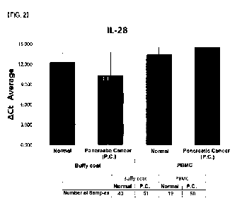
FIG. 2 is a graph showing expression level differences of IL-28 in buffy coat
and
PBMC samples obtained from pancreatic cancer patients and normal persons.
FIG. 3 is a schematic view illustrating a process of preparing a buffy coat
sample
in comparison with a PBMC sample.
FIG. 4 shows graphs of expression levels of IL-28 (upper) and IL-29 (lower) in
buffy coat and PBMC samples obtained from the same pancreatic cancer patients.
FIG. 5a is a graph showing expression levels of IL-28 in buffy coat and PBMC
samples obtained from pancreatic cancer patients in comparison with that that
in PBMC
samples from normal persons.
FIG. 5b is a graph showing expression levels of IL-28 in buffy coat and PBMC
samples obtained from pancreatic cancer patients in comparison with that that
in buffy
coat samples from normal persons.
FIG. 6a is a graph showing expression levels of IL-29 in buffy coat and PBMC
samples obtained from pancreatic cancer patients in comparison with that that
in PBMC
13
Date Recue/Date Received 2023-01-26
CA 03190426 2023-01-26
samples from normal persons.
FIG. 6b is a graph showing expression levels of IL-29 in buffy coat and PBMC
samples obtained from pancreatic cancer patients in comparison with that that
in buffy
coat samples from normal persons.
FIG. 7a is a graph showing expression levels of IL-28 in buffy coat and PBMC
samples obtained from pancreatic cancer patients in comparison with that that
in
samples (average value of IL-28 expression levels in PBMC and buffy coat
samples)
from normal persons.
FIG. 7b is a graph showing expression levels of IL-29 in buffy coat and PBMC
io samples obtained from pancreatic cancer patients in comparison with that
that in
samples (average value of IL-29 expression levels in PBMC and buffy coat
samples)
from normal persons.
Mode for Invention
A better understanding of the present disclosure may be obtained through the
following examples which are set forth to illustrate, but are not to be
construed as limiting
the present disclosure.
EXAMPLE 1: Sample Preparation
1.1. Buffy coat sample preparation
An examination was made of expression changes of pancreatic cancer
biomarkers between pancreatic cancer samples and normal samples. In this
regard,
mRNA expression of IL-29 and IL-28 was analyzed in buffy coats separated from
blood
of pancreatic cancer patients. For comparison, the same experiment was
conducted
with the blood-derived buffy coats from healthy persons (hereinafter referred
to as
normal subjects).
First, 5 cc of blood from each of pancreatic cancer patients and normal
subjects
(normal control) was sampled into an EDTA tube and stored at room temperature.
The
14
Date Recue/Date Received 2023-01-26
CA 03190426 2023-01-26
blood-containing EDTA tube was centrifuged at 800 g and 4 C for 10 min. In the
tube,
a plasma, a buffy coat, and red blood cells were layered in the order from the
top. After
removal of the plasma, the buffy coat was recovered in an amount of 250 pl and
stored
at -80 C.
Total RNA was extracted from 250 pl of the deep-frozen buffy coat using
NucleoSpin0 RNA Blood kit (MACHEREY-NAGEL) according to the recommended
protocol. From 1 pg of the RNA, cDNA was synthesized. For cDNA synthesis,
GoScriptTM Reverse Transcription system kit (Promega) was used.
1.2. Peripheral blood mononuclear cell (PBMC) sample preparation
An examination was made of expression changes of pancreatic cancer
biomarkers between pancreatic cancer samples and normal samples. In this
regard,
mRNA expression of IL-29 and IL-28 (IL-28A) was analyzed in peripheral blood
mononuclear cells from blood of pancreatic cancer patients. For comparison,
the same
is experiment was conducted with the peripheral blood mononuclear cells from
healthy
persons (hereinafter referred to as normal subjects).
First, 5 cc of blood from each of pancreatic cancer patients and normal
subjects
(normal control) was sampled into an EDTA tube and stored at room temperature.
When blood is high in viscosity, it is difficult to separate mononuclear cells
from the blood.
Thus, the blood sample was diluted at a ratio of 1:1 with 1X PBS. The
lymphocyte
separation medium (LymphosepTM, L0560-500, Biowest) was prepared as a density
gradient medium in a 15-ml conical tube, followed by adding the blood sample
on Ficoll,
with care to prevent mingling with the separation medium. Centrifugation at
800g for 30
min with minimum acceleration and deceleration resulted in separating the
blood into
plasma, peripheral blood mononuclear cells (PBMC), Lymphosep, granulocytes,
and
erythrocytes in the density-increasing order from the top to the bottom in the
tube. The
medium white cell layer (monocytes) was recovered. The cells were added with
PBS,
centrifuged at 400g for 3 min, and washed twice before use. The residual cells
were
Date Recue/Date Received 2023-01-26
CA 03190426 2023-01-26
stored at -80 C.
The PBMC thus prepared was added with 1 ml of Trizol to suspend the adherent
cells. After 200 pl of chloroform, centrifugation was conducted at 12,000g for
15 min.
Only the transparent uppermost supernatant was picked out and mixed with an
equal
volume of isopropanol before centrifugation at 12,000g for 15 min. After the
supernatant
was discarded, the pellet was washed with 1 ml of 75% ethanol by
centrifugation at
12,000g for 10 min. The supernatant was discarded and the pellet was
sufficiently dried
for 1 hour. The RNA pellet was added with 30 pl of nuclease-free water before
RNA
quantitation. For cDNA synthesis, 1 pg of RNA was used. cDNA synthesis was
carried
out using GoScriptTM Reverse Transcription system kit (Promega).
EXAMPLE 2. Real-time qPCR
Gene expression in the samples prepared in Example 1 was measured by real-
is time qPCR. The real-time qPCR was carried out in a probe-based multiplex
PCR assay.
For multiplex PCR, respective probes for the markers IL-29 (GenBank Accession
No.
NM 172140.2) and IL-28A (GenBank Accession No. NM 172138.2; hereinafter
_ _
referred to as" IL-28") were labeled with FAM dye while GAPDH for use as an
internal
reference gene was labeled with HEX dye. Primers and probes were purchased
from
IDT (Integrated DNA Technologies, Inc.).
To identify the expression of each gene in the samples, a reaction mix
including
GoTaq0 Probe qPCR Master Mix (Promega) was prepared in a final volume of 20 pl
according to the protocol. Gene expression assay was performed using
QuantStudio 3
and 5 Real-Time PCR system instrument (Applied Biosystems) under the standard
cycling condition provided by the software of the instrument. Real-time qPCR
results
were expressed as ACt values, which were differences between Ct values, that
is,
mRNA expression levels of the markers IL-29 and IL-28 and the internal
reference gene
GAPDH. All data for comparison of mRNA expression were analyzed in terms of
ACt.
16
Date Recue/Date Received 2023-01-26
CA 03190426 2023-01-26
Nucleotide sequences (5'¨>3') of the primers and probes used are summarized
in Table 2, below.
TABLE 2
Sequence (5'¨>3') SEQ ID
NO:
IL-29_F GGT TCA AAT CTC TGT CAC CAC A 4
primer
IL-29_R GAA GAC AGG AGA GCT GCA AC 5
primer
I L-28A_F CAG CCT CAG AGT GTT TCT TCT 6
primer
IL-28A_R TCC AGT CAC GGT CAG CA 7
primer
GPC-1_F GTC ATG AAG CTG GTC TAC TG 8
primer
GPC-1_R AGC CCT TGA GCA CAT TTC 9
primer
IL-29_Probe FAM/TCAAGAAGG/ZEN/CCAGGGACGCC/IBFQ 10
IL-28_Probe FAM- 11
TCATGTCTA/ZEN/GTTTCATTCCTGATCTCTGGTCT
-I BFQ
(in Table 2, the probes were structured to include 5' FAM dye, internal ZEN
Quencher, and 3' Iowa Black Fluorescent Quencher (IBFQ))
EXAMPLE 3: Measurement of Marker Expression in Each Sample
Results of the real-time qPCR performed in Example 2 are depicted in FIGS. 1
and 2.
FIG. 1 is a graph showing expression level differences of IL-29 in buffy coat
samples obtained from 52 pancreatic cancer patients and 44 normal persons and
in
PBMC samples obtained from 54 pancreatic cancer patients and 19 normal
persons.
As can be seen in FIG. 1, the average ACt value of IL-29 was significantly
reduced in
the buffy coat samples from the pancreatic cancer patients, compared to the
normal
17
Date Recue/Date Received 2023-01-26
CA 03190426 2023-01-26
persons. In contrast, the average ACt value of IL-29 in the PBMC samples were
rather
increased for the pancreatic cancer patients, compared to the normal persons,
with an
insignificant difference therebetween. From the data, it was identified that
the expression
level of IL-29 was significantly increased in pancreatic cancer patients,
compared to
normal persons, and the expression pattern of IL-29 in pancreatic cancer
patients
distinctively differed from buffy coat samples to PBMC samples.
FIG. 2 is a graph showing expression level differences of IL-28 in buffy coat
samples obtained from 51 pancreatic cancer patients and 43 normal persons and
in
PBMC samples obtained from 50 pancreatic cancer patients and 19 normal
persons.
io As can be seen in FIG. 2, the average ACt value of IL-28 was
significantly reduced in
the buffy coat samples from the pancreatic cancer patients, compared to the
normal
persons. In contrast, the average ACt value of IL-28 in the PBMC samples were
rather
increased for the pancreatic cancer patients, compared to the normal persons,
with an
insignificant difference therebetween. From the data, it was identified that
the expression
is level of IL-28 was significantly increased in pancreatic cancer patients,
compared to
normal persons, and the expression pattern of IL-28 in pancreatic cancer
patients
distinctively differed from buffy coat samples to PBMC samples.
Taken together, the data indicate that IL-29 and IL-28 are both useful as
markers
for pancreatic cancer due to their significant difference in expression level
between
20 pancreatic cancer patients and normal persons and that when the
markers are used,
more reliable results could be obtained from a buffy coat sample than a PBMC
sample.
In addition, a greater difference between pancreatic cancer patients and
normal persons
was observed for IL-28 than IL-29.
25
EXAMPLE 4: Comparison between Buffy Coat and PBMC from Same
Person
From the same persons (three pancreatic cancer patients (P.C)), buffy coat
samples and PBMC samples were prepared referring to Examples 1.1 and 1.2,
18
Date Recue/Date Received 2023-01-26
CA 03190426 2023-01-26
respectively. The prepared buffy coat and PBMC samples were measured for mRNA
expression levels (ACt) of IL-29 and IL-28 in the same manner as in Example 2.
Average mRNA expression levels of IL-29 and IL-28 are depicted in FIG. 4 (No.
of samples: three buffy coat samples; three PBMC samples). As shown in Figure
4, the
mRNA expression levels (ACt) of IL-29 and IL-28 were significantly higher in
the buffy
coat samples, compared to the PBMC samples,
Comparison of mRNA expression levels in buffy coat samples and PBMC
samples from same persons were made between pancreatic cancer patients and
normal persons and the results are depicted in FIGS. 5a, 5b, 6a, 6b, 7a, and
7b (all
io expressed as average values).
FIG. 5a shows IL-28 expression levels in buffy coat and PBMC samples from
pancreatic cancer patients in comparison with that in the PBMC samples from
normal
persons; and FIG. 5b shows IL-28 expression levels in buffy coat and PBMC
samples
from pancreatic cancer patients in comparison with that in the buffy coat
samples from
normal persons (no. of samples: 43 normal person buffy coat samples; 3
pancreatic
cancer buffy coat samples; 19 normal person PBMC samples; and 3 pancreatic
cancer
PBMC samples).
FIG. 6a shows IL-29 expression levels in buffy coat and PBMC samples from
pancreatic cancer patients in comparison with that in the PBMC samples from
normal
persons; and FIG. 6b shows IL-29 expression levels in buffy coat and PBMC
samples
from pancreatic cancer patients in comparison with that in the buffy coat
samples from
normal persons (no. of samples: 44 normal person buffy coat samples; 3
pancreatic
cancer buffy coat samples; 19 normal person PBMC samples; and 3 pancreatic
cancer
PBMC samples).
FIG. 7a shows IL-28 expression levels in buffy coat and PBMC samples from
pancreatic cancer patients in comparison with that in the samples from normal
persons
(average value of integrated IL-28 expression levels in both PBMC and buffy
coat
samples); and FIG. 7b shows IL-29 expression levels in buffy coat and PBMC
samples
19
Date Recue/Date Received 2023-01-26
CA 03190426 2023-01-26
from pancreatic cancer patients in comparison with that in the samples from
normal
persons (average value of integrated IL-29 expression levels in both PBMC and
buffy
coat samples) (no. of samples: 43 and 44 normal person buffy coat samples (for
IL-28
and IL-29, respectively); 3 pancreatic cancer buffy coat samples; 19 normal
person
PBMC samples; and 3 pancreatic cancer PBMC samples).
As shown in FIGS. 5a, 5b, 6a, 6b, 7a, and 7b, no significant differences of IL-
28
and IL-29 expression levels in PBMC samples were not observed between normal
persons and pancreatic cancer patients. In contrast, a remarkable difference
of IL-28
and/or IL-29 expression level in the buffy coat samples was detected between
normal
io persons and pancreatic cancer patients. These results imply that that
pancreatic cancer
patients can be diagnosed more accurately and efficiently by measuring the
levels of IL-
28 and/or IL-29 in buffy coat samples.
Date Recue/Date Received 2023-01-26