Note: Descriptions are shown in the official language in which they were submitted.
CA 03190722 2023-02-03
WO 2022/031783 PCT/US2021/044449
ADAPTER POLYPEPTIDES AND METHODS OF USING THE SAME
CROSS REFERENCE TO RELATED APPLICATIONS
[001] This application claims the benefit of U.S. Provisional Patent
Application No. 63/061,749
filed on August 5, 2020. Priority is claimed pursuant to 35 U.S.C. 119. The
above noted
patent application is incorporated by reference as if set forth fully herein.
INCORPORATION BY REFERENCE
[002] All publications, patents, and patent applications mentioned in this
specification are herein
incorporated by reference, in their entireties, to the same extent as if each
individual publication,
patent, or patent application was specifically and individually indicated to
be incorporated by
reference.
BACKGROUND
[003] Extracellular vesicles are secreted by a wide variety of cell types. In
general, extracellular
vesicles such as exosomes, microvesicles, and apoptotic bodies are membrane-
bound and can be
loaded with a therapeutic cargo. For example, exosomes are a type of membrane-
bound
extracellular vesicle that are secreted by most eukaryotic cells. Exosome
biogenesis may begin
with pinching off of endosomal invaginations into the multivesicular body,
forming intraluminal
vesicles. If the multivesicular body fuses with the plasma membrane of the
cell, the intraluminal
vesicles may be released as exosomes. Microvesicles are budded out from a cell
membrane
surface. Apoptotic bodies, on the other hand, are released from dead cells.
Exosomes,
microvesicles, and apoptotic bodies can be released in vivo or in vitro, such
as in cell-culture.
[004] Extracellular vesicles have been explored as a vehicle for encapsulating
and delivering
therapeutics. Directing the extracellular vesicles to a target is generally
challenging, as the
majority of the extracellular vesicles are degraded in the liver, spleen,
and/or kidney. Also,
designing and manufacturing extracellular vesicles for encapsulating
therapeutics for targeted
delivery is time-consuming and expensive. For example, an extracellular
vesicle designed for
targeting one cell type may not effectively target another cell type.
Therefore, there remains a
need for extracellular vesicle that can be readily modified to target multiple
cell types. There
also remains a need for extracellular vesicles that can encapsulate sufficient
quantity and quality
of therapeutics to be delivered to the targeted cell.
SUMMARY
[005] This disclosure provides extracellular vesicles designed to target a
wide variety of cell-
types, including different cells and organs within the body and cells
associated with a disease or
disorder. In some instances, the extracellular vesicles provided herein can be
readily modified to
-1-
CA 03190722 2023-02-03
WO 2022/031783 PCT/US2021/044449
specifically bind to a target. For example, they may contain an extracellular
domain (e.g., an
extracellular domain of a transmembrane protein within the membrane of the
extracellular
vesicle) that binds to a cell-surface marker. In general, the extracellular
vesicles provided herein
comprise an adapter polypeptide with an extracellular domain and optionally a
transmembrane
domain that binds to a cell-surface marker.
[006] Disclosed herein, in some aspects, are compositions comprising at least
one extracellular
vesicle, said extracellular vesicle comprising: at least one adapter
polypeptide comprising a
peptide sequence that binds to an Fc region of an antibody with a dissociation
constant (Kd) of
less than or equal to 10-9M, wherein said adapter polypeptide comprises an
extracellular domain;
said antibody complexed with said adapter polypeptide, wherein said antibody
binds a first cell-
surface marker associated with a diseased cell; and at least one therapeutic.
Described herein, in
some aspects, are compositions comprising at least one extracellular vesicle,
said extracellular
vesicle comprising: at least one adapter polypeptide comprising a peptide
sequence that is at
least 70% identical to a Fc receptor that specifically recognizes a Fc region
of an antibody,
wherein said adapter polypeptide comprises an extracellular domain; said
antibody complexed
with said adapter polypeptide, wherein said antibody binds a first cell-
surface marker associated
with a diseased cell; and at least one therapeutic. In some aspects, said Fc
receptor is a Fc-
gamma receptor, Fc-alpha receptor, or Fc-epsilon receptor. In some aspects,
said Fc receptor
comprises FcyRI (CD64), FcyRII (CD32), or FcyRIII (CD16). In some aspects,
said Fc receptor
is CD64. In some aspects, said adapter polypeptide further comprises a
targeting domain that
binds a second cell-surface marker associated with said diseased cell, wherein
said targeting
domain is attached to said extracellular domain of said adapter polypeptide.
In some aspects,
said targeting domain is selected from the group consisting of a tumor homing
peptide, a tumor
targeting domain, a tissue-targeting domain, a cell-penetrating peptide, a
viral membrane
protein, and any combination or fragment thereof In some aspects, said
diseased cell is a cancer
cell or a non-cancerous lesion cell. In some aspects, said first cell-surface
marker comprises
EGFR, PD-L1, or ROR1. In some aspects, said first cell-surface marker and said
second cell-
surface marker are different. In some aspects, said first cell-surface marker
and said second cell-
surface marker are identical. In some aspects, said antibody is a humanized
monoclonal
antibody. In some aspects, said antibody is selected from the group consisting
of humanized
anti-EGFR antibody clone C225, humanized anti-ROR1 antibody clone 2A2, and
humanized
anti-PD-Li antibody clone SP142. In some aspects, said humanized monoclonal
antibody
comprises an IgG. In some aspects, said humanized monoclonal antibody
comprises an IgG1 or
IgG3. In some aspects, said antibody is non-covalently complexed with said
adapter
polypeptide. In some aspects, said Fc region of said antibody is configured to
complex to said
-2-
CA 03190722 2023-02-03
WO 2022/031783 PCT/US2021/044449
adapter polypeptide in an acidic environment. In some aspects, said Fc region
of said antibody is
configured to be released from complexes to said adapter polypeptide in an
acidic environment.
In some aspects, said at least one therapeutic is within said extracellular
vesicle. In some
aspects, said at least one therapeutic is expressed on an extracellular
surface of said extracellular
vesicle. In some aspects, said at least one therapeutic is attached to said
extracellular domain. In
some aspects, said at least one therapeutic comprises a therapeutic
polynucleotide, a therapeutic
polypeptide, a therapeutic compound, a cancer drug, or a combination thereof.
In some aspects,
said therapeutic polynucleotide comprises a messenger RNA, a microRNA, a
shRNA, or a
combination thereof In some aspects, said extracellular vesicle is an exosome,
a microvesicle,
or an apoptotic body. In some aspects, said extracellular vesicle is an
exosome.
[007] Described herein, in some instances, are methods of treating a subject,
said methods
comprising administering a therapeutically effective amount of a
pharmaceutical composition to
said subject, wherein said pharmaceutical composition comprises the
compositions described
herein. In some aspects, said pharmaceutical composition comprises at least
one
pharmaceutically acceptable excipient. In some aspects, said subject has
cancer or a non-
cancerous lesion. In some aspects, said subject has glioma. In some aspects,
subject has
muscular dystrophy. In some aspects, said muscular dystrophy is selected from
the group
consisting of Duchenne muscular dystrophy, Becker muscular dystrophy,
facioscapulohumeral
muscular dystrophy, congenital muscular dystrophy, and myotonic dystrophy. In
some aspects,
said subject has a retinal disease. In some aspects, said retinal disease is
retinitis pigmentosa or
Leber's congenital amaurosis. In some aspects, said therapeutically effective
amount of said
pharmaceutical composition comprises a therapeutically effective dose. In some
aspects, said
subject is administered said therapeutically effective amount of said
pharmaceutical composition
at a therapeutically effective frequency. In some aspects, said subject is
administered said
therapeutically effective amount of said pharmaceutical composition at a
frequency of at least
once per year. In some aspects, said subject is administered said
therapeutically effective
amount of said pharmaceutical composition at a frequency of at least once
every six months. In
some aspects, said subject is administered said therapeutically effective
amount of said
pharmaceutical composition at a frequency of at least once per month. In some
aspects, said
subject is administered said therapeutically effective amount of said
pharmaceutical composition
at a frequency of at least once per week. In some aspects, said pharmaceutical
composition is an
aqueous formulation. In some aspects, said pharmaceutical composition is
formulated for
injection. In some aspects, said pharmaceutical composition is administered to
said subject
intranasally, intrathecally, intraocularly, intravitreally, retinally,
intravenously, intramuscularly,
-3-
CA 03190722 2023-02-03
WO 2022/031783 PCT/US2021/044449
intraventricularly, intracerebrally, intracerebellarly,
intracerebroventricularly,
intraperenchymally, subcutaneously, or a combination thereof
[008] Described herein, in some cases, are methods of producing a composition,
said methods
comprising: transfecting an extracellular vesicle donor cell with at least one
heterologous
polynucleotide encoding an adapter polypeptide, wherein said adapter
polypeptide comprises a
peptide sequence that is at least 70% identical to a Fc receptor, wherein said
Fc receptor
recognizes a Fc region of an antibody; collecting an extracellular vesicle
released from said
extracellular vesicle donor cell, wherein said extracellular vesicle released
from said
extracellular vesicle donor cell expresses said adapter polypeptide, wherein
said adapter
polypeptide comprises an extracellular domain, and wherein said extracellular
vesicle comprises
at least one therapeutic; and complexing said antibody to said extracellular
domain, wherein said
antibody binds a first cell-surface marker associated with a diseased cell. In
some aspects, said
Fc receptor is a Fc-gamma receptor, Fc-alpha receptor, or Fc-epsilon receptor.
In some aspects,
said Fc receptor is FcyRI (CD64), FcyRII (CD32), or FcyRIII (CD16). In some
aspects, said Fc
receptor is CD64. In some aspects, said adapter polypeptide further comprises
a targeting
domain that binds a second cell-surface marker associated with said diseased
cell, wherein said
targeting domain is attached to said extracellular domain. In some aspects,
said first cell-surface
marker or said second cell-surface marker is associated with a cancer cell or
a non-cancerous
lesion cell. In some aspects, said first cell-surface marker comprises EGFR,
PD-L1, or ROR1. In
some aspects, said first cell-surface marker and said second cell-surface
marker are different. In
some aspects, said first cell-surface marker and said second cell-surface
markers are identical. In
some aspects, said targeting domain is selected from the group consisting of a
tumor homing
peptide, a tumor targeting domain, a tissue-targeting domain, a cell-
penetrating peptide, a viral
membrane protein, and any combination or fragment thereof. In some aspects,
said at least one
therapeutic is within said extracellular vesicle. In some aspects, said at
least one therapeutic is
expressed on an extracellular surface of said extracellular vesicle. In some
aspects, said at least
one therapeutic is attached to said extracellular domain. In some aspects,
said at least one
therapeutic comprises a therapeutic polynucleotide, a therapeutic polypeptide,
a therapeutic
compound, a cancer drug, or a combination thereof In some aspects, said
therapeutic
polynucleotide comprises a messenger RNA, a microRNA, a shRNA, or a
combination thereof
In some aspects, said extracellular vesicle released from said extracellular
vesicle donor cell is
an exosome, a microvesicle, or an apoptotic body. In some aspects, said
extracellular vesicle
released from said extracellular vesicle donor cell is an exosome. In some
aspects, said
extracellular vesicle donor cell comprises electroporation, microfluidic
electroporation,
microchannel electroporation, or nanochannel electroporation. In some aspects,
said
-4-
CA 03190722 2023-02-03
WO 2022/031783 PCT/US2021/044449
microchannel electroporation or said nanochannel electroporation comprises use
of micropore
patterned silicon wafers, nanopore patterned silicon wafers, track etch
membranes, ceramic
micropore membranes, ceramic nanopore membranes, other porous materials, or a
combination
thereof. In some aspects, transfecting said extracellular vesicle donor cell
comprises
nanochannel electroporation, and wherein said at least one heterologous
polynucleotide is
nanoelectroporated into said extracellular vesicle donor cell via a
nanochannel located on a
biochip. In some aspects, transfecting said extracellular vesicle donor cell
comprises use of a
gene gun, micro-needle array, nano-needle array, sonication, or chemical
permeation. In some
aspects, said at least one heterologous polynucleotide is a plasmid.
[009] Described herein, in some cases, are compositions comprising at least
one extracellular
vesicle, comprising: at least one adapter polypeptide comprising a peptide
sequence that binds to
an Fc region of an antibody with a dissociation constant (Kd) of less than or
equal to 10-9M,
wherein said adapter polypeptide comprises an extracellular domain; said
antibody complexed
with said adapter polypeptide, wherein said antibody specifically binds a
first cell-surface
marker associated with an immune cell; and at least one viral mimic peptide.
Described herein,
in some cases, is a composition comprising at least one extracellular vesicle,
comprising: at least
one adapter polypeptide comprising a peptide sequence that is at least 70%
identical to a Fc
receptor that binds to an Fc region of an antibody, wherein said adapter
polypeptide comprises
an extracellular domain; said antibody complexed with said adapter
polypeptide, wherein said
antibody specifically binds a first cell-surface marker associated with an
immune cell; and at
least one viral mimic peptide. In some aspects, said Fc receptor is a Fc-gamma
receptor, Fc-
alpha receptor, or Fc-epsilon receptor. In some aspects, said Fc receptor
comprises FcyRI
(CD64), FcyRII (CD32), or FcyRIII (CD16). In some aspects, said Fc receptor is
CD64. In some
aspects, said adapter polypeptide further comprises a targeting domain that
binds a second cell-
surface marker associated with said immune cell, wherein said targeting domain
is attached to
said extracellular domain of said adapter polypeptide. In some aspects, said
immune cell is a T
cell, a B cell, a dendritic cell, a macrophage, or a natural killer (NK) cell.
In some aspects, said
first cell-surface marker comprises LILRA4, CD3, CD19, CD20, or CD28. In some
aspects, said
first cell-surface marker and said second cell-surface marker are different.
In some aspects, said
first cell-surface marker and said second cell-surface marker are identical.
In some aspects, said
antibody is a humanized monoclonal antibody. In some aspects, said antibody is
an IgG. In some
aspects, said antibody is an IgG1 or IgG3. In some aspects, said antibody is
non-covalently
complexed with said adapter polypeptide. In some aspects, said Fc region of
said antibody is
configured to complex to said adapter polypeptide in an acidic environment. In
some aspects,
said Fc region of said antibody is configured to be released from complexed to
said adapter
-5-
CA 03190722 2023-02-03
WO 2022/031783 PCT/US2021/044449
polypeptide in an acidic environment. In some aspects, said at least one viral
mimic peptide is
expressed on an extracellular surface of said extracellular vesicle. In some
aspects, said at least
one viral mimic peptide is attached to said extracellular domain. In some
aspects, said at least
one viral mimic peptide comprises a peptide sequence that is at least 70%
identical with a
SARS-CoV-2 viral protein. In some aspects, said SARS-CoV-2 viral protein
comprises an
Envelopment (E) protein, a Nucleocapsid (N) protein, a Membrane (M) protein,
or a Spike (S)
protein. In some aspects, said SARS-CoV-2 viral protein is said S protein. In
some aspects, said
extracellular vesicle comprises an exosome, a microvesicle, or an apoptotic
body. In some
aspects, said extracellular vesicle is an exosome.
[0010] Described herein, in some cases, are methods of vaccinating a subject,
said methods
comprising administering a therapeutically effective amount of a
pharmaceutical composition to
said subject, wherein said pharmaceutical composition comprises a composition
described
herein. In some aspects, said pharmaceutical composition comprises at least
one
pharmaceutically acceptable excipient. In some aspects, said therapeutically
effective amount of
said pharmaceutical composition comprises a therapeutically effective dose. In
some aspects,
said subject is administered said therapeutically effective amount of said
pharmaceutical
composition at a therapeutically effective frequency. In some aspects, said
subject is
administered said therapeutically effective amount of said pharmaceutical
composition at a
frequency of at least once per year. In some aspects, said subject is
administered said
therapeutically effective amount of said pharmaceutical composition at a
frequency of at least
once every six months. In some aspects, said subject is administered said
therapeutically
effective amount of said pharmaceutical composition at a frequency of at least
once per month.
In some aspects, said subject is administered said therapeutically effective
amount of said
pharmaceutical composition at a frequency of at least once per week. In some
aspects, said
pharmaceutical composition is an aqueous formulation. In some aspects, said
pharmaceutical
composition is formulated for injection. In some aspects, said pharmaceutical
composition is
administered to said subject intranasally, intrathecally, intraocularly,
intravitreally, retinally,
intravenously, intramuscularly, intraventricularly, intracerebrally,
intracerebellarly,
intracerebroventricularly, intraperenchymally, subcutaneously, or a
combination thereof
[0011] Described herein, in some cases, are methods of producing a composition
described
herein, said methods comprising: transfecting an extracellular vesicle donor
cell with at least one
heterologous polynucleotide encoding an adapter polypeptide, wherein said
adapter polypeptide
comprises a peptide sequence that is at least 70% identical to a Fc receptor,
wherein said Fc
receptor recognizes a Fc region of an antibody; collecting an extracellular
vesicle released from
said extracellular vesicle donor cell, wherein said extracellular vesicle
released from said
-6-
CA 03190722 2023-02-03
WO 2022/031783 PCT/US2021/044449
extracellular vesicle donor cell expresses said adapter polypeptide, wherein
said adapter
polypeptide comprises an extracellular domain, and wherein said extracellular
vesicle comprises
at least one viral mimic peptide; and complexing said antibody to said
extracellular domain,
wherein said antibody binds a first cell-surface marker associated with an
immune cell. In some
aspects, said Fc receptor is a Fc-gamma receptor, Fc-alpha receptor, or Fc-
epsilon receptor. In
some aspects, said Fc receptor comprises FcyRI (CD64), FcyRII (CD32), or
FcyRIII (CD16). In
some aspects, said Fc receptor is CD64. In some aspects, said adapter
polypeptide further
comprises a targeting domain that binds a second cell-surface marker
associated with said
immune cell, wherein said targeting domain is attached to said extracellular
domain. In some
aspects, said immune cell is a T cell, a B cell, a dendritic cell, a
macrophage, or a natural killer
(NK) cell. In some aspects, said first cell-surface marker comprises LILRA4,
CD3, CD19,
CD20, or CD28. In some aspects, said first cell-surface marker and said second
cell-surface
marker are different. In some aspects, said first cell-surface marker and said
second cell-surface
markers are identical. In some aspects, said antibody comprises a humanized
monoclonal
antibody. In some aspects, said antibody is an IgG. In some aspects, said
antibody is an IgG1 or
IgG3. In some aspects, said antibody is non-covalently complexed with said
adapter
polypeptide. In some aspects, said Fc region of said antibody is configured to
complex to said
adapter polypeptide in an acidic environment. In some aspects, said Fc region
of said antibody is
configured to be released from complexed to said adapter polypeptide in an
acidic environment.
In some aspects, said at least one viral mimic peptide is expressed on an
extracellular surface of
said extracellular vesicle. In some aspects, said at least one viral mimic
peptide is attached to
said extracellular domain. In some aspects, said at least one viral mimic
peptide comprises a
peptide sequence that is at least 70% identical with a SARS-CoV-2 viral
protein. In some
aspects, said SARS-CoV-2 viral protein comprises an Envelopment (E) protein, a
Nucleocapsid
(N) protein, a Membrane (M) protein, or a Spike (S) protein. In some aspects,
said SARS-CoV-2
viral protein is said S protein. In some aspects, said extracellular vesicle
comprises an exosome,
a microvesicle, or an apoptotic body. In some aspects, said extracellular
vesicle is an exosome.
In some aspects, transfecting said extracellular vesicle donor cell comprises
electroporation,
microfluidics electroporation, microchannel electroporation, or nanochannel
electroporation. In
some aspects, said microchannel electroporation or said nanochannel
electroporation comprises
use of micropore patterned silicon wafers, nanopore patterned silicon wafers,
track etch
membranes, ceramic micropore membranes, ceramic nanopore membranes, other
porous
materials, or a combination thereof In some aspects, transfecting said
extracellular vesicle donor
cell comprises nanochannel electroporation, and wherein said at least one
heterologous
polynucleotide is nanoelectroporated into said extracellular vesicle donor
cell via a nanochannel
-7-
CA 03190722 2023-02-03
WO 2022/031783 PCT/US2021/044449
located on a biochip. In some aspects, transfecting said extracellular vesicle
donor cell
comprises a use of a gene gun, micro-needle array, nano-needle array,
sonication, or chemical
permeation. In some aspects, said at least one heterologous polynucleotide is
a plasmid.
[0012] Disclosed herein, in some aspects, are compositions comprising at least
one extracellular
vesicle, said extracellular vesicle comprising: at least one adapter
polypeptide comprising a
peptide sequence that binds to an Fc region of a binding molecule with a
dissociation constant
(Kd) of less than or equal to 10-9M, wherein said adapter polypeptide
comprises an extracellular
domain; said binding molecule complexed with said adapter polypeptide, wherein
said binding
molecule binds a first cell-surface marker associated with a diseased cell;
and at least one
therapeutic. Described herein, in some aspects, are compositions comprising at
least one
extracellular vesicle, said extracellular vesicle comprising: at least one
adapter polypeptide
comprising a peptide sequence that is at least 70% identical to a Fc receptor
that specifically
recognizes a Fc region of a binding molecule, wherein said adapter polypeptide
comprises an
extracellular domain; said binding molecule complexed with said adapter
polypeptide, wherein
said binding molecule binds a first cell-surface marker associated with a
diseased cell; and at
least one therapeutic. In some aspects, said Fc receptor is a Fc-gamma
receptor, Fc-alpha
receptor, or Fc-epsilon receptor. In some aspects, said Fc receptor comprises
FcyRI (CD64),
FcyRII (CD32), or FcyRIII (CD16). In some aspects, said Fc receptor is CD64.
In some aspects,
said adapter polypeptide further comprises a targeting domain that binds a
second cell-surface
marker associated with said diseased cell, wherein said targeting domain is
attached to said
extracellular domain of said adapter polypeptide. In some aspects, said
targeting domain is
selected from the group consisting of a tumor homing peptide, a tumor
targeting domain, a
tissue-targeting domain, a cell-penetrating peptide, a viral membrane protein,
and any
combination or fragment thereof In some aspects, said diseased cell is a
cancer cell or a non-
cancerous lesion cell. In some aspects, said first cell-surface marker
comprises EGFR, PD-L1, or
ROR1. In some aspects, said first cell-surface marker and said second cell-
surface marker are
different. In some aspects, said first cell-surface marker and said second
cell-surface marker are
identical. In some aspects, said binding molecule is a humanized monoclonal
antibody. In some
aspects, said binding molecule is selected from the group consisting of
humanized anti-EGFR
antibody clone C225, humanized anti-ROR1 antibody clone 2A2, and humanized
anti-PD-Li
antibody clone SP142. In some aspects, said humanized monoclonal antibody
comprises an
IgG. In some aspects, said humanized monoclonal antibody comprises an IgG1 or
IgG3. In some
aspects, said binding molecule is non-covalently complexed with said adapter
polypeptide. In
some aspects, said Fc region of said binding molecule is configured to complex
to said adapter
polypeptide in an acidic environment. In some aspects, said Fc region of said
binding molecule
-8-
CA 03190722 2023-02-03
WO 2022/031783 PCT/US2021/044449
is configured to be released from complexes to said adapter polypeptide in an
acidic
environment. In some aspects, said at least one therapeutic is within said
extracellular vesicle. In
some aspects, said at least one therapeutic is expressed on an extracellular
surface of said
extracellular vesicle. In some aspects, said at least one therapeutic is
attached to said
extracellular domain. In some aspects, said at least one therapeutic comprises
a therapeutic
polynucleotide, a therapeutic polypeptide, a therapeutic compound, a cancer
drug, or a
combination thereof In some aspects, said therapeutic polynucleotide comprises
a messenger
RNA, a microRNA, a shRNA, or a combination thereof In some aspects, said
extracellular
vesicle is an exosome, a microvesicle, or an apoptotic body. In some aspects,
said extracellular
vesicle is an exosome.
[0013] Described herein, in some instances, are methods of treating a subject,
said methods
comprising administering a therapeutically effective amount of a
pharmaceutical composition to
said subject, wherein said pharmaceutical composition comprises the
compositions described
herein. In some aspects, said pharmaceutical composition comprises at least
one
pharmaceutically acceptable excipient. In some aspects, said subject has
cancer or a non-
cancerous lesion. In some aspects, said subject has glioma. In some aspects,
subject has
muscular dystrophy. In some aspects, said muscular dystrophy is selected from
the group
consisting of Duchenne muscular dystrophy, Becker muscular dystrophy,
facioscapulohumeral
muscular dystrophy, congenital muscular dystrophy, and myotonic dystrophy. In
some aspects,
said subject has a retinal disease. In some aspects, said retinal disease is
retinitis pigmentosa or
Leber's congenital amaurosis. In some aspects, said therapeutically effective
amount of said
pharmaceutical composition comprises a therapeutically effective dose. In some
aspects, said
subject is administered said therapeutically effective amount of said
pharmaceutical composition
at a therapeutically effective frequency. In some aspects, said subject is
administered said
therapeutically effective amount of said pharmaceutical composition at a
frequency of at least
once per year. In some aspects, said subject is administered said
therapeutically effective
amount of said pharmaceutical composition at a frequency of at least once
every six months. In
some aspects, said subject is administered said therapeutically effective
amount of said
pharmaceutical composition at a frequency of at least once per month. In some
aspects, said
subject is administered said therapeutically effective amount of said
pharmaceutical composition
at a frequency of at least once per week. In some aspects, said pharmaceutical
composition is an
aqueous formulation. In some aspects, said pharmaceutical composition is
formulated for
injection. In some aspects, said pharmaceutical composition is administered to
said subject
intranasally, intrathecally, intraocularly, intravitreally, retinally,
intravenously, intramuscularly,
-9-
CA 03190722 2023-02-03
WO 2022/031783 PCT/US2021/044449
intraventricularly, intracerebrally, intracerebellarly,
intracerebroventricularly,
intraperenchymally, subcutaneously, or a combination thereof
[0014] Described herein, in some cases, are methods of producing a
composition, said methods
comprising: transfecting an extracellular vesicle donor cell with at least one
heterologous
polynucleotide encoding an adapter polypeptide, wherein said adapter
polypeptide comprises a
peptide sequence that is at least 70% identical to a Fc receptor, wherein said
Fc receptor
recognizes a Fc region of a binding molecule; collecting an extracellular
vesicle released from
said extracellular vesicle donor cell, wherein said extracellular vesicle
released from said
extracellular vesicle donor cell expresses said adapter polypeptide, wherein
said adapter
polypeptide comprises an extracellular domain, and wherein said extracellular
vesicle comprises
at least one therapeutic; and complexing said binding molecule to said
extracellular domain,
wherein said binding molecule binds a first cell-surface marker associated
with a diseased cell.
In some aspects, said Fc receptor is a Fc-gamma receptor, Fc-alpha receptor,
or Fc-epsilon
receptor. In some aspects, said Fc receptor is FcyRI (CD64), FcyRII (CD32), or
FcyRIII (CD16).
In some aspects, said Fc receptor is CD64. In some aspects, said adapter
polypeptide further
comprises a targeting domain that binds a second cell-surface marker
associated with said
diseased cell, wherein said targeting domain is attached to said extracellular
domain. In some
aspects, said first cell-surface marker or said second cell-surface marker is
associated with a
cancer cell or a non-cancerous lesion cell. In some aspects, said first cell-
surface marker
comprises EGFR, PD-L1, or ROR1. In some aspects, said first cell-surface
marker and said
second cell-surface marker are different. In some aspects, said first cell-
surface marker and said
second cell-surface markers are identical. In some aspects, said targeting
domain is selected
from the group consisting of a tumor homing peptide, a tumor targeting domain,
a tissue-
targeting domain, a cell-penetrating peptide, a viral membrane protein, and
any combination or
fragment thereof. In some aspects, said at least one therapeutic is within
said extracellular
vesicle. In some aspects, said at least one therapeutic is expressed on an
extracellular surface of
said extracellular vesicle. In some aspects, said at least one therapeutic is
attached to said
extracellular domain. In some aspects, said at least one therapeutic comprises
a therapeutic
polynucleotide, a therapeutic polypeptide, a therapeutic compound, a cancer
drug, or a
combination thereof In some aspects, said therapeutic polynucleotide comprises
a messenger
RNA, a microRNA, a shRNA, or a combination thereof In some aspects, said
extracellular
vesicle released from said extracellular vesicle donor cell is an exosome, a
microvesicle, or an
apoptotic body. In some aspects, said extracellular vesicle released from said
extracellular
vesicle donor cell is an exosome. In some aspects, said extracellular vesicle
donor cell comprises
electroporation, microfluidic electroporation, microchannel electroporation,
or nanochannel
-10-
CA 03190722 2023-02-03
WO 2022/031783 PCT/US2021/044449
electroporation. In some aspects, said microchannel electroporation or said
nanochannel
electroporation comprises use of micropore patterned silicon wafers, nanopore
patterned silicon
wafers, track etch membranes, ceramic micropore membranes, ceramic nanopore
membranes,
other porous materials, or a combination thereof. In some aspects,
transfecting said extracellular
vesicle donor cell comprises nanochannel electroporation, and wherein said at
least one
heterologous polynucleotide is nanoelectroporated into said extracellular
vesicle donor cell via a
nanochannel located on a biochip. In some aspects, transfecting said
extracellular vesicle donor
cell comprises use of a gene gun, micro-needle array, nano-needle array,
sonication, or chemical
permeation. In some aspects, said at least one heterologous polynucleotide is
a plasmid.
[0015] Described herein, in some cases, are composition comprising at least
one extracellular
vesicle, comprising: at least one adapter polypeptide comprising a peptide
sequence that binds to
an Fc region of a binding molecule with a dissociation constant (Kd) of less
than or equal to 10-
9M, wherein said adapter polypeptide comprises an extracellular domain; said
binding molecule
complexed with said adapter polypeptide, wherein said binding molecule
specifically binds a
first cell-surface marker associated with an immune cell; and at least one
viral mimic peptide.
Described herein, in some cases, is a composition comprising at least one
extracellular vesicle,
comprising: at least one adapter polypeptide comprising a peptide sequence
that is at least 70%
identical to a Fc receptor that binds to an Fc region of a binding molecule,
wherein said adapter
polypeptide comprises an extracellular domain; said binding molecule complexed
with said
adapter polypeptide, wherein said binding molecule specifically binds a first
cell-surface marker
associated with an immune cell; and at least one viral mimic peptide. In some
aspects, said Fc
receptor is a Fc-gamma receptor, Fc-alpha receptor, or Fc-epsilon receptor. In
some aspects, said
Fc receptor comprises FcyRI (CD64), FcyRII (CD32), or FcyRIII (CD16). In some
aspects, said
Fc receptor is CD64. In some aspects, said adapter polypeptide further
comprises a targeting
domain that binds a second cell-surface marker associated with said immune
cell, wherein said
targeting domain is attached to said extracellular domain of said adapter
polypeptide. In some
aspects, said immune cell is a T cell, a B cell, a dendritic cell, a
macrophage, or a natural killer
(NK) cell. In some aspects, said first cell-surface marker comprises LILRA4,
CD3, CD19,
CD20, or CD28. In some aspects, said first cell-surface marker and said second
cell-surface
marker are different. In some aspects, said first cell-surface marker and said
second cell-surface
marker are identical. In some aspects, said binding molecule is a humanized
monoclonal
antibody. In some aspects, said binding molecule is an IgG. In some aspects,
said binding
molecule is an IgG1 or IgG3. In some aspects, said binding molecule is non-
covalently
complexed with said adapter polypeptide. In some aspects, said Fc region of
said binding
molecule is configured to complex to said adapter polypeptide in an acidic
environment. In some
-11-
CA 03190722 2023-02-03
WO 2022/031783 PCT/US2021/044449
aspects, said Fc region of said binding molecule is configured to be released
from complexed to
said adapter polypeptide in an acidic environment. In some aspects, said at
least one viral mimic
peptide is expressed on an extracellular surface of said extracellular
vesicle. In some aspects,
said at least one viral mimic peptide is attached to said extracellular
domain. In some aspects,
said at least one viral mimic peptide comprises a peptide sequence that is at
least 70% identical
with a SARS-CoV-2 viral protein. In some aspects, said SARS-CoV-2 viral
protein comprises
an Envelopment (E) protein, a Nucleocapsid (N) protein, a Membrane (M)
protein, or a Spike
(S) protein. In some aspects, said SARS-CoV-2 viral protein is said S protein.
In some aspects,
said extracellular vesicle comprises an exosome, a microvesicle, or an
apoptotic body. In some
aspects, said extracellular vesicle is an exosome.
[0016] Described herein, in some cases, are methods of vaccinating a subject,
said methods
comprising administering a therapeutically effective amount of a
pharmaceutical composition to
said subject, wherein said pharmaceutical composition comprises a composition
described
herein. In some aspects, said pharmaceutical composition comprises at least
one
pharmaceutically acceptable excipient. In some aspects, said therapeutically
effective amount of
said pharmaceutical composition comprises a therapeutically effective dose. In
some aspects,
said subject is administered said therapeutically effective amount of said
pharmaceutical
composition at a therapeutically effective frequency. In some aspects, said
subject is
administered said therapeutically effective amount of said pharmaceutical
composition at a
frequency of at least once per year. In some aspects, said subject is
administered said
therapeutically effective amount of said pharmaceutical composition at a
frequency of at least
once every six months. In some aspects, said subject is administered said
therapeutically
effective amount of said pharmaceutical composition at a frequency of at least
once per month.
In some aspects, said subject is administered said therapeutically effective
amount of said
pharmaceutical composition at a frequency of at least once per week. In some
aspects, said
pharmaceutical composition is an aqueous formulation. In some aspects, said
pharmaceutical
composition is formulated for injection. In some aspects, said pharmaceutical
composition is
administered to said subject intranasally, intrathecally, intraocularly,
intravitreally, retinally,
intravenously, intramuscularly, intraventricularly, intracerebrally,
intracerebellarly,
intracerebroventricularly, intraperenchymally, subcutaneously, or a
combination thereof
[0017] Described herein, in some cases, are methods of producing a composition
described
herein, said methods comprising: transfecting an extracellular vesicle donor
cell with at least one
heterologous polynucleotide encoding an adapter polypeptide, wherein said
adapter polypeptide
comprises a peptide sequence that is at least 70% identical to a Fc receptor,
wherein said Fc
receptor recognizes a Fc region of a binding molecule; collecting an
extracellular vesicle
-12-
CA 03190722 2023-02-03
WO 2022/031783 PCT/US2021/044449
released from said extracellular vesicle donor cell, wherein said
extracellular vesicle released
from said extracellular vesicle donor cell expresses said adapter polypeptide,
wherein said
adapter polypeptide comprises an extracellular domain, and wherein said
extracellular vesicle
comprises at least one viral mimic peptide; and complexing said binding
molecule to said
extracellular domain, wherein said binding molecule binds a first cell-surface
marker associated
with an immune cell. In some aspects, said Fc receptor is a Fc-gamma receptor,
Fc-alpha
receptor, or Fc-epsilon receptor. In some aspects, said Fc receptor comprises
FcyRI (CD64),
FcyRII (CD32), or FcyRIII (CD16). In some aspects, said Fc receptor is CD64.
In some aspects,
said adapter polypeptide further comprises a targeting domain that binds a
second cell-surface
marker associated with said immune cell, wherein said targeting domain is
attached to said
extracellular domain. In some aspects, said immune cell is a T cell, a B cell,
a dendritic cell, a
macrophage, or a natural killer (NK) cell. In some aspects, said first cell-
surface marker
comprises LILRA4, CD3, CD19, CD20, or CD28. In some aspects, said first cell-
surface marker
and said second cell-surface marker are different. In some aspects, said first
cell-surface marker
and said second cell-surface markers are identical. In some aspects, said
binding molecule
comprises a humanized monoclonal antibody. In some aspects, said binding
molecule is an IgG.
In some aspects, said binding molecule is an IgG1 or IgG3. In some aspects,
said binding
molecule is non-covalently complexed with said adapter polypeptide. In some
aspects, said Fc
region of said binding molecule is configured to complex to said adapter
polypeptide in an
acidic environment. In some aspects, said Fc region of said binding molecule
is configured to be
released from complexed to said adapter polypeptide in an acidic environment.
In some aspects,
said at least one viral mimic peptide is expressed on an extracellular surface
of said extracellular
vesicle. In some aspects, said at least one viral mimic peptide is attached to
said extracellular
domain. In some aspects, said at least one viral mimic peptide comprises a
peptide sequence that
is at least 70% identical with a SARS-CoV-2 viral protein. In some aspects,
said SARS-CoV-2
viral protein comprises an Envelopment (E) protein, a Nucleocapsid (N)
protein, a Membrane
(M) protein, or a Spike (S) protein. In some aspects, said SARS-CoV-2 viral
protein is said S
protein. In some aspects, said extracellular vesicle comprises an exosome, a
microvesicle, or an
apoptotic body. In some aspects, said extracellular vesicle is an exosome. In
some aspects,
transfecting said extracellular vesicle donor cell comprises electroporation,
microfluidics
electroporation, microchannel electroporation, or nanochannel electroporation.
In some aspects,
said microchannel electroporation or said nanochannel electroporation
comprises use of
micropore patterned silicon wafers, nanopore patterned silicon wafers, track
etch membranes,
ceramic micropore membranes, ceramic nanopore membranes, other porous
materials, or a
combination thereof In some aspects, transfecting said extracellular vesicle
donor cell
-13-
CA 03190722 2023-02-03
WO 2022/031783 PCT/US2021/044449
comprises nanochannel electroporation, and wherein said at least one
heterologous
polynucleotide is nanoelectroporated into said extracellular vesicle donor
cell via a nanochannel
located on a biochip. In some aspects, transfecting said extracellular vesicle
donor cell
comprises a use of a gene gun, micro-needle array, nano-needle array,
sonication, or chemical
permeation. In some aspects, said at least one heterologous polynucleotide is
a plasmid.
BRIEF DESCRIPTION OF THE DRAWINGS
[0018] This patent application contains at least one drawing executed in
color. Copies of this
patent or patent application with color drawing(s) will be provided by the
Office upon request
and payment of the necessary fee.
[0019] The novel features of the disclosure are set forth with particularity
in the appended
claims. A better understanding of the features and advantages of the present
disclosure will be
obtained by reference to the following detailed description that sets forth
illustrative aspects.
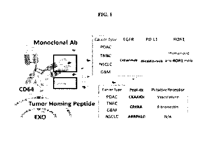
[0020] FIG. 1 depicts a schematic of a targeting extracellular vesicle ("EXO")
comprising a
monoclonal antibody (mAb) and a tumor homing peptide (THP) linked to a CD64 on
the
extracellular vesicle surface. These extracellular vesicles (EVs) can target
tumors and lesions,
with exemplary targets recited in FIG. 1. The EVs with CD64 or THP-CD64 can be
generated
by transfection of donor cells with human CD64 plasmid DNA or human THP-CD64
plasmid
DNA to express either human CD64 or human THP-CD64 on the surface of EVs
(including
exosomes) secreted from transfected donor cells. CD64 provides a biological
anchor for binding
to a humanized monoclonal antibody (hmAB). The extracellular Dl-D2 hinge of
human CD64
binds to the lower hinge region of Fc in human IgG1 with high affinity (a
dissociation constant
(Kd) as high as ¨10-9M (nanomolar). In addition to the ability of the bound
hmAb to
specifically recognize cell targets, targeting by a small tumor homing peptide
(THP) can also be
engineered onto the N-terminal of CD64. Dual targeting of both hmAbs and THPs
on the EV (or
exosome) surface enhances targeting and delivery to tumors and other lesions
in vivo. Examples
of hmAbs for cancer/tumor targeting include, but are not limited to, anti-
hEGFR such as
Cetuximab, anti-hPD-L1 such as Atezolizumab, and anti-humanized ROR1. Examples
of THPs
for cancer/tumor targeting include, but are not limited to, CKAAKN (CK), CREKA
(CR), and
ARRPKLD (AR).
[0021] FIG. 2A-2D illustrates an exemplary construct design for plasmids
encoding CD64 with
additional tumor homing peptides. FIG. 2A. Plasmids were constructed with the
vector carrying
genes for Ampicillin resistance (AmpR) and the EGFR marker for transformation
and
transfection, respectively. The functional CD64 was encoded by the coding
sequence of CD64
(CD64 CDS) driven by the EF-lapromoter. FIG. 2B. The CD64 CDS (355 amino
acids)
included a (i) signal peptide (SP), (ii) an extracellular region (D1, D2, and
D3), (iii) a
-14-
CA 03190722 2023-02-03
WO 2022/031783 PCT/US2021/044449
transmembrane (TM) region, and (iv) an intracellular (IC) domain, and the THPs
were inserted
into the gap between the signal peptide and the extracellular D1 region,
allowing expression of
the THP at the N-terminus of CD64. FIG. 2C. The THPs were connected by a Flag
(DYKDDDK) linker to the N- terminus of extracellular D1, limiting the
conformational block of
the Fc binding region at the D1-D2 hinge of CD64. FIG. 2D. List of exemplary
peptide and
nucleotide sequences of selected THPs [Flag control, CKAAKN (CK), CREKA (CR),
and
ARRPKLD (AR)].
[0022] FIG. 3A-3C illustrates how addition of the THP onto CD64 does not
affect binding
affinity with human IgG (hIgG). FIG. 3A. Schematic of purified CD64 proteins
with different
THPs bound to the immobilized hIgG on a solid support and reacted with ELISA
substrates. The
Ka value was determined by the monovalent modeling between CD64 and hIgG. FIG.
3B. The
affinity index Ka of hIgG and recombinant wild-type CD64 (wt CD64) was
measured. FIG. 3C.
The affinity index Kd of different engineered THP-CD64 proteins with hIgG
suggested that the
engineered CD64 with different THPs (Flag, CK, CR, AR) did not affect the high
binding
affinity with mAb in nM-level comparing to wt CD64.
[0023] FIG. 4A-4B illustrates EV number and content of endogenous mRNA from
nanochannel
electroporation (NEP) transfected mouse embryonic fibroblasts (MEFs) with THP-
CD64 and
therapeutic RNA plasmids. FIG. 4A. EV number per cell produced by untreated
MEFs in PBS
(control group), MEFs transfected with both THP-CD64 and human TP53 plasmids
by NEP, and
MEFs transfected with both THP-CD64 and shKRAS G12D mutation plasmids by NEP
at 24 h.
FIG. 4B. Fold change of TP53 mRNA in EVs produced by untreated and NEP-
transfected
MEFs by qRT-PCR.
[0024] FIG. 5A-5D illustrates that THP-CD64 expressing exosomes retained high
binding
affinity with hIgG. FIG. 5A. Schematic of purified exosomes with engineered
THP-CD64 were
captured by latex beads and incubated with anti-CD64-APC, anti-CD63-BV510 and
FITC-
conjugated hIgG for flow cytometry assay. FIG. 5B. Profiling of surface
expression followed
the standard protocol to gate the singlet bead and CD63+ exosome population in
order to
determine mean fluorescence intensity (MFI) of CD64 expression and hIgG
binding. FIG. 5C.
Surface co-expression of CD64 within the CD63+ exosomal population was
determined by MFI
of FITC and confirmed the exosomal expression of engineered CD64 with either
Flag, CK, CR
or AR THP. FIG. 5D. Surface co-expression of hIgG and CD64 within the CD63+
exosomal
population was determined by 1VIFI of FITC and confirmed the high binding
affinity of hIgG on
exosomes expressing CD64 with either Flag, CK, CR or AR THP.
[0025] FIG. 6 illustrates uptake of liposome and EVs in cancer spheroids from
a human
pancreatic cancer cell line, PANC-1. The purified EVs released from mouse
embryonic
-15-
CA 03190722 2023-02-03
WO 2022/031783 PCT/US2021/044449
fibroblast (MEF) cells after transfection of either Flag-CD64 or CK-CD64
plasmid DNA (CK-
CD64) were formulated with either humanized anti-EGFR mAb (Cetuximab) or hIgG.
The
cancer spheroids were treated with PKH67(green)-labeled liposome
(lipofectamine 3000) and
various EVs for 24 hours, and subsequently processed by fixation, permeation,
and staining with
anti-hIgG-TRITC (red) and DAPI (blue). The cross section of cancer spheroids
was imaged
under confocal microscopy. Cancer spheroid treatment with various EVs all
showed better
spheroid uptake than the commercial lipofectamine 3000 based on fluorescence
intensity and
distribution. Among various EVs, the dual targeting EV (CK-CD64-Cet Exo)
revealed the
highest spheroid uptake.
[0026] FIG. 7A-7C illustrates dual targeting of CK-CD64 and humanized anti-
EGFR mAb
(Cetuximab) enhances EV uptake in PANC-1 cancer spheroid cells, particularly
the
CD24+CD44+ subpopulation. FIG. 7A. The PANC-1 cancer spheroids were formed and
cultured for a week to reach a diameter of ¨500 jim, then treated with ¨109
PKH67-labled
exosomes in culture media for 24 h. FIG. 7B. The treated spheroids were
disassembled into
single-cell suspension to identify the subpopulations by CD24 and CD44
expression using flow
cytometry. FIG. 7C. The mean fluorescence intensity of PKH67 measured in
CD24lowCD44low or CD24+CD44+ subpopulations represent their EV uptake. The
engineered
EVs containing Flag-CD64, CK-CD64, CR-CD64, or AR-CD64 with humanized antibody
binding (Cetuximab: anti-EGFR, Atezolizumab: anti-PD-L1, or hIgG) all showed
good cellular
uptake, particularly for the CD44+CD24+ subpopulation. The dual targeting EVs
with anti-
hEGFR (Cetuximab) and CK-CD64 provided the best cellular uptake for both PANC-
1 cell
subpopulations.
[0027] FIG. 8 depicts that the uptake of extracellular vesicles is enhanced by
targeting ROR1,
which is highly expressed in 85% of pancreatic cancer, within spheroids formed
from PANC-1.
PANC-1 spheroids were formed and stably cultured for a week until a diameter
of 300-50011m
was obtained, and then treated with 10'10 PKH67-labeled exosomes in culture
media for 24
hours.
[0028] FIG. 9 depicts the enhanced uptake of ROR1-targeted extracellular
vesicles in vivo in a
PANC-1 orthotopic model. Mice (4 weeks post xenograftment of PANC-1
extracellular
vesicles) were treated with 1.0E12 / 50 Ill (intraperitoneal) injection with
extracellular vesicle
solution (250 pi) and sacrificed after 24 hours. PKH26 (Excitation / Emission:
535 / 580 nm);
GFP (465 / 540 nm); IVIS (Epi-illumination, Bin:(M)1, FOV:22, f2, 5s.
Distribution:
brain/heart/lung/liver/spleen/pancreas/kidney.
[0029] FIG.10 depicts the enhanced uptake of ROR1-targeted extracellular
vesicles by
penetration through tumor tissue. Anti-ROR1 targeting enriches the
extracellular vesicle uptake
-16-
CA 03190722 2023-02-03
WO 2022/031783 PCT/US2021/044449
in tumor lesions, whereas the update of the CK_peptide extracellular vesicle
is not significant
compared to the flag control.
[0030] FIG. 11A-11D illustrates an exemplary design of vacosomes and five
proposed vaccine
peptides (i.e. Spike, S-protein, fragments) from the epitope and structural
predictions for
COVID-19 vaccine development. FIG. 11A. ACE2 acts as the receptor for the SARS-
CoV-2
virus and allows it to infect the cell. FIG. 11B. A strong vaccination through
T-cell receptor
(TCR) complex can be synergistically achieved by vaccination peptides on the N-
terminal of
CD64 and co-stimulation by the preloaded anti-aCD3/CD28 mAb on the hinge D1-D2
of CD64.
Exosomes that overexpress various viral protein fragments fused to CD64 on the
exosomal
surface can serve as a vaccine (designated `vacosome'). FIG. 11C. The
formation of an
immunological synapse between engineered CD64 and TCR can be confirmed by the
fluorescent tag and T-cell surface markers staining using fluorescence-
activated cell sorter
(FACS). Similarly, the co-loading of mAb targeting antigen presenting cells
(APCs) such as B
cells (anti-aCD19/CD20) and dendritic cells (DCs) (anti-aLILRA4) should
enhance APC-T cell
responses. FIG. 11D. Five fusion S-protein fragment candidates that have high
potential to serve
as a vaccine peptide for COVID-19 are selected from the epitope and structural
predictions.
They can be expressed on vacosomes generated via NEP transfected donor cells
such as human
mesenchymal stem cells (MSCs) and DCs.
[0031] FIG. 12A-12B illustrates binding affinity strength of human
immunoglobins and
classical Fc receptors. Fc receptors embedded in the plasma membrane contain
intracellular
domains or subunits that can trigger a downstream activation or suppression.
FIG. 12A. IgG
affinity-altering variants are highlighted with respective human Fcy receptor
members, from
very high (deep orange), high (orange), medium (yellow), low (light blue), to
no binding (dark
blue). FcRn receptor binds to IgG subclasses under acidic conditions (e.g.
pH=6) but decrease
the binding ability in physiological conditions, pH =7.4. FIG. 12B. IgE has
very high binding
affinity with FccRI receptor, but low affinity with FccRII receptor. IgA has
low binding affinity
with FcaRI receptor. *** The binding affinity between human immunoglobins and
Fc repeaters
is shown as constant Ka at the level as Very High+++: ¨10-9M; High++: 10-9 to
10-8M;
Medium: ¨10-7 M; or Low: > 10-7 M.
[0032] FIG. 13A-13D illustrate the dynamics of EV release in NEP. FIG. 13A
depicts EVs
secretion profiles over time after NEP. FIG. 13B depicts fold change of TP53
mRNA
expression within the EVs over time was measured by qPCR. FIG. 13C depicts
CD64
expression on EVs surface was measured through ELISA for EVs collected every 8
h. FIG.
13D shows the expression level of KRASG12D shRNA within the EVs every 8 h
after NEP.
-17-
CA 03190722 2023-02-03
WO 2022/031783 PCT/US2021/044449
[0033] FIG. 14A-C illustrates sequential NEP (sNEP) designs for TP53 mRNA/CD64
EVs.
FIG. 14A provides EV number and TP53 mRNA expression in the (FIG. 14A) 8-h,
(FIG. 14B)
16-h, and (FIG. 14C) 24-h sNEP cases. Ctrl is one-time NEP with CD64 plasmid.
[0034] FIG. 15A-F provides characterization of the as-prepared targeting EVs
(tEVs). FIG.
15A provides size distribution of blank EVs (Control) and engineered EVs
obtained by NEP,
(FIG. 15B) exosomal biomarkers on as-prepared EVs, (FIG. 15C) SEM and (FIG.
15D)
CryoTEM images of representative EVs. (FIG. 15E) single EV capture and co-
localization
characterization using ILN biochips on TIRF microscope with fluorescence
labelled anti-CD64
and molecular beacons for KRASG12D shRNA and TP53 mRNA, (FIG. 15F) ratios of
EVs
containing CD64 protein, KRASG12D shRNA, TP53 mRNA, and co-localization of
CD64/
KRASG12D shRNA and CD64/TP53 mRNA.
[0035] FIG. 16A-G depict that binding tumor-specific antibodies (ahROR1 and
ethEGFR) on
CD64/EV surface can enhance cellular internalization of Elvis in PANC4 cells.
FIG. 16A
provides the uptake efficiency of humanized antibodies on CD64 flag-peptide,
FIG. 16B
provides the uptake efficiency of humanized antibodies on CD64 with CK-
peptide. FIG. 16C
quantifies the relative EV uptake by each formulation. FIG. 16D compares
staining of PANC-1
cells without treatment (Con), and with IgG EV, dEGFR BY, and GROWL EV
treatment for 4
hours. FIG. 16E-F provides an EV uptake assay on 3D tumor spheroids of P ANC-1
cells for
LEGER EVs (FIG. 16E) and etROR1 EVs (FIG. 16F). FIG. 16G provides a
substitution assay
with human serum (50%) for 6 hours at 37 C.
[0036] FIG. 17A-D depict a TRANS WELL - based transcytosis assay and results.
FIG. 17A
provides a schematic of the assay. FIG. 17B provides various inhibitors
selected to block
endocytosis and EV secretion (including pitstop 2, an inhibitor of clathrin-
mediated endocytosis;
methyl-P-cyclodextrin, an inhibitor of caveolae-mediated endocytosis;
cytochalasin D, an
inhibitor of micropinocytosis; and neticonazole, an inhibitor of exosomal
secretion). FIG. 17C
provides data from a transcytosis assay by PANC-1 using various inhibitors
("ctl": 1E10 non-
targeting EVs without inhibitors in upper PANC-1 cells; "Pos ctl": no upper
cellular layer).
FIG. 17D compares transcytosis levels by PANC-1 using targeting hmAbs on the
EV surface.
[0037] FIG. 18A-B demonstrates that human serum IgG does not affect human mAb
on the EV
surface. FIG. 18A-B depict that ahEGFR_EV (left panels) and ahRORI__EV (right
panels)
incubated with human serum (50%) for 6h at 37 C, then treated with monolayer
PANC-1
cells, maintained the same targeting ability after human serum incubation.
[0038] FIG. 19A-B depicts biodistribution of targeting EVs in a PANC-1
orthotopic NS mice.
FIG. 19A depicts in vivo imaging (IVIS) and FIG. 19B depicts expression in
brain, heart, lung,
liver, spleen, pancreas, and kidney.
-18-
CA 03190722 2023-02-03
WO 2022/031783 PCT/US2021/044449
DETAILED DESCRIPTION
[0039] While preferred aspects of the present disclosure have been shown and
described herein,
it will be obvious to those skilled in the art that such aspects are provided
by way of example
only. Numerous variations, changes, and substitutions will now occur to those
skilled in the art
without departing from the disclosure. It should be understood that various
alternatives to the
aspects of the disclosure described herein may be employed in practicing the
disclosure. It is
intended that the following claims define the scope of the disclosure and that
methods and
structures within the scope of these claims and their equivalents be covered
thereby.
[0040] Use of absolute or sequential terms, for example, "will," "will not,"
"shall," "shall not,"
"must," "must not," "first," "initially," "next," "subsequently," "before,"
"after," "lastly," and
"finally," are not meant to limit scope of the present aspects disclosed
herein but as exemplary.
[0041] As used herein, the singular forms "a", "an" and "the" are intended to
include the plural
forms as well, unless the context clearly indicates otherwise. Furthermore, to
the extent that the
terms "including", "includes", "having", "has", "with", or variants thereof
are used in either the
detailed description and/or the claims, such terms are intended to be
inclusive in a manner
similar to the term "comprising."
[0042] As used herein, "or" may refer to "and", "or," or "and/or" and may be
used both
exclusively and inclusively. For example, the term "A or B" may refer to "A or
B", "A but not
B", "B but not A", and "A and B". In some cases, context may dictate a
particular meaning.
[0043] As used herein, the phrases "at least one", "one or more", and "and/or"
are open-ended
expressions that are both conjunctive and disjunctive in operation. For
example, each of the
expressions "at least one of A, B and C", "at least one of A, B, or C", "one
or more of A, B, and
C", "one or more of A, B, or C" and "A, B, and/or C" means A alone, B alone, C
alone, A and B
together, A and C together, B and C together, or A, B and C together.
[0044] Any systems, methods, compositions, and platforms described herein are
modular and
not limited to sequential steps. Accordingly, terms such as "first" and
"second" do not
necessarily imply priority, order of importance, or order of acts.
[0045] The term "about" or "approximately" means within an acceptable error
range for the
particular value as determined by one of ordinary skill in the art, which will
depend in part on
how the value is measured or determined, e.g., the limitations of the
measurement system. For
example, "about" can mean within 1 or more than 1 standard deviation, per the
practice in the
given value. Where particular values are described in the application and
claims, unless
otherwise stated the term "about" should be assumed to mean an acceptable
error range for the
particular value.
-19-
CA 03190722 2023-02-03
WO 2022/031783 PCT/US2021/044449
[0046] The terms "increased", "increasing", or "increase" are used herein to
generally mean an
increase by a statically significant amount. In some cases, the terms
"increased," or "increase,"
mean an increase of at least 10% as compared to a reference level, for example
an increase of at
least about 10%, at least about 20%, or at least about 30%, or at least about
40%, or at least
about 50%, or at least about 60%, or at least about 70%, or at least about
80%, or at least about
90% or up to and including a 100% increase or any increase between 10-100% as
compared to a
reference level, standard, or control. Other examples of "increase" include an
increase of at least
2-fold, at least 5-fold, at least 10-fold, at least 20-fold, at least 50-fold,
at least 100-fold, at least
1000-fold or more as compared to a reference level.
[0047] The terms, "decreased", "decreasing", or "decrease" are used herein
generally to mean a
decrease by a statistically significant amount. In some cases, "decreased" or
"decrease" means a
reduction by at least 10% as compared to a reference level, for example a
decrease by at least
about 20%, or at least about 30%, or at least about 40%, or at least about
50%, or at least about
60%, or at least about 70%, or at least about 80%, or at least about 90% or up
to and including a
100% decrease (e.g., absent level or non-detectable level as compared to a
reference level), or
any decrease between 10-100% as compared to a reference level. In the context
of a marker or
symptom, by these terms is meant a statistically significant decrease in such
level. Other
examples of "decrease" include a decrease of at least 2-fold, at least 5-fold,
at least 10-fold, at
least 20-fold, at least 50-fold, at least 100-fold, at least 1000-fold or more
as compared to a
reference level. The decrease can be, for example, at least 10%, at least 20%,
at least 30%, at
least 40% or more, and is preferably down to a level accepted as within the
range of normal for
an individual without a given disease.
[0048] As used herein, a "cell" generally refers to a biological cell. A cell
is the basic structural,
functional and/or biological unit of a living organism. A cell can originate
from any organism
having one or more cells. Some non-limiting examples include: a prokaryotic
cell, eukaryotic
cell, a bacterial cell, an archaeal cell, a cell of a single-cell eukaryotic
organism, a protozoa cell,
a cell from a plant (e.g. cells from plant crops, fruits, vegetables, grains,
soy bean, corn, maize,
wheat, seeds, tomatoes, rice, cassava, sugarcane, pumpkin, hay, potatoes,
cotton, cannabis,
tobacco, flowering plants, conifers, gymnosperms, ferns, clubmosses,
hornworts, liverworts,
mosses), an algal cell, (e.g., Botryococcus braunii, Chlamydomonas
reinhardtii,
Nannochloropsis gaditana, Chlorella pyrenoidosa, Sargassum patens C. Agardh,
and the like),
seaweeds (e.g. kelp), a fungal cell (e.g., a yeast cell, a cell from a
mushroom), an animal cell, a
cell from an invertebrate animal (e.g. fruit fly, cnidarian, echinoderm,
nematode, etc.), a cell
from a vertebrate animal (e.g., fish, amphibian, reptile, bird, mammal), a
cell from a mammal
(e.g., a pig, a cow, a goat, a sheep, a rodent, a rat, a mouse, a non-human
primate, a human, etc.),
-20-
CA 03190722 2023-02-03
WO 2022/031783 PCT/US2021/044449
and etcetera. Sometimes a cell is not originating from a natural organism
(e.g. a cell is a
synthetically made, sometimes termed an artificial cell). A cell can be
derived from a cell line.
[0049] The terms "transfection" or "transfected" generally refers to
introduction of a nucleic
acid molecule into a cell by non-viral or viral-based methods. The nucleic
acid molecules can be
gene sequences encoding complete proteins or functional portions thereof In
some cases, the
nucleic acid molecules can be non-coding sequences. In some cases, the
transfection methods
are utilized for introducing nucleic acid molecules into a cell for generating
a transgenic animal.
Such techniques can include pronuclear microinjection, retrovirus mediated
gene transfer into
germ lines, gene targeting into embryonic stem cells, electroporation of
embryos, sperm
mediated gene transfer, and in vitro transformation of somatic cells, such as
cumulus or
mammary cells, or adult, fetal, or embryonic stem cells, followed by nuclear
transplantation.
[0050] "Nanoelectroporation" or "nanochannel electroporation" refers to
transfecting a cell with
at least one heterologous polynucleotide such as a vector by loading the at
least one
heterologous polynucleotide into a nanochannel and accelerating the at least
on heterologous
polynucleotide into the cell with by generating an electric field. The cell to
be transfected is
situated at an opening of the nanochannel, where the electric field of the
nanoelectroporation
creates pores in the cell membrane to allow the at least one heterologous
polynucleotide to be
introduced into the cell.
[0051] A "plasmid," as used herein, generally refers to a non-viral expression
vector, e.g., a
nucleic acid molecule that encodes for genes and/or regulatory elements
necessary for the
expression of genes. The term "vector," as used herein, generally refers to a
nucleic acid
molecule capable transferring or transporting a payload nucleic acid molecule.
The payload
nucleic acid molecule can be generally linked to, e.g., inserted into, the
vector nucleic acid
molecule. A vector can include sequences that direct autonomous replication in
a cell, or can
include sequences sufficient to allow integration into host cell gene (e.g.,
host cell DNA).
Examples of a vector can include, but are not limited to, plasmids (e.g., DNA
plasmids or RNA
plasmids), transposons, cosmids, bacterial artificial chromosomes, and viral
vectors. A "viral
vector," as used herein, generally refers to a viral-derived nucleic acid that
is capable of
transporting another nucleic acid into a cell. A viral vector is capable of
directing expression of a
protein or proteins encoded by one or more genes carried by the vector when it
is present in the
appropriate environment. Examples for viral vectors include, but are not
limited to Gamma-
retroviral, Alpha-retroviral, Foamy viral, lentiviral, adenoviral, or adeno-
associated viral vectors.
A vector of any of the embodiments of the present disclosure can comprise
exogenous,
endogenous, or heterologous control sequences such as promoters and/or
enhancers.
-21-
CA 03190722 2023-02-03
WO 2022/031783 PCT/US2021/044449
[0052] The term "nucleotide," as used herein, generally refers to a base-sugar-
phosphate
combination. A nucleotide can comprise a synthetic nucleotide. A nucleotide
can comprise a
synthetic nucleotide analog. Nucleotides are monomeric units of a nucleic acid
sequence (e.g.
deoxyribonucleic acid (DNA) and ribonucleic acid (RNA)). The term nucleotide
can include
ribonucleoside triphosphates adenosine triphosphate (ATP), uridine
triphosphate (UTP),
cytosine triphosphate (CTP), guanosine triphosphate (GTP) and
deoxyribonucleoside
triphosphates such as dATP, dCTP, dITP, dUTP, dGTP, dTTP, or derivatives
thereof Such
derivatives can include, for example, [aS]dATP, 7-deaza-dGTP and 7-deaza-dATP,
and
nucleotide derivatives that confer nuclease resistance on the nucleic acid
molecule containing
them. The term nucleotide as used herein can refer to dideoxyribonucleoside
triphosphates
(ddNTPs) and their derivatives. Illustrative examples of dideoxyribonucleoside
triphosphates
can include, but are not limited to, ddATP, ddCTP, ddGTP, ddITP, and ddTTP.
[0053] The terms "polynucleotide," "oligonucleotide," and "nucleic acid" are
used
interchangeably to refer to a polymeric form of nucleotides of any length,
either
deoxyribonucleotides or ribonucleotides, or analogs thereof, either in single-
, double-, or multi-
stranded form. In some cases, a polynucleotide is exogenous (e.g. a
heterologous
polynucleotide) to a cell. In some cases, a polynucleotide is endogenous to a
cell. In some
cases, a polynucleotide can exist in a cell-free environment. In some cases, a
polynucleotide is a
gene or fragment thereof. In some cases, a polynucleotide is DNA. In some
cases, a
polynucleotide is RNA. A polynucleotide can have any three dimensional
structure, and can
perform any function, known or unknown. In some cases, a polynucleotide
comprises one or
more analogs (e.g. altered backbone, sugar, or nucleobase). If present,
modifications to the
nucleotide structure may be imparted before or after assembly of the polymer.
Some non-
limiting examples of analogs include: 5-bromouracil, peptide nucleic acid,
xeno nucleic acid,
morpholinos, locked nucleic acids, glycol nucleic acids, threose nucleic
acids,
dideoxynucleotides, cordycepin, 7-deaza-GTP, fluorophores (e.g. rhodamine or
fluorescein
linked to the sugar), thiol containing nucleotides, biotin linked nucleotides,
fluorescent base
analogs, CpG islands, methyl-7-guanosine, methylated nucleotides, inosine,
thiouridine,
pseudourdine, dihydrouridine, queuosine, and wyosine. Non-limiting examples of
polynucleotides include coding or non-coding regions of a gene or gene
fragment, loci (locus)
defined from linkage analysis, exons, introns, messenger RNA (mRNA), transfer
RNA (tRNA),
ribosomal RNA (rRNA), short interfering RNA (siRNA), short-hairpin RNA
(shRNA), micro-
RNA (miRNA), non-coding RNA, ribozymes, cDNA, recombinant polynucleotides,
branched
polynucleotides, plasmids, vectors, isolated DNA of any sequence, isolated RNA
of any
sequence, cell-free polynucleotides including cell-free DNA (cfDNA) and cell-
free RNA
-22-
CA 03190722 2023-02-03
WO 2022/031783 PCT/US2021/044449
(cfRNA), nucleic acid probes, and primers. The sequence of nucleotides is
interrupted by non-
nucleotide components. Nucleotide or nucleic acid described herein can be
modified to comprise
modified nucleic acid, nucleic acid analog, modified sugars, sugar analogs,
modified nucleic
acid linkage, backbone phosphate modification, or a combination thereof.
[0054] As used herein, the terms "polypeptide", "peptide", and "protein" are
used
interchangeably to refer to a polymer of amino acid residues. In some cases, a
polypeptide refers
to a full-length polypeptide as translated from a coding open reading frame,
or as processed to
its mature form. In some cases, a polypeptide or peptide can be a degradation
fragment or a
processing fragment of a protein that nonetheless uniquely or identifiably
maps to a particular
protein. In some cases, a polypeptide can be a single linear polymer chain of
amino acids
bonded together by peptide bonds between the carboxyl and amino groups of
adjacent amino
acid residues. A polypeptide can be modified, for example, by the addition of
carbohydrate,
phosphorylation, etc. As used herein, the terms "fragment" or equivalent terms
can refer to a
locus of a protein that has less than the full length of the protein and
optionally maintains the
function of the protein.
[0055] "Percent identity" and "% identity" refers to the extent to which two
sequences
(nucleotide or amino acid) have the same residue at the same positions in an
alignment. For
example, "an amino acid sequence is X% identical to SEQ ID NO: Y" refers to %
identity of the
amino acid sequence to SEQ ID NO:Y and is elaborated as X% of residues in the
amino acid
sequence are identical to the residues of sequence disclosed in SEQ ID NO: Y.
Generally,
computer programs are employed for such calculations. Exemplary programs that
compare and
align pairs of sequences, include ALIGN, FASTA, gapped BLAST, BLASTP, BLASTN,
or
GCG.
[0056] The terms "antibody" and "immunoglobulin" are used interchangeably
herein and cover
fully assembled antibodies, antibody fragments that can bind antigen, for
example, Fab, F(ab')2,
Fv, single chain antibodies (scFv), diabodies, antibody chimeras, hybrid
antibodies, bispecific
antibodies, and the like. In some cases, a binding domain of an antibody is
any domain that
specifically binds to an antigen, including a binding domain of an antibody or
a non-antibody
binding domain. In some cases, an antibody binding domain binds to tumor
cells, such as an
antibody against a tumor cell surface receptor or a tumor antigen. An antibody
can be a
monoclonal antibody, a polyclonal antibody, a recombinant antibody, or an
antigen binding
fragment thereof, for example, a heavy chain variable domain (VH) and a light
chain variable
domain (VL). In some cases, a binding domain of a non-antibody scaffold can be
a lipocalin, an
anticalin, 'T-body', an affibody, a peptibody, a DARPin, an affimer, an
avimer, a knottin, a
monobody, an affinity clamp, an ectodomain, a receptor ectodomain, a receptor,
a cytokine, a
-23-
CA 03190722 2023-02-03
WO 2022/031783 PCT/US2021/044449
ligand, an immunocytokine, a centryin, a T-cell receptor, or a recombinant T-
cell receptor. In
some cases, a binding domain of an antibody construct is an antigen binding
domain from a
monoclonal antibody and comprises a light chain and a heavy chain. In some
cases, an antibody
is a derivatized antibody, such as an antibody modified by glycosylation,
acetylation, pegylation,
phosphorylation, amidation, derivatization by known protecting/blocking
groups, proteolytic
cleavage, or the like. An antibody can also be modified, such as by
defucosylation or
deglycosylation.
[0057] The terms "monoclonal antibody" and "mAb" are used interchangeably
herein and refer
to an antibody obtained from a substantially homogeneous population of
antibodies. In some
cases, the individual monoclonal antibodies obtained from the substantially
homogenous
population of antibodies are identical except for naturally occurring
mutations that may be
present in minor amounts. In some cases, a monoclonal antibody that binds to a
tumor antigen
comprises a light chain of a tumor antigen antibody and a heavy chain of a
tumor antigen
antibody. In some cases, the monoclonal antibody binds to an antigen on the
surface of an
immune cell (immune cell antigen) and comprises a light chain of an anti-
immune cell antigen
antibody and a heavy chain of an anti-immune cell antigen antibody. In some
cases, the
monoclonal antibody specifically binds to an antigen present on the surface of
an antigen
presenting cell (APC antigen) and comprises the light chain of an anti-APC
antigen antibody
and the heavy chain of an anti-APC antigen antibody, which bind an APC
antigen.
[0058] The term "antibody fragment" as used herein refers to a molecule that
comprises a
portion of an intact antibody, preferably the antigen-binding or variable
region of an intact
antibody. Examples of antibody fragments include Fab, Fab, F(ab')2, Fv
fragments, and single
chain fragment variable (scFv); diabodies; linear antibodies; single-chain
antibody molecules;
and multi-specific antibodies formed from antibody fragments. Digesting
antibodies with
papain produces two identical antigen-binding fragments, called "Fab"
fragments, each with a
single antigen-binding site, and a residual "Fc" fragment, whose name reflects
its ability to
crystallize readily. Pepsin treatment yields an F(ab')2 fragment that has two
antigen-combining
sites and is still capable of cross-linking antigen. Depending on the amino
acid sequence of the
constant domain of their heavy chains, immunoglobulins can be assigned to
different classes.
There are five major classes of human immunoglobulins: IgA, IgD, IgE, IgG, and
IgM, and
several of these may be further divided into subclasses (isotypes), e.g.,
IgGl, IgG2, IgG3, IgG4,
IgAl, and IgA2. The heavy-chain constant domains that correspond to the
different classes of
immunoglobulins are called alpha, delta, epsilon, gamma, and mu, respectively.
The subunit
structures and three-dimensional configurations of different classes of
immunoglobulins are well
known. Different isotypes have different effector functions.
-24-
CA 03190722 2023-02-03
WO 2022/031783 PCT/US2021/044449
[0059] The term "human antibody" includes all antibodies that have one or more
variable and
constant regions derived from human immunoglobulin sequences. In some cases,
all of the
variable and constant domains of the antibody are derived from human
immunoglobulin
sequences (referred to as a "fully human antibody").
[0060] The term "recombinant human antibody," as used herein, is intended to
include all
human antibodies that are prepared, expressed, created, or isolated by
recombinant methods,
such as antibodies isolated from a host cell such as a NSO or CHO cell or from
an animal (e.g. a
mouse) that is transgenic for human immunoglobulin genes or antibodies
expressed using a
recombinant expression vector transfected into a host cell. Such recombinant
human antibodies
may have variable and constant regions in a rearranged form. In some cases,
the recombinant
human antibodies have been subjected to in vivo somatic hypermutation. Thus,
the amino acid
sequences of the VH and VL regions of the recombinant antibodies are sequences
that, while
derived from and related to human germ line VH and VL sequences, may not
naturally exist
within the human antibody germ line repertoire in vivo.
[0061] "Humanized" antibodies are antibodies in which at least part of the
sequence has been
altered from its initial form to render it more like human immunoglobulins. In
some versions,
the heavy (H) chain and light (L) chain constant (C) regions are replaced with
human sequence.
This can be a fusion polypeptide comprising a variable (V) region and a
heterologous
immunoglobulin C region. In some versions, the complementarity determining
regions (CDRs)
comprise non-human antibody sequences, while the V framework regions have also
been
converted to human sequences. In some versions, V regions are humanized by
designing
consensus sequences of human and mouse V regions, and converting residues
outside the CDRs
that are different between the consensus sequences.
[0062] As used herein, the term "in vivo" is used to describe an event that
takes place in a
subject's body.
[0063] As used herein, the term "ex vivo" is used to describe an event that
takes place outside of
a subject's body. An "ex vivo" assay cannot be performed on a subject. Rather,
it is performed
upon a sample separate from a subject. Ex vivo is used to describe an event
occurring in an intact
cell outside a subject's body.
[0064] As used herein, the term "in vitro" is used to describe an event that
takes places
contained in a container for holding laboratory reagent such that it is
separated from the living
biological source organism from which the material is obtained. In vitro
assays can encompass
cell-based assays in which cells alive or dead are employed. In vitro assays
can also encompass
a cell-free assay in which no intact cells are employed.
-25-
CA 03190722 2023-02-03
WO 2022/031783 PCT/US2021/044449
[0065] As used herein, a "microenvironment" refers to the extracellular
environment in which
cells targeted by the extracellular vesicles described herein are located. In
some cases, the
microenvironment can have an extracellular space(s) in which proteases and
other soluble
proteins and factors are located. A microenvironment can contain, for example,
blood vessels,
immune cells, fibroblasts, bone marrow-derived inflammatory cells,
lymphocytes, signaling
molecules and the extracellular matrix (ECM).
[0066] "Treating" or "treatment" can refer to both therapeutic treatment and
prophylactic or
preventative measures, wherein the object is to prevent or slow down (lessen)
a targeted
pathologic condition or disorder. Those in need of treatment include those
already with the
disorder, as well as those prone to have the disorder, or those in whom the
disorder is to be
prevented. A therapeutic benefit can refer to eradication of a disorder being
treated or
amelioration of symptoms of a disorder being treated. Also, a therapeutic
benefit may be
achieved with the eradication or amelioration of one or more physiological
symptoms associated
with the underlying disorder such that an improvement is observed in the
subject,
notwithstanding that the subject can still be afflicted with the underlying
disorder. A
prophylactic effect can include delaying, preventing, or eliminating the
appearance of a disease
or condition, delaying or eliminating the onset of symptoms of a disease or
condition, slowing,
halting, or reversing the progression of a disease or condition, or any
combination thereof For a
prophylactic benefit, a subject at risk of developing a particular disease, or
a subject reporting
one or more of the physiological symptoms of a disease can undergo treatment,
even though a
diagnosis of a disease has not been made.
[0067] The terms "effective amount" and "therapeutically effective amount,"
are used
interchangeably herein and generally refer to a quantity of a pharmaceutical
composition, for
example a pharmaceutical composition comprising the composition described
herein, that is
sufficient to result in a desired activity upon administration to a subject in
need thereof Within
the context of the present disclosure, the term "therapeutically effective"
refers to that quantity
of a pharmaceutical composition that is sufficient to delay the manifestation,
arrest the
progression, relieve or alleviate at least one symptom of a disorder treated
by the methods of the
present disclosure.
[0068] The term "pharmaceutically acceptable carrier," "pharmaceutically
acceptable
excipient," "physiologically acceptable carrier," or "physiologically
acceptable excipient" refers
to a pharmaceutically-acceptable material, composition, or vehicle, such as a
liquid or solid
filler, diluent, excipient, solvent, or encapsulating material. A component is
"pharmaceutically
acceptable" in the sense of being compatible with the other ingredients of a
pharmaceutical
formulation. It can also be suitable for use in contact with the tissue or
organ of humans and
-26-
CA 03190722 2023-02-03
WO 2022/031783 PCT/US2021/044449
non-human mammals without excessive toxicity, irritation, allergic response,
immunogenicity,
or other problems or complications, commensurate with a reasonable
benefit/risk ratio.
[0069] The term "pharmaceutical composition" refers to the compositions
disclosed herein with
other chemical components, such as diluents or carriers. The pharmaceutical
compositions can
facilitate administration to the subject. Multiple techniques of administering
a compound exist in
the art including, but not limited to, oral, injection, aerosol, parenteral,
and topical
administration.
[0070] The terms "patient" or "subject" are used interchangeably herein and
encompass
mammals. Non-limiting examples of mammal include, any member of the mammalian
class:
humans, non¨human primates such as chimpanzees, and other apes and monkey
species; farm
animals such as cattle, horses, sheep, goats, swine; domestic animals such as
rabbits, dogs, and
cats; laboratory animals including rodents, such as rats, mice and guinea
pigs, and the like.
Overview
[0071] The present disclosure relates to the design and production of
extracellular vesicles
expressing at least one adapter polypeptide (e.g., Fc receptor, CD64) that can
be complexed with
an antibody. The antibody can direct and target the extracellular vesicle to a
cell expressing a
cell-surface marker (e.g., an antigen for the antibody) that can be recognized
and bound by the
antibody. The cell expressing the cell-surface marker can be a diseased cell.
In some cases, the
cell expressing the cell-surface marker is a cancer cell, a tumor cell, a non-
cancerous lesion cell,
a cell as part of a damaged tissue, a cell as part of a healthy tissue, or an
immune cell.
[0072] In some cases, the adapter polypeptide can be further engineered to
include an additional
targeting domain to enhance the targeting and accumulation of the
extracellular vesicle at the
targeted cell. This targeting domain can bind to the same or different cell-
surface marker
expressed by the same targeted cell as that targeted by the antibody complexed
to the adapter
polypeptide. The extracellular vesicle can be configured to target a different
cell (e.g., different
cell type, diseased cell) or different cellular marker by complexing the
adapter polypeptide with
another antibody that targets a different cell-surface marker expressed by the
different cell. The
extracellular vesicle can be designed to carry a payload such as a therapeutic
to be delivered to
the targeted cell. The therapeutic delivered by the extracellular vesicle can
include a therapeutic
polynucleotide, a therapeutic polypeptide, a therapeutic compound, or a cancer
drug, or a
combination thereof
[0073] This disclosure also provides methods of producing extracellular
vesicles comprising the
at least one adapter polypeptide and comprising large quantities and qualities
of a therapeutic
such as a therapeutic polynucleotide (e.g., therapeutic messenger RNA). Some
approaches
described herein involve transfecting at least one heterologous polynucleotide
by
-27-
CA 03190722 2023-02-03
WO 2022/031783 PCT/US2021/044449
nanoelectroporation into a cell, where the at least one heterologous
polynucleotide is transcribed
and/or translated into at least one adapter polypeptide and/or at least one
therapeutic. In some
cases, the adapter polypeptide comprises an Fc receptor or a fragment thereof,
which can be
complexed with a Fc region of an antibody. In some embodiments, the
transfected cell is
stimulated by the nanoelectroporation to produce and secrete a large number of
extracellular
vesicles comprising large quantities of the therapeutic (e.g., therapeutic
mRNA). The secreted
extracellular vesicles can be complexed with any antibody comprising an Fc
region, where the
complexed antibody targets and directs the extracellular vesicle to a targeted
cell expressing a
first cell-surface marker that can be recognized and bound by the complexed
antibody. The at
least one adapter polypeptide can include a targeting domain that binds to a
second cell-surface
marker expressed by the same targeted cell. As such, the accumulation of the
extracellular
vesicles comprising the dual targeting domains (e.g., the antibody and
targeting domains) at the
targeted cell may be increased compared to accumulation of extracellular
vesicle without the
targeting by the antibody, the targeting domain, or a combination thereof
Extracellular Vesicles
[0074] Described herein are compositions comprising an extracellular vesicle.
Also described
herein are methods for producing the extracellular vesicle. In some cases, the
extracellular
vesicle comprises at least one adapter polypeptide, an antibody complexed with
the adapter
polypeptide, and at least one therapeutic. In some cases, the adapter
polypeptide comprises a
targeting domain. In some cases, the at least one adapter polypeptide
comprises a peptide
sequence that is at least 40%, 50%, 60%, 70%, 80%, 90%, 95%, or 99% identical
to a peptide
sequence of a cell surface protein. In some cases, the adapter polypeptide
comprises a peptide
that is at least 70% identical to the CD63 surface protein. In some cases, the
adapter polypeptide
comprises a peptide that is at least 70% identical to another cell surface
protein. In some cases,
the adapter polypeptide comprises a peptide that is at least 70% identical to
a cell surface protein
selected from the group ROR1, PD-L1, EpCAM, EGFR, EGFRIII, EGFRVIII, GPC1,
GPC3,
DLL3, L1CAM, GLAST, and CD138.
[0075] In some cases, the at least one adapter polypeptide comprises a peptide
sequence that is
at least 40%, 50%, 60%, 70%, 80%, 90%, 95%, or 99% identical to a peptide
sequence of a Fc
binding domain, a Fc receptor, or a fragment thereof that recognizes and binds
a Fc region of the
antibody. In certain cases, the extracellular vesicle surface protein is the
Fc receptor. Fc receptor
includes a Fc-gamma receptor, Fc-alpha receptor, or Fc-epsilon receptor.
Exemplary Fc receptor
includes FcyRI (CD64), FcyRIIa (CD32a), FcyRIIb1(CD32b), FcyRIIb2 (CD32b),
FcyRIIcl
(CD32c), FcyRIIc2 (CD32c), FcyRIIc3 (CD32c), FcyRIIc4 (CD32c), FcyRIIc5
(CD32c),
FcyRIIIA (CD16a), FcyRIIIB (CD16b), FccRI, FccRII (CD23), FcaRI (CD89),
Fca/IIR, FcRn,
-28-
CA 03190722 2023-02-03
WO 2022/031783 PCT/US2021/044449
DC-SIGN, or plgR. In some instances, the at least one adapter polypeptide
comprises a peptide
sequence that is at least 40%, 50%, 60%, 70%, 80%, 90%, 95%, or 99% identical
to a peptide
sequence of any one of the Fc receptors described herein. In some instances,
the at least one
adapter polypeptide comprises a peptide sequence that is at least 40%, 50%,
60%, 70%, 80%,
90%, 95%, or 99% identical to a peptide sequence of any one of the Fc
receptors: FcyRI
(CD64), FcyRII (CD32), or FcyRIII (CD16). In some instances, the at least one
adapter
polypeptide comprises a peptide sequence that is at least 40%, 50%, 60%, 70%,
80%, 90%,
95%, or 99% identical to a peptide sequence of Fc receptor FcyRI (CD64).
[0076] In some cases, the extracellular vesicle described herein comprises at
least two adapter
polypeptides. In some cases, one of the at least two adapter polypeptides
comprises an adapter
polypeptide comprising the peptide sequence of a Fc binding domain, a Fc
receptor, or a
fragment thereof. In some cases, one of the at least two adapter polypeptides
comprises the
peptide sequence of CD47 or a fragment thereof. In some cases, the
extracellular vesicle
described herein comprises a first and a second adapter polypeptides, where
the first adapter
polypeptide comprises the peptide sequence of a Fc binding domain, a Fc
receptor, or a fragment
thereof and the second adapter polypeptide comprises the peptide sequence of
CD47 or a
fragment thereof
[0077] In some cases, the extracellular vesicle comprises an antibody
complexed with the
adapter polypeptide comprising a peptide sequence comprising a Fc binding
domain, Fc
receptor, or a fragment thereof In some cases, the antibody complexed with the
adapter
polypeptide binds to a first cell-surface marker of expressed by a targeted
cell. In some
instances, the adapter polypeptide comprises an extracellular domain. In some
cases, a targeting
domain is attached to the extracellular domain, where the targeting domain
binds to a second
cell-surface marker of expressed by the same targeted cell expressing the
first cell-surface
marker. In some instances, the targeting domain attached to the extracellular
domain binds to a
second cell-surface marker expressed by the same targeted cell. In some
instances, the targeted
cell is a cell as part of a healthy tissue or an immune cell. In some
instances, the targeted cell is a
diseased cell. In some cases, the diseased cell is a cancer cell, a tumor
cell, a non-cancerous
lesion cell, or a cell as part of a damaged tissue. In some cases, the first
and second cell-surface
marker expressed by the targeted cell are identical. In some cases, the first
and second cell-
surface marker expressed by the targeted cell are different.
[0078] In some instances, both the antibody and the targeting domain
respectively bind the first
and second cell-surface marker expressed by the targeted cell. In certain
cases, the antibody and
the targeting domain bind the first and second cell-surface marker
simultaneously. In certain
cases, the antibody and the targeting domain bind the first and second cell-
surface marker
-29-
CA 03190722 2023-02-03
WO 2022/031783 PCT/US2021/044449
sequentially. In some cases, the antibody is released from being complexed to
the adapter
polypeptide (i.e. no longer bound to the first cell-surface marker), while the
targeting domain
remains bound to the second cell-surface marker.
[0079] In some cases, the extracellular vesicle described herein is an
exosome. In some cases,
the exosome comprises an adapter polypeptide comprising a peptide sequence of
a Fc binding
domain, a Fc receptor, or a fragment thereof. The adapter polypeptide
comprising the Fc binding
domain, the Fc receptor, or the fragment thereof can be complexed with a Fc
region of any one
of the antibody described herein. In some cases, the adapter peptide comprises
a targeting
domain. In some cases, the targeting domain is a tumor homing peptide (THP), a
tissue homing
peptide, a tissue-targeting domain, a cell-penetrating peptide, a viral
membrane protein, or a
combination thereof In some cases, the targeting domain is a tissue homing
peptide or a tumor
homing peptide. In some cases, the targeting domain is a tumor homing peptide
(THP). In some
cases, the targeting domain is a tissue homing peptide.
[0080] In some cases, the first cell-surface marker can be any one the cell-
surface marker (e.g.
an antigen or a fragment thereof) described herein that can be recognized and
bound by an
antibody described herein. In some cases, the second cell-surface marker can
be recognized and
bound by the targeting domain can be the same or different from the first cell-
surface marker. In
some cases, the second cell-surface marker can be any macromolecules or
proteins expressed on
the surface of the cell. In some cases, the second cell-surface marker is
expressed by a cell of a
specific tissue. In some cases, the second cell-surface marker is expressed by
a cancerous cell or
by a non-cancerous lesion cell. Non-limiting example of the second cell-
surface marker includes
vascular receptor, fibronectin receptor, CD44, CD24, ESA, SSEA1, CD133, CD34,
CD19,
CD38, CD26, CD166, or CD90.
[0081] In some instances, the accumulation of the extracellular vesicle
comprising the antibody
complexed with the at least one adapter polypeptide at the cell expressing the
first and second
cell-surface marker is higher than accumulation of extracellular vesicle
without the antibody
complexed with the at least one adapter polypeptide at the cell expressing the
first and second
cell-surface marker. In some instances, the accumulation of the extracellular
vesicle comprising
the antibody complexed with the at least one adapter polypeptide at the cell
expressing the first
and second cell-surface marker is at least 2 fold, 5 fold, 10 fold, 50 fold,
100 fold, or higher
compared to the accumulation of extracellular vesicle without the antibody
complexed with the
at least one adapter polypeptide at the cell expressing the first and second
cell-surface marker. In
certain cases, the accumulation of the extracellular vesicle comprising the
antibody complexed
with the at least one adapter polypeptide at the cell expressing the first and
second cell-surface
marker is higher than accumulation of extracellular vesicle without the at
least one adapter
-30-
CA 03190722 2023-02-03
WO 2022/031783 PCT/US2021/044449
polypeptide at the cell expressing the first and second cell-surface marker.
In some instances, the
accumulation of the extracellular vesicle comprising the antibody complexed
with the at least
one adapter polypeptide at the cell expressing the first and second cell-
surface marker is at least
2 fold, 5 fold, 10 fold, 50 fold, 100 fold, or higher compared to the
accumulation of extracellular
vesicle without the at least one adapter polypeptide at the cell expressing
the first and second
cell-surface marker.
[0082] The antibody complexed with the adapter polypeptide comprises any one
of the antibody
fragment or the binding domain of an antibody described herein. In some cases,
the antibody is
fused to a Fc region, where the Fc region is recognized by the adapter
polypeptide. In some
instances, the Fc region comprises IgA, IgD, IgE, IgG, or IgM. In some cases,
the Fc region
comprises IgA, IgD, or IgG. In some instances, the Fc region comprises IgG.
IgG can be IgGl,
IgG2, IgG3, or IgG4. In some instances, the Fc region comprises IgG1 or IgG3.
In some cases,
the antibody described herein comprises a Fc region comprising IgG1 or IgG3 to
be complexed
with the adapter polypeptide described herein. In some cases, the antibody is
complexed to the
adapter polypeptide via non-covalent complexing of the adapter polypeptide to
the Fc region of
the antibody. In some cases, the antibody is a monoclonal antibody. In some
instances, the
antibody is a humanized antibody. In some cases, the antibody is a humanized
monoclonal
antibody.
[0083] In some cases, the extracellular vesicle described herein comprises at
least one adapter
polypeptide. In some instances, the adapter polypeptide comprises at least one
targeting domain
attached to the extracellular domain of the adapter polypeptide. In some
cases, the at least one
targeting domain is a tumor homing peptide (THP), a tissue-targeting domain, a
cell-penetrating
peptide, a viral membrane protein, or a combination thereof In some cases, the
tissue-targeting
domain is a tissue homing peptide.
[0084] In some instances, the at least one targeting domain is the tumor
homing peptide, where
the tumor homing peptide targets a cancerous cell. In some instances, the at
least one targeting
domain is the tumor homing peptide, where the tumor homing peptide targets a
cell as part of a
tumor (such as a spheroid tumor). In some instances, the at least one
targeting domain is the
tumor homing peptide, where the tumor homing peptide targets a non-cancerous
lesion cell.
[0085] In some cases, the adapter polypeptide comprises at least one, two,
three, four, five, or
more targeting domains. In some instances, the at least two targeting domains
can be identical.
In some cases, the at least two targeting domains can be different. The
targeting domain can be
complexed to the N-terminus of the adapter polypeptide. In an alternative, the
targeting domain
can be complexed to the C-terminus of the adapter polypeptide. In some
instances, the targeting
domain can be integrated into the adapter polypeptide. In some cases, the
targeting domain is
-31-
CA 03190722 2023-02-03
WO 2022/031783 PCT/US2021/044449
complexed to the adapter polypeptide via a peptide linker. In some cases, the
linker peptide
comprises 5 to 200 amino acids. In other cases, the linker peptide comprises 5
to 25 amino acids.
In some instances, the linker peptide can be rigid (e.g. (EAAAK)1-3,
A(EAAAK)4ALEA(EAAAK)4A, PAPAP, AEAAAKEAAAKA, or (AP)10.34,), flexible (e.g.
(GGGGS)1-4 or (Gly)6.8), or cleavable (e.g. VSQTSKLTRI,AETVFPDV, PLG sj, LWA,
RVLAEA, EDVVCCSMSY, GGIEGRGS, TREIRQPRI,GWE, AGNRVRRSVG,
RRRRRRRI,RR, or GELG,). In some cases, the linker peptide is a FLAG linker
comprising a
peptide sequence of DYKDDDDK. In certain cases, the FLAG linker can be a 3x
FLAG linker
comprising a peptide sequence of YKDHD-G-DYKDHD-I-DYKDDDDK.
[0086] In some cases, the adapter polypeptide comprises at least one tumor
homing peptide. In
some cases, the adapter polypeptide comprises at least two, three, four, five,
or more tumor
homing peptides. In some instances, the at least two tumor homing peptides are
identical. In
some cases, the at least two tumor homing peptides are different. In some
cases, the tumor
homing peptide is fused to an N-terminus of the adapter polypeptide. In some
cases, the tumor
homing peptide is fused to an C-terminus of the adapter polypeptide. In some
cases, the tumor
homing peptide can be integrated at any peptide location of the adapter
polypeptide. In some
instances, the tumor homing peptide comprises at least 3, 4, 5, 6, 7, 8, 9,
10, 15, 20, 25, 30, 40,
50, or 100 amino acids. In some cases, the tumor homing peptide is a CDX
(FKESWREARGTRIERG) peptide. In some cases, the tumor homing peptide is a CREKA
peptide. In some cases, the tumor homing peptide is a CKAAKN peptide. In some
cases, the
tumor homing peptide is a ARRPKLD peptide. Other exemplary tumor homing
peptide can
include those that target lung cancer (including SVSVGMKPSPRP, PRPSPKMGVSVS,
TDSILRSYDWTY, CSNIDARAC, and ARRPKLD), gastric cancer (including CGNSNPKSC,
GRRTRSRRLRRS, CTKNSYLMC, and AADNAKTKSFPV), pancreatic cancer (including
CRGRRST, CRSRKG, and CKAAKN), prostate cancer (including FRPNRAQDYNTN,
IAGLATPGWSHWLAL, CREAGRKAC, and CAGRRSAYC), squamous carcinoma (including
CSRPRRSEC, CGKRK, and CDTRL), melanoma (including TAASGVRSMH,
LTLRWVGLMS, CVNHPAFAC, and CLSDGKRKC), hepatocellular carcinoma (including
KSLSRHDHIHHH, and SFSIIHTPILPL), colon cancer (including CPHSKPCLC,
CPIEDRPMC, CTPSPFSHC, and VHLGYAT), bladder cancer (including CSNRDARRC and
CQDGRMGFC), breast cancer (including CREKA), glioma (including LWATFPPRPPWL
and
LLADTTHHRPWT), ovarian cancer (including CDGLGDDC, CDGWGPNC, and
RLLDTNRPLLPY), and head and neck cancer (including TSPLNIHNGQKL and
SPRGDLAVLGHKY).
-32-
CA 03190722 2023-02-03
WO 2022/031783 PCT/US2021/044449
[0087] In some cases, the adapter polypeptide comprises at least one tissue-
targeting domain,
which targets and directs the extracellular vesicle comprising the adapter
polypeptide to a specific
tissue. In some cases, the tissue-targeting domain is the tissue homing
domain. In some cases, the adapter
polypeptide comprises at least two, three, four, five, or more tissue-
targeting peptides. In some
instances, the at least two tissue-targeting peptides are identical. In some
cases, the at least two
tissue-targeting peptides are different. In some cases, tissue-targeting
peptide is fused to an N-
terminus of the adapter polypeptide. In some cases, the tissue-targeting
peptide is fused to a C-
terminus of the adapter polypeptide. In some cases, the tissue-targeting
peptide can be integrated
at any peptide location of the adapter polypeptide. In some instances, the
tissue-targeting peptide
comprises at least 3, 4, 5, 6, 7, 8, 9, 10, 15, 20, 25, 30, 40, 50, or 100
amino acids. Exemplary
tissue-targeting domain which targets endothelial or cardiac tissue includes
SIGYPLP,
LSIPPKA, FQTPPQL, LTPATAI, CNIWGVVLSWIGVFPEC, NTTTH, VHPKQHR(tetramer),
CRKRLDRNCCRTLTVRKC, CLWTVGGGC, QPWLEQAYYSTF, YPHIDSLGHWRR,
LLADTTHHRPWT, SAHGTSTGVPWP, VPWMEPAYQRFL, TLPWLEESYWRP, HWRR,
CSTSMLKAC, DDTRHWG, CARPAR, CKRAVR, CRSTRANPC, CPKTRRVPC,
CSGMARTKC, or CRPPR. Exemplary tissue-targeting domain which targets
pancreatic tissue
includes CRVASVLPC, SWCEPGWCR, LSGTPERSGQAVKVKLKAIP,
CHVLWSTRCCVSNPRWKC, or LSALPRT. Exemplary tissue-targeting domain which
targets
kidney tissue includes CLPVASC, ELRGD(R/M)AX(W/L), GV(K/R)GX3(T/S)RDXR,
HITSLLSHTTHREP, or ANTPCGPYTHDCPVKR. Exemplary tissue-targeting domain which
targets lung tissue includes CGFELETCCGFECVRQCPERC,
QPFMQCLCLIYDASCRNVPPIFNDVYWIAF, VNTANST, CTSGTHPRC, or
SGEWVIKEARGWKHW-VFYSCCPTTPYLDITYH. Exemplary tissue-targeting domain
which targets intestinal tissue includes YSGKWGW,
LETTCASLCYPSYQCSYTMPHPPVVPPHPMTYSCQY, YPRLLTP, CSQSHPRHC,
CSKSSDYQC, CKSTHPLSC, CTGKSCLRVG, SFKPSGLPAQSL, or CTANSSAQC.
Exemplary tissue-targeting domain which targets brain tissue can include
CLSSRLDAC,
GHKAKGPRK, HAIYPRH, THRPPMWSPVWP, HLNILSTLWKYRC, CAGALCY,
CLEVSRKNC, RPRTRLHTHRNR(D-aa), ACTTPHAWLCG, GLAHSFSDFARDFV,
GYRPVHNIRGHWAPG, TGNYKALHPHNG, CRTIGPSVC, CTSTSAPYC, CSYTSSTMC,
CMPRLRGC, TPSYDTYAAELR, RLSSVDSDLSGC, CAQK, or SGVYKVAYDWQH.
Additional exemplary tissue-targeting domain targeting various tissue includes
LMLPRAD
(targeting adrenal gland), CSCFRDVCC (targeting retina), CRDVVSVIC (targeting
retina),
CVALCREACGEGC (targeting skin hypodermal vasculature), GLSGGRS (targeting
uterus),
WYRGRL (targeting cartilage), CPGPEGAGC (targeting breast vasculature),
-33-
CA 03190722 2023-02-03
WO 2022/031783 PCT/US2021/044449
SMSIARLVSFLEYR (targeting prostate) , GPEDTSRAPENQQKTGC (targeting skin
Langerhans), CKGGRAKDC (targeting white fat vasculature), CARSKNKDC (targeting
wound
or damaged tissue), CHAQGSAEC (targeting thymus), LEPRWGFGWWLKLSTHTTESRSMV
(targeting ear or cochlea tissue), ACSTEALRHCGGGS (targeting retinal vessel)
or ASSLNIA
(targeting muscle tissue).
[0088] In some cases, the adapter polypeptide comprises at least two, three,
four, five, or more
cell-penetrating peptides. In some cases, the adapter polypeptide comprising
the cell-penetrating
peptide increases the rate of the extracellular vesicle being fused or
endocytosed by the targeted
cell. In some instances, the at least two cell-penetrating peptides are
identical. In some cases, the
at least two cell-penetrating peptides are different. In some cases, the cell-
penetrating peptide is
fused to an N-terminus of the adapter polypeptide. In some cases, the cell-
penetrating peptide is
fused to an C-terminus of the adapter polypeptide. In some cases, the cell-
penetrating peptide
can be integrated at any peptide location of the adapter polypeptide. In some
instances, the cell-
penetrating peptide comprises at least 3, 4, 5, 6, 7, 8, 9, 10, 15, 20, 25,
30, 40, 50, or 100 amino
acids. Non-limiting example of the cell-penetrating peptide includes
DSLKSYWYLQKFSWR,
DWLKAFYDKVAEKLKEAF, KSKTEYYNAWAVWERNAP,
GNGEQREMAVSRLRDCLDRQA, HTPGNSNKWKHLQENKKGRPRR,
DWLKAFYDKVAEKLKEAF, R9GPLGLAGE8, Ac-GAFSWGSLWSGIKNFGSTVKNYG,
RLRWR, LGQQQPFPPQQPY, ILGKLLSTAAGLLSNL, TFFYGGSRGKRNNFKTEEY, Ac-
LRKLRKRLLRX-Bpg-G, Ac-LRKLRKRLLR, or MVRRFLVTLRIRRACGPPRVRV.
[0089] In some cases, the adapter polypeptide comprises at least two, three,
four, five, or more
viral membrane proteins or fragments thereof In some cases, the adapter
polypeptide
comprising the viral membrane protein increases the rate of the extracellular
vesicle being fused
or endocytosed by the targeted cell. In some instances, the at least two viral
membrane proteins
are identical. In some cases, the at least two viral membrane proteins are
different. In some
cases, the viral membrane protein is fused to an N-terminus of the adapter
polypeptide. In some
cases, the viral membrane protein is fused to a C-terminus of the adapter
polypeptide. In some
cases, the viral membrane protein can be integrated at any peptide location of
the adapter
polypeptide. In some instances, the viral membrane protein comprises at least
3, 4, 5, 6, 7, 8, 9,
10, 15, 20, 25, 30, 40, 50, or 100 amino acids. Non-limiting example of the
viral membrane
protein includes hemagglutinin, glycoprotein 41, envelop protein, VSV G, HSV01
gB,
ebolavirus glycoprotein, or fusion-associated small transmembrane (FAST)
protein.
[0090] In some cases, the extracellular vesicle described herein comprises at
least one
therapeutic. In some cases, the therapeutic is a therapeutic polynucleotide.
In some cases, the
therapeutic is a therapeutic polypeptide. In some instances, the therapeutic
is a therapeutic
-34-
CA 03190722 2023-02-03
WO 2022/031783 PCT/US2021/044449
compound. In some cases, the therapeutic is a cancer drug comprising
therapeutic
polynucleotide, therapeutic polypeptide, therapeutic compound, or a
combination thereof. In
some instances, the extracellular vesicle comprises a plurality of
therapeutics, where the
plurality of therapeutics comprises therapeutic polynucleotide, therapeutic
polypeptide,
therapeutic compound, or a combination thereof
[0091] In some cases, the extracellular vesicle comprises the at least one
therapeutic, where the
at least one therapeutic is expressed on an extracellular surface of the
extracellular vesicle. In
some cases, the at least one therapeutic is expressed on the surface of the
extracellular vesicle by
attaching the at least one therapeutic to the adapter polypeptide. In some
instances, the at least
one therapeutic is expressed and inserted into the membrane of the
extracellular vesicle. In some
cases, the at least one therapeutic is within the extracellular vesicle.
[0092] In some cases, the extracellular vesicle can be any membrane-bound
particle. In some
cases, the extracellular vesicle can be any membrane-bound particle secreted
by a cell. In some
instances, the extracellular vesicle can be any membrane-bound particle
produced in vitro. In
some instances, the extracellular vesicle can be any membrane-bound particle
produced without
a cell. In some cases, the extracellular vesicle can be an exosome, a
microvesicle, a retrovirus-
like particle, an apoptotic body, an apoptosome, an oncosome, an exopher, an
enveloped virus,
an exomere, or other very large extracellular vesicle. In some cases, the
extracellular vesicle is
an exosome.
[0093] In some cases, the extracellular vesicle can have a diameter about 10
nm to about 10,000
nm. In some cases, the extracellular vesicle can have a diameter about 10 nm
to about 50 nm,
about 10 nm to about 100 nm, about 10 nm to about 500 nm, about 10 nm to about
1,000 nm,
about 10 nm to about 5,000 nm, about 10 nm to about 10,000 nm, about 50 nm to
about 100 nm,
about 50 nm to about 500 nm, about 50 nm to about 1,000 nm, about 50 nm to
about 5,000 nm,
about 50 nm to about 10,000 nm, about 100 nm to about 500 nm, about 100 nm to
about 1,000
nm, about 100 nm to about 5,000 nm, about 100 nm to about 10,000 nm, about 500
nm to about
1,000 nm, about 500 nm to about 5,000 nm, about 500 nm to about 10,000 nm,
about 1,000 nm
to about 5,000 nm, about 1,000 nm to about 10,000 nm, or about 5,000 nm to
about 10,000 nm.
In some cases, the extracellular vesicle can have a diameter about 10 nm,
about 50 nm, about
100 nm, about 500 nm, about 1,000 nm, about 5,000 nm, or about 10,000 nm. In
some cases, the
extracellular vesicle can have a diameter at least about 10 nm, about 50 nm,
about 100 nm, about
500 nm, about 1,000 nm, or about 5,000 nm. In some cases, the extracellular
vesicle can have a
diameter at most about 50 nm, about 100 nm, about 500 nm, about 1,000 nm,
about 5,000 nm, or
about 10,000 nm.
Antibodies
-35-
CA 03190722 2023-02-03
WO 2022/031783 PCT/US2021/044449
[0094] Described herein are compositions comprising an extracellular vesicle
comprising at
least one adapter polypeptide complexed with an antibody. In some cases, the
antibody is
complexed to the adapter polypeptide via non-covalent complexing between the
adapter
polypeptide to the Fc region of the antibody. In some cases, the antibody is a
monoclonal
antibody. In some instances, the antibody is a humanized antibody. In some
cases, the antibody
is a humanized monoclonal antibody.
[0095] In some instances, upon complexing with the at least one adapter
polypeptide, the
antibody directs the extracellular vesicle to a cell by binding to a cell-
surface marker expressed
by the cell. In some cases, the antibody directs the extracellular vesicle to
a diseased cell by
binding to a cell-surface marker expressed by the diseased cell. In some
cases, the diseased cell
is a cancer cell. In some instances, the diseased cell is a non-cancerous
lesion cell. In some
cases, the diseased cell is a tumor cell. In some instances, the cell-surface
marker is an antigen
associated with a cancer cell or a non-cancerous lesion cell. Exemplary cell-
surface marker
associated with the cancer cell or the non-cancerous lesion cell that can be
recognized and bound
by the antibody described herein includes 1-40-0-amyloid, 4-1BB (CD137), 5AC,
5'-
nucleotidase, 5T4, activated F9, F10, activin receptor-like kinase 1, ACVR2B,
adenocarcinoma antigen, alpha-fetoprotein, amyloid, angiopoietin 2,
angiopoietin 3,
anthrax toxin, protective antigen, A0C3 (VAP-1), AXL, B7-H3, Bacillus
anthracis anthrax,
BAFF, BAFF-R, BCMA, beta amyloid, B-lymphoma cell, Cis, C242 antigen, C5, CA-
125, CA-
125 (imitation), calcitonin, calcitonin gene-related peptide, calcitonin gene-
related peptide alpha,
Canis lupus familiaris IL31, carbonic anhydrase 9 (CA-IX), cardiac myosin,
CCL11 (eotaxin-1),
CCR2, CCR4, CCR5, CD11, CD18, CD123, CD125, CD134, CD147 (basigin), CD15,
CD152,
CD154 (CD4OL), CD19, CD19, CD3E, CD2, CD20, CD200, CD22, CD23 (IgE receptor),
CD25 (a chain of IL-2 receptor), CD27, CD276, CD278, aka ICOS, CD28, CD3, CD3
epsilon,
CD30 (TNFRSF8), CD319, CD33, CD37, CD38, CD3E, CD4, CD40, CD41 (integrin alpha-
IIb), CD44 v6, CD45, CD5, CD51, CD52, CD56, CD6, CD70, CD74, CD79B, CD80,
CD97B,
CEA, CEACAM5, CEA-related antigen, CFD, CGRP, Claudin 18 Isoform 2, CLDN18.2,
Clostridium difficile, clumping factor A, c-Met, coagulation factor III,
complement C5a, MCSF,
CSF1, CSF1R, CSF2, CTGF, CTLA-4, CXCR4 (CD184), cytomegalovirus,
cytomegalovirus glycoprotein B, dabigatran, dendritic cell-associated lectin
2, DLL3, DLL4,
DPP4, DRS, E. coli shiga toxin type-1, E. coli shiga toxin type-2, ebolavirus
glycoprotein,
EGFL7, EGFR, EGFR extracellular domain III, EGFR, cMet, EGFR, HER1, EGRF,
ERBB1 HER1, endoglin, endotoxin, EpCAM, EPHA3, ephrin receptor A3, episialin,
ERBB3
(HER3), ERBB3, HER3, Escherichia coli, F protein of respiratory syncytial
virus, FAP,
FCGRT, FGF 23, FGFR2, fibrin II, beta chain, fibronectin extra domain-B,
folate hydrolase,
-36-
CA 03190722 2023-02-03
WO 2022/031783 PCT/US2021/044449
folate receptor 1, folate receptor alpha, Frizzled receptor, GCGR, GD2
ganglioside, GDF-8,
gelatinase B, glypican 3, GMCSF, GMCSF receptor a-chain, GPNMB, GPRC5D, CD3,
growth
differentiation factor 8, GUCY2C, hemagglutinin, hepatitis B surface antigen,
hepatitis B virus,
HER1, HER2, HER2/neu, HER2/neu, CD3, HGF, HGFR, HHGFR, histone complex, HIV-1,
HLA-DR ?, HNGF, Hsp90, human scatter factor receptor kinase, human TNF, human
beta-
amyloid, ICAM-1, ICOSL, IFN-a, IFN-y, IgE, IgE Fc region, IGF-1 receptor
(CD221), IGF1,
IGF2, IGF1R, CD221, IGHE, IL 17A, IL 17A and IL 17F, IL 20, IL 3 receptor, IL-
1, IL-12, IL-
13, IL-17, IL17A and IL17F, ILIA, IL-113, IL2, IL-22, IL23, IL23A, IL31RA, IL-
4, IL-4Ra, IL-
5, IL-6, IL6 receptor, IL-6 receptor, IL6R, IL-6R, IL9, ILGF2, influenza A
virus hemagglutinin,
integrin a4(37, integrin a4, integrin a4(37, integrin a5(31, integrin a11b133,
integrin av(33,
integrin (37, interferon gamma, interferon receptor, interferon a/(3 receptor,
interferon gamma-
induced protein, interleukin 1 alpha, interleukin 13, interleukin 17 alpha,
interleukin 17 alpha,
TNF, interleukin 17A, ITGA2 (CD49b), ITGB2 (CD18), kallikrein, KIR2D, LAG3,
Lewis-Y
antigen, LFA-1 (CD11a), LINGO-1,1ipoteichoic acid, LIV-1, LOXL2, LRRC15, L-
selectin (CD62L), LTA, LYPD3, MASP-2, MCAM, MCP-1, mesothelin, MIF, MS4A1,
MSLN,
MST1R (aka RON), MUC1, mucin CanAg, mucosal addressin cell adhesion molecule,
myelin-
associated glycoprotein, myostatin, NACP, NCA-90 (granulocyte antigen), nectin-
4, neural
apoptosis-regulated proteinase 1, NGF, NGNA ganglioside, NKG2A, NOGO-A, Notch
1, Notch
receptor, NRP1, OX-40, oxLDL, PCDC1, PCSK9, PD-1, PDCD1, PDCD1, CD279, PDGF-R
a,
PDGFRA, PD-L1, phosphate-sodium co-transporter, phosphatidylserine, platelet-
derived growth
factor receptor beta, prostatic carcinoma cells, Pseudomonas aeruginosa,
Pseudomonas
aeruginosa type III secretion system, PTK7, rabies virus G glycoprotein,
rabies virus
glycoprotein, RANKL, respiratory syncytial virus, RGMA, RHD, Rhesus factor,
root plate-
specific spondin 3, ROR1, RSV fusion glycoprotein, RSVFR, RTN4, sclerostin,
SDC1, selectin
P, serum amyloid A protein, serum amyloid P component, SLAMF7, SLITRK6, SOST,
sphingosine-l-phosphate, Staphylococcus aureus, Staphylococcus aureus alpha
toxin,
Staphylococcus aureus bi-component leukocidin, STEAP1, TAG-72, tau protein, T-
cell
receptor, TEM1, tenascin C, TFPI, TGF beta 1, TGF beta 2, TGF-(3, TIGIT, TNFR
superfamily
member 4, TNF-a, TRAIL-R1, TRAIL-R2, TRAP, TROP-2, TSLP, tumor antigen
CTAA16.88,
tumor specific glycosylation of MUC1, TWEAK receptor, TYRP1 (glycoprotein 75),
VEGF-A,
VEGF-A and Ang-2, VEGFR-1, VEGFR2, vimentin, VSIR, VWF, or Zaire ebolavirus
glycoprotein. In some instances, the cell-surface marker that is recognized
and bound by the
antibody described herein comprises EGFR, PD-L1, or ROR1.
[0096] In some instances, the antibody complexed with the at least one adapter
polypeptide
comprises any one of the antibody fragments or binding domains of the
antibodies described
-37-
CA 03190722 2023-02-03
WO 2022/031783 PCT/US2021/044449
herein. In some cases, the antibody complexed with the at least one adapter
polypeptide
comprises a Fc region comprising a peptide sequence that binds one or more Fc
binding domains
or Fc receptors or fragments thereof described herein. The Fc region can be
from an antibody.
The Fc region of an antibody can be selected from the classes of
immunoglobins, IgA, IgD, IgE,
IgG, or IgM. Several different classes can be further divided into isotypes
such as IgGl, IgG2,
IgG3, IgG4, IgAl, and IgA2. The heavy-chain constant regions of Fc that
correspond to the
different classes of immunoglobulins can be a, 6, , y, and [t, respectively.
The light chains can
be one of either kappa or lc and lambda or X,. The Fc region comprises a
number of Fc domains,
CH1, CH2, CH3 and CH4, according to the type of antibody.
[0097] Any antibody comprising the Fc region comprising immunoglobin IgA, IgD,
IgE, IgG, or
IgM can be complexed with the adapter polypeptide comprising the Fc binding
domain, Fc
receptor, or a fragment thereof In some cases, the Fc region has an IgG1
isotype. In some cases,
the Fc region has an IgG2 isotype. In some cases, the Fc region has an IgG3
isotype. In some
cases, the Fc region has an IgG4 isotype. In some cases, the Fc region has a
hybrid isotype
comprising constant regions from two or more isotypes. Antibody to be
complexed with the
adapter polypeptide can include any antibody or antigen binding fragment
comprising an Fc
domain. Exemplary antibody to be complexed with the adapter polypeptide
described herein
comprises Cetuximab (including clone C225), Atezolizumab, anti-PD-Li
(including clone
SP142) or anti-ROR1 mAb (including clone 2A2).
[0098] In some cases, the Fc region comprises a peptide sequence having a Ka
for the at least
one adapter polypeptide comprising any one of the Fc binding domain, the Fc
receptor, or the
fragment thereof described herein. In some cases, the Fc region comprises a
peptide sequence
having a Ka for the at least one adapter polypeptide comprising the Fc
receptor or the fragment
thereof. In some cases, the Fc receptor comprises FcyRI (CD64), FcyRIIa
(CD32a),
FcyRIIbl(CD32b), FcyRIIb2 (CD32b), FcyRIIcl (CD32c), FcyRIIc2 (CD32c),
FcyRIIc3
(CD32c), FcyRIIc4 (CD32c), FcyRIIc5 (CD32c), FcyRIIIA (CD16a), FcyRIBB
(CD16b), FccRI,
FccRII (CD23), FcaRI (CD89), Fca/[tR, FcRn, DC-SIGN, or plgR. In some cases,
the Fc region
comprises a peptide sequence having a Ka for the at least one adapter
polypeptide comprising
FcyRI (CD64), FcyRIIa (CD32a), FcyRIIbl(CD32b), FcyRIIb2 (CD32b), FcyRIIcl
(CD32c),
FcyRIIc2 (CD32c), FcyRIIc3 (CD32c), FcyRIIc4 (CD32c), FcyRIIc5 (CD32c),
FcyRIIIA
(CD16a), or FcyRIIIB (CD16b). In some cases, the Fc region comprises a peptide
sequence
having a Ka for the at least one adapter polypeptide comprising FcyRI (CD64).
[0099] In some instance, the Fc region has a peptide sequence that is
modified, as compared to a
wild type Fc sequence, to alter at least one constant region-mediated
biological effector function
relative to the corresponding antibody comprising the wild type Fc region. For
example, an Fc
-38-
CA 03190722 2023-02-03
WO 2022/031783 PCT/US2021/044449
region can be modified by amino acid substitution to decrease or increase at
least one Fc-
mediated binding to an Fc receptor as determined to the change in dissociation
constant (Ka).
[00100] In some cases, any one of the antibody described herein can comprise a
Fc region that is
modified to increase at least one Fc region-mediated biological effector
function relative to an
antibody comprising an unmodified Fc domain. For example, an antibody
described herein with
a modified Fc region that binds to any one of the Fc binding domain or Fc
receptor described
herein with increased affinity than the corresponding antibody comprising the
wild type Fc
region. Such modified Fc region can be produced according to the methods known
to a skilled
artisan. In some instances, the Fc region described herein comprises a peptide
sequence having
at least one, two, three, four, five, six, seven, eight, nine, ten or more
amino acid modifications.
[00101] In some instances, the Fc region comprising the at least one amino
acid modification
exhibits decreased Ka of binding to any one of the Fc binding domain or Fc
receptor described
herein relative to the Ka of binding between wild type Fc to the same Fc
binding domain or Fc
receptor. In some instances, the Fc region comprising the at least one amino
acid modification
exhibits decreased Ka to any one of the Fc binding domain or Fc receptor
described herein
relative to the Ka of binding between wild type Fc to the same Fc binding
domain or Fc receptor
across a pH range of 6.5 to 8.4. In some instances, the Fc region comprising
the at least one
amino acid modification is configured to be complexed to the adapter
polypeptide comprising
the Fc receptor at an acidic pH or acidic microenvironment.
[00102] In some instances, the Fc region comprising the at least one amino
acid modification
exhibits increased Ka of binding to any one of the Fc binding domain or Fc
receptor described
herein relative to the Ka of binding between wild type Fc to the same Fc
binding domain or Fc
receptor. In some instances, the Fc region comprising the at least one amino
acid modification
exhibits increased Ka to any one of the Fc binding domain or Fc receptor
described herein
relative to the Ka of binding between wild type Fc to the same Fc binding
domain or Fc receptor
across a pH range of 6.5 to 8.4. In some instances, the Fc region comprising
the at least one
amino acid modification is configured to be released from being complexed to
the adapter
polypeptide comprising the Fc receptor at an acidic pH or acidic
microenvironment.
Fc Binding
[00103] In some instances, the adapter polypeptide comprising the Fc binding
domain or the Fc
receptor can be complexed with the Fc region of any one of the antibody
described herein. In
some cases, the Fc binding domain is a fragment of the Fc receptor that binds
to the Fc region of
the antibody. Exemplary Fc receptor includes FcyRI (CD64), FcyRIIa (CD32a),
FcyRIIb1(CD32b), FcyRIIb2 (CD32b), FcyRIIcl (CD32c), FcyRIIc2 (CD32c),
FcyRIIc3
(CD32c), FcyRIIc4 (CD32c), FcyRIIc5 (CD32c), FcyRIIIA (CD16a), FcyRIIIB
(CD16b), FccRI,
-39-
CA 03190722 2023-02-03
WO 2022/031783 PCT/US2021/044449
FccRII (CD23), FcaRI (CD89), Fca/pR, FcRn, DC-SIGN, or plgR. In some
instances, the at
least one adapter polypeptide comprises a peptide sequence that is at least
40%, 50%, 60%,
70%, 80%, 90%, 95%, or 99% identical to a peptide sequence of any one of the
Fc receptor:
FcyRI (CD64), FcyRII (CD32), or FcyRIII (CD16). In some instances, the at
least one adapter
polypeptide comprises a peptide sequence that is at least 40%, 50%, 60%, 70%,
80%, 90%,
95%, or 99% identical to a peptide sequence of Fc receptor FcyRI (CD64).
[00104] In some cases, the adapter polypeptide comprises a Fc binding domain
comprising
bacterial protein that binds to the Fc region of the antibody. In some cases,
the Fc binding
domain comprises Protein A, Protein G, Protein L, Protein Z, Protein LG,
Protein LA, Protein
AG, or a fragment thereof In some cases, the Fc binding domain comprises
peptide or
peptidomimetics. Exemplary Fc binding peptide sequence or peptidomimetic
includes
TWKTSRISIF, FGRLVSSIRY, EPIHRSTLTALL HWRGWV, HYFKFD, HFRRHL,
HWCitGWV, D2AAG, DAAG, cyclo[(Na-Ac)S(A)-RWHYFK-Lact-E], cyclo[(Na-Ac)-
Dap(A)-RWHYFK-Lact-E], cyclo[Link-M-WFRHYK], NKFRGKYK, NARKFYKG,
FYWHCLDE(1), FYCHWALE(2), FYCHTIDE, RRGW, KHRFNKD, APAR, PAM, Fc-111,
FcBP-1, FcBP-2, Fc-III-4C, FcRM,
Therapeutic Cargo
[00105] Described herein, in some cases, are extracellular vesicles comprising
at least one
therapeutic. In some instances, the at least one therapeutic is a therapeutic
polynucleotide
encoded by the at least one heterologous polynucleotide or vector (e.g.,
plasmid) transfected into
the cell that produces and secrete the extracellular vesicles. In some cases,
the at least one
therapeutic polynucleotide comprises a nucleic acid sequence that can be
translated into a
therapeutic polypeptide by the cell targeted and bound by the adapter
polypeptide described
herein.
[00106] In some cases, the extracellular vesicle comprises at least one
therapeutic
polynucleotide. In some cases, the extracellular vesicle comprises at least 1,
2, 5, 10, 50, 100,
500, 1,000, or more copies of the therapeutic polynucleotide. In some
instances, the extracellular
vesicle comprises at least two therapeutic polynucleotides. In some instances,
the extracellular
vesicle comprises at least two therapeutic polynucleotides, where the at least
two therapeutic
polynucleotides are different. In some cases, the at least two different
therapeutic
polynucleotides encapsulated by the extracellular vesicle comprise different
ratios. For example,
the ratio between the first and the second of the two different therapeutic
polynucleotides can be
1:1,000, 1:500, 1:100, 1:50, 1:10, 1:5, 1:4, 1:3, 1:2, or 1:1.
[00107] In some cases, the therapeutic polynucleotide comprises mRNA, rRNA,
SRP RNA,
tRNA, tmRNA, snRNA, snoRNA, gRNA, aRNA, crRNA, lncRNA, miRNA, ncRNA, piRNA,
-40-
CA 03190722 2023-02-03
WO 2022/031783 PCT/US2021/044449
siRNA, shRNA, or a combination thereof. In some cases, the therapeutic
polynucleotide
comprises mRNA. In some cases, the mRNA is intact, i.e. encoding a full length
of a protein. In
some cases, the mRNA encodes a portion of the protein. In some cases, the mRNA
comprises at
least 100, 200, 500, 1,000, 5,000, or more of RNA nucleotides. In some
instances, therapeutic
polynucleotide comprises DNA. In some instances, therapeutic polynucleotide
comprises DNA
such as vectors (e.g., plasmids) that encode therapeutic polypeptide.
[00108] In some instances, a copy number of the therapeutic polynucleotide
encapsulated in the
extracellular vesicle is at least 1, 2, 3, 5, 10, 100, or more copies of the
therapeutic
polynucleotide. In some instances, a copy number of the therapeutic
polynucleotide comprising
RNA encapsulated in the extracellular vesicle is at least 1, 2, 3, 5, 10, 100,
or more copies of the
therapeutic polynucleotide. In some instances, a copy number of the
therapeutic polynucleotide
comprising therapeutic messenger RNA encapsulated in the extracellular vesicle
is least 1, 2, 3,
5, 10, 100, or more copies of the therapeutic messenger RNA.
[00109] In some instances, a copy number of the therapeutic polynucleotide
(e.g. RNA
therapeutic) encapsulated in the extracellular vesicle produced from cell
transfected by
microchannel electroporating or nanochannel electroporating is increased
compared to a copy
number of the therapeutic polynucleotide encapsulated in the extracellular
vesicle produced
from cell transfected by other methods of transfection by at least 0.1 fold,
0.2 fold, 0.5 fold, 2
fold, 5 fold, 10 fold, 50 fold, 100 fold, 500 fold, 1,000 fold, 5,000 fold,
10,000 fold, or more. In
some instances, a copy number of the therapeutic polynucleotide (e.g. RNA
therapeutic)
encapsulated in the extracellular vesicle produced from cell transfected by
microchannel
electroporating or nanochannel electroporating is increased compared to a copy
number of the
therapeutic polynucleotide encapsulated in the extracellular vesicle by
introducing the
therapeutic polynucleotide directly into the extracellular vesicle (i.e.
directly transfecting the
therapeutic polynucleotide into the extracellular vesicle) by at least 0.1
fold, 0.2 fold, 0.5 fold, 2
fold, 5 fold, 10 fold, 50 fold, 100 fold, 500 fold, 1,000 fold, 5,000 fold,
10,000 fold, or more.
[00110] In some instances, a therapeutic polynucleotide (e.g., an RNA
therapeutic) encapsulated
within an extracellular vesicle produced from a cell transfected by
microchannel electroporation
or nanochannel electroporation as described herein is more intact compared to
a therapeutic
polynucleotide encapsulated in an extracellular vesicle produced from a cell
transfected by other
methods of transfection. For example, more copies of the RNA therapeutic
encapsulated in the
extracellular vesicle produced from cell transfected by microchannel
electroporating or
nanochannel electroporating are intact or full length messenger RNA that can
be translated into
therapeutic polynucleotide by the targeted cell. In some cases, the copy
number of intact RNA
therapeutic encapsulated in the extracellular vesicle produced from cell
transfected by
-41-
CA 03190722 2023-02-03
WO 2022/031783 PCT/US2021/044449
microchannel electroporating or nanochannel electroporating is increased by at
least 0.1 fold, 0.2
fold, 0.5 fold, 2 fold, 5 fold, 10 fold, 50 fold, 100 fold, 500 fold, 1,000
fold, 5,000 fold, 10,000
fold, or more compared to copy number of intact RNA therapeutic encapsulated
in the
extracellular vesicle produced from cell transfected by other methods of
transfection. In some
cases, the copy number of intact RNA therapeutic encapsulated in the
extracellular vesicle
produced from cell transfected by microchannel electroporating or nanochannel
electroporating
is increased by at least 0.1 fold, 0.2 fold, 0.5 fold, 2 fold, 5 fold, 10
fold, 50 fold, 100 fold, 500
fold, 1,000 fold, 5,000 fold, 10,000 fold, or more compared to copy number of
intact RNA
therapeutic encapsulated in the extracellular vesicle produced from
introducing the therapeutic
polynucleotide directly into the extracellular vesicle (i.e. directly
transfecting the therapeutic
polynucleotide into the extracellular vesicle).
[00111] In some instances, the extracellular vesicle described herein
comprises at least one
therapeutic polypeptide. In some cases, the at least one therapeutic
polypeptide is encoded by
the at least one heterologous polynucleotide or vector (e.g., plasmid)
transfected into an
extracellular vesicle donor cell.
[00112] In some cases, the therapeutic polynucleotides can be translated by
the extracellular
vesicle donor cells to obtain at least one therapeutic polypeptide. In some
cases, the therapeutic
polypeptide is attached to the adapter polypeptide described herein. In some
cases, the
therapeutic polypeptide is inserted into the membrane of the extracellular
vesicle. In some cases,
the therapeutic polypeptides encoded by the therapeutic polynucleotides can be
encapsulated by
the extracellular vesicles produced and secreted by the extracellular vesicle
donor cells. In some
cases, the extracellular vesicles can encapsulate both therapeutic
polynucleotides and therapeutic
polypeptides encoded by the nanoelectroporated vectors (e.g., plasmids). In
some cases, the
extracellular vesicles can be exosomes.
[00113] In some instances, the extracellular vesicle described herein can
comprise at least one
therapeutic compound. In some cases, the at least one therapeutic compound is
complexed or
anchored by any one of the adapter polypeptide described herein. In some
cases, the at least one
therapeutic compound is within the extracellular vesicle. Exemplary
therapeutic compound
includes cancer drug.
[00114] Treatment with Extracellular Vesicles
[00115] Described herein are methods of treating a disease in a subject by
administrating a
therapeutically effective amount of the composition or pharmaceutical
composition comprising
the extracellular vesicles described herein. In some cases, the extracellular
vesicle comprises the
at least one adapter polypeptide and at least one therapeutic described
herein. In some cases, the
adapter polypeptide comprises the Fc receptor to be complexed with a Fc region
of any one of
-42-
CA 03190722 2023-02-03
WO 2022/031783 PCT/US2021/044449
the antibodies described herein. In some cases, the adapter polypeptide
further comprises a
targeting domain comprising a tumor homing peptide, a tissue homing peptide, a
tissue-targeting
domain, a cell-penetrating peptide, a viral membrane protein, and any
combination or fragment
thereof. In some instances, the antibody and the targeting domain respectively
bind to a first and
second cell-surface marker associated with a diseased cell, wherein upon
binding to the diseased
cell the extracellular vesicle delivers the at least one therapeutic to the
diseased cell. In some
cases, the diseased cell is a cancer cell. In some cases, the diseased cell is
a non-cancerous lesion
cell. In some instances, the diseased cell is a tumor cell. In some instances,
the at least one
therapeutic comprises a therapeutic polynucleotide, a therapeutic polypeptide,
a therapeutic
compound, a cancer drug, or a combination thereof
[00116] In some cases, targeted cell uptake of the therapeutic delivered by
the extracellular
vesicle comprising the antibody complexed with the at least one adapter
polypeptide and the
targeting domain is increased by at least 0.1 fold, 0.2 fold, 0.5 fold, 2
fold, 5 fold, 10 fold, 50
fold, 100 fold, 500 fold, 1,000 fold, 5,000 fold, 10,000 fold, or higher
compared to targeted cell
uptake of the therapeutic delivered by the an extracellular vesicle without
the antibody
complexed with the adapter polypeptide. In some cases, targeted cell uptake of
the therapeutic
delivered by the extracellular vesicle comprising the antibody complexed with
the at least one
adapter polypeptide and the targeting domain is increased by at least 0.1
fold, 0.2 fold, 0.5 fold,
2 fold, 5 fold, 10 fold, 50 fold, 100 fold, 500 fold, 1,000 fold, 5,000 fold,
10,000 fold, or higher
compared to targeted cell uptake of the therapeutic delivered by the an
extracellular vesicle
without the adapter polypeptide. In some instances, the targeted cell with the
increased uptake of
the therapeutic delivered by the extracellular vesicle comprising the antibody
complexed with
the at least one adapter polypeptide and the targeting domain is a cancerous
cell, a non-
cancerous lesion cell, a cell as part of a tumor, or a cell as part of a
tissue.
[00117] In some cases, described herein are methods of treating a disease with
an extracellular
vesicle comprising an adapter polypeptide and a therapeutic polynucleotide as
described herein.
In some cases, described herein are methods of treating a tumor with an
extracellular vesicle
comprising an adapter polypeptide and a therapeutic polynucleotide. In some
cases, the methods
of treating the tumor comprise delivering a therapeutic polynucleotide, a
therapeutic
polypeptide, a therapeutic compound, a cancer drug, or a combination thereof,
via the
extracellular vesicles to the tumor cells. Non-limiting examples of the tumor
cells that can be
treated with the methods described herein include cells of lung cancer, breast
cancer, colorectal
cancer, prostate cancer, skin cancer, stomach cancer, liver cancer, breast
cancer, or brain cancer.
In some cases, the cancer cell targeted by the extracellular vesicles
represents a subpopulation
within a cancer cell population, such as a cancer stem cell.
-43-
CA 03190722 2023-02-03
WO 2022/031783 PCT/US2021/044449
[00118] In some cases, described herein methods of treating a disease by
administering the
extracellular vesicles comprising adapter polypeptide and therapeutic
polynucleotides to a
subject in need thereof In some cases, the extracellular vesicles comprising
adapter polypeptide
and therapeutic polynucleotides are administered daily, every day, every
alternate day, five days
a week, once a week, every other week, two weeks per month, three weeks per
month, once a
month, twice a month, three times per month, or more. The extracellular
vesicles comprising
adapter polypeptide and therapeutic polynucleotides can be administered for at
least 1 month, 2
months, 3 months, 4 months, 5 months, 6 months, 7 months, 8 months, 9 months,
10 months, 11
months, 12 months, 18 months, 2 years, 3 years, or more.
[00119] In the case wherein the subject's status improves, the dose of the
extracellular vesicles
comprising adapter polypeptide and therapeutic polynucleotides being
administered can be
temporarily reduced or temporarily suspended for a certain length of time
(i.e., a "drug
holiday"). In some instances, the length of the drug holiday varies between 2
days and 1 year,
including by way of example only, 2 days, 3 days, 4 days, 5 days, 6 days, 7
days, 10 days, 12
days, 15 days, 20 days, 28 days, 35 days, 50 days, 70 days, 100 days, 120
days, 150 days, 180
days, 200 days, 250 days, 280 days, 300 days, 320 days, 350 days, or 365 days.
The dose
reduction during a drug holiday can be from 10%-100%, including, by way of
example only,
10%, 15%, 20%, 25%, 30%, 35%, 40%, 45%, 50%, 55%, 60%, 65%, 70%, 75%, 80%,
85%,
90%, 95%, or 100%.
[00120] In some cases, an effective amount of the extracellular vesicles
comprising adapter
polypeptide and therapeutic polynucleotides can be administered to a subject
in need thereof
once per week, once every two weeks, once every three weeks, once every 4
weeks, once every
weeks, once every 6 weeks, once every 7 weeks, once every 8 weeks, once every
9 weeks,
once every 10 weeks, once every 11 weeks, once every 12 weeks, or once for
longer period of
time.
[00121] Once improvement of the subject's disease or conditions associated
with the disease
have occurred, a maintenance dose of extracellular vesicles is administered if
necessary.
Subsequently, the dosage or the frequency of administration, or both, can be
reduced, as a
function of the symptoms, to a level at which the improved disease, disorder
or condition is
retained.
[00122] In some cases, the amount of the extracellular vesicles comprising
adapter polypeptide
and therapeutic polynucleotides that correspond to such an amount varies
depending upon
factors such as the severity of the disease, the identity (e.g., weight) of
the subject or host in
need of treatment, but nevertheless is routinely determined in a manner known
in the art
according to the particular circumstances surrounding the case, including,
e.g., the specific
-44-
CA 03190722 2023-02-03
WO 2022/031783 PCT/US2021/044449
extracellular vesicles being administered, the route of administration, and
the subject or host
being treated. In some instances, the desired dose is conveniently presented
in a single dose or as
divided doses administered simultaneously (or over a short period of time) or
at appropriate
intervals, for example as two, three, four or more sub-doses per day. In some
cases, the dosage
can be at least partially determined by occurrence or severity of grade 3 or
grade 4 adverse
events in the subject.
[00123] The foregoing ranges are merely suggestive, as the number of variables
in regard to an
individual treatment regime is large, and considerable excursions from these
recommended
values are not uncommon. Such dosages are altered depending on a number of
variables, not
limited to the activity of the compound used, the disease or condition to be
treated, the mode of
administration, the requirements of the individual subject, the severity of
the disease or
condition being treated, and the judgment of the practitioner.
[00124] In some cases, toxicity and therapeutic efficacy of such therapeutic
regimens are
determined by standard pharmaceutical procedures in cell cultures or
experimental animals,
including, but not limited to, the determination of the LD50 (the dose lethal
to 50% of the
population) and the ED50 (the dose therapeutically effective in 50% of the
population). The
dose ratio between the toxic and therapeutic effects is the therapeutic index
and it is expressed as
the ratio between LD50 and EDS . Compounds exhibiting high therapeutic indices
are
preferred. The data obtained from cell culture assays and animal studies are
used in formulating
a range of dosage for use in human. The dosage of such compounds lies
preferably within a
range of circulating concentrations that include the ED50 with minimal
toxicity. The dosage
varies within this range depending upon the dosage form employed and the route
of
administration utilized.
Production of Extracellular Vesicles
[00125] Described herein, in some cases, are methods and systems for producing
the
extracellular vesicle comprising the adapter polypeptide described herein.
Also described herein,
in some cases, are methods and systems for producing the extracellular vesicle
comprising the
adapter polypeptide and the at least one therapeutic described herein.
[00126] In some cases, the method comprises introducing at least one
heterologous
polynucleotide into a cell. In some cases, the at least one heterologous
polynucleotide is a
vector. In some cases, the vector is a plasmid. In some instances, the at
least one heterologous
polynucleotide introduced into the cell encodes at least one adapter
polypeptide described
herein. In some cases, the at least one heterologous polynucleotide encodes at
least one targeting
domain. In some instances, the at least one heterologous polynucleotide
comprises at least
therapeutic polynucleotide described herein. In some instances, the at least
one heterologous
-45-
CA 03190722 2023-02-03
WO 2022/031783 PCT/US2021/044449
polynucleotide encodes the at least one therapeutic polynucleotide described
herein. In some
instances, the at least one heterologous polynucleotide encodes at least one
therapeutic
polypeptide described herein.
[00127] In some instances, at least two heterologous polynucleotides are
introduced into the
same cell, where a first heterologous polynucleotide comprising a first vector
(e.g., plasmid)
encoding at least one adapter polypeptide. In some cases, a second
heterologous polynucleotide
introduced into the same cell comprises a second vector (e.g., plasmid)
encoding the at least one
therapeutic polynucleotide or the at least one therapeutic polypeptide. The
first and the second
heterologous polynucleotide can be introduced into the same cell
simultaneously or sequentially.
In some cases, the first and the second heterologous polynucleotide can be
introduced into the
same cell by the same method of transfection. In some cases, the first and the
second
heterologous polynucleotide can be introduced into the same cell by the
different methods of
transfection.
[00128] In some cases, the heterologous polynucleotide can be introduced into
the cell via the
use of expression vectors. In the context of an expression vector, the vector
can be readily
introduced into the cell described herein by any method in the art. For
example, the expression
vector can be transferred into the cell by biological, chemical, or physical
methods of
transfection.
[00129] Biological methods of transfection for introducing the heterologous
polynucleotide of
interest into the cell can include the use of DNA and RNA vectors. Viral
vectors, and especially
retroviral vectors, have become the most widely used method for inserting
genes into non-
human mammalian cells. Other viral vectors, in some cases, are derived from
lentivirus,
poxviruses, herpes simplex virus I, adenoviruses and adeno-associated viruses,
and the like.
Exemplary viral vectors include retroviral vectors, adenoviral vectors, adeno-
associated viral
vectors (AAVs), pox vectors, parvoviral vectors, baculovirus vectors, measles
viral vectors, or
herpes simplex virus vectors (HSVs). In some instances, the retroviral vectors
include gamma-
retroviral vectors such as vectors derived from the Moloney Murine Keukemia
Virus (MoMLV,
MMLV, MuLV, or MLV) or the Murine Steam cell Virus (MSCV) genome. In some
instances,
the retroviral vectors also include lentiviral vectors such as those derived
from the human
immunodeficiency virus (HIV) genome. In some instances, AAV vectors include
AAV1, AAV2,
AAV4, AAV5, AAV6, AAV7, AAV8, or AAV9 serotype. In some instances, viral
vector is a
chimeric viral vector, comprising viral portions from two or more viruses. In
additional
instances, the viral vector is a recombinant viral vector.
[00130] Chemical methods of transfection for introducing the heterologous
polynucleotide into
the cell can include colloidal dispersion systems, such as macromolecule
complexes,
-46-
CA 03190722 2023-02-03
WO 2022/031783 PCT/US2021/044449
nanocapsul es, microspheres, beads, and lipid-based systems including oil-in-
water emulsions,
micelles, mixed micelles, and liposomes. An exemplary colloidal system for use
as a delivery
vehicle in vitro and in vivo is a liposome (e.g., an artificial membrane
vesicle). Other methods of
state-of-the-art targeted delivery of nucleic acids are available, such as
delivery of
polynucleotides with targeted nanoparticles or other suitable sub-micron sized
delivery system.
[00131] In the case where a non-viral delivery system is utilized, an
exemplary delivery vehicle
is a liposome. The use of lipid formulations is contemplated for the
introduction of the nucleic
acids into a host cell (in vitro, ex vivo, or in vivo). In another aspect, the
nucleic acid is
associated with a lipid. The nucleic acid associated with a lipid, in some
cases, is encapsulated in
the aqueous interior of a liposome, interspersed within the lipid bilayer of a
liposome, attached
to a liposome via a linking molecule that is associated with both the liposome
and the
oligonucleotide, entrapped in a liposome, complexed with a liposome, dispersed
in a solution
containing a lipid, mixed with a lipid, combined with a lipid, contained as a
suspension in a
lipid, contained or complexed with a micelle, or otherwise associated with a
lipid. Lipid,
lipid/DNA or lipid/expression vector associated compositions are not limited
to any particular
structure in solution. For example, in some cases, they are present in a
bilayer structure, as
micelles, or with a "collapsed" structure. Alternately, they are simply be
interspersed in a
solution, possibly forming aggregates that are not uniform in size or shape.
Lipids are fatty
substances which are, in some cases, naturally occurring or synthetic lipids.
For example, lipids
include the fatty droplets that naturally occur in the cytoplasm as well as
the class of compounds
which contain long-chain aliphatic hydrocarbons and their derivatives, such as
fatty acids,
alcohols, amines, amino alcohols, and aldehydes.
[00132] Lipids suitable for use are obtained from commercial sources. For
example, in some
cases, dimyristyl phosphatidylcholine ("DMPC") is obtained from Sigma, St.
Louis, Mo.; in
some cases, dicetyl phosphate ("DCP") is obtained from K & K Laboratories
(Plainview, N.Y.);
cholesterol ("Choi"), in some cases, is obtained from Calbiochem-Behring;
dimyristyl
phosphatidylglycerol ("DMPG") and other lipids are often obtained from Avanti
Polar Lipids,
Inc. (Birmingham, Ala.). Stock solutions of lipids in chloroform or
chloroform/methanol are
often stored at about -20 C. Chloroform is used as the only solvent since it
is more readily
evaporated than methanol. "Liposome" is a generic term encompassing a variety
of single and
multilamellar lipid vehicles formed by the generation of enclosed lipid
bilayers or aggregates.
Liposomes are often characterized as having vesicular structures with a
phospholipid bilayer
membrane and an inner aqueous medium. Multilamellar liposomes have multiple
lipid layers
separated by aqueous medium. They form spontaneously when phospholipids are
suspended in
an excess of aqueous solution. The lipid components undergo self-rearrangement
before the
-47-
CA 03190722 2023-02-03
WO 2022/031783 PCT/US2021/044449
formation of closed structures and entrap water and dissolved solutes between
the lipid bilayers
(Ghosh et al., 1991 Glycobiology 5: 505-10). However, compositions that have
different
structures in solution than the normal vesicular structure are also
encompassed. For example, the
lipids, in some cases, assume a micellar structure or merely exist as
nonuniform aggregates of
lipid molecules. Also contemplated are lipofectamine-nucleic acid complexes.
[00133] Physical methods of transfection for introducing the heterologous
polynucleotide into
the cell can include calcium phosphate precipitation, lipofection, particle
bombardment,
microinjection, gene gun, electroporation, micro-needle array, nano-needle
array, sonication, or
chemical permeation. Electroporation includes microfluidics electroporation,
microchannel
electroporation, or nanochannel electroporation. In certain cases, the
extracellular vesicle cell is
transfected with the at least one heterologous polynucleotide by microchannel
electroporation or
nanochannel electroporation. In some instances, the microchannel
electroporation or the
nanochannel electroporation comprises use of micropore patterned silicon
wafers, nanopore
patterned silicon wafers, track etch membranes, ceramic micropore membranes,
ceramic
nanopore membranes, other porous materials, or a combination thereof. In some
instances, the at
least one heterologous polynucleotide or the at least one vector (e.g.,
plasmid) is
nanoelectroporated into the extracellular vesicle donor cell via a nanochannel
located on a
biochip.
[00134] In some cases, extracellular vesicle donor cells can be grown and
attached on a surface
of a substrate. In some cases, the substrate comprises a biochip. In some
cases, the surface of the
substrate comprises metallic material. In some cases, the substrates comprise
metallic material.
Non-limiting examples of metallic material include aluminum (Al), indium tin
oxide (ITO,
In203:Sn02), chromium (Cr), gallium arsenide (GaAs), gold (Au), molybdenum
(Mo), organic
residues and photoresist, platinum (Pt), silicon (Si), silicon dioxide (5i02),
silicon on insulator
(SOI), silicon nitride (Si3N4) tantalum (Ta), titanium (Ti), titanium nitride
(TiN), tungsten (W).
In some cases, the metallic material can be treated or etched to create an
array or channels. In
some cases, the metallic surface can be treated or etched with phosphoric acid
(H3PO4), acetic
acid, nitric acid (HNO3), water (H20), hydrochloric acid (HC1), (HNO3), ceric
ammonium
nitrate ((NH4)2Ce(NO3)6, citric acid (C6E1807), hydrogen peroxide (H202), aqua
regia, iodine
solution, sulfuric acid (H2504), hydrofluoric acid (HF), potassium hydroxide
(KOH),
ethylenediamine pyrocatechol (EDP), tetramethylammonium hydroxide (TMAH),
buffered
oxide, ammonium fluoride (NH4F), SC1, C12, CC14, SiC14, BC13, SiC14, BC13,
CC12F2, CF4, 02,
CF4, SF6, NF3, CHF3, or a combination thereof.
[00135] In some cases, the metallic surface can be treated with a gas or
plasma to increase
hydrophilicity. In some cases, the metallic surface can be treated with a gas
or plasma to
-48-
CA 03190722 2023-02-03
WO 2022/031783 PCT/US2021/044449
increase hydrophobicity. Exemplary gas or plasma for increasing hydrophilicity
or
hydrophobicity of the metallic surface include oxygen, nitrogen, ammonia,
argon, chlorine,
fluorine, bromine, iodine, astatine, hydrogen, or a combination thereof.
[00136] In some cases, the extracellular vesicle donor cells can be grown and
attached to a
surface of a substrate made of polymers such as polypropylene, polyethylene,
polystyrene, ABS,
polyamide, polyethylene copolymer, epoxy, polyester, polyvinylchloride,
phenolic,
polytetrafluoroethylene, polyethylene copolymer, fluorinated ethylene
propylene,
polyvinylidene, silicone, natural rubber, latex, polyurethane, styrene
butadiene rubber,
fluorocarbon copolymer elastomer, polyethylene terephthalate, polycarbonate,
polyamide,
polyaramid, polyaryl ether ketone, polyacetal, polyphenylene oxide, PBT,
polysulfone,
polyethersulfone, polyarylsulfone, polyphenylene sulfide,
polytetrafluoroethylene, beryllium
oxide etc. In some cases, the surface made of polymers can be semi-permeable
with at least
one pore. In some instances, pore size of the semi-permeable polymer surface
can be
between about 0.01 p.m to about 10 p.m. In some embodiment, pore size of the
semi-
permeable polymer surface can be between about 0.01 p.m to about 0.05 p.m,
about 0.01
p.m to about 0.1 p.m, about 0.01 p.m to about 0.5 p.m, about 0.01 p.m to about
1 p.m, about
0.01 p.m to about 5 p.m, about 0.01 p.m to about 10 p.m, about 0.05 p.m to
about 0.1 p.m,
about 0.05 p.m to about 0.5 p.m, about 0.05 p.m to about 1 p.m, about 0.05 p.m
to about 5
p.m, about 0.05 p.m to about 10 p.m, about 0.1 p.m to about 0.5 p.m, about 0.1
p.m to about 1
p.m, about 0.1 p.m to about 5 p.m, about 0.1 p.m to about 10 p.m, about 0.5
p.m to about 1
p.m, about 0.5 p.m to about 5 p.m, about 0.5 p.m to about 10 p.m, about 1 p.m
to about 5 p.m,
about 1 p.m to about 10 p.m, or about 5 p.m to about 10 p.m. In some
embodiment, pore size
of the semi-permeable polymer surface can be between about 0.01 p.m, about
0.05 p.m,
about 0.1 p.m, about 0.5 p.m, about 1 p.m, about 5 p.m, or about 10 p.m. In
some
embodiment, pore size of the semi-permeable polymer surface can be between at
least
about 0.01 p.m, about 0.05 p.m, about 0.1 p.m, about 0.5 p.m, about 1 p.m, or
about 5 p.m. In
some embodiment, pore size of the semi-permeable polymer surface can be
between at
most about 0.05 p.m, about 0.1 p.m, about 0.5 p.m, about 1 p.m, about 5 p.m,
or about 10
[00137] In some cases, the surface of the polymer can be treated with a gas or
plasma to
increase hydrophilicity. In some cases, the surface of the polymer can be
treated with a gas or
plasma to increase hydrophobicity. Exemplary gas or plasma for increasing
hydrophilicity or
hydrophobicity of the metallic surface include oxygen, nitrogen, ammonia,
argon, chlorine,
fluorine, bromine, iodine, astatine, hydrogen, or a combination thereof.
Nanoelectroporation
-49-
CA 03190722 2023-02-03
WO 2022/031783 PCT/US2021/044449
[00138] In some cases, any cell can be electroporated by microchannel
electroporation or
nanochannel electroporation described herein to become extracellular vesicle
donor cell to
produce extracellular vesicles described herein. The extracellular vesicle
donor cell can be any
cell that can be genetically modified or manipulated to produce and secrete
extracellular vesicle
at a level that is higher than a basal level secretion of the extracellular
vesicle. As such, a cell
with low or negligible basal level of secretion of extracellular vesicle can
also be transfected by
microchannel electroporation or nanochannel electroporation to produce and
secrete the
extracellular vesicle described herein. In some cases, the extracellular
vesicle donor cell can be
an autologous cell. In such case, the extracellular vesicle donor cell is
obtained from a subject
who is also receiving, e.g., administered, the extracellular vesicle described
herein. In some
cases the donor cell is genetically modified (for example, genetically
modified with targeting
polypeptides on the EV surface and/or therapeutic RNAs in the EVs).
[00139] In some cases, extracellular vesicles produced and secreted from an
extracellular donor
call can induce pro-inflammatory alloimmune responses by T cells, thus posing
a challenge for
using the extracellular vesicles as therapeutics. In some cases, the
extracellular vesicle donor cell
can be an allogenic cell, where the extracellular vesicle donor cell is a cell
obtained from a
source which is of same species but genetically distinct from the subject who
is receiving the
extracellular vesicle described herein. In some cases, the extracellular
vesicle donor cell can be a
cell style that produces and secrete allogenic extracellular vesicles. For
example, mesenchymal
stem cells (MSCs) exhibits hypoimmunogenicity, because of lacking
histocompatibility complex
class II (WIC-II) and costimulatory molecule expression, allowing MSCs to
serve as
extracellular vesicle donor cells for producing and secreting allogenic
extracellular vesicles that
share similar anti-inflammatory and trophic properties as the parental MSCs
that produce and
secrete the allogenic extracellular vesicles.
[00140] In some cases, the extracellular vesicle donor cell to be
electroporated by microchannel
electroporation or nanochannel electroporation described herein can be any
eukaryotic cell. In
some instances, the extracellular vesicle donor cell can be cell from a cell
line, a stem cell, a
primary cell, or a differentiated cell. In some cases, the extracellular
vesicle donor cell can be
selected from the group consisting of mouse embryonic fibroblast (MEF), human
embryonic
fibroblast (HEF), dendritic cells mesenchymal stem cell, bone marrow-derived
dendritic cell,
bone marrow derived stromal cell, adipose stromal cell, endothelial cell,
enucleated cell, neural
stem cell, immature dendritic cell, and immune cell.
[00141] In some cases, the extracellular vesicle donor cell can be a
genetically modified cell of
any of cell described herein, where at least one heterologous polynucleotide
is introduced into
the cell. In some cases, the at least one heterologous polynucleotide is
transfected into the
-50-
CA 03190722 2023-02-03
WO 2022/031783 PCT/US2021/044449
extracellular vesicle by electroporation. In some cases, the electroporation
comprises
microchannel electroporation or nanochannel electroporation. In some
instances, the at least one
heterologous polynucleotide is transfected into the extracellular vesicle by
nanochannel
electroporation. In some instances, the heterologous polynucleotide
transfected into the
extracellular vesicle donor cell is integrated into the chromosome of the
nanoelectroporated cell.
In some cases, the heterologous polynucleotide transfected into the
extracellular vesicle donor
cell is not integrated into the chromosome of the nanoelectroporated cell. In
some cases, the
nanoelectroporated extracellular vesicle donor cell is stability transfected
with heterologous
polynucleotide. In some cases, the nanoelectroporated extracellular vesicle
donor cell is
transiently transfected with heterologous polynucleotide. In some cases, the
transfected
extracellular vesicle donor cell can be a cell derived from a cell line. In
some instances, the at
least one heterologous polynucleotide is a vector. In some cases, the vector
is a plasmid.
[00142] In some cases, the extracellular vesicle donor cell continuously
produces and secretes
the extracellular vesicle at a steady or a basal rate. In some cases, the
extracellular vesicle donor
cell produces and secretes the extracellular vesicle at a basal rate, where
additional extracellular
vesicle can be produced and secreted by stimulating the cell. For example, the
extracellular
vesicle donor cell can be stimulated to produce and secret extracellular
vesicle at a rate that is
higher than the basal rate by heat shocking the extracellular vesicle donor
cell or contacting the
extracellular vesicle donor cell with Ca'. In some cases, the extracellular
vesicle donor cell can
be stimulated to produce and secret extracellular vesicle at a rate that is
higher than the basal rate
by electroporating the at least one heterologous polynucleotide into the cell.
In some cases, the
extracellular vesicle donor cell can be stimulated to produce and secret
extracellular vesicle at a
rate that is higher than the basal rate by microchannel electroporation or
nanochannel
electroporation the at least one heterologous polynucleotide into the cell. In
some cases, the
extracellular vesicle donor cell can be stimulated to produce and secrete
extracellular vesicle at a
rate that is higher than the basal rate by nanochannel electroporating the at
least one
heterologous polynucleotide into the cell. In some instances, the
extracellular vesicle donor cell
stimulated by nanochannel electroporation can produce and secrete the
extracellular vesicle at a
rate that is at least 0.1 fold, 0.2 fold, 0.3 fold, 0.4 fold, 0.5 fold, 0.6
fold, 0.7 fold, 0.8 fold, 0.9
fold, 2 folds, 5 folds, 10 folds, 50 folds, 100 folds, 500 folds, 1,000 folds,
5,000 folds, 10,000
fold, 50,000 folds, 100.000 fold, or more higher than the basal rate of the
extracellular vesicle
donor cell producing and secreting the extracellular vesicle. In some cases,
the extracellular
vesicle donor cell stimulated by nanochannel electroporation can produce and
secrete the
extracellular vesicle at a rate that is at least 0.1 fold, 0.2 fold, 0.3 fold,
0.4 fold, 0.5 fold, 0.6
fold, 0.7 fold, 0.8 fold, 0.9 fold, 2 folds, 5 folds, 10 folds, 50 folds, 100
folds, 500 folds, 1,000
-51-
CA 03190722 2023-02-03
WO 2022/031783 PCT/US2021/044449
folds, 5,000 folds, 10,000 fold, 50,000 folds, 100.000 fold, or more higher
than the rate of the
extracellular vesicle donor cell stimulated by methods of transfection other
than
nanoelectroporation for producing and secreting the extracellular vesicle.
[00143] The extracellular vesicles produced and secreted by the extracellular
vesicle donor cells
can be collected and purified by centrifugation or ultracentrifugation, where
extracellular
vesicles are purified from other cellular debris or molecules. In some cases,
the extracellular
vesicles produced and secreted by the extracellular vesicle donor cells can be
collected and
purified by tangential flow filtration, where other cellular debris or
molecules other than the
extracellular vesicles described herein can be continuously removed.
[00144] In some cases, the heterologous polynucleotide transfected into the
extracellular vesicle
donor cell encodes at least one adapter polypeptide described herein. In some
cases, the
heterologous polynucleotide transfected into the extracellular vesicle donor
cell encodes at least
one therapeutic described herein. In some cases, the therapeutic is a
therapeutic polynucleotide.
In some instances, the therapeutic is a therapeutic polypeptide. In some
instances, the
extracellular vesicle donor cell transfected with the at least one
heterologous polynucleotide
produces and secretes extracellular vesicle comprising the at least one
adapter polypeptide. In
some instances, the extracellular vesicle donor cell transfected with at least
one heterologous
polynucleotide produces and secretes extracellular vesicle comprising the at
least one
therapeutic. In some instances, the extracellular vesicle donor cell
transfected with at least one
heterologous polynucleotide produces and secretes extracellular vesicle
comprising the at least
one adapter polypeptide and the at least one therapeutic.
[00145] In some cases, the heterologous polynucleotide transfected into the
extracellular vesicle
donor cell is a vector (e.g., plasmid). In some cases, the heterologous
polynucleotide encodes at
least one adapter polypeptide described herein. In some cases, the at least
one adapter
polypeptide comprises a peptide sequence of the Fc binding domain, Fc
receptor, or a fragment
thereof described herein. In some instances, the at least one adapter
polypeptide comprises a
peptide sequence of an extracellular domain. In some cases, the at least one
adapter polypeptide
comprises a peptide sequence of a targeting domain that is attached to the
extracellular domain
of the adapter polypeptide.
[00146] In some cases, the nanoelectroporated extracellular vesicle donor cell
produces and
secretes the extracellular vesicle comprising the at least one therapeutic
that is expressed on an
extracellular surface of the extracellular vesicle. In some cases, the
nanoelectroporated
extracellular vesicle donor cell produces and secretes the extracellular
vesicle comprising the at
least one therapeutic that is expressed on the surface of the extracellular
vesicle by attaching the
at least one therapeutic to the adapter polypeptide. In some cases, the
nanoelectroporated
-52-
CA 03190722 2023-02-03
WO 2022/031783 PCT/US2021/044449
extracellular vesicle donor cell produces and secretes the extracellular
vesicle comprising the at
least one therapeutic that is expressed on the surface of the extracellular
vesicle by attaching the
at least one therapeutic to the extracellular domain of the adapter
polypeptide. In some cases, the
nanoelectroporated extracellular vesicle donor cell produces and secretes the
extracellular
vesicle comprising the at least one therapeutic that is expressed and inserted
into the membrane
of the extracellular vesicle. In some cases, the nanoelectroporated
extracellular vesicle donor
cell produces and secretes the extracellular vesicle comprising the at least
one therapeutic that is
within the extracellular vesicle.
[00147] In some cases, the extracellular vesicle produced and secreted by the
nanoelectroporated extracellular vesicle donor cell is any membrane-bound
particle. In some
cases, the extracellular vesicle produced and secreted by the
nanoelectroporated extracellular
vesicle donor cell is an exosome, a microvesicle, a retrovirus-like particle,
an apoptotic body, an
apoptosome, an oncosome, an exopher, an enveloped virus, an exomere, or other
very large
extracellular vesicle. In some cases, the extracellular vesicle produced and
secreted by the
nanoelectroporated extracellular vesicle donor cell is an exosome.
[00148] In some cases, cells grown or attached to the metallic or polymer
surface can be
nanoelectroporated by nanoelectroporation systems described herein. In some
cases, the system
comprises a fluidic chamber with an upper boundary and a lower boundary. The
placement of
the substrate with the cells in the fluid chamber create an upper chamber and
a lower chamber.
In some cases, the system further comprises at least one nanochannel. In some
cases, the
nanochannel can be embedded within the substrate. In some cases, the
nanochannel comprises
pores of the semi-permeable polymer substrate. In some embodiment, the
nanochannel
comprises a height from about 0.01 um to about 500 um. In some embodiment, the
nanochannel
comprises a height from about 0.01 um to about 0.05 um, about 0.01 um to about
0.1 um, about
0.01 um to about 0.5 um, about 0.01 um to about 1 um, about 0.01 um to about 2
um, about
0.01 um to about 5 um, about 0.01 um to about 10 um, about 0.01 um to about 20
um, about
0.01 um to about 50 um, about 0.01 um to about 100 um, about 0.01 um to about
500 um, about
0.05 um to about 0.1 um, about 0.05 um to about 0.5 um, about 0.05 um to about
1 um, about
0.05 um to about 2 um, about 0.05 um to about 5 um, about 0.05 um to about 10
um, about 0.05
um to about 20 um, about 0.05 um to about 50 m, about 0.05 um to about 100
um, about 0.05
um to about 500 um, about 0.1 um to about 0.5 um, about 0.1 um to about 1 um,
about 0.1 um
to about 2 um, about 0.1 um to about 5 um, about 0.1 um to about 10 um, about
0.1 um to about
20 m, about 0.1 um to about 50 um, about 0.1 um to about 100 um, about 0.1 um
to about 500
um, about 0.5 um to about 1 um, about 0.5 um to about 2 um, about 0.5 um to
about 5 um,
about 0.5 um to about 10 um, about 0.5 um to about 20 um, about 0.5 um to
about 50 um, about
-53-
CA 03190722 2023-02-03
WO 2022/031783 PCT/US2021/044449
0.5 p.m to about 100 p.m, about 0.5 p.m to about 500 p.m, about 1 p.m to about
2 p.m, about 1 p.m
to about 5 p.m, about 1 p.m to about 10 p.m, about 1 p.m to about 20 p.m,
about 1 p.m to about 50
p.m, about 1 p.m to about 100 p.m, about 1 p.m to about 500 p.m, about 2 p.m
to about 5 p.m,
about 2 p.m to about 10 pm, about 2 p.m to about 20 p.m, about 2 p.m to about
50 p.m, about 2
p.m to about 100 p.m, about 2 p.m to about 500 p.m, about 5 p.m to about 10
p.m, about 5 p.m to
about 20 p.m, about 5 p.m to about 50 p.m, about 5 p.m to about 100 p.m, about
5 p.m to about 500
p.m, about 10 p.m to about 20 p.m, about 10 p.m to about 50 p.m, about 10 p.m
to about 100 p.m,
about 10 p.m to about 500 p.m, about 20 p.m to about 50 p.m, about 20 p.m to
about 100 p.m,
about 20 p.m to about 500 p.m, about 50 p.m to about 100 p.m, about 50 p.m to
about 500 p.m, or
about 100 p.m to about 500 p.m. In some embodiment, the nanochannels comprise
a height from
about 0.01 p.m, about 0.05 p.m, about 0.1 p.m, about 0.5 p.m, about 1 p.m,
about 2 p.m, about 5
p.m, about 10 p.m, about 20 p.m, about 50 p.m, about 100 p.m, or about 500
p.m. In some
embodiment, the nanochannels comprise a height from at least about 0.01 p.m,
about 0.05 p.m,
about 0.1 p.m, about 0.5 p.m, about 1 p.m, about 2 p.m, about 5 p.m, about 10
p.m, about 20 p.m,
about 50 p.m, or about 100 p.m. In some embodiment, the nanochannels comprise
a height from
at most about 0.05 p.m, about 0.1 p.m, about 0.5 p.m, about 1 p.m, about 2 pm,
about 5 p.m, about
pm, about 20 p.m, about 50 p.m, about 100 p.m, or about 500 p.m. In some
cases, the heights
of the nanochannel can be the same. In some cases, the heights of the
nanochannel can be the
different. In some cases, the heights of the nanochannel should be great
enough to accelerate the
molecules being nanoelectroporated in the high electric field zone (e.g.,
inside the nanochannel),
but also small enough to enable large molecules being nanoelectroporated to
squeeze through in
a brief electric pulse.
[00149] In some embodiment, the nanochannel comprises a diameter from about
0.01 nm to
about 10,000 nm. In some embodiment, the nanochannels comprise a diameter from
about 0.01
nm to about 0.1 nm, about 0.01 nm to about 0.5 nm, about 0.01 nm to about 1
nm, about 0.01
nm to about 5 nm, about 0.01 nm to about 10 nm, about 0.01 nm to about 50 nm,
about 0.01 nm
to about 100 nm, about 0.01 nm to about 500 nm, about 0.01 nm to about 1,000
nm, about 0.01
nm to about 5,000 nm, about 0.01 nm to about 10,000 nm, about 0.1 nm to about
0.5 nm, about
0.1 nm to about 1 nm, about 0.1 nm to about 5 nm, about 0.1 nm to about 10 nm,
about 0.1 nm
to about 50 nm, about 0.1 nm to about 100 nm, about 0.1 nm to about 500 nm,
about 0.1 nm to
about 1,000 nm, about 0.1 nm to about 5,000 nm, about 0.1 nm to about 10,000
nm, about 0.5
nm to about 1 nm, about 0.5 nm to about 5 nm, about 0.5 nm to about 10 nm,
about 0.5 nm to
about 50 nm, about 0.5 nm to about 100 nm, about 0.5 nm to about 500 nm, about
0.5 nm to
about 1,000 nm, about 0.5 nm to about 5,000 nm, about 0.5 nm to about 10,000
nm, about 1 nm
to about 5 nm, about 1 nm to about 10 nm, about 1 nm to about 50 nm, about 1
nm to about 100
-54-
CA 03190722 2023-02-03
WO 2022/031783 PCT/US2021/044449
nm, about 1 nm to about 500 nm, about 1 nm to about 1,000 nm, about 1 nm to
about 5,000 nm,
about 1 nm to about 10,000 nm, about 5 nm to about 10 nm, about 5 nm to about
50 nm, about 5
nm to about 100 nm, about 5 nm to about 500 nm, about 5 nm to about 1,000 nm,
about 5 nm to
about 5,000 nm, about 5 nm to about 10,000 nm, about 10 nm to about 50 nm,
about 10 nm to
about 100 nm, about 10 nm to about 500 nm, about 10 nm to about 1,000 nm,
about 10 nm to
about 5,000 nm, about 10 nm to about 10,000 nm, about 50 nm to about 100 nm,
about 50 nm to
about 500 nm, about 50 nm to about 1,000 nm, about 50 nm to about 5,000 nm,
about 50 nm to
about 10,000 nm, about 100 nm to about 500 nm, about 100 nm to about 1,000 nm,
about 100
nm to about 5,000 nm, about 100 nm to about 10,000 nm, about 500 nm to about
1,000 nm,
about 500 nm to about 5,000 nm, about 500 nm to about 10,000 nm, about 1,000
nm to about
5,000 nm, about 1,000 nm to about 10,000 nm, or about 5,000 nm to about 10,000
nm. In some
embodiment, the nanochannels comprise a diameter from about 0.01 nm, about 0.1
nm, about
0.5 nm, about 1 nm, about 5 nm, about 10 nm, about 50 nm, about 100 nm, about
500 nm, about
1,000 nm, about 5,000 nm, or about 10,000 nm. In some embodiment, the
nanochannel
comprises a diameter from at least about 0.01 nm, about 0.1 nm, about 0.5 nm,
about 1 nm,
about 5 nm, about 10 nm, about 50 nm, about 100 nm, about 500 nm, about 1,000
nm, or about
5,000 nm. In some embodiment, the nanochannel comprises a diameter from at
most about 0.1
nm, about 0.5 nm, about 1 nm, about 5 nm, about 10 nm, about 50 nm, about 100
nm, about 500
nm, about 1,000 nm, about 5,000 nm, or about 10,000 nm. In some cases, the
diameters of the
nanochannel can be the same. In some cases, the diameters of the nanochannel
can be the
different.
[00150] In some cases, the nanochannels can be arranged into a nanochannel
array. In some
cases, the nanochannels can be arranged into a nanochannel array with spacing
between the
nanochannels. In some instances, the spacing between the nanochannels can be
from about 0.01
p.m to about 5,000 p.m. In some instances, the spacing between the
nanochannels can be from
about 0.01 p.m to about 0.05 p.m, about 0.01 p.m to about 0.1 p.m, about 0.01
p.m to about 0.5
p.m, about 0.01 p.m to about 1 p.m, about 0.01 p.m to about 5 p.m, about 0.01
p.m to about 10 p.m,
about 0.01 p.m to about 50 p.m, about 0.01 p.m to about 100 pm, about 0.01 p.m
to about 500 p.m,
about 0.01 p.m to about 1,000 p.m, about 0.01 p.m to about 5,000 p.m, about
0.05 p.m to about 0.1
p.m, about 0.05 p.m to about 0.5 p.m, about 0.05 p.m to about 1 p.m, about
0.05 p.m to about 5
p.m, about 0.05 p.m to about 10 p.m, about 0.05 p.m to about 50 p.m, about
0.05 p.m to about 100
p.m, about 0.05 p.m to about 500 p.m, about 0.05 p.m to about 1,000 p.m, about
0.05 p.m to about
5,000 m, about 0.1 p.m to about 0.5 p.m, about 0.1 p.m to about 1 p.m, about
0.1 p.m to about 5
p.m, about 0.1 p.m to about 10 p.m, about 0.1 p.m to about 50 p.m, about 0.1
p.m to about 100 p.m,
about 0.1 p.m to about 500 p.m, about 0.1 p.m to about 1,000 p.m, about 0.1
p.m to about 5,000
-55-
CA 03190722 2023-02-03
WO 2022/031783 PCT/US2021/044449
m, about 0.5 p.m to about 1 m, about 0.5 p.m to about 5 m, about 0.5 p.m to
about 10 m,
about 0.5 p.m to about 50 m, about 0.5 p.m to about 100 m, about 0.5 p.m to
about 500 m,
about 0.5 p.m to about 1,000 m, about 0.5 p.m to about 5,000 m, about 1 p.m
to about 5 m,
about 1 p.m to about 10 pm, about 1 p.m to about 50 m, about 1 p.m to about
100 m, about 1
p.m to about 500 m, about 1 p.m to about 1,000 pm, about 1 p.m to about 5,000
m, about 5 p.m
to about 10 m, about 5 p.m to about 50 m, about 5 p.m to about 100 m, about
5 p.m to about
500 pm, about 5 p.m to about 1,000 m, about 5 p.m to about 5,000 m, about 10
p.m to about 50
m, about 10 p.m to about 100 m, about 10 p.m to about 500 m, about 10 p.m to
about 1,000
m, about 10 p.m to about 5,000 m, about 50 p.m to about 100 m, about 50 p.m
to about 500
m, about 50 p.m to about 1,000 m, about 50 p.m to about 5,000 m, about 100
p.m to about
500 pm, about 100 p.m to about 1,000 m, about 100 p.m to about 5,000 m,
about 500 p.m to
about 1,000 m, about 500 p.m to about 5,000 m, or about 1,000 p.m to about
5,000 m. In
some instances, the spacing between the nanochannels can be from about 0.01
m, about 0.05
m, about 0.1 m, about 0.5 m, about 1 m, about 5 m, about 10 m, about 50
m, about 100
m, about 500 m, about 1,000 m, or about 5,000 m. In some instances, the
spacing between
the nanochannels can be from at least about 0.01 m, about 0.05 m, about 0.1
m, about 0.5
m, about 1 m, about 5 m, about 10 m, about 50 m, about 100 m, about 500
m, or about
1,000 m. In some instances, the spacing between the nanochannels can be from
at most about
0.05 m, about 0.1 m, about 0.5 m, about 1 m, about 5 m, about 10 pm,
about 50 m,
about 100 m, about 500 m, about 1,000 m, or about 5,000 m.
[00151] In some cases, the nanoelectroporating system comprises upper and
lower electrode
layers for generating an electric field within the fluidic chamber. In some
cases, the electric field
generated by the electrodes for nanoelectroporation comprises an electric
field strength from
about 0.1 volt/mm to about 50,000 volt/mm. In some cases, the electric field
generated by the
electrodes for nanoelectroporation comprises an electric field strength from
about 0.1 volt/mm to
about 0.5 volt/mm, about 0.1 volt/mm to about 1 volt/mm, about 0.1 volt/mm to
about 5
volt/mm, about 0.1 volt/mm to about 10 volt/mm, about 0.1 volt/mm to about 50
volt/mm, about
0.1 volt/mm to about 100 volt/mm, about 0.1 volt/mm to about 500 volt/mm,
about 0.1 volt/mm
to about 1,000 volt/mm, about 0.1 volt/mm to about 5,000 volt/mm, about 0.1
volt/mm to about
10,000 volt/mm, about 0.1 volt/mm to about 50,000 volt/mm, about 0.5 volt/mm
to about 1
volt/mm, about 0.5 volt/mm to about 5 volt/mm, about 0.5 volt/mm to about 10
volt/mm, about
0.5 volt/mm to about 50 volt/mm, about 0.5 volt/mm to about 100 volt/mm, about
0.5 volt/mm
to about 500 volt/mm, about 0.5 volt/mm to about 1,000 volt/mm, about 0.5
volt/mm to about
5,000 volt/mm, about 0.5 volt/mm to about 10,000 volt/mm, about 0.5 volt/mm to
about 50,000
volt/mm, about 1 volt/mm to about 5 volt/mm, about 1 volt/mm to about 10
volt/mm, about 1
-56-
CA 03190722 2023-02-03
WO 2022/031783 PCT/US2021/044449
volt/mm to about 50 volt/mm, about 1 volt/mm to about 100 volt/mm, about 1
volt/mm to about
500 volt/mm, about 1 volt/mm to about 1,000 volt/mm, about 1 volt/mm to about
5,000
volt/mm, about 1 volt/mm to about 10,000 volt/mm, about 1 volt/mm to about
50,000 volt/mm,
about 5 volt/mm to about 10 volt/mm, about 5 volt/mm to about 50 volt/mm,
about 5 volt/mm to
about 100 volt/mm, about 5 volt/mm to about 500 volt/mm, about 5 volt/mm to
about 1,000
volt/mm, about 5 volt/mm to about 5,000 volt/mm, about 5 volt/mm to about
10,000 volt/mm,
about 5 volt/mm to about 50,000 volt/mm, about 10 volt/mm to about 50 volt/mm,
about 10
volt/mm to about 100 volt/mm, about 10 volt/mm to about 500 volt/mm, about 10
volt/mm to
about 1,000 volt/mm, about 10 volt/mm to about 5,000 volt/mm, about 10 volt/mm
to about
10,000 volt/mm, about 10 volt/mm to about 50,000 volt/mm, about 50 volt/mm to
about 100
volt/mm, about 50 volt/mm to about 500 volt/mm, about 50 volt/mm to about
1,000 volt/mm,
about 50 volt/mm to about 5,000 volt/mm, about 50 volt/mm to about 10,000
volt/mm, about 50
volt/mm to about 50,000 volt/mm, about 100 volt/mm to about 500 volt/mm, about
100 volt/mm
to about 1,000 volt/mm, about 100 volt/mm to about 5,000 volt/mm, about 100
volt/mm to about
10,000 volt/mm, about 100 volt/mm to about 50,000 volt/mm, about 500 volt/mm
to about 1,000
volt/mm, about 500 volt/mm to about 5,000 volt/mm, about 500 volt/mm to about
10,000
volt/mm, about 500 volt/mm to about 50,000 volt/mm, about 1,000 volt/mm to
about 5,000
volt/mm, about 1,000 volt/mm to about 10,000 volt/mm, about 1,000 volt/mm to
about 50,000
volt/mm, about 5,000 volt/mm to about 10,000 volt/mm, about 5,000 volt/mm to
about 50,000
volt/mm, or about 10,000 volt/mm to about 50,000 volt/mm. In some cases, the
electric field
generated by the electrodes for nanoelectroporation comprises an electric
field strength from
about 0.1 volt/mm, about 0.5 volt/mm, about 1 volt/mm, about 5 volt/mm, about
10 volt/mm,
about 50 volt/mm, about 100 volt/mm, about 500 volt/mm, about 1,000 volt/mm,
about 5,000
volt/mm, about 10,000 volt/mm, or about 50,000 volt/mm. In some cases, the
electric field
generated by the electrodes for nanoelectroporation comprises an electric
field strength from at
least about 0.1 volt/mm, about 0.5 volt/mm, about 1 volt/mm, about 5 volt/mm,
about 10
volt/mm, about 50 volt/mm, about 100 volt/mm, about 500 volt/mm, about 1,000
volt/mm, about
5,000 volt/mm, or about 10,000 volt/mm. In some cases, the electric field
generated by the
electrodes for nanoelectroporation comprises an electric field strength from
at most about 0.5
volt/mm, about 1 volt/mm, about 5 volt/mm, about 10 volt/mm, about 50 volt/mm,
about 100
volt/mm, about 500 volt/mm, about 1,000 volt/mm, about 5,000 volt/mm, about
10,000
volt/mm, or about 50,000 volt/mm.
[00152] In some instances, the electric field generated by the electrodes for
nanoelectroporation
comprises a plurality of pulses with pulse duration from about 0.01
millisecond/pulse to about
5,000 millisecond/pulse. In some instances, the electric field generated by
the electrodes for
-57-
CA 03190722 2023-02-03
WO 2022/031783 PCT/US2021/044449
nanoelectroporation comprises a plurality of pulses with pulse duration from
about 0.01
millisecond/pulse to about 0.05 millisecond/pulse, about 0.01
millisecond/pulse to about 0.1
millisecond/pulse, about 0.01 millisecond/pulse to about 0.5
millisecond/pulse, about 0.01
millisecond/pulse to about 1 millisecond/pulse, about 0.01 millisecond/pulse
to about 5
millisecond/pulse, about 0.01 millisecond/pulse to about 10 millisecond/pulse,
about 0.01
millisecond/pulse to about 50 millisecond/pulse, about 0.01 millisecond/pulse
to about 100
millisecond/pulse, about 0.01 millisecond/pulse to about 500
millisecond/pulse, about 0.01
millisecond/pulse to about 1,000 millisecond/pulse, about 0.01
millisecond/pulse to about 5,000
millisecond/pulse, about 0.05 millisecond/pulse to about 0.1
millisecond/pulse, about 0.05
millisecond/pulse to about 0.5 millisecond/pulse, about 0.05 millisecond/pulse
to about 1
millisecond/pulse, about 0.05 millisecond/pulse to about 5 millisecond/pulse,
about 0.05
millisecond/pulse to about 10 millisecond/pulse, about 0.05 millisecond/pulse
to about 50
millisecond/pulse, about 0.05 millisecond/pulse to about 100
millisecond/pulse, about 0.05
millisecond/pulse to about 500 millisecond/pulse, about 0.05 millisecond/pulse
to about 1,000
millisecond/pulse, about 0.05 millisecond/pulse to about 5,000
millisecond/pulse, about 0.1
millisecond/pulse to about 0.5 millisecond/pulse, about 0.1 millisecond/pulse
to about 1
millisecond/pulse, about 0.1 millisecond/pulse to about 5 millisecond/pulse,
about 0.1
millisecond/pulse to about 10 millisecond/pulse, about 0.1 millisecond/pulse
to about 50
millisecond/pulse, about 0.1 millisecond/pulse to about 100 millisecond/pulse,
about 0.1
millisecond/pulse to about 500 millisecond/pulse, about 0.1 millisecond/pulse
to about 1,000
millisecond/pulse, about 0.1 millisecond/pulse to about 5,000
millisecond/pulse, about 0.5
millisecond/pulse to about 1 millisecond/pulse, about 0.5 millisecond/pulse to
about 5
millisecond/pulse, about 0.5 millisecond/pulse to about 10 millisecond/pulse,
about 0.5
millisecond/pulse to about 50 millisecond/pulse, about 0.5 millisecond/pulse
to about 100
millisecond/pulse, about 0.5 millisecond/pulse to about 500 millisecond/pulse,
about 0.5
millisecond/pulse to about 1,000 millisecond/pulse, about 0.5
millisecond/pulse to about 5,000
millisecond/pulse, about 1 millisecond/pulse to about 5 millisecond/pulse,
about 1
millisecond/pulse to about 10 millisecond/pulse, about 1 millisecond/pulse to
about 50
millisecond/pulse, about 1 millisecond/pulse to about 100 millisecond/pulse,
about 1
millisecond/pulse to about 500 millisecond/pulse, about 1 millisecond/pulse to
about 1,000
millisecond/pulse, about 1 millisecond/pulse to about 5,000 millisecond/pulse,
about 5
millisecond/pulse to about 10 millisecond/pulse, about 5 millisecond/pulse to
about 50
millisecond/pulse, about 5 millisecond/pulse to about 100 millisecond/pulse,
about 5
millisecond/pulse to about 500 millisecond/pulse, about 5 millisecond/pulse to
about 1,000
millisecond/pulse, about 5 millisecond/pulse to about 5,000 millisecond/pulse,
about 10
-58-
CA 03190722 2023-02-03
WO 2022/031783 PCT/US2021/044449
millisecond/pulse to about 50 millisecond/pulse, about 10 millisecond/pulse to
about 100
millisecond/pulse, about 10 millisecond/pulse to about 500 millisecond/pulse,
about 10
millisecond/pulse to about 1,000 millisecond/pulse, about 10 millisecond/pulse
to about 5,000
millisecond/pulse, about 50 millisecond/pulse to about 100 millisecond/pulse,
about 50
millisecond/pulse to about 500 millisecond/pulse, about 50 millisecond/pulse
to about 1,000
millisecond/pulse, about 50 millisecond/pulse to about 5,000
millisecond/pulse, about 100
millisecond/pulse to about 500 millisecond/pulse, about 100 millisecond/pulse
to about 1,000
millisecond/pulse, about 100 millisecond/pulse to about 5,000
millisecond/pulse, about 500
millisecond/pulse to about 1,000 millisecond/pulse, about 500
millisecond/pulse to about 5,000
millisecond/pulse, or about 1,000 millisecond/pulse to about 5,000
millisecond/pulse. In some
instances, the electric field generated by the electrodes for
nanoelectroporation comprises a
plurality of pulses with pulse duration from about 0.01 millisecond/pulse,
about 0.05
millisecond/pulse, about 0.1 millisecond/pulse, about 0.5 millisecond/pulse,
about 1
millisecond/pulse, about 5 millisecond/pulse, about 10 millisecond/pulse,
about 50
millisecond/pulse, about 100 millisecond/pulse, about 500 millisecond/pulse,
about 1,000
millisecond/pulse, or about 5,000 millisecond/pulse. In some instances, the
electric field
generated by the electrodes for nanoelectroporation comprises a plurality of
pulses with pulse
duration from at least about 0.01 millisecond/pulse, about 0.05
millisecond/pulse, about 0.1
millisecond/pulse, about 0.5 millisecond/pulse, about 1 millisecond/pulse,
about 5
millisecond/pulse, about 10 millisecond/pulse, about 50 millisecond/pulse,
about 100
millisecond/pulse, about 500 millisecond/pulse, or about 1,000
millisecond/pulse. In some
instances, the electric field generated by the electrodes for
nanoelectroporation comprises a
plurality of pulses with pulse duration from at most about 0.05
millisecond/pulse, about 0.1
millisecond/pulse, about 0.5 millisecond/pulse, about 1 millisecond/pulse,
about 5
millisecond/pulse, about 10 millisecond/pulse, about 50 millisecond/pulse,
about 100
millisecond/pulse, about 500 millisecond/pulse, about 1,000 millisecond/pulse,
or about 5,000
millisecond/pulse. In some cases, the nanoelectroporation comprises 1 pulse, 2
pulses, 3 pulses,
4 pulses, 5 pulses, 6 pulses, 7 pulses, 8 pulses, 9 pulses, 10 pulses, 11
pulses, 12 pulses, 13
pulses, 14 pulses, 15 pulses, 16 pulses, 17 pulses, 18 pulses, 19 pulses, 20
pulses or more.
[00153] In some cases, the methods and systems of producing the extracellular
vesicles
comprising the adapter polypeptides and the therapeutic polynucleotides
comprise loading the
nanochannels with the plurality of heterologous polynucleotides (such as
vectors) to be
nanoelectroporated into the cells. In some cases, molecules other than
polynucleotides (e.g.
proteins, biomolecules, compounds, etc) can be loaded into the nanochannels to
be
nanoelectroporated into the cells. In some cases, the electric field generated
by the upper and the
-59-
CA 03190722 2023-02-03
WO 2022/031783 PCT/US2021/044449
lower electrodes accelerate the vectors (e.g., plasmids) into the cells. In
some cases, the electric
field generated for nanoelectroporation creates pores in the cells of the
membrane to allow the
nanoelectroporation of the vectors (e.g., plasmids). In some cases, the pores
in the membrane of
the extracellular vesicle donor cells can be formed at a focal point, e.g.
exit of the nanochannel
where the electric field directly contacts the cell membrane.
[00154] In some cases, an nanoelectroporated extracellular vesicle donor cell
can produce and
secrete at least 10%, 50%, 1 fold, 5 fold, 10 fold, 50 fold, 100 fold, 500
fold, 1000 fold, 5000
fold, or more extracellular vesicles than an extracellular vesicle donor cell
transfected by non-
nanoelectroporation (e.g. conventional bulk electroporation, gene gun,
lipofectamine
transfection, etc.)
[00155] In some instances, extracellular vesicles produced and secreted by
nanoelectroporated
extracellular vesicle donor cell comprises at least 50%, 1 fold, 2 fold, 5
fold, 100 fold, 500 fold,
1000 fold, or more therapeutic polynucleotide compared to extracellular
vesicles produced and
secreted by an extracellular vesicle donor cell transfected by non-
nanoelectroporation. In some
cases, the therapeutic polynucleotides encapsulated by the extracellular
vesicle produced and
secreted by the nanoelectroporated extracellular vesicle donor cell are at
least 10%, 20%, 30%,
40%, 50%, 60%, 70%, 80%, 90%, 95%, 99%, or more likely to be intact for
encoding
therapeutic polypeptide than therapeutic polynucleotide encapsulated by the
extracellular
vesicles produced and secreted by an extracellular vesicle donor cell
transfected by non-
nanoelectroporation.
Vaccines
[00156] Described herein are compositions comprising an extracellular vesicle
described herein.
In some cases, the extracellular vesicle comprises at least one adapter
polypeptide comprising a
peptide sequence that is at 40%, 50%, 60%, 70%, 80%, 90%, 95%, or 99%
identical to a peptide
sequence of any one of the Fc receptors described herein. In some instances,
the at least one
adapter polypeptide comprises a peptide sequence that is at least 40%, 50%,
60%, 70%, 80%,
90%, 95%, or 99% identical to a peptide sequence of any one of the Fc
receptors: FcyRI
(CD64), FcyRII (CD32), or FcyRIII (CD16). In some instances, the at least one
adapter
polypeptide comprises a peptide sequence that is at least 40%, 50%, 60%, 70%,
80%, 90%,
95%, or 99% identical to a peptide sequence of Fc receptor FcyRI (CD64). In
some cases, the
extracellular vesicle comprises an antibody complexed with the adapter
polypeptide. In some
cases, the antibody binds to a first cell-surface marker of an immune cell. In
some instances, the
extracellular vesicle further comprises at least one viral mimic peptide. The
at least one viral
mimic peptide can trigger an immune response that results in adaptive immunity
against the
-60-
CA 03190722 2023-02-03
WO 2022/031783 PCT/US2021/044449
virus where the viral mimic peptide is derived from. In some cases, the viral
mimic peptide is
attached to an extracellular domain of the adapter polypeptide.
[00157] In some cases, the adapter polypeptide comprises a targeting domain
attached to the
extracellular domain of the adapter polypeptide. In some cases, targeting
domain binds to a
second cell-surface marker associated with the same immune cell. In some
instance, the immune
cell is a myeloid cell, a T cell such as alpha beta cytotoxic T cell, a gamma
delta T cell, a
regulatory T cell, a natural killer T cell, a B cell, a helper T cell,
macrophages, mast cells, a
phagocyte, a lymphoid cell, a granulocyte, a macrophage, or a dendritic cell.
In some cases, the
immune cell is a T cell, a B cell, a dendritic cell, a macrophage, or a
natural killer (NK) cell.
[00158] In some instances, the first cell-surface marker comprises C5aR, CD10,
CD107, CD11,
CD117, CD123, CD125, CD135, CD138/Syndecan-1, CD14, CD16, CD163, CD18, CD19,
CD193, CD20, CD203, CD206, CD21, CD22, CD23, CD235, CD25, CD3, CD32, CD33,
CD34, CD36, CD38, CD4, CD41, CD42, CD44, CD45, CD45R, CD45RA, CD49, CD55,
CD56, CD61, CD65, CD68, CD7, CD71, CD8, CD9, CD90/Thyl, CD94, Clusterin,
CXCR3B-
specific, F4/80, FccRI, Glycophorin A, GP9, GZMB, HBEl-Specific, HLA-DR,
IL3Ra, Integrin
alpha-4, Integrin beta-1, Integrin beta-3, LILRA4, NKp4, P-selectin, Siglec-8,
or VEGFR-
1/FLT-1. In some cases, the first cell-surface marker comprises LILRA4, CD3,
CD19, CD20, or
CD28.
[00159] In some cases, the antibody complexed with the at least one adapter
polypeptide is a
monoclonal antibody. In some instances, the antibody is a humanized antibody.
In some cases,
the antibody is a humanized monoclonal antibody. In some cases, the antibody
is an IgG. In
some cases, the antibody is IgG1 or IgG3. In some cases, the antibody
comprises a Fc region to
be complexed with the adapter polypeptide comprising the Fc receptor. In some
cases, the
antibody is non-covalently complexed with the adapter polypeptide.
[00160] In some cases, the at least one viral mimic peptide is expressed on
the extracellular
surface of the extracellular vesicle. In some cases, the at least one viral
mimic peptide is
partially inserted into the membrane of the extracellular vesicle. In some
cases, the at least one
viral mimic peptide is attached to the extracellular domain of the adapter
polypeptide. In some
cases, both the at least one viral mimic peptide and the targeting domain are
attached to the same
extracellular domain of the adapter polypeptide. In some cases, the at least
one viral mimic
peptide is attached to the extracellular domain of a first adapter
polypeptide, while the targeting
domain is attached to a separate extracellular domain of a second adapter
polypeptide.
[00161] In some cases, the viral mimic peptide is derived from a viral protein
of a virus. The
virus can be a DNA virus or an RNA virus. A DNA virus can be a single-stranded
(ss) DNA
virus, a double-stranded (ds) DNA virus, or a DNA virus that contains both ss
and ds DNA
-61-
CA 03190722 2023-02-03
WO 2022/031783
PCT/US2021/044449
regions. An RNA virus can be a single-stranded (ss) RNA virus or a double-
stranded (ds) RNA
virus. A ssRNA virus can further be classified into a positive-sense RNA virus
or a negative-
sense RNA virus.
[00162] In some cases, the viral mimic peptide is derived from a coronavirus
protein of the
Coronaviridae family. The Coronaviridae family can include alphacoronavirus,
betacoronavirus,
deltacoronavirus, or gammacoronavirus. In some cases, the coronavirus includes
MERS-CoV,
SARS-CoV, or SARS-CoV-2. In some cases, the coronavirus protein is a SARS-CoV-
2 viral
protein. In some instances, the viral mimic peptide is derived from a viral
protein encoded by a
nucleic acid sequence provided in SEQ ID NO: 1. In some instances, the viral
mimic peptide is
derived from the SARS-CoV-2 viral protein is selected from the group
consisting of: orfl a,
orflab, Spike protein (S protein), 3a, 3b, Envelope protein (E protein),
Membrane protein (M
protein), p6, 7a, 7b, 8b, 9b, Nucleocapsid protein (N protein), orf14, nspl
(leader protein), n5p2,
nsp3, nsp4, nsp5 (3C-like proteinase), n5p6, nsp7, n5p8, nsp9, nsp10 (growth-
factor-like
protein), nsp12 (RNA-dependent RNA polymerase, or RdRp), nsp13 (RNA 5'-
triphosphatase),
nsp14 (3'-to-5' exonuclease), nsp15 (endoRNAse), and nsp16 (2'-0-ribose
methyltransferase).
[00163] In some instances, the viral mimic peptide is derived from a viral
protein that at least
about 70%, 75%, 80%, 85%, 90%, 95%, or 99% identical to any one of SEQ ID NOS:
2-5. In
some instances, the viral mimic peptide comprises a peptide sequence of SEQ ID
NOS: 6-10.
Sequence Type
of
Sequence
1 TTGATTGGTGATTGTGCAACTGTACATACAGCTAATAAATGGGATC >gi117981
TCATTATTAGTGATATGTACGACCCTAAGACTAAAAATGTTACAAA 742541ref1
AGAAAATGACTCTAAAGAGGGTTTTTTCACTTACATTTGTGGGTTTA NC 0455
TACAACAAAAGCTAGCTCTTGGAGGTTCCGTGGCTATAAAGATAAC 12.21:209
AGAACATTCTTGGAATGCTGATCTTTATAAGCTCATGGGACACTTC 89-25956
GCATGGTGGACAGCCTTTGTTACTAATGTGAATGCGTCATCATCTG Severe
AAGCATTTTTAATTGGATGTAATTATCTTGGCAAACCACGCGAACA acute
AATAGATGGTTATGTCATGCATGCAAATTACATATTTTGGAGGAAT respirator
ACAAATCCAATTCAGTTGTCTTCCTATTCTTTATTTGACATGAGTAA y
ATTTCCCCTTAAATTAAGGGGTACTGCTGTTATGTCTTTAAAAGAAG syndrome
GTCAAATCAATGATATGATTTTATCTCTTCTTAGTAAAGGTAGACTT coronavir
ATAATTAGAGAAAACAACAGAGTTGTTATTTCTAGTGATGTTCTTG us 2
TTAACAACTAAACGAACA
isolate
ATGTTTGTTTTTCTTGTTTTATTGCCACTAGTCTCTAGTCAGTGTGTT Wuhan-
AATCTTACAACCAGAACTCAATTACCCCCTGCATACACTAATTCTTT Hu-1,
-62-
CA 03190722 2023-02-03
WO 2022/031783 PCT/US2021/044449
# Sequence
Type of
Sequence
CACACGTGGTGTTTATTACCCTGACAAAGTTTTCAGATCCTCAGTTT complete
TACATTCAACTCAGGACTTGTTCTTACCTTTCTTTTCCAATGTTACTT genome
GGTTCCATGCTATACATGTCTCTGGGACCAATGGTACTAAGAGGTT
TGATAACCCTGTCCTACCATTTAATGATGGTGTTTATTTTGCTTCCA
CTGAGAAGTCTAACATAATAAGAGGCTGGATTTTTGGTACTACTTT
AGATTCGAAGACCCAGTCCCTACTTATTGTTAATAACGCTACTAAT
GTTGTTATTAAAGTCTGTGAATTTCAATTTTGTAATGATCCATTTTT
GGGTGTTTATTACCACAAAAACAACAAAAGTTGGATGGAAAGTGA
GTTCAGAGTTTATTCTAGTGCGAATAATTGCACTTTTGAATATGTCT
CTCAGCCTTTTCTTATGGACCTTGAAGGAAAACAGGGTAATTTCAA
AAATCTTAGGGAATTTGTGTTTAAGAATATTGATGGTTATTTTAAAA
TATATTCTAAGCACACGCCTATTAATTTAGTGCGTGATCTCCCTCAG
GGTTTTTCGGCTTTAGAACCATTGGTAGATTTGCCAATAGGTATTAA
CATCACTAGGTTTCAAACTTTACTTGCTTTACATAGAAGTTATTTGA
CTCCTGGTGATTCTTCTTCAGGTTGGACAGCTGGTGCTGCAGCTTAT
TATGTGGGTTATCTTCAACCTAGGACTTTTCTATTAAAATATAATGA
AAATGGAACCATTACAGATGCTGTAGACTGTGCACTTGACCCTCTC
TCAGAAAC
AAAGTGTACGTTGAAATCCTTCACTGTAGAAAAAGGAATCTATCAA
ACTTCTAACTTTAGAGTCCAACCAACAGAATCTATTGTTAGATTTCC
TAATATTACAAACTTGTGCCCTTTTGGTGAAGTTTTTAACGCCACCA
GATTTGCATCTGTTTATGCTTGGAACAGGAAGAGAATCAGCAACTG
TGTTGCTGATTATTCTGTCCTATATAATTCCGCATCATTTTCCACTTT
TAAGTGTTATGGAGTGTCTCCTACTAAATTAAATGATCTCTGCTTTA
CTAATGTCTATGCAGATTCATTTGTAATTAGAGGTGATGAAGTCAG
ACAAATCGCTCCAGGGCAAACTGGAAAGATTGCTGATTATAATTAT
AAATTACCAGATGATTTTACAGGCTGCGTTATAGCTTGGAATTCTA
ACAATCTTGATTCTAAGGTTGGTGGTAATTATAATTACCTGTATAGA
TTGTTTAGGAAGTCTAATCTCAAACCTTTTGAGAGAGATATTTCAAC
TGAAATCTATCAGGCCGGTAGCACACCTTGTAATGGTGTTGAAGGT
TTTAATTGTTACTTTCCTTTACAATCATATGGTTTCCAACCCACTAA
TGGTGTTGGTTACCAACCATACAGAGTAGTAGTACTTTCTTTTGAAC
TTCTACATGCACCAGCAACTGTTTGTGGACCTAAAAAGTCTACTAA
-63-
CA 03190722 2023-02-03
WO 2022/031783 PCT/US2021/044449
# Sequence
Type of
Sequence
TTTGGTTAAAAACAAATGTGTCAATTTCAACTTCAATGGTTTAACA
GGCACAGGTGTTCTTACTGAGTCTAACAAAAAGTTTCTGCCTTTCCA
ACAATTTGGCAGAGACATTGCTGACACTACTGATGCTGTCCGTGAT
CCACAGACACTTGAGATTCTTGACATTACACCATGTTCTTTTGGTGG
TGTCAGTGTTATAACACCAGGAACAAATACTTCTAACCAGGTTGCT
GTTCTTTATCAGGATGTTAACTGCACAGAAGTCCCTGTTGCTATTCA
TGCAGATCAACTTACTCCTACTTGGCGTGTTTATTCTACAGGTTCTA
ATGTTTTTCAAACACGTGCAGGCTGTTTAATAGGGGCTGAACATGT
CAACAACTCATATGAGTGTGACATACCCATTGGTGCAGGTATATGC
GCTAGTTATCAGACTCAGACTAATTCTCCTCGGCGGGCACGTAGTG
TAGCTAGTCAATCCATCATTGCCTACA
CTATGTCACTTGGTGCAGAAAATTCAGTTGCTTACTCTAATAACTCT
ATTGCCATACCCACAAATTTTACTATTAGTGTTACCACAGAAATTCT
ACCAGTGTCTATGACCAAGACATCAGTAGATTGTACAATGTACATT
TGTGGTGATTCAACTGAATGCAGCAATCTTTTGTTGCAATATGGCA
GTTTTTGTACACAATTAAACCGTGCTTTAACTGGAATAGCTGTTGAA
CAAGACAAAAACACCCAAGAAGTTTTTGCACAAGTCAAACAAATTT
ACAAAACACCACCAATTAAAGATTTTGGTGGTTTTAATTTTTCACA
AATATTACCAGATCCATCAAAACCAAGCAAGAGGTCATTTATTGAA
GATCTACTTTTCAACAAAGTGACACTTGCAGATGCTGGCTTCATCA
AACAATATGGTGATTGCCTTGGTGATATTGCTGCTAGAGACCTCAT
TTGTGCACAAAAGTTTAACGGCCTTACTGTTTTGCCACCTTTGCTCA
CAGATGAAATGATTGCTCAATACACTTCTGCACTGTTAGCGGGTAC
AATCACTTCTGGTTGGACCTTTGGTGCAGGTGCTGCATTACAAATA
CCATTTGCTATGCAAATGGCTTATAGGTTTAATGGTATTGGAGTTAC
ACAGAATGTTCTCTATGAGAACCAAAAATTGATTGCCAACCAATTT
AATAGTGCTATTGGCAAAATTCAAGACTCACTTTCTTCCACAGCAA
GTGCACTTGGAAAACTTCAAGATGTGGTC
AACCAAAATGCACAAGCTTTAAACACGCTTGTTAAACAACTTAGCT
CCAATTTTGGTGCAATTTCAAGTGTTTTAAATGATATCCTTTCACGT
CTTGACAAAGTTGAGGCTGAAGTGCAAATTGATAGGTTGATCACAG
GCAGACTTCAAAGTTTGCAGACATATGTGACTCAACAATTAATTAG
AGCTGCAGAAATCAGAGCTTCTGCTAATCTTGCTGCTACTAAAATG
-64-
CA 03190722 2023-02-03
WO 2022/031783 PCT/US2021/044449
# Sequence
Type of
Sequence
TCAGAGTGTGTACTTGGACAATCAAAAAGAGTTGATTTTTGTGGAA
AGGGCTATCATCTTATGTCCTTCCCTCAGTCAGCACCTCATGGTGTA
GTCTTCTTGCATGTGACTTATGTCCCTGCACAAGAAAAGAACTTCA
CAACTGCTCCTGCCATTTGTCATGATGGAAAAGCACACTTTCCTCGT
GAAGGTGTCTTTGTTTCAAATGGCACACACTGGTTTGTAACACAAA
GGAATTTTTATGAACCACAAATCATTACTACAGACAACACATTTGT
GTCTGGTAACTGTGATGTTGTAATAGGAATTGTCAACAACACAGTT
TATGATCCTTTGCAACCTGAATTAGACTCATTCAAGGAGGAGTTAG
ATAAATATTTTAAGAATCATACATCACCAGATGTTGATTTAGGTGA
CATCTCTGGCATTAATGCTTCAGTTGTAAACATTCAAAAAGAAATT
GACCGCCTCAATGAGGTTGCCAAGAATTTAAATGAATCTCTCATCG
ATCTCCAAGAACTTGGAAAGTATGAGCAGTATATAAAATGGCCATG
GTACATTTGGCTAGGTTTTATAGCTGGCTTGATTGCCATAGTAATGG
TGACAATTATGCTTTGCTGTATGACCAGTTGCTGTAGTTGTCTCAAG
GGCTGTTGTTCTTGTGGATCCTGCTGCAAATTTGATGAAGACGACTC
TGAGCCAGTGCTCAAAGGAGTCAAATTACATTACACATAA
ACGAACTTATGGATTTGTTTATGAGAATCTTCACAATTGGAACTGT
AACTTTGAAGCAAGGTGAAATCAAGGATGCTACTCCTTCAGATTTT
GTTCGCGCTACTGCAACGATACCGATACAAGCCTCACTCCCTTTCG
GATGGCTTATTGTTGGCGTTGCACTTCTTGCTGTTTTTCAGAGCGCT
TCCAAAATCATAACCCTCAAAAAGAGATGGCAACTAGCACTCTCCA
AGGGTGTTCACTTTGTTTGCAACTTGCTGTTGTTGTTTGTAACAGTT
TACTCACACCTTTTGCTCGTTGCTGCTGGCCTTGAAGCCCCTTTTCT
CTATCTTTATGCTTTAGTCTACTTCTTGCAGAGTATAAACTTTGTAA
GAATAATAATGAGGCTTTGGCTTTGCTGGAAATGCCGTTCCAAAAA
CCCATTACTTTATGATGCCAACTATTTTCTTTGCTGGCATACTAATT
GTTACGACTATTGTATACCTTACAATAGTGTAACTTCTTCAATTGTC
ATTACTTCAGGTGATGGCACAACAAGTCCTATTTCTGAACATGACT
ACCAGATTGGTGGT
2 MFIFLLFLTLTSGSDLDRCTTFDDVQAPNYTQHTSSMIRGVYYPDEIFRS SARS-
DTLYLTQDLFLPFYSNVTGFHTINHTFGNPVIPFKDGIYFAATEKSNVV CoV-2
Spike
RGWVFGSTMNNKSQSVIIINNSTNVVIRACNFELCDNPFFAVSKPMGT Protein (S
QTHTMIFDNAFNCTFEYISDAF SLDVSEKSGNFKHLREFVFKNKDGFLY Protein)
Amino
-65-
CA 03190722 2023-02-03
WO 2022/031783 PCT/US2021/044449
# Sequence
Type of
Sequence
VYKGYQPIDVVRDLP SGFNTLKPIFKLPLGINITNFRAILTAF SPAQDIW Acid
GT SAAAYFVGYLKPTTFMLKYDENGTITDAVDC SQNPLAELKC SVK SF Sequence,
GenBank
EIDKGIYQT SNFRVVP SGDVVRFPNITNLCPFGEVFNATKFP SVYAWER Accession
P59594
KKISNCVADYSVLYNSTFF STFKCYGVSATKLNDLCF SNVYADSFVVK
GDDVRQIAPGQTGVIADYNYKLPDDFMGCVLAWNTRNIDATSTGNYN
YKYRYLRHGKLRPFERDISNVPF SPDGKPCTPPALNCYWPLNDYGFYT
T TGIGYQP YRVVVL SFELLNAP AT VC GPKL STDLIKNQCVNFNFNGLT
GTGVLTP S SKRFQPFQQFGRDVSDFTDSVRDPKT SEILDISPC SFGGVSV
ITPGTNAS SEVAVLYQDVNCTDVSTAIHADQLTPAWRIYSTGNNVFQT
QAGCLIGAEHVD T S YECDIPIGAGIC A S YHT V SLLRS T SQKSIVAYTMSL
GADS STAY SNNTIAIP TNF SISIT TEVMPVSMAKT SVD CNMYIC GD S TEC
ANLLLQYGSFCTQLNRAL SGIAAEQDRNTREVFAQVKQMYKTPTLKY
FGGFNF S QILPDPLKP TKRSF IEDLLFNKVTL ADAGFMK Q YGECL GD IN
ARDLICAQKFNGL TVLPPLL TDDMIAAYTAALV S GT ATAGW TF GAGA
ALQIPF AMQMAYRFNGIGVT QNVL YENQKQIANQFNKAIS QIQE SL TT
TSTALGKLQDVVNQNAQALNTLVKQL S SNFGAIS SVLNDIL SRLDKVE
AEVQIDRLITGRLQ SLQTYVTQQLIRAAEIRASANL AATKM SEC VL GQ S
KRVDFCGKGYHLMSFPQAAPHGVVFLHVTYVP SQERNFTTAPAICHE
GKAYFPREGVFVFNGT SWF ITQRNFF SPQIITTDNTFVSGNCDVVIGIIN
NT VYDPLQPELD SFKEELDKYFKNHT SPDVDL GD IS GINA S VVNIQKEI
DRLNEVAKNLNE SLIDLQELGKYEQYIKWPWYVWL GF IAGLIAIVMVT
ILLCCMTSCCSCLKGAC S CGS C CKFDEDD SEPVLKGVKLHYT
3 MSDNGPQ SNQRSAPRITFGGPTDSTDNNQNGGRNGARPKQRRPQGLP S ARS -
NNTA SWF TAL TQHGKEELRFPRGQ GVP INTN S GPDD QIGYYRRATRRV C oV-2
Nucleoca
RGGD GKMKEL SPRWYF YYL GT GPEA SLP YGANKEGIVWVATEGALN psid
TPKDHIGTRNPNNNAATVL QLP Q GT TLPKGF YAEGSRGGS QA S SRS S S Protein
(N
RSRGN SRN S TP GS SRGN SP ARMA S GGGETALALLLLDRLNQLE SKV S G Protein)
Amino
KGQQQQGQTVTKKSAAEASKKPRQKRTATKQYNVTQAFGRRGPEQT .
Acid
QGNF GD QDLIRQ GTDYKHWPQIAQF AP SASAFFGMSRIGMEVTP S GT Sequence,
WLTYHGAIKLDDKDP QFKDNVILLNKHID AYKTFPP TEPKKDKKKKT GenBank
Accession
DEAQPLPQRQKKQPTVTLLPAADMDDF SRQLQNSMSGASADST P59595
QA
-66-
CA 03190722 2023-02-03
WO 2022/031783 PCT/US2021/044449
# Sequence
Type of
Sequence
4 MADNGTITVEELKQLLEQWNLVIGFLFLAWIMLLQFAYSNRNRFLYII SARS-
KLVFLWLLWPVTLACFVLAAVYRINWVTGGIAIAMACIVGLMWLSYF CoV-2
Membran
VASFRLFARTRSMWSFNPETNILLNVPLRGTIVTRPLMESELVIGAVIIR e Protein
GHLRMAGHSLGRCDIKDLPKEITVATSRTLSYYKLGASQRVGTDSGFA (M
Protein)
AYNRYRIGNYKLNTDHAGSNDNIALLVQ Amino
Acid
Sequence,
GenBank
Accession
P59596
MYSFVSEETGTLIVNSVLLFLAFVVFLLVTLAILTALRLCAYCCNIVNV SARS-
SLVKPTVYVYSRVKNLNSSEGVPDLLV CoV-2
Envelope
Protein (E
Protein)
Amino
Acid
Sequence,
GenBank
Accession
P59637
6 FKNIDGYFKIYSKHTPINLVRDLPQGF SAL NTD of S
Protein
7 PTKLNDLCFTNVYADSFVIRGDEVRQIAPG RBD1 of
S Protein
8 IRGDEVRQIAPGQTGKIADYNYKLPDDFTG RBD2 of
S Protein
9 RLFRKSNLKPFERDISTEIYQAGSTPCNGC RBD3 of
S Protein
1 GVEGFNCYFPLQSYGFQPTNGVGYQPYRVV RBD4 of
S Portein
0
1 MWFLTTLLLWVPVDG 1-15
1 amino
acids of
UniProtK
B FCGR
1 HUMA
N
1 MAAPGSARRPLLLLLLLLLLGLMHCASA LAMP1
2
1-
28 amino
acids
1 MVCFRLFPVPGSGLVLVCLVLGAVRSYA LAMP2
3 1-28
amino
acids
-67-
CA 03190722 2023-02-03
WO 2022/031783 PCT/US2021/044449
Sequence
Type of
Sequence
1 MVVMAPRTLFLLLSGALTLTETWA HLA-
4 G1-24
amino
acids
1 MAISGVPVLGFFIIAVLMSAQESWA HLA-
DRA 1-
25 amino
acids
1 MWFLTTLLLWVPVDGQVDTTKAVITLQPPWVSVFQEETVTLHCEVL Human
6 HLPGSSSTQWFLNGTATQTSTPSYRITSASVNDSGEYRCQRGLSGRSDP FCGR1A
amino
IQLEIHRGWLLLQVSSRVFTEGEPLALRCHAWKDKLVYNVLYYRNGK acids;
NP 0005
AFKFFHWNSNLTILKTNISHNGTYHCSGMGKHRYTSAGISVTVKELFP
57.1.
APVLNASVTSPLLEGNLVTLSCETKLLLQRPGLQLYFSFYMGSKTLRG
(Underlin
RNTSSEYQILTARREDSGLYWCEAATEDGNVLKRSPELELQVLGLQLP
e & bold:
TPVWFHVLFYLAVGIMFLVNTVLWVTIRKELKRKKKWDLEISLDSGH signal
EKKVISSLQEDRHLEEELKCQEQKEEQLQEGVHRKEPQGAT peptide)
[00164] Signal peptides (usually 16-30 amino acids long) which exist in front
of the N-terminal
of many precursor proteins can guide those proteins toward the intracellular
destiny in the
maturation and secretory trafficking process. For exosome-expressed human
transmembrane
protein CD64, the signal peptide can be MWFLTTLLLWVPVDG, 1-15 amino acids of
UniProtKB FCGR1 HUMAN (SEQ ID NO:11). Similar signal peptides which can guide
human target protein expression on exosome surface also include LAMP1 1-28
amino acids:
MAAPGSARRPLLLLLLLLLLGLMHCASA (SEQ ID NO:12); LAMP2 1-28 amino acids:
MVCFRLFPVPGSGLVLVCLVLGAVRSYA (SEQ ID NO:13); HLA-G 1-24 amino acids:
MVVMAPRTLFLLLSGALTLTETWA (SEQ ID NO:14); and HLA-DRA 1-25 amino acids:
MAISGVPVLGFFIIAVLMSAQESWA (SEQ ID NO:15). Introduction of such signal peptide
motifs both can protect the synthesized protein from being consumed in the
cytosol and
increases the protein expression on the exosome surface.
[00165] In some cases, the composition comprising the extracellular vesicle
can further
comprise an immune modulator or an adjuvant to enhance immune response
triggered by the
viral mimic peptide contacting the immune cell. Exemplary immune modulator
includes
pathogen-associated molecular patterns (PAMPs) molecule, damage-associated
molecular
patterns (DAMPs) molecule, Toll-like receptor agonist, STING agonist, RIG-I
agonist, tumor
necrosis factor (TNF) ligand, or cytokine (such as IL-2, IL-12, 1L-15 or
IL21). Exemplary
adjuvant includes inorganic compounds (e.g. alum, aluminum hydroxide, aluminum
phosphate,
-68-
CA 03190722 2023-02-03
WO 2022/031783 PCT/US2021/044449
or calcium phosphate hydroxide), mineral oil, paraffin oil, peanut oil,
bacterial products such as
inactivated B ordetella pertussis, nonbacterial organics like squalene, plant
saponins, Freund's
complete adjuvant, or Freund's incomplete adjuvant.
[00166] Described herein, in some instances, are methods of vaccinating a
subject in need
thereof, said method comprising administering a therapeutically effective
amount of a
pharmaceutical composition comprising the extracellular vesicle comprising the
viral mimic
peptide. In some cases, the pharmaceutical composition comprising the viral
mimic peptide is
administered to the subject at least once per day, at least once per week, at
least once per month,
at least once per year, or at least once per a period of time that is longer
than one year.
[00167] Once neutralizing antibody against the viral mimic peptide is observed
in the subject,
the administration can be stopped. Alternatively, a maintenance dose or a
booster dose of the
pharmaceutical composition comprising the extracellular vesicle comprising the
viral mimic
peptide is administered if necessary. Subsequently, the dosage or the
frequency of
administration, or both, can be reduced, as a function of the level of
neutralizing antibody
detected in the subject.
[00168] Described herein, in some cases, are methods of producing the
extracellular vesicle
comprising the viral mimic peptide. In some cases, the method comprises
introducing at least
one heterologous polynucleotide into an extracellular vesicle donor cell. In
some cases, the at
least one heterologous polynucleotide is a vector (e.g., plasmid). In some
instances, the at least
one heterologous polynucleotide introduced into the extracellular vesicles
cells encodes at least
one adapter polypeptide described herein. In some cases, the at least one
heterologous
polynucleotide encodes at least one targeting domain. In some instances, the
at least one
heterologous polynucleotide encodes the viral mimic peptide.
[00169] In some cases, the heterologous polynucleotide can be introduced into
the cell via the
use of expression vectors. In the context of an expression vector, the vector
can be readily
introduced into the cell described herein by any method in the art. For
example, the expression
vector can be transferred into the cell by biological, chemical, or physical
methods of
transfection described here. In some cases, the heterologous polynucleotide is
transfected into
the extracellular donor cell by nanoelectroporation as described herein.
Pharmaceutical Compositions
[00170] In some cases, the extracellular vesicles can be formulated into
pharmaceutical
composition. In some cases, the pharmaceutical composition comprising the
extracellular vesicle
comprises at least one pharmaceutically acceptable excipient. In some cases,
the pharmaceutical
composition comprising the extracellular vesicle can be administered to a
subject by multiple
administration routes, including but not limited to, parenteral, oral, buccal,
rectal, sublingual, or
-69-
CA 03190722 2023-02-03
WO 2022/031783 PCT/US2021/044449
transdermal administration routes. In some cases, parenteral administration
comprises
intravenous, subcutaneous, intramuscular, intracerebral, intranasal, intra-
arterial, intra-articular,
intradermal, intravitreal, intraosseous infusion, intraperitoneal, or
intrathecal administration. In
some instances, the pharmaceutical composition is formulated for local
administration. In other
instances, the pharmaceutical composition is formulated for systemic
administration. In some
cases, the pharmaceutical composition and formulations described herein are
administered to a
subject by intravenous, subcutaneous, and intramuscular administration. In
some cases, the
pharmaceutical composition and formulations described herein are administered
to a subject by
intravenous administration. In some cases, the pharmaceutical composition and
formulations
described herein are administered to a subject by administration. In some
cases, the
pharmaceutical composition and formulations described herein are administered
to a subject by
intramuscular administration.
Kits/Article of Manufactures
[00171] Disclosed herein, in certain cases, are kits and articles of
manufacture for use with one
or more methods and compositions described herein. Also described herein are
systems of
manufacturing the extracellular vesicles described herein. In some cases, the
system comprises
components to nanoelectroporate extracellular vesicle donor cell to stimulate
the production and
secretion of extracellular vesicles comprising the adapter polypeptides and
the therapeutic
described herein.
[00172] In some cases, the kit can include a carrier, package, or container
that is
compartmentalized to receive one or more containers such as vials, tubes, and
the like, each of
the container (s) comprising one of the separate elements to be used in the
methods described
herein. Suitable containers include, for example, bottles, vials, syringes,
and test tubes. In some
cases, the containers can be formed from a variety of materials such as glass
or plastic. A kit
typically includes labels listing contents and/or instructions for use, and
package inserts with
instructions for use. A set of instructions can also be included.
[00173] In one embodiment, a label is on or associated with the container. In
one embodiment, a
label is on a container when letters, numbers or other characters forming the
label are attached,
molded or etched into the container itself, a label is associated with a
container when it is
present within a receptacle or carrier that also holds the container, e.g., as
a package insert. In
one embodiment, a label is used to indicate that the contents are to be used
for a specific
therapeutic application. The label also indicates directions for use of the
contents, such as in the
methods described herein.
[00174] In certain cases, the extracellular vesicle comprising the adapter
polypeptide and the
therapeutic can be presented in a pack or dispenser device which contains one
or more unit
-70-
CA 03190722 2023-02-03
WO 2022/031783 PCT/US2021/044449
dosage forms containing a compound provided herein. In certain cases, the
extracellular vesicle
comprising the adapter polypeptide complexed with any one of the antibodies
described herein
and the therapeutic can be presented in a pack or dispenser device which
contains one or more
unit dosage forms containing a compound provided herein. The pack, for
example, contains
metal or plastic foil, such as a blister pack. In one embodiment, the pack or
dispenser device is
accompanied by instructions for administration. In one embodiment, the pack or
dispenser is
also accompanied with a notice associated with the container in form
prescribed by a
governmental agency regulating the manufacture, use, or sale of
pharmaceuticals, which notice
is reflective of approval by the agency of the form of the drug for human or
veterinary
administration. Such notice, for example, is the labeling approved by the U.S.
Food and Drug
Administration for drugs, or the approved product insert. In one embodiment,
the extracellular
vesicle comprising the adapter polypeptide and the therapeutic containing
provided herein
formulated in a compatible pharmaceutical carrier are also prepared, placed in
an appropriate
container, and labeled for treatment of an indicated condition. In some cases,
the kit comprises
articles of manufacture that are useful for developing vaccines, therapeutics,
adoptive therapies,
and methods of treatment described herein.
EXAMPLES
[00175] The following illustrative examples are representative of aspects of
the stimulation,
systems, and methods described herein and are not meant to be limiting in any
way.
Example 1. Design and Testing of THP-CD64 Plasmid DNA
[00176] A new platform of antibody and peptide linked extracellular vesicles
("EVs"),
particularly exosomes, was established by transfecting, donor cells with
plasmid DNA
expressing CD64, CD64-peptide, or other Fe receptors on the EV and exosorne
surface. CD64 is
also known as Fc-gamma receptor 1 (FcyR1), which binds to the hinge of the Fc
region of IgG1
and IgG3 by its extracellular D1 and D2 domains with high affinity with a
dissociation constant,
Kd, at nanomolar (nM) levels. Donor cells were also co-transfected with
plasmid DNAs in order
to add endogenous RNAs and proteins into the EVs and exosomes to function as
therapeutics.
These therapeutic EVs (tEVs) and exosomes (tExos) served as targeted drug
delivery vehicles to
cancer cells and tumors, non-cancer lesions, and damaged tissues. They were
also designed for
vaccine development and other medical treatments.
[00177] Tumor homing peptides ("THPs") designed to encode a FLAG tag, CKAAKN
(CK),
CREKA (CR), or ARRPKLD (AR) were added to the N-terminus of CD64, and various
humanized monoclonal antibodies (mAbs) bound onto the D1 and D2 domains of
CD64 to
achieve the dual targeting ability as shown in FIG. 1. The extracellular
vesicles ("EVs") with
CD64 or THP-CD64 were generated by transfection of donor cells with human CD64
plasmid
-71-
CA 03190722 2023-02-03
WO 2022/031783 PCT/US2021/044449
DNA or human THP-CD64 plasmid DNA expressing either human CD64 or human THP-
CD64
on the surface of EVs (such as exosomes) secreted from the transfected donor
cells. CD64
served as a biological anchor for binding a humanized monoclonal antibody
("hmAb"). The
extracellular D1 and D2 domains of human CD64 bound to the lower hinge region
of Fc in
human IgG1 with high affinity, e.g., a dissociation constant (Kd) at the
nanomolar level. In
addition to the specific recognition ability of the bound hmAb, targeting by
small tumor homing
peptides (THPs) was also engineered to the N-terminal of CD64. Dual targeting
of both the
hmAb and the THP on the EV (or exosome) surface enhanced targeting of the EV
(or exosome)
delivery to tumors and other lesions in vivo.
[00178] Plasmids were constructed with a vector containing genes for
Ampicillin resistance
(AmpR) and the EGFR marker for transformation and transfection, respectively.
The functional
CD64 was encoded by the coding sequence of CD64 (CD64 CDS) driven by the EF1-
promoter
(FIG. 2A). The CD64 CDS (355 amino acids) consisted of (i) signal peptide
(SP), (ii)
extracellular (D1, D2, and D3) domains, (iii) transmembrane (TM) domain, and
(iv) intracellular
(IC) domain, and the THPs were inserted into the gap of signal peptide and
extracellular D1 to
be expressed on the N-terminus of CD64 (FIG. 2B). THPs were connected by a
Flag
(DYKDDDK) linker to the N-terminus of extracellular D1, limiting the
conformational block on
the Fc binding region at D1-D2 hinge of CD64 (FIG. 2C). The peptide and
nucleotide
sequences of the THPs: Flag control, CKAAKN (CK), CREKA (CR), and ARRPKLD (AR)
are
listed in FIG. 2D.
[00179] To test whether tumor homing peptides ("THPs") linked to the N-
terminal of CD64
would change the interaction of CD64 with human immunoglobulin G (hIgG),
engineered CD64
proteins with different THPs were purified and reacted with immobilized hIgG
(coated onto a
96-well plate) in order to perform a sandwich Enzyme-Linked Immunosorbent
Assay (ELISA)
(FIG. 3A). The bound CD64 proteins with different THPs were reacted with anti-
CD64/Flag
and 2nd HRP antibodies, followed with ELISA substrates (Tetramethylbenzidine).
The
absorbance at 450 nm revealed the concentration of titration with engineered
CD64 proteins.
The binding titration curves were fitted to the monovalent modeling between
CD64 and hIgG to
determine the dissociation constant Kd (0.D. = [Bmaxx Con]/ [Kd + Con] where
O.D. was the
optical density of absorbance, Bmax was the maximum of binding in the unit of
0.D, and Con
was concentration). The Kd of hIgG and recombinant wild-type CD64 (wt CD64)
was first
measured as a reference (Kd = 0.0456 nM, FIG. 3B). The affinity index Kd of
different
engineered THP-CD64 proteins with hIgG was determined as Flag-CD64 (Kd =
0.0536 nM),
CK-CD64 (Kd = 0.0588 nM), CR-CD64 (Kd = 0.0658 nM), and AR-CD64 (Kd = 0.0506
nM)
(FIG. 3C). These results indicated that the engineered CD64 with different
THPs (Flag, CK,
-72-
CA 03190722 2023-02-03
WO 2022/031783 PCT/US2021/044449
CR, or AR) did not affect the high binding affinity to mAb in nM-levels, as
compared to
wt CD64.
Example 2. Nanochannel Electroporation (NEP) Triggered EV and Exosome Release
from
Transfeeted Cells
[001801 1'ITP-CD64 containing; EVs and exosomes were produced by using a
nanochannel
electroporation ("NEP") system. The donor cells were cultured on the chip
surface. After
culturing for one day, plasmids pre-loaded in the cargo chamber were injected
into individual
cells via nanochannels using a 25-250 V electric field (depending on cell type
and source) with
pulses at 10 ms per pulse at a 0,1 s interval. After cell tran.sfection,
released EVs (including
exosomes) which contained functional RNAs and surface receptors were purified
from collected
culture medium via centrifuge first to remove cells and large cell debris,
followed by tangential
flow filtration (TTT).
100181] FIG. 4 shows EV number and endogenous RNA content from NEP transfected
mouse
embryonic fibroblasts (NIEFs) with TITIP-0)64 and therapeutic RNA plasmids.
After 24 hours
of NEP treatment, cell culture medium was collected for purification and
recovery of EVs
through centrifuge and TFF. After purification, the engineered EVs were
further purified into a
high concentration with a volume about 200 uL using a spinning column. As
shown in FIG. 4A,
EV number of both human THP-CD64 + human TP53 group and human THP-CD64
shKRAS
G121) mutation group showed around 10-fold increase after NEP treatment when
compared with
the control group (i.e. without NEP treatment). RT¨qPCR of 1P53 rnRNA
expression in FIG.
4B revealed that EVs produced by -NEP contained a high quantity of transcribed
mRNAs
comparing to the control group without NEP treatment, an estimated ¨6,000 fold
increase based
on a Ct value of 27,5 vs. undetermined at 40.
[001821 Purified exosomes with engineered THP-CD64 were captured by latex
beads and
incubated with anti-CD64-APC, anti-CD6343V510, and FITC-conjugated higG for
flow
cytometry assay (FIG. 5A). Profiling of surface expression followed the
standard protocol to
gate the singlet bead and CD634- exosome population in order to determine the
mean
fluorescence intensity (WI) of CD64 expression and hIgG affiliation as shown
in FIG. 511.
Surface co-expression of CD64 within the CD63.4- exosomal population was
determined by MFI
of FITC and confirmed the exosornal expression of engineered CD64 with either
:Flag, CK., CR
or AR THP as shown in FIG. 5C. Surface co-expression of higG and CD64 within
the CD63+
exosomal population was determined by MFI of FITC and confirmed the high
binding affinity of
higG on exosomes expressing CD64 with either Flag, CK, CR or AR THP as shown
in FIG. 5.D.
Example 3. Uptake of THP-CD64 Containing Exosomes with or without hmAb hi
PANC4
Spheroids
-73-
CA 03190722 2023-02-03
WO 2022/031783 PCT/US2021/044449
[00183] Spheroids of a pancreatic cancer cell line, PANC-1 were formed by
hanging drop
method using cellulose and collagen type 1 and cultured for a week to reach a
diameter of ¨500-
6001.tm as shown in FIG. 7A. Pancreatic cancer stem cells (CSCs) are commonly
defined by
their surface expression of CD44 and CD24. Spheroids exceeding 4001.tm in
diameter develop a
hypoxic core, and this hypoxic microenvironment activates survival signaling
pathways and
reprograming to maintain cell viability. As PANC-1 cells were cultured stably
in spheroids, the
CD44+CD24+ population gradually increased.
[00184] The purified EVs released from mouse embryonic fibroblast (MEF) cells
after
transfection of either Flag-CD64 or CK-CD64 plasmid DNA (CK-CD64) were
formulated with
either humanized anti-EGFR mAb (Cetuximab) or hIgG. The cancer spheroids
formed from the
human pancreatic cancer cell line PANC-1 were treated with PKH67 (green)-
labeled liposome
(lipofectamine 3000) or various EVs for 24 h, and subsequently processed by
fixation,
permeation, and staining with anti-hIgG-TRITC (red) and DAPI (blue). The cross
section of
cancer spheroids was imaged under confocal microscopy. Cancer spheroid
treatment with
various EVs all showed better spheroid uptake than the commercial
lipofectamine 3000 based on
fluorescence intensity and distribution. Among various EVs, the dual targeting
exosome (CK-
CD64-Cet Exo) revealed the highest spheroid uptake as shown in FIG. 6.
[00185] To further evaluate cellular uptake of various THP-CD64 EVs with or
without human
monoclonal antibodies (hmAb), the treated spheroids were disassembled into
single-cell
suspension to identify the subpopulations by CD24 and CD44 expression using
flow cytometry
as shown in FIG. 7B. The mean fluorescence intensity of PKH67 measured in
CD2410CD441'
or CD24+CD44+ subpopulations represented their EV uptake. The engineered EVs
containing
Flag-CD64, CK-CD64, CR-CD64, or AR-CD64 with humanized antibody affiliation
(Cetuximab: anti-EGFR, Atezolizumab: anti-PD-L1, or hIgG) all showed good
cellular uptake,
particularly for the CD44+CD24+ subpopulation as shown in FIG. 7C. The dual
targeting EVs
with anti-hEGFR (Cetuximab) and CK-CD64 provided the best cellular uptake for
both PANC-1
cell subpopulations.
Example 4. Uptake of CK-CD64/anti-ROR1-containing Exosomes in PANC-1 Spheroids
and an Orthotopical Mouse Model
[00186] The purified EVs released from mouse embryonic fibroblast (MEF) cells
after
transfection of either Flag-CD64 or CK-CD64 plasmid DNA (CK-CD64) were
formulated with
humanized anti-ROR1. The cancer spheroids formed from the human pancreatic
cancer cell line
PANC-1 were treated with PKH67 (green)-labeled liposome (lipofectamine 3000)
or various
EVs for 24 h, and subsequently processed by fixation, permeation, and staining
with anti-hIgG-
TRITC (red) and DAPI (blue). The cross section of cancer spheroids was imaged
under confocal
-74-
CA 03190722 2023-02-03
WO 2022/031783 PCT/US2021/044449
microscopy. Among various EVs, the dual targeting exosome (CK-CD64-ROR1 Exo)
revealed
the highest spheroid uptake as shown in FIG. 8.
[00187] The PANC-1 cells were transduced with GP and luciferase for tracing
their localization
in vivo. Various EVs were tail vein injected into NOD scid gamma (NSG) mice 4
weeks after
xenografted orthotopically with PANC-1 cancer cells. Biodistribution of EVs in
brain, heart,
lung, liver, spleen, kidney and pancreas 24 hours after EV delivery was
examined by PKH26
staining of the EV lipid bilayer. EV concentration was ¨.10E12 / 50uL each and
donor cell was
MEF. FIG. 9 and FIG. 19A-B show that CK-CD64-ROR1 Exo revealed the highest EV
accumulation in the pancreas. The colocalization of PKH26, GFP, and luciferase
intensity
reflected the accuracy for the CK-CD64-ROR1 Exo delivered to pancreatic tumor
lesions.
[00188] FIG. 10 compares the average of EV uptake in liver, spleen and
pancreas of various
EVs by PKH staining and EV distribution in tumor tissue. It is clear that CK-
CD64-ROR1 Exo
could provide excellent pancreas targeting and tumor tissue uptake of EVs with
CK-CD64-
ROR1 targeting.
Example 5. Design Concept of Vacosomes for Vaccine Development
[00189] The Coronaviruses (CoV) are associated with significant risk to global
health as
evidenced by the various epidemics seen with several subtypes including SARS-
CoV-2, the
causative agent of the current COVID-19 pandemic. The Spike (S) protein is an
integral
structural component of the viral envelope and is a strategic target for
vaccine development.
Exosomes that overexpress various viral S-protein fragments fused to CD64 on
the exosomal
surface can serve as a vaccine (designated `vacosome') (FIG. 11). A strong
vaccination through
T-cell receptor (TCR) complex can be synergistically achieved by vaccination
peptides on the
N-terminal of CD64 and co-stimulation by the preloaded anti-aCD3/CD28 mAb on
the hinge
D1-D2 of CD64. The formation of an immunological synapse between the
engineered CD64 and
TCR can be confirmed by the fluorescent tag and T-cell surface markers
staining using
fluorescence-activated cell sorter (FACS). Similarly, the co-loading of mAb
targeting antigen
presenting cells (APCs) such as B cells (anti-aCD19/CD20) and dendritic cells
(DCs) (anti-
aLILRA4) should enhance APC-T cell responses. Five fusion S-protein fragment
candidates that
have high potential to serve as a vaccine peptide for COVID-19 are selected
from the epitope
and structural predictions. They can be expressed on vacosomes generated via
NEP transfected
donor cells such as human mesenchymal stem cells (MSCs) and DCs.
Example 6. Binding Affinity Strength of Human Immunoglobins and Classical Fc
Receptors
[00190] In addition to Fcy-IgG binding, there are other human immunoglobins
and Fc receptors.
Fc receptors embedded in the plasma membrane contain intracellular domains or
subunits that
-75-
CA 03190722 2023-02-03
WO 2022/031783 PCT/US2021/044449
can trigger a downstream activation or suppression. IgG affinity-altering
variants are highlighted
in FIG. 12A with respective human Fcy receptor members, from very high (deep
orange), high
(orange), medium (yellow), low (light blue), to no binding (dark blue). FcRn
receptor binds to
IgG subclasses under acidic conditions (e.g. pH=6) but decrease the binding
ability in
physiological conditions, pH =7.4. FIG. 12B shows that IgE has very high
binding affinity with
Feat' receptor, but low affinity with FccRII receptor. IgA has low binding
affinity with FcaRI
receptor.
Example 7. Construction of KRASG121) siRNA/CD64 and TP53 mRNA/CD64 targeting
EVs
(tEVs) through optimized NEP
[00191] In order to design engineered EVs for an efficient targeting delivery
in PDAC, the
dynamics of EV release triggered by cell stimulation, and the loading profiles
of therapeutics
(KrasG12D-specific shRNA and hTP53 mRNA) in secreted EVs as well as CD64-
protein
expression on EVs surface were investigated. As shown in Figure 13, EVs
secreted from MEFs
significantly increased and peaked around 16 h after NEP using the 1 um pored
Transwell at
150V with ten 10 ms pulses and then dropped quickly, while the expression
level of TP53-
mRNA was quickly peaked around 4 and 8 h, and then the expression dropped to a
very low
level 12 h after NEP. The expression trend of shRNA targeting KRASG12D (Figure
13D) is
similar to that of the EV secretion profile, i.e. significantly increased and
peaked around 16 h. In
comparison to the EV secretion and expression of nucleotides encapsulated in
EVs over time,
CD64 protein on EV surface could be expressed at a high level over a long time
period after
NEP with a peak around 24 h after NEP. Release profiles are considered to
produce EVs with
both CD64 proteins and desired nucleotides in EVs. Since there is a large peak
time difference
between TP53 expression and EVs secretion, a sequential transfection approach
of parent cells is
adopted. Three types of sequential transfection designs were compared where
CD64 plasmid
was transfected first followed with TP53 plasmid delivery with a time lag of
8, 16 and 24 h. As
shown in Figure 14, the 8 h case showed a dramatically increased EVs secretion
after the
second cell stimulation, however, the TP53 mRNA expression within the EVs was
very low,
which could be attributed to the excessive cell stimulation within a short
time period causing
poor cell viability. In contrast, the 16 h and the 24 h cases resulted in much
higher expressions
of TP53 mRNA. Between the two, the 16 h case showed both the highest TP53 mRNA
expression and very high EVs secretion. Therefore, TP53 mRNA/CD64 EVs were
produced
through a sequential NEP with the TP53 plasmid delivered 16 h after the first
transfection of the
CD64 plasmid. For KRASG12D shRNA/CD64 EVs, simultaneous delivery of both CD64
and
KRASG12D shRNA plasmids in NEP worked well.
Example 8. Characterization of the as-prepared targeting EVs (tEVs)
-76-
CA 03190722 2023-02-03
WO 2022/031783 PCT/US2021/044449
[00192] qNANO, SEM, cryo-TEM, Western Blot and immunolipoplex nanoparticle
(ILN)
biochip assay were conducted to observe and characterize the generated tEVs.
The qNANO
results (Figure 15A) indicate that the average diameter of blank EVs was
approximately 110
nm, while the engineered EVs including EVs with no cargos (PBS only) and with
therapeutics
(KRASG12D shRNA/CD64 EVs was taken as representative) showed a larger
diameter, which
suggested a large quantity of nucleotides and other biomolecules being
encapsulated in EVs.
Figure 15B shows that typical exosome proteins such as CD63, CD9 and TSG101
are highly
expressed in as-prepared EVs. As shown in the SEM and cryo-TEM images (Figures
15C-D),
these engineered vesicles remained spherical. In order to confirm the
encapsulated nucleic acids,
surface proteins, and their colocalization within the tEVs, an ILN biochip on
a TIRFM
microscope was utilized for the single EV capture and detection. According to
the calculated
TIRFM results (Figures 15E-F), the colocalization ratios of encapsulated
nucleotides (TP53
mRNA and KrasG12D shRNA) and CD64-CK surface protein within tEVs were 51.19%
and
58.31% respectively.
Example 9. Sequential Transwell Electroporation (sTEP) Protocol
[00193] Mouse embryonic fibroblast (MEF) cells, human bone marrow-derived stem
cells
(hBMSCs), and other cell types can be used for the therapeutic EV (tEV)
production. Described
herein is MEF as an example.
[00194] Mouse embryonic fibroblast (MEF) cells (obtained from Millipore Sigma)
are used to
generate the cell clones for therapeutic EV (tEV) production. For conventional
cell culture
using tissue culture flasks (Fisherbrand, Cat# FB012937), MEF cells are
maintained in
Dulbecco's minimal essential medium (DMEM) supplemented with 10% fetal bovine
serum
(FBS), 100 U/mL penicillin/streptomycin and incubated at 37 C in 5% CO2. MEF
cells are
seeded on Transwell electroporation (TEP) insert (e.g.12 mm) with 200,000¨
300,000 cells per
insert. When cells are grown to 80% confluency, the cells are washed 3 times
with lx DPBS and
replaced in serum-free media for TEP treatment. The cells are treated by TEP
using an
electroporation system (Gene Pulser Xcell from Bio-Rad) with consistent
electroporation
parameters. For optimization, the amount of EV releasing triggered by TEP,
loading profiles of
therapeutics (CD64 protein, KRASG12D siRNA and hTP53 mRNA) can be screened
over time
through hCD64 ELISA kit (From Biocompare) and qRT-PCR. Cell culture
conditioned media
(CCM) are collected over time after TEP treatment and are centrifuged at 200 x
g for 5 minutes
to remove cells and debris. The qNANO System (Izon Science) utilizes a tunable
resistive pulse
sensing (TRPS) method to count nanoparticle ranging from 50-330 nm in CCM
solution. To
measure CD64 protein, EVs are lysed by RIPA buffer (Thermo ScientificTM 89900)
and hCD64
ELISA kit (From Biocompare) and then are used following the protocol of
manufacture. To
-77-
CA 03190722 2023-02-03
WO 2022/031783 PCT/US2021/044449
measure the expression level of TP53 mRNA and siRNA targeting KRASG12D within
EVs, EVs
are purified from cell culture medium by Total Exosome Isolation (TEI) kit
(from Invitrogen).
Total RNA is then isolated from an RNA Purification Kit (from Norgen). The
relative
expression levels of human TP53 mRNA and KRASG12D siRNA can be measured by RT-
PCR
and then calculated by Livak method using 2 Act (see qRT-PCR for tEV RNAs,
below). The
KRASG12D shRNA/CD64 therapeutic EVs are harvested after one-time TEP
treatment, while
TP53 mRNA/CD64 therapeutic EVs are harvested after sequential TEP treatment.
For
KRASG12D shRNA/CD64 therapeutic EVs, the cells treated by one-time TEP with
KRASG12D
shRNA and CD64 plasmid mixture are incubated for 16 hours in serum-free media.
For TP53
mRNA/CD64 therapeutic EV, the cells are first treated by TEP with CD64 plasmid
and are
incubated for 16 hours. Then, the cells are treated by TEP with TP53 mRNA
plasmid and are
incubated for another 16 hours in serum-free media. Cell culture conditioned
media (CCM) is
collected accordingly and is centrifuged at 200 x g for 5 minutes to remove
cells and debris.
Centrifuge the cell removed CCM at 2000 x g for 30 minutes to remove cell
debris. Transfer the
cell debris removed CCM to a new tube without disturbing the pellet. CCM is
stored at 4 C or
is frozen and stored at -80 C for downstream tEV isolation and
characterization.
[00195] qRT-PCR for tEV RNAs. Centrifuge the cell media at 2000 x g for 30
minutes to
remove cells and debris. Transfer the supernatant containing the cell-free
culture media to a
new tube without disturbing the pellet. After NTA measurement of
electroporated cell culture
media, the samples are purified using total exosome isolation reagent (from
cell culture media,
Invitrogen, Cat# 4478359). If EV concentration from NTA data is less than
2e9/mL, the
samples are purified using total exosome isolation reagent (from serum,
Invitrogen, Cat#) (go to
step of discarding the supernatant using a pipette, below). Transfer the
required volume (1 mL)
of cell-free culture media to a new tube and add 0.5 volumes (0.5 mL) of the
Total Exosome
Isolation (TEI from cell culture media) reagent. Mix the culture media/reagent
mixture well by
vortexing and pipetting up and down until there is a homogenous solution.
Incubate samples at
4 C overnight. After incubation, centrifuge the samples at 10,000 x g for 1
hour at 4 C.
Discard the supernatant using pipet. EVs are contained in the pellet at the
bottom of the tube
(not visible in most cases). Resuspend the pellet in a convenient volume (100
uL) of RNase free
lx DPBS by vortexing and pipetting up and down until the solution become clear
again. If the
starting volume is 2 mL of culture media, transfer the resuspended solution
(100 uL) back to the
other tube with the pellet. For EV RNA extraction, use Plasma/Serum RNA
purification Mini
kit (Norgen Biotek, Cat# 55000). Warm up Lysis Buffer A at 60 C for 20 minutes
and mix well
until the solution become clear again if precipitates are present. Place 100
uL of TEl reagent
treated sample in a 1.5 mL tube and add 300 uL of Lysis Buffer A. Mix well by
vortexing for
-78-
CA 03190722 2023-02-03
WO 2022/031783 PCT/US2021/044449
seconds. Add 400 uL of 100% ethanol (200 proof). Mix well by vortexing for 10
seconds.
Transfer 400 uL of the mixture from the step of placing 100 uL of TEl reagent
treated sample
into a Micro Spin column. Centrifuge the mixture at 3,300 x g and room
temperature (RT) for 2
minutes. Discard the flowthrough and reassemble the spin column with its
collection tube.
Repeat the steps of adding 400 uL of 100% ethanol, mixing, and transferring
one more time to
transfer the sample mixture into the spin column. Apply 400 uL of Wash
Solution A to the
column and centrifuge at 3,300 x g and RT for 1 minute. Discard the
flowthrough and
reassemble the spin column with its collection tube. Repeat the steps of
discarding the
flowthrough, reassembling, and adding ethanol two more times for a total of
three (3) washes.
Spin the empty column at 13,000 x g and RT for 2 minutes and discard the
collection tube.
Transfer the spin column to a fresh 1.7 mL Elution tube. Apply from 12.5 uL of
Elution
Solution A of to the column and let stand RT for 2 minutes. Centrifuge the
spin column with the
Elution tube at 400 x g for 1 minute, followed by at 5,800 x g for 2 minutes.
For maximum
recovery, transfer the eluted buffer back to the spin column and let stand at
RT for 2 minutes.
Centrifuge again at 400 x g for 1 minute, followed by at 5,800 x g for 2
minutes. To measure
concentration of the extracted total RNA solution (about 10 uL), use NanoDrop
2000C
spectrophotometer (Thermo Scientific). The measuring volume is recommended
with 1 uL for
aqueous solutions of nucleic acids. Select RNA mode (RNA-40) to measure total
RNA
concentration. Raise the sampling arm and pipette the lx DPBS onto the lower
measurement
pedestal. Lower the sampling arm and initiate a spectral measurement using the
software on the
PC. Click Blank to measure and store the reference spectrum. Analyze a fresh
replicate of the
blank as though it were a sample by choosing Measure. The result should be a
spectrum that
varies no more than 0.04 A (10 mm absorbance equivalent). Raise the sampling
arm and pipette
the sample onto the lower measurement pedestal. Lower the sampling arm and
initiate a spectral
measurement using the software on the PC. When the measurement is complete,
raise the
sampling arm and wipe the sample from both the upper and lower pedestals using
a dry, lint-free
laboratory wipe. Confirm 260/280 - ratio of absorbance at 260 nm and 280 nm.
The ratio of
absorbance at 260 and 280 nm is used to assess the purity of DNA and RNA. A
ratio of ¨1.8 is
generally accepted as "pure" for DNA; a ratio of ¨2.0 is generally accepted as
"pure" for RNA.
If the ratio is appreciably lower in either case, it may indicate the presence
of protein, phenol or
other contaminants that absorb strongly at or near 280 nm. Confirm 260/230 -
ratio of
absorbance at 260 nm and 230 nm. This is a secondary measure of nucleic acid
purity. The
260/230 values for a "pure" nucleic acid are often higher than the respective
260/280 values and
are commonly in the range of 1.8-2.2. If the ratio is appreciably lower, this
may indicate the
presence of copurified contaminants. For normalization of RNA samples prior to
RT-PCR,
-79-
CA 03190722 2023-02-03
WO 2022/031783 PCT/US2021/044449
adjust sample volume from total RNA concentration. Don't exceed the total RNA
amount of
1000 ng (10 uL of 100 ng per uL). Prepare duplicate tubes if positive and
negative reverse
transcriptase (RNA) samples are to be used in the amplification reaction. Add
the following to
an RNase-free, 0.5-ml microcentrifuge tube on ice. Use DNase I, Amplification
Grade
(Invitrogen, Cat# 18-068-015); 1 uL of 10x DNase I Reaction buffer; 1 ul of
DNase I, Amp
Grade, 1 U per uL; 1 - 8 uL of the extracted total RNA sample with adjusted
volume for total
RNA normalization. Usually, add total RNA of 100 ng with 5 uL of 20 ng/uL.;
DEPC-treated
RNase free water up to 10 uL. Incubate tube(s) for 15 min at room temperature.
Inactivate the
DNase I by the addition of 1 11.1 of 25 mM EDTA solution to the reaction
mixture. Heat for 10
min at 65 C. The RNA sample is ready to use in reverse transcription, prior to
PCR
amplification. For Reverse Transcription (RT) process, use High Capacity cDNA
Reverse
Transcription kit with RNase Inhibitor (Applied Biosystems, Cat# 43-749-66).
Quantitatively
converting up to 2 tg (for a 20-4, reaction) of total RNA to cDNA Prepare the
2X Reverse
Transcription Master Mix: 2.0 uL of 10X RT Buffer; 0.8 uL of 25X dNTP Mix (100
mM); 2.0
uL of 10X RT Oligo (dT) Primer or Random primer; 1.0 uL of RNase Inhibitor;
1.0 uL of
Multi ScribeTm Reverse Transcriptase; 3.2 uL of Nuclease-free 1420; Add DNase
treated total
RNA of 10 uL to the 2X RT Master Mix of 10 uL to create a 1X mix of 20 uL.
Vortex briefly
to mix. Centrifuge briefly to bring the reaction mix to the bottom of the tube
and eliminate air
bubbles. Perform reverse transcription with Oligo dT or random hexamer method
in a thermal
cycler for 2 hour 15 minutes. Prepare reaction mixture for qRT-PCR experiment:
TaqMan
Fast Advanced Master Mix is supplied at a 2X concentration and contains:
AmpliTaqTm Fast
DNA Polymerase; Uracil-N glycosylase (UNG); dNTPs with dUTP; ROXTM dye
(passive
reference); Optimized buffer components. Keep the TaqMan Fast Advanced Master
Mix on
ice. Thaw TaqMan Assays on ice, then vortex and briefly centrifuge to
resuspend. Transfer
the appropriate volume of PCR reaction mix to each well of an optical reaction
plate (96-well
plate). 10 uL of 2X Master Mix (Applied Biosystems, TaqMan Fast Advanced
Master Mix,
Cat# 4444557); 1 uL of 20X TaqMan assay mix with target probe and primers for
TP53,
VEGFA and COL1A1 genes; 2 uL of cDNA template (20 ng with 2 uL of 10 ng/uL in
RT
sample; 7 uL of Nuclease-free H20. Seal the reaction plate with optical
adhesive film, then
centrifuge briefly to bring the PCR reaction mix to the bottom of the well and
eliminate air
bubbles. Open the plate document or experiment file that corresponds to the
reaction plate in the
system software. Load the reaction plate to the real-time PCR system. Start
the run on the
qPCR reaction. After all reaction is completed, view the amplification plots
for the reactions.
Use auto baseline and auto threshold setting to determine the threshold cycles
(Ct) for the
-80-
CA 03190722 2023-02-03
WO 2022/031783 PCT/US2021/044449
amplification curves. Use the comparative Ct (ACT) method with GAPDH reference
to analyze
data. Analyze the qPCR data using AACT method.
Example 10. Enriched EV internalization to PANC-1 pancreatic cancer cells
[00196] In addition to targeting, the disclosed CD64-enriched EVs comprising
humanized
monoclonal antibodies (hmAbs) and tissue homing peptides (THPs) on the EV
surface can
substantially enhance cellular internalization and tissue penetration
(including transcytosis).
PANC-1 cells were used as a pancreatic cancer model because of high surface
expression of
EGFR (Epidermal Growth Factor Receptor) and ROR1 (Receptor Tyrosine Kiriase
Like Orphan
Receptor 1). The PANC-1 cells were cultured and incubated with CD64/EVs with
one of the
following targeting formulations: (i) flag-control (no targeting moiety), (ii)
CK (CKAAKNK)-
peptide without IgG, (iii) CK-peptide with normal IgG (IgG without
specificity), (iv) CK-
peptide and ahEGFR IgG, and (v) CK-peptide and ahROR1 IgG. CD64/EVs were pre-
mixed
with individual hmAbs for an hour at room temperature and the unbonded hmAbs
were then
washed away. PANC-1 cells in monolayer culture were treated with formulated
CD64/EVs with
each hmAb for 24h, and then washed and suspended for flow cytometry. The flow
assay was
conducted to quantify the amount of internalized EVs which were fluorescence
labelled with
PKH67. The first comparison was the uptake efficiency of flag- (Fig. 16A) or
CK-peptide (Fig.
16B) expressed EVs loaded with different hmAbs. The fluorescence intensity
revealed that
ahROR1-targeted-EVs were taken-up better than ahEGFR-targeted-EVs or IgG-
control due to
the selectivity against surface ROR1 or EGFR on PANC-1 cells. Binding the
clinically
available hmAbs on CD64 at the EV surface, ahROR1 in particular, could
increase the amount
of EVs internalized to PANC-1 cancer cells by ¨60% for ahROR and ¨30% for
ahEGFR
comparing to the non-targeted IgG EVs (Fig. 16C). Moreover, additional CK-
peptides on the
EV surface can nearly double the uptake of ahROR1 EVs in PANC-1 cells (as
shown in mean
fluorescence intensity [MFI], Flag ocROR1: 5585 755.9; CK ocROR1: 112209
1914). Fig.
16C summarizes the quantitative results from the flow cytometry assay. To
further quantify the
hmAbs-assisted enrichment of EV uptake, the surface EGFR and ROR1 expression
on PANC-1
cell was stained with or without the hmAb-targeting formulation. From the
staining of PANC-1
cells with aROR1 EV treatment for 4 hours, a decrease of surface ROR1
expression was
observed, implying that the ahROR1 can induce a stronger EV internalization
than ahEGFR for
enhanced drug delivery (Fig. 16D). An EV uptake assay on 3D tumor spheroids of
PANC-1
cells was then performed. Consistently, ahROR1 EVs showed stronger surface
ROR1
internalization leading to enriched EV uptake, while ahEGFR EVs could target
PANC-1
surface well but with less internalization after 6-hour incubation (Figs. 16E
and 16F). Since
human IgG naturally exists in human serum at a concentration of 6-16g/L, a
substitution assay
-81-
CA 03190722 2023-02-03
WO 2022/031783 PCT/US2021/044449
was performed to evaluate the stability of ahROR1 and aEGFR on EVs. EVs were
first loaded
with the targeting antibodies (i.e. ahEGFR and ahROR1) and then incubated with
human serum
(50%) for 6 hours at 37 C. The purpose of this substitution assay was to
understand whether pre-
loaded hmAbs could be replaced by serum human IgG in blood circulation. After
comparing the
targeting ability of EVs before and after substitution assay, no significant
loss of the targeting
ability was observed (Fig. 16G and Fig. 18A-B). This data supports the
stability of the
humanized antibody on the CD64/EVs in clinic uses.
Example 11. Enhanced tissue penetration of targeting EVs
[00197] For each EV formulation with different targeting designs, a transwell-
based assay was
established to quantify the penetrative ability via multiple layers of PANC-I
cells. The activity
of transcytosis was determined by the EV exchange between the upper and bottom
layers of
PANC-1 cells separated by a 5 jun pored Transwell . membrane (Fig. 17A), The
upper layer
was comprised of PANC-1 cells over 90% confluence in monolayer culture to
mimic the tight
junction of human pancreatic duct epithelial cells. The EVs were fluorescence
labelled with
PKH67 and incubated with upper layer cells as the first recipient. As
transcytosis gradually
occurred, the fluorescence labelled EVs were up taken by the first recipient
cells but many
would secrete into extracellular region via exocytosis. Some of the
fluorescence labelled EVs in
the intermediary area would later be up taken by the second recipient cells on
the bottom layer,
leading to detectable fluorescence signals as an indicator. .A commercial
PEGylated liposom.e,
Invivolectamine (Theringfi,sher) was synthesized according to manufacturer
instruction as a
control. It was found that EVs could enter/exit the upper cell monolayer and
be up taken by the
bottom cell monolayer 2-3 folds better than liposomes (Fig. 17C). Various
inhibitors selected to
block endocytosis and EV secretion are given in Fig. 17B. Inhibiting clathrin-
and caveolae-
mediated endocytosis significantly reduced the EV transcytosis, suggesting
that EV entry into
recipient cells (PANC-1 in this case) was mainly mediated by clathrin- and
caveolae-mediated
endocytosis. Interestingly, the inhibition of EV secretion of upper PANC-1
cells by neticonazole
strongly decreased the transcytosis activity (Fig. 17C). With targeting hmAbs
on the EV
surface, the transcytosis activity could be enriched 3-4 folds comparing to
that of non-targeted
EVs (Fig. 17D). Again, ahROR14oaded CD64/EV surface could best enhance
transcytosis of
PANC-1 cells, and a combination of hmAb and CK-peptide on CD64/EVs would
further
improve transcytosis (Fig. 17D).
[00198] While the foregoing disclosure has been described in some detail for
purposes of clarity
and understanding, it will be clear to one skilled in the art from a reading
of this disclosure that
various changes in form and detail can be made without departing from the true
scope of the
disclosure. For example, all the techniques and apparatus described above can
be used in various
-82-
CA 03190722 2023-02-03
WO 2022/031783 PCT/US2021/044449
combinations. All publications, patents, patent applications, and/or other
documents cited in this
application are incorporated by reference in their entirety for all purposes
to the same extent as if
each individual publication, patent, patent application, and/or other document
were individually
and separately indicated to be incorporated by reference for all purposes.
-83-