Note: Descriptions are shown in the official language in which they were submitted.
Description
Development o f Drug Therapeutic Agent Containing Adaptor and Use Thereof
Technical Field
The present disclosure relates to a bispecific antibody and use thereof, and
particularly to a
bispecific antibody binding to CD20 and CD3 and use thereof
Background Art
CD20 is a glycosylated membrane protein expressed on the surface of B
lymphocytes, and human
B cells express the CD20 antigen throughout nearly the entire differentiation
cycle except for
precursor B cells and terminal plas ma cells (Tedder et al. J. Immunol.
135(2): 973-979, 1985).
CD20 is also a B-cell tumor-associated antigen and expressed in more than 95%
of B-cell
non-Hodgkin lymphoma (B-NHL) and other B-cell malignant tumors (Andeison et
al. Blood
63(6): 1424-1433, 1984). The CD20-specific chimeric antibody Rituximab had
been approved in
1997. The 5-year mean survival rate of patients who receive combination
treatment of Rituximab
and other drugs can reach 60-70%. The second-generation CD20-specific antibody
Ofatumumab
(Teeling et al. Blood 104(6): 1793-800, 2004) and the third-generation CD20-
specific antibodies,
such as obinutuzumab and Ocrelizumab, have been developed for the treatment of
chronic
lymphocytic leukemia or multiple sclerosis. Although the CD20-specific
monoclonal antibodies
are effective to most patients with B-NHL, more than 10% of patients are
primary and intractable.
In addition, about 30-40% of patients cured by Rituximab will suffer from
recrudesce within 2-3
years and become resistant to the existing treatment, and has a mean survival
time of only 6.3
months. Therefore, it is urgent to develop a therapeutic drug with a novel
mechanism for the needs
ofthes e patients.
A T-cell bispecific antibody (or also referred to as a T-cell adaptor) is a
special antibody molecule
that is artificially constructed, of which one end (antigen arm) recognizes an
antigen on the surface
of a target cell and the other end (CD3 arm) binds to the CD3 receptor of a T
cell. By using an
antibody targeting the CDR chain, CD3 on T cells is aggregated in a manner
similar to
TCR/peptide/HLA, so as to activate T cells and kill tumors. Killing of tumor
cells by bispecific
antibodies was reported in 1980s (Nature. 1985 Apr 18-24; 314 (6012): 62 8-31;
Nature. 1985 Jul
25-31; 316 (6026): 354-6), and in 2005, Gall et al. reported killing Rituximab-
resistant tumor cells
by using a CD2 Ox CD3 bispecific antibody (2005 Experimental Hematology 33:
452). Similar
reports include a bispecific antibody Bi20/FBTA05 constructed by Stanglmaier
et al. from mouse
and rat IgG (Int. J. Cancer: 123, 1181, 2008), a novel DVD-Ig antibody
developed by Wu et al.
1
CA 03190900 2023- 2- 24
WSLEGAL\ 092120 \ 00011 \33711631v1
(Nat Biotechnol. 25: 1290-1297, 2007), a CD20-TDB bispecific antibody
developed by Sun et al.
based on the CD3 antibody UCHT1 (Science translational medicine. 2015; 7:
287ra270-287ra270),
REGN1979 developed by Smith based on VelocImmune mice (Clin Trans Immunol.
2015; 4: e31),
etc.
A CD20x CD3 bispecific molecule is a T-cell bispecific (T CB) antibody
targeting CD20 expressed
on a B cell and the CDR chain (CD3e) present on a T cell. Mechanism of action
of the
CD20xCD3 bispecific molecule includes simultaneous binding to CD20+ B cells
and CD3+ T
cells to induce the activation of T cells and the T cell-mediated B cell
killing.
During a normal immune response, TCR binds, with low affinity (about 1-100
M), to an
exogenous peptide-human leukocyte antigen (HLA) complexes on tumsfected or
mutant cells and
conducts a activation signal into the nucleus through CD3, so as to activate
the expression of a
transcription factor and its downstream proteins (a cytokine, a granzyme,
perforin, etc.), and the
strength of the signal generated by the TCR complex will determine the fate of
T cells. The c, y, 6,
and C subunits of the signal transduction complex associate with each other to
form a CD3c-y
heterodimer, a CD3c-6 heterodimer, and a CD3C-C homodimer. However, most of
the early
developed CD3 bispecific antibodies based on a few murine antibodies such as
OKT3, UK,
UCHT1, and TR66 with high affinity. Through clinical studies, it is found that
too high affinity of
a CD3-specific antibody will lead to overactivation of T cells and release of
a large number of
cytokines to form cytokine storm syndrome; and meanwhile, high affinity also
leads to the
enrichment of bispecific antibodies in secondary lymphoid organs to reduce the
exposure in tumor
tissues. The Fc' receptor binding ability of the Fc portion of an antibody is
another important
factor affecting drug safety, because the Fey receptors are expressed in many
normal tissues, after
a bispecific antibody to the Fc' receptor on the cell membrane through Fc, the
cross-linking and
activation of the CD3 receptor bound to the other end will be caused by the
aggregation of the Fc'
receptor, resulting in severe off-target toxicity. For example, the early
approved catumaxomab
triggers rapid release of cytokines due to the binding of the Fc fragment to
the Fc' receptor
expressed by liver Kupffer cells. The human Ig it subtype or IgG4 subtype with
weak Fey
receptor binding ability is used, or further amino acids at corresponding
sites on CH2 are
substituted, e.g., Armour et al. substitute the 233rd site to the 236th site
(EU sequence serial
number) on IgG1 and IgG4 with a corresponding Ige sequence to reduce the
binding to the Fey
receptor (Fur. J. Immunol. 1999), Newman et al. introduce mutant Ser228Pro and
Leu235Glu into
IgG4 to stabilize the IgG4 structure and reduce the binding to the Fc'
receptor at the same time
(Clin Immunol. 2001), and Idusogie et al. have found that the substitution of
Asp270, Lys322,
Pro329 or Pro331 with Ala can reduce the binding of IgG1 to the complement Clq
(J Immunol.
2000).
2
CA 03190900 2023- 2- 24
WSLEGAL\092120\00011\33711631v1
Summary of the Invention
In order to solve the problems in the prior art, the present disclosure
provides a novel CD2O-CD3
iX bispecific antibody, which adopts a full-length IgG configuration. Through
the introduction of
charge and optimization of affinity, the id, bispecific antibody can be
preferentially localized to a
CD20+ tumor tissue, recruit and activate T cells at low concentration to
effectively kill target cells,
will not activate T cells in a case that there is no target cell; and
meanwhile, the id, bispecific
antibody will not bind to receptors such as FcyRI, FcyRIIA, and FcyRIIIA,
reducing the risk of
cytokine storm. Efficacy tests verify that an extremely low dose ofnovel CD2O-
CD3 iX bispecific
antibody can inhibit the tumor growth and effectively inhibit the growth of
transplantation tumors
in immune reconstituted mice. Toxicology studies in cynomolgus monkeys also
reveal that the
novel CD2O-CD3 iX bispecific antibody is well tolerated by the animals and can
effectively
eliminate B cells under low-dose conditions, and the efficacy and safety of
the novel CD2O-CD3
iX bispecific antibody are better than those ofsimilar antibodies.
Therefore, in an aspect, the present disclosure provides a bispecific antibody
or an antigen-binding
portion thereof.
In an aspect, the present disclosure provides a nucleic acid encoding the
bispecific antibody or the
antigen-binding portion thereofof the aforementioned aspect.
In an aspect, the present disclosure provides a vector containing the nucleic
acid of the
aforementioned aspect.
In an aspect, the present disclosure provides a cell containing the nucleic
acid or the vector of the
aforementioned aspect.
According to the antibody or the antigen-binding portion thereof of any one of
the aforementioned
aspects, the antibody orthe antigen-binding portion thereofis humanized.
In an aspect, the present disclosure provides a pharmaceutical composition or
kit containing the
antibody or the antigen-binding portion thereof or the nucleic acid encoding
the antibody or the
antigen-binding portion thereof of any one of the aforementioned aspects and a
pharmaceutically
acceptable carrier.
In an aspect, the present disclosure provides an antibody-drug conjugate
containing the antibody
or the antigen-binding portion thereof of any one of the aforementioned
aspects, or a bispecific or
multispecific molecule that is covalently attached to a therapeutic moiety.
In an aspect, the present disclosure provides a method for treating a CD20-
associated disease,
which includes the following steps: administering a therapeutically effective
amount of antibody
or antigen-binding fragment thereof, nucleic acid, vector, cell and/or
pharmaceutical composition
of any one of the aforementioned aspects to a maniacal.
3
CA 03190900 2023- 2- 24
WSLEGAL\092120\00011\33711631v1
In an aspect, the present disclosure provides use of the antibody or the
antigen-binding fragment
thereof, the nucleic acid, the vector, the cell and/or the pharmaceutical
composition of any one of
the aforementioned aspects in the preparation of a drug or kit for treating a
CD20-associated
disease in a mammal.
The antibody of the present disclosure can be used in a variety of
applications such as detection of
CD20 protein, and diagnosis, treatment or prevention ofCD20-as sociated
diseases.
BriefDescription ofthe Drawings
Fig. 1 shows flow cytometry results of human and monkey CD20 stably trans
fected cells, both the
human CD20 stably trans fected cells (CHO-hCD20-3G6) and the monkey CD20
stably
trans fected cells (CHO-cy CD20-1 G9) expressing high levels of CD20.
Fig. 2 shows SDS-PAGE results of recombinant CD3cy-Fc protein.
Fig. 3 shows the binding of a humanized CD3 antibody to recombinant CD3cy-Fc
protein.
Fig. 4 shows the binding of a humanized CD3 antibody to Jurkat cells.
Fig. 5 shows the binding of a humanized CD20 antibody to Raji cells.
Fig. 6 shows the binding of a humanized CD20 antibody to Daudi cells.
Fig. 7 shows the binding of a CD20xCD3 antibody combination to Raji cells.
Fig. 8 shows the binding of a CD20xCD3 antibody combination to Jurkat cells.
Fig. 9 shows the killing activity of different CD20xCD3 antibody combinations
to Raji cells and
the activation of T cells, wherein Fig. 9A shows TDCC killing effects mediated
by different
CD20 x CD3 antibody combination; Fig. 9B shows that the CD20xCD3 antibody
combinations can
stimulate upregulation of T cell activation markers CD69 and CD25 during TDCC
killing.
Fig. 10 shows the specific activation of NFAT pathway by different CD20xCD3
antibody
combinations.
Fig. 11 shows a CD20xCD3 id, bispecific antibody ofthe present disclosure.
Fig. 12 shows capillary electrophoresis results of a CD20xCD3 id, bispecific
antibody.
Fig. 13 shows the purification of a CD20xCD3 id, bispecific antibody.
Fig. 14 shows mass spectrometry results of a CD20xCD3 id, bispecific antibody.
Fig. 15 shows the binding of a CD20xCD3 id, bispecific antibody to CD20 stably
trans fected
cells.
Fig. 16 shows that a CD20xCD3 id, bispecific antibody binds to CD20 + tumor
cells with high
affinity.
Fig. 17 shows the binding of a CD20xCD3 id, bispecific antibody to recombinant
human and
cynomolgus monkey CD3cy antigens.
Fig. 18 shows the binding of a CD20xCD3 id, bispecific antibody to Jurkat
cells.
4
CA 03190900 2023- 2- 24
WSLEGAL\092120\00011\33711631v1
Fig. 19 shows the binding of a CD20xCD3 id, bispecific antibody to human
peripheral blood T
cells.
Fig. 20 shows the killing of Nalm-6 cells and the activation of T cells by a
CD20xCD3 id,
bispecific antibody, wherein Fig. 20A shows the killing of Nalm-6 cells, and
Fig. 20B shows the
activation of T cells.
Fig. 21 shows the killing of TMD-8 cells and the activation of T cells by a
CD20xCD3 id,
bispecific antibody, wherein Fig. 21A shows the killing of TMD-8 cells, and
Fig. 21B shows the
activation of T cells.
Fig. 22 shows the killing of Toledo cells and the activation of T cells by a
CD20xCD3 id,
bispecific antibody, wherein Fig. 22A shows the killing of Toledo cells, and
Fig. 22B shows the
activation of T cells.
Fig. 23 shows the activation of T cell signaling pathway by a CD20xCD3 id,
bispecific antibody.
Fig. 24 to Fig. 26 show that a CD20xCD3 id, bispecific antibody activates the
release of cytokines,
wherein Fig. 24 shows the TDCC activity of the CD20xCD3 id, bispecific
antibody to Raji cells
and the activation of T cells in the process; Fig. 25 shows the elimination of
autologous B cells by
the CD20xCD3 id, bispecific antibody and the activation of T cells in the
process; and Fig. 26
shows the release of related cytokines during killing of Raji cells and
elimination of autologous B
cells.
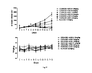
Fig. 27 shows an inhibiting effect of a CD20xCD3 id, bispecific antibody on
immunodeficient
mouse models of subcutaneous Raji transplantation tumor.
Fig. 28 and Fig. 29 show an inhibiting effect of a CD20xCD3 id, bispecific
antibody on
immunodeficient mouse models of subcutaneous transplantation tumor that are
inoculated with
human PBMCs and Raji, wherein Fig. 29 shows the comparison of efficacies of
low-dose groups.
Fig. 30 shows that cynomolgus monkey B cells are quickly eliminated after
administration of a
CD20xCD3 id, bispecific antibody.
Detailed Description o f the Invention
I. Definitions
In the present disclosure, unless otherwise specified, scientific and
technical terms used herein
have the meanings commonly understood by those skilled in the art. Moreover,
protein and
nucleic acid chemistry, molecular biology, cell and tissue culture,
microbiology, and
immunology-related terms and laboratory procedures used herein are terms and
conventional
procedures widely used in corresponding arts. Meanwhile, in order to better
understand the
present disclosure, definitions and explanations o f relevant terms are
provided below.
CD20 (also referred to as a B-lymphocyte antigen CD20, B-lymphocyte surface
antigen Bl,
CA 03190900 2023- 2- 24
WSLEGAL\ 092120 \ 00011 \33711631v1
Leu-16, Bp 35, BM5 or LF5; a human protein characterized in UniProt database
entry P11836) is a
hydrophobic transmembrane protein that is expressed on precursor B and mature
B-lymphocytes
and has a molecular weight of about 35 kD (Valentine, M.A. et al., J. Biol.
Chem. 264 (1989)
11282-11287; Tedder, T.F .et al., Proc. Natl .Acad. Sci. U.S.A. 85 (19 88)2 08-
21 2; Stamenkovic, I.
et al., J. Exp. Med. 167 (1988) 1975-1980; Einfeld, D.A. et al., EMBO J. 7
(1988) 711-717;
Tedder, T.F. et al., J. Immunol. 142 (1989) 2560-2568). The corresponding
human gene is
membrane-spanning 4-domains, subfamily A, member 1, also referred to as MS4A1.
The gene
encodes a member of the membrane-spanning 4A gene family. Members of the
nascent protein
family are characterized by common structural features and similar intron/exon
splice boundaries
and demonstrate unique expression patterns among hematopoietic cells and non-
lymphoid tissues.
The gene encodes a B-lymphocyte surface molecule that plays a role in the
development and
differentiation of B cells into plasma cells. The family member is localized
to 11q12, among a
cluster of family members. Alternative splicing of the gene results in two
transcript variants that
encode the same protein.
As used herein, the term "CD20" refers to any natural CD20 from any vertebrate
including
mammals such as primates (e.g., humans) and rodents (e.g., mice and rats),
unless otherwise
specified. The term encompasses "full length", unprocessed CD2 0 as well as
any form of
processed CD2 0 derived from a cell. The term also encompasses naturally
occurring variants of
CD20, such as splice variants and allelic variants. In an embodiment, CD20 is
human CD20. An
emplary amino acid sequence ofhuman CD20 is shown as SEQ ID NO: 1.
The terms "anti-CD20 antibody" and "CD20-binding antibody" refer to an
antibody that can bind
to CD20 with sufficient affinity such that the antibody is useful in targeting
CD20 as a diagnostic
and/or therapeutic agent. In an embodiment, the binding ability of the anti-
CD20 antibody to
unrelated non-CD20 protein is less than about 10% of the binding ability of
the antibody to CD20
that is determined by, for example, radioimmunoassay (RIA). In certain
embodiments, the
CD20-binding antibody has a dissociation constant (Ka) <1 M, 100<
nM, <10 nM, <1 nM, <0.1
nM, <0.01 nM or <0.001 nM (e.g., 10-8 M or less such as 10-8-10-13 M, and 10-9-
10-13 M). In
certain embodiments, the anti-CD20 antibody binds to conservative CD20
epitopes among CD20
from different species.
"CD3" refers to any natural CD3 from any vertebrate including mammals such as
primates (e.g.,
humans), non-human primates (e.g., Macaca fascicularis) and rodents (e.g.,
mice and rats), unless
otherwise specified. The term encompasses "full-length", unprocessed CD3 and
any form of
processed CD3 from a cell. The term also encompasses naturally occurring
variants of CD3, such
as splice variants and allelic variants. In an embodiment, CD3 is human CD3,
especially the
epsilon subunit of human CD3 (CDR). An amino acid sequence of human CDR is
shown in
6
CA 03190900 2023- 2- 24
WSLEGAL\ 092120 \ 00011 \33711631v1
UniProt (www.uniprot.org) accession number P07766 (version 144) or NCBI
(www.ncbi.nlm.nih.gov/) RefSeq NP_000724.1. An amino acid sequence of
cynomolgus monkey
CDR is shown in NCBI GenBank no. BAB71849.1.
The term "cell surface" is used according to its normal meaning in the art and
thus includes the
outside o f a cell which is accessible through binding to proteins and
othermolecules .
As used herein, unless otherwise specified, the term "about" or "approximate"
means within plus
or minus 10% of a given value or range. In a case that integers are required,
the term means
rounding up or down to the nearest integer within plus or minus 10% of a given
value or range.
With respect to antibody chain polypeptide sequences, the phrase substantially
identical is to be
understood as at least 60%, 65%, 70%, 75%, 80%, 85%, 90%, 95%, 96%, 97%, 98%,
99% or
higher sequence identity of an antibody chain to a reference polypeptide
sequence. With respect to
nucleic acid sequences, the term is to be understood as at least greater than
60%, 65%, 70%, 75%,
80%, 85%, 90%, 95%, 96%, 97%, 98%, 99% or higher sequence identity of a
nucleotide sequence
to a reference nucleic acid sequence.
The sequence "identity" has a meaning recognized in the art, and the
percentage of sequence
identity between two nucleic acids or polypeptide molecules or regions can be
calculated by
disclosed technologies. The sequence identity can be measured along the full
length of
polynucleotides or polypeptides or along regions of the molecules. There are
many methods for
measuring the identity between two polynucleotides or polypeptides, but the
term identity is well
known to the skilled person (Carrillo, H. & Lipman, D., SIAM J Applied Math
48: 1073 (1988)).
A "substitution" variant refers to a variant obtained by removing at least one
amino acid residue
from a natural sequence and inserting a different amino acid at the same site.
The substitution may
be a single substitution, that is, only one amino acid in the molecule is
substituted; or the
substitution may be multiple substitutions, that is, two or more amino acids
in the same molecule
are substituted. The multiple substitutions may be at consecutive sites.
Similarly, an amino acid
can be substituted with multiple residues, and thus such a variant includes
substitution and
insertion. An insertion variant is a variant obtained by inserting one or more
amino acids
immediately adjacent to an amino acid at a specific site on a natural
sequence. Immediately
adjacent to an amino acid means linking with the a-carboxyl or a-amino
functional group of the
amino acid. A "deletion" variant refers to a variant obtained by removing one
or more amino acids
from a natural amino acid sequence. Usually, a deletion variant has one or two
amino acid
deletions in a specific region of its molecule.
With respect to variable domains of antibodies, the term "variable" refers to
certain portions of
relevant nulecules that have extensive sequence differences between
antibodies, and are used for
the specific recognition and binding to a specific antibody against its
specific target. However, the
7
CA 03190900 2023- 2- 24
WSLEGAL\092120\00011\33711631v1
variability is not uniformly distributed throughout the variable domains of
antibodies. The
variability is concentrated in three segments known as complementarity-
determining regions
(CDRs; i.e., CDR1, CDR2, and CDR3) or hypervariable regions, which are located
within
variable domains of light chains and heavy chains. A more conservative portion
within the
variable domain is referred to as a framework region (FR) or framework
sequence. Each variable
domain of a natural heavy chain or light chain includes four FRs, which mainly
adopt a 13-folded
configuration and linked together through three CDRs, the CDRs form a loop,
and the loop is
linked with the 13-folded configuration and forms a part of the 13-folded
configuration in certain
cases. CDRs of each chain are usually linked adjacently by an FR region and
contribute to the
formation of a target binding site (epitope or determinant) of an antibody by
virtue of CDRs from
other chains. As used herein, the numbering of amino acid residues of
immunoglobulin is based on
the numbering system of amino acid residues of immunoglobulin developed by
Kabat et al., unless
otherwise specified. A CDR may have the ability to specifically bind to a
cognate epitope.
As used herein, an "antibody fragment" or "antigen-binding fragment" of an
antibody refers to any
portion of a full-length antibody, which is shorter than the full-length
antibody, at least contains a
partial variable region (e.g., one or more CDRs and/or one or more antibody-
binding sites) of the
antibody that binds to an antigen, and thus retains the binding specificity
and at least partial
specific binding ability of the full-length antibody. Therefore, the antigen-
binding fragment refers
to an antibody fragment that contains an antigen-binding portion binding to
the same antigen as
the antibody from which the antibody fragment is derived. The antibody
fragment includes an
antibody derivative that is produced by enzymatic treatment of the full-length
antibody, and a
synthesized derivative such as a recombinant derivative. The antibody includes
the antibody
fragment. Examples of the antibody fragment include, but are not limited to,
Fab, Fab', F(ab')2,
single-stranded Fv(scFv), Fv, dsFv, double antibody, Fd and a Fd' fragment,
and other fragments,
including a modified fragment. The fragment may include multiple chains that
are linked together
through, for example, disulfide bonds and/or peptide adaptors. The antibody
fragment generally
contains at least or about 50 amino acids, typically at least or about 200
amino acids. The
antigen-binding fragment includes any antibody fragment, which is inserted
into an antibody
framework (e.g., by replacing a corresponding region) to obtain an antibody
specifically binding
to (that is, with a binding constant Ka of about 107-108M-1) an antigen. A
"functional fragment"
or "analog of an anti-CD20 antibody" is a fragment or analog that can prevent
or substantially
reduce the ability of the receptor to bind to a ligand or initiate signal
transduction. As used herein,
the functional fragment generally has the same meaning as an antibody
fragment, and with respect
to an antibody, it may refer to a fragment that can prevent the receptor from
binding to a ligand or
from initiating signal transduction or substantially reduce the ability of the
receptor to bind to a
8
CA 03190900 2023- 2- 24
WSLEGAL\092120\00011\33711631v1
ligand or initiate signal transduction. The Fv fragment consists of a dimer
(VH-VL dimer) formed
by non-covalent binding of a variable domain of a heavy chain and a variable
region of a light
chain. In this configuration, three CDRs of each variable domain interact to
define a target binding
site on the surface of the VH-VL dimer, as in the case of an intact antibody.
The six CDRs
together confer the target binding specificity to the intact antibody.
However, even a single
variable domain (or half of Fv containing 3 target-specific CDRs only) can
still have the ability to
recognize and bind to a target.
As used herein, the term "bispecific antibody (BsAb)" refers to an antibody
and/or antigen-binding
molecule that can specifically bind to two different antigenic determinants,
and usually, the
bispecific antibody and/or antigen-binding molecule contains two antigen-
binding sites each of
which is specific to different antigenic determinants. In certain embodiments,
the bispecific
antibody and/or antigen-binding molecule can bind to two antigenic
determinants at the same time,
particularly two antigenic determinants that are expressed on two different
types feels.
As used herein, a "monoclonal antibody" refers to a population of identical
antibodies, meaning
that each individual antibody molecule in the population of monoclonal
antibodies is identical to
the other antibody molecules. This characteristic is in contrast to that of a
polyclonal population of
antibodies, which contain antibodies with a variety of different sequences.
Monoclonal antibodies
can be prepared by many well-known methods (Smith et al. (2004) J.Clin.
Pathol. 57, 912-917;
and Nelson et al., J Chin Pathol (2000), 53, 111-117). For example, monoclonal
antibodies can be
prepared by immortalizing B cells, for example, fusing with myeloma cells to
produce a
hybridoma cell line or infecting B cells with virus such as EBV. Recombination
can also be used
to prepare antibodies in vitro from clonal populations of host cells by
transforming the host cells
with plasmids carrying artificial sequences ofnucleotides encoding the
antibodies
As used herein, the term "hybridoma" or "hybridoma cell" refers to a cell or
cell line (usually
myelo ma or lymphoma cells) produced by fusing lymphocytes that produce
antibodies with cancer
cells that do not produce antibodies. As is known to those of ordinary skill
in the art, a hybridoma
can proliferate and continuously supply and produce specific monoclonal
antibodies. The method
for producing a hybridoma is known in the art. When the term "hybridoma" or
"hybridoma cell" is
referred to, it also includes a subclone and progeny cells of the hybridoma.
As used herein, a full-length antibody is an antibody that has two full-length
heavy chains (e.g.,
VH-CH1-CH2-CH3 and VH-CH1-CH2-CH3-CH4), two full-length light chains (VL-CL),
and
hinge regions, such as an antibody produced naturally by antibody-secreting B
cells and a
synthesized antibody with the same domains.
The term "chimeric antibody" refers to an antibody that contains a variable
region sequence
derived from a species and a constant region sequence derived from another
species. For example,
9
CA 03190900 2023- 2- 24
WSLEGAL\ 092120 \ 00011 \33711631v1
the variable region sequence is derived from a mouse antibody and the constant
region sequence is
derived from a human antibody.
A "humanized" antibody refers to a non-human (e.g., mouse) antibody form,
which is a chimeric
immunoglobulin, immunoglobulin chain or a fragment thereof (e.g., Fv, Fab,
Fab', F(ab')2 or other
antigen-binding subsequences of an antibody), containing the minimal sequence
derived from a
non-human immunoglobulin. Preferably, the humanized antibody is a human
immunoglobulin
(recipient antibody), and residues in a complementary determining region (CDR)
of the recipient
antibody are substituted with CDR residues in a non-human species, such as a
mouse, a rat and a
rabbit, that has the desired specificity, affinity and ability.
In addition, during humanization, amino acid residues within CDR1, CDR2 and/or
CDR3 regions
of VH and/or VL may be mutated, so as to improve one or more binding
characteristics (e.g., the
affinity). A mutation can be introduced, for example, by performing PCR-
mediated mutation, the
influence of which on antibody binding or other functional characteristics can
be assessed using
the in vitro or in vivo assay described herein. Usually, a conservative
mutation is introduced. Such
a mutation may be amino acid substitution, addition or deletion. In addition,
there are usually no
more than one or two mutations within a CDR. Therefore, the humanized antibody
of the present
disclosure also covers an antibody of which a CDR contains 1 or 2 amino acid
mutations.
As used herein, the term "CDR" refers to a complementarity-determining region,
and each heavy
chain or light chain of a known antibody molecule has 3 CDRs. CDR is also
referred to as a
hypeivariable region that exists in a variable region of each heavy chain or
light chain of an
antibody, and the primary structure of CDR has a site with very high
variability. In this description,
CDRs of a heavy chain are represented by CDR1, CDR2, and CDR3 derived from the
amino-terminus of the amino-terminal sequence of the heavy chain, and CDRs of
a light chain are
represented by CDR1, CDR2, and CDR3 derived from the amino-terminus of the
amino-terminal
sequence of the light chain. These sites are adjacent to each other in the
tertiary structure and
determine the specificity of an antigen to which an antibody binds.
As used herein, the term "epitope" refers to any antigenic determinant on an
antigen to which a
paratope of an antibody binds. An epitope determinant usually contains a
chemically active
surface type of a molecule, such as an amino acid and a sugar side chain, and
usually has specific
three-dimensional structural characteristics and specific charge
characteristics.
As used herein, the "specific binding" or "immunospecific binding" of an
antibody or an
antigen-binding fragment thereof are interchangeable herein, and refers to the
ability of the
antibody or the antigen-binding fragment thereof to form one or more non-
covalent bonds with the
same antigen through non-covalent interaction of the antibody and an antibody-
binding site of an
antigen. The antigen may be a separated antigen or present in tumor cells.
Usually, an antibody
CA 03190900 2023- 2- 24
WSLEGAL\ 092120 \ 00011 \33711631v1
immunospecifically binding (or specifically binding) to an antigen binds to
the antigen with an
affinity constant Ka of about lx107 M-1 or 1 x10⁸ M-1 or greater (or with a
dissociation constant
(Kd) of 1 x10⁷ M or lx 10-8 M or less). An affinity constant can be measured
by standard kinetic
methods for antibody responses, such as immunoassay, surface plasmon resonance
(SPR) (Rich
and Myszka (2000) Curt Opin. Biotechnol 11: 54; and Englebienne (1998)
Analyst. 123: 1599),
isothermal titration calorimetry (ITC), and other dynamic interaction known in
the art (also
referring to U.S. Patent No. 7,229,619 describing exemplary SPR and ITC
methods for calculating
the binding affinity of antibodies). Instruments and methods for real-time
detection and
monitoring of binding rates are known and commercially available (referring to
Bia Core 2000,
Biacore AB, Ups ala, Sweden and GE Healthcare Life Sciences; Malmqvist (2000)
Biochem. Soc.
Trans. 27: 335).
As used herein, the terms "polynucleotide" and "nucleic acid molecule" refer
to an oligomer or
polymer that contains at least two linked nucleotides or nucleotide
derivatives, including
deoxyribonucleic acid (DNA) and ribonucleic acid (RNA) that are usually linked
together through
phosphodiester bonds. As used herein, the term "nucleic acid molecule" is
intended to include
DNA molecules and RNA molecules. A DNA molecule may be single-stranded or
double-stranded,
and may be cDNA.
As used herein, a separated nucleic acid molecule is a nucleic acid molecule
that is separated from
other nucleic acid molecules present in a natural source of the nucleic acid
molecule. A
"separated" nucleic acid molecule, such as a cDNA molecule, may be
substantially free of other
cellular materials or culture medium when prepared by recombination, or
substantially free of
chemical precursors or other chemical components when chemically synthesized.
Exemplary
separated nucleic acid molecules provided herein include a separated nucleic
acid molecule
encoding the antibody or antigen-binding fragment provided.
As used herein, "operably linking" of nucleic acid sequences, regions,
elements or domains means
that the nucleic acid regions are functionally related to each other. For
example, a promoter can be
operably linked to a nucleic acid encoding a polypeptide such that the
promoter regulates or
mediates transcription of the nucleic acid.
"Conservative sequence modifications" of the sequences in the sequence
listings herein are also
provided, i.e., nucleotide and amino acid sequence modifications that do not
eliminate the binding
of an antibody encoded by the nucleotide sequence or containing the amino acid
sequence to an
antigen. These conservative sequence modifications include conservative
nucleotide and amino
acid substitutions, nucleotide and amino acid additions, and deletions. For
example, a modification
can be introduced into the sequence listings herein by the standard technology
(e.g., site-directed
mutagenesis and PCR-mediated mutagenesis) known in the art. The conservative
sequence
11
CA 03190900 2023- 2- 24
WSLEGAL\ 092120 \ 00011 \33711631v1
modifications include amino acid substitutions in which amino acid residues
are substituted with
amino acid residues with similar side chains. Families of amino acid residues
with similar side
chains are defined in the art. The families include amino acids with basic
side chains (e.g., lysine,
arginine, and histidine), amino acids with acidic side chains (e.g., aspartic
acid and glutamic acid),
amino acids with uncharged polar side chains (e.g., glycine, asparagine,
glutamine, serine,
threonine, tyrosine, cysteine, and tryptophan), amino acids with non-polar
side chains (e.g.,
alanine, valine, leucine, isoleucine, proline, phenylalanine, and methionine),
amino acids with 13
branched side chains (e.g., threonine, valine, and isoleucine), and amino
acids with aromatic side
chains (e.g., tyrosine, phenylalanine, tryptophan, and histidine). Therefore,
a predicted
nonessential amino acid residue in an anti-CD20 antibody is preferably
substituted with another
amino acid residue from the same side chain family. Methods for identifying
nucleotide and amino
acid conservative substitutions that do not eliminate binding to an antigen
are well known in the
art (for example, referring to Brummell et al., Biochem. 32: (1180-1187
(1993); Kobayashi et al.,
Protein Eng. 12(10): 879-884 (1999); Burks et al., Proc. Natl. Acad. Sci. USA
94: 412-417
(1997)).
As another option, in another embodiment, mutations can be introduced randomly
along all or a
portion of the anti-CD20 antibody coding sequence by, for example, saturation
mutagenesis, and
the obtained modified anti-CD20 antibodies can be screened for improved
binding activity.
As used herein, "expression" refers to the process of producing a polypeptide
by the transcription
and translation of a polynucleotide. An expression level of a polypeptide can
be assessed by any
method known in the art, including, for example, methods for determining the
amount of
polypeptides produced by a host cell. Such methods may include, but are not
limited to,
quantification of polypeptides in a cell lysate by ELISA, Coomassie blue
staining after gel
electrophores is , Lowry protein assay, and Bradford protein assay.
As used herein, a "host cell" is a cell for receiving, maintaining,
replicating and amplifying a
vector. The host cell can also be used for expressing polypeptides encoded by
the vector. When the
host cell divides, a nucleic acid contained in the vector is replicated such
that the nucleic acid is
amplified. The host cell may be a eukaryotic cell or prokaryotic cell.
Suitable host cells include,
but are not limited to, CHO cells, various COS cells, HeLa cells, and HEK
cells such as HEK 293
cells.
As used herein, the "vector" is a replicable nucleic acid from which one or
more heterologous
proteins can be expressed when the vector is transformed into an appropriate
host cell. The vector
includes those vectors into which nucleic acids encoding polypeptides or
fragments thereof can be
introduced, usually by restriction digestion and ligation. The vector also
includes those vectors
that contain nucleic acids encoding polypeptides. The vector is used to
introduce a nucleic acid
12
CA 03190900 2023- 2- 24
WSLEGAL\ 092120 \ 00011 \33711631v1
encoding a polypeptide into the host cell, so as to amplify the nucleic acid
or express/display the
polypeptide encoded by the nucleic acid. The vector is usually free, but can
be designed to allow
integration of a gene or a portion thereof into a chromosome of a genome.
Vectors for artificial
chromosome are also contemplated, such as a yeast artificial vector and a
mammal artificial
chromosome. Selection and use of such intermedia are well-known to those
skilled in the art.
As used herein, the vector also includes a "virus vector" or "viral vector".
The viral vector is an
engineered virus that is operably linked to an exogenous gene to transfer (as
an intermedium or
shuttle) the exogenous gene into cells.
As used herein, an "expression vector" includes vectors that can express DNA,
and the DNA is
operably linked to a regulatory sequence that can affect the expression of
such DNA fragments,
such as a promoter. Such additional fragments may include promoter and
terminator sequences,
and optionally may include one or more origins of replication, one or more
selectable markers,
enhancers, polyadenylation signals, etc. The expression vector is usually
derived from a plasmid
or virus DNA, or may contain elements of both. Therefore, the expression
vector refers to a
recombinant DNA or RNA construct, such as a plasmid, a phage, a recombinant
virus, and other
vectors, which expresses cloned DNA when introduced into an appropriate host
cell, Appropriate
expression vectors are well-known to those skilled in the art, and include
expression vectors that
can be replicated in eukaryotic cells and/or prokaryotic cells, free
expression vectors, and
expression vectors for integration into the genome of a host cell.
As used herein, "treatment" of a disease or disease condition in an individual
means that
individual's symptoms are partially or fully alleviated, or remain unchanged
after treatment.
Therefore, treatment includes prophylaxis, treatment and/or cure. Prophylaxis
refers to prevention
of an underlying disease and/or prevention of symptomatic deterioration or
disease development.
Treatment also includes any pharmaceutical use of any provided antibody or
antigen-binding
fragment thereo f and composition provided herein.
As used herein, a "therapeutic effect" means an effect resulting from
treatment of an individual
that alters, usually improves or ameliorates symptoms of a disease or disease
condition, or cures a
disease or dis ease condition.
As used herein, the "therapeutically effective amount" or "therapeutically
effective dose" refers to
the amount of a substance, compound, material or composition containing a
compound that is at
least sufficient to produce a therapeutic effect when administered to a
subject. Therefore, it is the
amount necessary to prevent, cure, ameliorate, arrest or partially arrest
symptoms of a disease or
disorder.
As used herein, the "prophylactically effective amount" or "prophylactically
effective dose" refers
to the amount of a substance, compound, material, or composition containing a
compound that,
13
CA 03190900 2023- 2- 24
WSLEGAL\092120\00011\33711631v1
when administered to a subject, will have the desired prophylactic effect,
such as preventing or
delaying the onset or recurrence of a disease or symptom, and reducing the
likelihood of the onset
or recurrence of a disease or symptom. A full prophylactically effective dose
does not have to
occur by administering one dose, and may only occur after administering a
series of doses.
Therefore, the prophylactically effective amount may be administered in one or
more
administrations.
As used herein, the term "patient" refers to a mammal, such as humans.
II. Detailed description of specific embodiments
In an aspect, the present disclosure provides a bispecific antibody or an
antigen-binding portion
thereof, which contains a first binding domain that binds to CD20 on the
surface of a target cell
and a second binding domain that binds to CD3 on the surface of a T cell. The
first binding
domain contains a first light chain and a first heavy chain, the first light
chain contains a light
chain CDR selected from amino acid sequences SEQ ID NO: 56, 57, 58, 61, 62,
63, or any variant
thereof, and/or the first heavy chain contains a heavy chain CDR selected from
amino acid
sequences SEQ ID NO: 66, 67, 68, 71, 76, 77, 78, 85, or any variant thereof.
According to the antibody or the antigen-binding portion thereof of the
aforementioned aspect, the
first light chain of the first binding domain contains a light chain CDR1
selected from amino acid
sequences SEQ ID NO: 56, 61, or any variant thereof, a light chain CDR2
selected from amino
acid sequences SEQ ID NO: 57, 62, or any variant thereof, a light chain CDR3
selected from
amino acid sequences SEQ ID NO: 58, 63, or any variant thereof; and/or the
first heavy chain of
the first binding domain contains a heavy chain CDR1 selected from amino acid
sequences SEQ
ID NO: 66, 76, or any variant thereof, a heavy chain CDR2 selected from amino
acid sequences
SEQ ID NO: 67, 77, or any variant thereof, a heavy chain CDR3 selected from
amino acid
sequences SEQ ID NO: 68, 71, 78, 85, or any variant thereof.
According to the antibody or the antigen-binding portion thereof of the
aforementioned aspect, the
first binding domain contains a light chain and heavy chain CDR combination
selected from:
(1) first light chain CDR1, CDR2 and CDR3 sequences respectively containing
SEQ ID NO:
56-58, and/or first heavy chain CDR1, CDR2 and CDR3 sequences respectively
containing SEQ
ID NO: 66-68;
(2) first light chain CDR1, CDR2 and CDR3 sequences respectively containing
SEQ ID NO:
56-58, and/or first heavy chain CDR1, CDR2 and CDR3 respectively containing
SEQ ID NO: 66,
67 and 71;
(3) first light chain CDR1, CDR2 and CDR3 sequences respectively containing
SEQ ID NO:
61-63, and/or first heavy chain CDR1, CDR2 and CDR3 sequences respectively
containing SEQ
ID NO: 76-78; and
14
CA 03190900 2023- 2- 24
WSLEGAL\092120\00011\33711631v1
(4) first light chain CDR1, CDR2 and CDR3 sequences respectively containing
SEQ ID NO:
61-63, and/or first heavy chain CDR1, CDR2 and CDR3 respectively containing
SEQ ID NO: 76,
77 and 85.
According to the antibody or the antigen-binding portion thereof of the
aforementioned aspect, the
first binding domain contains a light chain variable region selected from
amino acid sequences
SEQ ID NO: 54, 5 9 , or any variant thereof, and/or a heavy chain variable
region selected from
amino acid sequences SEQ ID NO: 64, 69, 72, 74, 79, 81, 83, or any variant
thereof.
In some embodiments, the first binding domain contains a first light chain
variable region of
amino acid sequence SEQ ID NO: 54 or any variant thereof, and a first heavy
chain variable
region of amino acid sequence SEQ ID NO: 64 or any variant thereof.
In some embodiments, the first binding domain contains a first light chain
variable region of
amino acid sequence SEQ ID NO: 54 or any variant thereof, and a first heavy
chain variable
region of amino acid sequence SEQ ID NO: 69 or any variant thereof.
In some embodiments, the first binding domain contains a first light chain
variable region of
amino acid sequence SEQ ID NO: 54 or any variant thereof, and a first heavy
chain variable
region of amino acid sequence SEQ ID NO: 72 or any variant thereof.
In some embodiments, the first binding domain contains a first light chain
variable region of
amino acid sequence SEQ ID NO: 59 or any variant thereof, and a first heavy
chain variable
region of amino acid sequence SEQ ID NO: 74 or any variant thereof.
In some embodiments, the first binding domain contains a first light chain
variable region of
amino acid sequence SEQ ID NO: 59 or any variant thereof, and a first heavy
chain variable
region of amino acid sequence SEQ ID NO: 79 or any variant thereof.
In some embodiments, the first binding domain contains a first light chain
variable region of
amino acid sequence SEQ ID NO: 59 or any variant thereof, and a first heavy
chain variable
region of amino acid sequence SEQ ID NO: 81 or any variant thereof.
In some embodiments, the first binding domain contains a first light chain
variable region of
amino acid sequence SEQ ID NO: 59 or any variant thereof, and a first heavy
chain variable
region of amino acid sequence SEQ ID NO: 83 or any variant thereof.
According to the antibody or the antigen-binding portion thereof of the
aforementioned aspect, the
second binding domain contains a second light chain CDR1 selected from amino
acid sequences
SEQ ID NO: 7, 14, or any variant thereof, a second light chain CDR2 selected
from amino acid
sequences SEQ ID NO: 8, 15, 20, or any variant thereof, and a second light
chain CDR3 selected
from amino acid sequences SEQ ID NO: 9, 21, or any variant thereof; and/or a
second heavy
chain CDR1 selected from amino acid sequences SEQ ID NO: 26, 31, 46, or any
variant thereof, a
second heavy chain CDR2 selected from amino acid sequences SEQ ID NO: 27, 47,
or any variant
CA 03190900 2023- 2- 24
WSLEGAL\092120\00011\33711631v1
thereof, a second heavy chain CDR3 selected from amino acid sequences 28, 34,
37, 40, 43, or
any variant thereof.
In some embodiments, the second binding domain contains a second light chain
and a second
heavy chain, and the second binding domain contains a light chain and heavy
chain CDR
combination selected from:
(1) second light chain CDR1, CDR2 and CDR3 sequences respectively containing
SEQ ID NO: 7,
8 and 9, and second heavy chain CDR1, CDR2 and CDR3 sequences respectively
containing SEQ
ID NO: 26, 27 and 28;
(2) second light chain CDR1, CDR2 and CDR3 sequences respectively containing
SEQ ID NO: 7,
8 and 9, and second heavy chain CDR1, CDR2 and CDR3 sequences respectively
containing SEQ
ID NO: 31, 27 and 28;
(3) second light chain CDR1, CDR2 and CDR3 sequences respectively containing
SEQ ID NO: 7,
8 and 9, and second heavy chain CDR1, CDR2 and CDR3 sequences respectively
containing SEQ
ID NO: 31, 27 and 34;
(4) second light chain CDR1, CDR2 and CDR3 sequences respectively containing
SEQ ID NO: 7,
8 and 9, and second heavy chain CDR1, CDR2 and CDR3 sequences respectively
containing SEQ
ID NO: 31, 27 and 37;
(5) second light chain CDR1, CDR2 and CDR3 sequences respectively containing
SEQ ID NO: 7,
8 and 9, and second heavy chain CDR1, CDR2 and CDR3 sequences respectively
containing SEQ
ID NO: 31, 27 and 40;
(6) second light chain CDR1, CDR2 and CDR3 sequences respectively containing
SEQ ID NO: 7,
8 and 9, and second heavy chain CDR1, CDR2 and CDR3 sequences respectively
containing SEQ
ID NO: 31, 27 and 43;
(7) second light chain CDR1, CDR2 and CDR3 sequences respectively containing
SEQ ID NO: 7,
8 and 9, and second heavy chain CDR1, CDR2 and CDR3 sequences respectively
containing SEQ
ID NO: 46, 47 and 28;
(8) second light chain CDR1, CDR2 and CDR3 sequences respectively containing
SEQ ID NO:
14, 15 and 9, and second heavy chain CDR1, CDR2 and CDR3 sequences
respectively containing
SEQ ID NO: 26, 27 and 28;
(9) second light chain CDR1, CDR2 and CDR3 sequences respectively containing
SEQ ID NO:
14, 15 and 9, and second heavy chain CDR1, CDR2 and CDR3 sequences
respectively containing
SEQ ID NO: 31, 27 and 28;
(10) second light chain CDR1, CDR2 and CDR3 sequences respectively containing
SEQ ID NO:
14, 15 and 9, and second heavy chain CDR1, CDR2 and CDR3 sequences
respectively containing
SEQ ID NO: 31, 27 and 34;
16
CA 03190900 2023- 2- 24
WSLEGAL\092120\00011\33711631v1
(11) second light chain CDR1, CDR2 and CDR3 sequences respectively containing
SEQ ID NO:
14, 15 and 9, and second heavy chain CDR1, CDR2 and CDR3 sequences
respectively containing
SEQ ID NO: 31, 27 and 37;
(12) second light chain CDR1, CDR2 and CDR3 sequences respectively containing
SEQ ID NO:
14, 15 and 9, and second heavy chain CDR1, CDR2 and CDR3 sequences
respectively containing
SEQ ID NO: 31, 27 and 40;
(13) second light chain CDR1, CDR2 and CDR3 sequences respectively containing
SEQ ID NO:
14, 15 and 9, and second heavy chain CDR1, CDR2 and CDR3 sequences
respectively containing
SEQ ID NO: 31, 27 and 43;
(14) second light chain CDR1, CDR2 and CDR3 sequences respectively containing
SEQ ID NO:
14, 15 and 9, and second heavy chain CDR1, CDR2 and CDR3 sequences
respectively containing
SEQ ID NO: 46, 47 and 28;
(15) second light chain CDR1, CDR2 and CDR3 sequences respectively containing
SEQ ID NO:
7, 8 and 21, and second heavy chain CDR1, CDR2 and CDR3 sequences respectively
containing
SEQ ID NO: 26, 27 and 28;
(16) second light chain CDR1, CDR2 and CDR3 sequences respectively containing
SEQ ID NO:
7, 8 and 21, and second heavy chain CDR1, CDR2 and CDR3 sequences respectively
containing
SEQ ID NO: 31, 27 and 28;
(17) second light chain CDR1, CDR2 and CDR3 sequences respectively containing
SEQ ID NO:
7, 8 and 21, and second heavy chain CDR1, CDR2 and CDR3 sequences respectively
containing
SEQ ID NO: 31, 27 and 34;
(18) second light chain CDR1, CDR2 and CDR3 sequences respectively containing
SEQ ID NO:
7, 8 and 21, and second heavy chain CDR1, CDR2 and CDR3 sequences respectively
containing
SEQ ID NO: 31, 27 and 37;
(19) second light chain CDR1, CDR2 and CDR3 sequences respectively containing
SEQ ID NO:
7, 8 and 21, and second heavy chain CDR1, CDR2 and CDR3 sequences respectively
containing
SEQ ID NO: 31, 27 and 40;
(20) second light chain CDR1, CDR2 and CDR3 sequences respectively containing
SEQ ID NO:
7, 8 and 21, and second heavy chain CDR1, CDR2 and CDR3 sequences respectively
containing
SEQ ID NO: 31, 27 and 43;
(21) second light chain CDR1, CDR2 and CDR3 sequences respectively containing
SEQ ID NO:
7, 8 and 21, and second heavy chain CDR1, CDR2 and CDR3 sequences respectively
containing
SEQ ID NO: 46, 47 and 28;
(22) second light chain CDR1, CDR2 and CDR3 sequences respectively containing
SEQ ID NO:
7, 20 and 21, and second heavy chain CDR1, CDR2 and CDR3 sequences
respectively containing
17
CA 03190900 2023- 2- 24
WSLEGAL\092120\00011\33711631v1
SEQ ID NO: 26, 27 and 28;
(23) second light chain CDR1, CDR2 and CDR3 sequences respectively containing
SEQ ID NO:
7, 20 and 21, and second heavy chain CDR1, CDR2 and CDR3 sequences
respectively containing
SEQ ID NO: 31, 27 and 28;
(24) second light chain CDR1, CDR2 and CDR3 sequences respectively containing
SEQ ID NO:
7, 20 and 21, and second heavy chain CDR1, CDR2 and CDR3 sequences
respectively containing
SEQ ID NO: 31, 27 and 34;
(25) second light chain CDR1, CDR2 and CDR3 sequences respectively containing
SEQ ID NO:
7, 20 and 21, and second heavy chain CDR1, CDR2 and CDR3 sequences
respectively containing
SEQ ID NO: 31, 27 and 37;
(26) second light chain CDR1, CDR2 and CDR3 sequences respectively containing
SEQ ID NO:
7, 20 and 21, and second heavy chain CDR1, CDR2 and CDR3 sequences
respectively containing
SEQ ID NO: 31, 27 and 40;
(27) second light chain CDR1, CDR2 and CDR3 sequences respectively containing
SEQ ID NO:
7, 20 and 21, and second heavy chain CDR1, CDR2 and CDR3 sequences
respectively containing
SEQ ID NO: 31, 27 and 43;
(28) second light chain CDR1, CDR2 and CDR3 sequences respectively containing
SEQ ID NO:
7, 20 and 21, and second heavy chain CDR1, CDR2 and CDR3 sequences
respectively containing
SEQ ID NO: 46, 47 and 28.
In some embodiments, the second binding domain contains a light chain variable
region selected
from amino acid sequences SEQ ID NO: 5, 10, 12, 16, 18, 22, or any variant
thereof, and/or a
heavy chain variable region selected from amino acid sequences SEQ ID NO: 24,
29, 32, 35, 38,
41, 44, 48, 50, 52, or any variant thereof.
In some embodiments, the second binding domain contains a second light chain
variable region of
amino acid sequence SEQ ID NO: 5 or any variant thereof, and a second heavy
chain variable
region of amino acid sequence SEQ ID NO: 48 or any variant thereof.
In some embodiments, the second binding domain contains a second light chain
variable region of
amino acid sequence SEQ ID NO: 5 or any variant thereof, and a second heavy
chain variable
region of amino acid sequence SEQ ID NO: 50 or any variant thereof.
In some embodiments, the second binding domain contains a second light chain
variable region of
amino acid sequence SEQ ID NO: 10 and any variant thereof, and a second heavy
chain variable
region of amino acid sequence SEQ ID NO: 50 and any variant thereof.
In some embodiments, the second binding domain contains a second light chain
variable region of
amino acid sequence SEQ ID NO: 12 and any variant thereof, and a second heavy
chain variable
region of amino acid sequence SEQ ID NO: 50 and any variant thereof.
18
CA 03190900 2023- 2- 24
WSLEGAL\092120\00011\33711631v1
In some embodiments, the second binding domain contains a second light chain
variable region of
amino acid sequence SEQ ID NO: 18 and any variant thereof, and a second heavy
chain variable
region of amino acid sequence SEQ ID NO: 24 and any variant thereof.
In some embodiments, the second binding domain contains a second light chain
variable region of
amino acid sequence SEQ ID NO: 18 and any variant thereof, and a second heavy
chain variable
region of amino acid sequence SEQ ID NO: 48 and any variant thereof.
In some embodiments, the second binding domain contains a second light chain
variable region of
amino acid sequence SEQ ID NO: 18 and any variant thereof, and a second heavy
chain variable
region of amino acid sequence SEQ ID NO: 50 and any variant thereof.
According to the antibody or the antigen-binding portion thereof of the
aforementioned aspect, the
first light chain contains a light chain selected from SEQ ID NO: 88, 96, 100,
102, 110, 118, or
any variant thereof, the first heavy chain contains a heavy chain selected
from SEQ ID NO: 86, 94,
98, 104, 112, 120, or any variant thereof.
According to the antibody or the antigen-binding portion thereof of the
aforementioned aspect, the
second light chain contains a light chain selected from SEQ ID NO: 92, 106,
114, or any variant
thereof, the second heavy chain contains a heavy chain selected from SEQ ID
NO: 90, 108, 116,
or any variant thereof.
In some embodiments, the first binding domain contains a first light chain of
amino acid sequence
SEQ ID NO: 88 or any variant thereof, and a first heavy chain of amino acid
sequence SEQ ID
NO: 86 or any variant thereof.
In some embodiments, the first binding domain contains a first light chain of
amino acid sequence
SEQ ID NO: 96 or any variant thereof, and a first heavy chain of amino acid
sequence SEQ ID
NO: 94 or any variant thereof.
In some embodiments, the first binding domain contains a first light chain of
amino acid sequence
SEQ ID NO: 100 or any variant thereof, and a first heavy chain of amino acid
sequence SEQ ID
NO: 98 or any variant thereof.
In some embodiments, the first binding domain contains a first light chain of
amino acid sequence
SEQ ID NO: 102 or any variant thereof, and a first heavy chain of amino acid
sequence SEQ ID
NO: 104 or any variant thereof.
In some embodiments, the first binding domain contains a first light chain of
amino acid sequence
SEQ ID NO: 110 or any variant thereof, and a first heavy chain of amino acid
sequence SEQ ID
NO: 112 or any variant thereof.
In some embodiments, the first binding domain contains a first light chain of
amino acid sequence
SEQ ID NO: 118 or any variant thereof, and a first heavy chain of amino acid
sequence SEQ ID
NO: 120 or any variant thereof.
19
CA 03190900 2023- 2- 24
WSLEGAL\092120\00011\33711631v1
In some embodiments, the second binding domain contains a second light chain
of amino acid
sequence SEQ ID NO: 92 or any variant thereof, and a second heavy chain of
amino acid
sequence SEQ ID NO: 90 or any variant thereof.
In some embodiments, the second binding domain contains a second light chain
of amino acid
sequence SEQ ID NO: 106 or any variant thereof, and a second heavy chain of
amino acid
sequence SEQ ID NO: 108 or any variant thereof.
In some embodiments, the second binding domain contains a second light chain
of amino acid
sequence SEQ ID NO: 114 or any variant thereof, and a second heavy chain of
amino acid
sequence SEQ ID NO: 116 or any variant thereof.
In some embodiments, the bispecific antibody contains a first light chain of
amino acid sequence
SEQ ID NO: 88 or any variant thereof, a first heavy chain of amino acid
sequence SEQ ID NO: 86
or any variant thereof, a second light chain of amino acid sequence SEQ ID NO:
92 or any variant
thereof, and a second heavy chain ofamino acid sequence SEQ ID NO: 90 or any
variant thereof.
In some embodiments, the bispecific antibody contains a first light chain of
amino acid sequence
SEQ ID NO: 96 or any variant thereof, a first heavy chain of amino acid
sequence SEQ ID NO: 94
or any variant thereof, a second light chain of amino acid sequence SEQ ID NO:
92 or any variant
thereof, and a second heavy chain ofamino acid sequence SEQ ID NO: 90 or any
variant thereof.
In some embodiments, the bispecific antibody contains a first light chain of
amino acid sequence
SEQ ID NO: 100 or any variant thereof, a first heavy chain of amino acid
sequence SEQ ID NO:
98 or any variant thereof, a second light chain of amino acid sequence SEQ ID
NO: 92 or any
variant thereof, and a second heavy chain of amino acid sequence SEQ ID NO: 90
or any variant
thereof.
In some embodiments, the bispecific antibody contains a first light chain of
amino acid sequence
SEQ ID NO: 102 or any variant thereof, a first heavy chain of amino acid
sequence SEQ ID NO:
104 or any variant thereof, a second light chain of amino acid sequence SEQ ID
NO: 106 or any
variant thereof, a second heavy chain of amino acid sequence SEQ ID NO: 108 or
any variant
thereof.
In some embodiments, the bispecific antibody contains a first light chain of
amino acid sequence
SEQ ID NO: 110 or any variant thereof, a first heavy chain of amino acid
sequence SEQ ID NO:
112 or any variant thereof, a second light chain of amino acid sequence SEQ ID
NO: 114 or any
variant thereof, and a second heavy chain of amino acid sequence SEQ ID NO:
116 or any variant
thereof.
In some embodiments, the bispecific antibody contains a first light chain of
amino acid sequence
SEQ ID NO: 118 or any variant thereof, a first heavy chain of amino acid
sequence SEQ ID NO:
120 or any variant thereof, a second light chain of amino acid sequence SEQ ID
NO: 114 or any
CA 03190900 2023- 2- 24
WSLEGAL\092120\00011\33711631v1
variant thereof, and a second heavy chain of amino acid sequence SEQ ID NO:
116 or any variant
thereof.
In some embodiments, the bispecific antibody contains a first light chain of
amino acid sequence
SEQ ID NO: 110 or any variant thereof, a first heavy chain of amino acid
sequence SEQ ID NO:
112 or any variant thereof, a second light chain of amino acid sequence SEQ ID
NO: 106 or any
variant thereof, a second heavy chain of amino acid sequence SEQ ID NO: 108 or
any variant
thereof.
In some embodiments, the bispecific antibody contains a first light chain of
amino acid sequence
SEQ ID NO: 102 or any variant thereof, a first heavy chain of amino acid
sequence SEQ ID NO:
104 or any variant thereof, a second light chain of amino acid sequence SEQ ID
NO: 114 or any
variant thereof, and a second heavy chain of amino acid sequence SEQ ID NO:
116 or any variant
thereof.
A CD2 0-binding antibody or an antigen-binding portion thereof that has at
least greater than 60%,
65%, 70%, 75%, 80%, 85%, 90%, 95%, 96%, 97%, 98%, 99% or higher sequence
identity with
the antibody or the antigen-binding portion thereof of any one of the
aforementioned aspects is
provided.
A nucleic acid encoding the antibody or the antigen-binding portion thereof of
any one of the
aforementioned aspects, or a nucleic acid molecule having at least greater
than 60%, 65%, 70%,
75%, 80%, 85%, 90%, 95%, 96%, 97%, 98%, 99% or higher sequence identity with
the antibody
or the antigen-binding portion thereofof any one o f the aforementioned
aspects is provided.
In some embodiments, a nucleic acid encoding the first light variable region
is a nucleic acid
sequence selected from SEQ ID NO: 55, 60, or any variant thereof.
In some embodiments, a nucleic acid encoding the first heavy chain variable
region is a nucleic
acid sequence selected from SEQ ID NO: 65, 70, 73, 75, 80, 82, 84, or any
variant thereof.
In some embodiments, a nucleic acid encoding the second light chain variable
region is a nucleic
acid sequence selected from SEQ ID NO: 6, 11, 13, 17, 19, 23, or any variant
thereof.
In some embodiments, a nucleic acid encoding the second heavy chain variable
region is a nucleic
acid sequence selected from SEQ ID NO: 25, 30, 33, 36, 39, 42, 45, 49, 51, 53,
or any variant
thereof.
In some embodiments, a nucleic acid encoding the first light chain is a
nucleic acid sequence
selected from SEQ ID NO: 89, 97, 101, 103, 111, 119, or any variant thereof.
In some embodiments, a nucleic acid encoding the first heavy chain is a
nucleic acid sequence
selected from SEQ ID NO: 87, 95, 99, 105, 113, 121, or any variant thereof.
In some embodiments, a nucleic acid encoding the second light chain is a
nucleic acid sequence
selected from SEQ ID NO: 93, 107, 115, or any variant thereof.
21
CA 03190900 2023- 2- 24
WSLEGAL\092120\00011\33711631v1
In some embodiments, a nucleic acid encoding the second heavy chain is a
nucleic acid sequence
selected from SEQ ID NO: 91, 109, 117, or any variant thereof.
A vector containing the above nucleic acid is provided.
A cell containing the above nucleic acid or vector is provided.
A composition containing the above bispecific antibody or antigen-binding
portion thereof,
nucleic acid, vector and/orcell is provided.
An antibody-thug conjugate containing the above bispecific antibody or antigen-
binding portion
thereofthat is covalently attached to a therapeutic moiety is provided.
Preferably, the therapeutic moiety is selected from a cytotoxic moiety, a
chemotherapeutic agent, a
cytokine, an immunos uppres s ant, an immuno stimulator, a lytic peptide, and
a radioisotope.
The antibody of the present disclosure is used as a therapeutic or diagnostic
tool for various
diseases in which CD20 is unfavorably expressed or found.
In an embodiment of a CD20-associated disease, the expression of CD20 is
increased in cells of a
diseased tissue or organ compared to the state in a healthy tissue or organ.
Increase refers to an
increase of at least 10%, especially at least 20%, at least 50%, at least
100%, at least 200%, at
least 500%, at least 1000%, at least 10000% or even more. In an embodiment,
expression is found
in a diseased tissue only, and the expression in a corresponding health tissue
is depressed.
According to the present disclosure, CD20-associated diseases include B-cell
diseases, such as
B-cell proliferative disorders, especially CD20-positive B-cell disorders.
"B-cell proliferative disorder" means a disease in which the number of B cells
in a patient is
increased compared to the number of B cells in a healthy subject, especially
in which the
increased number of B cells is a cause or marker of the disease. "CD20-
positive B-cell
proliferative disorder" is a B-cell proliferative disorder in which B cells,
especially malignant B
cells (except for normal B cells), express CD20.
Exemplary B-cell proliferative disorders include non-Hodgkin lymphoma (NHL),
acute
lymphoblastic leukemia (ALL), chronic lymphocytic leukemia (CLL), diffuse
large B-cell
lymphoma (DLBCL), follicular lymphoma (FL), mantle cell lymphoma (MCL),
marginal zone
lymphoma (MZL), and some types ofmultiple myeloma (MM) and Hodgkin lymphoma
(HL).
The method of the present disclosure is applicable to a variety of diseases,
depending on a
therapeutic agent used. However, this method is particularly useful in the
treatment of B-cell
proliferative disorders, particularly CD20-positive B-cell disorders, in which
(CD20-positive) B
cells are present in large numbers (i.e., the increased numbers of B cells are
present in a subject
with the disorder compared to a healthy subject). Thus, in an embodiment, the
disease is a B-cell
proliferative disorder, particularly a CD20-positive B-cell disorder.
In some embodiments, the therapeutic agent contains an antibody indicated for
the treatment of
22
CA 03190900 2023- 2- 24
WSLEGAL\092120\00011\33711631v1
cancer. In an embodiment, the therapeutic agent is indicated for the treatment
of cancer. In an
embodiment, the cancer is a B-cell proliferative disorder. In an embodiment,
the cancer is a
CD20-positive B-cell proliferative disorder. In an embodiment, the cancer is
selected from a group
consisting of non-Hodgkin lymphoma (NHL), acute lymphoblastic leukemia (ALL),
chronic
lymphocytic leukemia (CLL), diffuse large B-cell lymphoma (DLBCL), follicular
lymphoma (FL),
mantle cell lymphoma (MCL), marginal zone lymphoma (MZL), multiple myeloma
(MM), and
Hodgkin lymphoma (HL). In an embodiment, the therapeutic agent is an
immunotherapeutic
agent.
In some embodiments, the therapeutic agent contains an antibody specifically
binding to activated
T-cell antigen.
In an embodiment, the therapeutic agent contains an antibody specifically
binding to CD3,
particularly CDR.
In a specific embodiment, the disease is NHL, particularly relapsed/refractory
(r/r) NHL. In an
embodiment, the disease is DL13CL. In an embodiment, the disease is FL In an
embodiment, the
disease is MCL. In an embodiment, the disease is MZL.
A method for treating a disease or disorder with the anti-CD20 antibody of the
present disclosure
includes the following steps: a therapeutically effective amount of antibody
or antigen-binding
fragment thereof or nucleic acid molecule or vector or cell or pharmaceutical
composition of any
one of the aforementioned aspects is administered to a maniacal.
In some embodiments, the present disclosure provides a method for treating or
preventing cancer,
which includes the following steps: an antibody that can bind to CD20 is
administered to a patient,
so as to provide a serum level of at least 40 gg/mL. In different embodiments,
the antibody is
administered to provide at a serum level of at least 50 gg/mL, at least 150
gg/mL, at least 300
gg/mL, at least 400 gg/mL or at least 500 gg/mL. In different embodiments, the
antibody is
administered to provide a serum level of no more than 800 gg/mL, 700 gg/mL,
600 gg/mL, 550
gg/mL or 500 gg/mL. In an embodiment, the provided serum level is 40 gg/mL to
700 gg/mL,
preferably 40 gg/mL to 600 gg/mL, preferably 50 gg/mL to 500 gg/mL, such as
150 gg/mL to 500
gg/mL and 300 gg/mL to 500 gg/mL. The term "serum level" as used in this
description refers to
the concentration of the substance in question in the serum. In an embodiment,
the serum level is
provided for at least 7 days or at least 14 days. In an embodiment, the method
includes the
following steps: the antibody is administered in a dose of at least 300 mg/m2,
such as at least 600
mg/m2, preferably up to 1500 mg/m2, up to 1200 mg/m2 or up to 1000 mg/m2.
In some embodiments, the present disclosure provides a method for treating or
preventing cancer,
which includes the following step: an antibody that can bind to CD20 is
administered to a patient
in a dose of at least 300 mg/m2, such as at least 600 mg/m2, and preferably up
to 1500 mg/m2, up
23
CA 03190900 2023- 2- 24
WSLEGAL\ 092120 \ 00011 \33711631v1
to 1200 mg/m2 or up to 1000 mg/m2.
In some embodiments, the present disclosure provides a method for treating or
preventing cancer,
which includes the following steps: an antibody that can bind to CD20 is
administered to a patient
in whom the at least 50%, preferably 60%, 70%, 80% or 90%, of cancer cells are
positive to CD20
and/or at least 40%, preferably 50% or 60%, of cancer cells are positive to
the surface expression
of CD20. In this aspect, the present disclosure also provides a method for
treating or preventing
cancer, which includes the following steps: a. at least 50%, preferably 60%,
70%, 80% or 90%, of
CD20-positive cancer cells and/or at least 40%, preferably 50% or 60%, of
cancer cells in a
patient were identified and shown, the cancer cells are positive to the
surface expression of CD20;
and b. an antibody that can bind to CD20 is administered to the patient. In an
embodiment, at least
95% or at least 98% of cancer cells in the patient are positive to CD20. In an
embodiment, at least
70%, at least 80% or at least 90% of cancer cells in the patient are positive
to the surface
expression of CD20.
In an embodiment of the method of any aspect described herein, the outcome of
cancer treatment
is to achieve disease stabilization. In an embodiment, the disease
stabilization is achieved for at
least 2 months, at least 3 months or at least 6 months.
In some embodiments, the present disclosure provides a method for achieving
the disease
stabilization of a cancer patient, which includes the following steps: an
antibody that can bind to
CD20 is administered to a patient. In an embodiment, the disease stabilization
is achieved for at
least 2 months, at least 3 months or at least 6 months.
In an embodiment of the method of any aspect described herein, the antibody is
administered in a
single dose ormultiple doses.
In some embodiments, the present disclosure provides a method for treating or
preventing cancer,
which includes the following steps: an antibody that can bind to CD20 is
administered to a patient
in multiple doses.
In a case that the antibody is administered in multiple doses according to the
present disclosure,
the antibody is administered in preferably at least 3 doses, at least 4 doses,
at least 5 doses, at least
6 doses, at least 7 doses, at least 8 doses, at least 9 doses or at least 10
doses, and preferably up to
30 doses, 25 doses, 20 doses, 15 doses or 10 doses. Doses of the antibody are
preferably
administered at intervals of at least 7 days, at least 10 days, at least 14
days or at least 20 days.
Doses of the antibody are preferably administered at intervals of 7 to 30 days
or 10 to 20 days,
preferably about 14 days.
In an embodiment, the antibody is administered to provide a serum level of at
least 40 gg/mL. In
different embodiments, the antibody is administered to provide a serum level
of at least 50 gg/mL,
at least 150 gg/mL, at least 300 gg/mL, at least 400 gg/mL or at least 500
gg/mL. In different
24
CA 03190900 2023- 2- 24
WSLEGAL\092120\00011\33711631v1
embodiments, the antibody is administered to provide a serum level of no more
than 800 gg/mL,
700 gg/mL, 600 gg/mL, 550 gg/mL or 500 gg/mL. In an embodiment, the provided
serum level is
40 gg/mL to 700 gg/mL, preferably 40 gg/mL to 600 gg/mL, preferably 50 gg/mL
to 500 gg/mL,
such as 150 gg/mL to 500 gg/mL or 300 gg/mL to 500 gg/mL. In an embodiment,
the serum level
is provided for at least 7 days or at least 14 days. In an embodiment, the
method includes the
following steps: the antibody is administered in a dose of at least 300 mg/m2
such as at least 600
mg/m2, and preferably up to 1500 mg/m2, up to 1200 mg/m2 or up to 1000 mg/m2.
Use of the antibody or the antigen-binding fragment thereof or the nucleic
acid or the vector or the
cell or the pharmaceutical composition of any one of the aforementioned
aspects in the preparation
of a drug for treating a CD20-associated disorderin a maniacal is provided.
According to any one of the aforementioned aspects, optionally, the antibody
is conjugated to
other drugs such as a labelled or cytotoxic conjugate.
In an aspect, the present disclosure also includes a kit. For example, the kit
includes the antibody
or a fragment or homologue or derivative thereof of the present disclosure,
such as a labelled or
cytotoxic conjugate, antibody operating instructions, a conjugate that kills
specific types of cells,
etc. The instructions may include directions for using the antibody,
conjugate, etc. in vitro, in vivo,
or ex vivo. The antibody may be in the form of a liquid or solid, and is
usually lyophilized. This
kit may contain other appropriate reagents, such as a buffer, a reconstitution
solution, and other
components necessary for the intended use. A reagent combination packaged in
predetermined
doses and instructions for its use are contemplated, and the use is, for
example, therapeutic use or
diagnostic assay. In a case that the antibody is labelled with, for example,
an enzyme, the kit may
include a substrate and a cofactor required for the enzyme (e.g., a substrate
precursor that provides
a detectable chromophore or fluorophore). In addition, other additives, such
as a stabilizer and a
buffer (e.g., a blocking buffer and a lysis buffer), may be included. The
relative amounts of
various reagents can be varied such that a concentrate of a reagent solution
is provided, which
achieves user flexibility, space savings, reagent savings, etc. These reagents
may also be provided
in the form of dry powder, usually in a lyophilized form, including an
excipient, which can
provide a reagent solution at an appropriate concentration when dissolved.
Use of the antibody or the functional fragment or the nucleic acid or the
vector or the cell or the
pharmaceutical composition or the kit of any one of the aforementioned aspects
in the preparation
of a reagent for inhibiting the binding to CD20 is provided.
In addition, the antibody of the present disclosure can also be used for
immunoassay, a
purification method, and other methods using immunoglobulins or fragments
thereof. Such use is
well-known to those skilled in the art.
Correspondingly, the present disclosure also provides a composition containing
the anti-CD20
CA 03190900 2023- 2- 24
WSLEGAL\ 092120 \ 00011 \33711631v1
antibody or the fragment thereof of the present disclosure, and the antibody
is conveniently
combined with a pharmaceutically acceptable carrier, a diluent or an
excipient, as is common
practice in the art.
The term "pharmaceutical composition" refers to a preparation of various
preparations. A
preparation containing a therapeutically effective amount of polyvalent
antibody is a sterile liquid
solution, liquid suspension or lyophilized form, optionally containing a
stabilizer or excipient.
The antibody of the present disclosure may be used as a composition that is
administered alone, or
used in combination with other active agents.
In some embodiments, the humanized antibody of the present disclosure is
conjugated to a
therapeutic moiety (i.e., a drug). The therapeutic moiety may be, for example,
a cytotoxin, a
chemotherapeutic agent, a cytokine, an immunosuppressive drug, an
immunostimulant, a lytic
peptide or a radioisotope. Such a conjugate is referred to as an "antibody-
drug conjugate" or
"ADC" herein.
In some embodiments, the antibody is conjugated to a cytotoxic moiety. The
cytotoxic moiety may
be selected from, for example, paclitaxel; cytochalasin B; gramicidin D;
ethidium bromide;
emetine; mitomycin; etoposide; teniposide; vincristine; vinblastine;
colchicine; doxorubicin;
daunorubicin; dihydroxy anthrenedione; a tubulin inhibitor such as maytansine
or an analog or
derivative thereof; an antimitotic agent such as monomethyl auristatins E or F
or an analog or
derivative thereof; dolastatin 10 or 15 or analogs thereof; irinotecan or an
analog thereof;
mitoxantrone; mithramycin; actinomycin D; 1-dehydrotestosterone; a
glucocorticoid; procaine;
tetracaine; lidocaine; propranolol; puromycin; calicheamicin or an analog or
derivative thereof; an
anti-metabolite such as methotrexate, 6-mercaptopurine, 6-thioguanine,
cytarabine, fludarabine,
5-fluorouracil, decarbaime, hydroxyure, asparaginase, gemcitabine, or
cladribine; an alkylating
agent such as dichloromethyldiethylamine, a thiopurine, chlorambucil,
melphalan, carmustine
(BSNU), lomustine (CCNU), cyclophosphamide, busulfan, dibromomannitol,
streptozotocin,
dacarbaime (DTIC), procarbazine, or mitomycin C; a platinum derivative such as
cisplatin or
carboplatin; duocarmycin A, duocarmycin SA, rachelmycin (CC-1065), or analogs
or derivatives
thereof; an antibiotic such as actinomycin, bleomycin, daunorubicin,
doxorubicin, idarubicin,
mithramyc in, mito mycin, mito xantrone, perimycin,
anthramycin (AMC);
pyrrolo[2,1-c][1,4]-benzodiazepine (PDB); diphtheria toxin and related
molecules such as
diphtheria A chain and an active fragment and a hybrid molecule thereof, ricin
such as ricin A or
deglycosylated ricin A chain toxin, cholera toxin, a Shiga-like toxin such as
SLT I, SLT II, SLT
IIV, LT toxin, C3 toxin, a Shiga toxin, pertussis toxin, tetanus toxin, a
soybean Bowman-Birk
protease inhibitor, Pseudomonas exotoxin, alorin, saporin, modeccin, gelanin,
abrin A chain,
modeccin A chain, a-sarcin, Aleurites fordii protein, dianthin protein, a
Phytolacca americana
26
CA 03190900 2023- 2- 24
WSLEGAL\ 092120 \ 00011 \33711631v1
protein such as PAPI, PAPII and PAP-S, a Momordica charantia inhibitor,
curcin, crotin, a
Sapaonaria officinalis inhibitor, gelonin, mitomycin, restrictocin,
phenomycin, and enomycin
toxin; ribonuclease (RNase); DNase I, Staphylococcal endotoxin A; pokeweed
antiviral protein;
diphtheria toxin and Pseudomonas endotoxin.
In some embodiments, the antibody is conjugated to an auristatin or a peptide
analog, derivative or
prodrug thereof. It has been shown that auristatins interfere with microtubule
dynamics, GTP
hydrolysis, and nuclear and cell division, and have anticancer and antifungal
activity. For example,
auristatin E can react with p-acetylbenzoic acid or benzoylvaleric acid to
respectively produce
AEB and AEVB. Other typical auristatin derivatives include AFP, monomethyl
auristatin F
(MMAF), and monomethyl auristatin E (MMAE). Appropriate auristatins and
analogs, derivatives,
and prodrugs thereof, as well as appropriate adaptor for conjugating
auristatins to Ab are described
in, for example, U.S. patent No. 5,635,483, 5,7 80,5 88 and 6,214,345, as well
as international
patent
application publications W002088172, W0200401 0957, W02005081711,
W02005 0843 90, W020 061326 70, W003 0265 77, WO2 00700 860, W0207011968 and
W0205082023.
In some embodiments, the antibody is conjugated to pyrrolo[2,1-c][1,4]-
benzodiazepine (PDB) or
a peptide analog, derivative or prodrug thereof. Appropriate PDB and PDB
derivatives as well as
related technologies are described in, for example, Hartley J. A. et al.,
Cancer Res 2010; 70 (17):
6849-685 8; Antonow D. et al., Cancer J 2008; 14 (3): 15 4-169; Howard P. W.
et al., Bioorg Med
Chem Lett 2009; 19: 6463-6466, and Sagnou et al., Bioorg Med Chem Lett 2000;
10 (18):
2083-2086.
In some embodiments, the antibody is conjugated to a cytotoxic moiety of an
anthracycline,
maytansine, a calicheamicin, duocarmycin, rapamycin (CC-1065), dolastatin 10,
dolastatin 15,
irinotecan, monomethyl auristatin E, monomethyl auristatin F, PDB, or any
analog, derivative or
prodrug thereof.
In some embodiments, the antibody is conjugated to an anthracycline, or an
analog, derivative or
prodrug thereof. In some embodiments, the antibody is conjugated to
maytansine, or an analog,
derivative or prodrug thereof. In some embodiments, the antibody is conjugated
to a calicheamic in,
or an analog, derivative or prodrug thereof. In some embodiments, the antibody
is conjugated to
duocarmycin, or an analog, derivative or prodrug thereof. In some embodiments,
the antibody is
conjugated to rapamycin (CC-1065), or an analog, derivative or prodrug
thereof. In some
embodiments, the antibody is conjugated to dolastatin 10, or an analog,
derivative or prodrug
thereof. In some embodiments, the antibody is conjugated to dolastatin 15, or
an analog, derivative
or prodrug thereof. In some embodiments, the antibody is conjugated to
monomethyl auristatin E,
or an analog, derivative or prodrug thereof. In some embodiments, the antibody
is conjugated to
27
CA 03190900 2023- 2- 24
WSLEGAL\ 092120 \ 00011 \33711631v1
monomethyl auristatin F, or an analog, derivative or prodrug thereof. In some
embodiments, the
antibody is conjugated to pyrrolo[2,1-c][1,4]-benzodiazepine, or an analog,
derivative or prodrug
thereof. In some embodiments, the antibody is conjugated to irinotecan, or an
analog, derivative or
prodrug thereof.
In some embodiments, the antibody is conjugated to a cytokine (e.g., IL-2, IL-
4, IL-6, IL-7, IL-10,
IL-12, IL-13, IL-15, IL-18, IL-23, IL-24, IL-27, IL-28a, IL-28b, IL-29, KGF,
IFNa, 1FN3, IFNy,
GM-CSF, CD4OL, Flt3 ligand, stem cell factor, ancestim, and TNFa).
In some embodiments, the antibody is conjugated to a radioisotope or a chelate
containing a
radioisotope. For example, the antibody may be conjugated to a chelate adaptor
(e.g., DOTA,
DTPA, and thiacetam) that allows the antibody to be complexed with the
radioisotope. The
antibody may also or optionally contain one or more radiolabeled amino acids
or other
radiolabeled molecules or is conjugated to the radiolabeled amino acids or
radiolabeled molecules.
Radioisotopes include, but are not limited to, 3H, 14C5 15N5 35s5 90Y, 99Tc,
1251, 13115 186Re, 213K
225m, and 227Th. For therapeutic purposes, radioisotopes that emit 11 or a
particle radiation, such as
131159oy 2iim5212Bi567cu5186Re, 188Re, and 212Pb, may be used.
The technologies for conjugating molecules to antibodies are well-known in the
art. Usually, a
nucleic acid molecule is covalently linked to lysine or cysteine on an
antibody through an
N-hydroxysuccinimide eater or maleimide functional group. It is reported that
the homogeneity of
a conjugate can be improved by conjugation methods using engineered cysteine
or integrating
unnatural amino acids. Particularly, those skilled in the art can also expect
to use a tag (e.g., a tag
containing Gin peptide and a Q-tag) containing glutamine serving as an acyl
donor or polypeptide
engineering (e.g., amino acid deletions, insertions, substitutions and
mutations on a polypeptide)
to generate a reactive endogenous glutamine engineered Fc-containing
polypeptide. Then, a
transglutaminase can be covalently cross-linked with an amine donor agent
(e.g., a small molecule
containing or linked to a reactive amine) to form a population of stable and
homogeneous
engineered Fc-containing polypeptide conjugates in which the amine donor agent
is specifically
conjugated to the Fc-containing polypeptide through the tag containing
glutamine serving as an
acyl donortag or accessible/exposed/reactive endogenous glutamine sites
(W02012059882).
It should be understood that therapeutic agents according to the described
embodiments will be
administered with suitable pharmaceutically acceptable carriers, excipients,
and other reagents
incorporated into the preparation to provide improved transfer, delivery,
tolerability, etc. A large
number of appropriate preparations can be found in the pharmacopoeia known to
all medicinal
chemists: Remington 's Pharmaceutical Sciences (the 15th edition, Mack
Publishing Company,
Easton, Pa. (1975)), especially Chapter 87 of Blaug and Seymour therein. These
preparations
include, for example, powders, pastes, ointments, gels, waxes, oils, lipids,
lipid-containing (cation
28
CA 03190900 2023- 2- 24
WSLEGAL\092120\00011\33711631v1
or anion) carriers (such as LipofectinTm), DNA conjugates, anhydrous slurries,
oil-in-water and
water-in-oil emulsions, emulsion polyethylene glycols (polyethylene glycols of
various molecular
weights), semisolid gels, and semisolid mixtures containing polyethylene
glycol. Any of the above
mixtures is applicable to the treatment or therapy according to the present
disclosure under the
conditions that an active ingredient of the preparation is not inactivated by
the preparation, and the
preparation is physiologically compatible and tolerates the route of
administration.
In an embodiment, the antibody can be used as a therapeutic agent. Such a
therapeutic agent is
usually used for treating, alleviating and/or preventing a disease or
pathology associated with
aberrant CD20 expression, activity and/or signaling in a subject. A treatment
regimen can be
implemented using standard methods by identifying a subject, such as a human
patient suffering
from (or at risk of or developing) CD20-associated symptoms such as a disease
or disorder
associated with aberrant CD20 expression, activity and/or signaling. An
antibody preparation,
preferably an antibody preparation with high specificity and high affinity for
its target antigen, is
administered to a subject and will generally produce an effect due to its
binding to the target. The
administered antibody can eliminate or inhibit or hinder the expression,
activity and/or signal
transduction function of the target (e.g., CD20). The administered antibody
can eliminate or
inhibit or hinder the binding of the target (e.g., CD20) to an endogenous
ligand to which it
naturally binds. For example, the antibody binds to the target and regulates,
blocks, inhibits,
reduces, antagonizes, neutrali7es and/or hinders the expression, activity
and/or signal transduction
of CD20 in other ways. In some embodiments, in order to treat an abnormal CD20
expression-associated disease or disorder, an antibody having heavy chain and
light chain CDRs
can be administered to a subject.
In another embodiment, antibodies against CD20 can used in methods known in
the art related to
CD20 localization and/or quantification (e.g., measurement of CD20 and/or a
CD20 level in an
appropriate physiological sample, diagnostic methods, and protein imaging). In
a given
embodiment, an antibody having the specificity for CD20 or a derivative,
fragment, analog or
homolog thereof and containing an antigen-binding domain derived from the
antibody is used as a
pharmaceutically active compound (referred to as a "therapeutic agent"
hereinafter).
In another embodiment, an antibody having the specificity for CD20 is used for
separating CD20
polypeptide through a standard technology such as immunoaffinity,
chromatography and
immunoprecipitation. An antibody (or a fragment thereof) against CD20 protein
can be used for
detecting proteins in a biological sample. In some embodiments, detection of
CD20 in a biological
sample can be used as part of a clinical testing procedure, for example, used
for determining the
efficacy of a given treatment regimen. Detection can be facilitated by
conjugating (i.e., physically
linking) the antibody to a detectable substance. Examples of detectable
substances include various
29
CA 03190900 2023- 2- 24
WSLEGAL\092120\00011\33711631v1
enzymes, prosthetic groups, fluorescent materials, luminescent materials,
bioluminescent materials,
and radioactive materials. Examples of suitable enzymes include horseradish
peroxidase, alkaline
phosphatase, fl-galactosidase, and acetylcholinesterase; examples of suitable
prosthetic groups
include streptavidin/biotin and avidin/biotin; examples of suitable
fluorescent materials include
umbelliferone, fluorescein, fluorescein isothiocyanate, rhodamine,
dichlorotriazine fluorescein,
dansyl chloride, and phycoerythrin; an example of the luminescent material
includes luminol;
examples of the bioluminescent materials include luciferase, fluorescein and
aequorin, and
examples of suitable radioactive materials include 1251, 131I,35S, and 3 H.
In another embodiment, the antibody of the present disclosure can be used as a
reagent for
detecting the presence of CD20 or a protein fragment thereof in a sample. In
some embodiment,
the antibody contains a detectable label. The antibody is a polyclonal
antibody, or more preferably
a monoclonal antibody. The whole antibody or a fragment (e.g., Fab, scFv, and
F(ab)2) is used.
The term "label" with respect to an antibody is intended to include direct
labelling of the antibody
by conjugating (i.e., physically linking) a detectable substance to the
antibody, and indirect
labelling of the antibody by reaction with another directly labelled reagent.
Examples of indirect
labelling include detection of a primary antibody with a fluorescently-
labelled secondary antibody,
and end-labelling of the antibody with biotin to enable detection with
fluorescently-labeled
streptavidin. The term "biological sample" is intended to include tissues,
cells, and biological
fluids separated from a subject, as well as tissues, cells, and fluids present
in the subject. Therefore,
the term "biological sample" used includes blood and fractions or components
of blood, including
serum, plasma, and lymph. In other words, the detection method of the
embodiment can be used to
detect an analyte mRNA, protein or genomic DNA in a biological sample both in
vitro and in vivo.
For example, the analyte mRNA in-vitro detection technologies include Norhtern
hybridization
and in situ hybrization. The analyte protein in-vitro detection technologies
include enzyme-linked
immunosorbent assay (ELI SA), Western blotting, immunoprecipitation, and
immuno fluorescence.
The analyte genomic DNA in-vitro detection technologies include Southern
hybrization.
Immunoassay procedures are described, for example, in "ELISA: Theory and
Practice: Methods in
Molecular Biology", Nblume 42, J. R Crowther (editor) Human Press, Totowa, N.
J., 1995;
"Immunoassay", E. Diamandis and T. Christopoulus, Academic Press, Inc., San
Diego, Calif,
1996; and "Practice and Theory of Enzyme Immunoassays", P. Tijssen, Elsevier
Science
Publishers, Amsterdam, 1985. In addition, the analyte protein in-vivo
detection technologies
include introduction of a labelled anti-analyte protein antibody into a
subject. For example, the
antibody may be labelled with a radioactive label, and the presence and
position of the radioactive
label in the subject are detected by the standard imaging technology.
The antibody or the derivative, fragment, analog and homolog thereof described
herein can be
CA 03190900 2023- 2- 24
WSLEGAL\092120\00011\33711631v1
incorporated into pharmaceutical composition suitable for administration. The
principles and
considerations involved in the preparation of such a composition and guidance
for selecting
components are well known in the art, for example, with reference to
Remington's Pharmaceutical
Sciences: The Science And Practice Of Pharmacy, 19th edition (Alfonso R.
Gennaro et al. eds)
Mack Pub. Co., Easton, Pa.: 1995; Drug Absorption Enhancement: Concepts,
Possibilities,
Limitations, And Trends, Harwood Academic Publishers, Langhorne, Pa., 1994;
and Peptide And
Protein Drug Delivery (Advances In Parenteral Sciences, Nblume 4), 1991, M.
Dekker, New York.
Such a composition usually contains an antibody and a pharmaceutically
acceptable carrier. In a
case that an antibody fragment is used, the minimum inhibitory fragment that
specifically binds to
a binding domain of a target protein may be preferred. For example, based on
the variable region
of the antibody, a peptide molecule that retains the ability to bind to the
sequence of the target
protein can be designed. Such a peptide can by chemically synthesized and/or
by DNA
recombination (referring to, for example, Marasco et al., Proc. Natl. Acad.
Sci. USA, 90:
7889-7893 (1993)).
As used herein, the term "pharmaceutically acceptable carrier" is intended to
include any and all
solvents, dispersion media, coatings, antibacterial agents and antifungal
agents, isotonic agents
and absorption delaying agents, etc., compatible with pharmaceutical
administration. Suitable
pharmaceutically acceptable carriers are described in the latest edition of
Remington's
Pharmaceutical Sciences, which is a standard reference work in the art and
incorporated herein by
reference. Preferred examples of such carriers or diluents include, but are
not limited to, water,
saline, Ringer's solution, dextrose solution, and 5% human serum albumin.
Liposomes and
non-aqueous carriers, such as immobilized oils, can also be used. The use of
such media and
reagents for pharmaceutically active substances is well known in the art.
Except for their
incompatibility with the antibody, use of any conventional medium or reagent
in the composition
is envisioned.
The pharmaceutical composition of the embodiment is prepared to be compatible
with its intended
route of administration. Examples of routes of administration include
parenteral administration,
such as intravenous, intradermal, subcutaneous, oral (e.g., inhalation),
transdermal (i.e., topical),
transmucosal and rectal administration. A solution or suspension for
parenteral, intradermal or
subcutaneous administration may include the following components: a sterile
diluent for injection
such as water, a saline solution, a fixed oil, polyethylene glycol, glycerin,
propylene glycol, or
other synthesized solvent; an antibacterial agent such as benzyl alcohol or
methylparaben; an
antioxidant such as ascorbic acid or sodium bisulfite; a chelating agent such
as
ethylenediaminetetraacetic acid (EDTA); a buffer such as acetate, citrate, or
phosphate; and a
reagent that regulates osmotic pressure such as sodium chloride or dextrose.
The pH can be
31
CA 03190900 2023- 2- 24
WSLEGAL\092120\00011\33711631v1
regulated with an acid or base, such as hydrochloric acid or sodium hydroxide.
A parenteral
administration may be packaged into an ampoule, a disposable syringe or a
glass or plastic
multi-dose vial.
Pharmaceutical composition suitable for injection includes sterile aqueous
solutions
(water-soluble herein) or dispersions and sterile powders for the
extemporaneous preparation of
sterile injectable solutions or dispersions. For intravenous administration,
suitable
pharmaceutically acceptable carriers include normal saline, bacteriostatic
water, Cremophor ELTm
(BASF, Parsippany, N.J.) or phosphate buffered saline (PBS). In all cases, the
composition is
required to be sterile and should be fluid to the extent of ease of injection.
The composition is
required to be stable under the preparation and storage conditions and is
required to prevent the
contamination of microorganisms such as bacteria and fungi. The carrier may be
a solvent or
dispersion medium containing, for example, water, ethanol, and polyol (e.g.,
glycerin, propylene
glycol, and liquid polyethylene glycol), or an appropriate mixture thereof.
For example, for a
dispersion, the required particle size is maintained through a coating such as
lecithin, and
appropriate liquidity can be kept through a surfactant. The action of
microorganisms can be
prevented through various antibacterial and antifungal agents, such as a
paraben, chlorobutanol,
phenol, ascorbic acid, and thimerosal. In many cases, the composition
preferably contains an
isotonic agent, such as saccharide, a polyol (such as mannitol and sorbitol),
and sodium chloride.
Absorption of the composition for injection can be prolonged by adding a
reagent for delaying
absorption, such as aluminum monostearate and gelatin, in the composition.
As required, a sterile injectable solution can be prepared by incorporating a
required amount of
antibody into a suitable solvent with one or a combination (as required) of
the ingredients
enumerated above, filtering, and sterilizing. In general, a dispersion is
prepared by incorporating
an antibody into a sterile carrier containing an alkaline dispersion medium
and required other
ingredients among those enumerated above. For sterile powder for preparation
of a sterile
injectable solution, the preparation methods are vacuum-drying and freeze-
drying to obtain
powder that contains an active ingredient and any additional desired
ingredient from a
sterile-filtered solution ofthese ingredients described above.
For inhalation administration, a compound is delivered as an aerosol spray
from a pressurized
vessel or dispenser or nebulizer containing a suitable propellant, such as a
gas such as carbon
dioxide.
Systemic administration can also be performed via transmucosal or transdermal
routes. For
transmucosal or tran s dermal administration, a penetrant suitable for
permeating the barrier is used
in a preparation. Such a penetrant is generally known in the art and includes,
for example, a
detergent, a bile salt and a fusidic acid derivative for transmucosal
administration. Transmucosal
32
CA 03190900 2023- 2- 24
WSLEGAL\092120\00011\33711631v1
administration can be performed by using a nasal spray or suppository. For
transdermal
administration, one or more antibodies can be prepared into a paste, ointment,
gel or cream as
generally known in the art.
The compound can also be prepared into the form of suppository (e.g., with a
conventional
suppository base such as cocoa butter and other glycerides) or retention enema
for delivery via the
rectum.
In an embodiment, the antibody can be prepared with a carrier that will
protect the antibody from
rapid elimination from the body, such as a sustained release/controlled
release preparation,
including an implant and a microencapsulated delivery system. Biodegradable or
biocompatible
polymers such as ethylene vinyl acetate, a polyanhydride, polyglycolic acid,
collagen, a
polyorthoester, and polylactic acid, can be used. Preparation methods of such
preparations are
apparent to those skilled in the art.
It is especially advantageous to prepare parenteral compositions in the dose
unit form for ease of
administration and uniformity of dose. As used herein, the dose unit form
refers to a physically
separable unit suitable as a unit dose for a subject to be treated; and each
unit contains
predetermined amounts of one or more antibodies that are calculated to achieve
a desired
therapeutic effect when used in combination with a required drug carrier. The
specification for the
dose unit form of the embodiment is dictated by and directly dependent on the
unique
characteristics of the antibody and a particular therapeutic effect to be
achieved, and the
limitations inherent in the art of formulation of such antibodies fortreatment
of individuals.
The pharmaceutical composition may be placed in a vessel, pack or dispenser
together with
instructions for administration.
The preparation described herein may also contain more than one antibody,
preferably those with
complementary activities that do not adversely affect each other, depending on
the particular
condition to be treated. Alternatively or in addition, a composition may
contain, for example, an
agent that enhances its function, such as a cytotoxic agent, a cytokine, a
chemotherapeutic agent,
or a growth inhibitor. Such molecules are appropriately present together in
amounts effective for
the intended purpose. For example, the molecules may be present together in a
kit or use together.
In an embodiment, one or more antibodies can be administered in combination
treatment, that is,
administered in combination with other agents such as a therapeutic agent
(which can be used for
treating a pathological condition or disorder, such as various forms of
cancer, autoimmune
disorders and inflammatory diseases). The term "combination" herein refers to
substantially
synchronous, simultaneous or sequential administration of reagents. In a case
of sequential
administration, when a second compound is started to be administered, a first
compound of the
two compounds is still preferably detected at a treatment site at an effective
concentration. In one
33
CA 03190900 2023- 2- 24
WSLEGAL\ 092120 \ 00011 \33711631v1
case, "combination" may also be that the kit contains both the antibody of the
present disclosure
and other therapeutic agents.
For example, combination treatment may include co-preparation and/or co-
administration of one
or more antibodies described herein and one or more additional therapeutic
agents (e.g., one or
more cytokine and growth factor inhibitors, immunosuppressants, anti-
inflammatory agents,
metabolic inhibitors, enzyme inhibitors, and/or cytotoxins or cytostatic
agents, as described in
more detail below). Such combination treatment can advantageously utilize
lower doses of the
therapeutic agents administered, thus avoiding possible toxicity or
complications associated with
various monotherapies.
In an embodiment, the administration of the anti-CD20 antibody and the
therapeutic agent can
reduce the number of B cells in the subject.
In an embodiment, compared with a corresponding therapeutic regimen without
administration of
the anti-CD20 antibody, the therapeutic regimen effectively reduces the
cytokine release in the
subject that is associated with the administration o f the T cell activation
therapeutic agent.
For purposes of clarity and conciseness of description, features are described
herein as part of the
same or separate embodiments. However, it should be undeistood that the scope
of the present
disclosure may include some embodiments with combinations of all or some of
the described
features.
Example 1 CD20 stably transfected cells and recombinant CD3 antigen
1. Construction ofhuman or monkey CD20 stably transfected line
A lentiviral vector plasmid containing a human CD20 or cynomolgus monkey CD20
full-length
sequence (cDNA purchased from Sino Biological, HG11007-UT, CG90052-G) was
constructed,
the constructed lentiviral plasmid and a packaging plasmid were co-transfected
into HEK293T
cells by using a lentivirus packaging kit (Lenti-Pac HIV Expression Packaging
Kit, Gene Copoeia,
#HPK-LvTR-20), and a supernatant was collected after 48 h. 10 gL of culture
supernatant
containing lentiviral particles was added to CHO cells in the logarithmic
growth phase, and after 2
days, 10 gg/mL puromycin was added for screening. Resistant clones were
subjected to limiting
dilution to further obtain a stably transfected cell line CHO-human CD20
(clone 3G6) or
CHO-cynomolgus monkey CD20 (clone 1G9) highly expressing human or monkey CD20.
Fig. 1 shows that flow cytometry results of an APC-labelled CD20 antibody
(BioLegend #302309),
compared with untransfected CHO cells, both the human CD20 stably transfected
cells
(CHO-hCD20-3G6) and the monkey CD20 stably transfected cells (CHO-cyCD20-1G9)
express
high levels of CD20.
2. Construction ofhuman or monkey CD3cy heterodimer
Nucleotide sequences of extracellular regions of human CD3y (UniProt P09693,
Gln23-Asnii6) and
34
CA 03190900 2023- 2- 24
WSLEGAL\ 092120 \ 00011 \33711631v1
CDR (UniProt P07766, Gln23-Asp126) were synthesized, C-termini were
respectively fused with
human IgG Fc hole or Fc knob, and a human CD3ay-Fc heterodimer was formed by
expression
(an amino acid sequence of human CD3y IgG Fc (hole) is shown as SEQ ID NO: 1,
and an amino
acid sequence of human CD3c IgG Fc (knob) is shown as SEQ ID NO: 2).
Similarly, cynomolgus
monkey CD3y (UniProt Q95LI7, Gln23-Asniio) and CD3c (UniProt Q95LI5, Gln22-
Aspii7) were
synthesized, C-termini were respectively fused with cynomolgus monkey IgG Fc
hole or Fc knob,
a cynomolgus monkey CD3ay-Fc heterodimer was formed by expression (an amino
acid sequence
of cynomolgus monkey CD3y IgG Fc (hole) is shown as SEQ ID NO: 3, and an amino
acid
sequence of cynomolgus monkey CD3y IgG Fc (knob) is shown as SEQ ID NO: 4). A
recombinant plasmid expressing CD3y-Fc and CD3c-Fc was mixed with 3 mg/mL PEI
(Polysciences, #24765-2), co-transfected into HEK293E cells (culture medium
OPM-293 CD03
DPM), and cultured at 37 C and 120 rpm under 5% CO2 for 7 d, a culture
supernatant was
collected and purified by Protein A affinity chromatography to obtain a
recombinant human or
cynomolgus monkey CD3cy-Fc protein with the SDS-PAGE purity higher than 95%
(see Fig. 2).
Example 2 Humanized anti-CD3 antibodies
A murine anti-hybridoma CD3 antibody (EMBO J.1985. 4 (2): 337-344; J. Immunol.
1986, 137
(4): 1097-100; J. Exp. Med. 1991, 174: 3 19-32 6; J. Immunol. 1991,147 (9):
3047-52) recognizes
human and monkey CD3 receptors, and its sequence is as follows:
amino acid sequence of a light chain of the anti-CD3 mouse monoclonal antibody
(SEQ ID NO:
122):
QAVVT QFSALTT SP GET VT LT CRS S T GAVT TS NYA NW VQQ KP DHLFT GLIGGTNKRAP G
VPARFSGSLIGDKAALTIT GA QT ED EAIYF CA LW YS NLWVF GGGTKLTVL
amino acid sequence of a heavy chain of the anti-CD3 mouse monoclonal antibody
(SEQ ID NO:
123):
EVQLVES GGGLVQ P K GS LKLS CAA S GFTF NTYA NINW VRQAP GKGLEW VA RIRS ICYNNY
ATYYADS VKDRFTISRDDS QS ILYLQMNNLKT EDTAM YYCVRHGN FGNSYVS WFAYW G
QGTLVT VS S
The anti-CD3 mouse monoclonal antibody was humanized. Human germline gene
MI Gr_hVL7-43 with the highest homology was selected for light chain CDR
transplantation, and
human IGLJ3*02 was selected as FM4. Human IMGT_hVH3-73 was selected for heavy
chain
CDR transplantation, and human IGHJ4*01 was selected as FM4. Different heavy
chain variants
and light chain variants were designed and obtained (see Tables 1 and 2).
Table 1 Light chain variable region sequences ofhumanized anti-CD3 antibodies
SEQ ID NOs: 5-23
Humanized
LCVR LCDR1 LCDR2 LCDR3
anti-CD3
Amino Nucleotide Amino acid Amino acid Amino
acid
antibody
acid sequence sequence sequence
sequence
CA 03190900 2023- 2- 24
WSLEGAL\ 092120 \ 00011 \33711631v1
sequence
hVL1 5 6 7 8 9
hVL2 10 11 7 8 9
hVL3 12 13 14 15 9
hVIA. 16 17 14 15 9
hVL5 18 19 7 8 21
hVL6 22 23 7 20 21
hVL1:
QAVVT Q EPS LT VSP GGT VT LTC RS S TGAVT TS NYANW VQ QKP GQAPRGLIGGTNICRAPW
TPA RF SGSLL GGKA A LT LS GA QP ED EA EYYCA LWYS NLWVF GG GT KLTVL
hVL2:
QAVVT Q EPS LT VSP GGT VT LTC RS S TGAVT TS NYANW VQEKP GQAPRGLIGGTNICRAPW
TPA RF SGSLL GGKA A LT LS GA QP ED EA EYYCA LWYS NLWVF GG GT KLTVL
hVL3:
EAVVTQEPS LT VS P GGT VT LT CESSDGAVTTS NYA NW VQ EKP GQAPRGLIGGTNKEAPW
TPA RF SGSLL GGKA A LT LS GA QP ED EA EYYCA LWYS NLWVF GG GT KLTVL
hVL4:
QAVVT Q EPS LT VSP GGT VT LTC ES SD GAVT TS NYANW VQEKP GQA P RGLI GGTN KEA
PW
TPA RF SGSLL GGKA A LT LS GA QP ED EA EYYCA LWYS NLWVF GG GT KLTVL
hVL5:
QAVVT Q EPS LT VSP GGT VT LTC RS S TGAVT TS NYANW VQEKP GQAPRGLIGGTNICRAPW
TPA RF SGSLL GGKA A LTIT GA QA ED EA EYYCVLW YS NLW VF GG GT KLTVL
hVL6:
QAVVT Q EPS LT VSP GGT VT LTC RS S TGAVT TS NYANWF Q EKP GQA P RGLI YGT N ICR
APW
TPA RF SGSLL GGKA A LT LS GA QA ED EA EYYCVLW YS NLWVF GG GT KLTVL
Tab le 2 Heavy chain variable region sequences o f humanized anti-CD3
antibodies
SEQ ID NOs : 24-53
Humanized HCVR HCDR1 HCDR2 HCDR3
anti-CD3 Amino Nucleotide Amino acid Amino acid
Amino acid
antibody acid sequence sequence sequence
sequence
sequence
hVH1 24 25 26 27 28
hVH2 29 30 31 27 28
hVH3 32 33 31 27 34
hVH4 35 36 31 27 37
hVH5 38 39 31 27 40
hVH6 41 42 31 27 43
hVH7 44 45 46 47 28
hVH8 48 49 26 27 28
hVH9 50 51 26 27 28
hVH10 52 53 26 27 28
hVH1:
EVQLVESGGGLVQPGGSLRLS CAA S GFT FN TYAMNW VRQAP GKGLEW VS MRS ICYNNY
36
CA 03190900 2023- 2- 24
WSLEGAL\092120\00011\33711631v1
ATYYADSVKD RF TIS RD DS KNT LYLQMNS LRA EDTAVYYCVRHGNFGNS WSW FAYW G
QGTLVT VS S
hVH2:
EVQLVES GGGLVQPGGS LRLS CA A S GFT FS TYA WNW VRKAP GKGLEW VSRIRS ICYNNYA
TYYADSVICDRFTISRDD SKNTLYLQMN SLRAEDTAVYYCVRHGNFGNS WS WFAYW GQ
GTLVTVSS
hVH3:
EVQLVES GGGLVQPGGS LRLS CA A S GFT FS TYA WNW VRKAP GKGLEW VSRIRS ICYNNYA
TYYADSVICDRFTISRDD SKNTLYLQMN SLRAEDTAVYYCVRHGNFGES WS WFAYW GQ
GTLVTVSS
hVH4:
EVQLVES GGGLVQPGGS LRLS CA A S GFT FS TYA WNW VRKAP GKGLEW VSRIRS ICYNNYA
TYYADSVICDRFTISRDD SKNTLYLQMN SLRAEDTAVYYCVRHGNFGQS WS WFAYW GQ
GTLVTVSS
hVH5 :
EVQLVES GGGLVQPGGS LRLS CA A S GFT FS TYA WNW VRKAP GKGLEW VSRIRS ICYNNYA
TYYADSVICDRFTISRDD SKNTLYLQMN SLRAEDTAVYYCVRHGNFGDS WS WFAYW GQ
GTLVTVSS
hVH6:
EVQLVES GGGLVQPGGS LRLS CA A S GFT FS TYA WNW VRKAP GKGLEW VSRIRS ICYNNYA
TYYADSVICDRFTISRDD SKNTLYLQMN SLRAEDTAVYYCVRHGNFGTS WS WFAYW GQ
GTLVTVSS
hVH7:
EVQLVES GGGLVQPGGS LRLS CA A S GFT FS DYAMNW VRKAPGKGLEW VS RIRS ICYNNYA
TYYADSVEDRFTISRDDS KNT LYLQMNSLRA EDTAVYYCVRH GNFGNS WS WFAYW GQ
GTLVTVSS
hVH8:
EVQLVES GGGLVQPGGS LRLS CA A S GFT FN TYAMNW VRKAP GKGLEW VGRIRS ICYNNY
ATYYADSVKD RF TIS RD DS KNS LYLQMNS LKT EDTAVYYCA RHGNFGNS WS WFAYW G
QGTLVT VS S
hVH9:
EVQLVES GGGLVQPGGS LRLS CA A S GFT FN TYAMNW VRKAP GKGLEW VGRIRS ICYNNY
ATYYADSVKD RF TIS RD DS KNS LYLQMNS LKT EDTAVYYCVRHGNFGNS WS WFAYW G
QGTLVT VS S
hVH10:
37
CA 03190900 2023- 2- 24
WSLEGAL\092120\00011\33711631v1
EVQLVES GGGLVQP GGS LRLS CA A S GFT FN TYAMNW VRKAP GKGLEW VA RIRS ICYNNY
ATYYA DS VICD RF TIS RD DS KNS LYLQMNS LKT EDTAVYYCVRHGNFGNS WS WFAYW G
QGTLVTVSS
After the complete sequences being respectively synthesized, the humanized
light and heavy chain
variants were cloned into a eukaryotic expression vector containing an
antibody lambda light
chain constant region or human IgG4 heavy chain constant regions CH1-CH3, co-
trans fected into
HEK293E cells, and cultured at 37 C and 120 rpm under 5% CO2 for 5-6 d, and a
culture
supernatant was collected and purified through a Protein A chromatographic
column.
HIS A affinity assay
Human CD3gy protein was coated onto a plate and placed at 4 C overnight. After
the human
CD3ey protein was blocked with 2% skimmed milk, CD3 antibodies with different
dilution ratios
were added to the wells, the mixture was incubated for 1 h, HPR-labelled goat
anti-human IgG Fc
was added as a secondary antibody, and after a TMB solution was added for
color developing, the
reaction was terminated with concentrated sulfuric acid, and the absorbance at
450 nm was read
(results are shown in Fig. 3).
Fig. 3 shows a combination of humanized anti-CD3 antibodies (including aCD3-
hVH8/VL1,
aCD 3-h VH9/VL 1, a CD 3-h VH9/VL2, aC D3 -h VH 9/ VL3 , aC D3 -h VH 1/ VL5, a
CD3-h VH8/ VL5,
and aCD3-hVH9/VL5) binding to human CD3ey protein, and the humanized anti-CD3
antibody
binds to recombinant CD3cy protein with high affinity.
Jurkat T cell affinity assay
Jurkat cells in the logarithmic growth phase were taken, blocked with 3% BSA
for 30 min, added
to a 96-well U-shaped plate according to a density of 5x10⁴ cells per well,
and centrifuged, a
supernatant was discanled, 50 gL of gradient-diluted antibody (the initial
concentration of 30
gg/mL, 3-fold dilution, 5 gradients) was added to each well, and the mixture
was incubated at 4 C
for 1 h. After the primary antibody was washed away, Alexa Fluro647-labelled
goat anti-human
IgG Fc (Jackson ImmunoResearch, 109-606-170) diluted in 1: 300 was added as a
secondary
antibody, the mixture was incubated at 4 C for 45 min, and after being washed,
cells in each well
were resuspended in 50 gL of PBS for FACS (iQue, Intellicyt) (results are
shown in Fig. 4).
Fig. 4 shows a combination of humanized anti-CD3 antibodies binding to Jurkat
cells, and the
humanized anti-CD3 antibody hVH9/VL5 (aCD3-hVH9/VL5) binds to Jurkat cells
with moderate
affinity.
Example 3 Humanized anti-CD20 antibodies
Murine anti-CD20 antibodies or control antibodies were synthesized with
reference to documents
U55736137 and U58529902. Amino acid sequences of variable regions of the
antibodies are as
follows:
38
CA 03190900 2023- 2- 24
WSLEGAL\ 092120 \ 00011 \33711631v1
CD20-2B8:
QIVL SQS PA IL SA SP GEKVTMT CRAS SS VS YIHW FQ QKP GSSPKPWIYATSNLAS GVPVRFS
GS GS GT S YS LTI SRVEA EDA ATYYC QQWT SNP PTF GGGTKLEIK
QVQLQQP GA ELVKP GA SVKMS CKAS GYTFT SYNMHW VKQT P GRGLEWI GA IYPGNGDT
SYNQKFKGKATLTA DK SSSTAYMQLS SLT S ED SAVYYCA RSTYYGGDWYFNVW GA GIT V
TVSA
CD20-2F2:
EI VLT QS PATLS LS P GERATLS C RA S QS VS S YLAW YQQ KP GQA PRLLI YDA
SNRATGIPA RF S
GS GS GT DFT LTIS S LEP ED FAVYYCQ QR SNWPIT F GQ GT RLEI K
EVQ LVE S GGGLVQ P GR S LRL SCA A SGF TFNDYAMHWVRQAPCiKGLEW VSTI SWNS GSIG
YADSVKGRFTISRDNAKKSLYLQMNSLRAEDTALYYCA KDIQYGNYYYGMDVW GQGTT
VTVSS
CD20-11B8:
EI VLT QS PATLS LS P GERATLS C RA SQS VS S YLAW YQQKP GQAPRLLIYDASNRATGIPARFS
GS GS GT DFT LTIS S LEP ED FAVYYCQ QR SDWP LTF GGGTKVE1K
EVQLVQS GGGLVHP GGS LRLS CT GS GFT FS YHA MHWVRQA P CiK GLEW VSII GT GGVTYY
ADS VK GRFTIS RDNVKNS LYL QMNS LRAEDMAVYYCA RDYYGA GSF YD GLYGMD VW G
QGTTVTVS S
Through CDR transplantation and point mutation, the anti-CD20 antibodies were
optimally
designed and the isoelectric point was lowered: a light chain CDR of the
murine or control
anti-CD20 antibody was transplanted to the framework of human antibody
germline gene Vk3 -20,
and human KJ2*01 was used as the Joint region. A heavy chain CDR was
transplanted to the
framework of human VH1-69 or VH3-23, a mutation such as Lys7Asn was introduced
to reduce
the charge density of the Fv region, and human HJ4*01 was used as the Joint
region. Thus,
humanized Mab001 antibody, Mab002 antibody, Mab003 antibody, Mab004 antibody,
Mab005
antibody, Mab006 antibody, and Mab007 antibody Mab007 were respectively
obtained (see Tables
3 and 4).
A light chain variable region was cloned into a eukaryotic expression vector
containing the kappa
light chain constant region, and a heavy chain variable region was cloned into
a eukaryotic
expression vector containing the IgG4 CH1-3 constant regions. HEK293E cells
were trans fected,
and after 5-6 days of expression, a culture supernatant was collected and
purified by Protein A
affinity chromatography, and the antibody concentration was determined by
using a Nanodrop
spectrophotometer bas ed on the theoretical absorbance.
Table 3 Light chain variable region sequences ofhumanized anti-CD20 antibodies
Humanized SEQ ID NOs: 54-63
anti-CD20 LCVR LCDR1 LCDR2 LCDR3
39
CA 03190900 2023- 2- 24
WSLEGAL\092120\00011\33711631v1
antibody Amino Nucleotide Amino acid Amino acid
Amino acid
acid sequence sequence sequence
sequence
sequence
Mab001 54 55 56 57
58
Mab002 54 55 56 57
58
Mab003 54 55 56 57
58
Mab004 59 60 61 62
63
Mab005 59 60 61 62
63
Mab006 59 60 61 62
63
Mab007 59 60 61 62
63
Table 4 Heavy chain variable region sequences ofhumanized anti-CD20 antibodies
SEQ ID NOs: 64-85
Humanized HCVR HCDR1 HCDR2 HCDR3
anti-CD20 Amino Nucleotide Amino acid Amino acid Amino acid
antibody acid sequence sequence sequence sequence
sequence
Mab001 64 65 66 67
68
Mab002 69 70 66 67
71
Mab003 72 73 66 67
68
Mab004 74 75 76 77
78
Mab005 79 80 76 77
78
Mab006 81 82 76 77
78
Mab007 83 84 76 77
85
An amino acid sequence of a Mab001 antibody light chain variable region (VK)
is shown as SEQ
ID NO: 54, a nucleic acid encoding the Mab001 antibody light chain variable
region is shown as
SEQ ID NO: 55, and CDR1, CDR2 and CDR3 of the Mab001 antibody light chain
variable region
are shown as SEQ ID NO: 56-58.
EIVLTQSP GT LSLSP GERAT LS CR AS SSVS YIHWFQ QKPGQAPRPLIYATSNLAS GIPDRF SG
SGSGTDYTLTISRLEPEDFAVYYCQQW TS NPP TFGQGTKLEIK
An amino acid sequence of the Mab001 antibody heavy chain variable region (VH)
is shown as
SEQ ID NO: 64, a nucleic acid encoding the Mab001 antibody heavy chain
variable region is
shown as SEQ ID NO: 65, and CDR1, CDR2 and CDR3 of the Mab001 antibody heavy
chain
variable region are shown as SEQ ID NO: 66-68.
QVQLVQSGAEVKKPGSSVKVSCKASGYTFTS YNNTIIWVRQAPGQGLEWMGAIYP GNGD
TS YNQ KFQ GRVT LTA DKSSSTAYMELSSLRSEDTAVYYCA RS TYYGGDWYFNVW GQGT
LVTVSS
An amino acid sequence of the Mab002 antibody light chain variable region (VK)
is shown as
SEQ ID NO: 54, a nucleic acid encoding the Mab002 antibody light chain
variable region is
shown as SEQ ID NO: 55, and CDR1, CDR2 and CDR3 of the Mab002 antibody light
chain
variable region are shown as SEQ ID NO: 56-58.
EIVLTQSP GT LSLSP GERAT LS CR AS SSVS YIHWFQ QKPGQAPRPLIYATSNLAS GIPDRF SG
SGSGTDYTLTISRLEPEDFAVYYCQQW TS NPP TF GQGTKLEIK
An amino acid sequence of the Mab002 antibody heavy chain variable region (VH)
is shown as
CA 03190900 2023- 2- 24
WSLEGAL\092120\00011\33711631v1
SEQ ID NO: 69, a nucleic acid encoding the Mab002 antibody heavy chain
variable region is
shown as SEQ ID NO: 70, and CDR1, CDR2 and CDR3 of the Mab002 antibody heavy
chain
variable region are shown as SEQ ID NO: 66, 67 and 71.
QVQLVQ S GA EVKKP GS S VKVS CKA SGYTFTS YN MI-IW VRQAP GQGLEWM GA IYP GNGD
TS YNQ KFQ GRVT LTA DK SS STAYM ELSSLRSEDTAVYYCA RS TYYGGDWYFDYW GQ GT
LVTVSS
An amino acid sequence of the Mab003 antibody light chain variable region (VK)
is shown as
SEQ ID NO: 54, a nucleic acid encoding the Mab003 antibody light chain
variable region is
shown as SEQ ID NO: 55, and CDR1, CDR2 and CDR3 of the Mab003 antibody light
chain
variable region are shown as SEQ ID NO: 56-58.
EI VLT QSP GT LS LSP GERAT LS CR AS SSVS YIHWFQ QKP GQAPRP LIYATS NLAS GIPDRF
S G
SGSGTD YTLT IS RLEP EDFAVYYCQ QW TS NPP TF GQGTKLEIK
An amino acid sequence of the Mab003 antibody heavy chain variable region (VH)
is shown as
SEQ ID NO: 72, a nucleic acid encoding the Mab003 antibody heavy chain
variable region is
shown as SEQ ID NO: 73, and CDR1, CDR2 and CDR3 of the Mab003 antibody heavy
chain
variable region are shown as SEQ ID NO: 66-68.
EVQLVESGGGLVQPGGSLRLS CA A S GYT FT S YN MI-IW VRQAPGQGLEWM GA IYP GNGD
TS YNO }CFO GRVT LTA DK SS STAYM ELSSLRSEDTAVYYCA RS TYYGGDWYFNVW GQ GT
LVTVSS
An amino acid sequence of the Mab004 antibody light chain variable region (VK)
is shown as
SEQ ID NO: 59, a nucleic acid encoding the Mab004 antibody light chain
variable region is
shown as SEQ ID NO: 60, and CDR1, CDR2 and CDR3 of the Mab004 antibody light
chain
variable region are shown as SEQ ID NO: 61-63.
EIVLTQSP GT LS LSP GERAT LS CRAS QSVSSYLAW YQQKPGQAPRLLIYDAS NRAT GI PDR
FSGSGS GTDFT LT IS RLEP EDFAVYYCQQ RS NWP ITF GQ GT KLEI K
An amino acid sequence of the Mab004 antibody heavy chain variable region (VH)
is shown as
SEQ ID NO: 74, a nucleic acid encoding the Mab004 antibody heavy chain
variable region is
shown as SEQ ID NO: 75, and CDR1, CDR2 and CDR3 of the Mab004 antibody heavy
chain
variable region are shown as SEQ ID NO: 76-78.
EVQLLFS GGGLVQP GGS LRLS CA A S GFTF ND YA Miff VRQAPCiKGLEW VS T IS WNS GS IG
YADS VKGRFT IS RDNAKKT LYLQMNS LRA EDTAVYYC A KD IQ YGNYYY GMD VW GQ GT
LVTVSS
An amino acid sequence of the Mab005 antibody light chain variable region (VK)
is shown as
SEQ ID NO: 59, a nucleic acid encoding the Mab005 antibody light chain
variable region is
shown as SEQ ID NO: 60, and CDR1, CDR2 and CDR3 of the Mab005 antibody light
chain
41
CA 03190900 2023- 2- 24
WSLEGAL\092120\00011\33711631v1
variable region are shown as SEQ ID NO: 61-63.
EIVLTQSP GT LS LSP GERAT LS CR AS QSVSSYLAW YQQKP GQA PRLLIYDAS NRAT GI PD R
FSGSGS GTDFT LT IS RLEP EDFAVYYCQQ RS NWP ITF GQ GT KLEI K
An amino acid sequence of the Mab005 antibody heavy chain variable region (VH)
is shown as
SEQ ID NO: 79, a nucleic acid encoding the Mab005 antibody heavy chain
variable region is
shown as SEQ ID NO: 80, and CDR1, CDR2 and CDR3 of the Mab005 antibody heavy
chain
variable region are shown as SEQ ID NO: 76-78.
EVQLLFS GGGLVQP GGS LRLS CA A S GFTF ND YA Miff VRQAPCiKGLEW VS T IS WNS GS IG
YADS VKGRFT IS RDNS KNT LYL QMNS LRA EDTAVYYCA KD IQ YGNYYY GMD VW GQ GT L
VTVSS
An amino acid sequence of the Mab006 antibody light chain variable region (VK)
is shown as
SEQ ID NO: 59, a nucleic acid encoding the Mab006 antibody light chain
variable region is
shown as SEQ ID NO: 60, and CDR1, CDR2 and CDR3 of the Mab006 antibody light
chain
variable region are shown as SEQ ID NO: 61-63.
EIVLTQSP GT LS LSP GERAT LS CR AS QSVSSYLAW YQQKPGQAPRLLIYDAS NRAT GI PD R
FSGSGS GTDFT LT IS RLEP EDFAVYYCQQ RS NWP ITF GQ GT KLEI K
An amino acid sequence of the Mab006 antibody heavy chain variable region (VH)
is shown as
SEQ ID NO: 81, a nucleic acid encoding the Mab006 antibody heavy chain
variable region is
shown as SEQ ID NO: 82, and CDR1, CDR2 and CDR3 of the Mab006 antibody heavy
chain
variable region are shown as SEQ ID NO: 76-78.
EVQLLFS GGGVVQPGGSLRLS CA A SGFTFN D YA Miff VRQAP GKGLEW VS T IS WNS GS I
GYADS V KGRFTI S RDNS KNT LYLQMNSLRA EDTAVYYC A K D IQ YGNYYYGMD VW GQG
TLVTVSS
An amino acid sequence of the Mab007 antibody light chain variable region (VK)
is shown as
SEQ ID NO: 59, a nucleic acid encoding the Mab007 antibody light chain
variable region is
shown as SEQ ID NO: 60, and CDR1, CDR2 and CDR3 of the Mab007 antibody light
chain
variable region are shown as SEQ ID NO: 61-63.
EIVLTQSP GT LS LSP GERAT LS CR AS QSVSSYLAW YQQKPGQAPRLLIYDAS NRAT GI PD R
FSGSGS GTDFT LT IS RLEP EDFAVYYCQQ RS NWP ITF GQ GT KLEI K
An amino acid sequence of the Mab007 antibody heavy chain variable region (VH)
is shown as
SEQ ID NO: 83, a nucleic acid encoding the Mab007 antibody heavy chain
variable region is
shown as SEQ ID NO: 84, and CDR1, CDR2 and CDR3 of the Mab007 antibody heavy
chain
variable region are shown as SEQ ID NO: 76, 77 and 85.
EVQLLFS GGGVVQPGGSLRLS CAA SGFTFND YA Miff VRQAP GKGLEW VS T IS WNS GS I
GYADS V KGRFTI S RDNS KNT LYLQMNSLRA EDTAVYYC A K D IQ YGNYYYGMD YW GQG
42
CA 03190900 2023- 2- 24
WSLEGAL\092120\00011\33711631v1
TLVTVSS
Binding of humanized anti-CD20 antibody to tumor cells
A lymphoma cell line Raj i and Daudi cells in the logarithmic growth phase
were taken, 200 gg/mL
mouse IgG (Jackson ImmunoResearch) was added, and the mixture was blocked for
30 min.
5x 10⁴ cells and 50 gL of gradient-diluted antibody (the initial concentration
of 200 nM, 5-fold
dilution, a total of 8 gradients) were added to each well of a 96-well U-
shaped plate, and incubated
on ice for 45 min. After the cells were washed, 50 gL of Alexa Fluro647-
labelled goat anti-human
IgG Fc (1: 300 dilution) was added to each well, and the mixture was incubated
on ice for 45 min.
60 gL of PI (Sigma, P4170) was added to each well, and the mixture was
incubated for 5 min and
detected by using a flow cytometer (iQue Screener). Fig. 5 shows that the
binding of the
humanized anti-CD20 antibody to Raji cells, and Fig. 6 shows the binding of
the humanized
anti-CD20 antibody to Daudi cells.
Example 4 Combinatorial screening of humanized anti-CD20 antibodies and anti-
CD3
antibodies
1. Bifunctional antibodies for antibody combination screening
The humanized anti-CD20 antibodies Mab002, Mab005 and Mab007 that were
obtained after
optimization of isoelectric point, and the humanized anti-CD3 antibody were
selected to construct
bispecific antibodies. Referring to Schaefer W et al. (PNAS, 2011),
combinations of humanized
anti-CD20 antibodies and anti-CD3 antibodies were screened: the light chain
variable region of
the humanized anti-CD20 antibody was cloned into a eukaryotic expression
vector containing the
kappa light chain constant region, the heavy chain variable region was cloned
into a eukaryotic
expression vector containing the mutant human Ig G4 constant region, the light
chain variable
region of the humanized anti-CD3 antibody was cloned into a eukaryotic
expression vector
containing the lambda light chain constant region, the heavy chain hinge
region, and the constant
regions CH2 and CH3, the heavy chain variable region of the anti-CD3 antibody
was cloned into a
eukaryotic expression vector containing the human CH1 constant region, 4
plasmids encoding the
light and heavy chain genes were mixed in a ratio of 1: 1: 1: 1, co-trans
fected into HEK293E cells,
and expressed for 5-6 d, and a culture supernatant was collected and purified
by Protein A affinity
chromatography. SEC-HPLC results show that in the bifunctional antibodies
prepared by this
method, i.e., Mab002x CD3, Mab005x CD3, and Mab007xCD3, the monomer content is
greater
than 55%, which is used for the relative activity evaluation of the following
binding to target cells
and killing effects.
The bifunctional antibody sequences for antibody combination screening are
shown in Table 5.
Table 5 Bifunctional antibody sequences for antibody combination screening
Bifunctional antibody CD20 antibody CD3 antibody
Mab002xCD3, Chain 1: an amino acid sequence of Chain 3: an
amino acid sequence of
43
CA 03190900 2023- 2- 24
WSLEGAL\ 092120 \ 00011 \33711631v1
SEQ ID NO: 86-93, chain 1 is shown as SEQ ID NO: 86, chain 3 is
shown as SEQ ID NO: 90,
with charge variants and a nucleic acid encoding chain 1 is and a
nucleic acid encoding chain 3 is
introduced into the shown as SEQ ID NO: 87 shown as SEQ ID NO:
91
CD20 antigen arm and QVQLVQ S GA EVK KP GS S VKVS CK QAVVT QEP S LT VSP GGT VT
LTCRS S
CD3 arm: ASGYTFTSYNMHWVREAPGQGLE TGAVTTSNYANWVQEKPGQAPRG
MccD2o: G1n37Lys; WM GA I YP GN GDT S YN QK FQ GRVT LI GGT NKRA
PWTPA RF S GS LL GGK
VHcD20: G1n39G1u; LTA DK S S STA YM ELS S LRS EDTAVY A A LTIT GA QA
ED EA EYYCVLW YS N
VA.CD3: anaoGlu; YCARSTYYGGDW YFDYW GQ GT L LW VF GG GTKLT VL GQ
PKA APS VT L
VHcD3: an39Lys VT VS SA STK GP S VFP LA P CS RSTS E FPPSS EEL
QANKATLVC LIS DFYP GA
STA A L GC LVEDYFP EPVT VSWNS G VT VAWKA DS SP VKA GVETTTPSKQ
ALTSGVHTFPAVLQSSGLYSLSSVV SNNKYAASSYLSLTPEQWKSHRSY
TVPSS SLGT KT YT CNVDHKPSNT K SC Q VT H EGST VEKT VA PT E CS FS K
VD ERVFS KYGP PC PP CPA P EF EGGP YGP PC PP CPA P EF E GGPS VF LFPPKP
SVF LF PPKPK DT LMISRTPEVT CVV KDT LMISRT PEVT CVVVDVS Q EDP
VDVS Q EDP EVQF NW YVDGVEVH EVQFNW YVDGVEVHNAKTKPREE
NA KTKP REEQ FA ST YRVVS VLT VL QFA ST YRVVS VLT VLH QDW LNGK
HQDW LNGKEYKCKVSNKGLPS SI EYK CKVSN K GLPS S I EKT ISKAKGQ
EK TI S KA K GQP REPQ VC T LPP SQEE PREP Q VYT LPP C Q EEIVITKN Q VS LW
MTKNQVS LS CAVKGFYP SDIAVE CLVKGFYPS DIAVEW ESN GQP ENN
WESNGQPENNYKTT PP VLDS DGSF YKTTPPVLDSDGSF FLYS KLT VD KS
FLVSKLT VD KS RW Q E GNVF S CS V RWQEGNVFS CS VM H EA LHNH YT Q
MHEALHNHYTQKSLSLSLGK* KSLSLSLCiK*
Chain 2: an amino acid sequence of Chain 4: an amino acid sequence of
chain 2 is shown as SEQ ID NO: 88, chain 4 is shown as SEQ ID NO: 92,
and a nucleic acid encoding chain 2 is and a nucleic acid encoding chain 4 is
shown as SEQ ID NO: 89 shown as SEQ ID NO:
93
EI VLT QS P GT LS LSPGERAT LS CRA EVQLVES GGGLVQP GGSLRLS CAA
S S S VS Y1HWF Q KKP GQA P RP LIYAT S GFTF NT YA MNWVRKA P CiKGLEW
SN LA S GI PD RF S GS GS GTDYTLT IS VGRI RS KYNNYAT YYA DS VKD RFT I
RLEP EDFAVYYC Q QW T SNP PTF GQ SRDDSKNSLYLQMNSLKT EDTAVY
GT KLEI KRT VA A PS VFI FPP SDK KL YCVRHGNF GNSYVSWFAYW GQ GT
KS GTA S VVC LLNNF YP REA KVQW LVT VS SA ST K GP S VFP LA PC SR ST S E
KVDNALQS GNS Q FS VT EQDSKDS STA AL GC LVKD YF P EP VT VSWN S G
TYS LS ST LT LSKADYEKHKVYA CE ALTS GVHTFPAVLQ SS GLYS LS S VV
VTHQG S SPVT KSFNRGEC* TVPSS SLGT KT YT
CNVDHKPSNT K
VDKRV*
M ab 005 x CD3, Chain 1: an amino acid sequence of Chain 3: an
amino acid sequence of
SEQ 1D NO: 90-97, chain 1 is shown as SEQ ID NO: 94, chain 3 is
shown as SEQ 1D NO: 90,
with charge variants and a nucleic acid encoding chain 1 is and a
nucleic acid encoding chain 3 is
introduced into the shown as SEQ 1D NO: 95 shown as SEQ 1D NO:
91
CD20 antigen arm and EVQLLES GGGLVQP GGSLRLS CAA QAVVT Q EP S LT VSP GGT VT LT
C RS S
CD3 arm: S GFTFNDYAMHW VREAP CiKGLE
TGAVTTSNYANWVQEKPGQAPRG
ViccD2o: G1n38Lys; WVST I SWNS GSIGYA DS VK GRFT IS LI GGT NKRA
PWTPA RF S GS LL GGK
VHcD2o: G1n39Glu; RDNSKNT LYLQMNSLRAEDTAVY A A LTIT GA QA ED EA
EYYCVLW YSN
V4CD3: (11140G1U; YCAKDI QYGNYYYGMDVW GQ GT LW VF GG GTKLT VL GQ
PKA APS VT L
VHcD3: an39Lys LVT VS SA ST K GP S VFP LA PC SR ST S FPPSS EEL
QANKATLVC LIS DFYP GA
FSTA A LGC LVED YFP EP VT VSWNS VT VAWKA DS SP VKA GVETTTPSKQ
GA LT S GVH TFPAVL QS S GLYS L SS V SNN KYA A SS YLS LTPEQWKSH RS Y
VT VP S SS LGTKT YT CNVDH KPS NT SC Q VT H EGST VEKT VA PT ECS FS K
KVDERVESKYGPPCPP CPAPEFEG YGP PC PP CPAPEFEGGPSVF LFPPKP
GPSVF LFP PK PKDT LMISRTPEVTC KDT LMISRT PEVT CVVVDVS Q EDP
VVVDVS Q EDP EVQF NW YVD GVE EVQFNW YVDGVEVHNAKTKPREE
VHNA KT KP REEQ FA ST YRVVS VLT QFA ST YRVVS VLT VLH QDW LNGK
VLHQDWLNGKEYKCKVSNKGLPS EYK CKVSN K GLPS S I EKT ISKAKGQ
SI EKT ISKAKGQPREPQVCT LPPS Q PREP Q VYT LPP C Q EEIVITKNQ VS LW
EEIVITKNQVS LS CAVKGFYP SDIAV CLVKGFYPSDIAVEW ESN GQP ENN
EWESNGQPENNYKTT PP VLDS D G YKTTPPVLDSDGSF FLYS KLT VD KS
44
CA 03190900 2023- 2- 24
WSLEGAL\092120\00011\33711631v1
SFFLVSKLTVDK SRWQEGNVFS CS RWQEGNVFS CS VM H EA LHNH YT Q
VMHEALHNHYT QKSLSLSLGK* KSLSLSLCiK*
Chain 2: an amino acid sequence of Chain 4: an amino acid sequence of
chain 2 is shown as SEQ ID NO: 96, chain 4 is shown as SEQ ID NO: 92,
and a nucleic acid encoding chain 2 is and a nucleic acid encoding chain 4 is
shown as SEQ ID NO: 97 shown as SEQ ID NO:
93
EIVLTQSPGILSLSPGERATLSCRA EVQLVESGGGLVQPGGSLRLSCAA
SQSVSSYLAWYQKKPGQAPRLLIY SGFTFNTYAMNWVRKAPCiKGLEW
DA SNRAT GI PD RF S GS GS GTDFT LT VGRI RS KYNNYAT YYA DS VKD RFT I
ISRLEP ED FAVYYC Q QRSNWPITF G SRDDSKNSLYLQMNSLKT EDTAVY
QGTKLEIKRTVAAPSVFIFPPSDKK YCVRHGNF GNSYVSWFAYW GQ GT
LK SGTA S VVC LLNNF YP REA KVQ LVT VS SA ST K GP S VFP LA PC SR ST S E
WKVDNA LQ S GNSQES VT EQDS KD STAALGC LVKD YF P EP VT VSWNS G
ST YS LS ST LT LSKA DYEKHKVYA C A LT S GVHTFPAVLQSSGLYSLSSVV
EVTHQGLS SP VT KSFN RGEC* TVPSS S LGT KT YT
CNVDHKPSNTK
VDKRV*
Mab007xCD3, Chain 1: an amino acid sequence of Chain 3: an
amino acid sequence of
SEQ ID NO: 98-101 and chain 1 is shown as SEQ ID NO: 98, chain 3 is shown as
SEQ ID NO: 90,
90-93, and a nucleic acid encoding chain 1 is and a
nucleic acid encoding chain 3 is
with charge variants shown as SEQ ID NO: 99 shown as SEQ ID NO:
91
introduced into the EVQLLESGGGVVQP GGSLRLS CA QAVVT Q EP S LT VSP GGT
VT LT C RS S
CD20 antigen arm and A SGFTFNDYAMHWVREAP GKGLE TGAVTTSNYANWVQEKPGQAPRG
CD3 arm: WVSTISWNS GSIGYA DS VK GRFT IS LI GGT NKRA PWTPA
RF S GS LL GGK
MccD2o: G1n38Lys; RDNSKNT LYLQMNSLRAEDTAVY A A LTIT GA QA ED EA
EYYCVLW YS N
VHcD2o: G1n39G1u; YCAKDIQYGNYYYGMDYW GQ GT LW VF GG GTKLT VL GQ
PKAAPS VT L
V4CD3: CATI4OGIU; LVTVSSASTKGPSVFPLAPCSRSTS
FPPSSEELQANKATLVCLISDFYPGA
VHcD3: an39Lys FSTA A LGC LVED YFP EP VT VSWNS VT VAWKA DSSP VKA
GVETTTPSKQ
GALTSGVHTFPAVLQSSGLYSLSSV SNNKYAASSYLSLTPEQWKSHRSY
VTVPSSSLGTKTYTCNVDHKPSNT SCQVTHEGSTVEKTVAPTECSFSK
KVDERVESKYGPPCPPCPAPEFEG YGPPCPPCPAPEFEGGPSVFLFPPKP
GPSVF LFP PK PKDT LMISRTPEVTC KDT LMISRT PEVT CVVVDVS Q EDP
VVVDVSQEDPEVQFNWYVDGVE EVQFNWYVDGVEVHNAKTKPREE
VHNA KT KP REEQ FA ST YRVVS VLT QFA ST YRVVS VLT VLH QDW LNGK
VLHQDWLNGKEYKCKVSNKGLPS EYKCKVSNKGLPSSIEKTISKAKGQ
SIEKTISKAKGQPREPQVCTLPPSQ PREPQVYTLPPCQEEMTKNQVSLW
EEMTKNQVS LS CAVKGFYP SDIAV CLVKGFYPSDIAVEW ESN GQP ENN
EWESNGQPENNYKTTPPVLDSDG YKTTPPVLDSDGSFFLYSKLTVDKS
SFFLVSKLTVDK SRWQEGNVFS CS RWQEGNVFS CS VM H EA LHNH YT Q
VMHEALHNHYT QKS L S LS LGK* KSLSLSLCiK*
Chain 2: an amino acid sequence of Chain 4: an amino acid sequence of
chain 2 is shown as SEQ ID NO: 100, chain 4 is shown as SEQ ID NO: 92,
and a nucleic acid encoding chain 2 is and a nucleic acid encoding chain 4 is
shown as SEQ ID NO: 101 shown as SEQ ID NO:
93
EIVLTQSPGILSLSPGERATLSCRA EVQLVESGGGLVQPGGSLRLSCAA
SQSVSSYLAWYQKKPGQAPRLLIY SGFTFNTYAMNWVRKAPCiKGLEW
DA SNRAT GI PD RF S GS GS GTDFT LT VGRI RS KYNNYAT YYA DS VKD RFT I
ISRLEP ED FAVYYC Q QRSNWPITF G SRDDSKNSLYLQMNSLKT EDTAVY
QGTKLEIKRTVAAPSVFIFPPSDKK YCVRHGNF GNSYVSWFAYW GQ GT
LK SGTA S VVC LLNNF YP REA KVQ LVT VS SA ST K GP S VFP LA PC SR ST S E
WKVDNA LQ SGNSQESVTEQDSKD STAALGC LVKD YF P EP VT VSWNS G
ST YS LS ST LT LSKA DYEKHKVYA C A LT S GVHTFPAVLQSSGLYSLSSVV
EVTHQGLS SP VT KSFN RGEC* TVPSS S LGT KT YT
CNVDHKPSNTK
VDKRV*
2. Recognition of CD20+ cells and Jurkat cells
Raji cells and human peripheral blood leukemia T cell line Jurkat cells in the
logarithmic growth
phase were taken, 200 gg/mL mouse IgG was added, and the mixture was blocked
for 30 min.
CA 03190900 2023- 2- 24
WSLEGAL\ 092120 \ 00011 \33711631v1
5x 10⁴ cells and 50 gL of gradient-diluted antibody (the initial concentration
of 1800 nM, 5-fold
dilution, a total of 8 gradients) were added to each well of a 96-well U-
shaped plate, and incubated
on ice for 45 min. After the primary antibody was washed away, 50 gL of A lexa
Fluro647-labelled
goat anti-human Ig G Fc (1: 300 dilution) was added to each well, the mixture
was incubated on
ice for 45 min, 60 gL of PI was added to each well, and the mixture was
incubated for 5 min and
detected by using a flow cytometer.
Fig. 7 shows the binding of the anti-CD20xCD3 antibody combination to Raji
cells, and Fig. 8
shows the binding of the anti-CD20xCD3 antibody combination to Jurkat cells.
3. T-cell dependent cellularcytotoxicity (TDCC) assay
Freshly separated PBMCs were mixed with Raji cells in the logarithmic growth
phase
(effector/target cells=10: 1), 50 gL of gradient-diluted antibody (the initial
concentration of 66.7
nM, 10-fold dilution, 8 gradients) was added to each well, and the mixture was
incubated at 37 C
under 5% CO2 for 24 h. After culture was completed, the mixture was
centrifuged at 1000 rpm for
3 min, a supernatant was collected for LDH release assay, 50 gL of supernatant
was transferred to
a black ELISA plate, 50 gL of LDH detection substrate was added to each well,
the reaction was
terminated after 10 min, LDH was detected (Biotek Synergy HT), a kill rate was
calculated
according to the following formula. After the supernatant was discarded, the
remaining cells in the
wells were washed twice with 4% calf serum, 100 gg/mL human IgG was added, the
mixture was
blocked for 10 min, the T cell early activation marker CD69 and late marker
CD25 detection
antibodies (CD25-PE, CD4-APC, CD69-FITC, and CD8-APC) were added, and the
mixture was
incubated on ice for 20 min. After incubation was completed, 200 gL of 4% calf
serum was added
to each well for washing 3 times, a supernatant was discarded, 60 gL of PI (1:
200 dilution) was
added to each well, the mixture was incubated on ice for 5 min and detected by
using a flow
cytometer (BD FACS celesta).
The formula for calculating a kill rate is as follows:
experimental well ¨ target cell spontaneous death
kill rate (%) = __________________________________________________________ x
100%target cell maximum release ¨ target cell spontaneous death
Fig. 9A shows TDCC killing effects mediated by different anti-CD20xCD3
antibody
combinations on Raji cells; and Fig. 9B shows during TDCC killing, the anti-
CD20xCD3
antibody combinations can stimulate T cells to upregulate the activation
markers CD69 and CD25.
4. Activation of T cell signaling pathways
A luciferase reporter plasmid pGL4.30[1uc2P/NFAT-RE/Hygro] (Pro
mega, E8481) was
transfected into Jurkat cells, 200 gg/mL hygromycin B was added for screening,
and thus,
Jurkat-NFAT luc stably transfected reporter cells were obtained. CD20+ target
cells, the
anti-CD20xCD3 antibody, and the Jurkat-NFAT-luc reporter cells were co-
incubated, after the
anti-CD20xCD3 antibody bound to the target cells, the other end of the anti-
CD20xCD3 antibody
46
CA 03190900 2023- 2- 24
WSLEGAL\ 092120 \ 00011 \33711631v1
bound to the CD3 receptor on the surface of Jurkat cells to cause the
accumulation of CD3
intracellular signaling motifs, and an activation signal was transmitted to
the nucleus through the
NFAT pathway, which eventually leaded to the upregulation of the expression of
the reporter gene
luciferase. Jurkat-NFAT-luc reporter cells and Raji cells in the logarithmic
growth phase were
respectively taken, and centrifuged at 1000 rpm for 10 min, a supernatant was
discarded, and the
cells were resuspended to a concentration of 2x1 06 cells/mL. 50 pi of Raji
cell suspension was
added to each well of a 96-well plate and centrifuged at 300 g for 5 min, a
supernatant was
discarded, 50 gL of Jurkat-NFAT-luc cell suspension and 50 gL of gradient-
diluted antibody (the
initial concentration of 20 gg/mL, 10-fold dilution, 10 gradients) were added
to each well, and the
mixture was cultured at 37 C under 5% CO2 for 6 h. After culture was
completed, referring to the
manual of ONE-Glo Luciferase Assay System (Pro mega), 100 gL of detection
reagent was added
to each well, the mixture was placed at roomtemperature for 3 min, and
fluorescence signals were
detected (Biotek Synergy HT).
Fig. 10 shows that during co-culture with Raji cells, each anti-CD20xCD3
antibody can activate
the NFAT signaling pathway ofJurkat cells, and in a case without target cells,
the anti-CD20xCD3
antibodies do not activate the NFAT signaling pathway ofJurkat cells.
Example 5 Construction of CD20xCD3 i bispecific antibodies formed by different
types of
light chains
A humanized CD3 arm (X light chain and paired heavy chain) and a humanized
CD20 antigen arm
(lc light chain and paired heavy chain) were used to construct a novel
CD20xCD3 iX bispecific
antibody with the natural IgG configuration. In the case of free combination,
the 1c light chain or X
light chain usually tends to pair with a homologous heavy chain. Therefore,
CD20xCD3 id,001
was constructed to retain the natural sequences of the CD3 arm and the CD2 0
antigen arm.
Meanwhile, in order to further reduce possible mismatches, based on pairing of
the 1c and X light
chains with the homologous heavy chains, charge variants (Tan et al., 1998)
for stabilizing Fv
were introduced, or complementary charge pairs Glu123Lys/G1n124Lys and Lys152
Glu/Lys 218Glu
were introduced between CH1/Cic, the following 5 CD20xCD3 iX bispecific
antibodies were
designed and constructed:
(1) CD20xCD3 id,001 retaining the naturals equences ofthe CD3 arm and the CD20
antigen arm;
(2) CD20xCD3 id,002 with charge variants introduced into both the CD2 0
antigen arm and the
CD3 arm (ViccD2o: G1n38Lys; VHcD2o: G1n39G1u; VXcD3: G1n4oGlu; VHcD3:
G1n39Lys);
(3) CD20xCD3 id,003 with complementary charge pairs added between CH1/CK on
the basis of
CD20 x CD3 id,002 (Vic-CkcD2o:
Gln38Lys\Glu123Lys\GIn124Lys; VH-CH1CD20:
an39G1U \ly S 152 Glu\Lys 2 1 8G1u; VXcD3: G1n4oGlu; VHcD3: Gln39Ly5);
(4) CD20xCD3 id,004 with charge variants for stabilizing Fv introduced into
the CD20 antigen
47
CA 03190900 2023- 2- 24
WSLEGAL\092120\00011\33711631v1
arm (ViccD20: Ci1n38Lys; VHcD20: G1n39G1u); and
(5) CD20xCD3 ick0 05 with charge variants introduced into the CD3 arm (V4D3:
GIn4oGlu; VHcD3:
GIn39Lys).
The Fc portion of the CD20xCD3 la bispecific antibody adopted the human Ig G4
knob-into-hole
structure to achieve heterodimer pairing (Atwell et al., 1 9 97), and the
hinge region was kept stable
and interaction with Fcy receptors and Clq was weakened by mutations of
Ser228Pro, Leu235Glu
and Pro329Ala. Fig. 11 shows the novel CD20xCD3 la bispecific antibodies.
Sequences ofthe CD20xCD3 la bispecific antibodies are shown in Table 6.
Table 6 Sequences ofCD20xCD3 la bispecific antibodies
CD20 arm CD3 arm
CD20xCD3 ick001 (SEQ ID NO: 102-109):
lc light chain: an amino acid sequence of the lc X light chain: an amino acid
sequence of the X
light chain is shown as SEQ ID NO: 102, and light chain is shown as SEQ ID NO:
106, and
a nucleic acid encoding the lc light chain is a nucleic acid encoding the X
light chain is
shown as SEQ ID NO: 103 shown as SEQ ID NO: 107
EIVLTQSP GT LS LSP GERAT LS CRA S QS VS QAVVT QEPS LT VSP GGT VT LTC RS ST GAY
SYLAWYQQKP GQAPRLLIYDA SNRAT GI TT SNYANW VQQKPGQAPRGLIGGTNKR
PDRFS GS GS GT DFT LTIS RLEP EDFAVYYC A PWTPA RF S GS LL GGKA A LT IT GA QA ED
QQRSNWPITF GQ GT KLEI KRT VA A PS VFI EA EYYCVLW YSNLW VFGGGTKLTVLGQ
FPPSDEQLKS GTASVVCLLNNFYPREAK PKAAPSVT LFPPSS EEL QANKATLVC LIS
VQWKVDNA LQS CiN SQ ES VT EQDS KD ST DF YP GAVT VAW KA DSSP VKA GVETTTPS
YSLSSTLTLSKADYEKHKVYACEVTHQG KQSNNKYAASSYLSLTPEQWKSHRSYSC
LSSPVTKSFNRGEC* QVTHEGSTVEKTVAPTECS*
Heavy chain 1: an amino acid sequence of Heavy chain 2: an amino acid sequence
of
heavy chain 1 is shown as SEQ ID NO: 104, heavy chain 2 is shown as SEQ ID NO:
108,
and a nucleic acid encoding heavy chain 1 is and a nucleic acid encoding heavy
chain 2 is
shown as SEQ ID NO: 105 shown as SEQ ID NO: 109
EVQLLFS GGGVVQPGGSLRLS CA A SGFT EVQLVES GGGLVQP GGS LRLS CA A SGFT
FNDYAMHW VRQAPGKGLEW VSTISWN FNTYAMNWVRQAP GKGLEW VGRI RS K
SGSIGYA DS VK GRFTIS RDNS KNT LYLQ YNNYAT YYA DS VKDRFTIS RDDS KN SLY
MNSLRA EDTAVYYC A KDIQ YGNYYY G LQMNSLKTEDTAVYYCVRHGNF GNSYV
MDYW GQ GT LVT VS SA STK GPS VFP LA P SWFAYW GQ GTLVT VS SA STK GP S VFP LA
CSRSTSESTAALGCLVKDYFPEPVTVSW PCSRSTSFSTAALGCLVKDYFPEPVTVS
NS GA LTS GVHTFPAVLQ SS GLYS LS SVVT WNS GA LTS GVH TFPAVLQSS GLYSL SS V
VPSS S LGT KT YT CNVDHKPSNTKVDKRV VT VP SSSLGTKTYTCNVDHKPSNTKVDK
FS KYGPP CP PC PA PEF EGGPS VF LF PPK PK RVFS KYGP PC PP CPA P EF EGGP S VF LF
PP
DT LMIS RTP EVT CVVVD VS Q EDP EVQ FN KPKDTLMISRTPEVT CVVVD VS Q ED P EV
WYVDGVEVHNAKTKPREEQFNSTYRVV QFNWYVDGVEVHNAKTKPREEQFNSTY
SVLTVLHQDWLNGKEYKCKVSNKGLA S RVVSVLTVLHQDW LNGKEYKCKVSNK
SI EKT ISKA K GQ PREPQVCT LPPS QEEMT GLAS SI EKT IS KA K GQP REPQVYT LPP CQ
KNQVSLSCAVKGFYPSDIAVEWESNGQP EEMTKNQ VS LW C LVK GF YP SD IAVEW E S
ENNYKTTPPVLDSDGSFFLVSKLTVDKSR NGQPENNYKTTPPVLD SD GSFF LYSKLT
WQEGNVFS CS VM H EA LHNH YT QKS LS L VDK SRWQEGNVF SC S VMH EA LHNHYT
SLGIC* QKSLSLSLGK*
CD20xCD3 ick002 (SEQ ID NO: 1 10-11 7): V1ccD2o: G1n38Lys; VHcD2o: G1n39G1u;
VA.CD3:
G11140G111; VHCD3: G1n39Lys
lc light chain: an amino acid sequence of the lc X light chain: an amino acid
sequence of the X
light chain is shown as SEQ ID NO: 110, and light chain is shown as SEQ ID NO:
114, and
a nucleic acid encoding the lc light chain is a nucleic acid encoding the X
light chain is
shown as SEQ ID NO: 111 shown as SEQ ID NO: 115
EIVLTQSP GT LS LSP GERAT LS CRA S QS VS QAVVT QEPS LT VSP GGT VT LTCRS ST GAY
48
CA 03190900 2023- 2- 24
WSLEGAL\092120\00011\33711631v1
SYLAWYQISKP GQAPRLLIYDA SN RAT GI TT SNYANW VQEKP GQA PRGLI GGT NKR
PDRFS GS GS GT DFT LTISRLEPEDFAVYYC A PWTPA RF S GS LL GGKA A LT IT GA QA ED
QQRSNWPITF GQ GT KLEI KRT VA A PS VFI EA EYYCVLW YSNLW VF GGGTKLTVLGQ
FPPSDEQLKS GTA SVVCLLNNFYPREAK PKAAPSVT LFPPSS EEL QANKATLVC LI S
VQWKVDNA LQ S CiN SQ E S VT EQDS KD ST DFYP GAVT VAW KA DS SP VKA GVETTTPS
YSLSSTLTLSKADYEKHKVYACEVTHQG KQSNNKYAASSYLSLTPEQWKSHRSYSC
LSSPVTKSFNRGEC* QVTHEGSTVEKTVAPTECS*
Heavy chain 1: an amino acid sequence of Heavy chain 2: an amino acid sequence
of
heavy chain 1 is shown as SEQ ID NO: 112, heavy chain 2 is shown as SEQ ID NO:
116,
and a nucleic acid encoding heavy chain 1 is and a nucleic acid encoding heavy
chain 2 is
shown as SEQ ID NO: 113 shown as SEQ ID NO: 117
EVQLLFS GGGVVQPGGSLRLS CAA SGFT EVQLVES GGGLVQP GGS LRLS CA A SGFT
FNDYAMHW VREAP CiKGLEW VSTISWNS FNTYAMNWVRKAP GK GLEW VGRI RS K
GSIGYA DS VK GRFT IS RDNS KNT LYL QM YNNYAT YYA DS VKDRFTI S RDDS KN SLY
NS LRA EDTAVYYCA KDI QYGNYYYGMD LQM NS LKT EDTAVYYCVRHGNF GNSYV
YW GQ GT LVT VS SA STK GPS VFP LAP CS R SWFAYW GQ GTLVT VS SA STK GPS VFP LA
STSESTAALGCLVKDYFPEPVTVSWNSG PCSRSTSFSTAALGCLVKDYFPEPVTVS
ALTSGVHTFPAVLQSSGLYSLSSVVTVPS WNSGALTSGVHTFPAVLQSSGLYSLSSV
SS LGT KT YT CNVDHKPSNTKVDKRVESK VFW SSSLGTKTYTCNVDHKPSNTKVDK
YGP PC PP CPA P EF E GGPS VF LFPP KPKDT RVES KYGP PC PP CPA P EF EGGPSVF LF PP
LMISRT PEVT CVVVD VS Q EDP EVQF NW KPKDT LMISRTPEVT CVVVD VS Q EDP EV
YVDGVEVHNAKTKPREEQFNSTYRVVS QFNWYVDGVEVHNAKTKPREEQFNSTY
VLTVLHQDW LNGKEYKCKVSNKGLA SS RVVSVLTVLHQDW LNGKEYKCKVSNK
IEKTI SKA KGQP REP QVCT LPPSQEEMT K GLA S SI EKT IS KAK GQP REPQVYT LPP CQ
NQVS LS CAVKGFYPSDIAVEW FS N GQP E EEMTKNQ VS LW C LVK GF YP SD IAVEW E S
NNYKTTPPVLDSD GSF FLVSKLTVDKSR NGQPENNYKTTPPVLD SD GSFF LYSKLT
WQEGNVFS CS VM H EA LHNH YT QKSLSL VDKSRWQEGNVF SC S VMH EA LHNHYT
SLGK* QKSLSLSLGK*
CD20xCD3 id,003 (SEQ ID NO: 114-121): Vic-Ckamo: G1n38Lys\GluinLys\G1n124Lys;
VH-CH1cD2o: G1n39G1u\Lys152Glu\LyS218Ci1U; VXcD3: G111401311; VHcE=3: CA11394S
lc light chain: an amino acid sequence of the lc X light chain: an amino acid
sequence of the X
light chain is shown as SEQ ID NO: 118, and light chain is shown as SEQ ID NO:
114, and
a nucleic acid encoding the lc light chain is a nucleic acid encoding the X
light chain is
shown as SEQ ID NO: 119 shown as SEQ ID NO: 115
EI VLT QS P GT LS LSP GERAT LS CRA S QS VS QAVVT QEPS LT VSP GGT VT LTCRS ST
GAY
SYLAWYQKKP GQAPRLLIYDA SNRAT GI TT SNYANW VQEKP GQA PRGLI GGT NKR
PDRFS GS GS GT DFT LTISRLEPEDFAVYYC A PWTPA RF S GS LL GGKA A LT IT GA QA ED
QQRSNWPITF GQ GT KLEI KRT VA A PS VFI EA EYYCVLW YSNLW VF GGGTKLTVLGQ
FPPS DK KLKS GTA SVVCLLNNFYPREAK PKAAPSVT LFPPSS EEL QANKATLVC LI S
VQWKVDNA LQ S CiN SQ E S VT EQDS KD ST DFYP GAVT VAW KA DS SP VKA GVETTTPS
YSLSSTLTLSKADYEKHKVYACEVTHQG KQSNNKYAASSYLSLTPEQWKSHRSYSC
LSSPVTKSFNRGEC* QVTHEGSTVEKTVAPTECS*
Heavy chain 1: an amino acid sequence of Heavy chain 2: an amino acid sequence
of
heavy chain 1 is shown as SEQ ID NO: 120, heavy chain 2 is shown as SEQ ID NO:
116,
and a nucleic acid encoding heavy chain 1 is and a nucleic acid of heavy chain
2 is shown
shown as SEQ ID NO: 121 as SEQ ID NO: 117
EVQLLFS GGGVVQPGGSLRLS CAA SGFT EVQLVES GGGLVQP GGS LRLS CA A SGFT
FNDYAMHW VREAP CiKGLEW VSTISWNS FNTYAMNWVRKAP GK GLEW VGRI RS K
GSIGYA DS VK GRFT IS RDNS KNT LYL QM YNNYAT YYA DS VKDRFTI S RDDS KN SLY
NS LRA EDTAVYYCA KDI QYGNYYYGMD LQM NS LKT EDTAVYYCVRHGNF GNSYV
YW GQ GT LVT VS SA STK GPS VFP LAP CS R SWFAYW GQ GTLVT VS SA STK GPS VFP LA
STSESTAALGCLVEDYFPEPVTVSWNSG PCSRSTSFSTAALGCLVKDYFPEPVTVS
ALTSGVHTFPAVLQSSGLYSLSSVVTVPS WNSGALTSGVHTFPAVLQSSGLYSLSSV
SS LGT KT YT CNVDHKPSNTKVDERVFSK VT VP SSSLGTKTYTCNVDHKPSNTKVDK
YGP PC PP CPA P EF E GGPS VF LFPP KPKDT RVES KYGP PC PP CPA P EF EGGPSVF LF PP
LMISRT PEVT CVVVD VS Q EDP EVQF NW KPKDT LMISRTPEVT CVVVD VS Q EDP EV
49
CA 03190900 2023- 2- 24
WSLEGAL\092120\00011\33711631v1
YVDGVEVHNAKTKPREEQFNSTYRVVS QFNWYVD GVE VHNA KT KP REE QFN ST Y
VLTVLH QDW LNGKEYKCKVSNKGLA S S RVVSVLTVLH QDW LNGKEYKCKVSNK
IEKTI SKA KGQP REP QVCT LPPSQEEMT K GLA S SI EKT IS KAK GQP REPQVYT LPP CQ
NQVS LS CAVKGFYPSDIAVEW ESN GQP E EEMTKNQ VS LW C LVK GF YP SD IAVEW E S
NNYKTTPPVLDSD GSF FLVSKLTVDKSR NGQPENNYKTTPPVLD SD GSFF LYSKLT
WQEGNVFS CS VM H EA LHNH YT QKSLSL VDK SRWQEGNVF SC S VMH EA LHNHYT
SLCiK* QKSLSLSLGK*
CD20xCD3 id.004 (SEQ ID NO: 106-113): ViccD2o: an38Lys; VHcD2o: G1n39Gdu
K light chain: an amino acid sequence of the lc X light chain: an amino acid
sequence of the X
light chain is shown as SEQ ID NO: 110, and light chain is shown as SEQ ID NO:
106, and
a nucleic acid encoding the lc light chain is a nucleic acid encoding the X
light chain is
shown as SEQ ID NO: 111 shown as SEQ ID NO: 107
EIVLTQSP GT LS LSP GERAT LS CRA S QS VS QAVVT QEPS LT VSP GGT VT LTCRS ST GAY
SYLAWYQISKP GQAPRLLIYDA SNRAT GI TT SNYANW VQQKPGQAPRGLIGGTNKR
PDRFS GS GS GT DFT LTIS RLEP EDFAVYYC A PWTPA RF S GS LL GGKA A LT IT GA QA ED
QQRSNWPITF GQ GT KLEI KRT VA A PS VFI EA EYYCVLW YSNLW VFGGGTKLTVLGQ
FPPSDEQLKS GTASVVCLLNNFYPREAK PKAAPSVT LFPPSS EEL QANKATLVC LIS
VQWKVDNA LQS CiN SQ ES VT EQDS KD ST DF YP GAVT VAW KA DSSP VKA GVETTTPS
YSLSSTLTLSKADYEKHKVYACEVTHQG KQSNNKYAASSYLSLTPEQWKSHRSYSC
LSSPVTKSFNRGEC* QVTHEGSTVEKTVAPTECS*
Heavy chain 1: an amino acid sequence of Heavy chain 2: an amino acid sequence
of
heavy chain 1 is shown as SEQ ID NO: 112, heavy chain 2 is shown as SEQ ID NO:
108,
and a nucleic acid encoding heavy chain 2 is and a nucleic acid encoding heavy
chain 2 is
shown as SEQ ID NO: 113 shown as SEQ ID NO: 109
EVQLLFS GGGVVQPGGSLRLS CA A S GFT EVQLVES GGGLVQP GGS LRLS CA A SGFT
FNDYAMHW VREAPCiKGLEW VSTISWNS FNTYAMNWVRQAP GKGLEW VGRI RS K
GSIGYA DS VK GRFT IS RDNS KNT LYL QM YNNYAT YYA DS VKDRFTIS RDDS KN SLY
NS LRA EDTAVYYCA KDI QYGNYYYGMD LQM NS LKT EDTAVYYCVRH GNF GNSYV
YW GQ GT LVT VS SA STK GPS VFP LAP CS R SWFAYW GQ GTLVT VS SA STK GPS VFP LA
STSESTAALGCLVKDYFPEPVTVSWNSG PCSRSTSFSTAALGCLVKDYFPEPVTVS
ALTSGVHTFPAVLQSSGLYSLSSVVTVPS WNSGALTSGVHTFPAVLQSSGLYSLSSV
SS LGT KT YT CNVDHKPSNTKVDKRVESK VT VP SSSLGTKTYTCNVDHKPSNTKVDK
YGP PC PP CPA P EF E GGPS VF LFPP KPKDT RVFS KYGP PC PP CPA P EF EGGP S VF LF
PP
LMISRTPEVT CVVVD VS Q EDP EVQF NW KPKDTLMISRTPEVT CVVVDVS Q EDP EV
YVDGVEVHNAKTKPREEQFNSTYRVVS QFNWYVDGVEVHNAKTKPREEQFNSTY
VLTVLHQDW LNGKEYKCKVSNKGLA SS RVVSVLTVLHQDW LNGKEYKCKVSNK
IEKTI SKA KGQP REP QVCT LPPSQ EEMT K GLAS SI EKT IS KAK GQP REPQVYT LPP CQ
NQVS LS CAVKGFYPSDIAVEW FS N GQP E EEMTKNQ VS LW C LVK GF YP SD IAVEW E S
NNYKTTPPVLDSD GSF FLVSKLTVDKSR NGQPENNYKTTPPVLD SD GSFF LYSKLT
WQEGNVFS CS VM H EA LHNH YT QKSLSL VDK SRWQEGNVF SC S VMH EA LHNHYT
SLGIC* QKSLSLSLGK*
CD20 x CD3 id,005 (SEQ ID NO: 102-105, and 114-117): VXcn3: Glna 0 Gdu ;
VHcn3: an39Lys
K light chain: an amino acid sequence of the lc X light chain: an amino acid
sequence of the X
light chain is shown as SEQ ID NO: 102, and light chain is shown as SEQ ID NO:
114, and
a nucleic acid encoding the lc light chain is a nucleic acid encoding the X
light chain is
shown as SEQ ID NO: 103 shown as SEQ ID NO: 115
EIVLTQSP GT LS LSP GERAT LS CRA S QS VS QAVVT QEPS LT VSP GGT VT LTCRS ST GAY
SYLAWYQQKP GQAPRLLIYDA SNRAT GI TT SNYANW VQEKP GQA PRGLI GGT NKR
PDRFS GS GS GT DFT LTIS RLEP EDFAVYYC A PWTPA RF S GS LL GGKA A LT IT GA QA ED
QQRSNWPITF GQ GT KLEI KRT VA A PS VFI EA EYYCVLW YSNLW VFGGGTKLTVLGQ
FPPSDEQLKS GTASVVCLLNNFYPREAK PKAAPSVT LFPPSS EEL QANKATLVC LIS
VQWKVDNA LQS CiN SQ ES VT EQDS KD ST DF YP GAVT VAW KA DSSP VKA GVETTTPS
YSLSSTLTLSKADYEKHKVYACEVTHQG KQSNNKYAASSYLSLTPEQWKSHRSYSC
LSSPVTKSFNRGEC* QVTHEGSTVEKTVAPTECS*
Heavy chain 1: an amino acid sequence of Heavy chain 2: an amino acid sequence
of
heavy chain 1 is shown as SEQ ID NO: 104, heavy chain 2 is shown as SEQ ID NO:
116,
CA 03190900 2023- 2- 24
WSLEGAL\092120\00011\33711631v1
and a nucleic acid encoding heavy chain 1 is and a nucleic acid encoding heavy
chain 2 is
shown as SEQ ID NO: 105 shown as SEQ ID NO: 117
EVQLLFSGGGVVQPGGSLRLSCAASGFT EVQLVESGGGLVQPGGSLRLSCAASGFT
FNDYAMHWVRQAPGKGLEWVSTISWN FNTYAMNWVRKAPGKGLEWVGRIRSK
SGSIGYA DS VK GRFTIS RDNS KNT LYLQ YNNYAT YYA DS VKDRFTIS RDDS KN SLY
MNSLRA EDTAVYYC A KDIQ YGNYYY G LQMNSLKTEDTAVYYCVRHGNF GNSYV
MDYW GQ GT LVT VS SA STK GPS VFP LA P SWFAYW GQ GTLVT VS SA STK GP S VFP LA
CSRSTSESTAALGCLVKDYFPEPVTVSW PCSRSTSFSTAALGCLVKDYFPEPVTVS
NS GA LTS GVHTFPAVLQ SS GLYS LS SVVT WNS GA LTS GVH TFPAVLQSS GLYSL SS V
VPSSSLGTKTYTCNVDHKPSNTKVDKRV VTVPSSSLGTKTYTCNVDHKPSNTKVDK
FSKYGPPCPPCPAPEFEGGPSVFLFPPKPK RVFSKYGPPCPPCPAPEFEGGPSVFLFPP
DTLMISRTPEVTCVVVDVSQEDPEVQFN KPKDTLMISRTPEVTCVVVDVSQEDPEV
WYVDGVEVHNAKTKPREEQFNSTYRVV QFNWYVDGVEVHNAKTKPREEQFNSTY
SVLTVLHQDWLNGKEYKCKVSNKGLAS RVVSVLTVLHQDWLNGKEYKCKVSNK
SI EKT ISKA K GQ PREPQVCT LPPS QEEMT GLAS SI EKT IS KA K GQP REPQVYT LPP CQ
KNQVSLSCAVKGFYPSDIAVEWESNGQP EEMTKNQVSLWCLVKGFYPSDIAVEWES
ENNYKTTPPVLDSDGSFFLVSKLTVDKSR NGQPENNYKTTPPVLDSDGSFFLYSKLT
WQEGNVFS CS VM H EA LHNH YT QKS LS L VDK SRWQEGNVF SC S VMH EA LHNHYT
SLGK* QKSLSLSLGK*
Expression and purification of CD20xCD3 id, bispecific antibodies
Plasmids encoding corresponding antibody fragments were mid in a ratio of 1c
light chain: A,
light chain: heavy chain 1: heavy chain 2 being 2: 2: 1: 1, mixed with 3 mg/mL
PEI,
co-transfected into CHO-S cells, and cultured in 500 mL of CD CHO A GT medium
(Gibco#12490-001) at 37 C and 150 rpm under 5% CO2, and a 4% CHO Feed C+
supplement
(Gibco #A25031-05) was added at day 2, day 4 and day 6 of transient
transfection. After the cell
viability decreased to about 85%, a fermentation broth was harvested,
filtered, and purified by
Protein A affinity chromatography. The cell solution was concentrated and
resuspended in PBS,
the absorbance was measured (Nanodrop), and the protein concentration was
calculated based on
the theoretical extinction coefficient.
SEC-HPLC and capillary electrophoresis results show that the purity of a
monomer in the
CD20xCD3 id, bispecific antibody is close to or higher than 90% (see Table 7),
and a ratio of lc
light chain to A, light chain is close to 1: 1 (see Fig. 12).
Table 7 Purity of CD20xCD3 id, bispecific antibody monomers (SEC-HPLC)
SEC-HPLC (%)
id, bispecific antibody
Polymer Monomer Fragment
CD20xCD3 id,001 4.1 92.5 3.5
CD20xCD3 id,002 4.6 90.8 4.5
CD20xCD3 id,003 3.1 88.3 8.6
CD20xCD3 id,004 4.8 91.1 4.0
CD20xCD3 id,005 6.0 90.5 3.5
The CD20xCD3 id, bispecific antibodies were further re fined and purified by
Capto S ImpAct ion
exchange chromatography, and elution peaks were combined through gradient
elution. SEC-HPLC
results show that the monomer content is higher than 99% (see Fig.13). In the
purified
CD20xCD3 id,002 and CD20xCD3 id,003 samples, the light chain mismatch rate is
extremely
51
CA 03190900 2023- 2- 24
WSLEGAL\ 092120 \ 00011 \33711631v1
low (<1%), and no CD3 homodimers or CD20 homodimers are detected (see Fig.
14).
Example 6 Binding activity of CD20xCD3 ick. bispecific antibodies
The affinity of the CD20 antigen arm of the bispecific antibody was determined
by detecting the
binding to stably transfected cells overexpressing CD20 or CD20+ tumor cells,
and the affinity of
the CD3 arm of the bispecific antibody was determined by detecting the binding
to a recombinant
CD3 antigen, Jurkat cells or freshly separated peripheral blood T cells.
Detection results show that
the affinity of the novel CD20xCD3 id, bispecific antibody to tumor cells is
about 3 to 5 times
higher than the affinity to T cells. A positive control antibody bsAB1 was
synthesized and
expressed with reference to document US20170174781.
1. Binding of CD20xCD3 id, bispecific antibodies to human and monkey CD20
stably transfected
cells
The CHO-human CD20 and CHO-monkey CD20 stably transfected cells prepared in
Example 1
in the logarithmic growth phase were taken and adjusted with 4% calf serum
(Hyclone,
SH30626.06) to a concentration of 5x10⁵ cells/mL, 100 gL of cell suspension
was added to each
well of a 96-well U-shaped plate and centrifuged at 300 g for 5 min, a
supernatant was discaided,
100 gL of gradient-diluted antibody (the initial concentration of 1800 nM, 3-
fold dilution, 12
gradients) was added to each well, and the mixture was incubated at 4 C for 60
min. 50 gL of
Alexa Fluro647-labelled goat anti-human IgG Fc (1: 300 dilution) was added as
a secondary
antibody to each well, the mixture was incubated on ice for 20 min and washed
once, 50 gL of PI
solution (1: 300) was added to each well, and the mixture was incubated for 5
min and detected by
using a flow cytometer.
Fig. 15 and Table 8 show that the CD20xCD3 id, bispecific antibodies bind to
CD20 receptms on
cells with high affinity, and the affinity to human and monkey CD20 stably
transfected cells is
equivalent.
Table 8 Binding of CD20xCD3 id, bispecific antibodies to CD20 stably trans
fected cells
EC50 Human CD2O-CHO Monkey CD2O-CHO
CD20xCD3 id,001 10 nM 12 nM
CD20xCD3 id,002 9 nM 8 nM
CD20xCD3 id,003 7 nM 8 nM
CD20xCD3 id,004 8 nM 10 nM
CD20xCD3 id,005 9 nM 14 nM
KLHxC1)3 Not bind Not bind
2. Binding of CD20xCD3 id, bispecific antibodies to human CD20 + tumor cells
SU-DHL-4, Raji and NALM-6 cells in the logarithmic growth phase were taken,
200 gg/mL
mouse IgG (Jackson ImmunoResearch, 115-005-03) was added, the mixture was
blocked in an
ice-bath for 30 min, the cells were adjusted with 4% calf serum to a
concentration of 5x10⁵
cells/mL, 100 gL of cell suspension was added to each well of a 96-well U-
shaped plate and
centrifuged at 300 g for 5 min, a supernatant was discarded, 100 gL of
gradient-diluted antibody
52
CA 03190900 2023- 2- 24
WSLEGAL\ 092120 \ 00011 \33711631v1
(the initial concentration of 1800 nM, 3-fold dilution, 12 gradients) was
added to each well, and
the mixture was incubated at 4 C for 60 min. The primary antibody was washed
away, 50 gL of
Alexa Fluro647-labelled goat anti-human IgG Fc (1: 300 dilution) was added to
each well, the
mixture was incubated on ice for 20 min and washed once, 50 gL of PI was added
to each well,
and the mixture was incubated for 5 min and detected by using a flow
cytometer.
Detection results are shown in Fig. 16 and Table 9, the CD20xCD3 id,
bispecific antibodies bind
to CD20+ tumor cells SU-DHL-4, Raji and NALM-6 with high affinity.
Table 9 Binding of CD20xCD3 id, bispecific antibodies to CD20+ tumor cells
EC50 (nM) SU-DHL-4 Raji NALM-6
CD20xCD3 id,002 7 23 10
CD20xCD3 id,003 6 25 8
bsAB1 19 115 48
KLHxC1)3 Not bind Not bind Not bind
3. Binding of CD20xCD3 id, bispecific antibodies to recombinant human and
monkey CD3
antigens
1 gg/mL recombinant human or monkey CD3y/c antigen was coated onto a plate and
placed at
4 C overnight. After the recombinant human or monkey CD3y/c antigen was
blocked with
skimmed milk, 100 gL of antibody (the initial concentration of 60 gg/mL, 3-
fold dilution, a total
of 12 gradients) was added to each well, and the mixture was incubated at room
temperature for 1
h. 100 gL of goat anti-human Fr(ab)2 (1: 4000 dilution) was added as a
secondary antibody to each
well, and the mixture was incubated at room temperature for 1 h. TMB was added
for color
developing, 50 gL of concentrated sulfuric acid was added to terminate the
reaction, the optical
density at 450 nm was read by using a microplate reader, and a three-parameter
fitting curve was
drawn by using Graphpad software.
Detection results are shown in Fig. 17, the CD20xCD3 id, bispecific antibodies
recognize the
recombinant human and Cynomolgus monkey CD3cy antigens with equivalent
affinity.
4. Affinity assay ofCD20xCD3 id, bispecific antibodies to recombinant CD3
antigen
gg/mL recombinant human or monkey CD3cy antigen was bound to a CMS chip (GE
healthcare) by amino coupling, and the binding amount of antigen was
controlled to be about 200
RU. After the baseline was stable, the gradient-diluted antibody (the initial
concentration of 10
gg/mL, 2-fold dilution, 7 gradients) flowed through the chip at a flow rate of
30 gL/min, with a
binding time of 350 s and a dissociation time of 600 s. Kinetic constants were
calculated by fitting
data to a 1:1 binding model using Biacore T200 evaluation software. Affinity
assay results are
shown in Table 10.
Table 10 Affinity of CD20xCD3 id, bispecific antibodies to recombinant CD3
antigen
KD (nM) Human CD3cy Monkey CD3cy
CD20xCD3 id,002 24.2 31.5
53
CA 03190900 2023- 2- 24
WSLEGAL\ 092120 \ 00011 \33711631v1
CD20xCD3 id.003 26.4 34.3
KLHxCD3 21.6 21.6
5. Binding of CD20xCD3 id, bispecific antibodies to Jurkat cells
Jurkat cells in the logarithmic growth phase were taken, 200 gg/mL mouse IgG
(Jackson
ImmunoResearch, 115-005-03) was added, and the mixture was placed on ice for
30 min. The
cells were adjusted with 4% calf serum to a concentration of 5x 10⁵ cells/mL,
100 gL of cell
suspension was added to each well of a 96-well U-shaped plate and centrifuged
at 300 g, a
supernatant was centrifuged, 100 gL of gradient-diluted antibodies (the
initial concentration of
1800 nM, 3-fold dilution, 12 gradients) was added to each well, and the
mixture was incubated at
4 C for 60 min. 50 gL of A lexa Fluro647-labelled goat anti-human IgG Fc (1:
300 dilution) was
added as a secondary antibody to each well, the mixture was incubated on ice
for 20 min and
washed once, 50 gL of PI was added to each well, and the mixture was incubated
for 5 min and
detected by using a flow cytometer (BD C6).
Detection results are shown in Fig. 18 and Table 11, the CD20xCD3 id,
bispecific antibodies bind
to human leukemia T cell line Jurkat cells with moderate affinity, and EC50 is
about 71-120 nM,
which is 10 times lower than the binding force of the CD20 antigen arm to CD20
receptor.
Table 11 Binding of CD20xCD3 id, bispecific antibodies to Jurkat cells
EC50 Jurkat
CD20xCD3 id.002 71 nM
CD20xCD3 id.003 120 nM
bsAB1 ND
KLHxCD3 33 nM
ND: not reach saturation at high concentration
6. Binding of CD20xCD3 id, bispecific antibodies to human or Cynomolgus monkey
peripheral
blood T cells
Fresh human or Cynomolgus monkey peripheral blood was taken and separated by
Ficoll.Paque
Plu (GE, 17-144 0-03) to obtain PBMCs, The PBMCs was adjusted with 4% calf
serum (Hyclone,
SH3 0626.06) to a concentration of 5x10⁵ cells/mL, 100 gL of cell suspension
was added to each
well of a 96-well U-shaped plate and centrifuged, a supernatant was discarded,
100 gL of
gradient-diluted antibodies (the initial concentration of 1800 nM, 3-fold
dilution, 11 gradients)
was added to each well, and the mixture was incubated at 4 C for 60 min. 50 gL
of Alexa
Fluro647-labelled goat anti-human Ig G Fc (1: 300 dilution) was added as a
secondary antibody to
each well, the mixture was placed in an ice-bath for 20 min and washed once,
50 gL of PI was
added to each well, and the mixture was incubated for 5 min and detected by
using a flow
cyto meter (BD C6).
Detection results are shown in Fig. 19 and Table 12, the CD20xCD3 id,
bispecific antibodies
recognize human or Cynomolgus monkey peripheral blood CD4+T and CD8+T cells,
the affinity
54
CA 03190900 2023- 2- 24
WSLEGAL\092120\00011\33711631v1
to human T cells is about 65-98 nM, the affinity to Cynomolgus monkey T cells
is about 89-124
nM, the affinity is equivalent and 10 times weaker than the binding force of
the CD20 antigen arm
to CD20 receptor, which is conducive to the preferential enrichment of the
bispecific antibodies to
tumor cells.
Table 12 Binding of CD20xCD3 id, bispecific antibodies to peripheral blood T
cells
EC50 (nM) Human Human Monkey Monkey
CD4 T CD8 T CD4 T CD8 T
CD20xCD3 id,002 73 98 97 124
CD20xCD3 id,003 69 65 89 120
bsAB1 25 22 ND 150
KLH-CD3 62 65 69 85
ND: not reach saturation at high concentration
Example 7 CD20xCD3 it), bispecific antibody-mediated TDCC
Freshly separated PMBCs were taken and mixed with target cells NALM-6, TMD-8
and Toledo in
the logarithmic growth phase, respectively, in an effector/target cell ratio
of 8: 1, 50 gL of
gradient-diluted antibody (the initial concentration of 66.7 nM, 10-fold
dilution, 8 gradients) was
added to each well, and the mixture was cultured at 37 C under 5% CO2 for 24
h. After culture
was completed, 50 gL of supernatant was transferred to a new black ELISA
plate, 50 gL of LDH
detection substrate was added to each well, the reaction was terminated after
10 min, LDH release
was detected, the remaining cells in the wells were washed twice with 4% calf
serum, 100 gg/mL
human IgG was added, the mixture was incubated for 10 min, T cell activation
detection
antibodies (CD2 5-PE, CD4-APC, CD6 9-FITC, and CD8-APC) were added, and the
mixture was
incubated on ice for 20 min. The mixture was washed, a supernatant was
discarded, 60 gL of PI
was added to each well, the mixture was incubated on ice for 5 min and
detected by using a flow
cytometer.
Fig. 20 A and Fig. 20 B respectively show the killing of human lymphoid
leukemia cells Nalm-6
and the activation of T cells by the CD20xCD3 id, bispecific antibodies. Fig.
21A and Fig. 21B
respectively show the killing of TMD-8 cells and the activation of T cells by
the CD20xCD3 id,
bispecific antibodies. Fig. 22A and Fig. 22B respectively show the killing of
Toledo cells and the
activation of T cells by the CD20xCD3 id, bispecific antibodies. For tumor
cells Nalm-6, TMD-8
and Toledo with different CD20 expression levels, both CD20xCD3 id0 02 and
CD20xCD3 id0 03
can mediate effective T cell killing, the killing activity is equivalent to or
slightly stronger than
that o f the control antibody bsAB1, the activation effect on T cells is
milder than that of the latter.
Example 8 Activation of T cell activation pathway by CD20xCD3 ick. bispecific
antibodies
Jurkat-NFAT-luc reporter cells and CD20-positive target cells (SU-DHL-4, Raji
and NA LM-6
cells) in the logarithmic growth phase were taken and centrifuged, a
supernatant was discaided,
CA 03190900 2023- 2- 24
WSLEGAL\ 092120 \ 00011 \33711631v1
and the cells were resuspended to a concentration of 2x10⁶ cells/mL. 50 gL of
target cells were
inoculated into each well of a 96-well plate and centrifuged at 300 g for 5
min, a supernatant was
discarded, 50 gL of Jurkat-NFAT-luc reporter cells was inoculated into each
well of the 96-well
plate, 50 gL of gradient-diluted CD20xCD3 id, bispecific antibody or control
antibody KLHxCD3
(the initial concentration of 20 gg/mL, 10-fold dilution, 10 gradients) was
added to each well, and
the mixture was incubated at 37 C under 5% CO2 for 6 h. After culture was
completed, referring
to the manual of ONE-Glo Luciferase Assay System, 100 gL of detection reagent
was added to
each well, and the mixture was placed at room temperature for 3 min and
detected by using a
microplate reader (Biotek Synergy HT).
Detection results are shown in Fig. 23 and Table 13, and in a case that tumor
cells with different
CD20 expression levels serve as target cells, all the CD20xCD3 id, bispecific
antibodies can
activate the NFAT signaling pathway ofT cells.
Table 13 Activation of T cell NFAT pathway by CD20xCD3 id, bispecific
antibodies
EC50 (nM) SU-DHL-4 Raji NALM-6
CD20xCD3 id,002 0.10 0.02 0.14
CD20xCD3 id,003 0.11 0.02 0.14
KLHxCD3 -
Example 9 Activation of cytokine release by CD20xCD3 ick. bispecific
antibodies
When mediating the killing of B-NHL tumor cells Raji or autologous B cells,
the CD20xCD3 id,
bispecific antibodies can activate T cells and stimulate the release of
related cytokines in PBMCs.
2x 10⁵ fresh PBMCs were mixed with lx 105 Raji cells and 50 gL of gradient-
diluted antibody (the
initial concentration of 66.7 nM, 10-fold dilution, 8 gradients), and the
mixture was incubated at
37 C under 5% CO2 for 24 h. Contents of IL-2, IL-6, IFN-y, TNF-a, etc. in a
culture supernatant
were detected by multi-index flow protein quantification for micro samples
(cytometric bead array,
(CBA)). The supernatant in the wells was discarded, the remaining cells in the
wells were washed
twice with 4% calf serum, 100 gg/mL human IgG was added, the mixture was
blocked for 10 min,
T cell activation detection reagents (CD25-PE, CD4-APC, CD69-FITC and CD8-APC)
were
added, and the mixture was incubated on ice for 20 min. The mixture was
washed, a supernatant
was discarded, 60 gL of PI was added to each well, the mixture was incubated
for 5 min, and
CD4+ and CD8+T cell activation markers were detected by using a flow cytometer
(BD FACS
celesta). Steps of the killing test on autologous B cells were the same as
above, but no Raji cells
were added during incubation.
Fig. 24 shows the TDCC activity of the CD20xCD3 id, bispecific antibodies on
Raji cells and the
activation of T cells in the process. Fig. 25 shows the elimination of
autologous B cells by the
CD20xCD3 id, bispecific antibodies and the activation of T cells in the
process. Fig. 26 shows the
release of related cytokines during the killing of Raji cells or the
elimination of autologous B cells.
In a case that the CD20xCD3 id, bispecific antibody is co-incubated with Raji
cells highly
56
CA 03190900 2023- 2- 24
WSLEGAL\ 092120 \ 00011 \33711631v1
expressing CD20 and PBMCs, or is incubated with PBMCs only, the CD20xCD3 id,
bispecific
antibody-mediated killing activity on Raji cells or autologous B cells is
equivalent to or slightly
stronger than that of the control antibody bsABl. However, the activation
effect on T cells in the
TDCC process is relatively milder, upregulation of the expression of the
markers CD69 and CD25
is significantly lower or not higher than that of the control antibody.
Similarly, the stimulated
secretion amounts of cytokines IL-6, INF-y, IL-2 and TNF-a are significantly
less than or not
greater than that of the latter, and the massive release of IL-6, INF-y, etc.
can trigger severe
cytokine storm syndrome.
Example 10 Binding of CD20xCD3 ic), bispecific antibodies to Fey receptor
50 gg/mL His-Tag antibody was conjugated to a CMS chip through amino groups,
and
recombinant proteins FcyRI, FcyRIIAH131 and FcyRIIIAV158 with His tags (Sino
Biological,
#10256-H08H/10374-H08H1/10389-H08H1) were respectively captured for a capture
time of 40s
at a flow rate of 10 gUmin. After the baseline was stable, the gradient-
diluted antibody (the initial
concentration of 37.5 gg/mL, 2-fold dilution) flowed through the chip at a
flow rate of 30 gUmin
for a binding time of 120 s and a dissociation time of 200 s, and affinity
constants were obtained
by fitting with Biacore evaluation software.
It can be seen from Table 14, the CD20xCD3 id, antibodies do not bind to
FcyRI, FcyRIIAH131
and FcyRIIIAV158, the wild-type IgG4 control antibody binds to FcyRI with high
affinity and
weakly binds to FcyRIIAH131.
Table 14 Affinity of CD20xCD3 id, bispecific antibodies to Fcy receptor
KD FcyRI FcyRIIAH131
FcyRIIIAV158
CD20xCD3 id,002 Not bind Not bind Not bind
CD20xCD3 id,003 Not bind Not bind Not bind
IgG4 isotype control 14 nM Weak Not bind
Example 11 Immune reconstituted mouse models of subcutaneous Raji trans
plantation
tumors
6-8-week-old female B-NGD mice (Biocytogen) were selected and subcutaneously
inoculated
with 3 x10⁶ Raji cells, after the tumor grew to 60mm3, the mice were randomly
divided into groups
that were respectively set as a 3.0 mg/kg administration group, a 0.6 mg/kg
administration group,
a 0.12 mg/kg administration group, and a negative control group (KLHxCD3, 3
mg/kg). 1 x10⁷
PBMC cells were injected into each mouse via the tail vein, the mice were
subjected to
administration for the first time 3 days later, and the administration was
performed once every 5
days and in a total of 3 times. The tumor volume and body weight of the mice
were monitored.
After the experiment was completed, the mice were killed by neck dislocation,
and tumors were
taken out, weighed, and recorded.
Results are shown in Fig. 27, the in vivo efficacy of the CD20xCD3 id,
bispecific antibody is
57
CA 03190900 2023- 2- 24
WSLEGAL\ 092120 \ 00011 \33711631v1
dose-dependent, and the tumor inhibition rates of medium dose and high dose
are 82% and 89%,
respectively. The tumor-bearing mice tolerated the above doses well, without
adverse reactions
such as weight loss. Fig. 28 shows that the CD20xCD3 id, 002 achieves tumor
inhibition (n=5) in
4 mice in the low-dose group (0.12 mg/kg).
Example 12 Immunodeficient mouse models of subcutaneous Raji and human PBMC
mixed
tumor
6-8-week-old female B-NGD mice (Biocytogen) were selected, Raji (3 xl 06) and
human PBMC
(5x 10⁶) were mixed and inoculated subcutaneously in the mice, and after the
tumor volume
reached 60-100 mm3, the mice were randomly divided into groups. The groups
were respectively
set as a 3.0 mg/mL administration group, a 0.6 mg/mL administration group, a
0.12 mg/mL
administration group, and a negative group (KLHxCD3, 3 mg/kg). The
administration was
performed once every 5 days and in a total of 2 times. The tumor volume and
body weight of the
mice were monitored. After the experiment was completed, the mice were killed
by neck
dislocation, and tumors were taken out, weighed, and recorded.
Results are shown in Fig. 29, the in vivo efficacy of the CD20xCD3 id,
bispecific antibody is
dose-dependent, the tumor inhibition rates of low, medium and high doses are
65%, 98% and
162%, respectively, and the tumors in the high and medium dose groups are
completely inhibited
or regressed.
Example 13 Efficacy of CD20xCD3 ia.lbispecific antibody in Cynomolgus monkeys
8 Cynomolgus monkeys were divided into 4 dose groups, 2 monkeys per dose
group, half male
and half female. Doses of CD20xCD3 id,002 bispecific antibody that were
administered to the
dose groups were 0.3 mg/kg, 1 mg/kg, 3 mg/kg (once a week for 3 weeks, 4
times), and 1 mg/kg
(single administration). A dosing regimen is shown in Table 15. During the
administration period
and the recovery period, all the monkeys in each group were in good general
condition, without
toxic reactions, death or near-death. There is no obvious abnormal change in
body temperature in
each dose group, and the waveform of lead II electrocardiogram is normal.
There are no obvious
abnormalities in indicators such as heart rate, R-R interval, P-R interval, QT
interval, QRS
duration, systolic blood pressure, and diastolic blood pressure. At different
time points after
administration, changes in the number of B and T cell colonies in peripheral
blood were analyzed
by flow cytometry. B cells were identified with cell surface marker CD2 0 (CD2
0+ cells), and T
cells were identified with CD3 (CD3+ cells). 8 hours after administration, B
cells in peripheral
blood were quickly eliminated, and 24 houis after administration, the number
of B cells was less
than the lower limit of detection (see Fig. 30).
Table 15 Dosing regimen of CD20xCD3 id, bispecific antibodies
Administration route
Group Dose Sex Animal No.
and frequency
58
CA 03190900 2023- 2- 24
WSLEGAL\ 092120 \ 00011 \33711631v1
Low-dose IV, once a week/3 Male
2016221
0.3 mg/kg weeks
group
D1, D8, D15, and D22 Female
2016222
Medium-dose IV, once a week/3 Male
2016223
1 mg/kg weeks
group
D1, D8, D15, and D22 Female
2016224
High-dose IV, once a week/3 Male
2016225
3 mg/kg weeks
group
D1, D8, D15, and D22 Female
2016226
Medium-dose mg/kg IV, D1 single Female
2016227
1
group administration Male
2016228
59
CA 03190900 2023- 2- 24
WSLEGAL\092120\00011\33711631v1