Note: Descriptions are shown in the official language in which they were submitted.
IL-2 MUTANT AND APPLICATION THEREOF
TECHNICAL FIELD
The disclosure relates to the field of biomedicine, in particular to IL-2
mutants and uses
thereof
BACKGROUND
Interleukin-2 (IL-2), initially identified as a T cell growth factor (TCGF),
has been found to
bind to its receptors and activate the proliferation and activation of immune
cells such as T cells
and NK cells in subsequent studies.
IL-2 receptors include IL-2R a subunit (CD25), IL-2R 0 subunit (CD122) and IL-
2R y
subunit (CD132). Different subunits can form receptor complexes with different
affinity, including
high affinity receptor IL-2R apy, intermediate affinity receptor IL-2R 0y and
low affinity receptor
IL-2R a or IL-2R a13. Different cells express different types of IL-2R
subunits. For example,
traditional T cells (CD4+ T and CD8+ T) in their resting state generally
express on cell surface IL-2
receptor 0 (IL-2R 0, CD122) and IL-2 receptor y (IL-2R y, CD132), but hardly
express IL-2 a
receptor (IL-2R a, CD25). However, in addition to IL-2R 0 and IL-2R y ,IL-2R a
is constitutively
highly expressed in regulatory T cells (Tregs).
At present, researchers are trying to use IL-2 or its mutants to activate
immune cells or a
subset of immune cells to treat tumors or autoimmune diseases. For example,
high doses of IL-2
have been approved for the treatment of malignant melanoma or metastatic renal
cell carcinoma,
and a PEG-IL-2 conjugate, NKTR-358, has been approved for clinical trials of
autoimmune
diseases.
Therefore, it is of great significance for development of IL-2 drugs to
improve the stability
and yield of IL-2 and/or change its binding ability to certain receptor
complexes. In view of this,
the present disclosure is proposed.
SUMMARY
The present disclosure provides IL-2 mutants, fusion proteins, conjugates,
nucleic acid
fragments, vectors, host cells, methods for preparing the mutants or fusion
proteins, IL-2 mutants
CA 03191260 2023- 2- 28 1
or fusion proteins prepared according to the methods, pharmaceutical
compositions,
pharmaceutical uses, therapeutic methods, and methods for preferentially
stimulating regulatory
T cells.
In a first aspect, the disclosure provides an IL-2 mutant comprising one or
more mutation(s)
at Q13, L18, G27, Y31, A73, 1179, P82, 189, N90, V91, V93, F117 or R120
compared to
wild-type IL-2.
In some specific embodiments, the mutation is deletion, insertion or
substitution, preferably
substitution.
In some specific embodiments, the IL-2 mutant comprises one or more
mutation(s) of Q13L,
L18I, G27W, Y31V, A73L, 1179Q, P82L, I89L, N90Y, V91A, V93I, F117W or R120F.
In some specific embodiments, the IL-2 mutant comprises at least one group of
mutation(s)
in groups (a) - (h):
(a).mutations at Y31/A73/H79; preferably, Y31V/A73L/H79Q;
(b).a mutation at Q13; preferably, Q13L;
(c).a mutation at R120; preferably, R120F;
(d).mutations at Ll8N91/F117; preferably, L18IN91A/F117W;
(e).mutations at L1 8/189N93; preferably, L181/189LN931;
(f).mutations at G27/R120; preferably, G27W/R120F;
(g).mutations at P82/R120; preferably, P82L/R120F;
(h).mutations at N90/R120; preferably, N90Y/R120F.
In some specific embodiments, the IL-2 mutant has an amino acid sequence as
shown in
any one of SEQ ID NOs: 2 to 9.
In some specific embodiments, the IL-2 mutant has a Tm value higher than that
of the
wild-type IL-2.
In some specific embodiments, the wild-type IL-2 has an amino acid sequence as
shown in
SEQ ID NO: 60 or SEQ ID NO: 1.
In some specific embodiments, the IL-2 mutant further comprises one or more
mutation(s)
selected from the group consisting of mutation at 1116, D20, N88, V91 or Q126,
e.g., H16E,
D20A, D2OH, D20Y, N88A, N88I, N88G, N88R, N88D, V91R, V91K, Q126L or Q126F.
Preferably, the IL-2 mutant further comprises at least one group of
mutation(s) selected
CA 03191260 2023- 2- 28 2
from groups (i) - (iv):
(i). a mutation at 1116; preferably, 1116E;
(ii).a mutation at D20; preferably, D20A;
(iii). a mutation at V91; preferably V91R;
(iv). mutations at 1116/V91; preferably, H16EN91R.
In some specific embodiments, the IL-2 mutant comprises at least one group of
mutation(s)
in groups (a) - (n):
(a). mutations at H16/Y31/A73/H79; preferably, H16E/Y31V/A73L/H79Q;
(b). mutations at 1116/R120; preferably, H16E/R120F;
(c). mutations at H16/L18N91/F117; preferably, H16E/L18IN91A/F117W;
(d). mutations at 1116/L18/I89N93; preferably, H16E/L18I/I89LN93I;
(e). mutations at H16/G27/R120; preferably, H16E/G27W/R120F;
(f). mutations at H16/P82/R120; preferably, H16E/P82L/R120F;
(g). mutations at D20/Y31/A73/H79; preferably, D20A/Y31V/A73L/H79Q;
(h). mutations at D20/R120; preferably, D20A/R120F;
(i). mutations at V91/Y31/A73/H79; preferably, V91R/Y31V/A73L/H79Q;
(j). mutations at V91/Q13; preferably, V91R/Q13L;
(k). mutations at V91/R120; preferably, V91R/R120F;
(1). mutations at V91/L18/I89N93; preferably, V91R/L18I/I89LN93I;
(m). mutations at H16N91/Y31/A73/H79; preferably, H16EN91R/Y31V/A73L/H79Q;
(n). mutations at H16/V91/L18/I89N93, preferably, H16EN91R/L18I/I89LN93I.
In some specific embodiments, the IL-2 mutant has an amino acid sequence as
shown in
any one of SEQ ID NOs: 22 to 27, SEQ ID NOs:29 to 30, SEQ ID NOs:32 to 35 or
SEQ ID
NOs:37 to 38.
In some specific embodiments, the IL-2 mutant further comprises one or more
mutation(s)
selected from the group consisting of mutation at N26, N29, N30, N71, Q11,
L132, L70, P82,
G27 or F28.
Preferably, the IL-2 mutant further comprises one or more mutation(s) selected
from the
CA 03191260 2023- 2- 28 3
group consisting of N26Q, N29S, N30S, N71Q, Q11C, L132C, L70C, P82C, G27C or
F78C.
More preferably, the IL-2 mutant further comprises at least one group of
mutation(s) in
groups (a) - (g):
(a). N26Q;
(b). N29S;
(c). N30S;
(d). N71Q;
(e). Q11C/L132C;
(f). L70C/P82C;
(g). G27C/F78C.
In some specific embodiments, the IL-2 mutant further comprises one or more
mutation(s)
selected from the group consisting of mutation at F42, Y45 or L72, preferably,
F42A, Y45A or
L72G.
In some specific embodiments, the IL-2 mutant has a reduced binding ability to
IL-2143y
subunit complex compared to the wild-type IL-2; preferably, the binding
ability IL-247 subunit
complex/binding ability IL-2 apy subunit complex decreases.
In some specific embodiments, the mutant has a reduced stimulation ability to
non-regulatory T cells or NK (natural killer) cells compared to the wild-type
IL-2; the
stimulation can be selected from intracellular STAT5 phosphorylation or cell
proliferation.
In some specific embodiments, the mutant preferentially stimulates regulatory
T cells
(Tregs) in peripheral blood or T cell population compared to non-regulatory T
cells or NK
(natural killer) cells; said preferentially stimulating can be selected from
preferentially
stimulating STAT5 phosphorylation in regulatory T cells, preferentially
stimulating regulatory T
cell proliferation, increasing regulatory T cells to non-regulatory T cells
ratio, or increasing
regulatory T cells to NK cells ratio.
In a second aspect, the disclosure provides an IL-2 mutant comprising one or
more
mutation(s) at 1116, D20 or V91 compared to wild-type IL-2; preferably, the IL-
2 mutant
comprises at least one group of mutation(s) selected from the groups (i)-(iv):
(i). a mutation at 1116; preferably, H16E;
(ii).a mutation at D20; preferably, D20A;
CA 03191260 2023- 2- 28 4
(iii)a mutation at V91; preferably V91R;
(iv).mutations at H16/V91; preferably, H16EN91R.
In some specific embodiments, the IL-2 mutant has an amino acid sequence as
shown in
SEQ ID NOs: 21, 28, 31 or 36.
In some specific embodiments, the IL-2 mutant further comprises one or more
mutation(s)
selected from the group consisting of mutation at N26, N29, N30, N71, Q11,
L132, L70, P82,
G27 or F28.
Preferably, the IL-2 mutant further comprises one or more mutations selected
from the
group consisting of N26Q, N29S, N30S, N71Q, Q11C, L132C, L70C, P82C, G27C or
F78C.
More preferably, the IL-2 mutant further comprises at least one group of
mutation(s) in
groups (a) - (g):
(a). N26Q;
(b). N29S;
(c). N30S;
(d). N71Q;
(e). Q11C/L132C;
(f). L70C/P82C;
(g). G27C/F78C.
In some specific embodiments, the IL-2 mutant has a reduced binding ability to
IL-2R Vsy
subunit complex compared to the wild-type IL-2; preferably, the binding
ability IL-21tpy subunit
complex/binding ability IL-2ct43y subunit complex decreases.
In some specific embodiments, the mutant has reduced stimulation ability to
non-regulatory
T cells or NK (natural killer) cells compared to the wild-type IL-2, and the
stimulation can be
selected from intracellular STAT5 phosphorylation or cell proliferation.
In some specific embodiments, the mutant preferentially stimulates regulatory
T cells
(Tregs) in peripheral blood or T cell population compared to non-regulatory T
cells or NK cells;
said preferentially stimulating can be selected from preferentially
stimulating STAT5
phosphorylation in regulatory T cells, preferentially stimulating regulatory T
cell proliferation,
increasing regulatory T cells to non-regulatory T cells ratio, or increasing
regulatory T cells to
CA 03191260 2023- 2- 28 5
NK cells ratio.
In some specific embodiments, the mutation comprises deletion, insertion or
substitution,
preferably substitution.
In some specific embodiments, the wild-type IL-2 has an amino acid sequence as
shown in
SEQ ID NO: 60 or SEQ ID NO: 1.
In a third aspect, the present disclosure provides a fusion protein comprising
a first
polypeptide and a second polypeptide, wherein the first polypeptide is the IL-
2 mutant as
described above and the second polypeptide is a non-IL-2 polypeptide.
In some specific embodiments, the second polypeptide is an Fc, a tumor-antigen-
binding
molecule or an IL-2 receptor subunit;
optionally, the Fc is a human IgG Fc, for example a human IgG1 Fc;
preferably, the human IgG1 Fc comprises at least one group of mutation(s)
selected from
the groups (a) - (i):
(a). C220S;
(b). N297G;
(c). C220S and N297G;
(d). A327Q;
(e). L234A and L235A;
(f). A287C and L306C;
(g). A259C and L306C;
(h). R292C and V302C;
(i). V323C and I332C;
more preferably, the human IgG1 Fc has an amino acid sequence as shown in SEQ
ID NO:
11;
optionally, the tumor antigen comprises EDB-FN (extra domain of fibronectin),
Muc 1 , p53,
FAP, GD2, EpCAM, tenascin-C, CD20, CEA, MAdCAM-1 or WT1 (Wilms Tumor Protein
1);
optionally, the tumor-antigen-binding molecule is an antibody, such as scFv,
sdFv, Fab, Fab',
F(a1:02 or Fv;
optionally, the IL-2 receptor subunit is an IL-2 receptor a subunit.
In some specific embodiments, C-terminus of the first polypeptide is linked to
N-terminus
CA 03191260 2023- 2- 28 6
of the second polypeptide with or without a linker; or N-terminus of the first
polypeptide is
linked to C-terminus of the second polypeptide with or without a linker;
preferably, the linker is selected from: (G4S)n, (GGNGT)n or (YGNGT)n, and the
n is
selected from 1, 2, 3, 4 or 5;
more preferably, the C-terminus of the first polypeptide is linked to the N-
terminus of the
second polypeptide by a linker (G4S)3.
In some specific embodiments, the fusion protein comprises an amino acid
sequence as
shown in any one of SEQ ID NOs: 13 to 20 or SEQ ID NOs: 39 to 56.
In a fourth aspect, the present disclosure provides a conjugate comprising the
mutant or the
fusion protein as described above, and further comprising a stabilizer, drug
or tracer molecule
conjugated to the mutant or fusion protein; wherein the stabilizer can be
selected from
polyethylene glycol, such as monomethoxy polyethylene glycol.
In a fifth aspect, the present disclosure provides an isolated nucleic acid
fragment encoding
the mutant or the fusion protein as described above.
In a sixth aspect, the present disclosure provides a vector comprising the
nucleic acid
fragment as described above.
In a seventh aspect, the present disclosure provides a host cell comprising
the vector as
described above.
In some specific embodiments, the host cell is a prokaryotic cell or a
eukaryotic cell; the
prokaryotic cell or the eukaryotic cell can be selected from Escherichia coli,
yeast, insect cells or
mammalian cells, and the mammalian cells can be selected from a CHO cell line
or a HEK293
cell line.
In an eighth aspect, the present disclosure provides a method for preparing
the mutant or the
fusion protein as described above, wherein the method comprises culturing the
aforementioned
host cell and isolating the IL-2 mutant or fusion protein expressed by the
host cell.
In a ninth aspect, the present disclosure provides an IL-2 mutant or fusion
protein prepared
according to the aforementioned method.
In a tenth aspect, the present disclosure provides a pharmaceutical
composition comprising
the aforementioned mutant, fusion protein, conjugate, nucleic acid fragment,
vector or host cell;
and a pharmaceutically acceptable carrier, diluent or adjuvant;
CA 03191260 2023- 2- 28 7
preferably, the pharmaceutical composition is a pharmaceutical composition for
injection,
e.g. for intravenous or subcutaneous injection; more preferably, the
pharmaceutical composition
per dose comprises an effective amount of fusion protein to be administrated
to a subject; most
preferably, the effective amount is 0.001-10 mpk, such as 0.001 mpk, 0.002
mpk, 0.003 mpk,
0.004 mpk, 0.005 mpk, 0.006 mpk, 0.007 mpk, 0.008 mpk, 0.009 mpk, 0.01 mpk,
0.02 mpk, 0.03
mpk, 0.04 mpk, 0.05 mpk, 0.06 mpk, 0.07 mpk, 0.08 mpk, 0.09 mpk, 0.1 mpk, 0.2
mpk, 0.3 mpk,
0.4 mpk, 0.5 mpk, 0.6 mpk, 0.7 mpk, 0.8 mpk, 0.9 mpk, 1 mpk, 2 mpk, 3 mpk, 4
mpk, 5 mpk, 6
mpk, 7 mpk, 8 mpk, 9 mpk or 10 mpk.
In an eleventh aspect, the present disclosure provides use of the
aforementioned mutant,
fusion protein, conjugate, nucleic acid fragment, vector or host cell in the
manufacture of a
medicament for treating disease;
preferably, the medicament is a medicament for injection, e.g., for
intravenous or
subcutaneous injection;
preferably, the medicament per dose comprises an effective amount of fusion
protein to be
administrated to a subject; most preferably, the effective amount is 0.001-10
mpk, such as 0.001
mpk, 0.002 mpk, 0.003 mpk, 0.004 mpk, 0.005 mpk, 0.006 mpk, 0.007 mpk, 0.008
mpk, 0.009
mpk, 0.01 mpk, 0.02 mpk, 0.03 mpk, 0.04 mpk, 0.05 mpk, 0.06 mpk, 0.07 mpk,
0.08 mpk, 0.09
mpk, 0.1 mpk, 0.2 mpk, 0.3 mpk, 0.4 mpk, 0.5 mpk, 0.6 mpk, 0.7 mpk, 0.8 mpk,
0.9 mpk, 1 mpk,
2 mpk, 3 mpk, 4 mpk, 5 mpk, 6 mpk, 7 mpk, 8 mpk, 9 mpk or 10 mpk;
preferably, the medicament is used for treating an autoimmune disease,
proliferative disease,
or viral infection;
more preferably, the autoimmune disease comprises rheumatoid arthritis,
ankylosing
spondylitis, systemic lupus erythematosus, cutaneous lupus erythematosus,
lupus nephritis, IgA
nephropathy, Sjogren's syndrome, polymyositis, dermatomyositis, scleroderma,
psoriasis, plaque
psoriasis, alopecia areata, multiple sclerosis, amyotrophic lateral sclerosis,
inflammatory bowel
disease, ulcerative colitis, Crohn's disease, graft-versus-host disease, organ
transplant rejection,
autoimmune hepatitis, type I diabetes, autoimmune vasculitis, eczema or
asthma;
more preferably, the proliferative disease comprises neoplasm, solid tumor,
hematological
tumor, malignant ascites or malignant pleural effusion; wherein the solid
tumor can be benign or
malignant, primary or metastatic, the malignant solid tumor can be a cancer or
a sarcoma, for
example, epithelial cell carcinoma, endothelial cell carcinoma, squamous cell
carcinoma,
CA 03191260 2023- 2- 28 8
teratoma, lung tumor, papillomavirus-induced cancer, adenocarcinoma,
carcinoma, melanoma,
angiosarcoma, neuroblastoma, metastatic lung cancer, non-small cell lung
cancer, small cell lung
cancer, breast cancer, Merkel cell cancer, ovarian cancer, renal cell cancer,
metastatic renal
cancer, head and neck cancer, bladder cancer, non-muscle invasive bladder
cancer; the
hematological tumor can be leukemia, lymphoma, multiple myeloma, such as B-
cell lymphoma,
T-cell lymphoma, cutaneous T-cell lymphoma, T-cell large granular lymphocytic
leukemia;
more preferably, the viral infection is selected from HW infection, novel
coronavirus
infection or HPV viral infection.
In a twelfth aspect, the present disclosure provides a method for treating an
autoimmune
disease, proliferative disease, or viral infection, wherein the method
comprises a step of
administering to a subject an effective amount of the aforementioned IL-2
mutant, fusion protein,
conjugate, nucleic acid fragment, vector, host cell or pharmaceutical
composition;
preferably, the step of administering is performed via injection, e.g.
intravenous or
subcutaneous injection;
preferably, the effective amount is 0.001-10 mpk, such as 0.001 mpk, 0.002
mpk, 0.003
mpk, 0.004 mpk, 0.005 mpk, 0.006 mpk, 0.007 mpk, 0.008 mpk, 0.009 mpk, 0.01
mpk, 0.02
mpk, 0.03 mpk, 0.04 mpk, 0.05 mpk, 0.06 mpk, 0.07 mpk, 0.08 mpk, 0.09 mpk, 0.1
mpk, 0.2
mpk, 0.3 mpk, 0.4 mpk, 0.5 mpk, 0.6 mpk, 0.7 mpk, 0.8 mpk, 0.9 mpk, 1 mpk, 2
mpk, 3 mpk, 4
mpk, 5 mpk, 6 mpk, 7 mpk, 8 mpk, 9 mpk or 10 mpk;
preferably, the autoimmune disease comprises rheumatoid arthritis, ankylosing
spondylitis,
systemic lupus erythematosus, cutaneous lupus erythematosus, lupus nephritis,
IgA nephropathy,
Sjogren's syndrome, polymyositis, dermatomyositis, scleroderma, psoriasis,
plaque psoriasis,
alopecia areata, multiple sclerosis, amyotrophic lateral sclerosis,
inflammatory bowel disease,
ulcerative colitis, Crohn's disease, graft-versus-host disease, organ
transplant rejection,
autoimmune hepatitis, type I diabetes, autoimmune vasculitis, eczema or
asthma;
preferably, the proliferative disease comprises neoplasm, solid tumor,
hematological tumor,
malignant ascites or malignant pleural effusion; wherein the solid tumor is
optionally selected
from benign or malignant, primary or metastatic, the malignant solid tumor is
optionally selected
from a cancer or a sarcoma, for example, epithelial cell carcinoma,
endothelial cell carcinoma,
squamous cell carcinoma, teratoma, lung tumor, papillomavirus-induced cancer,
adenocarcinoma,
CA 03191260 2023- 2- 28 9
carcinoma, melanoma, angiosarcoma, neuroblastoma, metastatic lung cancer, non-
small cell lung
cancer, small cell lung cancer, breast cancer, Merkel cell cancer, ovarian
cancer, renal cell cancer,
metastatic renal cancer, head and neck cancer, bladder cancer, non-muscle
invasive bladder
cancer; the hematological tumor is optionally selected from selected from
leukemia, lymphoma,
multiple myeloma, such as B-cell lymphoma, T-cell lymphoma, cutaneous T-cell
lymphoma,
T-cell large granular lymphocytic leukemia;
more preferably, the viral infection is selected from HW infection, novel
coronavirus
infection or HPV viral infection.
In a thirteenth aspect, the present disclosure provides a method for
preferentially stimulating
a T cell population or regulatory T cells in peripheral blood, wherein the
method comprises a
step of contacting the T cell population or peripheral blood with the
aforementioned IL-2 mutant,
fusion protein, conjugate, nucleic acid fragment, vector, host cell, or
pharmaceutical
composition;
preferably, said preferential stimulating comprises:
(a) preferentially stimulating STAT5 phosphorylation of regulatory T cells
compared to
non-regulatory T cells or NK cells;
(b) preferentially stimulating proliferation of regulatory T cells compared to
non-regulatory
T cells or NK cells; and/or
(c) increasing regulatory T cells to non-regulatory T cells ratio, or
increasing regulatory T
cells to NK cells ratio.
Terms and Definitions
Unless otherwise defined in the present disclosure, the scientific and
technical terms related
to the present disclosure shall have the meanings commonly understood by those
skilled in the
art.
As used herein, unless otherwise stated, the term "IL2" or "IL-2" refers to
any natural or
recombinant IL-2 derived from any vertebrates, including mammals such as
primates (e.g.,
human) and rodents (e.g., mice and rats) and domesticated or farm mammals. The
"IL2" or
"IL-2" in the present disclosure includes any form ranging from unprocessed IL-
2 (e.g., IL-2
comprising a signal peptide at N-terminus) to mature IL-2 in a cell. The "IL2"
or "IL-2" in the
CA 03191260 2023- 2- 28 10
present disclosure also includes natural variants and fragments of IL-2, such
as splice variants or
allelic variants. The "IL2" or "IL-2" as used herein also includes non-
naturally occurring mutants,
such as IL-2 mutants artificially modified by genetic engineering.
The term "wild-type IL-2" is the same as the IL-2 mutant, except that each
amino acid at the
mutation positions of the IL-2 mutant is maintained as wild-type amino acid.
For example, if the
IL-2 mutant is an unprocessed IL-2, then the wild-type form of the mutant is
an unprocessed
IL-2; if the IL-2 mutant is a mature IL-2, then the wild-type form of the
mutant is a mature IL-2;
if the IL-2 mutant is a truncated form of IL-2, then the wild-type form of the
mutant is the
corresponding truncated form of IL-2 with a wild-type sequence. As an example,
the "wild-type
IL-2" in the present disclosure may have an amino acid sequence as follow:
APT S S S TKKTQLQLEHLLLDLQMILNGINNYKNPKLTRMLTFKFYMPKKATELKHL
QCLEEELKPLEEVLNLAQ SKNFHLRPRDLISNINVIVLELKGSETTFMCEYADETATIVEFL
NRWITFXQSIISTLT (SEQ ID NO: 60); wherein the 125th amino acid residue "X"
represents C,
5, A or V.
As used herein, the term "mutation" comprises amino acid substitution,
deletion, insertion,
or any combination thereof. The "mutation" in the present disclosure may be
generated by
genetic or chemical methods known in the art, including, but not limited to,
site-directed
mutagenesis, PCR, gene synthesis, and the like.
The numbering of "mutation site" of an IL-2 mutant starts from the first amino
acid residue
(A) of the "wild-type IL-2" as shown in SEQ ID NO: 60. As an example, the "Q13
mutation"
refers to mutation of the amino acid residue (Gln, Q) at position 13 of the
wild-type IL-2 as
shown in SEQ ID NO: 60. For example, the "Q13L mutation" refers to an IL-2
mutant in which
the amino acid Q (Gln) at position 13 of the wild-type IL-2 as shown in SEQ ID
NO: 60 is
mutated to L (Leu).
As used herein, the punctuation "I" used between mutation sites means "and",
which
indicates that the mutations before and after "I" coexist in the same IL-2
mutant at the same time.
For example, "Y31/A73/H79" means that mutations occur simultaneously at Y31,
A73 and 1179
in the same IL-2 mutant, and "Y31V/A73L/H79Q" means that Y3 1V, A73L and 1179Q
coexist in
the same IL-2 mutant at the same time.
As used herein, the term "Tm" (melting temperature) refers to a temperature at
which 50%
CA 03191260 2023- 2- 28 11
of protein is denatured. The "Tm" in present disclosure may be determined by
any methods well
known in the art. For example, the Tm value of protein can be determined by
the method shown
in example 3 or 6 of the present disclosure.
The term "fusion protein" in the present disclosure refers to a protein
product obtained by
connecting the coding regions of two or more genes by genetic recombination,
chemical or other
suitable methods, and expressing the protein product obtained by genetic
recombination under
the control of the same regulatory sequence. In the fusion protein of the
present disclosure, the
coding regions of two or more genes can be fused at one or several positions
by the sequence
encoding linker(s). Linker(s) can be used to construct the fusion protein of
the present disclosure.
As used herein, the term "linker" refers to a peptide used to link IL-2 to
another protein
molecule or protein fragment to ensure the correct folding and stability of
protein. Said another
molecule includes, but is not limited to, Fc. Preferably, the "linker" in the
disclosure is
(GGGGS)n, wherein n may be 0, 1, 2, 3, 4 or 5. If a linker sequence is too
short, it may affect the
folding of higher-order structure of two proteins, so that the two proteins
interfere with each
other. If a linker sequence is too long, it may involve immunogenicity,
because the linker
sequence itself is a new antigen.
As used herein, the term "second polypeptide" may be a single-chain
polypeptide, such as
scFv antibody. The "second polypeptide" also includes a multi-chain
polypeptide, wherein at
least one polypeptide chain is fused to IL-2 or a mutant thereof, and the
other polypeptide
chain(s) is or are linked to the at least one polypeptide chain as fused by
covalent or
non-covalent bond(s). For example, for Fab antibody, the heavy chain of Fab
can be fused to
IL-2 or a mutant thereof, and the light chain is linked to the heavy chain by
disulfide bond(s).
As used herein, the term "Fc" refers to the constant region of an
immunoglobulin chain, in
particular the carboxyl terminus of the constant region of an immunoglobulin
heavy chains or a
part thereof Fc has no antigen-binding activity and is the region where the
antibodies interact
with effector molecules or cells. "Fc" as used herein may be any Fc or a
variant thereof, which is
derived from human or non-human mammals. For example, an immunoglobulin Fc may
comprise a combination of two or more domains (CH1, CH2, CH3 or CH4) of heavy
chains and
an immunoglobulin hinge region. Fc can be derived from different species,
preferably derived
from human immunoglobulin. According to the amino acid sequence of the
constant region of
CA 03191260 2023- 2- 28 12
heavy chains, immunoglobulin can be divided into different classes, mainly
including five
classes of immunoglobulins: IgA, IgD, IgE, IgG and IgM. Some of them can be
further divided
into subclasses (isotypes), such as IgG-1, IgG-2, IgG-3 and IgG-4; IgA-1 and
IgA-2. Preferably,
"Fc" comprises at least one immunoglobulin hinge region, as well as the CH2
and CH3 domains
of the IgG. More preferably, "Fc" comprises a CH2 domain, a CH3 domain and an
immunoglobulin hinge region of IgG1 , and the starting amino acid position of
the hinge region
can be varied. Unless otherwise stated, the amino acid residues of the Fc,
constant region or
antibody of the present disclosure are numbered according to the EU numbering
system, also
known as EU index, as described in Kabat et al., Sequences of Proteins of
Immunological
Interest, 5th Edition, Public Health Service, National Institutions of Health,
Bethesda, Md., 1991.
The "antibody" of the present disclosure is used in the broadest sense and
encompasses
various antibody structures, including but not limited to monoclonal
antibodies, polyclonal
antibodies, multispecific antibodies (e.g., bispecific antibodies) and antigen-
binding fragments,
as long as they exhibit the desired antigen-binding activity. The antibodies
may include murine
antibodies, human antibodies, humanized antibodies, chimeric antibodies and
camel antibodies.
Illustratively, the antibody can be an immunoglobulin, which is a tetrapeptide
chain structure
composed of two identical heavy chains and two identical light chains
connected by interchain
disulfide bonds. The immunoglobulin heavy chain constant regions are different
in terms of
amino acid composition and sequence, and thus antigenicity. Therefore,
immunoglobulin can be
divided into five classes, or isotypes of immunoglobulin, namely IgM, IgD,
IgG, IgA and IgE,
and their corresponding heavy chains are la chain, 6 chain, y chain, a chain
and s chain
respectively. According to the difference of amino acid composition in the
hinge region and the
number and position of heavy chain disulfide bonds, the same class of Ig can
be divided into
different subclasses, for example, IgG can be divided into IgGl, IgG2, IgG3
and IgG4. Light
chains may be divided into lc chains or X chains according to the difference
of the constant
region. Each of the five classes of Ig can have lc chains or X chains. The
"antibody" in present
disclosure also includes scFv, sdFv, Fab, Fab ', F(a1:02 and Fv.
As used herein, the term "isolated" refers to removal of a material from its
original or
natural environment (e. g., the natural environment in which it naturally
exists). Therefore, the
natural polynucleotides or polypeptides present in living animals are not
isolated, but the same
CA 03191260 2023- 2- 28 13
polynucleotides or polypeptides isolated from some or all coexisting materials
in the natural
system by human intervention are isolated.
An "isolated nucleic acid fragment" is an RNA or DNA polymer, which is single-
stranded
or double-stranded, and optionally contains synthetic, unnatural or altered
nucleotide bases. The
isolated nucleic acid fragment in the form of a DNA polymer may consist of one
or more cDNAs,
genomic DNAs, or synthetic DNA fragments. The "nucleic acid fragment" of the
present
disclosure may be a part of a vector and integrated into a host cell
chromosome at a heterologous
site. The "nucleic acid fragment" of the present disclosure may be a part of a
composition. Since
such vector or composition is not a part of its natural environment, it is
still isolated.
As used herein, the term "vector" includes a nucleic acid vector, such as a
DNA vector (e. g.,
a plasmid), an RNA vector, a virus or other suitable replicon (e.g., a viral
vector). Various vectors
have been developed to deliver polynucleotides for encoding foreign proteins
into prokaryotic or
eukaryotic cells. The "vector" of the present disclosure may contain
additional sequence
elements for expressing proteins and/or integrating these polynucleotide
sequences into the
genome of mammalian cells, regulatory sequences (such as promoter and enhancer
regions) for
directing gene transcription, or sequences for enhancing the translation rate
of genes or
improving the stability or nuclear export of mRNA produced by gene
transcription. The sequence
elements include, for example, 5' and 3' untranslated regions, internal
ribosomal entry sites
(TRES) and polyadenylation signal sites in order to direct efficient
transcription of genes carried
on expression vectors. The "vector" of the present disclosure may further
comprise a
polynucleotide encoding a marker for selecting cells containing such a vector.
Examples of
suitable markers include genes encoding antibiotic resistance, such as
ampicillin,
chloramphenicol, kanamycin or nourseothricin.
As used herein, the term "host cell" refers to a cell into which an exogenous
nucleic acid has
been introduced, including progeny of such a cell. The "progeny" may have
exactly the same
nucleic acid content as their parent, or may contain mutations and is not
exactly the same as the
parent cell. The "progeny" includes mutant progeny that have the same function
or biological
activity as the function or biological activity screened or selected in the
original transformed
cells.
As used herein, the term "pharmaceutical composition" refers to a mixture
containing one or
CA 03191260 2023- 2- 28 14
more of IL-2 mutants, fusion proteins, nucleic acid fragments, vectors or host
cells of the present
disclosure. The mixture further comprises other components, including but not
limited to,
pharmaceutically acceptable carriers, diluents or adjuvants thereof The
purpose of the
pharmaceutical composition of the disclosure is to facilitate the
administration of drugs to
organisms, which promotes the absorption of active ingredients to exert their
biological activity.
The term "treatment" of the present disclosure refers to a surgical or
pharmaceutical
treatment, the purpose of which is to prevent, or mitigate (reduce) the
progression of undesirable
physiological changes or lesions, such as cell proliferative disorders (e.g.,
cancers or infectious
diseases), autoimmune diseases (e.g., systemic lupus erythematosus) in
subjects in need of
treatment. Beneficial or desired clinical results include, but are not limited
to, relief of symptoms,
alleviation of disease severity, stabilization (i.e., no deterioration) of the
disease state, delay or
slowdown of disease progression, improvement or mitigation of disease status,
and remission
(whether partial or complete), whether detectable or undetectable. The
subjects in need of
treatment include those who suffer from diseases or disorders, those who are
susceptible to
diseases or disorders or those who intend to prevent diseases or disorders.
When the terms
"alleviation", "reduction", "mitigation", "amelioration" and "remission" are
used, it also means
elimination, disappearance, non-occurrence, and the like.
As used herein, the term "subject" refers to an organism receiving treatment
for specific
diseases or disorders (such as cancers or infectious diseases or autoimmune
diseases) as
described herein. Examples of subjects and patients include mammals, such as
humans, primates,
pigs, goats, rabbits, hamsters, cats, dogs, guinea pigs, cattle or other
members of bovine family,
sheep and horses, receiving treatment for diseases or disorders.
As used herein, the term "effective amount" refers to an amount of a
therapeutic agent that
is effective in preventing or alleviating symptoms or progression of a
disease, when the
therapeutic agent is administered to a cell, tissue or subject, alone or in
combination with another
therapeutic agent. The "effective amount" also refers to an amount of a
compound that is
sufficient to relieve symptoms (such as, to treat, cure, prevent or relieve
related medical
conditions), or increase the rate of treating, curing, preventing or relieving
those conditions.
When an active ingredient is administered to an individual alone, the
therapeutically effective
amount refers solely to the active ingredient. When a combination is
administrated, the
CA 03191260 2023- 2- 28 15
therapeutically effective amount refers to the combined amount of active
ingredients that have a
therapeutic effect, whether they are administered in combination, continuously
or
simultaneously.
The term "autoimmune disease" of the present disclosure refers to a condition
characterized
by damage to cells, tissues and/or organs caused by the immune response of a
subject to its own
cells, tissues and/or organs. As an example, autoimmune diseases include but
are not limited to
rheumatoid arthritis, ankylosing spondylitis, systemic lupus erythematosus,
cutaneous lupus
erythematosus, lupus nephritis, IgA nephropathy, Sjogren's syndrome,
polymyositis,
dermatomyositis, scleroderma, psoriasis, plaque psoriasis, alopecia areata,
multiple sclerosis,
amyotrophic lateral sclerosis, inflammatory bowel disease, ulcerative colitis,
Crohn's disease,
graft-versus-host disease, organ transplant rejection, autoimmune hepatitis,
type I diabetes,
autoimmune vasculitis, eczema or asthma.
The term "proliferative disease" of the present disclosure refers to a
condition in which the
growth of cells or tissues is out of control and/or abnormal, which may lead
to the development
of undesired conditions or diseases. It may or may not be cancerous, including
but not limited to
neoplasms, solid tumors, hematological tumors, malignant ascites or malignant
pleural effusion.
The "solid tumor" of the present disclosure can be benign or malignant,
primary or metastatic;
and malignant solid tumors can be carcinomas or sarcomas. As an example, the
"solid tumor" of
the present disclosure includes, but is not limited to, epithelial cell
carcinoma, endothelial cell
carcinoma, squamous cell carcinoma, teratoma, lung tumor, papillomavirus-
induced cancer,
adenocarcinoma, carcinoma, melanoma, angiosarcoma, neuroblastoma, metastatic
lung cancer,
non-small cell lung cancer, small cell lung cancer, breast cancer, Merkel cell
cancer, ovarian
cancer, renal cell cancer, metastatic renal cancer, head and neck cancer,
bladder cancer,
non-muscle invasive bladder cancer. As an example, the "hematological tumor"
of the present
disclosure includes, but is not limited to, leukemia, lymphoma, multiple
myeloma, such as B-cell
lymphoma, T-cell lymphoma, cutaneous T-cell lymphoma, T-cell large granular
lymphocytic
leukemia.
As used herein, the term "IL-2 receptor a subunit" (IL-2Ra), also known as
"CD25", refers
to any natural IL-2 receptor a subunits or mutants thereof derived from any
vertebrates,
including mammals such as primates (e.g., humans) and rodents (e.g., mice and
rats). The"IL-2
CA 03191260 2023- 2- 28 16
receptor a subunit" includes "full-length" unprocessed IL-2 receptor a
subunits, any form of
processed IL-2 receptor a subunits derived from cells, naturally occurring IL-
2 receptor a
subunit variants (such as splice variants or allelic variants), and
artificially engineered mutants
on the basis of natural IL-2 receptor a subunits. In certain embodiments, the
IL-2 receptor a
subunit is a human IL-2 receptor a subunit with an exemplary sequence as shown
in SEQ ID NO:
57.
As used herein, the term "IL-2 receptor 0 subunit" (IL-2143), also known as
"CD122", refers
to any natural IL-2 receptor 13 subunits or mutants thereof derived from any
vertebrates,
including mammals such as primates (e.g., humans) and rodents (e.g., mice and
rats), The IL-2
receptor 13 subunit includes "full-length" unprocessed IL-2 receptor 13
subunits, any form of
processed IL-2 receptor 13 subunits derived from cells, naturally occurring IL-
2 receptor 13
subunit variants (such as splice variants or allelic variants), and
artificially engineered mutants
on the basis of natural IL-2 receptor 13 subunits. In certain embodiments, the
IL-2 receptor 13
subunit is a human IL-2 receptor 13 subunit with an exemplary sequence as
shown in SEQ ID NO:
58.
As used herein, the term "IL-2 receptor y subunit" (IL-2Ry), also known as
"CD132", refers
to any natural IL-2 receptor y subunits or mutants thereof derived from any
vertebrates, including
mammals such as primates (e.g., humans) and rodents (e.g., mice and rats), The
IL-2 receptor y
subunit includes "full-length" unprocessed IL-2 receptor y subunits, any form
of processed IL-2
receptor y subunits derived from cells, naturally occurring IL-2 receptor y
subunit variants(such
as splice variants or allelic variants), and artificially engineered mutants
on the basis of natural
IL-2 receptor y subunits. In certain embodiments, the IL-2 receptor y subunit
is a human IL-2
receptor y subunit with an exemplary sequence as shown in SEQ ID NO: 59.
As used herein, the term "Treg", also known as "regulatory T cell" or
"Tregulatory cell", refers to
a specialized CD4+ T cell type which can inhibit the response of other T
cells. Treg is
characterized by expressing IL-2 receptor a subunit (CD25) and transcription
factor Forkhead
box protein P3 (FOXP3), and plays a key role in inducing and maintaining
peripheral autologous
tolerance to antigens. Treg needs IL-2 to perform its function, develop and
induce its inhibitory
characteristics.
As used herein, the term "binding ability" refers to the binding or
interaction exhibited
CA 03191260 2023- 2- 28 17
between paired molecules. The common paired molecules include ligand and
receptor, antigen
and antibody, enzyme and substrate, etc., more specifically, for example, IL2
and IL-2RI3y
subunit complex, or IL-2 and IL -2Ra13y subunit complex. The "binding ability"
of the present
disclosure can be detected by conventional methods in the art, including but
not limited to
ELISA or FACS.
DESCRIPTION OF THE FIGURES
FIG. 1 shows the expression level of human IL-2 receptor a protein in the CHO-
K1
hIL-2-Ra recombinant cell line (clone 1A6) detected by flow cytometry (FACS),
wherein the
IL-2 receptor a antibody is purchased from BioLegend; and the negative control
refers to isotype
control;
FIG. 2 shows the expression level of human IL-2 receptor 13 protein in the CHO-
K1
hIL-2R13 recombinant cell line (clone 2A5) detected by flow cytometry (FACS),
wherein the
IL-2 receptor 13 antibody is purchased from BioLegend; and the negative
control refers to isotype
control;
FIG. 3 shows the expression level of human IL-2 receptor fry protein in the
CHO-K1
hIL-2RI3y recombinant cell line (clone 2E6) detected by flow cytometry (FACS),
wherein the
IL-2 receptor 13, y antibodies were purchased from BioLegend; and the negative
control refers to
isotype control;
FIG. 3A shows the expression level of human IL-2 receptor 13 protein;
FIG. 3B shows the expression level of human IL-2 receptor y protein;
FIG. 4 shows the expression level of human IL-2 receptor al3y protein in the
CHO-K1
hIL-2Ra13y recombinant cell line (clone 2D6) detected by flow cytometry
(FACS), wherein the
IL-2 receptor a, 13, y antibodies were purchased from BioLegend; and the
negative control refers
to the expression level of corresponding receptors in the parental CHO-K1
cells.
FIG. 4A shows the expression level of human IL-2 receptor a protein;
FIG. 4B shows the expression level of human IL-2 receptor 13 protein;
FIG. 4C shows the expression level of human IL-2 receptor y protein;
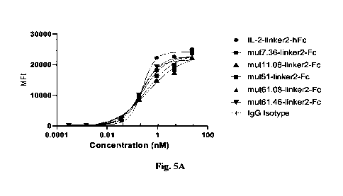
FIG. 5 shows the binding activity of IL-2 mutant protein to CHO-K1 IL-2
receptor af3y and
IL-2 receptor 13y recombinant cells detected by flow cytometry (FACS);
CA 03191260 2023- 2- 28 18
FIG. 5A shows the binding activity of mut7.36-1inker2-hFc, mut11.08-linker2-
hFc,
mut61-linker2-hFc, mut61.08-linker2-hFc or mut61.46-linker2-hFc to CHO-K1 IL-2
receptor
oc0y recombinant cells;
FIG. 5B shows the binding activity of mut11.31-linker2-hFc or mut7.66-1inker2-
hFc to
CHO-K1 IL-2 receptor a0y recombinant cells;
FIG. 5C shows the binding activity of mut7.36-1inker2-hFc, mut11.08-linker2-
hFc,
mut61-1inker2-hFc, mut61.08-1inker2-hFc or mut61.46-1inker2-hFc to CHO-K1 IL-2
receptor py
recombinant cells;
FIG. 5D shows the binding activity of mut11.31-linker2-hFc or mut7.66-1inker2-
hFc to
CHO-K1 IL-2 receptor 0y recombinant cells.
FIG. 6 shows the effect of IL-2 mutant protein on the level of STAT5
phosphorylation in
Tregs (FIGS. 6A-6C), CD4 CD25-FoxP3-T cells (FIGS. 6D-6F) and CD8+ T cells
(FIGS.
6G-6I);
FIG. 6A shows the effect of mut7.36-1inker2-hFc on the level of STAT5
phosphorylation in
Tregs;
FIG. 6B shows the effect of mut11.08-linker2-hFc on the level of STAT5
phosphorylation in
Tregs;
FIG. 6C shows the effect of mut11.31-linker2-hFc or mut7.66-1inker2-hFc on the
level of
STAT5 phosphorylation in Tregs;
FIG. 6D shows the effect of mut7.36-1inker2-hFc on the level of
phosphorylation in
CD4 CD25-FoxP3-T cells;
FIG. 6E shows the effect of mut11.08-linker2-hFc on the level of
phosphorylation in
CD4 CD25-FoxP3-T cells;
FIG. 6F shows the effect of mut11.31-linker2-hFc or mut7.66-1inker2-hFc on the
level of
phosphorylation in CD4 CD25-FoxP3-T cells;
FIG. 6G shows the effect of mut7.36-1inker2-hFc on the level of
phosphorylation in CD8+ T
cells;
FIG. 611 shows the effect of mut11.08-linker2-hFc on the level of
phosphorylation in CD8+
T cells;
CA 03191260 2023- 2- 28 19
FIG. 61 shows the effect of mut11.31-linker2-hFc or mut7.66-linker2-hFc on the
level of
phosphorylation in CD8+ T cells;
FIG. 7 shows the effect of IL-2 mutant protein on the level of proliferation
of Tregs (FIGS.
7A-7B), CD4 CD25-FoxP3-T cells (FIGS. 7C-7D), and CD8 CD25-T cells (FIGS. 7E-
7F);
FIG. 7A shows the effect of mut7.36-1inker2-hFc or mut7.66-1inker2-hFc on the
level of
proliferation of Tregs;
FIG. 7B shows the effect of mut11.08-linker2-hFc or mut11.31-linker2-hFc on
the level of
proliferation of Tregs;
FIG. 7C shows the effect of mut7.36-1inker2-hFc or mut7.66-1inker2-hFc on the
level of
proliferation of CD4 CD25-FoxP3-T cells;
FIG. 7D shows the effect of mut11.08-linker2-hFc or mut11.31-linker2-hFc on
the level of
proliferation of CD4 CD25-FoxP3-T cells;
FIG. 7E shows the effect of mut7.36-1inker2-hFc or mut7.66-1inker2-hFc on the
level of
proliferation of CD8 CD25-T cells;
FIG. 7F shows the effect of mut11.08-linker2-hFc or mut11.31-linker2-hFc on
the level of
proliferation of CD8 CD25-T cells;
FIG. 8 shows the effect of IL-2 mutant protein on the level of proliferation
of NK cells;
FIG. 9A shows the percentage of Tregs in CD4+ T cells in the spleen;
FIG. 9B shows the percentage of CD4 CD25-Foxop3- cells in CD4+ cells in the
spleen;
FIG. 9C shows the percentage of CD3 CD4- cells in CD3+ cells in the spleen;
FIG. 10A shows the percentage of Tregs in CD4+ cells in peripheral blood;
FIG. 10B shows the percentage of CD4 CD25-Foxop3- cells in CD4+ cells in
peripheral
blood;
FIG. 10C shows the percentage of CD3 CD4- cells in CD3+ cells in peripheral
blood;
Fig. 11A shows the change of A ear thickness in different groups of wild-type
mice DTH
models, wherein A ear thickness refers to the change of thickness of the right
ear before and after
stimulation;
FIG. 11B shows the change of body weight in different groups of wild-type mice
DTH
CA 03191260 2023- 2- 28 20
models;
Fig. 12A shows the change of A ear thickness in different groups of wild-type
mice DTH
models, wherein A ear thickness refers to the change of thickness of the right
ear before and after
stimulation;
FIG. 12B shows the change of body weight in different groups of wild-type mice
DTH
models;
FIG. 13A shows Treg/Tcon in different groups of NOG mice;
FIG. 13B shows Treg/CD8+ in different groups of NOG mice;
FIG. 14A shows the number of Tregs in different groups of NOG mice;
FIG. 14B shows the number of Tcons in different groups of NOG mice;
FIG. 14C shows the number of CD8+ cells in different groups of NOG mice;
FIG. 15A shows the change of body weight in different groups of NOG mice;
FIG. 15B shows the GVHD scores in different groups of NOG mice;
FIG. 16A shows the plasma drug concentration in wild-type mice after
subcutaneous
administration;
FIG. 16B shows the plasma drug concentration in wild-type mice after
subcutaneous
administration;
FIG. 17 shows the plasma drug concentration in wild-type mice after
intravenous
administration;
FIG. 18 shows the plasma drug concentration in mice inoculated with PBMCs
after
subcutaneous administration;
FIG. 19 shows the plasma drug concentration in cynomolgus monkey after
subcutaneous
administration;
FIG. 20A shows the number of Tregs in cynomolgus monkeys after subcutaneous
administration;
FIG. 20B shows the fold change of Treg number in cynomolgus monkeys after
subcutaneous administration;
FIG. 20C shows the percentage of Tregs in CD4+ T cells in cynomolgus monkeys
after
CA 03191260 2023- 2- 28 21
subcutaneous administration;
FIG. 20D shows the percentage of Ki67+ Tregs in cynomolgus monkeys after
subcutaneous
administration;
FIG. 20E shows the fold change of FoxP3 average fluorescence intensity in
cynomolgus
monkeys after subcutaneous administration;
FIG. 20F shows the fold change of CD25 average fluorescence intensity in
cynomolgus
monkeys after subcutaneous administration;
FIG. 21A shows the number of FoxP3-CD4+ cells in cynomolgus monkeys after
subcutaneous administration;
FIG. 21B shows the fold change of FoxP3-CD4+ cell number in cynomolgus monkeys
after
subcutaneous administration;
FIG. 21C shows the number of CD8+ T cells in cynomolgus monkeys after
subcutaneous
administration;
FIG. 21D shows the fold change of CD8+ T cell number in cynomolgus monkeys
after
subcutaneous administration;
FIG. 21E shows the number of NK cells in cynomolgus monkeys after subcutaneous
administration;
Figure 21F shows the fold change of NK cell number in cynomolgus monkeys after
subcutaneous administration.
DETAILED DESCRIPTION
The present disclosure will be further described below with reference to
specific examples.
The advantages and features of the present disclosure will become clear with
the description. If
specific conditions are not indicated in the examples, the conventional
conditions or the
conditions suggested by the manufacturer shall be followed. If the
manufacturer is not indicated,
the reagents or instruments used are conventional products that can be
purchased commercially.
The following examples of this disclosure are merely exemplary and not
intended to limit
the scope of the present disclosure. It should be understood by those skilled
in the art that the
CA 03191260 2023- 2- 28 22
details and forms of the technical solution of the present disclosure can be
modified or
substituted without departing from or exceeding the spirit or scope of the
disclosure, and such
modifications and substitutions all fall within the protection scope of the
present disclosure.
EXAMPLE 1 ¨ Design of IL-2 mutants with improved thermal stability and
construction of
expression plasmid
Various algorithms were used to obtain IL-2 mutants with improved thermal
stability, and
corresponding sequences were designed and synthesized. The nucleic acid
fragments encoding
wild-type IL-2 and the aforementioned IL-2 mutants were cloned into a pTT5
vector with an Fc
tag, and then the plasmids encoding the following fusion proteins were
prepared according to
established standard methods in molecular biology: IL-2-1inker2-hFc, mut0.08-
1inker2-hFc,
mut0.31-linker2-hFc, mut0.36-1inker2-hFc, mut0.39-1inker2-hFc, mut0.46-1inker2-
hFc,
mut0.57-1inker2-hFc, mut0.66-1inker2-hFc and mut0.68-1inker2-hFc.
The specific sequences of the aforementioned fusion proteins and components
are shown in
Table 1, wherein "IL-2" represents the wild-type IL-2, and "mutXX" represents
IL-2 mutants in
which mutation occurs compared to wild-type IL-2.
Table 1 Sequences of IL-2 mutants with improved stability
SEQ ID
Mutant Sequence information
NO
APTSSSTKKTQLQLEHLLLDLQMILNGINNYKNPKLTRMLTFKFYMPK
SEQ ID
IL2
KATELKHLQCLEEELKPLEEVLNLAQSKNFHLRPRDLISNINVIVLELK
NO: 1
GSETTFMCEYADETATIVEFLNRWITFAQSIISTLT
APTSSSTKKTQLQLEHLLLDLQMILNGINNVKNPKLTRMLTFKFYMPK
mut0.08 SEQ ID
KATELKHLQCLEEELKPLEEVLNLLQSKNFQLRPRDLISNINVIVLELK
(Y31V/A73L/H79Q) NO: 2
GSETTFMCEYADETATIVEFLNRWITFAQSIISTLT
APTSSSTKKTQLLLEHLLLDLQMILNGINNYKNPKLTRMLTFKFYMPK
mut0.31 SEQ ID
KATELKHLQCLEEELKPLEEVLNLAQSKNFHLRPRDLISNINVIVLELK
(Q13L) NO: 3
GSETTFMCEYADETATIVEFLNRWITFAQSIISTLT
APTSSSTKKTQLQLEHLLLDLQMILNGINNYKNPKLTRMLTFKFYMPK
mut0.36 SEQ ID
KATELKHLQCLEEELKPLEEVLNLAQSKNFHLRPRDLISNINVIVLELK
(R120F) NO: 4
GSETTFMCEYADETATIVEFLNFWITFAQSIISTLT
mut0.39 SEQ ID
APTSSSTKKTQLQLEHLILDLQMILNGINNYKNPKLTRMLTFKFYMPK
( Ll8IN91A/F117W ) NO: 5 KATELKHLQCLEEELKPLEEVLNLAQSKNFHLRPRDLISNINAIVLELK
CA 03191260 2023- 2- 28 23
SEQ ID
Mutant Sequence information
NO
GSETTFMCEYADETATIVEWLNRWITFAQSIISTLT
APTSSSTKKTQLQLEHLILDLQMILNGINNYKNPKLTRMLTFKFYMPK
mut0.46 SEQ ID
KATELKHLQCLEEELKPLEEVLNLAQSKNFHLRPRDLISNLNVIILELK
(L181/189LN931) NO: 6
GSETTFMCEYADETATIVEFLNRWITFAQSIISTLT
APTSSSTKKTQLQLEHLLLDLQMILNWINNYKNPKLTRMLTFKFYMPK
mut0.57 SEQ ID
KATELKHLQCLEEELKPLEEVLNLAQSKNFHLRPRDLISNINVIVLELK
(G27W/R120F) NO: 7
GSETTFMCEYADETATIVEFLNFWITFAQSIISTLT
APTSSSTKKTQLQLEHLLLDLQMILNGINNYKNPKLTRMLTFKFYMPK
mut0.66 SEQ ID
KATELKHLQCLEEELKPLEEVLNLAQSKNFHLRLRDLISNINVIVLELK
(P82L/R120F ) NO: 8
GSETTFMCEYADETATIVEFLNFWITFAQSIISTLT
APTSSSTKKTQLQLEHLLLDLQMILNGINNYKNPKLTRMLTFKFYMPK
mut0.68 SEQ ID
KATELKHLQCLEEELKPLEEVLNLAQSKNFHLRPRDLISNIYVIVLELK
(N90Y/R120F) NO: 9
GSETTFMCEYADETATIVEFLNFWITFAQSIISTLT
SEQ ID
1inker2 GGGGSGGGGSGGGGS
NO: 10
EPKSSDKTHTCPPCPAPELLGGPSVFLFPPKPKDTLMISRTPEVTCVVV
DVSHEDPEVKFNWYVDGVEVHNAKTKPREEQYGSTYRVVSVLTVLH
hFc SEQ ID
QDWLNGKEYKCKVSNKALPAPIEKTISKAKGQPREPQVYTLPPSRDEL
( C220S/N297G ) NO: 11
TKNQVSLTCLVKGFYPSDIAVEWESNGQPENNYKTTPPVLDSDGSFFL
YSKLTVDKSRWQQGNVFSCSVMHEALHNHYTQKSLSLSPGK
APTSSSTKKTQLQLEHLLLDLQMILNGINNYKNPKLTRMLTFKFYMPK
KATELKHLQCLEEELKPLEEVLNLAQSKNFHLRPRDLISNINVIVLELK
IL2-1inker2-hFc, also
GSETTFMCEYADETATIVEFLNRWITFAQSIISTLTGGGGSGGGGSGG
known as WT SEQ ID
GGSEPKSSDKTHTCPPCPAPELLGGPSVFLFPPKPKDTLMISRTPEVTC
IL-2-1inker2-hFc NO: 12 VVVDVSHEDPEVKFNWYVDGVEVHNAKTKPREEQYGSTYRVVSVLT
(C2205/N297G)
VLHQDWLNGKEYKCKVSNKALPAPIEKTISKAKGQPREPQVYTLPPSR
DELTKNQVSLTCLVKGFYPSDIAVEWESNGQPENNYKTTPPVLDSDGS
FFLYSKLTVDKSRWQQGNVFSCSVMHEALHNHYTQKSLSLSPGK
APTSSSTKKTQLQLEHLLLDLQMILNGINNVKNPKLTRMLTFKFYMPK
KATELKHLQCLEEELKPLEEVLNLLQSKNFQLRPRDLISNINVIVLELK
GSETTFMCEYADETATIVEFLNRWITFAQSIISTLTGGGGSGGGGSGG
mut0.08-1inker2-hFc SEQ ID
GGSEPKSSDKTHTCPPCPAPELLGGPSVFLFPPKPKDTLMISRTPEVTC
(Y31V/A73L/H79Q) NO: 13
VVVDVSHEDPEVKFNWYVDGVEVHNAKTKPREEQYGSTYRVVSVLT
VLHQDWLNGKEYKCKVSNKALPAPIEKTISKAKGQPREPQVYTLPPSR
DELTKNQVSLTCLVKGFYPSDIAVEWESNGQPENNYKTTPPVLDSDGS
CA 03191260 2023- 2- 28 24
SEQ ID
Mutant Sequence information
NO
FFLYSKLTVDKSRWQQGNVFSCSVMHEALHNHYTQKSLSLSPGK
APTSSSTKKTQLLLEHLLLDLQMILNGINNYKNPKLTRMLTFKFYMPK
KATELKHLQCLEEELKPLEEVLNLAQSKNFHLRPRDLISNINVIVLELK
GSETTFMCEYADETATIVEFLNRWITFAQSIISTLTGGGGSGGGGSGG
mut0.31-linker2-hFc SEQ ID GGSEPKSSDKTHTCPPCPAPELLGGPSVFLFPPKPKDTLMISRTPEVTC
(Q13L) NO: 14
VVVDVSHEDPEVKFNWYVDGVEVHNAKTKPREEQYGSTYRVVSVLT
VLHQDWLNGKEYKCKVSNKALPAPIEKTISKAKGQPREPQVYTLPPSR
DELTKNQVSLTCLVKGFYPSDIAVEWESNGQPENNYKTTPPVLDSDGS
FFLYSKLTVDKSRWQQGNVFSCSVMHEALHNHYTQKSLSLSPGK
APTSSSTKKTQLQLEHLLLDLQMILNGINNYKNPKLTRMLTFKFYMPK
KATELKHLQCLEEELKPLEEVLNLAQSKNFHLRPRDLISNINVIVLELK
GSETTFMCEYADETATIVEFLNFWITFAQSIISTLTGGGGSGGGGSGG
mut0.36-linker2-hFc SEQ ID GGSEPKSSDKTHTCPPCPAPELLGGPSVFLFPPKPKDTLMISRTPEVTC
(R120F) NO: 15
VVVDVSHEDPEVKFNWYVDGVEVHNAKTKPREEQYGSTYRVVSVLT
VLHQDWLNGKEYKCKVSNKALPAPIEKTISKAKGQPREPQVYTLPPSR
DELTKNQVSLTCLVKGFYPSDIAVEWESNGQPENNYKTTPPVLDSDGS
FFLYSKLTVDKSRWQQGNVFSCSVMHEALHNHYTQKSLSLSPGK
APTSSSTKKTQLQLEHLILDLQMILNGINNYKNPKLTRMLTFKFYMPK
KATELKHLQCLEEELKPLEEVLNLAQSKNFHLRPRDLISNINAIVLELK
GSETTFMCEYADETATIVEWLNRWITFAQSIISTLTGGGGSGGGGSGG
mut0.39-linker2-hFc SEQ ID GGSEPKSSDKTHTCPPCPAPELLGGPSVFLFPPKPKDTLMISRTPEVTC
( Ll8I/V91A/F117W ) NO: 16 VVVDVSHEDPEVKFNWYVDGVEVHNAKTKPREEQYGSTYRVVSVLT
VLHQDWLNGKEYKCKVSNKALPAPIEKTISKAKGQPREPQVYTLPPSR
DELTKNQVSLTCLVKGFYPSDIAVEWESNGQPENNYKTTPPVLDSDGS
FFLYSKLTVDKSRWQQGNVFSCSVMHEALHNHYTQKSLSLSPGK
APTSSSTKKTQLQLEHLILDLQMILNGINNYKNPKLTRMLTFKFYMPK
KATELKHLQCLEEELKPLEEVLNLAQSKNFHLRPRDLISNLNVIILELK
GSETTFMCEYADETATIVEFLNRWITFAQSIISTLTGGGGSGGGGSGG
mut0.46-linker2-hFc SEQ ID GGSEPKSSDKTHTCPPCPAPELLGGPSVFLFPPKPKDTLMISRTPEVTC
(L181/189LN93I ) NO: 17 VVVDVSHEDPEVKFNWYVDGVEVHNAKTKPREEQYGSTYRVVSVLT
VLHQDWLNGKEYKCKVSNKALPAPIEKTISKAKGQPREPQVYTLPPSR
DELTKNQVSLTCLVKGFYPSDIAVEWESNGQPENNYKTTPPVLDSDGS
FFLYSKLTVDKSRWQQGNVFSCSVMHEALHNHYTQKSLSLSPGK
mut0.57-linker2-hFc SEQ ID APTSSSTKKTQLQLEHLLLDLQMILNWINNYKNPKLTRMLTFKFYMPK
( G27W/R120F ) NO: 18 KATELKHLQCLEEELKPLEEVLNLAQSKNFHLRPRDLISNINVIVLELK
CA 03191260 2023- 2- 28 25
SEQ ID
Mutant Sequence information
NO
GSETTFMCEYADETATIVEFLNFWITFAQSIISTLTGGGGSGGGGSGG
GGSEPKSSDKTHTCPPCPAPELLGGPSVFLFPPKPKDTLMISRTPEVTC
VVVDVSHEDPEVKFNWYVDGVEVHNAKTKPREEQYGSTYRVVSVLT
VLHQDWLNGKEYKCKVSNKALPAPIEKTISKAKGQPREPQVYTLPPSR
DELTKNQVSLTCLVKGFYPSDIAVEWESNGQPENNYKTTPPVLDSDGS
FFLYSKLTVDKSRWQQGNVFSCSVMHEALHNHYTQKSLSLSPGK
APTSSSTKKTQLQLEHLLLDLQMILNGINNYKNPKLTRMLTFKFYMPK
KATELKHLQCLEEELKPLEEVLNLAQSKNFHLRLRDLISNINVIVLELK
GSETTFMCEYADETATIVEFLNFWITFAQSIISTLTGGGGSGGGGSGG
mut0.66-linker2-hFc SEQ ID GGSEPKSSDKTHTCPPCPAPELLGGPSVFLFPPKPKDTLMISRTPEVTC
(P82L/R120F) NO: 19
VVVDVSHEDPEVKFNWYVDGVEVHNAKTKPREEQYGSTYRVVSVLT
VLHQDWLNGKEYKCKVSNKALPAPIEKTISKAKGQPREPQVYTLPPSR
DELTKNQVSLTCLVKGFYPSDIAVEWESNGQPENNYKTTPPVLDSDGS
FFLYSKLTVDKSRWQQGNVFSCSVMHEALHNHYTQKSLSLSPGK
APTSSSTKKTQLQLEHLLLDLQMILNGINNYKNPKLTRMLTFKFYMPK
KATELKHLQCLEEELKPLEEVLNLAQSKNFHLRPRDLISNIYVIVLELK
GSETTFMCEYADETATIVEFLNFWITFAQSIISTLTGGGGSGGGGSGG
mut0.68-linker2-hFc SEQ ID GGSEPKSSDKTHTCPPCPAPELLGGPSVFLFPPKPKDTLMISRTPEVTC
(N90Y/R120F) NO: 20 VVVDVSHEDPEVKFNWYVDGVEVHNAKTKPREEQYGSTYRVVSVLT
VLHQDWLNGKEYKCKVSNKALPAPIEKTISKAKGQPREPQVYTLPPSR
DELTKNQVSLTCLVKGFYPSDIAVEWESNGQPENNYKTTPPVLDSDGS
FFLYSKLTVDKSRWQQGNVFSCSVMHEALHNHYTQKSLSLSPGK
EXAMPLE 2 - Production and purification of IL-2 mutants with improved thermal
stability
11EK293 cells (purchased from the Cell Bank of the Chinese Academy of
Sciences) were
transiently transfected (PEI, Polysciences) with the plasmids constructed in
Example 1 and then
expanded at 37 C in FreeStyle TM 293 Expression Medium (purchased from Gibco).
After 7
days, the cell culture medium was collected, and the cell components were
removed by
centrifugation to obtain the culture supernatant containing IL-2-hFc fusion
proteins.
The fusion proteins in the cell culture supernatant were purified using a 10
mL protein A
column (purchased from Bestchrom). The protein A column was first equilibrated
with 3 to 5
column volumes of an equilibrium buffer (PBS phosphate buffer, pH 7.4), and
then loaded with
CA 03191260 2023- 2- 28 26
the clear culture supernatant at a flow rate of 10 mL/min. After loading, the
protein A column
was washed with 3 to 5 column volumes of the equilibrium buffer. The proteins
bound to the
protein A column were eluted with an eluent buffer (0.02 M citric acid buffer,
0.1 M glycine, 0.1
M sodium chloride, pH 3.0), and the elution was monitored by a nucleic
acid/protein detector
(A280 ultraviolet absorption peak). The eluted proteins were collected and
neutralized with the
added buffer (1 M arginine, 0.4 M succinic acid, pH 9.0). The target proteins
were then collected
through a molecular sieve (purchased from Bestchrom) with a buffer system (20
mM PB, 200
mM sodium chloride, pH 6.0-6.5). The purified IL-2 mutant fusion proteins were
obtained by
aseptic filtration with a 0.22 gm filter and preserved in sterile condition.
The purified IL-2 mutant fusion proteins were tested and analyzed for protein
yield,
concentration (A280/1.4) and SEC purity. The purified IL-2 mutant fusion
proteins with
improved thermal stability (mutXX-1inker2-hFc) were qualified, and had a
significantly higher
yield compared to wild-type IL-2 (IL2-1inker2-hFc). The results of protein
yield, concentration
and purity are shown in Table 2.
Table 2. Detection results of IL-2 mutant fusion proteins with improved
thermal stability
Protein yield Protein SEC Protein
concentration
Mutant
(ngil-) purity (mg/ml)
IL-2-1inker2-hFc 0.95 97.25% 1.11
mut0.08-1inker2-hFc 8.95 99.85% 1.79
mut0.31-1inker2-hFc 1.54 98.49% 2.20
mut0.36-1inker2-hFc 2.98 99.99% 1.49
mut0.39-1inker2-hFc 43.10 99.58% 2.13
mut0.46-1inker2-hFc 13.82 99.92% 1.22
mut0.57-1inker2-hFc 1.25 99.91% 0.89
mut0.66-1inker2-hFc 7.39 99.90% 1.12
mut0.68-1inker2-hFc 11.90 99.99% 1.19
EXAMPLE 3 - Differential Scanning Fluorimetry (DSF) Assay of IL-2 mutants with
improved thermal stability
CA 03191260 2023- 2- 28 27
The buffer in Protein Thermal Shift Dye Kit (purchased from Applied
Biosystems, Cat. No.
4461146) diluted to 50 times, the IL-2 mutant proteins (purified by the method
described in
Example 2) diluted to 0.5 mg/mL, and the dye diluted to 2 times were added to
a 20 tit reaction
system. After being mixed evenly, the mixture was added into 8-tube strips
with 2 duplicate
tubes for each sample. The tubes were covered, centrifuged for 5-10 seconds,
and analyzed by
the Applied Biosystems 7500. The Tm values were then obtained by using
Boltzmann method to
analyze the melting curve. As shown in Table 3, compared to wild-type IL-2
(IL2-1inker2-hFc)õ
the IL-2 mutants (mutXX-1inker2-hFc) had increased Tm values by more than 3 C,
and thus had
significantly improved thermal stability.
Table 3. DSF assay results of IL-2 mutants with improved thermal stability
Mutant Tm ( C)
IL-2-1inker2-hFc 46.74
mut0.08-1inker2-hFc 54.91
mut0.31-linker2-hFc 57.53
mut0.36-1inker2-hFc 56.59
mut0.39-1inker2-hFc 55.93
mut0.46-1inker2-hFc 53.83
mut0.57-1inker2-hFc 57.98
mut0.66-1inker2-hFc 57.24
mut0.68-1inker2-hFc 54.77
EXAMPLE 4 - Design of IL-2 mutants (IL-2 mutants with decreased binding
ability to 137
subunits and IL-2 mutants with decreased binding ability to 137 subunits and
with
improved thermal stability) and construction of expression plasmid
Various algorithms including MOE software were used to simulate the
interaction interface
between human IL-2 and corresponding receptor a, fr, and y subunits to obtain
mutation sites
having decreased binding ability to fry subunits. The IL-2 mutant sequences
with mutation sites
having decreased binding activities to fry subunits were designed and
synthesized, together with
CA 03191260 2023- 2- 28 28
the IL-2 mutant sequences with a combination of such mutation sites with
mutation sites having
improved thermal stability. The nucleic acid fragments encoding wild-type IL-2
and the
aforementioned IL-2 mutants were cloned into a pTT5 vector with an Fc tag, and
then the
plasmids encoding the following fusion proteins were prepared according to
established standard
methods in molecular biology: IL-2-1inker2-hFc, mut7-1inker2-hFc, mut7.08-
1inker2-hFc,
mut7.36-1inker2-hFc, mut7.39-1inker2-hFc, mut7.46-1inker2-hFc ,mut7.57-1inker2-
hFc,
mut7.66-1inker2-hFc, mut8-1inker2-hFc, mut8.08-1inker2-hFc,
mut8.36-1inker2-hFc,
mutll-linker2-hFc, mut11.08-linker2-hFc, mut11.31-linker2-hFc, mut11.36-
linker2-hFc,
mut11.46-1inker2-hFc, mut61-1inker2-hFc, mut61.08-1inker2-hFc and mut61.46-
1inker2-hFc.The
specific sequences of the fusion proteins and components thereof are shown in
Table 4, where
"IL-2" represents wild-type IL-2 and "mutXX" represents IL-2 mutants in which
mutation occurs
compared to the wild-type IL-2.
Table 4 Sequences of IL-2 mutants
SEQ ID
Mutant NO Sequence information
SEQ IL2 ID Shown in Table 1
NO: 1
mut7 SE ID
APTSSSTKKTQLQLEELLLDLQMILNGINNYKNPKLTRMLTFKFYM
Q
PKKATELKHLQCLEEELKPLEEVLNLAQSKNFHLRPRDLISNINVIV
(Hi 6E) NO: 21
LELKGSETTFMCEYADETATIVEFLNRWITFAQSIISTLT
mut7.08 SEQ ID
APTSSSTKKTQLQLEELLLDLQMILNGINNVKNPKLTRMLTFKFYMPK
(H16E/Y31V/A73L/H79 No . 22 KATELKHLQCLEEELKPLEEVLNLLQSKNFQLRPRDLISNINVIVLELK
Q) = GSETTFMCEYADETATIVEFLNRWITFAQSIISTLT
mut7.36 SE ID
APTSSSTKKTQLQLEELLLDLQMILNGINNYKNPKLTRMLTFKFYM
Q
PKKATELKHLQCLEEELKPLEEVLNLAQSKNFHLRPRDLISNINVIV
(H16E/R120F ) NO: 23
LELKGSETTFMCEYADETATIVEFLNFWITFAQSIISTLT
mut7.39 SEQ ID
APTSSSTKKTQLQLEELILDLQMILNGINNYKNPKLTRMLTFKFYMP
(H16E/L18I/V91A/F117 No . 24 KKATELKHLQCLEEELKPLEEVLNLAQSKNFHLRPRDLISNINAIVL
W) = ELKGSETTFMCEYADETATIVEWLNRWITFAQSIISTLT
mut7.46 SE ID
APTSSSTKKTQLQLEELILDLQMILNGINNYKNPKLTRMLTFKFYMP
Q
1 KKATELKHLQCLEEELKPLEEVLNLAQSKNFHLRPRDLISNLNVIIL
(H16E/L18I/I89LN93I ) NO: '-' ELKGSETTFMCEYADETATIVEFLNRWITFAQSIISTLT
mut7.57 SE ID
APTSSSTKKTQLQLEELLLDLQMILNWINNYKNPKLTRMLTFKFYMPK
Q
KATELKHLQCLEEELKPLEEVLNLAQSKNFHLRPRDLISNINVIVLELK
( H16E/G27W/R120F ) NO:26
GSETTFMCEYADETATIVEFLNFWITFAQSIISTLT
mut7.66 SEQ ID
APTSSSTKKTQLQLEELLLDLQMILNGINNYKNPKLTRMLTFKFYM
CA 03191260 2023- 2- 28 29
SEQ ID
Mutant NO Sequence information
(H16E/P82L/R120F) NO: 27 PKKATELKHLQCLEEELKPLEEVLNLAQSKNFHLRLRDLISNINVIV
LELKGSETTFMCEYADETATIVEFLNFWITFAQSIIS¨TLT
mut8 SE ID
APTSSSTKKTQLQLEHLLLALQMILNGINNYKNPKLTRMLTFKFYMPK
Q
KATELKHLQCLEEELKPLEEVLNLAQSKNFHLRPRDLISNINVIVLELK
(D20A) NO: 28
GSETTFMCEYADETATIVEFLNRWITFAQSIISTLT
mut8.08
SEQ ID APTSSSTKKTQLQLEHLLLALQMILNGINNYKNPKLTRMLTFKFYMPK
( D20A/Y31V/A73L/H79 No : 29 KATELKHLQCLEEELKPLEEVLNLLQSKNFQLRPRDLISNINVIVLELK
Q) GSETTFMCEYADETATIVEFLNRWITFAQSIISTLT
mut8.36 SE ID
APTSSSTKKTQLQLEHLLLALQMILNGINNYKNPKLTRMLTFKFYMPK
Q
KATELKHLQCLEEELKPLEEVLNLAQSKNFHLRPRDLISNINVIVLELK
(D20A/R120F) NO: 30
GSETTFMCEYADETATIVEFLNFWITFAQSIISTLT
mutll SE ID
APTSSSTKKTQLQLEHLLLDLQMILNGINNYKNPKLTRMLTFKFYM
Q
PKKATELKHLQCLEEELKPLEEVLNLAQSKNFHLRPRDLISNINRIV
( V91R ) NO: 31
LELKGSETTFMCEYADETATIVEFLNRWITFAQSIISTLT
mut11.08
SEQ ID APTSSSTKKTQLQLEHLLLDLQMILNGINNVKNPKLTRMLTFKFYM
(V91R/Y31V/A73L/H79 No : 32 PKKATELKHLQCLEEELKPLEEVLNLLQSKNFQLRPRDLISNINRIV
Q) LELKGSETTFMCEYADETATIVEFLNRWITFAQSIISTLT
mut11.31 SE ID
APTSSSTKKTQLLLEHLLLDLQMILNGINNYKNPKLTRMLTFKFYM
Q
PKKATELKHLQCLEEELKPLEEVLNLAQSKNFHLRPRDLISNINRIV
(V91R/Q13L) NO: 33
LELKGSETTFMCEYADETATIVEFLNRWITFAQSIISTLT
mut11.36 SE ID
APTSSSTKKTQLQLEHLLLDLQMILNGINNYKNPKLTRMLTFKFYMPK
Q
KATELKHLQCLEEELKPLEEVLNLAQSKNFHLRPRDLISNINRIVLELK
(V91R/R120F) NO: 34
GSETTFMCEYADETATIVEFLNFWITFAQSIISTLT
mut11.46 SE ID
APTSSSTKKTQLQLEHLILDLQMILNGINNYKNPKLTRMLTFKFYMP
Q
,.1 KKATELKHLQCLEEELKPLEEVLNLAQSKNFHLRPRDLISNLNRIIL
(V91R/L181/189L/V931) NO: ' ELKGSETTFMCEYADETATIVEFLNRWITFAQSIISTLT
mut61 SE ID
APTSSSTKKTQLQLEELLLDLQMILNGINNYKNPKLTRMLTFKFYM
Q
PKKATELKHLQCLEEELKPLEEVLNLAQSKNFHLRPRDLISNINRIV
(H16EN91R) NO:36
LELKGSETTFMCEYADETATIVEFLNRWITFAQSIISTLT
mut61.08 SE ID
APTSSSTKKTQLQLEELLLDLQMILNGINNVKNPKLTRMLTFKFYM
Q
(H16E/V91R/Y31V/A73L/ No:37 PKKATELKHLQCLEEELKPLEEVLNLLQSKNFQLRPRDLISNINRIV
H79Q) LELKGSETTFMCEYADETATIVEFLNRWITFAQSIISTLT
mut61.46 SE ID
APTSSSTKKTQLQLEELILDLQMILNGINNYKNPKLTRMLTFKFYMP
Q
(H16EN91R/L181/189LN NO:38 KKATELKHLQCLEEELKPLEEVLNLAQSKNFHLRPRDLISNLNRIIL
931) ELKGSETTFMCEYADETATIVEFLNRWITFAQSIISTLT
SEQ ID
linker2 Shown in Table 1
NO:10
hFc SEQ ID Shown in Table 1
CA 03191260 2023- 2- 28 30
SEQ ID
Mutant NO Sequence information
(C2205/N297G) NO:11
SEQ ID
IL2-1inker2-hFc Shown in Table 1
NO: 12
APTSSSTKKTQLQLEELLLDLQMILNGINNYKNPKLTRMLTFKFYM
PKKATELKHLQCLEEELKPLEEVLNLAQSKNFHLRPRDLISNINVIV
LELKGSETTFMCEYADETATIVEFLNRWITFAQSIISTLTGGGGSGG
mut7-1inker2-hFc
SE ID GGSGGGGSEPKSSDKTHTCPPCPAPELLGGPSVFLFPPKPKDTLMISRT
Q
PEVTCVVVDVSHEDPEVKFNWYVDGVEVHNAKTKPREEQYGSTYRV
(Hi 6E) NO:39
VSVLTVLHQDWLNGKEYKCKVSNKALPAPIEKTISKAKGQPREPQVYT
LPPSRDELTKNQVSLTCLVKGFYPSDIAVEWESNGQPENNYKTTPPVL
DSDGSFFLYSKLTVDKSRWQQGNVFSCSVMHEALHNHYTQKSLSLSP
GK
APTSSSTKKTQLQLEELLLDLQMILNGINNVKNPKLTRMLTFKFYMPK
KATELKHLQCLEEELKPLEEVLNLLQSKNFQLRPRDLISNINVIVLELK
mut7.08-1inker2-hFc GSETTFMCEYADETATIVEFLNRWITFAQSIISTLTGGGGSGGGGSGG
SEQ ID GGSEPKSSDKTHTCPPCPAPELLGGPSVFLFPPKPKDTLMISRTPEVTCV
(H16E/Y31V/A73L/H79 NO :40 VVDVSHEDPEVKFNWYVDGVEVHNAKTKPREEQYGSTYRVVSVLTV
Q) LHQDWLNGKEYKCKVSNKALPAPIEKTISKAKGQPREPQVYTLPPSRD
ELTKNQVSLTCLVKGFYPSDIAVEWESNGQPENNYKTTPPVLDSDGSF
FLYSKLTVDKSRWQQGNVFSCSVMHEALHNHYTQKSLSLSPGK
APTSSSTKKTQLQLEELLLDLQMILNGINNYKNPKLTRMLTFKFYM
PKKATELKHLQCLEEELKPLEEVLNLAQSKNFHLRPRDLISNINVIV
mut7.36-1inker2-hFc LELKGSETTFMCEYADETATIVEFLNFWITFAQSIISTLTGGGGSGG
SW' ID GGSGGGGSEPKSSDKTHTCPPCPAPELLGGPSVFLFPPKPKDTLMIS
(H16E/R120F)
NO 41 RTPEVTCVVVDVSHEDPEVKFNWYVDGVEVHNAKTKPREEQYGS
:
TYRVVSVLTVLHQDWLNGKEYKCKVSNKALPAPIEKTISKAKGQP
REPQVYTLPPSRDELTKNQVSLTCLVKGFYPSDIAVEWESNGQPEN
NYKTTPPVLDSDGSFFLYSKLTVDKSRWQQGNVFSCSVMHEALHN
HYTQKSLSLSPGK
APTSSSTKKTQLQLEELILDLQMILNGINNYKNPKLTRMLTFKFYMP
KKATELKHLQCLEEELKPLEEVLNLAQSKNFHLRPRDLISNINAIVL
ELKGSETTFMCEYADETATIVEWLNRWITFAQSIISTLTGGGGSGG
mut7.39-linker2-hFc
SE ID GGSGGGGSEPKSSDKTHTCPPCPAPELLGGPSVFLFPPKPKDTLMIS
Q
(H16E/L18I/V91A/F117 No: 42 RTPEVTCVVVDVSHEDPEVKFNWYVDGVEVHNAKTKPREEQYGS
W) TYRVVSVLTVLHQDWLNGKEYKCKVSNKALPAPIEKTISKAKGQP
REPQVYTLPPSRDELTKNQVSLTCLVKGFYPSDIAVEWESNGQPEN
NYKTTPPVLDSDGSFFLYSKLTVDKSRWQQGNVFSCSVMHEALHN
HYTQKSLSLSPGK
APTSSSTKKTQLQLEELILDLQMILNGINNYKNPKLTRMLTFKFYMP
KKATELKHLQCLEEELKPLEEVLNLAQSKNFHLRPRDLISNLNVIIL
ELKGSETTFMCEYADETATIVEFLNRWITFAQSIISTLTGGGGSGGG
mut7.46-1inker2-hFc SW' ID GSGGGGSEPKSSDKTHTCPPCPAPELLGGPSVFLFPPKPKDTLMISR
Al TPEVTCVVVDVSHEDPEVKFNWYVDGVEVHNAKTKPREEQYGST
(H16E/L18I/I89LN93I ) NO: -' YRVVSVLTVLHQDWLNGKEYKCKVSNKALPAPIEKTISKAKGQPR
EPQVYTLPPSRDELTKNQVSLTCLVKGFYPSDIAVEWESNGQPENN
YKTTPPVLDSDGSFFLYSKLTVDKSRWQQGNVFSCSVMHEALHNH
YTQKSLSLSPGK
mut7.57-1inker2-hFc SE ID APTSSSTKKTQLQLEELLLDLQMILNWINNYKNPKLTRMLTFKFYMPK
Q
KATELKHLQCLEEELKPLEEVLNLAQSKNFHLRPRDLISNINVIVLELK
CA 03191260 2023- 2- 28 31
SEQ ID
Mutant NO Sequence information
( H16E/G27W/R120F ) NO :44 GSETTFMCEYADETATIVEFLNFWITFAQSIISTLTGGGGSGGGGSGG
GGSEPKSSDKTHTCPPCPAPELEGGPSVFLFPPKPKDTLMISRTPEVTCV
VVDVSHEDPEVKFNWYVDGVEVHNAKTKPREEQYGSTYRVVSVLTV
LHQDWLNGKEYKCKVSNKALPAPIEKTISKAKGQPREPQVYTLPPSRD
ELTKNQVSLTCLVKGFYPSDIAVEWESNGQPENNYKTTPPVLDSDGSF
FLYSKLTVDKSRWQQGNVFSCSVMHEALHNHYTQKSLSLSPGK
APTSSSTKKTQLQLEELLLDLQMILNGINNYKNPKLTRMLTFKFYM
PKKATELKHLQCLEEELKPLEEVLNLAQSKNFHLRLRDLISNINVIV
LELKGSETTFMCEYADETATIVEFLNFWITFAQSIISTLTGGGGSGG
mut7.66-linker2-hFc SE ID GGSGGGGSEPKSSDKTHTCPPCPAPELLGGPSVFLFPPKPKDTLMIS
Q
A c RTPEVTCVVVDVSHEDPEVKFNWYVDGVEVHNAKTKPREEQYGS
(H16E/P82L/R120F) NO: --' TYRVVSVLTVLHQDWLNGKEYKCKVSNKALPAPIEKTISKAKGQP
REPQVYTLPPSRDELTKNQVSLTCLVKGFYPSDIAVEWESNGQPEN
NYKTTPPVLDSDGSFFLYSKLTVDKSRWQQGNVFSCSVMHEALHN
HYTQKSLSLSPGK
APTSSSTKKTQLQLEHLLLALQMILNGINNYKNPKLTRMLTFKFYMPK
KATELKHLQCLEEELKPLEEVLNLAQSKNFHLRPRDLISNINVIVLELK
GSETTFMCEYADETATIVEFLNRWITFAQSIISTLTGGGGS GGGGS GG
mut8-linker2-hFc SE ID
GGSEPKSSDKTHTCPPCPAPELLGGPSVFLFPPKPKDTLMISRTPEVT
Q
CVVVDVSHEDPEVKFNWYVDGVEVHNAKTKPREEQYGSTYRVVS
(D20A) NO:46
VLTVLHQDWLNGKEYKCKVSNKALPAPIEKTISKAKGQPREPQVYT
LPPSRDELTKNQVSLTCLVKGFYPSDIAVEWESNGQPENNYKTTPP
VLDSDGSFFLYSKLTVDKSRWQQGNVFSCSVMHEALHNHYTQKSL
SLSPGK
APTSSSTKKTQLQLEHLLLALQMILNGINNYKNPKLTRMLTFKFYMPK
KATELKHLQCLEEELKPLEEVLNLLQSKNFQLRPRDLISNINVIVLELK
GSETTFMCEYADETATIVEFLNRWITFAQSIISTLTGGGGS GGGGS GG
mut8.08-linker2-hFc
SE ID GGSEPKSSDKTHTCPPCPAPELLGGPSVFLFPPKPKDTLMISRTPEVT
Q
( D20A/Y31V/A73L/H79 NO:47 CVVVDVSHEDPEVKFNWYVDGVEVHNAKTKPREEQYGSTYRVVS
Q)
VLTVLHQDWLNGKEYKCKVSNKALPAPIEKTISKAKGQPREPQVYT
LPPSRDELTKNQVSLTCLVKGFYPSDIAVEWESNGQPENNYKTTPP
VLDSDGSFFLYSKLTVDKSRWQQGNVFSCSVMHEALHNHYTQKSL
SLSPGK
APTSSSTKKTQLQLEHLLLALQMILNGINNYKNPKLTRMLTFKFYMPK
KATELKHLQCLEEELKPLEEVLNLAQSKNFHLRPRDLISNINVIVLELK
GSETTFMCEYADETATIVEFLNFWITFAQSIISTLTGGGGSGGGGSGG
mut8.36-linker2-hFc SE ID GGSEPKSSDKTHTCPPCPAPELLGGPSVFLFPPKPKDTLMISRTPEVT
Q
CVVVDVSHEDPEVKFNWYVDGVEVHNAKTKPREEQYGSTYRVVS
(D20A/R120F) NO:48
VLTVLHQDWLNGKEYKCKVSNKALPAPIEKTISKAKGQPREPQVYT
LPPSRDELTKNQVSLTCLVKGFYPSDIAVEWESNGQPENNYKTTPP
VLDSDGSFFLYSKLTVDKSRWQQGNVFSCSVMHEALHNHYTQKSL
SLSPGK
APTSSSTKKTQLQLEHLLLDLQMILNGINNYKNPKLTRMLTFKFYM
PKKATELKHLQCLEEELKPLEEVLNLAQSKNFHLRPRDLISNINRIV
mutll-linker2-hFc
LELKGSETTFMCEYADETATIVEFLNRWITFAQSIISTLTGGGGSGG
SEQ ID GGSGGGGSEPKSSDKTHTCPPCPAPELLGGPSVFLFPPKPKDTLMIS
(V91R) NO:49 RTPEVTCVVVDVSHEDPEVKFNWYVDGVEVHNAKTKPREEQYGS
TYRVVSVLTVLHQDWLNGKEYKCKVSNKALPAPIEKTISKAKGQP
REPQVYTLPPSRDELTKNQVSLTCLVKGFYPSDIAVEWESNGQPEN
NYKTTPPVLDSDGSFFLYSKLTVDKSRWQQGNVFSCSVMHEALHN
CA 03191260 2023- 2- 28 32
SEQ ID
Mutant NO Sequence information
HYTQKSLSLSPGK
APT S S STKKTQLQLEHLLLDLQMILNGINNVKNPKLTRMLTFKFYM
PKKATELKHLQCLEEELKPLEEVLNLLQSKNFQLRPRDLISNINRIV
LELKGSETTFMCEYADETATIVEFLNRWITFAQSIISTLTGGGGSGG
mut11.08-linker2-hFc
SE ID GGSGGGGSEPKSSDKTHTCPPCPAPELLGGPSVFLFPPKPKDTLMIS
Q
( V91R/Y31V/A73L/H79 No: 50 RTPEVTCVVVDVSHEDPEVKFNWYVDGVEVHNAKTKPREEQYGS
Q)
TYRVVSVLTVLHQDWLNGKEYKCKVSNKALPAPIEKTISKAKGQP
REPQVYTLPPSRDELTKNQVSLTCLVKGFYPSDIAVEWESNGQPEN
NYKTTPPVLDSDGSFFLYSKLTVDKSRWQQGNVF SC SVMHEALHN
HYTQKSLSLSPGK
APT S S STKKTQLLLEHLLLDLQMILNGINNYKNPKLTRMLTFKFYM
PKKATELKHLQCLEEELKPLEEVLNLAQSKNFHLRPRDLISNINRIV
LELKGSETTFMCEYADETATIVEFLNRWITFAQSIISTLTGGGGSGG
mut11.31-linker2-hFc SE ID GGSGGGGSEPKSSDKTHTCPPCPAPELLGGPSVFLFPPKPKDTLMIS
Q
RTPEVTCVVVDVSHEDPEVKFNWYVDGVEVHNAKTKPREEQYGS
(V91R/Q13L) NO: 51
TYRVVSVLTVLHQDWLNGKEYKCKVSNKALPAPIEKTISKAKGQP
REPQVYTLPPSRDELTKNQVSLTCLVKGFYPSDIAVEWESNGQPEN
NYKTTPPVLDSDGSFFLYSKLTVDKSRWQQGNVF SC SVMHEALHN
HYTQKSLSLSPGK
APTSSSTKKTQLQLEHLLLDLQMILNGINNYKNPKLTRMLTFKFYMPK
KATELKHLQCLEEELKPLEEVLNLAQSKNFHLRPRDLISNINRIVLELK
GSETTFMCEYADETATIVEFLNFWITFAQSIISTLTGGGGSGGGGSGG
mut11.36-linker2-hFc SEQ ID GGSEPKSSDKTHTCPPCPAPELLGGPSVFLFPPKPKDTLMISRTPEVTCV
(V91R/R120F) NO :52
VVDVSHEDPEVKFNWYVDGVEVHNAKTKPREEQYGSTYRVVSVLTV
LHQDWLNGKEYKCKVSNKALPAPIEKTISKAKGQPREPQVYTLPPSRD
ELTKNQVSLTCLVKGFYPSDIAVEWESNGQPENNYKTTPPVLDSDGSF
FLYSKLTVDKSRWQQGNVFSCSVMHEALHNHYTQKSLSLSPGK
APTSS STKKTQLQLEHLILDLQMILNGINNYKNPKLTRMLTFKFYMP
KKATELKHLQCLEEELKPLEEVLNLAQSKNFHLRPRDLISNLNRIIL
ELKGSETTFMCEYADETATIVEFLNRWITFAQSIISTLTGGGGSGGG
GSGGGGSEPKSSDKTHTCPPCPAPELLGGPSVFLFPPKPKDTLMISR
mut11.46-linker2-hFc SEQ ID
(V91R/L181/189L/V931)
TPEVTCVVVDVSHEDPEVKFNWYVDGVEVHNAKTKPREEQYGST
NO : 53
YRVVSVLTVLHQDWLNGKEYKCKVSNKALPAPIEKTISKAKGQPR
EPQVYTLPPSRDELTKNQVSLTCLVKGFYPSDIAVEWESNGQPENN
YKTTPPVLDSDGSFFLYSKLTVDKSRWQQGNVF SC SVMHEALHNH
YTQKSL SLSPGK
APTSS STKKTQLQLEELLLDLQMILNGINNYKNPKLTRMLTFKFYM
PKKATELKHLQCLEEELKPLEEVLNLAQSKNFHLRPRDLISNINRIV
LELKGSETTFMCEYADETATIVEFLNRWITFAQSIISTLTGGGGSGG
mut61-linker2-hFc SE ID
GGSGGGGSEPKSSDKTHTCPPCPAPELLGGPSVFLFPPKPKDTLMIS
Q
RTPEVTCVVVDVSHEDPEVKFNWYVDGVEVHNAKTKPREEQYGS
(H16EN91R) NO:54
TYRVVSVLTVLHQDWLNGKEYKCKVSNKALPAPIEKTISKAKGQP
REPQVYTLPPSRDELTKNQVSLTCLVKGFYPSDIAVEWESNGQPEN
NYKTTPPVLDSDGSFFLYSKLTVDKSRWQQGNVF SC SVMHEALHN
HYTQKSLSLSPGK
APTSS STKKTQLQLEELLLDLQMILNGINNVKNPKLTRMLTFKFYM
mut61.08-linker2-hFc SEQ ID PKKATELKHLQCLEEELKPLEEVLNLLQSKNFQLRPRDLISNINRIV
(H16E/V91R/Y31V/A73L/ NO :55 LELKGSETTFMCEYADETATIVEFLNRWITFAQSIISTLTGGGGSGG
GGSGGGGSEPKSSDKTHTCPPCPAPELLGGPSVFLFPPKPKDTLMIS
CA 03191260 2023- 2- 28 33
SEQ ID
Mutant NO Sequence information
H79Q)
RTPEVTCVVVDVSHEDPEVKFNWYVDGVEVHNAKTKPREEQYGS
TYRVVSVLTVLHQDWLNGKEYKCKVSNKALPAPIEKTISKAKGQP
REPQVYTLPPSRDELTKNQVSLTCLVKGFYPSDIAVEWESNGQPEN
NYKTTPPVLDSDGSFFLYSKLTVDKSRWQQGNVF SC SVMHEALHN
HYTQKSLSLSPGK
APTSSSTKKTQLQLEELILDLQMILNGINNYKNPKLTRMLTFKFYMP
KKATELKHLQCLEEELKPLEEVLNLAQSKNFHLRPRDLISNLNRIIL
ELKGSETTFMCEYADETATIVEFLNRWITFAQSIISTLTGGGGSGGG
mut61.46-linker2-hFc SE ID GSGGGGSEPKSSDKTHTCPPCPAPELLGGPSVFLFPPKPKDTLMISR
Q
(H16EN91R/L18I/I89LN NO=56 TPEVTCVVVDVSHEDPEVKFNWYVDGVEVHNAKTKPREEQYGST
931)
= YRVVSVLTVLHQDWLNGKEYKCKVSNKALPAPIEKTISKAKGQPR
EPQVYTLPPSRDELTKNQVSLTCLVKGFYPSDIAVEWESNGQPENN
YKTTPPVLDSDGSFFLYSKLTVDKSRWQQGNVF SC SVMHEALHNH
YTQKSLSLSPGK
EXAMPLE 5- Production and purification of IL-2 mutants
11EK293 cells (purchased from the Cell Bank of the Chinese Academy of
Sciences) were
transiently transfected (PEI, polysciences) with the plasmids constructed in
Example 4, and then
expanded at 37 C in FreeStyle TM 293 Expression Medium (purchased from Gibco).
After 7
days, the cell culture medium was collected, and the cell components were
removed by
centrifugation to obtain the culture supernatant containing IL-2-hFc fusion
proteins.
The fusion proteins in the cell culture supernatant were purified using a 10
mL protein A
column (purchased from Bestchrom). The protein A column was first equilibrated
with 3 to 5
column volumes of an equilibrium buffer (PBS phosphate buffer, p117.4), and
then loaded with
the clear culture supernatant at a flow rate of 10 mL/min. After loading, the
protein A column
was washed with 3 to 5 column volumes of the equilibrium buffer. The protein
bound to the
protein A column was eluted with an eluent buffer (0.02 M citric acid buffer,
0.1 M glycine, 0.1
M sodium chloride, pH 3.0), and the elution was monitored by a nucleic
acid/protein detector
(A280 ultraviolet absorption peak). The eluted proteins were collected and
neutralized with the
added buffer (1 M arginine, 0.4 M succinic acid, pH 9.0). The target proteins
were then collected
through a molecular sieve (purchased from Bestchrom) with a buffer system (20
mM PB, 200
mM sodium chloride, pH 6.0-6.5). The purified IL-2 mutant fusion proteins were
obtained by
aseptic filtration with a 0.22 gm filter and preserved in sterile condition.
The purified IL-2 mutant fusion proteins (mutXX-1inker2-hFc) were analyzed for
protein
concentration (A280/1.4) and SEC purity. The purified IL-2 mutant fusion
proteins were
CA 03191260 2023- 2- 28 34
qualified. The results of protein yield and concentration are shown in Table
5.
Table 5. Detection results of IL-2 mutant fusion proteins
. Protein
concentration
Mutant Protein SEC purity
(mg/ml)
IL-2-1inker2-hFc 97.25% 1.11
mut7-1inker2-hFc 100% 0.97
mut7.08-1inker2-hFc 99.84% 1.73
mut7.36-1inker2-hFc 99.94% 2.40
mut7.39-1inker2-hFc 99.77% 1.16
mut7.46-1inker2-hFc 99.90% 1.28
mut7.57-1inker2-hFc 99.89% 1.37
mut7.66-1inker2-hFc 99.89% 1.92
mut8-1inker2-hFc 97.19% 0.98
mut8.08-1inker2-hFc - 0.82
mut8.36-1inker2-hFc - 1.65
mutll-linker2-hFc 99.13% 1.65
mut11.08-1inker2-hFc 99.88% 1.29
mut11.31-linker2-hFc 100.00% 1.37
mut11.36-linker2-hFc 99.97% 1.15
mut11.46-linker2-hFc 99.90% 1.52
mut61-1inker2-hFc 99.93% 1.36
mut61.08-1inker2-hFc 99.99% 1.15
mut61.46-1inker2-hFc 99.99% 1.30
EXAMPLE 6 DSF Assay of IL-2 mutants
The buffer in Protein Thermal Shift Dye Kit (purchased from Applied
Biosystems, Cat. No.
4461146) diluted to 50 times, the IL-2 mutant proteins (purified in Example 5)
diluted to 0.5
mg/ml, and the dye diluted to 2 times were added to a 20 juL reaction system.
After being mixed
CA 03191260 2023- 2- 28 35
evenly, the mixture was added into 8-tube strips with 2 duplicate tubes for
each sample. The
tubes were covered, centrifuged for 5-10 seconds, and analyzed by the Applied
Biosystems 7500.
The Tm values were then obtained by using Boltzmann method to analyze the
melting curve.
The specific Tm values are shown in Table 6.
According to the results shown in Table 6, compared to wild-type IL-2 (IL2-
1inker2-hFc),
the IL-2 mutants (mutXX-1inker2-hFc) in Examples 4-5 had increased Tm values
by more than
5 C, and some IL-2 mutants had increased Tm values by more than 9 C. As can be
seen, all
mutants had significantly improved thermal stability.
Surprisingly, compared to wild-type IL-2, the IL-2 mutants with decreased
binding ability
to 13y subunits had higher Tm values and improved thermal stability. The
mutants which further
combine thermal stability mutations maintained high Tm values, and some of the
mutants
combining thermal stability mutations still had Tm values by more than 2 C and
up to 6 C as
compared to the IL-2 mutants comprising only mutations that decrease the
binding ability to 13y
subunits, and the thermal stability was further improved.
Table 6. DSF assay results of IL-2 mutants
Mutant Tm ( C)
IL-2-1inker2-hFc 45.95
mut7-1inker2-hFc 51.08
mut7.08-1inker2-hFc 54.70
mut7.36-1inker2-hFc 56.95
mut7.57-1inker2-hFc 57.54
mut7.66-1inker2-hFc 56.64
mut8-1inker2-hFc 55.24
mut8.08-1inker2-hFc 55.32
mut8.36-1inker2-hFc 56.84
mutll-linker2-hFc 56.63
mut11.08-linker2-hFc 58.80
mut11.31-linker2-hFc 59.37
CA 03191260 2023- 2- 28 36
Mutant Tm ( C)
mut11.36-linker2-hFc 57.56
IL-2-linker2-hFc 46.74
mut7-linker2-hFc 51.05
mut7.39-linker2-hFc 56.30
mut7.46-linker2-hFc 54.84
mutll-linker2-hFc 57.17
mut11.46-linker2-hFc 57.02
IL-2-1inker2-hFc 46.86
mut61-linker2-hFc 56.84
mut61.08-1inker2-hFc 57.71
mut61.46-1inker2-hFc 56.84
EXAMPLE 7 - Construction of stably transfected cell lines oyerexpressing human
or
mouse IL-2 receptor
A. Construction of stably transfected cell lines overexpressing human IL-2
receptor a
The amino acid sequence of the human IL-2 receptor a subunit (gene accession
number in
NCBI is P01589, the specific sequence is as shown in SEQ ID NO: 57) was cloned
into a
pLVX-IRES-Puro vector (purchased from YouBio, Cat NO.VT1464) for lentiviral
packaging.
The CHO-K1 cell line (purchased from the Cell Bank of the Chinese Academy of
Sciences) was
then transfected with lentivirus. After transfected with the virus for 72
hours, the CHO cells were
detected by flow cytometry with a known IL-2 receptor a subunit antibody (Art
NO. 302606,
purchased from BioLegend). When the transfected cells were detected to begin
to express human
IL-2 receptor a subunit, Puromycin (purchased from Gibco) was then added for
screening. After
the cells were recovered, they were subcloned into 96-well culture plates by
limiting dilution,
and cultured at 37 C with 5% (v/v) CO2. After about 2 weeks, some monoclonal
cells were
selected and expanded into 6-well plates. The expanded clones were further
screened by flow
cytometry with the known IL-2 receptor a subunit antibody. The monoclonal cell
lines with
better growth and higher fluorescence intensity were selected for further
expansion, re-detected
CA 03191260 2023- 2- 28 37
by flow cytometry and then frozen in liquid nitrogen to obtain stably
transfected cell lines
expressing human IL-2 receptor a subunit. Specific selection results are shown
in Table 7 and
FIG. 1. Positive cells (%) in Table 7 refer to the percentage of positive
cells in the total number
of cells. Table 7 shows that a series of CHO-K1 cell lines overexpressing IL-2
receptor a subunit
have been prepared.
Table 7. FACS characterization of CHO-K1 cells expressing human IL-2 receptor
a
IL-2 receptor a antibody IgG subtype control
Serial Average
Average
Number Transfected cell clone name Positive cells Positive
cells
(%) (%)
fluorescence
fluorescence
intensity intensity
1 CHO-K1 hIL-2Ra 1A6 100.00 38168 -
-
2 CHO-K1 hIL-2Ra 2D2 100.00 21817 -
-
3 CHO-K1 hIL-2Ra 2D5 100.00 18111
4 CHO-K1 hIL-2Ra 2B8 99.80 19018 0.04
80.70
B. Construction of stably transfected cell lines overexpressing human IL-2
receptor 13
The amino acid sequence of the human IL-2 receptor 13 subunit (gene accession
number in
NCBI is P14784, the specific sequence is as shown in SEQ ID NO: 58) was cloned
into a
pLVX-IRES-Puro vector (purchased from YouBio, Cat NO. VT1464) for lentiviral
packaging.
The CHO-K1 cell line (purchased from the Cell Bank of the Chinese Academy of
Sciences) was
then transfected with lentivirus. After transfected with the virus for 72
hours, the CHO-K1 cells
were detected by flow cytometry with a known IL-2 receptor 13 subunit antibody
(Cat NO.
339010, purchased from BioLegend). When the transfected cells were detected to
begin to
express human IL-2 receptor 13 subunit, Puromycin (purchased from Gibco) was
then added for
screening. After the cells were recovered, they were subcloned into 96-well
culture plates by
limiting dilution, and cultured at 37 C with 5% (v/v) CO2. After about 2
weeks, some
monoclonal cells were selected and expanded into 6-well plates. The expanded
clones were
further screened by flow cytometry with the known IL-2 receptor 13 subunit
antibody. The
monoclonal cell lines with better growth and higher fluorescence intensity
were selected for
further expansion, re-detected by flow cytometry and then frozen in liquid
nitrogen to obtain
stably transfected cell lines expressing human IL-2 receptor 13 subunit.
Specific selection results
CA 03191260 2023- 2- 28 38
are shown in Table 8 and FIG. 2. Positive cells (%) in Table 8 refer to the
percentage of positive
cells in the total number of cells. Table 8 shows that a series of CHO-K1 cell
lines
overexpressing IL-2 receptor 0 subunit have been prepared.
Table 8. FACS characterization of CHO-K1 cells expressing human IL-2 receptor
I:3
_______________________________________________________________________________
IL-2 receptor 13 antibody
IgG subtype control
Serial
Transfected cell clone Positive Average fluorescence Positive Average
fluorescence
Number
cells (%) intensity cells (%)
intensity
1 CHO-Kl hIL-2R13 2A5 100.00 7905
2 CHO-Kl hIL-2RI3 2B3 99.40 5081 0.03
41.50
3 CHO-Kl hIL-2R13 2H4 99.40 1226
C. Construction of stably transfected cell lines overexpressing human IL-2
receptor fry
The amino acid sequence of the human IL-2 receptor y subunit (gene accession
number in
NCBI is P31785, the specific sequence is SEQ ID NO: 59) was cloned into a pLVX-
IRES-Hygro
vector for lentiviral packaging. The CHO-K1 cell line overexpressing human IL-
2 receptor I:3
subunit (CHO-K1 hIL-2R 13, clone 2A5) was then transfected with lentivirus.
After transfected
with the virus for 72 hours, the CHO-K1 cells were detected by flow cytometry
with known IL-2
receptor 13 and yEl subunit antibodies (Cat NOs. 339010 and 338608, purchased
from BioLegend).
When the transfected cells were detected to begin to express human IL-2
receptor 0y subunits,
Puromycin and Hygromycin (purchased from Gibco, Thermo) were then added for
screening.
After the cells were recovered, they were subcloned into 96-well culture
plates by limiting
dilution, and cultured at 37 C with 5% (v/v) CO2. After about 2 weeks, some
monoclonal cells
were selected and expanded into 6-well plates. The expanded clones were
screened by flow
cytometry with the known IL-2 receptor 13 and y subunit antibodies. The
monoclonal cell lines
with better growth and higher fluorescence intensity were selected for further
expansion,
re-detected by flow cytometry and then frozen in liquid nitrogen to obtain
stably transfected cell
lines expressing human IL-2 receptor 0y subunits. Specific selection results
are shown in Table 9
and FIG. 3A- FIG. 3B. Positive cells (%) in Table 9 refer to the percentage of
positive cells in the
total number of cells. Table 9 shows that a series of CHO-K1 cell lines
overexpressing IL-2
receptor 0y subunits have been prepared.
CA 03191260 2023- 2- 28 39
Table 9. FACS characterization of CHO-K1 cells expressing human IL-2 receptor
py
IL-2 receptor 0 IL-2
receptor y
antibody
IgG subtype control antibody
IgG subtype control
Serial Transfected
Positive Average Positive Average Positive Average Positive Average
Number cell clone
cells fluorescence cells fluorescence cells fluorescence cells fluorescence
(%) intensity (%) intensity (%) intensity (%) intensity
CHO-K 1
1 100.00 4569 0.65 115 98.90 8932 23.30 1070
IL-2RI3y 2D3
CHO-K 1
2 hIL-2RI3y 99.70 2111 - - 99.00
31664 -
2C 1 1
CHO-K 1
3 100.00 2909 - - 98.80 11764
-
IL-2RI3y 2E6
D. Construction of stably transfected cell lines overexpressing human IL-2
receptor apy
The amino acid sequence of the human IL-2 receptor a subunit was cloned into a
pLVX-IRES-zsGreen vector for lentiviral packaging. The CHO-K1 cell line
overexpressing
human IL-2 receptor 13y subunits were transfected with ventivirus. After
transfected with the
virus for 72 hours, the CHO-K1 cells were detected by flow cytometry with the
known IL-2
receptor a, 13 and y subunit antibodies (ibid., purchased from BioLegend).
When the transfected
cells were detected to begin to express human IL-2 receptor a13y subunits,
Puromycin,
Hygromycin (purchased from Gibco, Thermo) and GFP (for fluorescent expression)
were added
for screening. After the cells were recovered, they were subcloned into 96-
well culture plates by
limiting dilution, and cultured at 37 C with 5% (v/v) CO2. After about 2
weeks, some
monoclonal cells were selected and expanded into 6-well plates. The expanded
clones were
screened by flow cytometry with the known IL-2 receptor a, 13 and y subunit
antibodies. The
monoclonal cell lines with better growth and higher fluorescence intensity
were selected for
further expansion, re-detected by flow cytometry and then frozen in liquid
nitrogen to obtain
stably transfected cell lines expressing human IL-2 receptor af3y subunits.
Specific selection
results are shown in Table 10 and FIG. 4A- FIG.4B. Positive cells (%) in Table
10 refer to the
percentage of positive cells in the total number of cells. Table 10 shows that
a series of CHO-K1
cell lines overexpressing IL-2 receptor a13y subunits have been prepared.
El Table 10. FACS characterization of CHO-K1 cells expressing human IL-2
receptor apy
CA 03191260 2023- 2- 28 40
IL-2 receptor a IL-2 receptor 0 IL-2 receptor y
antibody antibody
antibody
Serial Transfected cell
Average Average Average
Number clone Positive Positive
Positive
fluorescence fluorescence
fluorescence
cells (%) cells (%) cells
(%)
intensity intensity intensity
CHO-Kl hIL-2R apy
1 99.90 10667 100.00 3002
99.90 14863
1E3
CHO-Kl hIL-2R ay
2 99.90 5265 99.90 1324
99.80 2575
2D6
CHO-Kl hIL-2R ay
3 100.00 12255 88.60 465
99.60 4106
1A10
Table 11. Genes used in the construction of stably transfected cell lines in
Example 7 (A-D)
and sequence information of encoded proteins thereof
SEQ ID
Gene Sequence information
NO
MDSYLLMWGLLTFIMVPGCQAELCDDDPPEIPHATFKAMAYKEGTMLNCEC
KRGFRRIKSGSLYMLCTGNSSHSSWDNQCQCTSSATRNTTKQVTPQPEEQKE
SEQ ID RKTTEMQSPMQPVDQASLPGHCREPPPWENEATERIYHFVVGQMVYYQCVQ
hIL2Ra
NO: 57 GYRALHRGPAESVCKMTHGKTRWTQPQLICTGEMETSQFPGEEKPQASPEG
RPESETSCLVTTTDFQIQTEMAATMETSIFTTEYQVAVAGCVFLLISVLLLSGL
TWQRRQRKSRRTI
MAAPALSWRLPLLILLLPLATSWASAAVNGTSQFTCFYNSRANISCVWSQDG
ALQDTSCQVHAWPDRRRWNQTCELLPVSQASWACNLILGAPDSQKLTTVDI
VTLRVLCREGVRWRVMAIQDFKPFENLRLMAPISLQVVHVETHRCNISWEIS
QASHYFERHLEFEARTLSPGHTWEEAPLLTLKQKQEWICLETLTPDTQYEFQ
VRVKPLQGEFTTWSPWSQPLAFRTKPAALGKDTIPWLGHLLVGLSGAFGFIIL
SEQ ID
hIL2RI3
VYLLINCRNTGPWLKKVLKCNTPDPSKFFSQLSSEHGGDVQKWLSSPFPSSSF
NO: 58
SPGGLAPEISPLEVLERDKVTQLLLQQDKVPEPASLSSNHSLTSCFTNQGYFFF
HLPDALEIEACQVYFTYDPYSEEDPDEGVAGAPTGSSPQPLQPLSGEDDAYCT
FPSRDDLLLFSPSLLGGPSPPSTAPGGSGAGEERMPPSLQERVPRDWDPQPLG
PPTPGVPDLVDFQPPPELVLREAGEEVPDAGPREGVSFPWSRPPGQGEFRALN
ARLPLNTDAYLSLQELQGQDPTHLV
MLKPSLPFTSLLFLQLPLLGVGLNTTILTPNGNEDTTADFFLTTMPTDSLSVST
LPLPEVQCFVFNVEYMNCTWNSSSEPQPTNLTLHYWYKNSDNDKVQKCSHY
SEQ ID LFSEEITSGCQLQKKEIHLYQTFVVQLQDPREPRRQATQMLKLQNLVIPWAPE
hIL2Ry No: 59 NLTLHKLSESQLELNWNNRFLNHCLEHLVQYRTDWDHSWTEQSVDYRHKFS
LPSVDGQKRYTFRVRSRFNPLCGSAQHWSEWSHPIHWGSNTSKENPFLFALE
AVVISVGSMGLIISLLCVYFWLERTMPRIPTLKNLEDLVTEYHGNF SAWSGVS
KGLAESLQPDYSERLCLVSEIPPKGGALGEGPGASPCNQHSPYWAPPCYTLKP
CA 03191260 2023- 2- 28 41
SEQ ID
Gene Sequence information
NO
ET
EXAMPLE 8 ¨ Detection of the binding activity of IL-2 mutant to receptor-
expressing cells
by Flow cytometry (FACS)
CHO-K1 cells expressing IL-2 receptor apy (CHO-K1 hIL-2R apy 2D6) or CHO-Kl
cells
expressing IL-2 receptor Py (CHO-Kl hIL-2R py 2E6) were expanded to 90%
confluency in
T-75 cell culture flasks. The medium was removed, followed by washing of the
flask once with
PBS buffer (purchased from Hyclone, Cat NO. SH30256.01). The cells were
treated with 2 mL
of trypsin containing 0.25% EDTA (purchased from Invitrogen, Cat NO. 25200072)
for 2-3
minutes, neutralized with 8 mL of DMEM/F-12 (purchased from Gibco, Cat NO.
12634-010)
containing 10% (w/w) fetal bovine serum (purchased from Gibco, Cat NO. 10099-
141C)õ
pipetted for 3-4 times, then collected into a 15 ml centrifuge tube, counted,
and centrifuged at
1000rpm for 5 minutes at room temperature. After the culture medium was
discarded, the cells
were re-suspended with RPMI-1640 containing 2% (w/w) fetal bovine serum
(purchased from
Gibco, Cat NO. A10491-01) and then diluted to 1.43 x 10⁶ cells/ml. The cells
were added into a
U-shaped bottom 96-well FACS reaction plate at 70 juL per well and placed at 4
C or on ice for
later use. The IL-2 mutants to be detected were diluted with RPMI-1640
containing 2% (w/w)
fetal bovine serum, added into the cells at 30 juL per well, mixed well, and
incubated on ice for 1
hour. The plate was then washed twice with FACS buffer (PBS buffer containing
2% (w/w)
bovine serum albumin). Fluorescence-labeled secondary antibody (purchased from
Biolegend,
Cat NO. 409306) was added into the plate at 1001uL per well and incubated on
ice for 30 minutes.
The plate was then washed twice with FACS buffer. Detection and analysis were
performed by
FACS (FACS Canto II, purchased from BD Company). Alternatively, the cells were
suspended
with 100 L FACS buffer containing 2% (w/w) paraformaldehyde (purchased from
DingGuo,
Cat NO. AR-0211) and then stored at 4 C until further FACS detection. 100 L
PBS buffer was
added to each well before FACS detection. Detection and analysis was performed
by FACS. The
results are shown in FIG. 5A- FIG. 5D and Table 12-Table 13. The results show
that IL-2
CA 03191260 2023- 2- 28 42
mutants could bind to human IL-2 receptor ocf3y trimer on the cell surface.
The binding activity
of IL-2 mutants to human IL-2 receptor 0y dimer on the cell surface was weaker
than that of wild
type IL-2. The binding activity of mut61-1inker2-hFc, mut61 .08-linker2-hFc
and
mut61.46-1inker2-hFc to CHO-Kl IL-2R f3y dimer was greatly inhibited. The MFI
in the
following tables is the average fluorescence intensity value of the detected
cell populations.
Table 12. Binding Activity of IL-2 mutants to CHO-K1 IL2 receptor ocf3y
recombinant cell line (CHO-K1
IL2R ocf3y) detected by FACS
Protein concentration (nM) 23.529 4.7059 0.9412 0.1882
0.0376 0.0075 0.0015 0.0003
IL-2-linker2-hFc(MFI) 25072 22623 22295 9026 1728 457 161 251
Mut7.36-1inker2-hFc(MFI) 23892 19565 18132 9608 2416 570 272 171
Mut11.08-1inker2-hFc(MFI) 22230 17370 14857 8713 2736 621
268 251
Mut61-1inker2-hFc(MFI) 23858 18259 16403 9188 2307 544 224 194
Mut61.08-1inker2-hFc(MFI) 22558 19652 18992 9557 2131 575
303 190
Mut61.46-1inker2-hFc(MFI) 24244 19701 19386 10124 2384 521
280 186
Protein concentration (nM) 26.667 5.3333 1.0667 0.2133
0.0427 0.0085 0.0017 0.0003
Human IgG control 118 124 157 181 274 119
120 146
Protein concentration (nM) 23.529 4.7059 0.9412 0.1882
0.0376 0.0075 0.0015 0.0003
IL-2-1inker2-hFc(MFI) 17021 16362 15503 9402 1983 423 188 156
Mut7.66-1inker2-hFc(MFI) 15503 11604 9659 7683 2485 568 227 177
Mut11.31-1inker2-hFc(MFI) 13449 10906 9502 7521 2181 553
214 172
Protein concentration (nM) 26.667 5.3333 1.0667 0.2133
0.0427 0.0085 0.0017 0.0003
Human IgG control 187 136 138 139 144 145
140 146
Table 13. Binding Activity of IL-2 mutants to CHO-K1 IL2 receptor f3y
recombinant cell line (CHO-K1 IL2R
f3y) detected by FACS
Protein concentration (nM) 117.65 39.216 13.072 4.357 1.452
0.484 0.161 0.054
IL-2-1inker2-hFc(MFI) 1210 1226 1162 980 904 603
325 191
Mut7.36-1inker2-hFc(MFI) 615 438 318 250 210 165
156 153
Mut11.08-1inker2-hFc(MFI) 854 645 447 342 232 178 160 144
Mut61-1inker2-hFc(MFI) 169 155 142 142 145 146
148 142
CA 03191260 2023- 2- 28 43
Mut61.08-1inker2-hFc(MFI) 164 163 144 144 157 144 146 141
Mut61.46-1inker2-hFc(MFI) 148 159 132 131 134 133 136 134
Protein concentration (nM) 133.33 44.444 14.815 4.938 1.646
0.549 0.183 0.061
Human IgG control 130 130 136 134 139 141
141 146
Protein concentration (nM) 117.65 39.216 13.072 4.357 1.452
0.484 0.161 0.054
IL-2-1inker2-hFc(MFI) 2694 2703 2518 2170 1729 831 363 177
Mut7.66-1inker2-hFc(MFI) 905 597 357 216 157 136
124 118
Mut11.31-1inker2-hFc(MFI) 1320 950 560 285 180 134 121 115
Protein concentration (nM) 133.33 44.444 14.815 4.938 1.646
0.549 0.183 0.061
Human IgG control 259 100 101 107 101 102
104 109
EXAMPLE 9 ¨Activation of signaling pathways in different cells by IL-2 mutants
detected
by STAT5 phosphorylation assay
Frozen Peripheral Blood Mononuclear Cells (PBMCs) (purchased from Allcells)
were
thawed. 50 tit of 5x10⁵ PBMCs and 50 tit of an IL-2 mutant were added to each
well, and
allowed to react in a carbon dioxide incubator for 15 minutes. After the
reaction, 100 tit
precooled DPBS was added to each well to stop the reaction. After
centrifugation, PBMCs were
stained with Livedead Violet (Invitrogen-L 34964), fixed with Fix I (BD-
557870) at 37 C for 10
minutes, and permeabilized with PermIII (BD-558050) on ice for 30 minutes.
PBMCs were then
stained with CD3-AF700 (BD-557943), CD4-PerCP Cy5.5 (BD-560650), CD8-FTIC
(BD-555366), CD25-PE (BD-557138), FoxP3-AF647 (BD-560045), and pSTAT5-PE Cy7
(Invitrogen-25-9010-42) at room temperature for 1 hour and washed twice before
detection. The
results are shown in FIG 6A-FIG 61 and Table 14-Table 16. The results show
that, compared to
wild-type IL-2, the STAT5 phosphorylation level activated by IL-2 mutants was
similar in Treg
cells, but was significantly reduced in CD4 CD25-FoxP3- T cells or CD8+ T
cells. The MFI in the
following tables is the average fluorescence intensity value of STAT5
phosphorylation in the
detected cell populations.
Table 14. STAT5 phosphorylation signal activated by IL-2 mutants in Tregs
detected by FACS
Protein concentration (pM) 10000 1000 100 10 1 0.1
0.01
CA 03191260 2023- 2- 28 44
IL-2-1inker2-hFc(MFI) 3706 3498 3607 3440 2554 680 662
Mut7.36-1inker2-hFc(MFI) 3463 3498 3498 3139 1250 735 522
IL-2-1inker2-hFc(MFI) 3371 3451 3382 3619 1909 464 415
Mut11.08-1inker2-hFc(MFI) 3451 3440 3668 3558 1365 462
366
IL-2-1inker2-hFc(MFI) 3807 3731 3706 3833 1955 561 467
Mut7.66-1inker2-hFc(MFI) 3718 3743 3280 2323 668 544 419
Mut11.31-linker2-hFc(MFI) 3570 3522 3428 2149 680 465
473
Table 15. STAT5 phosphorylation signal activated by IL-2 mutants in CD4 CD25-
FoxP3-T cells
detected by FACS
Protein concentration (pM) 10000 1000 100 10 1
0.1
IL-2-linker2-hFc(MFI) 1601 1370 1130 612 377 333
Mut7.36-1inker2-hFc(MFI) 527 473 388 371 368 336
IL-2-1inker2-hFc(MFI) 1111 960 833 485 362
370
Mut11.08-1inker2-hFc(MFI) 718 584 496 382 355 354
IL-2-1inker2-hFc(MFI) 1532 1361 1205 789 529 501
Mut7.66-1inker2-hFc(MFI) 592 564 481 496 496 501
Mut11.31-1inker2-hFc(MFI) 612 582 557 481 481 467
Table 16. STAT5 phosphorylation signal activated by IL-2 mutants in CD8+ T
cell detected by
FACS
Protein concentration (pM) 10000 1000 100 10 1
0.1
IL-2-1inker2-hFc(MFI) 973 413 274 221 233
239
Mut7.36-1inker2-hFc(MFI) 270 264 245 247 259 236
IL-2-1inker2-hFc(MFI) 758 381 321 260 270
288
Mut11.08-1inker2-hFc(MFI) 336 295 296 281 286 294
IL-2-1inker2-hFc(MFI) 910 481 372 361 344
339
Mut7.66-1inker2-hFc(MFI) 326 344 319 331 343 346
Mut11.31-1inker2-hFc(MFI) 311 324 346 327 326 317
CA 03191260 2023- 2- 28 45
EXAMPLE 10 - Regulatory effect of IL-2 mutants on T cell proliferation
The frozen PBMCs were thawed, re-suspended in RPMI-1640 (purchased from Gibco,
Cat
NO. A10491-01) containing 10% FBS (purchased from Gibco, Cat NO. 10099-141C),
and
cultured in a six-well plate pre-coated with 100 ng/ml CD3 antibody (purchased
from BD, Cat
NO. 566685) for two days. The cells were collected, washed three times with
PBS buffer
(purchased from Hyclone, Cat NO. S1130256.01), re-suspended in RPMI-1640
containing 10%
FBS, and then cultured in a six-well plate for five days. The cells were then
collected, washed
once with PBS buffer, stained with Celltrace Violet (purchased from
Invitrogen, Cat NO.
C34557), washed once with culture medium, re-suspended with culture medium,
added into a
24-well plate (900 juL cell per well), added with 100 juL of the IL-2 mutant
protein samples and
then cultured for seven days. The cells were then collected, re-suspended with
PBS (purchased
from Sangon Biotech, Cat NO. B548117-0500) containing 1% BSA (purchased from
Sangon
Biotech Cat NO. A500023-0100), and added into a 96-well plate.
The cells were stained with BV605-CD8 (purchased from Biolengend, Cat NO.
344742) at
room temperature for 30 min, washed with PBS containing 1% BSA, fixed with 200
L/well of
fixing solution (purchased from eBioscience, Cat NO. 00-5523-00) at 4 C for
half an hour, and
washed with PBS containing 1% BSA. The cells were then permeabilized with 200
juL/well of
permeablizing solution (purchased from eBioscience, Cat NO. 00-5523-00) at 4
C for 30
minutes, and washed with PBS containing 1% BSA. The cells were further stained
with
APC-CY7-CD3 antibody (purchased from Biolengend, Cat NO. 344818), CD25
antibody
(purchased from Biolengend, Cat NO. 302606) and Foxp3 antibody (purchased from
ThermoFisher # 17-4777-42) at room temperature for 30 minutes, washed with PBS
containing
1% BSA, re-suspended in 2000_, PBS containing 1% BSA, and detected and
analyzed by FACS
(FACS Canto II, purchased from BD). The results are shown in FIG. 7A- FIG.7F
and Table
17-Table 19. The results show that, compared to wild-type IL-2, IL-2 mutants
had similar effects
on proliferation of Tregs, but slightly less effects on proliferation of CD4
CD25-FoxP3-T cells or
CD8 T CD25-T cells. The MFI in the following tables is the average
fluorescence intensity value
of Celltrace Violet in the detected cells, which was decreased in proliferated
cells. That is, in the
same detection time, the faster the cells proliferated, the lower the average
fluorescence intensity
was detected.
CA 03191260 2023- 2- 28 46
Table 17. Treg proliferation activated by IL-2 mutants detected by FACS
Protein concentration (nM) 10 1 0.1 0.01 0.001
0.0001 0.00001
IL-2-1inker2-hFc(MFI) 1995 1670 2857 3362 3624 3863 4518
Mut7.36-1inker2-hFc(MFI) 1823 1407 2070 3162 3555 4077 4389
Mut7.66-1inker2-hFc(MFI) 1583 1437 2068 3384 4392 4027 4887
IL-2-1inker2-hFc(MFI) 1807 1838 2651 3319 3459 3691 4758
Mut11.08-1inker2-hFc(MFI) 1633 1536 1884 2371 2930 3369
3953
Mut11.31-1inker2-hFc(MFI) 1735 1855 1985 2669 2626 3379
4323
Table 18. CD4 CD25-FoxP3-T cell proliferation activated by IL-2 mutants
detected by FACS
Protein concentration (nM) 10 1 0.1 0.01
0.001 0.0001 0.00001
IL-2-1inker2-hFc(MFI) 1653 2058 2216 3198 4578 5274 5602
Mut7.36-1inker2-hFc(MFI) 2361 2372 3317 4169 4971 5319 5815
Mut7.66-1inker2-hFc(MFI) 2258 2524 3160 4633 5374 5495 6022
IL-2-1inker2-hFc(MFI) 1679 1838 2055 2728 3997 4826 5314
Mut11.08-1inker2-hFc(MFI) 2254 2166 2538 3372 4154
5072 5391
Mut11.31-1inker2-hFc(MFI) 2332 2559 2885 3878 4779
5284 5509
Table 19. CD8 T CD25-T cell proliferation activated by IL-2 mutants detected
by FACS
Protein concentration (nM) 10 1 0.1 0.01
0.001 0.0001 0.00001
IL-2-1inker2-hFc(MFI) 1128 1422 1696 3303 4907 5767 6303
Mut7.36-1inker2-hFc(MFI) 1955 1921 2640 3732 4917 5562 6179
Mut7.66-1inker2-hFc(MFI) 1820 1983 2683 4038 5539 5553 6617
IL-2-1inker2-hFc(MFI) 1064 1236 1435 2497 4087 5010 5715
Mut11.08-1inker2-hFc(MFI) 1876 1682 1956 2965 4124 5195
5567
Mut11.31-1inker2-hFc(MFI) 1750 1945 2252 3463 4610 5406
5583
EXAMPLE 11. Regulatory effect of IL-2 mutants on NK cell proliferation
NK cells were sorted and counted using an NK sorting kit (purchased from
Miltenyi Biotec,
Cat NO. 130-092-657), and then re-suspended in MEM medium (purchased from
Gibco, Cat NO.
CA 03191260 2023- 2- 28 47
12634-010) comprising 25% bovine serum (purchased from Gibco, Cat NO. 10099-
141C), 0.2
mM inositol (purchased from Sigma Aldrich, Cat NO. I7508-50G), 0.1 mM P-
mercaptoethanol
(purchased from Sigma Aldrich, Cat NO. M3148-100ML), and 0.02 mM folic acid
(purchased
from Sigma Aldrich, Cat NO. F8758-5G). The cells were seeded into a 96-well
plate added with
Fc blocker (purchased from Biolengend, Cat NO. 422302) and an IL-2 mutant in
each well, and
cultured for three days. BrdU (purchased from Biolengend, Cat NO. 423401) was
added at the
last 18 hours. The cells were collected, washed and re-suspended with PBS
containing 1% BSA,
fixed with an equal volume of 4% paraformaldehyde (purchased from DingGuo, Cat
NO.
AR-0211) at room temperature for 30 minutes, and washed with PBS containing 1%
BSA.
The cells were permeabilized with 0.5% Triton-X 100 (purchased from Thermo
Fisher, Cat NO.
HFH10) at room temperature for 15 minutes, washed with PBS containing 1% BSA,
and
digested with Dnase I (purchased from Sigma Aldrich, Cat NO. D4513-1VL) at 37
C for one
hour. The cells were washed and re-suspended with PBS containing 1% BSA,
stained with
APC anti-BrdU antibody (purchased from Biolengend, Cat NO. 339808) at room
temperature for
20 minutes, and washed with PBS containing 1% BSA. The samples were re-
suspended in 200
juL of PBS containing 1% BSA, detected and analyzed by FACS (FACS Canto II,
purchased
from BD). The results are shown in Figure 8 and Table 20. The results show
that, compared to
wild-type IL-2, the effect of IL-2 mutants on NK cell proliferation was
significantly reduced.
The data in Table 20 is the proportion of BrdU positive cells in the NK cell
population.
Table 20. NK cell proliferation activated by IL-2 mutants detected by FACS
Protein concentration (nM) 100.00 33.33 11.11 3.70 1.23 0.41
0.14 0.05
IL-2-linker2-hFc(APC +%) 13.9 12.0 12.9 14.3 11.4
8.5 9.1 4.0
Mut7.36-linker2-hFc(APC +%) 7.7 5.6 7.0 4.6 4.8 3.2
3.5 3.6
Mut7.66-linker2-hFc(APC +%) 2.6 5.3 3.9 5.6 5.6 3.8
4.3 3.8
Mut11.08-linker2-hFc(APC +%) 9.1 12.3 7.2 5.3 6.3 4.8
4.2 3.2
Example 12. Pharmacodynamics (PD) results in wild-type mice after subcutaneous
administration
Balb/c mice, female, 6-8 weeks old, were purchased from Vital River.
Fc-linker-IL2 V91K and mut11.08-1inker2-hFc were diluted with PBS and
administered
CA 03191260 2023- 2- 28 48
subcutaneously to the back of mice at 200 gL per mouse. After administration,
the whole blood
and spleen samples of mice were collected at different time points for FACS
analysis. The
sequence of Fc-linker-IL2_V91K is as shown in SEQ ID NO: 60, which was
purified as
described in Example 2.
PKSSDKTHTCPPCPAPELLGGPSVFLFPPKPKDTLMISRTPEVTCVVVDVSHEDPEVK
FNWYVDGVEVHNAKTKPCEEQYGSTYRCVSVLTVLHQDWLNGKEYKCKVSNKALPAP
IEKTISKAKGQPREPQVYTLPPSRDELTKNQVSLTCLVKGFYPSDIAVEWESNGQPENNY
KTTPPVLDSDGSFFLYSKLTVDKSRWQQGNVFSCSVMHEALHNHYTQKSLSLSPGGGGG
SAPTSSSTKKTQLQLEHLLLDLQMILNGINNYKNPKLTRMLTFKFYMPKKATELKHLQCL
EEELKPLEEVLNLAQ SKNFHLRPRDLISNINKIVLELKGSETTFMCEYADETATIVEFLNR
WITFAQSIISTLT(SEQ ID NO:60)
The whole blood of mice was collected in a 2 mL EDTA/2K anticoagulant tube
(Xinkang
Medical, Cat. No. X424), and well mixed with the anticoagulant by upside down
tilting the tube
for full contact. 300 gL of whole blood was transferred to a FACS tube, which
was added with a
mixed solution of staining antibodies and incubated at room temperature in the
dark for 20 min.
The sample was then added with red blood cell lysis buffer (1 mL per sample)
(Hybri-Max Cat.
No. R7757-100mL) and allowed to stand at room temperature in the dark for 5
min, and
floccules were observed. The sample was then centrifuged at 400 g for 6 min at
20 C. After the
supernatant was discarded, the cells were dispersed. Red blood cell lysis was
repeated. Cell
washing: the cells were re-suspended in PBS (4 mL per sample) and centrifuged
at 500 g for 6
min at 4 C; the supernatant was discarded; and the cells were dispersed.
Fixation buffer (500 gL
per tube) was added into the tube drop by drop, while shaking the FACS tube
intermittently after
each addition. The sample was fixed at 4 C for an hour or overnight.
The spleens of mice were milled on a 70 pm cell strainer (Falcon Corning, Cat.
No.352350)
and centrifuged at 500 g. Each spleen was added with 3 mL red blood cell lysis
buffer and lysed
for 5 min, and then added with 20 mL PBS to terminate the lysis. The mixture
was centrifuged at
500 g for 5min. The cells were re-suspended with 5 mL PBS and screened by a 70
gm cell
strainer. After cell counting, 1 x 10⁶ cells were added into each FACS tube.
After Fc Blocker (5
gL per tube) (Biolengend, Cat. No.156 603) was added, the cells were vortexed
and incubated at
4 C for 20 min (vortexed every 10 min). The cells were then added with FACS
washing buffer (4
CA 03191260 2023- 2- 28 49
mL per tube) (PBS+1% BSA) and centrifuged at 400 g for 6 min at 4 C. The
supernatant was
discarded and the mouth of the tube was dried with absorbent paper. The cells
were added with
Anti-Mouse CD3e (BD Bioscience, Cat. No.740014), Anti-Mouse CD4 (BD
Bioscience, Cat.
No.553407) and Anti-mouse CD25 (Biolengend, Cat. No. 102008), vortexed and
incubated at
4 C for 20 min (vortexed every 10 min). The cells were then added with FACS
washing buffer (4
mL per tube) and centrifuged at 400 g for 6 min at 4 C. The supernatant was
discarded and the
mouth of the tube was dried with absorbent paper. After vortexing once,
fixation buffer (500 L
per tube) was added into the tube drop by drop, while shaking the tube
intermittently after each
addition. The sample was fixed at 4 C for an hour or overnight.
Fixation buffer: Fixation/Permeabilization Concentrate and
Fixation/Permeabilization
Diluent in Set3901 (eBioscience, Cat. No.00-5523-00) were mixed in a ratio of
1: 3 to prepare
the fixation buffer.
Permeabilization buffer and ddH20 were mixed well in a ratio of 1: 9 to
prepare the
permeabilization solution. The cells were added with the permeabilization
solution (2 mL per
tube) and centrifuged at 500 g for 6 min at 4 C. The supernatant was discarded
and the mouth of
the tube was dried with absorbent paper. The permeabilization process was
repeated with
permeabilization solution (3 mL per tube). The cells were added with Foxp3
antibodies (10 L
per tube) (eBioscience, Cat. No.25-5773-82) and kept at 4 C for 40 min
(vortexed every 20 min).
The cells were then added with FACS buffer (4 mL per tube) and centrifuged at
500 g for 6 min
at 4 C. The supernatant was discarded and the mouth of the tube was dried with
absorbent paper.
After vortexing once, 100 L FACS buffer was added into the tube to re-suspend
the cells for
detection. The percentages of Tregs (CD4+ CD25+ Foxp3+ ), CD4+ CD25- Foxp3-
and CD3+CD4-
T cells in groups of animals were represented as mean standard deviation
(Mean SEM), and
graphed and analyzed by Graphpad Prism 5 software.
As shown in FIG. 9A-9C and FIG. 10A-10C, when being administered
subcutaneously at a
single dose of 1 mpk, mut11.08-1inker2-hFc significantly increased the
percentage of Tregs and
decreased the percentage of CD4+ CD25- Foxp3- T cells in the spleen and
peripheral blood of
mice. The percentage of CD3+CD4- T cells was not significantly changed. The
efficacy of
mut11.08-1inker2-hFc was better than that of Fc-linker-IL2 V91K.
_
CA 03191260 2023- 2- 28 50
Example 13. Wild-type mice DTH (delayed-type hypersensitivity) model
Sensitization phase: Antigens were emulsified with 3 mg/mL KLH (Sigma, Cat.
No.H7017,
50 mg), IFA (Sigma, Cat. No. F5506, 10 mL) and CFA (Sigma, Cat. No. F5581, 10
mL) in a
volume ratio of 1:1:1 by using the Double-hubbed needle method. The antigens
could be fully
emulsified to form a viscous emulsion in about 1 hour. Each mouse was injected
with 100 L
emulsifier, that is, 100 vg KLH. Each mouse was injected subcutaneously with
emulsified KLH
at two sites (50 L at each site) in the middle part of scapula. At the same
time, the mice were
injected subcutaneously with WT IL-2-1inker2-hFc (WT IL-2, SEQ ID NO : 12),
Fc-linker-IL2 _V91K or mut11.08-1inker2-hFc at a dose of 1 mpk, 200 L per
mouse, once every
3 days, as WT IL-2-1inker2-hFc, Fc-linker-IL2_V91K and mut11.08-1inker2-hFc
group,
respectively; the mice were injected intraperitoneally with cyclosporin A
(CsA, Sigma, Cat. No.
F5581, 10 mL) at a dose of 10 mpk, 200 L per mouse, once per day, as CsA
group; the mice
were injected intraperitoneally with PBS as vehicle control group.
Stimulation was performed on the 5th day after sensitization. 10 mg/mL KLH was
diluted
to 1 g/ 1_, by 10 times with PBS. Each mouse was injected intradermally with
10 pL KLH (i.e.,
10 pg KLH) on right ear and 10 pL PBS on left ear as control.
Before sensitization, the left and right ears thickness of each mouse was
measured by a
spiral micrometer (0-25 mm, accuracy: 0.001, purchased from Nanjing SuCe
Measuring
Instruments Co., Ltd) and recorded. Before stimulation, the left and right
ears thickness of each
mouse was measured with the spiral micrometer as a baseline value. The ear
thickness was
measured at 24 h, 48 h, 72 h, and 96 h after stimulation. The changes of body
weight and ear
thickness in each group were represented as mean standard deviation (Mean
SEM), and
graphed and analyzed by Graphpad Prism 5 software.
The result is shown in FIG. 11A. After a single subcutaneous administration of
1 mpk,
compared to the vehicle control group, the change of A ear thickness in WT IL-
2 group was not
significant, while the A ear thickness of mice in mut11.08-linker2-hFc group
became less, which
suggests the anti-inflammatory effect of mut11.08-linker2-hFc, and the anti-
inflammatory
efficacy of mut11.08-1inker2-hFc is better than that of Fc-linker-IL2_V91K and
CsA. As shown
in FIG. 11B, there was no significant change in the body weight of animals in
the groups.
The results of the second round of experiments are shown in FIG. 12A. A single
dose of
CA 03191260 2023- 2- 28 51
Fc-linker-IL2 _V91K (1 mpk) was administered subcutaneously, while mut11.08-
1inker2-hFc was
administered subcutaneously at a single dose of 0.04, 0.2, 1 or 5 mpk. The
anti-inflammatory
effect of mut11.08-linker2-hFc was dose dependent. 0.2 mpk mut11.08-linker2-
hFc had the same
effect as 1 mpk Fc-linker-IL2_V91K, and no abnormality in body weight was
observed under
this condition. As shown in FIG. 12B, when 5 mpk mut11.08-1inker2-hFc was
administrated, the
body weight of mice fluctuated and decreased, while the body weight of other
groups was
normal.
Example 14. PD results in PBMC mice after subcutaneous administration
NOG mice, female, 11-12 weeks old, were purchased from Vital River. On Day 1,
PBMCs
were thawed, activated by adding CD3 (purchased from eBioscience, Cat.
No.16-0037-85/2106800, with a final concentration of 12.5 ng/mL) and CD28
(purchased from
eBioscience, Cat. No.16-0289-85/2073954, with a final concentration of 25
ng/mL) and
incubated overnight in a 5% CO2 incubator at 37 C for 16 hours. PBMCs (20x 106
per mouse,
400 L) were collected on Day 0 and inoculated into NOG mice by tail veins.
The inoculated
mice were randomly divided into five groups based on body weight, including
PBS control
group, mut11.08-1inker2-hFc groups (0.3 mpk, 1 mpk) and Fc-linker-IL2_V91K
groups (0.3 mpk,
1 mpk), with 3 mice in each group. The drug was then injected subcutaneously
into the neck
(Day 0) in a single dose. On Day 3 after administration, the spleen of
euthanized mice was taken
for FACS analysis, and data were recorded. The number and the fold change of
Tregs (CD4+
CD25+ Foxp3 ), Tcons (CD4+ CD25-) and CD8+ T cells in the groups of animals
were graphed
and analyzed by Graphpad Prism 8 software.
The experimental results are shown in FIG. 13A-13B and FIG. 14A-14C. After a
single
subcutaneous administration of 1 mpk, mut11.08-linker2-hFc increased the ratio
of Treg/Tcon
and the ratio of Treg/CD8 T on day 3 in a dose-dependent manner and showed a
better result
than Fc-linker-IL2 _V91K. On Day 3 after administration, except for the
significant change in the
number of Tregs, the number of Tcons increased but was not significant, and
the number of
CD8 T cells did not change significantly.
Example 15. PBMC mice graft-versus-host disease (GVHD) model
CA 03191260 2023- 2- 28 52
NOG mice, females, 13-14 weeks old, were purchased from Vital River. The
protocol was
same as Example 14. The NOG mice were randomly divided into three groups based
on body
weight, including G1 group (PBS, no activated PBMCs were inoculated, 3 mice),
G2 group
(PBS, 10 mice) and G3 group (0.2 mpk mut11.08-linker2-hFc was given, 10 mice),
wherein G2
and G3 groups were inoculated with activated PBMCs according to Example 14.
The drug was
then injected subcutaneously into the neck (Day 0) in a single dose. The mice
were weighed
twice per week, and scored after the appearance of GVHD characteristics
[Scoring System:
weight loss (0: < 10%, 1: 10%-20%, 2: > 20%, 3: > 30%); anemia (0: red or pink
tail, 1: white
tail); posture (0: normal, 1: hunchback); general activities (0: normal, 1:
limited); shedding (0:
without shedding, 1: shedding) and jaundice (0: white or red tail, 1: yellow
tail); the maximum
disease severity or death corresponds to 8]. The data were recorded. Note: 1.
If a mouse gets the
maximum disease severity score, other symptoms will not be scored; 2. After
death, the dead
mice were continually scored until the end of the experiment.
The experimental results are shown in FIG. 15A-15B.13 days after PBMCs
inoculation, the
body weight of the G2 group decreased, and GVHD symptoms occurred early in the
G2 group.
17 days after PBMCs inoculation, the body weight of the G3 group decreased,
and the overall
weight loss of the G3 group was less than that of the G1 group, and the weight
loss of the G3
group occurred later than that of the G2 group, which suggests mut11.08-
linker2-hFc effectively
inhibits the occurrence of GVHD in mice in accordance with pharmacological
expectation. The
mut11.08-linker2-hFc showed no toxic and side effects at 0.2 mpk, and good
anti-GVHD ability,
according to the body weight assessment. In addition, the animal mortality and
GVHD score of
the G3 group were lower than those of the G2 group, and there were significant
differences.
Example 16. Pharmacokinetics in mice
The method for determining the plasma drug concentration in mice in this
example was as
follows: The plate was coated with 1 g/mL hIL2R alpha protein
(ACROBiosystems, Cat. No.
ILA-1152119-100 g). A standard curve was formed with drugs in blank serum at
concentrations
ranging from 500 to 3.90625 ng/mL. Quality controls with high/medium/low
concentrations
were prepared, and all samples to be detected, standards and the quality
controls were diluted 40
times with diluents and then added into a plate in duplicate (100 l/well)
(samples can be diluted
CA 03191260 2023- 2- 28 53
additionally, according to the actual situation). The detection antibody
Peroxidase AffiniPure
Mouse Anti-Human IgG, Fc y fragment specific (Jackson, Cat. No. 209-035-098)
was diluted
10000 times and then added into the plate. The TMB chromogenic solution was
added into the
plate (100 1/well) for color development, which was then stopped by using 1 M
sulfuric acid
(50 1/well).
The plasma drug concentration in wild-type mice was determined after
subcutaneous
administration (1 mpk). The two experimental results are shown in FIG. 16A-16B
and Table
21-22. In both experiments, the exposure amount of mut11.08-1inker2-hFc was 4-
6 times higher
than that of Fc-linker-IL2 V91K (control group), and the Tmax was delayed
compared to
Fc-linker-IL2 V91K. The exposure amount of mut11.08-1inker2-hFc was higher
than that of
mutll-1inker2-hFc after a single subcutaneous administration (1 mpk).
Table 21. Pharmacokinetic parameters in wild-type mice after a single
subcutaneous
administration
T1/2 Tmax Cmax AUClast
Mean
(h) (h) (ng/mL ) (h*ng/mL)
Fc-linker-IL2 V91K NA* 6.00 1,430.94 28,208.77
mut 1 1-1inker2-hFc NA* 12.00 1843.32
43,254.14
mut11.08-linker2-hFc NA* 24.00 4,433.17 139,652.97
Table 22. Pharmacokinetic parameters in wild-type mice after a single
subcutaneous
administration
T1/2 Tmax Cmax
AUClast
Mean
(h) (h) (ng/mL ) (h*ng/mL)
Fc-linker-IL2 V91K NA* 6.00 1628.30
42,874.91
mut11.08-1inker2-hFc 11.51 24.00 5,391.62 177,902.19
The plasma drug concentration in wild-type mice was determined after
intravenous
administration (1 mpk). The results are shown in FIG. 17 and Table 23. The
exposure amount of
mut11.08-1inker2-hFc was higher than that of Fc-linker-IL2_V91K, but the
difference became
smaller (Cmax: 3 x ¨> 1.3 x, AUC: 4 x ¨> 2.4 x) compared to subcutaneous
administration. The
exposure amount of mut11.08-linker2-hFc was higher than that of mutll-linker2-
hFc, but the
difference became smaller (Cmax: 2.4 x ¨> 1.2 x, AUC: 3.2 x ¨> 1.6 x) compared
to subcutaneous
administration.
CA 03191260 2023- 2- 28 54
Therefore, the bioavailability of mut11.08-linker2-hFc administered
subcutaneously was
better than that of Fc-linker-IL2 V91K and mutl 1 -linker2-hFc. Stability
mutations increased
drug exposure and bioavailability.
Table 23. Pharmacokinetic parameters in wild type mice after a single
intravenous administration
T1/2 Tmax Cmax AUClast
Mean
(h) (h) (ng/mL ) (h*ng/mL)
Fc-linker-IL2 V91K 7.35 0.08 14,518.61
92,522.67
mutll-1inker2-hFc 5.27 0.08 15,450.84 138,450.96
mut11.08-1inker2-hFc 8.41 0.08 18,761.22 217851.01
Mice were inoculated with PBMCs by the method described in Example 14. The
plasma
drug concentration in wild-type mice was determined after subcutaneous
administration (1 mpk).
The results are shown in FIG. 18 and Table 24. In PBMCs mice, after a single
subcutaneous
administration (1 mpk), the exposure amount of mut11.08-linker2-hFc showed an
obvious
advantage, which was 5-8 times higher than that of Fc-linker-IL2_V91K, and T.
of
mut11.08-linker2-hFc was later than that of Fc-linker-IL2 V91K.
Table 24. Pharmacokinetic parameters in PBMC mice after a single subcutaneous
administration
Tmax Cmax
AUClast
Mean T1/2(h)
(h) (ng/mL )
(h*ng/mL)
Fc-linker-IL2 V91K 12.29 12.00 2,338.82
99848.69
mut11.08-1inker2-hFc 43.06 24.00 7,405.26
546719.71
Example 17. Pharmacokinetics and PD results in cynomolgus monkeys
The method for determining the plasma drug concentration of cynomolgus monkeys
in this
example was as follow: The plate was coated with 1 pg/mL hIL2R alpha protein
(ACROBiosystems, Cat. No. ILA-H52H9-100 g). A standard curve was formed with
drugs in
blank serum from cynomolgus monkey at concentrations ranging from 15-0.11718
ng/mL.
Quality controls with high/medium/low concentrations were prepared, and all
samples to be
detected, standards and the quality controls were diluted to 5 times and then
added into the plate
in duplicate (100 l/well). The detection antibody Goat Anti-Human IgG, Monkey
ads-BIOT
(SoutherBiotech,Cat. No. 2049-08) was diluted 1000 times and then added into
the plate, and
then Streptavidin-HRP (Thermo, Cat. No. 21126) diluted 5000 times was added
into the plate.
CA 03191260 2023- 2- 28 55
Finally, the TMB chromogenic solution (100 1/well) was added for color
development, which
was then stopped by using 1M sulfuric acid (50 ill/well). Cynomolgus monkey
blank serum was
purchased from Shanghai HkeyBio Technology Co., Ltd.
4 cynomolgus monkeys were administered IL-2 subcutaneously at a dose of 0.05
mpk, of
which 1 monkey was administered with WT IL-2-1inker2-hFc (abbreviated as WT IL-
2, as
shown in SEQ ID NO: 12), 1 monkey was administered with Fc-linker-IL2_V91K, 2
monkeys
were administered with mut11.08-1inker2-hFc (abbreviated as Mut11.08-1 and
Mut11.08-2,
respectively), and the plasma drug concentration was detected after
administration. The results
are shown in FIG. 19 and Table 25. At the dose of 0.05 mpk, WT IL-2 molecule
had a longer
terminal half-life, and Fc-linker-IL2_V91K and mut11.08 -1inker2-hFc had a
similar terminal
half-life. The rank of Cma. and AUC was mut11.08-1inker2-hFc > Fc-linker-
IL2_V91K > WT
IL-2.
Table 25. Pharmacokinetic parameters in cynomolgus monkey after a single
subcutaneous
administration
T1/2 Tmax Cmax AUClast
Mean
(h) (h) (ng/mL )
(h*ng/mL)
WT IL-2 204.97 4.00 57.70
1564.86
Fc-linker-IL2 V91K 13.20 24.00 125.07 4,669.24
mut11.08-1 11.05 24.00 185.40 8,165.21
mut11.08-2 11.80 24.00 355.50 15033.23
4 cynomolgus monkeys were used to determine the effect of a single
subcutaneous
administration of mut11.08-linker2-hFc on the expansion of Tregs from
cynomolgus monkeys. 1
monkey was administered with WT IL-2-1inker2-hFc, 1 monkey was administered
with
Fc-linker-IL2 V91K, and the other 2 monkeys were administered with mut11.08-
1inker2-hFc, at
a dose of 0.05 mpk (by a single subcutaneous administration). The peripheral
blood of
cynomolgus monkeys was collected before administration and on Days 1, 3, 5, 7,
10 and 14 after
administration. PBMCs were isolated from 2 mL blood sample of cynomolgus
monkey collected
at different time points and frozen. A vial of frozen PBMCs at each time point
was taken and
thawed for analysis together. The thawed PBMCs were re-suspended in 1 mL
Staining Buffer
(DPBS buffer containing 2% FBS), and then 200 [IL each was transferred to two
96-well
V-Bottom plates, which were labeled as Panel 1 and Panel 2. The two plates
were washed once
CA 03191260 2023- 2- 28 56
with PBS and then stained with Live/Dead Fixable Near-IR (Thermo, Cat. No.
134976) for 20
min. After staining was terminated by using Staining Buffer, Human TruStain
FcX (Biolegend,
Cat. No.422302) was added into the plates and incubated for 20 min. Then the
two plates were
stained with mixtures of different fluorescent antibodies for 30min. In panel
1, BV605 Mouse
Anti-Human CD3 (BD, Cat. No. 562994), PerCP-Cy5.5 Mouse Anti-Human CD4 (BD,
Cat. No.
552838), FITC Mouse Anti-Human CD8 (Biolegend, Cat. No. 301050) and BV421
Mouse
Anti-Human CD25 (Biolegend, Cat. No. 302630) were diluted in Brilliant stain
buffer (BD, Cat.
No. 563794). In panel 2, BV605 Mouse Anti-Human CD3 (BD, Cat. No. 562994),
PerCP-Cy5.5
Mouse Anti-Human CD4(BD, Cat. No. 552838), FITC Mouse Anti-Human CD8
(Biolegend, Cat.
No. 301050) and Brilliant Violet 421 anti-human CD16 (Biolegend, Cat. No.
302038) were
diluted in Brilliant stain buffer. The cells were washed once after staining
was terminated, and
fixed and permeabilized by using Foxp3/Transcription Factor Staining Buffer
Kit
(eBioscience,Cat. No. 00-5523-00). Mixtures of different fluorescent
antibodies were added into
the two plates to stain cells for 45 min. In panel 1, PE anti-human FOXP3
(Biolegend,Cat. No.
320208) and Ki67 Monoclonal Antibody APC (eBioscience,Cat. No. 17-5698-82)
were diluted
in Permeabilization Buffer. In panel 2, Ki67 Monoclonal Antibody APC was
diluted in
Permeabilization Buffer. After staining, the cells were washed once with
Permeabilization Buffer
and re-suspended with 400 [IL Staining Buffer, and 200 [IL samples were then
analyzed by FACS.
The number and the fold change of Tregs, CD4+ Foxp3- T cells, CD8 + T cells
and NK cells in the
groups of animals were graphed and analyzed by Graphpad Prism 9 software.
The experimental results are shown in FIG. 20A-20F. After a single
subcutaneous
administration (0.05 mpk), mut11.08-linker2-hFc significantly increased the
number and
percentage of Tregs in peripheral blood of cynomolgus monkeys. According to
the ratio of
Tregs/CD4 T, mut11.08-linker2-hFc favored Treg activation, and thus the
percentage of Tregs
was about 2 times that of Fc-linker-IL2 _V91K. Compared to Fc-linker-IL2 _
V91K,
mut11.08-linker2-hFc showed a greater effect on increasing the proliferation
of Tregs (11.08 vs
Fc-linker-IL2 V91K= 79/55 vs 18), while Fc-linker-IL2 V91K was better than WT
(18 vs 9.5)
_ _
in this respect. According to the analysis of Treg activation markers, there
was little difference
among Ki67 + Treg % after administration of different molecules. The
expression of Treg
activation markers (Foxp3 and CD25) increased significantly, which was
positively correlated
CA 03191260 2023- 2- 28 57
with the proliferation level of Tregs.
As shown in FIG. 21A-21F, after a single subcutaneous administration of
Fc-linker-IL2 _V91K (0.05 mpk), the maximum fold change of FoxoP3-CD4 T cells
was about
2 times, and that of CD8 T cells was slightly higher, about 4 times. The
effect of
mut11.08-linker2-hFc on proliferation of FoxoP3-CD4 T cells was about 2-3
times greater than
that of Fc-linker-IL2 _V91K, while the effect on the proliferation of CD8 T
cell was slightly
weaker than or similar to that of Fc-linker-IL2 V91K and was about 1.5-2 times
than that of
_
WT. The number of NK cells had not changed significantly after the
administration of
Fc-linker-IL2 _V91K. The effect of mut11.08-1inker2-hFc on proliferation level
of NK was
similar to that of Fc-linker-IL2 V91K.
_
CA 03191260 2023- 2- 28 58