Note: Descriptions are shown in the official language in which they were submitted.
CA 03192627 2023-02-22
WO 2022/046885
PCT/US2021/047521
ANTIBODIES FOR USE IN IMMUNOHISTOCHEMISTRY
(IHC) PROTOCOLS TO DIAGNOSE CANCER
RELATED APPLICATIONS
This Patent Convention Treaty (PCT) International Patent Application claims
the benefit of priority under 35 U.S.C. 119(e) to U.S. Provisional Patent
Application
Serial No. (USSN) 63/070,817, August 26, 2020. The aforementioned applications
are
expressly incorporated herein by reference in their entirety and for all
purposes.
TECHNICAL FIELD
This invention generally relates to immunohistochemistry (IHC) and cancer
diagnosis. In alternative embodiments, provided are recombinant rabbit anti-
human
p40 (p63 isoform DeltaNp63, or ANp63) antibodies, including products of
manufacture and kits comprising them, and methods for making and using them,
where the antibodies can be used for in vitro diagnostics by
immunohistochemistry
(IHC). In alternative embodiments, antibodies as provided herein are used in
IHC
protocols to diagnose a cancer, for example, a squamous-cell carcinoma (SCC)
or a
lung cancer such as non-small cell lung carcinoma (NCSLC) or pulmonary SCC.
BACKGROUND
Lung cancer is the third most common cancer worldwide. With major
advances in the molecular testing of lung cancers and the introduction of
targeted
therapies, the distinction between adenocarcinoma and squamous cell carcinoma
as
well as pathologic subtyping has become important.
The protein p63 is a member of the p53 gene family. The p63 gene contains
15 exons and has a high structural and sequence homology to p53. Several p63
isoforms have been identified, many having the same alternative N-terminal of
p63,
compared to the canonical p63, in which the first 108 amino acids (MNFETSRCAT
(SEQ ID NO:4)...QDSDLSDPMW (SEQ ID NO:5)) are substituted for
MLYLENNAQTQFSE (SEQ ID NO:6). These shorter splice variants of p63, termed
deltaNp63 (ANp63), exists in different version, all having the same
alternative N-
terminal. Complexity of these isoforms are generated at the COOH terminus,
where
splicing of exons leads to 5 different C-termini (alpha, beta, gamma, delta,
and
epsilon). These ANp63 isoforms are also collectively termed p40.
1
CA 03192627 2023-02-22
WO 2022/046885
PCT/US2021/047521
The canonical isoform of p63 contains a transactivation-competent 'TA'
domain with homology to p53, which regulates expression of the growth-
inhibitory
genes. In contrast, the ANp63 isoforms', or p40 proteins', alternative N-
terminal
contains a transcriptionally-inactive 'AN' domain, which is thought to
antagonize the
activity of TAp63 and p53 (see reference 1).
Recent studies showed that expression of the protein p40, or ANp63 isoforms,
is highly specific for squamous and basal cells and is superior to p63 for
diagnosing
lung squamous cell carcinoma (see reference 1). P63 can react in some cases
with
cellular structures in lung adenocarcinomas, which can in turn lead to
misdiagnosis.
Antibodies to p40, or ANp63 isoforms, have been shown to aid in
differentiating between lung squamous cell carcinoma and lung adenocarcinoma.
Recently p40 has been described to be suitable to detect the loss of basal
cells in
prostate cancer with good success, and to be more specific than p63 for these
cancers.
It was also concluded that p40, or ANp63 isoforms, was the main p63 isoform in
normal prostate basal cells, while the p63 expression seen in prostate
carcinomas is
due mainly to aberrant expression of a different p63 isoform (see reference
2).
SUMMARY
In alternative embodiments, provided are recombinant antibodies (Abs), or
antigen binding fragments thereof, or monomeric or dimeric antigen binding
proteins,
capable of specifically binding a human p40 (p63 isoform DeltaNp63, or ANp63)
protein or a polypeptide, or an antigen or an epitope comprising an amino acid
sequence MLYLENNAQTQFSEPQC-NH2 (SEQ ID NO:1).
In alternative embodiments, the recombinant antibody (Ab), or antigen binding
fragment thereof, or monomeric or dimeric antigen binding protein, is
fabricated as or
in the form of:
an antigen-binding fragment (Fab, or an Ab fragment having just one constant
and one variable domain of each of an Ab heavy and light chain),
a F(ab')2 (or an Ab digested by pepsin yielding two fragments: a F(ab')2
fragment and a pFc' (pepsin cleavage Fc) fragment),
a Fab' (a single chain of a F(ab')2 fragment),
a single-chain variable fragment (scFv) (or a fusion protein of a variable
region of an Ab heavy and light chain connected together with a linker peptide
optionally of about ten to about 25 amino acids in length),
2
CA 03192627 2023-02-22
WO 2022/046885
PCT/US2021/047521
a (scFv)2, or a di-scFv or a bi-scFv, or a single peptide chain having two
variable heavy and two variable light regions yielding tandem scFv,
a minibody (or a fusion protein of a variable region of an Ab heavy and light
chain connected together with an alkyl group, optionally a methyl or an ethyl
group)
a diabody (or an say with a linker peptide too short (optionally about five
amino acids) for the two variable regions to fold together forcing the says to
dimerize), a triabody or a tetrabody (or an sav with a linker peptide too
short
(optionally about one or two amino acids) for the two variable regions to fold
together
forcing the says to trimerize or tetramize),
a single-domain antibody (dAB) (or a single variable region of an Ab heavy or
Ab light chain),
a plurality of complementarity determining region (CDR) fragments, or
a multispecific antibody formed from two or more antibody fragments.
In alternative embodiments, the antibody or dimeric antigen binding protein
comprises a heavy chain variable region paired with or bound to a light chain
variable
region such that the antibody or the dimeric antigen binding protein is
capable of
specifically binding a human p40 (p63 isoform DeltaNp63, or ANp63) protein or
polypeptide, an antigen or an epitope comprising an amino acid sequence
MLYLENNAQTQFSEPQC-NH2 (SEQ ID NO:1).
In alternative embodiments, of the recombinant antibody (Ab), or antigen
binding fragment thereof, or monomeric or dimeric antigen binding protein: the
Ab,
or antigen binding fragment thereof, or monomeric or dimeric antigen binding
protein, comprises:
(a) a heavy chain variable region (VH) capable of specifically binding a
.. human p40 (p63 isoform DeltaNp63, or ANp63) protein or polypeptide, an
antigen or
an epitope comprising an amino acid sequence MLYLENNAQTQFSEPQC-NH2
(SEQ ID NO:1), comprising:
(1) an amino acid sequence comprising the three CDR1, CDR2 and CDR3
complementarity determining regions (CDRs) of SEQ ID NO:1, or CDR1
amino acid (aa) residues GFSLSSYG (residues 25 to 32 of SEQ ID NO:2),
CDR2 aa residues ISHITTT (residues 50 to 56 of SEQ ID NO:2), and CDR3
aa residues CRGQYGSGIIYAL (residues 94 to 106 of SEQ ID NO:2), or
3
CA 03192627 2023-02-22
WO 2022/046885
PCT/US2021/047521
(2) amino acid sequences having at least about 70%, 75%, 80%, 85%,
90%, 95%, 98% sequence identity, or between about 70% to 100% sequence
identity, to each of the three CDR1, CDR2 and CDR3 complementarity
determining regions (CDRs) of SEQ ID NO:2, or CDR1 amino acid (aa)
residues GFSLSSYG (residues 25 to 32 of SEQ ID NO:2), CDR2 aa residues
ISHITTT (residues 50 to 56 of SEQ ID NO:2), and CDR3 aa residues
CRGQYGSGIIYAL (residues 94 to 106 of SEQ ID NO:2), or
(3) an amino acid sequence having at least about 70%, 75%, 80%, 85%,
90%, 95%, 98% sequence identity, or between about 70% to 100% sequence
identity, to SEQ ID NO:2, or an amino acid sequence having complete
sequence identity to SEQ ID NO:2;
(b) a light chain variable region (VL) capable of specifically binding a human
p40 (p63 isoform DeltaNp63, or ANp63) protein or polypeptide, an antigen or an
epitope comprising an amino acid sequence MLYLENNAQTQFSEPQC-NH2 (SEQ
ID NO:1), comprising:
(1) an amino acid sequence comprising the three CDR1, CDR2 and CDR3
complementarity determining regions (CDRs) of SEQ ID NO:3, or CDR1
amino acid (aa) residues QSVYNNKN (residues 27 to 34 of SEQ ID NO:3),
CDR2 aa residues YAS (residues 52 to 54 of SEQ ID NO:3), and CDR3 aa
residues HGEFSCDSGDCSA (residues 91 to 103 of SEQ ID NO:3), or
(2) amino acid sequences having at least about 70%, 75%, 80%, 85%,
90%, 95%, 98% sequence identity, or between about 70% to 100% sequence
identity, to each of the three CDR1, CDR2 and CDR3 complementarity
determining regions (CDRs) of SEQ ID NO:3, or CDR1 amino acid (aa)
residues QSVYNNKN (residues 27 to 34 of SEQ ID NO:3), CDR2 aa
residues YAS (residues 52 to 54 of SEQ ID NO:3), and CDR3 aa residues
HGEFSCDSGDCSA (residues 91 to 103 of SEQ ID NO:3); or
(3) an amino acid sequence having at least about 70%, 75%, 80%, 85%,
90%, 95%, 98% sequence identity, or between about 70% to 100% sequence
identity, to SEQ ID NO:3, or an amino acid sequence having complete (100%)
sequence identity to SEQ ID NO:3; or
(c) a heterodimer capable of specifically binding a human p40 (p63 isoform
DeltaNp63, or ANp63) protein or polypeptide, an antigen or an epitope
comprising an
4
CA 03192627 2023-02-22
WO 2022/046885
PCT/US2021/047521
amino acid sequence MLYLENNAQTQFSEPQC-NH2 (SEQ ID NO:1), comprising
the heavy chain variable region (VH) of (a) and the light chain variable
region (VL)
of (b).
In alternative embodiments, of the recombinant antibody (Ab), or antigen
binding fragment thereof, or monomeric or dimeric antigen binding protein: the
heavy
chain variable region comprises an amino acid sequence:
QSVEESGGRLVKPDESLTLTCTVSGFSLSSYGVTWVRQAPGKGLEWIGYISHI
TTTYYASWAKGRFTISKTSPTTVDLKMTSLTTEDTATYFCCRGQYGSGIIYA
LWGPGTLVTISS (SEQ ID NO:2), or
SEQ ID NO:2 having one or more amino acid substitutions or deletions, and
the recombinant antibody (Ab), or antigen binding fragment thereof, or
monomeric or
dimeric antigen binding protein retains its ability to specifically bind to a
peptide or
epitope comprising SEQ ID NO:1, or a fragment of a polypeptide of SEQ ID NO:1,
a
p40 (or p63 isoform DeltaNp63) polypeptide or peptide or a fragment of a p40
polypeptide or peptide.
In alternative embodiments, the light chain variable region comprises an
amino acid sequence:
AQVLTQTPSPVSAAVGGTVTINCQASQSVYNNKNLAWYQQKPGQPPKLLIY
YASTLASGVPSRFSGSGSGTQFTLTISGVQCDDAATYYCHGEFSCDSGDCSA
FGGGTEVVVK (SEQ ID NO:3) , or
SEQ ID NO:3 having one or more amino acid substitutions or deletions, and
the recombinant antibody (Ab), or antigen binding fragment thereof, or
monomeric or
dimeric antigen binding protein retains its ability to specifically bind to a
peptide or
epitope comprising SEQ ID NO:1, or a fragment of a polypeptide of SEQ ID NO:1,
a
p40 (or p63 isoform DeltaNp63) polypeptide or peptide or a fragment of a p40
polypeptide or peptide.
In alternative embodiments, the heavy chain variable region comprises an
amino acid sequence:
QSVEESGGRLVKPDESLTLTCTVSGFSLSSYGVTWVRQAPGKGLEWIGYISHI
TTTYYASWAKGRFTISKTSPTTVDLKMTSLTTEDTATYFCCRGQYGSGIIYA
LWGPGTLVTISS (SEQ ID NO:2), or
SEQ ID NO:2 having one or more amino acid substitutions or deletions, and
the recombinant antibody (Ab), or antigen binding fragment thereof, or
monomeric or
5
CA 03192627 2023-02-22
WO 2022/046885
PCT/US2021/047521
dimeric antigen binding protein retains its ability to specifically bind to a
peptide or
epitope comprising SEQ ID NO:1, or a fragment of a polypeptide of SEQ ID NO:1,
a
p40 (or p63 isoform DeltaNp63) polypeptide or peptide or a fragment of a p40
polypeptide or peptide.
In alternative embodiments, the light chain variable region comprises an
amino acid sequence:
AQVLTQTPSPVSAAVGGTVTINCQASQSVYNNKNLAWYQQKPGQPPKLLIY
YASTLASGVPSRF SGSGSGTQFTLTISGVQCDDAATYYCHGEFSCDSGDCSA
FGGGTEVVVK (SEQ ID NO:3), or
SEQ ID NO:3 having one or more amino acid substitutions or deletions, and
the recombinant antibody (Ab), or antigen binding fragment thereof, or
monomeric or
dimeric antigen binding protein retains its ability to specifically bind to a
peptide or
epitope comprising SEQ ID NO:1, or a fragment of a polypeptide of SEQ ID NO:1,
a
p40 (or p63 isoform DeltaNp63) polypeptide or peptide or a fragment of a p40
polypeptide or peptide.
In alternative embodiments, the heavy chain variable region comprises an
amino acid sequence:
QSVEESGGRLVKPDESLTLTCTVSGFSLSSYGVTWVRQAPGKGLEWIGYISHI
TTTYYASWAKGRFTISKTSPTTVDLKMTSLTTEDTATYFCCRGQYGSGIIYA
LWGPGTLVTISS (SEQ ID NO:2), or
SEQ ID NO:2 having one or more amino acid substitutions or deletions, and
the recombinant antibody (Ab), or antigen binding fragment thereof, or
monomeric or
dimeric antigen binding protein retains its ability to specifically bind to a
peptide or
epitope comprising SEQ ID NO:1, or a fragment of a polypeptide of SEQ ID NO:1,
a
p40 (or p63 isoform DeltaNp63) polypeptide or peptide or a fragment of a p40
polypeptide or peptide, and
the light chain variable region comprises an amino acid sequence:
AQVLTQTPSPVSAAVGGTVTINCQASQSVYNNKNLAWYQQKPGQPPKLLIY
YASTLASGVPSRF SGSGSGTQFTLTISGVQCDDAATYYCHGEFSCDSGDCSA
FGGGTEVVVK (SEQ ID NO:3), or
SEQ ID NO:3 having one or more amino acid substitutions or deletions, and
the recombinant antibody (Ab), or antigen binding fragment thereof, or
monomeric or
dimeric antigen binding protein retains its ability to specifically bind to a
peptide or
6
CA 03192627 2023-02-22
WO 2022/046885
PCT/US2021/047521
epitope comprising SEQ ID NO:1, or a fragment of a polypeptide of SEQ ID NO:1,
a
p40 (or p63 isoform DeltaNp63) polypeptide or peptide or a fragment of a p40
polypeptide or peptide.
In alternative embodiments, the light chain variable region further comprises
at least a portion of a light chain constant region.
In alternative embodiments, the light chain constant region comprises an
amino acid sequence:
GDPVAPTVLIFPPAADQVATGTVTIVCVANKYFPDVTVTWEVDGTTQTTGIE
NSKTPQNSADCTYNLSSTLTLTSTQYNSHKEYTCKVTQGTTSVVQSFNRGDC
(SEQ ID NO:7).
In alternative embodiments, the heavy chain variable region further comprises
at least a portion of a heavy chain constant region.
In alternative embodiments, the heavy chain constant region comprises an
amino acid sequence:
GQPKAPSVFPLAPCCGDTPSSTVTLGCLVKGYLPEPVTVTWNSGTLTNGVRTF
PSVRQSSGLYSLSSVVSVTSSSQPVTCNVAHPATNTKVDKTVAPSTCSKPTCP
PPELLGGPSVFIFPPKPKDTLMISRTPEVTCVVVDVSQDDPEVQFTWYINNEQV
RTARPPLREQQFNSTIRVVSTLPIAHQDWLRGKEEKCKVHNKALPAPIEKTISK
ARGQPLEPKVYTMGPPREELSSRSVSLTCMINGFYPSDISVEWEKNGKAEDN
YKTTPAVLDSDGSYFLYSKLSVPTSEWQRGDVFTCSVMHEALHNHYTQKSIS
RSPGK
(SEQ ID NO:8).
In alternative embodiments, the light chain variable region further comprises
at least a portion of a light chain constant region; and, the heavy chain
variable region
further comprises at least a portion of a heavy chain constant region.
In alternative embodiments, the light chain comprises a variable region as set
forth in SEQ ID NO:3, and a constant region as set forth in SEQ ID NO:7, or
AQVLTQTPSPVSAAVGGTVTINCQASQSVYNNKNLAWYQQKPGQPPKLLIYY
ASTLASGVPSRFSGSGSGTQFTLTISGVQCDDAATYYCHGEFSCDSGDCSAFG
GGTEVVVKGDPVAPTVLIFPPAADQVATGTVTIVCVANKYFPDVTVTWEVD
GTTQTTGIENSKTPQNSADCTYNLSSTLTLTSTQYNSHKEYTCKVTQGTTSVV
QSFNRGDC (SEQ ID NO:9).
7
CA 03192627 2023-02-22
WO 2022/046885
PCT/US2021/047521
In alternative embodiments, the heavy chain comprises a variable region as set
forth in SEQ ID NO:2, and a constant region as set forth in SEQ ID NO:8, or
QSVEESGGRLVKPDESLTLTCTVSGF SLSSYGVTWVRQAPGKGLEWIGYISHI
TTTYYASWAKGRFTISKTSPTTVDLKMTSLTTEDTATYFCCRGQYGSGIIYAL
WGPGTLVTISSGQPKAP SVFPLAPCCGDTPSSTVTLGCLVKGYLPEPVTVTWN
SGTLTNGVRTFPSVRQSSGLYSLSSVVSVTSSSQPVTCNVAHPATNTKVDKTV
APSTCSKPTCPPPELLGGPSVFIFPPKPKDTLMISRTPEVTCVVVDVSQDDPEV
QFTWYINNEQVRTARPPLREQQFNSTIRVVSTLPIAHQDWLRGKEFKCKVHN
KALPAPIEKTISKARGQPLEPKVYTMGPPREELSSRSVSLTCMINGFYPSDISVE
WEKNGKAEDNYKTTPAVLDSDGSYFLYSKLSVPTSEWQRGDVFTCSVMHEA
LHNHYTQKSISRSPGK
(SEQ ID NO:10).
In alternative embodiments, provided is an antibody, or a heterodimeric
protein, having a light chain as set forth in SEQ ID NO:9, and a heavy chain
as set
forth in SEQ ID NO:10.
In alternative embodiments, the heavy chain constant region comprises amino
acid sequence from a IgG, IgM, IgA, IgD or IgE isotype; or the light chain
constant
region comprises amino acid sequence from a kappa (x) or lambda (X) isotype.
In alternative embodiments, the at least a portion of the heavy chain constant
.. region, at least a portion of the light chain constant region, or at least
a portion of the
heavy chain constant region and the light chain constant region, is or
comprises amino
acid sequence of a human, a rabbit, a mouse or a rat origin or comprises
constant
region amino acid sequence derived from a human, a rabbit, a mouse or a rat.
In alternative embodiments, at least a portion of the heavy chain constant
region, at least a portion of the light chain constant region, or at least a
portion of the
heavy chain constant region and the light chain constant region, is or
comprises a
synthetic amino acid sequence.
In alternative embodiments, the recombinant antibody, the antigen binding
fragment thereof, or monomeric or dimeric antigen binding protein, or the
heavy
chain constant region, or the light chain constant region, or the heavy chain
constant
region and the light chain constant region, further comprises or is bound to a
detectable agent or a binding moiety.
8
CA 03192627 2023-02-22
WO 2022/046885
PCT/US2021/047521
In alternative embodiments, the detectable agent or binding moiety is
covalently conjugated to the recombinant antibody (Ab), or antigen binding
fragment
thereof, or monomeric or dimeric antigen binding protein.
In alternative embodiments, the detectable agent or binding moiety comprises
a biotin, a fluorescent or chemiluminescent label, a fluorophore,
sulfoindocyanine,
nile red, rhodamine, perylene, fluorenyl, coumarin, 7-methoxycoumarin (Mca),
dabcyl, [2-(4-nitro-2,1,3-benzoxadiazol-7-yl)aminoethyl]trimethylammonium
(NBD),
Nile blue, Tamra, boron-dipyrromethene (BODIPY), or derivatives thereof, a
dye, a
radioisotope, a quantum dot or photoluminescent aqueous nanocrystal, a hapten,
or an
.. antibody binding epitope or domain.
In alternative embodiments, the fluorophore is or comprises a dansyl, a
fluorescein, a carboxyfluorescein (FAM) or a 6-FAM moiety. In alternative
embodiments, the dye is or comprises a cyanine dye, a Cy3 or a Cy5. In
alternative
embodiments, the hapten is or comprises a biotin, a theophylline, a
digoxigenin, a
carborane, a fluorescein or a bromodeoxyuridine moiety.
In alternative embodiments, provided are chimeric or recombinant nucleic
acids comprising: a nucleic acid sequence encoding a chimeric or recombinant
antibody or dimeric antigen binding protein as provided herein; or, a nucleic
acid
sequence comprising SEQ ID NO:2 or SEQ ID NO:3, wherein optionally the
chimeric
.. or recombinant nucleic acid further comprises and is operatively linked to
a
transcriptional regulatory element, and optionally the transcriptional
regulatory
element comprises a promoter, and optionally the promoter is an inducible
promoter
or a constitutive promoter.
In alternative embodiments, provided are expression cassettes, vectors,
recombinant viruses, artificial chromosomes, cosmids or plasmids comprising a
chimeric or a recombinant nucleic acid as provided herein.
In alternative embodiments, provided are cells comprising a chimeric or
recombinant antibody or dimeric antigen binding protein as provided herein, a
chimeric or recombinant nucleic acid as provided herein, or an expression
cassette,
.. vector, recombinant virus, artificial chromosome, cosmid or plasmid as
provided
herein, wherein optionally the cell is a bacterial, fungal, mammalian, yeast,
insect or
plant cell.
9
CA 03192627 2023-02-22
WO 2022/046885
PCT/US2021/047521
In alternative embodiments, provided are methods for detecting the presence
of a human p40 protein in a cell, a tissue, an organ or a portion of any of
the
foregoing, comprising:
(a) contacting the cell, tissue or organ or portion of any of the foregoing
with a
chimeric or recombinant antibody or dimeric antigen binding protein as
provided
herein, and
(b) detecting binding of the chimeric or recombinant antibody or dimeric
antigen binding protein with a p40 polypeptide or a MLYLENNAQTQFSEPQC-NH2
(SEQ ID NO:1)-comprising polypeptide in the cell, tissue or organ or portion
of any
of the foregoing,
thereby detecting the presence of a human p40 protein in a cell, a tissue, an
organ or a portion of any of the foregoing, comprising contacting the cell,
tissue or
organ or portion of any of the foregoing.
In alternative embodiments of methods as provided herein:
- the contacting comprises use of an immunohistochemistry (IHC) assay;
- the method further comprises contacting the chimeric or recombinant
antibody or dimeric antigen binding protein with a detectable agent to
indicate or
signal binding of the chimeric or recombinant antibody or dimeric antigen
binding
protein to the human p40 protein; or
- the detectable agent specifically binds to the chimeric or recombinant
antibody or dimeric antigen binding protein.
In alternative embodiments, provided are methods for detecting or diagnosing
a cancer, wherein optionally the cancer is: a lung cancer, a lung squamous
cell
carcinoma, a lung adenocarcinoma (adenocarcinoma of the lung), a non-small-
cell
lung cancer, an adenocarcinoma, or a squamous cell carcinoma, wherein the
method
comprises detection of expression or presence of a human p40 protein in a
cell, tissue
or organ sample, optionally in a cell, tissue or organ sample from an
individual in
need thereof, using a chimeric or recombinant antibody as provided herein to
detect
the expression or presence of the human p40 protein in the cell, tissue or
organ
sample, and detecting the expression or presence of the human p40 protein in
the cell,
tissue or organ sample detects or diagnoses the cancer. In alternative
embodiments,
wherein the detection comprises conducting an immunohistochemistry (IHC)
assay.
CA 03192627 2023-02-22
WO 2022/046885
PCT/US2021/047521
In alternative embodiments, provided are methods for treating, ameliorating or
preventing a cancer comprising first detecting or diagnosing the cancer using
a
method as provided herein, followed by treatment of the individual in need
thereof for
the treatment, amelioration or prevention of the cancer. In alternative
embodiments,
the cancer is a lung cancer, a lung squamous cell carcinoma, a lung
adenocarcinoma
(adenocarcinoma of the lung), a non-small-cell lung cancer, an adenocarcinoma,
or a
squamous cell carcinoma.
In alternative embodiments, provided are uses of a chimeric or recombinant
antibody as provided herein for detecting or diagnosing a cancer, or treating,
ameliorating or preventing a cancer, wherein optionally the use comprises use
of an
immunohistochemistry (IHC) assay. In alternative embodiments of the uses the
cancer is a lung cancer, a lung squamous cell carcinoma, a lung adenocarcinoma
(adenocarcinoma of the lung), a non-small-cell lung cancer, an adenocarcinoma,
or a
squamous cell carcinoma.
In alternative embodiments, provided are chimeric or recombinant antibodies
as provided herein for use in detecting or diagnosing a cancer, or treating,
ameliorating or preventing a cancer, wherein optionally the use comprises use
of an
immunohistochemistry (IHC) assay. In alternative embodiments, the cancer is a
lung
cancer, a lung squamous cell carcinoma, a lung adenocarcinoma (adenocarcinoma
of
the lung), a non-small-cell lung cancer, an adenocarcinoma, or a squamous cell
carcinoma.
In alternative embodiments, provided are kits comprising a chimeric or
recombinant
antibody as provided herein, or an expression cassette, vector, recombinant
virus,
artificial chromosome, cosmid or plasmid as provided herein. In alternative
embodiments, the kit comprises (or further comprises) components needed for an
immunohistochemistry (IHC) assay, or optionally comprises instructions for
practicing a method as provided herein.
The details of one or more exemplary embodiments of the invention are set
forth in the accompanying drawings and the description below. Other features,
objects, and advantages of the invention will be apparent from the description
and
drawings, and from the claims.
All publications, patents, patent applications cited herein are hereby
expressly
incorporated by reference in their entireties for all purposes.
11
CA 03192627 2023-02-22
WO 2022/046885
PCT/US2021/047521
DESCRIPTION OF DRAWINGS
The patent or application file contains at least one drawing executed in
color.
Copies of this patent or patent application publication with color drawing(s)
will be
provided by the Office upon request and payment of the necessary fee.
The drawings set forth herein are illustrative of exemplary embodiments
provided herein and are not meant to limit the scope of the invention as
encompassed
by the claims.
FIG. 1, FIG. 2, FIG. 3 and FIG. 4, graphically illustrate results of an ELISA
detecting anti-p40 antibodies in plasma from rabbits immunized with the p40
peptide
SEQ ID NO:1, where in the graph optical density OD is a function of plasma
dilutions, as discussed in detail in Example 1, below.
FIG. 5 illustrates a gel image showing the results of PCR reactions on cDNA
derived from B-cell cultures to amplify the heavy and light variable domains;
PCR-
amplified VH and VL domains of 4 selected clones are shown, as discussed in
detail in Example 1, below.
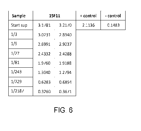
FIG. 6 illustrates Table 1, showing data from an ELISA-based reactivity
analysis of re-cloned rabbit B-cell clones derived from B-cell selection and
culture
against the target P40 biotin peptide MS1891.2, as discussed in detail in
Example 1,
below.
FIG. 7 illustrates Table 2, showing data from an ELISA-based reactivity
analysis of re-cloned rabbit B-cell clones derived from B-cell selection and
culture
against an irrelevant biotinylated peptide, as discussed in detail in Example
1, below.
FIG. 8 illustrates an image of clone 15F11 antibodies staining squamous
epithelial cells of tonsil, as discussed in detail in Example 1, below.
FIG. 9 illustrates an image of clone 15F11 antibodies staining squamous
epithelial cells of tonsil, as discussed in detail in Example 1, below.
FIG. 10 illustrates an image of clone 15F11 antibodies staining
cytotrophoblasts of placenta, as discussed in detail in Example 1, below.
FIG. 11 illustrates an image of clone 15F11 antibodies staining
cytotrophoblasts of placenta, as discussed in detail in Example 1, below.
FIG. 12 illustrates an image of clone 15F11 antibodies staining lung squamous
cell carcinoma, as discussed in detail in Example 1, below.
12
CA 03192627 2023-02-22
WO 2022/046885
PCT/US2021/047521
FIG. 13 illustrates an image of clone 15F11 antibodies staining lung
adenocarcinoma, as discussed in detail in Example 1, below.
FIG. 14 illustrates an image of anti-p40 clone 15F11, and BC28 antibodies
staining esophagus (100X image), as discussed in detail in Example 1, below.
FIG. 15 illustrates an image of anti-p40 clone 15F11, and BC28 antibodies
staining placenta (100X image) under various conditions, as discussed in
detail in
Example 1, below.
FIG. 16 illustrates an image of anti-p40 clone 15F11, and BC28 antibodies
staining tonsil, as discussed in detail in Example 1, below.
FIG. 17 illustrates an image of anti-p40 clone 15F11, and BC28 antibodies
staining tonsil, as discussed in detail in Example 1, below.
FIG. 18 illustrates an image of anti-p40 clone 15F11, and BC28 antibodies
staining normal prostate (100X image) under various conditions, as discussed
in detail
in Example 1, below.
FIG. 19 illustrates an image of anti-p40 clone 15F11, BC28 antibodies
staining kidney (100X image), as discussed in detail in Example 1, below.
FIG. 20 illustrates an image of anti-p40 clone 15F11, BC28 antibodies
staining appendix (100X image), as discussed in detail in Example 1, below.
FIG. 21 illustrates a first image of anti-p40 clone 15F11, BC28 antibodies
staining lung squamous cell carcinoma (100X image) under various conditions,
as
discussed in detail in Example 1, below.
FIG. 22 illustrates a second image of anti-p40 clone 15F11, BC28 antibodies
staining lung squamous cell carcinoma (100X image) under various conditions,
as
discussed in detail in Example 1, below.
FIG. 23 illustrates a first image of anti-p40 clone 15F11, BC28 antibodies
staining lung adenocarcinoma (100X image) under various conditions, as
discussed in
detail in Example 1, below.
FIG. 24 illustrates a second image of anti-p40 clone 15F11, BC28 antibodies
staining lung adenocarcinoma (100X image) under various conditions, as
discussed in
detail in Example 1, below.
FIG. 25 illustrates an image of anti-p40 clone 15F11, BC28 antibodies
staining lung large cell carcinoma (100X image) under various conditions, as
indicated in the figure.
13
CA 03192627 2023-02-22
WO 2022/046885
PCT/US2021/047521
FIG. 26 illustrates an image of anti-p40 clone 15F11, BC28 antibodies
staining mamma ductal carcinoma (100X image) under various conditions, as
discussed in detail in Example 1, below.
FIG. 27 illustrates an image of anti-p40 clone 15F11, BC28 antibodies
staining pancreatic adenocarcinoma (100X image) under various conditions, as
discussed in detail in Example 1, below.
FIG. 28 illustrates an image of anti-p40 clone 15F11, BC28 antibodies
staining diffuse large B-cell lymphoma (DLBCL) (100X image) under various
conditions, as discussed in detail in Example 1, below.
FIG. 29 Positive normal tonsil (p40 High Expressing (RE) and p40 Low
Expression (LE)) and negative control tissue tested with anti-p40 clone 15F11,
as
discussed in detail in Example 1, below.
FIG. 30 Lung squamous cell carcinoma (din pos) and negative lung
adenocarcinoma (din neg) tissues were tested with two-fold dilutions of the
antibody,
as discussed in detail in Example 1, below.
FIG. 31 illustrates TABLE 3, showing data from IHC used to test the
exemplary recombinant rabbit monoclonal anti-human p40 antibodies on a panel
of
cancer cells and tissues, as discussed in detail in Example 1, below.
FIG. 32 illustrates TABLE 4, showing data from staining with anti-p40 clone
15F11 as well as a negative control reagent, as discussed in detail in Example
1,
below.
Like reference symbols in the various drawings indicate like elements.
DETAILED DESCRIPTION
In alternative embodiments, provided are recombinant rabbit anti-human p40
(p63 isoform DeltaNp63, or ANp63) antibodies (Abs), including products of
manufacture and kits comprising them, and methods for making and using them,
where the antibodies can be used for in vitro diagnostics by
immunohistochemistry
(IHC). In alternative embodiments, antibodies as provided herein are used in
IHC
protocols to diagnose a cancer, for example, a squamous-cell carcinoma (SCC)
or a
lung cancer such as non-small cell lung carcinoma (NCSLC) or pulmonary SCC.
Rabbit anti-human p40 antibodies as provided herein were developed by B-
cell selection using the peptide antigen: P40 (1-16), MLYLENNAQTQFSEPQC-NH2
14
CA 03192627 2023-02-22
WO 2022/046885
PCT/US2021/047521
(SEQ ID NO:1). Serum samples of rabbits immunized using this peptide were
evaluated by immunohistochemistry (IHC). Post-immunization B-cells were
isolated
from the rabbit and supernatant from thousands of clones were screened using
enzyme-linked immunosorbent assays (ELISA). Supernatants of ELISA positive
.. clones were evaluated by IHC. Four clones were chosen for sequencing, after
which
the nucleic acids encoding the antibodies were synthesized and inserted into
expression vectors based on a pTT5 backbone. These expression vectors were
used in
human embryonic kidney 293 cells (HEK) cells for generation of recombinant
antibody. A clone designated "15F11" was chosen as best performing clone:
Sequence of antibody clone 15F11:
15F11 Heavy chain variable region:
QSVEESGGRLVKPDESLTLTCTVSGFSLSSYGVTWVRQAPGKGLEWIGYISHI
TTTYYASWAKGRFTISKTSPTTVDLKMTSLTTEDTATYFCCRGQYGSGIIYA
LWGPGTLVTISS (SEQ ID NO:2)
CDR regions are highlighted in bold and are defined according to IIVIGT
(ImMunoGeneTics, Laboratoire d'ImmunoGenetique Moleculaire (LIGM))
numbering: CDR1 aa 25 to 32, CDR2 aa 50 to 56, and CDR3 aa 94 to 106.
15F11 Light chain variable region:
AQVLTQTPSPVSAAVGGTVTINCQASQSVYNNKNLAWYQQKPGQPPKLLIY
YASTLASGVPSRF SGSGSGTQFTLTISGVQCDDAATYYCHGEFSCDSGDCSA
FGGGTEVVVK (SEQ ID NO:3)
CDR regions are highlighted in bold and are defined according to IMGT
numbering: CDR1 aa 27 to 34, CDR2 aa 52 to 54, and CDR3 aa 91 to 103.
In alternative embodiments, the exemplary recombinant anti-human p40 Abs
or dimeric antigen binding proteins comprising heavy chain variable region SEQ
ID
NO:2 and light chain variable region SEQ ID NO:3 are each bound to or fused
(or
only one is bound or fused) to an immunoglobulin heavy and light chain
constant
region, respectively, which can be for example, of human, rabbit, mouse or rat
origin,
or can be partially or entirely synthetic. The heavy and/or light chains can
be of any
isotype, for example, the heavy chain can comprise amino acid sequence from a
IgG,
CA 03192627 2023-02-22
WO 2022/046885
PCT/US2021/047521
IgM, IgA, IgD or IgE isotype; or the light chain can comprise amino acid
sequence
from a kappa (K) or lambda (X) isotype.
In alternative embodiments, exemplary recombinant anti-human p40 Abs, or
dimeric antigen binding proteins, comprising heavy chain variable region SEQ
ID
NO:2 and light chain variable region SEQ ID NO:3, are (or are configured or
assembled as) antibody (Ab) fragments, including for example, Abs or dimeric
antigen binding proteins as provided herein in the form of Fab, Fab', F(ab')2,
scFv,
(scFv)2, dAb, and complementarity determining region (CDR) fragments, linear
antibodies, single-chain antibody molecules, minibodies, diabodies, and multi
specific
antibodies formed from antibody fragments.
In alternative embodiments, exemplary recombinant anti-human p40 Abs, or
dimeric antigen binding proteins, comprising heavy chain variable region SEQ
ID
NO:2 and light chain variable region SEQ ID NO:3, are (or are configured or
assembled as) Fv fragments, i.e., as an antibody fragment which contains a
complete
antigen recognition and binding site, including a dimer of one heavy and one
light
chain variable domain in tight association, which can be covalent in nature,
for
example as an scFv. It is in this configuration that the three CDRs of each
variable
domain interact to define an antigen binding site on the surface of the VH-VL
dimer.
The six CDRs or a subset thereof can confer antigen binding specificity to the
antibody. In one embodiment, a single variable domain, or half of an Fv
comprising
only three CDRs specific for an antigen, has the ability to recognize and bind
antigen,
although usually at a lower affinity than the entire binding site.
In alternative embodiments, exemplary recombinant anti-human p40 Abs, or
dimeric antigen binding proteins, comprising heavy chain variable region SEQ
ID
NO:2 and light chain variable region SEQ ID NO:3, are (or are configured or
assembled as) F(ab')2 or Fab fragments, which contain a variable and constant
domain
of a light chain and a variable domain and the first constant domain (CH1) of
a heavy
chain. F(ab')2 antibody fragments comprise a pair of Fab fragments which are
linked,
for example, covalently linked, near their carboxy termini, for example, by
hinge
cysteines or equivalents, between them. In alternative embodiments, any
chemical
coupling of antibody fragments known in the art can be used.
In alternative embodiments, exemplary recombinant anti-human p40 Abs, or
dimeric antigen binding proteins, comprising heavy chain variable region SEQ
ID
16
CA 03192627 2023-02-22
WO 2022/046885
PCT/US2021/047521
NO:2 and light chain variable region SEQ ID NO:3, are (or are configured or
assembled as) single-chain Fv or scFv antibody fragments, which can comprise
the
VH and VL domains of antibody, wherein these domains are present in a single
polypeptide chain. In alternative embodiments, Fv polypeptides as provided
herein
further comprise a polypeptide linker between the VH and VL domains, which
enables
the scFv to form the desired structure for antigen binding.
In alternative embodiments, exemplary recombinant anti-human p40 Abs, or
dimeric antigen binding proteins, comprising heavy chain variable region SEQ
ID
NO:2 and light chain variable region SEQ ID NO:3, are (or are configured or
assembled as) diabodies, i.e., as small antibody fragments with two antigen-
binding
sites, which fragments comprise a heavy chain variable domain (VH) connected
to a
light chain variable domain (VI) in the same polypeptide chain (VH and VI). By
using a linker that is too short to allow pairing between the two domains on
the same
chain, the domains are forced to pair with the complementary domains of
another
chain and create two antigen-binding sites.
In alternative embodiments, exemplary recombinant anti-human p40 Abs, or
dimeric antigen binding proteins, comprising heavy chain variable region SEQ
ID
NO:2 and light chain variable region SEQ ID NO:3, are (or are configured or
assembled as) linear antibodies, for example, as antibodies described in
Zapata et al.
(1995 Protein Eng, 8(10):1057-1062). In alternative embodiments, linear
antibodies
as provided herein comprise a pair of tandem Fd segments (VH-CFH-VH-CFH)
which,
together with complementary light chain polypeptides, form a pair of antigen
binding
regions. In alternative embodiments, linear antibodies as provided herein are
bispecific or monospecific.
Expression of Recombinant Antibodies
In alternative embodiments, recombinant antibodies (Abs) or antigen binding
fragments thereof, or monomeric or dimeric antigen binding proteins as
provided
herein, including the exemplary recombinant anti-human p40 Abs comprising
heavy
chain variable region SEQ ID NO:2 and light chain variable region SEQ ID NO:3,
any of which may or may not have a signal peptide or other heterologous
peptide or
tag, are expressed as a recombinant polypeptide or Ab using any expression
vehicle or
expression system, for example, a vector such as a viral vector, a phage, a
plasmid or
a cosmid. In alternative embodiments, a constant heavy chain is expressed
together
17
CA 03192627 2023-02-22
WO 2022/046885
PCT/US2021/047521
with a light chain, for example, a heavy chain can be expressed with a kappa-1
light
chain.
In alternative embodiments, nucleic acids encoding the respective heavy and
light chains, or the heavy chain and the light chain, are encoded in separate
expression
.. vehicles, or in the same expression vehicle or expression system.
In some embodiments, the recombinant antibodies (Abs) or antigen binding
fragments thereof, or monomeric or dimeric antigen binding proteins as
provided
herein, including the heavy and light chains can be (cis- or trans-) as
provided herein,
are expressed from a pTT5Tm vector(s) (National Research Council Canada, NRC-
CNRC, Canada) or equivalents.
In alternative embodiments, the expression vehicles (such as a vector, plasmid
virus or phage) containing nucleic acids encoding recombinant antibodies (Abs)
or
antigen binding fragments thereof, or monomeric or dimeric antigen binding
proteins
as provided herein are expressed in in vitro expression systems or are
expressed in
cultured tissues or cells, for example, they are expressed in a human
embryonic
kidney (HEK) cell such as an HEK293-6E cell.
In alternative embodiment, the expression vehicle(s), for example, a vector or
vectors, expressing the recombinant antibodies (Abs) or antigen binding
fragments
thereof, or monomeric or dimeric antigen binding proteins as provided herein,
.. including heavy and/or light chains, are episomal or are chromosomally
integrated,
for example, in a stable cell line capable of synthesizing, optionally
inducible
synthesizing, the recombinant antibodies (Abs) or antigen binding fragments
thereof,
or monomeric or dimeric antigen binding proteins as provided herein, or heavy
and/or
light chains as provided herein.
In alternative embodiments, provided are nucleic acids encoding recombinant
antibodies (Abs) or antigen binding fragments thereof, or monomeric or dimeric
antigen binding proteins as provided herein. Nucleic acids as provided herein
can be
made, isolated and/or manipulated by, for example, cloning and expression of
cDNA
libraries, amplification of message or genomic DNA by PCR, and the like.
Nucleic
acids used to practice embodiments as provided herein, whether RNA, cDNA,
genomic DNA, vectors, viruses or hybrids thereof, may be isolated from a
variety of
sources, genetically engineered, amplified, and/or expressed/ generated
recombinantly. Recombinant polypeptides generated from these nucleic acids can
be
18
CA 03192627 2023-02-22
WO 2022/046885
PCT/US2021/047521
individually isolated or cloned and tested for a desired activity. Any
recombinant
expression system can be used, including bacterial, fungal, mammalian, yeast,
insect
or plant cell expression systems, or hybrid or synthetic expression systems.
Alternatively, these nucleic acids can be synthesized in vitro by well-known
chemical synthesis techniques, as described in, for example, Martin et al, ACS
Synth.
Biol. (2017) 6, 7, 1370-1379; Adams (1983) J. Am. Chem. Soc. 105:661; Belousov
(1997) Nucleic Acids Res. 25:3440-3444; Frenkel (1995) Free Radic. Biol. Med.
19:373-380; Blommers (1994) Biochemistry 33:7886-7896; Narang (1979) Meth.
Enzymol. 68:90; Brown (1979) Meth. Enzymol. 68:109; Beaucage (1981) Tetra.
Lett.
22:1859; U.S. Patent No. 4,458,066.
Techniques for the manipulation of nucleic acids, such as, for example,
subcloning, labeling probes (for example, random-primer labeling using Klenow
polymerase, nick translation, amplification), sequencing, hybridization and
the like
are well described in the scientific and patent literature, see, for example,
Sambrook,
ed., MOLECULAR CLONING: A LABORATORY MANUAL (2ND ED.), Vols. 1-
3, Cold Spring Harbor Laboratory, (1989); CURRENT PROTOCOLS IN
MOLECULAR BIOLOGY, Ausubel, ed. John Wiley & Sons, Inc., New York (1997);
LABORATORY TECHNIQUES IN BIOCHEMISTRY AND MOLECULAR
BIOLOGY: HYBRIDIZATION WITH NUCLEIC ACID PROBES, Part I. Theory
and Nucleic Acid Preparation, Tijssen, ed. Elsevier, N.Y. (1993).
Another useful means of obtaining and manipulating nucleic acids used to
practice embodiments as provided herein comprises screening and re-cloning
inserts
isolated or amplified from, for example, genomic clones or cDNA clones.
Sources of
nucleic acids include recombinant nucleic acid sequences, genomic or cDNA
libraries
contained and/or expressed in, for example, mammalian artificial chromosomes
(MACs), see, for example, U.S. Patent Nos. 5,721,118; 6,025,155; human
artificial
chromosomes, see, for example, Rosenfeld (1997) Nat. Genet. 15:333-335; yeast
artificial chromosomes (YAC); bacterial artificial chromosomes (BAC); P1
artificial
chromosomes, see, for example, Woon (1998) Genomics 50:306-316; P1-derived
.. vectors (PACs), see, for example, Kern (1997) Biotechniques 23:120-124;
cosmids,
recombinant viruses, phages or plasmids.
19
CA 03192627 2023-02-22
WO 2022/046885
PCT/US2021/047521
In alternative embodiments, nucleic acids as provided herein are operably
linked to transcriptional regulatory elements, including promoters, with can
be
constitutive or inducible transcriptional regulatory elements.
In alternative aspects, provided are "expression cassettes" comprising a
nucleotide sequence as provided herein, for example encoding a recombinant
antibody as provided herein. Expression cassettes can include at least a
transcriptional regulatory element, for example, a promoter, operably linked
with an
antibody coding sequence, and optionally can also include transcription
termination
signals. Additional factors necessary or helpful in effecting expression may
also be
used, for example, enhancers.
In alternative aspects, expression cassettes used to practice embodiments as
provided herein include plasmids, expression vectors, recombinant viruses, any
form
of recombinant "naked DNA" vector, and the like. In alternative aspects, an
expression vehicle or a "vector" used to practice embodiments as provided
herein can
comprise a nucleic acid that can infect, transfect, transiently or permanently
transduce
a cell. In alternative aspects, a vector used to practice embodiments as
provided
herein can be a naked nucleic acid, or a nucleic acid complexed with protein
or lipid.
In alternative aspects, vectors used to practice embodiments as provided
herein can
comprise viral or bacterial nucleic acids and/or proteins, and/or membranes
(for
example, a cell membrane, a viral lipid envelope, etc.). In alternative
aspects, vectors
used to practice embodiments as provided herein can include, but are not
limited to
replicons (for example, RNA replicons, bacteriophages) to which fragments of
DNA
may be attached and become replicated. Vectors thus include, but are not
limited to
RNA, autonomous self-replicating circular or linear DNA or RNA (for example,
plasmids, viruses, and the like, see, for example, U.S. Patent No. 5,217,879),
and can
include both the expression and non-expression plasmids. In alternative
aspects, the
vector used to practice embodiments as provided herein can be stably
replicated by
the cells during mitosis as an autonomous structure, or can be incorporated
within the
host's genome.
In alternative aspects, "promoters" used to practice embodiments as provided
herein include all sequences capable of driving transcription of a coding
sequence in a
cell, for example, a bacterial, yeast, fungal, plant, insect (for example,
baculovirus) or
mammalian cell. Thus, promoters used in the constructs include cis-acting
CA 03192627 2023-02-22
WO 2022/046885
PCT/US2021/047521
transcriptional control elements and regulatory sequences that are involved in
regulating or modulating the timing and/or rate of transcription of a gene.
For
example, a promoter used to practice embodiments as provided herein can be a
cis-
acting transcriptional control element, including an enhancer, a promoter, a
transcription terminator, an origin of replication, a chromosomal integration
sequence,
5' and 3' untranslated regions, or an intronic sequence, which are involved in
transcriptional regulation. These cis-acting sequences can interact with
proteins or
other biomolecules to carry out (turn on/off, regulate, modulate, etc.)
transcription.
"Constitutive" promoters used to practice embodiments as provided herein can
be those that drive expression continuously under most environmental
conditions and
states of development or cell differentiation. "Inducible" or "regulatable"
promoters
used to practice embodiments as provided herein can direct expression of a
nucleic
acid as provided herein under the influence of environmental conditions or
developmental conditions. Examples of environmental conditions that may affect
transcription by inducible promoters used to practice embodiments as provided
herein
include the presence of an inducing factor administered to a cell.
In alternative embodiments, polypeptides, including recombinant antibodies
(Abs) or antigen binding fragments thereof, or monomeric or dimeric antigen
binding
proteins as provided herein or as used to practice methods or other
embodiments as
provided herein can comprise any "mimetic" and/or "peptidomimetic" form. In
alternative embodiments, peptides and polypeptides used to practice
embodiments as
provided herein can comprise synthetic chemical compounds which have
substantially
the same structural and/or functional characteristics of the natural
polypeptide, for
example, a recombinant antibody as provided herein. The mimetic used to
practice
embodiments as provided herein can be either entirely composed of synthetic,
non-
natural analogues of amino acids, or, is a chimeric molecule of partly natural
peptide
amino acids and partly non-natural analogs of amino acids. The mimetic can
also
incorporate any amount of natural amino acid substitutions, for example,
conservative
amino acid substitutions, as long as such substitutions also do not
substantially alter
the mimetic's structure and/or activity. Routine experimentation will
determine
whether a mimetic is effective for practicing embodiments as provided herein,
for
example, if a mimetic composition is effective in specifically binding a human
p40
protein. Methodologies detailed herein and others known to persons skilled in
the art
21
CA 03192627 2023-02-22
WO 2022/046885
PCT/US2021/047521
may be used to select or guide one to choose effective mimetic for practicing
the
compositions and/or methods as provided herein.
Polypeptide mimetic compositions for practicing embodiments as provided
herein can comprise any combination of non-natural structural components. In
alternative aspects, mimetic compositions for practicing embodiments as
provided
herein can comprise one or all of the following three structural groups: a)
residue
linkage groups other than the natural amide bond ("peptide bond") linkages; b)
non-
natural residues in place of naturally occurring amino acid residues; or c)
residues
which induce secondary structural mimicry, i.e., to induce or stabilize a
secondary
structure, for example, a beta turn, gamma turn, beta sheet, alpha helix
conformation,
and the like. For example, a polypeptide can be characterized as a mimetic
when all
or some of its residues are joined by chemical means other than natural
peptide bonds.
In alternative embodiments, an exemplary heavy chain variable region and/or
light claim region comprises an amino acid sequence based on a sequence as set
forth
in SEQ ID NO:2 or SEQ ID NO:3, respectively, but having one or a plurality of
amino acid residue deletions or substitutions, wherein optionally all or some
of the
amino acid substitutions are conservative amino acid substitutions, and
wherein the
amino acid sequence (or, the variant polypeptide) with the one or several
deletions or
substitutions at least substantially retains the ability to specifically bind
to a peptide or
epitope comprising SEQ ID NO:1, or a fragment of a polypeptide of SEQ ID NO:1,
a
p40 (or p63 isoform DeltaNp63) polypeptide or peptide or a fragment of a p40
polypeptide or peptide, albeit the specific binding of the variant can have a
binding
affinity higher or lower than the polypeptide of SEQ ID NO:2 or SEQ ID NO:3.
In
alternative embodiments, the variant polypeptide has between one and about 50
amino
acid residue deletions or substitutions, wherein optionally all or some of the
amino
acid substitutions are conservative amino acid substitutions. In alternative
embodiments, the variant polypeptide has between about 1 to 5, 5 to 10, 10 to
15 or
15 to 20 amino acid residue deletions or substitutions.
In alternative embodiments, an exemplary heavy chain variable region
comprises an amino acid sequence as set forth in SEQ ID NO:2 having one, two,
three, four, five, six, seven, eight, nine or ten, or between about 1 and 50,
amino acid
substitutions or deletions, wherein optionally all or some of the
substitutions are
conservative amino acid substitutions, and retaining the ability to
substantially
22
CA 03192627 2023-02-22
WO 2022/046885
PCT/US2021/047521
specifically bind to a peptide or epitope comprising SEQ ID NO:1, or a
fragment of a
polypeptide of SEQ ID NO:1, a p40 (or p63 isoform DeltaNp63) polypeptide or
peptide or a fragment of a p40 polypeptide or peptide.
In alternative embodiments, an exemplary light chain variable region
comprises an amino acid sequence as set forth in SEQ ID NO:3 having one, two,
three, four, five, six, seven, eight, nine or ten, or between about 1 and 50,
amino acid
substitutions or deletions, wherein optionally all or some of the
substitutions are
conservative amino acid substitutions, and retaining the ability to
substantially
specifically bind to a peptide or epitope comprising SEQ ID NO:1, or a
fragment of a
polypeptide of SEQ ID NO:1, a p40 (or p63 isoform DeltaNp63) polypeptide or
peptide or a fragment of a p40 polypeptide or peptide.
Conservative amino acid substitutions are those that substitute a given amino
acid in a polypeptide by another amino acid of like characteristics. In
alternative
embodiments conservative substitutions are the following replacements:
replacements
of an aliphatic amino acid such as Alanine, Valine, Leucine and Isoleucine
with
another aliphatic amino acid; replacement of a Serine with a Threonine or vice
versa;
replacement of an acidic residue such as Aspartic acid and Glutamic acid with
another
acidic residue; replacement of a residue bearing an amide group, such as
Asparagine
and Glutamine, with another residue bearing an amide group; exchange of a
basic
residue such as Lysine and Arginine with another basic residue; and
replacement of
an aromatic residue such as Phenylalanine, Tyrosine with another aromatic
residue.
In alternative embodiments other variants are those in which one or more of
the
amino acid residues of a polypeptide of the invention includes a sub stituent
group.
Non-limiting examples of amino acids which may be substituted for an original
amino
acid in a protein and which may be regarded as conservative substitutions if
there is
little to no impact on the activity of the polypeptide include: Ala
substituted with ser
or thr; arg substituted with gln, his, or lys; asn substituted with glu, gln,
lys, his, asp;
asp substituted with asn, glu, or gln; cys substituted with ser or ala; gln
substituted
with asn, glu, lys, his, asp, or arg; glu substituted with asn, gln lys, or
asp; gly
substituted with pro; his substituted with asn, lys, gln, arg, tyr; ile
substituted with leu,
met, val, phe; leu substituted with ile, met, val, phe; lys substituted with
asn, glu, gln,
his, arg; met substituted with ile, leu, val, phe; phe substituted with trp,
tyr, met, ile, or
23
CA 03192627 2023-02-22
WO 2022/046885
PCT/US2021/047521
leu; ser substituted with thr, ala; thr substituted with ser or ala; trp
substituted with
phe, tyr; tyr substituted with his, phe, or trp; and val substituted with met,
ile, leu.
Purification and Isolation of Recombinant Proteins
In alternative embodiments, chimeric or the recombinant antibodies, antigen
.. binding fragments thereof, or monomeric or dimeric antigen binding
proteins, are
substantially purified or isolated, and optionally the substantially purified
or isolated
forms are the forms used in immunohistochemistry (IHC) methodologies and/or as
reagents, kits and/or products of manufacture as provided herein.
In alternative embodiments, chimeric or the recombinant antibodies, antigen
binding fragments thereof, or monomeric or dimeric antigen binding proteins,
are
substantially purified or isolated using: physicochemical fractionation, for
example,
using differential precipitation, size-exclusion or solid-phase binding of
immunoglobulins based on size, charge or other shared chemical characteristics
of
antibodies in typical samples; class-specific affinity, for example, solid-
phase binding
.. of particular antibody classes (for example, IgG or IgM) by immobilized
biological
ligands (for example, proteins, lectins, and the like) that have specific
affinity to
immunoglobulins, and this can purify all antibodies of the target class
without regard
to antigen specificity; or antigen-specific affinity, for example, affinity
purification of
only those antibodies in a sample that bind to a particular antigen molecule
through
their specific antigen-binding domains, where this purifies all antibodies
that bind the
antigen without regard to antibody class or isotype.
In alternative embodiments, chimeric or the recombinant antibodies, antigen
binding fragments thereof, or monomeric or dimeric antigen binding proteins,
are
substantially purified or isolated using standard isolation methodologies such
as
.. chromatography, for example, Ion Exchange (IEX) Chromatography, Hydrophobic
Interaction Chromatography (HIC), countercurrent chromatography,
immunoaffinity
and/or size exclusion chromatography.
In alternative embodiments, chimeric or the recombinant antibodies, antigen
binding fragments thereof, or monomeric or dimeric antigen binding proteins,
are
generated in bioreactors, e.g., a perfusion bioreactor, using continuous
expression and
purification processes, e.g., as described by Vogg et al Methods Mol Biol.
2018; vol
1850:147-178, or using stirred-tank or rocking bioreactor systems, followed by
purification.
24
CA 03192627 2023-02-22
WO 2022/046885
PCT/US2021/047521
Immunohistochemistry
In alternative embodiments, immunohistochemistry methodologies and/or
reagents used with the compositions (for example, a recombinant antibody (Ab),
or
antigen binding fragment thereof, or monomeric or dimeric antigen binding
protein as
provided herein capable of specifically binding a human p40 (p63 isoform
DeltaNp63,
or ANp63) protein or a polypeptide, or an antigen or an epitope comprising an
amino
acid sequence SEQ ID NO:1), products of manufacture, kits or methods as
provided
herein can include or comprise or comprise use of any IHC protocol, IHC
armamentarium, device and/or image or data analysis system, for practicing IHC
or
IHC reagents known in the art, for example, as described in U.S. patent nos.
(USPNs)
10,634,590 (describing a slide holder assembly fixture for use in IHC);
10,565,479
(describing methods for identifying blurred areas in digital images of stained
tissue);
10,564,076 (describing systems for analytical ( or IHC) sample preparation);
10,551,395 (describing an automated histological staining system); 10,551,378
(describing a tissue staining method); 10,504,224 (describing a digital tissue
image
analysis system for IHC); 10,501,777 (describing simultaneous, multiplexed
detection
and quantification of protein expression in IHC); 10,488,340 (describing
method for
extracting an image of a target fluorophore in a biological material);
10,453,195
(describing methods of detecting tissue areas of interest using digital
pathology
imaging); 10,438,381 (describing devices, systems and methods for generating a
digital image of a tissue section); 10,430,943 (describing methods and
programs for
automated nuclei area/number estimation for IHC image analysis); 10,416,176
(describing methods for processing specimens in an automated histological
staining
system); 10,393,633 (describing methods for processing and inhibiting the
degradation of an IHC sample); 10,217,011 (describing handling of IHC slides);
10,209,165 (describing automated or semi-automated methods for assessing the
quality of staining of a specimen containing cells); 10,126,216 (describing
methods
for fixing tissue samples for IHC); 9,423,322; 8,515,683 (describing methods
and
systems for automated detection of immunohistochemical (IHC) patterns), or
U.S.
patent application publication no US 2019/0178867 Al (describing detection of
specific tissue objects within thin sections of tissue samples as imaged in a
brightfield
microscope without using a chromogenic stain that is specific to those tissue
objects);
US 2019/0156510 Al (describing an image analysis method for analyzing an IHC
CA 03192627 2023-02-22
WO 2022/046885
PCT/US2021/047521
tissue sample); or, US 2019/0080450 Al (describing an automated determination
of
the staining quality of an IHC stained biological sample); or, US 2020/0316589
Al
(describing a multi-well solid support vessel for the processing and testing
of fixed
biological materials)..
In alternative embodiments, the recombinant antibodies, antigen binding
fragments thereof, or monomeric or dimeric antigen binding proteins, in IHC
protocols, or kits, as provided herein are substantially purified or isolated
or are in the
form of an unpurified or partially purified culture supernatant.
In alternative embodiments, methods as provided herein can use or comprise
reagents for detecting or visualizing an antibody-antigen interaction using
any
products or methods know in the art, for example, and IHC protocol or
reagents.
In alternative embodiments, methods as provided herein comprise use of
chromogenic immunohistochemistry (CIH), wherein a primary antibody (for
example,
a recombinant antibody (Ab), or antigen binding fragment thereof, or monomeric
or
dimeric antigen binding protein, as provided herein) or secondary antibody
(for
example, where the secondary antibody binds to the primary antibody, or the
recombinant antibody (Ab), or antigen binding fragment thereof, or monomeric
or
dimeric antigen binding protein as provided herein,) is conjugated to an
enzyme such
as peroxidase, for example, an immunoperoxidase), for example, a horseradish
peroxidase (HRP), that can catalyze a color-producing reaction. In alternative
embodiments, a chromogenic moiety used in methods as provided herein is or
comprises a coumarin; a rhodamine; 2,3,6,7-tetrahydro-11-oxo-1H,5H,11H-
[1]benzopyrano[6,7,8-ij]quinolizine-1- 0-carboxylic acid; 7-
(diethylamino)coumarin-
3-carboxylic acid; a coumarin derivative; a rhodamine derivative; a
tetramethylrhodamine; a diarylrhodamine derivative; QSY 7; QSY 9; QSY 21;
diazo
chromophores; DABSYL; tartrazine; triarylmethane compounds; fast red; fast
blue;
fuchsin; Cascade Blue acetyl; Dapoxylsulfonic acid/carboxylic acid
succinimidyl
ester; DY-405; Alexa Fluor 405 succinimidyl ester; Cascade Yellow succinimidyl
ester; pyridyloxazole succinimidyl ester (PyMPO); Pacific Blue succinimidyl
ester;
DY-415; 7-hydroxycoumarin-3-carboxylic acid succinimidyl ester; DYQ-425; 6-
FAM phosphoramidite; Lucifer Yellow; iodoacetamide; Alexa Fluor 430
succinimidyl ester; Dabcyl succinimidyl ester; NBD chloride/fluoride; QSY 35
succinimidyl ester; DY-485XL; Cy2 succinimidyl ester; DY-490; Oregon Green 488
26
CA 03192627 2023-02-22
WO 2022/046885
PCT/US2021/047521
carboxylic acid succinimidyl ester; Alexa Fluor 488 succinimidyl ester; BODIPY
493/503 C3 succinimidyl ester; DY-480XL; BODIPY FL C3 succinimidyl ester;
BODIPY FL C5 succinimidyl ester; BODIPY FL-X succinimidyl ester; DYQ-505;
Oregon Green 514 carboxylic acid succinimidyl ester; DY-510XL; DY-481XL; 6-
.. carboxy-4',5'-dichloro-2',7'-dimethoxyfluorescein succinimidyl ester (JOE);
DY-
520XL; DY-521XL; BODIPY R6G C3 succinimidyl ester; erythrosin isothiocyanate;
5-carboxy-2',4',5',7'-tetrabromosulfonefluorescein succinimidyl ester; Alexa
Fluor 532
succinimidyl ester; 6-carboxy-2',4,4',5'7,7'-hexachlorofluorescein
succinimidyl ester
(HEX); BODIPY 530/550 C3 succinimidyl ester; DY-530; BODIPY TMR-X
succinimidyl ester; DY-555; DYQ-1; DY-556; Cy3 succinimidyl ester; DY-547; DY-
549; DY-550; Alexa Fluor 555 succinimidyl ester; Alexa Fluor 546 succinimidyl
ester; DY-548; BODIPY 558/568 C3 succinimidyl ester; Rhodamine red-X
succinimidyl ester; QSY 7 succinimidyl ester; BODIPY 564/570 C3 succinimidyl
ester; BODIPY 576/589 C3 succinimidyl ester; carboxy-X-rhodamine (ROX);
succinimidyl ester; Alexa Fluor 568 succinimidyl ester; DY-590; BODIPY 581/591
C3 succinimidyl ester; DY-591; BODIPY TR-X succinimidyl ester; Alexa Fluor 594
succinimidyl ester; DY-594; carboxynaphthofluorescein succinimidyl ester; DY-
605;
DY-610; Alexa Fluor 610 succinimidyl ester; DY-615; BODIPY 630/650-X
succinimidyl ester; erioglaucine; Alexa Fluor 633 succinimidyl ester; Alexa
Fluor 635
succinimidyl ester; DY-634; DY-630; DY-631; DY-632; DY-633; DYQ-2; DY-636;
BODIPY 650/665-X succinimidyl ester; DY-635; Cy5 succinimidyl ester; Alexa
Fluor 647 succinimidyl ester; DY-647; DY-648; DY-650; DY-654; DY-652; DY-649;
DY-651; DYQ-660; DYQ-661; Alexa Fluor 660 succinimidyl ester; Cy5.5
succinimidyl ester; DY-677; DY-675; DY-676; DY-678; Alexa Fluor 680
succinimidyl ester; DY-679; DY-680; DY-682; DY-681; DYQ-3; DYQ-700; Alexa
Fluor 700 succinimidyl ester; DY-703; DY-701; DY-704; DY-700; DY-730; DY-731;
DY-732; DY-734; DY-750; Cy7 succinimidyl ester; DY-749; DYQ-4; Cy7.5
succinimidyl ester; 7-diethylaminocoumarin-3-carboxylic acid; succinimidyl
ester;
Dabsyl sulfonyl chloride; fluorescein isothiocyanate (FITC) carboxy
succinimidyl
ester (DY-495); Rhodamine Green carboxylic acid succinimidyl ester (DY-505);
eosin isothiocyanate (EITC); 6-carboxy-2',4,7,7'-tetrachlorofluorescein
succinimidyl
ester (TET); carboxyrhodamine 6G succinimidyl ester;
carboxytetramethylrhodamine
succinimidyl ester (TMR, TAMRA) (DY-554); QSY 9 succinimidyl ester;
27
CA 03192627 2023-02-22
WO 2022/046885
PCT/US2021/047521
sulforhodamine B sulfonyl chloride (DY-560); Texas Red (sulforhodamine 101);
gallocyanine; Fast Green FCF; Malachite Green; or, a QSY 21 succinimidyl
ester.
In alternative embodiments, methods as provided herein comprise use of
immunofluorescence, where a primary or a secondary antibody is tagged to
a fluorophore, such as fluorescein or fluorescein isothiocyanate (FITC), a
triarylmethane dye such as rhodamine or rhodamine derivatives (for example,
tetramethylrhodamine (TRITC), rhodamine 6G, rhodamine 123, rhodamine B,
carboxytetramethylrhodamine (TAMRA), tetramethylrhodamine (TMR),
sulforhodamine 101), aminomethylcoumarin acetate (AMCA), ALEXATM or
DYLIGHTTm fluors, or a fluorophore or dye as described in U.S. patent
application
no. US 2019/0018018 Al. 3,3'-Diaminobenzidine (DAB) also can be used.
In alternative embodiments, methods as provided herein comprise use of
a direct method or one-step staining method where a primary antibody (for
example,
recombinant antibodies (Ab), or antigen binding fragments thereof, or
monomeric or
dimeric antigen binding proteins as provided herein) is labeled and reacts
directly
with an antigen, for example, in a tissue sections. While this technique
utilizes only
one antibody and therefore is simple and rapid, the sensitivity may be lower
due to
little signal amplification.
In alternative embodiments, methods as provided herein comprise use of an
indirect method where an unlabeled primary antibody (first layer) binds to a
target antigen (for example, p40), for example, in a tissue or organ, and a
labeled secondary antibody (second layer) then is reacted with the primary
antibody.
The secondary antibody can be against the isotype, for example, IgG, of the
animal
species in which the primary antibody is derived. This method can be more
sensitive
than direct detection strategies because of signal amplification due to the
binding of
several secondary antibodies to each primary antibody if the secondary
antibody is
conjugated to a detecting agent such as a fluorescent or enzyme reporter.
In alternative embodiments, further amplification is achieved if the secondary
antibody is conjugated to several detecting molecules, for example, biotin
molecules,
which can recruit complexes of avidin-, streptavidin- or NEUTRAVIDINTm protein-
bound enzyme.
In alternative embodiments, the IHC is performed on tissue sections or tissue
biopsies, for example, paraformaldehyde (PFA) fixed tissues or organs, or
formalin-
28
CA 03192627 2023-02-22
WO 2022/046885
PCT/US2021/047521
fixed paraffin-embedded tissues. In alternative embodiments, a tissue is
sectioned or
sliced or used whole. Before sectioning, the tissue sample can be embedded in
a
medium, for example, paraffin wax or cryomedia. Tissue sections can be
sectioned or
sliced on a variety of instruments, most commonly using a microtome, cryostat,
or
vibratome. Specimens can be sectioned or sliced at a range of about 3 p.m to 5
gm.
The sections or slices can be mounted on slides, dehydrated using alcohol
washes of
increasing concentrations (for example, 50%, 75%, 90%, 95%, 100%), and cleared
using a detergent like xylene before being imaged under a microscope.
Depending on the method of fixation and tissue preservation, the sample may
require additional steps to make a p40 epitope available for antibody binding,
including deparaffinization and antigen retrieval. For formalin-fixed paraffin-
embedded tissues, antigen-retrieval is often necessary, and can comprise pre-
treating
the sections with heat or proteases.
In alternative embodiments, the IHC is performed using an ENVISION
DUOFLEX DOUBLESTAIN SYSTEMTm (EnVision DuoFLEX Doublestain System)
(Agilent, San Jose, CA), which allows for staining of two or more markers on a
single
slide. In alternative embodiments, the IHC is performed using an EnVision FLEX
HRP Magenta, High pH (Dako Omnis) system, and binding can be visualized by
EnVision FLEX HRP Magenta Chromogen. In alternative embodiments, the IHC is
performed using EnVision FLEX Mini Kit, High pH, which is a high-sensitivity
visualization system intended for use in IHC together with Dako ALTOSTAINERTm
instruments; this dual link system detects primary mouse and rabbit antibodies
and the
reaction is visualized by 3,3'-Diarninobenzidine DAB) ehromogen (I)AB forms a
water-insoluble brown precipitate when oxidized, for example, by a
peroxidase).
Products of Manufacture and Kits
Provided are products of manufacture and kits comprising chimeric or
recombinant anti-human p40 Abs as provided, and for practicing methods as
provided
herein using the chimeric or recombinant anti-human p40 Abs as provided
herein; and
optionally the products of manufacture and/or kits can further comprise some
or all
reagents needed to perform an immunohistochemistry (IHC), and optionally can
comprise instructions for practicing methods as provided herein.
In alternative embodiment, products of manufacture have attached thereto or
affixed (optionally covalently bound) on or onto a chimeric or recombinant
antibody
29
CA 03192627 2023-02-22
WO 2022/046885
PCT/US2021/047521
or a dimeric antigen binding protein as provided herein, and optionally
products of
manufacture as provided herein are or comprise arrays, chips, biochips,
slides, trays,
dishes (for example, microtiter dishes), phages or phagemids.
In alternative embodiment, immunohistochemistry methodologies and/or
reagents used to practice composition, product of manufacture (for example,
kit) or
method embodiments as provided herein can include or comprise or comprise use
of
any IHC protocol, IHC armamentarium, device and/or image or data analysis
system,
for practicing IHC or IHC reagents known in the art, for example, as described
in U.S.
patent nos. 10,565,479 (describing methods for identifying blurred areas in
digital
images of stained tissue); 10,564,076 (describing systems for analytical (or
IHC)
sample preparation); 10,551,395 (describing an automated histological staining
system); 10,551,378 (describing a tissue staining method); 10,504,224
(describing a
digital tissue image analysis system for IHC); 10,501,777 (describing
simultaneous,
multiplexed detection and quantification of protein expression in IHC);
10,488,340
(describing method for extracting an image of a target fluorophore in a
biological
material); 10,453,195 (describing methods of detecting tissue areas of
interest using
digital pathology imaging); 10,438,381 (describing devices, systems and
methods for
generating a digital image of a tissue section); 10,416,176 (describing
methods for
processing specimens in an automated histological staining system); 10,393,633
(describing methods for processing and inhibiting the degradation of an IHC
sample);
10,217,011 (describing handling of IHC slides); 10,209,165 (describing
automated or
semi-automated methods for assessing the quality of staining of a specimen
containing cells); 10,126,216 (describing methods for fixing tissue samples
for IHC);
9,423,322.
Any of the above aspects and embodiments can be combined with any other
aspect or embodiment as disclosed here in the Summary, Figures and/or Detailed
Description sections.
As used in this specification and the claims, the singular forms "a," "an" and
"the" include plural referents unless the context clearly dictates otherwise.
Unless specifically stated or obvious from context, as used herein, the term
"or" is understood to be inclusive and covers both "or" and "and".
Unless specifically stated or obvious from context, as used herein, the term
"about" is understood as within a range of normal tolerance in the art, for
example
CA 03192627 2023-02-22
WO 2022/046885
PCT/US2021/047521
within 2 standard deviations of the mean. About can be understood as within
20%,
19%, 18%, 17%, 16%, 15%, 14%, 13%, 12% 11%, 10%, 9%, 8%, 7%, 6%, 5%, 4%,
3%, 2%, 1%, 0.5%, 0.1%, 0.05%, or 0.01% of the stated value. Unless otherwise
clear from the context, all numerical values provided herein are modified by
the term
"about."
Unless specifically stated or obvious from context, as used herein, the terms
"substantially all", "substantially most of', "substantially all of' or
"majority of'
encompass at least about 85%, 90%, 95%, 97%, 98%, 99% or 99.5%, or more of a
referenced amount of a composition.
The entirety of each patent, patent application, publication and document
referenced herein hereby is incorporated by reference. Citation of the above
patents,
patent applications, publications and documents is not an admission that any
of the
foregoing is pertinent prior art, nor does it constitute any admission as to
the contents
or date of these publications or documents. Incorporation by reference of
these
documents, standing alone, should not be construed as an assertion or
admission that
any portion of the contents of any document is considered to be essential
material for
satisfying any national or regional statutory disclosure requirement for
patent
applications. Notwithstanding, the right is reserved for relying upon any of
such
documents, where appropriate, for providing material deemed essential to the
claimed
subject matter by an examining authority or court.
Modifications may be made to the foregoing without departing from the basic
aspects of the invention. Although the invention has been described in
substantial
detail with reference to one or more specific embodiments, those of ordinary
skill in
the art will recognize that changes may be made to the embodiments
specifically
disclosed in this application, and yet these modifications and improvements
are within
the scope and spirit of the invention. The invention illustratively described
herein
suitably may be practiced in the absence of any element(s) not specifically
disclosed
herein. Thus, for example, in each instance herein any of the terms
"comprising",
"consisting essentially of', and "consisting of' may be replaced with either
of the
.. other two terms. Thus, the terms and expressions which have been employed
are used
as terms of description and not of limitation, equivalents of the features
shown and
described, or portions thereof, are not excluded, and it is recognized that
various
31
CA 03192627 2023-02-22
WO 2022/046885
PCT/US2021/047521
modifications are possible within the scope of the invention. Embodiments of
the
invention are set forth in the following claims.
The invention will be further described with reference to the examples
described herein; however, it is to be understood that the invention is not
limited to
such examples.
EXAMPLES
Unless stated otherwise in the Examples, all recombinant DNA techniques are
carried out according to standard protocols, for example, as described in
Sambrook et
al. (2012) Molecular Cloning: A Laboratory Manual, 4th Edition, Cold Spring
Harbor
Laboratory Press, NY and in Volumes 1 and 2 of Ausubel et al. (1994) Current
Protocols in Molecular Biology, Current Protocols, USA. Other references for
standard molecular biology techniques include Sambrook and Russell (2001)
Molecular Cloning: A Laboratory Manual, Third Edition, Cold Spring Harbor
Laboratory Press, NY, Volumes I and II of Brown (1998) Molecular Biology
LabFax,
Second Edition, Academic Press (UK). Standard materials and methods for
polymerase chain reactions can be found in Dieffenbach and Dveksler (1995) PCR
Primer: A Laboratory Manual, Cold Spring Harbor Laboratory Press, and in
McPherson at al. (2000) PCR - Basics: From Background to Bench, First Edition,
Springer Verlag, Germany.
Example 1: Development of exemplary anti-p40 antibody clone 15F11
This example describes the development of the exemplary anti-p40 antibody
clone designated "15F11".
Peptide MLYLENNAQTQFSEPQC-N}12 (SEQ ID NO:1) was synthetized
and used for immunization of 3 rabbits.
After 47 days blood samples were taken from the rabbits and the titer
determined by ELISA titration against the peptide immunogen.
ELISA protocol
- coating: overnight (o/n) 4 C Plate 100 ng/well neutravidine M5544.9 in PBS,
wash
plates 3 times with PBST;
- blocking: 4.5 hours (h) room temperature (RT) block each well with 370 11.1
1%
BSA in PBST;
32
CA 03192627 2023-02-22
WO 2022/046885
PCT/US2021/047521
- coating: 1 h 37 C 100 ng/well biotin-antigen (MS1891.2) in 1% BSA (bovine
serum albumin) in PBST, wash plates 3 times with PBST;
- samples: 1.5 h RT add plasma dilution series, as mentioned in plate
format (see
results), in 100 ul 1% BSA in PBST per well, wash plates 3 times with PBST;
- detection antibody: 1.25 h RT add 100 ul Goat anti Rabbit IgG-HRP (MS169.4):
1/5000 in 1% BSA in PBST, wash plates 3 times with PBST. wash plates 3 times
with PBS;
- staining: add 50 tl TMB per well;
- stop reaction: 7 minutes add 50 ul 2M H2504 per well, measure A450.
Results (optical density OD as a function of plasma dilutions) are graphically
illustrated in FIG. 1.
It was decided to continue immunizations to achieve a higher titer. Retest was
made at day 61 after immunization start, and ELISAs were repeated, and results
are
graphically illustrated in FIG. 2, FIG. 3 and FIG. 4. All rabbits had a higher
plasma
titer against p40 biotin peptide.
Next step: Select and culture target reactive B-cells and screen B-cell
supernatants to identify the wells containing target reactive B-cells.
B-cell selection and culture was performed using spleen cells of one rabbit,
followed by screening B-cell culture supernatants in ELISA on P40 biotin
peptide.
Results: B-cell selection was performed, resulting in a successful selection
of
target reactive B-cells analyzed by target ELISA using B-cell culture
supernatants. In
both selection methods a subset of selected B-cells was cultured single cell
per well
and a subset as a back-up- multiple cells per well. Clones were selected based
on
measured 0D450 great than ( > ) 0.2 in the antigen ELISA (5 times background)
single
cell per well and multiple cells per well, respectively.
B-cell culture supernatants positive in ELISA were further functionally tested
in paraffin IHC (IHC-P) to identify clones producing antibody capable of
binding the
p40 antigen in FFPE (formalin fixed paraffin embedded) tissue.
The nucleic acids encoding the variable regions of the antibodies which
specifically bind to p40 from the well with B cells exhibiting desirable
results in the
IHC were cloned and sequenced. Additionally, a small scale expression of full
length
recombinant rabbit IgG was made in eukaryotic cells and tested by standard IHC
protocol.
33
CA 03192627 2023-02-22
WO 2022/046885
PCT/US2021/047521
Cloning and sequencing
As illustrated in FIG. 5, PCR reactions on cDNA derived from B-cell cultures
to amplify the heavy and light variable domains yielded PCR products of the
correct
size; FIG. 5 illustrates PCR-amplified VH and VL domains of the 4 selected
clones.
Gel purified nucleic acids encoding VH and VL domains of all 4 clones were
cloned into vectors, respectively, and subsequently subjected to colony PCR
for
sequence analysis. Sequences were translated and aligned.
Expression and validation
The sequences obtained from the respective VH and VL cloning were used
to synthesize codon optimized gene constructs for the respective VH and VL
fragments. These were subcloned into an expression vector (pTT5 backbone)
containing the rabbit constant heavy and light chains, respectively. Per clone
the
obtained recombinant sequence were expressed in the mammalian (HEK293-6E)
expression system, producing recombinant monoclonal rabbit anti-human p40
antibody.
Target ELISA was performed in duplicate to validate immunoreactivity of
the expressed antibodies on the antigen (Table 1, FIG. 6) and a biotinylated
control
peptide (Table 2, FIG. 7). Control plasma from rabbit A150733 (plus 47 days;
1:20,000 diluted) and PB ST plus 1% BSA were included as positive and negative
control, respectively. Reactivity screening revealed that all 4 re-cloned
antibodies
show strong reactivity against client's target (Table 1, FIG. 6), while
no/limited
reactivity against an included irrelevant biotinylated peptide was observed
(Table 2,
FIG. 7).
Table 1 (as illustrated in FIG. 6): ELISA-based reactivity analysis of
recloned
rabbit 13-cell clones derived from 13-cell selection and culture against
client's target
P40 biotin peptide (MS1891.2).
Table 2 (as illustrated in FIG. 7): ELISA-based reactivity analysis of
recloned
rabbit 13-cell clones derived from 13-cell selection and culture against an
irrelevant
biotinylated peptide.
Conclusions:
= Clone 15F11 was successfully cloned, sequenced, expressed and validated
for
specific reactivity against client's target (MS1891.2) in ELISA.
= Except for 1 amino acid difference in framework 4 of the VH, the variable
34
CA 03192627 2023-02-22
WO 2022/046885
PCT/US2021/047521
domain sequences of 2 clones are identical.
= Approximately 1.8 ml supernatant containing the corresponding rabbit IgG1
antibodies are used for further screening.
Staining performance of clone 15F11:
FIG. 8 illustrates an image of clone 15F11 antibodies staining squamous
epithelial cells of tonsil.
FIG. 9 illustrates an image of clone 15F11 antibodies staining squamous
epithelial cells of tonsil.
FIG. 10 illustrates an image of clone 15F11 antibodies staining
cytotrophoblasts of placenta.
FIG. 11 illustrates an image of clone 15F11 antibodies staining
cytotrophoblasts of placenta.
FIG. 12 illustrates an image of clone 15F11 antibodies staining lung squamous
cell carcinoma.
FIG. 13 illustrates an image of clone 15F11 antibodies staining lung
adenocarcinoma.
IHC on cancer cells
IHC was used to test the recombinant rabbit monoclonal anti-human p40
antibodies, an IgG isotypes, on a panel of cancer cells and tissues, as
illustrated in the
table of TABLE 3, where: -: negative, (+):> 1-9% positive cells, +: > 10%
positive
cells.
Results for IHC on cancer cells
mAb clone BC28:
mAb clone BC28, Biocare Medical, was used as reference for the two p40
antibodies tested. The protocol was performed on the Ventana BenchMark Ultra.
A
weak to strong nuclear staining reaction was seen in squamous epithelial cells
of
esophagus and tonsil, and basal cells of prostate glands. A weak to moderate
staining
reaction was observed in dispersed trophoblasts of placenta. No staining
reaction was
seen in endothelial cells, muscle cells and lymphocytes. In general, a high
signal to
noise ratio was provided.
The neoplasias staining profile is listed above. 21/23 lung squamous cell
carcinomas were positive using a cut-off greater than or equal to ( >) 10%.
All 23
tested lung adenocarcinomas were negative.
CA 03192627 2023-02-22
WO 2022/046885
PCT/US2021/047521
rmAb clone 15F11:
In this test, the best protocol with shortest TAT and highest signal-to-noise
ratio was found by using a titer of 1:250 of the primary Ab, heat induced
epitope
retrieval (HIER) in Target Retrieval SolutionTM (TRS) (Agilent, Dako) high pH
and
using EnVision FLEX+TM as detection system. If using HIER in TRS Low pH a
titer
of 1:50 gave the best result.
Virtually the result for the two selected protocols for rmAb clone 15F11 were
identical. Both demonstrated a moderate to strong nuclear staining reaction in
squamous epithelial cells of esophagus and tonsil, and basal cells of prostate
glands.
In dispersed trophoblasts of placenta, a weak to moderate nuclear staining
reaction
was seen. An increased titer for both protocols gave an inferior signal-to-
noise ratio.
The staining pattern for the neoplasias were similar to the reference Ab.
Conclusion for IHC on cancer cells
In this test performed, the best overall result was obtained by the
recombinant
(rmAb) clone 15F11 with HIER in TRS High pH. The result was comparable to the
reference based on mAb BC28 (used as a control) stained on BenchMark UltraTM,
Ventana.
IHC Tissue Staining
FIG. 14 illustrates an image of anti-p40 clone 15F11, and BC28 antibodies
staining esophagus (100X image).
FIG. 15 illustrates an image of anti-p40 clone 15F11, and BC28 antibodies
staining placenta (100X image) under various conditions, as indicated in the
figure.
FIG. 16 illustrates an image of anti-p40 clone 15F11, and BC28 antibodies
staining tonsil (24 hour fixation, 100X image).
FIG. 17 illustrates an image of anti-p40 clone 15F11, and BC28 antibodies
staining tonsil (144 hour fixation, 100X image).
FIG. 18 illustrates an image of anti-p40 clone 15F11, and BC28 antibodies
staining normal prostate (100X image) under various conditions, as indicated
in the
figure.
FIG. 19 illustrates an image of anti-p40 clone 15F11, BC28 antibodies
staining kidney (100X image).
FIG. 20 illustrates an image of anti-p40 clone 15F11, BC28 antibodies
staining appendix (100X image).
36
CA 03192627 2023-02-22
WO 2022/046885
PCT/US2021/047521
FIG. 21 illustrates a first image of anti-p40 clone 15F11, BC28 antibodies
staining lung squamous cell carcinoma (100X image) under various conditions,
as
indicated in the figure.
FIG. 22 illustrates a second image of anti-p40 clone 15F11, BC28 antibodies
staining lung squamous cell carcinoma (100X image) under various conditions,
as
indicated in the figure.
FIG. 23 illustrates a first image of anti-p40 clone 15F11, BC28 antibodies
staining lung adenocarcinoma (100X image) under various conditions, as
indicated in
the figure.
FIG. 24 illustrates a second image of anti-p40 clone 15F11, BC28 antibodies
staining lung adenocarcinoma (100X image) under various conditions, as
indicated in
the figure.
FIG. 25 illustrates an image of anti-p40 clone 15F11, BC28 antibodies
staining lung large cell carcinoma (100X image) under various conditions, as
indicated in the figure.
FIG. 26 illustrates an image of anti-p40 clone 15F11, BC28 antibodies
staining mamma ductal carcinoma (100X image) under various conditions, as
indicated in the figure.
FIG. 27 illustrates an image of anti-p40 clone 15F11, BC28 antibodies
staining pancreatic adenocarcinoma (100X image) under various conditions, as
indicated in the figure.
FIG. 28 illustrates an image of anti-p40 clone 15F11, BC28 antibodies
staining diffuse large B-cell lymphoma (DLBCL) (100X image) under various
conditions, as indicated in the figure.
FIG. 29 Positive normal tonsil (p40 High Expressing (RE) and p40 Low
Expression (LE)) and negative control tissue tested with anti-p40 clone 15F11.
IHC
intensity score (blue line) and background staining (red line) vs. antibody
dilution.
The normal tonsil tissues #95496 and #95570 were evaluated for both RE
structures
(squamous epithelial cells of the tonsil) and negative control structures (non-
epithelial
cells of the tonsil).
FIG. 30 Lung squamous cell carcinoma (din pos) and negative lung
adenocarcinoma (din neg) tissues were tested with two-fold dilutions of the
antibody.
37
CA 03192627 2023-02-22
WO 2022/046885
PCT/US2021/047521
Specific staining (blue line) and background staining (red line) vs. the
antibody
dilution. 2X represents half the 1X concentration etc.
FIG. 31 illustrates TABLE 3, showing data from IHC used to test the
exemplary recombinant rabbit monoclonal anti-human p40 antibodies on a panel
of
cancer cells and tissues.
FIG. 32 illustrates TABLE 4, showing data from staining with anti-p40 clone
15F11 as well as a negative control reagent.
References
1. Bishop, J.A., et al., p40 (DeltaNp63) is superior to p63 for the
diagnosis of
pulmonary squamous cell carcinoma. Mod Pathol, 2012. 25(3): p. 405-15.
2. Sailer, V., et al., Comparison of p40 (DeltaNp63) and p63 expression in
prostate tissues--which one is the superior diagnostic marker for basal cells?
Histopathology, 2013. 63(1): p. 50-6.
A number of embodiments of the invention have been described.
Nevertheless, it can be understood that various modifications may be made
without
departing from the spirit and scope of the invention. Accordingly, other
embodiments
are within the scope of the following claims.
38