Note: Descriptions are shown in the official language in which they were submitted.
CA 03192774 2023-02-22
WO 2022/063629 PCT/EP2021/075174
1
X-RAY IMAGING METHOD AND SYSTEM FOR REDUCING
OVERLAPPING OF NEIGHBORING TEETH IN PANORAMIC IMAGES
TECHNICAL FIELD OF THE INVENTION
The present invention relates to an x-ray imaging method of generating a
panoramic image
with reduced overlapping of neighboring teeth. The present invention also
relates to an x-
ray imaging system for generating a panoramic image with reduced overlapping
of
neighboring teeth.
BACKGROUND ART OF THE INVENTION
X-ray imaging system for generating panoramic images of a patient's teeth are
generally
known in the art. Such an-ray imaging system generally comprises an x-ray
source for
emitting x-ray towards a patient; an x-ray detector for detecting the x-rays
transmitted
through the jaw of the patient; an acquisition means adapted to acquire a
plurality of 2D x-
ray images respectively at a plurality of different radiographic directions by
means of
rotating the x-ray source and the x-ray detector around at least the jaw of
the patient; and
an image processing means which is adapted to reconstruct a panoramic image
based on
the 2D x-ray images and the radiographic directions respectively.
A commonly known problem with such panoramic problems is that the panoramic
image
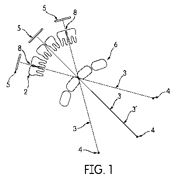
generally includes regions where neighboring teeth overlap as shown in Fig. 1.
As such,
panoramic images with overlapping teeth generally complicate or prevent a
successful
diagnosis.
DISCLOSURE OF THE INVENTION
An objective of the present invention is to overcome the above-mentioned
disadvantages of
the prior art. This objective has been achieved by the x-ray imaging method as
defined in
claim 1, and the x-ray imaging system as defined in claim 13. The subject-
matters of the
dependent claims relate to further developments and preferable embodiments.
The present invention provides a computer-implemented x-ray imaging method for
generating a panoramic image with reduced overlapping of neighboring teeth.
The method
comprises: a step of acquiring a plurality of 2D x-ray images respectively at
a plurality of
different radiographic directions by means of rotating an x-ray source and an
x-ray detector
around the jaw of a patient; a step of identifying one or more regions each
including at
CA 03192774 2023-02-22
WO 2022/063629 PCT/EP2021/075174
2
least one pair of overlapping neighboring teeth in the 2D x-ray images and/or
in temporary
panoramic images reconstructed from the 2D x-ray images for which an optimal
radiographic directions may be determined, among the corresponding
radiographic
directions, which reduces the overlap in the panoramic image to be
reconstructed; and a
step of determining one or more optimal radiographic directions respectively
among the
corresponding radiographic directions of the 2D x-ray images for which one or
more
regions each including at least one pair of overlapping neighboring teeth has
been
identified in the identifying step, to reduce the overlaps in the panoramic
image to be
reconstructed. Herein, the overlapping neighboring teeth are a pair of teeth,
wherein each
tooth of the pair may be on the upper jaw, or each tooth of the pair may be in
the lower
jaw, or one of the pair may be on the upper jaw and the other one may be on
the lower jaw.
A major advantageous effect of the present invention is that through the
reduction of the
overlap regions, the diagnosis can be improved.
Once the optimal radiographic directions are determined, the panoramic image
may be
reconstructed and displayed according to various different alternative
embodiments.
According to an alternative embodiment, at least a partial panoramic image is
reconstructed based on the 2D x-ray images with the determined optimal
radiographic
directions with respect to one or more identified regions. Optionally, the
reduced overlap
in the identified regions can be indicated by providing some additional
information on the
panoramic image so that the dentist can be apprised of the fact the
radiographic directions
have been optimized. The additional information may be an icon, a text, an
outline or the
like.
According to a further alternative embodiment, at least a partial panoramic
image is
reconstructed based on the 2D x-ray images and the radiographic directions
with respect to
one or more regions as in the prior art but is provided with additional
information which
further comprises insets at the identified regions showing at least partial
panoramic images
reconstructed based on the 2D x-ray images with determined optimal
radiographic
directions with respect to the one or more identified regions. Through the
insets, the dentist
can preview the identified regions with overlap reduction and thereby apprised
of the fact
the radiographic directions have been optimized. The insets can be preferably
selected/unselected for preview by the user through the use of an input means
like
keyboard cursers, a mouse or the like.
CA 03192774 2023-02-22
WO 2022/063629 PCT/EP2021/075174
3
According to a further alternative embodiment, at least a partial panoramic
image without
overlap reduction is displayed together with at least a partial panoramic
image with overlap
reduction, for instance, in a toggle mode which can be switched by the user
through the
input means such as a keyboard, mouse or the like. Thereby, the dentist can be
enabled to
recognize the effect of the optimized radiographic directions.
According to a further alternative embodiment, at least a partial panoramic
image is
reconstructed based on the 2D x-ray images corresponding to interpolated
radiographic
directions which have been obtained through spatial interpolation between the
determined
optimal radiographic directions of the corresponding identified regions.
According to the present invention, artificial intelligence (AI) algorithms
may be used in
the identification of the regions each including at least one pair of
overlapping neighboring
teeth in the 2D x-ray images and/or in the temporary panoramic images to be
reconstructed. According to the present invention, AT techniques may be also
used in the
determination of the optimal radiographic directions which reduce the overlaps
in the
panoramic image to be reconstructed. The artificial intelligence algorithm can
be trained
with an input of previously acquired 2D x-ray images and/or previously
reconstructed
panoramic images comprising manual annotations showing the said overlaps
respectively.
Furthermore, the artificial intelligence algorithm can be trained with an
input of previously
acquired 2D x-ray images or previously reconstructed panoramic images which
comprise
manual annotations showing the optimal radiographic directions respectively
that reduce
the overlaps in the panoramic image to be reconstructed.
The present invention also provides a computer-implemented x-ray imaging
system for
generating a panoramic image with reduced overlapping neighboring teeth. The x-
ray
imaging system comprises: an x-ray source for emitting x-ray towards a
patient; an x-ray
detector for detecting the x-rays transmitted through the jaw of the patient;
an acquisition
means adapted to acquire a plurality of 2D x-ray images respectively at a
plurality of
different radiographic directions by rotating the x-ray source and the x-ray
detector around
at least the jaw of the patient; and an image processing means for executing a
computer
program that causes the system to perform the x-ray imaging method of the
present
invention. The system is not provided essentially as a single apparatus. For
instance, the
image processing means may be in the cloud or in a computer in the practice of
the
radiologist.
CA 03192774 2023-02-22
WO 2022/063629 PCT/EP2021/075174
4
BRIEF DESCRIPTION OF THE DRAWINGS
In the subsequent description, the present invention will be described in more
detail by
using exemplary embodiments and by referring to the drawings, wherein
Fig. 1 ¨ is a partial schematic view of an x-ray imaging system according to
an
embodiment;
Fig. 2 ¨ is a schematic view of a panoramic image without overlap reduction in
the
identified regions having overlapping neighboring teeth;
Fig. 3 ¨ is a schematic view of a panoramic image with overlap reduction
according to an
embodiment;
Fig. 4 ¨ is a schematic view of a panoramic image with selectable/unselectable
insets with
overlap reduction according to an embodiment.
The reference numbers shown in the drawings denote the elements as listed
below and will
be referred to in the subsequent description of the exemplary embodiments.
1. Panoramic image (with overlap reduction)
la. Inset
1' Panoramic image (without overlap reduction)
2. 2D x-ray image
3. Radiographic direction
3' Optimal radiographic direction
4. X-ray source
5. X-ray detector
6. Jaw
7. Region
8. Overlapping neighboring teeth
9. X-ray imaging system
Fig. 1 shows schematic partial view of a computer-implemented x-ray imaging
system (9)
for generating a panoramic image (1) with reduced overlapping neighboring
teeth (8). The
x-ray imaging system (9) comprises: an x-ray source (4) for emitting x-ray
towards a
patient; an x-ray detector (5) for detecting the x-rays transmitted through
the jaw (6) of the
patient; an acquisition means adapted to acquire a plurality of 2D x-ray
images (2)
respectively at a plurality of different radiographic directions (3) by
rotating the x-ray
source (4) and the x-ray detector (5) around at least the jaw (6) of the
patient; an image
CA 03192774 2023-02-22
WO 2022/063629 PCT/EP2021/075174
5 processing means which is adapted to execute a computer program according
to the present
invention. Further details of the x-ray imaging system (9) which are generally
known to
those skilled in the art will be omitted to prevent unnecessary prolongation
of the
description.
The computer program comprises computer-executable codes for causing the
computer-
implemented x-ray imaging system (9) to execute the method steps of the
present
invention, which will be described in more detail later in the description.
The computer
program may be stored in a computer-readable storage means connected to the
computer-
implemented x-ray imaging system (9). The connection may be external such that
the
storage means is in the cloud, at a remote location or in the dentist's
practice.
The computer-implemented x-ray imaging method of the present invention is
suitable for
generating a panoramic image (1) with reduced overlapping of neighboring teeth
(8). The
method comprises a step of acquiring a plurality of 2D x-ray images (2)
respectively at a
plurality of different radiographic directions (3) by means of rotating an x-
ray source (4)
and an x-ray detector (5) around the jaw (6) of a patient; a step of
identifying one or more
regions (7) (see e.g. Fig. 2) each including at least one pair of overlapping
neighboring
teeth (8) in the 2D x-ray images (2) and/or in temporary panoramic images
reconstructed
from the 2D x-ray images (2) for which an optimal radiographic directions (3')
(See Fig. 2)
may be determined, among the corresponding radiographic directions (3), which
reduces
the overlap in the panoramic image (1) to be reconstructed; and a step of
determining one
.. or more optimal radiographic directions (3') respectively among the
corresponding
radiographic directions (3) of the 2D x-ray images for which one or more
regions (7) each
including at least one pair of overlapping neighboring teeth (8) has been
identified in the
identifying step, to reduce the overlaps in the panoramic image (1) to be
reconstructed.
In the subsequent description two different alternative embodiments will be
described for
reconstructing the panoramic image (1). A first alternative embodiment is
provided in Fig.
3 which shows a schematic view of a panoramic image (1) with overlap
reduction. The
panoramic image (1) with overlap reduction can be displayed on a display of
the computer-
implemented x-ray imaging system (9). In the first alternative embodiment, the
method
comprises a step of reconstructing at least a partial panoramic image (1)
based on the 2D x-
.. ray images (2) with determined optimal radiographic directions (3') with
respect to one or
more identified regions (7); and a optional step of representing additional
information (A)
CA 03192774 2023-02-22
WO 2022/063629 PCT/EP2021/075174
6
on the reconstructed panoramic image (1) to the user, which indicates the
reduced overlap.
As shown in Fig. 3, through the additional information (A), the dentist can be
apprised of
the fact that optimized radiographic directions (3') have been used in the
reconstruction.
The additional information may be an icon, a text, an outline or the like
informing the user
on the existence of the reduced overlap. Alternatively, the representation of
the additional
information (A) may be dispensed with. For comparison, in Fig. 2 a schematic
view of a
panoramic image (1') without overlap reduction has been shown. The panoramic
image
(1') without overlap reduction may be optionally displayed on the display of
the x-ray
imaging system (9) together with the panoramic image (1) with overlap
reduction, for
instance, in a toggle mode. Thereby the dentist can be enabled to recognize
the effect of the
optimized radiographic directions (3'). A second alternative embodiment is
provided in
Fig. 4 which shows a schematic view of a panoramic image (1') without overlap
reduction
but with additional information (A) which comprises insets (la) showing at
least partial
panoramic images (1) with overlap reduction. In the second alternative
embodiment, the
method according comprises: a step of reconstructing at least a partial
panoramic image
.. (1') based on the 2D x-ray images (2) and the radiographic directions (3)
with respect to
one or more regions (7); and a step of representing on the reconstructed
panoramic image
(1') additional information (A) which comprises the insets (la) at the
identified regions (7)
showing at least partial panoramic images (1) reconstructed based on the 2D x-
ray images
(2) with determined optimal radiographic directions (3') with respect to one
or more
identified regions (7). As shown in Fig. 4, through the additional information
(A)
including the insets (la), the dentist can preview the identified regions (7)
with overlap
reduction while being apprised of the optimized radiographic directions (3').
The insets
(la) can be preferably selected for preview by the user through the use of an
input means
like keyboard cursers, a mouse or the like. The inset (la) may pop up with a
preset
magnification or without magnification, and disappear when it is unselected.
Alternatively,
the insets (la) may be fixed. The size of the insets (la) may have the size of
the identified
regions (7). Also, in the second alternative embodiment, the additional
information (A)
may be an icon, a text, an outline or the like informing the user on the
existence of a
preview with reduced overlap.
According to a further alternative embodiment (not illustrated), the
radiographic directions
(3) in the vicinity of the optimized radiographic direction (3') can be
interpolated. In this
alternative embodiment, the method comprises a step of reconstructing at least
a partial
CA 03192774 2023-02-22
WO 2022/063629 PCT/EP2021/075174
7
panoramic image (1) whose image points are based on the 2D x-ray images (2)
corresponding to interpolated radiographic directions which have been obtained
through
spatial interpolation between the determined optimal radiographic directions
(3') of the
corresponding identified regions (7).
In the subsequent description, the use of deep learning techniques will
briefly described.
Trained artificial intelligence algorithms may be used in the identification
step and/or in
the determination steps.
According to a further embodiment, in the identifying step a trained
artificial intelligence
algorithm is used to identify one or more regions (7) each including at least
one pair of
overlapping neighboring teeth (8) in the 2D x-ray images (2) and/or in
temporary
panoramic images reconstructed from the 2D x-ray images (2). The artificial
intelligence
algorithm can be trained with an input of previously acquired 2D x-ray images
and/or
previously reconstructed panoramic images comprising manual annotations
showing the
said overlaps respectively. The use of the artificial intelligence is very
effective in view of
the speed and the reliability of the identification step. Alternatively, image
processing
techniques which are not based on deep learning may be used.
According to a further embodiment, in the determining step a trained
artificial intelligence
algorithm is used to determine one or more optimal radiographic directions
(3')
respectively among the corresponding radiographic directions (3) of the 2D x-
ray images
(2) for which one or more regions (7) each including at least one pair of
overlapping
neighboring teeth (8) has been identified, to reduce the overlaps in the
panoramic image
(1) to be reconstructed. In a first alternative of this embodiment, in the
determining step a
plurality of at least partial temporary panoramic images (1') are
reconstructed based on 2D
x-ray images (2) with respectively different radiographic directions (3) for
at least one or
more regions (7) each including at least one overlapping neighboring teeth
(8), and the
trained artificial intelligence algorithm is used to determine the optimal
radiographic
direction (3'), among the said different radiographic directions (3), that
reduces the
overlaps in the panoramic image (1) to be reconstructed, through comparing the
reconstructed plurality of said at least partial temporary panoramic images
(1') for the one
or more identified regions (7). In a second alternative of this embodiment, in
the
determining step the trained artificial intelligence algorithm is used to
determine the
optimal radiographic direction (3') that reduces the overlaps in the panoramic
image (1) to
CA 03192774 2023-02-22
WO 2022/063629 PCT/EP2021/075174
8
be reconstructed by comparing the set of originally acquired 2D x-ray images
(2) with
corresponding radiographic directions (3) for which one or more regions (7)
each including
at least one overlapping neighboring teeth (8) has been identified. The
artificial
intelligence algorithm can be trained with an input of previously acquired 2D
x-ray images
or previously reconstructed panoramic images which comprise manual annotations
showing the optimal radiographic directions respectively that reduce the
overlaps in the
panoramic image (1) to be reconstructed. The use of the artificial
intelligence is also here
very effective in view of the speed and the reliability of the determination
step. As
indicated above, image processing techniques which are not based on deep
learning may be
alternatively used.
