Note: Descriptions are shown in the official language in which they were submitted.
CA 03192917 2023-02-23
WO 2022/043793
PCT/IB2021/057002
1
HERMETIC ENCLOSURE FOR IMPLANTABLE SENSORS
TECHNICAL FIELD
[0001] The present invention relates to sensors that are implantable into a
patient's
body, and to systems and methods of using the same.
BACKGROUND
[0002] Tracking of physical disease and healing in humans often involves
measuring
anatomical properties of a patient's body. However, some measurements, such as
those that can
only be obtained internally, can be difficult to obtain. More recently, there
has been an interest
in sensors that can be implanted into a patient's body to track the health of
the patient over time.
For example, attempts have been made to use one or more strain gauges to track
healing in a
damaged or fractured bone. The one or more strain gauges are attached to an
orthopedic implant
that is in turn attached to the damaged or fractured bone. As the bone heals,
the bone
increasingly shares the load imparted by the patient's body on the orthopedic
implant. Thus, the
load imparted on the bone increases as the bone heals, while the load imparted
on the orthopedic
implant decreases. In principle, this change in loading can be measured over
time by the one or
more strain gauges to track the progress of healing in the bone. The
measurement can then be
communicated to a device outside of the body that can be accessed by a
physician.
SUMMARY
[0003] In one example, an anatomical implant comprises an implant body and a
rim.
The implant body has an outer surface, and the rim extends from the implant
body along an
CA 03192917 2023-02-23
WO 2022/043793
PCT/IB2021/057002
2
outward direction. The rim has an internal surface, and an external surface
opposite the internal
surface. The internal surface defines a pocket that can support a sensor
therein.
[0004] In another example, a system comprises the anatomical implant, the
sensor, and
a cap, where the cap is attached to the rim such that the sensor is
hermetically sealed within the
pocket.
[0005] In yet another example, a method comprises a step of receiving a sensor
within a
pocket defined in an anatomical implant, where the anatomical implant includes
a rim having an
internal surface, and an external surface opposite the internal surface, the
internal surface
defining the pocket. The method comprises a step of aligning a cap with an end
of the rim, and a
step of attaching the cap to the rim so as to hermetically seal the sensor
within the pocket.
BRIEF DESCRIPTION OF THE DRAWINGS
[0006] The foregoing summary, as well as the following detailed description of
embodiments of the application, will be better understood when read in
conjunction with the
appended drawings. For the purposes of illustrating the methods and bone
screws of the present
application, there is shown in the drawings representative embodiments. It
should be
understood, however, that the application is not limited to the precise
methods and devices
shown. In the drawings:
[0007] Fig. 1 shows a simplified schematic diagram of a measurement system
according to one example that is positioned relative to a patient so as to
measure an anatomical
condition of the patient, the system having a sensor supported by an
anatomical implant and
having an external reader that receives measurements from the sensor;
[0008] Fig. 2 shows a simplified block diagram of the system of Fig. 1
according to one
example;
[0009] Fig. 3 shows a perspective view of an anatomical implant according to
one
example, the implant having a rectangular-shaped rim that is configured to
support a sensor
therein;
[0010] Fig. 4 shows a top view of a portion of the anatomical implant of Fig.
3;
[0011] Fig. 5 shows a cross-sectional view of the anatomical implant of Fig.
3;
[0012] Fig. 6 shows a perspective view of an anatomical implant according to
another
example, the implant having a circular-shaped rim that is configured to
support a sensor therein;
[0013] Fig. 7 shows a top view of a portion of the anatomical implant of Fig.
6;
[0014] Fig. 8 shows a cross-sectional view of the anatomical implant of Fig.
6;
CA 03192917 2023-02-23
WO 2022/043793
PCT/IB2021/057002
3
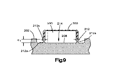
[0015] Fig. 9 shows a cross-sectional view of the implant of Figs. 3 or 6
according to
another example with a cap attached to a rim of the implant and a sensor
disposed within the rim,
the rim having a first height;
[0016] Fig. 10 shows a cross-sectional view of the implant of Figs. 3 or 6
according to
another example with a cap attached to a rim of the implant and a sensor
disposed within the rim,
the rim having a second height;
[0017] Fig. 11 shows a cross-sectional view of the implant of Figs. 3 or 6
according to
another example with a cap attached to a rim of the implant and a sensor
disposed within the rim,
the rim having a third height;
[0018] Fig. 12 shows a perspective view of a cap according to one example that
can
implement the cap of Figs. 9 or 10; and
[0019] Fig. 13 shows a partially-exploded perspective view of an implantable
sensor
system that comprises an anatomical implant, a sensor, and a sensor cover.
DETAILED DESCRIPTION
[0020] Electronic sensors that are implanted into a patient's body can contain
non-
biocompatible materials. As a result, the non-biocompatible materials of the
sensors should be
isolated from contact with the body such that only biocompatible materials are
in contact with
the body. To limit contact between non-biocompatible sensor materials and the
body, an
implantable sensor can be hermetically sealed within a biocompatible housing.
Typically, truly
hermetic enclosures are made of either glass, ceramic, or metal. An integral
element of the
sensor systems is an antenna that allows radiofrequency (RF) communication of
the data with the
outside reader. However, metal enclosures can disrupt the RF field and render
the
communication ineffective. Glass and ceramics, on the other hand, do not
create a barrier for RF
energy, and therefore, are preferable for use in hermetically sealing an
implantable sensor.
Depending on the application, some sensors, such as those with strain sensing
elements, may
require direct contact with the metallic implant. In such case, the glass or
ceramic enclosure
should be integrated with the metallic implant. For example, ceramic caps can
be laser-welded
to a metal anatomical implant. However, the heat generated at the weld can
compromise the
mechanical properties of the metal implant and lead to premature fatigue
failures.
[0021] As will be discussed below, in various examples, an anatomical implant
can be
implemented with a metal rim that defines a pocket therein. The pocket is
configured to support
the sensing elements of the sensor therein, and may also support other
elements therein that are
to be hermetically sealed, such as a PCB, an antenna, a battery, etc. A non-
metallic cap, such as
CA 03192917 2023-02-23
WO 2022/043793
PCT/IB2021/057002
4
a ceramic cap, that allows for RF communication can be laser-welded onto the
metal rim,
hermetically sealing the contents within the pocket. Excessive heat generated
during the welding
process can be absorbed by the rim, thereby protecting the underlying core
structure of the metal
implant.
[0022] Referring to Fig. 1, a system 10 is shown that is configured to track
health of a
patient overtime. In general, the system 10 comprises at least one implantable
sensor 100 that is
configured to be implanted into a patient's body 20. The system can also
comprise an
anatomical implant 200 configured to support the at least one sensor 100. The
anatomical
implant 200 can be any suitable anatomical implant such as (without
limitation) a bone plate, an
intramedullary nail, a bone anchor, a pedicle screw, a spine rod, an
intervertebral implant, and so
on. In addition, the bone plate can be formed from a metal such as titanium,
although in
alternative examples, the bone plate can be formed from another suitable
implantable material
such as, without limitation, a polymer such as polyether ether ketone (PEEK).
[0023] The system can further comprise an external wireless reader 116
configured to
wirelessly receive data from the at least one sensor 100 through the skin of
the patient when the
external wireless reader 116 is situated outside of the patient's body. The
data can then be
communicated to a computing device 30 that can be accessed by the patient or a
medical
professional. The computing device 30 can be physically separate from the
external wireless
reader 116 as shown or can be implemented as part of the external wireless
reader 116. In some
examples, the external wireless reader 116 can be configured to wirelessly
provide a source of
power to the at least one sensor 100, while in other examples, the at least
one sensor 100 can
comprise its own source of power, such as (without limitation) a battery.
[0024] Referring now to Fig. 2, a simplified block diagram of the system of
Fig. 1 is
shown according to one embodiment. The system comprises a sensor 100 that
comprises at least
one sensing element 102, and a measurement device 104 in communication with
the at least one
sensing element 102. Together, the at least one sensing element 102 and
measurement device
104 are configured to generate a measurement value that is proportional to a
value of an
anatomical property that a patient's body observed by the at least one sensing
element 102 when
the sensor 100 is implanted in the patient's body. The anatomical property can
be any suitable
property for tracking the health of a patient such as (without limitation)
strain, load, deflection,
rotation, temperature, pressure, pH level, oxygen level, and so on.
[0025] To generate the measurement value, each sensing element 102 has a
sensor
property having a value that changes in response to a change in a value of the
anatomical
CA 03192917 2023-02-23
WO 2022/043793
PCT/IB2021/057002
property observed by the sensing element 102. Thus, each sensing element 102
has a sensor
property having a value that is proportional to the value of the anatomical
property. For
example, the sensor property can be resistance, capacitance, inductance,
piezoelectricity, light
behavior, or another suitable sensor property. The measurement device 104 is
configured to
detect or measure the value of the sensor property, and the value of the
anatomical property can
be calculated from the value of the sensor property. In some embodiments, the
value of the
anatomical property can be calculated by multiplying the measured value of the
sensor property
by a constant.
[0026] Each sensing element 102 can be any suitable type of sensing element
for
tracking the health of a patient, and the sensor property can be any suitable
sensor property. For
example, the sensing element can be (without limitation) at least one of a
resistive sensing
element having a resistance that changes in response to a change in the
anatomical property, a
piezoelectric sensing element having a piezoelectric material that changes an
electrical charge in
response to a change in the anatomical property, a capacitive sensing element
having a
capacitance that changes in response to a change in the anatomical property,
an inductive sensing
element having an inductance that changes in response to a change in the
anatomical property, an
optical sensing element, and so on. In one example, each sensing element 102
can be a resistive
sensing element, the sensor property of each sensing element 102 can be an
electrical resistance
of the sensing element 102, and the anatomical property can be strain on the
anatomical body,
where the resistance of each sensing element 102 changes in response to a
change in strain on the
anatomical body.
[0027] The sensor 100 can comprise an internal wireless communicator 108 in
communication with the measurement device 104, and an antenna system 109 in
communication
with the internal wireless communicator 108. The antenna system 109 can
include an antenna
110, and optionally can include other components such as a shield that limits
the amount in
which the implant 200 beneath the shield is exposed to the magnetic field
generated by the
antenna 110, or prevent such exposure altogether. The internal wireless
communicator 108 is
configured to receive the measurement value from the measurement device 104
and provide the
measurement value to the antenna 110 in a suitable form for wireless
transmission. The internal
wireless communicator 108 can include a wireless transmitter or transponder
that receives the
measurement value from the measurement device 104 and prepares the measurement
value for
wireless transmission. For example, the wireless communicator 108 can include
processing
such as (without limitation) one or more of (i) memory configured to store the
measurement
CA 03192917 2023-02-23
WO 2022/043793
PCT/IB2021/057002
6
value, (ii) a digital-to-analog converter configured to convert the
measurement value to analog
format, (iii) a radio-frequency (RF) modulator configured to modulate the
measurement value,
(iv) an error-correction encoder configured to encode the measurement value,
and other
processing consistent with the wireless technology employed by the sensor 100.
[0028] In one example, the internal wireless communicator 108 can be
configured as a
passive radio-frequency identification (RFID) transponder. Alternatively, the
internal wireless
communicator can be configured using any other wireless communication
technology suitable
for communicating through the skin such as (without limitation) battery-
assisted passive RFID,
active RFID, Bluetooth, and Wi-Fi. The wireless communicator 108 can further
include a
unique identifier (ID) that can be used to distinguish the sensor 100 from
other sensors. In one
example, the unique ID can be an ID of an RFID tag. The antenna 110 is
configured to convert
an electrical signal corresponding to the measurement value from the wireless
communicator 108
into radio waves so as to transmit the measurement value wirelessly through
the patient's skin to
the external wireless reader 116 situated outside of the patient's body.
[0029] The sensor 100 can comprise a power device 106 configured to supply
power to
the measurement device 104 and wireless communicator 108. In at least some
examples, the
power device 106 can include an energy harvesting device configured to capture
energy from a
suitable energy source that is separate from the sensor 100. For example, the
energy source can
be radio waves communicated from the external wireless reader 116.
Alternatively, the power
device 106 can capture energy from the patient's body itself or from another
external source such
as a source external to the patient's body. For example, the energy source can
include (without
limitation) kinetic energy, electric fields, magnetic fields, and so on. In
some embodiments, the
power device 106 can include a battery.
[0030] One or more, up to all, of the measurement device 104, power device
106, and
wireless communicator 108 can each be implemented on a printed circuit board
(PCB) 112,
although embodiments of the disclosure are not so limited. One or more, up to
all, of the
measurement device 104, power device 106, and wireless communicator 108 can
each be
implemented in an integrated circuit (i.e., chip) that is mounted onto the
printed circuit board
112. The at least one sensing element 102, printed circuit board 112, and
antenna 110 can all be
supported by the anatomical implant 200 (shown in Fig. 1), which in turn can
be attached to an
anatomical body of the patient.
[0031] The external wireless reader 116 is configured to wirelessly receive
the
measurement value from the at least one sensor 100 through the skin of the
patient when the
CA 03192917 2023-02-23
WO 2022/043793
PCT/IB2021/057002
7
external wireless reader 116 is situated outside of the patient's body.
Moreover, in at least some
examples, the external wireless reader 116 can be configured to wirelessly
provide a source of
power to the at least one sensor 100. In at least one such example, the
external wireless reader
116 can be implemented as an RFID reader.
[0032] The external wireless reader 116 can include an antenna 118 and a
wireless
communicator 120. The wireless communicator 120 can include a transmitter and
a receiver. In
such examples, the communicator 120 can be considered to be a transceiver. In
at least some
examples, the external wireless reader 116 can further include a computing
device 122. The
computing device 122 can be configured to calculate a value of the anatomical
property based on
the measurement value. In one example, the computing device 122 can calculate
the value of the
anatomical property by multiplying the measurement value by a specified
constant.
Alternatively, the system can comprise a computing device 30 as shown in Fig.
1 that is
implemented separately from the external wireless reader 116. For example, the
computing
device 30 can be a computer configured to receive the measurement value from
the external
wireless reader 116 and present the value to a physician.
[0033] Turning now to Figs. 3 to 8, examples of the anatomical implant 200 of
Fig. 1
are shown. In each example, the implant 200 is a bone plate. However, as
discussed above,
implants of this disclosure may alternatively be any other suitable anatomical
implant. It will be
understood that the following description of various features of the implant
200 of Figs. 3 to 5
also applies to the features of implant 200 of Figs. 6 to 8 that have like
reference numerals.
[0034] The implant 200 has an implant body 201 that has an outer surface 202.
The
implant body 201 can be formed from a metal or other material as discussed
above. The outer
surface 202 can be configured to face away from the bone when the implant 202
is implanted
into a patient. The implant body 201 can have an inner surface 204. The inner
surface 204 can
be configured to face the bone when the implant 202 is implanted into a
patient. In some
examples, the inner surface 204 can be curved so as to conform to a surface of
the bone. In
alternative examples, such as when the implant is an intramedullary nail, the
implant can have an
inner surface, such as an inner surface that defines a cannulation, but the
implant need not have
an inner surface.
[0035] The anatomical implant 200 is configured to be attached to an
anatomical
structure, such as bone, using any suitable attachment. For example, the
implant 200 can define
at least one such as a plurality of bone anchor fixation holes 216
therethrough, wherein each
CA 03192917 2023-02-23
WO 2022/043793
PCT/IB2021/057002
8
bone anchor fixation hole 216 is configured to receive a fastener so as to
affix the implant 200 to
an anatomical structure such as a bone.
[0036] The implant 200 comprises a rim 212 that extends along an outward
direction
Do from the body 201 of the implant 200. The rim 212 can be formed from a
metal. The rim
212 defines a pocket 214 that extends therein along an inward direction Di,
opposite the outward
direction Do. The pocket 214 is configured to support at least a portion of a
sensor therein. The
pocket 214 is bounded by the rim 212. The rim 212 is configured such that a
cap 300 (shown in
Figs. 9 to 11) can be attached, such as laser welded, thereto so as to
hermetically seal the pocket
214. In some examples, the cap 300 can be formed from a ceramic or other
material that
provides little, if any, interference with RF signals. The pocket 214 can be
open at the outer
surface 202 when the cap 300 is not attached thereto and can terminate at an
interior surface 208
of the implant 200. The interior surface 208 can define a floor of the pocket
214. The rim 212
can define a closed shape in a plane that is perpendicular to the inward
direction Di. For
example, the rim 212 can define a rectangular shape as shown in Figs. 3 to 5,
a circular shape as
shown in Figs. 6 to 8, or any other suitable shape. The rim 212 can be
integral and monolithic
with the body 201. For example, the rim 212 and body 201 can be machined from
a single
monolithic piece of material. Alternatively, the rim 212 can be attached, such
as adhered or
welded, to the body 201. However, machining the rim 212 can be advantageous
over adhering
or welding the rim 212 to the body because adhering might not form a hermetic
seal and welding
could compromise the strength of the body 201.
[0037] The rim 212 has a first end 212a at the implant body 201 and a second
end 212b
that is spaced from the implant body 201. The second end 212b is a free end
that is not attached
to the implant body 201. The first end 212a is preferably integral and
monolithic with the body
201, although in alterative examples, it can be attached to the body 201. The
rim 212 has an
internal rim surface 212d and an external rim surface 212c that are opposite
one another. The
internal rim surface 212d can define the pocket 214. The external rim surface
212c can face
away from the pocket 214. The rim 212 can have a thickness t from the interior
rim surface 212
to the external rim surface 212c that is less than a dimension (such as a
length and/or width) of
the rim 212 in a plane that is perpendicular to the inward direction Di. In
some examples, the
thickness t can be less than a height of the rim 212 along the outward
direction Do.
[0038] The implant 200 defines a recess 206 that extends into the outer
surface 202
along the inward direction Di. The recess 206 can extend to the interior
surface 208 of the
implant. The interior surface 208 can define a floor of the recess 206. The
implant 200 can
CA 03192917 2023-02-23
WO 2022/043793
PCT/IB2021/057002
9
comprise an edge 210 at the outer surface 202 that defines an outer perimeter
of the recess 206.
In some examples, the outer perimeter can define a closed shape in a plane
that is perpendicular
to the inward direction. For example, the outer perimeter of the recess 206
can define a
rectangular shape as shown, a circular shape, or another other suitable shape.
The recess 206 can
be open at the outer surface 202 and can terminate at the interior surface
208. The interior
surface 208 can define a floor of the recess 206.
[0039] In some examples, the rim 212 can extend from the interior surface 208
along
the outward direction Do. The rim 212 can be disposed within the recess 206.
The rim 212 can
be inwardly spaced from the edge 210 of the recess 206 along a plane that is
perpendicular to the
outward direction Do so as to define a space between the rim 212 and the edge
210 of the recess
206. The space can extend entirely around the rim 212. The external surface
212c of the rim
212 can face the edge 210 of the recess 206. It will be understood that, in
alternative examples,
the implant body 201 can be devoid of the recess 206, and the rim 212 can
extend from the outer
surface 202 of the implant body 201.
[0040] Turning to Figs. 9 to 11, the rim 212 is configured to support at least
a portion
of the sensor 100 therein. In some examples, as shown in Figs. 9 and 10, the
pocket 214 within
the rim 212 can support a portion of the sensor 100 therein, and the cap 300
can be configured to
support another portion of the sensor 100 therein. In such examples, the rim
212 can have a
height as measured from the interior surface 208 to the free end 212b of the
rim 212 along the
outward direction Do that is less than a height of the sensor 100 when the
sensor 100 is mounted
to the implant 200. For example, the rim 212 can have a height Hi that is less
than or equal to a
distance from the interior surface 208 to the outer surface 202 as shown in
Fig. 9. As another
example, the rim 212 can have a height H2 that is greater than a distance from
the interior surface
208 to the outer surface 202 as shown in Fig. 10, and yet less than a height
of the sensor 100.
[0041] With specific reference to Fig. 12, in examples in which the cap 300
supports a
portion of the sensor 100 therein, the cap 300 can define a recess 310 that is
configured to
support the portion of the sensor 100 therein. The cap 300 can have a first
end 302, and a second
end 304 that is opposite the first end 302 along the outward direction Do. The
second end 304
can include a top wall 306. The cap 300 can have at least one sidewall 308
that extends from the
top wall 306 along the inward direction Di. The at least one sidewall 308 can
define the recess
310. The recess 310 can extend into the first end 302 of the cap 300 towards
the top wall 306
along the outward direction Do. The recess 310 can be configured to support a
portion of the
sensor 100 therein. The at least one sidewall 308 can enclose the recess 310.
For example, the at
CA 03192917 2023-02-23
WO 2022/043793
PCT/IB2021/057002
least one sidewall 308 can define a closed shape around the recess 310 along a
plane that is
perpendicular to the outward direction Do. The closed shape can be a rectangle
as shown in Fig.
12, a circle, or any other suitable shape. Thus, the cap 300 can have a shape
of a box that is open
at one end, a cylinder that is open at one end, or another suitable three-
dimensional shape that is
open at one end. The at least one sidewall 308 can have a cross-sectional
shape that conforms to
a shape of the free end 212b of the rim 212 such that the at least one
sidewall 308 is configured
to be hermetically sealed to the rim 212.
[0042] Referring to Fig. 11, in some examples, the pocket 214 can support an
entirety
of the sensor 100 therein, and the cap 300 is configured to close the pocket
214 without
supporting any portion of the sensor 100 therein. The cap 300 can define a
recess therein as
discussed above or can be devoid of a recess as shown in Fig. 11. In some
examples, the cap 300
can have a planar shape. In some such examples, the cap 300 can have a cross-
sectional shape
along a plane that is perpendicular to the outward direction Do that is
rectangular, circular, or
any other suitable shape. The cap 300 can have a cross-sectional shape that
conforms to a shape
of the free end 212b of the rim 212 such that the cap 300 is configured to be
hermetically sealed
to the rim 212.
[0043] With reference to Figs. 9 to 11, in some examples, the rim 212 can have
a
narrowed portion 212e between the first and second ends 212a and 212b of the
rim 212. The
narrowed portion 212e can have a thickness along a direction that extends from
the internal rim
surface 212d to the external rim surface 212c that is less than a thickness of
the first and second
ends 212a and 212b along the same direction. The narrowed portion 212e can
allow flexibility
of the rim 212.
[0044] Turning now to Fig. 13, an example of an implantable sensor system is
shown.
The system comprises an anatomical implant 200 and at least one implantable
sensor 100. The
anatomical implant 200 can be implemented as discussed above in relation to
implant 200. The
sensor 100 can comprise at least one sensing element 102, a printed circuit
board 112, and an
antenna 110. Further, the system can comprise a cover 300. It will be
understood that sensors
100a and 100b can each be implemented as shown in Fig. 9. In one example, the
at least one
sensing element 102 can be part of a strain gauge. The strain gauge can be
supported by the
body 201 of the implant 200 such that the strain gauge 200 is in direct
contact with the implant
body 201. In some examples, the at least one sensing element 102 can include
more than one
sensing element 102 supported by the implant body 201. The sensing elements
102 can be
CA 03192917 2023-02-23
WO 2022/043793
PCT/IB2021/057002
11
angularly offset from one another so as to detect one or both of torsional and
bending forces
imparted by the bone on the implant 200.
[0045] The printed circuit board 112 can include a substrate. One or more
integrated
circuits can be mounted onto the substrate. Further, the printed circuit board
112 can be
configured as described above in relation to printed circuit board 112. For
example, the one or
more integrated circuits can include an integrated circuit comprising the
power device 106, an
integrated circuit comprising the measurement device 104, and an integrated
circuit comprising
the wireless communicator 108. In at least one embodiment, the integrated
circuit comprising
the power device 106 can be implemented as an energy harvesting chip, the
integrated circuit
comprising the measurement device 104 can be implemented as a PicoStrain0
chip, and the
integrated circuit comprising the wireless communicator 108 can be implemented
as an RFID
chip.
[0046] When each of the at least one sensor 100 is assembled, the at least one
sensing
element 102, the printed circuit board 112, and the antenna 110 can be aligned
along the outward
direction Do of the implant 200. For example, the printed circuit board 112
can be disposed
between the at least one sensing element 102 and the antenna 110.
[0047] The cover 300 can be aligned with the at least one sensing element 102,
the
printed circuit board 112, and the antenna 110 along the outward direction Do.
Thus, the antenna
110 can be disposed between the printed circuit board 112 and at least a
portion of the cover 300,
such as the top wall 306 of the cover 300, with respect to the outward
direction Do. In the
assembled configuration, each sensor 100 can have an overall size in a plane
perpendicular to the
select direction between approximately 8 mm x 8 mm and approximately 20 mm x
20 mm, and
increments of 1 mm therebetween. In one example, each sensor 100 can have an
overall size in
the plane of approximately 12 mm x 12 mm. Each sensor 100 can further have an
overall
thickness in the select direction between approximately 2 mm and 4 mm,
although in alterative
examples the thickness can be below 2 mm or above 4 mm.
[0048] To assemble the implantable sensor system, the sensor 100 is inserted
into the
pocket 214 of the implant 200. The pocket 214 is then hermetically sealed by
attaching the cap
300 onto the rim 212 of the implant 200. In one example, the attaching step
can comprise
welding, such as laser welding, the cap 300 to the rim 212. When welding the
cap 300 to the rim
212, heat generated by the welding is absorbed by the rim 212, thereby
protecting the implant
body 201 from damage or weakening. This way the sensor 100 is hermetically
sealed with the
CA 03192917 2023-02-23
WO 2022/043793
PCT/IB2021/057002
12
cap 300, which allows for RF communication, while the load-bearing part of the
implant (i.e., the
implant body 201) is shielded from the excessive heat generated by the laser
welding process.
[0049] It should be noted that the illustrations and descriptions of the
examples shown
in the figures are for exemplary purposes only, and should not be construed
limiting the
disclosure. One skilled in the art will appreciate that the present disclosure
contemplates various
examples. Additionally, it should be understood that the concepts described
above with the
above-described examples may be employed alone or in combination with any of
the other
examples described above. It should further be appreciated that the various
alternative examples
described above with respect to one illustrated example can apply to all
examples as described
herein, unless otherwise indicated.
[0050] Conditional language used herein, such as, among others, "can,"
"could,"
"might," "may," "e.g.," and the like, unless specifically stated otherwise, or
otherwise
understood within the context as used, is generally intended to convey that
certain embodiments
include, while other embodiments do not include, certain features, elements,
and/or steps. Thus,
such conditional language is not generally intended to imply that features,
elements, and/or steps
are in any way required for one or more examples or that one or more examples
necessarily
include these features, elements and/or steps. The terms "comprising,"
"including," "having,"
and the like are synonymous and are used inclusively, in an open-ended
fashion, and do not
exclude additional elements, features, acts, operations, and so forth.
[0051] While certain examples have been described, these examples have been
presented by way of example only and are not intended to limit the scope of
the inventions
disclosed herein. Thus, nothing in the foregoing description is intended to
imply that any
particular feature, characteristic, step, module, or block is necessary or
indispensable. Indeed,
the novel methods and systems described herein may be embodied in a variety of
other forms;
furthermore, various omissions, substitutions, and changes in the form of the
methods and
systems described herein may be made without departing from the spirit of the
inventions
disclosed herein. The accompanying claims and their equivalents are intended
to cover such
forms or modifications as would fall within the scope and spirit of certain of
the inventions
disclosed herein.
[0052] It should be understood that the steps of the exemplary methods set
forth herein
are not necessarily required to be performed in the order described, and the
order of the steps of
such methods should be understood to be merely exemplary. Likewise, additional
steps may be
CA 03192917 2023-02-23
WO 2022/043793
PCT/IB2021/057002
13
included in such methods, and certain steps may be omitted or combined, in
methods consistent
with various embodiments of the present invention.
[0053] Although the elements in the following method claims, if any, are
recited in a
particular sequence with corresponding labeling, unless the claim recitations
otherwise imply a
particular sequence for implementing some or all of those elements, those
elements are not
necessarily intended to be limited to being implemented in that particular
sequence.
[0054] It will be understood that reference herein to "a" or "one" to describe
a feature
such as a component or step does not foreclose additional features or
multiples of the feature.
For instance, reference to a device having or defining "one" of a feature does
not preclude the
device from having or defining more than one of the feature, as long as the
device has or defines
at least one of the feature. Similarly, reference herein to "one of' a
plurality of features does not
foreclose the invention from including two or more, up to all, of the
features. For instance,
reference to a device having or defining "one of a X and Y" does not foreclose
the device from
having both the X and Y.