Note: Descriptions are shown in the official language in which they were submitted.
CA 03194226 2023-03-07
DESCRIPTION
Title of Invention:
COMPLEX CONTAINING NEURAL RETINA-CONTAINING CELL
AGGREGATES AND MATRIX, AND METHOD FOR
MANUFACTURING SAME
Technical Field
[0001] The present invention relates to a complex containing neural
retina-containing cell aggregates and a matrix, and a method for
manufacturing the same.
Background Art
[0002] Research on methods for producing neural retinas from
pluripotent stem cells has been actively made in relation to
transplantation therapies with retinas for diseases caused by the damage
of retinal tissue such as retinitis pigmentosa. For example, methods of
obtaining a neural retina by suspension-culturing an aggregate of
pluripotent stem cells in a medium containing a BMP signaling pathway
agonist (Patent Literatures 1 and 2 and Non Patent Literature 1) are
known as the methods for producing neural retinas from pluripotent
stem cells.
[0003] Meanwhile, techniques using gelatin have been studied so far
for transplantation. Examples thereof include a method of solidifying
a retinal tissue in vivo in gelatin in order to improve the ease of
dissection in dissecting a photoreceptor layer (Non Patent Literature 2),
1
Date Recue/Date Received 2023-03-07
CA 03194226 2023-03-07
a method of embedding a retinal tissue in vivo in temperature-sensitive
gelatin (Non Patent Literature 3), and a method of embedding human
fetus-derived retinal tissue in gelatin in order to protect fragile fetal
tissue (Non Patent Literature 4).
[0004] Fibrin gel is administered to humans for the purpose of tissue
repair by the adhesion and sealing of tissue. One example thereof
includes Bolheal(R) for tissue adhesion. Also, a laminate of fibrin gel
and sheet-shaped cell cultures is disclosed in which the fibrin gel is used
for enhancing the strength of transplant tissue (Patent Literature 3).
Furthermore, a report on the transplantation of retinal progenitor cells
embedded in fibrin gel has been made (Non Patent Literature 5).
[0005] However, use of a matrix such as gelatin or fibrin gel for the
purpose of adhesion of two or more neural retinas has not yet been
known.
Citation List
Patent Literature
[0006]
Patent Literature 1: W02015/025967
Patent Literature 2: W02016/063986
Patent Literature 3: Japanese Unexamined Patent Publication No.
2016-52271
Non Patent Literature
[0007]
Non Patent Literature 1: Atsushi Kuwahara et al., "Generation of a
ciliary margin-like stem cell niche from self-organizing human retinal
2
Date Recue/Date Received 2023-03-07
CA 03194226 2023-03-07
tissue", Nature Communications, 6, Article number: 6286 (2015)
Non Patent Literature 2: Taylor et al., IOVS 1989 Aug; Volume 30,
Issue 8
Non Patent Literature 3: M' Barek et al., Sci. Transl. Med. 2017 Dec;
Volume 9, Issue 421
Non Patent Literature 4: Seiler et al., Prog Retin Eye Res. 2012 Nov; 31
(6): 661-687
Non Patent Literature 5: Tamer A.E. Ahmed et al., Frontiersin
Bioengineering and Biotechnology, Biomaterials, February 2015,
Volume 2, Article 85, 1-11
Summary of Invention
Technical Problem
[0008] Methods of conveniently causing adhesion of a plurality of
tissues have been desired as methods for formulating transplant tissue
for application to regenerative medicine. Accordingly, an object of the
present invention is to provide a complex containing two or more neural
retina-containing cell aggregates.
Solution to Problem
[0009] The present inventors have conducted studies on a method of
forming a complex by causing adhesion or embedding of two or more
neural retina-containing cell aggregates using some "glue". The
"glue", which is foreign matter, may hinder the post-transplant
engraftment of the complex and become foreign matter in vivo.
Hence, the present inventors have thought that a matrix which is a
3
Date Recue/Date Received 2023-03-07
CA 03194226 2023-03-07
substance that adheres strongly in a small amount and is degraded in
vivo is preferable as the "glue".
[0010] As a result of conducting diligent studies, the present inventors
have successfully produced a complex of two or more neural
retina-containing cell aggregates and a matrix by embedding the two or
more neural retina-containing cell aggregates using fibrin gel or gelatin
as a "glue". In this method, two or more neural retina-containing cell
aggregates are embedded in a matrix and thereby permit suction into
and ejection from an injector without disrupting the matrix gel.
[0011] Specifically, the present invention relates to each of the
following aspects.
[1]
A complex comprising:
two or more cell aggregates each containing a neural retina derived
from a pluripotent stem cell; and
a matrix,
the two or more cell aggregates being arranged in the matrix.
[2]
The complex according to [1], wherein the neural retina is a
transplant neural retina sheet having the following features (1) to (10):
(1) being derived from a pluripotent stem cell,
(2) having a three-dimensional structure,
(3) comprising a neural retinal layer having a plurality of layer
structures including a photoreceptor layer and an inner layer,
(4) the photoreceptor layer comprising one or more cells selected from
the group consisting of a photoreceptor progenitor cell and a
4
Date Recue/Date Received 2023-03-07
CA 03194226 2023-03-07
photoreceptor cell,
(5) the inner layer comprising one or more cells selected from the group
consisting of a retinal progenitor cell, a ganglion cell, an amacrine cell,
a bipolar cell, a horizontal cell, and a Muller glial cell,
(6) the surface of the neural retinal layer having an apical surface,
(7) the inner layer being present inside the photoreceptor layer present
along the apical surface,
(8) the area of the neural retinal layer being 50% or more with respect to
the total area of the surface of the transplant neural retina sheet,
(9) the area of a continuous epithelium structure being 80% or more
with respect to the total area of the apical surface of the neural retinal
layer, and
(10) the expression of neural retina-related cell-related gene being found
and the expression of non-neural retina-related cell-related gene being
not found in the transplant neural retina sheet, wherein the non-neural
retina-related cell-related gene comprising one or more genes selected
from the group consisting of brain and spinal cord tissue marker gene
and eyeball-related tissue marker gene.
[3]
The complex according to [1] or [2], wherein the cell aggregates
have a major axis of 600 ium to 2500 gm, and the two or more cell
aggregates are arranged in a row in the matrix.
[4]
The complex according to any of [1] to [3], wherein the matrix
is fibrin gel or gelatin.
[5]
5
Date Recue/Date Received 2023-03-07
CA 03194226 2023-03-07
A method for manufacturing a complex in which two or more
neural retina-containing cell aggregates are arranged in a matrix,
comprising:
(1) a first step of preparing two or more cell aggregates of neural retinas
from pluripotent stem cells; and
(2) a second step of contacting the two or more cell aggregates in a
predetermined arrangement with the matrix or a precursor of the matrix,
followed by the gelation of the matrix.
[6]
The manufacturing method according to [5], comprising, in the
second step, arranging 2 to 20 cell aggregates in a row.
[7]
The manufacturing method according to [5] or [6], wherein the
matrix is fibrin gel, and in the second step, the gelation is performed by
arranging the two or more cell aggregates in a row, and contacting the
two or more cell aggregates arranged with any one of a fibrinogen
solution and a thrombin solution and further with the other solution of
the fibrinogen solution and the thrombin solution so that the fibrinogen
and the thrombin are reacted.
[8]
The manufacturing method according to [5] or [6], wherein the
matrix is gelatin.
[9]
A pharmaceutical composition comprising the complex
according to any of [1] to [3].
[10]
6
Date Recue/Date Received 2023-03-07
CA 03194226 2023-03-07
A method for treating a disease caused by the damage of a
retinal cell or retinal tissue or the injury of retinal tissue, comprising
transplanting the complex according to any of [1] to [3] to a subject in
need of transplantation.
Advantageous Effects of Invention
[0012] According to the present invention, a complex comprising two
or more neural retina-containing cell aggregates and a matrix can be
provided, and the simultaneous transplantation of a plurality of neural
retina (NR)-containing cell aggregates can be easily carried out.
Brief Description of Drawings
[0013]
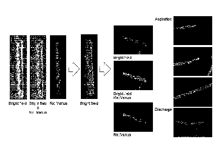
[Figure 1] Figure 1 is bright field stereo microscope images,
fluorescence stereo microscope images and bright field + fluorescence
stereo microscope images in which the process of encapsulating a
plurality of (8 to 13) transplant neural retinas (Caps) in fibrin gel
(complex) and aspiration and discharge using Surflo were studied in
Example 1.
[Figure 2] Figure 2 is bright field stereo microscope images,
fluorescence stereo microscope images and bright field + fluorescence
stereo microscope images in which the process of encapsulating a
plurality of (8 to 10) transplant neural retinas (Caps) in gelatin
(complex) and aspiration and discharge using a 1 ml syringe with a
catheter tip were studied in Example 2.
[Figure 3] Figure 3 is fluorescence microscope images showing results
7
Date Recue/Date Received 2023-03-07
CA 03194226 2023-03-07
of perfoiming immunostaining on cell aggregates containing a
transplant neural retina with Crx and Chx10 in Reference Example 1.
[Figure 4] Figure 4 is fluorescence microscope images showing results
of performing immunostaining on cell aggregates containing a
transplant neural retina with Rx and Recoverin in Reference Example 1.
[Figure 5] Figure 5 is a conceptual view of preparing a Cap and a Ring
from a typical cell aggregate.
[Figure 6] Figure 6 is a conceptual view of preparing a Cap and a Ring
from cell aggregates having various shapes. Portions indicated in
black color and gray color mean non-target tissue.
[Figure 7] Figure 7 shows images of typical grafts and a schematic view
of a graft as well as the heights, major axes and minor axes of grafts in
Reference Example 3.
[Figure 8] Figure 8 is confocal fluorescence microscope images
showing results of perfoiming immunostaining on grafts with Crx,
Chx10, Rx and Recoverin in Reference Example 4.
[Figure 9A] Figure 9A shows results of analyzing gene expression for
RNA extracted from a Cap and a Ring by quantitative PCR in Reference
Example 5.
[Figure 9B] Figure 9B shows results of analyzing gene expression for
RNA extracted from a Cap and a Ring by quantitative PCR in Reference
Example 5.
[Figure 10] Figure 10 is images showing results of analyzing RNA
extracted from a Ring by quantitative PCR, then subretinally
transplanting a graft (cap) to a rat, and observing an image of
post-transplant engraftment under a fluorescence microscope in
8
Date Recue/Date Received 2023-03-07
CA 03194226 2023-03-07
Reference Example 6.
[Figure 11] Figure 11 is fluorescence microscope images showing
results of performing immunostaining on a Cap and a Ring prepared
from one cell aggregate in Reference Example 7.
[Figure 12] Figure 12 is fluorescence microscope images showing
results of performing immunostaining on a cap prepared from one cell
aggregate in Reference Example 8.
Description of Embodiments
[0014] [Definition]
The "stem cells" refer to undifferentiated cells haying
differentiation potency and proliferation potency (particularly,
self-renewal ability). In the stem cells, subgroups of pluripotent stem
cells, multipotent stem cells and unipotent stem cells, are included
according to the differentiation potency. The pluripotent stem cells
refer to stem cells that can be cultured in vitro and has an ability
(pluripotency) to be able to differentiate into three germ layers
(ectoderm, mesoderm, endodenn) and/or all cell lineages belonging to
the extraembryonic tissue. The multipotent stem cells refer to stem
cells haying an ability to differentiate into a plurality of tissues or cells,
although the definition is not applied to all of them. The unipotent
stem cells refer to stem cells haying an ability to be able to differentiate
into a predetermined tissue or cells.
[0015] The "pluripotent stem cells" can be induced from, e.g., a
fertilized egg, a cloned embryo, germline stem cells, tissue stem cells
and somatic cells. Examples of the pluripotent stem cells can include
9
Date Recue/Date Received 2023-03-07
CA 03194226 2023-03-07
embryonic stem cells (ES cells), embryonic germ cells (EG cells) and
induced pluripotent stem cells (iPS cells). Muse cells (Multi-lineage
differentiating stress enduring cells) obtained from the mesenchymal
stem cells (MSC) and GS cells prepared from germ cells (for example,
testis) are included in the pluripotent stem cells.
[0016] Human embryonic stem cells were established in 1998 and have
been used also for regenerative medicine. The embryonic stem cells
can be produced by culturing inner cell aggregate on feeder cells or a
culture medium containing bFGF. The method for producing
embryonic stem cells is described, for example, in W096/22362,
W002/101057, U55,843,780, U56,200,806, U56,280,718. The
embryonic stem cells are available from a predeteintined institution and
also, commercially available. For example, human embryonic stem
cells such as KhES-1, KhES-2 and KhES-3 are available from the
Institute for Frontier Life and Medical Sciences, Kyoto University.
Human embryonic stem cells such as human ES cells genetically
engineered so as to have Rx::Venus, Rx::AcGFP and Crx::Venus
reporter genes (derived from KhES-1, Non Patent Literature 1) are
available from RIKEN.
[0017] The "induced pluripotent stem cells" refers to cells having
pluripotency, which is induced by reprogramming somatic cells by a
method known in the art.
[0018] The induced pluripotent stem cells were established in mouse
cells by Yamanaka et al., in 2006 (Cell, 2006, 126 (4), pp. 663-676).
The induced pluripotent stem cells were also established in human
fibroblasts in 2007. The
induced pluripotent stem cells have
Date Recue/Date Received 2023-03-07
CA 03194226 2023-03-07
pluripotency and self-renewal ability similarly to embryonic stem cells
(Cell, 2007, 131 (5), pp. 861-872; Science, 2007, 318 (5858), pp.
1917-1920; Nat. Biotechnol., 2008, 26(1), pp. 101-106).
[0019] The induced pluripotent stem cells more specifically refer to
cells which are induced to be pluripotent by reprogramming somatic
cells differentiated into, for example, fibroblasts and peripheral blood
mononuclear cells, by allowing any one of sets of a plurality of genes
selected from a reprogramming gene group containing 0ct3/4, 5ox2,
Klf4, Myc (c-Myc, N-Myc, L-Myc), Glis 1, Nanog, 5a114, 1in28 and
Esrrb to express. Examples of a preferable set of reprogramming
factors may include (1) 0ct3/4, 5ox2, Klf4, and Myc (c-Myc or L-Myc)
and (2) 0ct3/4, 5ox2, Klf4, Lin28 and L-Myc (Stem Cells, 2013; 31:
458-466).
[0020] Other than producing induced pluripotent stem cells through
direct reprogramming by gene expression, the pluripotent stem cells can
be artificially induced from somatic cells, for example, by adding a
chemical compound (Science, 2013, 341, pp. 651-654).
[0021] Alternatively, an induced pluripotent stem cell strain is available.
For example, human induced pluripotent cell strains established by
Kyoto University, such as 201B7 cell, 201B7-Ff cell, 253G1 cell,
253G4 cell, 1201C1 cell, 1205D1 cell, 1210B2 cell and 1231A3 cell,
are available form Kyoto University and iPS Academia Japan, Inc. As
the induced pluripotent stem cells, for example, Ff-I01 cell, Ff-I14 cell
and QHJI01s04 cell established by Kyoto University, are available from
Kyoto University.
[0022] In the specification, the pluripotent stem cells are preferably
11
Date Recue/Date Received 2023-03-07
CA 03194226 2023-03-07
embryonic stem cells or induced pluripotent stem cells, more preferably
induced pluripotent stem cells.
[0023] In the specification, the pluripotent stem cells are human
pluripotent stem cells, preferably human induced pluripotent stem cells
(iPS cells) or human embryonic stem cells (ES cells) (e.g., embryonic
stem cell established from the embryo within 14 days after fertilization).
[0024] Pluripotent stem cells such as human iPS cells can be subjected
to maintenance culture and expansion culture performed by methods
known to those skilled in the art.
[0025] The "retinal tissue" means a tissue in which a single type or a
plurality of types of retinal cells constituting each retinal layer in a
retina in vivo are present according to a predetermined order. The
"neural retina" is a retinal tissue and means a tissue containing an inside
neural retinal layer that does not contain a retinal pigment epithelial
layer among retinal layers mentioned later.
[0026] The "retinal cells" mean cells constituting each retinal layer in a
retina in vivo or progenitor cells thereof. In the retinal cells, cells such
as photoreceptor cells (rod photoreceptor cell, cone photoreceptor cell),
horizontal cells, amacrine cells, intermediate neuronal cells, retinal
ganglion cells (ganglion cell), bipolar cells (rod bipolar cell, cone
bipolar cell), Muller glial cells, retinal pigment epithelial (RPE) cells,
ciliary body, their progenitor cells (e.g., photoreceptor progenitor cell,
bipolar progenitor cell), and retinal progenitor cells are included, though
not limited thereto. Among the retinal cells, examples of cells
constituting a neural retinal layer (also referred to as neural retina cells
or neural retina-related cells) specifically include cells such as
12
Date Recue/Date Received 2023-03-07
CA 03194226 2023-03-07
photoreceptor cells (rod photoreceptor cell, cone photoreceptor cell),
horizontal cells, amacrine cells, intermediate neuronal cells, retinal
ganglion cells (ganglion cell), bipolar cells (rod bipolar cell, cone
bipolar cell), Muller glial cells, and their progenitor cells (e.g.,
photoreceptor progenitor cell, bipolar progenitor cell). In other words,
in the neural retina-related cells, neither retinal pigment epithelial cells
nor ciliary body cells are included.
[0027] The "matured retinal cells" mean cells that may be contained in
the retinal tissue of a human adult, and specifically mean differentiated
cells such as photoreceptor cells (rod photoreceptor cell, cone
photoreceptor cell), horizontal cells, amacrine cells, intermediate
neuronal cells, retinal ganglion cells (ganglion cell), bipolar cells (rod
bipolar cell, cone bipolar cell), Muller glial cells, retinal pigment
epithelial (RPE) cells, and ciliary body cells. The "immature retinal
cells" mean progenitor cells (e.g., photoreceptor progenitor cell, bipolar
progenitor cell, retinal progenitor cell) destined for differentiation into
matured retinal cells.
[0028] The photoreceptor progenitor cells, the horizontal progenitor
cells, the bipolar progenitor cells, the amacrine progenitor cells, the
retinal ganglion progenitor cells, the Muller glial progenitor cells, and
the retinal pigment epithelial progenitor cells refer to progenitor cells
destined for differentiation into photoreceptor cells, horizontal cells,
bipolar cells, amacrine cells, retinal ganglion cells, Muller glial cells,
and retinal pigment epithelial cells, respectively.
[0029] The "retinal progenitor cells" are progenitor cells capable of
differentiating into any one of the immature retinal cells such as
13
Date Recue/Date Received 2023-03-07
CA 03194226 2023-03-07
photoreceptor progenitor cells, horizontal progenitor cells, bipolar
progenitor cells, amacrine progenitor cells, retinal ganglion progenitor
cells, Muller glial cells, and retinal pigment epithelial progenitor cells,
and refer to progenitor cells also capable of eventually differentiating
into any one of the matured retinal cells such as photoreceptor cells, rod
photoreceptor cells, cone photoreceptor cells, horizontal cells, bipolar
cells, amacrine cells, retinal ganglion cells, and retinal pigment
epithelial cells.
[0030] The "photoreceptor cells" are present in the photoreceptor layer
of a retina in vivo and plays a role in absorbing light stimuli and
converting them to electrical signals. The photoreceptor cells have
two types, cones which function in the light and rods which function in
the dark (referred to as cone photoreceptor cells and rod photoreceptor
cells, respectively). Examples of the cone photoreceptor cells can
include S cone photoreceptor cells which express S-opsin and receive
blue light, L cone photoreceptor cells which express L-opsin and receive
red light, and M cone photoreceptor cells which express M-opsin and
receive green light. The photoreceptor cells are matured after
differentiation from photoreceptor progenitor cells. Whether or not
cells are photoreceptor cells or photoreceptor progenitor cells can be
readily confirmed by those skilled in the art, for example, through the
expression of cell markers (Crx and Blimpl expressed in photoreceptor
progenitor cells, recoverin expressed in photoreceptor cells, rhodopsin,
S-opsin and MIL-opsin expressed in mature photoreceptor cells, etc.)
mentioned later or the formation of an outer segment structure. In an
embodiment, the photoreceptor progenitor cells are Crx-positive cells,
14
Date Recue/Date Received 2023-03-07
CA 03194226 2023-03-07
and the photoreceptor cells are rhodopsin-, S-opsin- and
MIL-opsin-positive cells. In an embodiment, the rod photoreceptor
cells are NRL- and rhodopsin-positive cells. In an embodiment, the S
cone photoreceptor cells are S-opsin-positive cells, the L cone
photoreceptor cells are L-opsin-positive cells, and the M cone
photoreceptor cells are M-opsin-positive cells. Specifically, in the
specification, the photoreceptor cells conceptually include
photoreceptor progenitor cells and matured photoreceptor cells.
[0031] The neural retina contains preferably 10% or more, more
preferably 20% or more, of a photoreceptor cell or a photoreceptor
progenitor cell.
[0032] The neural retina contains preferably 10% or more, more
preferably 20% or more, of a neural retinal progenitor cell. Also, the
neural retina contains preferably 10% or more of a photoreceptor
progenitor cell. In an embodiment, the neural retina contains 3% or
more of a matured photoreceptor cell.
[0033] The presence of neural retina-related cells can be confirmed
from the presence or absence of expression of a neural retina-related
cell-related gene (hereinafter, also referred to as "neural retina-related
cell marker" or "neural retina marker"). The presence or absence of
expression of the neural retina-related cell marker, or the ratio of neural
retina-related cell marker-positive cells in a cell population or a tissue
can be readily confirmed by those skilled in the art. Examples thereof
include an approach using an antibody, an approach using nucleic acid
primers, and an approach using sequencing reaction. As the approach
using an antibody, the expression of a protein of the neural retina-related
Date Recue/Date Received 2023-03-07
CA 03194226 2023-03-07
cell marker can be confirmed, for example, by dividing the number of
predetermined neural retina-related cell marker-positive cells by the
total number of cells in accordance with an approach such as flow
cytometry or immunostaining using a commercially available antibody.
As the approach using nucleic acid primers, the expression of RNA of
the neural retina-related cell marker can be confirmed by, for example,
PCR, semiquantitative PCR, or quantitative PCR (e.g., real-time PCR).
As the approach using sequencing reaction, the expression of RNA of
the neural retina-related cell marker can be confirmed using, for
example, a nucleic acid sequencer (e.g., next-generation sequencer).
[0034] Examples of the neural retina-related cell marker include Rx
(also referred to as Rax) and PAX6 expressed in retinal progenitor cells,
Rx, PAX6 and Chx10 (also referred to as Vsx2) expressed in neural
retinal progenitor cells, and Crx and Blimpl expressed in photoreceptor
progenitor cells. Examples thereof also include Chx10 strongly
expressed in bipolar cells, PKCa, Goa, VSX1 and L7 expressed in
bipolar cells, Tun and Brn3 expressed in retinal ganglion cells,
calretinin and HPC-1 expressed in amacrine cells, calbindin expressed
in horizontal cells, recoverin expressed in photoreceptor cells and
photoreceptor progenitor cells, rhodopsin expressed in rod cells, Nrl
expressed in rod photoreceptor cells and rod photoreceptor progenitor
cells, S-opsin and LM-opsin expressed in cone photoreceptor cells,
RXR-y expressed in cone cells, cone photoreceptor progenitor cells and
ganglion cells, TR132, OTX2 and 0C2 expressed in cone photoreceptor
cells that appear at the early phase of differentiation among cone
photoreceptor cells, or progenitor cells thereof, and Pax6 commonly
16
Date Recue/Date Received 2023-03-07
CA 03194226 2023-03-07
expressed in horizontal cells, amacrine cells and ganglion cells.
[0035] The "positive cells" mean cells expressing a predetermined
marker on the cell surfaces or within the cells. For example, the
"Chx10-positive cells" mean cells expressing Chx10 protein.
[0036] The "retinal pigment epithelial cells" mean epithelial cells
present outside the neural retina in a retina in vivo. Whether or not
cells are retinal pigment epithelial cells can be readily confirmed by
those skilled in the art, for example, through the expression of cell
markers (RPE65, MITF, CRALBP, MERTK, BEST1, TTR, etc.), the
presence of melanin granules (brown-black), intercellular tight junctions,
or polygonal/flagstone-like characteristic cell morphology. Whether or
not cells have a function of retinal pigment epithelial cells can be
readily confirmed from the ability to secrete cytokines such as VEGF
and PEDF. In an embodiment, the retinal pigment epithelial cells are
RPE65-positive cells, MITF-positive cells, or RPE65-positive and
MITF-positive cells.
[0037] The "retinal layer" means individual layers constituting the
retina, and examples thereof can specifically include retinal pigment
epithelial layer, photoreceptor layer, outer limiting membrane, outer
nuclear layer, outer plexiform layer, inner nuclear layer, inner plexiform
layer, ganglion cell layer, nerve fiber layer and inner limiting
membrane.
[0038] The "neural retinal layer" means individual layers constituting
the neural retina, and examples thereof can specifically include
photoreceptor layer, outer limiting membrane, outer nuclear layer, outer
plexiform layer, inner nuclear layer, inner plexiform layer, ganglion cell
17
Date Recue/Date Received 2023-03-07
CA 03194226 2023-03-07
layer, nerve fiber layer and inner limiting membrane. The
"photoreceptor layer" means a retinal layer that is formed in the
outermost of the neural retina and is rich in one or more cells selected
from the group consisting of a photoreceptor cell (rod photoreceptor cell,
cone photoreceptor cell), a photoreceptor progenitor cell and a retinal
progenitor cell. Each layer other than the photoreceptor layer is
referred to as an inner layer. Which retinal layer the individual cells
constitute can be confirmed by a known method, for example, by
determining the presence or absence of expression or expression level of
a cell marker.
[0039] In the case of retinal tissue at a stage where the appearance ratio
of photoreceptor cells or photoreceptor progenitor cells is low, a layer
containing proliferating neural retinal progenitor cells is referred to as
"neuroblastic layer" and includes inner neuroblastic layer and outer
neuroblastic layer. Those skilled in the art can make a judgment from
the shade of color (the outer neuroblastic layer is light, and the inner
neuroblastic layer is dark) by a known method, for example, under a
bright field microscope.
[0040] The "ciliary body" includes "ciliary body" and "ciliary marginal
zone" in the process of development and of an adult. Examples of a
marker of the "ciliary body" include Zic 1 , MAL, HNF lbeta, FoxQ 1,
CLDN2, CLDN1, GPR177, AQP1 and AQP4. Examples of the
"ciliary marginal zone (CMZ)" can include a tissue that is present in a
boundary region between the neural retina and the retinal pigment
epithelium in a retina in vivo, and is a region containing tissue stem cells
of the retina (retinal stem cells). The ciliary marginal zone is also
18
Date Recue/Date Received 2023-03-07
CA 03194226 2023-03-07
called ciliary margin or retinal margin, and the ciliary marginal zone, the
ciliary margin and the retinal margin are equivalent tissues. The ciliary
marginal zone is known to play an important role in the supply of retinal
progenitor cells or differentiated cells to retinal tissue, the maintenance
of a retinal tissue structure, etc. Examples of a marker gene of the
ciliary marginal zone can include Rdh10 gene (positive), Otx/ gene
(positive) and Zic/ (positive). The
"ciliary marginal zone-like
structure" is a structure similar to the ciliary marginal zone.
[0041] The "cell aggregate" is not particularly limited as long as a
plurality of cells mutually adhere to form a three-dimensional structure,
and refers to, for example, a mass formed by the aggregation of cells
dispersed in a vehicle such as a culture medium, or a mass of cells
formed through cell division. In the cell aggregate, the case of forming
a predetermined tissue is also included.
[0042] The "sphere-like cell aggregate" means a cell aggregate having a
stereoscopic shape close to a spherical shape. The stereoscopic shape
close to a spherical shape is a shape having a three-dimensional
structure, and examples thereof include a spherical shape that exhibits a
circle or an ellipse when projected onto a two-dimensional surface, and
a shape formed by fusing a plurality of spherical shapes (e.g., which
exhibits a shape formed by 2 to 4 circles or ellipses overlapping when
two-dimensionally projected). In an embodiment, the core part of the
aggregate has a vesicular lamellar structure and is characterized in that
the central part is observed to be dark and the outer edge portion is
observed to be bright under a bright field microscope.
[0043] The "epithelial tissue" is a tissue formed by covering the body
19
Date Recue/Date Received 2023-03-07
CA 03194226 2023-03-07
surface or the surface of a lumen (digestive tract, etc.), body cavity
(pericardial cavity, etc.) or the like with cells without any space. The
cells forming the epithelial tissue are referred to as epithelial cells.
The epithelial cells have a polarity in the apical-basal direction. The
epithelial cells can mutually and firmly join via adherence junction
and/or tight junction to form a layer of the cells. A tissue formed from
a single layer or dozen layers overlapping of this layer of the cells is the
epithelial tissue. In a tissue capable of forming the epithelial tissue,
retinal tissue, brain and spinal cord tissue, eyeball tissue, neural tissue or
the like of a fetal stage and/or an adult is also included. In the
specification, the neural retina is also the epithelial tissue. The
"epithelial structure" means a structure characteristic of the epithelial
tissue, such as apical surface or basal membrane.
[0044] In the specification, an epithelial structure containing neural
tissue is referred to as neural epithelium. Particularly, an epithelial
structure containing a neural retina is referred to as neural retinal
epithelium.
[0045] The "continuous epithelial tissue" is a tissue having a continuous
epithelium structure. The continuous epithelium structure is a
structure where the epithelial tissue is continuously formed. The
epithelium tissue continuously formed is a state in which 10 cells to 107
cells, for example, in the tangent direction of the epithelial tissue,
preferably 30 cells to 107 cells, further preferably 102 cells to 107 cells,
in the tangent direction, are aligned. The continuous epithelial
structure does not have a structure where an apical surface is divided, as
found in a rosette-like structure. In an embodiment, the number of
Date Recue/Date Received 2023-03-07
CA 03194226 2023-03-07
cells per area of the cross section of retinal tissue having the continuous
epithelial structure, for example, the number of cells per area of the
cross section of retinal tissue having the continuous epithelial structure
in an embodiment of cells in a frozen section having a thickness on the
order of 10 iiim, is 10 cells to 900 cells, preferably 30 cells to 300 cells,
more preferably 50 cells to 250 cells, still more preferably 75 cells to
160 cells, per 100 um2, for example, in the case of evaluating the
number of nuclei of cells in the frozen section having a thickness on the
order of 10 iiim.
[0046] For example, in the continuous epithelium formed in retinal
tissue, the retinal tissue has an apical surface intrinsic to the epithelial
tissue. The apical surface is formed almost in parallel to, for example,
at least photoreceptor layer (outer nuclear layer) among the layers
forming a neural retinal layer and continuously on the surface of the
retinal tissue. For example, in the case of a cell aggregate containing
retinal tissue prepared from pluripotent stem cells, the apical surface is
formed on the surface of the aggregate, and continuous neural
epithelium is formed in which 10 cells or more, preferably 30 cells or
more, more preferably 100 cells or more, further preferably 400 cells or
more of photoreceptor cells or photoreceptor progenitor cells are
regularly and continuously arranged in a row in the tangent direction of
the surface. A neural retina containing such continuous neural
epithelium is neural retinal epithelium containing continuous
epithelium.
[0047] In an embodiment, epithelial tissue is polarized so that "apical
surface" and "basal membrane" are formed. The "basal membrane"
21
Date Recue/Date Received 2023-03-07
CA 03194226 2023-03-07
refers to a basal side layer (basal membrane) produced by epithelial
cells, is rich in laminin and IV-type collagen, and has a thickness of 50
to 100 nm. The "apical surface" refers to the surface (upper surface
layer) fonned on the opposite side to the "basal membrane". In an
embodiment, in the retinal tissue developed to the extent that
photoreceptor cells or photoreceptor progenitor cells are observed, the
"apical surface" refers to a surface in contact with photoreceptor layer
(outer nuclear layer) in which outer limiting membrane is formed and
photoreceptor cells and photoreceptor progenitor cells are present.
Such an apical surface can be identified by, for example,
immunostaining (known to those skilled in the art) using an antibody
against an apical surface marker (e.g., atypical PKC (hereinafter,
abbreviated to "aPKC"), E-cadherin, N-cadherin).
[0048] Whether retinal tissue contains continuous epithelium can be
confirmed from the continuity (i.e., undivided foul') of the apical
surface of retinal tissue. The continuity of the apical surface can be
determined, for example, by immunostaining a marker of the apical
surface (e.g., aPKC, E-cadherin, N-cadherin) and a marker of
photoreceptor cells or photoreceptor progenitor cells positioned on the
apical surface side (e.g., Crx or recoverin), and analyzing obtained
images, etc. for the positional relationship of the apical surface to a
photoreceptor layer and each retinal layer. A retinal layer other than
the apical surface or the photoreceptor layer (outer nuclear layer) can be
identified by, for example, DAPI staining which stains the nuclei of
cells, PI staining, Hoechst staining, or immunostaining with a marker
protein (Rx, Chx10, Ki67, Crx, etc.) or the like localized in the nuclei of
22
Date Recue/Date Received 2023-03-07
CA 03194226 2023-03-07
cells.
[0049] Specifically, the continuity of the apical surface can be identified
from the continuous presence of cells co-expressing a marker of cells
present on the apical surface side, i.e., a photoreceptor cell marker or a
photoreceptor progenitor cell marker, and a marker capable of staining
the nuclei of cells.
[0050] [Complex]
The complex of the present invention comprises: two or more
cell aggregates each containing a neural retina derived from a
pluripotent stem cell; and a matrix, the two or more cell aggregates
being arranged in the matrix. Hereinafter, the neural retina-containing
cell aggregates, the matrix, and the complex comprising them will be
described in detail.
[0051] (1) Neural retina-containing cell aggregate
In an embodiment, the neural retina-containing cell aggregates
are cell aggregates mentioned later (sphere-like cell aggregates). In an
embodiment, the neural retina-containing cell aggregates are
sheet-shaped retinal tissues dissected from sphere-like cell aggregates.
The sheet-shaped retinal tissues are preferably sheet-shaped neural
retinas containing continuous neural retinal epithelium (hereinafter, also
referred to as neural retina sheets). In an embodiment, the neural
retina-containing cell aggregates are transplant neural retinas, preferably
transplant neural retina sheets. The transplant neural retinas or the
transplant neural retina sheets are human neural retinas suitable for
transplantation in humans and more preferably consist of only the neural
retinas.
23
Date Recue/Date Received 2023-03-07
CA 03194226 2023-03-07
[0052] In the neural retina, a neural retinal layer containing at least a
photoreceptor layer is formed, and the photoreceptor layer contains at
least one or more cells selected from the group consisting of a
photoreceptor cell, a photoreceptor progenitor cell and a retinal
progenitor cell. The photoreceptor layer is formed at least in the
outmost of the cell aggregate. Also,
photoreceptor cells or
photoreceptor progenitor cells may be present inside the cell aggregate.
Alternatively, the photoreceptor layer may be formed in the inside.
Photoreceptor cells, etc. are present continuously, i.e., by mutual
adhesion, in the tangent direction of the surface of the cell aggregate.
The photoreceptor cells, etc. are present continuously in the tangent
direction of the surface of the cell aggregate, thereby forming a
photoreceptor layer containing the photoreceptor cells, etc. The
tangent direction refers to a direction tangent to the surface of the cell
aggregate, i.e., a direction along which the photoreceptor cells, etc. in
the photoreceptor layer are arranged, and is the direction in parallel to
the neural retina or the lateral direction.
[0053] The neural retina is derived from a pluripotent stem cell, and it
is preferable to be derived from a human pluripotent stem cell. The
human pluripotent stem cell is preferably a human embryonic stem cell
or a human induced pluripotent stem cell, more preferably a human
induced pluripotent stem cell. Also, it is preferable that the neural
retina should be a transplant neural retina.
[0054] In the specification, one embodiment of the retinal tissue
includes retinal tissue containing a surface (apical surface) and a back
surface (basal surface), wherein the surface constitutes an apical surface
24
Date Recue/Date Received 2023-03-07
CA 03194226 2023-03-07
containing a neural retinal layer which is epithelial tissue by forming the
adherence junction between cells, and the back surface constitutes a
basal surface adjacent to the inner layer of the neural retina. Such
retinal tissue can be referred to as neural retinal epithelium. For such
retinal tissue, it is preferable to be a neural retina sheet which is a
sheet-shaped retinal tissue. The surface has a smooth shape with less
change in curvature, and the back surface has an irregular shape with
large change in curvature. In an embodiment, the change in the
curvature of the surface of the retinal tissue may be, for example, close
to change in curvature of an ellipse (e.g., a major axis of 1 to 10 with
respect to a minor axis of 1) (also referred to as continuous change in
curvature). In an embodiment, the change in the curvature of the back
surface of the retinal tissue may be, for example, close to sharp change
in curvature that goes back and force between positive values and
negative values, as in "teeth of a saw" (also referred to as sharp change
in curvature).
[0055] (Method for producing cell aggregate)
Each cell aggregate containing a neural retina is derived from a
pluripotent stem cell and, specifically, can be obtained by differentiating
pluripotent stem cells. Examples of a differentiation method for a
neural retina include, but are not particularly limited to, methods
disclosed in W02011/055855, W02013/077425, W02015/025967,
W02016/063985, W02016/063986, W02017/183732, PLoS One. 2010
Jan 20; 5 (1): e8763, Stem Cells. 2011 Aug; 29 (8): 1206-18, Proc Natl
Acad Sci USA. 2014 Jun 10; 111 (23): 8518-23, and Nat Commun.
2014 Jun 10; 5: 4047.
Date Recue/Date Received 2023-03-07
CA 03194226 2023-03-07
[0056] Examples of a method for producing retinal tissue can include
methods described in Bryce T. McLelland et al., IOVS, May 2018, Vol.
59, No. 6, p. 2586. Other examples of the method for producing
retinal tissue include methods described in the following literatures.
Nakano, T. et al. Cell Stem Cell 10, 771-785 (2012).
Kawahara A. et al. Nature Communications, 6, p. 6286 (2015).
Kuwahara A, Yamasaki S, et al. Sci Rep. 2019 Dec 12; 9 (1): 18936.
Lamba, D.A., Gust, J. & Reh, T.A. Cell Stem Cell 4, 73-79, (2009).
Zhu, J., Cifuentes, H., Reynolds, J. & Lamba, D.A. Cell Stem Cell 20,
374-384. e375 (2017)
Meyer, J.S. et al. Stem Cells 29, 1206-1218, (2011).
Zhong, X. et al. Nat Commun 5, doi: 10.1038/ncomms5047 (2014).
Boucherie, C., et al. Stem Cells 31, 408-414, doi: 10.1002/stem.1268
(2013).
Gonzalez-Cordero, A. et al. Nat Biotechno131, 741-747, (2013).
Mellough, C.B. et al. Stem Cells 33, 2416-2430, (2015).
Hallam, D. et al. Stem Cells, doi: 10.1002/stem.2883 (2018).
Reichman, S. et al. PNAS 111, 8518-8523, (2014).
Gagliardi, G. et al. Stem Cell Reports 11, 665-680, (2018).
Tucker, B.A., et al. Stem Cells Transl Med2, 16-24, (2013).
Wahlin, K.J. et al. Sci Rep 7, 766, (2017).
DiStefano, T. et al. Stem Cell Reports 10, 300-313, (2018).
[0057] In a specific embodiment, the cell aggregate containing a neural
retina can be prepared by a method comprising the following steps (A),
(B), (C) and (D):
(A) culturing pluripotent stem cells in a culture medium for pluripotent
26
Date Recue/Date Received 2023-03-07
CA 03194226 2023-03-07
stem cell culture in the absence of feeder cells;
(B) forming a cell aggregate by suspension-culturing the cells obtained
in the step (A);
(C) further suspension-culturing the cell aggregate obtained in the step
(B) in a culture medium containing a BMP signaling pathway agonist;
and
(D) suspension-culturing and maturing the cell aggregate obtained in the
step (C).
The step (A) may further involve a TGF13 family signaling
pathway inhibitor and/or a sonic hedgehog signaling pathway agonist.
Also, the step (B) may involve a sonic hedgehog signaling
pathway agonist and/or a Wnt signaling pathway inhibitor, as mentioned
later.
[0058] This method is also disclosed in, for example, W02015/025967,
W02016/063985, and W02017/183732. For more details,
see
W02015/025967, W02016/063985,
W02017/183732,
W02019/017492, W02019/054514, W02019/054515, etc.
[0059] The "culture medium for pluripotent stem cell culture" that is
used in the step (A) is a culture medium that allows pluripotent stem
cells to be cultured under feeder-free conditions. Examples of the
culture medium include culture media containing a factor for
maintaining undifferentiated state.
[0060] In the specification, the factor for maintaining undifferentiated
state is not particularly limited as long as it is a substance having an
action of suppressing the differentiation of pluripotent stem cells.
Examples of the factor for maintaining undifferentiated state that is
27
Date Recue/Date Received 2023-03-07
CA 03194226 2023-03-07
generally used by those skilled in the art can include FGF signaling
pathway agonists, TGF13 family signaling pathway agonists, and insulin.
Examples of the FGF signaling pathway agonist specifically include
fibroblast growth factors (e.g., bFGF, FGF4, FGF8). Examples of the
TGF13 family signaling pathway agonist include TGFI3 signaling
pathway agonists and Nodal/activin signaling pathway agonists.
Examples of the TGF13 signaling pathway agonist include TGF131 and
TGF132. Examples of the Nodal/activin signaling pathway agonist
include Nodal, activin A, and activin B. In the case of culturing human
pluripotent stem cells (human ES cells, human iPS cells), the culture
medium in the step (A) preferably contains bFGF as the factor for
maintaining undifferentiated state.
[0061] The concentration of the factor for maintaining undifferentiated
state in the culture medium that is used in the step (A) is a concentration
capable of maintaining the undifferentiated state of the pluripotent stem
cells to be cultured, and can be appropriately set by those skilled in the
art. For example, specifically, in the case of using bFGF as the factor
for maintaining undifferentiated state in the absence of feeder cells, its
concentration is usually on the order of 4 ng to 500 ng/mL, preferably
on the order of 10 ng to 200 ng/mL, more preferably on the order of 30
ng to 150 ng/mL.
[0062] Many synthetic media have been developed or are commercially
available as culture media for pluripotent stem cell culture applicable
under feeder-free conditions. Examples thereof include Essential 8
medium (manufactured by Life Technologies Corp.). The Essential 8
medium contains L-ascorbic acid-2-phosphate magnesium (64 mg/L),
28
Date Recue/Date Received 2023-03-07
CA 03194226 2023-03-07
sodium selenium (14 jig/L), insulin (19.4 mg/L), NaHCO3 (543 mg/L),
transferrin (10.7 mg/L), bFGF (100 ng/mL), and the TGF13 family
signaling pathway agonist (TGF131 (2 ng/mL) or Nodal (100 ng/mL)) as
additives in DMEM/F12 medium (Nature Methods, 8, 424-429 (2011)).
Examples of other commercially available feeder-free media include
S-medium (manufactured by DS Pharma Biomedical Co., Ltd.),
StemPro (manufactured by Life Technologies Corp.), hESF9 (Proc. Natl.
Acad. Sci. USA. 2008 Sep 9; 105 (36): 13409-14), mTeSR1
(manufactured by STEMCELL Technologies Inc.), mTeSR2
(manufactured by STEMCELL Technologies Inc.), TeSR-E8
(manufactured by STEMCELL Technologies Inc.), and StemFit
(manufactured by Ajinomoto Co., Inc.). In the step (A), the present
invention can be conveniently carried out by using these. By using
these culture media, it is possible to perform the culture of pluripotent
stem cells under feeder-free conditions. The culture medium that is
used in the step (A) is, as one example, a serum-free medium that is not
supplemented with any of the BMP signaling pathway agonist, the Wnt
signaling pathway agonist and the Wnt signaling pathway inhibitor.
[0063] The culture medium that is used in the preparation of the cell
aggregate containing a neural retina, i.e., the culture medium that is used
in the steps (B), (C) and (D), can employ a basal medium for cell
proliferation (also referred to as a basal medium), unless otherwise
specified. The basal medium for cell proliferation is not particularly
limited as long as the culture of cells is possible. A basal medium
commercially available as a culture medium for cell proliferation can be
appropriately used. Specifically, examples thereof can include culture
29
Date Recue/Date Received 2023-03-07
CA 03194226 2023-03-07
media that can be used in the culture of animal cells, such as BME
medium, BGJb medium, CMRL 1066 medium, Glasgow
MEM(GMEM) medium, Improved MEM Zinc Option medium, IMDM
medium, Medium 199 medium, MEM medium, Eagle MEM medium,
aMEM medium, DMEM medium, F-12 medium, DMEM/F12 medium,
IMDM/F12 medium, Ham's medium, RPMI 1640 medium, Fischer's
medium, Leibovitz's L-15 medium and mixtures of these media.
Alternatively, a culture medium supplemented with N2 medium which
is an assisted culture medium may be used.
[0064] In the specification, the TGF13 family signaling pathway
inhibitor refers to a substance inhibiting the TGF13 family signaling
pathway, i.e., the signaling pathway transduced by the Smad family.
Specifically, examples thereof can include TGF13 signaling pathway
inhibitors (e.g., 5B431542, LY-364947, 5B505124, A-83-01),
Nodal/activin signaling pathway inhibitors (e.g., SB431542, A-83-01)
and BMP signaling pathway inhibitors (e.g., LDN193189,
dorsomorphin). These substances are commercially available and can
be obtained.
[0065] In the specification, the sonic hedgehog (hereinafter, also
referred to as "Shh") signaling pathway agonist is a substance capable of
enhancing signal transduction mediated by Shh. Examples of the Shh
signaling pathway agonist include SHH, partial peptides of SHH (e.g.,
sonic hedgehog N-terminus (Shh-N), recombinant human sonic
hedgehog (C24I1) N-terminus (SHH-C2411), recombinant mouse sonic
hedgehog (C25I1) N-terminus (SHH-C2511)), hedgehog family proteins
other than Shh (e.g., Hh, IHH, DHH, EHH, TwHH), PMA
Date Recue/Date Received 2023-03-07
CA 03194226 2023-03-07
(purmorphamine), and SAG (smoothened agonist).
[0066] In the step (A), the concentrations of the TGF13 family signaling
pathway inhibitor and the sonic hedgehog signaling pathway agonist
can be concentrations capable of inducting differentiation into retinal
cells. For example, SB431542 is used at a concentration of usually 0.1
to 200 M, preferably 2 to 50 M. A-83-01 is used at a concentration
of usually 0.05 to 50 M, preferably 0.5 to 5 M. LDN193189 is used
at a concentration of usually 1 to 2000 nM, preferably 10 to 300 nM.
SAG is used at a concentration of usually 1 to 2000 nM, preferably 10
to 700 nM. PMA is used at a concentration of usually 0.002 to 20 M,
preferably 0.02 to 2 M.
[0067] In the culture of pluripotent stem cells under feeder-free
conditions in the step (A), a suitable matrix may be used as a scaffold in
order to provide a scaffold as a replacement for feeder cells to the
pluripotent stem cells. Examples of the matrix that can be used as a
scaffold include laminin (Nat Biotechnol 28, 611-615, (2010)), laminin
fragments (Nat Commun 3, 1236, (2012)), basal membrane preparations
(Nat Biotechnol 19, 971-974, (2001)), gelatin, collagen, heparan sulfate
proteoglycan, entactin, and vitronectin.
[0068] The culture time of the pluripotent stem cells in the step (A) is
not particularly limited within a range in which an effect of improving
the quality of the cell aggregate to be formed in the step (B) can be
achieved in the case of culture in the presence of the TGF13 family
signaling pathway inhibitor and/or the sonic hedgehog signaling
pathway agonist (e.g., from 100 nM to 700 nM), and is usually from 0.5
to 144 hours. In an embodiment, it is preferably from 2 to 96 hours,
31
Date Recue/Date Received 2023-03-07
CA 03194226 2023-03-07
more preferably from 6 to 48 hours, further preferably from 12 to 48
hours, still further preferably from 18 to 28 hours (e.g., 24 hours).
[0069] The culture medium that is used in the step (B) may be a
serum-containing medium or a serum-free medium. A serum-free
medium is suitably used from the viewpoint of circumventing
contamination with chemically undetermined components. In order to
circumvent the complication of preparation, examples thereof include
serum-free media supplemented with an appropriate amount of a serum
replacement such as commercially available KSR. The amount of
KSR added to the serum-free medium is usually from about 1% to about
30%, preferably from about 2% to about 20%.
[0070] For the formation of the aggregate, first, dispersed cells are
prepared by the dispersion operation of the cells obtained in the step (A).
The "dispersed cells" obtained by dispersion operation include a state in
which 70% (preferably 80% or more) or more are single cells and 30%
or less (preferably 20% or less) of 2- to 50-cell masses are present.
The dispersed cells include a state in which the mutual adhesion (e.g.,
surface adhesion) of cells has been mostly lost.
[0071] A suspension of the dispersed cells is seeded into an incubator,
and the dispersed cells are cultured under conditions of non-adhesive to
the incubator, thereby causing the aggregation of a plurality of cells to
form an aggregate. In an embodiment, when a predetermined number
of dispersed stem cells is placed in each well of a multi-well plate
(U-bottom, V-bottom) such as a 96-well plate and this is statically
cultured, the cells aggregate rapidly, thereby forming one aggregate in
each well (SFEBq). In the case of suspension-culturing cells using a
32
Date Recue/Date Received 2023-03-07
CA 03194226 2023-03-07
96-well plate, a liquid prepared so as to attain about 1 x 10³ to about 1 x
105 cells (preferably about 3 x 10³ to about 5 x 10⁴ cells or about 4 x 10³
to about 2 x 10⁴ cells) per well is added to the wells, and the plate is left
standing to form aggregates.
[0072] In an embodiment, the culture medium that is used in the step
(B) contains a sonic hedgehog signaling pathway agonist.
In other words, in a specific embodiment, the cell aggregate
containing a neural retina can be prepared by a method comprising the
following steps (A), (B) and (C):
(A) culturing pluripotent stem cells in a culture medium containing a
factor for maintaining undifferentiated state and optionally containing a
TGF13 family signaling pathway inhibitor and/or a sonic hedgehog
signaling pathway agonist in the absence of feeder cells;
(B) forming a cell aggregate by suspension-culturing the cells obtained
in the step (A) in a culture medium containing a sonic hedgehog
signaling pathway agonist; and
(C) further suspension-culturing the cell aggregate obtained in the step
(B) in a culture medium containing a BMP signaling pathway agonist.
As the sonic hedgehog signaling pathway agonist in the step (B),
the one mentioned above can be used at the concentration mentioned
above (e.g., from 10 nM to 300 nM). The sonic hedgehog signaling
pathway agonist is preferably contained in the culture medium from the
start of suspension culture. A ROCK inhibitor (e.g., Y-27632) may be
added to the culture medium. The culture time is, for example, from
12 hours to 6 days. The culture medium that is used in the step (B) is,
as one example, a culture medium that is not supplemented with one or
33
Date Recue/Date Received 2023-03-07
CA 03194226 2023-03-07
more (preferably all) selected from the group consisting of a BMP
signaling pathway agonist, a Wnt signaling pathway agonist, a TGF13
family signaling pathway inhibitor and a TGF13 family signaling
pathway agonist.
[0073] In an embodiment, the cell aggregate in the step (B) is at a stage
of differentiation where a pluripotency marker is expressed.
Specifically, it is a state of differentiation where one or more markers
selected from 0ct3/4, 5ox2, Klf4, Nanog, 5a114, 1in28, Esrrb and Esrrb
are detectable.
[0074] In the specification, the BMP signaling pathway agonist is a
substance capable of enhancing the signaling pathway mediated by
BMP. Examples of the BMP signaling pathway agonist include BMP
protein such as BMP2, BMP4 and BMP7, GDF protein such as GDF7,
anti-BMP receptor antibodies, and BMP partial peptides. The BMP2
protein, the BMP4 protein and the BMP7 protein are available from, for
example, R&D Systems, Inc., and the GDF7 protein is available from,
for example, Wako Pure Chemical Industries, Ltd.
[0075] Examples of the culture medium that is used in the step (C)
include serum-free media and serum media (preferably serum-free
media) supplemented with a BMP signaling pathway agonist. The
serum-free medium and the serum medium can be provided as
mentioned above. The culture medium that is used in the step (C) is,
as one example, a culture medium that is not supplemented with one or
more (preferably all) selected from the group consisting of a Wnt
signaling pathway agonist, a TGF13 family signaling pathway inhibitor
and a TGF13 family signaling pathway agonist. Alternatively, the
34
Date Recue/Date Received 2023-03-07
CA 03194226 2023-03-07
culture medium that is used in the step (C) is, as one example, a culture
medium that is not supplemented with a sonic hedgehog signaling
pathway agonist. Alternatively, the culture medium that is used in the
step (C) is a culture medium that may be supplemented with a Wnt
signaling pathway agonist.
[0076] The concentration of the BMP signaling pathway agonist can be
a concentration capable of inducing differentiation into retinal cells.
For example, human BMP4 protein is added to the culture medium so as
to attain a concentration of about 0.01 nM to about 1 1.1M, preferably
about 0.1 nM to about 100 nM, more preferably about 1 nM to about 10
nM, further preferably about 1.5 nM (55 ng/mL).
[0077] The BMP signaling pathway agonist can be added about 24
hours or later after the start of suspension culture in the step (A), and
may be added to the culture medium within several days (e.g., within 15
days) after the start of suspension culture. Preferably, the BMP
signaling pathway agonist is added to the culture medium between Day
1 and Day 15, more preferably between Day 1 and Day 9, most
preferably on Day 3, after the start of suspension culture.
[0078] In a specific embodiment, a part or the whole of the culture
medium is exchanged with a culture medium containing BMP4, for
example, on Days 1 to 9, preferably Days 1 to 3, after the start of
suspension culture in the step (B), and the medium is prepared such that
the final concentration of BMP4 becomes about 1 to 10 nM. Culture
can be performed for, for example, 1 to 12 days, preferably 2 to 9 days,
further preferably 2 to 5 days, in the presence of BMP4. In this
context, in order to maintain the concentration of BMP4 at the same
Date Recue/Date Received 2023-03-07
CA 03194226 2023-03-07
concentration, a part or the whole of the culture medium can be
exchanged with a culture medium containing BMP4 once or about twice.
Alternatively, the concentration of BMP4 may be decreased in stages.
For example, the concentration of the BMP signaling pathway agonist
(BMP4) is maintained from Days 2 to 10 after the start of suspension
culture in the step (B), and then, the concentration of the BMP signaling
pathway agonist (BMP4) may be decreased in stages from Days 6 to 20
after the start of suspension culture in the step (B).
[0079] Culture conditions such as culture temperature and CO2
concentration in the step (A) to the step (C) can be appropriately set.
The culture temperature is, for example, from about 30 C to about 40 C,
preferably about 37 C. The CO2 concentration is, for example, from
about 1% to about 10%, preferably about 5%.
[0080] Retinal cells at various stages of differentiation can be produced
as retinal cells contained in the cell aggregate by varying the culture
period in the step (C). In other words, retinal cells in the cell aggregate
containing immature retinal cells (e.g., retinal progenitor cell,
photoreceptor progenitor cell) and matured retinal cells (e.g.,
photoreceptor cell) at various ratios can be produced. The ratio of
matured retinal cells can be increased by extending the culture period in
the step (C).
[0081] The step (B) and/or the step (C) may employ a method disclosed
in W02017/183732. Specifically, in the step (B) and/or the step (C),
the cell aggregate can be formed by suspension culture in a culture
medium further containing a Wnt signaling pathway inhibitor.
[0082] The Wnt signaling pathway inhibitor that is used in the step (B)
36
Date Recue/Date Received 2023-03-07
CA 03194226 2023-03-07
and/or the step (C) is not particularly limited as long as it is capable of
suppressing signal transduction mediated by Wnt, and may be any of a
protein, a nucleic acid, a low-molecular compound, and the like.
Signals mediated by Wnt are transduced via Wnt receptor present as a
heterodimer of frizzled (Fz) and LRP5/6 (low-density lipoprotein
receptor-related protein 5/6). Examples of the Wnt signaling pathway
inhibitor include, but are not limited to, substances acting directly on
Wnt or Wnt receptor (anti-Wnt neutralizing antibody, anti-Wnt receptor
neutralizing antibody, etc.), substances suppressing the expression of a
gene encoding Wnt or Wnt receptor (e.g., antisense oligonucleotide,
siRNA), substances inhibiting the binding of Wnt to Wnt receptor
(soluble Wnt receptor, dominant negative Wnt receptor, etc., Wnt
antagonist, Dkkl, Cerberus protein, etc.), and substances inhibiting
bioactivity caused by signal transduction ascribable to Wnt receptor
[e.g., low-molecular compounds such as CKI-7
(N-(2-aminoethyl)-5-chloroisoquinoline-8-sulfonamide), D4476
(4- [4-(2,3-dihydro -1,4-b enzo dioxin-6-y1)-5-(2-pyridiny1)-1H-imidazol-2
-yl]benzamide), IWR-1-endo (IWR1e)
(4- [(3 aR,4 S ,7R,7aS)-1,3 ,3a,4,7,7a-hexahydro -1,3-dioxo-4,7-methano -2
H-isoindo1-2-y1]-N-8-quinolinyl-benzamide), and IWP-2
(N-(6-methyl-2-benzothiazoly1)-2-[(3,4,6,7-tetrahydro-4-oxo-3-phenylt
hieno[3,2-d]pyrimidin-2-yl)thio]acetamide)]. One or two or more of
these may be contained as the Wnt signaling pathway inhibitor. CKI-7,
D4476, IWR-1-endo (IWR1e), IWP-2, and the like are known Wnt
signaling pathway inhibitors, and commercially available products, etc.
can be appropriately obtained. IWRle is preferably used as the Wnt
37
Date Recue/Date Received 2023-03-07
CA 03194226 2023-03-07
signaling pathway inhibitor.
[0083] The concentration of the Wnt signaling pathway inhibitor in the
step (B) can be a concentration capable of inducing the favorable
formation of the cell aggregate. For example, IWR-1-endo is added to
the culture medium so as to attain a concentration of about 0.1 M to
about 100 M, preferably about 0.3 M to about 30 M, more
preferably about 1 M to about 10 M, further preferably about 3 M.
In the case of using a Wnt signaling pathway inhibitor other than
IWR-1-endo, it is desirable to be used at a concentration that exhibits
Wnt signaling pathway inhibitory activity equivalent to the
concentration of IWR-1- endo .
[0084] In the step (B), the timing of adding the Wnt signaling pathway
inhibitor to the culture medium is preferably earlier. The Wnt
signaling pathway inhibitor is added to the culture medium usually
within 6 days, preferably within 3 days, more preferably within 1 day,
more preferably within 12 hours, from the start of suspension culture in
the step (B), further preferably at the start of suspension culture in the
step (B). Specifically, for example, the addition of a basal medium
supplemented with the Wnt signaling pathway inhibitor, or the exchange
of a part or the whole of the culture medium with the basal medium can
be perfoi _____ med. Although a period for which the Wnt signaling pathway
inhibitor is allowed to act on the cells obtained in the step (A) in the step
(B) is not particularly limited, preferably, it is added to the culture
medium at the start of suspension culture in the step (B) and then
allowed to act until the completion of the step (B) (immediately before
addition of a BMP signaling pathway agonist). Further preferably, as
38
Date Recue/Date Received 2023-03-07
CA 03194226 2023-03-07
mentioned later, exposure to the Wnt signaling pathway inhibitor is
continued even after the completion of the step (B) (i.e., during the
period of the step (C)). In an embodiment, as mentioned later, the
action of the Wnt signaling pathway inhibitor is continued even after the
completion of the step (B) (i.e., during the period of the step (C)), and
the action may be performed until retinal tissue is formed.
[0085] In the step (C), as the Wnt signaling pathway inhibitor, any of
the Wnt signaling pathway inhibitors mentioned above can be used.
Preferably, the same type as the Wnt signaling pathway inhibitor used in
the step (B) is used in the step (C).
[0086] The concentration of the Wnt signaling pathway inhibitor in the
step (C) can be a concentration capable of inducing retinal progenitor
cells and retinal tissue. For example, IWR-1-endo is added to the
culture medium so as to attain a concentration of about 0.1 M to about
100 M, preferably about 0.3 M to about 30 M, more preferably
about 1 M to about 10 M, further preferably about 3 M. In the
case of using a Wnt signaling pathway inhibitor other than IWR-1-endo,
it is desirable to be used at a concentration that exhibits Wnt signaling
pathway inhibitory activity equivalent to the concentration of
IWR-1-endo. The concentration of the Wnt signaling pathway
inhibitor in the culture medium in the step (C) is preferably 50 to 150,
more preferably 80 to 120, further preferably 90 to 110, when the
concentration of the Wnt signaling pathway inhibitor in the culture
medium in the step (B) is defined as 100. It is more preferable to be
equivalent to the concentration of the Wnt signaling pathway inhibitor
in the culture medium in the step (B).
39
Date Recue/Date Received 2023-03-07
CA 03194226 2023-03-07
[0087] The timing of addition of the Wnt signaling pathway inhibitor to
the culture medium is not particularly limited within a range that can
achieve the formation of an aggregate containing retinal cells or retinal
tissue, and is preferably earlier. Preferably, the Wnt signaling pathway
inhibitor is added to the culture medium at the start of the step (C).
More preferably, the Wnt signaling pathway inhibitor is added in the
step (B) and then also continuously (i.e., from the start of the step (B))
contained in the culture medium in the step (C). Further preferably,
the Wnt signaling pathway inhibitor is added at the start of suspension
culture in the step (B) and then also continuously contained in the
culture medium in the step (C). For example, a BMP signaling
pathway agonist (e.g., BMP4) can be added to the cultures (suspension
of aggregates in a culture medium containing a Wnt signaling pathway
inhibitor) obtained in the step (B).
[0088] A period for which the Wnt signaling pathway inhibitor is
allowed to act is not particularly limited, but is preferably from 2 days
to 30 days, more preferably from 6 days to 20 days, from 8 days to 18
days, from 10 days to 18 days, or from 10 days to 17 days (e.g., 10
days), with the start of suspension culture in the step (B) as a
commencement when the Wnt signaling pathway inhibitor is added at
the start of suspension culture in the step (B). In another embodiment,
the period for which the Wnt signaling pathway inhibitor is allowed to
act is preferably from 3 days to 15 days (e.g., 5 days, 6 days, 7 days),
more preferably from 6 days to 10 days (e.g., 6 days), with the start of
suspension culture in the step (B) as a commencement when the Wnt
signaling pathway inhibitor is added at the start of suspension culture in
Date Recue/Date Received 2023-03-07
CA 03194226 2023-03-07
the step (B).
[0089] A neural retina having a ciliary marginal zone-like structure can
also be produced by culturing the cell aggregate obtained by the method
mentioned above in a serum-free medium or a serum medium
containing a Wnt signaling pathway agonist and/or a FGF signaling
pathway inhibitor for a period on the order of 2 days to 4 days (step (D)),
followed by culture in a serum-free medium or a serum medium
containing neither a Wnt signaling pathway agonist nor a FGF signaling
pathway inhibitor for about 30 days to about 200 days (from 30 days to
150 days, from 50 days to 120 days, from 60 days to 90 days) (step (E)).
[0090] In an embodiment, a neural retina having a ciliary marginal
zone-like structure can be produced by the step (D) and the step (E)
from the cell aggregate obtained in the steps (A) to (C), the cell
aggregate being of Days 6 to 30 or Days 10 to 20 (Day 10, Day 11, Day
12, Day 13, Day 14, Day 15, Day 16, Day 17, Day 18, Day 19 or Day
20) after the start of suspension culture in the step (B).
[0091] The Wnt signaling pathway agonist is not particularly limited as
long as it is capable of enhancing signal transduction mediated by Wnt.
Examples of a specific Wnt signaling pathway agonist can include
GSK313 inhibitors (e.g., 6-bromoindirubin-3'-oxime (BIO), CHIR99021,
kenpaullone). For example, in the case of CHIR99021, the range of
about 0.1 1.1M to about 1001.1M, preferably about 1 1.1M to about 30 1.1M,
can be included.
[0092] The FGF signaling pathway inhibitor is not particularly limited
as long as it can inhibit signal transduction mediated by FGF.
Examples of the FGF signaling pathway inhibitor include SU-5402,
41
Date Recue/Date Received 2023-03-07
CA 03194226 2023-03-07
AZD4547, and BGJ398. For example, SU-5402 is added at a
concentration of about 0.1 M to about 100 M, preferably about 1 M
to about 30 M, more preferably about 5 M.
[0093] The culture medium that is used in the step (D) is, as one
example, a culture medium that is not supplemented with one or more
(preferably all) selected from the group consisting of a BMP signaling
pathway agonist, a Wnt signaling pathway inhibitor, a SHH signaling
pathway agonist, a TGF13 family signaling pathway inhibitor and a
TGF13 family signaling pathway agonist.
[0094] A part of the step (E) or the whole step can perform culture
using a culture medium for continuous epithelial tissue maintenance
disclosed in W02019/017492. Specifically, the continuous epithelium
structure of the neural retina can be maintained by culture using a
culture medium for continuous epithelial tissue maintenance. One
example of the culture medium for continuous epithelial tissue
maintenance can include a medium in which Neurobasal medium (e.g.,
manufactured by Thermo Fisher Scientific Inc., 21103049) is blended
with B27 supplement (e.g., Thermo Fisher Scientific Inc., 12587010).
[0095] For the culture in the step (E), exchange with the culture
medium for continuous epithelial tissue maintenance in stages is
preferable for achieving both the differentiation and/or maturation of
retinal cells (particularly, photoreceptor cell) and the maintenance of the
continuous epithelium structure. For
example, culture can be
perfonned using a basal medium for cell proliferation (e.g., a culture
medium in which DMEM/F12 medium is supplemented with 10% fetal
bovine serum, 1% N2 supplement, and 100 M taurine) for first 10 days
42
Date Recue/Date Received 2023-03-07
CA 03194226 2023-03-07
to 30 days, a mixture of a basal medium for cell proliferation and a
culture medium for continuous epithelial tissue maintenance (culture
medium in which a medium in which DMEM/F12 medium is
supplemented with 10% fetal bovine serum, 1% N2 supplement, and
100 iuM taurine, and a medium in which Neurobasal medium is
supplemented with 10% fetal bovine serum, 2% B27 supplement, 2 mM
glutamine, and 100 iuM taurine, are mixed at a ratio of 1:3) for next 10
days to 40 days, and a culture medium for continuous epithelial tissue
maintenance (e.g., a culture medium in which Neurobasal medium is
supplemented with 10% fetal bovine serum, 2% B27 supplement, 2 mM
glutamine, and 100 iuM taurine) for next 20 days to 140 days.
[0096] In a part of the step (E) or the whole step, in the case of using
any medium of the basal medium for cell proliferation, the culture
medium for continuous epithelial tissue maintenance or a mixture of
these media, a thyroid hormone signaling pathway agonist may be
further contained. For the details of a culture method using a culture
medium containing a thyroid hormone signaling pathway agonist, see
W02019/054514. By culture in a culture medium containing a thyroid
hormone signaling pathway agonist, the production of a cell aggregate
containing a neural retina becomes possible in which the ratio of bipolar
cells, amacrine cells, ganglion cells or horizontal cells, etc. contained in
the neural retina is low and the ratio of photoreceptor progenitor cells
has been increased.
[0097] In the specification, the thyroid hormone signaling pathway
agonist is a substance capable of enhancing signal transduction
mediated by thyroid hormone, and is not particularly limited as long as
43
Date Recue/Date Received 2023-03-07
CA 03194226 2023-03-07
it is capable of enhancing the thyroid hormone signaling pathway.
Examples of the thyroid hormone signaling pathway agonist include
triiodothyronine (hereinafter, also abbreviated to T3), thyroxin
(hereinafter, also abbreviated to T4), and thyroid hormone receptor
(preferably TRI3 receptor) agonists.
[0098] Examples of the thyroid hormone receptor agonist known to
those skilled in the art can include compounds such as diphenylmethane
derivatives, diaryl ether derivatives, pyridazine derivatives, pyridine
derivatives and indole derivatives described in International Publication
No. WO 97/21993, International Publication No. WO 2004/066929,
International Publication No. WO 2004/093799, International
Publication No. WO 2000/039077, International Publication No. WO
2001/098256, International Publication No. WO 2003/018515,
International Publication No. WO 2003/084915, International
Publication No. WO 2002/094319, International Publication No. WO
2003/064369, Japanese Unexamined Patent Publication No.
2002-053564, Japanese Unexamined Patent Publication No.
2002-370978, Japanese Unexamined Patent Publication No.
2000-256190, International Publication No. WO 2007/132475,
International Publication No. WO 2007/009913, International
Publication No. WO 2003/094845, International Publication No. WO
2002/051805 or International Publication No. WO 2010/122980.
[0099] In the case of using T3 as the thyroid hormone signaling
pathway agonist, it can be added to the culture medium so as to attain,
for example, the range of 0.1 to 1000 nM. Preferably, examples
thereof include concentrations having thyroid hormone signaling
44
Date Recue/Date Received 2023-03-07
CA 03194226 2023-03-07
enhancing activity that corresponds to T3 with a concentration of 1 to
500 nM; more preferably 10 to 100 nM; further preferably 30 to 90 nM;
still more preferably around 60 nM. In the case of using T4 as the
thyroid hormone signaling pathway agonist, it can be added to the
culture medium so as to attain, for example, the range of 1 nM to 500
M. Preferably, it is the range of 50 nM to 50 M; more preferably
500 nM to 5 M. In the case of using other thyroid hormone receptor
agonists, the concentration can exhibit activity equivalent to the agonist
activity exhibited by T3 or T4 with the concentration mentioned above.
[0100] The culture medium that is used in the step (E) may
appropriately contain L-glutamine, taurine, serum, or the like. The
culture medium that is used in the step (E) is, as one example, a culture
medium that is not supplemented with one or more (preferably all)
selected from the group consisting of a BMP signaling pathway agonist,
a FGF signaling pathway inhibitor, a Wnt signaling pathway agonist, a
Wnt signaling pathway inhibitor, a SHH signaling pathway agonist, a
TGF13 family signaling pathway inhibitor and a TGF13 family signaling
pathway agonist.
[0101] In a specific embodiment, the cell aggregate containing a neural
retina can be prepared by a method comprising the following steps (A)
to (E):
(A) culturing pluripotent stem cells in a culture medium containing a
factor for maintaining undifferentiated state and optionally containing a
TGF13 family signaling pathway inhibitor and/or a sonic hedgehog
signaling pathway agonist in the absence of feeder cells;
(B) forming a cell aggregate by suspension-culturing the cells obtained
Date Recue/Date Received 2023-03-07
CA 03194226 2023-03-07
in the step (A) in a culture medium optionally containing a Wnt
signaling pathway inhibitor and/or a sonic hedgehog signaling pathway
agonist;
(C) further suspension-culturing the cell aggregate obtained in the step
(B) in a culture medium containing a BMP signaling pathway agonist;
(D) culturing the cell aggregate obtained in the step (C) in a serum-free
medium or a serum medium containing a Wnt signaling pathway
agonist and/or a FGF signaling pathway inhibitor for a period on the
order of 2 days to 4 days; and
(E) culturing the cell aggregate obtained in the step (D) in a serum-free
medium or a serum medium containing neither a Wnt signaling pathway
agonist nor a FGF signaling pathway inhibitor and optionally containing
a thyroid honnone signaling pathway agonist for about 30 days to about
200 days.
[0102] In a specific embodiment, the cell aggregate containing a neural
retina can be prepared by a method comprising the following steps (A)
to (E):
(A) culturing pluripotent stem cells in a culture medium containing a
factor for maintaining undifferentiated state and containing a TGF13
family signaling pathway inhibitor and/or a sonic hedgehog signaling
pathway agonist in the absence of feeder cells for 12 hours to 48 hours;
(B) forming a cell aggregate by suspension-culturing the cells obtained
in the step (A) in a culture medium containing a Wnt signaling pathway
inhibitor and/or a sonic hedgehog signaling pathway agonist for 12
hours to 72 days (24 hours to 48 hours);
(C) further suspension-culturing the cell aggregate obtained in the step
46
Date Recue/Date Received 2023-03-07
CA 03194226 2023-03-07
(B) in a culture medium containing a BMP signaling pathway agonist
for 8 days to 15 days (10 days to 13 days);
(D) culturing the cell aggregate obtained in the step (C) in a serum-free
medium or a serum medium containing a Wnt signaling pathway
agonist and/or a FGF signaling pathway inhibitor for 2 days to 4 days;
and
(E) culturing the cell aggregate obtained in the step (D) in a serum-free
medium or a serum medium containing neither a Wnt signaling pathway
agonist nor a FGF signaling pathway inhibitor and optionally containing
a thyroid honnone signaling pathway agonist for about 10 days to about
200 days.
[0103] In this context, the step (E) may comprise the step of performing
culture in a basal medium for cell proliferation for 10 days to 30 days,
subsequently perfonning culture in a mixture of a basal medium for cell
proliferation and a culture medium for continuous epithelial tissue
maintenance containing a thyroid hormone signaling pathway agonist
for 10 days to 40 days, and further performing culture in a culture
medium for continuous epithelial tissue maintenance containing a
thyroid hormone signaling pathway agonist for 20 days to 140 days.
In an embodiment, the step (E) comprises performing culture in
the presence of a thyroid honnone signaling pathway agonist for 20
days to 60 days (30 days to 50 days).
In an embodiment, the culture period from the step (B) to the
step (E) is from 70 days to 100 days (from 80 days to 90 days).
[0104] The cell aggregate containing a neural retina can be produced by
the method mentioned above, though not limited thereto. In an
47
Date Recue/Date Received 2023-03-07
CA 03194226 2023-03-07
embodiment, the cell aggregate containing a neural retina can also be
obtained as a mixture of cell aggregates. In another embodiment, for
example, one cell aggregate may be produced per well of a 96-well
plate, and cell aggregates containing a neural retina may be obtained
one by one.
[0105] The neural retina according to the present invention comprises a
neural retinal layer including a photoreceptor layer and an inner layer,
contains, in the photoreceptor layer, one or more cells selected from the
group consisting of a photoreceptor progenitor cell and a photoreceptor
cell, has a structure of epithelial tissue, and has an apical surface and a
basal surface. It is preferable that the structure of epithelial tissue
should be a continuous epithelial structure.
[0106] In an embodiment, the neural retina according to the present
invention can be obtained by dissecting it from a cell aggregate of a
neural retina using tweezers, a knife, scissors, or the like. A neural
retina sheet can be obtained by dissecting it from a cell aggregate of a
sphere-like neural retina. The neural retina sheet means a stratified
sheet-shaped structure that maintains the layer structure of the neural
retinal layer mentioned above.
[0107] In an embodiment, in the cell aggregate containing a neural
retina, a plurality of neural retinas may be present with an overlap (e.g.,
see conceptual views (1) and (2) in Figure 6). In an embodiment, the
cell aggregate containing a neural retina contains at least first epithelial
tissue and second epithelial tissue, wherein the first epithelial tissue
contains a human neural retina, and the second epithelial tissue has the
continuity of the slope of a tangent line to a surface different from the
48
Date Recue/Date Received 2023-03-07
CA 03194226 2023-03-07
continuity of the slope of a tangent line to the surface of the first
epithelial tissue, and may contain a cell other than a retinal cell and/or a
retinal pigment epithelial cell. Examples of the cell other than a retinal
cell contained in the second epithelial tissue include cells of
eyeball-related tissue and brain and spinal cord tissue. Examples of
the eyeball-related tissue include retinal pigment epithelial cells and
ciliary marginal zone structures. The neural retina according to the
present invention can be obtained by dissecting a neural retina from the
aggregate so as not to contain the second epithelial tissue visually. The
neural retina according to the present invention may contain a region on
the first epithelial tissue most distant from the second epithelial tissue,
particularly, a region of the center and/or its neighborhood of the first
epithelial tissue, in the cell aggregates. In an embodiment, the neural
retina according to the present invention may contain a region of the
center and/or its neighborhood of continuous epithelial tissue.
[0108] In an embodiment, the cell aggregate containing a neural retina
contains first epithelial tissue (target epithelial tissue) containing the
transplant neural retina, and second epithelial tissue (non-target
epithelial tissue) having the continuity of the slope of a tangent line to a
surface different from the continuity of the slope of a tangent line to the
surface of the first epithelial tissue, and containing a non-neural
retina-related cell. In this context, the first epithelial tissue refers to
epithelial tissue that does not substantially contain a non-neural
retina-related cell (non-target cell) and allows the transplant neural
retina to be dissected. On the other hand, the second epithelial tissue is
epithelial tissue that may contain a neural retina, but is ineligible for
49
Date Recue/Date Received 2023-03-07
CA 03194226 2023-03-07
dissecting the transplant neural retina because of containing non-target
cells. In another embodiment, the cell aggregate containing a neural
retina contains only the first epithelial tissue (target epithelial tissue)
containing the transplant neural retina and does not contain non-target
epithelial tissue.
[0109] The transplant neural retina contains at least a photoreceptor
layer. The photoreceptor layer is formed at least in the outmost of the
cell aggregate. Also, photoreceptor cells or photoreceptor progenitor
cells may be present in the inside. Alternatively, the photoreceptor
layer may be formed in the inside. Photoreceptor cells, etc. are present
continuously, i.e., by mutual adhesion, in the tangent direction of the
surface of the cell aggregate. The photoreceptor cells, etc. are present
continuously in the tangent direction of the surface of the cell aggregate,
thereby forming a photoreceptor layer containing the photoreceptor cells,
etc. The tangent direction refers to a direction tangent to the surface of
the cell aggregate, i.e., a direction along which the photoreceptor cells,
etc. in the photoreceptor layer are arranged, and is the direction in
parallel to the neural retina or the lateral direction. The slope of a
tangent line to the surface of epithelial tissue refers to a direction along
which cells are arranged when individual cells in the epithelial tissue are
arranged in a predetermined direction, and refers to the direction in
parallel to the epithelial tissue (or epithelial sheet) or the lateral
direction.
[0110] The cell aggregate that is used for preparing the transplant
neural retina may contain non-target tissue other than a neural retina.
Examples of the non-target tissue include epithelial tissue other than a
Date Recue/Date Received 2023-03-07
CA 03194226 2023-03-07
neural retina, i.e., second epithelial tissue containing non-target
epithelial tissue. Examples of the second epithelial tissue include
eyeball-related tissue and brain and spinal cord tissue. The
eyeball-related tissue means a non-neural retinal tissue surrounding
eyeball tissue, and examples thereof include retinal pigment epithelial
cells, ciliary body (e.g., ciliary marginal zone), lens, and cornea. The
brain and spinal cord tissue means neural tissue of the brain and the
spinal cord, and examples thereof include the forebrain, the
telencephalon, the cerebrum, the diencephalon, the hypothalamus, the
midbrain, the hindbrain, the cerebellum, and the spinal cord. In an
embodiment, the brain and spinal cord tissue may contain pituitary
gland.
[0111] One example of the cell aggregate containing the first epithelial
tissue and the second epithelial tissue includes cell aggregates shown in
a conceptual view of Figure 5 and conceptual views (3) and (5) of
Figure 6. The conceptual view of Figure 5 shows one example of a
cell aggregate in which eyeball-related tissue (retinal pigment epithelial
cells, ciliary body) (black portion of Figure 5) is present as the second
epithelial tissue in a part of a neural retina which is the first epithelial
tissue. The conceptual view (3) of Figure 6 shows one example of a
cell aggregate in which eyeball-related tissue (retinal pigment epithelial
cells, ciliary body) (black portion of the conceptual view (3) of Figure
6) is further present as the second epithelial tissue when a plurality of
neural retinas are present with an overlap (e.g., conceptual views (1) and
(2) of Figure 6). The conceptual view (5) of Figure 6 shows one
example of a cell aggregate in which brain and spinal cord tissue
51
Date Recue/Date Received 2023-03-07
CA 03194226 2023-03-07
(cerebrum, etc.) (gray portion of the conceptual view (5) of Figure 6) is
present as the second epithelial tissue. As shown in the conceptual
view (4) of Figure 6, non-target tissue may be contained inside the cell
aggregate containing a transplant neural retina. This case does not
apply to the definition "having the continuity of the slope of a tangent
line to a surface different from the continuity of the slope of a tangent
line to the surface of the first epithelial tissue", and therefore does not
apply to the second epithelial tissue. It is preferable that the transplant
neural retina and the sample for quality evaluation should be selected
from a cell aggregate that does not contain non-target tissue in the
inside.
[0112] <Sampling step>
In the case of dissecting a transplant neural retina from a cell
aggregate, it is desirable to use containing a marker-positive cell of a
photoreceptor cell or a photoreceptor progenitor cell as an index. In
the case of dissecting a transplant neural retina from a cell aggregate, it
is desirable to use containing a marker-positive cell of a retinal
progenitor cell or a neural retinal progenitor cell as an index. It is also
desirable to use not containing a marker-positive cell of a non-target cell
as an index.
[0113] For obtaining a transplant neural retina that contains a
marker-positive cell of a photoreceptor cell or a photoreceptor
progenitor cell and contains a marker-positive cell of a non-target cell
below a certain level (or is negative to a non-target cell marker), it is
desirable to evaluate the quality of the transplant neural retina in
advance.
52
Date Recue/Date Received 2023-03-07
CA 03194226 2023-03-07
[0114] The quality of the transplant neural retina can be evaluated, for
example, by sampling a part or the whole of a cell aggregate containing
a neural retina having an epithelial structure derived from a pluripotent
stem cell as a sample for quality evaluation (hereinafter, referred to as
"sampling step"), and detecting a marker expressed in the obtained
sample by a method known to those skilled in the art. The sampling of
a part of the cell aggregate as the sample for quality evaluation means
selecting some (one or more) cell aggregates, or all cell aggregates from
among a plurality of cell aggregates, and isolating (e.g., dissecting) a
portion of the selected cell aggregates as the sample for evaluation using
tweezers, scissors and/or a knife, etc. The sampling of the whole of
the cell aggregate as the sample for quality evaluation means selecting
some (one or more) cell aggregates from among a plurality of cell
aggregates, and separately picking up the whole of the selected one or
more cell aggregates as the sample for quality evaluation. In the case
of selecting one or more cell aggregates from among a plurality of cell
aggregate, random sampling is preferable. In the specification, the cell
aggregate in the case of sampling a part of the cell aggregate as the
sample for quality evaluation is referred to as "cell aggregate containing
the sample for quality evaluation", and the cell aggregate in the case of
sampling the whole of the cell aggregate as the sample for quality
evaluation is referred to as "cell aggregate of the sample for quality
evaluation".
[0115] In an embodiment, the sample for quality evaluation is a part of
a cell aggregate containing a neural retina having an epithelial structure
derived from a pluripotent stem cell. By sampling a part of the cell
53
Date Recue/Date Received 2023-03-07
CA 03194226 2023-03-07
aggregate as the sample for quality evaluation, there is an advantage that
a neural retina contained in the remaining portion can be used in
transplantation without completely destroying the cell aggregate.
Specifically, provided that the sample for quality evaluation which is a
part of the cell aggregate is determined as being accepted by a
determination step mentioned later, a neural retina having an epithelial
structure in the cell aggregate containing the sample for quality
evaluation is regarded as being applicable as the transplant neural retina
and can be used in transplantation.
[0116] In the cell aggregate, it can be determined that a site having a
continuous epithelium structure where an outer neuroblastic layer and
an inner neuroblastic layer appear to be divided as two layers is the
neural retina. On the other hand, eyeball-related tissue as the second
epithelial tissue, particularly, retinal pigment epithelial cells, assume
black color visually or under a microscope and therefore, can readily be
distinguished from the neural retina by those skilled in the art. Also,
brain and spinal cord tissue as the second epithelial tissue, visually or
under a microscope, cannot be confirmed to have a continuous
epithelium structure, which is a morphological feature, on the surface of
the cell aggregate, cannot be confirmed to have morphological features
intrinsic to the neural retina, and/or appears to have a dull color, and
thus, can readily be distinguished from the neural retina by those skilled
in the art by focusing thereon. Thus, those skilled in the art can isolate
the transplant neural retina and the sample for quality evaluation from
the first epithelial tissue containing the neural retina even in a cell
aggregate containing the second epithelial tissue.
54
Date Recue/Date Received 2023-03-07
CA 03194226 2023-03-07
[0117] As mentioned above, in an embodiment, the sample for quality
evaluation is set and sampled depending on a predetermined positional
relationship with the transplant neural retina or a candidate of the
transplant neural retina. In other words, a region to be dissected as the
sample for quality evaluation can be fixed by the setting of the
transplant neural retina or its candidate. In this context, in an
embodiment, the transplant neural retina (also referred to as a graft or a
cap) and its candidate can be defined by the position in the cell
aggregate mentioned above (e.g., being the center and/or its
neighborhood of the epithelial tissue (continuous epithelial tissue), and
in the case of having the second epithelial tissue, being a region on the
first epithelial tissue most distant from the second epithelial tissue), and
a size described in (Transplant neural retina sheet) mentioned later, etc.
Thus, those skilled in the art can set a neural retina having these features
as the transplant neural retina or its candidate.
In the case of sampling the transplant neural retina and the
sample for quality evaluation from the same cell aggregate, the sample
for quality evaluation (also referred to as a ring) can be set as a region
continuous or adjacent at least partially to the transplant neural retina set
as mentioned above, and a region as narrow as possible within a range
that permits quality evaluation, by those skilled in the art. In the case
of sampling a part of one or more cell aggregates among the cell
aggregates of the same lot as the sample for quality evaluation, the
sample for quality evaluation can be sampled as the transplant neural
retina mentioned above or its candidate portion in the cell aggregates by
those skilled in the art. In this case, a size to be dissected as the
Date Recue/Date Received 2023-03-07
CA 03194226 2023-03-07
sample for quality evaluation may be a size described in (Transplant
neural retina sheet) mentioned later, or may be smaller. Thus, the
sample for quality evaluation can be set and sampled depending on the
positional relationship with the transplant neural retina or its candidate
and the size.
[0118] <Detection step>
The method for evaluating the quality of a transplant neural
retina according to the present invention comprises detecting the
expression of a neural retina-related cell-related gene and a non-neural
retina-related cell-related gene (non-target cell-related gene) in the
sample for quality evaluation (detection step). It is preferable for the
detection step to quantitatively detect the expression levels of the genes.
The non-target cell-related gene comprises one or more genes selected
from the group consisting of brain and spinal cord tissue marker gene
and eyeball-related tissue marker gene.
[0119] (Neural retina-related cell-related gene)
The neural retina-related cell-related gene (target cell-related
gene) means a gene expressed by neural retina-related cells. As the
neural retina-related cell-related gene, a gene highly expressed in
photoreceptor cells (rod photoreceptor cell, cone photoreceptor cell),
horizontal cells, amacrine cells, intermediate neuronal cells, retinal
ganglion cells (ganglion cell), bipolar cells (rod bipolar cell, cone
bipolar cell), Muller glial cells, or progenitor cells of these cells, neural
retinal progenitor cells, or the like as compared with non-target cells is
preferable. Examples of the neural retina-related cell-related gene
include the neural retina-related cell markers described above, and RAX,
56
Date Recue/Date Received 2023-03-07
CA 03194226 2023-03-07
Chx10, SIX3, SIX6, RCVRN, CRX, NRL and NESTIN are preferable.
GenBank IDs of the neural retina-related cell markers are shown in
Table 1 below.
[Table 1]
Gene name GenBank ID
RAX NM 013435.2
Chx10 NM 182894.2
SIX3 NM 005413.4
SIX6 NM 007374.2
RCVRN NM 002903.2
CRX NM 000554.6
NRL NM 006177.4
NESTIN NM 006617.2
[0120] The neural retina-related cell-related gene is preferably the gene
described in Table 1, though not limited thereto. Other examples of
the neural retina-related cell-related gene include Rax2, Vsxl, Blimp],
RXRG, S-opsin, M/L-opsin, rhodopsin, Brn3, and L7.
[0121] (Non-neural retina-related cell-related gene)
Identification can be performed by detecting a gene (hereinafter,
referred to as non-neural retina-related cell-related gene or non-target
cell-related gene) expressed in a non-target cell induced as a by-product
in the process of producing the cell aggregate containing a neural retina
as a medicine raw material, or a cell or tissue having the possibility that
the non-target cell is produced as a by-product.
[0122] In an embodiment, examples of the non-neural retina-related
cell-related gene (non-target cell-related gene) include brain and spinal
cord tissue marker gene and eyeball-related tissue marker gene. In an
57
Date Recue/Date Received 2023-03-07
CA 03194226 2023-03-07
embodiment, as the non-neural retina-related cell-related gene,
undifferentiated iPS cell marker gene may be contained.
[0123] In an embodiment, the brain and spinal cord tissue marker gene
may be one or more genes selected from the group consisting of
telencephalon marker gene, diencephalon/midbrain marker gene and
spinal cord marker gene. The diencephalon/midbrain marker gene
may be one or more genes selected from the group consisting of
diencephalon marker gene, midbrain marker gene, and hypothalamus
marker gene regarding the hypothalamus which is a part of the
diencephalon.
[0124] In an embodiment, the eyeball-related tissue marker gene may
be one or more genes selected from the group consisting of optic stalk
marker gene, ciliary body marker gene, lens marker gene and retinal
pigment epithelium marker gene.
[0125] The telencephalon marker gene means a gene expressed in the
telencephalon. The telencephalon marker gene may comprise one or
more genes selected from the group consisting of FoxG1 (also called
Bfl), Emx2, Dlx2, Dix] and Dlx5. GenBank IDs of the telencephalon
marker genes are shown in Table 2 below.
[Table 2]
58
Date Recue/Date Received 2023-03-07
CA 03194226 2023-03-07
Gene name Gen Bank ID
FOXG/ NM 005249.4
NM 004098.4
Emx2
NM 001165924.1
DLX2 NM 004405.4
NM 178120.5
DLX1
NM 001038493.1
NM 005221.6
DLX5 XM 005250185.3
XM 017011803.1
[0126] The telencephalon marker gene is preferably the gene described
in Table 2, though not limited thereto. Other examples of the
telencephalon marker gene include Emxl, LHX2, LHX6, LHX7, and
Gsh2.
[0127] The diencephalon/midbrain marker gene means a gene
expressed in the diencephalon and/or the midbrain. The
diencephalon/midbrain marker gene may comprise one or more genes
selected from the group consisting of OTX1, OTX2 and DMBX1.
GenBank IDs of the diencephalon/midbrain marker genes are shown in
Table 3 below. The diencephalon/midbrain marker gene may comprise
a hypothalamus marker mentioned later regarding the hypothalamus
which is a region of the diencephalon. In
other words, the
diencephalon/midbrain marker gene may comprise one or more genes
selected from the group consisting of OTX1, OTX2, OTX2, DMBX1, Rx,
Nkx2.1, OTP, FGER2, EFNA5 and GAD].
[Table 3]
59
Date Recue/Date Received 2023-03-07
CA 03194226 2023-03-07
Gene name Gen Bank ID
NM 001199770.1
OTX1
NM 014562.4
NM 001270523.1
NM 001270524.1
OTX2 NM 001270525.1
NM 021728.3
NM 172337.2
NM 172225.1
NM 147192.2
DMBX1
XM 011540668.2
XM 017000289.1
[0128] The hypothalamus marker gene means a gene expressed in the
hypothalamus. The hypothalamus marker gene may comprise one or
more genes selected from the group consisting of Rx, Nkx2.1, Dmbxl,
OTP, gad], FGFR2 and EFNA5. GenBank IDs of the hypothalamus
marker gene are shown in Table 4 below.
[Table 4]
Date Recue/Date Received 2023-03-07
CA 03194226 2023-03-07
Gene name Gen Bank ID
Rx NM 013435.2
Nkx2.1 NM 3, 003317 NM _001079668.2
=
OTP NM 032109.2
gad1 NM 3, 000817 NM _013445.3
=
XM 005246444.3, XM_011510922.1
XM 017003756.1, XM_017003758.2
XM 2, 017003757 XM
_024452783.1
=
FGFR2 NM 022970.3, NM 000141.4
NM 023029.2, NM_001144913.1
NM 001144914.1, NM_001144915.1
NM 001144916.1, NM_001144917.1
NM 001144918.1, NM_001144919.1
NM 1, 001320654 NM
_001320658.1
=
NR_073009.1, XM_006717708.3
XM 006717710.4, XM_017015920.2
XM 017015921.2, XM_017015924.2
XM 017015925.2, XM_024447888.1
XM 024447887.1, XM_024447890.1
XM 1, 024447889 XM
_024447892.1
=
XM 024447891.1
EFNA5 NM 001962.3, XM 006714565.3
XM 3, 011543250 XM
_011543251.2
=
XM 017009205.1
[0129] The spinal cord marker gene means a gene expressed in the
spinal cord. The spinal cord marker gene may comprise one or more
genes selected from the group consisting of HoxB2, HoxA5, HOXC5,
HOXD1, HOXD3 and HOXD4. GenBank IDs of the spinal cord
marker gene are shown in Table 5 below.
61
Date Recue/Date Received 2023-03-07
CA 03194226 2023-03-07
[Table 5]
Gene name Gen Bank ID
NM 002145.3
HOXB2
XM 005257275.4
HOXA5 NM 019102.4
NM 018953.3
HOXC5
NR_003084.2
HOXD/ NM 024501.3
NM 006898.4
XM 005246509.4
XM 005246511.4
HOXD3
XM 005246513.5
XM 011511065.3
XM 011511066.3
NM 014621.3
HOXD4
XM 005246514.4
[0130] The spinal cord marker gene is preferably the gene described in
Table 5, though not limited thereto. Other examples of the spinal cord
marker gene include a gene group forming the Hox cluster.
[0131] Meanwhile, in an embodiment, in the case of using retinoids
(examples thereof include retinoic acid, retinal, retinol, all-trans-retinoic
acid, and 11-cis-retinoic acid) in a production step, the expression of
HOX gene (e.g., HOXC5, HOXA5 and HOXB2) may be found even if a
good product of retinal tissue is produced. It is considered that the
expression of the HOX gene is regulated by retinoic acid signals, and
the HOX gene expression increases to an extent that does not influence
differentiation into retinal tissue. This effect of the retinoic acid
62
Date Recue/Date Received 2023-03-07
CA 03194226 2023-03-07
signals is considered to be ascribable to the promotion of posterior shift
along the anteroposterior axis. Thus, in the case of using retinoids in a
production step (particularly, in the case of using retinoids at the time or
later when differentiation into the retina has started), the HOX gene
(e.g., HOXC5, HOXA5 and HOXB2) can be excluded from subject
genes of quality evaluation, or the quality of a transplant neural retina
can be determined as being good even if the expression of these genes is
found.
[0132] The optic stalk marker gene means a gene expressed in the optic
stalk. The optic stalk marker gene may comprise one or more genes
selected from the group consisting of GREM1, GPR17, ACVR1C,
CDH6, Pax2, Pax8, GAD2 and SEMA5A. GenBank IDs of the optic
stalk marker genes are shown in Table 6 below.
[Table 6]
63
Date Recue/Date Received 2023-03-07
CA 03194226 2023-03-07
Gene name Gen Bank ID
NM 013372.7, NM_001191322.1
GREM/ NM 1, 001191323 NM _001368719.1
=
XM 017022077.1
NM 001161417.1, NM_005291.2
GPR17 NM 1, 001161415 NM _001161416.1
=
XM 017003833.2
NM 145259.3 NM 001111031.1
, ACVR1 C _
NM 1, 001111032 NM _001111033.1
=
NM 004932.4, NM_001362435.1
CDH6 XM 011513921 3, XM _017008910.2
=
XR_001741972.2
NM 000278.4, NM_003987.4
Pax2 NM 003988.4, NM 003989.4
NM 4, 003990 NM _001304569.1
=
NM 003466.4 NM 013952.3
, _
Pax8
NM 3, 013953 NM _013992.3
=
GAD2 NM 2, 001134366 NM _000818.2
=
NM 003966.3, XM_006714506.3
XM 006714507.3, XM_011514155.2
SEMA5A XM 011514156.2, XM 011514157.2
XM 2, 011514158 XM _011514159.2
=
XM 017010016.2
[0133] The lens marker gene means a gene expressed in the lens. The
lens marker gene may comprise one or more genes selected from the
group consisting of CRYAA and CRYBAl. GenBank IDs of the lens
marker genes are shown in Table 7 below.
[Table 7]
64
Date Recue/Date Received 2023-03-07
CA 03194226 2023-03-07
Gene name GenBank ID
NM 000394.3
CRYAA
NM 001363766.1
NM 005208.4
CRYBA1
XM 017024198.1
[0134] The ciliary body marker gene means a gene expressed in the
ciliary body and/or the ciliary marginal zone. The ciliary body marker
gene may comprise one or more genes selected from the group
consisting of Zicl , MAL, HNF lbeta, FoxQl, CLDN2, CLDN1, GPR177,
AQP1 and AQP4. GenBank IDs of the ciliary body marker genes are
shown in Table 8 below.
[Table 8]
Date Recue/Date Received 2023-03-07
CA 03194226 2023-03-07
Gene name GenBank ID
Zic/ NM 003412.4
NM 002371.4 NM 022438.2
, MAL _
NM 2, 022439 NM _022440.2
=
NM 4, 000458 NM _001165923.3
=
NM 001304286.1 XM 011525160.1
, HNF1beta _
XM 011525161.1, XM_011525162.2
XM 2, 011525163 XM
_011525164.1
=
FoxQ/ NM 033260.4
NM 001171092.1 NM 020384.3
, _
CLDN2
NM 001171095.1
CLDN1 NM 021101.5
NM 024911.7, NM_001002292.3
GPR177 NM 001193334.1, XM 011542191.2
XM 3, 011542192 XM
_017002390.2
=
AQP1 NM 3, 198098 NM _001329872.1
=
NM 001650.7, NM_004028.4
NM 001317384.2 NM 001317387.2
, AQP4 _
NM 001364286.1, NM_001364287.1
NM 1, 001364289 XM
_011525942.3
=
[0135] The retinal pigment epithelium marker gene means a gene
expressed in retinal pigment epithelial cells. Examples of the retinal
pigment epithelium marker gene include the retinal pigment epithelium
markers described above, and may comprise one or more genes selected
from the group consisting of MITF, TTR and BEST]. GenBank IDs of
the retinal pigment epithelium marker genes are shown in Table 9
below.
66
Date Recue/Date Received 2023-03-07
CA 03194226 2023-03-07
[Table 9]
Gene name GenBank ID
NM 000248.3, NM_006722.2
NM 198158.2, NM_198159.2
NM 198177.2, NM_198178.2
MITF NM 001184967.1, NM 001184968.1
NM 001354604.1, NM_001354605.1
NM 1, 001354606 NM _001354607.1
=
NM 001354608.1
NM 004183.4, NM_001139443.1
NM 001300786.1, NM_001300787.1
NM 001363591.1, NM_001363592.1
NM 1, 001363593 NR_ 134580.1
=
XM 005274210.4 XM 005274215.4
, BEST1 _
XM 005274216.4, XM_005274219.4
XM 005274221.4, XM_011545229.3
XM 011545230.3, XM_011545233.3
XM 017018230.2, XR_001747952.2
XR 2, 001747953 XR_ 001747954.2
=
TTR NM 000371.3
[0136] In an embodiment, the non-target cell-related gene may further
comprise undifferentiated pluripotent stem cell marker gene.
[0137] The undifferentiated pluripotent stem cell marker gene may
comprise one or more genes selected from the group consisting of
0ct3/4, Nanog and 1in28. Preferably, the undifferentiated pluripotent
stem cell marker gene is one or more genes selected from the group
consisting of 0ct3/4, Nanog and 1in28. GenBank IDs of the
undifferentiated pluripotent stem cell marker genes are shown in Table
67
Date Recue/Date Received 2023-03-07
CA 03194226 2023-03-07
below.
[Table 10]
Gene name Gen Bank ID
NM 002701.6 NM 203289.5
, _
Oct3/4
NM 001173531.2 NM 001285986.1
, (POU5F/) _
NM 001285987.1
Nanog NM 024865.4, NM _001297698.1
lin28 NM 024674.6, XM _011542148.2
SOX2 NM 003106.4
KLF4 NM 001314052.1, NM _004235.6
c-Myc NM 001354870.1, NM _002467.6
NM 001367484.1, NM _147193.2
XM 017000409.1 XM 017000411.1
Glisl , _
XM 017000408.1, XM _017000410.1
XM 017000412.1
NM 001318031.1, NM_020436.5
Sall4 XM 011528921.2, XM _011528922.2
XM 005260467.4
NM 004452.3, XM _011536553.2
XM 024449508.1 XM 011536547.2
, Esrrb _
XM 011536554.2, XM_011536550.2
XM 024449509.1, XM _017021085.1
[0138] (Detection approach)
5 In an embodiment, although the detection of the expression of
the neural retina-related cell-related gene and the non-neural
retina-related non-target cell-related gene is not particularly limited,
examples thereof include approaches such as Western blotting,
68
Date Recue/Date Received 2023-03-07
CA 03194226 2023-03-07
immuno staining, flow cytometry analysis/flow cytometers (FACS(R)
manufactured by Becton, Dickinson and Company), etc.), Northern
blotting, electrophoresis, PCR (preferably, quantitative PCR (qPCR)
and/or real-time PCR), gene chip analysis, and next-generation
sequencers. Among them, quantitative PCR is useful from the
viewpoint of quantitativeness, detection sensitivity, stability of results
and rapidness. Furthermore, by applying an apparatus that is used for
perfoi _____ ming single-cell quantitative PCR (e.g., Biomark HD
(manufactured by Fluidigm Corp.)) to usual quantitative PCR, it is
possible to evaluate a plurality of samples for quality evaluation in a
short time.
[0139] In an embodiment, the respective expression levels of the neural
retina-related cell-related gene and the non-neural retina-related
cell-related gene in two or more samples for quality evaluation may be
simultaneously detected by quantitative PCR. The quantitative PCR
may be performed by, for example, a method comprising the following
steps (1) to (5). Specific methods are known to those skilled in the art.
(1) providing a flow channel plate having one sample well group
consisting of 8 or more and 800 or less independent sample wells, one
or more primer well groups consisting of 8 or more and 800 or less
independent primer wells, and flow channels connecting the
independent sample wells in the sample well group with the
independent primer wells in each primer well group, solutions
containing nucleic acids obtained from the two or more of the samples
for quality evaluation (sample solutions), and a solution containing one
or a plurality of primers specific for each of one or more of the neural
69
Date Recue/Date Received 2023-03-07
CA 03194226 2023-03-07
retina-related cell-related genes or the non-neural retina-related
cell-related genes (primer solution);
(2) adding the sample solutions at one sample solution/one
sample well for each of the samples for quality evaluation to the sample
well group;
(3) adding the primer solution to one or more primer wells in the
one or more primer well groups so as to be different primer well groups;
(4) separately mixing the primers with the nucleic acids via the
flow channels; and
(5) perfointing quantitative PCR using the mixture obtained in
(4).
[0140] Flow cytometry analysis using a flow cytometer capable of
detecting the ratios of expressing cells is also useful. In recent years,
improvement in detection rate has advanced, and a flow cytometer
capable of evaluating multiple samples with high-throughput properties
(FACS(R), etc.) is also available. Thus, for the detection of the
expression of the neural retina-related cell-related gene and the
non-neural retina-related non-target cell-related gene, use of a
high-throughput flow cytometer is also useful. It is possible for such a
high-throughput flow cytometer to use a commercially available product
(e.g., MACSQuant(R) Analyzers: manufactured by Miltenyi Biotec).
[0141] <Determination step>
It can be determined that, when the expression of the neural
retina-related cell-related gene is found and the expression of the
non-neural retina-related cell-related gene is not found, the transplant
neural retina sheet dissected from the cell aggregate is applicable to
Date Recue/Date Received 2023-03-07
CA 03194226 2023-03-07
transplantation, i.e., is suitable for transplantation (determination step).
[0142] "The expression of the neural retina-related cell-related gene is
found" means that, for a detection method for gene expression, the
expression of the neural retina-related cell-related gene at a level
substantially detectable by the detection method (e.g., detection lower
limit value or more) is found. Also, "the expression of the non-neural
retina-related cell-related gene is not found" means that, for a detection
method for gene expression, the expression of the non-neural
retina-related cell-related gene cannot be substantially detected by the
detection method (e.g., less than detection lower limit value). The
substantial detectability means that the gene is detected beyond an
extent that cannot regard the gene as substantially functioning. Those
skilled in the art are appropriately capable of setting it according to the
genes and the detection method. For example, in the case of a
detection method for gene expression with quantitativeness, it can be
determined that the range of more than 0% to 10% or less or more than
0% to 5% or less with reference to the detection lower limit value of the
gene expression is not the substantially detectable level (i.e., the
expression of the gene cannot be detected).
[0143] In an embodiment, it is preferable to determine being applicable
as the transplant neural retina when the following reference 1 and
reference 2 are satisfied in the quantitative PCR:
reference 1: the difference between the threshold cycle (Ct)
value of the neural retina-related cell-related gene and the Ct value of an
internal standard gene (ACt value) is 10 or less, and
reference 2: the difference between the Ct value of the
71
Date Recue/Date Received 2023-03-07
CA 03194226 2023-03-07
non-neural retina-related cell-related gene and the Ct value of the
internal standard gene (ACt value) is 5 or more.
[0144] The threshold cycle (Ct) value means the number of cycles that
reaches a predetermined amount of an amplification product in a region
where the amplification of a gene by PCR occurs exponentially. The
Ct value has an inverse correlation with the initial amount of the gene
and as such, is used in the calculation of the initial copy number of the
gene. In an embodiment, the "2^Ct value (Ct value power of 2)" is
inversely proportional to the initial amount of the gene and as such, is
used in the calculation of the initial copy number of the gene.
Specifically, a sample containing a 2-fold initial amount of a gene has a
Ct value more rapid by one cycle than that of a sample containing the
gene at only half the copy number before amplification. The
predetermined amount of an amplification product can fall within the
region where the amplification of a gene by PCR occurs exponentially,
and can be set by those skilled in the art.
[0145] The internal standard gene means a gene whose difference in
expression level is small among samples. As the internal standard
gene, the one known to those skilled in the art can be appropriately used,
and examples thereof include 18S ribosomal RNA, 13 actin, HPRT, a
tubulin, transferrin receptor, ubiquitin, and GAPDH, with GAPDH
being preferable.
[0146] The Ct value is inversely correlated with the initial amount of a
gene and therefore depends on the expression level of the gene within
cells. Specifically, when the concentration of a nucleic acid-containing
solution is constant, the Ct value differs depending on the internal
72
Date Recue/Date Received 2023-03-07
CA 03194226 2023-03-07
standard gene used and the difference between the Ct value of a
predetermined gene and the Ct value of the internal standard gene (ACt
value) is influenced by the internal standard gene used. In the
specification, the ACt value is described with reference to a value with
GAPDH used as the internal standard gene, unless otherwise specified.
[0147] In the case of using an internal standard gene other than
GAPDH as the internal standard gene, the ACt values of the reference 1
and the reference 2 can be corrected by comparing the expression levels
of GAPDH and the internal standard gene other than GAPDH.
[0148] In an embodiment, in the case of using 0 actin as the internal
standard gene, the Ct value of GAPDH is lower by about 1 than the Ct
value of 13 actin, i.e., the absolute amount of GAPDH RNA is about
twice the absolute amount of 13 actin RNA, as to GAPDH and 13 actin in
the production method of the present application. Therefore,
reference 1: the difference between the threshold cycle (Ct)
value of the neural retina-related cell-related gene and the Ct value of an
internal standard gene (ACt value) is 9 or less, and
reference 2: the difference between the Ct value of the
non-neural retina-related cell-related gene and the Ct value of the
internal standard gene (ACt value) is 4 or more, can hold.
[0149] In an embodiment, in the case of using HPRT as the internal
standard gene, the Ct value of GAPDH is lower by about 7 than that of
HPRT, i.e., the absolute amount of GAPDH RNA is about V (128
times) of the absolute amount of HPRT RNA, as to GAPDH and HPRT
in the production method of the present application. Therefore,
reference 1: the difference between the threshold cycle (Ct)
73
Date Recue/Date Received 2023-03-07
CA 03194226 2023-03-07
value of the neural retina-related cell-related gene and the Ct value of an
internal standard gene (ACt value) is 3 or less, and
reference 2: the difference between the Ct value of the
non-neural retina-related cell-related gene and the Ct value of the
internal standard gene (ACt value) is -2 or more, can hold.
[0150] The neural retina-related cell-related gene can be the gene
mentioned above. As the neural retina-related cell-related gene, a
plurality of genes are present. Specifically, even in the case of
sampling it from the same neural retina-related cells, the Ct value of the
reference 1 may differ depending on the type of the neural retina-related
cell-related gene. Those skilled in the art can set a ACt value from
which the expression of the neural retina-related cell-related gene can be
determined on a gene basis, from known infoimation such as the
expression site or expression level of the neural retina-related
cell-related gene.
[0151] For example, as for the Chx10 gene, when GAPDH is used as
the internal standard, the ACt value may be 20 or less, preferably 15 or
less, more preferably 10 or less.
[0152] For example, as for the recoverin gene, when GAPDH is used as
the internal standard, the ACt value may be 16 or less, preferably 11 or
less, more preferably 6 or less.
[0153] In general, the difference between the Ct value of the neural
retina-related cell-related gene and the Ct value of the internal standard
gene (e.g., GAPDH) (ACt value) may be, for example, 25 or less, 20 or
less, 15 or less or 10 or less. The difference between the Ct value of
the neural retina-related cell-related gene and the Ct value of the internal
74
Date Recue/Date Received 2023-03-07
CA 03194226 2023-03-07
standard gene may be, for example, -10 or more, -5 or more, 0 or more
or 5 or more.
[0154] The non-neural retina-related cell-related gene can be the gene
mentioned above. As the non-neural retina-related cell-related gene, a
plurality of genes are present. Specifically, even in the case of
sampling it from the same non-retinal cells, the Ct value differs
depending on the non-neural retina-related cell-related gene. Those
skilled in the art can set a ACt value from which the expression of the
non-neural retina-related cell-related gene can be determined on a gene
basis, from known information such as the expression site or expression
level of the non-neural retina-related cell-related gene. For example,
as for the PAX2 gene, when GAPDH is used as the internal standard,
the ACt value may be 5 or more. As for the HOXB2 gene, when
GAPDH is used as the internal standard, the ACt value may be 5 or
more. In general, the difference between the Ct value of the
non-neural retina-related cell-related gene and the Ct value of the
internal standard gene may be 30 or less, 25 or less or 20 or less. Also,
the difference between the Ct value of the non-neural retina-related
cell-related gene and the Ct value of the internal standard gene may be,
for example, 0 or more, 3 or more or 5 or more.
[0155] In an embodiment, the neural retina according to the present
invention is a transplant neural retina sheet having the following
features:
(1) being derived from a pluripotent stem cell,
(2) having a three-dimensional structure,
(3) comprising a neural retinal layer having a plurality of layer
Date Recue/Date Received 2023-03-07
CA 03194226 2023-03-07
structures including a photoreceptor layer and an inner layer,
(4) the photoreceptor layer comprising one or more cells selected from
the group consisting of a photoreceptor progenitor cell and a
photoreceptor cell,
(5) the inner layer comprising one or more cells selected from the group
consisting of a retinal progenitor cell, a ganglion cell, an amacrine cell
and a bipolar cell,
(6) the surface of the neural retinal layer having an apical surface,
(7) the inner layer being present inside the photoreceptor layer present
along the apical surface,
(8) the area of the neural retinal layer being 50% or more with respect to
the total area of the surface of the transplant neural retina sheet,
(9) the area of a continuous epithelium structure being 80% or more
with respect to the total area of the apical surface of the neural retinal
layer, and
(10) the expression of neural retina-related cell-related gene being found
and the expression of non-neural retina-related cell-related gene being
not found in the transplant neural retina sheet, and the non-neural
retina-related cell-related gene comprising one or more genes selected
from the group consisting of brain and spinal cord tissue marker gene
and eyeball-related tissue marker gene.
[0156] In an embodiment, the transplant neural retina sheet is
preferably a sheet-shaped cell aggregate dissected as a part containing a
neural retina from a sphere-like cell aggregate derived from a
pluripotent stem cell. A specific preparation method is as described in,
for example, International Application No. PCT/JP2020/011254.
76
Date Recue/Date Received 2023-03-07
CA 03194226 2023-03-07
[0157] In an embodiment, the neural retina (3) comprises a neural
retinal layer having a plurality of layer structures including a
photoreceptor layer and an inner layer. As described in (6) and (7),
although the photoreceptor layer is present outside (surface) the neural
retina, an ectopic photoreceptor layer may be present in the inner layer.
[0158] In an embodiment, in the neural retina, (5) the inner layer
comprises one or more cells selected from the group consisting of a
retinal progenitor cell, a ganglion cell, an amacrine cell and a bipolar
cell, but may comprise one or more cells selected from the group
consisting of an ectopic photoreceptor progenitor cell and photoreceptor
cell. In an embodiment, a neural retina in which the content of a
ganglion cell, an amacrine cell and a horizontal cell is 30% or less of the
total number of cells, a neural retina in which the content of a ganglion
cell, an amacrine cell, a horizontal cell and a bipolar cell is 30% or less
of the total number of cells, and/or a neural retina in which the content
of a bipolar cell is 10% or less of the total number of cells is also
provided.
[0159] In an embodiment, in the neural retina, (8) the area of the neural
retinal layer is 40% or more, preferably 50% or more, more preferably
60% or more, with respect to the total area of the surface of the neural
retina. In the neural retina, (9) the area of a continuous epithelium
structure is 60% or more, preferably 70% or more, more preferably 80%
or more, with respect to the total area of the apical surface of the neural
retinal layer.
[0160] The neural retina-related cell-related gene and the non-neural
retina-related cell-related gene (brain and spinal cord tissue marker gene
77
Date Recue/Date Received 2023-03-07
CA 03194226 2023-03-07
and eyeball-related tissue marker gene) are the genes mentioned above.
[0161] (10) The expression of neural retina-related cell-related gene
being found and the expression of non-neural retina-related cell-related
gene being not found in the transplant neural retina sheet can be
revealed by isolating a part of the transplant neural retina sheet, and
detecting the expression of the genes. For a transplant neural retina
sheet isolated from a cell aggregate in which the expression of neural
retina-related cell-related gene is substantially found and the expression
of non-neural retina-related cell-related gene is not substantially found,
the detection of gene expression in the transplant neural retina sheet
itself is unnecessary. The expression of the gene being substantially
found or the expression being not found is determined depending on
whether or not to be a level substantially detectable by the detection
method.
[0162] The neural retina-related cell-related gene in the transplant
neural retina sheet may be, for example, one or more selected from the
group consisting of Rx, Chx10, Pax6 and Crx. The ratio of cells
expressing the neural retina-related cell-related gene (positive cell) to
the total number of cells differs depending on the stage of differentiation
into the neural retina.
[0163] In an embodiment, the ratio of a Rx-positive cell to the total
number of cells in the transplant neural retina sheet may be 30% or
more, 40% or more, 50% or more, or 60% or more. In an embodiment,
the ratio of a Chx10-positive cell or a Pax6-positive cell to the total
number of cells in the transplant neural retina sheet may be 10% or
more, 20% or more, 30% or more, 40% or more, or 50% or more. In
78
Date Recue/Date Received 2023-03-07
CA 03194226 2023-03-07
an embodiment, the ratio of a Crx-positive cell to the total number of
cells in the transplant neural retina sheet may be 10% or more, 20% or
more, 30% or more, 40% or more, or 50% or more.
[0164] In an embodiment, the ratio of the Rx-positive cell to the total
number of cells in the transplant neural retina sheet may be 30% or
more and 80% or less, 40% or more and 70% or less, 45% or more and
60% or less, or 50% or more and 60% or less. In an embodiment, the
ratio of the Chx10-positive cell or the Pax6-positive cell to the total
number of cells in the transplant neural retina sheet may be 10% or
more and 80% or less, 20% or more and 70% or less, 30% or more and
60% or less, or 40% or more and 50% or less. In an embodiment, the
ratio of the Crx-positive cell to the total number of cells in the transplant
neural retina sheet is 10% or more and 70% or less, 10% or more and
60% or less, 20% or more and 60% or less, 30% or more and 60% or
less, 40% or more and 60% or less, or 50% or more and 60% or less.
[0165] In an embodiment, (1) the ratio of a Chx10-positive and
Pax6-positive cell (neural retinal progenitor cell) may be 10% or more
and 50% or less or 10% or more and 30% or less, (2) the ratio of a
Chx10-positive and Pax6-negative cell (progenitor cell biased toward a
bipolar cell) may be 10% or more and 25% or less or 15% or more and
25% or less, and (3) the ratio of a Chx10-negative and Pax6-positive
cell (ganglion cell and amacrine cell) may be 10% or more and 25% or
less or 10% or more and 20% or less, to the total number of cells in the
transplant neural retina sheet.
[0166] In another embodiment, (1) the ratio of the Chx10-positive and
Pax6-positive cell (neural retinal progenitor cell) may be 20% or more
79
Date Recue/Date Received 2023-03-07
CA 03194226 2023-03-07
and 40% or less, (2) the ratio of the Chx10-positive and Pax6-negative
cell (progenitor cell biased toward a bipolar cell) may be 5% or more
and 20% or less, and (3) the ratio of the Chx10-negative and
Pax6-positive cell (ganglion cell and amacrine cell) may be 5% or more
and 20% or less or 5% or more and 15% or less, to the total number of
cells in the transplant neural retina sheet.
[0167] In an embodiment, the transplant neural retina sheet according to
the present invention is a transplant neural retina determined as being
applicable as the transplant neural retina by the method for evaluating
the quality of a transplant neural retina, and may be an isolated
sheet-shaped transplant neural retina.
[0168] In an embodiment, the transplant neural retina sheet according to
the present invention has been isolated from a cell aggregate containing
a neural retina, and may be a transplant neural retina sheet containing a
region of the center and/or its neighborhood of continuous epithelial
tissue in the cell aggregate.
[0169] In an embodiment, the neural retina is a transplant neural retina
sheet, and the transplant neural retina sheet
(1) has been isolated from a cell aggregate containing a neural retinal
layer,
(2) contains a region of the center and/or its neighborhood of continuous
epithelial tissue in the cell aggregate, and
(3) is from 600 iiim to 2500 iiim in major axis, from 200 iiim to 1500 iiim
in minor axis, and from 100 iiim to 1000 iiim in thickness.
[0170] In an embodiment, the transplant neural retina sheet according to
the present invention has been isolated from a cell aggregate containing
Date Recue/Date Received 2023-03-07
CA 03194226 2023-03-07
a neural retina, a retinal pigment epithelial cell and a ciliary marginal
zone structure, and may be a transplant neural retina sheet that has the
continuity of the slope of a tangent line to a surface different from the
continuity of the slope of a tangent line to the surface of a neural retinal
epithelial structure, and contains a region on an epithelial structure most
distant from a portion containing the retinal pigment epithelial cell.
[0171] In an embodiment, the transplant neural retina sheet according to
the present invention has been isolated from a cell aggregate containing
a neural retina and brain and spinal cord tissue, and may be a transplant
neural retina sheet that has the continuity of the slope of a tangent line
to a surface different from the continuity of the slope of a tangent line to
the surface of a neural retinal epithelial structure, and contains a region
on an epithelial structure most distant from a portion containing the
brain and spinal cord tissue.
[0172] In an embodiment, the transplant neural retina sheet according to
the present invention has been isolated from two or more cell aggregates
each containing neural retinal epithelium, and may be a transplant
neural retina sheet containing the central part of morphologically
favorable and/or large-size neural retinal epithelium suitable for
isolation thereamong.
[0173] In an embodiment, the neural retina is a transplant neural retina
sheet, and the transplant neural retina sheet
(1) has been isolated from a cell aggregate containing at least first
epithelial tissue and second epithelial tissue, wherein
the first epithelial tissue contains a human neural retina, and the
second epithelial tissue has the continuity of the slope of a tangent line
81
Date Recue/Date Received 2023-03-07
CA 03194226 2023-03-07
to a surface different from the continuity of the slope of a tangent line to
the surface of the first epithelial tissue, and contains a non-neural
retina-related cell,
(2) contains a region on the first epithelial tissue most distant from the
second epithelial tissue, and
(3) is from 600 iiim to 2500 iiim in major axis, from 200 iiim to 1500 iiim
in minor axis, and from 100 iiim to 1000 iiim in thickness, wherein
the second epithelial tissue is a tissue selected from the group
consisting of eyeball-related tissue, brain and spinal cord tissue and
other tissues different from the neural retina of the first epithelial tissue.
[0174] In an embodiment, the major axis of the transplant neural retina
sheet according to the present invention may be, for example, from 300
iiim to 3300 iiim and is preferably from 600 iiim to 2500 iiim, more
preferably from 1100 iiim to 1700 iiim.
[0175] In an embodiment, the minor axis of the transplant neural retina
sheet according to the present invention may be, for example, from 100
iiim to 2000 iiim and is preferably from 200 iiim to 1500 iiim, more
preferably from 400 iiim to 1100 iiim.
[0176] In an embodiment, the height of the transplant neural retina
sheet according to the present invention may be, for example, from 50
iiim to 1500 iiim and is preferably from 100 iiim to 1000 iiim, more
preferably from 200 iiim to 700 iiim.
[0177] In an embodiment, the volume of the transplant neural retina
sheet according to the present invention may be, for example, from
0.001 mm3 to 4.0 mm3 and is preferably from 0.01 mm3 to 1.5 mm3,
more preferably from 0.07 mm3 to 0.57 mm3.
82
Date Recue/Date Received 2023-03-07
CA 03194226 2023-03-07
[0178] Methods for measuring the major axis, minor axis and height of
the transplant neural retina sheet are not particularly limited, and they
can be measured, for example, from an image taken under a microscope.
For example, a front image taken with a cut surface turned to an
objective lens side, and a side image taken with the cut surface inclined
so as to be perpendicular to an objective lens are taken under a stereo
microscope as to the transplant neural retina sheet dissected from a cell
aggregate, and they can be measured from the taken images. In this
context, the major axis means the longest line segment among line
segments connecting two end points on the sheet cross section in the
front image, and the length thereof The minor axis means the longest
line segment among line segments connecting two end points on the
sheet cross section in the front image and orthogonal to the major axis,
and the length thereof The height means the longest line segment
among line segments orthogonal to the sheet cross section and having a
point intersecting the sheet cross section and the apex of the retina sheet
as end points, and the length thereof. The volume of the sheet means a
volume calculated according to the following calculation expression by
approximating a graft as being an ellipsoid halved such that the cross
section passes through the major axis.
Volume = 2/3 x Ratio of the circumference of a circle (Tr) x
(Major axis / 2) x (Minor axis / 2) x Height
[0179] (2) Matrix
The matrix (hereinafter, also referred to as matrix gel) according
to the present invention can be a matrix that can embed two or more
neural retina-containing cell aggregates alive and is capable of
83
Date Recue/Date Received 2023-03-07
CA 03194226 2023-03-07
becoming gel-like, preferably hydrogel. Examples thereof include
fibrin gel, gelatin, collagen, pectin, hyaluronic acid, and alginic acid.
It is preferable to be fibrin gel or gelatin. In the specification, the
"hydrogel" is a polymer cross-linked into a three-dimensional network
structure, the inside of which contains an incorporated liquid such as
water. Such hydrogel has biodegradability and the desired melting
point and is therefore easy to handle. In addition, a high graft survival
rate can be expected. The biodegradability means being degraded by
an enzyme or the like in vivo, and absorbed or excreted.
[0180] The hydrogel according to the present invention can be hydrogel
having a melting point in the range of 20 C to 40 C. Specifically, it is
characterized by being melted at a body temperature when transplanted.
Hereinafter, its physical properties will be described in detail by taking
"gelatin" as an example. For hydrogel, such as fibrin gel, other than
gelatin, it is preferable to have physical properties similar to those of
gelatin mentioned later.
[0181] The matrix gel is preferably fibrin gel. The fibrin gel is
gel-like fibrin that is obtained by reacting a fibrinogen solution with a
thrombin solution. In the specification, a substance having a property
of forming gel through reaction is referred to as a matrix precursor, and,
for example, thrombin and fibrinogen which react to form fibrin gel are
one example of the matrix precursor. The fibrinogen solution can be
prepared by dissolving a fibrinogen powder or the like in a dissolving
liquid that contains aprotinin and can dissolve fibrinogen, and its
concentration is not particularly limited, but is, for example, from 40 to
480 mg/ml, preferably from 80 mg/mL to 320 mg/mL (e.g., 160
84
Date Recue/Date Received 2023-03-07
CA 03194226 2023-03-07
mg/mL). When the activity of blood coagulation factor VIII contained
in 1 ml of normal human plasma is defined as 1 unit, it can be from 37.5
units/mL to 225 units/mL (e.g., 75 units/mL). The thrombin solution
can be prepared by dissolving a thrombin powder or the like in a
thrombin-dissolving liquid containing calcium chloride hydrate, and its
concentration is not particularly limited, but is, for example, from 125
units/mL to 750 units/mL (e.g., 75 units/mL). The fibrinogen solution
and the thrombin solution are contacted or mixed so that they can be
reacted with each other. In this respect, it is preferable that the
fibrinogen solution and the thrombin solution should be used in the
range of 1:1 to 1:9, preferably 1:3 to 1:4, in terms of an activity ratio.
[0182] The matrix gel is preferably gelatin. The
gelatin is a
solubilized form of water-insoluble collagen, for example, by
pretreatment with an acid or an alkali and thermal hydrolysis. In the
specification, it means gelatin in the state of hydrogel unless otherwise
specified, but is also clearly described as "hydrogel of gelatin".
Collagen is divided into three random molecules through the disruption
of its triple-helical molecular structure by heating and thereby
solubilized. Beef bone and bovine hide, porcine skin, pig bone, fish
skin, or the like is typically used as a starting material of gelatin. In
order to extract gelatin from these collagen starting materials, the
pretreatment of the starting material is performed using an inorganic
acid such as hydrochloric acid or sulfuric acid, or lime. Depending on
the pretreatment conditions of the starting material, the former is also
called acid-treated gelatin (or A-type gelatin), and the latter is also
called alkali-treated (limed) gelatin (B-type gelatin). The acid-treated
Date Recue/Date Received 2023-03-07
CA 03194226 2023-03-07
gelatin and the alkali-treated gelatin differ in the properties of gelatin.
By perfointing hydrolysis by heat treatment, the molecular weight of
gelatin is decreased, and the solubility thereof is improved. Its jelly
strength is reduced under this condition. For example, the heat
treatment can be performed with warm water (50 C to 80 C) and may
be repeated a plurality of times. It is
also possible to obtain
commercially available gelatin. Examples thereof include gelatin
LS-H (Nitta Gelatin Inc., alkali-treated porcine skin gelatin,
no-heat-treated gelatin, high jelly strength) and gelatin LS-W (Nitta
Gelatin Inc., alkali-treated porcine skin gelatin, heat-treated gelatin, low
jelly strength). Alkali-
treated (limed) gelatin (B-type gelatin) is
preferable, and heat-treated gelatin is preferable.
[0183] Collagen is constituted by three polypeptide chains (a chains)
having a molecular weight of about 100,000. A collagen molecule is
denatured by heat treatment and thereby divided into three a chains (a
components). In addition, a dimer of the a chains (0 component; a
molecular weight of about 200,000) and a trimer thereof (y component;
a molecular weight of about 300,000) may be produced. Depending
on the treatment step of gelatin, some intermolecular or intramolecular
bonds of collagen or gelatin are randomly cleaved. Therefore, gelatin
is an aggregate of molecules having various molecular weights.
Commercially available gelatin usually has a molecular weight
distribution of several tens of thousands to several millions, though
differing depending on the presence or absence of heat treatment, etc.
In an embodiment, a molecular weight distribution in which 50% or
more (preferably 60% or more, 70% or more, 80% or more, or 90% or
86
Date Recue/Date Received 2023-03-07
CA 03194226 2023-03-07
more) is included in the range of about 100,000 to about 300,000 is
preferable for gelatin. Also, a preferable average molecular weight of
gelatin is in the range of about 50,000 to about 1,000,000, about
100,000 to about 800,000, about 100,000 to about 600,000, about
200,000 to about 500,000, or about 300,000 to about 500,000.
[0184] The molecular weight distribution and the average molecular
weight can be measured by methods known to those skilled in the art.
For example, the molecular weight distribution can be predicted by
determining a chromatogram of an aqueous gelatin solution by a gel
filtration method using high-performance liquid chromatography.
Further, the average molecular weight can be predicted on the basis of
pullulan or the like.
[0185] The "isoionic point" means pH that is exhibited by a protein or
an aqueous solution of an ampholyte in the absence of other ions
(except for a hydrogen ion produced by the ionization of water, a
hydroxide ion, and an ion of the ampholyte itself of interest). The
isoionic point can be measured by a method known to those skilled in
the art. As a specific example, the hydrogen ion concentration of a test
solution (aqueous gelatin solution) desalted with an ion-exchange resin
can be measured using a pH meter. For example, since alkali treatment
performed in a gelatin production step deamidates a great majority, the
isoionic point is as low as about pH 5. By contrast, the acid-treated
gelatin has a low rate of deamidation and exhibits an isoionic point from
pH 7 to 9 which is close to collagen. The aqueous gelatin solution is +
(positively) charged in a pH region lower than the isoionic point and -
(negatively) charged in a higher pH region. In the specification, it is
87
Date Recue/Date Received 2023-03-07
CA 03194226 2023-03-07
preferable that the hydrogel (gelatin, etc.) should exhibit an isoionic
point from about pH 4 to about pH 7, from about pH 5 to about pH 7, or
from about pH 6 to about pH 7.
[0186] The hydrogel containing gelatin is heated or cooled so that the
solution changes a phase from gel to sol or from sol to gel. The
hydrogel of gelatin, etc. is converted to gel (jelly) by losing fluidity
through cooling and converted to sol (aqueous solution) by acquiring
fluidity through heating. When gelatin is taken as an example, gelatin
is converted to jelly because some gelatin molecules take a helical
structure similar to that of collagen by cooling and thereby form an
intermolecular network. In this context, the "melting point" means a
temperature at which solation occurs under a predetermined pressure,
and the "freezing point" means a temperature at which gelation occurs
under a predetermined pressure. As mentioned above, stronger gel is
formed by continuous cooling. In the specification, the melting point
of the hydrogel is from 20 C to 40 C (e.g., from 20 C to 35 C, from
C to 35 C, from 30 C to 40 C, or from 35 C to 40 C). In general,
the melting point of gel is a measure for the strength of a network. The
melting point of the hydrogel (e.g., gelatin) is elevated with the
20 elevation of the concentration and molecular weight of the hydrogel.
For example, the melting point and the freezing point tend to be
elevated as a solid content is increased with a saccharide. Thus, it is
possible to vary the melting point and the freezing point within
predetermined ranges.
25 [0187] A method for measuring the melting point of the hydrogel is not
particularly limited, and it can be measured by, for example, a method
88
Date Recue/Date Received 2023-03-07
CA 03194226 2023-03-07
prescribed in MS K6503. Specifically, gel having a height of 45 mm
from a position of a 5 mm end is prepared from a test solution (e.g., a
gelatin solution having a concentration of 10 w/w%) in a glass tube
having 10 mm in diameter, and placed in a water tank such that the 5
mm end is positioned at the bottom. A temperature at which the gel is
melted when heated so that the upper end of an air bubble is elevated by
mm may be defined as a melting point.
[0188] A method for measuring the freezing point of the hydrogel is not
particularly limited, and it can be measured by, for example, a method
10 prescribed in MS K6503. Specifically, a test solution (e.g., a gelatin
solution having a concentration of 10 w/w%) set to 35 C is gradually
cooled with stirring in a water tank of 15 C having a buffering bath of
35 C. A temperature at which a return phenomenon (phenomenon in
which air bubbles or the like resulting from the stirring of a solution do
not inertially move in the direction of stirring and are instead brought
back to the opposite direction, when the stirring is stopped) is found
may be defined as a freezing point. In order to facilitate confirming
the return phenomenon, a filter paper strip or the like may be placed in
the test solution.
[0189] The "jelly strength" of the hydrogel means the mechanical
strength of an object that has formed gel. It is usually expressed as
force required for deforming gel in a predetermined shape or force
required for breaking gel (unit: g, dyne(s)/cm2 or g/cm2), and is typically
a measure for the hardness of gel. 1 dyne is defined as force which
when acting on a body of mass 1 g produces an acceleration of 1 cm/s2
in that direction. For example, the jelly strength of the hydrogel
89
Date Recue/Date Received 2023-03-07
CA 03194226 2023-03-07
containing gelatin varies depending on specific properties such as a
molecular weight under the same conditions, though varying due to its
concentration, temperature, pH and a coexistent substance, etc. The
"jelly strength" of gelatin is tested by, for example, a method prescribed
in Pharmaceutical and Food Safety Bureau (PFSB) Notification No.
0531(3) of the Japanese Pharmacopoeia or MS K6503. Specifically,
jelly is prepared by cooling a gelatin solution at 10 C for 17 hours, and
load required for pushing down its surface by 4 mm using a plunger
having a diameter of 1/2 inches (12.7 mm) is defined as jelly strength.
Usually, the jelly strength is increased with increase in gelatin
concentration. Usually, the jelly strength of gelatin is largely
decreased at acidic pH (e.g., pH 4 or less) and alkaline pH (e.g., pH 8 or
more). The jelly strength is elevated with the elevation of a molecular
weight within a certain range (e.g., a molecular weight of 30,000 to
70,000), whereas the jelly strength becomes constant at a certain level
or more (e.g., a molecular weight of 100,000 or more). Usually,
gelation requires cooling at 20 C or less. For example, the jelly
strength becomes higher at a lower cooling temperature within a cooling
temperature range on the order of, for example, 4 C to 20 C. Since
reaction for molecular orientation or network fonnation proceeds at a
relatively slow rate, an intermolecular network is formed in the inside
even after gel formation. Therefore, the jelly strength is elevated over
about 1 to 5 hours after the start of cooling. Also, the jelly strength is
elevated by rapid cooling. This is because an intermolecular network
is finely formed without much time for molecular orientation.
[0190] In the specification, the jelly strength may be in a range that
Date Recue/Date Received 2023-03-07
CA 03194226 2023-03-07
does not influence a cell or tissue to be embedded, for example, at 10 C
to 20 C, and to an extent that the hydrogel does not collapse in the usual
transplantation operation, etc. of the complex. In hydrogel (gelatin)
whose jelly strength is on the order of 5% by weight to 30% by weight,
the jelly strength of the hydrogel (gelatin) may be, for example, 50 g or
more, 100 g or more, 200 g or more, 500 g or more, 1000 g or more,
1200 g or more, 1300 g or more, 1400 g or more or 1500 g or more in a
method prescribed in MS K6503. The jelly strength of the hydrogel
(gelatin) may be 3000 g or less, 2500 g or less or 2000 g or less.
[0191] The "viscosity" is an index that represents the strength of
stickiness of a fluid. Those skilled in the art can measure the viscosity
by a known method. It can be measured by, for example, a method
prescribed in MS K6503.
Specifically, a time for which a
predetermined amount of a gelatin solution (60 C, 6.67%) flows down
in a pipette-based viscometer may be converted to a viscosity value
(unit: mPa.$).
[0192] The viscosity in a sol state of the hydrogel containing gelatin is
influenced by the concentration of gelatin, etc., the temperature of the
system, pH, coexistent salts, etc. In general, the viscosity is elevated
with the elevation of the concentration of gelatin, etc. or decrease in
temperature. For example, the viscosity of B-type gelatin depends on
pH, and the viscosity becomes minimal around pH of an isoionic point.
On the other hand, A-type gelatin is not found to have a marked
relationship between viscosity and pH.
[0193] The viscosity of the hydrogel containing gelatin is not
particularly limited as long as it is viscosity to an extent that the
91
Date Recue/Date Received 2023-03-07
CA 03194226 2023-03-07
complex mentioned later can be formed. Since live cells are embedded
therein, it is necessary to embed cells using the hydrogel of about 40 C
(from about 30 C to about 50 C) in consideration of damage on the
cells. Thus, examples thereof include hydrogel that exhibits viscosity
required for embedding cells at about 40 C (from about 30 C to about
50 C), for example, viscosity on the order of 2 to 50 mPa.s (from 5 to
30 mPa.$).
[0194] The "rate of foaming" means the ratio of the total volume (VI)
including foam to the original volume (Vo) of a sample (VI/Vo). A
lower rate of foaming is preferable. Specifically, it is 1.2 or less
(preferably 1.15 or less, 1.1 or less, or 1.05 or less). The rate of
foaming can be measured by, for example, a PAGI method, a method for
testing photographic gelatin, the 10th edition (2006 edition).
Specifically, 50 mL of a test solution of 50 C placed in a measuring
cylinder is vibrated for 1 minute at an amplitude of 300 mm with a
frequency of 145 times/min. Then, the total volume including foam
may be read 3 minutes after the termination of vibration.
[0195] It is preferable that the hydrogel described in the specification
should meet the criteria of a purity test prescribed in the Japanese
Pharmacopoeia, because it is administered in vivo by transplantation.
For example, the specifications and testing methods of the quality of
gelatin and purified gelatin are prescribed in the Japanese
Pharmacopoeia. Specifically, the following criteria are satisfied.
(1) Off-flavor and insoluble matter: 40 mL of water is added to
1.0 g of the product, and when it is melted by heating, the solution has
no unpleasant odor. This solution is clear or turbid only slightly, and
92
Date Recue/Date Received 2023-03-07
CA 03194226 2023-03-07
its color is not darker than that of solution A for color comparison.
(2) Sulfite: 60 ppm or less
(3) Heavy metal: 50 ppm or less (20 ppm or less)
(4) Arsenic: 1 ppm or less
(5) Mercury: 0.1 ppm or less
(6) Loss in weight on drying: 15.0% or less
(7) Ignition residue: 2.0% or less
[0196] A higher concentration of the hydrogel is preferable from the
viewpoint of preventing collapse ascribable to various operations or
dissolution, etc. during operation. The concentration of the hydrogel is
the ratio of the hydrogel to a vehicle (solvent) in which the hydrogel is
dissolved. In the case of using, for example, gelatin, the concentration
of the hydrogel may be from 10% by weight to 50% by weight and is
preferably from 25% by weight to 50% by weight, from 30% by weight
to 50% by weight, or from 20% by weight to 40% by weight.
[0197] The weighed hydrogel may be dissolved in a suitable vehicle
such that the concentration of the hydrogel falls within the range
mentioned above. The vehicle may be a vehicle that does not
influence cells. Examples thereof include buffered salt solutions such
as HBS S.
[0198] The pH of the hydrogel is preferably around neutrality for the
purpose of reducing damage on tissue to be embedded or a living body
after transplantation. It may be, for example, from 6 to 8 or from 6.5
to 7.5 and is preferably from 7 to 7.5. The pH of the hydrogel may be
measured as pH in a solution or sol state. As mentioned above, jelly
strength is not largely reduced within the pH range. A method for
93
Date Recue/Date Received 2023-03-07
CA 03194226 2023-03-07
measuring the pH of the hydrogel is not particularly limited, and it can
be measured using, for example, a commercially available pH meter or
a pH test strip.
[0199] In an embodiment, it is preferable the hydrogel should be
hydrogel having biodegradability. After the complex is transplanted to
a human, the transplanted neural retina comes in gradual contact with a
retina in vivo by the biodegradation of the biodegradable hydrogel so
that a high graft survival rate is obtained. Gelatin, which is obtained
by treating the main in vivo constituent collagen, is hydrogel having
biodegradability.
[0200] In an embodiment, the hydrogel in the specification is preferably
hydrogel that has a melting point in the range of 20 C to 40 C and
satisfies one or more of the following physical properties:
(1) preparation method: alkali treatment and/or heat treatment
(2) average molecular weight: from about 100,000 to about 500,000
(3) concentration: from 10% by weight to 50% by weight (preferably
from 25% by weight to 50% by weight, from 30% by weight to 50% by
weight, or from 20% by weight to 40% by weight)
(4) jelly strength: 50 g or more, 100 g or more, 200 g or more, 500 g or
more, 1000 g or more, 1200 g or more, 1300 g or more, 1400 g or more
or 1500 g or more (3000 g or less, 2500 g or less or 2000 g or less)
(5) pH :from 6 to 8 (preferably from 6.5 to 7.5 or from 7 to 7.5)
(6) isoionic point: acidic region (from about pH 4 to about pH 7, from
about pH 5 to about pH 7, or from about pH 6 to about pH 7)
(7) rate of foaming: 1.2 or less (preferably 1.15 or less, 1.1 or less, or
1.05 or less)
94
Date Recue/Date Received 2023-03-07
CA 03194226 2023-03-07
(8) viscosity: from about 5 to 30 mPa.s at about 40 C (from about 30 C
to 50 C)
[0201] (3) Complex
<Structure of complex>
In an embodiment, the complex comprises two or more neural
retina-containing cell aggregates and a matrix, the two or more cell
aggregates being arranged in the matrix. In this context, the phrase
"arranged in the matrix" means a state where the two or more cell
aggregates are buried in the matrix gel, i.e., are present in a state
covered with the matrix gel. Since the two or more cell aggregates are
covered with the matrix gel, the cell aggregates are not in direct contact
with the external environment (preservation solution, etc.) of the
complex. Although a transplant neural retina sheet (Cap) dissected
from the cell aggregates, particularly, sphere-like cell aggregates, is
small and is therefore difficult to handle as it is, there is an advantage
that the cell aggregates are arranged in the matrix gel and thereby
become easy to handle. Also, it becomes possible to simultaneously
transplant a plurality of cell aggregates.
[0202] In the complex, the two or more cell aggregates may be in
partial contact with each other or may not be in contact with each other.
It is preferable that the two or more cell aggregates should be separated
via the matrix gel and be not in contact with each other, from the
viewpoint of reducing damage on cells due to mechanical contact. The
phrase "not in contact with each other" means that the cell aggregates
are not in physical contact with each other. The cell aggregates that
are not in contact with each other can be confirmed, for example,
Date Recue/Date Received 2023-03-07
CA 03194226 2023-03-07
visually (e.g., under a microscope).
[0203] In an embodiment, it is preferable that the two or more neural
retina-containing cell aggregates should be arranged in a row in the
matrix gel. In this context, the term "in a row" means that the two or
more neural retina-containing cell aggregates assume a continuous
linear pattern, and the linear pattern may be a straight line or a curve
that roughly extends in a predetermined direction. It is preferable that
2 to 20 cell aggregates should be arranged in a row, and it is more
preferable that 4 to 16, 4 to 14, 4 to 10, or 6 to 8 cell aggregates should
be arranged in a row. Although their respective apical surfaces or
basal surfaces may face the same direction or may face different
directions, it is preferable that the two or more neural retinas should be
aligned side by side in a row such that the respective apical surfaces
face a direction roughly perpendicular to the linear direction in the same
direction.
[0204] In an embodiment, each neural retina may be a transplant neural
retina (also referred to as a graft or Cap) dissected from a cell aggregate
or a neural retina sheet obtained by dispersing a cell aggregate into
single cells which are then prepared into a sheet again. The complex
of the present invention contains two or more neural retina-containing
cell aggregates and can thereby treat a wider range of damage states of
retinal tissue.
[0205] [Method for manufacturing complex]
The method for manufacturing a complex in which two or more
neural retina-containing cell aggregates are arranged in a matrix
according to the present invention comprises: (1) a first step of
96
Date Recue/Date Received 2023-03-07
CA 03194226 2023-03-07
preparing two or more cell aggregates of neural retinas from pluripotent
stem cells; and (2) a second step of contacting the two or more cell
aggregates in a predetermined arrangement with the matrix or a
precursor of the matrix, followed by the gelation of the matrix.
[0206] In the first step (1), it is preferable that the two or more neural
retina-containing cell aggregates should be derived from human
pluripotent stem cells, and it is also preferable to be transplant tissue.
[0207] A method for producing the neural retina-containing cell
aggregates (sphere-like cell aggregates, neural retina sheets, transplant
neural retina sheets) from pluripotent stem cells is as described above.
Detachment with an enzyme (e.g., collagenase) that degrades an
extracellular matrix may be perfonned for the recovery of the neural
retina-containing cell aggregates which have been cultured on the
extracellular matrix (e.g., collagen) and produced. In this case, it is
preferable to perform washing a plurality of times with a culture
medium or the like, in order to prevent contamination with the enzyme.
[0208] In an embodiment, the second step (2) comprises contacting the
two or more cell aggregates in a predetermined arrangement with a
matrix precursor, followed by the gelation of the matrix. When the
matrix is fibrin gel, the matrix precursor is thrombin and fibrinogen.
In general, thrombin is a gelling agent and is not called precursor.
However, in the specification, thrombin is also referred to as a matrix
precursor. The gelation may be perfonned by contacting all the two or
more cell aggregates with any one of a fibrinogen solution and a
thrombin solution as matrix precursors and then further with the other
solution of the fibrinogen solution and the thrombin solution so that
97
Date Recue/Date Received 2023-03-07
CA 03194226 2023-03-07
they are reacted, or the gelation may be performed by contacting some
cell aggregates of the two or more cell aggregates with any one of a
fibrinogen solution and a thrombin solution, and then contacting the
remaining cell aggregates of the two or more cell aggregates with the
other solution of the fibrinogen solution and the thrombin solution so
that they are reacted. In this context, the contact with a solution means
that the cell aggregates are contacted so as to be soaked in the
fibrinogen solution or the thrombin solution.
[0209] It is preferable that the fibrinogen solution and the thrombin
solution should be used in the range of 1:1 to 1:9, preferably 1:3 to 1:4,
in terms of an activity ratio.
[0210] In an embodiment, the second step (2) comprises contacting the
two or more cell aggregates in a predetermined arrangement with the
matrix, followed by the gelation of the matrix. Examples of the matrix
include gelatin, collagen, pectin, hyaluronic acid, and alginic acid, as
described above. It is preferable to be gelatin. A
suitable
concentration of a gelatin solution and a gelation method are as
mentioned above.
[0211] Contacting the two or more cell aggregates in a predetermined
arrangement with the matrix or a precursor of the matrix means that the
two or more cell aggregates are aligned in the desired arrangement (e.g.,
in a row) and then contacted with the matrix or the precursor of the
matrix, or the two or more cell aggregates are aligned in the desired
arrangement (e.g., in a row) in the matrix or the precursor of the matrix.
It can be carried out, for example, by providing a container having a
narrow groove, and aligning the two or more cell aggregates at a
98
Date Recue/Date Received 2023-03-07
CA 03194226 2023-03-07
predetermined interval and in a predetermined orientation. The shape
of the groove may be a V shape or may be a U shape or may be a flat
bottom, and the shape is not limited. The diameter of the groove may
serve as the diameter of the complex, and for example, the depth may be
from 100 gm to 2000 gm. Also, the length of the groove may serve as
the length of the complex and may be, for example, from 300 gm to
50000 gm, though the length is not particularly limited and it is
preferable to adjust it to a length that permits transplantation. It is also
preferable that an interval on the order of 10 gm to 50 gm should be
provided between the two or more cell aggregates. It is preferable that
the two or more cell aggregates should be aligned side by side in a row
such that the apical surfaces of their respective neural retinas face the
same direction, particularly, a direction roughly perpendicular to the
extending direction of the groove. In an embodiment, the cell
aggregates become easy to align side by side in a row by setting the
width of the groove to almost the same as the minor axis of the cell
aggregates to about twice the minor axis.
[0212] In the case of a matrix precursor, the gelation may be performed
by adding any one of a fibrinogen solution and a thrombin solution
which are precursors of fibrin gel to the groove, arranging therein the
two or more cell aggregates at a predetermined interval and in a
predetermined orientation, and gently further adding the other solution
of the fibrinogen solution and the thrombin solution such that the two or
more cell aggregates are not washed away from a predetermined
position. In the case of gelatin, the gelation may be performed by
adding a gelatin solution to the groove, arranging therein the two or
99
Date Recue/Date Received 2023-03-07
CA 03194226 2023-03-07
more cell aggregates at a predetermined interval and in a predetermined
orientation, and gently further adding the gelatin solution such that the
two or more cell aggregates are not washed away from a predetermined
position, followed by cooling. Before this operation, operation of
blending the cell aggregates with the matrix precursor or the matrix may
be performed. Specifically, the final gelatin concentration of interest
can be easily attained by staged blending with the matrix precursor or
the matrix having a concentration lower than the concentration of
gelation.
[0213] In an embodiment, the complex obtainable by the manufacturing
method of the present invention peintits suction into and ejection from
an injector without disrupting the matrix gel. More
neural
retina-containing cell aggregates can thereby be introduced to a
transplantation site by single transplantation. Since the matrix gel has
biodegradability, the neural retinas to be transplanted are engrafted by
coming in gradual contact with a retina in vivo.
[0214] [Pharmaceutical composition, therapeutic product and treatment
method]
An aspect of the present invention includes a pharmaceutical
composition containing a complex obtainable in the present invention as
an active ingredient. The pharmaceutical composition preferably
further contains a pharmaceutically acceptable carrier, in addition to the
complex of the present invention.
[0215] The pharmaceutical composition can be used in the treatment of
a disease caused by the damage of a retinal cell or retinal tissue or the
injury of retinal tissue. Examples of the disease caused by the damage
100
Date Recue/Date Received 2023-03-07
CA 03194226 2023-03-07
of a retinal cell or retinal tissue include ophthalmic diseases such as
retinal degenerative diseases, macular degeneration, age-related macular
degeneration, retinitis pigmentosa, glaucoma, corneal diseases, retinal
detachment, central serous chorioretinopathy, cone dystrophy, and cone
rod dystrophy. Examples of the injury state of retinal tissue include a
state in which photoreceptor cells, retinal pigment epithelial cells, or the
like die of degeneration.
[0216] As the pharmaceutically acceptable carrier, a physiological
aqueous solvent (physiological saline, buffer, serum-free medium, etc.)
can be used. If necessary, the pharmaceutical composition may be
blended with a preservative, a stabilizer, a reducing agent, a tonicity
agent, and the like which are usually used in a medicine containing
tissues or cells to be transplanted in medical transplantation.
[0217] An aspect of the present invention includes a therapeutic product
for a disease caused by the damage of a retinal cell or retinal tissue or
the injury of retinal tissue, comprising a complex obtainable in the
present invention.
[0218] The therapeutic product of the present invention can treat the
damage state of a retinal cell or retinal tissue or the injury state of
retinal
tissue by transplanting the complex obtainable in the present invention
to a patient having the disease caused by the damage of a retinal cell or
retinal tissue or the injury of retinal tissue. Examples of the disease
caused by the damage of a retinal cell or retinal tissue or the injury of
retinal tissue include the diseases mentioned above.
[0219] An aspect of the present invention includes a composition for
transplantation, comprising a complex obtainable in the present
101
Date Recue/Date Received 2023-03-07
CA 03194226 2023-03-07
invention. The complex of the present invention can be used for
transplanting it to the eye fundus in a patient. Also, the complex of the
present invention can be used because it can be transplanted such that
the neural retinas of the transplanted complex are engrafted in the
recipient patient while facing the neural retinal layer of the patient.
The composition for transplantation preferably further contains a
pharmaceutically acceptable carrier, in addition to the complex of the
present invention. The pharmaceutically acceptable carrier is as
mentioned above.
[0220] One aspect of the present invention includes a method for
treating a disease caused by the damage of a retinal cell or retinal tissue
or the injury of retinal tissue, comprising the following steps:
(1) the step of transplanting a complex obtainable in the present
invention to the eye fundus in a patient; and
(2) the step in which the neural retinas of the transplanted complex are
engrafted in vivo in the patient while facing the neural retinal layer of
the patient.
[0221] One aspect of the present invention includes a method for
treating a disease caused by the damage of a retinal cell or retinal tissue
or the injury of retinal tissue, comprising transplanting a complex
obtainable in the present invention to a subject in need of
transplantation (e.g., subretinally to an eye having the ophthalmic
disease). As the therapeutic product for a disease caused by the
damage of a retinal cell or retinal tissue, or in order to make up for a
corresponding injured site in the injury state of the retinal tissue, the
complex of the present invention can be used. The disease caused by
102
Date Recue/Date Received 2023-03-07
CA 03194226 2023-03-07
the damage of retinal tissue or the injury state of retinal tissue can be
treated by transplanting the complex of the present invention to a patient
having the disease caused by the damage of a retinal cell or retinal
tissue, or a patient with the injury state of retinal tissue, in need of
transplantation, and making up for the damaged retinal tissue.
Examples of a transplantation method include a method of subretinally
transplanting the transplant complex of the present invention to an
injured site through an incision to an eyeball. Examples of a method
for transplantation include a method of perfointing infusion using a thin
tube, and a method of performing transplantation by sandwiching
between tweezers, and examples of the thin tube include injection
needles.
Examples
[0222] <Example 1 Preparation of complex gel of fibrin gel and
plurality of transplant neural retinas>
A study was made on a complex in which a plurality of neural
retina-containing cell aggregates (grafts) were encapsulated in fibrin gel.
As the grafts, neural retinal tissue pieces (also referred to as Caps) were
used. The Caps were prepared as follows: Human ES cells genetically
engineered so as to have Rx::Venus reporter gene (derived from KhES-1
strain (Non Patent Literature 1)) were cultured under feeder-free
conditions in accordance with the method described in "Scientific
Reports, 4, 3594 (2014)". As a feeder-free medium, StemFit medium
(trade name: AKO3N, manufactured by Ajinomoto Co., Inc.) was used,
and as a scaffold as an alternative to feeder cells, Laminin511-E8 (trade
103
Date Recue/Date Received 2023-03-07
CA 03194226 2023-03-07
name, manufactured by Nippi, Inc.) was used.
[0223] Specific operation of maintenance culture for human ES cells
was performed as follows: First, human ES cells that reached
sub-confluency (state where about 60% of the culture area was covered
by cells) were washed with PBS and separated into single cells by use
of TrypLE Select (trade name, manufactured by Life Technologies).
Then, the separated human ES single cells were seeded in plastic culture
dishes coated with Laminin 511-E8 and cultured under feeder-free
conditions in StemFit medium in the presence of Y27632 (ROCK
inhibitor, 10 1,1M). When 6-well plates (for cell culture, culture area:
9.4 cm2, manufactured by AGC TECHNO GLASS., LTD) were used as
the plastic culture dishes, the number of separated human ES single
cells to be seeded was specified as 1.2 x 10⁴ cells per well. One day
after seeding, the medium was exchanged with StemFit medium not
containing Y27632. Thereafter, the medium was exchanged with
Y27632-free StemFit medium once every 1 to 2 days. Thereafter, the
cells were cultured under feeder-free conditions until 1 day before
reaching sub-confluency. The human ES cells the 1 day before the
sub-confluency were cultured for 1 day (preconditioning treatment)
under feeder-free conditions in the presence of 5B431542 (TGF13
signaling pathway inhibitor, 5 1,1M) and SAG (Shh signaling pathway
agonist, 300 nM).
[0224] The human ES cells were washed with PBS, then treated for cell
dispersions using TrypLE Select, and further separated into single cells
by pipetting. Thereafter, the separated human ES single cells were
suspended in 100 !IL of a serum-free medium such that the density of
104
Date Recue/Date Received 2023-03-07
CA 03194226 2023-03-07
cells per well of a non-cell adhesive 96-well culture plate (trade name:
PrimeSurface, 96-well V-bottom plate, manufactured by Sumitomo
Bakelite Co., Ltd.) was 1.2 x 10⁴ cells, and subjected to suspension
culture in the conditions of 37 C and 5% CO2. The serum-free
medium (gfUDM + KSR) used herein is a serum-free medium prepared
by adding 10% KSR and 450 iuM 1-monothioglycerol and 1X
Chemically defined lipid concentrate to a mixture of culture fluids
containing F-12 medium and IMDM medium in a ratio of 1:1.
[0225] At the initiation time of the suspension culture (Day 0 from
initiation of the suspension culture), Y27632 (ROCK inhibitor, final
concentration: 10 iuM) and SAG (Shh signaling pathway agonist, 300
nM, 30 nM or 0 nM) were added to the serum-free medium. Day 3
from initiation of the suspension culture, 50 iaL of a medium containing
exogenous human recombinant BMP4 at a final concentration of 1.5 nM
was added using a medium, which did not contain Y27632 or SAG and
contained human recombinant BMP4 (trade name: Recombinant Human
BMP-4, manufactured by R&D). Day 6 or later from initiation of the
suspension culture, a half of the medium was exchanged with a culture
medium, which did not contain Y27632, SAG or human recombinant
BMP4, once every 3 days.
[0226] Further, the aggregates of Day 14 from initiation of the
suspension culture were transferred to a 90-mm low adhesive plate
(manufactured by Sumitomo Bakelite Co., Ltd.) and cultured for 3 days
in the conditions of 37 C and 5% CO2 in a serum-free medium
(DMEM/F12 medium supplemented with 1% N2 Supplement)
containing a Wnt signaling pathway agonist (CHIR99021, 3 iuM) and a
105
Date Recue/Date Received 2023-03-07
CA 03194226 2023-03-07
FGF signaling pathway inhibitor (SU5402, 5 iuM). Thereafter, culture
was performed using a DMEM/F12 medium, which did not contain the
Wnt signaling pathway agonist or the FGF signaling pathway inhibitor
and contained serum (NucTO medium), in a 90-mm low adhesive plate
(manufactured by Sumitomo Bakelite Co., Ltd.). Day 40 or later from
initiation of the suspension culture, culture was performed using a
mixture of a NucTO medium and a NucT2 medium (NucT1 medium).
Day 60 or later from initiation of the suspension culture, culture was
perfonned using a neurobasal medium containing thyroid gland
hormone signaling pathway agonist T3 (NucT2 medium). A plurality
of Caps were dissected from the aggregates of Day 82 from initiation of
the suspension culture using microscissors. A study was made on the
formation of a complex using the neural retinal tissue pieces (Caps) thus
obtained.
[0227] As a container for Caps to be aligned and left standing, a
container was prepared in which a groove having a width on the order
of 1 mm, a depth on the order of 1 mm, and a length on the order of 20
mm was dug in silicone gel using a surgical knife. 8 to 13 dissected
Caps were aligned at roughly equal intervals in the groove using
tweezers.
[0228] First, 50 iut of fibrinogen was added to the groove in which the
Caps were aligned, then blended, and removed. Again, 50 iut of
fibrinogen was added and wholly spread. Next, 50 iut of thrombin
was added and converted to fibrin by incubation at room temperature
for 20 minutes. In this way, a complex (complex gel) in which
vertically continuous Caps were disposed in fibrin gel was able to be
106
Date Recue/Date Received 2023-03-07
CA 03194226 2023-03-07
prepared (Figure 1).
[0229] The ease of handling of the complex gel was studied. The
complex gel prepared as fibrin gel was recovered from the groove using
tweezers and a surgical knife and was consequently able to be recovered
in a solid state. This fibrin gel was transferred into HBSS, and
whether to endure the operation of aspiration and discharge was
confirmed using 20 G Surflo. As a result, it was able to be confirmed
that the gel could be unproblematically taken in and out in the state of
the complex without collapsing (Figure 1). This complex gel was able
to be confirmed to have strength that endured operation at the time of
transplantation.
[0230] Thus, it was found that complex gel that is easy to handle can be
prepared by adding thrombin and fibrinogen to neural retinal tissue
pieces (Caps) so that fibrin gel is prepared in which a plurality of neural
retina Caps become solid.
[0231] <Example 2 Preparation of complex gel of gelatin and plurality
of transplant neural retinas>
A study was made on a complex in which a plurality of neural
retina-containing cell aggregates (grafts) were encapsulated in gelatin.
As the grafts, neural retinal tissue pieces (also referred to as Caps) were
used. The Caps were prepared as follows: Human ES cells genetically
engineered so as to have Rx::Venus reporter gene (derived from KhES-1
strain (Non Patent Literature 1)) were cultured under feeder-free
conditions in accordance with the method described in "Scientific
Reports, 4, 3594 (2014)". As a feeder-free medium, StemFit medium
(trade name: AKO3N, manufactured by Ajinomoto Co., Inc.) was used,
107
Date Recue/Date Received 2023-03-07
CA 03194226 2023-03-07
and as a scaffold as an alternative to feeder cells, Laminin511-E8 (trade
name, manufactured by Nippi, Inc.) was used.
[0232] A plurality of Caps were dissected from aggregates of Day 82
from initiation of the suspension culture, which were prepared in the
same manner as in Example 1, using microscissors. A study was made
on the formation of a complex using the neural retinal tissue pieces
(Caps) thus obtained.
[0233] As a container for Caps to be aligned and left standing, a
container was prepared in which a groove having a width on the order
of 1 mm, a depth of 1 mm, and a length on the order of 20 mm was dug
in silicon gel using a surgical knife. 8 to 10 dissected Caps were
aligned at roughly equal intervals in the groove using tweezers.
[0234] First, 50 !IL of 10% gelatin (w/v) (LS-W from Nitta Gelatin
Inc.) was added to the groove in which the Caps were aligned, then
blended, and removed. Again, 50 !IL of 20% gelatin (w/v) was added,
then blended, and removed. Next, 50 !IL of 30% gelatin was added
and incubated at 4 C for 20 minutes. In this way, a complex (complex
gel) in which solidified, vertically continuous Caps were disposed was
able to be prepared (Figure 2).
[0235] The ease of handling of the complex gel was studied. The
complex gel of the solidified gelatin was recovered from the groove
using tweezers and a surgical knife and was consequently able to be
recovered in a solid state. This gelatin was transferred into HBSS, and
whether to endure the operation of aspiration and discharge was
confirmed using a 1 ml syringe with a catheter tip. As a result, it was
able to be confirmed that the gel could be unproblematically taken in
108
Date Recue/Date Received 2023-03-07
CA 03194226 2023-03-07
and out in the state of the complex without collapsing (Figure 2). This
complex gel was able to be confirmed to have strength that endured
operation at the time of transplantation.
[0236] Thus, it was found that complex gel that is easy to handle can be
prepared by adding gelatin to neural retinal tissue pieces (Caps) so that a
plurality of neural retina Caps become solid by solidification.
[0237] <Reference Example 1 Production of retina sheet containing
neural retina>
Human iPS cells (DSP-SQ strain, established by Sumitomo
Dainippon Pharma Co., Ltd.) were subjected to feeder-free culture in
accordance with the method described in Scientific Reports, 4, 3594
(2014). As a feeder-free medium, StemFit medium (AKO3N,
manufactured by Ajinomoto Co., Inc.) was used, and as a feeder-free
scaffold, Laminin511-E8 (manufactured by Nippi, Inc.) was used.
[0238] Operation of differentiation was carried out as follows: Human
iPS cells (DSP-SQ strain) were cultured in feeder-free using StemFit
medium until 2 days before the cells reached sub-confluency (state
where about 30% of the culture area is covered by cells). The human
iPS cells the 2 days before the sub-confluency were subjected to
feeder-free culture for 2 days (preconditioning treatment) in the
presence of SAG (300 nM).
[0239] The preconditioned human iPS cells were treated for cell
dispersions using TrypLE Select (manufactured by Life Technologies)
and further separated into single cells by pipetting. Thereafter, the
separated human iPS single cells were suspended in 100 IA of a
serum-free medium such that the density of cells per well of a non-cell
109
Date Recue/Date Received 2023-03-07
CA 03194226 2023-03-07
adhesive 96-well culture plate (PrimeSurface, 96 V-bottom plate,
manufactured by Sumitomo Bakelite Co., Ltd.) was 1.3 x 10⁴ cells, and
subjected to suspension culture in the conditions of 37 C and 5% CO2.
The serum-free medium (gfUDM + KSR) used herein is a serum-free
medium prepared by adding 10% KSR and 450 iuM 1-monothioglycerol
and 1X Chemically defined lipid concentrate to a mixture of culture
fluids containing F-12 medium and IMDM medium in a ratio of 1:1.
At the initiation time of the suspension culture (Day 0 from initiation of
the suspension culture), Y27632 (final concentration 20 iuM) and SAG
(final concentration 10 nM) were added to the serum-free medium.
Day 2 from initiation of the suspension culture, 50 Ill of a fresh
serum-free medium (the same one as mentioned above), which did not
contain Y27632 or SAG and contained human recombinant BMP4
(manufactured by R&D), was added such that the final concentration of
exogenous human recombinant BMP4 became 1.5 nM (55 ng/ml).
[0240] Four days later (that is, Day 6 from initiation of the suspension
culture), the medium was exchanged with the serum free medium,
which did not contain Y27632, SAG or human recombinant BMP4.
Operation of medium exchange was carried out as follows: 60 Ill of the
medium in the incubator was discarded, 90 jil of a fresh serum-free
medium (the same one as mentioned above) was added. This
operation was carried out to control the total medium volume to be 180
pl. Thereafter, a half of the medium was exchanged with serum-free
medium, which did not contain Y27632, SAG or human recombinant
BMP4, once every 2 to 4 days. The operation for exchanging a half
volume of the medium was as follows. A half volume, i.e., 90 1.11, of
110
Date Recue/Date Received 2023-03-07
CA 03194226 2023-03-07
the medium in the incubator was discarded, 90 Ill of a fresh serum-free
medium (the same one as mentioned above) was added to control the
total medium volume to be 180 pl.
[0241] The cell mass obtained on Day 13 from initiation of the
suspension culture was cultured in a serum free medium (prepared by
adding 1% N2 supplement to DMEM/F12 medium) containing
CHIR99021 (3 iuM) and SU5402 (5 iuM), for 3 days, i.e., up to Day 16
from initiation of the suspension culture.
[0242] The resultant cell aggregate on Day 16 from initiation of the
suspension culture was cultured in each of the serum media shown in
the following [1], [2] and [3] in the condition of 5% CO2 up to Day 75
from initiation of the suspension culture.
[1] Day 16 to day 40 from initiation of the suspension culture:
DMEM/F12 medium containing 10% fetal bovine serum, 1% N2
supplement and 100 ILIM taurine (hereinafter referred to as medium A).
[2] Day 40 to day 60 from initiation of the suspension culture:
Mixture of culture fluids containing medium A and a medium,
which was Neurobasal medium containing 10% fetal bovine serum, 2%
B27 supplement, 2 mM glutamine, 60 nM T3 and 100 ILIM taurine
(hereinafter referred to as medium B) in a ratio of 1:3.
[3] On and after Day 60 from initiation of the suspension
culture: medium B.
[0243] The cell mass on Day 75 from initiation of the suspension
culture was observed under an inverted microscope to confirm
morphology. It was found here that a neuroepithelial structure was
formed.
111
Date Recue/Date Received 2023-03-07
CA 03194226 2023-03-07
[0244] The cell mass on Day 75 from initiation of the suspension
culture was fixed with 4% paraformaldehyde, frozen and sectioned.
The frozen sections were subjected to immunostaining to stain a neural
retina marker, Chx10 (anti-Chx10 antibody, Exalpha Biologicals, sheep)
and a photoreceptor progenitor cell marker Crx (anti-Crx antibody,
Takara Bio Inc., rabbit) (Figure 3). Other frozen sections were
subjected to immunostaining to stain a neural retina marker Rx (anti-Rx
antibody, Takara Bio Inc., guinea pig) and a photoreceptor cell marker
recoverin (anti-recoverin antibody, Proteintech Group, rabbit) (Figure 4).
The nuclei of the cells were stained with DAPI.
[0245] These sections stained were observed using a fluorescence
microscope (manufactured by Keyence Corp.) to obtain immunostained
images. The photographs in which the produced cells were observed
under a fluorescence microscope are shown in Figure 3 and Figure 4.
The upper boxes of Figure 3 and Figure 4 are images taken with a
low-magnification lens, and the lower boxes are images taken with a
high-magnification lens.
[0246] From the DAPI-stained images of Figure 3 and Figure 4, it was
found that neural tissue densely packed with cells was formed on the
surface of the cell mass and this neural tissue formed a continuous
epithelium structure. As a result of analyzing the image of Figure 3, it
was found that in this neural tissue, a Crx-positive layer (photoreceptor
layer) with a thickness on the order of 2 to 5 cells was formed on the
surface of the cell mass, a Chx10-positive layer with a thickness on the
order of 5 to 20 cells was formed inside the Crx-positive layer, and a
layer in which Crx-positive cells were sparsely present was further
112
Date Recue/Date Received 2023-03-07
CA 03194226 2023-03-07
formed inside it (Figure 3). It was found that the surface of this cell
mass was morphologically an apical surface. Furthermore, as a result
of analyzing the image of Figure 4, it was found that in this neural tissue,
a recoverin-positive layer (photoreceptor layer) was formed and a
Rx-positive layer was also formed. From these results, it was found
that in this neural tissue, a photoreceptor layer containing Crx-positive
cells and recoverin-positive cells was fonned on the surface, a retinal
progenitor cell layer containing Chx10-positive cells was formed inside
the photoreceptor layer, and a cell layer was also formed inside the
retinal progenitor cell. In short, it was found that by this production
method, a neural retina containing a photoreceptor layer and a retinal
progenitor cell layer can be prepared from human iPS cells and this
neural retina has a continuous epithelium structure.
[0247] <Reference Example 2 Preparation and evaluation of graft>
A three-dimensional retina prepared from human iPS cells
consists of a neural retina having a neuroepithelial structure with the
continuity of the composition or distribution of cells. This neural
retina having the neuroepithelial structure has a layer structure
constituted by a photoreceptor layer and an inner layer and has a
characteristic appearance and morphology (Figure 3).
[0248] Individual human three-dimensional retinas differ in shape with
a size on the order of 1 to 2 mm. Although a neural retina that is used
in transplantation is a main product owing to the characteristics of a
production method using self-organization culture, eyeball-related tissue
(RPE, ciliary body, etc.) and brain and spinal cord tissue (telencephalon,
spinal cord, etc.) which are non-neural retinas are produced as
113
Date Recue/Date Received 2023-03-07
CA 03194226 2023-03-07
by-products. Therefore, the central part of a neural retina that did not
contain a non-neural retina was dissected to obtain a retina piece (graft,
cap) (Figure 5 and Figure 6). Specifically, a neighboring part of the
retina piece (Cap) was used as a sample for quality evaluation (Ring),
and only a Cap corresponding to a Ring adapted for references was used
as a transplant neural retina by conducting analysis (preferably,
quantitative PCR).
[0249] Figure 5 and Figure 6 are conceptual views of typical cell
aggregates. A site at which the neuroepithelial structure (preferably
continuous epithelium structure) intrinsic to the neural retina where a
photoreceptor layer and an inner layer appeared to be divided as two
layers was found, was regarded as a graft (cap). A site that is a
neighboring site of the Cap and exhibits a neuroepithelial structure
(preferably continuous epithelium structure) similar to that of the Cap
was regarded as a sample for quality evaluation (ring). A site other
than the Cap and the Ring was referred to as a root.
[0250] An approach of isolating a Cap and a Ring from a
neuroepithelial structure contained in one cell aggregate is as mentioned
below.
[0251] <Reference Example 3 Shape of graft (cap)>
Grafts (caps) were prepared by the following method (Figure 7).
First, a bright-field image (phase contrast image) of a cell aggregate on
Day 99 from initiation of suspension culture prepared from human iPS
cells (DSP-SQ strain) in accordance with the method described in
Reference Example 1 was taken under an inverted microscope
(manufactured by Olympus Corp.). After confirming that a neural
114
Date Recue/Date Received 2023-03-07
CA 03194226 2023-03-07
retina was present on the cell aggregate, the cell aggregate was
transferred to under a stereo microscope, and various sizes of the neural
retina were dissected as grafts by use of the method given below.
Also, study was made on the influence of a graft size on the operation of
transplantation with a device for transplantation.
[0252] (Dissection of retina sheet of neural retina)
The cell aggregate was subjected to observation by an inverted
microscope (ECLIPSE Ti, manufactured by Nikon Corp.) as a
bright-field image (phase contrast image). The observation was
performed, particularly, focusing on features of the morphology of
individual cells and the mutual adhesion state between the cells. In the
cell aggregate, a site having a continuous epithelium structure where an
outer neuroblastic layer (containing photoreceptor layer and neural
retinal progenitor cell layer) and an inner neuroblastic layer appeared to
be divided as two layers was determined as the neural retina. Also, a
tissue in which a continuous epithelium structure was not found, and a
site having a continuous epithelium structure where, however, an outer
neuroblastic layer and an inner neuroblastic layer were not able to be
distinguished from each other and appeared to be one layer, were
determined as by-products. Thereafter, while observed under a stereo
microscope, tissue pieces were prepared by dissecting the neural retina
from the cell aggregate under the stereo microscope using tapered
tweezers and scissors.
[0253] (Influence of dissected retina sheet on transplantation operation)
A front image taken with a cut surface turned to an objective
lens side, and a side image taken with the cut surface inclined so as to
115
Date Recue/Date Received 2023-03-07
CA 03194226 2023-03-07
be perpendicular to an objective lens were taken under a stereo
microscope as to the dissected grafts. Thereafter, the major axes,
minor axes, and heights of the grafts were measured from the taken
images.
For the measurement, the major axis was defined as the longest line
segment among line segments connecting two end points on the retina
sheet cross section in the front image, and the length thereof. The
minor axis was defined as the longest line segment among line segments
connecting two end points on the retina sheet cross section in the front
image and orthogonal to the major axis, and the length thereof. The
height was defined as the longest line segment among line segments
orthogonal to the retina sheet cross section in the side image and having
a point intersecting the retina sheet cross section and the surface of the
retina sheet as end points, and the length thereof. The volume of the
graft was calculated according to the following calculation expression
by approximating the graft as being an ellipsoid halved such that the
cross section passed through the major axis.
Volume = 2/3 x Ratio of the circumference of a circle (Tr) x
(Major axis / 2) x (Minor axis / 2) x Height
[0254] As a result, it was found that the loading of a graft in a device
for transplantation, the stability of the graft in the device for
transplantation and the discharge of the graft from the device for
transplantation were influenced by the size of the graft. It was also
suggested that, particularly, the minor axis was a useful parameter.
The major axis, the minor axis, the height and the volume were
calculated as to each of 11 grafts for which the operation of
116
Date Recue/Date Received 2023-03-07
CA 03194226 2023-03-07
transplantation with the device for transplantation was favorable.
Results of determining an average value, the maximum value and the
minimum value as to each parameter were summarized in Table 11.
From this result, it was found that the graft (cap) was at least from 0.8 to
1.7 mm in major axis, from 0.4 to 1.1 mm in minor axis, from 0.2 to 0.7
mm in height, from about 0.07 to about 0.57 mm3 in apparent volume.
[Table 11]
Major axis Minor axis Height Volume
[mm] [mm] [mm] [mm]
Average value 1.184 0.646 0.421 0.184
Maximum value 1.606 1.051 0.640 0.565
Minimum value 0.870 0.462 0.258 0.070
[0255] <Reference Example 4 Composition of cell in graft (cap)>
Grafts (caps) were prepared by the following method (Nos:
18001MF, d89, H5). First, grafts (caps) were isolated by the methods
described in Reference Examples 2 and 3 from a cell aggregate on Day
89 from initiation of suspension culture prepared from human iPS cells
(DSP-SQ strain) in accordance with the method described in Reference
Example 1.
[0256] The graft was fixed with 4% paraformaldehyde, frozen and
sectioned. The frozen sections were subjected to immunostaining to
stain a neural retina marker, Chx10 (anti-Chx10 antibody, Exalpha
Biologicals, sheep) and a photoreceptor progenitor cell marker Crx
(anti-Crx antibody, Takara Bio Inc., rabbit) (Figure 8). Other frozen
sections were subjected to immunostaining to stain a neural retina
117
Date Recue/Date Received 2023-03-07
CA 03194226 2023-03-07
marker Rx (anti-Rx antibody, Takara Bio Inc., guinea pig) and a
photoreceptor cell marker recoverin (anti-recoverin antibody,
Proteintech Group, rabbit) (Figure 8). The nuclei of the cells were
stained with DAPI. These sections stained were observed using a
confocal laser microscope (manufactured by Olympus Corp.) to obtain
immuno stained images.
[0257] From the stained images, it was found that neural tissue densely
packed with cells was formed on the surface (left side in the drawing) of
the graft (cap) and this neural tissue foinied a neuroepithelial structure
(particularly, continuous epithelium structure) (Figure 8). It was
further found that in this neural tissue, a Crx-positive layer
(photoreceptor layer, Figure 8) with a thickness on the order of 2 to 10
cells was formed on the surface of the cell mass, a Chx10-positive layer
with a thickness on the order of 5 to 20 cells was formed inside the
Crx-positive layer, and a layer in which Crx-positive cells were present
was further formed inside it (Figure 8). It was found that the surface
of this graft (cap) was morphologically an apical surface. Furthermore,
it was found that in this neural tissue, a recoverin-positive layer
(photoreceptor layer, Figure 8, arrow) was formed and a Rx-positive
layer was also formed. From these results, it was found that in this
neural tissue, a photoreceptor layer containing Crx-positive cells and
recoverin-positive cells was formed on the surface, a retinal progenitor
cell layer containing Chx10-positive cells was formed inside the
photoreceptor layer, and a cell layer was also formed inside the retinal
progenitor cell. In short, it was found that the graft (cap) can prepare a
neural retina containing a photoreceptor layer and a retinal progenitor
118
Date Recue/Date Received 2023-03-07
CA 03194226 2023-03-07
cell layer and this neural retina has a continuous epithelium structure.
[0258] <Reference Example 5 Verification of equivalent state between
Cap and ring>
Gene expression between a Cap and a Ring was compared by
the following method. First, a cell aggregate on Day 99 from initiation
of suspension culture was prepared from human iPS cells (DSP-SQ
strain) in accordance with the method described in Reference Example 1,
and used as lot 1. Further, a cell aggregate on Day 82 from initiation
of suspension culture was prepared from human iPS cells (DSP-SQ
strain) in accordance with the method described in Reference Example 1,
and used as lot 2. The main product neural retina and by-products
were determined as to these two lots using a microscope by the methods
described in Reference Example 3 to isolate a Cap of the neural retina
and caps of the by-products. Rings were isolated by dissection under a
stereo microscope using tapered tweezers and scissors, as in the grafts.
From the caps and the rings isolated from the neural retina and the
by-products, total RNA was extracted using a spin column
(manufactured by Qiagen N.V., RNeasy Micro kit) by the method
described in the manual attached to the kit.
[0259] The concentration of the total RNA was measured in
measurement equipment (Nanodrop, manufactured by Thermo Fisher
Scientific Inc.), and then, it was reversely transcribed into cDNA using
reverse transcriptase and primers (Reverse Transcription Master Mix Kit,
manufactured by Fluidigm Corp.). The cDNA was subjected to
multiplex-PCR reaction (Pre-Run) using all the probes used in the test
and using a PCR apparatus (Veriti 96 well thermal cycler, manufactured
119
Date Recue/Date Received 2023-03-07
CA 03194226 2023-03-07
by Applied Biosystems). Thereafter, the Pre-Run reaction solution was
injected to multi-wells with flow channels (96.96 Dynamic Array IFC,
manufactured by Fluidigm Corp.) using IFC Controller HX
(manufactured by Fluidigm Corp.), and the expression level of marker
gene in the neural retina and the by-products other than the neural retina
was measured by real-time PCR using a multi-sample real-time PCR
system (Biomark HD, manufactured by Fluidigm Corp.). The probes
for PCR used in the test are shown in Table 12.
[0260]
[Table 12]
120
Date Recue/Date Received 2023-03-07
CA 03194226 2023-03-07
Gene
Classification Probe ID GenBank ID
name
NM_001256799.2, NM_001289745.2
Internal GAPDH Hs02758991_g1 NM_001289746.1, NM_001357943.1
standard NM_002046.7
ActinB Hs01060665_g1 NM_001101.5
RAX Hs00429459_m1 NM_013435.2
Chx10 Hs01584047_m1 NM_182894.2
SIX3 Hs00193667_m1 NM_005413.4
SIX6 Hs00201310 m1 NM 007374.2 _ _
Neural retina
RCVRN Hs00610056_m1 NM_002903.2
CRX Hs00230899_m1 NM_000554.6
NRL Hs00172997_m1 NM_006177.4
NESTIN Hs04187831_g1 NM_006617.2
FOXG1 Hs01850784_s1 NM_005249.4
Emx2 Hs00244574_m1 NM_004098.4, NM_001165924.1
Nkx2.1 Hs00968940_m1 NM_003317.3, NM_001079668.2
Cerebrospinal Dmbx1 Hs00542612 m1 NM_172225.1, NM_147192.2
_
XM_011540668.2, XM_017000289.1
HOXB2 Hs01911167_s1 NM_0021453, XM_005257275.4
HoxA 5 Hs00430330_m1 NM_019102.4
NM_000248.3, NM_006722.2
NM_198158.2, NM_198159.2
NM_198177.2, NM_198178.2
MITF Hs01117294_m1 NM_001184967.1, NM_001184968.1
NM_001354604.1, NM_001354605.1
NM_001354606.1, NM_001354607.1
Eyeball NM_001354608.1
aqp1 Hs01028916_m1 NM_198098.3, NM_001329872.1
Z/C/ Hs00602749_m1 NM_003412.4
NM_000278.4, NM_003987.4
PAX2 Hs01057416_m1 NM_003988.4, NM_003989.4
NM_003990.4, NM_001304569.1
NM_002701.6, NM_203289.5
Undifferentiated POU5F1 Hs00999632_gl NM_001173531.2, NM_001285986.1
iPSC NM_001285987.1
Nanog Hs04260366_g1 NM_024865.4, NM_001297698.1
121
Date Recue/Date Received 2023-03-07
CA 03194226 2023-03-07
[0261] The results are shown in a heatmap in Figures 9A and 9B. The
gene expression levels were evaluated from ACt values calculated from
the difference between the Ct value of the target gene and the Ct value
of the GAPDH gene used as an internal standard. A lower ACt value
represents a higher gene expression level, and a higher ACt value
represents a lower gene expression level. The gray color corresponds
to a high gene expression level, and the black color corresponds to a low
gene expression level (a lighter color corresponds to a higher level of
gene expression). As a result of examining gene expression in each
Cap and ring, the neural retina marker gene group was expressed in the
Cap and the Ring isolated from the neural retina in both the lot 1 and the
lot 2. On the other hand, as a result of examining gene expression in
the caps and the rings isolated from the by-products, the expression
level of the neural retina marker gene group was low and the expression
levels of the by-product marker gene groups were high, on the contrary
to the neural retina, in both the lots. Moreover, as a result of
comparing gene expression between the Cap and the Ring isolated from
the same cell aggregate, it was found that the expression level of the
neural retina marker gene or the expression level of the by-product
marker gene was equivalent between the Cap and the Ring isolated from
any of the neural retina and the by-products.
[0262] From these results, it was able to be demonstrated that, provided
that the Ring is the neural retina, the Cap is also the neural retina. It
was also able to be demonstrated that gene expression is equivalent
between the Cap and the ring.
[0263] <Reference Example 6 Transplantation results of graft>
122
Date Recue/Date Received 2023-03-07
CA 03194226 2023-03-07
The gene expression of a Ring was analyzed by the method
described in Reference Example 5, and then, the corresponding Cap
(neural retina sheet) was used as a retina sheet. The retina sheet was
dipped in "Viscoat diluted 4-fold (Viscoat:Opeguard = 1:3)" as a vehicle
for transplantation to prepare a composition for transplantation. This
was transplanted to a retinal degenerative nude rat to evaluate images of
post-transplant engraftment.
[0264] First, a cell aggregate was prepared from human iPS cells
(DSP-SQ strain) in accordance with the method described in Reference
Example 1. Thereafter, a Cap and a Ring were isolated by the method
described in Reference Example 5 from the cell aggregate on Day 75 or
later from initiation of suspension culture. The isolated Cap was
preserved using a commercially available preservation solution while
the gene analysis of the Ring was carried out. The isolated Ring was
subjected to gene expression analysis by real-time PCR using Biomark
HD (manufactured by Fluidigm Corp.) in accordance with the method
described in Reference Example 5. From the results of the gene
expression analysis, a Ring that expressed the neural retina marker gene
and did not express the by-product marker gene was selected, and a Cap
corresponding to this Ring was selected as a graft (Retina sheet to be
transplanted). The graft was washed with a buffer (manufactured by
Thermo Fisher Scientific Inc.) and then dipped in a vehicle for
transplantation to prepare a composition for transplantation. This was
subretinally transplanted to a retinal degenerative nude rat
(photoreceptor cell degenerative model, SD-Fmml
Tg(5334ter)3LavRrrc nude rat) using the injector described in the
123
Date Recue/Date Received 2023-03-07
CA 03194226 2023-03-07
known literature (Shirai et al., PNAS 113, E81-E90).
[0265] The eye tissue obtained on Days 230 to 240 from initiation of
the suspension culture was fixed with paraformaldehyde (PFA fixed)
and subjected to sucrose replacement. The eye tissue fixed was frozen
and sectioned by use of a cryostat. These frozen sections were
subjected to immunostaining to stain a human nucleus (anti-HuNu
antibody, Merck Millipore, mouse, or anti-HNA antibody), a
photoreceptor cell marker recoverin (anti-recoverin antibody,
Proteintech Group, rabbit) and a bipolar cell marker PKCa (anti-PKCa
antibody, R&D systems, Inc., goat).
[0266] Results of summarizing quality evaluation results by the gene
expression analysis of rings and transplantation results of grafts are
shown in Table 13. A method for calculating ACt values employed the
method described in Reference Example 5. The gene expression
analysis of rings passed them on the quality evaluation test (ring-PCR
test) when the ACt value of a neural retina marker gene, recoverin, was
10 or less and each of the ACt values of by-product marker genes
FOXG1, HOXB2, ZIC1 and OCT3/4 was 5 or more. As for the
transplantation results, engraftment was evaluated as being favorable
when human nucleus-positive and recoverin-positive photoreceptor cells
were able to be subretinally detected. It was determined that swelling
was not detected unless the transplantation site was much thicker than
the proper size of engraftment.
[0267] A typical image of engraftment is shown in Figure 10. As a
result of evaluating images of post-transplant engraftment as to 14 eyes
passed on the quality evaluation test before transplantation,
124
Date Recue/Date Received 2023-03-07
CA 03194226 2023-03-07
recoverin-positive photoreceptor cells were detected in all the 14 eyes.
Thus, favorable engraftment was found. Since these cells were
HuNu-positive, it was found that the recoverin-positive photoreceptor
cells were derived from the transplanted caps. Swelling was not
detected in any of the 14 eyes.
[0268] From the results described above, a graft that was subretinally
favorably engrafted, i.e., in which photoreceptor cells were engrafted
without causing swelling, was able to be selected by examining the
expression levels of the neural retina and by-products marker genes
before transplantation by the gene expression analysis of the ring.
[0269] The safety of "Viscoat diluted 4-fold (Viscoat:Opeguard = 1:3)",
a vehicle for transplantation, in subretinal administration was able to be
demonstrated. Specifically, although hyaluronic acid and chondroitin
sulfate are contained in Viscoat diluted 4-fold, the safety of hyaluronic
acid and chondroitin sulfate at the concentrations used in this test in
subretinal administration was able to be demonstrated.
[0270]
[Table 13]
Graft
Transplantation results
Engraftment of
Strain Quality evaluation test photoreceptor
Swelling
cell
All cases of 14
All cases of 14
eyes
DSP-SQ Pass on ring-PCR test eyes
Favorable
Not observed
engraftment
[0271]<Reference Example 7 Expression of marker in Cap and Ring>
125
Date Recue/Date Received 2023-03-07
CA 03194226 2023-03-07
In Reference Example 5, it was demonstrated by PCR that the
composition of cells was equivalent between a Cap and a Ring.
Accordingly, whether the composition of cells and a tissue structure
were equivalent between the Cap and the Ring was then examined by
immuno staining.
[0272] First, human iPS cells (DSP-SQ strain) were differentiated into
retinas by the production method described in Reference Example 1.
Thereafter, a Cap and a Ring of the neural retina were isolated by the
method described in Reference Example 5 from a cell aggregate on Day
120 from initiation of suspension culture. Thereafter, the Cap and the
Ring were washed, then fixed with 4% paraformaldehyde (PFA fixed)
and subjected to sucrose replacement. The Cap and the Ring fixed
were frozen and sectioned by use of a cryostat. These frozen sections
were subjected to immunostaining to stain a nucleus with DAPI, a
photoreceptor progenitor cell marker Crx (anti-Crx antibody, Takara Bio
Inc., rabbit), a neural retina marker Chx10 (anti-Chx10 antibody,
Exalpha Biologicals, sheep), a rod photoreceptor progenitor cell marker
NRL (anti-NRL antibody, Bio-Techne Corp., goat), a telencephalon
marker FOXG1 (anti-FOXG1 antibody, Takara Bio Inc., rabbit), an
optic stalk marker PAX2 (anti-PAX2 antibody, Thermo Fisher Scientific
Inc., rabbit), and an undifferentiated pluripotent stem cell marker
NANOG (anti-NANOG antibody, Merck, mouse).
[0273] The results of the immunostaining are shown in Figure 11. As
for the Cap and the Ring dissected from the same cell aggregate, the
results of immunostaining the Ring are shown in the upper boxes, and
the results of immunostaining the Cap are shown in the lower boxes.
126
Date Recue/Date Received 2023-03-07
CA 03194226 2023-03-07
From these results, it was found that Crx-positive photoreceptor
progenitor cells, Chx10-positive neural retina, and NRL-positive rod
photoreceptor progenitor cells were expressed in a continuous layer
pattern in both the Cap and the ring. FOXG1-positive telencephalon,
PAX2-positive optic stalk, or NANOG-positive pluripotent stem cells
were not detected in any of the Cap and the Ring. When Crx-, Chx10-
and NRL-stained images were compared, it was confirmed that these
neural retina markers exhibited almost equivalent distribution between
the Cap and the ring.
[0274] <Reference Example 8 Ratios of photoreceptor progenitor cell
and neural retinal progenitor cell constituting transplant neural retina
sheet>
The ratios of photoreceptor progenitor cells and neural retinal
progenitor cells to cells constituting a neural retina sheet prepared from
a cell aggregate differentiated from pluripotent stem cells were analyzed
and quantified by an immunostaining method, immunohistochemistry
(IHC).
[0275] Human iPS cells (DSP-SQ strain) were differentiated into
retinas by the production method described in Reference Example 1.
Thereafter, caps and rings were isolated by the method described in
Reference Example 3 from the cell aggregates on Days 84, 92 and 93
from initiation of suspension culture. The gene expression analysis of
the isolated rings was carried out by the method described in Reference
Example 6. A Ring that expressed the neural retina marker gene and
did not express the by-product marker gene was selected by the method
described in Reference Example 6, and a Cap corresponding to this
127
Date Recue/Date Received 2023-03-07
CA 03194226 2023-03-07
Ring was used as a transplant neural retina sheet. In this way, one
transplant neural retina sheet from the cell aggregate on Day 84 from
initiation of suspension culture, two transplant neural retina sheets from
the cell aggregate on Day 92 from initiation of suspension culture, and
one transplant neural retina sheet from the cell aggregate on Day 93
from initiation of suspension culture, were prepared. In other words, a
total of four transplant neural retina sheets were prepared.
[0276] The obtained transplant neural retina sheets were cultured for 7
days in B medium for analysis. The cultured transplant neural retina
sheets were fixed with 4% paraformaldehyde, frozen and sectioned.
The frozen sections were subjected to immunostaining to stain a neural
retinal progenitor cell marker, Chx10 (anti-Chx10 antibody, Exalpha
Biologicals, sheep) and a photoreceptor progenitor cell marker Crx
(anti-Crx antibody, Takara Bio Inc., rabbit). Other frozen sections
were subjected to immunostaining to stain a neural retina marker Rx
(anti-Rx antibody, Takara Bio Inc., guinea pig) and a photoreceptor cell
marker recoverin (anti-recoverin antibody, Proteintech Group, rabbit).
The nuclei of the cells were stained with DAPI. These sections stained
were observed using a fluorescence microscope (manufactured by
Keyence Corp.) to obtain immunostained images. One example
thereof (D3) is shown in Figure 12.
[0277] The immunostained images were analyzed in ImageJ (version
1.52a, manufactured by National Institutes of Health (NIH)) to analyze
the number of DAPI-positive cells, the number of DAPI-positive and
Chx10-positive cells, and the number of DAPI-positive and
Crx-positive cells as to each of the four transplant neural retina sheets.
128
Date Recue/Date Received 2023-03-07
CA 03194226 2023-03-07
The immunostained images were analyzed in the same manner to
analyze the number of DAPI-positive cells and the number of
DAPI-positive and Rx-positive cells. From these numerical values, the
ratio of Chx10-positive cells, the ratio of Crx-positive cells, and the
ratio of Rx-positive cells were calculated. The obtained results are
shown in Table 14.
[Table 14]
The number of Ratio of positive cell (%)
days from initiation
Cap ID No
of suspension
culture (d) Chx10 Crx Rx
B3 84+7 44.6 29.7 39.5
D2 92+7 38.9 41.5 44.3
D3 92+7 22.7 39.0 53.5
G3 93+7 36.6 55.5 53.5
[0278] From the results described above, it was found that the ratio of
Chx10-positive cells contained in the transplant neural retina sheet
dissected from the cell aggregate was on the order of 23 to 45%, the
ratio of Crx-positive cells was on the order of 30 to 56%, and the ratio
of Rx-positive cells was on the order of 40 to 54%.
[0279] In short, it was suggested that about 34% (about 23 to 45%)
Chx10-positive neural retinal progenitor cells, about 40% (30 to 56%)
Crx-positive photoreceptor progenitor cells, and about 47% (40 to 54%)
Rx-positive cells were contained in the transplant neural retina sheet.
[0280] <Reference Example 9 Ratios of photoreceptor progenitor cell
and neural retinal progenitor cell constituting transplant neural retina
129
Date Recue/Date Received 2023-03-07
CA 03194226 2023-03-07
sheet>
The composition of cells constituting transplant neural retina
sheets prepared from cell aggregates differentiated from various
pluripotent stem cells was examined by an immunostaining method,
flow cytometry (also referred to as FACS).
[0281] Human iPS cells (QHJI01s04 strain) were differentiated into
retinas by the production method described in Reference Example 1.
Thereafter, a Cap and a Ring were isolated by the method described in
Reference Example 3 from the cell aggregate on Day 88 from initiation
of suspension culture. The Cap was used as a transplant neural retina
sheet. The transplant neural retina sheet was preserved at a low
temperature of 17 C for 2 days. Five transplant neural retina sheets
obtained were combined as one sample, washed with PBS,
enzymatically treated at 37 C for 30 minutes using a neuronal cell
dispersion solution (manufactured by FUJIFILM Wako Pure Chemical
Corp, containing papain), and dispersed into single cells by pipetting to
obtain a single-cell suspension. The obtained single-cell suspension
was fixed using a fixative solution (manufactured by Becton, Dickinson
and Company, CytoFix) to obtain a sample for FACS. The sample for
FACS was subjected to blocking and permeation treatment (cell
membrane perforation) using Perm/Wash buffer (manufactured by
Becton, Dickinson and Company) containing serum. Then,
immunostaining was perfonned with the following antibodies
fluorescently labeled: anti-Chx10 antibody (manufactured by Santa
Cruz Biotechnology, Inc.), anti-Pax6 antibody (manufactured by Becton,
Dickinson and Company), and anti-Crx antibody (manufactured by
130
Date Recue/Date Received 2023-03-07
CA 03194226 2023-03-07
Santa Cruz Biotechnology, Inc.). Then, analysis was conducted by
flow cytometry using an analyzer (manufactured by Becton, Dickinson
and Company).
[0282] As a result, it was found that a Chx10-positive and
Pax6-positive fraction (neural retinal progenitor cell fraction) occupied
11.5%, a Chx10-positive and Pax6-negative fraction (progenitor cell
fraction biased toward bipolar cells) occupied 23.4%, a Chx10-negative
and Pax6-positive fraction (ganglion cell and amacrine cell fraction)
occupied 10.7%, and a Crx-positive cell fraction (photoreceptor
progenitor cell fraction) occupied 17.4%.
[0283] Further, human iPS cells (DSP-SQ strain) were differentiated
into retinas by the production method described in Reference Example 1.
Thereafter, 11 cell aggregates on Day 88 from initiation of suspension
culture were prepared, and 11 each of caps and rings were isolated by
the method described in Reference Example 3 from each of the cell
aggregates. The 11 caps were combined as one sample. Likewise,
the 11 rings were combined as one sample. The Cap sample and the
Ring sample were each washed with PBS and enzymatically treated at
37 C for 30 minutes using a neuronal cell dispersion solution
(manufactured by FUJIFILM Wako Pure Chemical Corp, containing
papain) to obtain respective single-cell suspensions of the Cap and the
ring. The obtained respective single-cell suspensions of the Cap and
the Ring were fixed using a fixative solution (manufactured by Becton,
Dickinson and Company, CytoFix) to obtain samples for FACS. The
samples for FACS were subjected to blocking and perforation using
Perm/Wash buffer (manufactured by Becton, Dickinson and Company)
131
Date Recue/Date Received 2023-03-07
CA 03194226 2023-03-07
containing serum and subjected to immunostaining with the following
antibodies fluorescently labeled: anti-Chx10 antibody (manufactured by
Santa Cruz Biotechnology, Inc.), anti-Crx antibody (manufactured by
Santa Cruz Biotechnology, Inc.), and anti-SSEA-4 antibody. Isotype
controls were used as negative controls for immunostaining. Then,
analysis was conducted by flow cytometry using an analyzer
(manufactured by Becton, Dickinson and Company). The ratio of
Chx10-positive cells, the ratio of Crx-positive cells, and the ratio of
SSEA-4-positive cells were calculated from delta from their respective
isotype controls. The results are described in Table 15.
[Table 15]
The number of days Ratio of positive cell (%)
Sample from initiation of
suspension culture (d) Chx10 Crx SSEA-4
Cap 88 29.4 15.8 <1
Ring 88 28.1 21.7 <1
[0284] In the Cap sample, the ratio of neural retinal progenitor cell
marker Chx10-positive cells was 29.4%, the ratio of photoreceptor
progenitor cell marker Crx-positive cells was 15.8%, and the ratio of
pluripotent stem cell marker SSEA-4-positive cells (non-target cells)
was less than 1%. In the Ring sample, the ratio of neural retinal
progenitor cell marker Chx10-positive cells was 28.1%, the ratio of
photoreceptor progenitor cell marker Crx-positive cells was 21.7%, and
the ratio of pluripotent stem cell marker SSEA-4-positive cells
(non-target cells) was less than 1%.
132
Date Recue/Date Received 2023-03-07
CA 03194226 2023-03-07
[0285] From these results, first, it was found that the Cap sample and
the Ring sample were neural retinas containing Chx10-positive cells and
Crx-positive cells and did not substantially contain undifferentiated iPS
cells. Furthermore, it was able to be demonstrated that the ratios of
Chx10-positive cells and Crx-positive cells contained in the Cap sample
were equivalent to the ratios of Chx10-positive cells and Crx-positive
cells contained in the Ring sample. Moreover, it was able to be
demonstrated that, provided that the Ring sample was the neural retina,
the Cap was also the neural retina.
[0286] Besides, it was found that, in the case of using such a Cap or a
Ring (preferably cap) as a transplant neural retina sheet, the
Chx10-positive cell fraction (neural retinal progenitor cell fraction)
contained in this transplant neural retina sheet occupied about 30%
(about 20 to 40%), and the Crx-positive cell fraction (photoreceptor
progenitor cell fraction) occupied about 17% (about 10 to 30%).
133
Date Recue/Date Received 2023-03-07